Note: Descriptions are shown in the official language in which they were submitted.
1
MONITORING TEMPERATURE WITH FLUORESCENCE
CROSS-REFERENCE TO RELATED APPLICATIONS
[0001] This application claims priority to and the benefit of U.S.
Provisional
Patent Application Serial No. 61/711,631 filed 9 October 2012.
BACKGROUND
[0002] Polymerase chain reaction ("PCR") is a technique widely used
in
molecular biology. It derives its name from one of its key components, a DNA
polymerase used to amplify a piece of DNA by enzymatic replication. Typically,
PCR employs a thermostable polymerase, deoxynucleotide triphosphates
("dNTPs"),
a pair of primers, and a template DNA. A single PCR reaction (or cycle) often
involves (1) increasing the sample temperature to a temperature sufficient to
melt or
denature a double-stranded DNA molecule into single-stranded templates, (2)
cooling
the sample to allow a DNA primer to bind or anneal to each template, and
optionally
(3) re-adjusting the sample temperature to optimize the enzymatic addition of
dNTPs
onto a terminus of each bound primer to form a new DNA molecule. As PCR
progresses, the generated DNA (the "amplicon") is itself used as a template
for further
replication. This sets in motion a chain reaction in which the DNA template is
exponentially amplified. With PCR, it is possible to amplify a single or few
copies of
a DNA across multiple orders of magnitude, generating millions or more copies
of the
DNA.
[0003] Efficient PCR depends on accurately and reproducibly reaching
product/template denaturation (or melting) and primer annealing temperatures
during
thermal cycling. This, in turn, depends on accurately measuring and
controlling the
sample and/or solution temperature. PCR sample temperature measurement and
control can be performed manually or through automated instrumentation such as
a
thermal cycler (or thermocycler). Temperature sensors in many thermal cycling
instruments measure the temperature of the metal block or air chamber
surrounding
the PCR tube that contains the amplification solution. With such "external"
temperature sensors, accurate measurements can sometimes be obtained during
equilibrium when the temperature is held constant. During temperature
transitions,
however, the solution temperature frequently lags behind the instrument block
or
chamber temperature, potentially leading to inaccuracy and inconsistency in
CA 2887302 2019-11-01
2
temperature monitoring and control ¨ an effect that becomes even more
pronounced
as the PCR cycling speeds increase.
[0004] Direct
sensor contact within the PCR solution, while potentially more
accurate than external temperature measurement, also can be problematic. Such
direct, internal measurement of PCR is often disfavored because of product
contamination, PCR inhibition, added thermal mass of the sensor, and
obstruction of
optical measurements. Many of these concerns become more acute as the sample
volume decreases. In larger samples, however, direct physical sensors measure
the
temperature of only one location that may not accurately reflect the
temperature of the
entire solution.
[0005] Over
time, temperature cycling for PCR has become faster. At faster
speeds, most of the cycle time is spent in temperature transition, and the
solution
temperature seldom tracks the measured instrument temperature. Attempts to
improve identifying the solution temperature during fast PCR cycling include
prediction algorithms that depend on sample volume and sensors with the same
thermal response as the samples so that they are kinetically matched. However,
since
the biochemical reactions in PCR are rapid and there are many ways to change
the
temperature of a sample quickly (especially small samples), the limiting
factor for
consistent PCR (especially at fast speeds) appears to be accurate temperature
measurement.
BRIEF SUMMARY
[0006] The
present invention extends to using luminescence to monitor the
internal condition of a sample. One or more embodiments of the invention
described
herein include using one or more intrinsic luminescence properties of a
condition-
sensitive reagent to monitor the condition of a sample accurately. Certain
embodiments include,st for example, methods and systems that use the intrinsic
luminescence of a pH- and/or temperature-sensitive reagent to monitor the
temperature of a sample during PCR therrnocycling and/or instrument
calibration with
improved accuracy. In at least one embodiment, the intrinsic fluorescence of a
temperature-sensitive reagent changes as a function of temperature in a known
and/or
predictable manner. Accordingly, certain embodiments include using sample
fluorescence as an internal temperature monitor to reflect the average
temperature
throughout a sample or to control thermocycling.
CA 2887302 2019-11-01
3
[0007] In an embodiment, a PCR mixture that includes at least one
condition-
sensitive reagent is described. The PCR mixture may include one or more
reagents
needed for performing PCR (e.g., at least one nucleic acid template, a
plurality of
nucleic acid primers that include at least one forward primer and at least one
reverse
primer configured to anneal to at least one portion of the at least one
template nucleic
acid, a thermostable polymerase, dNTPs, etc.) and the condition-sensitive
reagent. In
one embodiment the condition sensitive reagent may include at least one
temperature-
sensitive reagent capable of emitting a temperature-dependent luminescent
signal in
response to excitation. In at least one embodiment, the luminescent signal
emitted
from the at least one temperature-sensitive reagent has a fluorescent signal
that
changes by at least 50% between 95 C and 50 C. For example, the at least one
temperature-sensitive reagent may exhibit a temperature sensitivity of about
1%/ C.
In at least one embodiment, the amount of luminescent signal emitted from the
temperature-sensitive reagent is not directly proportional to an amount of
nucleic acid
present in the sample. For example, it is preferred that the temperature-
sensitive
reagent is not a dsDNA binding dye, the luminescent signal of the temperature-
sensitive reagent is not affected by dsDNA denaturation (or melting), and/or
the
temperature-sensitive reagent is not tethered to a nucleic acid.
[0008] In another embodiment, a method of measuring the temperature
of a
sample is described. The method may include (1) providing a sample to be
measured
that includes a known amount of a temperature-sensitive reagent that emits a
luminescent signal in response to excitation, wherein an amount of luminescent
signal
emitted by the temperature-sensitive reagent varies as a function of
temperature in a
known and predictable manner. The method further includes (2) measuring the
amount of luminescent signal emitted from the temperature-sensitive reagent,
and (3)
determining the temperature of the sample as a function of the luminescent
signal
emitted by the temperature-sensitive reagent.
[0009J In certain embodiments, the temperature of the sample can be
measured
directly by observing the amount of luminescent signal emitted from the known
amount of the temperature-sensitive reagent. In other embodiments, the
temperature
of the sample may be determined by (a) observing the amount of luminescent
signal
from the temperature-sensitive reagent at a first temperature, (b) observing
the amount
of luminescent signal from the temperature-sensitive reagent at a second
temperature,
CA 2887302 2019-11-01
4
and (c) determining the ratio of luminescent signal between the first and
second
temperatures.
[0010] In yet another embodiment, a method of calibrating a sample
heating
device is described. The method may include (1) providing a calibration sample
that
includes the temperature-sensitive reagent described above. The method may
further
include (2) measuring a device-determined temperature for the sample, (3)
stimulating
the temperature-sensitive reagent to induce emission of the luminescent signal
therefrom, (4) measuring the amount of luminescence emitted from the
temperature-
sensitive reagent, and/or (5) measuring a luminescence-determined temperature
of the
calibration sample as a function of the luminescent signal emitted by the
temperature-
sensitive reagent. The method may also include (6) adjusting the device-
determined
temperature to reflect the luminescence-determined temperature.
Illustratively, the
temperature-sensitive reagent is a fluorescent dye.
[0011] Still other embodiments include a PCR system configured to
employ
temperature-dependent luminescence as an indication of internal sample
temperature.
The system may include (1) a sample vessel configured to receive a sample, (2)
a
sample temperature controlling device configured to manipulate the temperature
of
the sample, and/or (3) a sample temperature control mechanism configured to
utilize
the sample temperature controlling device to regulate the temperature of the
sample.
In at least one embodiment, the sample temperature controlling mechanism
includes a
sample temperature raising mechanism and a sample temperature lowering
mechanism. The system may also include (4) a sample luminescence measuring
element configured to quantify an amount of temperature-sensitive luminescence
emitted by the sample. In at least one embodiment, the sample temperature
control
mechanism regulates the temperature of the sample based on sample
luminescence.
100121 Thus, in one illustrative embodiment, method are provided for
measuring a
temperature of a sample, the methods comprising providing a sample that
includes a
temperature-sensitive reagent that emits a luminescent signal in response to
excitation; wherein an amount of luminescent signal emitted by the temperature-
sensitive reagent changes as a function of temperature in a known manner;
measuring
the amount of luminescent signal emitted from the temperature-sensitive
reagent; and
determining a temperature of the sample as a function of the luminescent
signal
emitted by the temperature-sensitive reagent. In specific examples, the
temperature-
CA 2887302 2019-11-01
5
sensitive reagent comprises a fluorescent dye and the emitted luminescent
signal
comprises fluorescence. In other specific examples, the fluorescent dye
comprises
sulforhodamine 13 and the sample comprises a PCR mixture.
[0013] In other illustrative embodiments, methods of calibrating a
sample heating
device are provided, the methods comprising providing a sample that includes a
temperature-sensitive reagent that emits a luminescent signal in response to
excitation; wherein the luminescent signal emitted from the temperature-
sensitive
reagent changes as a function of temperature in a known and predictable
manner,
stimulating the temperature-sensitive reagent to induce emission of the
luminescent
signal therefrom; determining a luminescence-determined temperature of the
sample
based on the luminescent signal emitted by the temperature-sensitive reagent;
determining a device-determined temperature for the sample; and adjusting a
temperature setting of the sample heating device based on at least one of the
luminescence-determined temperature and device-determined temperature.
[0014] In yet other embodiments, thermocycling systems are provided, the
systems configured to employ temperature-dependent luminescence as an
indication
of average internal sample temperature comprising a sample vessel configured
to
receive a sample; a sample temperature controlling device configured to
manipulate a
temperature of the sample; a sample temperature control mechanism configured
to
utilize the sample temperature controlling device to regulate the temperature
of the
sample; wherein the sample temperature control mechanism comprises a sample
temperature raising mechanism and a sample temperature lowering mechanism; and
a
sample luminescence measuring element configured to quantify an amount of
temperature-sensitive luminescence emitted by the sample; wherein the sample
temperature control mechanism regulates the temperature of the sample based on
sample luminescence.
[0015] In still other illustrative embodiments PCR mixtures are
provided, the PCR
mixture comprising a temperature-sensitive reagent that emits a luminescent
signal in
response to excitation; wherein an amount of luminescent signal emitted from
the
temperature-sensitive reagent is not directly proportional to an amount of
nucleic acid
present in the sample; and wherein the temperature-sensitive reagent emits a
fluorescent signal that changes between 95 C and 50 C.
CA 2887302 2019-11-01
6
[0016] In additional illustrative embodiments, PCR kits are provided,
the PCR
kits comprising a temperature-sensitive reagent that emits a luminescent
signal in
response to excitation; and a perceivable protocol for using the temperature-
sensitive
reagent to determine the temperature of a sample.
[0017] In more illustrative embodiments, methods of controlling a
thermocycling
profile of a sample using feedback control are provided, the methods
comprising
providing a sample at a first temperature, wherein the sample includes a
condition-
sensitive reagent that emits a luminescent signal in response to excitation;
stimulating
the condition-sensitive reagent to induce emission of the luminescent signal
therefrom; and detecting the luminescent signal emitted by the condition-
sensitive
reagent; wherein a predetermined value of the luminescent signal indicates an
appropriate time to initiate a change to a next phase in the thermocycling
profile.
[0018] In yet more illustrative embodiments, thermal cycling devices
are
provided, the thermal cycling devices configured to execute a thermocycling
profile
of a sample using feedback temperature control, comprising a sample vessel
configured to receive a sample having at least one temperature-sensitive
reagent that
emits a luminescent signal in response to excitation, wherein the temperature-
sensitive reagent comprises a passive reference reagent; a sample temperature
controlling component configured to regulate a temperature of the sample and
to
initiate a change to a next phase in the thermocycling profile in response to
a
triggering event, where the triggering event comprises detection of a
predetermined
value of the luminescent signal.
100191 Additional features and advantages of the embodiments of the
invention
will be set forth in the description that follows or may be learned by the
practice of
such embodiments. The features and advantages of such embodiments may be
realized and obtained by means of the instruments and combinations
particularly
pointed out in the appended claims. These and other features will become more
fully
apparent from the following description and appended claims, or may be learned
by
the practice of such embodiments as set forth hereinafter.
BRIEF DESCRIPTION OF THE DRAWINGS
[0020] In order to describe the manner in which the above-recited and
other
advantages and features of the invention can be obtained, a more particular
description of the invention briefly described above will be rendered by
reference to
CA 2887302 2019-11-01
CA 02887302 2015-04-09
WO 2014/058919 PCT/US2013/063939
7
specific embodiments thereof which are illustrated in the appended drawings.
Understanding that these drawings depict only typical embodiments of the
invention
and are not therefore to be considered to be limiting of its scope, the
invention will be
described and explained with additional specificity and detail through the use
of the
accompanying drawings in which:
[0021] Fig. 1 illustrates a block diagram of an exemplary embodiment of a
thermal cycling system in accordance with aspects of the disclosure.
[0022] Fig. 2 illustrates a schematic diagram of fluorescence-based
temperature
control for a thermal cycling system.
[0023] Fig. 3 illustrates a temperature-sensitivity profile for
sulforhodamine B
(monosodium salt) excited at 530 nm held at 45 C (x-line), 55 C (solid line),
65 C
(dash-dotted line), 75 C (dashed line), 85 C (dotted line), and 95 C (triangle
line).
[0024] Fig. 4 illustrates temperature sensitivity and insensitivity of
sulforhodamine B (monosodium salt) excited at 490 nm, with spectral data
displayed
as a log of the ratio of (emission intensity (e.i.) at 45 C / emission
intensity (e.i.) at
95 C) across wavelength.
[0025] Fig. 5A-5C show derivative-melting plots for 3 forensic single-
nucleotide
polymorphisms, rs763869 (Fig. 5A), rs876724 (Fig. 5B), and rs917118 (Fig. 5C),
amplified using fluorescence-based cycling control.
[0026] Figs. 6A-6B illustrate amplification using "0"s hold times with
fluorescence-based temperature control. Fig. 6A illustrates the real-time
amplification curve, while Fig. 6B illustrates negative derivative melting
curves
analyzed using the quantum method of background removal.
[0027] Fig. 7 illustrates fluorescence during heating and cooling on the
LightCycler 480 without (solid line) and with (dotted line) oil overlay.
[0028] Fig. 8 illustrates fluorescence during heating on the LightCycler
480.
[0029] Figs. 9A-9C illustrate instrument equilibration and thermal
degradation of
sulforhodamine B assessed at 94 C (Fig. 9A), 80 C (Fig. 9B), and 50 C (Fig.
9C) on
three different instruments.
[0030] Figs. 10A-10C illustrate fluorescence quenching of sulforhodaminc B
at
80 C on a LightCyclerOR 1.5 (Fig. 10A), LightCycler 2.0 (Fig. 10B), and
LightCycler0 480 (Fig. 10C).
SUBSTITUTE SHEET (RULE 26)
CA 02887302 2015-04-09
WO 2014/058919
PCT/US2013/063939
8
[0031] Fig. 11A illustrates calibration curves correlating temperature to
fluorescence on nine real-time PCR instruments. Instruments included Class I
(dashed lines), Class II (solid lines), Class III (dotted line) and Class IV
(dash-dotted
line).
[0032] Fig. 11B illustrates the slope of a linear plot of the data
illustrated in Fig.
5A for the LightCycler 1.5 (y = 1890X + 0.013, R2 = 0.999).
[0033] Fig. 12 illustrates temperature traces of a typical PCR cycle on a
LightCycler 1.5 determined by fluorescence ("Solution" ¨ solid line), a micro-
thermocouple ("Thermocouple" ¨ dashed line), and displayed by the instrument
("Instrument" ¨ dotted line).
[0034] Figs. 13A-13C illustrate temperature traces of PCR cycles on a
LightCycler 480 determined by fluorescence ("Solution" ¨ solid lines), a
micro-
thermocouple ("Thermocouple" ¨ dashed lines), and displayed by the instrument
("Instrument" ¨ dotted lines) for a 30 IA sample + 20 pt oil at 0.57 C/s (Fig.
13A), a
10 pt sample + 15 [LL oil at 0.29 C/s (Fig. 13B), and a 5 IA sample + 5 pL oil
at
0.14 C/s (Fig. 13C).
[0035] Fig. 14 illustrates solution temperature measurements determined
by
fluorescence during denaturation (+) and annealing (x) segment holds on the
EcoTM
instrument. The denaturation (94 C) and annealing (55 C) temperature targets
are
shown as solid horizontal lines.
[0036] Figs. 15A-15C illustrate temperature mismatch or hysteresis
between the
solution and instrument temperatures during heating and cooling on the
LightCycler
1.5 (Fig. 15A), LightCycler 480 (Fig. 15B), and Rotor-Gene Q (Fig. 15C).
Ideal
solution-instrument temperature correlations are shown as dotted lines.
[0037] Figs. 16A-16F illustrate solution-instrument temperature hysteresis
during
heating and cooling on a LightCycler 1.5 capillary instrument (Figs. 16A-16C)
at
0.2 C/s (Fig. 16A), 1 C/s (Fig. 16B), and 5 C/s (Fig. 16C) ramp rates, and on
a plate-
based instrument at (Figs. 16D-16F) at 0.11 C/s (Fig. 16D), 0.29 C/s (Fig.
16E), and
0.57 C/s (Fig. 16F) ramp rates.
[0038] Figs. 17A-17B illustrate negative derivative plots of melting curves
generated on the LightCycler 480 using solution (dashed lines) and instrument
(solid lines) temperatures at 0.14 C/s (Fig. 17A) and 0.01 C/s (Fig. 17B).
SUBSTITUTE SHEET (RULE 26)
CA 02887302 2015-04-09
WO 2014/058919 PCT/US2013/063939
9
DETAILED DESCRIPTION
I. INTRODUCTION AND DEFINITIONS
[0039] The
present invention extends to using luminescence to monitor an internal
condition of a sample. One or more embodiments of the invention described
herein
include using intrinsic luminescence properties of a condition-sensitive
reagent to
monitor the condition of a sample accurately. Certain embodiments include, for
example, methods and systems that use the intrinsic luminescence of a pH-
and/or
temperature-sensitive reagent to monitor accurately the temperature of a
sample
during PCR thermocycling and/or instrument calibration. In at least one
embodiment,
the intrinsic fluorescence of a temperature-sensitive reagent changes as a
function of
temperature in a known and/or predictable manner.
Accordingly, certain
embodiments include using sample fluorescence as an internal temperature
monitor to
reflect the average temperature throughout a sample or to control
thermocycling.
[0040] As used
herein, "nucleic acid," "template nucleic acid" and similar terms
include "nucleotide(s)," "oligonucleotide(s)," and "DNA(s)," as well as
RNA(s),
nucleic acid analogs, and nucleic acid substitutes, (i.e. naturally occurring
or synthetic
analogs, substitutes, or equivalents; for example, those having other than a
phosphodiester backbone), whether single-stranded, double-stranded, or
otherwise
configured. Illustrative examples of such substitutes, including the so called
"peptide
nucleic acids" (PNAs) and the so called "locked nucleic acids" (LNAs), as well
as
non-analogous nucleic acid substitutes are also included herein. Where
appropriate,
such terms may also include one or more dNTPs.
[0041] As used herein, "dNTP" and similar terms also include
deoxyribonucleotide and/or deoxyribonucleotide triphosphate analogues,
substitutes,
equivalents and the like as previously discussed. Where appropriate, "dNTPs"
and
similar terms may also include other nucleotides and/or nucleotide
triphosphates
(NTPs), including ribonucleotides and/or ribonucleotide triphosphates and
their
analogues, substitutes, equivalents and the like as previously discussed.
Furthermore,
nucleosides and nucleotides are both contemplated herein.
[0042] As used herein, "base pair," "base pairing," and similar terms refer
to the
association of complementary nucleic acids as previously defined and are not
limited
to canonical Watson-Crick base pairing or association via hydrogen bonding.
SUBSTITUTE SHEET (RULE 26)
CA 02887302 2015-04-09
WO 2014/058919
PCT/US2013/063939
[0043] As used herein, "double-stranded" refers to the base pairing of at
least one
pair of nucleic acids, as previously defined, and is not limited to nucleic
acids of any
particular length or base pairs from separate nucleic acid strands.
[0044] As used herein, "primer," "nucleic acid primer," and similar terms
may
5 also refer to a group, collection, plurality, and/or set of primers.
[0045] While PCR is the method used in the examples herein that requires
temperature control, it is understood that any amplification, non-
amplification, or
other analysis and/or method in which temperature control is important may
benefit
by the invention. Illustrative amplification procedures include polymerase
chain
10 reaction (PCR); strand displacement amplification (SDA); nucleic acid
sequence-
based amplification (NASBA); cascade rolling circle amplification (CRCA);
target
based-helicase dependent amplification (HDA); transcription-mediated
amplification
(TMA), and the like. Therefore, when the term PCR is used, it should be
understood
to include other alternative amplification and non-amplification analysis
and/or
methods. Other illustrative analysis methods that may benefit by the invention
include melting analysis, high resolution melting analysis, and isothermal
amplification methods that require close temperature control. It is also
understood
that methods according to certain embodiments described herein may use nucleic
acids obtained from other sources, including naturally occurring and synthetic
nucleic
acids.
[0046] As used herein, "sample plate," "sample container," and similar
terms
refer to a receptacle comprising at least one sample vessel or compartment,
and does
not necessarily imply the presence of a sample, known or unknown, within the
sample
vessel(s) thereof Non-limiting examples of illustrative configurations include
single
vessel (Eppendorf) tubes, 8-tube strips, 12-tube strips, 48-well plates, 96-
well plates,
384-well plates, 1536-well plates. Furthermore, use of a multi-well sample
plate or a
multi-tube strip herein is illustrative only. Additional illustrative examples
of sample
containers, including sample tubes, capillaries, flexible pouches, arrays,
carousels,
and the like are known in the art and are also included herein.
[0047] As used herein, "sample vessel," "sample compartment," "sample
well,"
and similar terms refer to at least a portion or partition of a receptacle
that is
configured to provide a barrier that limits fluid communication between
adjacent
portions or partitions, and does not imply the presence of a sample, known or
SUBSTITUTE SHEET (RULE 26)
CA 02887302 2015-04-09
WO 2014/058919 PCT/US2013/063939
11
unknown, within the sample vessel, compartment, or well. Non-limiting,
illustrative
examples include wells or tubes of a sample plate or tube strip, blisters
formed in a
sample pouch, individual capillary tubes, and similar compartments.
[0048] As used
herein, "condition-sensitive reagent," "condition-dependent
reagent," and similar terms refer to any molecule, component, chemical,
compound,
dye, and/or other material whose property or properties change as a function
of at
least one condition and/or physical or other property and that is thereby
capable of
directly or indirectly demonstrating, suggesting, or revealing an actual,
experimental,
or approximate condition and/or physical or other property.
R) [0049] In
certain embodiments, the condition-sensitive reagent may emit a
luminescent signal in response to a stimulus. For example, the condition-
sensitive
reagent may include a temperature-sensitive fluorescent dye that emits a
fluorescent
signal in response to exposure to a stimulus (illustratively, light having a
given
wavelength) sufficient to excite or otherwise induce emission of a fluorescent
signal
from the dye.
[0050] It is
noted, however, that reference to fluorescence and/or fluorescent
reagents is illustrative only, and that non-fluorescent forms of luminescence
are also
contemplated within the scope of this invention. For example,
chemiluminescence,
bioluminescence, radioluminescence,
electroluminescence,
electrochemiluminescence, mechanoluminescence, crystalloluminescence,
thermoluminescence, sonoluminescence, phosphorescence and other forms of
photoluminescence, and the like are contemplated herein.
[0051] In addition, condition-sensitive reagents that respond to non-
electromagnetic stimuli are also contemplated within the scope of this
disclosure.
Indeed, a condition sensitive reagent may include any reagent that responds to
any
suitable condition and/or physical or other property and that emits an amount
of
luminescent signal in response to a stimulus. Accordingly, any stimulus
capable of
stimulating such a luminescent response is contemplated within the scope of
this
disclosure. Therefore, a radioluminescent reagent that emits a temperature-
sensitive
or temperature-dependent signal (or amount of signal) in response to a
stimulus
(including ionizing radiation such as beta particles) is contemplated herein.
[0052] Thus,
while for convenience, reference may be made to a particular form
of luminescence and/or luminescent reagents and/or a corresponding stimulus
for the
SUBSTITUTE SHEET (RULE 26)
CA 02887302 2015-04-09
WO 2014/058919
PCT/US2013/063939
12
same (such as fluorescence, fluorescent dye, and electromagnetic radiation,
illustratively); all forms of luminescence, condition-sensitive and/or
luminescent
reagents, and corresponding stimuli are contemplated herein.
[0053] It is also noted that reference to temperature, temperature-
dependence,
temperature-sensitivity, and/or the use of temperature-dependent and/or
temperature-
sensitive reagents is illustrative only. For instance, the condition-sensitive
reagents
described in the systems, compositions, and methods disclosed herein may be
sensitive to pH changes, ion or ionic strength changes, as well as changes in
viscosity,
density, specific gravity and other forms of physical and/or other property
changes
and/or corresponding reagents; all of which are contemplated within the scope
of this
disclosure. Thus, while for convenience, reference may be made to a particular
condition, condition-sensitivity, condition dependence, and/or condition-
sensitive
and/or condition-dependent reagent (such as temperature, illustratively); all
conditions, condition-sensitivity, condition dependence, and/or condition-
sensitive
and/or condition-dependent reagents are contemplated herein.
[0054] Condition-sensitive reagents may also include, illustratively,
nucleic
acid(s), protein(s), probe(s), and/or other molecule(s) with one or more
bound,
tethered, conjugated, and/or otherwise associated indicators of a condition
and/or
physical or other property, such as dyes, molecules, moieties, units, and so
forth.
Furthermore, reagents sensitive to a first condition and/or physical or other
property
(such as pH, illustratively), which may change as a function of a second
condition
and/or physical or other property (such as temperature, illustratively) are
also included
herein. In one aspect, the condition-sensitive reagent may not bind one or
more
nucleic acids with substantial specificity and/or may not display a
substantial change
in luminescence signal and/or emission upon binding one or more nucleic acids.
As
used herein, "binding" a nucleic acid may also refer to intercalating into
and/or
between nucleic acid secondary, tertiary, and/or quaternary structure, or
minor groove
binding.
[0055] As used herein, "passive reagent," "passive reference reagent,"
"passive
dye," "passive reference dye," and similar terms refer to a condition-
sensitive reagent
that emits a luminescent signal in response to a stimulus, and (1) that does
not inhibit
and/or interfere with PCR and/or PCR product formation and/or (2) wherein the
amount of luminescent signal emitted from the condition-sensitive reagent is
not
SUBSTITUTE SHEET (RULE 26)
CA 02887302 2015-04-09
WO 2014/058919
PCT/US2013/063939
13
indicative of an amount of a nucleic acid present in a sample. In one aspect,
the
condition-sensitive reagent constituting the passive dye, passive reference
dye, etc.
may not bind one or more nucleic acids with substantial specificity and/or may
not
display a substantial change in luminescence signal and/or emission upon
binding one
or more nucleic acids.
[0056] As used herein, "temperature-sensitive," "temperature-dependent,"
and
similar terms refer to a property, characteristic, and/or tendency of any
reagent,
matter, element, or other material to indicate a change in temperature in a
perceivable
manner or otherwise respond to a change in temperature.
[0057] As used herein, "quantitative indicator of PCR product formation"
and
similar terms refer to any molecule, component, chemical, compound, dye,
reagent
and/or other material that is capable of demonstrating, suggesting, or
otherwise
revealing an actual, experimental, or approximate quantity of PCR product in a
sample, solution, suspension, and/or reaction mixture. One illustrative
example is a
dsDNA binding dye or other dsDNA-binding reagent. Such an indicator may also
illustratively include a nucleic acid, protein, probe, and/or other molecule
with one or
more bound, tethered, conjugated, and/or otherwise associated indicators of
PCR
progress, such as dyes, molecules, moieties, units, and so forth.
[0058] As used herein, "device-determined temperature," "instrument
temperature" and similar terms refer to a temperature measured, quantified,
observed,
determined, or otherwise ascertained by a device, instrument, element, member,
hardware, sensor and/or other matter designed to perform the same without
necessarily contacting the measured matter directly and/or by measuring
temperature(s) external to the measured matter (i.e., an external temperature
of the
measured matter).
[0059] As used herein, "thermocouple-determined temperature,"
"thermocouple
temperature," "temperature measured through direct contact," and similar terms
refer
to a temperature measured, quantified, observed, determined, or otherwise
ascertained
by contacting the measured matter with a temperature measuring device.
[0060] As used herein, "solution temperature," "luminescence-determined
temperature," "fluorescence-determined temperature," and similar terms refer
to a
temperature measured, quantified, observed, determined, or otherwise
ascertained
through the use of a condition-sensitive reagent.
SUBSTITUTE SHEET (RULE 26)
CA 02887302 2015-04-09
WO 2014/058919
PCT/US2013/063939
14
[0061] As used here, "thermal cycling profile," "thermocycling profile,"
and
similar terms refer to any process, procedure, method, strategy, or other plan
of action
by which a sample or sample temperature is regulated and/or manipulated from a
first
temperature to at least a second temperature. Such profiles may be
accomplished
manually or via automation, including software or other programs configured to
accomplish the same. In one aspect, such profiles may include multiple rounds
of
cycling through a plurality of temperatures. For instance, in an illustrative
PCR or
other profile, the plurality of temperatures may include at least one
"melting"
temperature, and at least one "annealing" temperature, wherein the melting
temperature is higher than the annealing temperature, and the profile may
include
cycling through the melting and annealing temperatures one or more times. In
certain
aspects, a profile may include a third "elongation" temperature,
illustratively between
the melting and annealing temperatures. In addition, profiles generally
involve
several phases, including (1) at least one ramp or ramping period in which the
sample
temperature is changed from the first temperature to at least the second
temperature,
wherein each ramp or ramping period is configured with at least one ramp or
ramping
rate or speed at which the temperature is changed, and (2) at least one hold
or holding
period in which the sample temperature is held or otherwise kept substantially
constant or otherwise unchanged, wherein the hold or holding period(s) are
greater
than or equal to zero seconds.
PCR MIXTURES
[0062] A PCR mixture that includes at least one condition-sensitive
reagent is
described. The PCR mixture may include one or more reagents needed for
performing PCR (e.g., at least one nucleic acid template, a plurality of
nucleic acid
primers that include at least one forward primer and at least one reverse
primer
configured to anneal to at least one portion of the at least one template
nucleic acid, a
thermostable polymerase, dNTPs, etc.) and the condition sensitive reagent.
[0063] In at least one embodiment, the condition-sensitive reagent
includes a
temperature sensitive reagent. The temperature sensitive reagent may produce a
detectable signal (e.g., a luminescence signal and/or emission) in response to
a
stimulus, wherein the amount of the signal produced by the temperature
sensitive
reagent changes in response to changes in the temperature of a medium in which
the
temperature sensitive reagent is included. In one embodiment, the temperature-
SUBSTITUTE SHEET (RULE 26)
CA 02887302 2015-04-09
WO 2014/058919 PCT/US2013/063939
sensitive reagent may not bind one or more nucleic acids with substantial
specificity
and/or may not display a substantial change in luminescence signal and/or
emission
upon binding one or more nucleic acids, and/or the amount of luminescent
signal
emitted from the temperature-sensitive reagent is not indicative of an amount
of a
5 nucleic acid present in the sample. Furthermore, in certain embodiments,
the PCR
mixture may include a passive reference dye.
[0064] In certain embodiments, the amount of luminescent signal emitted
by
and/or from the temperature-sensitive reagent may change as a function of
temperature in a known and/or predictable manner. For instance, temperature-
10 dependent and/or temperature-sensitive changes in the amount of
fluorescent signal
emitted from a temperature-sensitive passive dye may have been previously
studied,
determined, concluded, and/or recorded.
[0065] In at least one embodiment, a temperature-sensitive reagent emits
a
luminescent signal that changes by a measurable amount within a defined
temperature
15 range. For example, the temperature-sensitive reagent may emit a
luminescent signal
that changes by about (or at least) 50% between about 95 C and about 50 C. One
will appreciate, however, that certain embodiments may include a temperature-
sensitive reagent that emits a luminescent signal that changes by other
amounts,
including by a factor less than 50% between about 95 C and about 50 C.
Similarly,
signal changes of about (or at least) 5%, 10%, 20%, 40%, 60%, 80%, or 100%
between about 95 C and about 50 C are contemplated herein. It is understood
that
the signal change may be an increase or decrease, and if the signal change is
an
increase, a change of more than 100% is also contemplated.
[0066] Likewise, the temperature range in which the luminescent signal
emitted
by the temperature-sensitive reagent changes by a measurable amount, or by a
specific or pre-determined factor or percentage, may be larger or smaller than
between two or more selected temperature points (e.g., between about 95 C and
about
50 C). For instance, the luminescent signal may increase by about 50% or more
between about 50 C and about 0 C, between about 75 C and about 55 C, between
about 95 C and about 65 C, between about 95 C and 45 C, between about 65 C and
about 32 C, or between any temperature ranges compatible with the use of such
a
reagent.
SUBSTITUTE SHEET (RULE 26)
CA 02887302 2015-04-09
WO 2014/058919
PCT/US2013/063939
16
[0067] In at
least one illustrative embodiment, the fluorescent signal emitted from
the temperature-sensitive reagent at certain illustrative wavelength(s)
changes by
about 1%/ C in a relevant temperature range. For example, a temperature-
sensitive
reagent may have and/or display a temperature sensitivity of about 1%/ C
between
about 95 C and about 50 C at certain illustrative wavelength(s). Furthermore,
temperature-sensitive and/or temperature-dependent signal changes of less than
1%/ C and greater than 1%/ C are also contemplated herein.
[0068] It is
noted that signal changes may include signal increases, signal
decreases, or other signal changes characteristic to the reagent. While
different
reagents or dyes, illustratively, may react differently to temperature
changes, the
fluorescence of most fluorescent dyes decrease as temperature is increased.
[0069] In
certain embodiments, the luminescent signal emitted by the
temperature-sensitive reagent changes in a substantially linear fashion or
manner
relative to (or dependent upon) sample temperature. In other
illustrative
embodiments, exponential, sigmoidal, logarithmic, logistic, and other
mathematically-
defined curve fits (including derivatives or other calculations or
modifications) may
substantially define the temperature-sensitive and/or temperature-dependent
change in
reagent luminescence and/or luminescent signal emission in a relevant
temperature
range. Illustratively, observation of the linearity of luminescent signal
change may
require or otherwise be subject to an initial "warm up" period for a
luminescence
signal detecting element or device.
[0070] In
certain embodiments, the temperature-sensitive reagent may exhibit a
thermal degradation of less than or equal to about 2.2% per hour at about 80 C
and/or
about 5.4% per hour at about 94 C. One will appreciate, however, that some
illustrative temperature-sensitive reagents may exhibit higher degrees of
thermal
degradation without departing from the scope of this disclosure. An
illustrative
reagent may also exhibit substantial thermal stability (i.e. absence of
substantial
thermal degradation within a relevant margin of error) at about 50 C for up to
1 hour
or more. One will appreciate, however, that thermal stability may be both
temperature- and reagent-specific, and that variations in thermal stability
between
reagents may not necessarily render any specific reagent unacceptable or
otherwise
disfavored. Illustratively, dyes exhibiting thermal degradation at higher
temperatures
SUBSTITUTE SHEET (RULE 26)
CA 02887302 2015-04-09
WO 2014/058919 PCT/US2013/063939
17
may be acceptable if rapid cycling is used and the dyes are not held at the
higher
temperatures for any significant period of time.
MON Likewise, illustrative temperature-sensitive reagents may exhibit
negligible, measurable, or even substantial luminescence quenching at relevant
temperature(s), within a relevant time frame, without departing from the scope
of the
invention. For instance, in at least one embodiment, the temperature-sensitive
reagents may exhibit no substantial, appreciable, and/or apparent fluorescence
quenching over about one hour at about 50 C. Furthermore, in certain
embodiments,
the absence of substantial fluorescence quenching may be exhibited
irrespective of
whether the samples are continuously illuminated or only illuminated
periodically.
One will appreciate, however, that luminescence quenching may be time-,
temperature- and/or reagent-specific, and that variations in fluorescence
quenching
properties between reagents may not necessarily render any specific reagent
unacceptable or otherwise disfavored.
[0072] In a specific illustrative example, the temperature-sensitive
reagent
included in the PCR mixture may include sulforhodamine B. Sulforhodamine B is
relatively stable under temperature cycling conditions and produces known and
predictable changes in fluorescence as a function of temperature. In addition,
sulforhodamine B may be classified as a passive reference dye at least insofar
as (1) it
does not inhibit and/or interfere with PCR and/or PCR product formation and/or
(2)
the amount of luminescent signal emitted therefrom is not indicative of an
amount of
a nucleic acid present in a sample. The fluorescence signal (measured at about
514
rim, illustratively) of sulforhodamine B decreases by about 1.55% for each 1 C
increase when excited at 514 nm. Although the most efficient excitation for
sulforhodamine B is at 560 nm, a common excitation wavelength of 470-483 nm
may
be used in certain illustrative embodiments (e.g., to compare across
instruments).
Under these conditions, a temperature sensitivity of 1.19%/ C may be observed.
It is
understood that the lower excitation efficiency at these wavelengths may be
compensated by a higher concentration of sulforhodamine B than may typically
be
used to maintain signal and limit noise.
[0073] Reference to sulforhodamine B, however, is illustrative only and
is not
meant to limit the scope of reagents disclosed herein. In certain embodiments,
any
suitable condition-sensitive reagent that exhibits the qualities necessary for
the scope
SUBSTITUTE SHEET (RULE 26)
CA 02887302 2015-04-09
WO 2014/058919
PCT/US2013/063939
18
of use may be sufficient. Thus, when a temperature-sensitive passive dye that
exhibits (1) a substantially linear increase of about 50% in fluorescent
between about
95 C and about 50 C (approximately 1%/ C), and/or (2) thermal degradation
averages of about 2.2% per hour at about 80 C and 5.4% per hour as about 94 C,
and/or (3) no appreciable fluorescence quenching at about 50 C for 1 hour is
desirable, a reagent such as sulforhodamine B, which exhibits such properties,
may be
favorable. Likewise, if about a 60% increase in fluorescent or luminescent
signal
between about 95 C and about 50 C is desired, a reagent such as fluorescein,
which
exhibits such properties, may be used. Non-limiting examples of other
fluorescent
dyes include eosin B, ethyl eosin, and snarf-1. However, it is understood that
various
other condition-sensitive reagents and/or temperature-sensitive passive dyes
may be
used.
[0074] Some embodiments of the present invention may also include at
least one
template nucleic acid. Furthermore, in certain embodiments, the template
nucleic acid
may include any number of nucleotides sufficient to function as a template for
PCR
and need not be any minimum or maximum length.
[0075] A PCR mixture according to at least one embodiment of the present
invention may also include a plurality of nucleic acid primers (e.g., at least
one
forward primer and at least one reverse primer). For instance, certain
illustrative PCR
mixtures may include a pair of (i.e., two kinds of, types of, sets of, and/or
separate
sequence-defined) nucleic acid primers. In some embodiments, each kind of
primer
may be configured to anneal to a separate strand of template nucleic acid, and
may be
provided in equal or different concentrations, as is known in the art. Certain
embodiments of the present invention may also include one or more dNTPs.
[0076] Some embodiments of the present invention may also include a nucleic
acid polymerase configured to incorporate one or more dNTPs onto a terminus of
a
nucleic acid primer and/or nascent nucleic acid or chain. One will appreciate,
however, that certain embodiments may include an RNA polymerase,
transcriptase,
reverse transcriptase, ligase or other enzyme configured to perform a
predetermined
molecular or other function. For example, some illustrative embodiments may
include a control or substitute enzyme not configured to incorporate dNTPs or
other
molecules onto a primer and/or nascent nucleic acid or chain.
SUBSTITUTE SHEET (RULE 26)
CA 02887302 2015-04-09
WO 2014/058919
PCT/US2013/063939
19
[0077] Certain
illustrative embodiments may further include a quantitative
indicator of PCR product formation. Such an indicator may include a dsDNA-
binding
dye or other reagent. In at least one embodiment, the luminescence emission of
the
quantitative indicator of PCR product formation may not entirely overlap the
luminescence of a temperature-sensitive reagent. For example, in certain
illustrative
embodiments, the signal from a passive dye can be spectrally separated from
fluorescent channels that are used to monitor the progress of the PCR,
allowing both
target production and temperature to be monitored by fluorescence.
[0078]
Furthermore, certain embodiments may contain one or more additional
to reagents configured for normalization of instrument optics. For example, an
additional fluorescence reference dye may be included in certain illustrative
embodiments. In some
embodiments, the additional reagent may also (or
alternatively) directly and/or indirectly indicate a quantity of the mixture
or a reagent
added to the mixture.
[0079] In some embodiments, the PCR mixture(s), sample(s), and/or one or
more
reagents described herein may be provided in or as a solution, dissolved in a
solvent,
and/or otherwise in liquid form. In at least one embodiment, the reaction
mixture(s),
sample(s), and/or one or more of the reagents are provided in freeze-dried,
lyophilized, gelatinous, or in solid and/or semi-solid form. Indeed, such
matter may
be provided in any form or physical state compatible with the same.
Furthermore,
reagents may be provided in any combination and, in certain embodiments, as a
kit
containing some or all of the materials, components and/or reagents necessary
for a
user to then add one or more reaction-specific materials, components, and/or
reagents,
thereby completing a recipe, formula, or list of reagents necessary and/or
sufficient
for successful PCR.
[0080] Some
embodiments may also include instruction(s) and/or protocol(s) for
using the condition-sensitive reagent. In certain embodiments, the
instructions may
describe, detail, outline, or otherwise provide a method of measuring,
calculating, or
otherwise determining a temperature, condition, and/or physical or other
property of a
sample, a method of calibrating a sample-heating, thermocycling, or other
device or
system, and/or any other compatible method of use for said condition-sensitive
reagent.
SUBSTITUTE SHEET (RULE 26)
CA 02887302 2015-04-09
WO 2014/058919
PCT/US2013/063939
[0081] It is
noted that a PCR mixture according to an embodiment of the present
invention may include, incorporate, or otherwise comprise properties,
reagents, steps,
components, members, and/or elements described in other systems, methods,
and/or
mixtures disclosed herein.
5 III. PCR SYSTEMS
[0082] Certain
embodiments of the present invention may also involve or include
a PCR system configured to employ or otherwise utilize the temperature-
sensitive
luminescence of a temperature-sensitive reagent as an indication or indicator
of
(internal) sample temperature.
10 [0083]
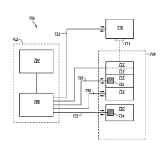
Referring to Fig. 1, a block diagram of an illustrative system 700 that
includes control element 702, a thermocycling element 708, and an optical
system 710
according to exemplary aspects of the disclosure is shown.
[0084] In at
least one embodiment, the system may include at least one sample
vessel 714. In certain embodiments, the sample vessel may include one or more
15 samples 712. An illustrative sample 712 may include a PCR mixture
configured to
permit and/or effect amplification of a template nucleic acid. Certain
illustrative
embodiments may also include at least one sample block or chamber 716
configured
to receive the at least one sample vessel 714. The sample vessel 714 may
include one
or more individual sample vessels in individual, strip, plate, or other
format, and,
20 illustratively, may be provided as or received by a sample block or
chamber 716.
[0085] One or
more embodiments may also include sample temperature
controlling devices 718, 720 configured to manipulate and/or regulate the
temperature
of the sample(s). Such a sample temperature controlling device may be
configured to
raise, lower, and/or maintain the temperature of the sample(s). In one
example,
sample controlling device 718 includes a heating system and sample controlling
device 720 includes a cooling system. Illustrative sample temperature
controlling
devices include (but are not limited to) heating and/or cooling blocks,
elements,
exchangers, coils, radiators, refrigerators, filaments, induction heaters,
irradiative
heating (including IR heating), Peltier devices, forced air blowers, handlers,
vents,
distributors, compressors, condensers, water baths, ice baths, flames and/or
other
combustion or combustible forms of heat, hot packs, cold packs, dry ice, dry
ice
baths, liquid nitrogen, microwave- and/or other wave-emitting devices, means
for
cooling, means for heating, means for otherwise manipulating the temperature
of a
SUBSTITUTE SHEET (RULE 26)
CA 02887302 2015-04-09
WO 2014/058919
PCT/US2013/063939
21
sample, and/or any other suitable device configured to raise, lower, and/or
maintain
the temperature of the sample(s). It is understood that in some embodiments a
single
temperature controlling device may operate as both a heating system and a
cooling
system.
[0086] Certain embodiments of the PCR system may also include an optical
system 710 configured to detect an amount of luminescence and/or temperature-
sensitive luminescence emitted by the sample 712 (or a portion or reagent
thereof).
Such an optical system 710 may include an optical member configured to query
the
luminescence of the sample 712. Illustrative optical systems include single-
and
to multi-channel fluorimeters.
[0087] At least one embodiment further includes a CPU 706 that functions
in part
as a sample temperature control or controlling mechanism. In certain
embodiments,
the sample temperature control or controlling mechanism may regulate the
temperature controlling devices 718, 720 via connections 730, 732 to adjust
the
temperature of the sample 712 based on sample luminescence and/or any value
calculated therefrom. For instance, the mechanism may effect a temperature
change
in response to a specific and/or predetermined amount or level of sample
luminescence detected by the optical system 710 and/or an optical member or
element
711 thereof The mechanism may also (or alternatively) regulate temperature
controlling devices 718, 720 to adjust the temperature of the sample 712 based
on
measurable and/or determinable factors other than luminescence, including
sample
temperature, sample pH, and the like, which may be calculated and/or
determined
from (at least) sample luminescence. Such a mechanism may involve utilizing
one or
more sample temperature controlling devices 718, 720 following detection
and/or
quantification of a predetermined amount or level of luminescence (e.g.,
temperature-
sensitive fluorescence) and/or other measurable and/or determinable factor(s).
[0088] In at least one embodiment of the PCR system 700, the CPU 706 may
execute instructions or be programmed or configured to operate, control,
execute, or
otherwise advance at least the sample temperature controlling mechanism based
on
sample luminescence or a value or parameter calculated therefrom, or to run or
otherwise execute software designed to perform the same. Manual, mechanical,
electrical, and/or other methods and/or devices configured to operate and/or
otherwise
affect the sample temperature controlling mechanism are also contemplated
herein.
SUBSTITUTE SHEET (RULE 26)
22
For example, a mechanical or electrical trigger configured to alternate the
positioning
of the sample vessel or sample container 714 between a sample temperature
controlling device 718 configured to raise the temperature of a sample 712 and
a
sample temperature controlling device 720 configured to lower the temperature
of a
sample 712 in response to detection and/or quantification of predetermined
amounts
of sample luminescence by the optical system 710 and/or optical element 711
(or a
parameter calculated therefrom) is contemplated within the scope of this
invention.
The CPU 706 may accept input from or provide output to a user interface or
terminal
704.
100891 Certain illustrative embodiments of a PCR system may further include
at
least one sample temperature measuring device 728 or 734. Such a device may
include a thermometer, thermistor, thermocouple, or other device capable of
measuring a sample temperature. The sample temperature measuring device 728,
734
may be configured to measure the internal sample temperature directly (through
direct
contact with the sample 712) or measure an external temperature for the sample
712
(without directly contacting the sample 712) and may provide data to CPU 706
via
connection 726, 732. Such indirect contact may involve measuring the
temperature of
a sample vessel or container 714, heating and/or cooling sources such as
temperature
controlling devices 718, 720, sample vessel or container receiving member or
receptacle, and/or any other indicator of sample temperature. In some
embodiments,
sample temperature may be inferred from the temperature of associated members
and/or elements such as a heating or cooling block or chamber, such as sample
block
or chamber 716. It is also understood that in some embodiments sample
temperature
controlling devices 718, 720 may receive input directly from one or more
temperature
measuring devices 728 or 734 or optical system 710, and may operate
automatically
based on one or more of the device-determined temperature, the thermocouple-
determined temperature, or the solution temperature, with or without going
through
the CPU 706.
[0090] Additional examples of illustrative features, components,
elements, and or
members of illustrative PCR systems and/or thermal cyclers (thermocyclers) are
known in the art and/or described in U.S. Patent Application Serial No.
13/834,056.
CA 2887302 2019-11-01
23
[0091] It is
noted that a PCR system according to an embodiment of the present
invention may include, incorporate, or otherwise comprise properties,
reagents, steps,
components, members, and/or elements described in other systems, methods,
and/or
mixtures disclosed herein.
IV. METHODS OF MEASURING A TEMPERATURE
[0092] Certain
embodiments of the present invention may include methods of
measuring, calculating, or otherwise determining a temperature, condition, or
physical, or other property of a sample. In at least one embodiment, the
method may
include providing a sample that includes at least one condition-sensitive
reagent that
emits a luminescent signal in response to at least one stimulus. In some
embodiments,
the amount of luminescent signal emitted by a temperature-sensitive reagent
may be
used to measure, calculate, or otherwise determine a luminescence-determined
temperature for the sample. For example, the temperature-sensitive reagent may
include sulforhodamine B, fluorescein, and/or any other fluorescent dye that
emits a
temperature-sensitive fluorescent signal in response to exposure to light
having a
given wavelength.
10093] In at
least one embodiment, a dedicated sample containing the fluorescent
temperature-sensitive dye can be monitored to control cycling during PCR.
Other
embodiments may involve adding the fluorescent dye directly to a PCR mixture
so
that real-time amplification and temperature monitoring for cycling control
may be
conducted simultaneously for each sample or zone individually. This is
attractive as
well-to-well variation and temperature validation can be conducted on a run-by-
run
basis. In the case where the fluorescent-dye for temperature monitoring is
added
directly to the PCR mixture, color compensation may be required due to
spectral
overlap as discussed further in U.S. Patent Serial No. 6,140,054.
[0094j One will
appreciate that reference to a single sample, reagent, or condition
is illustrative only. Certain embodiments may include providing a plurality of
substantially identical or non-identical samples. Such samples may include
sample
replicates, positive and/or negative control samples, and/or independent
sample
variants. Likewise, a sample according to certain illustrative embodiments may
include a plurality of temperature-sensitive reagents. At least one of such
reagents
CA 2887302 2019-11-01
CA 02887302 2015-04-09
WO 2014/058919 PCT/US2013/063939
24
may include and/or represent a control reagent. Furthermore, the sample may
include
a PCR mixture, illustratively.
[0095] In at least one embodiment, the method may further include
stimulating
the temperature-sensitive reagent sufficiently to induce the luminescent
signal. For
example, an embodiment may include stimulating a fluorescent dye with
electromagnetic radiation or light having a given wavelength sufficient to
induce
emission of a fluorescent signal.
[0096] In certain embodiments, the amount of luminescent signal emitted
from
the temperature-sensitive reagent changes as a function of temperature and/or
another
condition or property in a known and/or predictable manner. Certain
embodiments
may also (or alternatively) include the determination of such temperature-
sensitive
changes.
[0097] Certain embodiments may also include measuring the amount of
luminescent signal emitted from the temperature-sensitive reagent. Such
measuring
may include detecting and/or quantifying the emitted luminescent signal such
that a
sample and/or solution temperature may be determined, calculated, and/or
inferred
therefrom. Thus, certain illustrative embodiments may also include determining
the
temperature of the sample as a function of at least the luminescent signal
emitted by
the condition-sensitive reagent.
[0098] In certain illustrative embodiments, the determination of sample
temperature as a function of luminescent signal may include comparing the
measured
amount of luminescent signal emitted from the sample and/or reagent to a
standard for
amounts of luminescent signal emitted by that reagent at various temperatures
to
determine the temperature at which the reagent is known to emit the measured
amount
of luminescent signal. One will appreciate, however, that the determination of
sample
temperature as a function of luminescent signal may include comparing the
measured
amount of luminescent signal emitted from the reagent to a standard for
amounts of
luminescent signal emitted by the reagent as a function of some other physical
or
other property (e.g., pH), that may change as a function of temperature, to
determine
the temperature at which the reagent is known to emit the measured amount of
luminescent signal.
[0099] In at least one embodiment, the temperature of the sample can be
measured directly by observing or otherwise detecting the amount of
luminescent
SUBSTITUTE SHEET (RULE 26)
CA 02887302 2015-04-09
WO 2014/058919
PCT/US2013/063939
signal emitted from a known amount of temperature-sensitive reagent present in
the
sample. In another embodiment, the temperature of the sample may be determined
by
calculating the ratio of two different fluorescent signals, wherein one signal
is
generally temperature sensitive and the second signal is generally temperature
5 insensitive. In one such embodiment, the temperature is calculated by at
least (a)
observing or otherwise detecting the amount of luminescent signal emitted from
the
temperature-sensitive reagent at a first wavelength, wherein the signal from
the
temperature-sensitive reagent is temperature sensitive at the first
wavelength, (b)
observing or otherwise detecting the amount of luminescent signal emitted from
the
10 temperature-sensitive reagent at a second wavelength, wherein the signal
from the
temperature-sensitive reagent is less temperature sensitive at the second
wavelength,
and (c) determining the ratio of luminescent signal at the first and second
wavelengths. In another such embodiment, the temperature is calculated by at
least
(a) observing or otherwise detecting the amount of luminescent signal emitted
from
15 the temperature-sensitive reagent, (b) observing or otherwise detecting
the amount of
luminescent signal emitted from a second reagent that is generally temperature
insensitive, and (c) determining the ratio of luminescent signal from the two
reagents.
Because fluorescent ratios from at least two signals are being compared, as
opposed to
observing or otherwise detecting the absolute fluorescence, it may not be
necessary
20 that the amount of the temperature-sensitive reagent in the sample be
known in certain
embodiments.
[00100] Some embodiments may include measuring the temperature of the sample
by at least one other method. For instance, an embodiment may include
measuring a
device-determined temperature and/or a thermocouple-determined temperature for
the
25 sample. In certain embodiments, the difference or variance between a
reagent
luminescence-determined temperature and a device- or thermocouple-determined
temperature is (or is known to be) less than or equal to about 1 degree
Celsius. For
example, the amount of fluorescence emitted by a temperature-sensitive reagent
such
as sulforhodamine B may be used to calculate or otherwise determine a
luminescence-
determined temperature for a PCR mixture within about 1 degree Celsius of a
thermocouple-measured temperature of the sample. In certain embodiments, the
luminescence-determined temperature accurately and/or substantially reflects
the
SUBSTITUTE SHEET (RULE 26)
CA 02887302 2015-04-09
WO 2014/058919 PCT/US2013/063939
26
(average) internal temperature of the sample. Temperature determinations with
greater or less than about 1 degree Celsius variation are also contemplated
herein.
[00101] In certain illustrative embodiments, a raw, measured luminescence-
determined temperature may be processed prior to being adopted or otherwise
used as
the temperature of the sample. Furthermore, determining the temperature of a
sample
as a function of at least the luminescent signal emitted by the reagent may
also
include averaging, factoring, calculating, and/or otherwise processing the
luminescence-determined temperature with at least one other factor and/or
determined
temperature. For example, determining the temperature of a sample in a manner
.. consistent with the luminescent signal emitted by the reagent may include
determining the average between a raw (or processed) luminescence-determined
solution temperature and a thermocouple-determined temperature or otherwise
incorporating both the luminescence-determined solution temperature and
thermocouple-determined temperature to arrive at a calculated sample
temperature.
[00102] In at least one embodiment of the present invention, a second reagent
that
produces a second luminescent signal may be provided. In certain illustrative
embodiments, the second luminescent signal may indicate the quantity or amount
of
sample, reagent, or mixture (e.g., the signal from a fluorescent or
luminescent signal
normalizing reagent). In some embodiments, the second luminescent signal may
indicate an amount of nucleic acid present in the sample. For example, the
second
reagent may include a quantitative indicator of PCR product formation and/or
other
DNA binding reagent. One will appreciate however that the second reagent may
also
(or alternatively) include a positive or negative control reagent or another
condition-
sensitive reagent.
.. [00103] In certain embodiments, the sample may be provided as a PCR sample
and/or mixture or a post-PCR sample and/or mixture. One will appreciate,
however,
that the sample may be any type of biochemical, industrial, commercial,
scientific, or
other experiment or reaction. Referring briefly to Fig. 1, CPU 706 may output
to the
user interface 704 a graph of the signal from the second reagent plotted
against the
temperature as determined using the temperature-sensitive reagent.
[00104] In at least one embodiment, a method of measuring the temperature of a
sample may further include stimulating the temperature-sensitive reagent to
induce
the luminescent signal, measuring the luminescence emitted from the sample,
SUBSTITUTE SHEET (RULE 26)
27
determining the temperature of the sample as a function of at least the
luminescent
signal emitted by the reagent, measuring the temperature of the sample by at
least one
other method, and/or any other step or portion of any method described herein
at a
plurality of time points and/or temperatures. For example, the method may
include a
step(s) involving measuring the fluorescence emitted from an excited (or
otherwise
stimulated) sample at a plurality of time points during PCR cycling or melting
during
or after PCR. One will appreciate, however, that measuring the luminescence
emitted
from a non-stimulated sample is also contemplated herein.
[00105] In yet other embodiments, the luminescent signal from a temperature-
sensitive reagent is used to adjust a melting curve generated from a
biological or other
substrate. For example, a melting curve of a PCR amplicon may be generated
post-
PCR, and the temperatures displayed for the melting curve may be adjusted
based on
the luminescent signal generated from the temperature-sensitive reagent during
melting of the PCR amplicon.
[00106] Various embodiments of illustrative methods are available to
correlate
fluorescence emission with solution temperature. In one embodiment (termed
single-
dye/single-color), a single dye is excited at a specific wavelength and
changes in
emission intensity are monitored in a single spectral band, as discussed
further in the
following references: (1) J. Sakakibara, K. Hishida, M. Maeda, Vortex
structure and
heat transfer in the stagnation region of an impinging plane jet (simultaneous
measurements of velocity and temperature fields by digital particle image
velocimetry
and laser-induced fluorescence), Int. J. Heat Mass Transfer 40 (1997) 3163-
3176; (2)
F. Lemoine, M. Wolff, M. Lebouche, Simultaneous concentration and velocity
measurements using combined laser-induced fluorescence and laser Doppler
velocimetry: Application to turbulent transport, Exp. Fluids 20 (1996) 319-
327; (3) F.
Lemoine, Y. Antoine, M. Wolff, M. Lebouche, Simultaneous temperature and 2D
velocity measurements in a turbulent heated jet using combined laser-induced
fluorescence and LDA, Exp. Fluids 26 (1999) 315-323; and (4) P. Lavieille, F.
Lemoine, G. Lavergne, F. Virepinte, M. Lebouche, Temperature measurements on
droplets in monodisperse stream using laser-induced fluorescence, Exp. Fluids
29
(2000) 429-437.
CA 2887302 2019-11-01
28
1001071 Other embodiments may employ a ratio where fluorescence on two
(single-dye/two-color) or even three (single-dye/three-color) spectral bands
is
measured, as discussed further in the following references: (1) P. Lavieille,
F.
Lemoine, G. Lavergne, M. Lebouche, Evaporating and combusting droplet
temperature measurements using two-color laser-induced fluorescence, Exp.
Fluids 21
(2001) 45-55; (2) M. Bruchhausen, F. Guillard, F. Lemoine, Instantaneous
measurement of two-dimensional temperature distributions by means of two-color
planar laser induced fluorescence (PLIF), Exp. Fluids 38 (2005) 123-131; (3)
P.
Lavieille, A. Delconte, D. Blondel, M. Lebouche, F. Lemoine, Non-intrusive
temperature measurements using three-color laser-induced fluorescence, Exp.
Fluids
36 (2004) 706-716; and (4) M. Bruchhausen, A. Delconte, Temperature
measurements in polydisperse sprays by means of laser-induced fluorescence
(LIF) on
three spectral bands, Atomization and Sprays 16 (2006) 599-614.
[00108] The some
embodiments, measured fluorescence intensity from a
spectral band that is sensitive to temperature is normalized by the intensity
measured
on a second spectral band that is insensitive to temperature, with the intent
of
improving temperature accuracy. Additional implementations excite a single dye
(typically one that is sensitive to changes in pH) at two different excitation
wavelengths (dual excitation single-dye/single-color). A ratio is then
calculated using
emission intensity measured on the same spectral band, as discussed further in
J. Han,
K. Burgess, Fluorescent Indicators for Intracellular pH, Chem Rev. 110 (2010)
2709-
2728.
[00109] It is
noted that a method of measuring a temperature, condition, or physical
or other property of a sample according to an embodiment of the present
invention
may include, incorporate, or otherwise comprise properties, reagents, steps,
components, members, and/or elements described in other systems, methods,
and/or
mixtures disclosed herein.
V. METHODS OF CALIBRATING A DEVICE
[00110] Certain embodiments of the present invention may also involve a method
of calibrating a sample-heating, thermocycling, or other device or system.
Such a
method may include providing a calibration sample that includes at least one
condition-sensitive reagent that emits a luminescent signal in response to a
stimulus.
CA 2887302 2019-11-01
CA 02887302 2015-04-09
WO 2014/058919 PCT/US2013/063939
29
The calibration sample may be provided as a suspension, solution, and/or other
form
or physical state of sample compatible with the methods herein disclosed and
described.
[00111] Some embodiments may also involve stimulating the condition-sensitive
reagent to induce emission of the condition-dependent or condition-sensitive
luminescent signal therefrom. Appropriate qualitative and quantitative reagent
stimulation may be required in certain embodiments. For example, for an
illustrative
fluorescent reagent such as sulforhodamine B, electromagnetic radiation may be
a
qualitatively appropriate form of stimulation, while an amount of, specific
wavelength
(such as 575 nm) or range of (such as 470-480 nm), and/or length of exposure
to such
electromagnetic radiation may be an appropriate quantitative factor or
parameter for
effective reagent stimulation.
[00112] Certain embodiments may also include measuring the amount of
luminescence (or luminescent signal) emitted from the condition-sensitive
reagent.
Referring again to Fig. 1, such a measuring step may involve an optical system
710,
optical element 711, or other luminescence measuring or detection member,
element,
or device configured to measure emission of luminescent signals.
[00113] In at least one embodiment, a method of calibrating a device may also
include measuring and/or determining a luminescence-determined temperature as
a
function of (at least) the luminescence signal emitted by the condition-
sensitive
reagent.
[00114] Certain illustrative embodiments may also include measuring a device-
determined temperature. In certain embodiments, the device-determined
temperature
for the sample is measured by inference from an external or indirect source.
In certain
illustrative embodiments, the temperature of a sample block or chamber 716 in
which
a sample 712 and/or sample vessel or container 714 is placed may be measured,
optionally adjusted for known or suspected variation or discrepancy, and
adopted as a
device-determined temperature for the sample.
[00115] In certain embodiments, the method may also involve adjusting the
device
and/or the device-determined temperature to reflect (at least) the
luminescence-
determined temperature. Such a step may be termed a calibration step in
certain
illustrative embodiments. In at least one embodiment, adjusting the device
and/or the
device-determined temperature may involve adopting the raw or processed
SUBSTITUTE SHEET (RULE 26)
30
luminescence-determined temperature. One will appreciate, however, that
adjusting
the device and/or the device-determined temperature to reflect the
luminescence-
determined temperature may also (or alternatively) involve averaging and/or
otherwise factoring one or more luminescence-determined temperatures and/or
one or
more temperatures otherwise measured or determined (e.g. internally; through
direct
contact with the sample). Other methods for measuring, determining, factoring,
and/or calculating temperatures from a plurality of values and/or data points
are
known in the art and described in U.S. Patent Application Publication No. 2013-
0157376.
[00116] It is noted that calibrating and/or adjusting the device-determined
temperature according to factors other than and/or in addition to the
luminescence-
determined temperature may still constitute adjusting the device-determined
temperature to reflect or represent the luminescence-determined temperature
and/or in
a manner consistent with the luminescence-determined temperature. Therefore,
all
such (or similar) methods or steps of methods may involve adjusting the device-
determined temperature in a manner consistent with the luminescence-determined
temperature.
[00117] In certain illustrative embodiments, the adjusting step may
include
adjusting the output of temperature controlling device(s) 718, 720 to provide
more
heating and/or cooling. This adjusting step may include a single adjustment
for one
or more of the temperature controlling devices or may include multiple
adjustments
for one or more elements of such a device (such as a thermoelectric Peltier,
illustratively). The adjusting step may involve manually performed steps
and/or
automated steps performed with the aid of a CPU 706, computer, or other
hardware
and/or software.
[00118] It is noted that a method of calibrating a sample-heating or other
device
according to an embodiment of the present invention may include, incorporate,
or
otherwise comprise reagents, steps, components, members, and/or elements
described
in other systems, methods, and/or mixtures disclosed herein.
VI. METHODS OF FLUORESCENCE FEEDBACK
[00119] Certain embodiments of the present invention may also involve a method
of using the luminescent signal to adjust temperature of a sample-heating,
thermocycling, or other device or system prior to or during PCR. Such a method
may
CA 2887302 2019-11-01
CA 02887302 2015-04-09
WO 2014/058919 PCT/US2013/063939
31
include providing a PCR sample that includes at least one condition-sensitive
reagent
that emits a luminescent signal in response to a stimulus. The feedback sample
may
be provided as a suspension, aqueous solution, or other form or physical state
of
sample compatible with the methods herein disclosed and described.
.. [00120] In one embodiment, rather than thermocycling between device-
determined
temperatures or thermocouple-determined temperatures, thermocycling occurs
between luminescence-determined temperatures, and the luminescence signal is
used
to determine the start or end of programmed "hold" periods and/or trigger
ramping to
the next temperature in thermal cycling.
[00121] One illustrative example was constructed through modification of a
LightCycler 24. The LightCycler 24 is a rapid-cycle real-time PCR instrument
that
uses air for temperature control. The LightCycler 24 was modified to allow for
two
methods of cycling control to be directly compared: (1) control by kinetically
matched external thermocouple; and (2) control by fluorescence. Temperature
control
.. by fluorescence was achieved through a single-dye/single-color approach,
although a
single-dye/two-color approach and additional multi-dye and multichannel
methods are
contemplated within this disclosure. Temperature control by fluorescence
provided
equivalent or improved thermocycling control. While the LightCycler 24 was
used as
the initial instrument, it is expected that the methods will be applicable to
any real-
.. time thermocycler.
[00122] In the
illustrative example, fluorescent-based temperatures (or the
fluorescent value(s) from which they were calculated) were converted to
voltage
equivalents to replace standard thermocouple voltage input(s) for thermal
cycling
control. In other
words, sample fluorescence (or fluorescence-determined
.. temperature) was used instead of measured temperatures (i.e. thermocouple
and/or
instrument-determined temperatures) to control thermal cycling.
Illustratively,
sample temperature was determined as a function of fluorescent signal.
Essentially,
the voltage equivalents of measured amounts of fluorescent signal emitted from
a
sample were compared to and/or coordinated with a calibration standard for
amounts
.. of fluorescent signal emitted by that reagent at various temperatures.
Thus, the
calibration standard was used to provide a temperature point at which the
reagent was
known to emit a measured amount of fluorescent signal, such that fluorescent
signal
was correlated with sample temperature. One will appreciate that fluorescent-
based
SUBSTITUTE SHEET (RULE 26)
CA 02887302 2015-04-09
WO 2014/058919
PCT/US2013/063939
32
temperatures could also be used directly or converted to voltage equivalents
to replace
standard instrument temperature-related voltage inputs for thermal cycling
control.
[00123] A
schematic diagram of an illustrative example of fluorescence-based
temperature cycling control is shown in Fig. 2. In certain embodiments, the
fluorescent dye 808, illustratively in a sample container (not shown) in block
818 is
excited by light 810 from an illustrative light-emitting diode (LED) 812 or
other
suitable light source. The light 810 excites emission 814, 814a from the
fluorescent
dye included in samples 808 and 808a, respectively. The intensity of the
emission
814, 814a from the fluorescent dye included in samples 808 and 808a,
respectively,
may be detected by an illustrative photomultiplier (tube) (PMT) 816 or other
fluorescence detection apparatus. In some embodiments, excitation 810 and/or
emission 814 pathways may be filtered through block 818.
[00124] Certain
illustrative embodiments of this process involve an initial
calibration to determine reference temperatures 826 for a reference sample 808
that
may be used to calculate fluorescence-based temperatures 828 for a sample 808a
during subsequent cycling. During initial calibration, reference fluorescence
intensity
values 820 and temperature measurement values 824 (via a thermocouple 822)
from
sample 808 may be recorded and used to calculate fluorescence-based or
fluorescence-determined reference temperatures 826, which may be correlated
with
the fluorescence intensities 820 from which they were calculated and/or
derived (e.g.
to create a standard). These calculated reference temperatures or values 826
(or the
standard which correlates fluorescence intensities with their respective
temperatures)
may then be used to control temperature of block 818, illustratively by
triggering the
next thermal cycling phase during PCR of sample 808a. For example, during PCR
an
illustrative fluorescent emission 814a (from sample 808a), which corresponds
to a
known reference temperature 826, may be detected by the PMT 816 and used to:
(1)
determine the (fluorescence-based) temperature 828 of the sample; (2)
determine a
cycle point in the temperature cycle; and/or (3) trigger or initiate a change
to another
phase in a thermal cycling profile.
[00125] One will
appreciate, however, that thermocouple-determined
temperatures or temperature readings may also be used during cycling. The
modified
LightCycler 24 may accept voltage inputs from both standard thermocouples and
fluorescence-based equivalents. Thus, in
certain embodiments, a standard
SUBSTITUTE SHEET (RULE 26)
CA 02887302 2015-04-09
WO 2014/058919
PCT/US2013/063939
33
thermocouple may be placed in an adjacent sample, well, or capillary for
comparison
and for use in determining reference temperature values.
[00126] Illustratively, a calibration curve may be acquired
continuously by a
slow ramping (for example, at a target rate 0.05 C/s ¨ 0. PC/s) transition or
a step and
hold transition. In one embodiment, one point is recorded at the end of each
holding
period (is ¨ 10s) at a set temperature increment (0.1 C - 1 C). The
illustrative
instrument may be able to hold at a set temperature for up tolOs, to determine
reference values and for extension during cycling.
[00127] The illustrative instrument may also be able to cycle between
three set
temperatures (denaturation, annealing, and extension) for a number of cycles
sufficient to complete the reaction. In some embodiments, PCR reactions of 35
cycles
can be completed in about 3 minutes and 45 seconds, illustratively when using
2
trigger temperatures (denaturation and annealing/extension). The
optical
configuration may allow for the excitation and detection of at least two dyes
(one for
real-time monitoring, one for temperature control). The dye used for
temperature
control may be monitored at one, two, three or more wavelengths to provide
ratios
that improve the signal to noise ratio. In some illustrative embodiments, the
excitation may be continuous or intermittent. For example, in one embodiment,
the
excitation light is intermittently flashed between an excitation wavelength
that excites
the real-time monitoring dye, and an excitation wavelength that excites the
temperature control dye. It is understood that the frequency of excitation
between the
two wavelengths should be high enough so that temperature control can be
achieved
without substantial error. For example, if the rate of transition is 10 C/s,
then flashing
the wavelength that excites the temperature control dye at 100 Hz should allow
a
resolution of about 0.1 C.
[00128] To determine whether the methods described herein result in
improved
temperature control, thermal cycling controlled by the following methods may
be
compared:
[00129] Method A) An external thermocouple
[00130] Method B) Fluorescence - single-dye/single-color approach, where a
single dye is excited at a specific wavelength and changes in emission
intensity are
monitored in a single spectral band.
SUBSTITUTE SHEET (RULE 26)
CA 02887302 2015-04-09
WO 2014/058919
PCT/US2013/063939
34
[00131] Method C) Fluorescence - single-dye/two-color approach, where
the
measured fluorescence intensity from a spectral band that is sensitive to
temperature
is normalized by the intensity measured on a second spectral band that is
insensitive
to temperature.
[00132] It is noted that Methods B and C are illustrative only, and that
other
methods of controlling temperature using fluorescence are contemplated,
including
implementations that excite a single dye (typically one that is sensitive to
changes in
pH) at two different excitation wavelengths (dual excitation single-dye/single-
color).
A ratio may then be calculated using emission intensity measured on the same
spectral band. Other approaches achieve normalization with two dyes (two-
dye/two-
color) configurations, wherein a temperature-sensitive dye is normalized using
the
signal from another dye with similar or opposing temperature sensitivity.
[00133] The Cq value may serve as a good indicator of temperature
accuracy,
particularly at annealing. If the annealing temperature is too low, primers
may non-
specifically hybridize. In such a case, the Cq value may be significantly
lower, but
the wrong product may have been amplified. Alternatively, if the annealing
temperature is too high, primers may have difficulty in hybridizing to the
DNA,
lowering the efficiency, and pushing the Cq to later cycles. If temperature
accuracy is
enhanced, Method C may produce the lowest average Cq value when compared to
Method A or B, with Method C producing the correct product.
[00134] Temperature accuracy may also be ascertained through the
reproducibility of Cq values from real-time curves. The standard deviation of
the Cq
values may be calculated for cycling controlled through Methods A, B, and C.
Factoring in potential pipetting uncertainties, the Cq values may have a
standard
deviation of no more than 0.5 cycles.
[00135] Samples amplified from Methods A, B, and C in Step 3 may be
melted
to determine product purity. Predicted melting curve may be used for
comparison.
These comparisons (of melting temperature, shape and number of peaks) may
verify
that lower Cq values are resultant from enhanced solution temperature accuracy
during cycling and not as a result of non-specific amplification.
[00136] In certain embodiments, the fluorescence-based solution
temperature
from Method C may align more closely with recordings made by the physically
inserted thermocouple when compared to Methods A or B. As a quantitative
SUBSTITUTE SHEET (RULE 26)
CA 02887302 2015-04-09
WO 2014/058919
PCT/US2013/063939
measure, the mean solution-thermocouple temperature difference (and standard
deviation) may be compared during the three transition periods (denaturation
to
annealing, annealing to extension, and extension to denaturation) for cycles
1, 10, 20,
and 30, illustratively. Values may be calculated for each method, and Method C
may
5 demonstrate the lowest mean value and standard deviation for all temperature
transitions across all cycles.
VII. USE OF RATIOS FOR ADDED CONTROL
[00137] Many dye and excitation/detection configurations exist for
determining
solution temperatures based upon changes in fluorescence. These approaches
fall into
io distinct categories. Each may be used within the scope of the present
invention, and
illustrative examples of each are briefly discussed below.
S ingle-dye/S ingle-b and Method
[00138] The single-dye/single-band method may employ the excitation of
a
temperature-sensitive dye at a set wavelength. Changes in the emission
intensity,
15 detected on a specific wavelength band across temperature, may be
monitored. As
temperature increases, fluorescence intensity of the dye may decrease, due to
more
significant collisional quenching occurring during the excited-state lifetime,
which
may reduce quantum yield. To obtain fluorescence-temperature correlations, a
calibration curve may be generated at equilibration temperatures (T) and
20 corresponding intensities (I). These data, along with a reference
temperature
(Treference) and intensity ('reference) measurement, allows for a calibration
constant, C,
to be determined. The value of C is equal to the slope of the linear line
formed by
1 1 I(T)
plotting ( T ) versus ln (I(Tr
I(Treference)
re nc e)) .
Treference
[00139] When the calibration constant is combined with reference
25 measurements and real-time fluorescence readings, the average solution
temperature
may be calculated using the following equation:
1(T) 1 1
ln( ) [Equation 1]
I(Treference) ) ¨ c ( T Treference
[00140] It is understood that the single-dye/single-band approach may
be
limited in accuracy because measurements are based on absolute intensity. In
30 instances where variables such as excitation intensity, dye
concentration, and sample
volume cannot be kept sufficiently constant, their influence can substantially
decrease
SUBSTITUTE SHEET (RULE 26)
CA 02887302 2015-04-09
WO 2014/058919 PCT/US2013/063939
36
the accuracy of the solution temperature measurement. Particularly in the case
of
laser-induced fluorescence, fluctuations in incident laser intensity can
produce
inaccurate temperature measurements. This has led to alternative approaches
that
employ ratios. The use of ratios allows for self-normalization and may largely
account
for variations in excitation intensity and changes in sample volume and dye
concentration that may occur.
Single-dye/Two-band Method
[00141] The use of a ratio allows for experimental variation to be
mitigated,
thus increasing the accuracy of fluorescence-based solution temperature
measurements. The single-dye/two-band method may employ two distinct spectral
bands of a single fluorescent dye: one that may be sensitive to temperature
and
another that may be generally temperature insensitive. Self-normalization is
achieved
by taking a ratio of the intensities recorded on the two spectral bands. In
this case,
two calibration constants are calculated: one for each spectral band. The
individual
constants may be subtracted to determine the final calibration constant, CF.
As with
the single-band approach, a reference temperature and intensity measurement
may be
employed, as shown in Equation 2.
R(T) 1 1
[00142] ln( ) [Equation 2]
R(Treference) ) ¨ CF ( T Treference
Wherein
R(T) is the ratio at the measured temperature
R(Tref.c.) is the ratio at the reference temperature
TreferCTICC is the reference temperature and
CF is the final calibration constant.
Single-dye/Three-band Method
[00143] For improved temperature accuracy at longer optical pathlengths
and/or high dye concentrations (where fluorescence re-absorption may be
significant)
a single-dye/three-color method has been developed, and may be employed,
wherein
multiple ratio calculations are made using data from three spectral bands.
Ratiometric pH Indicators
[00144] Fluorescent pH-sensitive dyes may be used as an extension of the
single-dye/two-band or other method. Through at least either single-
excitation/dual-
emission or dual-excitation/single-emission configurations, these dyes may be
similarly self-normalizing.
SUBSTITUTE SHEET (RULE 26)
CA 02887302 2015-04-09
WO 2014/058919
PCT/US2013/063939
37
[00145] PCR
mixtures may include a buffer to aid in sensitivity (through
optimizing polymerase activity) and specificity (by enhancing primer/DNA
hybridization). An illustrative commonly used buffer is Tris, as it is well
matched to
the optimal pH range for Taq polymerase (7 ¨ 8 pH units at 80 C). The pH of
Tris
buffer solutions are also temperature dependent, thus allowing for the optimal
pH
range for PCR to be maintained while changes in temperature are correlated
with
observed changes in pH.
[00146]
Fluorescent pH-sensitive dyes achieve different molecular forms (due
to progressive dissociation) as the solution pH changes across temperature.
Different
to forms of the molecule may exhibit different emission spectra, most
notably alterations
in intensity at specific wavelengths. The ratio of the intensities at
prominent features
(such as spectral peaks at set wavelengths) allows for self-normalization of
the dye.
Changes in temperature can then be monitored by fluorescence when a
fluorescent
pH-sensitive dye is added to the reaction mixture.
VI. EXAMPLES
[00147] In addition to the illustrative embodiments discussed above, the
following
illustrative examples and results may be useful in enabling a person of
ordinary skill
in the art to make and/or use the invention. Importantly, the following
examples and
results are illustrative only and are not intended to limit the scope of the
present
invention. Furthermore, different results may be observed when practicing
certain
embodiments of the present invention. As such, the following results are not
intended
to limit the scope of the present invention.
Real-Time PCR Instruments
[00148] Several real-time PCR instruments were used, based on availability and
sample format. Four of these were carousel-based instruments: the LightCycler0
24
(Idaho Technology), LightCycler0 1.5 (Roche Applied Science), LightCycler0 2.0
(Roche Applied Science), and the Rotor-Gene Q (Qiagen). In addition, five
plate-
based instruments were used for this study: the LightCycler0 480 (Roche
Applied
Science), ECOTM (Illumina), iCycler (Bio-Rad), CFX96TM (Bio-Rad), and the
StepOnePlusTM (Life Technologies).
[00149] Different
instruments vary widely in their ability to collect fluorescence
during temperature cycling. Instruments naturally split into one of four
classes. Class
I instruments are most flexible and can continuously acquire fluorescence
during
SUBSTITUTE SHEET (RULE 26)
CA 02887302 2015-04-09
WO 2014/058919
PCT/US2013/063939
38
heating, cooling and temperature holds. Class II-IV instruments are more
limited.
During temperature cycling, Class II and III instruments only allow
fluorescence
collection once each temperature hold, while Class IV instruments can only
acquire
once each cycle. During temperature transitions, Class II instruments can
collect
.. heating or cooling curves, while Class III and IV are limited to only
melting curves.
[00150] Four Class I instruments, three Class II instruments, and one each of
Class
III and Class IV were studied. All instruments used LED excitation except the
iCycler (tungsten halogen lamp) and the LightCycler 480 (xenon lamp). Typical
excitation ranges were 470-480 nm, with the exception of the Bio-Rad
instruments
that were excited around 575 nm for more efficient excitation of
sulforhodamine B.
Detection formats included CCD cameras (iCycler, LightCycler0 480 and the
EcoTm), PMTs (Rotor-Gene Q) and photodiodes (all others). Detection ranges
were
typically within 600-650 nm. However, the only melting channel on the EcoTM
(505-
545 nm) was incompatible with sulforhodamine B, so fluorescein was used as the
temperature sensitive dye. Five or 10 L samples were used on most
instruments,
although a large volume range (5-70 L) was studied on the LightCycler 480.
An
oil overlay of 2 ?IL was used with LightCycler capillaries, and 0-20 L/well
on plate
based instruments. PCR cycle times were much faster on the carousel-based
LightCyclers (18-27 s/cycle) than on other instruments (66-120 s/cycle). The
most
relevant characteristics and experimental conditions for the real-time
instruments are
listed in Table 1 (below).
SUBSTITUTE SHEET (RULE 26)
Oil
Emission Solution Calibration Calibration
Instru-
o,
en Excitation (nm) Overlay
During During
o, Instrument Vendor Curve Rate Data
Density ment
(nm) Volume
"1
o Source
Volume Cycling Ramping
o Detector (ILL)
( C/s) (points/ C) Class
en (IL)
,--i
Idaho
" LightCycl- 470 650-690
er 24 Technol- 10 2 0.1 122
Continuous I
LED Photodiode
E.
c.) ogy
a
LightCycl- 470 645
Roche 10 2 0.1 50
Continuous I
er 1.5 LED Photodiode
LightCycl- 470 640
a
Roche 10 2 0.1 44
Continuous I cq
er 2.0 LED Photodiode
Lu
_1
640
=
LightCycl- 483
a re
Roche CCD 5-70 0-20 0.05 11
Continuous I
2 er 480 Xenon Camera
1¨
m
2:
m
,
H`n Rotor- 470 605-615
Once each Heating or i
Qiagen 10 15 0.031 2.0
II (/)
õ cf, Gene Q LED PMT
hold Cooling b
1 L. I
. en
,.,
560-590 605-635
i¨
=
i
Once each Heating or
8 iCycler Bio-Rad Tungsten CCD 10 15 0.025
1.8 II 1¨
0
hold Cooling"
P
Halogen Camera
(/)
1-
m
575 610-650
Once each Heating or =
CFX96 Bio-Rad 10 15 0.018 2.5
LED Photodiode
hold Cooling" H u)
StepOne- Applied 470 580
Once each c
Biosyst- 10 15 0.022 2.5 Heating
III
Plus LED Photodiode hold
ems
505-545
o, 452-486
Once each b
,-= Eco illumina CCD 5 5 0.091 10
Heating IV
o, LED cycle In Camera
o .0
..
¨, a.) aThe fastest possible continuous acquisition rate is 0.57 Cls
(programmed as 1 acquisition/ C for a single color).
o
l'l 4:1
0 CI bFluorescence is acquired once each temperature increment or step.
E.
cFluorescence acquisition can be either continuous or "step and hold".
CA 02887302 2015-04-09
WO 2014/058919 PCT/US2013/063939
Reagents
[00151] Fluorescence was measured in an illustrative "mock" PCR solution
without polymerase and containing (Sigma-Aldrich), 50 mM Tris (pH 8.3), 2 mM
5 MgCl2, 0.2 mM each deoxynucleotide triphosphate (Roche), 500 lug/m1
bovine serum
albumin (Sigma), and 0.08% (v/v) glycerol. For the instruments other than the
EcoTM,
0.6 mM sulforhodamine B was used. For the EcoTM instrument, 4 [iM fluorescein
(Matheson, Coleman and Bell) was used in place of sulforhodamine B for optical
compatibility of the dedicated melting channel. One will appreciate however,
that
10 control and/or "mock" PCR solutions, as well as PCR mixtures and/or
other sample
mixtures, according to the present invention may vary in certain reagent
composition
and concentration as disclosed herein and as otherwise known in the art.
Polyrnerase Chain Reaction
[00152] A 74 bp product of the 3'-untranslated region of the F2 gene
bracketing
15 rs1799963 was used in certain examples, illustratively, to test for
sulforhodamine B
inhibition of PCR. The reaction included the above mock PCR solution with the
addition of 0.4 U/10 jii of KlenTaq polymerase (AB Peptides), 50 ng/10 41 of
human
genomic DNA, 0.5 WVI of primers GGTTCCCAATAAAAGTGACTCTAG (Seq. ID
No. 1) and CTGAGCCCAGAGAGCTGC (Seq. ID No. 2) and 3 mM MgCl2. PCR
20 .. was performed in glass capillaries in 10 ILLL volumes in a LightScanner0-
32 (LS-32)
thermal cycler (BioFire Diagnostics). An initial denaturation at 95 C for 30 s
was
followed by 35 cycles of 95 C for 0 s, 60 C for 0 s, and 74 C for 2 s.
[00153] In other examples, three forensic single-nucleotide polymorphisms
(SNPs)
¨ rs876724, rs917118, and rs763869 ¨ were amplified using the following
primers:
25 rs876724 CCACTGCACTGAAGTATAAGT (Seq. ID No. 3) and
TTAGCAGAGTGTGACAAAAAA (Seq. ID No. 4), rs917118
AAGATGGAGTCAACATTTTACAAG (Seq. ID No. 5) and
GATGACTGAGGTCAACGAG (Seq. ID No. 6) and rs763869
AGGATGTTTGTTTATATTATTTCTAACTCA (Seq. ID No. 7) and
30 CTACTCCCTCATAATGTAATGC (Seq. ID No. 8). Each 10 !IL reaction contained
naM Tris-HC1 pH 8.3, 2.0 mM Mg2+, 200 [iM each dNTP, 1X LCGreen Plus
(BioFire Diagnostics), 0.4 U KlenTaql (Ab Peptides) with 64 ng anti-Taq
antibody
SUBSTITUTE SHEET (RULE 26)
CA 02887302 2015-04-09
WO 2014/058919
PCT/US2013/063939
41
(eEnzyme), 0.5 lig BSA, 0.5 iLiM primers and 50 ng genomic DNA. After an
initial
denaturation at 95 C for 1 min, amplification conditions on the modified
LightCycler
were 85 C for 10 s and 60 C for 10 s for 35 cycles. To demonstrate the utility
of
fluorescence-based temperature control for use with more rapid protocols, the
rs917118 target was amplified using "0"s hold times. The use of "0"s hold
times was
aided by increasing the concentration of primers by 5-fold (final
concentration 2.5
laM). After an initial denaturation at 95 C for 1 min, 35 cycles of 83 C for
"0"s and
61 C for "0"s were performed. In all illustrative examples, thermal cycling
was
controlled by fluorescence-based temperature measurements.
Fluorescent dye temperature-sensitivity
[00154] The temperature-sensitivity of 22 fluorescent dyes was examined on a
custom multicolor fluorimeter with xenon excitation, spectral dispersion (405-
590
nm) on a grating and focused onto a fiber optic (delta RAM, Photon Technology
International). The fiber optic illuminated the end of a glass capillary
(LightCycler,
Roche Applied Science) placed within a heating unit (HR-1, BioFire
Diagnostics).
Fluorescent emission was collected by another fiber optic at a right angle to
the
capillary, delivering light onto a CCD spectrometer (DV420-0E, Andor
Technology)
collecting, 1024 bins between 400 ¨ 850 nm. A J-type micro-thermocouple
(5SRTC,
Omega) was inserted into the sample capillary for physical temperature
measurements.
[00155] Fluorescent dyes were tested in the "mock" (no polymerase) PCR
solution
described above. Final concentrations for each dye are listed in Table 2,
along with
excitation wavelengths. Twenty-five 1AL sample volumes were heated from 45 C
to
95 C (in discrete 10 C increments, with 100 points averaged per step). The
ramp rate
between steps during heating was approximately 0.4 C/s. The entire emission
spectrum was then recorded and the fold-change in fluorescence (45-95 C)
calculated.
SUBSTITUTE SHEET (RULE 26)
CA 02887302 2015-04-09
WO 2014/058919 PCT/US2013/063939
42
Table 2
Dye Source Excitation
Concentra Average Fold-
Wavelength Emissio Change
(nm)3 tion ( M) n Peak
in
Wavelen Fluoresce
gth nce (45-
5 6
(nm) 95 C)
2',7'- Sigma- 455/470/490 80 531 1.56
Dichlorofluorescein Aldrich
5(6) Sigma- 4701490 4 520 1.38
Carboxyfluorescein Aldrich
5(6) Marker 45514701490 4000 542 1.17
Carboxynapthofluo Gene
rescein Technolog
ies, Inc.
Coumarin 6 Sigma- 455/470/490 800 521 2
Aldrich
Eosin B Sigma- 530 40 575 12.48
Aldrich
Ethyl Eosin Sigma- 455/470 800 557 2
Aldrich
490/530 80
Fluorescein Sigma- 45514701490 800 516 1.26
Aldrich
Fluorescein Sodium Matheson, 455/470/490 40 522 1.5
Salt Coleman,
and Bell
HPTS Invitrogen 40514551470 80 517 1.85
/490
Merocyanine 540 Sigma- 47014901530 40 581 7
Aldrich
Oregon Green 488 Invitrogen 455/470/490 40 522 1.15
Resorufin Sigma- 455 800 591 1.25
Aldrich
SUBSTITUTE SHEET (RULE 26)
CA 02887302 2015-04-09
WO 2014/058919 PCT/US2013/063939
43
470/490/530 .. 80
Rhodamine 101 Sigma- 455/470/490 4 599 1.21
Aldrich
530 40
Rhodamine 110 Invitrogen 455/470/490 40 525
1.32
Rhodamine 6G Sigma- 470/490/530 40 560 1.26
Aldrich
Rhodamine B Sigma- 470/490/530 4 575 3.26
Aldrich
Rose Bengal Sigma- 490/530 20 574 2.2
Aldrich
Snarf-1 Invitrogen 455/470/490 160 644
1.28
/530
Sulforhodamine Sigma- 470/490/530 40 607 /.53
101 Aldrich
Sulforhodamine B Sigma- 470 4,000 582 2.85
(monosodium salt) Aldrich
Dye content is 75%. 2Dye content is 95%.
3Fluorescent trends as temperature is increased at a specific wavelength are
denoted in
bold (fluorescence increases), italicized (fluorescence decreases) or regular
text
(variable). The wavelength where the greatest changes in fluorescence were
present is
underlined.
4 Listed concentrations do not account for percentage dye content. Where dye
content
is specified, these concentrations will be less.
5
Emission peak wavelengths were averaged for all tested excitation
wavelength/concentration combinations for each dye.
6The maximum fold-change in fluorescence from 45 to 95 C (at the underlined
excitation wavelength). Increased (bold) or decreased (italicized)
fluorescence
between the measurements at 45 C and 95 C is indicated.
[00156] The spectral profiles of the 22 dyes were recorded at multiple
excitation wavelengths and temperatures (data not shown). For each dye, the
fold
change in fluorescence during heating from 45 ¨ 95 C is shown in Table 2,
along with
general trends in fluorescence (i.e. increased (bold) or decreased
(italicized) intensity
levels) that occurred during heating. The average fold-change in fluorescence
for all
dyes was 0.74 ( 0.45). Eosin B, excited at 530 nm, exhibited the greatest fold-
change
SUBSTITUTE SHEET (RULE 26)
CA 02887302 2015-04-09
WO 2014/058919 PCT/US2013/063939
44
in fluorescence (fluorescence decreased by a factor of 12.48), although
differences in
fluorescence levels at 85 C and 95 C were limited for that dye. An
illustrative
example for sulforhodamine B (monosodium salt), excited at 530 nm, is shown in
Fig.
3.
1001571 Fig. 3 illustrates a temperature-sensitivity profile for (2,400 04)
sulforhodaminc B (monosodium salt) in 25 ',it sample volumes held at
temperatures
ranging from 45 C (x-line) to 95 C (triangle line) in 10 C intervals.
Specifically,
sample(s) were held at 45 C (x-line), 55 C (solid line), 65 C (dash-dotted
line), 75 C
(dashed line), 85 C (dotted line), and 95 C (triangle line). Regions of high
temperature-sensitivity (near 580 nm) are clearly discernible from regions of
low
temperature-sensitivity (near 525 nm).
1001581 Dyes exhibiting at least a 2-fold change in fluorescence were
selected
for more in-depth examination. Dyes that exhibited variable trends in
fluorescence
with increasing temperature or minimal differences (<5%) in fluorescence
intensity at
.. high temperatures (85 C to 95 C), were excluded from further consideration,
although it is understood that such dyes may be useful in certain embodiments.
Ethyl
eosin, merocyanine 540, rhodamine B, snarf-1, sulforhodamine B (acid form),
and
sulforhodaminc B (monosodium salt) were studied further.
1001591 To determine temperature sensitive and temperature insensitive regions
of
.. the emission spectrum, spectral data were displayed as a ratio (emission
intensity at
45 C / emission intensity at 95 C) on a log scale across wavelength (see e.g.
Fig. 4).
For instance, Fig. 4 illustrates temperature sensitivity of sulforhodamine B
(monosodium salt) between 45 C and 95 C, excited at 490 nm, with spectral data
displayed as a ratio (emission intensity (e.i.) at 45 C / emission intensity
(e.i.) at
95 C) on a log scale across wavelength ranging from 400nm to 700nm,
illustratively.
It is understood that regions of low temperature-sensitivity have a ratio
close to zero
while regions of high temperature sensitivity have values furthest away from
zero.
Bands for each dye having the greatest temperature-sensitivity and
insensitivity,
respectively, were identified and are listed in Table 3.
SUBSTITUTE SHEET (RULE 26)
CA 02887302 2015-04-09
WO 2014/058919 PCT/US2013/063939
Table 3
Dye Concentrati Excitatio Degradatio Temperature Temperature
on (tM) n n -Sensitive -
Insensitive
Waveleng (Fold- Band (nm) Band
(nm)
th (nm) change)'
Ethyl Eosin 800 455 0.76 535-600 490-515
Ethyl Eosin 800 470 0.83 535-590 490-515
Merocyanine
40 470 0.41 580-650 500-525
540
Merocyanine
40 490 0.54 575-625 525-550
540
Merocyanine
40 530 0.53 575-700 530-550
540
Rhodamine B 4 470 0.99 570-610 525-550
Rhodamine B 4 490 0.98 570-610 525-550
Rhodamine B 4 530 0.98 560-590 514-540
Snarf-1 160 455 0.96 630-680 500-525 or
580-600
Snarf-1 160 470 0.97 630-680 500-525 or
560-600
Snarf-1 160 490 0.89 625-675 525-575
Sulforhodamine
B, 8 455 0.85 575-610 500-550
Acid form
Sulforhodamine
B, 8 470 0.93 575-620 500-550
Acid form
Sulforhodamine
B, 8 490 0.95 575-620 525-550
Acid form
Sulforhodamine 8 530 0.86 575-620 525-550
B, Acid form
Sulforhodamine
B
4,000 470 1.0 575-610 516-545
(monosodium
salt)
Sulforhodamine
B
2,400 490 0.98 575-610 516-545
(monosodium
salt)
Sulforhodamine 2,400 530 0.97 575-610 516-545
B (monosodium
salt)
1
Samples were heated from 45 to 95 C and then cooled to 45 C. Values were
calculated at the emission peak wavelength. The fluorescence at the second
measurement was divided by the fluorescence of the first measurement. Thus, a
value
5 of 1 indicates that no degradation occurred.
SUBSTITUTE SHEET (RULE 26)
46
Fluorescent dye degradation
[00160] The
custom multicolor instrument described above to assess
temperature sensitivity was also used to assess the amount of dye degradation
after a
single heating and cooling cycle. After the sample temperature reached 95 C,
samples were cooled to 45 C (using passive cooling), and an additional
spectral
measurement was made.
[00161] For most
dyes, dye degradation caused a decrease in fluorescence even
after a single heating and cooling cycle (see Table 3). Illustratively,
Sulforhodamine
B, excited at 470 nm, had a value of 1, indicating that no degradation
occurred.
Values for ethyl eosin (excited at 455 nm) and merocyanine 540 (excited at 470
nm)
were 0.76 and 0.41, respectively, showing more degradation in these two dyes.
Fluorescence-based and thermocouple temperature comparisons during
melting
[00162] The six dyes that
exhibited the greatest sensitivity to temperature were
further examined (see Table 4). Dye concentrations and excitation wavelengths
are
detailed in Table 4. Calibration constants for each dye were calculated from
temperature and fluorescence data acquired from 45 C to 95 C in 10 C
increments.
Using a second sample with a 30 1.11, final volume (25 [IL sample with 5 [IL
oil
overlay), an initial holding period at 50 C was used to determine reference
temperature and intensity values. The sample was then heated to 95 C at an
approximate rate of 0.05 C/s.
Fluorescence-based solution temperatures were
calculated for both single-dye/single-color and single-dye/two-color
configurations
using sulforhodamine B (acid form). Solution temperatures using the single-
dye/two-
color method were calculated as described in P. Lavieille, F. Lemoine, G.
Lavergne,
M. Lebouche, Evaporating and combusting droplet temperature measurements using
two-color laser-induced fluorescence, Exp. Fluids 21 (2001) 45-55.
Fluorescence-
based temperatures were compared to thermocouple readings. Because
sulforhodamine B (monosodium salt) exhibited the strongest temperature
sensitivity
as well as the most repeatable fluorescence after heating and cooling, it was
used for
thermal cycling control, as well as testing of all of the instruments except
the EcoTM.
CA 2887302 2019-11-01
CA 02887302 2015-04-09
WO 2014/058919
PCT/US2013/063939
47
Table 4
Temperature
Difference
(Fluorescenc
e-Thermo-
couple)
Dye Concentratio Excitatio Utilize Emissio Calibratio Mean
n ([1,M) n (nm) d n (nm) n Max SD
Bands Constant (
C) ( C)2
Ethyl 2 2+4.
800 470 1 550-625 -1445 9.8 9.
Eosin
Merocyan
40 490 1 575-625 4267 21.3 7.1
3.
-me 540 7
Rhodamin
4 490 1 574-610 2551 1.1 0.1+0.
e B
Snarf-1 160 490 1 640-675 1712 5.4
5
Sulfor-
hodamine
80 490 1 574-610 2570 0.6 0.4+0.
B, Acid
1
Form
Sulfor-
525-550
hodamine 0.1+0.
80 490 2 and 1856 0.3
B, Acid 1
574-610
Form
Sulfor-
hodamine
1.2+0.
B (mono- 4,000 490 1 574-610 2040 2.4
9
sodium
salt)
The absolute temperature difference between fluorescence-based and
thermocouple
temperatures.
2Thermocouple temperatures are subtracted from fluorescence-based
temperatures.
5
Correlating Temperature to Passive Dye Fluorescence
[00163] To correlate fluorescence intensity to temperature, calibration curves
were
generated on each instrument by slowly heating. To limit dye degradation, a 50-
95 C
range was completed in less than 45 minutes. Rates of 0.018 - 0.1 C/s were
used.
The fastest rate (0.1 C/s) was used on the LightCyclert carousel instruments
because
of rapid thellnal equilibrium, single sample acquisition, and high data
densities (44-
122 points/ C). Of similar rate (0.05 and 0.091 C/s), but with decreased data
density
(11 and 10 points/ C), were the LightCycler0 480 and EcoTM, respectively. All
other
SUBSTITUTE SHEET (RULE 26)
CA 02887302 2015-04-09
WO 2014/058919
PCT/US2013/063939
48
instruments fell into a third range of lower rates (0.018-0.031 C/s) required
by slower
data acquisition for 1.8 ¨ 2.5 points/ C.
[00164] Three possible sources of bias; instrument drift, thermal degradation,
and
fluorescence quenching were examined on the LightCycler0 1.5, 2.0, and 480. To
examine instrument drift and thermal degradation of the dye, temperatures were
held
at 50 C, 80 C and 94 C for one hour. Once each minute, the sample was
illuminated
and fluorescence acquired. To test for fluorescence quenching, the temperature
was
held at 80 C and data collected for 1 hour during either continuous or
intermittent
(once per minute) illumination.
Calculation of Solution Temperatures from Fluorescence
[00165] Fluorescence emission has been modeled previously, correlating
fluorescence intensity to either concentration or temperature, depending on
which
variable is kept constant. Dyes that decrease in fluorescence with increasing
temperature result from quenching of the excited state and lowered quantum
yield.
.. Temperature can be related to fluorescence through a calibration constant:
[00166] C = ln (¨I k.1 )
'ref) T Tref
[00167] Fluorescence intensities I are measured at temperatures T (in degrees
Kelvin) and related to a reference fluorescence intensity ('ref) of the
temperature-
sensitive reagent at a reference temperature (Tref). Experimentally, plots of
1
in (¨) against (¨ ¨ ¨) form a straight line with slope C and intercept 0. The
iref T Tref
calibration constant is dependent on the physical characteristics of the
fluorescent
molecule in a particular solvent. Reference temperatures were 50 C for
calibration
constant determination and melt curve analysis and 55 C for PCR cycling
experiments on each instrument. Solution temperatures were determined from
fluorescence using C, the reference temperature, and the reference
fluorescence:
1
[00168] T= in(uiref)
+ (1/Tref)
[00169] Fluorescence was acquired according to the capability of each
instrument:
continuously, at temperature holds, or during transitions (heating and/or
cooling).
Instrument-specific calibration constants were used to convert fluorescence to
solution temperatures for comparison to temperatures displayed by the
instruments.
SUBSTITUTE SHEET (RULE 26)
CA 02887302 2015-04-09
WO 2014/058919 PCT/US2013/063939
49
The calibration constant may be used to provide a quantitative method of
judging the
overall temperature sensitivity of the reagent/dye and/or optics. A higher
value of the
calibration constant may correlate to greater temperature sensitivity and
system
precision. Unless otherwise specified, the fastest transition rates were used
on each
instrument during cycling. One will appreciate, however, that the equations,
variables, and calculations discussed herein are illustrative only, and that a
variety of
reference temperatures, reference fluorescence intensities, and/or other
variables,
equations, and/or calculations may be used in the calculation and/or
determination of
temperature(s) from fluorescence emissions.
Micro-thermocouple measurements
[00170] When physical instrument configuration allowed, sample temperatures
were also monitored with a J-type micro-thermocouple (5SRTC, Omega) with
conditioning, digitization and display (USB-TC01, National Instruments) to
correlate
with solution and instrument temperatures. Other thermocouples are known in
the art
and contemplated herein.
Melting curve analysis
[00171] Melting curves were obtained on the LC480 using both instrument and
solution temperatures. Bacteriophage lambda DNA (New England BioLabs) at 2
lag/m1 and lx LCGreen Plus (BioFire Diagnostics) were included in the mock
PCR
solution. Both DNA melting (LCGreen Plus, excitation at 450 nm, emission at
500
nm) and temperature (sulforhodamine B, excitation at 558 nm, emission at 610
nm)
were monitored simultaneously by fluorescence during heating of the sample
from
60-95 C. Data were collected at either 2 acquisitions/ C (0.14 C/s), or at 40
acquisitions/ C (0.01 C/s).
Sulforhodamine B fluorescence was converted to
temperature and negative derivative melting plots were displayed using either
solution
or instrument temperatures.
Fluorescence-based temperature cycling control
[00172]
Temperature or thermal cycling using fluorescence-determined
temperatures to control cycling was demonstrated using the modified
LightCycler 24
described above. As illustrated in Figs. 5A-5C), the three forensic single-
nucleotide
polymorphisms (SNPs) rs876724 (Fig. 5A), rs917118 (Fig. 5B), and rs763869
(Fig.
5C) were genotyped after amplification utilizing fluorescence-based thermal
cycling
control with 10 s holding times (quantification cycle (Cq) value of 24). After
an initial
SUBSTITUTE SHEET (RULE 26)
50
denaturation at 95 C for 1 min, amplification conditions were 85 C for 10 s
and 60 C
for 10 s for 35 cycles, resulting in a protocol time of 15 minutes and 46
seconds.
Melting analysis using the quantum method of background removal (see U.S.
Patent
Application No. 61/872,173) identified all genotypes, with all 3 genotypes
(wild-type
(dotted line), variant (dashed line) and heterozygotes (solid line)) clearly
distinguishable (see Figs. 5A-5C) for all targets.
1001731 The rs917118 target was also used to demonstrate successful
amplification under "0"s holding times, requiring only 3 minutes and 45 s to
complete
35 cycles (see Figs. 6A-6B). While the Cq increased to 28 (see Fig. 6A),
robust
results were produced that enabled all 3 genotypes to be clearly identified
(see Fig.
6B). Fig. 6B illustrates negative derivative melting curves analyzed using the
quantum method of background removal. Illustratively, all 3 genotypes (wild-
type
(dotted line), variant (dashed line) and heterozygotes (solid line)) are
clearly
distinguishable.
Illustrative Results
[00174] It is noted that the results disclosed herein are illustrative
only, and that
different results may be observed when practicing certain embodiments of the
present
invention. As such, the following results are not intended to limit the scope
of the
present invention.
1001751 In certain illustrative embodiments, in order for absolute
fluorescence to
serve as a temperature monitor, the instrument and reagent/dye should be
substantially
stable over time. Although many real-time PCR instruments use a heated lid
instead
of an oil overlay to limit evaporation and condensation, more consistent
fluorescence
readings may be obtained with oil than without it (see Figs. 7-8). Oil
overlays may
stabilize fluorescence across multiple cycles, particularly during hold times
and at
temperature extremes (see Fig. 7).
[00176] In an illustrative example, a 70 1.., reaction volume was
compared to a 50
1i1_, volume with a 20 pi, oil overlay. Data illustrated in Fig. 7 include
initial heating
and cooling curves at 0.05 C/s followed by three cycles between 55 C to 95 C
at
ramp rates of 0.57, 0.11, and 0.29 C/s. The solid and dashed lines denote the
absence
and presence of an oil overlay, respectively. Dotted lines are for vertical
alignment
only. Without oil, fluorescence decreased about 8% per hour and short term
CA 2887302 2019-11-01
CA 02887302 2015-04-09
WO 2014/058919
PCT/US2013/063939
51
anomalies were also present, effects that could affect solution temperature
measurement by fluorescence.
[00177] Fluorescence artifacts may also be noted during temperature ramps
typically used for melting curve analysis. For
example, Fig. 8 illustrates
(sulforhodamine B) fluorescence during heating on a LightCycler 480
instrument,
with an illustrative ramp rate of 0.05 C/s. Dashed lines denote a 10 ILLL
sample
volume with no oil overlay, dotted lines denote a 81,LL sample volume with a 2
viL oil
overlay, and solid lines denote a 5 vtL sample volume with a 5 !AL oil
overlay. With
too little or no oil overlay, undulating curves were observed that could be
mistaken
for PCR products when displayed on derivative plots. Only those samples with
an
appropriate oil overlay showed a smooth decrease of fluorescence with time and
temperature. Illustratively, oil overlay may improve results on any and/or all
instruments, although changes may be minimal on capillary instruments.
However, it
is understood that oil overlay is illustrative only, and other techniques may
be used to
stabilize fluorescence.
[00178] In
addition to evaporation and/or condensation, other potential artifacts
affecting fluorescence may include instrument drift (sometimes referred to as
"warm
up" of the instrument), thermal degradation of the dye, and fluorescence
quenching
(each of which will now be addressed in turn).
[00179] With some instruments, although some initial instrument drift may be
observed, this may be reduced substantially by "warming" up the instruments
(illustratively, for 20 minutes). For example, in certain illustrative
examples, when
samples were incubated at 50, 80, or 94 C, minor instrument drifts up to +/-
4%
fluorescence were seen over the first 20 minutes (see Figs. 9A-9C and 10A-10C)
on
three different instruments. After 20 minutes, any change in fluorescence over
time
may become relatively linear.
[00180] Furthermore, in at least one embodiment, no substantial thermal
degradation of a temperature-sensitive reagent may be observed at certain
(lower)
temperatures, while a measurable amount of thermal degradation may be observed
at
other (higher) temperatures. For example, Figs. 9A-9C show that in certain
illustrative embodiments, after instrument equilibration, average
sulforhodamine B
fluorescence may be stable substantially at 50 C (Fig. 9C), decrease about
2.2%/hour
at 80 C (Fig. 9B), and/or decrease about 5.4%/hour at 94 C (Fig. 9A) for one
or more
SUBSTITUTE SHEET (RULE 26)
CA 02887302 2015-04-09
WO 2014/058919
PCT/US2013/063939
52
of a LightCycler 1.5 (filled circle), LightCycler 2.0 (filled square), and
LightCycler0 480 (filled triangle). Illustratively, for the first 20 minutes
(dashed
line) fluorescence is variable as the instrument equilibrates. After 20
minutes,
fluorescence decreases at a temperature-dependent rate. Instruments were
turned off
for 1 hour between each experiment. Across all instruments, the average change
in
fluorescence was less than about 1%/hour at 50 C, 2.2%/hour at 80 C, and
5.4 /0/hour at 94 C. Thus, minor thermal degradation of the dye may occur
under
certain conditions and may be both temperature and time dependent.
[00181] In certain embodiments, this temperature-dependent degradation may be
reduced by rapid cycling to minimize denaturation and overall cycling times
(an
illustrative embodiment in which fluorescence monitoring of temperature is
highly
useful). Another option for fluorescent estimation of temperature is to use
fluorescence ratios so that the requirements for absolute fluorescence
stability are not
so strict.
[00182] In certain illustrative embodiments, no substantial fluorescence
quenching
may be observed (for sulforhodamine B, illustratively). For example, Figs. 10A-
10C
show no appreciable fluorescence quenching of sulforhodamine B over one hour
at
80 C on a LightCyc1er0 1.5 (Fig. 10A), LightCycler 2.0 (Fig. 10B), and
LightCycler 480 (Fig. 10C). In some embodiments, after instrument
equilibration,
fluorescence decreases at about 2.2%/hour, irrespective of whether the samples
are
continuously illuminated (solid lines) or only illuminated once each minute
(filled
triangles). This decrease is consistent with thermal degradation alone (see
Figs. 9A-
9C), with no apparent and/or substantial contribution of fluorescence
quenching.
During typical cycling, sulforhodamine B fluorescence decreased by 0.1% at
extension (runtime 17 minutes) on an illustrative capillary instrument
compared to
0.7% on an illustrative plate instrument (runtime 60 minutes). In one
illustrative
example, due to programming constraints associated with continuously acquiring
data
on the LightCycler 480, multiple segments that cycled between 79 C and 80 C
were
programmed during the 1 hour hold period (see Fig. 10C).
[00183] In an illustrative embodiment, to test potential inhibition of PCR by
sulforhodamine B, quantitative real-time PCR results, gel analysis, and high-
resolution melting curves were examined. Samples containing sulforhodamine B
had
quantification cycles that differed by only about 0.10 when compared to
standard
SUBSTITUTE SHEET (RULE 26)
CA 02887302 2015-04-09
WO 2014/058919
PCT/US2013/063939
53
samples containing no sulforhodamine B. Gel analysis confirmed both the purity
and
size of the product in samples containing sulforhodamine B. Finally, high-
resolution
melting curves comparing standard samples to those with sulforhodamine B were
similar in intensity and peak temperature.
[00184] Calibration curves for nine illustrative instruments are shown in
Fig. 11A.
Instruments included Class I (dashed lines), Class II (solid lines), Class III
(dotted
lines) and Class IV (dash ¨ dot lines). In an illustrative example, slow
heating rates
(0.018-0.1 C/s) were used to equilibrate solution and instrument temperatures.
Eight
of the nine instruments successfully monitored sulforhodamine B fluorescence,
even
though the excitation wavelengths of many real-time instruments were not
optimal for
sulforhodamine B. In
certain illustrative embodiments, the fluorescence of
sulforhodamine B approximately doubles as the temperature decreases from 95 to
50 C (see e.g. Class II (solid lines) instruments). For one illustrative
example (the
EcoTM instrument) fluorescein was used because of available optics (i.e.,
fluorescein
was compatible with the fixed melting emission wavelength available),
resulting in a
calibration curve of different shape than the sulforhodamine B curves. In
certain
embodiments, fluorescein fluorescence increases about 60% as the temperature
decreased from 95 to 50 C. Calibration constants were derived from the slope
of
linear plots of the data (as shown in Fig. 11B for the LightCycler 1.5 (y =
1890X +
0.013, R2 = 0.999)). By way of illustration only, the calibration constant was
1097
for fluorescein and ranged from 1787 to 2831 for sulforhodamine B (see Table 5
below). Without being bound to theory, the different calibration constants for
sulforhodamine B among instruments presumably arise from different optical
characteristics and efficiencies.
SUBSTITUTE SHEET (RULE 26)
54
Table 5. Solution Temperatures during Denaturation, Annealing, and Extension
Segments
Calibration Cycle Solution Temperatures d ( C)
Instrument Hold Timec (s)
Constant a Time b (s) "
Target = Target = Target =
94 C 55 C 74 C
LightCycler 1787 18c 10 88.5 57.9 70.8 ¨
73.1
24
ghtCycler 1.5 1890 27c 10 95.1 56.2 76.5 ¨
76.2
LightCycler 1953 25` 10 92.3 56.1 73.7 ¨
75.4
2.0
LightCycler 2042 79 1, 2, 5,
10,20, 85.4 ¨ 92.4 59.5 ¨ 54.8 68.2 ¨ 73.5
480' 50
Rotor-Gene Q 2831 85 1, 2, 5,
10, 20, 93.3 ¨91.3 59.2 ¨ 57.4 77.7 ¨ 77.1
iCycler 2661 120 5, 7,
10, 15, 20, 87.4 ¨ 93.3 58.3 ¨53.3 67.5 ¨66.8
CFX96 2122 80 1, 2, 5,
10,20, 94.8 ¨ 95.5 57.0 ¨ 56.3 75.3 ¨ 76.7
StepOnePlus 1779 78 10, 15,
20, 30, 94.9 ¨97.7 56.4 ¨ 54.7 73.8 ¨ 75.1
Eco 1097 66 1, 2, 5,
10, 20, 85.6 ¨ 95.9 58.1 ¨56.2 73.6 ¨ 72.3
aThe calibration constant is a unitless constant defined the slope of the
linear line
formed by plotting (1/T(K) - 1/Tref(K)) against 1n(I/Iref). See Fig. 11B and
U.S.
5 Patent Application No. 61/872,173. All calibration constants are for
sulforhodamine
B except for the Eco instrument (fluorescein).
bTime required to complete a 94 C, 55 C, 74 C cycle using hold times as
recommended by the manufacturer, that is, 10 s at each segment for all
instruments
except the capillary LightCyclers, where 0 s was used for denaturation and
annealing.
10 Cycle
times were obtained by monitoring a single sample on the LightCycler
instruments.
'Hold times at each segment, except for the capillary LightCyclers where 0 s
was used
at denaturation and annealing.
dThe temperature range indicates the minimum and maximum solution temperatures
at
15 the
holding times monitored. Only a single value at the peak is given for the 0 s
holds
at denaturation and annealing on the capillary LightCyclers.
'The sample volume was 10 I covered with 15 I of oil.
CA 2887302 2019-11-01
CA 02887302 2015-04-09
WO 2014/058919
PCT/US2013/063939
[00185] In certain illustrative embodiments, using fluorescence as a
temperature
monitor during PCR may be dependent on data acquisition that may vary greatly
from
device to device, ranging from one data point per cycle to continuous
acquisition.
Similarly, the exportable data may vary from only cycle number and
fluorescence, to
5 comprehensive spreadsheets including time, program, cycle, segment,
fluorescence
and temperature. Software and available optics may also restrict the
excitation and
emission wavelengths that can be used. Even with these limitations, sample
temperatures may be successfully monitored by fluorescence on many different
instruments (including all the instruments disclosed herein), albeit some with
greater
10 difficulty than others.
[00186] Different instruments may vary greatly in their fluorescence
collection
capabilities during temperature cycling. In an illustrative example,
continuous
acquisition throughout temperature cycling and melting was only possible on
Class I
instruments. Furthermore, the greatest data density and cycling speeds were
obtained
15 on the capillary Class I instruments, although only one sample could be
measured at a
time. Individual samples were monitored throughout rapid cycle PCR with cycle
times varying between 18-25 s depending on the instrument model. Solution
temperatures (derived from fluorescence) (solid line) were compared to
instrument
(dotted line) and thermocouple (dashed line) readings on 3 capillary Class I
20 instruments, and the results for one model (a LightCycler 1.5,
illustratively) are
shown in Fig. 12.
[00187] By way of example only, the instrument was programmed for maximum
transition rates (20 C/s), but achieved rates between 3.7 - 7.0 C/s during
heating and
1.9 ¨ 8.0 C/s during cooling. The extension temperature target was 74 C with a
10 s
25 hold, using denaturation (94 C) and annealing (55 C) temperature spikes
("0" s hold).
Cycle time was 27 s. In certain embodiments, discrepancies between
temperatures are
exacerbated during rapid transitions and minimized during hold periods. During
rapid
transitions, the thermocouple trace best aligns with the solution temperature,
both
lagging behind the instrument temperature. Thus, in certain embodiments,
solution
30 and thermocouple temperatures may cluster together and may lag behind the
instrument temperature trace.
[00188] At least one heating block instrument (the LightCycler0 480,
illustratively) may also be classified as a Class 1 instrument because
fluorescence may
SUBSTITUTE SHEET (RULE 26)
CA 02887302 2015-04-09
WO 2014/058919
PCT/US2013/063939
56
be monitored throughout temperature cycling. In certain embodiments, up to 96
or
384 samples may be monitored simultaneously and both excitation and emission
wavelengths may be flexible. However, when fluorescence is acquired
continuously,
the ramp rate may be limited to 0.57 C/s or less ¨ slower than typical PCR
cycles.
[00189] In one illustrative example, three different combinations of sample
volumes and ramp rates were studied to examine instrument (dotted lines),
thermocouple (dashed lines), and solution (solid lines) temperature mismatch
during
PCR cycling (see Figs. 13A-13C). The target temperatures were 94 C for
denaturation, 55 C for annealing, and 74 C for extension, all with 10 s holds.
The
to .. volumes and transition rates were (Fig. 13A) 30 ILLL sample + 20 ittL
oil at 0.57 C/s,
(Fig. 13B) 10 pt sample + 15 ILLL oil at 0.29 C/s, and (Fig. 13C) 5 tiL sample
+ 5 ttL
oil at 0.14 C/s. Cycle times were 4.5, 6.1 and 9.3 min, respectively ¨ 10-20
fold
slower than on the capillary instruments. Calibration constants were
determined
under each condition and were 1977, 2042, and 2055, respectively. As with the
capillary LightCyclers, the solution and thermocouple temperature traces
clustered
together and lagged behind the instrument readings, particularly during
temperature
transitions.
[00190] In certain illustrative embodiments, the solution and thermocouple
temperatures may track together and may be averaged as the best estimate of
sample
temperature, then quantitatively compared to the displayed instrument
temperature.
Table 6 (below) lists, as an illustrative example, the mean differences
between sample
and instrument temperatures during transitions on both capillary and 96-well
Class I
instruments. Illustratively, sample temperatures consistently lagged behind
instrument readings and differences were greatest with faster transition rates
and
.. larger sample volumes on the 96-well plate, reaching a maximum difference
of about
5.1 C during cooling and about 4.5 C during heating with 50 pL volumes and a
transition rate of 0.57 C/s. Even though the transition rates with capillaries
may be
10-20 fold faster than on a 96-well plate, temperature discrepancies may be
less,
illustratively averaging about 3.8 C during cooling and about 0.4 (approach
to
extension) and about 1.4 C (approach to denaturation) during heating.
[00191] Another illustrative way to quantify temperature mismatches is the
time
required for the sample temperature to reach the instrument reading (see Table
6). In
the illustrative example shown in Table 6, the capillary format had an average
lag or
SUBSTITUTE SHEET (RULE 26)
CA 02887302 2015-04-09
WO 2014/058919
PCT/US2013/063939
57
delay time of 0.47 s across all transitions, compared to 5.0 s for the 96-well
plate.
The maximum time delay on the 96-well plate was 8.3 s, occurring during the
approach to denaturation with 50 uL samples at a transition rate of 0.57 C/s.
Table 6. Sample and instrument temperature differences and time delays during
temperature transitions.
LC 1.5 LC480 LC480 LC480
Volume
10+2 5 + 5 10 + 15 30 + 20
(sample + oil, [EL)
Transition Rate ( C/s) 20b 0.14 0.29 0.57
Cycle Time (min) 0.44 9.3 6.1 4.5
Temperature Transition Difference
between Sample and Instrument Temperatures
( C, Mean SD)
95 to 55 C -3.8 1.5 -1.3 0.2 -3.1 0.3
-5.1 1.5
55 to 74 C 0.4 + 2.3 0.2 + 0.2 1.6 + 0.6 4.5 + 0.6
74 to 95 C 1.4 + 1.5 0.1 + 0.3 1.3 + 0.7 3.4 + 1.9
Temperature Transition Time Delay (s, mean)
95 to 55 C 0.5 6.1 7.7 6.1
55 to 74 C 0.6 1.4 4.4 4.8
74 to 95 C 0.3 2.0 4.1 8.3
[00192] Continuous monitoring of fluorescence throughout cycling may not be
possible on certain instruments (e.g., class II-IV instruments,
illustratively).
However, in certain embodiments, a single fluorescence acquisition during a
holding
segment may be sufficient to determine the solution temperature. Accordingly,
in
some embodiments, a temperature trace can be reconstructed by acquiring
different
time points on multiple runs. The minimum time before acquisition may vary
between instruments, ranging from about 1 to 10 s (or more). In at least one
embodiment, holding times up to 50 s (or more) may be analyzed (see Table 5).
[00193] Considering all instruments, the first instrument temperature readings
obtained during the annealing holds may be approximately 1.1-4.5 C too high.
Also,
it is noted that the greatest temperature errors in one illustrative
embodiment were 8.4
and 8.6 C too cool after 1 s of denaturation on 2 different instruments.
SUBSTITUTE SHEET (RULE 26)
CA 02887302 2015-04-09
WO 2014/058919
PCT/US2013/063939
58
[00194] An embodiment in which solution temperatures are determined during
illustrative denaturation (+) and annealing (x) holds on a Class IV instrument
is
shown in Fig. 14. Solution temperature measurements may be determined by
(fluorescein) fluorescence during denaturation (94 C temperature target) and
annealing (55 C temperature target) segment holds on an EcoTM instrument. In
at
least one embodiment, after one second of annealing, the solution temperature
may be
3.1 C too hot. Similarly, after one second of denaturation, the solution
temperature
may be 8.4 C too cool. Accordingly, up to five to 10 s (or more) of programmed
hold
times may be required for the solution to equilibrate near the target
temperatures.
[00195] Thus, in certain embodiments, a continuous trace of the solution
temperature derived from fluorescence may illustrate that thermocouple and
solution
temperatures cluster together and both lag behind the instrument temperature
during
typical PCR cycles. Thus, instruments may display and/or indicate that a
target
temperature has been reached before the sample has, in fact, reached the
target
temperature. Discrepancies between sample and instrument temperatures may
depend
on the instrument and may be greater during cooling than heating transitions.
Furthermore, greater errors may occur at faster cycling speeds. The magnitude
of the
instrument errors during transitions may be about 1-5 C and/or may exceed 5 C
under
some conditions
[00196] Another way to demonstrate differences between solution and instrument
temperatures on Class I-II instruments is by the mismatch or hysteresis
between
heating and cooling segments. By way of example, temperature hysteresis (lag
of the
solution temperature compared to the instrument temperature) may be displayed
by
plotting both measurements against each other during heating and cooling (see
Figs.
15A-15C). Figs. 15A-15C illustrate temperature mismatch or hysteresis between
the
solution and instrument temperatures during heating and cooling on the
LightCycler0
1.5 (Fig. 15A), LightCycler0 480 (Fig. 15B), and Rotor-Gene Q (Fig. 15C).
Ideal
solution-instrument temperature correlations are shown as dotted lines. On
Class I
instruments (Figs. 15A and 15B) multiple cycles were achieved by concatenating
heating and cooling segments without pause between them (3 cycles are shown).
On
the Class II instrument (Fig. 15C), although heating and cooling segments
could be
connected, fluorescence acquisitions were discontinued during a mandatory
pause for
equilibration (one heating and one cooling segment are shown). Ramp rates were
SUBSTITUTE SHEET (RULE 26)
CA 02887302 2015-04-09
WO 2014/058919
PCT/US2013/063939
59
C/s for the LightCycler 1.5, 0.57 C/s (9 times slower) on the LightCycler
480,
and 0.1 C/s (5.7 time slower yet again) on the Rotorgene Q. Solution to
instrument
temperature differences reached a maximum of 4.5 C on the LightCycler 1.5,
8.1 C
on the LightCycler 480, and 7.5 C on the Rotorgene Q.
5 [00197] Figs. 16A-16F illustrate solution-instrument temperature
hysteresis during
heating and cooling on capillary and plate-base instruments. Instrument and
solution
temperatures were compared on a capillary instrument (LightCycler 1.5) at 0.2
C/s
(Fig. 16A), PC/s (Fig. 16B) and 5 C/s (Fig. 16C) ramp rates. Ideal solution-
temperature correlations are noted by a dotted line on each graph. In some
embodiments, data generated at the 0.2 C/s ramp rate may closely align to the
ideal
for both heating and/or cooling. However, at 5 C/s, the discrepancy between
instrument and solution temperatures may reach up to 4.5 C.
[00198] Instrument and solution temperature differences were also examined on
a
plate-based instrument (LightCycler 480) at 0.11 C/s (Fig. 16D), 0.29 C/s
(Fig.
16E), and 0.57 C/s (Fig. 16F) ramp rates, all using 30 pL sample and 20 pL
oil. In
certain embodiments, the maximum discrepancy between instrument and solution
temperatures may reach 8.1 C or higher. In at least one embodiment, even at
the
slowest ramp rate (0.11 C/s), hysteresis levels may be comparable to those
present at
a rate of 1 C/s on the capillary instrument, suggesting that sample format and
methods of heat transfer may be of importance.
[00199] Illustratively, all instruments studied showed hysteresis loops,
indicating a
mismatch between solution and instrument temperatures during heating and
cooling.
In certain embodiments, temperature mismatches may be exaggerated at faster
ramp
rates (see Figs. 15A-15C and Figs. 16A-16F), although some instruments may be
inherently better matched than others. For example, a capillary instrument run
at
5 C/s may show less hysteresis than a 96-well instrument run at 0.57 C/s
and/or about
the same hysteresis as another 96-well instrument run at 0.1 C/s (see Figs.
15A-15C
and Figs. 16A-16F).
[00200] Therefore, the large temperature differential observed at faster ramp
rates
may also be confirmed by temperature hysteresis of concatenated
heating/cooling
transitions showing loops with areas reflecting the magnitude of the
temperature
errors (see Figs. 15A-15C and Figs. 16A-16F). As a result, the time delay
required
for the sample to reach the indicated instrument temperature during
transitions may be
SUBSTITUTE SHEET (RULE 26)
CA 02887302 2015-04-09
WO 2014/058919 PCT/US2013/063939
up to 0.3-0.6 s or more for air/capillary systems and up to 1.4-8.3 s or more
for typical
96-well format instruments.
[00201] In at least one illustrative embodiment, the accuracy of melting curve
analysis may be discerned by contrasting solution verses instrument
temperatures (see
5 Figs. 17A-17B). For example, negative derivative plots of melting curves
may be
generated on a LightCycler 480 using solution (dashed lines) and instrument
(solid
lines) temperatures at (Fig. 17A) 0.14 C/s) and (Fig. 17B) 0.01 C/s. The
illustrative
96-well, Class I instrument may enable simultaneous collection of both DNA
helicity
and solution temperature in separate fluorescence channels. In some
embodiments,
10 Lambda DNA helicity may be visualized by the DNA dye LCGreen0 Plus and
solution temperatures may be determined by sulforhodamine B fluorescence.
Similarly, a volume of 20 1,1 without oil overlay may be used according to
manufacturer's recommendations. In certain illustrative embodiments, at a rate
of
0.14 C/s, the melting curve derived from instrument temperatures may be 1.1 C
15 higher than curves derived from solution temperatures. At 0.01 C/s, this
lag may be
reduced approximately 5-fold to 0.2 C. Therefore, the solution temperatures
may be
said to lag behind instrument temperatures by 1.1 C at 0.14 C/s and 0.2 C at
0.01 C/s.
[00202] Given the observed temperature lag during heating and cooling, it is
not
20 surprising that hold times are often required by commercial cyclers
during thermal
cycling so that the temperature of the sample has enough time to reach the
target
temperatures of annealing, denaturation and extension. Reflecting this
limitation, in
certain embodiments, the only commercial cyclers to allow 0 s "hold" times are
the
most responsive, circulating air/capillary thermal cyclers. Minimum allowed
hold
25 times on other instruments may range from 1-10 s, even though hold times
may not be
necessary if the target temperatures are reached. Even with these required
hold times,
however, target temperatures may seldom be attained. For example, solution
temperatures after the required annealing holds may range from 1-5 C above the
target temperature. After required denaturation holds, solution temperatures
on some
30 instruments may be more than 8 C below target. While target temperatures
may not
be reached using minimum programmable holding times, solution temperatures may
eventually stabilize to target temperatures in certain illustrative
embodiments (see Fig.
14).
SUBSTITUTE SHEET (RULE 26)
61
[00203] This time delay may elevate the apparent temperatures of
melting
curves to an extent that appears to depend on the rate of heating. This error
can
exceed 1 C on common instruments at typical melting rates (see Figs. 17A-17B).
An
increase in apparent melting temperature with increasing melting rate may
accurately
reflect the kinetics of DNA melting. Another explanation for this apparent
correlation
is an artifact resulting from an unintentional mismatch between the sample and
instrument temperatures. Thus, current real-time instruments may inadequately
control and/or record solution temperatures accurately during cycling, and
errors may
increase as cycle times become shorter. Temperature calibration is typically
performed at equilibrium temperatures, not while the temperature is changing
as
occurs during PCR. Hence, simple re-calibration often will not improve the
dynamic
temperature errors revealed here. This may help explain the common perception
that
PCR protocols are not transferable between instrument brands. Better solution
temperatures can be obtained during cycling by matching the thermal response
of the
sensor to the sample, or by using the methods described herein.
[00204] Once again using the custom multicolor fluorimeter, methods using one
or
two emission bands were examined for sulforhodamine B (acid form). Increased
temperature accuracy was achieved with two spectral bands. The maximum
temperature difference between the thermocouple and two-color analysis was 0.3
C,
compared to 0.6 C with a single-color. Implementation of a fluorescence ratio
decreased the error by about a factor of 2.
[00205] Single nucleotide variants were successfully amplified using
fluorescence-based temperature control using sulforhodamine B on the modified
LightCycler 24. Quantification cycles (Cq) values in the low 20s suggest
efficient
amplification, even when holding times of 10 s, or even when minimal "0" s
holds are
used. It is noted that zero second holding times may benefit from higher
primer
concentrations to speed the annealing process, as discussed further in C.T.
Wittwer,
K. Ririe, R. Rasmussen, in: F. Ferre (Ed.), Gene Quantification, Birkhauser,
New
York, 1998. pp. 129-144. Accurate temperature cycling allows cycle times to be
minimized, as equilibrium holding times (imposed to allow the solution
temperature
to "catch up" to instrument readings) can be reduced or eliminated altogether.
CA 2887302 2019-11-01
CA 02887302 2015-04-09
WO 2014/058919
PCT/US2013/063939
62
[00206] In a 35 cycle comparison of fluorescence- and thermocouple-
based
temperature cycling control, the average difference between temperature
measurements was 0.5 0.4 C. These results show that fluorescence-based
temperature control produced reproducible results across all 35 cycles. When
differences at annealing and denaturation were considered, these differences
were 0.3
0.2 C and 0.9 0.3 C, respectively. Thus, the fluorescence-based temperature
tracks well with thermocouple measurements, particularly at lower
temperatures. The
larger (and consistent) discrepancy at denaturation suggests a systematic
source of
error that occurs at higher temperature. Inherently, the amount of
fluorescence
change per C for sulforhodamine B (monosodium salt) is lower at higher
temperatures.
[00207] When absolute differences between temperature measurements and
setpoint temperatures were compared at denaturation and annealing, the
fluorescence-
based temperature met target temperatures better than instrument determined
temperatures. Average fluorescence-based and setpoint temperature differences
were
0.4 0.2 C and 0.4 0.3 C at annealing and denaturation, respectively. When
thermocouple and setpoint temperatures were compared, these values increased
to 0.6
0.3 C and 1.1 0.5 C for annealing and denaturation, respectively.
[00208] The use of fluorescence as a temperature monitor provides a
noninvasive
method of assessing the actual, average solution temperature that should
remain
robust even at the most rapid cycling speeds. Fluorescent monitoring of a
passive
reference dye, illustratively, to assess sample temperatures can improve PCR
and
melting analysis. By directly monitoring the solution temperature, errors at
denaturation and annealing can be documented and correlated to efficiency,
yield, and
specificity by (1) making note of any abnormalities during heating or cooling;
(2)
observing unexpected results in efficiency, yield, and/or specificity; and (3)
correlating the unexpected result with the abnormality. Furthermore, cycle
times can
be minimized and instrument performance validated on a run-by-run and sample-
by-
sample basis. The need to increase the accuracy of both quantitative real-time
PCR
and melting analysis require greater and greater temperature accuracy. For
example,
the success of high resolution melting analysis for both genotyping and
variant
scanning may depend on temperature accuracies <1 C. Determining solution
SUBSTITUTE SHEET (RULE 26)
CA 02887302 2015-04-09
WO 2014/058919 PCT/US2013/063939
63
temperatures by fluorescence is a viable and non-intrusive method for
addressing
temperature measurement issues during PCR and high-resolution melting
analysis.
[00209] It is noted that products, processes, devices, systems, mixtures,
and
methods according to certain embodiments of the present invention may include,
incorporate, or otherwise comprise properties, features, components, members,
and/or
elements described in other embodiments, including systems, methods, products,
devices, and/or embodiments of the same disclosed herein. Thus, reference to a
specific feature in relation to one embodiment should not be construed as
being
limited to applications only within said embodiment.
a) [00210] While various aspects and embodiments have been disclosed
herein, other
aspects and embodiments are contemplated. The various aspects and embodiments
disclosed herein are for purposes of illustration and are not intended to be
limiting.
Additionally, the words "including," "having," "involving" and variants
thereof (e.g.,
"includes," "has," and "involves") as used herein, including the claims, shall
be open
ended and have the same meaning as the word "comprising" and variants thereof
(e.g., "comprise" and "comprises").
[00211] The present invention may be embodied in other specific forms without
departing from its spirit or essential characteristics. The described
embodiments are
to be considered in all respects only as illustrative and not restrictive. The
scope of
the invention is, therefore, indicated by the appended claims rather than by
the
foregoing description. While certain embodiments and details have been
included
herein and in the attached invention disclosure for purposes of illustrating
the
invention, it will be apparent to those skilled in the art that various
changes in the
methods and apparatus disclosed herein may be made without departing from the
scope of the invention, which is defined in the appended claims. All changes
which
come within the meaning and range of equivalency of the claims are to be
embraced
within their scope.
SUBSTITUTE SHEET (RULE 26)