Note: Descriptions are shown in the official language in which they were submitted.
CONFIGURATION AND SPATIAL PLACEMENT OF FRONTAL ELECTRODE
SENSORS TO DETECT PHYSIOLOGICAL SIGNALS
TECHNICAL FIELD
[0002] This patent document relates to systems, devices, and processes
for acquiring and
analyzing physiological signals.
BACKGROUND
[0003] Electroencephalography (EEG) is the recording of electrical
activity exhibited by
the brain using electrodes positioned on a subject's scalp, forming a spectral
content of neural
signal oscillations that comprise an EEG data set. For example, the electrical
activity of the
brain that is detected by EEG techniques can include voltage fluctuations,
e.g., resulting from
ionic current flows within the neurons of the brain. In some contexts, EEG
refers to the
recording of the brain's spontaneous electrical activity over a short period
of time, e.g., less
than an hour. EEG can be used in clinical diagnostic applications including
epilepsy, coma,
encephalopathies, brain death, and other diseases and defects, as well as in
studies of sleep
and sleep disorders. In some instances, EEG has been used for the diagnosis of
tumors,
stroke and other focal brain disorders.
[0004] One example of an EEG technique includes recording of event-
related potentials
(ERPs), which refer to EEG recorded brain responses that are correlated with a
given event
(e.g., simple stimulation and complex processes). For example, an ERP includes
an electrical
brain response ¨ a brain wave ¨ related to the sensory, motor, and/or
cognitive processing.
ERPs are associated with brain measures of perception (e.g., visual, auditory,
etc.) and
cognition (e.g., attention, language, decision making, etc.). A typical ERP
waveform
includes a temporal evolution of positive and negative voltage deflections,
termed
components. For example, typical components are classified using a letter
(N/P:
negative/positive) and a number (indicating the latency, in milliseconds from
the stimulus
1
CA 2887535 2020-03-06
CA 02887535 2015-04-08
WO 2014/059431
PCT/US2013/064892
event), for which this component arises.
SUMMARY
[0005] Devices, systems, and techniques are disclosed for acquiring
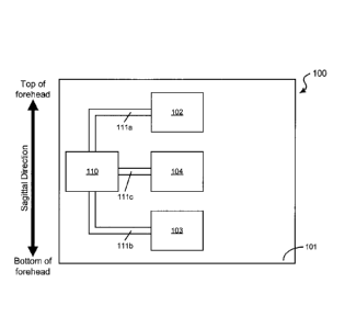
physiological signals
of interest using a limited quantity of electrode sensors, e.g., which can be
used to determine
cognitive and/or sensory performance, psychological states, and/or behavioral
preferences.
[0006] In one aspect, a physiological sensor device includes a substrate
formed of an
electrically insulative material and structured to allow physical contact of
the device with the
frontal region of the head of a user, a first electrode configured at a first
location on the
substrate to acquire an electrophysiological signal of the user, a second
electrode configured
at a second location on the substrate to acquire a second electrophysiological
signal of the
user as a reference signal to the electrophysiological signal, and a third
electrode configured
on the substrate to acquire a third electrophysiological signal of the user as
an electrical
ground signal, in which the third electrode is configured at a third location
at least partially
between the first and the second locations on the substrate, and the first
location is configured
posterior to the second and third locations along a sagittal direction in the
frontal region when
the physiological sensor device is properly placed on the frontal region of
the user, and in
which the device is operable when electrically coupled to an electrical
circuit to detect
physiological signals of the user.
[0007] Implementations of the physiological sensor device can optionally
include one or
more of the following features. In some implementations of the device, for
example, the first,
second, and third electrodes can be linearly arranged on the substrate. For
example, the
detected physiological signals can be electroencephalography signals sensed
from the brain
of the user. In some examples, the detected electroencephalography signals can
be associated
with an event-related potential. For example, the detected physiological
signals can be
electromyography signals sensed from head muscles of the user associated with
the user's
eye blinking or facial expressions. In some implementations of the device, for
example, the
substrate can be formed of a mechanically flexible material structured to
adhere to skin or a
wearable item of the user. In some implementations, for example, the device
can further
include electrical interface components formed separately on the substrate and
electrically
coupled to the first, second, and third electrodes, respectively, via
electrically conductive
conduits, in which the electrical circuit is an external electrical circuit
electrically coupled to
the electrical interface components via wires. In some implementations, for
example, the
electrical circuit can include a signal processing circuit formed on the
exemplary
2
CA 02887535 2015-04-08
WO 2014/059431
PCT/US2013/064892
mechanically flexible substrate in electrical communication with the first,
second, and third
electrodes via electrically conductive conduits, in which the signal
processing circuit can
amplify the acquired physiological signals. For example, the electrical
circuit can include a
transmitter unit on the substrate in electrical communication with the signal
processing circuit
to transmit the amplified physiological signals to at least one of a data
processing unit or a
remote computer system. In some implementations, for example, the device can
further
include a power supply module electrically coupled to the electrical circuit
to provide
electrical power to the transmitter unit. In some examples, the physiological
sensor device
can be configured as a wearable patch worn on the user's scalp. In some
examples, the
physiological sensor device can be configured in a region of the wearable item
capable of
physical contact with the user's scalp. In some implementations, for example,
the device can
further include a fourth electrode configured at a fourth location on the
substrate to acquire a
second electrophysiological signal of the user, and a fifth electrode
configured at a fifth
location on the substrate to acquire a third electrophysiological signal of
the user, in which
the fourth location is configured left of the first location, and the fifth
location is configured
right of the first location.
[0008] In some implementations of the physiological sensor device, for
example, the
device is implemented in a system to provide a cognitive or sensory
assessment. The system
can include a data processing system in communication with the physiological
sensor device
and structured to include one or more memory units and one or more processors
configured
to process the detected physiological signals as physiological data to
generate an infoimation
set including one or more quantitative values associated with a cognitive-
sensory profile
category indicative of one or more aspects of cognitive or sensory functions.
For example,
the one or more processors of the data processing unit can be configured to
process the
physiological signals detected by the physiological sensor device to generate
the information
set by selecting time intervals of interest within the physiological data
based on the presented
stimuli and the cognitive-sensory profile category, grouping, into one or more
grouped data
sets, the physiological data corresponding to the selected time intervals of
interest, and
providing a statistical measure of a relationship across or within the grouped
data sets to
generate the one or more quantitative values. For example, the one or more
quantitative
values can include a quantitative score depicting a level of one or both of
cognitive and
sensory performance based on at least one of the user's attention, memory,
learning ability,
confabulation characteristics, pattern integration ability, semantic
integration ability, target
detection ability, emotional valence, preference, or awareness, and wherein
the quantitative
3
score depicts the level at a particular time. In some implementations, the
system can further
include a stimulus delivery device to produce a sequence of stimuli based on
the cognitive-
sensory profile category that is presented to the user wearing the
physiological sensor device,
wherein the stimuli includes at least one of a visual, auditory, olfactory,
tactile, or gustatory
stimulating medium, in which the physiological sensor device is interfaced to
the user to
detect the physiological signals exhibited by the user before, during, and
after a presentation
of the sequence of stimuli. In some implementations, the data processing
system can include
a local computer proximate to and in communication with the physiological
sensor device to
receive the detected physiological signals from the physiological sensor
device, the local
computer configured to conduct initial processing of the detected
physiological signals to
produce initial physiological signal data, and a remote computer in
communication with the
local computer via a communication network or link to receive the initial
physiological signal
data from the local computer and to process the initial physiological signal
data to generate
the information set including one or more quantitative values associated with
the cognitive-
sensory profile category. For example, the local computer can be a mobile
communications
device including a smartphone or tablet that is in wireless communications
with the
physiological sensor device.
[0009] In another aspect, a method to provide a cognitive or sensory
assessment of a
subject includes acquiring electrophysiological signals of the subject from
the frontal region
of the subject's head to produce physiological data using a sensor device, and
processing the
physiological data to generate an information set including one or more
quantitative values
associated with a cognitive-sensory profile category indicative of one or more
aspects of
cognitive or sensory functions, in which the sensor device includes a
substrate formed of an
electrically insulative material and structured to allow physical contact of
the sensor device
with the frontal region of the head of the subject, and three electrodes
including a recording
electrode, a reference electrode, and a ground electrode to acquire the
electrophysiological
signals of the subject from three respective positions arranged on the
substrate along the
sagittal direction of the frontal region, in which the recording electrode is
configured
posterior to the ground and reference electrodes, and the ground electrode is
configured
between the recording and reference electrodes.
4
CA 2887535 2020-03-06
[0009a] In one embodiment, there is provided a physiological sensor device.
The device
includes: a substrate formed of an electrically insulative material and
structured to allow
physical contact of the device with the frontal region of the head of a user;
a first electrode
configured at a first location on the substrate to acquire an
electrophysiological signal of the
user; a second electrode configured at a second location on the substrate to
acquire a second
electrophysiological signal of the user as a reference signal to the
electrophysiological signal;
and a third electrode configured on the substrate to acquire a third
electrophysiological signal
of the user as an electrical ground signal. The third electrode is configured
at a third location
at least partially between the first and the second locations on the
substrate, and the first
location is configured posterior to the second and third locations along a
sagittal direction in
the frontal region when the physiological sensor device is properly placed on
the frontal
region of the user. When electrically coupled to an electrical circuit, the
device is operable to
detect physiological signals of the user.
[0009b] In another embodiment, there is provided a wearable physiological
sensor device.
The device includes: a substrate formed of a mechanically flexible and an
electrically
insulative material and structured to allow physical contact of the device
with the frontal
region of the head of a user; and three electrodes on the substrate arranged
along a sagittal
direction in the frontal region when the physiological sensor device is
properly placed on the
frontal region of the user, including: a first electrode configured at a first
location on the
substrate to acquire an electrophysiological signal of the user, a second
electrode configured
at a second location anterior to the first location on the substrate to
acquire a second
electrophysiological signal of the user as a reference signal to the
electrophysiological signal,
and a third electrode configured at a third location at least partially
between the first and the
second locations on the substrate to acquire a third electrophysiological
signal of the user as
an electrical ground signal. The device further includes: an electrical
circuit on the substrate
in electrical communication with the first, second, and third electrodes via
electrically
conductive conduits, the electrical circuit including an amplification circuit
and a signal
processing circuit to amplify and signal process the electrophysiological
signals; a transmitter
unit on the substrate in electrical communication with the electrical circuit
to transmit the
amplified and signal processed physiological signals to at least one of a data
processing unit
4a
CA 2887535 2020-03-06
or a remote computer system; and a power supply module electrically coupled to
the
transmitter unit to provide electrical power to the transmitter unit.
[0009c] In another embodiment, there is provided a method to provide a
cognitive or
sensory assessment of a subject. The method involves acquiring
electrophysiological signals
of the subject from the frontal region of the subject's head to produce
physiological data
using a sensor device including: a substrate formed of an electrically
insulative material and
structured to allow physical contact of the sensor device with the frontal
region of the head of
the subject, and three electrodes including a recording electrode, a reference
electrode, and a
ground electrode to acquire the electrophysiological signals of the subject
from three
respective positions arranged on the substrate along the sagittal direction of
the frontal
region. The recording electrode is configured posterior to the ground and
reference
electrodes, and the ground electrode is configured between the recording and
reference
electrodes. The method further involves processing the physiological data to
generate an
information set including one or more quantitative values associated with a
cognitive-sensory
profile category. The cognitive-sensory profile category is indicative of one
or more aspects
of cognitive functions or sensory functions.
BRIEF DESCRIPTION OF THE DRAWINGS
[0010] FIGS. 1A and 1B show block diagrams of an exemplary frontal
electrode
physiological sensor device of the disclosed technology.
4b
CA 2887535 2020-03-06
CA 02887535 2015-04-08
WO 2014/059431
PCT/US2013/064892
[0011] FIG. 1C shows a diagram of an exemplary system of the disclosed
technology for
acquisition, analysis, and evaluation of physiological signals to produce an
individual or
group knowledge and/or state of awareness profile.
[0012] FIGS. 1D-1F show process diagrams of exemplary methods to generate
a
quantitative information set of an exemplary cognitive and/or sensory profile.
[0013] FIG. 1G shows a diagram of an exemplary method to determine an
electrode
configuration on the frontal region of a subject's head.
[0014] FIG. 2 shows a diagram of an exemplary sequence of presented
visual stimuli.
[0015] FIG. 3A shows diagrams illustrating an exemplary frontal electrode
configuration
using a conventional EEG system and exemplary results from its implementation
for
detecting the EEG signal responses.
[0016] FIG. 3B shows diagrams illustrating an exemplary frontal electrode
configuration
using an exemplary three-electrode sensor device and exemplary results from
its
implementation for detecting the EEG signal responses.
[0017] FIGS. 4A and 4B show exemplary three-electrode configurations using
exemplary
rigid electrodes along a gradient potential configuration and along an
isopotential
configuration. respectively.
[0018] FIG. 5 shows data plots of an exemplary EEG online recording using
the
exemplary rigid electrodes before stimuli presentation for both gradient
potential and
isopotential configurations.
[0019] FIG. 6 shows data plots of an exemplary EEG online recording using
the
exemplary rigid electrodes during an exemplary stimuli presentation for both
gradient
potential and isopotential configurations.
[0020] FIG. 7A shows an exemplary data plot of ERP wavefoims from a
single subject
for "Targets" and "Distractors" using the exemplary rigid electrodes.
[0021] FIG. 7B shows an exemplary data plot of ERP waveforms from a
single subject
for "Reward" using the exemplary rigid electrodes.
[0022] FIG. 8 shows an image of exemplary fabricated, custom-designed
rigid electrodes.
[0023] FIGS. 9A-9D show exemplary data plots of ERP waveforms acquired
using
various exemplary rigid electrodes from a single subject for "Targets",
"Distractors-, and
"Reward".
[0024] FIG. 10 shows a schematic of an exemplary epidermal electronics
frontal three-
electrode design.
[0025] FIGS. 11A and 11B show exemplary three-electrode configurations
using
5
CA 02887535 2015-04-08
WO 2014/059431
PCT/US2013/064892
exemplary flexible epidermal electrodes along a gradient potential
configuration and along an
isopotential configuration, respectively.
[0026] FIG. 12 shows data plots of an exemplary EEG online recording
using the
exemplary flexible epidermal electrodes before stimuli presentation for both
gradient
potential and isopotential configurations.
[0027] FIG. 13 shows data plots of an exemplary EEG online recording
using the
exemplary flexible epidermal electrodes during an exemplary stimuli
presentation for both
gradient potential and isopotential configurations.
[0028] FIG. 14A shows an exemplary data plot of ERP waveforms from a
single subject
for "Targets" and "Distractors" using the exemplary flexible epidermal
electrodes.
[0029] FIG. 14B shows an exemplary data plot of ERP waveforms from a
single subject
for "Reward" using the exemplary flexible epidermal electrodes.
[0030] FIG. 15 shows a diagram of an exemplary sequence of stimuli for a
mismatch
negativity ERP.
[0031] FIGS. 16A and 16B show data plots of exemplary group average ERP
waveforms
of the elicited mismatch negativity, deviants and standards in a frontal
channel of an
exemplary rigid EEG electrode cap and with flexible epidermal electrode
sensors,
respectively.
[0032] FIGS. 16C and 16D also show data plots of exemplary ERP waveforms
a single
subject of the elicited mismatch negativity, deviants and standards in a
frontal channel of an
exemplary rigid EEG electrode cap and with flexible epidermal electrode
sensors,
respectively.
DETAILED DESCRIPTION
[0033] Establishing reliable correlations between one's brain signals and
the associated
cognitive/psychological states (e.g., thoughts) can provide valuable and
desired applications
for clinic and other uses. Such correlations, extensively explored in
fundamental sciences,
have been the focus of various translational attempts into specialized
applications such as
assessment of cognitive impairment and enabling the physically impaired to
communicate.
[0034] Some systems to characterize cognitive and psychological states
have relied upon
various behavioral and brain imaging techniques, e.g., such as functional
resonance magnetic
imaging (fMRI) and electroencephalography. For example, fMRI is an indirect
measure of
brain function by correlated metabolic function (e.g., oxygen consumption in
the blood flow),
whereas EEG is a direct measure of brain activity by recording changes of the
electrical fields
6
CA 02887535 2015-04-08
WO 2014/059431
PCT/US2013/064892
present at the scalp, deriving from electrical activity produced by neural
cells.
[0035] There are several important factors in determining sensory and/or
cognitive
information about a subject For example, such factors can include the type of
stimuli that
can evoke a subject's response, duration of the stimuli, inter-stimuli
interval, number of
repetitions of each presentation of stimuli, the levels of the stimuli (e.g.,
sound, brightness or
contrast levels, etc.), markers associated with the onset of presentation of
each stimuli, etc., as
well as the recording sensors and systems. Also, the physiological
parameter(s) of use (e.g.,
voltage, power, frequency, etc.), the related time window for analysis, and
the analysis
structure can affect the brain signal recordings and correlated cognitive
evaluation.
Deviations or mistakes from one or multiple of these parameters can make the
difference
between a useful or artifact driven, useless method.
[0036] Some traditional EEG recording techniques include an EEG cap
covering the
whole scalp, e.g., placed over the hair. These full cap EEG systems are
typically neither
comfortable nor aesthetically pleasing, and in some cases require the use of
conductive gel,
which is cumbersome to the user, and may require technical application, etc.
Some EEG
recording techniques do not utilize a full cap, but nonetheless include skin-
mounted
electrodes along with other electrodes that are spatially disparate and
require a bulky headset
that is not efficient in terms of portability and comfort, and/or such skin-
mounted electrode
systems suffer from poor signal quality revealing inadequate signal to noise
ratio to optimal
detection of ERPs. For example, one class of skin-mounted electronics systems
used an
electrode configuration having frontal electrodes and non-frontal electrodes
(e.g., some
placed behind the subject's ears) to acquire muscular and brain signals, but
with signal
resolution only able to extract coarse muscular and brain signals that
included eye blink and
alpha rhythm oscillations when the subject's eyes were closed, and thus
incapable to
adequately detect finer brain signals, such as ERPs. These techniques are
either cumbersome
or unable to acquire relevant brain signals to extract relevant brain signals
reflective of
behavioral and brain measures of interest, e.g., for characterization of
cognitive and/or
psychological states.
[0037] For example, measurements of event-related potentials for sensory,
motor and/or
cognitive analysis can include techniques that capitalize in measuring
transient electric shifts
(e.g., ERP components) that are time-locked to the onset of a presented
stimulus (e.g., visual,
auditory, olfactory, gustatory, or tactile) and reflect the underlying brain
activity during the
investigated neuropsychological process. For example, ERP components can be
indicative of
multiple sensory, motor and cognitive functions. The amplitude modulation and
scalp
7
CA 02887535 2015-04-08
WO 2014/059431
PCT/US2013/064892
distribution of a variety of ERPs represent reliable and effective brain
markers for normal
neuropsychological processing of a wide range of cognitive operations.
Moreover, abnomial
modulation and latencies of ERPs have been associated with various sensory and
cognitive
deficits linked to neuropsychiatric disorders, such as schizophrenia,
Alzheimer's and
Parkinson' s .
[0038] As such, the use of these measures of brain activity is of great
value to biomedical
research and development and clinical applications of effective diagnostic
tools for
neurological and neuropsychiatric disorders. However, today's use of ERP brain
markers is
still confined to sophisticated laboratory settings and medical facilities.
Moreover, traditional
methods to record EEG signals are clunky, cumbersome, and unable to be used
effectively in
general purpose environments.
[0039] Devices, systems, and methods are disclosed for acquiring
physiological signals of
interest using a limited quantity of electrode sensors, e.g., which can be
used to deteimine
cognitive and/or sensory performance, psychological states, and/or behavioral
preferences.
[0040] In one aspect, a physiological sensor device includes a substrate
formed of an
electrically insulative material and structured to allow physical contact of
the physiological
sensor device with the frontal region of the head of the user, and, an optimal
configuration of
three electrodes on the substrate providing a 'minimized device footprint when
the device is
properly applied on the user's forehead. The three electrodes include a
recording electrode, a
.. reference electrode, and a ground electrode to acquire the
electrophysiological signals of the
subject from three respective positions arranged on the substrate along the
sagittal direction
of the frontal region, in which the recording electrode is configured
posterior to the ground
and reference electrodes, and the ground electrode is configured between the
recording and
reference electrodes.
[0041] The disclosed technology integrates advanced cognitive neuroscience,
neurophysiology, psychology and electromagnetics in optimal configurations of
physiological signal detection electrodes frontally placed on the forehead to
enable individual
or group evaluation of a variety of cognitive aspects and physiological/health
monitoring,
e.g., including but not limited to, evaluation of cognitive state, knowledge,
learning
mechanisms, behavioral preferences, vulnerability and/or symptoms of
neurological and
neuropsychiatric pathologies. The disclosed technology can be implemented in
devices that
provide easy and user-friendly operation, portability, and comfort, thereby
permitting real-
world usage and systematic health monitoring. Additionally, for example, the
disclosed
technology can be used in a variety of health, education, entertainment, and
marketing
8
CA 02887535 2015-04-08
WO 2014/059431
PCT/US2013/064892
applications.
[0042] For example, the disclosed technology includes physiological
sensor devices and
methods using frontal EEG recording electrodes located on a user's forehead
for versatile,
rapid, and non-obtrusive physiological data acquisition (e.g., including brain
signal
monitoring) that do not overlap with hair. For example, in some
implementations, the
exemplary physiological sensor devices are configured to a small size and can
be formed with
a variety of different materials (e.g., which can be tailored for specific
applications), such that
the devices may be easily applied, barely or not even felt by the user, or
seen by others. For
example, application and operation of such devices can be performed by the
user, e.g.,
following simple instructions, without any need for technical expertise to
apply or operate the
device or system. This can significantly mitigate problems present in existing
systems
including the need of technical expertise for operation and lack of comfort
and portability of
sensor devices.
[0043] For example, the disclosed systems can be used by general users
outside a clinical
setting, with safety and accuracy, allowing for the freedom to use in a wide
variety of
contexts and locations, significantly reducing the cost and requirements of
use for brain
monitoring systems. The disclosed devices and methods can be effectively used
by non-
experts to place the exemplary frontal electrode sensor device on the forehead
of evaluated
persons (or even allow the subjects to place the frontal electrodes on
themselves) to optimally
extract brain signals, e.g., which in some implementations can be associated
with event-
related potentials (ERPs), and to provide a cognitive and/or sensory profile
of the subject or
subjects. For example, such non-expert users need not be neuroscientists,
psychologists, nor
specialized physicians to implement the physiological data acquisition or
interpret the
generated cognitive and/or sensory profile information of the user provided by
the analysis of
the acquired physiological data. For example, the non-expert users can
implement the
disclosed systems and methods to obtain awareness and mental information
profiles of the
evaluated person(s), e.g., either themselves or others. Additionally, for
example,
implementations of the disclosed devices, systems and methods can also be used
within the
context of brain-machine interfaces and expands the possible applications of
such systems.
[0044] In some aspects, the disclosed technology includes techniques for
designing an
optimal sensor configuration for frontal electrode placement on a subject's
forehead to
accurately detect brain event-related potentials. In some examples, the
techniques can use
infoimation from specific stimuli presentation paradigms (e.g., sensory
stimulation can
include visual, auditory, olfactory, gustatory or somatosensory cues) and
relate the presented
9
CA 02887535 2015-04-08
WO 2014/059431
PCT/US2013/064892
stimuli with recorded brain electrophysiological signals (e.g., EEG) in
specific temporal
windows (e.g., based on physiology data related to the neuropsychological
mechanisms
underlying ERPs) and spatial regions (e.g., based on neuroanatomy and on scalp
topographic
voltage mapping and neural generators source analysis) of interest.
[0045] Exemplary Embodiments of the Disclosed Devices, Systems, and Methods
[0046] In one exemplary embodiment, a physiological sensor device of the
present
technology includes a substrate that is formed of an electrically insulative
material and
structured to allow physical contact of the device with the frontal region of
the head of a user,
a recording electrode configured at a first location on the substrate to
acquire an
electrophysiological signal of the user, a reference electrode configured at a
second location
on the substrate to acquire a second electrophysiological signal of the user
as a reference
signal to the electrophysiological signal; and a ground electrode configured
at a third location
on the substrate to acquire a third electrophysiological signal of the user as
an electrical
ground signal. The physiological sensor device is configured such that the
first electrode is
configured posterior to the third and second electrodes along a sagittal
direction in the frontal
region, and the third electrode is positioned at least partially between the
first and the second
locations on the substrate. The physiological sensor device is operable when
electrically
coupled to an electrical circuit to detect physiological signals of the user.
[0047] In some implementations of the exemplary frontal electrode
physiological sensor
device, the recording electrode, the ground electrode, and the reference
electrode are linearly
arranged on the substrate. For example, the arrangements of the three
electrodes can be
aligned in a substantially straight line along the sagittal direction of the
frontal region of the
user's head, with the recording electrode (e.g., at the first position)
posteriorly positioned to
the ground electrode, which is posteriorly positioned to the reference
electrode.
[0048] In some implementations, for example, the physiological signals
detected by the
exemplary frontal electrode physiological sensor device can be
electroencephalography
(EEG) signals sensed from the brain of the user. For example, the EEG signals
can he
associated with an event-related potential, e.g., based on a stimulus
presented to the user
wearing the device on the frontal region of the user's head. In other
implementations, for
example, the physiological signals detected by the exemplary frontal electrode
physiological
sensor device can be electromyography (EMG) signals sensed from head muscles
(e.g.,
including facial muscles) of the user. For example, the EMU signals can be
resultant from
eye blinks of the user in response to an event-related potential, e.g., based
on a stimulus
presented to the user wearing the device on the frontal region of the user's
head.
CA 02887535 2015-04-08
WO 2014/059431
PCT/US2013/064892
[0049] In some embodiments, for example, the exemplary frontal electrode
physiological
sensor device can include electrical interface components (e.g., electrical
contact pads)
formed separately on the substrate and electrically coupled to the recording,
ground, and
reference electrodes, e.g., via electrically conductive conduits, in which the
electrical
interface components provide an electrical coupling site to be connected
(e.g., via wires) to
an external electrical circuit, e.g., electrical signal amplifier and/or
processing unit.
[0050] In some embodiments, for example, the exemplary frontal electrode
physiological
sensor device can include (i) electrical circuits for signal
amplification/processing and (ii) a
transmitter unit, all on the mechanically flexible substrate in electrical
communication with
the recording, ground, and reference electrodes, e.g., via electrically
conductive conduits. In
this embodiment, the sensor device is configured to record the physiological
signals, amplify
and process them, and transmit the recorded physiological signals to a remote
device, e.g.,
further electrical signal processing unit, such as an amplifier, and/or a
computer system.
Also, for example, the exemplary frontal electrode physiological sensor device
can include a
power supply module electrically coupled to the transmitter unit to provide
electrical power
to the transmitter unit.
[0051] In some embodiments, for example, the exemplary frontal electrode
physiological
sensor device can include one or more recording electrodes configured on the
substrate to
acquire multiple channels of electrophysiological signals of the user. For
example, the
exemplary frontal electrode physiological sensor device can include two
additional recording
electrodes (in which the device includes five electrodes: three recording
electrodes, one
reference electrode, and one ground electrode), in which the additional
recording electrodes
are proximate to the first recording electrode, ground electrode, and
reference electrode
arranged in the sagittal direction. In this example, the two additional
electrodes can be
linearly arranged in the same or similar sagittal direction as the first
recording electrode. In
other examples, some of the additional electrodes can be positioned to the
left of the first
recording electrode, while others additional recording electrode can be
positioned to the right
of the first recording electrode.
[0052] In some implementations, for example, the exemplary frontal
electrode
physiological sensor device is configured as an epideimal electronic sensor
(EES) device in
which the substrate is formed of a mechanically flexible and/or stretchable
material structured
to mechanically conform to and/or adhere to the skin or a wearable item of the
user. In some
examples of an epideimal physiological sensor device of the present
technology, the device
can include ultrathin silicon islands interconnected by serpentine-like wires
that all rest on a
CA 02887535 2015-04-08
WO 2014/059431
PCT/US2013/064892
biologically inert flexible polymer. In some implementations, for example, the
epidermal
physiological sensor device can include a processing unit configured on the
flexible substrate
and structured to include transistors, capacitors, resistors, inductors,
and/and other circuit
elements, etc., to process the electrophysiological signals acquired by the
electrodes. In some
implementations, for example, the processing unit of the epidermal
physiological sensor
device can include a processor and a memory unit. The epidermal physiological
sensor
device can be configured to have a thickness approximate to that of a human
hair.
[0053] FIG. lA shows a block diagram of an exemplary embodiment of a
frontal
electrode sensor device 100 capable to acquire electrophysiological signals
from the frontal
region of the head of a subject The device 100 includes a substrate 101 of an
electrically
insulative material, which, in some device implementations, can be made of a
mechanically
flexible material. In some examples, the substrate 101 can include
polydimethylsiloxane
(PDMS), thin polyurethane with acrylic adhesive, or polyvinyl alcohol (PVA),
among others.
The frontal electrode sensor device 100 includes a three-electrode
configuration, including a
recording electrode 102, a reference electrode 103, and a ground electrode 104
configured
between the recording electrode 102 and the reference electrode 103 on the
basal side of the
substrate 101 (e.g., the detection side of the device 100 that is in contact
with the skin of the
user). The electrodes of the device 100 are configured along a sagittal
direction in the frontal
region such that the recording electrode 102 is positioned posteriorly to the
ground electrode
104, which is positioned posteriorly to the reference electrode 103. The
ground electrode 104
is positioned at least partially between the recording electrode 102 and the
reference electrode
103 on the substrate 101. This recording-ground-reference electrode
arrangement on the
frontal region of the user's head or forehead region can minimize the overall
footprint of the
electrodes of the frontal electrode sensor device 100, a significant benefit
for such sensor
devices. This recording-ground-reference electrode arrangement also provides
good signal
isolation between the recording electrode and the reference electrode, thus
enabling more
sensitive and high quality signal recording operation. The general alignment
of the electrodes
in the sagittal direction, rather than the horizontal direction that is
perpendicular to the
sagittal direction, is a notable feature of this recording-ground-reference
electrode
arrangement and can provide beneficial sensing operations with respect to
acquiring various
cognitive/psychological state signals with desired accuracy.
[0054] In some embodiments of the device 100, for example, the recording
electrode 102,
the ground electrode 104, and the reference electrode 103 are linearly
arranged on the
substrate 100. For example, the arrangements of the three electrodes can be
aligned in a
12
CA 02887535 2015-04-08
WO 2014/059431
PCT/US2013/064892
substantially straight line along the sagittal direction, with the recording
electrode. In other
embodiments of the device 100, for example, the three electrodes can be
arranged in a
nonlinear alignment that includes the recording electrode 102 positioned
posteriorly to the
ground electrode 104 that is positioned posteriorly to the reference electrode
103, with the
ground electrode 104 at least partially between the recording electrode 102
and the reference
electrode 103 on the substrate 101.
[0055] The frontal electrode sensor device 100 is operable to acquire
electrophysiological
data when electrically coupled to an electrical circuit. In the exemplary
embodiment shown
in FIG. 1A, the frontal electrode sensor device 100 includes an electrical
circuit 110 on the
substrate 101 electrically coupled to the recording electrode 102, the
reference electrode 103,
and the ground electrode 104 via individual electrical interconnects 111a,
111b, and 111c,
respectively. In some embodiments, for example, the electrical circuit 110 can
include a
transmitter unit in electrical communication with each of the electrodes 102,
103, and 104,
e.g., via the electrically conductive conduits 111 a, 111b, and 111c,
respectively. In this
embodiment, the device 100 can record the physiological signals and transmit
the recorded
physiological signals to a remote electrical signal processing unit, e.g.,
such as an amplifier,
and/or a computer system. Also, for example, the electrical circuit 110 can
include a power
supply module electrically coupled to the transmitter unit to provide
electrical power to the
transmitter unit.
[0056] In some embodiments, for example, as shown in FIG. 1B, the frontal
electrode
sensor device 100 can include electrically conductive interface (contact) pads
112a, 112b, and
112c coupled to the interconnects 111a, 111b, and 111c, respectively, to
provide a conductive
surface to electrically interface an external electrical circuit to the
electrodes 102, 103, and
104 of the device 100. For example, the external electrical circuit can be an
electrical signal
.. processing unit, e.g., such as a signal amplifier, and/or a computer
system.
[0057] For example, the acquired recording, reference, and ground signals
are received
by the signal processing unit that processes the acquired signals in a
differential amplifier to
amplify the difference between the recording and reference
electrophysiological signals. The
ground signals recorded by the device 100 (via the ground electrode 104) can
be connected to
the ground channel of the exemplary differential amplifier, e.g., to
synchronize the signal
parameters between the device 100 and the amplifier. For example, the ground
electrode 104
can minimize leakage currents that may flow through the subjects via the
recording system,
and thus decrease any artifacts. For example, the ground electrode 104, when
electrically
coupled to an electrical circuit (e.g., such as the external electrical
circuit), need not be
13
CA 02887535 2015-04-08
WO 2014/059431
PCT/US2013/064892
connected to the ground of the electrical circuit. Alternative roles of the
ground electrode can
include serving as an electrode for actively canceling interference. For
example, the ground
electrode can be electrically connected to a "driven right leg" feedback
circuit, e.g., which is
used in some biological signal amplification systems that measure very small
electrical
signals emitted by the body (e.g., EEG, EMG, ECG). For example, the frontal
electrode
sensor device 100 can acquire referential recordings of electrophysiological
signals at the
frontal region. The position of the reference electrode 103, as well as its
spacing with respect
to the recording electrode 102 (or, in some implementations, other recording
electrodes in
addition to the recording electrode 102) is important, since the recordings of
interest will be
__ detei mined by a comparison of the activity recorded by the recording
electrode 102 with
respect to the activity recorded by the reference electrode 103. For example,
if such signals
were the same, then the detected signal reading would be zero. From this
perspective, for
example, one could position the recording electrode 102 at a site that will
allow for detection
of the physiological signal of interest and position the reference electrode
103 at a substantial
distance away from it at a site that will not capture the physiological signal
of interest (or
show a significant reduction of the signal of interest). However, this
presents a challenge that
becomes greater when it is important to minimize the footprint of the device
100 (e.g., the
occupied spatial area or "real estate" by the whole array of electrodes) on
the forehead. For
example, in the examples shown in FIGS. lA and 1B, the electrodes 102, 103,
and 104 are
positioned and spaced in such a manner that the signals captured are
significantly different,
and thereby relevant, as well as occupy a minimal total area occupied by
electrodes 102, 103,
and 104. Methods are described in this patent document to determine optimal
configurations
of location and spacing are complex and can integrate psychological,
neurophysiological and
engineering principles. In the example shown in FIGS. IA and 1B, the position
of the
reference electrode 103 is located in a substantially linear alignment with
respect to the
recording electrode 102, and both electrodes 102 and 103 and the ground
electrode 104 are
also arranged on a mid-sagittal line through the center of the frontal region,
in this example.
The signal-processed signals are provided as physiological data, which can
subsequently be
processed to provide a cognitive and/or sensory profile.
[0058] In some implementations, the device 100 can be configured as an
epidermal
electronics physiological sensor device that can be worn directly on skin or a
wearable item
in contact with the frontal region. In some implementations, for example, the
device 100 can
include an additional electrically insulative layer or layers, e.g.,
configured on the apical side
of the device 100 (e.g., the non-detection side, not in contact with the skin
of the user). The
14
additional layer(s) can provide further support for the device 100. In some
examples, the
additional layer(s) can include various artistic designs, such that, when worn
by the user
directly on the user's skin, the device 100 can also serve as a (temporary)
tattoo.
[0059] In some implementations, the device 100 can be included in a
system to provide a
.. cognitive or sensory assessment of the user. Some examples of such systems
are provided in
PCT Patent Application PCT/US13/62491, entitled "SYSTEMS AND METHODS FOR
SENSORY AND COGNITIVE PROFILING," filed September 27, 2013.
[0060] An exemplary modular system including the frontal electrode
sensor device 100
of the disclosed technology for acquisition, analysis and evaluation of
physiological signals
to produce an individual or group cognitive and/or sensory profile is shown in
FIG. 1C. For
example, the system can be implemented to provide a cognitive performance
profile, a
sensory performance profile, and a cognitive and sensory performance profile
indicative of a
subject's cognitive and/or sensory ability at the time of the assessment. For
example, the
type of cognitive and/or sensory profile can be selected by the user (e.g.,
such as the subject
or a system operator) to provide a set of information including a quantitative
level of
cognitive and/or sensory performance, e.g., including, but not limited to
attention, memory,
learning, confabulation, pattern integration, semantic integration, target
detection, emotional
valence, preference, and state of awareness. The system allows an operator to
select the type
of profile to be produced. In some implementations, the system can be
implemented to
.. provide the cognitive and/or sensory profile using only physiological data
acquired from the
subject, e.g., with no overt behavioral response elicited from the subject. In
some
implementations, the system can be implemented to provide the cognitive and/or
sensory
profile including previously acquired physiological data from the subject, or
other subjects
(e.g., group data). The system can thereby, for example, be implemented to
provide a
cognitive and/or sensory profile about a group. FIG. 1C shows a diagram of an
exemplary
system 10 configured to include independent modular units or devices that can
be configured
in a variety of different embodiments.
CA 2887535 2020-03-06
[0061] The system 10 includes a stimulus presentation module 130 to
configure a
specific stimulus (e.g. visual and/or auditory) presentation structure 131 to
effectuate a
presentation of a stimulus or a sequence of stimuli to a subject. In some
examples, the
stimulus presentation module 130 is embodied in a computing device, e.g.,
including a
processor and memory unit. For example, the stimuli can include any stimulus
type,
including a visual, auditory, olfactory, tactile, and/or gustatory stimulating
medium.
Examples of visual stimuli can include images, written words, etc. Examples of
auditory
stimuli can include spoken words, animal vocalizations, synthesized sounds,
etc. The
specific stimulus presentation structure 131 can be configured to include, but
is not limited
to, a particular type or types of stimuli, the duration of presentation of the
stimuli, an inter-
stimuli interval, a number of repetitions (if any) of each presentation,
magnitude and/or
frequency parameters associated with type of stimuli (e.g., intensity of sound
or brightness or
contrast level of light), a digital marker associated with the presentation of
each stimuli, and a
label or category of the stimuli (e.g., target or non-target).
[0062] The system 10 can include a stimulus delivery module 135 in
communication
with the stimulus presentation module 130 to present the stimulus or the
sequence of stimuli
(e.g. visual and/or auditory) to the subject, e.g., based on the stimulus
presentation structure
131. For example, the stimulus delivery module 135 can include at least one of
a visual
display, an auditory speaker, and an actuator to provide an olfactory,
tactile, and/or gustatory
stimulus. In some implementations, for example, the stimulus presentation
module 130 and
the stimulus delivery module 135 can be configured in the same device, e.g.,
such as a
computer or mobile communication and/or computing device.
[0063] The system 10 includes a physiological data acquisition module
140, which can
be embodied as the frontal electrode sensor device 100, to acquire
physiological signals of
the subject before, during, and/or after the presentation of the stimuli or
sequence of stimuli
via the stimulus delivery module 135. For example, the frontal electrode
sensor device 100
can be implemented to acquire electrophysiological signals from the subject,
e.g., including,
but is not limited to, electroencephalography (EEG) signal data and
electromyography
(EMG) signal data. In some implementations, for example, the frontal electrode
sensor
device 100
16
CA 2887535 2020-03-06
can include electrophysiological sensing electrodes, e.g., EEG and/or EMG
electrodes, or
other types of electrophysiological sensing electrodes, coupled to a signal
acquisition device,
e.g., such as an analog or digital amplifier coupled to a memory.
[0064] In some embodiments, for example, the frontal electrode sensor
device 100 can
be configured in a standard EEG system with rigid electrodes attached to a cap
worn by the
subject. In some embodiments, for example, the frontal electrode sensor device
100 can be
configured in a portable EEG system using flexible electronics that can be
worn on the
subject, e.g., directly applied the subject's skin or worn in a wearable item
(e.g., such as a
hat) by the subject with the frontal electrode sensor device 100 in physical
contact with the
frontal region of the subject's scalp. For example, the frontal electrode
sensor device 100 can
be configured in a standard EMG system with rigid electrode or a portable EMG
system
16a
CA 2887535 2020-03-06
CA 02887535 2015-04-08
WO 2014/059431
PCT/US2013/064892
using flexible electronics that can be worn on the subject, in which the
frontal electrode
sensor device 100 is in physical contact with the frontal region of the
subject's scalp. In this
exemplary configuration, the frontal electrode sensor device 100 in the rigid
electrode
standard EMG system or portable flexible electronics EMG system is capable of
detecting
movements that can be associated with drowsiness or facial expressions of the
subject.
[0065] The system 10 includes an analysis pre-processing module 145 to
receive the
acquired physiological signals as data, and in some implementations, to
perform pre-
processing analysis techniques on the acquired data. For example, the analysis
pre-
processing module 145 can be implemented to identify exemplary onset markers
in the
acquired electrophysiological data (e.g., EEG data), segment the
electrophysiological data,
filter raw signal data to increase signal to noise, etc. In some
implementations, for example,
the analysis pre-processing 145 can be embodied in a computer device in
communication
with the exemplary device 100. In some implementations, for example, the
analysis pre-
processing module 145 can be configured in the same exemplary device that
embodies the
physiological acquisition module 140 (e.g., such as the frontal electrode
sensor device 100).
[0066] The system 10 includes a profile generation module 150 to process
the
physiological data acquired by the frontal electrode sensor device 100 to
provide a cognitive
or sensory assessment of the subject, or in some examples, of a group. For
example, the
profile generation module 150 processes the physiological to generate an
information set 152
that includes one or more quantitative values that are associated with the
selected profile
category, e.g., such as a knowledge evaluation or state of awareness profile.
For example, the
information set 152 provides more than a measure of psychological and
neurophysiological
natural events. For example, the profile can provide an individual (or group)
assessment of
one's (or group's) level of knowledge of specific issues (e.g., determination
of a given person
knowledge about a specific topic, event, learned skill or even preference)
and/or state of
conscious (or unconscious) awareness. In some implementations of the system
10, for
example, the profile generation module 150 can also include processing
behavioral signal
data, e.g., acquired from the subject or group of individuals that include or
do not include the
subject, from a behavioral signal data acquisition module (not shown in FIG.
1C) to provide
the cognitive or sensory assessment of the subject or of a group.
[0067] FIG. 11) shows a process diagram of an exemplary method 170 to
generate the
infoimation set associated with the cognitive and/or sensory profile, e.g.,
implemented by the
profile generation module 150, using the physiological data acquired by the
exemplary
frontal electrode sensor device 100. In some implementations, for example, the
method 170
17
CA 02887535 2015-04-08
WO 2014/059431
PCT/US2013/064892
can also include using behavioral signal data acquired from the subject, or
group of
individuals that include or do not include the subject. The behavioral signal
data can be
processed in implementations of at least some or all of the processes of the
method 170. The
method 170 can include a process 171 to identify a time interval associated
with the
physiological signals (and/or behavioral signal data) based upon the presented
stimuli and the
selected profile category. For example, a time interval can include
contiguous,
discontinuous, continuous, discrete, or single time points. The method 170 can
include a
process 172 to group the data (e.g., physiological and/or behavioral)
corresponding to the
time interval into one or more grouped data sets. For example, the process 172
can include
grouping the physiological data (and/or behavioral data) based on a pre-
assigned category of
the individual stimulus and/or an associative relationship of consecutive
stimuli. The method
170 can include a process 173 to provide a statistical measure of a
relationship across or
within the grouped data sets to generate the one or more quantitative values
for the selected
profile category. In some implementations, for example, the method 170 can
include a
process to enhance the signal of the physiological (and/or behavioral data) in
the grouped
data sets.
[0068] FIG. 1E shows a process diagram of an exemplary method 180 to
generate the
information set associated with the cognitive and/or sensory profile using
previous individual
and/or group information, e.g., implemented by the profile generation module
150, using the
physiological data acquired by the exemplary frontal electrode sensor device
100. In some
implementations, for example, the method 180 can also include using behavioral
signal data
acquired from the subject, or group of individuals that include or do not
include the subject.
The method 180 can include a process 181 to identify a time interval
associated with the
physiological signals (and/or behavioral signal data) based upon the presented
stimuli and the
selected profile category. The method 180 can include a process 182 to group
the data, e.g.,
physiological data (and/or behavioral data), corresponding to the time
interval into one or
more grouped data sets. For example, the process 182 can include grouping the
physiological
data (and/or behavioral data) based on a pre-assigned category of the
individual stimulus
and/or an associative relationship of consecutive stimuli. The method 180 can
include a
process 182 to provide a statistical measure of a relationship across or
within the grouped
data sets using previous physiological data (and/or behavioral data) acquired
from the subject
and/or other subjects (e.g., including one or more groups) to generate the one
or more
quantitative values for the selected profile category.
18
[0069] FIG. 1F shows a process diagram of an exemplary method 190 to
generate the
information set associated with the cognitive and/or sensory profile using a
guided
classification technique, e.g., implemented by the profile generation module
150, using the
physiological data acquired by the exemplary frontal electrode sensor device
100. In some
implementations, for example, the method 190 can also include using behavioral
signal data
acquired from the subject, or group of individuals that include or do not
include the subject.
The method 190 can include a process 191 to identify a time interval
associated with the
physiological signals (and/or behavioral signal data) based upon the presented
stimuli and the
selected profile category. The method 190 can include a process 192 to group
the data, e.g.,
physiological data (and/or behavioral data) corresponding to the time interval
into one or
more initial grouped data sets. The method 190 can include a process 193 to
classify each
stimulus of the sequence of stimuli presented to the subject using a
statistical test involving
the initial grouped data sets. The method 190 can include a process 194 to re-
group the
physiological data (and/or behavioral data) corresponding to the time interval
into one or
more grouped data sets based on the classified stimuli. The method 190 can
include a
process 195 to provide a statistical measure of a relationship across or
within the grouped
data sets to generate the one or more quantitative values for the selected
profile category.
[0070] In some examples, the profile generation module 150 can
implement guided
classification algorithms with context specific parameters to guide and choose
from a variety
of classification and statistical methods 151, e.g., including, but not
limited to, ANOVA
based techniques 151a, support vector machine based techniques 151b, and
minimum
description length techniques 151c, among others. In some implementations, the
profile
generation module 150 can be embodied on a computer system or communication
network
(referred to as 'the cloud') that includes one or more remote computational
processing
devices (e.g., servers in the cloud).
[0071] The system 10 can be configured to include a brain-machine
interface module
155 to refine the generated cognitive and/or sensory profiles and/or actuate
an interaction
between a user and a machine. In one example, the brain-machine interface
module 155 can
provide a feedback delivery of a new stimulus or multiple stimuli to the
stimulus presentation
module 130 based on the cognitive and/or sensory profile of an individual
subject or group
19
CA 2887535 2020-03-06
subject that has been generated from the profile generation module 150, e.g.,
from an on-
going implementation of the system 10 or a previously generated profile by the
system 10.
For example, the brain-machine interface module 155 can adaptively change or
design
stimuli paradigms that optimally extract information from the subject that is
analytically
processed to maximize a desired objective. The brain-machine interface module
155 can also
adaptably change and/or adjust stimuli presentation (posterior matching
algorithm). For
example, some implementations of the brain-machine interface module 155 can
include, but
are not limited to, assisted-learning and target detection applications.
[0072] In some implementations of the system 10, the profile generation
module 150, the
.. stimulus presentation module 130, the stimulus delivery module 135, and the
brain-machine
interface module 155 can be embodied in a single computing system, e.g., a
desktop
computer, a laptop computer, or a mobile communications device including a
smartphone or
tablet, that interacts with the physiological data acquisition module 140
(e.g., the frontal
electrode sensor device 100). In other implementations, the modules 150, 130,
135, and 155
can be configured in two or more computing devices in communication with each
other and
including various combinations of the modules 150, 130, 135, and 155. In some
implementations, the system 10 can be configured to just include the
physiological data
acquisition module 140 and the profile generation module 150. In such
exemplary
implementations, the system 10 can use environmental stimuli (e.g., light,
sounds, smells,
tastes, and/or tactile contacts) that are presently available in the subject's
surroundings.
[0073] In some aspects, a method to provide a cognitive or sensory
assessment of a
subject using the physiological sensor devices of the disclosed technology
includes acquiring
electrophysiological signals of the subject from the frontal region of the
subject's head to
produce physiological data using a sensor device, and processing the
physiological data to
generate an information set including one or more quantitative values
associated with a
cognitive-sensory profile category indicative of one or more aspects of
cognitive or sensory
functions. The sensor device includes a substrate formed of an electrically
insulative
material and structured to allow physical contact of the sensor device with
the frontal region
of the head of the subject, and three electrodes including a recording
electrode, a reference
electrode, and a ground electrode to acquire the electrophysiological signals
of the subject
CA 2887535 2020-03-06
from three respective positions arranged on the substrate along the sagittal
direction of the
frontal region, in which the recording electrode is configured posterior to
the ground and
reference electrodes, and the ground electrode is configured between the
recording and
reference electrodes.
[0074] In some implementations of the method to provide the cognitive
and/or sensor
assessment, for example, the method can further include presenting a sequence
of stimuli to
the subject, the sequence of stimuli based on the cognitive-sensory profile
category, in which
the acquiring the physiological signals is implemented before, during, and
after the
presenting the sequence of stimuli. In some implementations, for example, the
method can
further include selecting the cognitive-sensory profile category from among a
cognitive
performance profile, a sensory performance profile, and a cognitive and
sensory performance
profile. For example, the sequence of stimuli can include at least one of a
visual, auditory,
olfactory, tactile, or gustatory stimulating medium based on the selected
cognitive-sensory
profile category. For example, the one or more quantitative values can include
a quantitative
score depicting a level of one or both of cognitive and sensory performance
based on at least
one of the subject's attention, memory, learning ability, confabulation
characteristics, pattern
integration ability, semantic integration ability, target detection ability,
emotional valence,
preference, or awareness state, and wherein the quantitative score depicts the
level at a
particular time. In some implementations, for example, the method can further
include
identifying a time interval associated with the physiological signals based on
the cognitive-
sensory profile category, grouping the physiological data corresponding to the
time interval
into one or more grouped data sets, and providing a statistical measure of a
relationship
across or within the grouped data sets to generate the one or more
quantitative values for the
selected cognitive-sensory profile category.
[0075] FIG. 1G shows an illustrative diagram depicting an exemplary method
160 to
determine optimal spatial placement of frontal electrode sensors to acquire
EEG event-related
potentials. The method 160 includes presenting the stimulus presentation
structure 131 using
the stimulus delivery module 135 to a subject, who can wear a conventional
'full scalp' EEG
acquisition device 141 with electrodes located at positions across the
subject's head. The
stimulus presentation structure 131 can be used to effectuate a presentation
of a stimulus or a
21
CA 2887535 2020-03-06
sequence of stimuli to a subject. The EEG response of the subject to the
presentation of the
stimulus or sequence of stimuli is acquired using the 'full scalp' EEG
acquisition device 141
(e.g. traditional full scalp EEG acquisition) to acquire the EEG signal data
from a plurality of
electrodes across a plurality of regions of the brain, e.g., including
frontal, parietal, occipital,
.. and temporal cerebral regions. The acquired data can be analyzed using the
pre-processing
module 145 to implement pre-processing analysis techniques on the acquired
data. In some
examples, the pre-processing analysis techniques include producing one or more
topographical voltage maps 146 of the electrophysiological data, e.g.,
topographical voltage
mapping, which can be over different temporal and/or spatial parameters. For
example, the
pre-processing module 145 can utilize the data produced in the topographical
voltage map(s)
146 in data processing techniques to optimize spatial arrangement parameters
147 of the
electrodes that can be included in a frontal electrode physiological sensor
device. For
example, the spatial arrangement parameters can include the number of
electrodes, electrode
type and size, location / placement of each electrode, spacing between the
electrodes, other
specification of optimal frontal placement of each electrode, etc. For
example, determination
of the location and/or placement of the 'electrodes can be based on the type
of signal to be
recorded (e.g., signal of interest, a reference signal, and ground signal).
For example, the
electrode location and/or placement parameters can include relative distances
of the
electrodes, particular placements of the electrodes with respect to the
subject's frontal region
of his/her head, etc. For example, such parameters (e.g., type, quantity,
size, placement,
relative position, etc.) of the electrodes configured in exemplary
physiological sensor devices
of the disclosed technology account for topography, amplitude, and
localization of
physiological signals of interest (e.g., including event-related potential
brain markers), as
well as specific metal conductance, resistance, and spacing parameters.
22
CA 2887535 2020-03-06
[0076] Exemplary Implementations of the Disclosed Devices with Methods
and Systems
for Profiling Cognitive-Sensory Function
[0077] Described are exemplary implementations of the disclosed frontal
electrode
physiological sensor devices and systems and methods using such devices for
providing a
.. cognitive and/or sensory assessment of a subject (or a group) indicative of
one or more
aspects of cognitive or sensory functions. The described exemplary
implementations include
eliciting and extracting various brain ERPs (e.g., P300, notion/feeling of
'reward', and
mismatch negativity) measured by EEG recordings using visual stimuli and
auditory stimuli
to produce an information set providing quantitative values corresponding to
the cognitive
performance, sensory performance, and/or awareness state profile. In some
examples of the
disclosed methods and systems, eye tracking data can be used in addition to
the exemplary
EEG recording physiological data acquired by the exemplary frontal electrode
physiological
sensor devices for providing the cognitive and/or sensory assessment.
[0078] In the described examples, specific stimuli sets are presented
while recording
.. EEG signals from the subject to elicit event-related potentials of
interest, as well as correlated
neural frequency oscillations. The exemplary ERPs used in the exemplary
implementations
include, but are not limited to, the P300, notion/feeling of 'reward', and the
mismatch
negativity. Other exemplary ERPs that can be implemented to provide an
exemplary
cognitive-sensory profile using the disclosed technology can include the N400,
among others.
As described below, exemplary applications of the exemplary frontal electrode
physiological
sensor devices with the disclosed methods and systems use the exemplary P300,
'reward',
and mismatch negativity ERPs as illustrative examples to described how the
exemplary
methods can be implemented, e.g., stimuli design and presentation,
physiological signal (e.g.,
EEG) recording, physiological data (e.g.,. ERP) analysis, and cognitive and/or
sensory profile
22a
CA 2887535 2020-03-06
CA 02887535 2015-04-08
WO 2014/059431
PCT/US2013/064892
generation (e.g., including inferred cognitive and/or awareness states).
[0079] The disclosed cognitive and/or sensory profile generation methods
and systems
can be used to measure brain markers, but in addition, it evaluates and
transfoims this
infoimation into a new type of purposeful data that creates an individual
knowledge
evaluation and/or state of awareness profile. Moreover, in some
implementations, for
example, the disclosed methods and systems can use this profile to guide a
brain-machine
interface system.
[0080] 1. P300 and "Reward"
[0081] The P300 is a brain endogenous response characterized by a
positive-going
electrical response between 300 and 800 ms, with a central-parietal maxima
scalp
distribution. 'the P300 is inversely correlated with an item's subjective
probability of
occurrence. For example, the P300 has been used in visual target detection
tasks, where the
target elicits higher amplitude P300s than the other items.
[0082] Additionally, in the exemplary implementations described herein
using the P300,
an arbitrary visual cue (e.g., green circle) was created, and the tested
subjects were instructed
to associate it with a notion or feeling of "reward". For example, the
subjects were instructed
to associate the exemplary green circle with a cue of good task perfoimance
and informed
that the occurrence of the cue during testing would be correlated with
increase compensation
after the test, thus, creating an association of this arbitrary cue with a
notion of "reward".
The brain responses to this "reward" stimulus was subsequently analyzed. The
exemplary
illustrations of the disclosed methods using P300 illustrates the broad
applicability of the
methods across various EEG recording techniques. For example, the method can
be applied
to assess cognitive and/or sensory profiles using brain data (e.g., EEG
signals) recorded using
a traditional rigid electrodes EEG system with the disclosed frontal electrode
configurations,
.. as well as with EEG data acquired using wearable, flexible epideimal
electronic sensors with
the disclosed frontal electrode configurations.
[0083] 1.1. Exemplary Stimulus Presentation Structure
[0084] In some exemplary implementations of the P300 and "reward" ERPs,
visual
stimulation were used. For example, the stimuli were comprised of multiple
image
categories, e.g., including animals; cars; faces; flowers; houses; random
objects; motorcycles;
airplanes; and buildings. The exemplary pool of stimuli was obtained from
various
resources.
[0085] FIG. 2 shows a diagram of an exemplary sequence of presented
visual stimuli.
This diagram portrays images of specific presented exemplars and the pre-
programmed
23
CA 02887535 2015-04-08
WO 2014/059431
PCT/US2013/064892
pseudo-randomized order of presentation Having the adequate stimuli
presentation structure
for each solution is an intrinsic and important part of the exemplary method.
In this example,
the relevant aspect is the distinction between the images that represent pre-
deteimined
"Targets" versus all other images (labeled as "Distractors") and versus the
green circle
previously associated with an indication of "Reward". The adequate stimuli
structure (e.g., in
this case, the specific content of images of interest as targets embedded in a
sequence of other
images, location of presentation, timing of presentation, timing of inter-
stimulus interval,
etc.) and the described specialized subsequent analysis are important in the
exemplary
method to how to use relevant brain markers, e.g., in this exemplary case, the
P300 and the
____________________________ "reward" elicited ERPs, to evaluate and detei
mine individual knowledge, levels of attention
and preferences to specific items.
[0086] 1.2. Exemplary Stimulus Delivery Device
[0087] The exemplary pool of stimuli was obtained from various resources.
After
obtaining the stimuli pool, each exemplar's relative luminance was controlled
using a
computer implemented method (e.g., programmed with a MATLAB script). For
example,
the computer implemented method was implemented to first load in a color
image, and
calculate its relative luminance using the following exemplary formula, where
Y, R, G, and B
represent relative luminance, red gun values, green gun values, and blue gun
values,
respectively:
Y = .2126R + .7152G + .0722B
(1)
[0088] For example, the desired relative luminance was set to be a value
equal to 120.
After the script measured the initial relative luminance of each image, it
either added or
subtracted ROB values to every pixel within the image in order to achieve an
average relative
luminance of 120. Images were then saved at 100% quality.
[0089] After controlling for luminance, another computer implemented
process (e.g.,
programmed using a MATLAB script) was used to place a centrally positioned
fixation dot
on each stimulus exemplar. For example, this helped the subject to maintain
fixation and
minimize any frequent eye saccades. This exemplary process first measured the
dimensions
of an uploaded image. It used these measurements to calculate the center of
the image and
subsequently create a fixation dot using the standard equation of a circle.
Pixels within a
seven pixels length radius around the center were altered by changing the
pixels' red gun to
255, the green gun to 0, and the blue gun to 0.
[0090] Lastly, the visual stimuli for the fixation dot and an arbitrary
visual cue for
24
CA 02887535 2015-04-08
WO 2014/059431
PCT/US2013/064892
"reward" were created. For example, for the fixation dot, a computer
implemented process
(e.g., programmed using a MATLAB script) was used to create a grey background
image
(e.g., red gun equal to 150; green gun equal to 150; blue gun equal to 150)
with a height and
width of 350 pixels. Then, the exemplary script ran a nested for-loop using
the standard
equation of a circle to alter pixels within a seven pixels length radius to
red, e.g., by changing
the image's red gun to 255, the green gun to 0, and the blue gun to 0. For the
"reward",
imaging software was used to create a green circle (e.g., red gun equal to 0;
green gun equal
to 255; blue gun equal to 0) on a 350x350 pixels grey background (e.g., red
gun equal to 150;
green gun equal to 150; blue gun equal to 150) background.
[0091] The exemplary stimulus presentation paradigm that was used in this
example
stimuli presentation process was programmed using Cogent 2000, and included
presenting
visual stimuli serially with brief presentation durations. For example, the
pool of stimuli, not
including the fixation dot and green circle, was divided into two groups, one
for each of the
two recording techniques. Each technique included 900 stimuli, for a total of
1800 stimuli
across recording techniques. For example, within a technique, the 900 stimuli,
including
targets and distractors, each presented for 100 ms, were divided into three
presentation
blocks. The green circle stimulus presentation lasted 1000 ms and was shown 30
times
within each presentation block. The fixation dot was visible during every
target trial,
distractor trial, and inter-stimulus interval (ISI).
[0092] For example, in block 1, targets were human faces. In block 2,
targets were cars.
In block 3, targets were animals. In addition to randomizing the order of the
recording
techniques, for example, the order of the presentation blocks within each
technique was also
randomized. Presentation blocks were never repeated consecutively (e.g., block
1, block 2,
block 3, block 3, block 2, block 1, block 1, block 3, block 2) across
techniques. Because the
subject was instructed to count how many times he/she saw a particular target,
the correct
number of targets was varied for each block. For example, in block 1 (target:
faces), there
were 56 targets and 244 distractors. For example, in block 2 (target: cars),
there were 62
targets and 238 distractors. For example, in block 3 (target: animals), there
were 60 targets
and 240 distractors. Distractors were composed of all of the non-target object
categories.
For example, in block 1 (target: faces), the distractors included cars,
animals, flowers, houses,
etc. The exemplary MATLAB code began by prompting the experimenter to enter
the
subject's initials and choose which block to present. Depending on the chosen
block number,
the script calculated which object category would be target, the number of
targets, and the
number of distractors. Afterward, it randomized the order of stimulus
presentation using the
CA 02887535 2015-04-08
WO 2014/059431
PCT/US2013/064892
MATLAB randperm() function. It ran the randperm() function twenty times to
better
randomize the presentation sequence. Then, it created inter-stimulus intervals
(ISI) for each
trial using the randi() function. The inter-stimulus intervals ranged from 500
ms to 600 ms.
In addition to configuring the display, sound card, and parallel port, a log
file was configured
and initialized within Cogent 2000. This log file was used to create a history
of every trial
regarding its stimulus type (e.g., target, distractor and green circle).
Subsequently, the stimuli
were loaded into memory buffers. The aforementioned steps were executed prior
to stimulus
presentation, e.g., to reduce computational load and increase latency
precision. The stimulus
presentation included a for-loop that iterated down the pre-determined
presentation order.
For example, based on the value of the current stimulus in the presentation
order, the
computer implemented process calculated its stimulus type and sent the
appropriate
infounation regarding its stimulus type to the log file and parallel port,
whose trigger was
sent to the EEG recording computer. Then, the program presented the 1ST. At
the end of
each presentation, the parallel port was reset to zero to prepare for the next
trial.
[0093] 1.3. Exemplary EEG Recordings Using Traditional Full Scalp EEG
Acquisition
[0094] To prepare the exemplary subjects for EEG recording, each subject
was seated in
a chair in a recording chamber to begin an EEG capping process. For the
exemplary
implementations using the rigid electrode modality (e.g., Brain Products),
this process
involved placing a traditional EEG cap on the subject's head and securing it
with an elastic
chin strap. In some examples, either a 56 cm or a 58 cm diameter cap was used,
e.g., based
on the estimated size of the subject's head. Next, Signa electrode gel (e.g.,
from Parker
Laboratories) was injected using a curved, plastic syringe under each of the
cap's electrodes
to create a conductive bridge between the electrode itself and the subject's
scalp. Also, for
example, wooden Q-tips were used to massage the gel in order to build a
stronger
conductance by lowering the impedance. For example, use of this technique
lowered the
impedance levels to <5 ks-2 for each electrode, e.g., including the ground and
reference.
[0095] Before starting the exemplary implementation using EEG recordings,
subjects
were given an instructions document to read. For example, this document
described the
general organization of the experimental paradigm and what they would be
viewing, namely
targets, distractors, a fixation dot and a green circle. It was also explained
that in each
presentation block, the target would change. For example, in block 1, the task
was to count
how many times they saw an image with one or more human faces. In block 2, the
task was
to count how many times they saw an image with one or more cars. In block 3,
the task was
26
CA 02887535 2015-04-08
WO 2014/059431
PCT/US2013/064892
to count how many times they saw an image with one or more animals. The
subjects were
instructed to regard all other photographs as distractors and not count them.
After each
presentation block, the subjects were asked to report how many targets they
saw. For
example, the green circle indicated reward. The subjects were seated in front
of the
presentation monitor and asked to just maintain visual fixation on a red,
central fixation dot
throughout the duration of the experiment and restrict their motor movements
as much as
possible to prevent motion artifacts in the neurophysiological data.
Afterwards, the recording
room's lights were then dimmed, and the stimulation process and EEG recordings
began.
[0096] In these exemplary implementations, a traditional EEG system with
rigid
electrodes was used to acquire brain waves. The exemplary EEG system included
a
BrainAmp DC 32-channel system; BrainVision Recorder; Fast n Easy 32-channel
EEG
recording cap size 56 cm; Fast n Easy 32-channel EEG recording cap size 58 cm;
PCB
Ribbon Cable for BrainCap-MR with 5k resistors; and BrainCap MR Box 1.2.
[0097] 1.4. Exemplary EEG Analytical Processing and ERP Analysis
[0098] Data analysis techniques included multiple steps and processes,
e.g., including the
processing of marker data and individual statistical analysis.
[0099] Processing of marker data: For example, after each recording
session, the
exemplary EEG recordings system produced three files: data file (.eeg), header
file (.vhdr),
and marker file (.vmrk). The marker files contained the event triggers for
each stimulus
onset. In this example, because of output limitations within the parallel
port, the Cogent 2000
log file was used to hold more readable infoimation regarding an exemplar's
stimulus type
(e.g., target, distractor, or green circle). From there, a process (e.g.,
programmed using a
MATLAB script) was used to replace the event triggers in the marker file
(.vmrk) with the
event codes from the log file in a one-to-one replacement. For example, the
first marker in
the .vmrk file was replaced by the first marker in the log file; the second
marker in the .vmrk
file was replaced by the second marker in the log file, etc.
[00100] Exemplary individual statistical analysis: Using the abovementioned
exemplary
markers for onset of stimulus presentation, an ERP analysis was performed
following
exemplary analytical methods to calculate ERPs (e.g., using BrainVision
Analyzer 2). For
example, ERP waveforms were calculated for "Targets", "Distractors" and
"Reward-.
Subsequently, for example, from each ERP waveform the spatial location and
timing of the
ERP components of interest were determined. For example, for each time-window
of
interest, topographic voltage maps for each ERP component of interest were
calculated using
the available voltage information from every electrode in the EEG cap (total
32 electrodes).
27
CA 02887535 2015-04-08
WO 2014/059431
PCT/US2013/064892
In the exemplary implementations described herein, a combination of MATLAB and
Statsoft
Statistica (version 8.0) software was used for statistical analyses.
[00101] 1.5. Exemplary Configuration of Optimal Frontal Electrode Placement
[00102] The determination of an optimal electrode placement using our method
can be
obtained by integrating all this information, for example: (i.) Sensory or
cognitive
neuropsychological mechanism of interest; (ii.) Designing an adequate stimulus
presentation
and delivery method for eliciting the neuropsychological mechanism of
interest; (iii.) Tag
chronological markers associated with onset of stimulus presentation; (iv.)
Record EEG
signals from the subject during stimulation; (v.) Analyze EEG data and
calculate ERPs for
each condition / marker of interest; (vi.) Identify ERP components of interest
in every
channel of the dataset; (vii.) For each component of interest, determine
latency (time of
occurrence) and spatial distribution (which electrodes show the ERP of
interest); (viii.) Based
on the latency of each component of interest, detemtine a time-window of
occurrence for
each component and create topographic voltage maps; (ix.) For each ERP
component of
interest, investigate where in the scalp it has a significant "expression"
(i.e., in which
electrodes is it present and how is its voltage distributed in the topographic
voltage map); (x.)
Use this information to determine the best timing and the optimal location for
electrodes
placement to detect modulations of the ERP of interest, as well as its
expression in frontal
electrodes. For example, from this, a determination of an optimal placement
and
configuration of electrodes for detection of an ERP of interest can be made.
[00103] In the exemplary frontal electrode configuration, using the disclosed
electrode
configuration optimization method, spatial locations and placement (e.g.,
including distance
between electrodes and electrode sizes) were determined in a way to minimize
the used
forehead "real estate" occupied, while keeping enough spacing to respect each
electrode
signal integrity along the scalp surface and detect the brain activity of
interest. Typically, for
example, the reference electrode is positioned away from the recording
electrode and in a
location where the "brain function of interest" is minimal or provides no
expression. In this
way, when one is differentiating the reference signal from the recording
signal, it wouldn't be
'subtracting' anything of interest. However, large separations use relatively
large amounts of
spatial area on the forehead. In devices of the disclosed technology, a
minimal configuration
of electrodes are placed on the frontal region of the subject that require the
minimal amount
of spatial area (e.g., placing the recording and reference electrodes close
together) while still
providing reliable physiological signal readings and detection. The disclosed
electrode
configuration optimization method was implemented to obtain such
configurations.
28
CA 02887535 2015-04-08
WO 2014/059431
PCT/US2013/064892
[00104] For example, assuming additive white Gaussian noise in the sensor, the
accuracy
of an optimal classifier (e.g., given by a likelihood ratio test) is a
monotonic function of the
energy in the difference between the scalp potential of the recording
electrode under the null
hypothesis (for example the "Distractors" in the P300 paradigm) and scalp
potential of the
recording electrode under the alternate hypothesis (for example the "Targets"
in the P300
paradigm). Thus, provided is a criterion for maximizing classification
accuracy which is
equivalent to maximizing the energy in the difference between the recording
electrode's
potential in "Targets" and "Distractors" setting.
[00105] If an electrode pair is placed across isopotential lines (e.g., same
or identical
voltage potential values), then the potential difference is sensitive to
current flow from
regions of high to low, or low to high, potentials. "[his is referred to as
placing the electrode
pair along a gradient potential. However, if the bipolar pair is placed along
an isopotential
line, then a zero, or close to zero differential potential is recorded.
Maximizing energy in the
difference is done when the placement of the frontal electrodes is along the
gradient of the
frontal scalp distribution of the brain signal of interest.
[00106] From the exemplary analysis in both ERPs used in the exemplary
implementations, e.g., P300 and "reward", the gradients of the voltage scalp
distribution are
oriented vertically. For better detection of these ERPs, the recording
electrode, ground
electrode and reference electrode should be placed from top to bottom (e.g.,
from the top of
the forehead towards the nose), which is across the isopotential line.
Moreover, placing the
recording electrode, ground electrode, and reference electrode orthogonal to
that direction
(e.g., along the isopotential line) will result in a degraded classification
performance ¨
approaching that of chance.
[00107] Moreover, for example, in addition to increasing signal-to-noise
characteristics in
the detected electrophysiological signals, the disclosed top-to-bottom (e.g.,
top of the
forehead towards the nose) frontal region electrode configuration also is
capable of detecting
relevant neurophysiological signals of the underlying functional-neuroanatomy
(e.g., the
human brain has symmetrical hemispheres along a mid-sagittal line and that
many sensory
and cognitive processes occur with functional and anatomical hemispheric
asymmetries). For
example, placing the electrodes along the brain axial (or transversal) plane
(e.g., along the
forehead from left to right), instead of the disclosed top of the forehead
towards the nose
orientation, is likely to create significant vulnerabilities to brain
hemispheric artifacts leading
to erroneous EEG/ERP readings. Also, the exemplary methodology of the
disclosed
technology also suggests that the greater the expression of an event-related
potential is in
29
CA 02887535 2015-04-08
WO 2014/059431
PCT/US2013/064892
frontal voltage scalp distributions, the easier it is to classify.
[00108] FIG. 3A shows diagrams illustrating an exemplary frontal electrode
configuration
using a conventional EEG system and exemplary results from its implementation
for
detecting the EEG signal response to P300 ERP detection of "Targets" and
"Distractors". In
.. FIG. 3A, an image 300 is shown of an exemplary subject wearing a
conventional EEG rigid
electrode detection cap 301. In FIG. 3A, a diagram 305 is shown depicting a
tridimensional
reconstruction and top and right side schematical views of the 32 electrodes'
location in the
conventional EEG rigid electrode detection cap 301. In FIG. 3A, a diagram 306
is shown
depicting a two dimensional views of the 32 electrodes' location in the
conventional EEG
.. rigid electrode detection cap 301. As shown in FIG. 3A, a diagram 310 shows
an exemplary
topographic voltage map (e.g., with indication of the frontal expression of
ERP of interest, in
this exemplary case, a P300 ERP for "Targets" in an exemplary analyzed time-
window (e.g.,
344-396 ms). As shown in FIG. 3A, a data plot 315 shows the exemplary ERP
waveforms
from a frontal electrode (Fpl ) for both "Targets"(- red line) and
"Distractors" (- black line).
.. As shown in the topographic voltage map 310, a hemispherically symmetric
distribution is
present, where isopotential lines are on the transversal plane. As such, the
frontal electrodes
are placed orthogonal to the transversal plane, across the isopotential line.
[00109] FIG. 3B shows diagrams illustrating an exemplary frontal electrode
configuration
using an exemplary three-electrode frontal sensor device and exemplary results
from its
implementation for detecting the EEG signal response to P300 ERP detection of
"Targets"
and "Distractors". FIG. 3B shows a diagram 320 of the frontal electrode sensor
device 100 in
physical contact with the frontal region of a subject's head and aligned
across an isopotential
line (e.g., with the recording electrode placed toward the top of the
forehead, the reference
electrode placed toward the nose on the forehead, and the ground electrode
placed between
.. the recording and reference electrodes). FIG. 3B shows a data plot 325 of
the ERP
waveforms acquired from the frontal recording electrode (e.g., Rec Fl - white
circle 102) for
both "Targets"- red line and "Distractors" - black line. The disclosed frontal
electrode
configuration in the 3-electrode sensor devices can detect the ERPs of
interest ("Targets" and
"Distractors") as reliably as a full cap traditional EEG system. In this
example, the polarity
of the ERPs ("Targets" and "Distractors-) is reversed between the exemplary
frontal system
and the EEG cap frontal electrode due to the relative position of the
"Reference" electrode in
regards to the "Recording" electrode. For example, in the full cap system
shown in FIG. 3A,
the "Recording" electrode occupies a significantly more anterior position than
the
"Reference", while in the exemplary system shown in FIG. 3B, the "Reference-
electrode is
CA 02887535 2015-04-08
WO 2014/059431
PCT/US2013/064892
in a lower and slightly more anterior position than the "Recording" electrode.
[00110] 1.6. Exemplary Implementations of Frontal Electrode Configurations for
ERP
detection across different technologies
[00111] In these exemplary implementations, the disclosed frontal electrode
configurations
were implemented across different electrode types and materials, e.g.,
including EEG
recording technologies using a traditional full EEG cap with rigid electrodes
and three classes
of frontal electrode sensor technologies containing a ground, reference, and
recording
electrode, using: (1) exemplary rigid sensors (e.g., obtained from Brain
Products); (2)
exemplary custom-designed rigid sensors; and (3) an exemplary epidermal
electronics sensor
device with flexible electronics electrodes.
[00112] For example, after subject preparation, stimulus presentation, EEG
recordings and
processing of marker files, as described in the previous sections, a
combination of MATLAB
and Statsoft Statistica (version 8.0) software were used for statistical
analyses. For example,
after data processing and analysis, BrainVision Analyzer exported text files
containing data
values in regards to condition, subject, trial, electrode channel, and mean
voltage amplitude.
For each recording technology, the mean voltage amplitude was extracted from a
frontal
electrode for the following time intervals of interest, for example: Targets
and Distractors,
400 ms to 500 ins after stimulus onset; Reward stimuli, 456 ins to 556 ms
after stimulus
onset. The same exemplary parameters were used in all the exemplary techniques
(e.g., the
exemplary rigid electrodes from Brain Products, the exemplary custom-designed
rigid
electrodes, and the exemplary EES flexible electrode sensors). For example,
these data were
written to text files by BrainVision Analyzer and subsequently loaded into an
exemplary
MATLAB program that we created to sort and organize the data in a more
accessible format.
Specifically, for example, the exemplary script allowed an easier data
selection process by
column, e.g., using MATLAB's variable editor. After selecting, data were put
into Statistica
data spreadsheets. In some examples, one-way (factor 1: condition) repeated
measures
ANOVAs were performed on each Statistica spreadsheet for the comparison
between
distractors and targets for each of the EEG recording techniques. For example,
each
spreadsheet was specific to the following: 1) EEG technology; and 2)
comparison:
distractors vs. targets. For the reward condition, for example, a T-test was
perfoimed
comparing the reward's extracted mean amplitude values against zero. For the
rewards
condition, for example, each spreadsheet was specific to the following: 1) EEG
technology;
and 2) comparison: reward vs. zero.
[00113] 1.6.1. Using Rigid EEG Sensors in Frontal Configurations
31
[00114] Two frontal configurations including a horizontal anatomical alignment
of the
electrodes along the transverse direction (e.g., left to right axis) and a
vertical anatomical
alignment of the electrodes along the sagittal direction (e.g., posterior ¨
anterior axis) were
implemented using exemplary rigid EEG electrodes (Brain Products). FIGS. 4A
and 4B
show exemplary three-electrode configurations using rigid electrodes along a
gradient
potential configuration (e.g. a first example of rigid EEG sensors
configuration) and along an
isopotential configuration (e.g. a second example of rigid EEG sensors
configuration),
respectively. In one example, a gradient potential configuration (referred to
here as "vertical-
sagittal") used three electrodes placed from the top of the forehead towards
the nose, as
follows: "Recording Fl" electrode, "Ground" electrode, 'Reference" electrode,
as shown in
FIG. 4A. In another example, an isopotential configuration (referred here as
"horizontal-
transverse") used three electrodes placed along the forehead from left to
right, as follows:
"Reference" electrode, "Ground" electrode, "Recording Fl" electrode, as shown
in FIG. 4B.
[00115] FIG. 5 shows data plots of an exemplary EEG online recording acquired
using the
exemplary rigid electrodes (e.g., obtained from Brain Products) before stimuli
presentation
for both gradient potential and isopotential configurations (e.g. the rigid
EEG sensors
configurations shown in FIGS. 4A and 4B). The data plot 510 shows the
exemplary EEG
data for the isopotential configuration of the exemplary three-electrode
frontal rigid EEG
sensor. The data plot 520 shows the exemplary EEG data for the gradient
potential
configuration of the exemplary three-electrode frontal rigid EEG sensor. Eye
blinks were
used to better illustrate the signal-to-noise (SNR) ratio of the response in
this example.
[00116] FIG. 6 shows data plots of an exemplary EEG online recording acquired
using the
exemplary rigid electrodes during an exemplary stimuli presentation for both
gradient
potential and isopotential configurations (e.g. the rigid EEG sensors
configurations shown in
FIGS. 4A and 4B). The data plot 610 shows the exemplary EEG response data for
the
isopotential configuration of the exemplary three-electrode frontal rigid EEG
sensor. The
data plot 620 shows the exemplary EEG response data for the gradient potential
configuration of the exemplary three-electrode frontal rigid EEG sensor. Eye
blinks were
used to better illustrate the signal-to-noise (SNR) ratio of the response in
this example.
32
CA 2887535 2020-03-06
[00117] As shown in FIGS. 5 and 6, it is noted that in both cases the eye
blinks are more
pronounced in the gradient potential directions as compared to the
isopotential ones, e.g.,
indicating a better performance, as predicted, for the "vertical-sagittal"
configuration.
[00118] FIG. 7A shows an exemplary data plot of ERP waveforms acquired using
the
exemplary rigid electrodes from a single subject for "Targets" (red line) and
"Distractors"
(black line) using both the "vertical-sagittal" configuration (gradient
potential configuration,
shown by thicker lines) and the "horizontal-transverse" configuration
(isopotential
configuration, shown by thinner lines).
32a
CA 2887535 2020-03-06
CA 02887535 2015-04-08
WO 2014/059431
PCT/US2013/064892
[00119] FIG. 7B shows an exemplary data plot of ERP waveforms acquired using
the
exemplary rigid electrodes from a single subject for "Reward" (blue line)
using both the
"vertical-sagittal" configuration (gradient potential configuration, shown by
thicker lines) and
the "horizontal-transverse" configuration (isopotential configuration, shown
by thinner lines).
[00120] The exemplary results of the exemplary implementations using the
exemplary
rigid electrode sensors showed adequate detection for all tested ERPs across
the isopotential
line in the gradient potential configuration, e.g., Targets vs. Distractors
N=132, F=5.100,
p<0.05; and Reward N= 45, T=-3.03, p<0.005. Conversely, the exemplary results
of the
exemplary implementations using the exemplary rigid electrode sensors showed
inadequate
detection for all tested ERPs along the isopotential configuration, e.g.,
Targets vs. Distractors
N=144, F=0.001, p=0.96; and Reward N= 45, 1=-1.45, p=0.15. The disclosed three-
electrode frontal configuration using the gradient potential configuration is
capable of
efficient ERP detection using rigid electrodes.
[00121] 1.6.2. Using Custom-designed Rigid EEG Sensors in the Gradient
Potential
Configuration
[00122] Different rigid electrode sensors using the disclosed optimal three-
electrode
frontal configuration were compared in the following exemplary
implementations. For
example, the customized-designed rigid electrode sensors were fabricated.
[00123] For example, rigid forehead sensors were fabricated using an exemplary
micro-
fabrication method involving gold, polyimide, and Tegaderm. The exemplary
micro-
fabrication method included a first process to deposit Au (e.g., 200 nm) onto
adhesive
polyimide tape by E-beam evaporation or sputtering. The exemplary micro-
fabrication
method included a second process to cut polyimide tape into small rectangle,
e.g., with aspect
ratio of 1:3. The 3 square portions are called left, middle, and right, with
adhesive side facing
up. The exemplary micro-fabrication method included a third process to fold
the left of the
tape towards the middle and adhere firmly. The exemplary micro-fabrication
method
included a fourth process to attach pre-cut anisotropic conductive film (ACF)
lead onto the
mid-line of the right square, with the conductive side of the ACF facing up.
The exemplary
micro-fabrication method included a fifth process to fold the right square
towards the middle
square and adhere firmly. So far, for example, one single-lead electrode is
made. The
exemplary micro-fabrication method included a process to repeat the first to
fifth processes,
e.g., until 3 single-lead electrodes are made. The exemplary micro-fabrication
method
included a process to peel a piece of Tegaderm open to halfway. The exemplary
micro-
fabrication method included a process to attach the 3 single-lead electrodes
on the adhesive
33
CA 02887535 2015-04-08
WO 2014/059431
PCT/US2013/064892
side of the Tegaderm, along the mid-line. The exemplary micro-fabrication
method included
a process to re-attach (e.g., slowly) the Tegaderm back to its waxy paper. The
exemplary
micro-fabrication method included a process to solder the 3 ACF leads onto 3
"Deutsches
Institut fur Normung" (DIN) lead-wire cables.
[00124] The exemplary 3 DIN lead-wire cables served as inputs to the EEG
recording
system. FIG. 8 shows an image 801 of the exemplary fabricated, custom-designed
rigid
electrodes. In the image 802 of FIG. 8, the application of this exemplary
frontal three-
electrode gradient potential (vertical-sagittal) configuration is illustrated
on an exemplary
subject.
___________________________________________ [00125] FIG. 9A shows an exemplary
data plot of ERP wavefot Ins acquired using the
exemplary rigid electrodes (e.g. obtained from Brain Products) from a single
subject for
"Targets" (red line) and "Distractors" (black line) using the "vertical-
sagittal" configuration
(gradient potential configuration). FIG. 9B shows an exemplary data plot of
ERP wavefofins
acquired using the exemplary rigid electrodes (e.g. obtained from Brain
Products) from a
single subject for "Reward" (blue line) using both the "vertical-sagittal"
configuration
(gradient potential configuration). FIG. 9C shows an exemplary data plot of
ERP waveforms
acquired using the exemplary custom-designed rigid electrodes from a single
subject for
"Targets" (red line) and "Distractors" (black line) using the "vertical-
sagittar configuration
(gradient potential configuration). FIG. 9D shows an exemplary data plot of
ERP waveforms
acquired using the exemplary custom-designed rigid electrodes from a single
subject for
"Reward" (blue line) using both the "vertical-sagittal" configuration
(gradient potential
configuration). As shown in FIGS. 9A-9D, the exemplary custom-designed sensors
can also
adequately detect all tested ERPs in the "vertical-sagittar configuration
(gradient potential
configuration), e.g., Targets vs. Distractors N=134, F=9.26, p<0.005; and
Reward N= 43, T=-
-2.42, p<0.05. The disclosed three-electrode frontal vertical-sagittal
configuration (gradient
potential configuration) is capable of efficient ERP detection using various
type rigid
electrodes.
[00126] 1.6.3. Using Epidermal Electronics Sensors in Frontal Configurations
[00127] FIG. 10 shows a schematic of an exemplary EES frontal three-electrode
design
1000. The exemplary EES frontal three-electrode design 1000 can be used to
configure the
electrodes of the device 100, as shown in the diagram of FIG. IA. As shown in
FIG. 10, the
EES frontal three-electrode design 1000 includes a recording electrode 1001
structured to
include ultrathin electrode islands 1010 with protruding, serpentine-like
wires 1011, which all
rest on a biologically inert, flexible, stretchable, and/or conforming
substrate (e.g., polymer).
34
The EES frontal three-electrode design 1000 includes a ground electrode 1002
separated
from the recording electrode 1001 on the substrate by a first distance 1020
(e.g., in this
example, 12.0 mm) and structured to include the electrode islands 1010 and the
protruding,
serpentine-like wires 1011. The EES frontal three-electrode design 1000
includes a reference
electrode 1003 separated from the ground electrode 1002 on the substrate by a
second
distance 1022 (e.g., in this example, 12.0 mm) and structured to include the
electrode islands
1010 and the protruding, serpentine-like wires 1011. The arrangement of the
recording
electrode 1001, ground electrode 1002, and the reference electrode 1003 are
aligned in the
sagittal direction. In this example, the electrode island structures 1010 are
configured to have
two perpendicular ends 1024 and 1026, where one end 1024 is aligned in
parallel with the
protruding, serpentine-like wires 1011 and having a 3.0 mm size, while the
other end 1026 is
aligned perpendicular with the protruding, serpentine-like wires 1011 and
having a 6.3 mm
size. Further, in this example, the island structures 1010 have a length 1028
(e.g., in this
example 24.6 mm). Other examples of the exemplary EES frontal three-electrode
design
1000 can include different sizes and spacings of the electrode structures.
1001281 In this example, the two exemplary frontal configurations, horizontal
anatomical
alignment of the electrodes along the transverse direction and the vertical
anatomical
alignment of the electrodes along the sagittal direction, were implemented
using the flexible
EES sensors. In the vertical-sagittal configuration (gradient potential
configuration, an
example of an EES sensors configuration), the three sensors were placed from
the top of the
forehead towards the nose, as follows: "Recording Fl" electrode, "Ground"
electrode,
"Reference" electrode, as shown in FIG. 11A. In the horizontal-transverse
configuration
(isopotential configuration, another example of an EES sensors configuration),
the three
sensors were placed along the forehead from left to right, as follows:
"Reference" electrode,
"Ground" electrode, "Recording Fl" electrode, as shown in FIG. 11B.
1001291 Exemplary implementations of the disclosed frontal electrode
physiological
sensor configurations were performed using an epidermal electronics system.
For example,
in such implementations, the subject's forehead was first cleaned using an
alcohol swab and
sterile gauze. After allowing the alcohol to dry, exemplary three-electrode
EES flexible
sensor devices were placed on the subject's forehead. Each of three sheets of
ACF was
CA 2887535 2020-03-06
electrically coupled to a DIN cable on one end and electrically bonded to the
EES device's
interface pads on the other, aimed toward the subject's right hand side. For
example, the
subject was instructed to tilt his/her head back as the EES device was wet
with tap water
using a curved, plastic syringe. At the same time, the subject used a paper
towel to cover
their eyes from the water. The EES device was gently rubbed until it adhered
and was flush
with the subject's forehead. In some examples, a no-sting liquid bandage was
used to more
firmly bond the EES device to the forehead. While allowing the bandage to dry,
the EES
device's DIN cables were taped to the subject's head using masking tape. For
example, this
prevented the cables from falling into the subject's field of view and also
prevented the
cables from pulling on the EES device itself. Also for example, the DIN cables
were clipped
to the subject's shirt collar to prevent pulling. The DIN cables were
subsequently plugged
into a traditional EEG amplification system.
[00130] FIG. 12 shows data plots of an exemplary EEG online recording acquired
using
the exemplary EES flexible electrodes before stimuli presentation for both
gradient potential
and isopotential configurations (e.g. the EES sensors configurations shown in
FIGS. 11A and
11B). The data plot 1210 shows the exemplary EEG data for the isopotential
configuration
of the exemplary three-electrode frontal flexible EES sensors. The data plot
1220 shows the
exemplary EEG response data for the gradient potential configuration of the
exemplary three-
electrode frontal flexible EES sensors. Eye blinks were used to better
illustrate the signal-to-
noise (SNR) ratio of the response in this example.
[00131] FIG. 13 shows data plots of an exemplary EEG online recording acquired
using
the exemplary EES flexible electrodes during an exemplary stimuli presentation
for both
gradient potential and isopotential configurations (e.g. the EES sensors
configurations shown
in FIGS. 11A and 11B). The data plot 1310 shows the exemplary EEG data for the
isopotential configuration of the exemplary three-electrode EES flexible
sensors. The data
plot 1320 shows the exemplary EEG response data for the gradient potential
configuration of
the exemplary three-electrode EES flexible sensors. Eye blinks were used to
better illustrate
the signal-to-noise (SNR) ratio of the response in this example.
36
CA 2887535 2020-03-06
[00132] As shown in FIGS. 12 and 13, it is noted that in both cases the eye
blinks are
more pronounced in the gradient potential directions as compared to the
isopotential ones,
e.g., indicating a better performance, as predicted, for the "vertical-
sagittal" configuration.
[00133] FIG. 14A shows an exemplary data plot of ERP waveforms acquired using
the
exemplary EES flexible electrodes from a single subject for "Targets" (red
line) and
"Distractors" (black line) using both the "vertical-sagittal" configuration
(gradient potential
configuration, shown by thicker lines) and the "horizontal-transverse"
configuration
(isopotential configuration, shown by thinner lines).
[00134] FIG. 14B shows an exemplary data plot of ERP waveforms acquired using
the
exemplary EES flexible electrodes from a single subject for "Reward" (blue
line) using both
the "vertical-sagittal" configuration (gradient potential configuration, shown
by thicker lines)
and the "horizontal-transverse" configuration (isopotential configuration,
shown by thinner
lines).
36a
CA 2887535 2020-03-06
CA 02887535 2015-04-08
WO 2014/059431
PCT/US2013/064892
[00135] The exemplary results of the exemplary implementations using the
exemplary
EES flexible electrodes showed adequate detection for all tested ERPs across
the isopotential
line in the gradient potential configuration, e.g., Targets vs. Distractors
N=178, F=12.69,
p<0.0005; Reward N= 45, T=-3.39, p<0.005. Conversely, the exemplary results of
the
exemplary implementations using the exemplary EES flexible electrodes showed
inadequate
detection for all tested ERPs along the isopotential configuration, e.g.,
Targets vs. Distractors
N=178, F=2.39, p=0.12; Reward N= 45, 1=-1.74, p=0.08. The disclosed three-
electrode
frontal configuration using the gradient potential configuration is capable of
efficient ERP
detection using EES flexible electrodes.
[00136] II. Mismatch Negativity
[00137] Mismatch negativity (MMN) is an ERP modulation that can be correlated
with a
wide range of neurological and neuropsychiatric disorders. MMN is thought to
reflect pre-
attentive detection of a deviant stimulus and can be calculated as the
difference wave between
the responses to deviants (e.g., infrequent) and to standard (e.g., frequent)
stimuli in an
'oddball' paradigm. For example, scientific studies on patients suffering from
a variety of
mental disorders, e.g., including schizophrenia, Alzheimer's disease, and
autism spectrum
disorder (ASD), have systematically reported that these patient show a
decreased ability to
detect novel stimuli than healthy subjects. Consistent with this behavior
deficit, the
amplitude of the MMN is reduced, and thus the MMN can be treated as a marker
of either
progressive pathology or vulnerability for these disorders.
[00138] 1.1. Exemplary MMN Implementations
[00139] Exemplary implementations of the MMN using rigid and flexible EES
electrode
sensors were performed on subjects. For example, the stimuli were comprised of
auditory
stimuli using 1500 Hz pure tones of varying intensities (e.g., 50 and 80 dB)
with durations of
100 ms (10 ms rise/fall), with 700 ms between tones. FIG. 15 shows a diagram
of an
exemplary sequence of stimuli for the MMN oddball paradigm. For example, in
the diagram
of FIG. 15, the 'S' represents a standard stimulus and 'D' represents a
deviant stimulus. In
the exemplary sequence of stimuli, deviants were used 20%, and standards were
used 80%.
The exemplary implementations included two conditions, for example: condition
1:
standard-low and deviant high; and condition 2: standard-high and deviant low.
Each
comprised of 1040 trials (e.g., standards and deviants (ratio of deviants to
standards was
1:4)).
[00140] FIG. 16A shows a data plot of exemplary group average (e.g., from 5
subjects) of
ERP waveforms of the elicited MMN (black line), deviants (red line) and
standards (blue
37
CA 02887535 2015-04-08
WO 2014/059431
PCT/US2013/064892
line) in a frontal channel of an exemplary rigid EEG electrode cap, (e.g., N =
1017; F =
93.976; p < .001). FIG. 16B shows a data plot of exemplary group average
(e.g., from 5
subjects) of ERP waveforms of the elicited MMN (black line), deviants (red
line) and
standards (blue line) using an exemplary flexible EES 3-electrode sensor with
the
.. abovementioned sagittal (gradient potential) configuration on the subjects'
forehead, (e.g., N
= 1251; F = 51.520; p < .001).
[00141] FIG. 16C shows a data plot of exemplary ERP waveforms from a single
subject of
the elicited MMN (black line), deviants (red line) and standards (blue line)
in a frontal
channel of an exemplary rigid EEG electrode cap, (e.g., N = 128; F = 36.567; p
< .001). FIG.
16D shows a data plot of exemplary ERP waveforms from a single subject of the
elicited
MMN (black line), deviants (red line) and standards (blue line) using an
exemplary flexible
EES 3-electrode sensor with the abovementioned sagittal (gradient potential)
configuration
on the subject's forehead (e.g., N = 129; F = 109.06; p < .001).
[00142] Implementations of the subject matter and the functional operations
described in
this patent document can be implemented in various systems, digital electronic
circuitry, or in
computer software, firmware, or hardware, including the structures disclosed
in this
specification and their structural equivalents, or in combinations of one or
more of them.
Implementations of the subject matter described in this specification can be
implemented as
one or more computer program products, i.e., one or more modules of computer
program
.. instructions encoded on a tangible and non-transitory computer readable
medium for
execution by, or to control the operation of, data processing apparatus. The
computer
readable medium can be a machine-readable storage device, a machine-readable
storage
substrate, a memory device, a composition of matter effecting a machine-
readable propagated
signal, or a combination of one or more of them. The term "data processing
apparatus"
encompasses all apparatus, devices, and machines for processing data,
including by way of
example a programmable processor, a computer, or multiple processors or
computers. The
apparatus can include, in addition to hardware, code that creates an execution
environment
for the computer program in question, e.g., code that constitutes processor
firmware, a
protocol stack, a database management system, an operating system, or a
combination of one
or more of them.
[00143] A computer program (also known as a program, software, software
application,
script, or code) can be written in any foim of programming language, including
compiled or
interpreted languages, and it can be deployed in any form, including as a
stand-alone program
or as a module, component, subroutine, or other unit suitable for use in a
computing
38
CA 02887535 2015-04-08
WO 2014/059431
PCT/US2013/064892
environment. A computer program does not necessarily correspond to a file in a
file system.
A program can be stored in a portion of a file that holds other programs or
data (e.g., one or
more scripts stored in a markup language document), in a single file dedicated
to the program
in question, or in multiple coordinated files (e.g., files that store one or
more modules, sub
programs, or portions of code). A computer program can be deployed to be
executed on one
computer or on multiple computers that are located at one site or distributed
across multiple
sites and interconnected by a communication network.
[00144] The processes and logic flows described in this specification can be
performed by
one or more programmable processors executing one or more computer programs to
perform
functions by operating on input data and generating output. The processes and
logic flows
can also be performed by, and apparatus can also be implemented as, special
purpose logic
circuitry, e.g., an FPGA (field programmable gate array) or an ASIC
(application specific
integrated circuit).
[00145] Processors suitable for the execution of a computer program include,
by way of
example, both general and special purpose microprocessors, and any one or more
processors
of any kind of digital computer. Generally, a processor will receive
instructions and data
from a read only memory or a random access memory or both. The essential
elements of a
computer are a processor for performing instructions and one or more memory
devices for
storing instructions and data. Generally, a computer will also include, or be
operatively
coupled to receive data from or transfer data to, or both, one or more mass
storage devices for
storing data, e.g., magnetic, magneto optical disks, or optical disks.
However, a computer
need not have such devices. Computer readable media suitable for storing
computer program
instructions and data include all forms of nonvolatile memory, media and
memory devices,
including by way of example semiconductor memory devices, e.g., EPROM, EEPROM,
and
flash memory devices. The processor and the memory can be supplemented by, or
incorporated in, special purpose logic circuitry.
[00146] While this patent document contains many specifics, these should not
be
construed as limitations on the scope of any invention or of what may be
claimed, but rather
as descriptions of features that may be specific to particular embodiments of
particular
inventions. Certain features that are described in this patent document in the
context of
separate embodiments can also be implemented in combination in a single
embodiment.
Conversely, various features that are described in the context of a single
embodiment can also
be implemented in multiple embodiments separately or in any suitable
subcombination.
Moreover, although features may be described above as acting in certain
combinations and
39
CA 02887535 2015-04-08
WO 2014/059431
PCT/US2013/064892
even initially claimed as such, one or more features from a claimed
combination can in some
cases be excised from the combination, and the claimed combination may be
directed to a
subcombination or variation of a subcombination.
[00147] Similarly, while operations are depicted in the drawings in a
particular order, this
should not be understood as requiring that such operations be performed in the
particular
order shown or in sequential order, or that all illustrated operations be
performed, to achieve
desirable results. Moreover, the separation of various system components in
the
embodiments described in this patent document should not be understood as
requiring such
separation in all embodiments.
[00148] Only a few implementations and examples are described and other
implementations, enhancements and variations can be made based on what is
described and
illustrated in this patent document.