Note: Descriptions are shown in the official language in which they were submitted.
WO 2014/063128
PCT/US2013/065803
CANCER CELL TRAP
CROSS-REFERENCE TO RELATED APPLICATIONS
This application claims the benefit of U.S. Provisional Appl. No. 61/716,526
filed on October 20, 2012
STATEMENT OF FEDERALLY SPONSORED RESEARCH AND
DEVELOPMENT
This invention was made with government support under Grant No. R01,
EB007271-01 awarded by The National Institutes of Health. The government has
certain rights in the invention.
FIELD OF THE INVENTION
The field of the invention relates generally to the field of cancer. The field
of
the invention also relates to cancer cell traps and the use thereof for
treating and/or
preventing cancer metastasis, and for diagnosis and detection of cancer
metastasis.
BACKGROUND OF THE INVENTION
Metastasis or metastatic disease is the spread of a disease from one organ or
part to another non-adjacent organ or part. Metastatic disease is primarily
but not
uniquely associated with malignant tumor cells and infections (Klein, 2008,
Science
321(5897):1785-88; Chiang & Massague, 2008, New Engl. I Med. 359(26):2814-23).
Metastatic tumors are very common in the late stages of cancer. For example,
the high
lethality of melanoma is caused by melanoma cells' ability to metastasize to
almost
any part of the body. It should be noted that cancer metastasis to different
organs is a
common complication of many cancers and is responsible for 90% of human cancer
deaths. Currently, patients with stage III and IV metastatic melanoma are
often treated
with surgical resection, radiation, chemotherapy, biochemotherapy, or
combinations
-1 -
Date Recue/Date Received 2020-06-01
CA 02888472 2015-04-15
WO 2014/063128
PCT/US2013/065803
thereof. Unfortunately, these treatments, often associated with profound
systemic side-
effects, do not substantially improve outcome.
The most common places for the metastases to occur are the lungs, liver,
brain,
and the bones. There is also a propensity for certain tumors to seed in
particular organs.
.. For example, prostate cancer usually metastasizes to the bones. In a
similar manner,
colon cancer has a tendency to metastasize to the liver. Stomach cancer often
metastasizes to the ovary in women. Breast tumor cells often metastasize to
bone
tissue. Studies have suggested that these tissue-selective metastasis
processes are due
to specific anatomic and mechanical routes.
Cancer metastasis can be divided into a series of steps and pathways including
invasion through extracellular matrix, intravasation into lymphatic or blood
vessels,
survival in circulation, extravasation to a distant site, and progressive
growth at that
site. See e.g., Chambers, A.F., A.C. Groom, and LC. MacDonald, Nat Rev Cancer,
2002. 2(8): p. 563-72; Fidler, I.J., Nat Rev Cancer, 2003. 3(6): p. 453-8; and
Folkman, J., Seinin Cancer Biol, 1992. 3(2): p. 65-71.
Despite intensive research efforts, detailed mechanisms of cancer metastasis
are
not entirely understood. The lack of an animal model, which can be used to
quantify
the extent of cancer metastasis in a controllable manner is, at least
partially,
responsible for this deficiency. Several in vitro and in vivo models have been
used in
the past to assess cancer metastasis. Most studies of metastasis have been
carried out
on rodents with tumor xenografts. See
e.g., Welch DR. Clin Exp Metastasis
1997;15:272-306; Gupta GP, Perk J, Acharyya S, de Candia P, Mittal V, Todorova-
Manova K, et al., Proc Nail Acad Sci USA 2007;104:19506-19511; and Yamamoto
M, Kikuchi H, Ohta M, Kawabata T, Hiramatsu Y, Kondo K, et al. Cancer Res
.. 2008;68:9754-9762.
In assays of spontaneous metastasis, tumor cells are injected into a site,
preferably an orthotopic location. The primary tumor forms and metastases
develop
which are then monitored through time. Although this assay measures the
complete
metastatic process, this method is usually qualitative and time consuming. See
e.g.,
Cespedes MV, Casanova I, Parreno M, Mangues R. Clin Transl Oncol 2006; 8:318-
329; and Talmadge JE, Singh RK, Fidler IJ, Raz A. Am J Pathol 2007;170:793-
804.
-2-
CA 02888472 2015-04-15
WO 2014/063128
PCT/US2013/065803
Metastasis evaluation has also been carried out by quantifying tumor growth in
vital organs following by injection of tumor cells into the bloodstream. This
method
can only provide information about the post-intravasation stage of metastasis.
It should
also be noted that several transgenic mouse strains have been used to study
primary
.. tumorigenesis and spontaneous metastases. See e.g., Talmadge JE, Singh RK,
Fidler
IJ, Raz A. Am .1- Pathol 2007;170:793-804; Khanna C, Hunter K. Carcinogenesis
2005;26:513-523; Schwertfeger KL, Xian W, Kaplan AM, Burnett SH, Cohen DA,
Rosen JM. Cancer Res 2006;66:5676-5685; and Taketo MM, Edelmann W.
Gastroenterology 2009;136:780-798. A significant disadvantage of these systems
however is the expense, unpredictability, and lack of versatility.
Numerous reports implicate inflammatory signals in the facilitation of
metastatic cell escape from the original tumor and spread to new sites. See
e.g.,
Lorusso, G. and C. Ruegg, Histochem Cell Biol, 2008. 130(6): p. 1091-103; Lu,
H.,
W. Ouyang, and C. Huang, Mol Cancer Res, 2006. 4(4): p. 221-33; Marx, J.,
Science,
2004. 306(5698): p. 966-8; and Pollard, J.W., Nat Rev Cancer, 2004. 4(1): p.
71-8.
Furthermore, increasing evidence suggests that inflammatory responses play an
important role in tumor development and progression. See e.g., Lorusso, G. and
C.
Ruegg, Histochem Cell Biol, 2008. 130(6): p. 1091-103; Lu, H., W. Ouyang, and
C.
Huang, Mal Cancer Res, 2006. 4(4): p. 221-33; Aggarwal, B.B., et al., Biochem
Pharrnacol, 2006. 72(11): p. 1605-21; Arias, J.I., M.A. Aller, and J. Arias,
Mol
Cancer, 2007. 6: p. 29; and Melnikova, V.O. and M. Bar-Eli, Pigment Cell
Melanoma
Res, 2009. 22(3): p. 257-67.
For example, inflammatory chemokines, such as CXCL12 (SDF-1)/CXCR4,
CCR7/CCL21, MIP-1 a/CCL3, IL-8/CXCL8 and RANTES/CCL5, have been
associated with metastasis of breast cancer, melanoma, myeloma, colorectal
carcinoma, ovarian carcinoma and lung cancer. Ben-Baruch, A., Cancer
Metastasis
Rev, 2006. 25(3): p. 357-71; Gomperts, B.N. and R.M. Stricter, Contrib Micro
biol,
2006. 13:170-90; Kakinuma, T. and S.T. Hwang, .1 Leukoc Biol, 2006. 79(4):639-
51;
Opdenakker, G. and J. Van Damme, . Int J Dev Biol, 2004. 48(5-6): p. 519-27;
Shields,
J.D., et al.. Oncogene, 2007. 26(21): p. 2997-3005; and Soria, G. and A. Ben-
Baruch,
Cancer Lett, 2008. 267(2): p. 271-85.
-3-
CA 02888472 2015-04-15
WO 2014/063128
PCT/US2013/065803
Human and murine tumors are also found to secrete various inflammatory
cytokines, CXC chemokines and their receptors. Ben-Baruch, A., Cancer
Metastasis
Rev, 2006. 25(3): p. 357-71; Germano, G., P. Allavena, and A. Mantovani,
Cytokine, 2008. 43(3): p. 374-9; Luboshits, G., et at., Cancer Res, 1999.
59(18): p.
4681-7; Mantovani, A., et al., Immunol Today, 1992. 13(7): p. 265-70; and
Negus,
R.P., et al., J Clin Invest, 1995. 95(5): p. 2391-6.
Inflammatory chemokine receptors such as CXCR4 and CCR7 are commonly
expressed in human breast cancer. Muller, A., et al., Nature, 2001. 410(6824):
p. 50-6.
Blocking CCL21 has been shown to reduce the migration of metastatic melanoma
cells. Lanati, S., et al., Cancer Res, 2010.
These results support the idea that inflammatory chemokines play an important
role in triggering the cancer cell migration in vivo. Recent studies have
revealed that
B16F10 melanoma cells contain 280-fold higher histamine than non-cancerous
melanocytes and histamine release may be important in melanoma cell migration
and
growth. See e.g., Davis, S.C., et al., Inflanun Res, 2010; Medina, V.A. and
E.S.
Rivera, Br J Pharmacol, 2010. 161(4): p. 755-67; and Medina, V.A., et at.,
Free Radie
Biol Med, 2009. 46(11): p. 1510-5.
In addition, many growth factors, such as erythropoietin (EPO), have been
shown to promote the migration and spreading of melanoma cells and other
cancer
cells. See e.g., Mirmohammadsadegh, A., et al., J Invest Dermatol, 2010.
130(1): p.
201-10; and Shi, Z., et al., Mol Cancer Res, 2010. 8(4): p. 615-26.
Some recent publications allege that nanospheres can be fabricated to target
and
then to eradicate tumor cells via localized drug delivery or induced immune
reactions.
See Hara, K., et al., Oncol Rep, 2006. 16(6): p. 1215-20; Ruoslahti, E., S.N.
Bhatia,
and M.J. Sailor, J Cell Biol, 2010. 188(6): p. 759-68; Torchilin, V.P., Handb
Exp
Pharmacol, 2010(197): p. 3-53.
Early detection of metastatic cancer can significantly impact the prognosis of
individuals suffering from cancer and determine appropriate course of
treatment. In
general, when a primary tumor is detected, one or more of the nearby
(regional) lymph
nodes may be removed and assayed for spread of the cancer to the lymph nodes.
Detection of cancer cells in lymph nodes (diagnosis of lymph node metastasis)
-4-
CA 02888472 2015-04-15
WO 2014/063128
PCT/US2013/065803
provides useful information for determining operation range or for determining
postoperative chemotherapy. However, even if cancer cells are present in lymph
nodes, the cancer cells may be overlooked if a section is prepared from a
cancer cell-
free cut surface and the section is subjected to tissue diagnosis. In
addition, diagnosis
.. results may vary depending on the level of skill of a medical pathologist
who makes
the diagnosis. Further, cancer cells may not be present in a nearby lymph node
even
though the cancer cells have metastasized to distant locations or have
metastatic
potential.
Despite extensive research on the mechanisms of cancer metastasis, there is
not
.. an effective approach to suppress or prevent the development of metastasis.
There is an
urgent need in the art to efficiently suppress, minimize or prevent the
development of
metastatic tumors in patients. There is also a need in the art for sensitive
and robust
methods to detect metastatic cancer cells. The present invention fulfills
these and other
needs.
The foregoing description includes information that may be useful in
understanding the present invention. It is not an admission that any of the
information
provided herein is prior art or relevant to the presently claimed invention,
or that any
publication specifically or implicitly referenced is prior art.
SUMMARY OF THE INVENTION
In one aspect, the invention provides a cancer cell trap, wherein metastatic
cancer
cells migrate and accumulate in the cancer cell trap. In some embodiments, the
cancer
cell trap optionally comprises one or more bioactive agents. In some
embodiments, the
cancer cell trap comprises one or more chemotherapeutic agents. In some
embodiments,
the chemotherapeutic agent and/or bioactive agent is released from the cancer
cell trap.
In some embodiments, the cancer cell trap is formulated as a pharmaceutical
composition, comprising one or more pharmaceutically acceptable excipients.
In some embodiments, the cancer cell trap is capable of releasing one or more
bioactive agents such as proteins, chemokines, and growth factors. In
some
embodiments, the release is controlled release or extended release over a
period of time,
enabling the recruitment and accumulation of cancer cells in the cancer cell
trap over
time.
-5-
CA 02888472 2015-04-15
WO 2014/063128
PCT/US2013/065803
In some embodiments, the cancer cell trap is selected from the group
consisting of
a scaffold structure, a hydrogel, microparticles and nanopartices. In some
embodiments,
the cancer cell trap comprises a microbubble scaffold. In some embodiments,
the cancer
cell trap is a tissue scaffold. In some embodiments, the scaffold comprises a
degradable
polymer and polypeptides. In some embodiments, the scaffold is highly porous,
enabling
the release of bioactive agents and accumulation of cells therein.
In some embodiments, the cancer cell trap comprises an in situ solidified
hydrogel.
In some embodiments, the cancer cell trap is fabricated from a polyethylene
glycol based
in situ gelling hydrogel.
In some embodiments, the hydrogel comprises materials selected from the group
consisting of one or more polymeric materials, polysaccharides, polyethylene
glycol-poly
acrylic acid interpenetrating network (PEG-PAA-IPN) hydrogel, polyethylene
glycol,
extracellular matrix proteins, fibrinogen, hydrogel microparticles and
combinations
thereof.
In some embodiments, the scaffold comprises poly(lactide-co-glycolide)
(PLGA) copolymers, albumin, collagen, gelatin, immunoglobulins, extracellular
matrix
proteins, fibronectin and combinations thereof.
In some embodiments, the cancer cell trap comprises one or more bioactive
proteins or molecules. In some embodiments, the bioactive proteins or
molecules are
selected from the group consisting of IL-1, IL-4, IL-8, IL-10, IL-13, IL-17,
CCL2,
CCL5, CCL9, CCL18, CCL19, CCL20, CCL21, CCL25, CCL27, CCR4, CCR5,
CCR7/CCL21, CCR9, CCR10, CCL18, CCL2/MCP-1, MIP-1a/CCL3, CXCL1,
CXCL2, CXCL3, CXCL4, CXCL5, CXCL8, CXCL12/SDF-la, CXCR2, CXCR3,
CXCR4, CXCR7, erythropoietin (EPO), CCL5/RANTES, hepatocyte growth factor
activator (HGFA), insulin-like growth factor-1 (IGF-1), cylooxygenase-2 (COX-
2),
CXCL14, prostaglandin E2, platelet derived growth factor, vascular endothelial
growth
factor (VEGF) and combinations thereof..
In another aspect, the invention provides a method of treating or preventing
cancer metastasis comprising administering to a subject in need thereof an
effective
amount of a cancer cell trap of the invention, wherein metastatic cancer cells
migrate and
-6-
CA 02888472 2015-04-15
WO 2014/063128
PCT/US2013/065803
accumulate in the cancer cell trap, thereby treating or preventing metastasis
in the
subject.
In some embodiments, cancer stem cells migrate to the cancer cell trap.
In some embodiments, the cancer is selected from the group consisting of
melanoma, prostate cancer, leukemia, squamous cell carcinoma, astrocytoma,
Kaposi's
sarcoma, glioblastoma, lung cancer, bladder cancer, head and neck cancer,
ovarian
cancer, uterine cancer, breast cancer, lung cancer, glioma, colorectal cancer,
genitourinary cancer, gastrointestinal cancer, thyroid cancer and skin cancer.
The cancer cell trap may be administered to the subject or patient using
methods
known in the medical and pharmaceutical arts. In some embodiments, the cancer
cell
trap is implanted into the subject. In some embodiments, the cancer cell trap
is injected
into the subject. In some embodiments, the subject is a mammal such as a
human.
In some embodiments, the methods of the invention can be combined with any
cancer treatment. In some embodiments, the treatment is selected from the
group
consisting of surgery, chemotherapy, and radiation.
In some embodiments, the method of the invention further comprises subjecting
the implanted or injected cancer cell trap to radiation treatment thereby
killing the
metastatic cancer cells that have migrated to the cancer cell trap. In some
embodiments,
the cancer cell trap is removed from the patient after a period of time.
In another aspect, the invention provides a method of detecting cancer
metastasis,
comprising administering to a subject in need thereof a cancer cell trap,
wherein
metastatic cancer cells migrate and accumulate in the cancer cell trap; and
assaying the
cancer cell trap for the presence of metastatic cancer cells, thereby
detecting cancer
metastasis in the subject. In some embodiments, the cancer cells are removed
from the
cancer cell trap and evaluated. In some embodiments, the cells are removed
from the trap
while the trap is still present in the subject. In some embodiments, the
cancer cell trap is
removed from the subject and the cells are optionally removed before they are
evaluated.
It is to be understood that both the foregoing general description of the
invention
and the following detailed description are exemplary, and thus do not restrict
the scope of
the invention.
BRIEF DESCRIPTION OF THE FIGURES
-7-
CA 02888472 2015-04-15
WO 2014/063128
PCT/US2013/065803
The skilled artisan will understand that the drawings, described below, are
for
illustration purposes only. The drawings are not intended to limit the scope
of the present
teachings in any way.
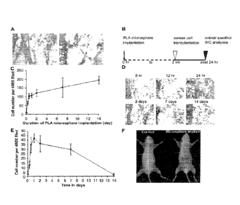
FIG. 1. Foreign body reactions trigger tumor cell migration. Pre-existing 1-
day
old subcutaneous implants were found to attract the immigration of CD I lb.
inflammatory
cells (A, left) and intraperitoneally transplanted Bl6F10 melanoma cells (A,
right). To
determine the influence of inflammatory signals in cancer cell migration,
varying degrees
of inflammatory stimuli intensities were stimulated from 6 h to 2 weeks
according to the
experimental time table (B). It was found that large numbers of CD1 lb.
inflammatory
cells were recruited to the implantation sites in 12 h and the influx of
inflammatory cells
was slowed down after that. These results depict different stages of
biomaterial-mediated
inflammatory responses (C). The stages of inflammatory responses also affect
the extent
of melanoma cell recruitment (D). Melanoma cell accumulation in the implant
area
reached a peak around 24 h post microsphere implantation (E). Inflammation-
induced
cancer metastasis is also detected in optical imaging method by labeling
melanoma cells
with Kodak X-Sight 761 near-infrared nanospheres (F).
FIG. 2. Immunohistochemical staining of subcutaneous tissues surrounding the
PLA microspheres with or without the treatment of dexamethasone (Dex). The
accumulation of inflammatory cell (CD1 lb.) in tissue implanted with PLA
microspheres
(A, top left) or PLA microspheres soaked with dexamethansone (A, top right)
can be
observed (200X). The recruitment of melanoma cells (HMB45.) was also observed
in
tissues implanted with PLA microspheres (A, bottom left) or dexamethansone
embedded
PLA microspheres (A, bottom right) (400X). Quantification of the numbers of
inflammatory cells and melanoma cells in the subcutaneous tissues with both
treatments
were graphed and statistically analyzed (B). Data are mean SD (n . 6 per
group). *P <
0.05, t-test.
FIG. 3. Extent of foreign body responses and melanoma cell recruitment to
different biomaterial implants. Immunohistochemistry staining of the tissue
was carried
out to assess the degree of foreign body reactions and quantify the
accumulation of
CD11b. inflammatory cells and HMB45. melanoma cells surround the implants,
including PLA, aluminum hydroxide and Glasperlen (A). The quantification
analysis of
-8-
CA 02888472 2015-04-15
WO 2014/063128
PCT/US2013/065803
cell recruitment was graphed (B) and the correlation between the melanoma cell
numbers
and inflammatory cell numbers in surrounding tissue of implanted microspheres
statistically analyzed (C). Data are mean SD (n . 5 per group). *P <0.05,
ANOVA.
FIG. 4. Biodistribution evaluation of B16F10 cell recruitment to the
microsphere
implant area based on immunohistological analyses. To observe the
biodistribution, GFP-
expressing B1 6F10 cells were administered intraperitoneally 24 h following
PLA
microsphere implantation. High densities of cancer cells were found in the
lymph nodes,
spleen and implantation area. However, relatively low densities of cancer
cells were
found in skin, lung, liver, and kidney.
FIG. 5. Cancer cell recruitment in response to inflammatory stimulus is
universal
in different cancer cell types, including Lewis lung cancer (LLC), human MDA-
MB-231
breast cancer, human PC-3 prostate cancer, JHU-31 rat prostate cancer. Animal
bearing
PLA implant transplanted with non-labeled cancer cells served as control
FIG. 6. AMD3100 treatment inhibited the cell recruitment of B16F10 melanoma
to the implant site (A). However, AMD3100 blockage exerted no effect on the
accumulation of melanoma cells in lymph node (B). On the other hand,
CCR7/CCL21
pathway in Bl6F10 melanoma cell accumulation in the inflamed sites was also
examined
by CCL21 neutralizing antibody treatments. In contrast, the number of tumor
cells
migration to microsphere implantation site was not affected (C). However, the
presence
of B16F10 melanoma cells in the lymph node drastically diminished (D). *P <
0.05, t-
test.
FIG. 7. (A). EPO and SDF-la loaded tissue scaffold along with control
scaffolds
were tested for their melanoma recruitment ability using a murine melanoma
metastasis
model. Real time in vivo imaging showed accumulation of labeled B16F10
melanoma
cells around the tissue scaffolds. (B). EPO and SDF-1 a loaded tissue scaffold
along with
control scaffolds were tested for their melanoma recruitment ability using a
murinc
melanoma metastasis model. EPO-releasing tissue scaffolds showed enhanced >1
fold
accumulation of melanoma cells detected using Kodak imaging system. (C) EPO
releasing scaffolds significantly enhanced the life span of cancer bearing
animals. *P <
0.05, t-test.
FIG. 8. Metastatic cancer cell trap using chemokine-releasing hydrogel.
-9-
CA 02888472 2015-04-15
WO 2014/063128
PCT/US2013/065803
FIG. 9. Schematic illustration of the cancer metastasis animal model.
FIG. 10. (A). BSA microbubbles (MB) used as porogens to fabricate PLGA
scaffolds. Microbubble image under a light microscope. (B) SEM image of BSA MB
scaffolds showed large pores and honeycomb like pore wall structure. (C)
Prominent blue
.. protein stains were found in almost all walls of the large pores in BSA MB
scaffold. (D)
The bioactivity of IGF-1 released at various time points from MB-IGF-1
scaffolds and
IGF-1 soaked scaffolds.
FIG. 11. (A). PEG-based hydrogel for controlled protein release. The fluid
phase
of hydrogel at room temperature becomes solid at 37 C. (B) PEG-based hydrogel
for
.. controlled protein release. Imaging of in vivo release of NIR-labeled BSA
from various
concentrations of hydrogel (0, 3, vs. 5%) with time. (C) PEG-based hydrogel
for
controlled protein release. The quantitative results show the controlled slow
release
properties of PEGd hydrogel.
FIG. 12. (A-H) Characterization of gelatin MB scaffolds. Scanning electron
microscopy images of (A) control (low mag) and (B) Gelatin MB scaffold (low
mag).
Scale bar: 100 pm. Scanning electron microscopy images of (C) control (high
mag) and
(D) gelatin MB (high mag). Scale bar: 50 pm. Coomassie blue staining of
internal cross
sections was done to determine the internal architecture and protein
localization in (E)
control and (F) gelatin MB scaffolds. (G) Chart showing porosity and
mechanical
.. strength of control and gelatin MB scaffolds. (H) Chart showing release of
NIR dye
conjugated EPO was determined using a fluorescence plate reader.
FIG. 13. Schematic illustration of protein-loaded PEG particle.
FIG. 14. Effect of Cancer cell traps On Leukemia Cancer Cells. (A) Mice
infected with leukemia cancer were implanted with either EPO releasing
scaffolds or
control scaffold (no EPO). After implantation of the cancer cell traps, the
numbers of
leukemia cells in the blood in both groups of animals was monitored. It was
found that
while leukemia cell numbers increased with time, the leukemia cell number
increase was
substantially slowed down. These results are demonstrated in chart (A). With
the release
of EPO, it was found that leukemia transplanted mice survival was around ¨90
days.
.. However, cancer cell traps (EPO-releasing) had ¨20% increase of survival
duration as
shown in the following chart (B).
-10-
CA 02888472 2015-04-15
WO 2014/063128
PCT/US2013/065803
FIG. 15. Effect of Cancer cell traps on Melanoma Cancer Cells. The number of
melanoma cancer cells were recruited to the implant sites of hydrogel cancer
cell traps
releasing with either RANTES, IL-8, or saline (as control) was monitored. The
results of
these experiments are demonstrated in chart (A). The survival duration of the
treated
animals was also monitored. The results of these experiments are summarized in
chart
(B).
FIG. 16. Effect of Cancer cell traps On PC3 Prostate Cancer Cells. The number
of prostate cancer cells at the sites of implanted tissue scaffolds capable of
releasing
either VEGF (50 ng/implant) or EPO (1,000 ID/implant) was monitored. The
results of
these experiments are demonstrated in chart (A). The survival duration of the
treated
animals was also monitored. The results of these experiments are summarized in
chart
(B).
FIG. 17. Effect of localized release of VEGF, EPO or SDF-la on PC3 Prostate
Cancer cell recruitment. Following transplantation for 24 hours, the
distribution of near
infrared dye-labeled cells was then monitored using whole-body imaging system.
PC3
cells were recruited to the implantation site of hydrogel cancer trap
releasing various
chemokines (VEGF, EPO and SDF-1a). The implant-associated fluorescence
intensities
were then quantified using by ImageJ software. (n=3).
FIG. 18. (A) Representative images of BSA-NIR fluorescence intensities at the
HA particle injection sites at different time. (B) The release kinetics of NIR
dye-labeled
BSA release from HA particles (labeled as "C") or saline (NIR dye+ saline) at
different
time points.
FIG. 19. Embodiment of a cancer cell trap for use as a diagnostic to detect
metastatic cancer cells.
FIG. 20. Flowchart model depicting metastatic cancer diagnosis and treatment.
FIG. 21. The numbers of cancer foci were quantified on the lung of Lewis Lung
Carcinoma cell transplanted animals without (control) or with hydrogel cancer
cell traps
implanted in either subcutaneous or intraperitoneal space.
FIG. 22. Percentages of circulating melanoma cells were found in the
peripheral
blood from animals implanted with cancer cell traps released different cancer
cell
chemokines/growth factors.
-11-
CA 02888472 2015-04-15
WO 2014/063128
PCT/US2013/065803
FIG. 23. Percentages of circulating Lewis Lung Carcinoma cancer cells were
found in the peripheral blood from animals implanted with cancer cell traps
released
different cancer cell chemokines/growth factors.
FIG. 24. Comparison of biodistribution of LLC cells in various organs isolated
from animals bearing hydrogel cancer cell traps, scaffold cancer cell traps,
or nothing (as
controls).
FIG. 25. Quantification of FITC-labeled PC3 prostate cancer cells recruited to
the
EPO-loaded particles vs. EPO + doxorubicin (300 iLtg/1 ml/implant) -loaded
particles
after implantation for different periods of time.
FIG. 26. Quantification of FITC-labeled B16F10 melanoma cancer cells recruited
to the EPO-loaded particles vs. EPO + Paclitaxel (30 mg/ml/implant)-loaded
particles
after implantation for different periods of time.
FIG. 27. Quantification of circulating AML cells following cancer cell traps
implantation. Cancer cell traps were fabricated using EPO-loaded poly-glycolic
acid
scaffolds. Blank PLGA scaffolds were used as controls. Three pairs of animals
(a single
pair of animals in each of panels A-C is shown) were tested. All three sets of
data showed
that EPO-loaded cancer cell traps not only reduce the percentages of
circulating cancer
cells but also prolonged the life span of cancer-bearing animals.
FIG. 28. The effectiveness of EPO-loaded cancer cell traps on prolonged the
life
span of AML model. The life span of the animals with or without cancer cell
traps was
determined based on either "days after trap implantation" (A) or "days after
cancer cell
transplantation" (B). Both sets of data show the substantial improvement of
life span of
animals following cancer cell trap implantation. The cancer cell trap
implantation also
improves the overall survival of cancer bearing mice (C).
FIG. 29. Histology of cancer stem cells around scaffold implants.
DETAILED DESCRIPTION OF THE INVENTION
The present invention is based on the surprising discovery that metastatic
cancer
cells migrate and accumulate in a "cancer cell trap" when placed in a subject.
The
metastasis of the cancer can thereby be detected in the subject having cancer.
In some
-12-
CA 02888472 2015-04-15
WO 2014/063128
PCT/US2013/065803
embodiments, the cancer cell trap can also suppress or prevent metastatic
tumor
formation in the subject, thereby prolonging survival of the subject. Without
being
bound by theory as to how the invention works, it is believed that the cancer
cell trap may
induce a chemokine concentration gradient in blood and as a result,
circulating metastatic
cancer cells preferentially migrate and accumulate in the cancer cell trap
instead of vital
organs.
Reference will now be made in detail to the presently preferred embodiments of
the invention which, together with the drawings and the following examples,
serve to
explain the principles of the invention. These embodiments describe in
sufficient detail to
enable those skilled in the art to practice the invention, and it is
understood that other
embodiments may be utilized, and that structural, biological, and chemical
changes may
be made without departing from the spirit and scope of the present invention.
Unless
defined otherwise, all technical and scientific terms used herein have the
same meanings
as commonly understood by one of ordinary skill in the art.
One skilled in the art may refer to general reference texts for detailed
descriptions
of known techniques discussed herein or equivalent techniques. These texts
include
Current Protocols in Molecular Biology (Ausubel et. al., eds. John Wiley &
Sons, N.Y.
and supplements thereto), Current Protocols in Immunology (Coligan et al.,
eds., John
Wiley St Sons, N.Y. and supplements thereto), Current Protocols in
Pharmacology
(Enna et al., eds. John Wiley & Sons, N.Y. and supplements thereto) and
Remington: The
Science and Practice of Pharmacy (Lippincott Williams & Wilicins, 2Vt edition
(2005)),
for example.
Definitions of common terms in molecular biology may be found, for example, in
Benjamin Lewin, Genes VII, published by Oxford University Press, 2000 (ISBN
019879276X); Kendrew et al. (eds.); The Encyclopedia of Molecular Biology,
published
by Blackwell Publishers, 1994 (ISBN 0632021829); and Robert A. Meyers (ed.),
Molecular Biology and Biotechnology: a Comprehensive Desk Reference, published
by
Wiley, John & Sons, Inc., 1995 (ISBN 0471186341).
For the purpose of interpreting this specification, the following definitions
will
apply and whenever appropriate, terms used in the singular will also include
the plural
and vice versa. In the event that any definition set forth below conflicts
with the usage of
-13-
WO 2014/063128
PCT/US2013/065803
that word in any other document,
the definition set forth below shall always control for purposes of
interpreting
this specification and its associated claims unless a contrary meaning is
clearly intended
(for example in the document where the term is originally used). The use of
"or" means
"and/or" unless stated otherwise. The use of "a" herein means "one or more"
unless
stated otherwise or where the use of "one or more" is clearly inappropriate.
The use of
"comprise," "comprises," "comprising," "include," "includes," and "including"
are
interchangeable and not intended to be limiting. Furthermore, where the
description of
one or more embodiments uses the term "comprising," those skilled in the art
would
understand that, in some specific instances, the embodiment or embodiments can
be
alternatively described using the language "consisting essentially of" and/or
"consisting
of."
"Cancer cell trap" as encompassed by the present invention refers to a
material
that enables the migration and accumulation of metastatic cancer cells in the
material for
a period of time. In some embodiments, the cancer cell trap is capable of
releasing one or
more molecules selected from proteins, chemokines, growth factors,
therapeutics,
chemotherapeutic agents, anti-cancer agents and combinations thereof.
As used herein, the term "about" means plus or minus 10% of the numerical
value
of the number with which it is being used.
A "therapeutically effective amount" or "effective amount" as used herein is
an
amount sufficient to decrease, suppress, prevent or ameliorate the symptoms
associated
with cancer, including suppressing or decreasing the formation of metastatic
tumors.
As used herein, "treat" and all its forms and tenses (including, for example,
treating, treated, and treatment) can refer to therapeutic or prophylactic
treatment. In
certain aspects of the invention, those in need thereof of treatment include
those already
with a pathological condition of the invention (including, for example, a
cancer), in
which case treating refers to administering to a subject (including, for
example, a human
or other mammal in need of treatment) a therapeutically effective amount of a
composition so that the subject has an improvement in a sign or symptom of a
pathological condition of the invention. The improvement may be any observable
or
measurable improvement. Thus, one of skill in the art realizes that a
treatment may
-14-
Date Recue/Date Received 2020-06-01
CA 02888472 2015-04-15
WO 2014/063128
PCT/US2013/065803
improve the patient's condition, but may not be a complete cure of the
pathological
condition. In other certain aspects of the invention, those in need of
treatment include
those already with cancer as well as those prone to have cancer or in those in
whom
cancer metastasis is to be prevented.
As used herein, -cancer" refers to a pathophysiological condition whereby a
cell or
cells is characterized by dysregulated and/or proliferative cellular growth
and the ability
to induce said growth, either by direct growth into adjacent tissue through
invasion or by
growth at distal sites through metastasis, which includes but is not limited
to, carcinomas
and sarcomas, such as, for example, acute lymphoblastic leukemia, acute
myeloid
leukemia, adrenocortical cancer, AIDS-related cancers, AIDS-related lymphoma,
anal
cancer, astrocytoma (including, for example, cerebellar and cerebral), basal
cell
carcinoma, bile duct cancer, bladder cancer, bone cancer, brain stem glioma,
brain tumor
(including, for example, ependymoma, medulloblastoma, supratentorial primitive
neuroectodermal, visual pathway and hypothalamic glioma), cerebral
astrocytoma/malignant glioma, breast cancer, bronchial adenomas/carcinoids,
Burkitt's
lymphoma, carcinoid tumor (including, for example, gastrointestinal),
carcinoma of
unknown primary site, central nervous system lymphoma, cervical cancer,
chronic
lymphocyti c leukemia, chronic myelogenous leukemia, chronic
myeloproliferative
disorders, colon cancer, colorectal cancer, cutaneous T-Cell lymphoma,
endometrial
cancer, ependymoma, esophageal cancer, Ewing's Family of tumors, extrahepatic
bile
duct cancer, eye cancer (including, for example, intraocular melanoma,
retinoblastoma,
gallbladder cancer, gastric cancer, gastrointestinal carcinoid tumor,
gastrointestinal
stromal tumor (GIST), germ cell tumor (including, for example, extracranial,
extragonadal, ovarian), gestational trophoblastic tumor, glioma, hairy cell
leukemia, head
and neck cancer, squamous cell head and neck cancer, hepatocellular cancer,
Hodgkin's
lymphoma, hypopharyngeal cancer, islet cell carcinoma (including, for example,
endocrine pancreas), Kaposi's sarcoma, laryngeal cancer, leukemia, lip and
oral cavity
cancer, liver cancer, lung cancer (including, for example, non-small cell),
lymphoma,
macroglobulinemia, malignant fibrous histiocytoma of bone/osteosarcoma,
medulloblastoma, melanoma, Merkel cell carcinoma, mesothelioma, metastatic
squamous
neck cancer with occult primary, mouth cancer, multiple endocrine neoplasia
syndrome,
-15-
CA 02888472 2015-04-15
WO 2014/063128
PCT/US2013/065803
multiple myeloma/plasma cell neoplasm, mycosis fungoides, myelodysplastic
syndromes,
myelodysplastic/myeloproliferative diseases, myeloma, nasal cavity and
paranasal sinus
cancer, nasopharyngeal cancer, neuroblastoma, non-Hodgkin's lymphoma, oral
cancer,
oral cavity cancer, osteosarcoma, oropharyngeal cancer, ovarian cancer
(including, for
example, ovarian epithelial cancer, germ cell tumor), ovarian low malignant
potential
tumor, pancreatic cancer, paranasal sinus and nasal cavity cancer, parathyroid
cancer,
penile cancer, pharyngeal cancer, pheochromocytoma, pineoblastoma and
supratentorial
primitive neuroectodermal tumors, pituitary tumor, plasma cell
neoplasm/multiple
myeloma, pleuropulmonary blastoma, pregnancy and breast cancer, primary
central
nervous system lymphoma, prostate cancer, rectal cancer, retinoblastoma,
rhabdomyosarcoma, salivary gland cancer, soft tissue sarcoma, uterine sarcoma,
Sezary
syndrome, skin cancer (including, for example, non-melanoma or melanoma),
small
intestine cancer, supratentorial primitive neuroectodermal tumors, T-Cell
lymphoma,
testicular cancer, throat cancer, thymoma, thymoma and thymic carcinoma,
thyroid
cancer, transitional cell cancer of the renal pelvis and ureter, trophoblastic
tumor
(including, for example, gestational), unusual cancers of childhood and
adulthood,
urethral cancer, endometrial uterine cancer, uterine sarcoma, vaginal cancer,
viral
induced cancers (including, for example, HPV induced cancer), vulvar cancer,
Waldenstrom's macroglobulinemia, Wilms' Tumor, and women's cancers.
The term "hydrogel" is used in the conventional sense to refer to water-
swellable
polymeric or polysaccharide-based matrices that can absorb a substantial
amount of water
to form elastic gels, wherein "matrices" are three-dimensional networks of
macromolecules held together by covalent or noncovalent crosslinks. Some of
these
hydrogel can be solidified with temperature- or pH-changes. Upon placement in
the body,
the hydrogel can be used as carrier to release a variety of biomolecules.
As used herein, terms such as "drug," "agent," "pharmaceutical" may be used
interchangeably. In general, these terms refer to any chemical substance used
in the
treatment, cure, prevention, or diagnosis of a disease or condition or to
otherwise change
the physical or mental status of a human or other animal, regardless of
molecular weight.
A pharmaceutical composition may also be prepared using a drug in combination
with a
drug delivery vehicle of the invention. The pharmaceutical composition can
comprise a
-16-
CA 02888472 2015-04-15
WO 2014/063128
PCT/US2013/065803
drug in a suitable polymeric form and a biologically acceptable carrier.
Suitable
polymeric forms include microcapsules, microparticles, films, polymeric
coatings, and
nanoparticles.
Cancer Cell Trap
In one embodiment, the invention provides a cancer cell trap for treating,
preventing and/or diagnosing cancer metastasis, wherein metastatic cancer
cells are
capable of migrating and accumulating in the cancer cell trap over a period of
time
when the cancer cell trap is placed into a subject.
In accordance with some embodiments of the invention, the cancer cell trap can
be fabricated with the capability to release one or more bioactive molecules
and/or
drugs, such as proteins, chemokines, growth factors and chemotherapeutic or
anti-
cancer agents.
The cancer cell trap can be made from one or more materials and the materials
that can be used in fabricating the cancer cell trap are not limiting.
Preferably, the
material is biocompatible and generally non-toxic to the subject's healthy,
non-
cancerous cells.
In some embodiments, the cancer cell trap comprises one or more materials
selected from water soluble polymers, including, but not limited to, dextran,
derivatives of poly-methacrylamide, PEG, maleic acid, malic acid, and maleic
acid
anhydride and may include these polymers and a suitable coupling agent,
including 1-
ethyl-3 (3-dimethylaminopropy1)-carbodiimide, also referred to as
carbodiimide. In
some embodiments, polymers may be degradable or nondegradable or of a
polyelectrolyte material. In some embodiments, degradable polymer materials
include
poly-L-glycolic acid (PLGA), poly-DL-glycolic, poly-L-lactic acid (PLLA), PLLA-
PLGA copolymers, poly(DL-lactide)-block-methoxy polyethylene glycol,
polycaprolacton, poly(caprolacton)-block-methoxy polyethylene glycol (PCL-
MePEG), poly(DL-lactide-co-caprolactone)-block-methoxy polyethylene glycol
(PDLLACL-MePEG), some polysaccharide (e.g., hyaluronic acid, polyglycan,
chitoson), proteins (e.g., fibrinogen, albumin, collagen, extracellular
matrix), peptides
(e.g., RGD, polyhistidine), nucleic acids (e.g., RNA, DNA, single or double
stranded),
-17-
CA 02888472 2015-04-15
WO 2014/063128
PCT/US2013/065803
viruses, bacteria, cells and cell fragments, organic or carbon-containing
materials, as
examples. Nondegradable materials include natural or synthetic polymeric
materials
(e.g., polystyrene, polypropylene, polyethylene teraphthalate, polyether
urethane,
polyvinyl chloride, silica, polydimethyl siloxane, acrylates, arcylamides,
poly
(vinylpyridinc), polyacrolcine, polyglutaraldehyde), some polysaccharides
(e.g.,
hydroxypropyl cellulose, cellulose derivatives, dextrant, dextrose, sucrose,
ficoll ,
percoll , arabinogalactan, starch), and hydrogels (e.g., polyethylene glycol,
ethylene
vinyl acetate, N-isopropylacrylamide, polyamine, polyethyleneimine, poly-
aluminum
chloride).
In some embodiments, the cancer cell trap comprises materials selected from
the
group consisting of a scaffold structure, hydrogel, nanoparticles and/or
microparticles.
In some embodiments, the cancer cell trap comprises one or more materials with
controlled release properties capable of releasing bioactive molecules and/or
chemotherapeutic agents. In some embodiments, the hydrogel cancer cell trap is
a
liquid composition and is injected or implanted in the subject. In some
embodiments,
the nanoparticles and/or microparticles cancer cell trap is a liquid
composition of
particles and is injected or implanted in the subject. In some embodiments,
the
scaffold structure is a solid composition and is implanted in the subject or
injected via
a surgical procedure. In some
embodiments, the scaffold structure, hydrogel,
microparticles and/or nanoparticles are injected via 19-21 gauge needles.
In some embodiments, the cancer cell traps are implanted or injected in the
subcutaneous space and/or intraperitoneal cavities.
In some embodiments, the cancer cell trap comprises effective amounts of one
or more bioactive molecules. In some embodiments, the bioactive molecules are
added
to the cancer cell trap by physical absorption. In some embodiments, the
bioactive
molecules facilitate the recruitment and migration of metastatic cancer cells
to the
cancer cell trap. In some embodiments, the bioactive molecules are selected
from the
group consisting of IL-1, IL-4, IL-8, IL-10, IL-13, IL-17, CCL2, CCL5, CCL9,
CCL18, CCL19, CCL20, CCL21, CCL25, CCL27, CCR4, CCR5, CCR7/CCL21,
CCR9, CCR10, CCL18, CCL2/MCP-1, MIP-1a/CCL3, CXCL1, CXCL2, CXCL3,
CXCL4, CXCL5, CXCL8, CXCL12/SDF-la, CXCR2, CXCR3, CXCR4, CXCR7,
-18-
CA 02888472 2015-04-15
WO 2014/063128
PCT/US2013/065803
erythropoietin (EPO), CCL5/RANTES, hepatocyte growth factor activator (HGFA),
insulin-like growth factor-1 (IGF-1), cylooxygenase-2 (COX-2), CXCL14,
prostaglandin E2, platelet derived growth factor, vascular endothelial growth
factor
(VEGF) and combinations thereof. Bioactive fragments and variants can also be
used.
In some embodiments, the cancer cell trap releases an effective amount of
bioactive molecules after it is injected or implanted in a subject. In some
embodiments
the release is over an extended period of time. In some embodiments, the
bioactive
molecules are released over a period of 1-6 months. In some embodiments, the
bioactive molecules are released over a period of about 1 week, 2 weeks, 3
weeks, or 4
weeks. In some embodiments, the bioactive molecules are released over a period
of
about 14 days. In some embodiments, the bioactive molecules are released over
a
period of about 7-10 days. In some embodiments, the bioactive molecules are
released
over a period of about 2-7 days.
By the term "effective amount" with regard to the bioactive molecules, is
meant
an amount that produces the desired effect for which it is administered, viz.,
inducing
the recruitment and migration of the metastatic cancer cells to the cancer
cell trap. The
exact amount will depend on the particular agent, the subject to be treated,
and will be
ascertainable by a person skilled in the art using known methods and
techniques for
determining effective doses. In some embodiments, the amount of the bioactive
molecule to be administered includes between about 0.05 ng/kg/day to about 1
mg/kg/day. In some embodiments, the amount of bioactive molecule that can be
administered in amounts between about 0.1 ng/kg/day to about 1 gg/kg/day.
In some embodiments, the bioactive molecules may be released in the following
concentrations ranges: 1L-8 (0.01- 250 ng/day/1 ml or 1 cubic cm of implant),
CCLI9
(10 ag - 1000 ng/day/1 ml or 1 cubic cm of implant), CCL20 (0.1 - 4000 nano
moles/
day/1000 ml or 1000 cubic cm of implant), CCL21 (0.01 -100 micro
moles/day/1000
ml or 1000 cubic cm of implant), CCL2/MCP-1 (0.05 - 100 ng/day/1 ml or 1 cubic
cm
of implant ), CCL3 (10-1000 ng/day/1 ml or 1 cubic cm of implant), CXCL12/SDF-
la
(0.5-500 nano moles/day/1000 ml or 1000 cubic cm of implant), CCL5/RANTES
(0.01
- 1000 ng/day/1 ml or 1 cubic cm of implant), and EPO (1-10000 I.U./day/1 ml
or 1
-19-
CA 02888472 2015-04-15
WO 2014/063128
PCT/US2013/065803
cubic cm of implant ), CCL5/ RANTES (0.2 - 500 ng/day/1 ml or 1 cubic cm of
implant), and VEGF (0.01 - 100 ng/day/1 ml or 1 cubic cm of implant).
In some embodiments, the bioactive molecules may be released in the following
concentrations ranges: IL-8 (0.1- 20 ng/day/1 ml or 1 cubic cm of implant ),
CCLI9
(100 [tg - 100 ng/day/1 ml or 1 cubic cm of implant), CCL20 (1 - 400 nano
moles/day/
1000 ml or 1000 cubic cm of implant), CCL21 (0.1 -10 micro moles/day/ 1000 ml
or
1000 cubic cm of implant), CCL2/MCP-1 (0.5 - 10 ng/day/1 ml or 1 cubic cm of
implant), CCL3 (1-100 ng/day/ 1 ml or 1 cubic cm of implant), CXCL12/SDF-la (5-
50 nano moles/day/1000 ml or 1000 cubic cm of implant), CCL5/RANTES (0.1 - 10
.. ng/day/1 ml or 1 cubic cm of implant), and EPO (1-100 I.U./day/1 ml or 1
cubic cm of
implant), CCL5/ RANTES (2 - 50 ng/day/1 ml or 1 cubic cm of implant), and VEGF
(0.1 - 10 ng/day/1 ml or 1 cubic cm of implant).
In some embodiments, the cancer cell trap may be fabricated to release
independently or combinations of recombinant human HGF/SF (10 ng/day/1 ml or 1
cubic cm of implant), MCP-1 (0.5 to 10 ng/day/1 ml or 1 cubic cm of implant),
CXCL12/ SDF- 1 a (5 to 50 nano moles/day/1000 ml or 1000 cubic cm of implant),
CCL5/ RANTES (0.5 to 10 ng/day/1 ml or 1 cubic cm of implant), and EPO (Ito
100
I.U./ day/ 1 ml or 1 cubic cm of implant).
In some embodiments, the cancer cell trap may be fabricated to release
hepatocyte growth factor/scatter factor (HGF/SF), MCP-la, RANTES, SDF- la, MCP-
1, EPO, histamine, or MIP-la, and combinations thereof. In some embodiments,
these
cancer cell traps may be fabricated using methods described in Otsuka, S. and
G. Bebb,
J Thorac Oncol, 2008. 3(12): p. 1379-83.
In some embodiments, the cancer cells are recruited to the cancer cell trap
based
on the chemokine gradient and localized concentrations of the chemokine.
In some embodiments wherein EPO is released, the injection quantity is about
600 units/0.027 milliliter of hydrogel/particle cancer traps or 27 cubic
millimeters
scaffold traps. In some embodiments, the release rate is about 1.5 to about
2.5
international units/day. In some embodiments, EPO is released over a period of
greater
than about 30 days.
-20-
CA 02888472 2015-04-15
WO 2014/063128
PCT/US2013/065803
In some embodiments wherein RANTES/CCL5 is released, the injection
quantity is about 600 ng/milliliter of hydrogel/particle cancer traps or 1
cubic
centimeters scaffold traps. In some embodiments, the release rate is about 10
ng/day.
In some embodiments, RANTES/CCL5 is released over a period of greater than
about
21 days.
In some embodiments wherein hepatocyte growth factor (HGFISF) is released,
the injection quantity is about 900 ng/milliliter of hydrogel/particle cancer
traps or 1
cubic centimeters scaffold traps. In some embodiments, the release rate is
about 15
ng/day. In some embodiments, hepatocyte growth factor (HGF/SF) is released
over a
period of greater than about 28 days.
In some embodiments wherein SDF-1a is released, the injection quantity is
about 10 jig/milliliter of hydrogel/particle cancer traps or 1 cubic
centimeters scaffold
traps. In some embodiments, the release rate is about 100 ng/day. In some
embodiments, SDF-1 a is released over a period of greater than about 24 days.
In some embodiments, the cancer cell trap of the present invention may be
fabricated to release: RANTES (10-500 jig/kg body weight), EPO (1-20 IU /kg
body
weight), SDF-la (0.1-10 mg/ kg body weight), MCP-1 (0.1-10 mg/ kg body
weight),
and MIP-la (0.1-10 mg/ kg body weight).
In some embodiments, two or more bioactive molecules are released from the
cancer cell trap.
The cancer cell trap is used to recruit metastatic cancer cells. The
metatstatic
cancer cell is not limiting, and can include any metastatic cancer cell. In
some
embodiments, the metastatic cancer cell is selected from the group consisting
of
melanoma, prostate cancer, leukemia, squamous cell carcinoma, astrocytoma,
Kaposi's
sarcoma, glioblastoma, lung cancer, bladder cancer, head and neck cancer,
ovarian
cancer, uterine cancer, breast cancer, lung cancer, glioma, colorectal cancer,
genitourinary cancer, gastrointestinal cancer, thyroid cancer and skin cancer.
In some embodiments, the cancer cell trap may comprise effective amounts of
one or more anti-cancer or chemotherapeutic agents, which can be used to kill
or
inhibit the growth of metastatic cancer cells. In some
embodiments, the
chemotherapeutic agent is released from the cancer cell trap and also kills or
inhibits
-21-
CA 02888472 2015-04-15
WO 2014/063128
PCT/US2013/065803
circulating metastatic cells in addition to the cells accumulated in the
cancer cell trap.
A suitable chemotherapeutic or anti-cancer agent for use in the invention can
be any
chemical substance known to be useful for treating cancer, for example,
Abraxane,
Aldara, Alimta, Aprepitant, Arimidex, Aromasin, Arranon, Arsenic Trioxide,
Avastin,
Bcvacizumab, Bexarotene, Bortczomib, Cetuximab, Clofarabine, Clofarcx, Clolar,
Dacogen, Dasatinib, Ellence, Eloxatin, Emend, Erlotinib, Faslodex, Femara,
Fulvestrant, Gefitinib, Gemtuzumab Ozogamicin, Gemzar, Gleevec, Herceptin,
Hycamtin, Imatinib Mesylate, Iressa, Kepivance, Lenalidomide, Lev-ulan,
Methazolastone, Mylosar, Mylotarg, Nanoparticle Paclitaxel, Nelarabine,
Nexavar,
Nolvadex, Oncaspar, Oxaliplatin, Paclitaxel, Paclitaxel Albumin-stabilized
Nanoparticle Formulation, Palifermin, Panitumumab, Pegaspargase, Pemetrexed
Disodium, Platinol-AQ, Platinol, Revlimid, Rituxan, Sclerosol Intrapleural
Aerosol,
Sorafenib Tosylate, Sprycel, Sunitinib Malate, Sutent, Synovir, Tamoxifen,
Tarceva,
Targretin, Taxol, Taxotere, Temodar, Temozolomide, Thalomid, Thalidomide,
Topotecan Hydrochloride, Trastuzumab, Trisenox, Vectibix, Velcade, Vidaza,
Vorinostat, Xcloda, Zoledronic Acid, Zolinza, Zometa, doxorubicin, adriamycin,
b eomycin , daunorubicin, dactinomycin , epirubicin, i darubi cm,
mitoxantrone,
valrubicin, hydroxyurea, mitomycin, fluorouracil, 5-FU, methotrexate,
floxuridine,
interferon alpha-2b, glutamic acid, plicamycin, 6-thioguanine, aminopterin,
pemetrexed, raltitrexed, cladribine, clofarabine, fludarabine, mercaptopurine,
pentostatin, capecitabine, cytarabine, carmustine, BCNU, lomustine, CCNU,
cytosine
arabinoside, cyclophosphamide, estramustine, hydroxyurea, procarbazine,
mitomycin,
busulfan, medroxyprogesterone, estramustine phosphate sodium, ethinyl
estradiol,
estradiol, megestrol acetate, methyltestosterone, diethylstilbestrol
diphosphate,
chlorotrianisene, testolactone, mephalen, mechlorethamine, chlorambucil,
chlormethine, ifosfamide, bethamethasonc sodium phosphate, dicarbazine,
asparaginasc, mitotanc, vincristinc, vinblastine, ctoposidc, teniposide,
Topotecan, 1FN-
gamma, irinotecan, campto, irinotecan analogs, carmustine, fotemustine,
lomustine,
streptozocin, carboplatin, oxaliplatin, BBR3464, busulfan, dacarbazine,
mechlorethamine, procarbazine, thioTEPA, uramustine, vindesine, vinorelbine,
alemtuzumab, tositumomab, methyl aminolevulinate, porfimer, verteporfin,
lapatinib,
-22-
CA 02888472 2015-04-15
WO 2014/063128
PCT/US2013/065803
nilotinib, vandetanib, ZD6474, alitretinoin, altretamine, amsacrine,
anagrelide,
denileukin diftitox, estramustine, hydroxycarbamide, masoprocol, mitotane,
tretinoin,
or other anticancer agents, including, for example, cytotoxic agents, DNA-
alkylating
agents, anti-tumor antibiotic agents, anti-metabolic agents, tubulin
stabilizing agents,
tubulin destabilizing agents, hormone antagonist agents, topoisomerase
inhibitors,
protein kinase inhibitors, HMG-CoA inhibitors, CDK inhibitors, cyclin
inhibitors,
caspase inhibitors, metal loproteinase inhibitors, antisense nucleic acids,
triple-helix
DNAs, nucleic acids aptamers, and molecularly-modified viral, bacterial or
exotoxic
agents. In further particular aspects of the invention, an anticancer agent
comprises two
or more of the foregoing anticancer agents.
In some embodiments, the cancer cell trap can be fabricated with a combination
of anti-cancer or chemotherapeutic agents. In some embodiments, a combination
of
agents includes, for example, CHOP (Cytoxan, Hydroxyrubicin (Adriamycin),
Oncovin (Vincristine), Prednisone), CHOP-R (CHOP, rituximab), FOLFOX
(Fluorouracil, leucovorin (folinic acid), oxaliplatin), VAD (Vincristine,
Adriamycin
(doxorubicin), dexamethasone), Thal/Dex (Thalidomide, dexamethasone), COP or
CVP (Cyclophosphamide, vincristine (Oncovin), and prednisone), m-BACOD
(Methotrexate, bleomycin, doxorubicin (Adriamycin), cyclophosphamide,
vincristine
(Oncovin), dexamethasone (Decadron)), ProMACE-CytaBOM (Prednisone,
doxorubicin (adriamycin), cyclophosphamide, etoposide, cytarabine, bleomycin,
vincristine (Oncovin), methotrexate, leucovorin), COPP (Cyclophosphamide,
Oncovin
(vincristine), procarbazine, prednisone), MACOP-B (Methotrexate, leucovorin,
doxorubicin (Adriamycin), cyclophosphamide, vincristine (Oncovin), prednisone,
bleomycin), MOPP (Mechlorethamine, vincristine (oncovin), procarbazine,
prednisone), ProMACE-MOPP (Methotrexate, doxorubicin (Adriamycin),
cyclophosphamide, etoposide, MOPP), ABVD (Adriamycin, bleomycin, vinblastine,
dacarbazine), BEACOPP (Bleomycin, etoposide, Adriamycin (doxorubicin),
cyclophosphamide, Oncovin (vincristine), procarbazine, predni sone), Stanford
V
(Doxorubicin (Adriamycin), mechlorethamine, bleomycin, vinblastine,
vincristine
(Oncovin), etoposide (VP-16), prednisone), ECF (Epirubicin, cisplatin,
fluorouracil),
-23-
CA 02888472 2015-04-15
WO 2014/063128
PCT/US2013/065803
BEP (Bleomycin, etoposide, platinum (cisplatin)), and PCV (Procarbazine,
lomustine
(CCNU), vincristine).
By the term "effective amount" with regard to the chemotherapeutic agent is
meant an amount that produces the desired effect for which it is administered,
viz.,
killing or inhibiting the growth of the metastatic cancer cells. The exact
amount will
depend on the particular agent, the subject to be treated, and will be
ascertainable by a
person skilled in the art using known methods and techniques for determining
effective
doses. In some embodiments, the amount of the chemotherapeutic agent to be
administered includes between about 0.01 g/kg/day to about 100 mg/kg/day. In
some
embodiments, the amount of chemotherapeutic agent that can be administered
includes
between about 0.1 mg/kg/day to about 10 mg/kg/day.
In some embodiments, the cancer cell trap is fabricated to incorporate and/or
release paclitaxel, doxorubicin, and/or vincristine. In some embodiments, the
cancer
cell traps can be fabricated to release doxorubicin at a rate of about 0.1-
1000 g/day,
0.5-500 jug/day, 1-100 jig/day or 2-20 g/day per 1 ml of hydrogel/particle
cancer traps
or 1 cubic centimeters scaffold traps . In some embodiments, the cancer cell
traps can
be fabricated to release paclitaxel at a rate of about 0.01-500 mg/day, 0.1-
100 mg/day,
0.1-50 mg/day, 0.2-20 mg/day or 0.2-2 mg/day per 1 ml of hydrogel/particle
cancer
traps or 1 cubic centimeters scaffold traps.
The cancer cell traps can be fabricated into any type of shape. In some
embodiments, solid cancer cell traps have a disc shape. In some embodiments,
solid
cancer cell traps can be fabricated to have a tubular shape. In some
embodiments, the
tubular structure has an opening on one or both sides. in some embodiments,
the
tubular structure has a porous structure which allows infiltration of cancer
cells from
the sides and the opening to the inner lumen of the cancer cell trap. In some
embodiments, the cancer cells can be recovered from the inner lumen of the
cancer cell
trap via a needle, such as an 18-21 gauge needle.
Scaffold Cancer Cell Trap
In some embodiments, the cancer cell trap is fabricated as a scaffold
structure.
In some embodiments, the cancer cell trap is a tissue scaffold. In some
embodiments,
-24-
CA 02888472 2015-04-15
WO 2014/063128
PCT/US2013/065803
the cancer cell trap comprises one or more extracellular matrix components. In
some
embodiments, the cancer cell trap is a microbubble scaffold. In some
embodiments,
the cancer cell trap is made from synthetic polymers. In some embodiments, the
cancer
cell trap is made from polymers and proteins. In some embodiments, the
scaffold
structure is prepared from one or more proteins, polymers, and combinations
thereof.
In some embodiments, the proteins are extracellular matrix proteins, such
collagen I,
collagen III, elastin and fibronectin. In some embodiments, the scaffold is
degradable.
In some embodiments, the scaffold comprises a biodegradable polymer and one or
more polypeptides. In some embodiments, scaffolds can be created from tissues
wherein the cells are removed, leaving behind a scaffold structure comprising
extracellular matrix components.
In some embodiments, the scaffold structure is generally porous in nature. In
some embodiments, the porosity ranges from about 10-97%, about 25-98%, about
50-
95% and about 80-90%.
In some embodiments, the scaffolds can be fabricated from biodegradable
polymers such as aliphatic polyesters, alginate, cellulose, chitin, chitosan,
collagen,
copolymers of glycolide, copolymers of lactide, el astin, fibrin, glycolide/l-
lactide
copolymers (PGA/PLLA), glycolide/trimethylene carbonate copolymers (PGA/TMC),
glycosaminoglycans, lactide/tetramethylglycolide copolymers,
lactide/trimethylene
carbonate copolymers, lactide/E-caprolactone copolymers, lactide/cy-
valerolactone
copolymers, L-lactide/dl-lactide copolymers, methyl methacrylate-N-vinyl
pyrrolidone
copolymers, modified proteins nylon-2 PHBA/y-hydroxyvalerate copolymers
(PHBA/HVA), PLA/polyethylene oxide copolymers, PLA-polyethylene oxide (PELA),
poly (amino acids), poly (trimethylene carbonates), poly hydroxyalkanoate
polymers
(PHA), poly(alklyene oxalates), poly(butylene diglycolate), poly(hydroxy
butyrate)
(PHB), poly(n-vinyl pyrrolidonc), poly(ortho esters), polyalky1-2-
cyanoacrylates,
polyanhydrides, polycyanoacrylates, polydepsipeptides, polydihydropyrans, Poly-
dl-
lactide, (PDLLA), polyesteramides, polyesters of oxalic acid, polyglycolide
(PGA),
polyiminocarbonates, polylactides (PLA), poly-l-lactide (PLLA),
polyorthoesters,
poly-p-dioxanone (PDO), polypeptides, polyphosphazenes, polysaccharides,
polyurethanes (PU) polyvinyl alcohol (PVA) poly-13-hydroxypropionate (PHPA),
poly-
-25-
CA 02888472 2015-04-15
WO 2014/063128
PCT/US2013/065803
13-hydroxybutyrate (PBA), poly-a-valerolactone poly-I3-alkanoic acids, poly-I3-
malic
acid (PMLA), poly-E-caprolactone (PCL), pseudo-Poly(Amino Acids), starch,
trimethylene carbonate (TMC), and/or tyrosine based polymers.
In some embodiments, the scaffold is fabricated from PLGA, albumin, collagen,
gelatin, immunoglobulins, extracellular matrix proteins, fibronectin and
combinations
thereof. In some embodiments, the scaffold comprises a degradable polymer and
polypeptides.
In some embodiments, the scaffold structure is a microbubble scaffold (MB),
which results in a porous scaffold that is capable of incorporating cells and
also
releasing bioactive molecules. Microbubble scaffolds can be prepared, for
example,
according to techniques discussed in Nair et al., Novel polymeric scaffolds
using
protein microbubbles as porogen and growth factor carriers. Tissue Eng Part C
Methods, 2010. 16(1): p. 23-32. In some embodiments, microbubbles are first
prepared and then combined with polymers to form the microbubble scaffold.
Microbubbles can also be loaded with bioactive molecules to produce scaffolds
that
release bioactive molecules in accordance with some embodiments of the
invention.
In some embodiments, the microbubbles can be prepared as follows: a solution
of protein such as BSA (e.g., 5% w/v, 10% w/v, 20% w/v or 50% w/v) is overlaid
with
nitrogen gas. The mixture is sonicated using a probe sonicator (Ultrasonix,
Bothell,
WA) at 20 kHz for 10 s. This procedure results in the formation of nitrogen
gas¨filled
MB that are surrounded by a BSA protein shell. The MBs can be transferred to
glass
tubes and kept at 48 C. To observe the physical structure of MB, a small
droplet of the
MB can be placed on a glass slide and then imaged under a microscope (Leica
Microsystems, Wetzlar, Germany). The MB size distribution generally ranges
from 50
to 200 ium in diameter. To synthesize a biomolecule¨loaded MB (labeled as MB-
chemokine), a chemokine, such as IGF-1 (for example, 500 ng/mL) solution is
mixed
with BSA solution before sonication under nitrogen gas as described above.
In some embodiments, the microbubbles can then be combined with various
concentrations of polymer solution (e.g., 5% w/v, 7.5% w/v, and 10% w/v) to
create MB-
embedded porous scaffolds. Such MB¨polymer mixtures can be phase separated at
various temperatures (0 C, 20 C, and 196 C). Briefly, in some embodiments,
7.5% w/v
-26-
CA 02888472 2015-04-15
WO 2014/063128
PCT/US2013/065803
PLGA can be dissolved in 1,4-dioxane by vortexing for about 20 min until the
polymer
completely dissolved in the solvent. In some embodiments, the polymer solution
can then
be mixed with the BSA-MB or biomolecule-loaded BSA-MB (e.g., 5% w/v BSA) in a
ratio of 1:1. After gentle agitation for about 3 min at room temperature, the
polymer-
solution mixtures in glass Petri dishes (5 cm diameter) arc then quenched in
liquid
nitrogen to induce phase separation. The solidified scaffolds can then
lyophilized for 48 h
at 0.03 mbar vacuum, for example, in a Freezone 12 lyophilizer (Labconco,
Kansas City,
MO). For producing biomolecule loaded MB-embedded scaffolds, biomolecule-
loaded
MB (for example, MB-IGF-1, MB manufactured in the presence of 500 ng/mL IGF-1)
is
used as porogens.
In some embodiments, the microbubble scaffold of the present invention may be
fabricated from a single protein or protein mixtures in different ratios. In
some
embodiments, the microbubble scaffold is fabricated from albumin, collagen,
gelatin,
immunoglobulins, extracellular matrix proteins, fibronectin, and combinations
thereof.
In some embodiments, the microbubble scaffold releases one or more
biomolecules. In some embodiments, the microbubble scaffold is capable of
releasing
biomolecules in the following concentrations ranges: IL8 (0.1- 20 ng/1 cubic
centimeters scaffold/day), CCLI9 (100 pg - 100 ng/1 cubic centimeters
scaffold/day),
CCL20 (1 - 400 nmole/1000 cubic centimeter scaffold/day), CCL2I (0.1 -10
micromole/1000 cubic centimeter scaffold/day ), CCL2/MCP-1 (0.5 - 10 rig/1
cubic
centimeter scaffold/day), CCL3 (1-100 ng/1 cubic centimeter scaffold/day),
CXCLI2/
SDF-Ia (5-50 nanonmole/1000 cubic centimeter scaffold/day), CCL5/ RANTES (0.1 -
10 ng/1 cubic centimeter scaffold/day), and EPO (1-100 I.U./ 1 cubic
centimeter
scaffold/day), CCL5/ RANTES (2 - 50 ng/1 cubic centimeter scaffold/day), and
VEGF
(0.1 - 10 ng/1 cubic centimeter scaffold/day).
In some embodiments, the microbubble scaffolds have a porosity ranging from 70-
98 %. In some embodiments, the microbubble scaffold has a pore size ranging
from 10
lam to 300 pm. In some embodiments, the pore size is selected from about 20 m
to
about 200 pm, from about 40 pm to about 150 pm, from about 80 pm to about 130
pm,
and from about 100 juna to about 120 p.m.
-27-
CA 02888472 2015-04-15
WO 2014/063128
PCT/US2013/065803
The microbubble scaffold may have a bolus release of 5 to 35% of loaded
biomolecule. For example, the microbubble scaffold may be fabricated to have a
bolus
release of 20% of biomolecule, including chemokine, growth factor or protein,
within
the first 24 hours.
In some embodiments, the scaffolds arc fabricated to provide a sustained
release
biomolecules of approximately 2-10% of total amounts per day.
Nanoparticles and or Microparticles
In some embodiments, the cancer cell traps can also be fabricated using
microparticles and/or nanoparticles. In some embodiments, the particles are
capable of
releasing various bioactive molecules.
In some embodiments, the nanoparticles and microparticles can be fabricated
from a single protein or protein mixtures in different ratios. For instance,
the scaffolds
may be fabricated from PLGA, albumin, collagen, gelatin, immunoglobulins,
extracellular matrix proteins, or fibronectin, and combinations thereof.
As used herein, terms such as "microparticle," "nanoparticle," "microscopic
particle" or "functionalized particle" are used to refer to microscopic (few
micrometers
in size to few millimeters in size) or submicroscopic (less than one
micrometer) solid
colloidal objects, generally cylindrical or spherical in shape with a
semipermeable shell
or shaped like a permeable nano-ball. In some embodiments, the nanoparticle
and
microparticle compositions are in liquid form. In some
embodiments, the
compositions are injected or implanted surgically in a subject. In some
embodiments,
the particles are injected using an 18-23 gauge needle.
One or more biomolecules or drugs or other relevant materials (e.g., those
used
for diagnostic purposes, such as in nuclear medicine or in radiation therapy)
may be
dissolved within the nanoparticles or microparticles, entrapped, encapsulated,
absorbed, adsorbed, covalently linked, or otherwise attached, using techniques
known
by persons skilled in the art.
Furthermore, particles of the present invention may be coated. When a relevant
.. material as just described is added to a particles, it may be considered a
tagged particle.
-28-
CA 02888472 2015-04-15
WO 2014/063128
PCT/US2013/065803
In some embodiments, the particles of the present invention can be made as a
metal particle, carbon particle, graphite particle, polymer particle, hydrogel
particle,
polysaccharide particle, liquid particle or porous particle. Thus, micro- and
nanoparticles may be metal, carbon, graphite, polymer, and may be loaded with
a light
or color absorbing dye, an isotope, biomolecules/cytokines/chemokines/growth
factors,
a radioactive species, chemotherapy drugs, or be porous having gas-filled
pores.
In some embodiments, the particles comprise one or more polymers or
polyelectrolytes, including copolymers of water soluble polymers, including,
but not
limited to, dextran, derivatives of poly-methacrylamide, PEG, maleic acid,
malic acid,
and maleic acid anhydride and may include these polymers and a suitable
coupling
agent, including 1-ethy1-3(3-dimethylaminopropy1)-carbodiimide, also referred
to as
carbodiimide. Polymers may be degradable or nondegradable in the body or
polyelectrolyte materials. Degradable polymer materials include poly-L-
glycolic acid
(PLGA), poly-DL-glycolic, poly-L-lactic acid (PLLA), PLLA-PLGA copolymers,
poly(DL-lactide)-block-m-ethoxy polyethylene glycol, polycaprolacton,
poly(caprolacton)-block-mahoxy polyethylene glycol (PCL-MePEG), poly(DL-
lactide-co-caprolactone)-block-methoxy polyethylene glycol (PDLLACL-MePEG),
some polysaccharide (e.g., hyaluronic acid, polyglycan, chitoson), proteins
(e.g.,
fibrinogen, albumin, collagen, extracellular matrix), peptides (e.g., RGD,
polyhistidine), nucleic acids (e.g., RNA, DNA, single or double stranded),
viruses,
bacteria, cells and cell fragments, as examples. Nondegradable materials
include
natural or synthetic polymeric materials (e.g., polystyrene, polypropylene,
polyethylene teraphthalate, polyether urethane, polyvinyl chloride, silica,
polydimethyl
siloxane, acrylates, arcylamides, poly (vinylpyridine), polyacroleine,
polyglutaraldehyde), some polysaccharides (e.g., hydroxypropyl cellulose,
cellulose
derivatives, dextran , dextrose, sucrose, ficoll , percoll , arabinogalactan,
starch), and
hydrogels (e.g., polyethylene glycol, ethylene vinyl acetate, N-
isopropylacrylamide,
polyamine, polyethyleneimine, poly-aluminum chloride).
In some embodiments, the particles of the present invention are produced by
conventional methods known to those of ordinary skill in the art. Techniques
include
emulsion polymerization in a continuous aqueous phase, emulsion polymerization
in
-29-
CA 02888472 2015-04-15
WO 2014/063128
PCT/US2013/065803
continuous organic phase, interfacial polymerization, solvent deposition,
solvent
evaporation, dissolvation of an organic polymer solution, cross-linking of
water-
soluble polymers in emulsion, dissolvation of macromolecules, and carbohydrate
cross-linking. These fabrication methods can be performed with a wide range of
polymer materials mentioned above. Examples of materials and fabrication
methods
for making nanoparticles have been published. (See Kreuter, J. 1991.
Nanoparticles-
preparation and applications. In: M. Donbrow (Ed.): Microcapsules and
nanoparticles
in medicine and pharmacy. CRC Press, Boca Raton, Fla., pp. 125-148; Hu, Z, Gao
J.
Optical properties of N-isopropylacrylamide microgel spheres in water.
Langmuir
2002;18:1306-67; Ghezzo E, et al., Hyaluronic acid derivative microspheres as
NGF
delivery devices: Preparation methods and in vitro release characterization.
Int J
Pharm 1992;87:21-29; incorporated by reference herein.)
In some embodiments, the nanoparticles and microparticles are prepared in
accordance with methods disclosed in U.S. Patent Publication No. 2006/0040892,
entitled Process for synthesizing oil and surfactant-free hyaluronic acid
nanoparticles
and microparticles; Fessi et al., International Journal of Pharmaceutics, 55
(1989) R1-
R4; and Weng et al., I Biomater. Sci. Polymer Edn, Vol. 15, No. 9, pp. 1167-
1180
(2004), which are hereby incorporated by reference.
In some embodiments, the drug and/or biomolecule can either be adsorbed or
absorbed to a premade nanoparticle or it can be incorporated into the
nanoparticle
during the manufacturing process. Methods of absorption, adsorption, and
incorporation are common knowledge to those skilled in the art. In some
embodiments,
the choice of the monomer and/or polymer, the solvent, the emulsifier, the
coating and
other auxiliary substances will be dictated by the particular nanoparticle
being
fabricated and can be chosen, without limitation and difficulty, by those
skilled in the
art. The ratio of drug to particle (e.g., polymer) may be varied as
appropriate for drug
delivery. In addition, the removal of solvent or emulsifier may include a
number of
methods well known to one of ordinary skill in the art.
Hydrogel cancer cell traps
-30-
CA 02888472 2015-04-15
WO 2014/063128
PCT/US2013/065803
In some embodiments, the cancer cell trap comprises a hydrogel. In some
embodiments, the hydrogel possess controlled release properties. For example,
in
some embodiments, the cancer cell trap can be an in situ solidified hydrogel.
In some
embodiments, the cancer cell trap can be fabricated using a hydrogel base.
In some embodiments, the cancer cell trap is fabricated from a polyethylene
glycol based in situ gelling hydrogel. In some embodiments, the hydrogel
releases one
or more chemotherapeutics. In some embodiments, the hydrogel releases one or
more
biomolecules.
The hydrogel may be fabricated from a material selected from the group
consisting of one or more polymeric materials, polysaccharides, polyethylene
glycol-
poly acrylic acid interpenetrating network (PEG-PAA-IPN) hydrogel,
polyethylene
glycol, extracellular matrix proteins, fibrinogen, hydrogel microparticles and
combinations thereof.
Various native and synthetic hydrogel and hydrogel-derived compounds are
useful in the cancer cell traps of the present invention. In some embodiments,
the
hydrogel gel may include, but is not limited to, alginate hydrogels SAF-Gel
(ConvaTec, Princeton, N.J.), Duoderm Hydroactive Gel (ConvaTec), Nu-gel
(Johnson
& Johnson Medical, Arlington, Tex.); Carrasyn (V) Acemannan Hydrogel
(Carrington
Laboratories, Inc., Irving, Tex.); glycerin gels Elta Hydrogel (Swiss-American
Products, Inc., Dallas, Tex.) and K-Y Sterile (Johnson & Johnson).
Hydrogels obtained from natural sources can also be used. Suitable hydrogels
include natural hydrogels, such as for example, gelatin, collagen, silk,
elastin, fibrin
and polysaccharide-derived polymers like agarose, and chitosan, glucomannan
gel,
hyaluronic acid, polysaccharides, such as cross-linked carboxyl-containing
polysaccharides, or a combination thereof.
Synthetic hydrogels include, but are not limited to those formed from
polyvinyl
alcohol, acrylamides such as polyacrylic acid and poly(acrylonitrile-acrylic
acid),
polyurethanes, polyethylene glycol (e.g., PEG 3350, PEG 4500, PEG 8000),
silicone,
polyolefins such as polyisobutylene and polyisoprene, copolymers of silicone
and
polyurethane, neoprene, nitrile, vulcanized rubber, poly(N-vinyl-2-
pyrrolidone),
acrylates such as poly(2-hydroxy ethyl methacrylate) and copolymers of
acrylates with
-31-
WO 2014/063128
PCT/US2013/065803
N-vinyl pyrolidone, N-vinyl lactams, polyacrylonitrile or combinations
thereof. In
some embodiments, the hydrogel materials may further be cross-linked to
provide
further strength as needed. Examples of different types of polyurethanes
include
thermoplastic or thermoset polyurethanes, aliphatic or aromatic polyurethanes,
polyetherurethane, polycarbonate-urethane or silicone polyether-urethane, or a
combination thereof.
The hydrogel of the present invention can also be made from one or more
materials capable of forming a viscous gel upon solvation. (e.g., poly lactic
acids
(PLA), poly lactic coglycolic acids (PLGA), collagen, hyaluronic acic (HY),
alginate,
chitosan, glycosaminoglycans (GAGS), etc.). Other resorbable and non-
resorbable
polymer materials may be suitable for practicing this invention. The
appropriate
polymer matrix or material to be processed in practicing the present invention
may be
determined by several factors including, but not limited to, the desired
mechanical and
material properties, the surgical application for which the material is being
produced,
and the desired degradation rate of the device in its final application.
The hydrogel cancer cell trap may be prepared using various known methods.
For example, the hydrogel cancer cell trap can be prepared using coacervation,
spray
drying, or emulsion. In some embodiments, the hydrogels are prepared in
accordance
with the methods described in Ta et al., Journal of Controlled Release 126
(2008)
205-216 . In some
embodiments, the
hydrogel cancer cell trap can be prepared using the methods described in Kuzma
et al.,
U.S. Patent No. 8,475,820 (Method of manufacturing an implantable device)
In some embodiments, the hydrogel comprises hyaluronic acid (HA) particles
encapsulated with BSA. In some embodiments, the cancer cell trap may be
comprised
of uniformly sized hyaluronic acid ("HA") particles that are substantially
free from oil
and surfactant contaminants.
In some embodiments, the polymeric matrix of the hydrogel cancer cell trap can
be hydrated prior to implantation to form the hydrogel, and the device
implanted into a
subject in a hydrated state. Alternatively, the implant may self-hydrate upon
-32-
Date Recue/Date Received 2020-06-01
CA 02888472 2015-04-15
WO 2014/063128
PCT/US2013/065803
implantation as a dry implant, and thus, no hydration of the implant prior to
implantation is necessary.
In some embodiments, the hydrogel of the present invention may be porous. For
example, the pores in the hydrogel system may range in size from 10 Angstroms
(1x10
9 11) to several microns. Other suitable ranges include from 50 Angstroms to
0.1
microns and from 0.1 microns to 1 micron. When the molecule for delivery is a
macromolecule, the pore size is suitably over 50 Angstroms.
In some embodiments, the pores may contain diffusion enhancers. Diffusion
enhancers include, but are not limited to, saline, isotonic water, and
phosphate buffered
saline. These pores provide larger spaces that permit the passage of
macromolecular
active agents into the surrounding environment.
When a hydrogel attains it maximum level of hydration, the water content of
the
hydrogel is referred to as "equilibrium water content" (EWC). The percent
water
content of the hydrogel (any state of hydration) is determined as described in
U.S. Pat.
No. 6,361,797. See also U.S. Pat. No.; 8,475,820.
In some embodiments, a hydrogel described herein can have an EWC value in
the range of from about 20% to about 90%, about 35% to about 85%, or about 50%
to
about 80%, as desired. In some embodiments, increases in EWC value can
correspond
with an increase in release rate.
Method qf Treating or Preventing Cancer Metastasis
In some embodiments, the invention provides a method of treating or preventing
cancer metastasis comprising administering to a subject in need thereof an
effective
amount of a cancer cell trap of the invention, wherein metastatic cancer cells
migrate and
accumulate in the cancer cell trap, thereby treating or preventing metastasis
in the
subject.
In some embodiments, the duration of cancer cell trap treatment should be
based
on the stage of the cancer metastasis. In some embodiments, the subject is
treated for 1, 2,
3, 4, 5, 6, 7, 8, 9, 10, 11, or 12 months or longer. In some embodiments,
additional
treatments may be needed.
-33-
CA 02888472 2015-04-15
WO 2014/063128
PCT/US2013/065803
In some embodiments, the amount of the cancer cell traps needed for each
treatment ranges from about 1 to about 500 ml(cubic centimeter)/subject. In
some
embodiments, the amount ranges from about 1 to about 100 ml (cubic
centimeter)/subject, from about 3 to about 50 ml (cubic centimeter)/subject,
and from
about 5 to about 15 ml (cubic centimeter)/subject.
In some embodiments, the cancer cell trap is placed into a subject, such as by
implantation or injection for a period of time. In some embodiments, the
cancer cell trap
is removed after a period of time. In some embodiments, the cancer cell trap
is replaced
with a new cancer cell trap after a period of time. In some embodiments, the
cancer cell
trap is removed and replaced with a new cancer cell trap every 1-2 weeks, 3
weeks 4
weeks, 5 weeks 6 weeks, 7 weeks, 8 weeks, 3 months, 4 months 5 months 6
months, or
about every year.
In some embodiments, the subject is administered a single cancer cell trap. In
some embodiments, more than one trap is administered to the subject. In some
embodiments, about 2, 3, 4, 5, 6, 7, 8, 9, 10, 11, 12, 13, 14, 15, 16, 17, 18
19 or 20 or
more cancer cell traps arc administered.
In some embodiments, the cancer cell traps useful in the methods of the
invention
comprise one or more chemotherapeutic agents. In some embodiments, the cancer
cell
trap is exposed to localization radiation after a period of time following
administration,
allowing the recruited cancer cells to be killed and eradicated at the implant
sites(s).
In some embodiments, the methods of the present invention are combined with
one or more other known cancer treatments, such as radiation, surgery,
chemotherapy
or administration of other anti-cancer agents. In some embodiments, the cancer
cell
trap may be combined with a chemotherapeutic agent. For example, the cancer
cell
trap may release one or more chemotherapy drugs or chemotherapeutic agents.
The terms "subject", "individual" and "patient" arc defined herein to include
animals such as mammals, including but not limited to primates, cows, sheep,
goats,
horses, dogs, cats, rabbits, guinea pigs, rats, mice or other bovine, ovine,
equine,
canine, feline, rodent, or murine species. In some embodiments, the subject is
a
human.
-34-
CA 02888472 2015-04-15
WO 2014/063128
PCT/US2013/065803
The cancer cell trap may be administered to the subject or patient using
methods known in the medical arts. In some embodiments, the cancer cell trap
is
implanted into the subject. In some embodiments, the cancer cell trap is
injected into
the subject.
Diagnosis and/or Detection of Cancer Metastasis
In some embodiments, the cancer cell trap of the present invention can be used
as a diagnostic tool to evaluate the existence and/or extent of cancer
metastasis in a
subject. When used as a diagnostic tool, the cancer cell trap is introduced in
the
subject to recruit cancer cells. In some embodiments, the present invention is
a method
for detecting cancer metastasis comprising administering a cancer cell trap to
a subject,
wherein cancer cells in said subject migrate to the trap and the cancer cell
is recovered
and evaluated.
In some embodiments, the invention provides a method of detecting cancer
metastasis, comprising administering to a subject in need thereof a cancer
cell trap,
wherein metastatic cancer cells migrate and accumulate in the cancer cell
trap; and
assaying the cancer cell trap for the presence of metastatic cancer cells,
thereby detecting
cancer metastasis in the subject. In some embodiments, the cancer cells are
removed
from the cancer cell trap or the region surrounding the trap and evaluated. In
some
embodiments, the cells are removed from the trap while the trap is still
present in the
subject. In some embodiments, the cancer cell trap is removed from the subject
and the
cells optionally removed from the trap before they are evaluated. The cells
can be
evaluated using known methods and techniques in the identification of
metastatic cells,
such as, for example, histological staining, polymerase chain reaction,
immunocytochemistry and flow cytometry.
In some embodiments, the present invention provides a method of monitoring
the effectiveness of a treatment for cancer in a subject, comprising
introducing a cancer
cell trap in a subject, and assaying for the presence of cancer cells over one
or more
periods of time.
In some embodiments, the cancer cell traps can be fabricated as a tubular
structure. In some embodiments, the tubular structure has an opening on one or
both
-35-
WO 2014/063128
PCT/US2013/065803
sides. In some embodiments, the tubular structure has a porous structure which
allows
infiltration of cancer cells from the sides and the opening to the inner lumen
of the
cancer cell trap. In some embodiments, the cancer cells can be recovered from
the
inner lumen of the cancer cell trap via a needle, such as an 18-23 gauge
needle.
Compositions Comprising Cancer Cell Trap
In some embodiments, the cancer cell traps of the present invention are
administered to a subject as a pharmaceutical composition, which may contain
salts,
buffers, preservatives, or other pharmaceutical excipients.
The compositions can be formulated for parenteral, subcutaneous, intradermal,
intramuscular, intraperitoneal or intravenous administration, or injectable
administration.
In some embodiments, suitable forms for such administration include sterile
suspensions
and emulsions. Such compositions can be in admixture with a suitable carrier,
diluent, or
excipient such as sterile water, physiological saline, glucose, and the like.
In some
embodiments, the compositions can also be lyophilized. The compositions can
contain
auxiliary substances such as wetting or emulsifying agents, pH buffering
agents, gelling
or viscosity enhancing additives, preservatives, and the like, depending upon
the route of
administration and the preparation desired. Texts, such as Remington: The
Science and
Practice of Pharmacy, Lippincott Williams & Wilkins; 20th edition (Jun. 1,
2003) and
Remington's Pharmaceutical Sciences, Mack Pub. Co.; 18th and 19th editions
(December
1985, and June 1990, respectively), can
be consulted to prepare suitable preparations. The presence of such additional
components can influence the physical state, solubility, stability, rate of in
vivo release,
and rate of in vivo clearance, and are thus chosen according to the intended
application,
such that the characteristics of the carrier are tailored to the selected
route of
administration.
Suitable parenteral vehicles include sodium chloride solution, Ringer's
dextrose,
dextrose and sodium chloride, lactated Ringer's or fixed oils. Intravenous
vehicles include
fluid and nutrient replenishers, electrolyte replenishers (such as those based
on Ringer's
dextrose), and the like. In some embodiments, the compositions for parenteral
administration may be in the form of a sterile injectable preparation, such as
a sterile
-36-
Date Recue/Date Received 2020-06-01
CA 02888472 2015-04-15
WO 2014/063128
PCT/US2013/065803
injectable aqueous or nonaqueous solutions, suspensions, and emulsions.
Examples of
non-aqueous solvents are propylene glycol, polyethylene glycol, vegetable oils
such as
olive oil, and injectable organic esters such as ethyl oleate. Suspensions may
be
formulated according to methods well known in the art using suitable
dispersing or
wetting agents and suspending agents. The sterile injectable preparation may
also be a
sterile injectable solution or suspension in a parenterally acceptable diluent
or solvent,
such as a solution in 1,3-butanediol. Suitable diluents include, for example,
water,
Ringer's solution and isotonic sodium chloride solution. In addition, sterile
fixed oils
may be employed conventionally as a solvent or suspending medium. For this
purpose,
any bland fixed oil may be employed including synthetic mono- or diglycerides.
In
addition, fatty acids such as oleic acid may likewise be used in the
preparation of
injectable preparations.
In some embodiments, the compositions are preferably isotonic with the blood
or other body fluid of the recipient. The isotonicity of the compositions can
be attained
using various excipients, such as sodium tartrate, propylene glycol or other
inorganic
or organic solutes. In some embodiments, sodium chloride is used. Buffering
agents
can be employed, such as acetic acid and salts, citric acid and salts, boric
acid and
salts, and phosphoric acid and salts. In some embodiments of the invention,
phosphate
buffered saline is used for suspension.
In some embodiments, the viscosity of the compositions can be maintained at
the selected level using a pharmaceutically acceptable thickening agent. In
some
embodiments, methylcellulose is used because it is readily and economically
available
and is easy to work with. Other suitable thickening agents include, for
example,
xanthan gum, carboxymethyl cellulose, hydroxypropyl cellulose, carbomer, and
the
like. The concentration of the thickener can depend upon the agent selected.
In some
embodiments, viscous compositions are prepared from solutions by the addition
of
such thickening agents.
In some embodiments, a pharmaceutically acceptable preservative can be
employed to increase the shelf life of the compositions. Benzyl alcohol can be
suitable,
although a variety of preservatives including, for example, parabens,
thimerosal,
-37-
CA 02888472 2015-04-15
WO 2014/063128
PCT/US2013/065803
chlorobutanol, or benzalkonium chloride can also be employed. A suitable
concentration of the preservative can be from 0.02% to 2% based on the total
weight
although there can be appreciable variation depending upon the agent selected.
In some embodiments the composition is designed for immediate release of
bioactive molecules and/or chemotherapeutic agents. In other embodiments the
composition is designed for sustained release. In further embodiments, the
composition
comprises one or more immediate release surfaces and one or more sustained
release
surfaces.
The compositions of the present invention may be prepared by any method
known or hereafter developed in the art of pharmacology. In general, such
preparatory
methods include the step of bringing the active ingredient into association
with an
excipient and/or one or more other accessory ingredients, and then, if
necessary and/or
desirable, dividing, shaping and/or packaging the product into a desired
single- or
multi-dose unit.
A pharmaceutical composition in accordance with the invention may be
prepared, packaged, and/or sold in bulk, as a single unit dose, and/or as a
plurality of
single unit doses. As used herein, a "unit dose" is discrete amount of the
pharmaceutical composition comprising a predetermined amount of the active
ingredient. The amount of the active ingredient is generally equal to the
dosage of the
active ingredient, which would be administered to a subject, and/or a
convenient
fraction of such a dosage such as, for example, one-half or one-third of such
a dosage.
Although the descriptions of pharmaceutical compositions provided herein are
principally directed to pharmaceutical compositions, which are suitable for
administration to humans, it will be understood by the skilled artisan that
such
compositions are generally suitable for administration to any other animal,
e.g., to non-
human animals, e.g. non-human mammals. Modification of pharmaceutical
compositions suitable for administration to humans in order to render the
compositions
suitable for administration to various animals is well understood, and the
ordinarily
skilled veterinary pharmacologist can design and/or perform such modification
with
merely ordinary, if any, experimentation. Subjects to which administration of
the
pharmaceutical compositions is contemplated include, but are not limited to,
humans
-38-
WO 2014/063128
PCT/US2013/065803
and/or other primates; mammals, including commercially relevant mammals such
as
cattle, pigs, horses, sheep, cats, dogs, mice, and/or rats; and/or birds,
including
commercially relevant birds such as poultry, chickens, ducks, geese, and/or
turkeys.
Relative amounts of the active ingredient, the pharmaceutically acceptable
excipient, and/or any additional ingredients in a pharmaceutical composition
in
accordance with the invention will vary, depending upon the identity, size,
and/or
condition of the subject treated and further depending upon the route by which
the
composition is to be administered. By way of example, the composition may
comprise
between 0.1% and 100%, e.g., between 0.5 and 50%, between 1-30%, between 5-
80%,
at least 80% (w/w) active ingredient.
The cancer cell trap may be administered to the subject or patient using
methods known in the medical arts. in some embodiments, the cancer cell trap
is
implanted into the subject. In other embodiments, the cancer cell trap is
injected into
the subject. For example, the cancer cell trap may be injected intravenously,
intraocularly, intravitreally, intramuscularly, intracardiacly,
intraperitoneally, or
subcutaneously.
Application of the teachings of the present invention to a specific problem is
within the capabilities of one having ordinary skill in the art in light of
the teaching
contained herein. Examples of the compositions and methods of the invention
appear
in the following non-limiting Examples.
EXAMPLES
Example 1. Two step in vivo model to study cancer metastasis
The present investigations were aimed at the development of a reproducible
animal model to investigate the processes governing inflammation-mediated
cancer
metastasis.
First, subcutaneous implantation of biomaterial microspheres was used to
create
.. localized inflammatory responses. This maneuver is based on extensive
studies
showing that the implantation of biomaterials will prompt varying levels of
-39-
Date Recue/Date Received 2020-06-01
CA 02888472 2015-04-15
WO 2014/063128
PCT/US2013/065803
inflammatory responses. See e.g., Kamath S, Bhattacharyya D, Padukudru C,
Timmons RB, Tang L. J Biomed Mater Res A 2008; 86:617-626; Nair A, Zou L,
Bhattacharyya D, Timmons RB, Tang L. Langmuir 2008, 24:2015-2024; and Weng H,
Zhou J, Tang L, Hu Z. Tissue responses to thermally-responsive hydrogel
nanoparticles. J Biomater Sci Polym Ed 2004, 15:1167-1180.
Second, metastatic cancer cells were then injected into the peritoneal cavity,
which has widely been used to study cancer migration via lymph nodes and
circulation.
See e.g., Carvalho MA, Zecchin KG, Seguin F, Bastos DC, Agostini M, Rangel AL,
et
al. Int J Cancer 2008;123:2557-2565; Gerber SA, Rybalko VY, Bigelow CE, Lugade
AA, Foster TH, Frelinger JG, et al. Am J Pathol 2006;169:1739-1752; and Hippo
Y,
Yashiro M, Ishii M, Taniguchi H, Tsutsumi S, Hirakawa K, et al. Differential
gene
expression profiles of scirrhous gastric cancer cells with high metastatic
potential to
peritoneum or lymph nodes. Cancer Res 2001;61:889-895.
After cancer cell transplantation for different periods of time, lymph nodes,
subcutaneous microsphere implants, and surrounding tissues were recovered for
histological analyses. The numbers of cancer cells can be quantified in both
lymph
nodes and implantation site tissues to reflect the extent of cancer
metastasis. Finally,
chemokine-releasing scaffolds were fabricated to test the influence of various
chemokines on promoting melanoma recruitment to scaffold implants in vivo.
Materials & Methods
Cancer cell culture
B16F10 melanoma cells, Lewis Lung carcinoma (LLC) cells, rat prostate cancer
cell line UHU-31), human prostate adenocarcinoma (PC-3), and human breast
cancer
cell line (MDA-MB-231) used in this investigation were purchased from American
Type Culture Collection (ATCC) (Manassas, Virginia, USA). B16F10 melanoma
cells
are skin melanoma cell line isolated from C57BL/6 J mice. LLC cells isolated
from
C57BL/6J mice are widely used as a model for cancer metastasis. JHU-31 cells
are
derived from rat and exhibit a high rate of metastasis to the lung and lymph
nodes
(>75%). PC-3 cells originate from a 62-year-old male Caucasian with bone
metastatic
prostate adenocarcinoma. MDA-MB-231 cells are derived from breast
adenocarcinoma
metastasized pleural effusion. All cells were maintained in DMEM supplemented
with
-40-
CA 02888472 2015-04-15
WO 2014/063128
PCT/US2013/065803
10% heat inactivated fetal bovine serum at 37 0(5% CO2 humidified environment.
For
in vivo tracking, some of the cancer cells were labeled with Kodak X-Sight 761
Nanospheres (Carestream Health Inc., New Haven, CT, USA) using known methods
(including a method from the user manual). Nair A, Shen J, Lotfi P, Ko CY,
Zhang
CC, Tang L. Biomaterial implants mediate autologous stem cell recruitment in
mice.
Acta Biomater 2011; and Thevenot PT, Nair AM, Shen J, Lotfi P, Ko CY, Tang L.
The
effect of incorporation of SDF-lalpha into PLGA scaffolds on stem cell
recruitment
and the inflammatory response. Biontaterials 2010; 31:3997-4008.
Microsphere preparation
To prompt various degrees of foreign body reactions, micro spheres made of
different materials, including poly-L-lactic acid (PLA), aluminum hydroxide
(Alhydrogel 85), glass (Glasperlen ), was used in this investigation. PLA
microspheres were synthesized according to a modified precipitation method.
See
Weng H, Zhou J, Tang L, Hu Z. Tissue responses to thermally-responsive
hydrogel
nanoparticles. J Biomater Sci Polym Ed 2004; 15:1167-1180; Carvalho MA,
Zecchin
KG, Seguin F, Bastos DC, Agostini M, Rangel AL, et al. Fatty acid synthasc
inhibition
with Orlistat promotes apoptosis and reduces cell growth and lymph node
metastasis in
a mouse melanoma model. Int J Cancer 2008; 123:2557-2565; Gerber SA, Rybalko
VY, Bigelow CE, Lugade AA, Foster TH, Frelinger JG, et al. Preferential
attachment
of peritoneal tumor metastases to omental immune aggregates and possible role
of a
unique vascular microenvironment in metastatic survival and growth. Am J
Pathol
2006; 169:1739-1752; Hippo Y, Yashiro M, Ishii M, Taniguchi H, Tsutsumi S,
Hirakawa K, et al. Differential gene expression profiles of scirrhous gastric
cancer cells
with high metastatic potential to peritoneum or lymph nodes. Cancer Res 2001;
61:889-895; Nair A, Shen J, Lotfi P, Ko CY, Zhang CC, Tang L. Biomaterial
implants
mediate autologous stem cell recruitment in mice. Acta Biomater 2011; Thevenot
PT,
Nair AM, Shen J, Lotfi P, Ko CY, Tang L. The effect of incorporation of SDF-
lalpha
into PLGA scaffolds on stem cell recruitment and the inflammatory response.
Biomaterials 2010; 31:3997-4008; and Fessi H, Puisieux F, Devissaguet JP,
Ammoury
N, Benita S. Nanocapsules formation by interfacial polymer deposition
following
solvent displacement. Int J Pharm 1989; 55:R1-R4.
-41-
CA 02888472 2015-04-15
WO 2014/063128
PCT/US2013/065803
The average sizes of the microspheres were 8.23 2.12, 10, and 450-500 gm in
diameter, respectively. All microspheres were sterilized with 70% ethanol and
then
transferred to phosphate buffered saline (PBS, 100 mM, pH 7.2) prior to
experiments.
Chentokine-releasing scaffold fabrication
Chemokines SDF-1 a: (Prospec-Tany TechnoGene Ltd., Rehovot, Israel) and
EPO (Cell Sciences, Canton, MA, USA) releasing PLGA scaffolds were fabricated
using our previously published method. Nair A, Thevenot P, Dey J, Shen J, Sun
MW,
Yang J, et al. Novel polymeric scaffolds using protein microbubbles as porogen
and
growth factor carriers. Tissue Eng Part C Methods 2010; 16:23-32.
Briefly, albumin microbubbles made by sonicating 10% w/v bovine serum
albumin under nitrogen gas bubbling, loaded with SDF-a: (1 gg/m1) or EPO (100
IU)
were added to 10% w/v PLGA solution in 1,4-dioxane. Such mixtures were frozen
in
liquid nitrogen and lyophilized for at least 72 h to result in the formation
of 3-D
degradable cancer cell traps loaded with either SDF-a or EPO.
Cancer metastasis animal model
The animal experiments were carried out using C57BL/6 mice (6-10 week old)
from Jackson Laboratory (Bar Harbor, ME, USA). This murine cancer metastasis
model comprised of two consecutive steps. First, biomaterial microspheres (75
mg/0.5
ml saline/mouse) were implanted in the dorsal subcutaneous space of mice to
elicit
localized subcutaneous inflammation. Second, after microsphere implantation
for
different periods of time (6 hours-14 days), cancer cells (5 x 10⁶ cells/0.2
ml/ mouse)
were transplanted in the peritoneal cavity. 24 h after tumor cell trans-
plantation the
animals were sacrificed. The vital organs, lymph nodes, the microsphere
implants and
surrounding tissues were then recovered and frozen in OCT embedding media
(Polysciences Inc., Warrington, PA, USA) at -80 C. The peripheral blood was
also
collected for further analysis. Eight gm thick sections were sliced using a
Leica
Cryostat (CM1850) and placed on poly-L-lysine coated slides for histological
and
immunohistochemical analyses. To reduce the extent of foreign body responses,
some
PLA microspheres, prior to administration were soaked with anti-inflammatory
agent,
dexamethasone (0.1 mg drug/0.5 ml microsphere suspension). For whole body
imaging
of cancer cell migration, parallel studies were carried out to monitor the
migration of
-42-
CA 02888472 2015-04-15
WO 2014/063128
PCT/US2013/065803
X-Sight 761 Nanosphere-labeled B16F10 cells in C57BL/B6 mice. The animals were
then imaged using Kodak In-Vivo Imaging System FX Pro (Carestream Health Inc.,
New Haven, CT, USA).
Biodistribution analysis
To track the cell migration in animals, B16F10 cells were transduced by Ad5
virus bearing green fluorescent protein (pEGFP-N1, Clontech Laboratories Inc.,
Mountain View, CA, USA) at MOI of 50 for 24 h before injection to the
peritoneal
cavity. The GFP Ad5 infectivity to B16F10 was assessed by GFP expression
visualized
by fluorescent microscopy prior to experiments. For biodistribution analyses,
GFP-B16F10 (1.0 x 10⁷) suspended in the culture medium (0.2 ml) were injected
into
the peritonea of C57BL/6 mice as described before. After sacrificing the
animals, the
tissue sections were analyzed under fluorescent microscope. Cancer cell
densities in
different tissues were quantified to reflect the degree of cancer metastasis.
In some
studies, biodistribution analyses can also be done using FITC-labeled cancer
cells and
ex vivo organ imaging method. At the end of study, all organs were isolated
from the
animals and the distribution of cancer cells in various organs was determined
using
Kodak In-Vivo Imaging Systems.
Influence of chemokine inhibitors and neutralizing antibodies in cancer cell
migration.
To determine the role of CXCR4/SDF-1 a: and CCR7/CCL21 pathways in
cancer metastasis, AMD3100 and CCL21 neutralizing antibodies were used to
block
CXCR4/ SDF-la and CCR7/CCL21 pathways, respectively. Specifically,
microsphere-implanted animals were injected intraperitoneally with either
AMD3100
(250 gg/0.1 ml/mouse, Sigma-Aldrich Inc., St. Louis, MO, USA) or CCL21
neutralizing antibody (1 mg/0.1 ml/mouse: R&D Systems Inc., Minneapolis, MN,
USA) 1 h prior and 12 h post tumor injection.
Histological quantification of inflammatoty responses and cancer cell
migration.
Immunohistological analyses for CD11b+ inflammatory cells and HMB45+
melanoma cells were carried out to assess the degree of implant-mediated
-43-
CA 02888472 2015-04-15
WO 2014/063128
PCT/US2013/065803
inflammatory responses and melanoma cell migration, respectively. Briefly,
tissue
sections were incubated with the primary anti-melanoma antibody (HMB45, 1:50
dilution, Abeam, Cambridge, MA, USA) or anti-mouse CD11 b antibody (1:20
dilution, Serotec Inc., Raleigh, NC, USA) for 1 h at 37 C. After washing
thrice with
PBS, the slides were then incubated with either HRF'-conjugated or F1TC-
conjugated secondary antibody (1:500 dilution, Jackson lmmunoResearch
Laboratories, West Grove, PA, USA) for 1 h at 37 C. FITC-conjugated antibody
incubated tissue section was ready for image analysis. HRP-conjugated antibody
incubated sections were developed with a DAB liquid Substrate System for 15
min. All
tissue section images were taken using a Leica fluorescence microscope (Leica
Microsystems GmbH, Wetzlar, Germany) equipped with a Retiga-EXi CCD camera
(Qlmaging, Surrey, BC, Canada) as described earlier. Nair A, Shen J, Lotfi P,
Ko CY,
Zhang CC, Tang L. Biomaterial implants mediate autologous stem cell
recruitment in
mice. Acta Biomater 2011; Thevenot PT, Nair AM, Shen J, Lotfi P, Ko CY, Tang
L.
The effect of incorporation of SDF-I alpha into PLGA scaffolds on stem cell
recruitment and the inflammatory response. Bioinaterials 2010; 31:3997-4008;
and
Nair A, Thevenot P, Dey J, Shen J, Sun MW, Yang J, et al. Novel polymeric
scaffolds
using protein microbubbles as porogen and growth factor carriers. Tissue Eng
Part C
Methods 2010; 16:23-32.
Statistical analyses
Statistical comparison between different groups was carried out using Student
t-
test or one-way ANOVA. Differences were considered statistically significant
when p
<0.05.
Recruitment of cancer cells toward biomaterial implants
Biomaterial-mediated inflammatory responses involve a series of processes with
the participation of various immune cells and inflammatory
cytokines/chemokines. It
was found that 1-day old subcutaneous implants attract the infiltration of
inflammatory
cells (CD1 lb+) and intraperitoneally transplanted Bl 6F 0 melanoma cells
(HMB45+)
(Fig. 1A). The immigration of melanoma cells into distant inflammatory sites
suggests
that inflammatory signals may serve as chemoattractants for melanoma cells. To
test
this, the influence of varying degrees of inflammatory responses on melanoma
cell
-44-
CA 02888472 2015-04-15
WO 2014/063128
PCT/US2013/065803
migration was analyzed. To create a localized environment with varying
inflammatory
intensities, poly-L-lactic acid (PLA) microspheres were implanted in the
subcutaneous
space for different periods of time (6 h, 12 h, 24 h, 2 days, 7 days and 14
days) (Fig.
1B). As expected, most of the inflammatory cell (CD11b+) recruitment occurs
within
12 h following microsphere implantation with insignificant increase after 24 h
(Fig.
1C). To determine the importance of stage and intensity of the inflammatory
processes
in cancer cell migration, Bl6F10 melanoma cells were transplanted in the
peritonea of
mice bearing subcutaneous microsphere implants for different periods of time.
At
various time points following the initiation of inflammation, the numbers of
melanoma
cells immigrating into subcutaneous microsphere implantation sites were
analyzed
(Fig. 1D and E). Interestingly, it was found that the numbers of recruited
melanoma
cells varied greatly in mice bearing implants for different periods of time
(Fig. 1D and
E). The accumulation of melanoma cells appears to be responding to acute
inflammatory responses triggered by microsphere implantation for 12 h up to 7
days.
However, only a few melanoma cells were recruited to the sites with
microspheres
implanted for less than 6 h and longer than 2 weeks (Fig. 1D). The specificity
of the
inflammatory response- mediated cancer metastasis could also be demonstrated
by an
optical imaging method using B 1 6F10 melanoma cells labeled with near-
infrared
Kodak X-Sight 761 Nanospheres. The in vivo image shows that transplanted
melanoma
cells were only recruited to the dorsal skin site with microsphere implants
(Fig. 1F).
Effect of inflammation-suppression on cancer cell immigration
To verify the importance of inflammatory reactions in triggering melanoma cell
immigration, similar subcutaneous PLA microsphere implantations were carried
out in
the presence or absence of the anti-inflammatory agent, dexamethasone. As
expected,
dexamethasone-treated microspheres prompted substantially less inflammatory
cell
(CD11b+) recruitment than saline-incubated microsphere controls (Fig. 2A,
panel of
photos: CD11b+ vs. HMB45+, dexamethasone-treated vs. controls).
Coincidentally,
the recruitment of melanoma cells (HMB45+) was also diminished by localized
release
of dexamethasone (Fig. 2A). The effects of locally released dexamethasone on
the
reduction of inflammatory cell and melanoma cells are statistically
significant (Fig.
2B). These results provide strong support to the idea that inflammatory
reactions are
-45-
CA 02888472 2015-04-15
WO 2014/063128
PCT/US2013/065803
essential for the initiation of cancer cell migration from the peritoneal
cavity to
subcutaneous microsphere implantation sites.
Effect of biomaterial properties on cancer cell recruitment.
It is well established that different materials trigger varying degrees of
inflammatory responses. If the extent of biomaterial- mediated inflammatory
responses
affects the degree of cancer cell immigration, materials with different tissue
compatibility might differentially influence melanoma cell recruitment. To
test this
hypothesis, microspheres made of PLA, aluminum hydroxide, and glass were
tested
using the same animal model. As expected, these implanted microspheres
triggered
different extent of inflammatory responses and melanoma cell immigration as
shown
by immunohistochemical analysis (Fig. 3A). PLA microspheres were found to
prompt
more inflammatory cell (CD11b+) and melanoma cell (HMB45+) recruitment than
microspheres made of aluminum hydroxide and glass (Fig. 3B). By comparing the
numbers of both cell types, our results showed that there is an excellent
correlation (R2
= 0.9197) between the extent of inflammatory reactions and melanoma cell
recruitment
(Fig. 3C). These results lend strong support to our hypothesis that
inflammatory
responses influence the migration and, perhaps, metastatic behavior of
melanoma cells.
Assessment of cancer cell biodistribution
Although our histological results support the idea that localized inflammatory
responses attract melanoma cell immigration from the peritoneal cavity to the
subcutaneous implantation site, it is not clear whether the inflamed
tissuelmicrosphere
implantation site is the only target for the migrating melanoma cells. The
overall
distribution of GFP-expressing Bl6F10 melanoma cells in major organs
(including
lung, liver, kidney, spleen, and lymph nodes) was assessed using histological
analyses.
In addition to the subcutaneous implantation site, high numbers of cancer
cells were
found in the lymph nodes and spleen. However, relatively low densities of
cancer cells
were found in skin, lung, liver, and kidney (Fig. 4). The accumulation of
melanoma
cells in the spleen may be associated with its blood filter activities. The
accumulation
of large numbers of cancer cells in the lymph nodes suggests that melanoma
cell
migration from peritoneum to the blood might involve passage through the
lymphatic
system.
-46-
CA 02888472 2015-04-15
WO 2014/063128
PCT/US2013/065803
Inflammatory responses have been implicated in the process of metastasis of
various cancers. Ikebe M, Kitaura Y, Nakamura M, Tanaka H, Yamasaki A, Nagai
S,
et al. Lipopolysaccharide (LPS) increases the invasive ability of pancreatic
cancer cells
through the TLR4/MyD88 signaling pathway. J Surg Oncol 2009; 100:725-731; and
Koller FL, Hwang DG, Dozier EA, Fingleton B. Epithelial interleukin-4 receptor
expression promotes colon tumor growth. Carcinogenesis 2010; 31: I 010-1017.
Although our results so far support the hypothesis that inflammation will
cause
B16F10 melanoma cells to accumulate in the inflamed area, it was not clear
whether
other types of cancer cells might respond similarly. By labeling several
cancer cells
.. (Lewis lung cancer, human MDA-MB- 231 breast cancer, human PC-3 prostate
cancer,
rat JHU-31 prostate cancer), originating from different sources, with a NIR
probe, the
same animal model was tested. Interestingly, it was found that all cancer
cells tested
here migrated to the subcutaneous implantation sites, although the extent of
cancer cell
migration varied between the cell types (Fig. 5).
Molecular pathway associated with inflammation-mediated cancer migration
Despite of the above observations on the recruitment of cancer cells to
microsphere implantation sites, it was still not clear whether this animal
model might
reflect cellular and physiological responses resembling other earlier cancer
metastasis
models. To test the relevance of this model, the molecular processes governing
the
foreign body reaction-mediated cancer migration was first identified. Since
both
CXCR4/CXCL12 and CCR7/CCL21 pathways have been shown to play an important
role in melanoma cancer metastasis, the potential role of both pathways in
foreign
body reaction-mediated cancer migration was assessed.
It was first tested whether CXCR4/CXCL12 pathway was involved in our
animal model. Indeed, treatment with AMD3100, an antagonist of the SDF-la
receptor
- CXCR4, drastically reduced the recruitment of both melanoma cells and
inflammatory cells to the subcutaneous microsphere implantation sites (Fig.
6A). On
the other hand, AMD3100 treatment had no effect on the accumulation of
melanoma
cells in lymph nodes (Fig. 6B). To test the importance of CCR7/CCL21 pathway
in
B16F10 melanoma cell accumulation in the inflamed sites, microsphere-bearing
mice
were treated with either CCL21 neutralizing antibody or saline prior to
melanoma cell
-47-
CA 02888472 2015-04-15
WO 2014/063128
PCT/US2013/065803
trans- plantation. It was observed that the number of tumor cells migrating to
the
microsphere implantation site was not affected by the treatment with CCL21
neutralizing antibody (Fig. 6C). On the other hand, CCL21 neutralizing
antibody
treatment dramatically diminished the presence of Bl6F10 melanoma cells in the
lymph nodes (Fig. 6D). These results show that CCR7/CCL21 pathway, but not
CXCR4/ CXCL12 pathway, is critical to melanoma migration through lymphatic
system. On the other hand, CXCR4/CXCL12 pathway, but not CCR7/CCL21 pathway,
is essential to cell immigration into the subcutaneous implantation site.
Application of chemokine-releasing scaffolds to enhance cancer cell
recruitment
Previous results support that implant-associated inflammatory products
actively
recruit circulating cancer cells. The next question is whether cancer cell
recruitment
can be enhanced by cancer cell migration-specific chemokines. To find the
answer, I
was interested in SDF-la and EPO, since both of these chemokines have been
shown
to enhance cancer cell migration and are also upregulated on metastatic cancer
cells.
Gomperts BN, Strieter RM. Chemokine-directed metastasis. Contrib Microbiol
2006;
13:170-190; and Lugade, A.A., et al., J Immunol, 2005. 174(12): p.7516-23.
To test this hypothesis, SDF-1-releasing scaffolds and EPO-releasing scaffolds
were fabricated. Our results have shown that these scaffolds are capable of
releasing
10% of the loaded drug for duration of approximately 10 days. When implanted
subcutaneously in mice and followed with NIR-labeled B16F10 melanoma cell
transplantation, it was found that the localized release of EPO prompted the
highest
cancer cell recruitment, as compared to SDF-la; which was not significantly
different
from the control (Fig. 7A&B). The survival duration of scaffold-bearing
animals was
also evaluated after completion of the in vivo imaging detection. Very
interestingly, it
was found that the release of EPO significantly prolonged the survival (>30%)
of the
cancer bearing mice as compared to SDF-la; loaded scaffolds (Fig. 7C).
Example 2. Efficacy of Cancer Cell Traps in Treating Various Metastatic
Cancers.
Leukemia cancer
Using AML cell line, the effectiveness of cancer cell traps to treat leukemia
was
tested. Mice were induced with leukemia with AML cell line injection. After
implanted
-48-
CA 02888472 2015-04-15
WO 2014/063128
PCT/US2013/065803
for different periods of time to achieve 40% leukemia cells in circulation,
the animals
were then implanted with either EPO releasing scaffolds or control scaffold
(no EPO).
The numbers of leukemia cells in the blood in both groups of animals was then
monitored. It was found that leukemia cell numbers increased with time. On the
other
hand, the leukemia cell number increase was substantially slow down as shown
in
Figure 14.
With the release of EPO, it was found that leukemia transplanted mice survival
around 90 days. However, cancer cell traps (EPO-releasing) have 20% increase
of
survival duration as shown in Figure 14.
Melanoma cancer
Using near-infrared labeled B16FIO melanoma cell transplanted mice; the
effect of cancer cell traps in reducing cancer metastasis was studied.
Specifically, PEG
hydrogel was used as the carrier of the cancer cell traps. PEG hydrogel was
mixed with
RANTES (100 ng/ml), IL8 (10 ng/ml) or saline (as control). C57 mice were
transplanted intravenously with melanoma cells (107/mouse) and followed with
subcutaneous injection of 1 ml of cancer cell trap gel. After implantation for
24 hours,
it was found that there are substantially more melanoma cells were recruited
to the
cancer cell traps releasing either RANTES or 1L-8 than control as shown in
Figure 15.
In addition, by compared with control, the numbers of melanoma cells in the
peripheral
blood were reduced 76% or 82% in mice bearing RANTES or IL-8 releasing
hydrogel,
respectively.
The survival duration of various treated animals was also monitored. It was
found that RANTES- releasing and IL-8-releasing hydrogel implants
substantially
increase the lifespan of animals for >20% by compared with controls (see
Figure 15).
Prostate Cancer
Using near-infrared labeled PC-3 prostate cancer cell (107/animal)
intraperitoneally transplanted mice, the efficacy of various cancer cell traps
in reducing
prostate cancer metastasis was determined. For that, tissue scaffolds capable
of
releasing either VEGF (50 ng/implant) or EPO (1,000 ID/implant) were
fabricated.
These cancer cell traps were implanted in the peritonea of cancer-bearing
mice. The
extent of cancer cell recruitment using Kodak in vivo imaging system was
monitored.
-49-
CA 02888472 2015-04-15
WO 2014/063128
PCT/US2013/065803
After implantation for 24 hours, there was substantially more prostate cancer
cells were
recruited to the cancer cell traps releasing either VEGF or EPO than control
as shown
in Figure 16. Furthermore, the numbers of prostate cancer in the peritoneal
lavage
fluids was also measured. It was found that, compared with control, the
numbers of
prostate cancers in the peritonea fluids were reduced 86% or 91% in mice
bearing
VEGF-releasing or FPO- releasing scaffolds, respectively.
The survival duration of various treated animals was also monitored. It was
found that VEGF- releasing and EPO-releasing implants substantially increase
the
lifespan of animals for >25% by compared with controls (see Figure 16).
Example 3: Fabrication of protein-releasing degradable tissue scaffolds.
Although physical adsorption has been used in many studies to create growth
factor releasing scaffolds, such methods only permit the release of growth
factors for
1-2 days. To improve release duration, chemical conjugation processes have
been
developed to produce growth factor-coated scaffolds. Unfortunately, such
chemical
reactions often alter the scaffold material properties and bioactivity of
incorporated
protein and require additional complex chemical reactions. To overcome such
deficiencies, a novel two-step porous scaffold fabrication procedure has been
created in
which albumin micro bubbles (MB) were used as a porogen (Figure 10A) and
growth
factor carrier. Nair, A., et al., Novel polymeric scaffolds using protein
microbubbles as
porogen and growth factor carriers. Tissue Eng Part C Methods, 2010. 16(1): p.
23-
32.
First MB embedded scaffolds showed pore sizes ranging from 100 to 150 1..tm
with an interconnected matrix (Figure 10A). Also, protein deposition was
observed
along the pores as indicated by commassie blue protein stain, which implies
that MBs
were responsible for the large pore sizes (Figure 10B). It was then tested
whether MBs
could protect the growth factors from solvent inactivation during scaffold
fabrication
processes. For that, insulin-like growth factor-1 (IGF-1), a potent stimulator
of
collagen production, was chosen as model chemokine. Indeed, MBs were able to
protect the bioactivity of the growth factor even after exposure to organic
solvents
often used in scaffold fabrication. These IGF-1 loaded MBs were incorporated
in
PLGA scaffolds and release studies were conducted. IGF-1 released from MB
-50-
CA 02888472 2015-04-15
WO 2014/063128
PCT/US2013/065803
scaffolds was three times more bioactive than IGF-1 soaked control scaffolds
(Figure
10B).
Example 4. Development of injectable cancer cell trap.
The main disadvantage of porous scaffold is that surgical procedure or trocar
is
needed for implantation. To improve the situation, studies have been launched
to
synthesize water based temperature sensitive hydrogel with protein release
properties.
The results of this effort have led to the production of polyethylene glycol-
poly acrylic
acid interpenetrating network (PEG-PAA-IPN) hydrogel. Polyethylene
nanoparticles
using a precipitation polymerization method were first synthesized. See Tong
Cai,
M.M., and Zhibing Hu, Monodisperse Thermoresponsive Micro gels of Poly
(ethylene
glycol) Analogue-Based Biopolymers. Langmuir, 2007. 23(17): p. 8663-8666. The
PEG nanoparticles were then used as seeds to form a secondary polyacrylic acid
(PAA)
network. At room temperature, PEG-PAA-IPN can be easily blended with a variety
of
chemokines and drugs. As the temperature increases following subcutaneous
injection
into the body the PEG-PAA nanoparticles swell to form a solid and porous
implant
(Fig. 11A). The release of NIR-labeled bovine scrum albumin (BSA) from
hydrogels
containing 0, 3, vs. 5% nanoparticles was also monitored. As expected, PEG-PAA-
IPN
substantially prolonged the release of NIR-BSA (Fig. 11B). The duration of BSA
release depends on polymer weight percentages (Fig 11C). Similar controlled
release
properties were also found using other proteins, such as insulin, and EPO. Our
results
support the idea that PEG-P AA-IPN hydrogel can be easily made to release of a
variety of proteins in controlled fashion.
Example 5. Fabrication of Microbubble Scaffolds
Chemokine/growth factor loaded PLGA scaffolds were fabricated using our
protein microbubble fabrication method. Briefly, 2-20% w/v protein solution
with various
chemokines was overlaid with nitrogen gas and sonicated using a probe
sonicator
(Ultrasonix, Bothell, WA) at 20 kHz for 10 seconds. Protein solution can be
composed of
single proteins or protein mixtures in different ratios. The potential protein
candidates
including albumin, collagen, gelatin, immunoglobulins, extracellular matrix
proteins,
fibronectin, etc. Protein microbubble solutions can be added to PLGA (3-15%
w/v in 1,4
dioxane) in a 1:1 ratio and gently agitated. They were then quenched in liquid
nitrogen
-51-
CA 02888472 2015-04-15
WO 2014/063128
PCT/US2013/065803
and lyophilized for 72 hours at 0.01 ¨ 0.1 mBar vacuum in a Freezone 12
lyophilizer
(Labconco, Kansas City, MO, USA).
The microbubble (MB) scaffolds were analyzed using Scanning Electron
Microscopy. Without the presence of any porogen, control phase separated
scaffolds only
possessed 20 iLtm pores (Figure 12A). However, gelatin MB scaffolds showed the
presence of large pores ranging 10 - 200 gm (Figure 12B). Protein distribution
and
internal architecture of the scaffolds was determined by staining histological
sections
with Coomassie Blue as described earlier. As expected, without the presence of
porogen,
scaffold section does not retain Coomassie Blue dye (Figure 12C). In contrast,
Coomassie Blue staining of the gelatin MB scaffold sections indicated the
presence of
protein around the pores and throughout the matrix of the gelatin MB scaffolds
(Figure
12C). The compressive strength of the scaffolds was tested using an MTS
Insight 2
machine fitted with a 500 N load cell. Samples (5 mm width and 5 mm thickness)
were
compressed to 10% strain at a deflection rate of 2 mm/min. The Young's modulus
was
calculated from the slope of the curve similar to our earlier publication.
Despite of pore
size difference, there was no difference in porosity between gelatin MB
scaffolds and
controls and the fabrication technique did not indicate towards a significant
compromise
on the compressive strength of the gelatin MB scaffolds (Figure 12G). Finally,
the release
kinetics of cytokines/growth factors from MB scaffolds was determined using
Oyster
800-conjugated EPO and Oyster 800-conjugated SDF. Interestingly, it was found
that
there was a bolus release (20%) of both chemokines within the first 24 hours.
The
scaffolds then had a sustained release of both chemokines for approximately 2%
of total
amounts per day (Figure 12H).
Example 6. Hydrogel cancer cell traps on prostate cancer cell recruitment.
Using near-infrared labeled PC3 prostate cell transplanted mice, the effect of
hydrogel cancer cell traps in reducing cancer metastasis was studied.
Specifically, PEG
hydrogel was fabricated as following. Carboxyl-terminal PEG derivative polymer
was
synthesized using free radical polymerization. In brief, 4,4'-azobi s (4-cyan
oval eri c acid)
and various weight ratios (10:1-20:1) of 2-(2-Methoxyethoxy)ethyl methacrylate
(MEO2MA) and oligo(ethylene glycol) monomethyl ether methacrylates
(Mw:475;0E0MA475) were dissolved in ethanol to form a 20 wt% monomer solution.
-52-
CA 02888472 2015-04-15
WO 2014/063128
PCT/US2013/065803
The solution was purged with nitrogen gas for 10 min and incubated at 60 C for
6 h. The
solvent was then removed with evaporation under vacuum and the crude polymers
are re-
dissolved in DI water and were purified with exhaust dialysis against DI water
and then
lyophilized. The low critical solution temperature (LCST) of the carboxyl-
terminal PEG
derivative polymers was determined using an UV-vis spectrometer. By changing
the
molar ratios of MEO2MA to 0E0MA475, the carboxyl-terminal PEG derivative
polymer
with LCST of 32 C was achieved and the polymer used to fabricate thermogelling
bioactive hydrogel scaffold as described below. Incorporation of
chemokines/cytokines
to PEG hydrogel was achieved by physical adsorption. PEG hydrogel was mixed
with
vascular endothelial growth factor (VEGF) (100 ng/ml), erythropoietin (EPO)
(100
international unit/m1), stromal derived factor-1a (SDF-1a) or saline (as
control). The
hydrogel samples were then injected into the subcutaneous cavity (under the
skin) of C57
mice via 20 gauge needles. The mice were then transplanted intravenously with
PC3
prostate cancer cells (5x10⁶/mouse). After transplantation for 24 hours, large
numbers of
PC3 cells were found to accumulate at the hydrogel implant sites as reflected
by the
increase of fluorescence intensities. Our results show that the localized
release of VEGF,
EPO or SDF-la increased the recruitment of PC3 prostate cancer cells to the
implant
sites (cancer cell traps). See FIG. 17.
Example 7. Hyaluronic acid particles' protein release rate.
Different sizes of hyaluronic acid (HA) particles (2, 10, 20 and 40ium in
diameter)
were fabricated according to our published procedures (U.S. Pat. No.
7,601,704; Zou L,
Nair A, Weng H, Tsai YT, Hu Z, Tang L. Intraocular pressure changes: an
important
determinant of the biocompatibility of intravitreous implants. PLoS One.
2011;6(12):e28720. PMCID: 3237488). The particles were produced as described
as
following. Acetone were added to a 0.5 wt% HA solution in a weight ratio of
100:80
(acetone: HA solution) and the mixture were stirred for 2 hours. Adipic acid
dihydrazide
(ADH) and EDAC (molar ratio of ADH to EDAC: 1:1) were added to the mixture in
a
weight ratio of 0.05:100 (ADH: HA) to form a crosslinked mixture. This mixture
was
then stirred at 20 C for approximately 24 hours, and then the extra acetone in
a weight
ratio of approximately 160:100 (acetone: HA solution) were added to form the
final
mixture. The final mixture was stirred for 20 hours and dialyzed against
distilled water to
-53-
CA 02888472 2015-04-15
WO 2014/063128
PCT/US2013/065803
form HA particles. By changing HA concentration while keeping acetone/water
weight
ratio range from 2.5 to 3.8, HA particles with different size can then be
made. By
changing weight ratio of ADH to HA (0.01/100 to 0.20/100), a series of HA
particles
with varying cross-linking densities were then prepared.
To maximize the loading efficiency of various cancer cell chemokines, a
"breathing-in" method was employed for the encapsulation of various
macromolecules
within HA particles (Blackburn WH, Dickerson EB, Smith MH, McDonald JF, Lyon
LA.
Peptide-fiinctionalized nanogels for targeted siRNA delivery. Bioconjugate
chemistry.
2009;20(5):960-8. PMCID: 2765502). In brief, lyophilized HA particles were re-
suspended in solutions containing the vascular endothelial growth factor
(VEGF) (100
ng/ml), erythropoietin (EPO) (100 international unit/ml), stromal derived
factor-1a (SDF-
la). Importantly, this was done using a loading solution volume that is almost
completely
imbibed by the swelling particles.
Studies have been carried out to test whether HA particles can be used for
subcutaneous protein delivery. Briefly, BSA-labeled with near-infrared (NIR)
dye
(Oyster -800, Boca Scientific) following manufacture instruction was first
synthesized.
BSA-NIR was injected in the subcutaneous space (under the skin) of Balb/C mice
with
control (BSA-NIR) or BSA-NIR-loaded HA particles. The release of BSA-NIR was
then
monitored daily using Kodak In Vivo FX Pro system (f/stop, 2.5; excitation
filter: 760
nm; emission filter: 830 nm: 4X4 binning). For imaging analyses, regions of
interest were
drawn over the injection sites in the fluorescence images, and the mean
intensities for all
pixels in the fluorescence imaging were calculated. It was found that the
encapsulation
of BSA into HA particles substantially prolonged the release rates of NIR-
labeled BSA
(Example #2) to >14 days. See FIGS. 18A-B.
Example 8. Cancer cell trap for metastatic cancer treatment and diagnosis.
Cancer cell traps are implants designed to trigger the recruitment of
metastatic
cancer cells. Such device can be used for both cancer diagnosis and cancer
treatment.
The recruited cancer cells can be extracted from cancer cell traps for
diagnosis
purpose. For that, cancer cell traps may be fabricated as a tubular structure
with opening
on one or both sides of the implants. See FIG. 19. The porous structure allows
the
infiltration of cancer cells from the sides and the opening to the inner lumen
of the cancer
-54-
CA 02888472 2015-04-15
WO 2014/063128
PCT/US2013/065803
cell trap. The cancer cells containing tissue fluid can then be recovered from
the inner
lumen of the cancer cell traps via a 18-23 gauge needles. The types of the
recruited cells
can then be determined using flow cytometry method.
For cancer treatment purpose, cancer cell traps can be made to release anti-
cancer
drugs. Cancer cell traps can also be exposed to localization radiation. These
methods will
allow the recruited cancer cells to be eradicated at the implant sites. See
FIG. 20.
Example 9. Cancer cell trap design.
Cancer cell Materials Physical Property Delivery
trap Methods
Solid Tubular shape, disk shape ..
Injection
Implants via trocar,
Polymers of water soluble
polymers, including, but not implantati
limited to, dextran, derivatives
of poly-methacrylamide, PEG, on via
maleic acid, malic acid, and surgical
maleic acid anhydride and may
include these polymers and a procedure
Particles suitable coupling agent, microparticles and Injection
including 1-ethyl-3 (3-
dimethylaminopropy1)- nanoparticles via 19-23
carbodiimide, also referred to as gauge
carbodiimide. Polymers may
be degradable or nondegradable needles
Solution or of a polyelectrolyte material. Hydrogel (solution with
Injection
Degradable polymer materials
include poly-L-glycolic acid high viscosity or via
19-23
(PLGA), poly-DL-glycolic,
becoming solidified in gauge
poly-L-lactic acid (PLLA),
PLLA-PLGA copolymers, body temperature) needles
poly(DL-lactide)-block-
methoxy polyethylene glycol,
polycaprolacton,
poly(caprolacton)-block-
methoxy polyethylene glycol
(PCL-MePEG), poly(DL-
lactide-co-caprolactone)-block-
methoxy polyethylene glycol
(PDLLACL-MePEG), some
polysaccharide (e.g., hyaluronic
acid, polyglycan, chitoson),
proteins (e.g., fibrinogen,
albumin, collagen, extracellular
matrix), peptides (e.g., RGD,
polyhistidine), nucleic acids
-55-
CA 02888472 2015-04-15
WO 2014/063128 PCT/US2013/065803
(e.g., RNA, DNA, single or
double stranded), viruses,
bacteria, cells and cell
fragments, organic or carbon-
containing materials, as
examples. Nondegradable
materials include natural or
synthetic polymeric materials
(e.g., polystyrene,
polypropylene, polyethylene
teraphthalate, polyether
urethane, polyvinyl chloride,
silica, polydimethyl siloxane,
acrylates, arcylamides, poly
(vinylpyri.dine), polyacroleine,
polyglutaraldehyde), some
polysaccharides (e.g.,
hydroxypropyl cellulose,
cellulose derivatives, dextran ,
dextrose, sucrose, ficoll ,
percoll , arabinogalactan,
starch), and hydrogels (e.g.,
polyethylene glycol, ethylene
vinyl acetate, N-
isopropylacrylamide,
polyamine, polyethyleneimine,
poly-aluminum chloride).
Example 10. Chemokine concentrations and duration.
The Experiments have been carried out to determine the release rates of
various
cancer stem cell chemokines/growth factors from cancer cell traps. The optimal
release
rates for each biomoleculcs are listed below.
Biomol ecul es Injection quantity Release rates
Duration
Erythropoietin (EPO) 600 international 1.5 -2.5 international >30 days
units/0.027 units/ 1 milliliter of
milliliter cancer hydrogel/particle
trap gel or 1 cubic cancer traps or 1
centimeters scaffold cubic centimeters
traps /kg body scaffold traps / days
weight
RANTES/CCL5 600 ng/1 ml cancer 10 ng/ 1 milliliter of >21 days
trap gel or 1 cubic hydrogel/particle
centimeters scaffold cancer traps or 1
traps /kg body cubic centimeters
weight scaffold traps / day
-56-
CA 02888472 2015-04-15
WO 2014/063128 PCT/US2013/065803
Hepatocyte growth 900 ng/1 ml cancer 15 ng/ 1 milliliter of >28 days
factor (HGF/SF) trap gel or 1 cubic hydrogel/particle
centimeters scaffold cancer traps or 1
traps /kg body cubic centimeters
weight scaffold traps / day
Stromal derived 10 lag/1 ml cancer 100 ng/ 1 milliliter of >24 days
factor-la (SDF-1a) trap gel or 1 cubic .. hydrogel/particle
centimeters scaffold cancer traps or 1
traps /kg body cubic centimeters
weight scaffold traps / day
Cancer cell trap size and dimension for human patients. All studies carried
out thus far
used mice cancer models. Since the cancer cells are recruited based on the
chemokines
gradient, localized concentrations, but not the systemic concentrations, are
the
determining factors. In other words, the effectiveness of the cancer cell
traps are
determined on the localized release rates as listed in the above table.
Example 11. Cancer cell trap implantation sites.
Cancer cell traps can be implanted in the subcutaneous space and
intraperitoneal
cavities. The animal experiments were carried out using C57BL/6 mice (6-10
week old)
from Jackson Laboratory (Bar Harbor, ME, USA). This murine cancer metastasis
model
is composed of two consecutive steps. First, Lewis Lung Carcinoma (LLC) cancer
cells
(5 x 10⁵ cells/0.2 ml/mouse) were transplanted into the animals via
intravenous injection.
Second, EPO- loaded nanoparticle cancer cell traps (600 international units/1
ml) were
injected into the subcutaneous space or intraperitoneal space. After
implantation of
cancer cell traps for 4 weeks, the numbers of metastasis cancer foci in the
lung were then
quantified. It is found that the implantation of cancer cell traps in both of
subcutaneous
space (under the skin) and the intraperitoneal space (inside the peritoneal
cavities) are
both effective in reducing LLC cancer cell foci formation in the lung. See
FIG. 21.
The fabrication of temperature sensitive hydrogel nanoparticles is described
in the
recent publication (Cai T, Hu P, Sun M, Zhou J, Tsai Y-T, Baker DW, Tang L.
Novel
thermogelling dispersions of polymer nanoparticles for controlled protein
release.
Nanomedicine 8 (8): 1301-8, 2012). Poly(oligo(ethylene glycol)) nanoparticles
were
prepared using a precipitation polymerization method. Specifically, 6.3g
OEGEEM,
0.86g MEO4MA, along with 0.02 g of ethylene glycol dimethacrylate (EGDMA) as a
-57-
CA 02888472 2015-04-15
WO 2014/063128
PCT/US2013/065803
crosslinking agent, 0.08 g sodium dodecyl sulfate (SDS), and 0.61g methacrylo1-
1-lysine
were added into 400 g of distilled water in a three-neck flask, the flask was
placed in a
circulating bath of water at 70 C under nitrogen gas for 30 minutes. 0.20 g
of
ammonium persulfate (APS) was dissolved in 5 g water and added to the solution
to
initiate polymerization. The reactions were carried out at 70 C for 6 hours
under N2 gas.
The resultant poly(oligo(ethylene glycol) nanoparticles were purified with
dialysis
against DI water for one week. The above-prepared nanoparticles were then used
as
seeds to form a second network based on polyacrylic acid (PAAc). 252 g of the
Poly(oligo(ethylene glycol) nanoparticle solution were mixed with 0.3 g N, N-
methylenebisacrylamide (BIS) and 3.0 g acrylic acid in a flask at 23 C for 24
hours. 0.2
g TEMED and 0.2 g of ammonium persulfate (APS) were each then dissolved in 5 g
of
water and then added into the flask. The reaction was carried out in the
nitrogen
environment for 30 mins. The resultant nanoparticles were purified by
dialyzing against
DI water for one week and centrifuged for further use.
Example 12. Effectiveness of cancer cell traps on reducing circulating cancer
cells.
The effectiveness of hydrogel cancer cell traps on reducing or eliminating
circulating cancer cells were tested. The animal experiments were carried out
using
C57BL/6 mice (6-10 week old) from Jackson Laboratory (Bar Harbor, ME, USA).
Near-
infrared dye labeled B16F10 melanoma cancer cells or LLC cancer cells (5 x 10⁶
cells/0.2 ml/mouse) were transplanted into the animals via intravenous
injection. PEG
hydrogel loaded with various chemokines/growth factors (EPO, 600 international
units/1
ml; SDF- la 10 ug/1 ml, RANTES/CCL5 ¨ 600 ng/ml, or HGF/SF ¨ 900 ng/ml) were
injected into the subcutaneous space on the back of the animals. After cancer
cell
transplantation for 24 hours, blood was drawn from each animals and the
percentages of
cancer cells among total number of white blood cells were then quantified
using flow
cytometry methods. It was found that various cancer cell traps were able to
reduce the
number of cancer cells in the circulation. See FIGS. 22 and 23.
Example 13. Chemokine concentrations and duration.
Further studies were carried out to determine whether the implantation of
cancer
cell traps can reduce cancer cell spreading ¨ metastasis. To find the answer,
C57 mice
were transplanted with near-infrared dye-labeled LLC cancer cells (5 x 10⁶
cells/0.2
-58-
CA 02888472 2015-04-15
WO 2014/063128
PCT/US2013/065803
ml/mouse) were transplanted into the animals via intravenous injection. EPO-
loaded PEG
hydrogel (600 international units/1 ml; labeled as hydrogel) and EPO loaded
PLA
scaffold (600 international units/1 ml; labeled as scaffolds) were injected or
implanted
into the subcutaneous space on the back of the animals, respectively. After
cancer cell
transplantation for 24 hours, all organs were isolated from the animals and
the
distribution of LLC cells in various organs was determined using Kodak In-Vivo
Imaging
Systems. Indeed, it was found that the implantation of hydrogel cancer cell
traps and
scaffold cancer cell traps substantially reduce the numbers of recruited
cancer cells in the
liver, spleen and lungs which are the main organs for LLC metastasis. See FIG.
24.
Example 14. Localized release of chemotherapy drugs on cancer eradication.
The fabrication of temperature sensitive hydrogel nanoparticles is described
in the
recent publication (Cai T, Hu P, Sun M, Zhou J, Tsai Y-T, Baker DW, Tang L.
Novel
thermogelling dispersions of polymer nanoparticles for controlled protein
release.
Nanomedicine 8 (8): 1301-8, 2012). The nanoparticles were loaded with EPO (600
international units/1 ml) in the presence or absence of doxorubicine (300
gimp and
paclitaxel (30 mg/ml). For that, EPO (600 international units), doxorubicine
(300 lag) or
paclitaxel (30 mg) was mixed with 50 jig of hydrogel nanoparticles at room
temperature.
After implantation, the average in vivo release rates for doxorubicin and
paclitaxel were
measured at 10 ps/day and 1 mg/day, respectively.
Further studies were carried out to determine whether the implantation of
chemotherapy drug-loaded cancer cell traps can kill cancer cells at the
implant sites. To
find the answer, C57 mice were transplanted with fluorescein isothiocyanate
(FITC)-
labeled LLC cancer cells (5 x 10⁶ cells/0.2 ml/mouse) or FITC-labeled melanoma
cells
via intravenous injection. After implantation for different periods of time
(1, 2, 4, and 7
days), animals were sacrificed. Implants and surrounding tissues were isolated
and then
sectioned for histological analyses. To quantify cell recruitment, tissue
section images
were taken using a Leica fluorescence microscope (Lcica Microsystems Wetzlar
GmbH,
Wetzlar, Germany) equipped with a Qlmaging Retiga-EXi CCD camera (Qlmaging,
Surrey, BC, Canada). The tissue section images at a magnification of 400X
(viewing area
0.24 mm2) were then used to quantify the cell numbers per view field by cell
counter
plugin of ImageJ processing program.
-59-
CA 02888472 2015-04-15
WO 2014/063128
PCT/US2013/065803
As anticipated, the supplement of chemotherapy drugs (doxorubicin and
paclitaxel) substantially reduced the numbers of recruited PC3 cells and
melanoma cells.
These results support that the cancer cell traps can be used to eradicate
(>95% in 7 days)
circulating cancer cells at the implant sites. See FIGS. 25 and 26.
Example 15. Localized chemotherapy drugs.
AE9 AML
(acute myeloblasti c leukemia) model was established by
transplantation of 0.5X10⁵ AE9 cells with 0.8X10⁵ competitor cells in C57B116
mice
after irradiation at 1Gy. The animals were then kept in the cages until that
the peripheral
blood AML cells were detected >10% by flow cytometry. The mice were randomly
paired and implanted with EPO-PLGA scaffold or blank PLGA scaffold. Cell
numbers
and life span after scaffold implantation was monitored. See FIGS. 27A-C and
28A-C.
Cancer cell traps were found recruit not only cancer cells but also cancer
stem
cells. It was discovered that many of the recruited AML cells possess a stem
cell marker.
These results support that cancer cell traps may substantially weaken cancer
metastasis
by specifically removing cancer stem cells from the circulation. C-kit
staining and GFP+
shown in FIG. 29 includes cancer stain cells.
While the present teachings are described in conjunction with various
embodiments, it is not intended that the present teachings be limited to such
embodiments. On the contrary, the present teachings encompass various
alternatives,
modifications, and equivalents, as will be appreciated by those of skill in
the art.
30
-60-