Note: Descriptions are shown in the official language in which they were submitted.
CA 02888746 2015-04-17
WO 2014/062995 PCT/US2013/065570
IDENTIFICATION AND ANALYSIS OF FETAL TROPHOBLAST CELLS IN
CERVICAL MUCUS FOR PRENATAL DIAGNOSIS
BACKGROUND OF THE INVENTION
1. Field of the Invention
The present invention relates to cell isolation. More
specifically, the present
invention relates to non-invasive methods of cell retrieval and isolation.
2. Description of the Related Art
It is thought that due to changing demographics, increased exposure to
environmental toxins and intervention in the reproductive process,
developmental
abnormalities may be on the rise. The risk to any pregnant couple of having a
live born
infant with a chromosomal abnormality or structural defect has been previously
estimated to be between 3% and 5%. Because of this considerable risk, much
effort has
been expended in recent decades to identify pregnancies at risk of chromosomal
anomalies and genetic disorders at an early gestational age. The current
standard of
care involves screening maternal analytes and ultrasound markers, each alone
or in
combination, to identify at risk pregnancies, followed by referral for
definitive diagnostic
tests that include amniocentesis and chorionic villous sampling. While the
former
screening modalities have considerable rates of false positives and false
negatives, the
latter diagnostic tests are invasive and carry significant risk of fetal loss.
Indeed,
Mujezinovic et al. conducted a systematic analysis of 45 studies and reported
a fetal
loss rate of 1.9% for amniocentesis and 2% for chorionic villous sampling.
Therefore,
the search to develop safer methods to obtain genetic material from the fetus
is ongoing
and desperately needed.
Another alternative for prenatal diagnosis is preimplantation genetic
diagnosis
(PGD), which involves screening for chromosome abnormalities or single gene
disorders in an embryo prior to implantation. The main advantage is avoidance
of
elective pregnancy termination, while offering a high likelihood that the
fetus will be free
of a specific disorder. Although PGD is an attractive method for prenatal
diagnosis, it is
an adjunct of assisted reproductive technology that requires in vitro
fertilization, which
SUBSTITUTE SHEET (RULE 26)
CA 02888746 2015-04-17
WO 2014/062995 PCT/US2013/065570
has its own risks and high costs. Thus, PGD is not feasible as a universal
diagnostic
tool for genetic abnormalities in the general population.
Identification of fetal cells in maternal serum has been attempted, but this
approach has been hindered by the relative rarity of fetal cells in maternal
blood (1 fetal
cell per 106-107 maternal cells) and associated difficulties in their
isolation and analysis.
Overall, the projected clinical efficacy has been disappointing. Nevertheless,
recent
discovery of fetal nucleic acids in maternal plasma has introduced several new
possibilities for noninvasive prenatal screening of chromosomal aneuploidies.
Anomalies are revealed after the first ten weeks of gestation by measuring the
allelic
ratio of single nucleotide polymorphisms in the coding region of the human
genome,
analysis of DNA fragments with different patterns of DNA methylation between
fetal and
maternal DNA, enrichment of the fractional concentration of fetal DNA in
maternal
plasma using physical or chemical methods, and the development of more precise
digital polynnerase chain reaction (PCR)-based methods for fetal nucleic acid
analysis.
Specific inheritable diseases could also be diagnosed with fetal DNA, but due
to the
fragmented nature of circulating cell-free fetal DNA, maternal plasma
screening is not
considered a reliable approach.
Prior to 13-15 weeks of gestation, it is believed that small areas of erosions
allow
trophoblast cells to cross the decidua capsularis and reach the uterine
cavity. This
process becomes less likely after the amniochorionic membrane seals the
uterine cavity
and the internal cervical os, which is thought to occur at three months of
gestation. In
1971, Shettles suggested that during early pregnancy, a similar shedding
occurs into
the uterine cavity, making chorionic cellular elements from the degenerating
villi
available in the endocervical canal. The possibility of capturing fetal cells
from
accessible regions of the reproductive tract suggests new approaches for early
prenatal
diagnosis. The isolation of fetal cells from the cervix and the endometrial
cavity offers an
attractive non-invasive alternative for very early (6-14 weeks, possibly as
early as 5
weeks) diagnosis. Since its first description, several investigators have
reported the
feasibility of isolating fetal cells from the cervical mucus or from fluid
obtained by lavage
of the endometrial cavity with varying degrees of success. The existing
literature
suggests that the present status of transcervical cell (TCC) sampling in
prenatal
2
SUBSTITUTE SHEET (RULE 26)
CA 02888746 2015-04-17
WO 2014/062995 PCT/US2013/065570
diagnosis is experimental, but carries excellent potential for both genetic
diagnosis and
prediction of pregnancy outcome as laboratory methods are refined and
standardized.
The ideal method that would reliably yield fetal cells in appreciable quantity
should have no negative impact on the ongoing pregnancy and be free from
infectious
or traumatic complications. It should also be simple to perform and cost
effective, with
1.0 minimal
inter-observer variability. A number of techniques have been devised to
retrieve
TCC samples from the endocervical canal and the endometrial cavity, including
smears
obtained with cotton swabs or a cytobrush, aspiration of cervical mucus with a
catheter,
endometrial biopsy with a PipeIle, and lavage of the endocervical canal or the
uterine
cavity, all with variable levels of success.
At present, the existing literature differs vastly and is often contradictory
in
projecting the relative efficacy of the currently available methods for
retrieving fetal cells.
Previously, emphasis has been placed on the feasibility of obtaining fetal
cells and
establishing their diagnostic utility, rather than a direct comparison of the
relative
efficacy of the various methods in randomized control trials, as recently
reported. It has
been noted that the post-collection processing of the TCC samples has
tremendous
variation from one study to another, which directly affects the yield of
useful information.
Techniques used to identify the fetal cells and the diagnostic end points
(fetal sex vs
gene disorders) have also differed, yielding heterogeneous groups for
comparison with
non-uniform results. Thus, there is a lack of information on well-described
techniques
for sample collection and analysis, resulting in considerable dependence on
the
technique and skill of individual operators.
For example, in the landmark 1971 report by Shettles, identification of the Y
chromosome was used to determine fetal sex from midcervical mucus samples
obtained with cotton swabs. A limitation of using cotton swabs to retrieve TCC
samples
is the entrapment of cells within the cotton, which may reduce yield. The use
of a
cytobrush for cervical mucus retrieval or lavage of the endocervical canal
with normal
saline offers viable alternatives for TCC collection. A cytobrush inserted
through the
external os to a maximum depth of 2 cm and rotated at least a full turn during
removal
provides fetal cells in diagnostic quantities. However, other investigators
failed to
reproduce this success. Aspiration of the endocervical mucus with a single
cannula also
3
SUBSTITUTE SHEET (RULE 26)
CA 02888746 2015-04-17
WO 2014/062995 PCMJS2013/065570
results in the detection of fetal cells in up to 70% of TCC samples from
mothers with
male fetuses. Furthermore, Kingdom et al. demonstrated that lavage of the
endocervical
canal retrieves more trophoblast cells than the cytobrush, and that cytobrush
specimens
may have a higher incidence of debris and maternal endocervical cells. A more
effective
method in terms of fetal cell yield is intrauterine lavage (IUL), in which a
flexible catheter
connected to a syringe filled with normal saline is used to flush the
endonnetrial cavity.
IUL and the other methods for TCC sampling are illustrated in an article by
Adinolfi and
Sherlock.
Human leukocyte antigen (HLA)-G is a class lb major histocompatability complex
protein that is expressed by human extravillous cytotrophoblast cells and is
absent in all
other uterine and placental cell populations. In 2003, Bulmer et al. employed
MAbs
against HLA-G to identify cytotrophoblasts cells in TCC samples retrieved by
IUL.
Cytotrophoblast cells characterized by their large, irregular hyperchromatic
nuclei were
HLA-G positive and were identified in 12 of 23 (52%) TCC samples.
Interestingly,
molecular examination of DNA by QF-PCR in HLA-G positive elements collected by
laser capture micro-dissection from four of the patients revealed fetal
markers,
demonstrating the utility of this approach for prenatal genetic diagnosis. The
combined
immunohistochemical and molecular approach used in this study revealed
considerable
variation between the samples. The sensitivity of MAb labeling was relatively
low even
though HLA-G reactivity provides high specificity for identification of fetal-
derived
trophoblast cells. HLA-G is expressed by extravillous cytotrophoblast cellular
elements,
but not by syncytial fragments, limiting its ability to identify all fetal
cells. The necessity
for a set of MAbs reacting exclusively against antigens expressed on specific
subpopulations of trophoblast cells will be crucial for an immunohistochemical
approach
to identify fetal cells comprehensively. More recently, it was demonstrated
that
extravillous cytotrophoblast cells could be consistently (>95% of specimens)
identified
using HLA-G as an antigenic marker in TCC specimens collected by cytobrush
into a
fixative rinse and prepared on microscope slides free of interfering mucus.
Slides
stained with the same antibody against HLA-G used by Bulmer et al. and
counterstained with hennatoxylin reveal a small number of antibody-labeled
cytotrophoblast cells on a dense background of cervical cell nuclei.
Trophoblast
4
SUBSTITUTE SHEET (RULE 26)
CA 02888746 2015-04-17
WO 2014/062995 PCT/US2013/065570
frequency was approximately one in two thousand for all pregnancies
successfully
sampled between gestation weeks six and fourteen, while this value was reduced
four
to five-fold in specimens retrieved from women with ectopic pregnancy or
blighted
ovum. These findings suggest that, in addition to genetic testing, information
can be
gleaned from TCC analysis alerting clinicians to at-risk pregnancies.
The recovery and analysis of fetal cells shed from the placenta into the
cervical
canal could provide wider availability of prenatal genetic diagnostics to the
general
patient population. With improvements in the efficacy and safety of
trophoblast
collection by TCC sampling using the cytobrush, and in the identification and
isolation of
those cells expressing trophoblast markers, small quantities of fetal DNA
could be
readily obtained for genetic testing. New sensitive technologies, such as
those now
under development for analysis of fetal DNA in maternal serum, could yield
extensive
information about the fetal genome from modest numbers of isolated cells. The
ability to
procure cytotrophoblast cells by TCC as early as six weeks of gestation could
make this
vital information available much earlier than current technologies, including
the analysis
of fetal DNA in maternal serum. It would therefore be useful to develop a non-
invasive
method for isolated trophoblasts.
SUMMARY OF THE INVENTION
According to the present invention there is provided a method of retrieving
fetal
cells from an endocervical sample by removing the mucus from the endocervical
sample by disassociating fetal cells and maternal cells in the endocervical
sample; and
isolating disassociated fetal cells from other cells in the endocervical
sample. Also
provided is a method of retrieving fetal cells from an endocervical sample, by
obtaining
a mixture of disassociated cells prepared by the above method, treating the
cells with a
fetal-specific antibody, identifying cells that have bound to the fetal-
specific antibody,
and isolating the identified cells.
The disassociated cell prepared by the above method can be analyzed and used
for a variety of purposes including, but not limited to, the identification of
fetal cells
among cervical cells, determination of fetal cell density to predict high risk
pregnancy,
5
SUBSTITUTE SHEET (RULE 26)
CA 02888746 2015-04-17
WO 2014/062995 PCMJS2013/065570
genetic analysis of fetal cells, and determination of growth factor or other
biomarker
expression to predict obstetrical disorders, including preeclampsia.
BRIEF DESCRIPTION OF THE DRAWINGS
Other advantages of the present invention will be readily appreciated as the
same becomes better understood by reference to the following detailed
description
when considered in connection with the accompanying drawings wherein:
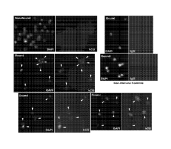
Figure 1 show cells isolated from TCS that express 13-hCG. Each field was
imaged to show the fluorescence of DAPI nuclear stain (left) or secondary
antibody
(right). The cells in the Bound fraction were all labeled by anti (3-hCG,
indicated by the
arrowheads in matched DAPI and hCG images, while none of the Non-bound cells
were
labeled. Bound cells labeled with non-immune IgG were also not fluorescent,
indicating
a low non-specific binding.
Figure 2 shows sex determination with isolated trophoblast cells. PCR analysis
of
genes on the X (DMD) and Y (SRY) chromosomes using isolated DNA from foreskin
fibroblast (Fb) cells, individual fixed Fb cells or ten individual isolated
trophoblast cells,
as indicated, using primers for just SRY, just DMD or both genes in a
multiplex assay.
The fetus of the patient in the upper gel appears to be male, while the lower
gel shows a
female fetus. Some of the reactions in the lower sample appeared to fail, most
likely due
to loss of the cell during transfer into the PCR tube.
DESCRIPTION OF THE PREFERRED EMBODIMENTS
The present invention provides a method for obtaining and using fetal material
obtained en masse during the first trimester of pregnancy from the cervix or
uterine
cavity to perform prenatal diagnosis. The method includes disassociating the
fetal cells
and maternal cells from the mucus of a sample and isolating the disassociated
fetal
cells from other cells in an endocervical sample. Additionally, the methods of
the
present invention enable non-invasive acquisition of EVT cells and permits
comparison
of protein expression levels with pregnancy outcomes. These findings
identified a robust
panel of EVT biomarkers that could inform during the first and second
trimester about
patient risk for PE or IUGR or other obstetrical disorders. The methods of the
present
invention can be used as a clinical laboratory service. The method includes
the steps of
6
SUBSTITUTE SHEET (RULE 26)
CA 02888746 2015-04-17
WO 2014/062995 PCT/US2013/065570
collecting cells, placing the collected cells in a fixative solution, removing
the mucus by
acidification, washing the remaining cells by centrifugation, and preparing
the cells on
microscope slides.
The specimens can be obtained using standard non-invasive methods known to
those of skill in the art. Examples include, but are not limited to,
intrauterine lavage,
.. aspiration of cervical mucus, or removal of surface tissue from the
cervical as or
endocervical canal. The preferred method is to collect mucus from the
endocervical
canal using a cytological brush inserted about 2 cm past the external os and
rotating to
remove and capture the mucus plug, while minimally abrading the cervical
tissue. The
cytological brush is then rinsed into a fixative solution composed of low pH
(4-6) buffer
and an alcohol. For example, a standard 3% acetic acid, 7% sodium acetate, 50%
methanol mixture can be used. Clinicians can be instructed to collect
specimens using
the ThinPrep0 kit from women found to be pregnant and still in the first or
second
trimester. This kit contains a cytological brush and includes 20 ml of
fixative solution.
The collected cells are isolated from the mucus by acidification.
Acidification can
be accomplished using methods known to those of skill in the art. For example
addition
of a 3% solution of acetic acid to reduce the pH of the fixative containing
cells to 2 to 4,
corresponding to a dilution of the acetic acid solution by 10 to 20 fold in
the fixative.
Once the specimen has been obtained, fetal cells, or other cell types of
clinical
importance, such as immunological cell subtypes, can be isolated and
identified in
collected samples. This can be accomplished using methods known to those of
skill in
the art including, but not limited to, using evidence for the presence of the
male Y
chromosome, comparison of allelic profile with maternal allelic profile and
expression of
trophoblast marker molecules (e.g., cytokeratin7, hCG, HLA-G, placental
alkaline
phosphatase, hyaluronic acid targeted by monoclonal antibody NDOG1, and the
unknown target of monoclonal antibody FT141.1, a.k.a. FT1.41.1). In most
cases, the
analysis of fetal cells would involve genetic diagnosis by fluorescent in situ
hybridization
(FISH) or the polynnerase chain reaction (PCR). The methods can be used to
predict
pregnancy outcome based on tests performed on the cells collected using the
ThinPrep0 kit.
7
SUBSTITUTE SHEET (RULE 26)
CA 02888746 2015-04-17
WO 2014/062995 PCT/US2013/065570
The major disadvantages of sampling fetal cells deposited in the cervical
mucus
plug are that there are many more maternal cells present than fetal cells and
that mucus
interferes with many tests due to aggregation of cells and background
fluorescence of
the mucus. The first problem will be addressed using robust fluorescent
markers for
trophoblast cells. A limitation of HLA-G as a marker is that it does not
recognize all
trophoblast subpopulations (e.g., syncytial trophoblast fragments).
Cytokeratin is
expressed by all trophoblast subpopulations, but it may also be found in some
maternal
cell types, leading to false positives. The problem of mucus has been solved
by
acidification to dissolve it. The number of trophoblast cells present in the
samples,
which could vary, may limit the method. If the number is too low, flow
cytometry would
become impractical. However, immunofluorescence microscopy would be a viable
approach as long as several HLA-G-positive cells can be located in a
microscopic field
prepared from up to 1 ml of sample.
The recovery and analysis of fetal cells shed from the placenta into the
cervical
canal provides wider availability of prenatal genetic diagnostics to the
general patient
population. With improvements in the efficacy and safety of trophoblast
collection by
TCC sampling using the cytobrush, and in the identification and isolation of
those cells
expressing trophoblast markers, small quantities of fetal DNA could be readily
obtained
for genetic testing. New sensitive technologies, such as those now under
development
for analysis of fetal DNA in maternal serum, could yield extensive information
about the
fetal genome from modest numbers of isolated cells. The ability to procure
cytotrophoblast cells by TCC as early as six weeks of gestation could make
this vital
information available much earlier than current technologies, including the
analysis of
fetal DNA in maternal serum. Over the next few years, more studies using TCC
sampling for prenatal diagnosis of chromosome abnormalities, paternity
testing,
screening for abnormal pregnancies in the first trimester and early diagnosis
of
obstetrical problems are expected, all of which could be performed using the
cells
isolated from the methods described herein.
Additionally, ectopic pregnancy complicates about 1-2% of all pregnancies and
occurs when the developing blastocyst implants at a site other than the fundus
of the
uterine cavity, most commonly in the fallopian tube. Delayed clinical
diagnosis of this
8
SUBSTITUTE SHEET (RULE 26)
CA 02888746 2015-04-17
WO 2014/062995 PCT/US2013/065570
abnormality can result in a dismal maternal outcome. The presence of fetal
trophoblast
cells in the cervical canal during the first trimester provides a non-invasive
approach for
predicting abnormal pregnancy through transcervical sampling.
In the present embodiment, a commercially available kit (ThinPrep , Hologic
Corporation, Marlborough, MA) is used to collect cells during the first
trimester of
pregnancy from the cervix. This is minimally invasiveness, as PAP smear tests
are
routinely recommended during pregnancy. Using the ThinPrep kit, a provided
cytobrush is used to collect mucus and cellular material from the cervix
between the
inner os and outer os, as directed by the manufacturer. The collected cells
are rinsed
into PreservCyt0 transport medium supplied by the manufacturer in a vial.
PreservCyt0
transport medium contains a methanol-acetic acid-based fixative. Samples are
stored at
room temperature or under refrigeration until analysis.
A slide preparation can be made by first acidifying the specimen to dissolve
mucus and free trapped cells for immunohistochennical staining. The specimen
is then
placed into a Shandon EZ mega funnel affixed to a microscope slide and
centrifuged in
a Cytospin3 centrifuge. This procedure yields evenly spread cells within a
delineated
area on the slide and free of interfering mucus. Alternatively, an automated
processor
for preparation of cytological slides could be used. One example is the
ThinPrep2000
(Holog ic)
The cells are then stained with antibody against HLA-G, a major
histocompatibility protein expressed only by fetal trophoblast cells. Other
trophoblast
markers can be used, for example the 3 subunit of chorionic gonadotropin (3-
CG), or
placental lactogen (PL), among others, but some (e.g., cytokeratin 7) are less
specific.
Immunofluorescence microscopy or flow cytometry is used to identify the
fluorescently-
labeled trophoblast cells. A protein of interest can be queried in the HLA-G-
positive cells
using an appropriate antibody and a secondary antibody with a different
fluorescent
label (double labeling). Alternatively, a FISH procedure could be used for
genetic
analysis of the HLA-G positive cells, for example it could be used to detect
chromosome
number or a particular gene sequence if there were a way to identify the fetal
cells, such
as the presence of the Y chromosome. A different strategy would be necessary
for a
female fetus, however. It has been demonstrated (Imudia et al., 2009) that it
is possible
9
SUBSTITUTE SHEET (RULE 26)
CA 02888746 2015-04-17
WO 2014/062995 PCMJS2013/065570
to use an enzyme-linked secondary antibody for HLA-G identification that can
be
visualized by bright field microscopy (e.g., with diaminobenzidine substrate
for a
peroxidase tag), and the cells of interest could be isolated by laser capture
microdissection for genetic analysis by PCR.
It has been found that placentas from women with the hypertensive disorder pre-
have altered expression of several proteins (epidermal growth factor [EGF],
transforming growth factor-a [TGFA], heparin-binding EGF-like growth factor
[HBEGF]).
The fluorescent double labeling method therefore can be used to screen the
expression
of these proteins in trophoblasts isolated from cervical collections during
the first
trimester, months before any clinical symptoms present. Therefore, this method
could
provide a diagnostic tool to identify women at risk for developing pre-
eclampsia later
during their pregnancy.
Currently, chorionic villous sampling (CVS) or amniocentesis can be used for
prenatal diagnosis of fetal chromosomal abnormality. Both methods are invasive
and
associated with potential pregnancy loss. CVS can only be performed after 10
weeks,
and amniocentesis has to be done after 14 weeks gestation. It is much less
desirable to
perform termination of pregnancy after the second trimester begins. The
methods of the
present invention allow the test to be performed in early first trimester and
in a non-
invasive manner.
In another embodiment, the methods can be used to test the expression of
biomarkers that are indicative of obstetrical disorders. The biomarkers can
include
growth hormones, proteins, and RNA. By way of example, the methods can be used
to
test the expression of proteins by double-labeling cells with fluorescent
antibodies to
determine if EGF, TGFA or HBEGF are reduced in trophoblast cells. These
changes
have been observed in the trophoblast cells of placentas delivered from women
with
pre-eclampsia. Since 5% of all pregnant women eventually develop pre-
eclampsia, this
would be a beneficial test to perform routinely at the time of a positive
pregnancy test.
Those women found to be at risk for the disorder, could be instructed to take
precautions against developing hypertension long before clinical symptoms
first appear.
In another embodiment, the method can include conducting genetic analysis of
transcervical trophoblast cells. Trophoblast DNA can be obtained (1) by laser
capture
SUBSTITUTE SHEET (RULE 26)
CA 02888746 2015-04-17
WO 2014/062995 PCMJS2013/065570
microscopy of anti-HLA-G labeled cells (or using other antibodies that
distinguish
trophoblast cells from resident maternal cells of the cervix), or (2) with
anti-HLA-G
affinity magnetic beads/nanoparticles to isolate trophoblast cells. Genetic,
immunological or other biochemical analyses can then be performed by a variety
of
whole-cell approaches. For example, PCR with or without reverse transcription,
.. innnnunohistochemistry, whole genome amplification (WGA) followed by
comparative
genomic hybridization or sequencing, metabolite assays, small compound assays
and
other tests would be adaptable. Alternatively, fetal and maternal DNA can be
assessed
in unfractionated transcervical samples using a digital PCR approach.
The genetic analysis can include, for example, FISH, sequencing or PCR based
methods. Alternatively, magnetic beads can also be used prior to
imnnunofluorescence
as a way to enrich for the cells of interest and streamline analysis. Dynal
Magnetic
beads are available from lnvitrogen (Carlsbad, CA) with secondary antibodies
attached
or chemical coupling groups that can be used to attach anti-HLA-G. They are
mixed
with the cells after acidification and neutralization and decorate target
cells
(trophoblast). Holding a magnet against the test tube or inserting the tube
into a device
like the DynaMagTm-Spin magnet (Life Technologies) for 5 minutes, the
suspended cells
are aspirated off, leaving behind the magnet-bound cells coated with beads.
After three
washes, it is possible to enrich about 1,000 to 10,000 fold, which should be
adequate to
isolate most of the trophoblast cells. The cells can be examined
microscopically to verify
the presence of beads and manually remove any cells without beads that
contaminate
the sample. Then, additional testing can be conducted as disclosed in more
detail
herein.
In another embodiment, the fetal trophoblast cells can be isolated from the
resident maternal cells after their collection so they can be used in genetic
or
biochemical tests. The specificity of the anti-HLA-G antibody is used for this
purpose by
coupling it to magnetic nanoparticles for trophoblast isolation. For example,
the method
can use 10 pl of 250 nm nanoparticles conjugated to anti-mouse IgG or Protein
A
(Clemente Associates, Madison, CT) and incubated with 5 pg of mouse monoclonal
anti-HLA-G antibody (Clone: 4H84, BD Biosciences, San Diego, CA; or clone
G233;
Exbio, Prague) overnight on a rotary shaker at 4 C. The particles are
separated from
11
SUBSTITUTE SHEET (RULE 26)
CA 02888746 2015-04-17
WO 2014/062995 PCT11JS2013/065570
unbound antibody by placing tubes in a DynaMagTm-Spin magnet (Life
Technologies)
for 5 minutes and then removing the liquid while the magnetic nanoparticles
are
retained. Cells collected from a transcervical specimen are then added to the
nanoparticles and incubated at room temperature for one to 24 hours on a
rotary shaker
at 4 C. The sample is magnetized and unbound cells are removed. After three
washes,
the retained cells are recovered. Analysis of the immunomagnetically isolated
cells by
immunofluorescence microscopy with anti-3hCG to identify trophoblast cells
will reveal
95-100% labeling of the isolated cells and no staining of the depleted cells
that were
removed during magnetization (Table 1). In one test performed using this
methodology,
approximately 500-2000 cells were recovered from each patient specimen. This
approach to isolate the trophoblast cells based on their binding to antibodies
that
distinguish them from maternal cervical cells can also be used with other
technologies.
For example a microfluidic device could be constructed to sort the cells based
on a
magnetic or fluorescent marker conjugated to antibody.
In addition to the high purity of 3-hCG expressing cells after immunomagnetic
isolation, cells in the non-bound fraction were not labeled by anti-3-hCG, nor
were
bound cells labeled with non-immune control IgG (Fig. 1).
The method, as described above, uses the isolated cells for biochemical or
genetic testing to gain information about the fetus or placenta. The isolated
cells are
sorted into individual or small groups of cells for testing by dispersion in a
multi-well
plate (such as a Terasaki multi-well plate) and sorting with a Stripper
micropipetter
(Origio MidAtlantic Devices, Mt. Laurel, NJ). In a test group, groups of 50
cells are
suspended in 200 pi of PBS and centrifuged onto a slide utilizing a Shandon
Cytospin 3
cytocentrifuge at 1500 RPM for 5 min. These fetal cells can be used for
analysis of
protein expression by immunofluorescence microscopy or for molecular analysis
by
FISH. For example, the cells were labeled with antibodies that recognize
trophoblast
specific proteins or proteins that are expressed by various trophoblast
subpopuiations.
The results indicated that the isolated cells are not from the chorionic
villi, but are
deeply invasive extravillous trophoblast cells (Table 2). This shows that
trophoblast cells
invading at the base of the placenta migrate as far as the cervix where they
are
collected by transcervical sampling. This can be beneficial for the further
development
12
SUBSTITUTE SHEET (RULE 26)
CA 02888746 2015-04-17
WO 2014/062995 PCT1US2013/065570
of test protocols and increases the amount of information that can be obtained
during
pregnancy. A similar approach could be used clinically to screen the isolated
cells for
protein biomarkers of fetal disorders or maternal obstetrical disorders.
Alternatively, the cells can be sorted or identified and isolated for
molecular
biological analysis using methods borrowed from single cell methods used for
genetic
1.0 analysis of cells biopsied from preinnplantation embryos generated by
in vitro fertilization
(IVF). Isolated trophoblast cells (up to 100) can be sorted with a Stripper
micropipetter
as single cells that are placed individually into thin-walled PCR tubes with 2
to 6 pl of
RNase-free water and frozen at -80 C. These cells can be used for testing that
probes
their RNA or DNA using amplification methods such as PCR or WGA. For RNA
testing,
.. it is necessary to stabilize RNA after fixed cells are removed from the
fixative solution.
Therefore, the initial cell washes in PBS, incubations with HLA-G-coupled
nanoparticles
and manipulation of cells into aliquots are all done using PBS supplemented
with 20
mM rbonucleoside-vanadyl complex (New England BioLabs, Inc.) to prevent RNA
degradation. Cells should be lysed immediately for RNA purification and either
stored at
-80 C in a chaotropic lysis solution or converted to cDNA before storage. It
is also
possible to perform protein analysis that is scaled down for single or small
numbers of
cells, such as ELISA or mass spectrometry. After WGA, the DNA (5-50
micrograms)
can be used in microarray or deep sequencing approaches to scan for genetic
mutations, identify chromosome number disorders (aneuploidies) or obtain the
entire
genomic sequence for personalized medicine. Genetic polymorphisms can be
assessed
for comparison to parental polymorphisms in order to confirm that the
amplified DNA is
of fetal, not parental origin, as a quality assurance control.
In another embodiment, the fetal cells can be isolated from the transcervical
specimens without an initial fixation using the preservative in the kit. This
enables
.. better recovery of cells and a more accurate analysis of less stable cell
constituents
(e.g., metabolites, RNA) and the ability to proliferate cells to obtain
increased amounts
of fetal DNA or produce metaphase cells for karyotyping.
The isolation can be done by rinsing the cytobrush (used as detailed herein)
in
ice-cold RPM! 1640 tissue culture medium containing 10% fetal bovine serum and
50
pg/m1 gentamicin or another comparable combinations of culture medium with
13
SUBSTITUTE SHEET (RULE 26)
CA 02888746 2015-04-17
WO 2014/062995 PCT11JS2013/065570
antibiotics. The specimen can be quickly brought to the laboratory and washed
three
times by centrifugation and resuspension in sterile PBS at 4 C. Magnetic
nanoparticles
conjugated to anti-mouse IgG that have been bound with mouse anti-HLA-G were
then
combined with the cells and incubated at 4 C for 1 hour. Fetal cells were
recovered by
incubation at 4 C in a DynaMag-Spin magnet (Life Technologies) and removal of
unbound cells. This step was repeated two more times and the bound cells were
recovered in 100 pl of ice-cold PBS.
Isolated fetal cells were either cultured in standard trophoblast culture
medium
(Kilburn et al. 2000) or fixed for immunohistochemistry. Cultured cells formed
small
colonies within 2-3 days, indicating that they were proliferating. Fixed cells
were labeled
with antibody against the beta subunit of hCG or BCL-2, followed by
fluorescent
secondary antibody. All isolated cells were positively labeled with both
antibodies,
indicating that they were indeed trophoblast and not apoptotic.
In addition to the benefits outlined above, a benefit of the single cell
approach is
that it can reduce the probability of false results to nearly zero. The main
source of error
is in the contamination of isolated trophoblast cells with maternal cells. In
general, there
has not been greater than 5% contamination with maternal cells. Assays of
replicate
individual trophoblast cells were used for multiplex amplification of
sequences from
genes on the X (DMD) or Y (SRY) chromosomes to determine the gender of the
corresponding fetuses. Every reaction should generate a product for X if a
cell is
present and PCR is working, but only male cells will generate a Y amplicon.
Analysis of
the PCR products by agarose gel electrophoresis should therefore produce a
single
band if the cell is female or two bands if male. Ten cells from each sample
were
analyzed and produced either all single bands (Female Fetus) or all double
bands (Fig.
2). The occasional single band in a male sample is presumably a contaminating
maternal cell, although we have not found this in 18 patients that were
analyzed this
way (Table 3). Maternal cells in female samples cannot be distinguished. The
high
purity of trophoblast cells is revealed in the male samples where there were
no cells that
produced a single band. To produce a false diagnosis, all cells would have to
be
maternal. In the case of a female fetus, the diagnosis would still be female,
and correct.
In the case of a male fetus, the presence of even one fetal cell would produce
a double
14
SUBSTITUTE SHEET (RULE 26)
CA 02888746 2015-04-17
WO 2014/062995 PCMJS2013/065570
band to indicate that the fetus is probably not female. The test could then be
repeated
or the sample further investigated. The probability that all replicates would
be maternal
cells decreases exponentially with the number of replicates and reaches 1 in
8,000 with
only three replicates, assuming the isolated cells contain 95% trophoblast
cells. This
level of assurance is reached with four replicates if the isolated cells
contain only 90%
1.0 trophoblasts. For 90% purity, the probability of a false result (P) is
0.1 for one replicate
(N) and decreases ten-fold with each additional single cell replicate, where P
=
The above discussion provides a factual basis for the methods and uses
described herein. The methods used with and the utility of the present
invention can be
shown by the following non-limiting examples.
EXAMPLES
Example 1:
Materials and Methods:
Patients, age 18-40, presenting for early prenatal care with a normal
intrauterine
pregnancy (IUP; n=37), ectopic pregnancy (EP; n=10) or blighted ovum (BO; n=5)
were
enrolled for collection of transcervical specimens using a cytological brush
and a
ThinPrep kit (Hologic). Cells collected in PreservCyt fixative solution were
cleared of
mucus by acidification, washed by centrifugation and an aliquot was prepared
on a
microscope slide using a Cytospin3 centrifuge. Slides were labeled with
monoclonal
antibody G233 recognizing HLA-G, a MHC antigen specifically expressed by
trophoblast cells. All HLA-G positive cells were identified and counted on
each slide.
After staining with hematoxylin, the total number of cells on each slide was
determined
and the ratio of HLA-G positive cells to total cells was calculated. Data were
compared
using one-way ANOVA, the Student-Newman-Keuls posthoc test and receiver
operating
characteristic (ROC) analysis.
Results:
The mean gestational ages of normal IUP, EP and BO were 9, 8 and 10 weeks,
respectively. Trophoblast cells were observed in 35/37 normal IUP, 6/10 EP and
4/5 BO
specimens. The frequency of HLA-G positive cells in the IUP cervical samples
was
approximately 1 in 2000, which was 5 to 10-fold higher (p<0.001) than the
average
frequency in samples from patients with EP or BO. The latter two groups were
not
SUBSTITUTE SHEET (RULE 26)
CA 02888746 2015-04-17
WO 2014/062995 PCMJS2013/065570
significantly different. Significantly, ROC analysis showed that EP and BO
pregnancies
were distinguishable from normal pregnancies with 93% sensitivity and 95%
specificity.
Conclusion:
Trophoblast cells can be reliably identified among cervical cells in the first
trimester by immunohistochemical staining for HLA-G. Abnormal pregnancies are
predictable based on trophoblast abundance.
Example 2:
Preeclampsia (PE) and intrauterine growth restriction (IUGR) are common
adverse pregnancy outcomes with no reliable means for early detection.
Attempts using
serum protein panels to identify patients with PE or IUGR earlier than the
presenting
symptoms has been inconsistent. Both disorders are linked to deficient
remodeling of
the uterine vasculature by extravillous trophoblast (EVT) cells. EVT residing
in the
endocervical canal can be captured in a non-invasive procedure similar to a
PAP test
and isolated free of maternal cells. Serum bionnarkers of IUGR and PE are
dysregulated
in EVT earlier in gestation than their altered levels can be detected in
serum.
Methods:
PAP specimens (N=23) were collected at 5-20 weeks of gestation using a
cytobrush. Medical records were subsequently searched for diagnosis of PE or
IUGR.
EVT cells (500-1500) were isolated using HLA-G antibody coupled to magnetic
nanoparticles. Cells (-50) were affixed to slides using a Cytospin 3
cytocentrifuge,
assessed for purity with anti-p-hCG, and labeled by immunofluorescence with
antibodies (R&D Systems) against galectin 13 (LGALS13, a.k.a. PP13), galectin
14
(LGALS14), placental growth factor (PGF), pregnancy-associated plasma protein
A
(PAPPA), alpha fetal protein (AFP), endoglin (ENG), or fms-related tyrosine
kinase 1
(FLT-1). Fluorescence intensity (Fl) was quantified for individual cells by
image
analysis. Fl values of 20 cells were averaged for each patient and compared by
ANOVA
between normal and adverse outcome groups, using a post-hoc Tukey test.
Results:
Nine patients eventually developed PE or IUGR, while 14 had normal
pregnancies. Expression of LGALS13, LGALS14, PAPPA and PGF were each
decreased (p<0.05) in EVT from pregnancies that later developed PE/IUGR
compared
16
SUBSTITUTE SHEET (RULE 26)
CA 02888746 2015-04-17
WO 2014/062995 PCT11JS2013/065570
to normal pregnancies. FLT-1, ENG, and AFP were each increased (p<0.05) with
PE/IUGR.
Conclusions:
A novel approach for non-invasive acquisition of EVT cells permits comparison
of
protein expression levels with pregnancy outcomes. These findings identified a
robust
1.0 panel of EVT biomarkers that could inform during the first and second
trimester about
patient risk for PE or IUGR.
Example 3:
At 5 weeks gestation, trophoblastic cells can be non-invasively retrieved from
the
endocervical canal using a cytobrush. These cells can be isolated from
material cells
using the fetal specific marker HLA-G.
Methods:
Following isolation of trophoblast cells from pregnant patients in the first
and
second trimester, specimens were analyzed by innmunohistochemistry for HLA-G
expression. Trophoblast cells were separated from maternal cells using a
column-free
magnetic nanoparticle separation procedure. Purity of the trophoblast
specimens was
calculated by staining for 13-hCG. Following whole genome amplification (WGA)
of
maternal and fetal cell DNA, single nucleotide polymorphism (SNP) assay and
gender
identification was performed by polymerase chain reaction.
Results:
Maternal and fetal cells were compared after isolation from 5 patient
specimens.
Average total trophoblast recovery was 700 cells, average purity was above
90%. A
minimum of 10 pg of DNA was obtained after WGA using either single cells or
groups of
5-100 cells. SNP assays demonstrated allelic differences between maternal and
trophoblast cells in all specimens. Gender was identified and confirmed from
patient
records.
Conclusions:
Trophoblast cells can be retrieved and isolated from the endocervical canal
with
acceptable purity based on their high degree of (3-hCG expression.
Furthermore, fetal
DNA was distinct from maternal DNA indicating its utility as a platform for
non-invasive
prenatal testing of the intact fetal genome.
17
SUBSTITUTE SHEET (RULE 26)
CA 02888746 2015-04-17
WO 2014/062995 PCT/US2013/065570
Throughout this application, various publications, including United States
patents,
are referenced by author and year and patents by number. Full citations for
the
publications are included.
The invention has been described in an illustrative manner, and it is to be
understood that the terminology which has been used is intended to be in the
nature of
words of description rather than of limitation.
Obviously, many modifications and variations of the present invention are
possible in light of the above teachings. It is, therefore, to be understood
that within the
scope of the appended claims, the invention may be practiced otherwise than as
specifically described.
18
Date Recue/Date Received 2020-05-04
CA 02888746 2015-04-17
WO 2014/062995 PCMJS2013/065570
REFERENCES
lmudia AN, Kumar S, Diamond MP, Decherney AH, Armant DR. Transcervical
retrieval
of fetal cells in the practice of modern medicine: a review of the current
literature
and future direction. Fertil. Steril. 2010; 93: 1725-1730.
lmudia AN, Suzuki Y, Kilburn BA, Yelian FD, Diamond MP, Romero R, Armant DR.
Retrieval of trophoblast cells from the cervical canal for prediction of
abnormal
pregnancy: a pilot study. Hum. Reprod. 2009; 24: 2086-2092.
Orr JW, Jr., Barrett JM, Orr PF, Holloway RW, Holinnon JL. The efficacy and
safety of
the cytobrush during pregnancy. Gynecol. Oncol. 1992; 44: 260-262.
Rivlin ME, Woodliff JM, Bowlin RB, Moore JL, Jr., Martin RW, Grossman JH, 3rd,
Morrison JC. Comparison of cytobrush and cotton swab for Papanicolaou smears
in pregnancy. J. Reprod. Med. 1993; 38: 147-150.
Paraiso MF, Brady K, Helmchen R, Roat 11/V . Evaluation of the endocervical
Cytobrush
and Cervex-Brush in pregnant women. Obstet. Gynecol. 1994; 84: 539-543.
Foster JC, Smith HL. Use of the Cytobrush for Papanicolaou smear screens in
pregnant
women. J. Nurse. Midwifery 1996; 41: 211-217.
Holt J, Stiltner L, Jamieson B, Fashner J. Clinical inquiries. Should a nylon
brush be
used for Pap smears from pregnant women? J. Fam. Pract. 2005; 54: 463-464.
O'Leary P, Breheny N, Dickinson JE, Bower C, Goldblatt J, Hewitt B, Murch A,
Stock R.
First-trimester combined screening for Down syndrome and other fetal
anomalies. Obstet. Gynecol. 2006; 107: 869-876.
Wapner RJ. Invasive prenatal diagnostic techniques. Semin. Perinatol. 2005;
29: 401-
404.
Handyside AH, Kontogianni EH, Hardy K, Winston RM. Pregnancies from biopsied
human preimplantation embryos sexed by Y-specific DNA amplification. Nature
(London) 1990; 344: 768-770.
Munne S, Howles CM, Wells D. The role of preimplantation genetic diagnosis in
diagnosing embryo aneuploidy. Curr. Opin. Obstet. Gynecol. 2009; 21: 442-449.
12. Lun FM, Tsui NB, Chan KC, Leung TY, Lau TK, Charoenkwan P, Chow KC,
Lo
WY, Wanapirak C, Sanguansermsri T, Cantor CR, Chiu RW, Lo YM. Noninvasive
prenatal diagnosis of monogenic diseases by digital size selection and
relative
mutation dosage on DNA in maternal plasma. Proc. Natl. Acad. Sci. U. S. A.
2008; 105: 19920-19925.
Chiu RW, Chan KC, Gao Y, Lau VY, Zheng W, Leung TY, Foo CH, Xie B, Tsui NB,
Lun
FM, Zee BC, Lau TK, Cantor CR, Lo YM. Noninvasive prenatal diagnosis of fetal
chromosomal aneuploidy by massively parallel genomic sequencing of DNA in
maternal plasma. Proc. Natl. Acad. Sci. U. S. A. 2008; 105: 20458-20463.
Devaney SA, Palomaki GE, Scott JA, Bianchi DW. Noninvasive fetal sex
determination
using cell-free fetal DNA: a systematic review and meta-analysis. JAMA 2011;
306: 627-636.
Cioni R, Bussani C, Bucciantini S, Scarselli G. Fetal cells in a transcervical
cell sample
collected at 5 weeks of gestation. Journal of Maternal Fetal & Neonatal
Medicine
2005; 18: 271-273.
Kilburn BA, Wang J, Duniec-Dnnuchowski ZM, Leach RE, Romero R, Armant DR.
Extracellular matrix composition and hypoxiz regulate the expression of HLA-G
and integrins in a human trophoblast cell line. Biol. Reprod. 2000; 62:739-
747.
19
SUBSTITUTE SHEET (RULE 26)
CA 02888746 2015-04-17
WO 2014/062995
PCMJS2013/065570
Table 1. Purification of Trophoblast Cells. Trophoblast cells in 24
transcervical specimens from
pregnant women were isolated with HLA-G-coupled to anti-human IgG
nanoparticles. HLA-G-labeled
cells counts were used to predict the number of cells expected after
purification, and to estimate recovery
rates. Purified cells were assessed for expression of p-hCG by
immunofluorescence microscopy and the
percentage of positively labeled cells is shown (n=55 to 1500 counts per
specimen).
Patient # Expected Trophoblasts # Trophoblast Recovered f3-
hCG+
(% Expected) %
1 609 998 (164) 95
2 1260 248 (20) 99
3 466 660 (141) 99
4 1108 345 (31) 98.8
5 2467 832 (35) 99.2
6 239 270(116) 99.6
7 2222 1462 (66) 97.8
8 1009 818(81) 99.6
9 677 855(119) 99.6
581 720 (124) 99.3
11 570 570 (100) 99.5
12 1027 622 (61) 100
13 314 314 (282) 100
14 850 578 (68) 98.9
593 510(86) 100
16 820 705 (86) 100
17 842 623 (74) 98
18 575 593 (103) 100
19 1045 728 (70) 98.9
1109 495 (45) 96.4
21 879 758 (86) 100
22 529 1463 (276) 98.4
23 1095 1020 (93) 100
24 609 660 (108) 100
Average 101 13% 99
0.25%
20
SUBSTITUTE SHEET (RULE 26)
CA 02888746 2015-04-17
WO 2014/062995 PCT11JS2013/065570
Table 2. Reactivity of Isolated Cells with an Antibody Panel to Distinguish
Subtype. All cells tested
were stained or unstained as indicated in the right column with antibodies
against proteins listed in the
left column, The middle three columns indicate the known expression patterns
of the proteins in human
trophoblast.
Villous Villous Extravillous
Protein HLA-G4- Cells
Syncytotrophoblast Cytotrophoblast Trophoblast
HLA-G - - + +
hCG, [I subunit + + + +
KRT7 + + + +
hPL + + + +
PSG-1 + - - -
a6 integrin + + - -
E-cadherin + 'A' -I+ -
VE-cadherin - + +
- PECAM1 - + +
-
- al integrin - + +
MMP9 - + + + 10
21
SUBSTITUTE SHEET (RULE 26)
CA 02888746 2015-04-17
WO 2014/062995 PCMJS2013/065570
Table 3. Summary of Fetal Sexing Results. FOR assays were conducted, as in
Fig. 2 to amplify
sequences on X and Y chromosomes. P, patient #. Cells giving a band (Pos.
Cells) / total cells assayed is
shown for X and Y chromosomes. Fetal sex was confirmed by ultrasound or at
birth.
Gestational Age X Chromosome Y Chromosome Verified
Patient
Weeks Pos. Cells (%) Pos. Cells (/o) Gender
1 6 25/30 (83.3) 0/30 (0) Female
2 9.2 10/10(100) 10/10(100) Male
3 14.6 18/20(90) 18/20(90) Male
4 7.6 25/25 (100) 0/25 (0) Female
6 17.6 10/10 (100) 0/10 (0) Female
7 10 10/10(100) 10/10(100) Male
8 17.5 10/10(100) 10/10(100) Male
9 7.3 515 (100) 5/5 (100) Male
11 10/10(100) 10/10(100) Male
11 15.2 9/10 (90) 9/10 (90) Male
13 7.5 10/10(100) 10/10(100) Male
14 12 10/10(100) 0/10(0) Female
12.4 10110 (100) 0/10 (0) Female
21 12 9110 (90) 0/10 (0) Female
23 10 10/10 (100) 0/10 (0) Female
24 8 25/28(89.3) 25/28(89.3) Male
14 6/6 (100) 6/6 (100) Male
26 12 24/26(92.3) 24/26(92.3) Male
22
SUBSTITUTE SHEET (RULE 26)