Note: Descriptions are shown in the official language in which they were submitted.
CA 02890004 2015-04-29
WO 2014/070946 PCT/US2013/067604
1
SELECTIVE AMPLIFICATION AND REAL-TIME PCR DETECTION OF RARE
MUTATIONS
CROSS-REFERENCE TO RELATED APPLICATIONS
[0001] This application claims priority to U.S. Provisional Patent
Application
No. 61/720,959, entitled "SELECTIVE AMPLIFICATION AND REAL-TIME PCR
DETECTION OF RARE MUTATIONS," filed October 31, 2012, the entire content of
which
is hereby incorporated by reference.
REFERENCE TO SEQUENCE LISTING, TABLE, OR COMPUTER PROGRAM LISTING
[0002] The present application is being filed along with a Sequence
Listing in
electronic format. The Sequence Listing is provided as a file entitled
GENOM122.txt, last
saved October 30, 2013, which is 7.66 kb in size. The information is
incorporated herein by
reference in its entirety.
BACKGROUND OF THE INVENTION
Field of the Invention
[0003] The present embodiments relate to molecular diagnostics, and in
particular, to compositions in detecting sequence variants, such as SNPs,
insertions deletions,
and altered methylation patterns, from samples. The embodiments disclosed
herein can be
used to detect (and quantify) sequence variants present in samples that
include an excess of
wild-type sequences.
Description of the Related Art
[0004] With the advent of molecular diagnostics and the discovery of
numerous
nucleic acid biomarkers useful in the diagnosis and treatment of conditions
and diseases,
detection of nucleic acid sequences, and sequence variants, mutations and
polymorphisms has
become increasingly important. In many instances, it is desirable to detect
sequence variants
or mutations (which may in some instances, differ by one a single nucleotide)
present in low
copy numbers against a high background of wild-type sequences. For example, as
more and
more somatic mutations are shown to be biomarkers for cancer prognosis and
prediction of
therapeutic efficacy, the need for efficient and effective methods to detect
rare mutations in a
sample is becoming more and more critical.
CA 02890004 2015-04-29
WO 2014/070946 PCT/US2013/067604
2
[0005] In the case in which one or more allelic variants is/are
present in low copy
number compared to wild-type sequences, the presence of excess wild-type
target sequence
creates challenges to the detection of the less abundant variant target
sequence. Nucleic acid
amplification/detection reactions almost always are performed using limiting
amounts of
reagents. A large excess of wild-type target sequences, thus competes for and
consumes
limiting reagents. As a result amplification and/or detection of rare mutant
or variant alleles
under these conditions is substantially suppressed, and the methods may not be
sensitive
enough to detect the rare variants or mutants. Various methods to overcome
this problem
have been attempted. These methods are not ideal, however, because they either
require the
use of a unique primer for each allele, or the performance of an intricate
melt-curve analysis.
Both of these shortcomings limit the ability and feasibility of multiplex
detection of multiple
variant alleles from a single sample.
SUMMARY OF THE INVENTION
[0006] Detection of rare sequence variants in biological samples
presents
numerous challenges. The methods and kits disclosed herein provide for
improved, efficient
means to detect rare mutations within a high background of wild-type allelic
sequences using
real-time amplification methods.
[0007] In one aspect, the embodiments disclosed herein relate to
methods to
detect a first variant target sequence in a sample comprising nucleic acids.
The method can
include the steps of providing the biological sample for analysis, and
contacting the
biological sample with a pair of amplification primers. The amplification
primers can
include a forward primer and a reverse primer which together are configured to
amplify a
target amplicon or target region. The target amplicon, or target region, can
include either a
wild-type target allele sequence or a variant target allele sequence of
interest. The
amplification primers can flank the wild-type target sequence or variant
target allele
sequence, such that the amplification primers amplify both wild-type target
sequences and
variant target allele sequences under amplification or primer extension
conditions. The
sample can also be contacted with a blocking primer that preferentially
hybridizes to the wild
type target allele sequence compared to a first variant target allele sequence
under
amplification conditions. The sample can also be contacted with one or more
reporter
probes, wherein the reporter probe(s) include an oligonucleotide that
preferentially hybridizes
to the first variant target allele sequence compared to the wild-type target
sequence under
CA 02890004 2015-04-29
WO 2014/070946 PCT/US2013/067604
3
amplification conditions, and a detectable moiety. The sample can be contacted
with the
amplification primers, the blocking oligonucleotide and the detector probe(s)
under
amplification conditions. Hybridization of the reporter probe to the first
variant target allele
sequence can be measured, wherein hybridization of the reporter probe to the
first variant
target allele sequence produces a detectable signal indicative of the presence
and/or amount
of first variant target allele species in the biological sample.
[0008] In another aspect, the embodiments disclosed herein provide
kits and
compositions for the detection of a rare sequence variant or mutant allele
from a sample. The
kits or compositions can include an amplification primer pair that includes a
forward and
reverse primer that flank a target region, that includes within the target
region the target
variant or mutant allele sequence of interest. The kits and compositions can
also include a
blocking oligonucleotide that is non-extendible by a polymerase, and which
preferentially
hybridizes to a wild type target allele sequence compared to the variant or
mutant target allele
sequence. The kits and compositions can also include a detector probe. The
detector probe
can include an oligonucleotide that preferentially binds to the variant or
mutant target allele
sequence compared to the wild-type target allele sequence. The detector probe
also includes
a detectable moiety that enables detection of hybridization of the detector
probe to the variant
or mutant target allele sequence.
BRIEF DESCRIPTION OF THE DRAWINGS
[0009] Figure 1 is a schematic of an exemplary embodiment illustrating
a method
for detection of a single, rare variant allele according to the embodiments
disclosed herein.
[0010] Figure 2 is a schematic of an exemplary embodiment illustrating
a method
for the simultaneous detection of more than one rare, variant allele according
to the
embodiments disclosed herein.
[0011] Figure 3 is a schematic of an exemplary embodiment illustrating
a method
for the detection of methylation variants according to the embodiments
disclosed herein.
[0012] Figures 4A-D is a schematic showing the different, possible
species of
molecular complexes in in reaction mixtures containing an analyte (A),
amplification primer
(P), blocking oligonucleotide (B), detector probe (D) and polymerase (E).
[0013] Figure 5 shows the equilibrium between the various species of
molecular
complexes shown in Figures 4A-D.
CA 02890004 2015-04-29
WO 2014/070946 PCT/US2013/067604
4
[0014] Figure 6 illustrates an exemplary method to estimate the
fraction
extendible target species, i.e., "fe.," of the complexes shown in Figure 4,
according to the
embodiments disclosed herein
[0015] Figure 7 shows a mathematical model for an amplification
reaction on a
sample comprising two different target species, according to the embodiments
disclosed
herein.
[0016] Figure 8 depicts the various reporter probes blocking
oligonucleotides and
forward amplification primers used in the simulated real-time PCR assays
discussed in
EXAMPLE 1.
[0017] Figures 9A-B show simulated amplification curves of real-time
amplification reactions using the various conditions described in EXAMPLE 1,
with a
mixture of wild-type and G34T mutant KRAS nucleic acids, present in a ratio of
10000:100
(wt : mutant). Figure 9A shows the amplification curve (relative fluorescence
v. cycle
number) of the reaction under the described parameters, wherein the W.T. fe.,
as explained
in EXAMPLE 1, is approximately 0.159. Figure 9B shows the amplification curve
(relative
fluorescence v. cycle number) of the reaction under the described, wherein the
WT fe. is
approximately 0.717, as described in EXAMPLE 1.
[0018] Figure 10A depicts a target region of the DAPK-1 promoter
region as
described in EXAMPLE 2, including the location of cytosine residues that are
potentially
methylated. CpG sites are boxed.
[0019] Figure 10B depicts a schematic showing a reaction to detect
methylation
variants in the DAPK-1 promoter, as described in EXAMPLE 2.
DETAILED DESCRIPTION OF THE PREFERRED EMBODIMENT
[0020] It is to be understood that both the foregoing general
description and the
following detailed description are exemplary and explanatory only and are not
intended to
limit the scope of the current teachings. In this application, the use of the
singular includes
the plural unless specifically stated otherwise. Also, the use of "comprise",
"contain", and
"include", or modifications of those root words, for example but not limited
to, "comprises",
"contained", and "including", are not intended to be limiting. Use of "or"
means "and/or"
unless stated otherwise. The term "and/or" means that the terms before and
after can be taken
together or separately. For illustration purposes, but not as a limitation, "X
and/or Y" can
mean "X" or "Y" or "X and Y".
CA 02890004 2015-04-29
WO 2014/070946 PCT/US2013/067604
[0021] Whenever a range of values is provided herein, the range is
meant to
include the starting value and the ending value and any value or value range
there between
unless otherwise specifically stated. For example, "from 0.2 to 0.5" means
0.2, 0.3, 0.4, 0.5;
ranges there between such as 0.2-0.3, 0.3-0.4, 0.2-0.4; increments there
between such as 0.25,
0.35, 0.225, 0.335, 0.49; increment ranges there between such as 0.26-0.39;
and the like.
[0022] The section headings used herein are for organizational
purposes only and
are not to be construed as limiting the subject matter described in any way.
All literature and
similar materials cited in this application including, but not limited to,
patents, patent
applications, articles, books, treatises, and internet web pages, regardless
of the format of
such literature and similar materials, are expressly incorporated by reference
in their entirety
for any purpose. In the event that one or more of the incorporated literature
and similar
materials defines or uses a term in such a way that it contradicts that term's
definition in this
application, this application controls. While the present teachings are
described in
conjunction with various embodiments, it is not intended that the present
teachings be limited
to such embodiments. On the contrary, the present teachings encompass various
alternatives,
modifications, and equivalents, as will be appreciated by those of skill in
the art.
[0023] The embodiments disclosed herein provide improved methods for
detection of mutant or variant alleles. The methods disclosed herein
advantageously
overcome many of the limitations of previous methods of molecular detection of
rare
mutations, and enable detection of multiple alleles within a single real-time
PCR reaction,
without the requirement for multiple, allele-specific amplification primers.
Detection of Variant or Mutant Alleles
[0024] Provided herein are methods for analyzing a sample for allelic
variants
within a target sequence. Allelic variants have been implicated in genetic
disorders,
susceptibility to different diseases, responses to various therapeutics and
the like.
Accordingly, the importance of detection of allelic variants or mutations in
target sequences
cannot be underestimated. As used herein, the term "target sequence" refers to
a nucleic acid
sequence of interest, e.g., a genomic DNA, an mRNA, a cDNA, or the like, to be
queried for
the presence of allelic variants, e.g., rare allelic variants or mutations. As
used herein, the
term "rare allelic variant" or "variant target sequence," refers to a target
sequence that is
present at a lower copy number in a sample compared to an alternative allelic
variant, such as
a wild-type target sequence. For example, the variant target sequence may be
present in a
sample at a frequency of less than 1/10, 1/100, 1/1,000, 1/10,000, 1/100,000,
1/1,000,000,
CA 02890004 2015-04-29
WO 2014/070946 PCT/US2013/067604
6
1/10,000,000, 1/100,000,000, 1/1,000,000,000, or less (or any frequency in
between),
compared to another allelic variant or wild-type target sequence. For example,
a rare allelic
variant or variant target sequence, may be present at less than 2, 3, 4, 5, 6,
7, 8, 9, 10, 20, 30,
40, 50, 60, 70, 80, 90, 100, 200, 300, 500, 750, 1000, 2500, 5000, 7500,
10000, 25000,
50000, 75000, 100000, 250000, 500000, 750000, 1000000, or more, copies in a
sample. In
some embodiments, the term allelic variant can refer to single nucleotide
polymorphisms,
substitutions, insertions, deletions, or the like.
[0025] The methods disclosed herein can be used in the detection of
numerous
allelic variants, including nonsense mutations, missense mutations,
insertions, deletions, and
the like. Owing to the advantageous sensitivity and specificity of detection
afforded by the
methods disclosed herein, the methods can detect the presence of a rare
allelic variant within
a sample, amongst a high wild-type background. Accordingly, although the
skilled artisan
will appreciate that the methods disclosed herein can be used in a variety of
settings to detect,
e.g., germline mutations, the methods are particularly well-suited for use in
the detection of
somatic mutations, such as mutations present in tumors. Non-limiting examples
of rare,
somatic mutations useful in the diagnosis, prognosis, and treatment of various
tumors
include, for example, mutations in ABL, AKT1, AKT2, ALK, APC, ATM, BRAF, CBL,
CDH1, CDKN2A, CEBPA, CRLF2, CSF1R, CTNNB1, EGFR, ERBB2, EZH2,FBXW7,
FGFR, FGFR2, FGFR3, FLT3, FOXL2, GATA1, GATA2, GNAQ, GNAS, HNF1A, HRAS,
IDH1, IDH3, JAK2, KIT, KRAS, MEK1, MET, MPL, NF2, NOTCH1, NOTCH2, NPM,
NRAS, PCA3, PDGFRA, PIK3CA, PIK3R1, PIK3R5, PTCH1, PTEN, PTPN11, RB1, RET,
RUNX1, SMAD4, SMARCB, SMO, STK11, TET2, P53, TSHR, VHL, WT1, and others.
Exemplary mutant alleles associated with cancer useful in the embodiments
disclosed herein
include, but are not limited to those described in publications listed on the
world wide web
site for COSMIC (Catalogue Of Somatic Mutations In Cancer) available at
sanger.ac.uk/genetics/CGP/cosmic/add info. Exemplary mutations are listed in
Table 1,
annexed hereto.
[0026] DNA methylation is an important mechanism of epigenetic gene
regulation. Rare changes in the DNA methylation patterns of genes associated
with cell
growth and differentiation have been linked to a variety of cancers. As such,
detection of
rare, altered DNA methylation patterns offers potential in cancer diagnosis,
treatment and
therapeutic monitoring. By way of example, epigenetic silencing of tumor
suppressor genes
through hypermethylation of their promoter regions is frequently associated
with the onset of
CA 02890004 2015-04-29
WO 2014/070946 PCT/US2013/067604
7
disease and detection of such changes may have utility in early diagnosis.
Accordingly, in
some embodiments, the methods disclosed herein can be advantageously used to
detect rare,
altered DNA methylation patterns, e.g., to enhance the specificity of
detection of low levels
of DNA methylation in a background of high levels of unmethylated DNA, to
enhance the
sensitivity and specificity of detection of rare methylation events, and/or to
enhance the
detection of unmethylated DNA or loss of methylation in a background of highly
methylated
DNA. Non-limiting examples of variations in DNA methylation that can be
advantageously
queried using the methods described herein include, but are not limited to the
detection of
methylation of the promoter region of Human Death Associated Kinase Protein-1
(DAKP-1)
gene, promoter in genes involved in cell cycle, growth differentiation and
development (e.g.,
BRCA1, CCNA, CCND2, CDKN1C, CDKN2A (p14ARF), CDKN2A (p16), SFN, TP73, and
the like), cell adhesion genes, e.g., CDH1, CDH13, OPCML (a0BCAM), PCDH10 and
the
like; transcription factors, e.g., ESR1, HIC1, PRDM2, RASSF1, TP73, HIC1,
HNF1B,
RUNX3, WT1.; hormone receptors, e.g., ESR1; drug metabolism genes, e.g.,
GSTP1, and the
like; genes involved in apoptosis and anti-apoptosis, e.g., PYCARD, TNFRSF10C,
TNFRSF10D, APC and the like, phosphatases, e.g., PTEN, DNA methylation, e.g.,
MGMT,
PRDM2; extracellular matrix molecules, e.g., ADAM23, SLIT2, THBS1, as well as
other
genes, e.g., RASSF1, and the like; miRNAs, e.g., let-7g, mir-10a, mir-124-2,
mir-126, mir-
149, mir-155, mir-15b Cluster (mir-15b, mir-16-2), mir-17 cluster (mir-17, mir-
18a, mir-19a,
mir-19b-1, mir-20a, mir-92 a-1), miR-191 Cluster (miR-191, miR-425), mir-210,
mir-218-1,
mir-218-2, mir-23b Cluster (mir-23b, mir-24-1, mir-27b), mir-301a, mir-30c-1
Cluster (mir-
30c-1, mir-30e), mir-32, mir-378, mir-7-1, and the like.
[0027] The methods disclosed herein can be used to analyze nucleic
acids of
samples. The term "sample" as described herein can include bodily fluids
(including, but not
limited to, blood, urine, feces, serum, lymph, saliva, anal and vaginal
secretions, perspiration,
peritoneal fluid, pleural fluid, effusions, ascites, and purulent secretions,
lavage fluids,
drained fluids, brush cytology specimens, biopsy tissue (e.g., tumor samples),
explanted
medical devices, infected catheters, pus, biofilms and semen) of virtually any
organism, with
mammalian samples, particularly human samples.
[0028] In some embodiments, the sample is processed prior to the
nucleic acid
testing. For example, in some embodiments, the sample is processed to extract
and/or
separate and/or isolate nucleic acids from other material present in the
sample. In some
embodiments, the sample is analyzed directly, e.g., without prior nucleic acid
extraction
CA 02890004 2015-04-29
WO 2014/070946 PCT/US2013/067604
8
and/or isolation. In some embodiments, the sample is processed in order to
isolate genomic
DNA. In some embodiments, the sample is processed in order to isolate mRNA. In
some
embodiments, the sample is processed by using RT-PCR to generate cDNA, prior
to the
nucleic acid testing. Methods for processing samples and nucleic acids in
accordance with
the methods disclosed herein are well-known, and are described, e.g., in
Current Protocols in
Molecular Biology, Greene Publ. Assoc. Inc. & John Wiley & Sons, Inc., NY,
N.Y.;
Sambrook et al. (1989) Molecular Cloning, Second Ed., Cold Spring Harbor
Laboratory,
Plainview, N.Y.); Maniatis et al. (1982) Molecular Cloning, Cold Spring Harbor
Laboratory,
Plainview, N.Y.; and elsewhere.
Detection of Sequence Variants
[0029] Provided herein are methods useful in the detection of sequence
variants,
i.e., insertions, deletions, nonsense mutations, missense mutations, and the
like. In the
methods for detecting allelic variants or variant target sequences disclosed
herein, the sample,
which comprises the nucleic acids to be analyzed, are contacted with an
amplification primer
pair, i.e., comprising a forward primer and a reverse primer that flaffl( the
target sequence or
target region containing a sequence of interest (e.g., a wild-type, mutant, or
variant allele
sequence) to be analyzed. By "flanking" the target sequence, it is understood
that the variant
or wild-type allelic sequence is located between the forward and reverse
primers, and that the
binding site of neither the forward nor reverse primer comprises the variant
or wild-type
allelic sequence to be assessed. For example, in some embodiments, the variant
or wild-type
allelic sequence to be assessed is removed from or positioned away from the 3'
end of either
oligonucleotide by 1,2, 3, 4, 5, 6, 7, 8, 9, 10, 11, 12, 13, 14, 15, 16, 17,
18, 19, 20, 21, 22, 23,
24, 25, 26, 27, 28, 29, 30, 31, 32, 33, 34, 35, 36, 37, 38, 39, 40, 41, 42,
43, 44, 45, 46, 47, 48,
49, 50, or more, e.g., 100 or more, 200 or more, 300 or more, 400 or more, 500
or more, etc.,
nucleotides. Amplification primers that flaffl(, but that do not overlap with,
the variant target
sequence or the wild-type target sequence are thus not "allele-specific"
amplification primers,
and are capable of amplification of various different alleles or variants of a
sequence of
interest. Thus, in some embodiments, the amplification primers are configured
to amplify
various mutant or variant alleles and wild type alleles non-preferentially. As
discussed in
further detail below, the addition of blocking oligonucleotides to an
amplification reaction
suppresses the amplification of wild-type target sequences and enables
preferential
amplification of non-wild-type, e.g., variant, mutant or rare variant alleles.
CA 02890004 2015-04-29
WO 2014/070946 PCT/US2013/067604
9
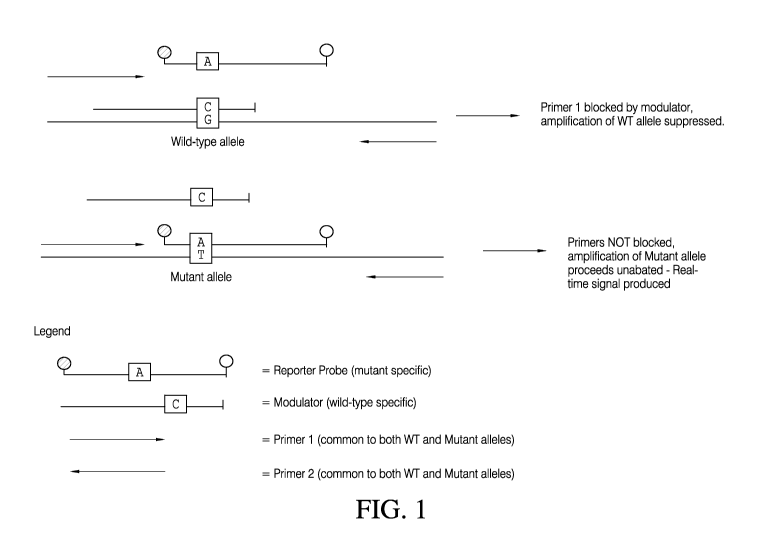
[0030] Figures 1 and 2 are depictions of exemplary methods according
to the
embodiments disclosed herein for the detection of sequence variants. As shown
in Figures 1
and 2, amplification primers (i.e., forward primer 1 and reverse primer 2)
flaffl( the wild type
and mutant allele sequences of interest, and comprise sequences common to both
wild-type
and mutant or variant allele sequences. Accordingly, as shown in Figure 2, in
contrast to
methods that utilize allele-specific amplification primers to achieve
preferential amplification
of rare sequences, the present methods advantageously enable the simultaneous
amplification
of multiple variant sequences, using a single amplification primer pair.
Detection of Altered DNA Methylation Patterns
[0031] Also provided are methods for the detection of DNA methylation
variants,
i.e., DNA that has an altered methylation pattern ¨ e.g., is methylated at
cytosine residues
that are non-methylated in wild-type DNA, or includes unmethylated cytosine
residues that
are methylated in wild-type DNA.
[0032] In some embodiments, the sample DNA is treated with an agent
the
selectively modifies unmethylated cytosine residues. By way of example only,
in some
embodiments, the sample nucleic acids are treated with sodium bisulphite,
according to art-
accepted methods. (See, e.g., Formmer, et al. (1992) Proc. Nat. Acad. Sci. USA
89:1827-
1831). Treatment with sodium bisulphite sulphonates unmethylated cytosines,
but not
methylated cytosines. Following sulphonation, the sample is subjected to
conditions (e.g.,
alkaline conditions, or any other appropriate conditions), that deaminate the
sulphonated
DNA to yield a uracil-bisulphite derivative that is in turn converted to
uracil by alkaline
desulphonation. Selective conversion of the unmethylated cytosine residues on
both strains
(i.e., the first strand and the second strand) generates novel sequences,
referred to as
"modified target DNA," for convenience, as illustrated in Figure 3. The
modified sample
nucleic acids are then subjected to an amplification (and/or detection)
reaction, as discussed
below.
[0033] In some embodiments, provided herein are methods to detect, or
enhance
the specificity of detection of rare methylation events, e.g., by performing a
methylation-
specific amplification reaction (e.g., methylation specific PCR). Modified
sample nucleic
acids are contacted with a forward and a reverse amplification primer that
specifically
hybridize to opposite strands of the modified sample nucleic acids, i.e., the
forward primer
hybridizes to the first strand of the modified nucleic acids (e.g., modified
sample nucleic
acids, or modified target DNA) and the reverse primer hybridizes to the second
strand of the
CA 02890004 2015-04-29
WO 2014/070946 PCT/US2013/067604
modified nucleic acids (e.g., modified sample nucleic acids, or modified
target DNA), and
amplify the region between the two primers under amplification conditions.
[0034] Referring to Figure3, the forward primer (P1), comprises a
sequence that
is complementary to (specifically hybridizes to) modified target DNA B, the
target nucleotide
sequence of the second strand following cytosine modification, i.e., the
unique sequence
generated by specific modification of unmethylated cytosine residues as
discussed above.
The forward primer thus contains one or more adenine residues that are located
in the primer
to hybridize to uracil residues present in the modified sample nucleic acids
(e.g., modified
sample nucleic acids, or modified target DNA). Accordingly, in some
embodiments, the
forward primer comprises one or more adenine residues that will base-pair with
uracil
residues in the second strand template sequence (converted from unmethylated
cytosine
residues in the second strand original sample sequence), i.e., modified target
DNA B(as
shown in Figure 3. In some embodiments, the one or more adenine residues that
base-pair
with uracil residues in the template sequence include an adenine residue
located at the 3' end
of the forward primer P1, as shown in Figure 3. As such, extension will occur
when the
original sample DNA prior to modification of the unmethylated cytosines (e.g.,
by bisulphite
treatment), comprises an unmethylated cytosine residue at the same position
(shown in
Figure 3). If the second strand of the template contains methylated cytosine
residues, then
treatment with bisulphite will not generate a novel sequence, and the
adenosine residues in
the methylation-specific primer will be mismatched with the methylated
cytosines in the
second strand of the template nucleic acids. As such, amplification will not
occur when the
second strand of the original sample nucleic acids (prior to modification)
comprises a
methylated cytosine residue at the same position (not shown). In some
embodiments, the
forward primer is fully complementary to a target sequence that comprises
methylated
cytosines and is also fully complementary to a target sequence that comprises
unmethylated
cytosines (see, e.g., EXAMPE 2, below). For example, in some embodiments, the
forward
primer hybridizes to a target sequence that does not include potentially
methylated cytosine
residues.
[0035] In some embodiments, the reverse primer (depicted as P2 in
Figure 3) is
complementary to the unique first strand sequence generated by amplification
from the
forward primer following modification of the sample nucleic acids. The unique
first strand
sequence generated by amplification is depicted as P 1 -extu in Figure 3.
Accordingly, in
some embodiments, the reverse primer comprises one or more thymine residues,
which
CA 02890004 2015-04-29
WO 2014/070946 PCT/US2013/067604
11
correspond to the position of one or more uracil residues (converted from
unmethylated
cytosine residues in the second strand original sample sequence, i.e.,
modified target DNA
B), and that base-pair with adenine residues present in the extension product
from the
forward primer (P 1 -extu). In some embodiments, the one or more thymine
residues
corresponding to the position of one or more uracil residues (converted from
unmethylated
cytosine residues in the second strand original sample sequence), is at the 3'
end of the
reverse primer. As such, extension will occur when the second strand of the
original sample
DNA comprises an unmethylated cytosine residue at the same position (shown in
Figure 3),
and will not occur when the second strand of the original sample DNA comprises
a
methylated cytosine residue at the same position (not shown). The extension
product from
P2 is depicted as P2-extu in Figure 3.
[0036] In some embodiments, the methods comprise contacting the
treated sample
(e.g., a sample that has been treated to selectively modify cytosine residues)
with
methylation-specific forward and reverse primers as described herein, under
amplification
conditions, as described below. In some embodiments, the methods include
contacting the
treated sample with a methylation-specific probe (e.g., by including the
methylation-specific
probe in the reaction mixture prior to amplification, or by contacting the
sample with the
methylation-specific probe post-amplification). Methylation-specific probes
can include
sequences that are complementary to and thus hybridize to the unique amplicons
produced by
successful extension from the forward and reverse methylation-specific
primers, as described
above. In some embodiments, the methylation specific probe comprises one or
more cytosine
residues that correspond to the position of a methylated cytosine residue
present in the
sample nucleic acids (e.g., and that are thus present as cytosine residues on
the P2-extu
strand, or second strand of the amplified, modified target sequences). As
shown in Figure 3,
the methylated cytosine residues are not converted to uracil by bisulphite
treatment, and thus
the first and second strands of the amplicons produced by P1 and P2 (P1 -extu
and P2-extu,
respectively, in Figure 3) contain a guanine-cytosine base pair. In some
embodiments, the
methylation specific probe (shown as Rme in Figure 3) also contains one or
more thymine
residues that correspond to the position of an unmethylated cytosine residue
in the sample
nucleic acids (and thus, a uracil residue in the modified sample nucleic
acids, modified target
DNA B). In some embodiments, the methylation-specific probe contains a
detectable label or
detectable moiety, as discussed in further detail below.
CA 02890004 2015-04-29
WO 2014/070946 PCT/US2013/067604
12
[0037] In some embodiments, the amplification reaction mixture also
includes a
"modulator oligonucleotide" or "blocking oligonucleotide." In some
embodiments,
modulator oligonucleotides or blocker oligonucleotides are used selectively
suppress non-
specific hybridization of the methylation-specific amplification primers
and/or methylation-
specific reporter probes. Accordingly, modulator oligonucleotides or blocking
probes can be
used to overcome the potential for false positive results owing to the
presence of mixed
populations of methylated and unmethylated target nucleic acid sequences, as
may be
encountered in clinical samples. As shown in Figure 3, in some embodiments, a
blocking
probe is used to enhance the specificity of methylation-specific
amplification. For example,
in some embodiments, the blocking probe (shown as "B" in Figure 3) that
competes with
both primer P2 and the reporter probe Rme for hybridization with the amplified
target. The
sequence of the modulator oligonucleotide or blocking oligonucleotide B is
designed such
that it preferentially hybridizes, in this case, to amplification product
derived from
unmethylated DNA target strand A. The Tm of the modulator oligonucleotide or
blocking
oligonucleotide B is designed to be substantially similar to the Tm of the
forward and reverse
methylation-specific amplification primers (P1 and P2, and, reporter probe
Rme). In some
embodiments, the Tm of the blocking probe differs by less than 15 C, 14 C, 13
C, 12 C,
11 C, 10 C, 9 C, 8 C, 7 C, 6 C, 5 C, 4 C, 3 C, 2 C, or 1 C, or less, from the
methylation-
specific amplification primers and/or reporter probe. As such, in some
embodiments, the
reactions are optimized to allow discrimination between methylated an
unmethylated DNA
forms, e.g., by balancing concentration and the conditions of hybridization
(in particular
temperature and salt concentration, as well as other factors known in the
art). In general, the
higher the Tm of the blocking probe relative to that of the primer and/or
reporter probe with
which it competes, the lower the concentration of probe required to suppress
non-specific
amplification and/or detection of target nucleic acids. As discussed in
further detail below,
the blocker oligonucleotides are designed such that they cannot be extended
from their 3'
ends.
Amplification Primers
[0038] Amplification primers useful in the embodiments disclosed
herein are
preferably between 10 and 45 nucleotides in length. For example, the primers
can be at least
10, 11, 12, 13, 14, 15, 16, 17, 18, 19, 20, 21, 22, 23, 24, 25, 26, 27, 28,
29, 30, 31, 32, 33, 34,
35, 36, 37, 38, 39, 40, 41, 42, 43, 44, 45, or more nucleotides in length.
Primers can be
provided in any suitable form, included bound to a solid support, liquid, and
lyophilized, for
CA 02890004 2015-04-29
WO 2014/070946 PCT/US2013/067604
13
example. In some embodiments, the primers and/or probes include
oligonucleotides that
hybridize to a reference nucleic acid sequence over the entire length of the
oligonucleotide
sequence. Such sequences can be referred to as "fully complementary" with
respect to each
other. Where an oligonucleotide is referred to as "substantially
complementary" with respect
to a nucleic acid sequence herein, the two sequences can be fully
complementary, or they
may form mismatches upon hybridization, but retain the ability to hybridize
under stringent
conditions or standard PCR conditions as discussed below. As used herein, the
term
"standard PCR conditions" include, for example, any of the PCR conditions
disclosed herein,
or known in the art, as described in, for example, PCR 1: A Practical
Approach, M. J.
McPherson, P. Quirke, and G. R. Taylor, Ed., (c) 2001, Oxford University
Press, Oxford,
England, and PCR Protocols: Current Methods and Applications, B. White, Ed.,
(c) 1993,
Humana Press, Totowa, NJ. The amplification primers can be substantially
complementary
to their annealing region, comprising the specific variant target sequence(s)
or the wild type
target sequence(s). Accordingly, substantially complementary sequences can
refer to
sequences ranging in percent identity from 100, 99, 98, 97, 96, 95, 94, 93,
92, 91, 90, 89, 85,
80, 75 or less, or any number in between, compared to the reference sequence.
Conditions
for enhancing the stringency of amplification reactions and suitable in the
embodiments
disclosed herein, are well-known to those in the art. A discussion of PCR
conditions, and
stringency of PCR, can be found, for example in Roux, K. "Optimization and
Troubleshooting in PCR," in PCR PRIMER: A LABORATORY MANUAL, Diffenbach,
Ed. 0 1995, Cold Spring Harbor Laboratory Press, Cold Spring Harbor, NY; and
Datta, et al.
(2003) Nucl. Acids Res. 31(19):5590-5597.
[0039] "Stringent conditions" or "high stringency conditions", as
defined herein,
may be identified by those that: (1) employ low ionic strength and high
temperature for
washing, for example 0.015 M sodium chloride/0.0015 M sodium citrate/0.1%
sodium
dodecyl sulfate at 50 C; (2) employ during hybridization a denaturing agent,
such as
formamide, for example, 50% (v/v) formamide with 0.1% bovine serum
albumin/0.1%
Fico11/0.1% polyvinylpyrrolidone/50 mM sodium phosphate buffer at pH 6.5 with
750 mM
sodium chloride, 75 mM sodium citrate at 42 C; or (3) employ 50% formamide, 5
x SSC
(0.75 M NaC1, 0.075 M sodium citrate), 50 mM sodium phosphate (pH 6.8), 0.1%
sodium
pyrophosphate, 5 x Denhardt's solution, sonicated salmon sperm DNA (50
jig/ml), 0.1%
SDS, and 10% dextran sulfate at 42 C, with washes at 42 C in 0.2 x SSC (sodium
CA 02890004 2015-04-29
WO 2014/070946 PCT/US2013/067604
14
chloride/sodium citrate) and 50% formamide at 55 C, followed by a high-
stringency wash
consisting of 0.1 x SSC containing EDTA at 55 C.
[0040]
"Moderately stringent conditions" may be identified as described by
Sambrook et al., Molecular Cloning: A Laboratory Manual, New York: Cold Spring
Harbor
Press, 1989, and include the use of washing solution and hybridization
conditions (e.g.,
temperature, ionic strength and %SDS) less stringent that those described
above. An
example of moderately stringent conditions is overnight incubation at 37 C in
a solution
comprising: 20% formamide, 5 x SSC (150 mM NaC1, 15 mM trisodium citrate), 50
mM
sodium phosphate (pH 7.6), 5 x Denhardt's solution, 10% dextran sulfate, and
20 mg/ml
denatured sheared salmon sperm DNA, followed by washing the filters in 1 x SSC
at about
37-50 C. The skilled artisan will recognize how to adjust the temperature,
ionic strength, etc.
as necessary to accommodate factors such as oligonucleotide length and the
like.
[0041] In
some embodiments, primer pairs comprising a forward and reverse
primer are used in the amplification methods described herein, e.g., to
produce target
amplicons. In some embodiments, the Tm of the forward and reverse primers are
substantially similar, e.g., differ by less than 15 C, 14 C, 13 C, 12 C, 11 C,
10 C, 9 C, 8 C,
7 C, 6 C, 5 C, 4 C, 3 C, 2 C, or 1 C, or less.
Blocker Oligonucleotides
[0042] In
an amplification reaction wherein reagents such as polymerase and
dNTPs are limiting, when a sample comprises a large excess of wild-type target
sequences
compared to variant or mutant target sequences or alleles, (e.g., 10 fold, 100
fold, 1000 fold
or more excess of wild-type target sequence compared to variant or mutant
sequence), the
kinetics of the amplification reaction are driven such that the limiting
reagents are consumed
in the amplification of wild-type sequences, while amplification and/or
detection of the rare
variant, rare mutant, alleles is suppressed. In order to shift the equilibrium
to favor
amplification of the rare variant or mutant alleles, blocker oligonucleotides
can be added to
the reaction.
[0043] As
used herein, the term "blocker oligonucleotide" refers to an
oligonucleotide that binds to a strand of DNA within the target amplicon, and
that is designed
to preferentially bind to the wild-type allele sequence (e.g., the abundant
allelic sequence,
such as a wild-type allele sequence) compared to the target variant sequence
(e.g., the rare
allelic variant). The blocker oligonucleotide generally comprises a
modification, or
modifications, as discussed below, that prevent primer extension by a
polymerase. Thus, a
CA 02890004 2015-04-29
WO 2014/070946 PCT/US2013/067604
blocker oligonucleotide can tightly bind to a wild type allele in order to
suppress
amplification of the wild-type allele while amplification of the variant
target allele sequence
is allowed to occur. As
explained above, blocker oligonucleotides can also be
advantageously used in the methods described herein for the detection of
methylation
variants, e.g., in methylation specific amplification reactions as discussed
above.
[0044]
Blocker oligonucleotides as disclosed herein refer to oligonucleotides that
are incapable of extension by a polymerase, for example, when hybridized to
its
complementary sequence in an amplification assay, e.g., PCR. Several different
means of
modifying oligonucleotides to render them incapable of extension by a
polymerase are
known and useful in the embodiments disclosed herein. By way of example,
common
examples of oligonucleotide modifications include, for example, 3'-OH
modifications and
dideoxy nucleotides. Numerous 3'-OH blocking materials are known and suitable,
and
include cordycepin (3'-deoxyadenosine) and other 3 '-moieties such as those
described in
Josefen, M. et al. (2009) Mol. Cell Probes 23:201-223 McKinzie, P. et al.
(2006)
Mutagenesis, 21(6):391-397; Parson, B. et al. (2005) Methods Mol. Biol.,
291:235-245;
Parsons, B. et al. (1992) Nucl. Acids. Res., 25:20(10):2493-2496, and Morlan,
J. et al. (2009)
PLoS One 4(2):e4584, the disclosures of which relating to oligonucleotide
modifications are
hereby incorporated by reference. In some embodiments, the 3'-OH is blocked
with a (3-
amino-2-hydroxy)- propoxyphosphoryl. In some embodiments, the 3'-OH is blocked
by
introduction of a 3'-3'-A-5' linkage such as those described in U.S. Patent
No. 5660989.
[0045] In
some embodiments, the blocker oligonucleotide comprises a moiety that
binds within the minor groove of double-stranded DNA at its 3' end, which
prevents
polymerase extension. A variety of moieties that bind to the minor groove of
DNA suitable
for the blocker oligonucleotides disclosed herein are known in the art, and
include, but are
not limited to those described in U.S. Patent No. 5,801,155, Wemmer, et al.
(1997) Curr.
Opin. Structural Biol. 7:355-361, Walker, et al. (1997) Biopolymers 44:323-
334, Zimmer, et
al. (1986) Molec. Biol. 47:31-112, and Reddy, B. et al. (1999) Pharmacol.
Therap. 84:1-111.
Methods for incorporating or attaching minor-groove binding moieties to
oligonucleotides
are well-known. For example, methods described in US. Patent Nos 5512677,
5419966,
5696251, 5585481, 5492610, 5736626, 5801155 and 6727356 are suitable for
modifying
oligonucleotides to generate a blocking oligonucleotide.
CA 02890004 2015-04-29
WO 2014/070946 PCT/US2013/067604
16
[0046] In some embodiments, the blocking oligonucleotides disclosed
herein can
include a minor-groove binding moiety located at the 5' end, the 3' end, or at
a position
within the oligonucleotide.
[0047] The skilled artisan will readily appreciate that the exemplary
"blocking"
modifications discussed above are provided by way of illustration only, and
that any blocking
modification known or discovered in the future can be used in the blocking
oligonucleotides
and methods disclosed herein.
[0048] In some embodiments, the blocker oligonucleotides comprise one
or more
modifications that increase the Tm of the oligonucleotide. For example, in
some
embodiments the blocker oligonucleotide can comprise one or more nucleosidic
bases
different from the naturally occurring bases (i.e., adenine, cytosine,
thymine, guanine and
uracil). In some embodiments, the modified bases effectively hybridize to
nucleic acid units
that contain naturally occurring bases. In some embodiments, the modified
base(s) increase
the difference in the Tm between matched and mismatched sequences, and/or
decrease
mismatched priming efficiency, thereby improving the specificity and
sensitivity of the assay.
[0049] Non-limiting examples of modified bases useful in the
embodiments
disclosed herein include the general class of base analogues 7-deazapurines
and their
derivatives and pyrazolopyrimidines and their derivatives (described in PCT WO
90/14353;
and U.S. application Ser. No. 09/054,630, the disclosures of each of which are
incorporated
herein by reference in regards to the base analogues). Examples of base
analogues of this
type include, for example, the guanine analogue 6-amino-1H-pyrazolo[3,4-
d]pyrimidin-
4(5H)-one (ppG), the adenine analogue 4-amino-1H-pyrazolo[3,4-d]pyrimidine
(ppA), and
the xanthine analogue 1H-pyrazolo[4,4-d]pyrimidin-4(5H)-6(7H)-dione (ppX).
These base
analogues, when present in an oligonucleotide of some embodiments of the
methods and
compositions disclosed herein, strengthen hybridization.
[0050] Additionally, in some embodiments, modified sugars or sugar
analogues
can be present in one or more of the nucleotide subunits of a blocker
oligonucleotide. Sugar
modifications useful in the embodiments disclosed herein include, but are not
limited to,
attachment of substituents to the 2', 3' and/or 4' carbon atom of the sugar,
different epimeric
forms of the sugar, differences in the a or 13-configuration of the glycosidic
bond, and other
anomeric changes. Sugar moieties useful in the embodiments disclosed herein
include, but
are not limited to, pentose, deoxypentose, hexose, deoxyhexose, ribose,
deoxyribose, glucose,
arabinose, pentofuranose, xylose, lyxose, and cyclopentyl.
CA 02890004 2015-04-29
WO 2014/070946 PCT/US2013/067604
17
[0051] In
some embodiments the blocker oligonucleotide can contain one or more
locked nucleic acid (LNA)-type modifications. LNA
modifications useful in the
embodiments disclosed herein can involve alterations to the pentose sugar of
ribo- and
deoxyribonucleotides that constrains, or "locks," the sugar in the N-type
conformation seen in
A-form DNA. In some embodiments, this lock can be achieved via a 2'-0, 4'-C
methylene
linkage in 1,2:5,6-di-O-isopropylene-.alpha.-D-allofuranose. In other
embodiments, this
alteration then serves as the foundation for synthesizing locked nucleotide
phosphoramidite
monomers. (See, for example, Wengel J., Ace. Chem. Res., 32:301-310 (1998),
U.S. Pat. No.
7,060,809; Obika, et al., Tetrahedron Lett 39: 5401-5405 (1998); Singh, et
al., Chem
Commun 4:455-456 (1998); Koshkin, et al., Tetrahedron 54: 3607-3630 (1998),
the
disclosures of each of which are incorporated herein by reference
[0052] In
some embodiments, modified bases useful in the embodiments
disclosed herein include 8-Aza-7-deaza-dA (ppA), 8-Aza-7-deaza-dG (ppG), 2'-
Deoxypseudoisocytidine (iso dC), 5-fluoro-2'-deoxyuridine (fdU), locked
nucleic acid
(LNA), or 2'-0,4'-C-ethylene bridged nucleic acid (ENA) bases. Other examples
of modified
bases that can be used in the embodiments disclosed herein are described in
U.S. Pat. No.
7,517,978 (the disclosure of which is incorporated herein by reference).
[0053]
Many modified bases, including for example, LNA, ppA, ppG, 5-Fluoro-
dU (fdU), are commercially available and can be used in oligonucleotide
synthesis methods
well known in the art. In some embodiments, synthesis of modified primers and
probes can
be carried out using standard chemical means also well known in the art. For
example, in
certain embodiments, the modified moiety or base can be introduced by use of a
(a) modified
nucleoside as a DNA synthesis support, (b) modified nucleoside as a
phosphoramidite, (c)
reagent during DNA synthesis (e.g., benzylamine treatment of a convertible
amidite when
incorporated into a DNA sequence), or (d) by post-synthetic modification
according to art-
accepted techniques.
[0054] In
some embodiments, the primers or probes are synthesized so that the
modified bases are positioned at the 3' end of the blocker oligonucleotide. In
some
embodiments, the modified base are located between, 1-6 nucleotides, e.g., 2,
3, 4 or 5
nucleotides away from the 3'-end of the blocker oligonucleotide.
[0055]
Modified internucleotide linkages can also be present in oligonucleotides,
e.g., the blocker oligonucleotides in the embodiments disclosed herein.
Modified linkages
useful in the embodiments disclosed herein include, but are not limited to,
peptide,
CA 02890004 2015-04-29
WO 2014/070946 PCT/US2013/067604
18
phosphate, phosphodiester, phosphodiester, alkylphosphate, alkanephosphonate,
thiophosphate, phosphorothioate, phosphorodithioate, methylphosphonate,
phosphoramidate,
substituted phosphoramidate and the like. Several further modifications of
bases, sugars
and/or internucleotide linkages, that are compatible with their use in
oligonucleotides serving
as probes and/or primers, will be apparent to those of skill in the art.
[0056] In some embodiments, the blocker oligonucleotide binds to a
sequence
which overlaps with the annealing region of the forward or reverse
amplification primer. For
example, in some embodiments, the blocker oligonucleotide and the forward or
reverse
primer are identical across 5, 6, 7, 8, 9, 10, 11, 12, 13, 14, 15, 16, 17, 18,
19, 20, 21, 22, 23,
24, 25, or more consecutive nucleotides. In some embodiments, the overlap in
sequence
identity between the blocker oligonucleotide and the forward or reverse
amplification primer
exists over 10%, 15%, 20%, 25%, 30%, 35%, 40%, 45%, 50%, 55%, 60%, 65%, 70%,
75%,
80%, 85%, or more, or any percentage in between, of the length of the blocker
oligonucleotide and/or amplification primer. In some embodiments, the
amplification primer
comprises one or more nucleotides, e.g., 1, 2, 3, 4, 5, 6, 7, 8, 9, 10, 11,
12, 13, 14, 15, 16, 17,
18, 19, 20, 21, 22, 23, 24, 25 or more, on its 5' end that are not identical
to the blocker
oligonucleotide (but that are complementary or substantially complementary to
the reference
sequence). In some embodiments, the blocker oligonucleotide comprises one or
more
nucleotides, e.g., 1,2, 3, 4, 5, 6, 7, 8,9, 10, 11, 12, 13, 14, 15, 16, 17,
18, 19, 20, 21, 22, 23,
24, 25 or more, on its 3' end that are not identical to the amplification
primer (but that are
complementary or substantially complementary to the reference sequence).
[0057] As shown in Figures 1 and 2, the blocker oligonucleotide
preferentially
binds to the wild-type target sequence compared to the mutant or variant
target sequence.
Also shown in Figures 1 and 2 is the overlap between the amplification primer
(i.e., primer 1
as shown) and the blocker oligonucleotide. As shown in Figures 1 and 2,
binding of the
blocker oligonucleotide to the wild type allele target sequence prevents
binding and
extension of the amplification primer, thereby suppressing amplification of
the wild-type
sequence. In contrast to the wild-type allele sequence, the amplification
primer will
preferentially bind to the mutant allele sequence, over the blocking
oligonucleotide. Thus,
the amplification is not blocked and the amplification of the mutant target
allele sequence
proceeds unimpeded. By this means, the present method advantageously allows
for
simultaneous and preferential amplification of one or more variant or mutant
target allele
sequences.
CA 02890004 2015-04-29
WO 2014/070946 PCT/US2013/067604
19
Reporter Probes
[0058] To
detect the presence and/or amount of variant target sequence(s) e.g.,
rare variant or mutant template nucleic acids in the sample, the sample is
contacted with one
or more allele-specific reporter probes. In some embodiments, the methods
disclosed herein
provide for the detection of more than one variant or mutant allele sequence
in a sample.
Accordingly, in some embodiments, a sample can be contacted with 1, 2, 3, 4,
5, 6, 7, 8 or
more, reporter probes. Each reporter probe preferentially binds to a cognate
allelic variant
compared to the wild type allelic sequence. As discussed above, in some
embodiments,
reporter probes can be advantageously used to detect methylation variants,
e.g., in
methylation-specific amplification as discussed above.
[0059] The
reporter probes can comprise a detectable moiety. In some
embodiments, the probe can include a detectable label. Labels of interest
include directly
detectable and indirectly detectable radioactive or non-radioactive labels
such as fluorescent
dyes and the like. Directly detectable labels refer to detectable moieties
that provide a
directly detectable signal without interaction with one or more additional
chemical agents.
Indirectly detectable labels are those labels which interact with one or more
additional
members to provide a detectable signal. In this latter embodiment, the label
is a member of a
signal producing system that includes two or more chemical agents that work
together to
provide the detectable signal. Examples of indirectly detectable labels
include biotin or
digoxigenin, which can be detected by a suitable antibody coupled to a
fluorochrome or
enzyme, such as alkaline phosphatase.
[0060] In
some embodiments, the label is a directly detectable label. Directly
detectable labels of particular interest include fluorescent labels.
Fluorescent labels suitable
in the detector probes of the embodiments disclosed herein include fluorophore
moieties.
Specific fluorescent dyes of interest include: xanthene dyes, e.g.,
fluorescein and rhodamine
dyes, such as fluorescein isothiocyanate (FITC), 2-[ethylamino)-3-(ethylimino)-
2-7-
dimethy1-3H-xanthen-9-yl]benzoic acid ethyl ester monohydrochloride
(R6G)(emits a
response radiation in the wavelength that ranges from about 500 to 560 nm),
1,1,3,3,3',3'-
Hexamethylindodicarbocyanine iodide (HIDC) (emits a response radiation in the
wavelength
that ranged from about 600 to 660 nm), 6-carboxyfluorescein (commonly known by
the
abbreviations FAM and F), 6-carboxy-2',4',7',4,7-hexachlorofluorescein (HEX),
6-carboxy-
4',5'-dichloro-2',7'-dimethoxyfluorescein (JOE or J),
N,N,N',N'-tetramethy1-6-
carboxyrhodamine (TAMRA or T), 6-carboxy-X-rhodamine (ROX or R), 5-
CA 02890004 2015-04-29
WO 2014/070946 PCT/US2013/067604
carboxyrhodamine-6G (R6G5 or G5), 6-carboxyrhodamine-6G (R6G6 or G6), and
rhodamine 110; cyanine dyes, e.g. Cy3, Cy5 and Cy7 dyes; coumarins, e.g.,
umbelliferone;
benzimide dyes, e.g. Hoechst 33258; phenanthridine dyes, e.g. Texas Red;
ethidium dyes;
acridine dyes; carbazole dyes; phenoxazine dyes; porphyrin dyes; polymethine
dyes, e.g.
cyanine dyes such as Cy3 (emits a response radiation in the wavelength that
ranges from
about 540 to 580 nm), Cy5 (emits a response radiation in the wavelength that
ranges from
about 640 to 680 nm), etc; BODIPY dyes and quinoline dyes. Specific
fluorophores of
interest include: Pyrene, Coumarin, Diethylaminocoumarin, FAM, Fluorescein
Chlorotriazinyl, Fluorescein, R110, Eosin, JOE, R6G, HIDC,
Tetramethylrhodamine,
TAMRA, Lissamine, ROX, Napthofluorescein, Texas Red, Napthofluorescein, Cy3,
and
Cy5, and the like. In preferred embodiments, the reporter probe can be a
molecular beacon
probe, a TAQMANTm probe, or a SCORPIONTM probe.
[0061] In some embodiments, the reporter probe(s) have a Tm that is
higher than
the Tm of the forward and reverse amplification primers used in the methods
disclosed herein.
For example, in some embodiments, the probes, e.g., molecular beacon probes or
the like,
have a Tm that is greater than 4 C, 5 C, 6 C, 7 C, 8 C, 9 C, 10 C, 11 C, 12 C,
13 C, 14 C,
15 C, 16 C, 17 C, 18 C, 19 C, 20 C, 21 C, 22 C, 23 C, 24 C, or 25 C, or more
than either
amplification primer used to generate an amplicon to which the oligonucleotide
probe
hybridizes. For example, a molecular beacon probe can have a Tm that is at
least 5-10 C
higher than either amplification primer pair used to generate the amplicon to
which the
molecular beacon hybridizes. In some embodiments, the reporter probe(s) have a
Tm that is
the same or lower than the forward and reverse amplification primers disclosed
herein.
[0062] As used herein, the term "Tm" and "melting temperature" are
interchangeable terms which refer to the temperature at which 50% of a
population of double
stranded polynucleotide molecules become dissociated into single strands. The
Tm of
particular nucleic acids, e.g., primers, or oligonucleotide probes, or the
like can be readily
calculated by the following equation: Tm=69.3+0.41 x (G+C)%-650/L, wherein L
refers to
the length of the nucleic acid. The Tm of a hybrid polynucleotide may also be
estimated
using a formula adopted from hybridization assays in 1 M salt, and is commonly
used for
calculating the Tm for PCR primers: [(number of A+T) x 2 C+(number of G+C) x 4
C], see,
for example, Newton et al. (1997) PCR (2nd ed; Springer-Verlag, New York).
Other more
sophisticated computations exist in the art, which take structural as well as
sequence
CA 02890004 2015-04-29
WO 2014/070946 PCT/US2013/067604
21
characteristics into account for the calculation of Tm. A calculated Tm is
merely an estimate;
the optimum temperature is commonly determined empirically.
[0063] In some embodiments, the reporter probe can comprise an
oligonucleotide
that is shorter in length than the forward or reverse amplification primer.
For example, in
some embodiments, the reporter probe(s) is 1, 2, 3, 4, 5, 6, 7, 8, 9, 10, or
more nucleotides
shorter than either the forward or reverse amplification primer.
[0064] In some embodiments, the reporter probe(s) bind to an
overlapping
sequence, as the blocker oligonucleotide. For example, in some embodiments,
the reporter
probe(s) and the blocker oligonucleotide are identical across 5, 6, 7, 8, 9,
10, 11, 12, 13, 14,
15, 16, 17, 18, 19, 20, 21, 22, 23, 24, 25, or more consecutive nucleotides.
In some
embodiments, the overlap in sequence identity between the reporter probe(s)
and the blocker
oligonucleotide exists over 10%, 15%, 20%, 25%, 30%, 35%, 40%, 45%, 50%, 55%,
60%,
65%, 70%, 75%, 80%, 85%, or more, or any percentage in between, of the length
of the
blocker oligonucleotide and/or reporter probe(s). In some embodiments, the
blocker
oligonucleotide comprises one or more nucleotides, e.g., 1, 2, 3, 4, 5, 6, 7,
8, 9, 10, 11, 12,
13, 14, 15, 16, 17, 18, 19, 20, 21, 22, 23, 24, 25 or more, on its 5' end that
are not identical to
the reporter probe (but that are complementary or substantially complementary
to the
reference sequence). In some embodiments, the reporter probe comprises one or
more
nucleotides, e.g., 1,2, 3, 4, 5, 6, 7, 8,9, 10, 11, 12, 13, 14, 15, 16, 17,
18, 19, 20, 21, 22, 23,
24, 25 or more, on its 3' end that are not identical to the blocker probe (but
that are
complementary or substantially complementary to the reference sequence).
[0065] As shown in Figures 1 and 2, the reporter probe(s) is allele-
specific. That
is, the reporter probe is complementary to the variant or mutant allele
sequence(s) being
assayed, and non-complementary to the wild-type allele sequence. As shown in
Figures 1
and 2, binding of the detector probe to the mutant or variant target allele
sequence does not
block or impede amplification by the amplification primers. Binding of the
reporter probe to
the mutant allele sequence (e.g., within sample template sequence or amplicon
sequences)
produces a detectable signal. As shown in Figure 2, in some embodiments,
reaction mixtures
can contain more than one detector probe, wherein each detector probe is
specific for a
different variant or mutant target allele sequence, and wherein each detector
probe comprises
a different detectable moiety. Accordingly, detection and identification of
different mutant
alleles in a single sample/reaction mixture is possible.
CA 02890004 2015-04-29
WO 2014/070946 PCT/US2013/067604
22
[0066] In addition to the sample, amplification primers, blocker
oligonucleotide,
and reporter probe(s), the reaction mixture includes a polymerase. The skilled
artisan will
appreciate that many polymerases known to those in the art are suitable for
the methods
described herein. For example, thermostable polymerases (including
commercially available
polymerases) obtained from Thermus aquaticus, Thermus thermophilus,
Thermococcus
litoralis, Pyrococcus furiosus, Pyrococcus woosii and other species of the
Pyrococcus genus,
Bacillus stearothermophilus, Sulfolobus acidocaldarius, Thermoplasma
acidophilum,
Thermus flavus, Thermus ruber, Thermus brockianus, Thermotoga neapolitana,
Thermotoga
maritima and other species of the Thermotoga genus, and Methanobacterium
thermoautotrophicum, and mutants of each of these species are useful in the
embodiments
disclosed herein. Preferable thermostable polymerases can include, but are not
limited to, Taq
DNA polymerase, Th DNA polymerase, Tma DNA polymerase, or mutants, derivatives
or
fragments thereof
[0067] Usually the reaction mixture will further comprise four
different types of
dNTPs corresponding to the four naturally occurring nucleoside bases, i.e.,
dATP, dTTP,
dCTP, and dGTP. In the methods of the invention, each dNTP will typically be
present in an
amount ranging from about 10 to 5000 M, usually from about 20 to 1000 M,
about 100 to
800 M, or about 300 to 600 M.
[0068] The reaction mixture can further include an aqueous buffer
medium that
includes a source of monovalent ions, a source of divalent cations, and a
buffering agent. Any
convenient source of monovalent ions, such as potassium chloride, potassium
acetate,
ammonium acetate, potassium glutamate, ammonium chloride, ammonium sulfate,
and the
like may be employed. The divalent cation may be magnesium, manganese, zinc,
and the
like, where the cation will typically be magnesium. Any convenient source of
magnesium
cation may be employed, including magnesium chloride, magnesium acetate, and
the like.
The amount of magnesium present in the buffer may range from 0.5 to 10 mM, and
can range
from about 1 to about 6 mM, or about 3 to about 5 mM. Representative buffering
agents or
salts that may be present in the buffer include Tris, Tricine, HEPES, MOPS,
and the like,
where the amount of buffering agent will typically range from about 5 to 150
mM, usually
from about 10 to 100 mM, and more usually from about 20 to 50 mM, where in
certain
preferred embodiments the buffering agent will be present in an amount
sufficient to provide
a pH ranging from about 6.0 to 9.5, for example, about pH 6.0, 6.5, 7.0, 7.5,
8.0, 8.5, 9.0, or
9.5. Other agents that may be present in the buffer medium include chelating
agents, such as
CA 02890004 2015-04-29
WO 2014/070946 PCT/US2013/067604
23
EDTA, EGTA, and the like. In some embodiments, the reaction mixture can
include BSA, or
the like. In addition, in some embodiments, the reactions can include a
cryoprotectant, such
as trehalose, particularly when the reagents are provided as a master mix,
which can be stored
over time.
[0069] In preparing a reaction mixture, the various constituent
components may
be combined in any convenient order. For example, the buffer may be combined
with primer,
polymerase, and then template nucleic acid, or all of the various constituent
components may
be combined at the same time to produce the reaction mixture.
[0070] Alternatively, commercially available premixed reagents can be
utilized in
the methods disclosed herein, according to the manufacturer's instructions, or
modified to
improve reaction conditions (e.g., modification of buffer concentration,
cation concentration,
or dNTP concentration, as necessary), including, for example, TAQMANO
Universal PCR
Master Mix (Applied Biosystems), OMNIMIXO or SMARTMIXO (Cepheid), IQ™
Supermix (Bio-Rad Laboratories), LIGHTCYCLERO FastStart (Roche Applied
Science,
Indianapolis, IN), or BRILLIANT QPCR Master Mix (Stratagene, La Jolla, CA).
[0071] The reaction mixture can then be subjected to amplification, or
primer
extension conditions. For example, in some embodiments, the reaction mixture
is subjected
to thermal cycling or isothermal amplification. Thermal cycling conditions can
vary in time
as well as in temperature for each of the different steps, depending on the
thermal cycler used
as well as other variables that could modify the amplification's performance.
In some
embodiments, a 2-step protocol is performed, in which the protocol combines
the annealing
and elongation steps at a common temperature, optimal for both the annealing
of the primers
and probes as well as for the extension step. In some embodiments, a 3-step
protocol is
performed, in which a denaturation step, an annealing step, and an elongation
step are
performed.
[0072] In some embodiments, the compositions disclosed herein can be
used in
connection with devices for real-time amplification reactions, e.g., the BD
MAX (Becton
Dickinson and Co., Franklin Lakes, NJ), the VIPER (Becton Dickinson and Co.,
Franklin
Lakes, NJ), the VIPER LT (Becton Dickinson and Co., Franklin Lakes, NJ), the
SMARTCYLCERO (Cepheid, Sunnyvale, CA), ABI PRISM 7700 (Applied Biosystems,
Foster City, CA), ROTOR-GENE TM (Corbett Research, Sydney, Australia),
LIGHTCYCLERO (Roche Diagnostics Corp, Indianapolis, IN), ICYCLERO (BioRad
CA 02890004 2015-04-29
WO 2014/070946 PCT/US2013/067604
24
Laboratories, Hercules, CA), IMX40000 (Stratagene, La Jolla, CA), CFX96TM Real-
Time
PCR System (Bio-Rad Laboratories Inc.), and the like.
[0073] In some embodiments, the compositions disclosed herein can be
used in
methods comprising isothermal amplification of nucleic acids. Isothermal
amplification
conditions can vary in time as well as temperature, depending on variables
such as the
method, enzyme, template, and primer or primers used. Examples of
amplification methods
that can be performed under isothermal conditions include, but are not limited
to, some
versions of LAMP, SDA, and the like.
[0074] Isothermal amplification can include an optional denaturation
step,
followed by an isothermal incubation in which nucleic acid is amplified. In
some
embodiments, an isothermal incubation is performed without an initial
denaturing step. In
some embodiments, the isothermal incubation is performed at least about 25 C,
for example
about 25 C, 26, 27, 28, 29, 30, 31, 32, 33, 34, 35, 36, 37, 38, 39, 40, 41,
42, 43, 44, 45, 46,
47, 48, 49, 50, 51, 52, 53, 54, 55, 56, 57, 58, 59, 60, 61, 62, 63, 64, 65,
66, 67, 68, 69, 70, 71,
72, 73, 74, or 75 C, including ranges between any of the listed values. In
some
embodiments, the isothermal incubation is performed at about 37 C. In some
embodiments,
the isothermal incubation is performed at about 64 C. In some embodiments, the
isothermal
incubation is performed for 180 minutes or less, for example about 180, 165,
150, 135, 120,
105, 90, 75, 60, 45, 30, or 15 minutes, including ranges between any two of
the listed values.
[0075] In some embodiments, the accumulation amplicons of the target
sequences, i.e., the variant or mutant target allele sequence(s) are monitored
in real-time.
Methods for monitoring and assaying amplification reactions in real-time are
widely known,
and the skilled artisan will appreciate that any of the art-accepted
techniques of real-time
amplification are suitable for use in the embodiments disclosed herein.
Exemplary
descriptions of real-time amplification useful in the embodiments disclosed
herein can be
found, for example, in U.S. Patent No.6,783,984; U.S. Patent NO. 6,303, 305,
and the like.
As used herein, the term "Ct" or "Ct value" refers to threshold cycle and
signifies the cycle
(or fractional cycle) of an amplification assay in which signal from a
reporter that is
indicative of amplicon generation (e.g., fluorescence), first become
detectable above a
background level. In some embodiments, the threshold cycle or "Ct" is the
cycle number at
which nucleic acid amplification becomes exponential. In some embodiments,
e.g., in
embodiments wherein amplification proceeds via isothermal amplification,
threshold time
values are used to signify the time in an amplification assay in which signal
from a reporter
CA 02890004 2015-04-29
WO 2014/070946 PCT/US2013/067604
that is indicative of amplicon generation (e.g., fluorescence), first becomes
detectable above a
background level. In some embodiments, the threshold time value is the time at
which
nucleic acid amplification becomes exponential.
[0076] As used herein, the term "delta Ct" or "ACt" refers to the
difference in the
numerical cycle number at which the signal passes a fixed threshold between
two different
samples or reactions. In some embodiments ACt refers to the difference in
numerical cycle
number at which exponential amplification is reached between two different
samples or
reactions. The ACt can be used to identify the specificity between a matched
reporter probe
to the corresponding target nucleic acid sequence and a mismatched reporter
probe to the
same corresponding sequence.
[0077] Various methods to calculate Ct values and threshold time
values are
known in the art and are useful in the embodiments disclosed herein. By way of
example
only, methods described in U.S. Patent No's 6783984, 6303305, and the like can
be used in
calculating Ct values and threshold time values in the methods disclosed
herein.
Accordingly, in some embodiments, the methods include the step of determining
the Ct value
or threshold time value, for each target allele sequence of interest (e.g.,
mutant or target allele
sequences).
[0078] The present embodiments are based, in part, upon the discovery
that using
a combination of amplification primers, oligonucleotide blockers, and allele-
specific detector
probes, one can render amplification of rare allele sequences
thermodynamically more
favorable, thereby enabling their detection in samples that contain
predominantly wild-type
or other variant allele sequences. Figures 4-7 illustrate the concepts
described herein,
including the thermodynamic consideration used in practicing the embodiments
disclosed
herein.
[0079] Figure 4 depicts the molecular species present in a reaction
mixture that is
subjected to primer extension or amplification conditions. "A" represents the
"analyte" or
target region of interest that comprises either the wild-type or variant or
mutant allele
sequence. As shown in Figure 4A, the molecular species in the reaction mixture
include the
analyte, the reporter probe ("D"), the blocker oligonucleotide ("B"), the
amplification
primer(s) ("P"), and the polymerase ("E"). Figure 4B shows bi-molecular
species, including
amplification primer bound to its cognate sequence on the analyte ("PA"),
reporter probe
bound to its cognate sequence on the analyte ("DA"), blocker oligonucleotide
bound to its
cognate sequence on the analyte (wild-type target allele sequence) ("BA"), and
blocker
CA 02890004 2015-04-29
WO 2014/070946 PCT/US2013/067604
26
oligonucleotide that is partially bound to the analyte (variant or mutant
target allele sequence)
("Ab"). Figure 4C depicts tri-molecular species, such as (1) complexes between
the
amplification primer, its cognate analyte, and polymerase ("PAE"); (2)
complexes between
the amplification primer, its cognate analyte, and a reporter probe ("PAD");
and (3)
complexes between the amplification primer, its cognate analyte and an
oligonucleotide
blocker ("PAb"). Figure 4D depicts possible tetra-molecular species, including
(1)
complexes between an amplification primer, its cognate analyte sequence,
reporter probe, and
polymerase ("PADE"); and (2) complexes between an amplification primer, its
cognate
analyte, a blocker oligonucleotide, and polymerase ("PAbE"). The PAb and PAbE
species
represent the case in which nucleotide at and near the 5' end of the blocker
are unhybridized
to the analyte, but the remaining nucleotides of the blocker are hybridized to
the analyte. In
all cases, primers, probes, blockers may hybridize with wild-type or variant
DNA; however
the perfectly matched hybrids (e.g. blocker with wild-type DNA) will be
thermodynamically
more stable than hybrids containing mismatches (e.g. blocker with variant
DNA).
[0080] The molecular complexes shown in Figures 4A-4D exist in a multi-
state
equilibrium, as shown in Figure 5. The association between each of the mono-
molecular
species is described by an equilibrium constant, K. The embodiments disclosed
herein area
based, in part, upon the discovery that equilibrium constants for the various
molecular
species shown in Figure 4 can be advantageously used to model reaction
conditions to
maximize amplification of rare variant or mutant allele sequences compared in
samples
comprising an excess of copies (e.g., 5X, 10X, 20X, 30X, 40X, 50X, 100X, 500X,
750X,
1000X, or greater) of wild-type allele sequence compared to variant or mutant
allele
sequence, while minimizing detrimental effects on amplification efficiency. In
accordance
with the methods disclosed herein, the equilibrium constants for the complexes
depicted in
Figure 4 can be estimated using enthalpy (dH) and entropy (dS) changes
associated with
melting of each of the duplexes, at each temperature. dH and dS values for
each hybrid can
be estimated or calculated using any art-accepted methods. By way of example,
dH and dS
can be calculated using publicly available algorithms, such as those available
on the world
wide web site hypertext transfer
protocol://mfold.rna.albany.edull?q=DINAMelt/Two-state-
melting. The skilled artisan will appreciate that many known algorithms for
calculation of
dH and dS can be used in the methods disclosed herein. Figure 5 shows the
calculation of
individual equilibrium constants according to the methods disclosed herein.
CA 02890004 2015-04-29
WO 2014/070946 PCT/US2013/067604
27
[0081] The present inventors discovered that equilibrium constants can
be used to
estimate the fraction of analyte "A" bound to blocker oligonucleotide,
detector probes, and
amplification primers, in a reaction mixture, e.g., in multi-state
equilibrium, and that these
values are useful in methods of maximizing amplification of rare allele
sequences. The
fraction of analyte, represented by "a" in various complexes within the
reaction can be
determined using the equations shown in Figure 6, using the starting
concentrations of
amplification primer (Po), blocker oligonucleotide (Bo), detector probe (Do),
and polymerase
(Eo), and the respective equilibrium constants, K1-K5, for each of the
different complexes, as
discussed in connection with Figure 5. Figure 7 shows a model estimator for
the number of
amplicons, A, or 13,, after n cycles, for two different targets (e.g., a wild-
type target allele
sequence and a rare mutant or rare variant target allele sequence), in a
single reaction with
limiting reagents (e.g., polymerase), calculated using the fraction of
extendible complexes,
`fe.," determined using the equations shown in Figure 6. The present
embodiments are
based, in part, upon the discovery that the fe. must be less than about 0.5,
i.e., less than 0.4,
0.3, 0.2, 0.1, or less, for adequate blocking of amplification/detection of
wild-type target
allele sequences such that variant or mutant target allele sequences present
in a sample at an
initial copy number that is at 100-fold less (e.g., 200-fold, 300-fold, 400-
fold, 500 fold, 600-
fold, 700-fold, 800-fold, 900-fold, 1000-fold, 10000-fold or greater) than
that of the wild-
type target sequences.
Kits
[0082] Aspects of the disclosure also relate to kits containing the
reagents and
compositions to carry out the methods described herein. Such a kit can
comprise a carrier
being compartmentalized to receive in close confinement therein one or more
containers,
such as tubes or vials. One of the containers may contain at least one
unlabeled or detectably
labeled primer or probe disclosed herein. The primers, including amplification
primers,
oligonucleotide blockers and detector probes can be present in dried form
(e.g., lyophilized or
other) or in an appropriate buffer as necessary. One or more containers may
contain one or
more enzymes or reagents to be utilized in PCR reactions. These enzymes may be
present by
themselves or in admixtures, in dried form or in appropriate buffers.
[0083] Finally, the kit can include all of the additional elements
necessary to carry
out the methods disclosed herein, such as buffers, extraction reagents,
enzymes, pipettes,
CA 02890004 2015-04-29
WO 2014/070946 PCT/US2013/067604
28
plates, nucleic acids, nucleoside triphosphates, filter paper, gel materials,
transfer materials,
autoradiography supplies, and the like.
[0084] The kits according to the present invention will comprise at
least: (a) a
blocker oligonucleotide, (b) a forward and reverse amplification primer, (c)
an allele specific
detector probe, and (d) instructions for using the provided amplification
primer pair, blocker
oligonucleotide, and allele specific detector probe.
[0085] In some embodiments, the kits include additional reagents that
are required
for or convenient and/or desirable to include in the reaction mixture prepared
during the
methods disclosed herein, where such reagents include: one or more
polymerases; an aqueous
buffer medium (either prepared or present in its constituent components, where
one or more
of the components may be premixed or all of the components may be separate),
and the like.
The various reagent components of the kits may be present in separate
containers, or may all
be pre-combined into a reagent mixture for combination with template nucleic
acid.
[0086] In addition to the above components, in some embodiments, the
kits can
also include instructions for practicing the methods disclosed herein. These
instructions can
be present in the kits in a variety of forms, one or more of which may be
present in the kit.
One form in which these instructions can be present is as printed information
on a suitable
medium or substrate, e.g., a piece or pieces of paper on which the information
is printed, in
the packaging of the kit, in a package insert, etc. Yet another means would be
a computer
readable medium, e.g., diskette, CD, etc., on which the information has been
recorded. Yet
another means that may be present is a website address that may be used via
the internet to
access the information at a removed site.
EXAMPLES
[0087] The following examples are provided to demonstrate particular
situations
and settings in which this technology may be applied and are not intended to
restrict the
scope of the invention and the claims included in this disclosure.
EXAMPLE 1
[0088] The following example demonstrates that the methods disclosed
herein can
be used to effectively detect multiple rare variant target allele sequences in
samples
comprising an excess (100 fold or more) of wild-type or alternative variant or
mutant target
allele sequences.
CA 02890004 2015-04-29
WO 2014/070946 PCT/US2013/067604
29
[0089] KRAS allelic variants G34T, G34C, G34A, and G38A, which are
commonly used in the diagnosis prognosis of various cancers, as well as
predicting the
sensitivity of tumors to certain therapeutics, were used as an exemplary
system to
demonstrate the efficacy of the methods described herein. Figure 8 shows the
target region
of interest in KRAS, including the wild-type sequence, as well as the position
of the G34A,
G34T and G38A variants.
[0090] Shown in Figure 8 are three different amplification primers,
i.e., Primer
1.0, Primer 1.2 and Primer 1.3 designed to amplify the target region of
interest. Also shown
are four different blocker oligonucleotides, i.e., blocker oligonucleotide
1.4, blocker
oligonucleotide 1.3, blocker oligonucleotide 1.2 and blocker oligonucleotide
1.1 that include
non-extendible 3'-OH modifications in accordance with the methods described
above, and
that are designed to preferentially binding to the wild-type target allele
sequence compared to
the various mutant allele sequences present at positions 34, 35, and 38 of
KRAS, as shown in
Figure 8. Also shown are seven different detector probes, i.e., probes 1.2,
2.1, 3.0, 4.1, 5.1,
6.0 and 7.0 designed for the detection of G34A, wt, G34T, G35A, G35G, G35T and
G38A
alleles. The detector probes are configured to generate a detectable,
fluorescent signal upon
hybridization to target, measurable in real time.
[0091] Using the methods described herein above, the entropy,
enthalpy,
equilibrium constants, and fraction of each molecular species present at
equilibrium were
calculated as shown in Figures 5 and 6. These values were calculated for both
wild-type
and G34T DNA. Among the values calculated are the fraction of extendible
molecular
species (fe.) wild-type (WT fe.) and G34T (mutant fe.) DNA. The calculated
values also
include the fraction of analyte (either wild-type or G34T) bound to extendible
species
containing a detector probe(s) (represented by PADE in Figures 5 and 6). The
PADE
species produce target amplification and detectable signal during PCR, whereas
the other
extendible species (PAE and PAbE) produce amplification but not detectable
signal. For
reaction mixtures containing more than one detector probe, the fraction of
analyte involved in
each PADE species was calculated, and these values are used to estimate the
signal produced
by each respective probe. The various fe. values were used to perform PCR
simulations in
which the samples contained a 100-fold excess of wild-type target allele
sequence compared
to the G34T mutant allele sequence.
[0092] Figure 9A shows the results of a simulated PCR reaction
containing
primer 1.2, blocker 1.1, and detector probes 1.2, 2.1, 3.0 and 7Ø As shown,
a specific signal
CA 02890004 2015-04-29
WO 2014/070946 PCT/US2013/067604
is detectable for G34T, whereas either very weak or no signal is produced from
probes
directed to the mutant target alleles not present in the sample. For this
reaction, WT fe. was
0.159 and mutant fe. was 0.909, predictive of suppression of wild-type target
amplification,
but strong amplification of mutant target. In contrast, Figure 9B shows the
results of PCR
simulation for reaction mixtures containing the same detector probes (1.2,
2.1, 3.0 and 7.0),
but a different primer (primer 1.3) and blocker (blocker 1.4). Again, the wild-
type allele is
present in 100-fold excess over the mutant G34T allele. This primer-blocker
combination
results in calculated values for mutant fe. of 0.906, and WT fe. of 0.767, the
latter of which
is predictive of significant amplification of both wild-type and mutant target
alleles. As
shown in Figure 9B, only weak signal is produced for the probe directed at the
G34T allele,
while significantly stronger signals are produced from probes directed at
mutant alleles not
present in the reaction mixture. These non-specific signals are produced by
hybridization of
probes to wild-type DNA, which because of the insufficient suppression of
amplification by
the blocker 1.4, is present at much higher levels than the G34T allele
throughout the course
of the PCR reaction.
[0093] The foregoing data demonstrate that the methods disclosed
herein can be
used to effectively detect and identify rare mutant or variant target allele
sequences against a
background of excess wild-type sequences. The methods disclosed herein thus
represent an
extremely efficient, efficacious means to detect sequence polymorphisms and
mutations that
have wide-ranging clinical and experimental uses.
EXAMPLE 2
[0094] The following example demonstrates how the methods disclosed
herein
can be used to detect methyl cytosine residues in the death associated protein
-1 (DAPK-1)
promoter region. Changes in methylation status within the promoter region of
DAKP-1 are
frequently associated in with a variety of types of cancer and therefore
accurate assessment of
methylation patterns can be an important diagnostic indicator (Raval et al.,
(2007), Cell, 129:
879-890; Candiloro et al Epigenetics 2011 6: 500-507).
[0095] Figure 10A shows a 105bp target sequence within the promoter
region of
DAKP-1. CpG sites, which are often the sites of altered cytosine methylation
patters, are
shown in boxes. Figure 10A also shows the unique sequences generated following
treatment
of the DAKP-1 promoter target sequence, when the sample DNA is originally
fully
CA 02890004 2015-04-29
WO 2014/070946 PCT/US2013/067604
31
unmethylated, or fully methylated. Specifically, as shown, there are nine
cytosine residues
that are potentially methylated, and that would be resistant to bisulphite
treatment.
[0096] Figure 10B shows a shorter, 61bp region within the target
sequence
shown in Figure 10A. As shown by the asterisks, four potential methylation
sites, e.g., at
nucleotide positions 47026, 47031, 47039 and 47062 exist within this region.
Table 2 below
illustrates the 16 possible DNA methylation patters within the DAPK-1 promoter
region
shown in Figure 10B.
TABLE 2
Combination DPAK-1 Nucleotide Position
47,026 47,031 47,039 47,062
1 X X X := X
2 X X X.
3 X X X
..
4 X X X
X X X
6 X X
7 X X
.:.:.:.:.:.:.:.:.:.:
8 X X
9 X X
X X
11 X X
12 X
13 H.X.
14
X
16
X: methyl cytosine residue; Shaded box corresponds to
residues detected by Reporter Probe-R
[0097] Figure 10B illustrates how the use of methylation-specific
amplification
primers, methylation-specific reporter probes, and methylation-specific
modulator
oligonucleotides can be used to determine whether a sample comprising the DAPK-
1
promoter target sequence comprises aberrant methylation. Primer P1 is fully
complementary
to sample DNA that is either fully methylated or unmethylated following
modification with
CA 02890004 2015-04-29
WO 2014/070946 PCT/US2013/067604
32
sodium bisulphite. By contrast, primer P2 includes a guanine residue that is
mismatched with
a converted uracil residue in the modified sample nucleic acids from a fully
unmethylated
sample, but which is complementary to modified sample nucleic acids from a
fully
methylated sample. Due to the fact that the mismatch is not at the 3' end of
the reverse
primer, however, amplification can still occur under standard amplification
conditions. The
reporter probe R contains 2 cytosine residues that are mismatched with the
modified sample
nucleic acids from a fully unmethylated sample, but which are complementary to
modified
sample nucleic acids from a fully methylated sample. As such, the reporter
probe
preferentially hybridizes to the amplicon derived from sample nucleic acids
that are
methylated, compared to amplicons derived from sample nucleic acids that are
unmethylated.
Blocking probe B includes 3 thymine residues that hybridize to uracil residues
present in the
modified unmethylated sample, but that are mismatched with the guanine
residues present in
the modified methylated sample. Blocking probe contains a modification at its
3' end that
inhibits extension. As such, the blocking probe will preferentially hybridize
to amplicons
derived from the unmethylated sample nucleic acids, as compared to the
methylated sample
nucleic acids. Accordingly, using primers P 1 , P2, reporter probe R, and
blocking
oligonucleotide B, on can preferentially amplify and detect rare methylated
sample nucleic
acids, e.g., within a sample comprising an abundance of unmethylated nucleic
acids.
[0098] The embodiments described and claimed herein is not to be
limited in
scope by the specific embodiments herein disclosed, since these embodiments
are intended as
illustrations of several aspects of the invention. Any equivalent embodiments
are intended
within the scope of this invention. Indeed, various modifications of the
embodiments in
addition to those shown and described herein will become apparent to those
skilled in the art
from the foregoing description. The appended claims are intended to cover such
modifications.