Note: Descriptions are shown in the official language in which they were submitted.
CA 02890256 2015-04-30
WO 2014/072888
PCT/1B2013/059786
ANTI-IL-13 RECEPTOR ALPHA 2 ANTIBODIES
AND ANTIBODY-DRUG CONJUGATES
Field of the Invention
This invention pertains to anti-IL-13 receptor alpha 2 (IL-13-Ra2) antibodies
and
antibody-drug conjugates for the treatment of cancer.
Sequence Listing
The instant application contains a Sequence Listing which has been submitted
via EFS and is hereby incorporated by reference in its entirety.
Background of the Invention
High levels of IL-13-Ra2 have been identified in a number of tumor cells,
including pancreatic, breast, ovarian and malignant gliomas. In contrast, only
a few
types of normal tissues express IL-13-Ra2, and only at low levels. The
treatment of
cancer has improved over the past decade with surgery, radiation therapy, and
chemotherapy as the primary treatment options. Such treatments can extend
survival
and/or relieve symptoms in many patients but are not likely to produce a cure
for many
patients. There remains a significant need for additional therapeutic options
for cancers.
Therefore, anti-IL-13-Ra2 antibody-drug conjugates that can exert a clinically
useful cytotoxic effect on IL-13-Ra2 expressing tumor cells, particularly
without exerting
undesirable effects on non-IL-13-Ra2 expressing cells, fulfill an unmet
clinical need in
the treatment of various IL-13-Ra2 expressing tumor cells.
Summary of the Invention
The present invention provides anti-IL-13-Ra2 antibody-drug conjugates and
methods of use for the treatment of cancer.
The present invention provides an isolated antibody or antigen-binding
fragment
that specifically binds to human IL-13-Ra2 wherein the antibody comprises: a
heavy
chain variable region comprising a CDR1, CDR2, and CDR3 of the VH sequence of
1
CA 02890256 2015-04-30
WO 2014/072888
PCT/1B2013/059786
SEQ ID NO: 1 and, a light chain variable region comprising a CDR1, CDR2, and
CDR3
of the VL sequence of SEQ ID NO: 5.
The present invention further provides an isolated antibody or antigen-binding
fragment that specifically binds to human IL-13-Ra2 wherein the antibody
comprises: (a)
a heavy chain CDR1 comprising SEQ ID NO: 2; (b) a heavy chain CDR2 comprising
SEQ ID NO: 3; (c) a heavy chain CDR3 comprising SEQ ID NO: 4; (d) a light
chain
CDR1 comprising SEQ ID NO: 6; (e) a light chain CDR2 comprising SEQ ID NO: 7;
and,
(f) a light chain CDR3 comprising SEQ ID NO: 8.
The present invention further provides an isolated antibody or antigen-binding
fragment that specifically binds to human IL-13-Ra2 wherein said isolated
antibody
further comprises the heavy chain variable region amino acid sequence of SEQ
ID NO:
1 and the light chain variable region amino acid sequence of SEQ ID NO: 5.
The present invention further provides an isolated antibody or antigen-binding
fragment that specifically binds to human IL-13-Ra2 wherein said isolated
antibody
comprises a heavy chain comprising the amino acid sequence of SEQ ID NO: 50
and
wherein said isolated antibody comprises a light chain comprising the amino
acid
sequence of SEQ ID NO: Si.
The present invention further provides an antibody-drug conjugate comprising a
cytotoxic agent conjugated to an antibody or antigen-binding fragment thereof
that
specifically binds to human IL-13-Ra2,
The present invention further provides an antibody-drug conjugate that
specifically binds to human IL-13-Ra2 wherein said conjugate has the formula:
Ab-(L-
D)p, wherein; (a) Ab is the antibody or antigen-binding fragment thereof of
the present
invention; (b) L-D is a linker-drug moiety, wherein L is a linker, and D is a
drug; and (c) p
is an integer from 1 to about 8.
The present invention further provides an antibody-drug conjugate that
specifically binds to human IL-13-Ra2 wherein the linker is selected from the
group
consisting of maleimidocaproyl (mc) and maleimidocaproyl-Val-Cit-PABA (vc).
2
CA 02890256 2015-04-30
WO 2014/072888
PCT/1B2013/059786
The present invention further provides an antibody-drug conjugate that
specifically binds to human IL-13-Ra2 wherein the linker-drug moiety has the
formula
designated vc-0101 or mc-3377 as shown in Example 14.
The present invention further provides an antibody-drug conjugate that
specifically binds to human IL-13-Ra2 wherein Ab comprises: (a) a heavy chain
variable
region comprising a CDR1, CDR2, and CDR3 of the VH sequence of SEQ ID NO: 1;
and, (b) a light chain variable region comprising a CDR1, CDR2, and CDR3 of
the VL
sequence of SEQ ID NO: 5.
The present invention further provides an antibody-drug conjugate that
specifically binds to human IL-13-Ra2 wherein Ab comprises: (a) a heavy chain
CDR1
comprising SEQ ID NO: 2; (b) a heavy chain CDR2 comprising SEQ ID NO: 3; (c) a
heavy chain CDR3 comprising SEQ ID NO: 4; (d) a light chain CDR1 comprising
SEQ
ID NO: 6; (e) a light chain CDR2 comprising SEQ ID NO: 7; and, (f) a light
chain CDR3
comprising SEQ ID NO: 8.
The present invention further provides an antibody-drug conjugate wherein L-D
is
selected from the group consisting of vc-0101, mc-3377, mc-0131, MalPeg-6121,
MalPeg-0131, mc-6121, vc-3906, vc-6780, mc-8261, mc3906, and MalPeg-8261.
The present invention further provides an antibody-drug conjugate that
specifically binds to human IL-13-Ra2 wherein said conjugate utilizes site-
specific
conjugation on engineered cysteine residues and has the formula: Ab-(L-D)p, or
a
pharmaceutically acceptable salt thereof wherein; (a) Ab is the antibody or
antigen-
binding fragment thereof comprising a heavy chain variable region comprising a
CDR1,
CDR2, and CDR3 of the VH sequence shown in SEQ ID NO: 1; a light chain
variable
region comprising a CDR1, CDR2, and CDR3 of the VL sequence shown in SEQ ID
NO: 5; and an engineered Fc region comprising at least one pair of amino acid
substitutions selected from the group consisting of the amino acid sequence of
SEQ ID
NO:33 and SEQ ID NO:34; or an engineered Fc region and at least one engineered
light chain constant region selected from group consisting of L443C (SEQ ID
NO: 28),
Q347C (SEQ ID NO: 29), kK183C (SEQ ID NO: 31), L443C/kA111C (SEQ ID NOS: 28
and 30), L443C/kK183C (SEQ ID NOS: 28 and 31), Q347C/kA111C (SEQ ID NOS: 29
3
CA 02890256 2015-04-30
WO 2014/072888
PCT/1B2013/059786
and 30), and Q347C/kK183C (SEQ ID NOS: 29 and 31); (b) L-D is a linker-drug
moiety,
wherein L is a linker, and D is a drug; and (c) p is an integer from 1 to
about 8.
The present invention further provides the antibody-drug conjugate described
above that utilizes site specific conjugation on engineered cysteine residues,
wherein
the linker-drug moiety has the formula designated vc-0101 or mc-3377 as shown
in
Example 14, or a pharmaceutically acceptable salt or solvate form thereof, and
p is an
integer of about 4.
The present invention further provides the antibody-drug conjugate of the
present
invention that utilizes the Multifunctional Antibody Conjugates (MAC)
technology,
wherein said antibody or antigen binding portion thereof specifically binds to
human IL-
13Ra2 wherein the antibody has the mutation D185A at position 185 of the LC as
shown in SEQ ID NO: 52, and the antibody is covalently conjugated to at least
one
drug moiety through a linker attached to a side chain of K188 of the LC of SEQ
ID
NO:49; wherein the drug moiety has the formula designated 0101 or 3377 as
shown in
Example 13, or a pharmaceutically acceptable salt or solvate form thereof, and
p is an
integer in a range whose lower limit may be selected from the group consisting
of about
1.5, about 1.6, about 1.7, about 1.8, about 1.9, and about 2.0, and whose
upper limit
may be selected from the group consisting of about 2.0, about 2.1, about 2.2,
about 2.3,
about 2.4, about 2.5. In some aspects, p is about 2.
The present invention further provides a pharmaceutical composition comprising
an antibody-drug conjugate of the present invention and a pharmaceutically
acceptable
carrier.
The present invention further provides a method of treating an IL-13-Ra2
expressing cancer in a patient in need thereof, comprising administering to
said patient
an antibody-drug conjugate of the present invention.
The present invention further provides a method of treating an IL-13-Ra2
expressing cancer wherein said cancer is selected from the group consisting of
carcinomas of the bladder, breast, cervix, colon, malignant gliomas,
endometrium,
kidney, lung, esophagus, ovary, prostate, pancreas, melanoma, stomach, and
testes.
4
CA 02890256 2015-04-30
WO 2014/072888
PCT/1B2013/059786
More preferably, the present invention provides a method of treating an IL-13-
Ra2 expressing cancer wherein said cancer is selected from the group
consisting of,
lung, colon, stomach, pancreatic, ovarian, malignant gliomas, and melanoma.
The invention further provides an antibody-drug conjugate of the present
invention for use in therapy.
The invention further provides use of an antibody-drug conjugate of the
present
invention for the manufacture of a medicament for therapy.
The invention further provides the use of an antibody-drug conjugate of the
present invention, wherein said use is for the treatment of an IL-13-Ra2
expressing
cancer.
The invention further provides a nucleic acid that encodes an IL-13-Ra2
antibody, a vector comprising said nucleic acid, and a host cell comprising
said vector.
The invention further provides a process for producing an IL-13-Ra2 antibody
wherein said process comprises culturing the host cell comprising the above
mentioned
vector and recovering the antibody from the cell culture.
The invention further provides a process for producing an IL-13-Ra2 antibody-
drug conjugate comprising: (a) linking a linker selected from the group
consisting of
maleimidocaproyl and maleimidocaproyl-Val-Cit-PABA to a drug selected from the
group consisting of 0101 and 3377 resulting in a linker-drug moeity; (b)
conjugating said
linker-drug moeity to the antibody recovered from the cell culture of
indicated above;
and, (c) purifying the antibody-drug conjugate.
The invention further provides an isolated antibody that competes with an
antibody or antigen-binding fragment thereof of the present invention for
specific binding
to human IL-13-Ra2.
The invention further provides an antibody¨drug conjugate comprising an
antibody or antigen-binding fragment thereof of the present invention for
specific binding
to human IL-13-Ra2.
The invention further provides a method for predicting whether a subject with
cancer will respond to an antibody-drug conjugate of the present invention
comprising:
5
CA 02890256 2015-04-30
WO 2014/072888
PCT/1B2013/059786
determining whether a biological sample of said cancer from the subject
expresses hIL-
13-Ra2.
The invention further provides a process of determining the level of hIL-13-
Ra2 in
a biological sample comprising the steps of: testing a sample from a subject
suspected
to have cancer in a immunoassay using an antibody of the present invention;
determining the cell surface levels of hIL-13-Ra2 on said sample; and,
comparing the
cell surface levels of hIL-13-Ra2 with that of a reference subject or
standard.
The invention further a method of treating an IL-13-Ra2 expressing cancer said
method comprising: determining the level of hIL-13-Ra2 in a biological sample
comprising the steps of: testing a sample from a subject suspected to have
cancer in a
immunoassay using an antibody of the present invention; determining the cell
surface
levels of hIL-13-Ra2 on said sample; comparing the cell surface levels of hIL-
13-Ra2
with that of a normal reference subject or standard; and administering an
antibody¨drug
conjugate of the present invention.
Brief Description of the Drawings
Fig. 1: Binding specificity of chimerc antibodies ch07 and ch08 to hIL-13-Ra2
but not hIL-13Ra1.
Fig. 2A and Fig. 2B: ch07, ch08 and antibodies MAB614 and ab27414 have
distinct binding epitopes to IL-13Ra2.
Fig. 2C: Biacore analysis indicating that ch07 and ch08 lack competition in
binding.
Fig. 3: ch07 and ch08 are non-neutralizing antibodies.
Fig. 4A ¨ 4G: SEQ ID NOS: 1 ¨55.
Detailed Description of the Invention
The present invention provides IL-13-Ra2 antibody-drug conjugates for the
treatment of cancer. In order that the present invention is more readily
understood,
certain terms and general techniques are first defined.
6
CA 02890256 2015-04-30
WO 2014/072888
PCT/1B2013/059786
Ali amino acid abbreviations used in this disolos-ure are those accepted by
the
United States Patent and Trademark Office as set forth in 37 C.F.R. 1.822
(d)(1).
Unless otherwise defined herein, scientific and technical terms used in
connection with the present invention shall have the meanings that are
commonly
understood by those of ordinary skill in the art. Further, unless otherwise
required by
context, singular terms shall include pluralities and plural terms shall
include the
singular. Generally, nomenclature used in connection with, and techniques of,
cell and
tissue culture, molecular biology, immunology, microbiology, genetics and
protein and
nucleic acid chemistry and hybridization described herein are those well known
and
commonly used in the art.
The methods and techniques of the present invention are generally performed
according to conventional methods well known in the art and as described in
various
general and more specific references that are cited and discussed throughout
the
present specification unless otherwise indicated. See, e.g., Sambrook J. &
Russell D.
Molecular Cloning: A Laboratory Manual, 3rd ed., Cold Spring Harbor Laboratory
Press,
Cold Spring Harbor, N.Y. (2000); Ausubel et al., Short Protocols in Molecular
Biology: A
Compendium of Methods from Current Protocols in Molecular Biology, Wiley, John
&
Sons, Inc. (2002); Harlow and Lane Using Antibodies: A Laboratory Manual, Cold
Spring Harbor Laboratory Press, Cold Spring Harbor, N.Y. (1998); and Coligan
et al.,
Short Protocols in Protein Science, Wiley, John & Sons, Inc. (2003).
An "antibody" or "Ab" is an immunoglobulin molecule capable of specific
binding
to a target, such as a carbohydrate, polynucleotide, lipid, polypeptide, etc.,
through at
least one antigen recognition site, located in the variable region of the
immunoglobulin
molecule. As used herein, the term "antibody" encompasses not only intact
polyclonal or
monoclonal antibodies, but also any antigen binding fragment (i.e., "antigen-
binding
portion") or single chain thereof, fusion proteins comprising an antibody, and
any other
modified configuration of the immunoglobulin molecule that comprises an
antigen
recognition site including, for example without limitation, scFv, single
domain antibodies
(e.g., shark and camelid antibodies), maxibodies, minibodies, intrabodies,
diabodies,
triabodies, tetrabodies, v-NAR and bis-scFv (see, e.g., Hollinger and Hudson,
2005,
7
CA 02890256 2015-04-30
WO 2014/072888
PCT/1B2013/059786
Nature Biotechnology 23(9): 1126-1136). An antibody includes an antibody of
any
class, such as IgG, IgA, or IgM (or sub-class thereof), and the antibody need
not be of
any particular class. Depending on the antibody amino acid sequence of the
constant
region of its heavy chains, immunoglobulins can be assigned to different
classes. There
are five major classes of immunoglobulins: IgA, IgD, IgE, IgG, and IgM, and
several of
these may be further divided into subclasses (isotypes), e.g., IgG1, IgG2,
IgG3, IgG4,
IgA1 and IgA2. The heavy-chain constant regions that correspond to the
different
classes of immunoglobulins are called alpha, delta, epsilon, gamma, and mu,
respectively. The subunit structures and three-dimensional configurations of
different
classes of immunoglobulins are well known.
The term "isolated" refers to a molecule that is substantially free of its
natural
environment. For instance, an isolated antibody is substantially free of
cellular material
or other proteins from the cell or tissue source from which it was derived.
The term "antigen-binding fragment" of an antibody, as used herein, refers to
one
or more fragments of an intact antibody that retain the ability to
specifically bind to a
given antigen (e.g., target IL-13-Ra2). Antigen binding functions of an
antibody can be
performed by fragments of an intact antibody. Examples of binding fragments
encompassed within the term "antigen binding portion" of an antibody include
Fab; Fab';
F(ab')2; an Fd fragment consisting of the VH and CH1 domains; an Fv fragment
consisting of the VL and VH domains of a single arm of an antibody; a single
domain
antibody (dAb) fragment (Ward et al., 1989 Nature 341:544-546), and an
isolated
complementarity determining region (CDR).
A "variable region" of an antibody refers to the variable region of the
antibody
light chain (VL) or the variable region of the antibody heavy chain (VH),
either alone or
in combination. As known in the art, the variable regions of the heavy and
light chain
each consist of four framework regions (FRs) connected by three
complementarity
determining regions (CDR1, CDR2, and CDR3) also known as hypervariable
regions,
contribute to the formation of the antigen binding site of antibodies. If
variants of a
subject variable region are desired, particularly with substitution in amino
acid residues
outside of a CDR region (i.e., in the framework region), appropriate amino
acid
8
CA 02890256 2015-04-30
WO 2014/072888
PCT/1B2013/059786
substitution, preferably, conservative amino acid substitution, can be
identified by
comparing the subject variable region to the variable regions of other
antibodies which
contain CDR1 and CDR2 sequences in the same canonincal class as the subject
variable region (Chothia and Lesk, J Mol Biol 196(4): 901-917, 1987). When
choosing
FR to flank subject CDRs, e.g., when humanizing or optimizing an antibody, FRs
from
antibodies which contain CDR1 and CDR2 sequences in the same canonical class
are
preferred.
A "CDR" of a variable domain are amino acid residues within the variable
region
that are identified in accordance with the definitions of the Kabat, Chothia,
the
acccumulation of both Kabat and Chothia, AbM, contact, and/or conformational
definitions or any method of CDR determination well known in the art. Antibody
CDRs
may be identified as the hypervariable regions originally defined by Kabat et
al. See,
e.g., Kabat et al., 1992, Sequences of Proteins of Immunological Interest, 5th
ed.,
Public Health Service, NIH, Washington D.C. The positions of the CDRs may also
be
identified as the structural loop structures originally described by Chothia
and others.
See, e.g., Chothia et al., 1989, Nature 342:877-883. Other approaches to CDR
identification include the "AbM definition," which is a compromise between
Kabat and
Chothia and is derived using Oxford Molecular's AbM antibody modeling software
(now
Accelrys ), or the "contact definition" of CDRs based on observed antigen
contacts, set
forth in MacCallum et al., 1996, J. Mol. Biol., 262:732-745. In another
approach,
referred to herein as the "conformational definition" of CDRs, the positions
of the CDRs
may be identified as the residues that make enthalpic contributions to antigen
binding.
See, e.g., Makabe et al., 2008, Journal of Biological Chemistry, 283:1156-
1166. Still
other CDR boundary definitions may not strictly follow one of the above
approaches, but
will nonetheless overlap with at least a portion of the Kabat CDRs, although
they may
be shortened or lengthened in light of prediction or experimental findings
that particular
residues or groups of residues or even entire CDRs do not significantly impact
antigen
binding. As used herein, a CDR may refer to CDRs defined by any approach known
in
the art, including combinations of approaches. The methods used herein may
utilize
CDRs defined according to any of these approaches. For any given embodiment
9
CA 02890256 2015-04-30
WO 2014/072888
PCT/1B2013/059786
containing more than one CDR, the CDRs may be defined in accordance with any
of
Kabat, Chothia, extended, AbM, contact, and/or conformational definitions.
The terms "IgG Fc region", "Fe region", "Fe domain" and "Fc", as
interchangeably
used herein refer to the portion of an IgG molecule that correlates to a
crystallizable
fragment obtained by papain digestion of an IgG molecule. The Fc region
consists of
the C-terminal half of the two heavy chains of an IgG molecule that are linked
by
disulfide bonds. It has no antigen binding activity but contains the
carbohydrate moiety
and the binding sites for complement and Fc receptors, including the FcRn
receptor
(see below). The Fc fragment contains the entire second constant domain CH2
(residues 231-340 of human IgG1, according to the Kabat numbering system) and
the
third constant domain CH3 (residues 341-447).
By "engineered Fc polypeptide", "engineered Fc region" and "engineered Fc" as
the terms are interchangeably used herein, is meant an Fc polypeptide, or
portion
thereof, comprising at least one mutation, e.g., an amino acid substitution,
introducing a
site for conjugation. Preferably, the mutation introduces a cysteine in place
of the
naturally-occurring amino acid residue at that position, where the mutation
creates a
reactive site (e.g., a reactive sulfhydryl group) for conjugation of a moiety
to the Fc.
The term "monoclonal antibody" or "mAb" refers to an antibody that is derived
from a single copy or clone, including e.g., any eukaryotic, prokaryotic, or
phage clone,
and not the method by which it is produced. Preferably, a monoclonal antibody
of the
invention exists in a homogeneous or substantially homogeneous population.
"Humanized" antibody refers to forms of non-human (e.g. murine) antibodies
that
are chimeric immunoglobulins, immunoglobulin chains, or fragments thereof
(such as
Fv, Fab, Fab', F(ab')2 or other antigen-binding subsequences of antibodies)
that contain
minimal sequence derived from non-human immunoglobulin. Preferably, humanized
antibodies are human immunoglobulins (recipient antibody) in which residues
from a
complementary determining region (CDR) of the recipient are replaced by
residues from
a CDR of a non-human species (donor antibody) such as mouse, rat, or rabbit
having
the desired specificity, affinity, and capacity.
CA 02890256 2015-04-30
WO 2014/072888
PCT/1B2013/059786
"Human antibody" or "Fully Human antibody" refers to those antibodies derived
from transgenic mice carrying human antibody genes or from human cells.
The term "chimeric antibody" is intended to refer to antibodies in which the
variable region sequences are derived from one species and the constant region
sequences are derived from another species, such as an antibody in which the
variable
region sequences are derived from a mouse antibody and the constant region
sequences are derived from a human antibody.
A "therapeutic agent" is an agent that exerts a cytotoxic, cytostatic, and/or
immunomodulatory effect on cancer cells or activated immune cells. Examples of
therapeutic agents include cytotoxic agents, chemotherapeutic agents,
cytostatic
agents, and immunomodulatory agents.
A "chemotherapeutic agent" is a chemical compound useful in the treatment of
cancer.
A "cytotoxic effect" refers to the depletion, elimination and/or the killing
of a target
cell(s). A "cytotoxic agent" refers to an agent that has a cytotoxic and/or
cytostatic effect
on a cell.
A "cytostatic effect" refers to the inhibition of cell proliferation. A
"cytostatic
agent" refers to an agent that has a cytostatic effect on a cell, thereby
inhibiting the
growth and/or expansion of a specific subset of cells.
"Antibody-drug conjugate" or "ADC" refers to antibodies or antibody fragments
thereof, including antibody derivatives that bind to IL-13-Ra2 and are
conjugated to
cytotoxic, cytostatic, and/or therapeutic agents.
"Anti- IL-13-Ra2 Antibody-Drug conjugate" refers to an anti- IL-13-Ra2
antibody
or antigen binding fragment thereof, as described herein linked to a cytotoxic
drug (D)
via a linker (L).
"Linker (L)" describes the direct or indirect linkage of the antibody to the
drug.
Attachment of a linker to a mAb can be accomplished in a variety of ways, such
as
through surface lysines, reductive-coupling to oxidized carbohydrates, and
through
cysteine residues liberated by reducing interchain disulfide linkages. A
variety of ADC
11
CA 02890256 2015-04-30
WO 2014/072888
PCT/1B2013/059786
linkage systems are known in the art, including hydrazone-, disulfide- and
peptide-
based linkages.
"Drug (D)" is any substance having biological or detectable activity, for
example,
therapeutic agents, detectable labels, binding agents, etc., and prodrugs,
which are
metabolized to an active agent in vivo. The terms drug, drug moiety, payload,
and
compound are used interchangeably.
"L-D" is a linker-drug moiety resulting from a cytotoxic drug (D) linked to a
linker
(L).
The term "epitope" refers to that portion of a molecule capable of being
recognized by and bound by an antibody at one or more of the antibody's
antigen-
binding regions. Epitopes often consist of a chemically active surface
grouping of
molecules such as amino acids or sugar side chains and have specific three-
dimensional structural characteristics as well as specific charge
characteristics. The
term "antigenic epitope" as used herein, is defined as a portion of a
polypeptide to
which an antibody can specifically bind as determined by any method well known
in the
art, for example, by conventional immunoassays. A "nonlinear epitope" or
"conformational epitope" comprises noncontiguous polypeptides (or amino acids)
within
the antigenic protein to which an antibody specific to the epitope binds. Once
a desired
epitope on an antigen is determined, it is possible to generate antibodies to
that epitope,
e.g., using the techniques described in the present specification. During the
discovery
process, the generation and characterization of antibodies may elucidate
information
about desirable epitopes. From this information, it is then possible to
competitively
screen antibodies for binding to the same epitope. An approach to achieve this
is to
conduct competition and cross-competition studies to find antibodies that
compete or
cross-compete with one another e.g., the antibodies compete for binding to the
antigen.
The term "binding affinity (KD)" as used herein, is intended to refer to the
dissociation rate of a particular antigen-antibody interaction, The KD is the
ratio of the
rate of dissociation (K,J), also called the "off-rate (koff)", to the
association rate (Ks), or
"on- rate (k0õ)", Thus, KD equals ka / kõ and is expressed as a molar
concentration
(M), It follows that the smaller the KD, the stronger the affinity of binding.
Therefore, a KD
12
CA 02890256 2015-04-30
WO 2014/072888
PCT/1B2013/059786
of 1 pM indicates weak binding affinity compared to a KD of 1 nM. KD values
for
antibodies can be determined using methods well established in the art. One
method
for determining the KD of an antibody is by using surface plasmon resonance,
typically
using a biosensor system such as a BiacoreO system.
The term "specifically binds" as used herein in reference to the binding
between
an antibody and an IL-13-Ra2 antigen and the antibody binds the IL-13-Ra2
antigen
with a KD less than about 30 nM as determined by surface plasmon resonance
(SPR) at
25 C.
"Pharmaceutically acceptable salt" as used herein refers to pharmaceutically
acceptable organic or inorganic salts of a molecule or macromolecule.
The term "potency" is a measurement of biological activity and may be
designated as IC50, or inhibitory concentration of an antibody or antibody
drug
conjugate to the antigen IL-13-Ra2, needed to inhibit 50% of growth of an IL-
13-Ra2
positive cell line as described in Example is.
"EC50" is a measurement of binding capacity and is defined as the half maximal
effective concentration of an antibody or antibody-drug conjugate that is
needed to
produce a response halfway between the baseline and maximum.
The phrase "effective amount" or "therapeutically effective amount" as used
herein refers to an amount necessary (at dosages and for periods of time and
for the
means of administration) to achieve the desired therapeutic result. An
effective amount
is at least the minimal amount, but less than a toxic amount, of an active
agent which is
necessary to impart therapeutic benefit to a subject.
The terms "inhibit" or "neutralize" as used herein with respect to bioactivity
of an
antibody of the invention mean the ability of the antibody to substantially
antagonize,
prohibit, prevent, restrain, slow, disrupt, eliminate, stop, reduce or reverse
e.g.
progression or severity of that which is being inhibited including, but not
limited to , a
biological activity.
The term "compete", as used herein with regard to an antibody, means that a
first
antibody, or an antigen-binding portion thereof, binds to an epitope in a
manner
sufficiently similar to the binding of a second antibody, or an antigen-
binding portion
13
CA 02890256 2015-04-30
WO 2014/072888
PCT/1B2013/059786
thereof, such that the result of binding of the first antibody with its
cognate epitope is
detectably decreased in the presence of the second antibody compared to the
binding
of the first antibody in the absence of the second antibody. The alternative,
where the
binding of the second antibody to its epitope is also detectably decreased in
the
presence of the first antibody, can, but need not be the case. That is, a
first antibody
can inhibit the binding of a second antibody to its epitope without that
second antibody
inhibiting the binding of the first antibody to its respective epitope.
However, where each
antibody detectably inhibits the binding of the other antibody with its
cognate epitope or
ligand, whether to the same, greater, or lesser extent, the antibodies are
said to "cross-
compete" with each other for binding of their respective epitope(s). Both
competing and
cross-competing antibodies are encompassed by the present invention.
Regardless of
the mechanism by which such competition or cross-competition occurs (e.g.,
steric
hindrance, conformational change, or binding to a common epitope, or portion
thereof),
the skilled artisan would appreciate, based upon the teachings provided
herein, that
such competing and/or cross-competing antibodies are encompassed and can be
useful
for the methods disclosed herein.
The terms "polynucleotide" or "nucleic acid molecule", as used herein, are
intended to include DNA molecules and RNA molecules. A nucleic acid molecule
may
be single-stranded or double-stranded, but preferably is double-stranded DNA.
The polynucleotides that encode the antibodies of the present invention may
include the following: only the coding sequence for the variant, the coding
sequence for
the variant and additional coding sequences such as a functional polypeptide,
or a
signal or secretory sequence or a pro-protein sequence; the coding sequence
for the
antibody and non-coding sequence, such as introns or non-coding sequence 5'
and/or
3' of the coding sequence for the antibody. The term `polynucleotide encoding
an
antibody" encompasses a polynucleotide which includes additional coding
sequence for
the variant but also a polynucleotide which includes additional coding and/or
non-coding
sequence. It is known in the art that a polynucleotide sequence that is
optimized for a
specific host cell/expression system can readily be obtained from the amino
acid
sequence of the desired protein (see G EN FART AG, Regensburg, Germany).
14
CA 02890256 2015-04-30
WO 2014/072888
PCT/1B2013/059786
A "host cell" includes an individual cell or cell culture that can be or has
been a
recipient for vector(s) for incorporation of polynucleotide inserts. Host
cells include
progeny of a single host cell, and the progeny may not necessarily be
completely
identical (in morphology or in genomic DNA complement) to the original parent
cell due
to natural, accidental, or deliberate mutation. A host cell includes cells
transfected in
vivo with a polynucleotide(s) of this invention.
The term "vector" means a construct, which is capable of delivering, and,
preferably, expressing, one or more gene(s) or sequence(s) of interest in a
host cell.
Examples of vectors include, but are not limited to, viral vectors, naked DNA
or RNA
expression vectors, plasmid, cosmid or phage vectors, DNA or RNA expression
vectors
associated with cationic condensing agents, DNA or RNA expression vectors
encapsulated in liposomes, and certain eukaryotic cells, such as producer
cells.
The term "expression control sequence" means a nucleic acid sequence that
directs transcription of a nucleic acid. An expression control sequence can be
a
promoter, such as a constitutive or an inducible promoter, or an enhancer. The
expression control sequence is operably linked to the nucleic acid sequence to
be
transcribed.
The polynucleotides encoding the antibodies of the present invention will
typically
include an expression control polynucleotide sequence operably linked to the
antibody
coding sequences, including naturally-associated or heterologous promoter
regions
known in the art. Preferably, the expression control sequences will be
eukaryotic
promoter systems in vectors capable of transforming or transfecting eukaryotic
host
cells, but control sequences for prokaryotic hosts may also be used. Once the
vector
has been incorporated into the appropriate host cell line, the host cell is
propagated
under conditions suitable for expressing the nucleotide sequences, and, as
desired, for
the collection and purification of the antibodies. Preferred eukaryotic cell
lines include
the CHO cell lines, various COS cell lines, HeLa cells, myeloma cell lines,
transformed
B-cells, or human embryonic kidney cell lines. The most preferred host cell is
a CHO
cell line.
15
CA 02890256 2015-04-30
WO 2014/072888
PCT/1B2013/059786
Antibodies-
Antibodies of the invention can be produced using techniques well known in the
art, e.g., recombinant technologies, phage display technologies, synthetic
technologies
or combinations of such technologies or other technologies readily known in
the art
(see, for example, Jayasena, S.D., Clin. Chem., 45: 1628-50 (1999) and
Fe!louse, F.A.,
et al, J. Mol. Biol., 373(4):924-40 (2007)).
Tables I and 2 below depict preferred CDRs for the antibodies of the present
invention.
Table 1
Antibody LCDR1 LCDR2 LCDR3
hu07 TASLSVSSTYLH STSNLAS HQYHRSPLT
SEQ ID NO: 14 SEQ ID NO: 15 SEQ ID NO: 16
hu08 KASQDVGTAVA SASYRST QHHYSAPWT
SEQ ID NO: 6 SEQ ID NO: 7 SEQ ID NO: 8
Table 2
Antibody HCDR1 HCDR2 HCDR3
hu07 TKYGVH VKWAGGSTDYNSALMS DHRDAMDY
SEQ ID NO: 10 SEQ ID NO: 11 SEQ ID NO: 12
hu08 SRNGMS TVSSGGSYIYYADSVKG QGTTALATRFFDV
SEQ ID NO: 2 SEQ ID NO: 3 SEQ ID NO: 4
An embodiment of the present invention includes an antibody or antigen binding
fragment thereof, that comprises:
a) a light chain variable region comprising:
i) a LCDR1 having an amino acid sequence selected from the group
consisting of SEQ ID NOs: 6 and 14;
ii) a LCDR2 having an amino acid sequence selected from the group
consisting of SEQ ID NOs: 7 and 15; and
16
CA 02890256 2015-04-30
WO 2014/072888
PCT/1B2013/059786
iii) a LCDR3 having an amino acid sequence selected from the group
consisting of SEQ ID NOs: 8 and 16; and
b) a heavy chain variable region comprising:
i) a HCDR1 having an amino acid sequence selected from the group
consisting of SEQ ID NOs: 2 and 10;
ii) a HCDR2 having an amino acid sequence selected from the group
consisting of SEQ ID NOs: 3 and 11; and
iii) a HCDR1 having an amino acid sequence selected from the group
consisting of SEQ ID NOs: 4 and 12.
A preferred antibody or antigen binding portion thereof, of the invention
comprises:
a) a LCVR comprising: a LCDR1 of SEQ ID NO: 6, a LCDR2 of SEQ ID NO: 7,
and a LCDR3 of SEQ ID NO: 8; and
b) a HCVR comprising: a HCDR1 of SEQ ID NO: 2, a HCDR2 of SEQ ID NO: 3,
and a HCDR3 of SEQ ID NO: 4.
Another preferred antibody or antigen binding portion thereof, of the
invention
comprises:
a) a LCVR comprising: a LCDR1 of SEQ ID NO: 14, a LCDR2 of SEQ ID NO:
15, and a LCDR3 of SEQ ID NO: 16; and
b) a HCVR comprising: a HCDR1 of SEQ ID NO: 10, a HCDR2 of SEQ ID NO:
11, and a HCDR3 of SEQ ID NO: 12.
Preferred monoclonal antibodies of the invention are referred to herein as
hu08
(a humanized anti- IL-13-Ra2 IgG1 antibody); and, hu07 (a humanized anti- IL-
13-Ra2
IgG1 antibody). The SEQ ID NOs of the amino acid sequences encoding mAbs hu08
and hu07 are provided in Table 3 below:
17
CA 02890256 2015-04-30
WO 2014/072888
PCT/1B2013/059786
Table 3
mAb LC HC LC LCDR LCDR LCDR HCV HCDR HCDR HCDR
VR 1 2 3 R 1 2 3
hu08 51 50 5 6 7 8 1 2 3 4
hu07 53 52 41 14 15 16 48 10 11 12
An embodiment of the invention is an antibody or antigen binding fragment
thereof that specifically binds to the same IL-13Ra2 epitope as an antibody
comprising
a first amino acid sequence that is at least 90%, 92%, 94%, 95%, 96%, 97%, 98%
or
99% identical to SEQ ID NO: 1 and a second amino acid sequence that is at
least 90%,
92%, 94%, 95%, 96%, 97%, 98% or 99% identical to SEQ ID NO: 5.
Another embodiment of the invention is an antibody or antigen binding fragment
thereof that specifically binds to the same IL-13Ra2 epitope as an antibody
comprising
a first amino acid sequence that is at least 90%, 92%, 94%, 95%, 96%, 97%, 98%
or
99% identical to SEQ ID NO: 48 and a second amino acid sequence that is at
least
90%, 92%, 94%, 95%, 96%, 97%, 98% or 99% identical to SEQ ID NO: 41.
In some embodiments, the antibody or antigen binding fragment thereof
specifically binds to IL-13Ra2, and the antibody or fragment thereof
competitively
inhibits the binding of an antibody comprising a first amino acid sequence
that is at least
90%, 92%, 94%, 95%, 96%, 97%, 98% or 99% identical to SEQ ID NO: 1 and a
second
amino acid sequence that is at least 90%, 92%, 94%, 95%, 96%, 97%, 98% or 99%
identical to SEQ ID NO: 5.
In some embodiments, the antibody or antigen binding fragment thereof
specifically binds to IL-13Ra2, and the antibody or fragment thereof
competitively
inhibits the binding of an antibody comprising a first amino acid sequence
that is at least
90%, 92%, 94%, 95%, 96%, 97%, 98% or 99% identical to SEQ ID NO: 48 and a
second amino acid sequence that is at least 90%, 92%, 94%, 95%, 96%, 97%, 98%
or
99% identical to SEQ ID NO: 41.
18
CA 02890256 2015-04-30
WO 2014/072888
PCT/1B2013/059786
Representative materials of the present invention were deposited in the
American Type Culture Collection (ATCC) on November 6, 2012. A vector having
ATCC Accession No. PTA-13304 is a polynucleotide encoding a human anti-IL-13
antibody light chain variable region designated as hu08-VLv1.0, and vector
having
ATCC Accession No. PTA-13305 is a polynucleotide encoding a human anti-IL-13
antibody heavy chain variable region, designated hu08-VHv1Ø The deposits
were
made under the provisions of the Budapest Treaty on the International
Recognition of
the Deposit of Microorganisms for the Purpose of Patent Procedure and
Regulations
thereunder (Budapest Treaty). This assures maintenance of a viable culture of
the
deposit for 30 years from the date of deposit. The deposit will be made
available by
ATCC under the terms of the Budapest Treaty, and subject to an agreement
between
Pfizer, Inc. and ATCC, which assures permanent and unrestricted availability
of the
progeny of the culture of the deposit to the public upon issuance of the
pertinent U.S.
patent or upon laying open to the public of any U.S. or foreign patent
application,
whichever comes first, and assures availability of the progeny to one
determined by the
U.S. Commissioner of Patents and Trademarks to be entitled thereto according
to 35
U.S.C. Section 122 and the Commissioner's rules pursuant thereto (including 37
C.F. R.
Section 1.14 with particular reference to 886 OG 638).
Conjugation of Drug moieties to an Antibody
The drug moiety has, or is modified to include, a group reactive with a
conjugation point on the antibody. For example, a drug moiety can be attached
by
alkylation (e.g., at the epsilon-amino group lysines or the N-terminus of
antibodies),
reductive amination of oxidized carbohydrate, transesterification between
hydroxyl and
carboxyl groups, amidation at amino groups or carboxyl groups, and conjugation
to
thiols. In some embodiments, the number of drug moieties, p, conjugated per
antibody
molecule ranges from an average of 1 to 8; 1 to 7, 1 to 6, 1 to 5, 1 to 4, 1
to 3, or 1 to 2.
In some embodiments, p ranges from an average of 2 to 8, 2 to 7, 2 to 6, 2 to
5, 2 to 4
or 2 to 3. In other embodiments, p is an average of 1, 2, 3, 4, 5, 6, 7 or 8.
In some
embodiments, p ranges from an average of about 1 to about 8; about 1 to about
7,
19
CA 02890256 2015-04-30
WO 2014/072888
PCT/1B2013/059786
about 1 to about 6, about 1 to about 5, about 1 to about 4, about 1 to about
3, or about 1
to about 2. In some embodiments, p ranges from about 2 to about 8, about 2 to
about
7, about 2 to about 6, about 2 to about 5, about 2 to about 4 or about 2 to
about 3. For
examples of chemistries that can be used for conjugation, see, e.g., Current
Protocols
in Protein Science (John Wiley & Sons, Inc.), Chapter 15 (Chemical
Modifications of
Proteins).
Linkers
The drug moiety can be linked to an antibody by a linker. Suitable linkers
include, for example, cleavable and non-cleavable linkers. A cleavable linker
is typically
susceptible to cleavage under intracellular conditions. Suitable cleavable
linkers
include, for example, a peptide linker cleavable by an intracellular protease,
such as
lysosomal protease or an endosomal protease. In exemplary embodiments, the
linker
can be a dipeptide linker, such as a valine-citrulline (val-cit), a
phenylalanine-lysine
(phe-lys) linker, or maleimidocapronic ¨valine-citruline-p-
aminobenzyloxycarbonyl (vc)
linker. Another linker is Sulfosuccinimidy1-44N-maleimidomethyl]cyclohexane-1-
carboxylate (smcc). Sulfo-smcc conjugation occurs via a maleimide group which
reacts
with sulfhydryls (thiols, -SH), while its Sulfo-NHS ester is reactive toward
primary
amines (as found in Lysine and the protein or peptide N-terminus). Yet another
linker is
maleimidocaproyl (mc). Other suitable linkers include linkers hydrolyzable at
a specific
pH or a pH range, such as a hydrazone linker. Additional suitable cleavable
linkers
include disulfide linkers. The linker may be covalently bound to the antibody
to such an
extent that the antibody must be degraded intracellularly in order for the
drug to be
released e.g. the mc linker and the like.
The preferred linkers of the present invention are maleimidocapronic ¨valine-
citruline-p-aminobenzyloxycarbonyl (vc) and maleimidocaproyl (mc).
Engineered Fc Polypeptide
It has been previously reported that certain residues presumably present on
the
surface of the CH2 or CH3 domain of the heavy chain of antibodies, or on the
constant
domain of the light chain, or otherwise accessible, are suitable for the
substitution of the
naturally-occurring wild type amino acid with, for example, cysteine, and are
therefore
CA 02890256 2015-04-30
WO 2014/072888
PCT/1B2013/059786
useful to engineer a site capable of conjugation to various agents (see US
Provisional
Patent Application USSN 61/580169) herein incorporated by reference.
Amino acid modifications can be made by any method known in the art and many
such methods are well known and routine for the skilled artisan. For example,
but not by
way of limitation, amino acid substitutions, deletions and insertions may be
accomplished using any well-known PCR-based technique. Amino acid
substitutions
may be made by site-directed mutagenesis (see, for example, Zoller and Smith,
1982,
Nucl. Acids Res. 10:6487-6500; and Kunkel, 1985, Proc. Natl. Acad. Sci USA
82:488).
In some embodiments, the engineered Fc polypeptide of the disclosure may be
used to prepare an antibody, or antigen binding fragment thereof, such that
the antibody
or fragment thereof thereby comprises the engineered Fc region which can be
used to
conjugate, at the engineered residue (i.e., the amino acid substituted
compared to wild
type unmodified Fc), a wide variety of moieties.
In some embodiments, the engineered kappa light chain constant polypeptide of
the disclosure may be used to prepare an antibody, or antigen binding fragment
thereof,
such that the antibody or fragment thereof thereby comprises an engineered CL
region
comprising an amino acid mutation, or portion thereof, which can be used to
conjugate,
at the engineered amino acid residue, a wide variety of moieties.
The IL-13-Ra2 antibodies of the present invention may encompass an
engineered Fc polypeptide where 1, 2, or more amino acids chosen from
positions: 347,
392, 398, 422 and 443 of the antibody heavy chain wherein the numbering system
of
the constant region is that of the EU index as set forth in Kabat et al.
(1991, NIH
Publication 91- 3242, National Technical Information Service, Springfield, VA,
hereinafter "Kabat") of a parent, native, or wild type antibody, substituted
with another
amino acid (including natural and non-natural/synthetic amino acids).
It should be noted that a single substitution in an Fc polypeptide, for
example of a
cysteine residue, normally results in the display of two corresponding
residues in the
resultant IgG antibody due to the homodimeric nature of IgG antibody
molecules. Thus,
the resultant engineered IgG antibodies of the invention may display at least
1, 2, 3, 4,
or more reactive groups for the purpose of conjugation to a drug or compound.
In an
21
CA 02890256 2015-04-30
WO 2014/072888
PCT/1B2013/059786
embodiment, one or more of the substitutions is with a cysteine residue, and
the
resulting engineered antibodies may display at least 1, 2, 3, 4, or more thiol
groups for
the purpose of conjugation to a drug or compound.
In other embodiments, the engineered Fc polypeptide of the disclosure
comprises one or more substitutions selected from the positions 347, 392, 398,
422 and
443, of the heavy chain of an antibody, and wherein the numbering system of
the
constant region is that of the EU index as set forth in Kabat et al. (supra).
In some embodiments, the engineered Fc polypeptide of the disclosure
comprises at least one pair of amino acid substitutions selected from the
group
consisting of: (a) the amino acid sequence of SEQ ID NO:33; and, (b) the amino
acid
sequence of SEQ ID NO:34.
In some embodiments, the engineered Fc polypeptide of the disclosure
comprises one substitution selected from the group consisting of (a) the amino
acid
sequence of SEQ ID NO:28; and (b) the amino acid sequence of SEQ ID NO:29.
Engineered CK polypeptide
The IL-13-Ra2 antibodies of the present invention may encompass an
engineered antibody light chain constant region (LC), or a portion thereof,
where 1, 2, or
3 amino acids chosen from positions 111, 183, or 188, of the antibody light
chain,
wherein the numbering system of the light chain constant region is that of the
Kabat
numbering system as set forth in Kabat et al. (1991, NIH Publication 91- 3242,
National
Technical Information Service, Springfield, VA, hereinafter "Kahan, of a
parent, native,
or wild type antibody, substituted with another amino acid (including natural
and non-
natural/synthetic amino acids).
In some embodiments, the engineered LC polypeptide of the disclosure
comprises one or more substitutions selected from the group consisting of (a)
the amino
acid sequence of SEQ ID NO:30; (b) the amino acid sequence of SEQ ID NO:31;
and
(c) the amino acid sequence of SEQ ID NO:32.
In other embodiments, due to the dimeric nature of many antibodies (e.g., IgGs
comprise two light chains and two heavy chains each heavy chain comprising an
Fc
polypeptide), an antibody of the invention may comprise at least one
engineered Fc
22
CA 02890256 2015-04-30
WO 2014/072888
PCT/1B2013/059786
polypeptide and may further comprise at least one engineered light chain
constant
polypeptide thereby providing at least two site-specific conjugation sites ¨
one in the Fc
polypeptide and another in the CL polypeptide. Preferred antibodies of the
invention
that comprise at least one engineered Fc polypeptide and at least one
engineered light
chain constant region polypeptide selected from group consisting of
L443C/kA111C
(SEQ ID NOS: 28 and 30), L443C/kK183C (SEQ ID NOS: 28 and 31), Q347C/kA111C
(SEQ ID NOS: 29 and 30), and Q347C/kK183C (SEQ ID NOS: 29 and 31).
MAC Conjugation Technology
The term multifunctional antibody conjugate, or MAC, refers to an antibody as
defined herein, or antigen binding portion thereof, covalently conjugated
through the
constant kappa region to at least one drug moiety that exerts a biological
effect to a
target. Preferably, the antibody, or antigen binding portion thereof comprises
K90 and
H91 of SEQ ID NO:52, SEQ ID NO:53, SEQ ID NO:54, or SEQ ID NO:55, and the drug
moiety is conjugated at K90. MAC technology has been described previously in
W02012/007896 and in USSN 61/584,675, which are incorporated herein by
reference.
The drug moiety exerts a biological effect on the target and may be a peptide,
small molecule, protein, nucleic acid molecule, toxin, aptamer, or antigen
binding
antibody or fragment thereof. The drug moiety may be a drug having cytotoxic
activity
against target cells. In some aspects, the cytotoxin is in the class of
compounds known
as auristatin. Representative auristatins are compounds 0101 and 3377
described
herein in Example 13.
Reaction of the drug moiety with the constant light domain of an antibody is
particularly desirable to minimize, or prevent, any interference with binding
of the Fc
portion of the antibody to Fc receptors (such as FcyR and FcRn) or binding of
the
antibody to its respective target. Conversely, conjugation of the respective
drug moiety
to the Fc portion of an antibody may decrease the antibody half-life in vivo
and/or its
capacity to interact with the immune system (effector function). Conjugation
of the drug
moiety in the variable heavy chain (VH) or variable light chain (VL) region of
the
antibody carry a risk of diminishing the binding of the antibody to its
cognate.
23
CA 02890256 2015-04-30
WO 2014/072888
PCT/1B2013/059786
One of the advantages of the MAC technology is that depending on the reagents
and reaction conditions (especially the leaving group ester and molar ratio of
linker
antibody), compositions and samples of the invention can be generated with a
defined
number of drug moieties relative to a defined number of antibodies. This can
be
especially useful when balancing the relative reactivity's and therapeutic
windows of the
drug moiety and antibody. Moreover, in some situations, increasing the number
of drug
moieties per antibody beyond a certain threshold may not result in increased
target
binding or therapeutic effect. It is useful therefore, to be able to control
the number of
drug moieties conjugated per antibody, and in doing so, direct the location of
conjugation so as to minimize Fc or combining site interference. In some
situations,
therefore, aspects of the invention that allow for reduced conjugation,
preferentially
decorating only a single lysine residue, such as K90 of the hu08 LC constant
region,
SEQ ID NO: 52, SEQ ID NO:53, SEQ ID NO:54, or SEQ ID NO:55, can be
advantageous. Furthermore, whereas conjugation to K90 is reliable and robust,
conjugation to other antibody surface lysines, each of slightly different
reactivity and pl
can result in an heterogeneous sample of conjugated antibodies that can
release
conjugated molecules at inopportune or irregular times, such as during
circulation and
prior to delivery of the drug moiety to the target by antibody recognition.
A further aspect of the present invention is the discovery that certain
mutations of
D77 of the wild type constant kappa chain (SEQ ID NO: 55) improves the
accessibility
and/or reactivity of the K90 site for drug conjugation. In addition, the
present invention
provides for known polymorphisms of the kappa chain V/A at position 45 and A/L
at
position 83 (giving the 3 identified human constant kappa polymorphisms
Km(1):V45/L83, Km(1,2): A45/L83, and Km(3) A45/V83). Accordingly, the present
invention provides for MACs comprising SEQ ID NO:53. In some aspects, the
present
invention provides for a MAC of Km(3) polymorphism, wherein the kappa constant
domain is selected from the group consisting of SEQ ID NO:52, SEQ ID NO:54 and
SEQ ID NO:55.
The present invention further provides an antibody that specifically binds to
human IL-13Ra2 wherein said antibody has a LC constant region as shown in SEQ
ID
24
CA 02890256 2015-04-30
WO 2014/072888
PCT/1B2013/059786
NO: 52, said LC constant region having a lysine residue at position 80 (K80)
and an
alanine residue substituted for an aspartic acid residue at position 77
(D77A).
The present invention further provides an antibody that specifically binds to
human IL-13Ra2 wherein said antibody has a LC constant region as shown in SEQ
ID
NO: 53, wherein position 45 is V or A, position 83 is A or L, and position 77
is selected
from the group consisting of A, G, I, V, L, R, S, T, Q, P, N, M, H and W. In
some
aspects, where position 45 is V, position 83 is L. In some aspects of SEQ ID
NO:53,
position 77 is selected from the group consisting of A, G, I, V, L, R, S, T,
Q, P, N, M, H
and W. The variability of residues at positions 45 and 83 in SEQ ID NO:53 may
be
selected so as to only provide for any one, two or all three of the Km(1),
Km(1,2), and
Km(3) polymorphisms.
The present invention further provides an antibody that specifically binds to
human IL-13Ra2 wherein said antibody has a LC constant region as shown in SEQ
ID
NO: 54, wherein position 77 is selected form the group consisting of A, G, I,
V, L, R, S,
T, Q, P, N, M, H and W.
The present invention further provides an antibody that specifically binds to
human IL-13Ra2 wherein said antibody has a LC constant region as shown in SEQ
ID
NO: 55.
Therapy for Cancer
Cancers, including, but not limited to, a tumor, metastasis, or other disease
or
disorder characterized by uncontrolled cell growth, can be treated or
prevented by
administration of an antibody-drug conjugate of the present invention.
Exemplary anti-IL-13-Ra2 ADCs are useful for treating cancer in which IL-13-
Ra2
is expressed or overexpressed, relative to normal (e.g., non-cancerous
tissue).
Treatment or prevention of an IL-13-RA2 -expressing cancer, according to the
methods
described herein, can be achieved by administering to a subject in need of
such
treatment an effective amount of an anti-IL-13-Ra2 ADC. In some embodiments,
an
anti-IL-13-Ra2 full length antibody or antigen-binding fragment thereof or
derivative
thereof that is conjugated to a cytotoxic agent will be administered. In some
exemplary
embodiments, an ADC of the present invention will (i) bind to IL-13-Ra2
expressing
CA 02890256 2015-04-30
WO 2014/072888
PCT/1B2013/059786
cancer cells, and (ii) exert a cytotoxic or cytostatic effect to, for example,
inhibit the
proliferation of the IL-13-Ra2 expressing cancer cells, or kill IL-13-Ra2
expressing
cancer cells.
In other embodiments, the anti-IL-13-Ra2 ADCs are co-administered with
another therapeutic agent, or administered sequentially with another
therapeutic agent.
In some embodiments, the anti-IL-13-Ra2 ADCs are co-administered with
chemotherapeutics, including standard of care chemotherapeutics, or
administered
sequentially.
In some embodiments, the other therapeutic agent will be an agent that is
standard of care for the specific disease to be treated or is part of a
salvage regimen for
the specific disease to be treated. Anti-cancer agents and chemotherapeutic
regimens
include, for example, anti-cancer antibodies, including, for example, anti-
CD52
antibodies (e.g., Alemtuzumab), anti-CD20 antibodies (e.g., Rituximab), and
anti-CD40
antibodies (e.g., SGN40); chemotherapeutic regimens including, for example,
CHOP
(cyclophosphamide, doxorubicin, vincristine, and prednisone); CVP
(cyclophosphamide,
vincristine, and prednisone); RCVP (Rituximab+CVP); RCHOP (Rituximab+CHOP);
RICE (RituximAb+ifosamide, carboplatin, etoposide); RDHAP,
(Rituximab+dexamethasone, cytarabine, cisplatin); RESHAP (Rituximab+etoposide,
methylprednisolone, cytarabine, cisplatin); gemcitabine; combination treatment
with
vincristine, prednisone, and anthracycline, with or without asparaginase;
combination
treatment with daunorubicin, vincristine, prednisone, and asparaginase;
combination
treatment with teniposide and Ara-C (cytarabine); combination treatment with
methotrexate and leucovorin; combination treatment with bleomycin,
doxorubicin,
etoposide, mechlorethamine, prednisone, vinblastine, and vincristine; small
molecule
inhibitors; and proteosome inhibitors including, for example, bortezomib.
In some embodiments, methods for treating cancer including administering to a
patient in need thereof an effective amount of an anti-IL-13-Ra2 ADC in
combination
with radiation treatment, and optionally another therapeutic agent. In some
embodiments, the anti-IL-13-Ra2 ADC is administered concurrently or
sequentially with
an anti-cancer agent (e.g., a chemotherapeutic agent) and/or with radiation
therapy. In
26
CA 02890256 2015-04-30
WO 2014/072888
PCT/1B2013/059786
some embodiments, the chemotherapeutic agent or radiation therapy is
administered at
least an hour, five hours, 12 hours, a day, a week, a month, several months
(e.g., up to
three months), prior or subsequent to administration of a compound of the
present
invention.
The ADCs of the present invention can be in the form of a pharmaceutical
composition for administration that are formulated to be appropriate for the
selected
mode of administration, and pharmaceutically acceptable diluent or excipients,
such as
buffers, surfactants, preservatives, solubilizing agents, isotonicity agents,
stabilizing
agents, carriers, and the like. Remington's Pharmaceutical Sciences, Mack
Publishing
Co., Easton Pa., 18th ed., 1995, provides a compendium of formulation
techniques as
are generally known to practitioners.
These pharmaceutical compositions may be administered by any means known
in the art that achieve the generally intended purpose to treat cancer. The
preferred
route of administration is parenteral, defined herein as referring to modes of
administration that include but not limited to intravenous, intramuscular,
intraperitoneal,
subcutaneous, and intraarticular injection and infusion. The dosage
administered will be
dependent upon the age, health, and weight of the recipient, kind of
concurrent
treatment, if any, frequency of treatment, and the nature of the effect
desired.
Compositions within the scope of the invention include all compositions
wherein
an ADC is present in an amount that is effective to achieve the desired
medical effect
for treating cancer. While individual needs may vary from one patient to
another, the
determination of the optimal ranges of effective amounts of all of the
components is
within the ability of the clinician of ordinary skill.
Diaanostic
The antibodies or antibody fragments of the invention can also be used to
detect
hIL-13-Ra2 in a biological sample in vitro or in vivo. In one embodiment, the
anti- hIL-
13-Ra2 antibodies of the invention are used to determine the level of hIL-13-
Ra2 in a
tissue or in cells derived from the tissue. In a preferred embodiment, the
tissue is a
diseased tissue. In a preferred embodiment of the method, the tissue is a
tumor or a
biopsy thereof. In a preferred embodiment of the method, a tissue or a biopsy
thereof is
27
CA 02890256 2015-04-30
WO 2014/072888
PCT/1B2013/059786
first excised from a patient, and the levels of hIL-13-Ra2 in the tissue or
biopsy can then
be determined in an immunoassay with the antibodies or antibody fragments of
the
invention. The tissue or biopsy thereof can be frozen or fixed. The same
method can
be used to determine other properties of the hIL-13-Ra2 protein, such as its
level of cell
surface levels, or cellular localization.
The above-described method can be used to diagnose a cancer in a subject
known to or suspected to have a cancer, wherein the level of hIL-13-Ra2
measured in
said patient is compared with that of a normal reference subject or standard.
Said
method can then be used to determine whether a tumor expresses hIL-13-Ra2,
which
may suggest that the tumor will respond well to treatment with the antibody-
drug
conjugates of the present invention. Preferably, the tumor is a cancer of the
lung, colon,
stomach, pancreatic, ovarian, malignant gliomas, and melanoma, or other
carcinomas
in which hIL-13-Ra2 is expressed, and other cancers yet to be determined in
which hIL-
13-Ra2 is expressed predominantly.
An embodiment of the invention is a method of treating an IL-13-Ra2 expressing
cancer said method comprising: determining the level of hIL-13-Ra2 in a
biological
sample comprising the steps of: obtaining a sample from a subject suspected to
have
cancer; testing said sample in a immunoassay using an antibody of the present
invention; determining the cell surface levels of hIL-13-Ra2 on said sample;
comparing
the cell surface levels of hIL-13-Ra2 with that of a normal reference subject
or standard;
and administering an antibody¨drug conjugate of the present invention to said
subject.
The present invention further provides for monoclonal antibodies, humanized
antibodies and epitope-binding fragments thereof that are further labeled for
use in
research or diagnostic applications. In preferred embodiments, the label is a
radiolabel,
a fluorophore, a chromophore, an imaging agent or a metal ion.
A method for diagnosis is also provided in which said labeled antibodies or
epitope-binding fragments thereof are administered to a subject suspected of
having a
cancer, and the distribution of the label within the body of the subject is
measured or
monitored.
28
CA 02890256 2015-04-30
WO 2014/072888
PCT/1B2013/059786
Kit
The present invention also includes kits, e. g. comprising a described
cytotoxic
conjugate and instructions for the use of the cytotoxic conjugate for killing
of particular
cell types. The instructions may include directions for using the cytotoxic
conjugates in
vitro, in vivo or ex vivo. Typically, the kit will have a compartment
containing the
cytotoxic conjugate. The cytotoxic conjugate may be in a lyophilized form,
liquid form, or
other form amendable to being included in a kit. The kit may also contain
additional
elements needed to practice the method described on the instructions in the
kit, such a
sterilized solution for reconstituting a lyophilized powder, additional agents
for
combining with the cytotoxic conjugate prior to administering to a patient,
and tools that
aid in administering the conjugate to a patient.
All publications and patent documents cited above or in the following examples
are hereby incorporated by reference in their entirety for all purposes to the
same extent
as if each were so individually denoted.
The invention will be further described with reference to the following
examples;
however, it is to be understood that the invention is not limited to such
examples.
The following examples of specific aspects for carrying out the present
invention
are offered for illustrative purposes only, and are not intended to limit the
scope of the
present invention in any way.
Example 1
Generation and evaluation of murine anti-IL-13Ra2
antibodies mu07 and mu08
Anti-hIL-13Ra2 antibodies were prepared in mice using human IL-13Ra2
antigen and standard methods for immunization. (Zhang, C., Antibody Methods
and
Protocols, Methods in Molecular Biology, vol. 901, DOI 10.1007/978-1-61779-931-
0_7,
Springer Science+Business Media, LLC 2012). Two murine antibodies, mu07 and
mu08, were identified that bound to A375 cells, a melanoma cell line which
endogenously expresses high levels of IL-13Ra2 on the cell surface.
29
CA 02890256 2015-04-30
WO 2014/072888
PCT/1B2013/059786
An important characteristic of an antibody in an ADC is rapid internalization
after
binding to its receptor. The antibodies mu07 and mu08 were evaluated and were
found
to be internalized 38% and 31% respectively, after al hour incubation with
A375 cells at
37 C.
Example 2
Variable regions of murine anti-IL-13Ra2 antibodies mu07 and mu08
The mu07and mu08 anti-IL-13Ra2 antibody heavy chain and light chain variable
regions were cloned using the SMARTer cDNA synthesis system (Clontech
Laboratories Inc. of Mountain View, Calif.) followed by PCR amplification. The
cDNA
was synthesized by standard techniques and amplified by PCR using a primer
which
anneals to the SMARTer IIA oligo sequence and mouse constant region specific
primer (mouse Kappa for the light chain and mouse IgG1 for the heavy chain)
with PCR
SuperMix High Fidelity (lnvitrogen, Carlsbad, CA.). Heavy chain and light
chain variable
region PCR products were subcloned into the pCR4-TOPO vector (Invitrogen,
Carlsbad,
CA) and the nucleic acid sequence was determined.
The amino acid sequences of the mu07 and mu08 heavy chain variable regions
are set forth as amino acid residues of SEQ ID NO:25 and amino acid residues
of SEQ
ID NO:23, respectively. The amino acid sequences of the mu07and mu08 light
chain
variable regions are set forth in SEQ ID NO:26 and SEQ ID NO:24, respectively.
Example 3
Binding Specificity and Binding Kinetics of
Chimeric Antibodies ch07 and ch08
Chimeric antibodies 07 and 08 (ch07 and ch08) were constructed having murine
heavy chain and light chain variable region sequences with human IgG1 heavy
chain
constant regions and human kappa light chain constant regions using methods
known
in the art. To assess the binding activity and specificity of ch07 and ch08, a
standard
direct ELISA protocol was performed utilizing recombinant hIL-13-Ra2 and hIL-
13Ral ,
receptors for IL-13 cytokine. The binding was detected by horseradish
peroxidase
(HRP) conjugated goat anti-human IgGKappa. The results in Figure 1 demonstrate
that
CA 02890256 2015-04-30
WO 2014/072888
PCT/1B2013/059786
both chimeric antibodies can bind specifically to hIL-13Ra2 but not hIL-13Ra1.
The
ED50 is 0.15nM and 0.076nM for ch07 and ch08, respectively.
To assess the binding kinetics of the ch07 and ch08 antibodies, SPR (Surface
Plasmon Resonance) experiments were conducted on a Biacore T100 or T200
instrument using a Biacore human Fab Capture Kit (GE Healthcare). All data
was
analyzed using the Biacore T100 evaluation software version 2.0 with a 1:1
Langmuir
binding model.
Ka, Kd and KD are shown on Table 5. At pH7.4, binding affinity to hIL-13-Ra2
for
both ch07 and ch08 are in the pM range, 648pM and 964pM, respectively. ch07
dissociation from hIL-13-Ra2 is about 2 fold slower than ch08. At pH6.0, ch07
and
ch08 binding affinity to hIL-13-Ra2 are in the low nM range. Dissociation
rates of ch08
and ch07 are very similar. Higher KDs at pH6.0 than pH7.4 are due to a slower
association rate.
Table 5: Kinetics analysis of chimeric antibody ch07 and ch08
Ka (1/Ms) Kd (1/s) KD(M)
ch07-pH 7.4 3.61E+05 2.34E-04 6.48E-10
ch07-pH 6.0 6.43E+04 3.37E-04 5.24E-9
ch08-pH 7.4 4.32E+05 4.16E-04 9.64E-10
ch08-pH 6.0 7.12E+04 3.81E-04 5.36E-9
Example 4
Binding Epitopes of Antibodies ch07 and ch08
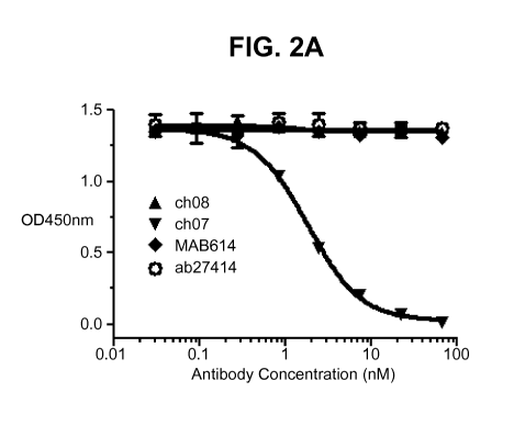
A competition ELISA was performed to examine whether ch07 and ch08 have
distinct binding epitopes. Prior to the competition ELISA experiment, ch07 and
ch08
were biotinylated. The EC50 of the biotinylated ch07 (biotin-ch07) and ch08
(biotin-
ch08) were determined by direct standard ELISA. For the competition ELISA,
recombinant hIL-13-Ra2 was coated onto 96-well plates at 50u1 of 2pg/mL in PBS
overnight at 4 C. The plates were then blocked and washed following a standard
ELISA protocol. 3-fold serially diluted ch07 and ch08 (2x final concentration)
were
31
CA 02890256 2015-04-30
WO 2014/072888
PCT/1B2013/059786
mixed with a constant amount of biotin-ch07 (FIG 2A) or biotin-ch08 (FIG 2B),
respectively and were added to the plate and incubated for 1 hour at room
temperature.
The amount of biotinylated chimeric antibody bound was detected by HRP
conjugated
streptavidin at 1:5000 for 1 hour. The results are shown in FIG 2. Unlabelled
chimeric
ch07 competes in binding to hIL-13-Ra2 with biotin-ch07 while unlabelled ch08
shows
no sign of competition (FIG 2A). Similar results are obtained when the same
set of
antibodies were used to compete with biotin-ch08 (FIG 2B). This clearly
demonstrates
that antibodies ch07 and ch08 have distinct binding epitopes to hIL-13Ra2.
The competition ELISA was also performed with two commercially available
antibodies, monoclonal mouse IgG1, MAB614 (R&D Systems) and monoclonal mouse
IgG1, ab27414 (Abcam). FIG 2A and Fig 2B show that both commercial antibodies
do
not compete with either biotin-ch07 or biotin-ch08 for binding to IL-13Ra2,
indicating
that antibodies ch07 and ch08 have different binding epitopes than the two
commercial
antibodies.
This result was confirmed by a BiaCore experiment. About 100RU of hIL-13-Ra2
was immobilized on CM5 chip using amine coupling chemistry. ch07 (100nM and
200nM) and ch08 (100nM and 200nM) were sequentially injected over hIL-13-Ra2
experiment channel and control channel at flow rate lOul/min for 150s. 50RU
was
reached when 100nM ch07 was injected to immobilized hIL-13Ra2. No further RU
increased when ch07 concentration was increased to 200nM, indicating that the
binding
sites on hIL-13-Ra2 for ch07 were saturated. With the injection of 100nM ch08,
100RU
was added. This positive binding signal indicates that the two antibodies lack
competition. The results further confirm that antibodies ch07 and ch08 have
different
binding epitopes (FIG 2C).
Example 5
ch07 and ch08 Neutralization Studies
In order to assess whether ch07 and ch08 can neutralize IL-13 function, a
competition ELISA was performed. An anti-Flag Ab was coated onto 96-well
plates and
incubated overnight at 4 C. Plates were then blocked and washed following
standard
32
CA 02890256 2015-04-30
WO 2014/072888
PCT/1B2013/059786
ELISA protocol. A 3-fold serial dilution of mouse antibodies, chimeric
antibodies,
positive control naked IL-13Ra2, and negative control hIL-21R were incubated
with a
constant amount of biotinylated IL-13Ra2-Fc (4x ED50) and a constant amount of
IL-13
(4x ED50) at RT for lh. 100u1of the complex was added to the ELISA plate and
incubated for 1 hour at room temperature. The amount of biotin-IL-13-Ra2 bound
was
detected by HRP conjugated streptavidin at 1:5000 for 1 hour. The results are
shown in
FIG 3. Both antibodies 07 and 08 (murine and chimeric forms) do not compete
with IL-
13 for the IL-13-Ra2 binding site while naked IL-13-Ra2 competes with
biotinylated IL-
13Ra2. This indicates that antibodies 07 and 08 (murine and chimeric forms)
are non-
neutralizing antibodies.
Example 6
Humanization of mu08
Monoclonal murine antibody mu08 (Seq ID NOs: 23 and 24) was humanized
utilizing DP-54 and DPK9 as human acceptor frameworks. Humanized 08 antibodies
(hu08) were prepared by CDR grafting with or without back mutations. The CDRs
of the
murine mu08 antibody were identified using the Kabat scheme.
A hu08 heavy chain variable region (VH version 1.0) was constructed by
directly
grafting the CDRs of mu08 onto a human DP-54 framework region. The version 1.1
was made by back mutations of frame work DP-54 at positions A40T, G42D, G44R
and
N835. Both v1.0 and v1.1 were cloned into pSMED2 vector containing the hIgG1
constant region. The nucleotide sequences encoding humanized hu08 heavy chain
variable regions are SEQ ID: NO: 17 for v1.0 and SEQ ID: NO: 20 for v1.1. The
amino
sequences encoding hu08 heavy chain variable regions are SEQ ID: NO: 1 for
v1.0 and
SEQ ID: NO: 19 for v1.1.
A hu08 light chain variable region (VK version 1.0) was constructed by
directly
grafting the CDRs of murine mu08 onto a human DPK9 framework region. The
version
1.1 was made by back mutations of frame work DPK9 at positions K39I, 560D,
T725,
T73F and T74I. Both versions v1.0 and v1.1 were cloned into pSMEN3 vector
containing the hIgKappa constant region. The nucleotide sequences encoding
hu08
33
CA 02890256 2015-04-30
WO 2014/072888
PCT/1B2013/059786
light chain variable regions are SEQ ID: NO: 18 for v1.0 and SEQ ID: NO: 22
for v1.1.
The amino acid sequences encoding hu08 light chain variable regions are SEQ
ID: NO:
for v1.0 and SEQ ID: NO: 21 for v1.1.
5 Example 7
Characterization of Humanized hu08
Humanized hu08 binding to recombinant hIL-13-Ra2 was evaluated by a
standard direct ELISA. Recombinant hIL-13-Ra2 was coated onto a 96-well plate.
Serially diluted chimeric antibodies or humanized antibodies in combinations
of heavy
and light chains of versions 1.0 and 1.1 e.g. hu08 v1.0 HC/1.0 LC, hu08 v1.0
HC/v1.1
LC, hu08 v1.1 HC/v1.0 LC, and hu08 v1.1 HC/v1.1 LC, were added to the plate
and
incubated at room temperature for 1-2 hours. The binding was detected by HRP
conjugated goat anti-human Ig Kappa. The results are shown in Table 6 below
and
demonstrate that all four combinations of humanized antibodies are able to
bind to
recombinant hIL-13-Ra2 and the ED50 is comparable to ch08.
Standard FAGS (Fluorescent Activated Cell Sorter) analysis was performed to
assess the binding activity of the antibodies to cell surface IL-13Ra2 . A375
cells were
washed with ice-cold PBS containing 1% bovine serum albumin and 0.001% sodium
azide. Cells were incubated with serial dilutions of antibodies hu08 v1.0 and
hu08 v1.1
e.g. hu08 v1.0 HC/1.0 LC, hu08 v1.0 HC/v1.1 LC, hu08 v1.1 HC/v1.0 LC, and hu08
v1.1
HC/v1.1 LC, for 30 min at 4 C and then stained with phosphatidylethanolamine-
labeled
goat anti-human IgG, fixed in PBS containing 4% paraformaldehyde, and analyzed
on a
FACScan (BD Biosciences). The data is consistent with the binding to
recombinant
receptor and illustrates that the binding to cell surface antigen for all 4
combinations is
comparable to ch08 (Table 6).
A competition ELISA was performed to assess whether hu08 1.0 and hu08 v1.1
compete with ch08 for IL-13-Ra2 binding. Recombinant hIL-13-Ra2 was coated
onto
96-well plates at 50u1 of 2pg/m1 in PBS overnight at 4 C. Plates were then
blocked and
washed following standard ELISA protocol. A 3-fold serially diluted ch08, hu08
1.0 and
hu08 v1.1, and negative control ch07 (2x final concentration) were mixed with
34
CA 02890256 2015-04-30
WO 2014/072888
PCT/1B2013/059786
biotinylated ch08 (2x EC50). 500 of the antibody and biotin-ch08 mixture were
added to
the plate and incubated for 1 hour at room temperature. The amount of biotin-
ch08
bound was detected by HRP conjugated streptavidin. The results are shown in
Table 6.
All 4 combinations of humanized antibodies are similar to chimeric antibody
ch08 in
competing with biotinylated ch08, indicating that hu08 1.0 and hu08 v1.1
retained the
same binding epitope as ch08 and have similar affinity to soluble and cell
surface IL-13-
Ra2.
Table 6
EC50 (nM) EC50 (nM) 1050 (nM)
Antibody rec hIL-13Ra2 A375 cells Biotin-ch08
ch08Hc+Lc 0.152 5.637 1.979
hu08 v1.0/1.0 0.175 4.098 2.120
hu08 v1.0/1.1 0.143 6.522 2.144
hu08 v1.1/1.0 0.163 4.152 2.562
hu08 v1.1/1.1 0.198 5.157 3.164
hu08 v1.0 HC/v1.0 LC and hu08 v1.1HC /v1.1LC were scaled up to generate
purified proteins. Both antibodies were transiently expressed into HEK293F
suspension
cells. Surprisingly, humanization of 08 has improved the antibody production
yield by 5-
6 fold compared to the yield of ch08 (Table 7).
Table 7
Antibody Expression level
name (ug/ml)
ch08 15
hu08v1.0/1.0 89.6
hu08v1.1/1.1 72.3
There is a direct correlation between the thermal stability of a protein or
protein
domain with the overall stability of the protein or protein domain. A higher
melting point
of a protein or protein domain often provides improved manufacturability and
longer
shelf life/stability. Thermal stability of ch08 and hu08 (v 1.0 and v1.1) was
examined by
CA 02890256 2015-04-30
WO 2014/072888 PCT/1B2013/059786
Differential Scanning Calorimetry (DSC). Thermal unfolding of chimeric and
humanized
antibodies by DSC was performed using a standard protocol on a MicroCal VP-DSC
instrument. Both of the humanized antibodies, hu08 v1.0/1.0 and hu08 v1.1/1.1,
show a
higher Tm2 (Fab) than the chimeric version ch08 (Table 8). This demonstrates
that
humanization of cu08 improves the thermal stability of this antibody.
Table 8
CH2 Fab CH3
Antibody Tm1 ( C) Tm2 (t) Tm3 (t)
ch08 73.41+0.47 70.44+0.04 84.35+0.09
huO8v1.0/1.0 73.48 0.19 80.29 0.02 85.48 0.14
huO8v1.1/1.1 71.12+0.13 80.23+0.02
The binding kinetics of hu08 antibodies was conducted on a Biacore T100 as
described above with the results shown in Table 9
Table 9
Antibody Antigen pH ka (1/Ms)on kd (1/s)off KD
(nM)
hu08 v1.0 hIL-13Ra2 7.4 9.75E+04 2.46E-04 2.52E-
09
hu08 v1.0 hIL-13Ra2 6.0 5.29E+04 2.34E-04 4/42E-
09
Example 8
Humanization of mu07
The general strategy of humanizing monoclonal murine antibody mu07 is the same
as described for mu08 in Example 6. A humanized hu07 heavy chain variable
region (VH
version 1.0) was constructed by directly grafting the CDRs of mu07 onto a
human DP-54
framework region. The versions 1.1-1.5 were made by back mutations of frame
work DP-
54 at various positions (Table 10). All versions were cloned into pSMED2
vector
containing hIgG1 constant region.
36
CA 02890256 2015-04-30
WO 2014/072888
PCT/1B2013/059786
A hu07 light chain variable region (VK version 1.0) was constructed by
directly
grafting the CDRs of mu07 onto a human DPK9 framework region. The versions 1.1-
1.7 were made by back mutations of frame work DPK9 at various positions (Table
10).
All versions were cloned into pSMEN3 vector containing hlg Kappa constant
region.
SEQ ID numbers of the amino acid sequences encoding hu07 variable regions are
listed in Table 10.
Table 10:
hu07 VH hu07 VK
VH aa Back-mutation VL aa Back-mutation
Variants Variants
SEQ ID SEQ ID
hu07 VH 9 hu07 VK 13
v1.0 v1.0
hu07 VH 37 T28S, F29L, hu07 VK 42 K41S, A42S,
v1.1 A49G, F67L, v1.1 D70S
N76S
hu07 VH 38 R71K hu07 VK 43 L47W
v1.2 v1.2
hu07 VH 39 T28S, F29S, hu07 VK 44 F71Y
v1.3 R71K v1.3
hu07 VH 40 T28S, F29S, hu07 VK 45 L47W, F71Y
v1.4 v1.4
hu07 VH 41 T28S, F29S, hu07 VK 46 K41S, A42S,
v1.5 A49G, R71K v1.5 D70S, L47W
hu07 VK 47 K41S, A42S,
v1.6 D70S, F71Y
hu07 VK 48 K41S, A42S,
v1.7 D7OS, L47W,
F71Y
37
CA 02890256 2015-04-30
WO 2014/072888
PCT/1B2013/059786
Example 9
Characterization of Humanized hu07
To evaluate the binding/competition properties of the various versions of huO,
transient transfections with 19 combinations of hu07 heavy and light chains
were
performed in COS-1 M6 cells. 6 heavy chains and 3 light chains were included:
chimeric heavy chain, humanized v1.0-1.5 heavy chain, chimeric light chain and
humanized v1.0-1.1 light chain. Conditioned media (CM) was harvested 2 days
after
transfection and subjected for direct binding to recombinant IL-13-Ra2 by
standard
ELISA, cell surface receptor binding by cell-based ELISA using A375 cells, and
competition ELISA with biotinylated ch07, utilizing protocols known in the
art.
Based on initial screening data from CM, heavy chain v1.5 was selected for
further study. Heavy chain v1.5 was paired with chimeric, humanized v1.0 and
v1.1
light chain. Table 11 summarizes the binding activity, competition properties
and
cytoxicity of hu07 antibodies. hu07 v1.5 paired with chimeric light chain Kc
demonstrates a similar ED50 and IC50 to the chimeric antibody. The competition
activity on the A375 cells and recombinant protein were decreased when this
heavy
chain v1.5 was paired with light chain v1.0 and v1.1.
Table 11
IC50 (nM) ED50 (nM) ED50 (nM) IC50 (nM) IC50
(nM)
Competition ELISA rhIL- cELISA
Competition ELISA Saporin Assay
ELISA Plate 13Ra2 A375 cells A375 cells A375
cells
ch07 2.21 0.217 0.85 17.27
0.08
huO7v1.5/kc 3.03 0.183 0.87 7.67
ND
huO7v1.5/v1.0 62.00 0.754 1.17 12.27
0.19
huO7v1.5/v1.1 46.62 1.214 1.29 19.00
0.20
38
CA 02890256 2015-04-30
WO 2014/072888
PCT/1B2013/059786
hu07 heavy chain v1.5 was used for light chain optimization. Transient
transfections with 10 combinations of hu07 heavy and light chains were
performed in
COS-1 M6 cells. The heavy chain v1.5 was paired with chimeric light chain and
humanized versions v1.0-1.7. CM was harvested and used in experiments to
determine
binding affinity to recombinant hIL-13a2 (rhIL-13a2) by ELISA and competition
activity
to biotinylated ch07 by competition ELISA. The data indicates that the
combination of
heavy chain v1.5 and light chain v1.7 is optimal. This result was confirmed
with purified
protein (Table 12).
Table 12
IC50 (nM)
ED50 (nM)
rhIL-13Ra2 Competition ELISA
Plate
ch07 0.39 2.21
huO7Hv1.5/kv1.7 0.39 7.7
Example 10
Species cross reactivity of ch07, hu07, ch08 and hu08
Cynomolgous (cyno) monkey IL-13-Ra2 (extracellular domain (ECD)) and
transmembrane domain (TM) were isolated from cyno monkey testis and adipose
tissues by RT-PCR. The amino acid sequence of cyno IL-13-Ra2 is shown in SEQ
ID
NO: 27. The identity is 94% between human and cyno IL-13Ra2.
The ECD/TM domain of cyno IL-13-Ra2 fused with Flag tag at the C-terminal end
was cloned into the pSMED2 expression vector. HEK293 suspension cells were
transiently transfected with cyno-IL-13-Ra2 containing plasmid and pSMED2
vector (as
a mock transfection). The cells were harvested 72 hours later and subjected to
FAGS
analysis. 4 antibodies including ch07, ch08, hu08v1.0/1.0 and hu08v1.1/1.1were
tested. The binding on the cell surface cyno-IL-13-Ra2 was detected with R-
Phycoerythrin-labeled goat anti-human or mouse IgG. The data demonstrates that
39
CA 02890256 2015-04-30
WO 2014/072888
PCT/1B2013/059786
ch07, ch08, huO8v1.0/1.0 and huO8v1.1/1.1 are able to bind to the cell surface
cyno-IL-
13-Ra2 and have similar binding affinities (ED50) (Table 13).
Table 13
Antibody ED50 (nM)
ch07 1.494
ch08 1.814
huO8v1.0/1.0 1.961
huO8v1.1/1.1 2.141
The binding of hu07 and hu08 to mouse IL-13-Ra2 was evaluated by direct
ELISA. The identity between human and mouse IL-13-Ra2 at the amino acid level
is
approximately 64%. Recombinant mIL-13-Ra2 or hIL-13-Ra2 (as positive control)
was
coated onto a 96-well plate. Purified chimeric ch07 and ch08 were serially
diluted and
added to the antigen coated plate. The bound antibodies were detected by HRP
conjugated goat anti-human IgGFc specific secondary antibody. There was no
detectable signal for mIL-13-Ra2 binding while the binding to the positive
control hIL-13-
Ra2 was strong, indicating that hu07 and hu08 do not cross-react to murine mIL-
13Ra2.
Example 11
Binding to Human Cell Lines Expressing IL13R-a2
Cell lines expressing the IL-13-Ra2 antigen and the negative control cells
were
plated at a density of 200,000 cells per well of 96 deep well plates and kept
on ice. The
mouse monoclonal antibodies mu07 or mu08 prepared in 3% bovine serum albumin
BSA in Dulbecco's phosphate buffered saline (DPBS) were added to the plate at
a final
concentration of 10 pg/mL. The plates were then incubated on ice for 1 hour
followed
by 2 washes. The secondary antibody, PE (phycoerythrin) conjugated goat anti-
mouse
CA 02890256 2015-04-30
WO 2014/072888
PCT/1B2013/059786
IgG Fc was added to the wells. After 30 minutes of incubation at 4 C, the mean
fluorescence intensity was then analyzed by FACS on a FACScan TM (BD
Biosciences).
The data in Table 14 indicates that the mu07 and mu08 antibodies bind to a
diverse panel of IL-13R-a2 positive cell lines from various disease
indications.
Table 14
Mean Fluorescent Intensity IL-13-Ra2
Human Cell Line
mu07 mu08 Expression
PC3MM2 (prostate) 84000 72000 3+
U87MG (glioblastoma) 61000 62000 3+
A375 (melanoma) 53000 46000 3+
H460 (lung, cisplatin resistant) 28000 22000 2+
Hs766T (pancreatic) 13000 14000 2+
A498 (renal) 13000 5000 1+
SW626 (ovarian) 9000 8000 1+
H460 (lung) 800 300 0
Example 12
Internalization
Antibody internalization is a critical characteristic for delivering ADCs for
cytotoxicity in IL-13-Ra2 expressing cells. Internalization of the antibody
after binding to
IL-13-Ra2 was examined using mu07 and mu08 antibodies and a positive control
mouse monoclonal antibody (ab27414), on four cell lines (PC3MM2, A375, Hs766T,
and
H460R ). The antibodies (10 pg/mL) were incubated with the various cells for 1
hour on
ice and unbound antibody was removed by washing twice with cold media. The
cell
culture plates were incubated at 37 C. Samples of the cells were fixed at 15
minutes
41
CA 02890256 2015-04-30
WO 2014/072888
PCT/1B2013/059786
and at 4 hours. The percent internalization at different timepoints is shown
in Table 15.
The data show that mu07 and mu08 are readily internalized into IL-13-Ra2
expressing
cells.
Table 15
% internalization
min 4 hours
Primary Abs
A375 Hs766T PC3MM2 H460R A375 Hs766T PC3MM2 H460R
nnu07 63.1 69.6 88.6 74.4 38.1 69.0 50.3
38.4
nnu08 68.0 64.1 81.9 55.9 61.8 76.3 41.6
47.4
ab27414 49.3 60.8 60.2 57.5 68.2 73.6 49.7 49.2
Example 13
Synthesis of compounds 0101 and 3377
10 Compounds 0101 and 3377 were prepared according to the methods described
in US Patent Application No.13/670,612, herein incorporated by reference.
Experimental for compound 0101 (#54 in the schematic)
Preparation of 2-Methylalanyl-N-[(3R,45,55)-3-methoxy-1-{(25)-2-[(1R,2R)-1-
15 methoxy-2-methyl-3-oxo-3-{[(1S)-2-phenyl-1-(1,3-thiazol-2-
ypethyl]aminolpropyl]pyrrolidin-1-y11-5-methyl-1-oxoheptan-4-y1]-N-methyl-L-
valinamide
(#54)
42
CA 02890256 2015-04-30
WO 2014/072888
PCT/1B2013/059786
s HAEiTcUi, Et3mN,
Fmoc j, 0H N Fmoc,N Yõ,ir
H I 0 0 0 H I 0,, 0 0
0 0õ 0
74%
0 NH
#32 #19 #53
CH2Cl2
Et2NH,
H2NYy
0 I 0 0
0
NH
#54
Step 1. Synthesis of N-[(9H-fluoren-9-ylmethoxy)carbony1]-2-methylalanyl-N-
[(3R,4S,5S)-3-methoxy-1-{(2S)-24(1 R,2R)-1-methoxy-2-methyl-3-oxo-3-{[(1S)-2-
phenyl-
1-(1,3-thiazol-2-ypethyl]aminolpropyl]pyrrolidin-1-y11-5-methyl-1-oxoheptan-4-
y1]-N-
methyl-L-valinamide (#53). According to general procedure D, from #32 (2.05 g,
2.83
mmol, 1 eq.) in dichloromethane (20 mL, 0.1 M) and N,N-dimethylformamide (3
mL), the
amine #19 (2.5 g, 3.4 mmol, 1.2 eq.), HATU (1.29 g, 3.38 mmol, 1.2 eq.) and
triethylamine (1.57 mL, 11.3 mmol, 4 eq.) was synthesized the crude desired
material,
which was purified by silica gel chromatography (Gradient: 0% to 55% acetone
in
heptane), producing #53 (2.42 g, 74%) as a solid. LC-MS: m/z 965.7 [M+H+],
987.6
[M+Na], retention time = 1.04 minutes; HPLC (Protocol A): m/z 965.4 [M+H+],
retention
time = 11.344 minutes (purity > 97%); 1H NMR (400 MHz, DMSO-d6), presumed to
be a
mixture of rotamers, characteristic signals: 6 7.86-7.91 (m, 2H), [7.77 (d,
J=3.3 Hz) and
7.79 (d, J=3.2 Hz), total 1H], 7.67-7.74 (m, 2H), [7.63 (d, J=3.2 Hz) and 7.65
(d, J=3.2
Hz), total 1H], 7.38-7.44 (m, 2H), 7.30-7.36 (m, 2H), 7.11-7.30 (m, 5H), [5.39
(ddd,
J=11.4, 8.4, 4.1 Hz) and 5.52 (ddd, J=11.7, 8.8, 4.2 Hz), total 1H], [4.49
(dd, J=8.6, 7.6
Hz) and 4.59 (dd, J=8.6, 6.8 Hz), total 1H], 3.13, 3.17, 3.18 and 3.24 (4s,
total 6H), 2.90
and 3.00 (2 br s, total 3H), 1.31 and 1.36 (2 br s, total 6H), [1.05 (d, J=6.7
Hz) and 1.09
(d, J=6.7 Hz), total 3H].
Step 2. Synthesis of 2-methylalanyl-N-R3R,45,55)-3-methoxy-1-{(25)-2-
[(1 R,2R)-1-methoxy-2-methyl-3-oxo-3-{[(1S)-2-phenyl-1-(1,3-thiazol-2-
43
CA 02890256 2015-04-30
WO 2014/072888
PCT/1B2013/059786
ypethyl]aminolpropyl]pyrrolidin-1-y11-5-methyl-1-oxoheptan-4-y1]-N-methyl-L-
valinamide
(#54)
According to general procedure A, from #53 (701 mg, 0.726 mmol) in
dichloromethane (10 mL, 0.07 M) was synthesized the crude desired material,
which
was purified by silica gel chromatography (Gradient: 0% to 10% methanol in
dichloromethane). The residue was diluted with diethyl ether and heptane and
was
concentrated in vacuo to afford #54 (406 mg, 75%) as a white solid. LC-MS: m/z
743.6
[M+H+], retention time = 0.70 minutes; HPLC (Protocol A): m/z 743.4 [M+H+],
retention
time = 6.903 minutes, (purity > 97%); 1H NMR (400 MHz, DMSO-d6), presumed to
be a
mixture of rotamers, characteristic signals: 6 [8.64 (br d, J=8.5 Hz) and 8.86
(br d,
J=8.7 Hz), total 1H], [8.04 (br d, J=9.3 Hz) and 8.08 (br d, J=9.3 Hz), total
1H], [7.77 (d,
J=3.3 Hz) and 7.80 (d, J=3.2 Hz), total 1H], [7.63 (d, J=3.3 Hz) and 7.66 (d,
J=3.2 Hz),
total 1H], 7.13-7.31 (m, 5H), [5.39 (ddd, J=11, 8.5, 4 Hz) and 5.53 (ddd,
J=12, 9, 4 Hz),
total 1H], [4.49 (dd, J=9, 8 Hz) and 4.60 (dd, J=9, 7 Hz), total 1H], 3.16,
3.20, 3.21 and
3.25 (4 s, total 6H), 2.93 and 3.02 (2 br s, total 3H), 1.21(s, 3H), 1.13 and
1.13 (2 s,
total 3H), [1.05 (d, J=6.7 Hz) and 1.10 (d, J=6.7 Hz), total 3H], 0.73-0.80
(m, 3H).
Experimental for compound 3377 (#115 in the schematic)
Preparation of N,2-dimethylalanyl-N-{(1S,2R)-4-{(2S)-2-[(1R,2R)-3-{[(1S)-1-
carboxy-2-phenylethyl]amino}-1-methoxy-2-methyl-3-oxopropyl]pyrrolidin-1-y11-2-
methoxy-1-[(1S)-1-methylpropy1]-4-oxobutyll-N-methyl-L-valinamide,
trifluoroacetic acid
salt (#115).
44
CA 02890256 2015-04-30
WO 2014/072888
PCT/1B2013/059786
HATU, Hunig's base
, Et2NH
Fmoc:Nlj 0 Fmoc--01riN CH2Cl2
..õ.7,õõ 0,,o NH0
0 0
62% 67%
\ NH
0
dimer acid#5
#113 10¨
#67
1 N-R9H-fluoren-9-ylmethoxy)carbonyI]-N,2-dimethylalanine
HATU, Hunig's base, CH2Cl2
2 Li0H, THF, water ____________________________________ HNY-
yklij*N'rryNri?...
0
\
NH
#114
#115 0
411
# OH
Step 1. Synthesis of methyl N-{(2R,3R)-3-[(25)-1-{(3R,4S,5S)-4-[{N-[(9H-
fluoren-
9-ylmethoxy)carbony1]-L-valyll(methyl)amino]-3-methoxy-5-
methylheptanoyllpyrrolidin-
2-y1]-3-methoxy-2-methylpropanoyll-L-phenylalaninate (#113). To a stirring
mixture of
dimer acid#5 (12. 1g, 23.0 mM) and #67 (11.5 g, 23.0 mM) in 75 mL of
dichloromethane
under nitrogen, HATU (10.8 g, 27.6 mM) was added followed by Hunig's base
(12.1 mL,
69.0 mM). The reaction was allowed to stir at room temperature for 15 hours.
Reaction
was concentrated to a smaller volume, taken up with ethyl acetate and washed
with 1 N
HCI two times. The organic layer was then washed with brine, dried over sodium
sulfate, filtered, and concentrated in vacuo. Residue was then purified by
silica gel
chromatography (Gradient: 0% to 70% acetone in heptanes), producing #113 (12.3
g,
62%) as a white solid. LC-MS (Protocol Q): m/z 855.3 [M+H-], 877.2 [M+Na],
retention
time = 2.32 minutes; HPLC (Protocol R): /z855.5 [M+H-], retention time = 9.596
minutes (purity > 97%).
Step 2. Synthesis of methyl N-{(2R,3R)-3-methoxy-3-[(25)-1-{(3R,45,55)-3-
methoxy-5-methyl-4-[methyl(L-valyl)amino]heptanoyllpyrrolidin-2-y1]-2-
methylpropanoyll-L-phenylalaninate (#114). According to general procedure A,
from
#113 (12 g, 14 mmol, 1 eq.), dichloromethane (60 mL, 0.24 M) and diethylamine
(40
mL, 390 mM) was synthesized #114 (5.9 g, 67%) white/slight yellow solid after
purification by silica gel chromatography (Gradient: 0% to 25% methanol in
dichloromethane). LC-MS (Protocal Q): m/z 633.0 [M+H-], retention time = 1.19
CA 02890256 2015-04-30
WO 2014/072888
PCT/1B2013/059786
minutes. H PLC (Protocol A): /z633.5 [M+H-], retention time = 7.142 minutes
(purity >
98%).
Step 3. Synthesis of N,2-dimethylalanyl-N-{(1S,2R)-4-{(2S)-2-[(1R,2R)-3-{[(1S)-
1-carboxy-2-phenylethyl]amino}-1-methoxy-2-methyl-3-oxopropyl]pyrrolidin-1-y11-
2-
methoxy-1-[(1S)-1-methylpropyI]-4-oxobutyll-N-methyl-L-valinamide-
trifluoroacetic acid
salt (#115). To a stirring mixture of N-[(9H-fluoren-9-ylmethoxy)carbonyI]-N,2-
dimethylalanine (167 mg, 0.493 mM), #114 (260 mg, 0.411 mM), and HATU (188 mg,
0.493 mM) in 10 mL of dichloromethane, Hunig's base (0.14 mL, 0.82 mM) was
added.
The reaction was allowed to stir at room temperature for 1 hour and 20
minutes.
Reaction was reduced down. THF (9 mL) was added to crude material and to this
stirring mixture lithium hydroxide (49.2 mg, 2.06 mM) dissolved in 3 mL of
water was
added. The reaction was allowed to stir at room temperature for 4 hours.
Reaction was
concentrated down followed by purification by medium pressure reverse phase
C18
chromatography (Gradient: 5% to 45% water in acetonitrile with 0.02% TFA in
each
phase) #115 (218 mg, 64%) white solid. LC-MS (Protocol Q): m/z 718.7 [M+H-],
740.6
[M+Na], retention time = 1.21 minutes. HPLC (Protocol A at 45 C): m/z 718.4
[M+H-],
retention time = 6.903 minutes.
Example 14
Preparation of anti-IL-13-Ra2 ADCs
The ADCs of the present invention can be prepared using a section of the
linker
having a reactive site for binding to a chemical compound and introducing
another
section of the linker having a reactive site for an antibody. In one aspect, a
linker has a
reactive site which has an electrophilic group that is reactive with a
nucleophilic group
present on an antibody unit, such as an antibody. Useful nucleophilic groups
on an
antibody include but are not limited to, sulfhydryl, hydroxyl and amino
groups. The
heteroatom of the nucleophilic group of an antibody is reactive to an
electrophilic group
on a linker and forms a covalent bond to a linker. Useful electrophilic groups
include,
but are not limited to, maleimide and haloacetamide groups.
A linker has a reactive site which has a nucleophilic group that is reactive
with an
46
CA 02890256 2015-04-30
WO 2014/072888
PCT/1B2013/059786
electrophilic group present on an antibody unit. The electrophilic group on an
antibody
provides a convenient site for attachment to a linker. Useful electrophilic
groups on an
antibody include, but are not limited to, aldehyde and ketone carbonyl groups.
The
heteroatom of a nucleophilic group of a linker can react with an electrophilic
group on an
antibody and form a covalent bond to the antibody. Useful nucleophilic groups
on a
linker include, but are not limited to, hydrazide, oxime, amino, hydrazine,
thiosemicarbazone, hydrazine carboxylate, and arylhydrazide.
As used herein, "mc-" refers to:.
crsi
As used herein, "vc-" refers to:
H cr o osss'
H H
0 0
NH
0 NH2
. The anti-IL-13-Ra2 ADCs were prepared via partial reduction of the mAb
with
tris(2-carboxyethyl)phosphine (TCEP) followed by reaction of reduced cysteine
residues
with the desired maleimide terminated linker-payload. In particular, hu08 was
partially
reduced via addition of 2.2 molar excess of tris(2-carboxyethyl)phosphine
(TCEP) in
100 mM HEPES (4-(2-hydroxyethyl)-1 -pperazineethanesuifonic acrd buffer), pH
7.0 and
1 mM diethylenetriaminepentaacetic acid (DTPA) for 2 h at 37 C. The desired
linker-
payload was then added to the reaction mixture at a linker-payload/mAb molar
ratio of
7.0 maleimidocapronic ¨ valine-citruline-p-aminobenzyloxycarbonyl- aurstatin-
0101 [vc-
47
CA 02890256 2015-04-30
WO 2014/072888
PCT/1B2013/059786
0101, see below]), or 7.0 maleimidocapronic-auristatin-3377 [mc-3377, see
below] and
reacted for an additional 1 hat 25 C in the presence of 15% v/v of
dimethylacetamide
(DMA). After the 1 h incubation period, N-ethylmaleimide (3 fold excess for vc-
0101
and for mc-3377) was added to cap the unreacted thiols and is allowed to react
for 15
minutes, followed by addition of 6 fold excess L-Cys to quench any unreacted
linker-
payload. The reaction mixture was dialyzed overnight at 4 C in phosphate
buffered
saline (PBS), pH 7.4, and purified via SEC (AKTA explorer, Superdex 200 10/30
GL
column). The ADC was further characterized via size exclusion chromatography
(SEC)
for purity, hydrophobic interaction chromatography (HIC), and liquid
chromatography
electrospray ionization tandem mass spectrometry (LC-ESI MS) to calculate drug-
antibody ratio (loading). The protein concentration was determined via UV
spectrophotometer.
mc-3377 (conjugation to antibody X through a cysteine residue)
0 0
0
X
N
N\
0 0
0 NH
0
OH
111
vc-0101 (conjugation to antibody X through a cysteine residue)
0
0
X s
0 OVH
XvN.7NN 0 0 0
0 0 ;N. NH
\ 0
NH
O'N'NH2
48
CA 02890256 2015-04-30
WO 2014/072888
PCT/1B2013/059786
Example 15
In Vitro Cytotoxicity Assay
Cell lines expressing the IL-13-Ra2 antigen and a negative control cell line,
were
cultured with increasing concentrations of anti-IL-13-Ra2 ADC. After four
days, viability
of each culture is assessed. ICso values were calculated by logistic non-
linear
regression and are presented as ng Ab/mL. The preferred linker payloads were
vc-
0101 and mc-3377.
Humanized anti-IL-13Ra2 antibody hu08 was conjugated to various linker-
payload combinations as provided in Table 16. The antibody drug conjugates
were
prepared according to the methods described in US Patent Application
No.13/670,612,
which is incorporated herein by reference, with the corresponding nomenclature
and
experimental chemical synthesis schematic number indicated in Table 16.
Table 16
ADC Linker-Payload Corresponding ADC Linker-Payload
#
hu08-vc-0101 I L13 Ra2 -AB08-v1010-hG1-
(C)_mcValCitPABC-#54
hu08-mc-3377 I L13Ra2 -AB08-v1010-hG1-(0)_mc-#115
hu08-mc-0131 I L13Ra2 -AB08-v1010-hG1-(0)_mc-0#118
hu08-Malpeg-6121 I L13Ra2 -AB08-0010-hG1-(C)_MalPeg6C2-
#117
hu08-Malpeg-0131 I L13Ra2 -AB08-v1010-hG1-
(C)_Mal(H20)Peg602-0#118
hu08-mc-6121 I L13Ra2 -AB08-v1010-hG1-(0)_mc-#117
hu08-vc-3906 I L13 Ra2 -AB08-v1010-hG1-
(C)_mcValCitPABC-#226
hu08-vc-6780 I L13Ra2 -AB08-v1010-hG1-(0)_mcValCitPABC-
#112
hu08-mc-8261 I L13Ra2 -AB08-v1010-hG1-(C)_mc-#69
hu08-mc-3906 I L13 Ra2 -AB08-v1010-hG1-(C)_mc-#226
hu08-MalPeg-8261 I L13 Ra2 -AB08-v1010-hG1-(C)_MalPeg6C2-
#69
hulgG8.8-vc-0101 hulgG8.84-mcValCitPABC-#54
hulgG8.8-mc-3377 hulgG8.84-mc-#115
Further, a mutant version of hu08 (huO8MAC) was generated as described in
Example 21 and according to standard protocols. Compound 0101 was conjugated
to a
cleavable linker to form the structure:
49
CA 02890256 2015-04-30
WO 2014/072888 PCT/1B2013/059786
N
0 )---S
FH 0 ift 0)Wiljr=rNic.IN---- /I
F la 0.1r,....õ.00õ-jõXN mip, 0 2- r
(21 0 20 0
H 0 H
F 1111111" F 0
F L
H2N:10
)
and thereafter conjugated to huO8MAC according to the techniques described
herein to
form huO8MAC -0101.
The data demonstrates that the anti-IL-13-Ra2 antibody hu08v1.0/1.0 conjugated
to six different auristatin payloads was effective against both of the IL-13-
Ra2 positive
cell lines tested (PC3MM2 and A375), having an IC50 ranging from 1.1 to 4.9 ng
Ab/mL
or 7.3-32.7 pM) (Table 17). Further, huO8MAC-0101 was effective against both
of the
IL-13Ra2 positive cell lines tested (PC3MM2 and A375), having an IC50 of 7.9
ng
Ab/mL. All ADCs were not active against the IL-13-Ra2 negative cell line,
H460, and
the non- IL-13-Ra2 binding control ADCs, hIgG8.8-vc-0101 and hIgG8.8-mc-3377,
were
not active against any of the cell lines tested.
Table 17
IC50 (ng Ab/mL)
ADC DAR
PC3MM2 A375 H460
hu08-vc-0101 3.2 2.5 3.8 >400000
hu08-mc-3377 4.3 1.2 2.2 >400000
hu08-mc-0131 3.2 1.3 2.1 >400000
hu08-MalPeg-6121 3.3 3.5 3.4 >400000
hu08-MalPeg-0131 2.9 2.9 4.9 >400000
hu08-mc-6121 3.3 1.1 2.4 >400000
hu08-mc-3906 3 1.5 2.9 >400000
hu08 vc-6780 4 1.2 2.2 >400000
huO8MAC-0101 1.9 4.9 7.9 >400000
hIgG8.8-vc-0101 3.7 >400000 >400000 >400000
hIgG8.8-mc-3377 4.3 >400000 >400000 >400000
CA 02890256 2015-04-30
WO 2014/072888
PCT/1B2013/059786
Example 16
Subcutaneous Xenograft Models
Female, athymic (nude) mice were injected s.c. with PC3MM2 or A375 tumor
cells. Mice with staged tumors, approximately 0.2 to 0.8 g (n = 8 to 10
mice/treatment
group), were administered intravenously q4d x 4 with normal saline (vehicle),
huO8v1.0/1.0 ADCs with linker-payloads vc-0101, vc-6780, vc-3906, mc-8261, mc-
0131, mc-6121, mc-3377, MalPeg-8261, MalPeg-0131, MalPeg-6121, and MalPeg-
3906, and a non-binding Ab (hIgG8.8) conjugated with vc-0101 or mc-3377, at a
dose of
2 or 3 mg Ab/kg. The ADCs were dosed based on Ab content. Tumors were measured
at least once a week and their size is calculated as mm3 = 0.5 x (tumor
width2) x (tumor
length).
The in vivo efficacy results listed in Table 18 show a range of anti-tumor
activity
with the various ADCs tested. The relative order of potency is hu08-vc-0101 >
hu08-vc-
6780 hu08-mc-0131 > hu08-mc-6121 > hu08-mc-3906 > hu08-MalPeg-0131 >hu08-
MalPeg-6121 > hu08-MalPeg-3906 > hu08-mc-8261.
The data in Table 19 indicates that ADCs hu08-vc-0101 and hu08-mc-3377 were
efficacious 3mg/kg in reducing tumor growth in the PC3MM2. Compared to the
vehicle
control group which was terminated at Day 15 due to large size of tumors
(>2500 mm3
from some of animals), hu08-vc-0101 and hu07-mc-3377 have 5 out of 8 or 3 out
of 8
animals without measurable tumors at Day 76, respectively. Similar to the
vehicle
control group, irrelevant ADC control groups of hIgG8.8-vc-0101 at 3 mg/kg and
hIgG8.4-mc-3377 at 10 mg/kg was terminated at Day 15 and Day 19 respectively
due to
the large size of tumors in the groups, respectively. These results
demonstrate that the
significant therapeutic efficacy (some animals were cured of disease) from
hu08-vc-
0101 and hu07-mc-3377 was IL-13-Ra2 target-mediated.
51
CA 02890256 2015-04-30
WO 2014/072888
PCT/1B2013/059786
Table 18
PC3MM2 xenograft, tumor volume (mm3 +/- SEM)
Dose
ADC (mg/kg)
Day Day Day Day Day Day Day Day
Q4dx4
-1 3 8 16 20 30 42 52
638 1149 1707
Vehicle 0 + + + GT GT GT GT GT
27 82 133
hu08- 642 1036 1176
Malpeg- 2 + + + GT GT GT GT GT
3906 36 60 51
642 1088 1429
hu08-mc-
2 + + + GT GT GT GT GT
8261
51 121 158
637 1004 778
hu08-mc-
2 + + + GT GT GT GT GT
0131
44 73 83
hu08- 638 947 1000 693 780
Malpeg- 2 + + + + + GT GT GT
6121 36 85 126 129 198
hu08- 649 1085 1040
Malpeg- 2 + + + GT GT GT GT GT
0131 39 54 88
646 899 557 243 201 113 207 532
hu08-yc-
2 + + + + + + + +
0101
36 54 49 28 20 17 49 151
641 850 652 279 217 230
hu08-yc-
2 + + + + + + GT GT
6780
28 100 54 55 45 133
636 909 821 441 414
hu08-mc-
2 + + + + + GT GT GT
6121
37 63 93 83 104
637 875 806 611
hu08-mc-
2 + + + + GT GT GT GT
3906
26 48 70 150
hu08- 645 991 1220
Malpeg- 2 + + + GT GT GT GT GT
8261 34 71 115
52
CA 02890256 2015-04-30
WO 2014/072888 PCT/1B2013/059786
GT= group terminated due to large tumor size
Table 19
Dose PC3MM2 xenograft, tumor volume(mm3+/-SEM)
ADC (mg/kg) Day Day Day Day Day Day Day Day
Q4dx4 -1 2 10 19 40 51 61 71
340 573 1441 1975
vehicle 0 + + + + GT GT GT GT
16 47 176 272
328 459 295 544
hIgG8.84-
+ + + + GT GT GT GT
mc-3377
27 63 121 258
337 385 41 0 78 346 616 902
hu08-
3 + + + + + + + 9
mc-3377
21 36 12 0 36 147 243 364
339 433 38 6 110 230
hu08-vc-
3 + + + + + + GT GT
0101
18 45 14 6 110 230
333 449 686
hIgG8.8-
3 + + + GT GT GT GT GT
vc-0101
23 15 134
GT= group terminated due to large tumor size.
5
The data in Table 20a indicates that ADCs of hu08-vc-0101 and hu08 ¨mc-3377
were efficacious at 3 mg/kg in a second in vivo xenograft model using A375
cells. The
vehicle control group and irrelevant ADC control groups of hIgG8.8-vc-0101 and
hIgG8.8-mc-3377 were terminated due to large tumor size at Day 19, 22, 27,
10 respectively. Treatment with hu08-vc-0101 and hu08-mc-3377 at 3 mg/kg
caused
tumor regression in all animals and provided significant survival advantage.
No
measureable tumor was observed in all animals treated with hu08-mc-3377 at Day
19 -
22. Although some tumors relapsed, there were 5 out of 10 animals without
measurable tumors at Day 71. The group treated with hu08-vc-0101 was monitored
over 100 days and 8 of animals had no measurable tumors at Day 19-100. These
results demonstrate potent antitumor activities of hu08-vc-0101 and hu08-mc-
3377
against IL-13-Ra2 positive tumors.
53
CA 02890256 2015-04-30
WO 2014/072888
PCT/1B2013/059786
Table 20a
ADC Dose A375 Xenograft Tumor Volume (me SEM)
(mg/kg) Day Day Day Day Day Day Day
Day
Q4dx4 -1 2 13 19 40 51 61 71
Vehicle 0 340 573 1441 1975
+ + + + GT GT GT
GT
16 47 176 272
hIgG8.8- 3 328 459 295 544
mc-3377
+ + + + GT GT GT
GT
27 63 121 258
Hu08-mc- 3 337 385 41 0 78 346 616
902
3377
+ + + + + + + +
21 36 14 0 36 147 243
364
Hu08-vc- 3 336 407 43 7 122 256 0 0
0101
+ + + + + + +
+
19 42 15 15 7 122 0 0
hIgG8.8- 3 333 449 686 1145
vc-0101
+ + + + GT GT GT
GT
23 15 135 212
GT= group terminated due to large tumor size.
The data in Tables 20b and 20c indicates that ADCs hu08-vc-0101 and hu08-
mc3377 were efficacious in a third in vivo xenograft model using the HEY-C2
ovarian
cancer cell line. The vehicle control group and negative ADC control group's
hIgG8.8-
vc0101 (3 mg/kg and 10 mg/kg) and hIgG8.8-mc-3377 (10 mg/kg) were terminated
due
to large tumor size as indicated with the GT designation. Treatment with hu08-
vc-0101
and hu08-mc-3377 at dose levels of 1, 3 and 10 mg/kg, provided a dose-
dependent
response. Five out of nine animals treated with 3 mg/kg of hu08-vc-0101 and
seven out
of nine animals treated with 10 mg/kg of hu08-vc-0101 survived to end of the
study (Day
103). Similarly, five out of nine animals treated with 3 mg/kg of hu08-mc-3377
and nine
out of nine animals treated with 10 mg/kg of hu08-mc-3377 survived to the end
of the
study (Day 103).
54
CA 02890256 2015-04-30
WO 2014/072888 PCT/1B2013/059786
Table 20b
HEY-C2 Xenograft, tumor volume (me SEM)
Dose
ADC (mPk) Day Day Day Day Day Day Day Day Day Day Day Day
Q4d
-1 3 12 23 30 40 51 60 72 79 95 103
211 375 919 2311
vehicle 0 + + + GT GT GT GT GT GT GT GT
16 21 53 156
211 324 337 510 665
1 + + + + GT GT GT GT GT GT GT
17 23 25 128 203
211 319 250 207 239 336 602
hABO8
3 + + + + GT GT GT GT GT
mc_3377
17 38 37 41 64 131 258
211 303 181 114 106 84 69 53 42 41 121 308
10 + + + + + + + + + + + +
16 28 17 10 7 16 14 14 11 12 59 166
212 375 557 650
hIgG 8.8
10 + + + GT GT GT GT GT GT GT GT
mc_3377
18 46 53 183
CA 02890256 2015-04-30
WO 2014/072888
PCT/1B2013/059786
Table 20c
Dose HEY-C2 Xenograft, tumor volume (mm3 SEM)
ADC (mpk) Day Day Day Day Day Day Day Day Day Day Day Day
Q4d -1 3 12 23 30 40 51 60 72 79 95 103
211 335 331 479 697
1 GT GT GT GT GT GT GT
16 27 43 96 177
210 324 258 200 222 354
hABO8
3 GT GT GT GT GT GT
vc_0101
18 19 36 56 90 239
211 333 295 204 142 94 66 83 208 324
+ + + + + + + + + + GT GT
16 29 26 15 14 13 35 129 219
212 384 567 963
3
GT GT GT GT GT GT GT GT
hIgG 8.8 22 35 95 204
vc_0101 211 354 349 198 151 165 413 922
10 + + + + + + + + GT GT GT GT
37 48 38 31 33 128 287
GT= group terminated due to large tumor size.
Example 17
Cysteine Mutant Generation for Site-Specific Conjugation
5 Site specific conjugation of linker-payloads to antibodies was done in
order to
improve homogeneous drug loading and avoid ADC subpopulations with altered
antigen-binding or altered pharmacokinetics, often observed by conventional
conjugation methods. One such site-specific conjugation method is to introduce
cysteine residues at specific sites in the amino acid sequence of the target
antibody. A
10 number of amino acid positions in the constant heavy chain and constant
light chain
have been previously identified (see patent application USSN 61/580,169) and
were
substituted with a cysteine residue at the specific amino acid position in the
huO8v1.0/1.0 antibody. All cysteine mutations were constructed by site-
directed
mutagenesis or overlapping PCR based on pSMED-huO8v1.0 and pSEMN3-huO8v1Ø
15 Below is the list of cysteine mutants derived from huO8v1.0/1.0 and the
respective SEQ
ID numbers (Table 21).
56
CA 02890256 2015-04-30
WO 2014/072888
PCT/1B2013/059786
Table 21
aa
Antibody Mutation Site
SEQ ID
NO:
L443C 28
HC
Q347C 29
Single mutant kA1110 30
LC kK1830 31
kK188C 32
L4430/K3920 33
HC
L4430/V4220 34
L4430/kA1110 28/30
Double
mutant
L443C/kK183C 28/31
H 0/LO
Q3470/kA1110 29/30
Q3470/kK183C 29/31
Example 18
Characterization of Cysteine Mutants
Cysteine mutants of huO8v1.0/1.0 were expressed either in transiently
transfected HEK293 suspension cell cultures in freestyle 293 expression medium
(Invitrogen, calsdad, CA) or in CHO cell culture stable pools. The antibodies
were
isolated from the cell culture medium by Protein A (ProA) chromatography under
standard conditions. The column fractions were pooled and concentrated using a
Millipore spin tube equipped with a 30,000 MWCO membrane. The protein was then
loaded onto a size exclusion column (Superdex 200) equilibrated with PBS-CMF,
pH
7.2. Peak fractions were pooled and concentrated using a Millipore spin tube
equipped
with a 30,000 MWCO membrane and finally filtered through a 0.22um filter. Data
is
shown in Table 22 from transient expression and in Table 23 from CHO cell
stable
pools. Wild type huO8v1.0/1.0 and its cysteine derivatives have comparable
final yield
57
CA 02890256 2015-04-30
WO 2014/072888
PCT/1B2013/059786
and purity after ProA capture, reach up to 99% purity after SEC and are stable
after 2-3
cycles of freezing and thawing. This data demonstrates that the cysteine
mutants
maintain their expression profile and purification properties compared to
huO8v1.0/1Ø
Table 22
Antibody Name Final Yield Purity (ProA
Purity (SEC) FIT 2-3 cycles
(mg/L) capture)
huO8v1.0/1.0 26.5 97 /0 ND stable
huO8v1.0/1.0- 56.0 94 /0 99% stable
L443C
huO8v1.0/1.0- 30.0 97 /0 99% stable
03470
huO8v1.0/1.0- 52.8 96.4% 99% stable
A111C
huO8v1.0/1.0- 44.4 97.0% 99% stable
K183C
huO8v1.0/1.0- 27.9 96.2% 99% stable
K188C
huO8v1.0/1.0- 28.0 85.0% 99% stable
K392C/L443C
huO8v1.0/1.0- 40.5 99.7% ND stable
V422C/L443C
huO8v1.0/1.0- 31.4 96.6% 99% stable
03470/A1110
huO8v1.0/1.0- 43.7 ND 99% stable
L4430/A111C
huO8v1.0/1.0- 29.0 97.2% 99% stable
03470/K1830
huO8v1.0/1.0- 57.0 ND 99% stable
L4430/K1830
10
58
CA 02890256 2015-04-30
WO 2014/072888
PCT/1B2013/059786
Table 23
Final Yield Purity (ProA
Antibody Name Purity (SEC) F/T 2-3
cycles
(mg/L) capture)
huO8v1.0/1.0-
26.74 94.0% 99.0% stable
Q347C
huO8v1.0/1.0-
38.33 89.0% 99.0% stable
K392c/L443c
huosvi.o/to 91.0 95% 99.0% stable
Binding properties of huO8v1.0/1.0 cysteine mutants.
The binding properties of the cysteine mutants to hIL-13-Ra2 were evaluated by
a standard ELISA and a competition ELISA. The results show that all of the Cys
mutants have a similar ED50 compared to wild type huO8v1.0/1.0 (Table 23).
This
conclusion was confirmed by a competition ELISA with biotinylated ch08. Table
24
demonstrates that all the Cys mutants have a similar IC50 as wild type hu08,
indicating
that the binding affinity is the same as wild type hu08. ch07 was used as a
negative
control.
20
59
CA 02890256 2015-04-30
WO 2014/072888 PCT/1B2013/059786
Table 24
ED50 (nM) 1050 (nM)
hu08 0.11 2.01
hu08/L443C 0.10 2.25
hu08/L443C/K392C 0.09 2.10
hu08/L443C/V422C 0.09 2.17
hu08/L443C/kA111C 0.12 2.32
hu08/L443CAK183C 0.11 2.45
hu08/0347C 0.10 2.07
hu08/0347CAA111C 0.10 2.44
hu08/0347CAK183C 0.13 2.41
hu08/kA111C 0.22 4.07
hu08/kK183C 0.11 2.47
hu08/kK188C 0.18 2.73
hu08/kD185A 0.108 2.364
ch07 0.10 2.11
Thermal stability of huO8v1.0/1.0 cysteine mutants.
The thermal stability of the anti-IL-13-Ra2 cysteine mutant mAbs was analyzed
by Capillary Differential Scanning Calorimetry (DSC) using a MicroCal's
Capillary-DSC
system equipped with an autosampler (Northampton, MA). A standard protocol was
utilized. The heat capacity difference between the sample cell and reference
cell was
recorded and analyzed using the non-2 states model fit to 3 thermal
transitions in the
Origin7.0 software (OriginLab, Northampton, MA). A baseline thermogram was
also
generated with PBS buffer in both the sample and reference cells, and used for
subtraction of any system heat not associated with protein denaturation. The
results in
CA 02890256 2015-04-30
WO 2014/072888 PCT/1B2013/059786
Table 25 show that 2 single and 3 double cysteine mutant antibodies have
comparable
Tms as the parental huO8v1.0/1Ø
Table 25
DSC (differential scanning calorimetry)
huO8v1.0/1.0 and its cysteine
mutants Tml ( C) Tm2 ( C) Tm3 ( C)
CH2 Fab CH3
Single L443C 72.85 0.17 80.30 0.02
Mutant 03470 72.78 0.15 80.18 0.02
K392C/L443C 74.09 0.27 80.17 0.22
78.14 0.15
Double
0347C/kK1830 72.59 0.23 79.82 0.12
mutant
L4430/kK1830 72.68 0.12 79.93 0.02
85.58 0.18
huO8v1.0/1.0 73.48 0.19 80.29 0.02
85.48 0.14
Thermal stability of huO8v1.0/1.0 cysteine mutant ADCs
Wild type huO8v1.0/1.0 and 5 cysteine mutants were conjugated with vc-0101 as
described in US Provisional Patent Application 61/580,169. Thermal stability
of all
ADCs was measured by Capillary Differential Scanning Calorimetry (DSC, see
details
above). As shown below in Table 26, the Tm1 of the wild type huO8v1.0/1.0 vc-
0101
conjugate are significantly lower 5 C) than its naked antibody (see Table 8).
The
Tm2 of the ADC is about 1-2 C lower than naked antibody while the Tm3 is
comparable.
In terms of the huO8v1.0/1.0 cysteine mutants, Tm1, Tm2 and Tm3 of the vc-0101
conjugates are similar or slightly lower than 1-2 C) its corresponding
naked
antibodies (see Table 25). This indicates that cysteine mutants are more
thermal stable
than wild type antibody after conjugation.
61
CA 02890256 2015-04-30
WO 2014/072888
PCT/1B2013/059786
Table 26
Conjugation DSC (differential scanning
calorimetry)
Antibody linkerDAR Tm2 ( C)
Tm1 (t)
Tm3 (t)
payload CH2 Fab
CH3
L443C vc-0101 2.1 73.27 0.19
79.29 0.01
Single mutant
Q347C vc-0101 2.1 71.49 0.09
79.16 0.59 77.25 0.59
K392C/L443C vc-0101 3.7 69.02 0.07 78.83 0.02 76.70 0.11
Double mutant Q347C/kK183C vc-0101 4.3 70.15 0.08 78.21 0.12
76.20 1.10
L443C/kK183C vc-0101 4.0 71.30 0.13 77.85 0.02 84.16 0.11
huO8v1.0/1.0 vc-0101
3.2 68.42 0.09 77.77 0.10 79.45 0.02 85.03 0.07
Plasma and Glutathione stability of huO8v1.0/1.0 cysteine mutants conjugated
with vc-0101.
Sample preparation for GSH stability: 30 g of a hu08-vc-0101 ADC or hu08-cys
mutant-vc-0101 ADC in PBS was mixed with glutathione (GSH) solution to produce
a
final concentration of 0.5mM GSH. The ADC in 0.5mM GSH and control ADC (OmM
GSH) were incubated at 37 C and sampled at 0, 3, and 6 days. TCEP (tris(2-
carboxyethyl)phosphine) was used for reduction.
Sample preparation for mouse plasma stability: 90 g ADC sample in PBS was
mixed with mouse plasma, diluted 1:1 with 20% MPER (Mammalian Protein
Extraction
Reagent). The ADC/plasma samples were incubated at 37 C and aliquots were
taken
at 0, 1, and 2 days and immune-precipitated with biotinylated recombinant hIL-
13-Ra2
protein. The ADCs were eluted with 0.15% formic acid solution and neutralized
with
concentrated Tris HCI buffer to pH 7.8. Samples were deglycosylated by adding
PNGaseF (Peptide: N-Glycosidase F) and reduced with TCEP.
LC/MS analysis procedure: Aliquots of the ADC/plasma and ADC/GSH stability
samples were acidified by adding 0.1% formic acid solution with 10%
acetonitrile and
62
CA 02890256 2015-04-30
WO 2014/072888
PCT/1B2013/059786
followed by LC/MS analysis on an Agilent 1100 capillary HPLC coupled with a
Water
Xevo G2 Q-TOF mass spectrometer. The analytes were loaded onto a Zorbax
Poroshell 300SB C8 column (0.5 mm X 75 mm, maintained at 800C) with 0.1%
formic
acid, and eluted using a gradient of 20-40% buffer B (80% acetonitrile, 18% 1-
propanol,
2% water with 0.1% formic acid) at a flow rate of 20 I/min over 5.5 minutes.
Mass
spectrometric detection was carried out in positive, sensitivity mode with
capillary
voltage set at 3.3 kV. Data analysis was performed with MaxEnt 1 function in
MassLynx
and intensities were used for loading calculation based on the following
formula:
Loading =2*[LC1/(LCO+LC1)]+2*HC1/(HCO+HC1+HC2)]+4*HC2/(HCO+HC1+HC2)].
The ADCs of hu08 and its cys mutant conjugated with vc-0101(from Table 26)
were subjected to plasma and GSH stability assay. The results demonstrate that
ADCs
derived from cysteine mutants are more stable than the ADCs derived from the
conventional conjugation technology (Table 27).
Table 27
Plasma Stability GSH Stability
Cys mutant-vc-0101 ADC % of loading % of loading
Day 2 Day 6
Single L443C 95.0% 94.7%
mutant Q347C 95.0% 100.0%
K392C/L443C 89.7% 100.0%
Double
Q347C/kK183C 81.6% 97.4%
mutant
L443C/kK183C 91.9% 86.8%
hu08 62.5%
63
CA 02890256 2015-04-30
WO 2014/072888
PCT/1B2013/059786
Example 19
In Vitro Cytotoxicity Assay of Cys mutant ADCs
Cell lines expressing the IL-13-Ra2 antigen or the IL-13-Ra2 negative cell
line,
H460, were cultured with increasing concentrations of hu08v1.0/1.0 cysteine
mutants
conjugated with vc-0101 or mc-3377. After four days, viability of each culture
was
assessed. ICso values were calculated by logistic non-linear regression and
are
presented in ng/mL (Tables 28a and 28b).
Table 28a
ADC IC 50 (ng/mL)
DAR
(variant-vc-0101)
PC3MM2 A375
3.2 1.93 0.22 1.70 0.98
Wild type hu08
Q347C/kK183C 4.3 1.13 0.15
1.77 0.46
2.1 2.66 0.60 1.65 0.09
03470
2.1 2.40 1.41
L4430
3.7 1.41 0.63
K3920/L4430
4.0 0.83
L4430/kK183C 1.72
64
CA 02890256 2015-04-30
WO 2014/072888
PCT/1B2013/059786
Table 28b
ADC IC50 (ng Ab/mL)
(variant-mc-3377) DAR
PC3MM2 A375 H460
Wild type hu08 3.5 1.8 1.8 >400000
L4430 2.3 3.9 3.3 >400000
K3920+L4430 3.5 2.2 1.3 >400000
L4430+kK183C 4 1.9 2.3 >400000
Example 20
Subcutaneous Xenograft Models of Cys mutant ADCs
Female, athymic (nude) mice were injected s.c. with PC3MM2 tumor cells. Mice
with staged tumors, approximately 0.2 to 0.5 g (n = 8 to 10 mice/treatment
group) were
administered a single dose at 1.5 or 4.5 mg/kg intravenously with normal
saline
(vehicle) cysteine mutants L443C-vc-0101, K392C/L443C-vc-0101, L443C/K183C-vc-
0101, Q347C-vc-0101, or hAB-vc-0101, or 3, 6, and 12 mg/kg L443C-mc-3377,
K392C/L443C-mc-3377, L443C/K183C-mc-3377. All ADCs were dosed based on Ab
content. Tumors were measured at least once a week and their size (mm3 SEM)
was
calculated as mm3 = 0.5 x (tumor width2) x (tumor length).
The data in Table 29 indicates that L443C-vc-0101, K392C/L443C-vc-0101,
L443C/K183C-vc-0101, and hu08-vc-0101, at both 1.5 mg/kg and 4.5 mg/kg all
inhibit
the growth of PC3MM2 xenografts compared to vehicle control group. L443C/K183-
vc-
0101 is the most potent compound tested in the experiment as indicated with
the
longest monitoring time at Day 70 before the termination of the group at the
dose level
of 4.5 mg/kg.
65
CA 02890256 2015-04-30
WO 2014/072888
PCT/1B2013/059786
Table 29
Dose PC3MM2 xenograft, tumor volume (mm3 SEM)
ADC
Single Day Day Day Day Day Day Day Day Day Day Day Day
dose 0 3 6 10 13 18 21 32 42 53 63 70
341 469 681 857 1058 1272
vehicle
+ + + + + GT GT GT GT GT GT
8 25 42 60 115 167
353 421 429 428 472 567 629
L443C vc-0101 1.5 + + + + + GT GT
GT GT GT
26 51 65 65 93 133 176
343 441 417 344 356 479 565 1219 1411
K392C+L443C vc-
0101
1.5 + + + + + GT GT GT
31 22 23 27 57 61 154 157
350 431 453 396 390 464 446 842
L443C+Kk183C
1.5 + + + + + GT GT GT GT
vc-0101
18 35 49 44 49 46 54 171
335 451 468 439 461 600 680 1502
hu08-vc-0101
1.5 + + + + + GT GT GT GT
16 28 47 65 86 116 135 406
358 397 334 199 164 143 124 156 242 509 618
L443C-vc-0101 4.5 + + + + + +
GT
8 13 24 25 17 23 20 47 86 193 232
351 363 353 196 169 136 118 230 318 553
K392C+L443C-
4.5 + + + +
GT GT
vc-0101
19 27 28 10 9 17 27 98 142 235
343 417 397 219 156 121 93 105 175 461 423 694
L443C+Kk1830
4.5 + + + + + +
-vc-0101
17 32 44 18 18 8 10 27 77 221 134 222
GT= group terminated due to large tumor size.
5
The data in Table 30 indicates that Q347C-vc-0101 and Q347C/K183C-vc-0101,
all inhibit the growth of PC3MM2 xenografts at both 1.5 mg/kg and 4.5 mg/kg
compared
to vehicle control group. Further, the data demonstrates that huO8MAC-0101
inhibits
the growth of PC3MM2 xenografts at 1.5 mg/kg compared to the vehicle control
group.
10 The most potent compound was Q347C/K183C-vc-0101 as indicated at the
dose level
of 4.5 mg/kg with the longest monitoring time of Day 77. These results show
that site-
66
CA 02890256 2015-04-30
WO 2014/072888
PCT/1B2013/059786
specific vc-0101 conjugates are comparable or superior in efficacy to wild-
type hu08-vc-
0101 conjugates.
Table 30
ADC Dose PC3MM2 xenograft, tumor volume (mm3 SEM)
single Day Day Day Day Day Day Day Day Day Day
dose 0 5 8 12 15 20 30 41 55
77
Vehicle
0 325 590 782 1140 GT GT GT GT GT GT
+ + + +
9 41 79 142
Q347+kC183-vc- 1.5 337 383 324 347 381 481 770 GT GT GT
0101 + + + + + + +
17 39 35 46 63 89 121
Q347-vc-0101 1.5 333 341 308 309 352 473 757 GT GT GT
+ + + + + + +
11 14 32 39 54 79 238
328 393 352 432 556 732
huO8MAC-0101 1.5 + + + + + + GT GT GT GT
49 96 106 124 236 305
Q347+kC183-vc- 4.5 336 252 171 145 128 88 113 28 75 128
0101 + + + + + + + + +
+
17 19 18 13 8 14 37 28 75
128
Q347-vc-0101 4.5 338 278 176 136 130 128 267 GT GT GT
+ + + + + + +
16 28 20 16 30 41 112
hu08-vc-0101
1.5 333 431 281 299 362 450 956 GT GT GT
+ + + + + + +
12 40 25 32 47 58 166
GT= group terminated due to large tumor size
The data in Table 31 a and b indicates that L443C-mc-3377, K392C/L443C-mc-
3377, L443C/K183C-mc-3377, and hu08-mc-3377 caused tumor regression in a dose-
dependent manner compared to vehicle control group. L443C/K183C-mc-3377 was
the
most potent compound tested in the experiment as indicated with the small
average
tumor size at 12 mg/kg dose level compared to other compounds. In addition,
there
were 6 out of 8 animals without measurable tumors from the group treated with
L443C/K183C-mc3377 at 12 mg/kg at Day 60.
67
CA 02890256 2015-04-30
WO 2014/072888 PCT/1B2013/059786
Table 31a
Dose PC3MM2 xenograft, tumor volume (mm3 +/- SEM)
ADC (mg/kg)
single Day Day Day Day Day Day Day Day Day
dose
0 7 14 21 28 38 45 50 60
339 1021 1737
vehicle 0 + + + GT GT GT GT GT GT
19 112 194
341 350 461 601 954
3 + + + + + GT GT GT GT
24 61 82 139 250
333 352 280 366 501 1030
0443
mc_3377 6 + + + + + GT GT GT
31 51 54 103 147 307
338 293 177 153 278 559 752 871
12 + + + + + + + + GT
27 45 39 22 52 117 179 267
341 387 462 496 977
3 + + + + + GT GT GT GT
26 64 132 197 381
0392+0443 335 237 123
133 179 469 606 768
mc_3377 6 + + + + + + + + GT
31 37 37 48 85 193 252 334
340 312 213 150 208 261 507 695
12 + + + + + + + + GT
23 40 45 49 79 94 242 319
343 386 336 452 809
3 + + + + + GT GT GT GT
27 50 60 125 245
332 311 189 135 129 237 385 491
C443+kC183
mc_3377 6 + + + + + + + + GT
28 58 32 27 37 101 185 271
336 238 103 49 65 61 70 109 158
12 + + + + + + + + +
32 44 30 25 26 52 55 87 123
68
CA 02890256 2015-04-30
WO 2014/072888
PCT/1B2013/059786
Table 31 b
PC3MM2 xenograft, tumor volume (mm3 +/- SEM)
Dose
(mg/kg)
ADC single
Day Day Day Day Day Day Day Day Day
dose
0 7 14 21 28 38 45 50 60
336 340 285 436 732
3 + + + + GT GT GT GT
29 33 75 131 265
327 231 89 30 84 184 285 402
mc_3377 6 + + + + + + + GT
35 45 29 16 25 42 108 158
340 180 81 52 76 171 282 354 442
12 + + + + + + + + +
28 14 28 28 45 102 179 233 328
GT= group terminated due to large tumor size
Example 21
Site-specific conjugation with MAC Technology
A method of preparing a multifunctional antibody conjugate (MAC) comprising an
antibody or antigen binding fragment thereof has been described previously
(W02012/007896 and USSN 61/584,675.). An aspect of the present invention is a
method of preparing a MAC utilizing an antibody or antigen binding fragment
thereof
that specifically binds to human IL-13Ra2 wherein the antibody has the
mutation D185A
at position 185 of the LC as shown in SEQ ID NO: 52, and the antibody is
covalently
conjugated to at least one drug moiety through a linker attached to a side
chain of K188
of the LC of SEQ ID NO:49; said method comprising: covalently attaching the
drug
moiety using a PFP (Pentafluorophenyl) ester and reacting the Effector Moiety-
linker-
leaving group complex so formed with the antibody at a molar ratio of between
about
3.5:1 to about 4.5:1 of drug moiety:antibody. In some aspects, the molar ratio
is about
3.7:1 to about 4.3:1. The MAC described herein is designated as huO8MAC-0101
and
69
CA 02890256 2015-04-30
WO 2014/072888
PCT/1B2013/059786
comprises the mutation D185A of the LC of SEQ ID NO: 52; and, the drug moiety
0101
(Example 13), which is conjugated to a side chain of the lysine residue at
position 188
(K188) of the LC of SEQ ID NO: 52. In vitro activity of huO8MAC-0101 is shown
in
Table 17 (Example 15). In vivo activity in the PC3MM2 xenograft model is shown
in
Table 30 (Example 20).