Note: Descriptions are shown in the official language in which they were submitted.
CA 2890263 2017-03-09
WO 2014/071074 PCT/US2013/067873
ANTI-VEGF/DLL4 DUAL VARIABLE DOMAIN
IMMUNOGLOBULINS AND USES THEREOF
[001] This application claims the benefit of priority under 35 U.S.C. 119
of
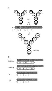
U.S. Provisional Application No. 61/721,072, filed November 1,2012, and U.S.
Provisional Application No. 61/787,927, filed March 15, 2013.
[002] Disclosed herein are multivalent and multispecific binding proteins,
methods of making the binding proteins, and their uses in the diagnosis,
inhibition,
prevention and/or treatment of cancers, tumors, and/or other angiogenesis-
dependent
diseases.
[003] Engineered proteins, such as multispecific binding proteins capable
of
binding two or more antigens, are known in the art. Such multispecific binding
proteins
can be generated using cell fusion, chemical conjugation, or recombinant DNA
techniques. There are a variety of multispecific binding protein structures
known in the
art; however many such structures and methods have distinct disadvantages.
[004] Bispecific antibodies have been produced using quadroma technology.
However, the presence of mis-paired by-products and significantly reduced
production
yields with this technology means that sophisticated purification procedures
are
required. Bispecific antibodies can also be produced by chemical conjugation
of two
different mAbs. However, this approach does not yield homogeneous
preparations.
[005] Other approaches used previously include coupling of two parental
antibodies with a hetero-bifunctional crosslinker, production of tandem single-
chain Fv
molecules, diabodies, bispecific diabodies, single-chain diabodies, and di-
diabodies.
However, each of these approaches have disadvantages. In addition, a
multivalent
antibody construct comprising two Fab repeats in the heavy chain of an IgG and
capable of binding four antigen molecules has been described (see PCT
Publication
No. WO 0177342 and Miller et al. (2003) J. lmmunol. 170(9): 4854-61).
[006] Ligand-receptor systems have co-evolved to maintain specificity.
Their
interactions activate specific signaling for a particular biological activity.
However, non-
ligand-receptor binding proteins such as mono-specific antibodies, bi- or
multi-specific
binding proteins, noncompetitive antibody combinations or other receptor
binding
proteins to an extracellular domain (ECD) of a receptor may recognize epitopes
distinct
from a receptor ligand-binding site. Binding to such a distinct epitope(s) on
the ECD of
CA 2890263 2017-03-09
WO 2014/071074 PCT/US2013/067873
a receptor may transduce conformational changes to the intracellular domain,
which
may result in a novel unexpected signaling cascade.
[007] US Patent No. 7,612,181
provides a novel family of binding proteins capable of binding two or more
antigens with high affinity, which are called dual variable domain binding
proteins (DVD
binding protein) or dual variable domain immunoglobulins (DVD-le). DVDs
molecules
are tetravalent dual-specific Ig-like proteins capable of binding two distinct
epitopes on
the same molecule or two different molecules simultaneously. DVDs are unique
binding
proteins comprised of two variable domains fused to the N-terminus of a
bivalent
antibody. The variable domains may be directly fused to one another or
connected. via
synthetic peptide linkers of assorted length and amino acid composition. DVDs
can be
engineered with intact and functional Fc domains, allowing then to mediate
appropriate
effector functions. DVD format, due to its flexibility of choice of antibody
pair,
orientation of two antigen-binding domains and the length of the linker that
joins them,
may provide for novel therapeutic modalities.
[008] While a variety of structures are provided in the art, some with
advantages and disadvantages, specific constructs are required for preparing
multivalent binding proteins with specific properties and which bind to
specific targets.
Additionally, new variable domain sequences can further improve the properties
of the
binding proteins. Specifically, improved DVDs that bind to DLL4 and VEGF could
prove
beneficial. Accordingly, disclosed herein are dual variable domain
immunoglobulins
using the binding protein framework disclosed in US Patent No. 7,612,181
and containing particular first and
second polypeptide chains, each comprising first and second variable domain
sequences (e.g., those listed in Table 2) that form functional binding sites
for VEGF
and DLL4. In some embodiments, the first and second polypeptide chains
comprise
first and second variable domain sequences that each contain the three CDRs
from
one of the sequences listed in Table 2 and form functional binding sites for
VEGF and
DLL4.
[009] DLL4 is a ligand involved in cell-to-cell signaling through the Notch
receptor pathway. Such cell-to-cell communication is required for many
biological
processes such as differentiation, proliferation, and homeostasis. The Notch-
signaling
pathway is one system that is utilized by a wide range of eukaryotes. This
pathway,
especially the Notch receptor, is also critical for functional tumor
angiogenesis. Thus,
inhibition of Notch receptor function, blockage of the Notch receptor, and/or
blockage of
2
CA 02890263 2015-04-30
WO 2014/071074
PCT/US2013/067873
the Notch-signaling pathway are potential strategies for anticancer
compositions and
therapies. Small molecule inhibitors of the Notch receptor have often proven
to be toxic
because they suppress wild type (normal) tissue expression of Notch receptors
throughout the body. Thus, different members of the Notch-signaling pathway
should
be considered as potential targets for therapeutics. A vasculature ligand for
the Notch
receptor is Delta 4 or Delta-like 4 (DLL4). Largely expressed in the
vasculature, DLL4
is critical for vascular development (Yan et at., Din. Cancer Res., 13(24):
7243-7246
(2007); Shutter et al., Genes Dev., 14(11): 1313-1318 (2000); Gale et at.,
Proc. Natl.
Acad. Sci. USA, 101(45): 15949-15954 (2004); Krebs et at., Genes Dev., 14(11):
1343-
1352 (2000)). Mice heterozygous for DLL4 are embryonically lethal due to major
defects in vascular development (Gale et al., Proc. Natl. Acad. Sci. USA,
101(45):
15949-15954 (2004); Duarte et al., Genes Dev., 18(20): 2474-2478 (2004); Krebs
et
at., Genes Dev., 18(20): 2469-2473 (2004)).
[010] The expression of DLL4 can be induced by VEGF (Liu et at,, Moi. Cell
Biol., 23(1): 14- 25 (2003); Lobov et al., Proc. Natl. Acad. Sci. USA, 104(9):
3219-3224
(2007)). VEGF is a signal protein produced by cells involved in angiogenesis.
Additionally, DLL4 can negatively regulate VEGF signaling, in part through
repressing
VEGFR2 and inducing VEGR1 (Harrington et al., Microvasc. Res., 75(2): 144-154
(2008); Suchting et at., Proc. Natl. Acad. Sci. USA, 104(9): 3225-3230
(2007)).
Exquisite coordination between DLL4 and VEGF is essential for functional
angiogenesis, making both DLL4 and VEGF potential targets for therapeutic
intervention.
[011] In addition to their physiological role, DLL4 and VEGF are also up-
regulated in tumor blood vessels (Gale et al., Proc. Natl. Acad. Sci. USA,
101(45):
15949-15954 (2004); Mailhos et at., Differentiation, 69(2-3): 135-144 (2001);
Patel et
al., Cancer Res., 65(19): 8690-8697 (2005); Patel et at., Clin. Cancer Res.,
12(16):
4836-4844 (2006); Noguera-Troise et at., Nature, 444(7122): 1032-1037 (2006)).
Blockade of DLL4 has been shown to inhibit primary tumor growth in multiple
models
(Noguera-Troise et at., Nature, 444(7122): 1032-1037 (2006); Ridgway et al.,
Nature,
444(7122): 1083-1087 (2006); Scehnet et al., Blood, 109(11): 4753-4760
(2007)). The
inhibition of DLL4 is even effective against tumors that are resistant to anti-
VEGF
therapy. Thus, the combinatorial inhibition of both DLL4 and VEGF could
provide an
enhanced anti-tumor therapy. Interestingly, unlike VEGF inhibition that
reduces tumor
vessel formation, DLL4 blockade leads to an increase in tumor vasculature
density
wherein the vessels are abnormal, cannot support efficient blood transport,
and are
3
CA 2890263 2017-03-09
WO 2014/071074 PCT/US2013/067873
effectively nonfunctional. Thus, disruption of both VEGF and DLL4 provides for
different methods of action for potential anti-cancer treatment.
[012] While antibodies and various binding constructs are known in the art,
there remains a need for better targeting and efficiency of binding to VEGF
and DLL4,
e.g., to treat cancer and tumorogenesis. There is thus a need in the art for
improved
multivalent binding proteins capable of binding DLL4 and VEGF. Accordingly,
novel
binding proteins are provided, wherein the binding proteins are capable of
binding
DLL4 and VEGF. In some embodiments, the binding proteins are capable of, e.g.,
binding to DLL4 and VEGF with improved binding affinity and/or neutralization
potency.
[013] Binding proteins capable of targeting two epitopes are provided,
wherein the binding proteins are capable of binding DLL4 and VEGF. In an
embodiment, binding proteins capable of binding epitopes of DLL4 and VEGF with
high
affinity are provided. In an embodiment, the binding proteins comprise a dual
variable
domain binding protein framework that contains the CDR and variable domain -
sequences listed in Table 2. In an embodiment, the dual variable domain
binding
protein framework comprises the framework disclosed in US Patent No.7,612,181.
[014] In one embodiment, binding proteins comprising a polypeptide chain
that can bind two epitopes of two different proteins (VEGF and DLL4) are
provided,
wherein the polypeptide chain comprises VD1-(X1)n-VD2-C-(X2)n, wherein VD1 is
a
first variable domain, VD2 is a second variable domain, C is a constant
domain, X1
represents an amino acid or polypeptide, X2 represents an Fc region and n is 0
or 1,
are provided. In some embodiments, the VD1 and VD2 in the binding protein are
heavy
chain variable domains. In certain embodiments, VD1 and VD2 are capable of
binding
an epitope of DLL4 and an epitope of VEGF. In some embodiments, C is a heavy
chain
constant domain, such as CH1. In certain embodiments, X1 is a linker with the
proviso
that X1 is not CH1.
[015] In various embodiments, the binding protein disclosed herein
comprises a polypeptide chain that binds an epitope of DLL4 and an epitope of
VEGF,
wherein the polypeptide chain comprises VD1-(X1)n-VD2-C-(X2)n, wherein VD1
comprises a first heavy chain variable domain, VD2 comprises a second heavy
chain
variable domain, C comprises a heavy chain constant domain, X1 comprises a
linker,
and X2 comprises an Fc region. In an embodiment, X1 is a linker with the
proviso that it
is not CH1. In an embodiment, the VD1 and VD2 heavy chain variable domains
each
comprise three CDRs chosen from the CDRs in SEQ ID NO: 39, 41, 43, 45, 47, 49,
51,
4
CA 02890263 2015-04-30
WO 2014/071074 PCT/US2013/067873
or 53 (i.e., CDRs 1-3 from one of those SEQ ID NOs), wherein at least one of
the VD1
and/or VD2 heavy chain variable domains comprises the three CDRs in SEQ ID NO:
39. In another embodiment, the binding protein is capable of binding DLL4 and
VEGF.
In an embodiment, the VD1 and V02 heavy chain variable domains comprise SEQ ID
NO: 39, 41, 43, 45, 47, 49, 51, or 53, wherein at least one of the VD1 and/or
VD2
heavy chain variable domains comprises SEQ ID NO: 39.
[016] In various embodiments, the binding protein disclosed herein
comprises a polypeptide chain that binds an epitope of DLL4 and an epitope of
VEGF,
wherein the polypeptide chain comprises VD1-(X1)n-VD2-C-(X2)n, wherein VD1
comprises a first light chain variable domain, VD2 comprises a second light
chain
variable domain, C comprises a light chain constant domain, X1 comprises a
linker,
and X2 does not comprise an Fc region. In an embodiment, X1 is a linker with
the
proviso that it is not a CH1 or a CL. In an embodiment, the VD1 and VD2 light
chain
variable domains each comprise three CDRs chosen from the CDRs in SEQ ID NO:
40, 42, 44, 46, 48, 50, 52, or 54 (i.e., CDRs 1-3 from one of those SEQ ID
NOs),
wherein at least one of the VD1 and/or VD2 light chain variable domains
comprises the
three CDRs in SEQ ID NO: 40. In another embodiment, the binding protein is
capable
of binding DLL4 and VEGF. In an embodiment, the VD1 and VD2 light chain
variable
domains each comprise SEQ ID NO: 40, 42, 44, 46, 48, 50, 52, or 54, wherein at
least
one of the VD1 and/or VD2 light chain variable domains comprises SEQ ID NO:
40.
[017] In another embodiment, a binding protein that binds an epitope of
DLL4 and an epitope of VEGF is disclosed. In some embodiments, the binding
protein
comprises first and second polypeptide chains, wherein each of the first and
second
polypeptide chains independently comprises VD1-(X1)n-VD2-C-(X2)n, wherein VD1
is
a first variable domain, VD2 is a second variable domain, C is a constant
domain, X1 is
a linker, X2 is an Fc region, and n is 0 or 1, wherein the VD1 domains on the
first and
second polypeptide chains form a first functional target binding site and the
VD2
domains on the first and second polypeptide chains form a second functional
target
binding site. In an embodiment, X2 comprises an Fc region when n=1, and X2
does
not comprise an Fc region when n=0. In some embodiments, the X1 sequences on
the
first and second polypeptide chains are the same. In other embodiments, the X1
sequences on the first and second polypeptide chains are different. In some
embodiments X1 on at least one of the polypeptide chains is not a CHI domain
and/or
a CL domain. In one embodiment, the X1 sequence is a short (e.g., 6, 5, 4, 3,
or 2
amino acid) linker. In another embodiment, the the X1 sequence is a long
(e.g., 6, 7, 8,
CA 02890263 2015-04-30
WO 2014/071074
PCT/US2013/067873
9, 10,11, 12, 15, 20, 25, 30, or greater amino acid) linker. In another
embodiment, the
X1 sequence on one of the two polypeptide chains is a short linker and the X1
sequence on the other polypeptide chain is a long linker. In an embodiment,
the VD1
and VD2 heavy chain variable domains each comprise three CDRs from SEQ ID NO:
39, 41, 43, 45, 47, 49, 51, or 53 (i.e., CDRs 1-3 from one of those SEQ ID
NOs),
wherein at least one of the VD1 and/or VD2 heavy chain variable domains
comprises
the three CDRs in SEQ ID NO: 39; and the VD1 and VD2 light chain variable
domains
comprise three CDRs from SEQ ID NO: 40, 42, 44, 46, 48, 50, 52, or 54 (i.e.,
CDRs 1-
3 from one of those SEQ ID NOs), wherein at least one of the VD1 and/or VD2
light
chain variable domains comprises the three CDRs in SEQ ID NO: 40. In another
embodiment, the binding protein is capable of binding DLL4 and VEGF. In an
embodiment, the VD1 and VD2 heavy chain variable domains comprise SEQ ID NO:
39, 41, 43, 45, 47, 49, 51, or 53, wherein at least one of the VD1 and/or VD2
heavy
chain variable domains comprises SEQ ID NO: 39, and the VD1 and VD2 light
chain
variable domains comprise SEQ ID NO: 40, 42, 44, 46, 48, 50, 52, or 54,
wherein at
least one of the VD1 and/or VD2 light chain variable domains comprises SEQ ID
NO:
40.
[018] In various
embodiments, a binding protein is disclosed that is capable
of binding VEGF and DLL4. In some embodiments, the binding protein comprises
first
and second polypeptide chains, wherein each of the first and second
polypeptide
chains independently comprises VD1-(X1)n-VD2-C-(X2)n, wherein VD1 is a first
variable domain, VD2 is a second variable domain, C is a constant domain, X1
is a
linker, X2 is an Fc region, and n is 0 or 1, wherein the VD1 domains on the
first and
second polypeptide chains form a first functional target binding site and the
VD2
domains on the first and second polypeptide chains form a second functional
target
binding site. In an embodiment, the variable domains that form a functional
target
binding site for VEGF comprise three CDRs from SEQ ID NO: 41 and three CDRs
from
SEQ ID NO: 42 (e.g., CDRs 1-3 from SEQ ID NO: 41 and CDRs 1-3 from SEQ ID NO:
42 present on separate chains, with the CDRs on each chain arranged in the
specified
order and separated by suitable framework sequences to form a functional
binding
site). In an embodiment, the variable domains that form a functional target
binding site
for DLL4 comprise three CDRs from SEQ ID NO: 39 and three CDRs from SEQ ID NO:
40 (e.g., CDRs 1-3 from SEQ ID NO: 39 and CDRs 1-3 from SEQ ID NO: 40 present
on separate chains, with the CDRs on each chain arranged in the specified
order and
separated by suitable framework sequences to form a functional binding site).
In an
embodiment, the binding protein comprises a functional target binding site for
VEGF
6
CA 2890263 2017-03-09
WO 2014/071074 PCT/US2013/067873
comprising three CDRs from SEQ ID NO: 41 and three CDRs from SEQ ID NO: 42
(e.g., CDRs 1-3 from SEQ ID NO: 41 and CDRs 1-3 from SEQ ID NO: 42), and a
functional target binding site for DLL4 comprising three CDRs from SEQ ID NO:
39 and
three CDRs from SEQ ID NO: 40 (e.g., CDRs 1-3 from SEQ ID NO: 39 and CDRs 1-3
from SEQ ID NO: 40). In an embodiment, the binding protein comprises a
functional
target binding site for VEGF comprising SEQ ID NO: 41 and SEQ ID NO: 42, and a
functional target binding site for DLL4 comprising SEQ ID NO: 39 and SEQ ID
NO: 40.
In an embodiment, the binding protein comprises a first polypeptide chain
comprising
SEQ ID NO: 56 and a second polypeptide chain comprising SEQ ID NO: 64. In an
embodiment, the binding protein comprises a first polypeptide chain comprising
SEQ
ID NO: 73 and a second polypeptide chain comprising SEQ ID NO: 74. In some
embodiments, the variable domains that form a binding site for DLL4 comprise
those
from US application publication no. 20110217237, and/or the variable domains
that
form a binding site for VEGF comprise those from US application publication no
20100076178. In an
embodiment, the binding protein comprises h1A11.1-SL-Av. In an embodiment, the
h1A11.1-SL-Av binding protein comprises an Fc region from a human IgG1 LALA
mutant.
[019] The development
and production of a binding protein suitable for use
as a human therapeutic agent, e.g., as an anticancer/antitumor agent, can
require
more than the identification of a binding protein capable of binding to a
desired target
or targets. For instance, a candidate may bind its target(s) but exhibit a
reduced ability
to inhibit or neutralize its desired target, may prove difficult to stably
formulate, may
exhibit undesirable pharmacokinetic properties, or may prove difficult to
produce in a
suitable expression system (e.g., expression in a host cell such as CHO).
Thus,
factors to consider in developing a suitable therapeutic agent include, but
are not
limited to, (a) the binding kinetics (on-rate, off-rate and affinity) for both
the inner and
outer antigen-binding domains, (b) potencies in various biochemical and
cellular
bioassays, (c) in vivo efficacies in relevant tumor models, (d)
pharmacokinetic and
pharmacodynannics properties, (e) manufacturability, including protein
expression level
in selected cell lines, scalability, post-translational modification,
physicochemical
properties such as monomer percentage, solubility, and stability (intrinsic,
freeze/thaw,
storage stability, etc.), (f) formulation properties, (g) potential
immunogenicity risk, and
(h) toxicological properties of a molecule. Binding mode and valency may also
be
evaluated, as these can affect binding properties and cellular potencies of a
molecule.
For some binding proteins, even small changes in the amino acid sequences of
the
7
CA 02890263 2015-04-30
WO 2014/071074 PCT/US2013/067873
variable domains, constant domains, and/or linkers can potentially impact
(positively or
negatively) one or more of these factors, and so the combination of factors
can be
evaluated to select a lead candidate. Once a lead candidate is identified,
further in vivo
evaluation of therapeutic properties are conducted, included evaluation of
safety,
efficacy, and potency in animal and human subjects.
[020] It has been found, unexpectedly, that a binding protein comprising
SEQ ID NO: 56 and SEQ ID NO: 64, or comprising SEQ ID NO: 73 and SEQ ID NO:
74, exhibited a superior combination of properties in this regard, such as a
binding
kinetic (i.e., a dissociation constant) surpassing a therapeutically useful
threshold,
improved neutralization ability, enhanced in vivo efficacy, superior
formulatability, a
desirable glycosylation pattern, a favorable pharmacokinetic profile, and
efficient
expression in host cells, as compared to other evaluated binding proteins
comprising
different variable domains and/or linker sequences. The compared binding
proteins
can include those having the same variable domain sequences and orientations
but
with different linkers, as well as those having altered binding domain
orientations
and/or other variable domain sequences (e.g., affinity matured sequences,
fully human
sequences, etc.). In some embodiments, these superior properties are dependent
on
the selection of particular variable domain sequences (e.g., SEQ ID NOs: 39-
42),
particular orientations of the VEGF and DLL4 binding domains in the inner and
outer
positions (e.g., the orientations provided in SEQ ID NO: 56 and SEQ ID NO:
64),
particular linker sequences (e.g., the heavy chain variable domains and short
linker
sequence used in SEQ ID NO: 56 and the light chain variable domains and long
linker
sequence used in SEQ ID NO: 64), and/or particular constant domain sequences
(e.g.,
the constant domain sequences used in SEQ ID NOs: 73 and 74). In some
embodiments, merely changing the linker sequence can have a significant impact
on
functional properties. For example, selecting the linker sequence used in SEQ
ID NO:
73 and 74 (along with the variable and constant domains included in those SEQ
ID
NOs) can provide unexpected improvement in the human therapeutic properties of
the
binding protein. For instance, Table 9 demonstrates the effects of different
linker
sequences on in vivo anti-tumor efficacy, as measured in colorectal
adenocarcinoma
and glioblastoma xenograft models. Thus, for example, using the sequences in
SEQ
ID NOs: 56 and 64, or in SEQ ID NOs: 73 and 74 (including the linker sequences
included therein), can produce an unexpected gain in anti-tumor efficacy in
vivo.
[021] In various embodiments, a binding protein disclosed herein exhibits a
desired binding affinity, blocking ability, and/or neutralization potency for
VEGF and/or
8
CA 02890263 2015-04-30
WO 2014/071074
PCT/US2013/067873
DLL4, for instance levels roughly comparable to those observed for antibodies
against
VEGF (e.g., AVASTINO) or DLL4 (e.g., antibody h1A11.1). In some embodiments,
the
binding protein exhibits increased affinity, blocking ability, and/or
neutralization potency
for VEGF and/or DLL4, as compared to DVD-Ig binding proteins comprising other
variable domain sequences or linkers, In some embodiments, the binding protein
comprises a functional target binding site for VEGF comprising three CDRs from
SEQ
ID NO: 41 and three CDRs from SEQ ID NO: 42, and a functional target binding
site for
DLL4 comprising three CDRs from SEQ ID NO: 39 and three CDRs from SEQ ID NO:
40; or comprises a functional target binding site for VEGF comprising SEQ ID
NO: 41
and SEQ ID NO: 42, and a functional target binding site for DLL4 comprising
SEQ ID
NO: 39 and SEQ ID NO: 40; or comprises SEQ ID NO: 56 and SEQ ID NO: 64; or
comprises SEQ ID NO: 73 and SEQ ID NO: 74. For instance, the binding protein
(e.g.,
the binding protein comprising SEQ ID NO: 56 and SEQ ID NO: 64, or comprising
SEQ
ID NO: 73 and SEQ ID NO: 74) can be capable of binding to VEGF with a
dissociation
constant (KD) of at most about 7.0 x 10⁻¹⁰M, as measured by surface plasmon
resonance, and/or blocking VEGF activity with an IC50 of at most about 3.8 nM,
as
measured in a VEGFR1 Competition ELISA; and/or capable of binding to DLL4 with
a
dissociation constant (KD) of at most about 1.0 x 10⁻⁸ M, as measured by
surface
plasmon resonance, and/or blocking DLL4 activity with an IC50 of at most about
1.09
nM, as measured in a Notch Competition ELISA.
[022] In some embodiments, the binding protein (e.g., the binding
protein
comprising SEQ ID NO: 56 and SEQ ID NO: 64, or comprising SEQ ID NO: 73 and
SEQ ID NO: 74) can exhibit increased neutralization potency for DLL4 when also
in the
presence of VEGF, as compared to a mixture of VEGF and DLL4 antibodies (e.g.,
the
parental antibodies used to provide variable domains for the binding protein).
In some
embodiments, the binding protein can exhibit an order of magnitude increase in
the
neutralization potency for DLL4 when also in the presence of VEGF, as compared
to a
mixture of VEGF and DLL4 antibodies (e.g., the parental antibodies used to
provide
variable domains for the binding protein). For instance, Table 24 demonstrates
that a
binding protein comprising SEQ ID NO: 73 and SEQ ID NO: 74 can exhibit an
order of
magnitude increase in the neutralization potency for DLL4 when also in the
presence of
at least about 1.2 nM VEGF (e.g., at least about 1.2, 1.5, 2, 2.5, 5, 10, 50,
150, or
more), as compared to a mixture of VEGF and DLL4 antibodies (the parental
antibodies used to provide variable domains for the binding protein). This
DLL4
neutralization property may be beneficial because, in the in vivo situation of
treatment
for a tumor, VEGF levels are usually higher in the vicinity of a tumor than in
the
9
CA 02890263 2015-04-30
WO 2014/071074
PCT/US2013/067873
circulation generally, allowing for both improved targeting and enhanced
functional
DLL4 neutralization acitivity at the tumor site.
[023] In various embodiments, a binding protein (e.g., the binding protein
comprising SEQ ID NO: 56 and SEQ ID NO: 64, or comprising SEQ ID NO: 73 and
SEQ ID NO: 74) exhibits improved properties, e.g., improved safety, increased
stability,
greater potency, reduced inflammation or immune response, or other beneficial
in vivo
human therapeutic properties, as compared to other treatments for cancers
and/or
vascularized tumors. Treatments suitable for comparison can include
administration of
a small molecule anti-cancer agent, or an antibody against VEGF (e.g.,
AVASTINCI)
and/or DLL4 (e.g., antibody h1A11.1), or a DVD-Ig binding protein comprising
other
variable domain sequences and/or linkers. In some embodiments, the binding
protein
exhibits improved properties over a current standard of care treatment for
cancer
and/or a vascularized tumor. For instance, the binding protein can exhibit
improved
binding kinetics, superior in vivo therapeutic efficacy, enhanced
formulatability
(including reduced aggregation and improved storage stability), improved
pharmacokinetics, reduced inflammation or immune response, and/or enhanced
host
cell expression levels.
[024] In some embodiments, a binding protein (e.g., the binding protein
comprising SEQ ID NO: 56 and SEQ ID NO: 64, or comprising SEQ ID NO: 73 and
SEQ ID NO: 74) exhibits superior (e.g., additive and/or superadditive) effects
in
combination with one or more anti-cancer agents, as compared to an anti-VEGF
antibody or an anti-DLL4 antibody in combination with one or more anti-cancer
agent,
or as compared to a DVD-Ig binding protein comprising other variable domain
sequences and/or linkers in combination with one or more anti-cancer agent.
For
instance, the superior binding properties can be those identified in Tables 27-
30 and
34. For instance, the binding protein can exhibit at least about a 50% or
greater (e.g.,
45, 46, 47, 48, 49, 50, 51, 52, 53, 54, 55, 60, 65, 70, 75, 80, 85, 90, 95,
100, 125,
150% or more) inhibition of tumor groth or delay in tumor growth after
administration
alone or in combination with one or more anti-cancer agent, as compared to an
untreated tumor. The anti-cancer agent can be, for instance, one or more of
Irinotecan,
FOLFIRI, Temozolomide, Gemcitabine, Paclitaxel, 5-FU, and Capecitabine, or any
other small molecule or biologic agent used in the treatment of a particular
cancer. The
binding protein alone or in combination with the one or more anti-cancer agent
can be
used as a medicament to treat, e.g., colon cancer, gliobastoma, pancreatic
cancer, or
breast cancer.
CA 02890263 2015-04-30
WO 2014/071074
PCT/US2013/067873
[025] In an embodiment, a Dual Variable Domain (DVD-Ig) binding protein
comprises two first and two second polypeptide chains as described in the
previous
paragraph (i.e., comprising four polypeptide chains), wherein each of the
polypeptide
chains independently comprises VD1-(X1)n-VD2-C-(X2)n, wherein VD1 is a first
variable domain, VD2 is a second variable domain, C is a constant domain, X1
is a
linker, X2 is an Fc region, and n is 0 or 1. In some embodiments, the first
chain is a
heavy chain and is paired with a second chain that is a light chain. Such a
DVD-Ig
binding protein comprises four functional target binding sites. In some
embodiments,
the X1 linker on the first and second polypeptide chains are the same or
different. In
some embodiments, the DVD-Ig binding proteins comprise at least two variable
domain
sequences (e.g., VD1 and VD2) capable of binding two or more epitopes (e.g.,
two,
three, or four) of the same or different proteins, in any orientation. In some
embodiments, VD1 and VD2 are independently chosen. In an embodiment, the VD1
and VD2 heavy chain variable domains each comprise three CDRs from SEQ ID NO:
39, 41, 43, 45, 47, 49, 51, or 53, wherein at least one of the VD1 and/or VD2
heavy
chain variable domains comprises the three CDRs in SEQ ID NO: 39, and the VD1
and
VD2 light chain variable domains comprise three CDRs from SEQ ID NO: 40, 42,
44,
46, 48, 50, 52, or 54, wherein at least one of the VD1 and/or VD2 light chain
variable
domains comprises the three CDRs in SEQ ID NO: 40. In another embodiment, the
binding protein is capable of binding DLL4 and VEGF. In an embodiment, the VD1
and
VD2 heavy chain variable domains each comprise SEQ ID NO: 39, 41, 43, 45, 47,
49,
51, or 53, wherein at least one of the VD1 and/or VD2 heavy chain variable
domains
comprises SEQ ID NO: 39, and the VD1 and VD2 light chain variable domains each
comprise SEQ ID NO: 40, 42, 44, 46, 48, 50, 52, or 54, wherein at least one of
the VD1
and/or VD2 light chain variable domains comprises SEQ ID NO: 40.
[026] In another embodiment, the Dual Variable Domain binding protein
comprises a heavy chain and a light chain sequence as shown in Table 2,
wherein at
least one of the VD1 and/or VD2 heavy chain variable domains comprises SEQ ID
NO:
39 and/or at least one of the VD1 and/or VD2 light chain variable domains
comprises
SEQ ID NO: 40.
[027] In a further embodiment, any of the heavy chain, light chain, two
chain,
or four chain embodiments includes at least one X1 linker comprising the
linkers
selected from SEQ ID NO: 1-38. In an embodiment, X2 is an Fc region. In
another
embodiment, X2 is a variant Fc region.
11
CA 02890263 2015-04-30
WO 2014/071074
PCT/US2013/067873
[028] In still another embodiment, the Fc region, if present in the first
polypeptide, is a native sequence Fc region or a variant sequence Fc region.
In yet
another embodiment, the Fc region is an Fc region from an IgG1, an Fc region
from an
IgG2, an Fc region from an IgG3, an Fc region from an IgG4, an Fc region from
an IgA,
an Fc region from an IgM, an Fc region from an IgE, or an Fc region from an
IgD. In
certain embodiments, the Fc region is an Fc region from a human IgG1 LALA
mutant,
which is a mutant of the b12 antibody that provides protection against the HIV
virus.
[029] A method of making a binding protein that binds two different target
proteins is provided. In an embodiment, the method of making a binding protein
comprises the steps of a) obtaining a first parent antibody, or antigen
binding portion
thereof, that binds a first epitope; b) obtaining a second parent antibody, or
antigen
binding portion thereof, that binds a second epitope; c) preparing
construct(s) encoding
any of the binding proteins described herein; and d) expressing the
polypeptide chains,
such that a binding protein that binds the first and the second epitope is
generated.
[030] In any of the embodiments herein, the VD1 heavy chain variable
domain, if present, and light chain variable domain, if present, can be from a
first parent
antibody or antigen binding portion thereof; the VD2 heavy chain variable
domain, if
present, and light chain variable domain, if present, can be from a second
parent
antibody or antigen binding portion thereof. The first and second parent
antibodies can
be the same or different.
[031] In one embodiment, the first parent antibody or antigen binding
portion
thereof, binds a first antigen, and the second parent antibody or antigen
binding portion
thereof, binds a second antigen. In an embodiment, the first and second
antigens are
different antigens. In another embodiment, the first parent antibody or
antigen binding
portion thereof binds the first antigen with a potency different from the
potency with
which the second parent antibody or antigen binding portion thereof binds the
second
antigen. In yet another embodiment, the first parent antibody or antigen
binding portion
thereof binds the first antigen with an affinity different from the affinity
with which the
second parent antibody or antigen binding portion thereof binds the second
antigen.
[032] In another embodiment, the first parent antibody or antigen binding
portion thereof, and the second parent antibody or antigen binding portion
thereof are a
human antibody, CDR grafted antibody, humanized antibody, and/or affinity
matured
antibody.
12
CA 02890263 2015-04-30
WO 2014/071074 PCT/US2013/067873
[033] In another embodiment, the binding protein possesses at least one
desired property exhibited by the first parent antibody or antigen binding
portion
thereof, or by the second parent antibody or antigen binding portion thereof.
Alternatively, the first parent antibody or antigen binding portion thereof
and the second
parent antibody or antigen binding portion thereof possess at least one
desired
property exhibited by the binding protein. In an embodiment, the desired
property is
one or more antibody parameters. In another embodiment, the antibody
parameters
are antigen specificity, affinity to antigen, potency, biological function,
epitope
recognition, stability, solubility, production efficiency, immunogenicity,
pharmacokinetics, bioavailability, tissue cross reactivity, or orthologous
antigen binding.
In an embodiment, the binding protein is multivalent. In another embodiment,
the
binding protein is multispecific. The multivalent and or multispecific binding
proteins
described herein have desirable properties particularly from a therapeutic
standpoint.
For instance, the multivalent and or multispecific binding protein may (1) be
internalized
(and/or catabolized) faster than a bivalent antibody by a cell expressing an
antigen to
which the antibodies bind; (2) be an agonist binding protein; and/or (3)
induce cell
death and/or apoptosis of a cell expressing an antigen to which the
multivalent binding
protein is capable of binding. The "parent antibody", which provides at least
one
antigen binding specificity of the multivalent and or multispecific binding
protein, may
be one that is internalized (and/or catabolized) by a cell expressing an
antigen to which
the antibody binds; and/or may be an agonist, cell death-inducing, and/or
apoptosis-
inducing antibody, and the multivalent and or multispecific binding protein as
described
herein may display improvement(s) in one or more of these properties.
Moreover, the
parent antibody may lack any one or more of these properties, but may acquire
one or
more of them when constructed as a multivalent binding protein as described
herein.
[034] In another embodiment, the binding protein has an on rate constant
(K0) to one or more targets of at least about 102 M-1s-1; at least about 103M-
1s-1; at least
about 104M-'s-1; at least about 105M-1s-1; or at least about 106M-1s-1, as
measured by
surface plasmon resonance. In an embodiment, the binding protein has an on
rate
constant (K0n) to one or more targets from about 102M-1s-1 to about 103M-1s-1;
from
about 103M-1s-1 to about 104 M-"1s-1; from about 10 M1s1 to about 105M-1s-1;
or from
about 105M-1s-1 to about 106M-1s-1, as measured by surface plasmon resonance.
[035] In another embodiment, the binding protein has an off rate constant
(Koff) for one or more targets of at most about 10-2s-1; at most about 10-
3sa1; at most
about 10-4s-1; at most about 10-5s-1; or at most about 10-6s-1, as measured by
surface
13
CA 02890263 2015-04-30
WO 2014/071074
PCT/US2013/067873
plasmon resonance. In an embodiment, the binding protein has an off rate
constant
(Koff) to one or more targets of about 10-2s-1 to about 10-3s-1; of about 10-
3s-1 to about
10-4s-1; of about 10-4s-1to about 10-8s-1; or of about 10-8s-lto about 10-6s-
1, as
measured by surface plasmon resonance.
[036] In another embodiment, the binding protein has an equilibrium
dissociation constant (KD) to one or more targets of at most about 10-7M; at
most about
10-8M; at most about 10-9M; at most about 10-10M; at most about 10-11M; or at
most
about 10-12M. In an embodiment, the binding protein has an equilibrium
dissociation
constant (KO to its targets of about 10-7M to about 10-8M; of about 10-8M to
about 10-9
M; of about 10-9M to about 10-10M; of about 10-1 M to about 10-11M; or of
about 10-11M
to about 10-12M.
[037] In some embodiments, an anti-DLL4/anti-VEGF binding protein
exhibits increased potency (e.g., increased ability to interfere with, inhibit
and/or
neutralize DLL4 and/or VEGF activity) as compared to an anti-DLL4 or anti-VEGF
antibody. In some embodiments, the potency of the binding protein can be
evaluated
in any assay for evaluating VEGF and/or DLL4 activity, e.g., a VEGF and/or
DLL4
binding ELISA assay, a BIACORETM assay, a DLL4-Notch reporter assay, a VEGF-
stimulated Endothelial Cell Proliferation/Survival assay, or any other assay
known to
one of skill in the art. In some embodiments, the binding protein exhibits
increased
DLL4 potency in the presence of VEGF.
[038] In another embodiment, a conjugate is provided, comprising any of the
binding proteins described herein and further comprising an agent. In an
embodiment,
the agent is an immunoadhesion molecule, an imaging agent, a therapeutic
agent, or a
cytotoxic agent. In an embodiment, the imaging agent is a radiolabel, an
enzyme, a
fluorescent label, a luminescent label, a bioluminescent label, a magnetic
label, or
biotin. In another embodiment, the radiolabel is 3H, 14C, 35s, 90y, 99-rc,
111in, 1251, 1311,
177LU, 166H0, or153Sm. In yet another embodiment, the therapeutic or cytotoxic
agent is
an anti-metabolite, an alkylating agent, an antibiotic, a growth factor, a
cytokine, an
anti-angiogenic agent, an anti-mitotic agent, an anthracycline, toxin, or an
apoptotic
agent. In some embodiments, the agent is one or more of: irinotecan,
leucovorin, 5-FU,
temozolomide, gemcitabine, and paclitaxel. In an embodiment, the agent is
irinotecan.
In an embodiment, the agent is leucovorin. In an embodiment, the agent is 5-
FU. In
an embodiment, the agent is irinotecan, leucovorin, and 5-FU. In an
embodiment, the
agent is temozolomide. In an embodiment, the agent is gemcitabine. In an
embodiment, the agent is paclitaxel.
14
CA 02890263 2015-04-30
WO 2014/071074
PCT/US2013/067873
[039] In another embodiment, the conjugate comprises a binding protein
and
a drug. In an embodiment, the binding protein in the conjugate comprises first
and
second polypeptide chains, wherein each of the first and second polypeptide
chains
independently comprises VD1-(X1)n-VD2-C-(X2)n, wherein VD1 is a first variable
domain, VD2 is a second variable domain, C is a constant domain, X1 is a
linker, and
X2 is an Fc region, wherein the VD1 domains on the first and second
polypeptide
chains form a first functional target binding site and the VD2 domains on the
first and
second polypeptide chains form a second functional target binding site, and
wherein
the binding protein is capable of binding VEGF and DLL4. In some embodiments,
the
VD1 and VD2 domains comprise CDRs or variable domain sequences from any of the
sequences disclosed in Table 2, paired and arranged to form functional binding
sites
for VEGF and DLL4. In an embodiment, the drug in the conjugate is selected
from the
group consisting of a mitotic inhibitor, an antitumor antibiotic, an
immunonnodulating
agent, a vector for gene therapy, an alkylating agent, an antiangiogenic
agent, an
antimetabolite, a boron-containing agent, a chemoprotective agent, a hormone,
an
antihormone agent, a corticosteroid, a photoactive therapeutic agent, an
oligonucleotide, a radionuclide agent, a topoisomerase inhibitor, a tyrosine
kinase
inhibitor, and a radiosensitizer. In another embodiment, the drug is selected
from the
group consisting of Ixempra, dolastatin 10, dolatstin 15, auristatin E,
auristatin PE,
monomethyl auristatin D (MMAD or auristatin D derivative), monomethyl
auristatin E
(MMAE or auristatin E derivative), monomethyl auristatin F (MMAF or auristatin
F
derivative), auristatin F phenylenediamine (AFP), auristatin EB (AEB),
auristatin EFP
(AEFP), 5-benzoylvaleric acid-AE ester (AEVB), methotrexate, daunorubicin,
vincristine, maytansine, maytansinol, 0-3 esters of maytansinol, ansamitocin
P1,
ansamitocin P2, ansamitocin P3, ansamitocin P4, docetaxel, paclitaxel,
nanoparticle
paclitaxel, vindesine sulfate, vincristine, vinblastine, vinorelbine,
actinomycines,
actinomycin D, anthramycin, chicamycin A, DC-18, mazethramycin, neothramycin
A,
neothramycin B, prothracarcin B, SG2285, sibanomicin, sibiromycin,
anthracyclines,
daunorubicin, doxorubicin, epirubicin, idarubicin, calicheamicins, 021,
031,
PSAG, 6il, duocarmycins, adozelesin, bizelesin, and carzelesin, bleomycin,
mitomycin, plicamycin, bacillus calmette-guerin (BOG), levamisole, cancer
vaccines,
recombinant bivalent human papillomavirus (HPV) vaccine types 16 and 18
vaccine,
recombinant quadrivalent human papillomavirus (HPV) types 6, 11, 16, and 18
vaccine,
sipuleucel-T, cytokines, parathyroid hormone; thyroxine; insulin; proinsulin;
relaxin;
prorelaxin; glycoprotein hormones such as follicle stimulating hormone (FSH),
thyroid
stimulating hormone (TSH), and luteinizing hormone (LH), hepatic growth
factor;
CA 02890263 2015-04-30
WO 2014/071074
PCT/US2013/067873
fibroblast growth factor, prolactin, placental lactogen, tumor necrosis
factor, mullerian-
inhibiting substance, mouse gonadotropin-associated peptide, inhibin, activin,
vascular
endothelial growth factor, integrin, thrombopoietin (TPO), nerve growth
factors such as
NGF, platelet-growth factor, transforming growth factors (TGFs), insulin-like
growth
factor-I and -II, erythropoietin (EPO), osteoinductive factors, interferons
such as
interferon a, p, and 7, colony stimulating factors (CSFs), granulocyte-
macrophage-C-SF
(GM-CSF), and granulocyte-CSF (G-CSF), interleukins (Is) such as IL-1, IL-1a,
IL-2,
IL-3, IL-4, IL-5, IL-6, IL-7, IL-8, IL-9, IL-11, IL-12, tumor necrosis factor
and other
polypeptide factors including LI F and kit ligand (KL), colony-stimulating
factors,
erythropoietin (epoetin), filgrastim, sargramostim, promegapoietin,
Oprelvekin,
imnnunomodulating gene therapeutics, nucleic acid encoding a functional,
therapeutic
gene that is used to replace a mutated or otherwise dysfuntional (e.g.
truncated) gene
associated with cancer, nucleic acid that encodes for or otherwise provides
for the
production of a therapeutic protein to treat cancer, alkyl sulfonates,
busulfan, nitrogen
mustards, chlorambucil, cyclophosphamide, estramustine, ifosfamide,
mechlorethamine, and melphalan, nitrosoureas, carmustine, fotemustine,
lomustine,
nimustine, streptozocin, triazines and hydrazines, dacarbazine, procarbazine,
temozolomide, ethylenimimes, thiopeta, diaziquone, mitomycin C, methylamine
derivatives, epoxides, altretamine, dianhydrogalactitol, dibromodulcitol,
angiostatin,
ABX EFG, C1-1033, PKI-166, EGF vaccine, EKB-569, GVV2016, ICR-62, EMD 55900,
CP358, PD153035, AG1478, IMC-C225, OSI-774, Erlotinib, angiostatin, arrestin,
endostatin, BAY 12-9566 and w/fluorouracil or doxorubicin, canstatin,
carboxyamidotriozole and with paclitaxel, EMD121974, S-24, vitaxin,
dimethylxanthenone acetic acid, IM862, Interleukin-12, Interleukin-2, NM-3,
HuMV833,
PTK787, RhuMab, angiozyme, IMC-1C11, Neovastat, marimstat, prinomastat, BMS-
275291, COL-3, MM1270, SU101, SU6668, SU11248, SU5416, with paclitaxel, with
gemcitabine and cisplatin, and with irinotecan and cisplatin and with
radiation,
tecogalan, temozolomide and PEG interferon a2b, tetrathiomolybdate, TNP-470,
thalidomide, CC-5013 and with taxotere, tumstatin, 2-methoxyestradiol, VEGF
trap,
mTOR inhibitors (deforolimus, everolimus, and temsirolimus), tyrosine kinase
inhibitors
(e.g., imatinib, gefitinib, dasatinib, sunitinib, nilotinib, lapatinib,
sorafenib,
phosphoinositide 3-kinases (PI3K), folic acid antagonists, methotrexate, 4-
amino-folic
acid, lometrexol, pemetrexed, trimetrexate, a pyrimidine antagonists,
azacitidine,
capecitabine, cytarabine, decitabine, 5-fluorouracil, 5-fluoro-2'-deoxyuridine
5'-
phosphate, 5-fluorouridine triphosphate, gemcitabine, foxuridine, a purine
antagonist
azathioprine, cladribine, mercaptopurine, fludarabine, pentostatin, 6-
thioguanine,
16
CA 02890263 2015-04-30
WO 2014/071074
PCT/US2013/067873
adenosine deaminase inhibitors, Cladribine, Fludarabine, Nelarabine,
Pentostatin,
borophycin, bortezomib, chemoprotective agents, amifostine, dexrazoxane,
mesna,
androgens, estrogens, medroxyprogesterene acetate, progestins,
aminoglutethimide,
anastrozole, bicalutamide, chlorotrianises, cyproterone acetate, degarelix,
exemestane,
flutamide, fulvestrant, goserelin, letrozole, leuprolide, lupron,
medroxyprogesterone
acetate, Megestrol acetate, tamoxifen, triptorelin, asparaginase, dacarbazine,
hydroxyurea, levamisole, mitotane, procarbazane, tretinoin, glucocorticoids,
prednisone, chromagens, dyes, antisense oligonucleotides whether naturally
occurring
or synthesized using standard and/or non-standard nucleotides (including RNA
interference (RNAi)), double-stranded RNA (dsRNA), small interfering RNA
(siRNA),
microRNA (miRNA), aptamers, CpG oligonucleotides, ribozymes, angiozyme,
177Lu, 212Bi,213Bi,211At, 62cLI, e4cu, 67cu, 90y, 1251, 1311, 32p, 33p, 47,sd,
111Ag, 67Ga, 142pr,
153sm, 161Tb, 166Dy, 166H0, 186Re, 188Re, 180Re, 212pb, 223Ra, 225 -A d,
"Fe, 75Se, "As, "Sr,
99Mo, 105Rh, 109pd, 143pr, 149pm, 169Er, 1941r, 198Au, 199Ad, 211,Pb, Co-58,
Go-67, Br-80m,
Tc-99m, Rh-103m, Pt-109, In-111 1, Sb-119,1-125, Ho-161 , Os-189m, Ir-192, Dy-
152,
At-211 , Bi-212, Ra-223, Rn-219, Po-215, Bi-21 1, Ac-225, Fr-221, At-217, Bi-
213, Fm-
255, 110, 13N, 150, 75Br, 198Ad, 224Ad, 1261, 133.,
i "Br, 113mln, 95Ru, 97Ru, I"Ru, 105Ru, 107Hg,
203Hg, 121m-re, ,122m-re, 125m-re, 165Tm,1671m, 168-1-m, 107pt, 109pd, 105Rb,
142pr, 143pr, 161Tb,
166Ho, 199Au, 57Co, "Co, 51Cr, 59Fe, 75se, 201-n, 225.d,
76Br, I69Yb, taxane, cisplatin,
metronidazole, misonidazole, desmethylmisonidazole, pimonidazole, etanidazole,
nimorazole, mitomycin C, RSU 1069, SR 4233, E09, RB 6145, nicotinamide, 5-
bromodeoxyuridine (BUdR), 5-iododeoxyuridine (lUdR), bromodeoxycytidine,
fluorodeoxyuridine (FUdR), hydroxyurea, hematoporphyrin derivatives,
Photofrin(r),
benzoporphyrin derivatives, NPe6, tin etioporphyrin (SnET2), pheoborbide a,
bacteriochlorophyll a, naphthalocyanines, phthalocyanines, zinc
phthalocyanine,
camptothecins, irinotecan, topotecan, amsacrine, daunorubicin, doxotrubicin,
epipodophyllotoxins, ellipticines, epirubicin, etoposide, razoxane,
teniposide, Axitinib,
Bosutinib, Cediranib, Dasatinib, Erlotinib, Gefitinib, Imatinib, Lapatinib,
Lestaurtinib,
Nilotinib, Semaxanib, Sunitinib, Vandetanib, abrin, abrin A chain, alpha
toxin, Aleurites
fordii proteins, amatoxin, crotin, curcin, dianthin proteins, diptheria toxin,
diphtheria A
chain, nonbinding active fragments of diphtheria toxin, deoxyribonuclease
(Dnase),
gelonin, mitogellin, modeccin A chain, momordica charantia inhibitor,
neomycin,
onconase, phenomycin, Phytolaca americana proteins (PAPI, PAPII, and PAP-S),
pokeweed antiviral protein, Pseudomonas endotoxin, Pseudomonas exotoxin,
exotoxin
A chain from Pseudomonas aeruginosa, restrictocin, ricin, ricin A chain,
ribonuclease
(Rnase), sapaonaria officinalis inhibitor, saporin, alpha-sarcin,
Staphylcoccal
17
CA 02890263 2015-04-30
WO 2014/071074
PCT/US2013/067873
enterotoxin-A, tetanus toxin, cisplatin, carboplatin, and oxaliplatin
(Eloxatin, Sanofi
Aventis), proteasome inhibitors, PS-341, HDAC inhibitors, vorinostat,
belinostat,
entinostat, mocetinostat, panobinostat, COX-2 inhibitors, substituted ureas,
heat shock
protein inhibitors, Geldanamycin, adrenocortical suppressants, tricothecenes,
Al2,
19D12, Cp751-871, H7C10, alphalR3, ScFV/FC, EM/164, Matuzumab, Erbitux,
Vectibix, mAb 806, Nimotuxumab, AVEO, AMG102, 5D5 (0A-5d5), H244G11, Ab #14
(MM 121-14), Herceptin, 1B4C3; 2D1012, NVP-AEW541-A, BMS-536,924 (1H-
benzoimidazol-2-y1)-1H-pyridin-2-one), BMS-554,417, Cycloligan, TAE226, PQ401
,
lressa, CI-1033 (PD 183805), Lapatinib (GVV-572016), Tykerb, Tarceva, PKI-166,
PD-
158780, EKB-569, Tyrphostin AG 1478 (4-(3-Chloroanillino)-6,7-
dimethoxyquinazoline), PHA665752, ARQ 197, Capecitabine, 5-Trifluoromethy1-2'-
deoxyuridine, Methotrexate sodium, Raltitrexed, Pemetrexed, Tegafur, Cytosine
Arabinoside (Cytarabine), 5-azacytidine, 6-mercaptopurine (Mercaptopurine, 6-
MP),
Azathioprine, 6-thioguanine, Pentostatin, Fludarabine phosphate, Cladribine (2-
CdA, 2-
chlorodeoxyadenosine), Ribonucleotide Reductase Inhibitor, Cyclophosphamide,
Neosar, ifosfamide, Thiotepa, BCNU--4 1,3-bis(2-chloroethyl)-1-nitosourea,
CCNU--> 1,
-(2-chloroethyl)-3-cyclohexy1-1-nitrosourea (methyl CCNU), Hexamethylmelamine,
busulfan, Procarbazine HCL, Dacarbazine (DTIC), chlorambucil, melphalan,
carboplatin, oxaliplatin, doxorubicin HCL, daunorubicin citrate, mitoxantrone
HCL,
actinomycin D, etoposide, topotecan HCI, teniposide, irinotecan HCL(CPT-I1),
vincristine, vinblastine sulfate, vinorelbine tartrate, vindesine sulphate,
pac1itaxel,
docetaxel, abraxane, ixabepilone, imatinib mesylate, sunitinib malate,
sorafenib toslate,
nilotinib hydrochloride monohydrate, L-asparaginase, alpha interferon,
Avastin, IL-2,
Aldesleukin, Proleukin, IL-12, Toremifene citrate, Fulvestrant, raloxifene
HCL,
anastrazole, letrozole, Fadrozole (CGS 16949A), exemestane, leuprolide
acetate,
Lupron, goserelin acetate, triptorelin pamoate, buserelin, Nafarelin,
cetrorelix,
bicalutamide, nilutamide, megestrol acetate, somatostatin Analogs,
prendinsolone,
dexamethasone, ketoconazole, sirolimus, temsirolimus (CCI-779), deforolimus
(AP23573), Irinotecan; Leucovorin; Folfiri; 5-FU; Enalapril; Nifedipine;
Clonidine;
Temozolomide; Gemcitabine; Capecitabine; Paclitaxel; Regorafenib; Pertuzumab
and
everolimus (RAD001).
[040] In some embodiments, a composition is disclosed comprising one or
more binding protein as disclosed herein and one or more additional agent,
e.g., a
chemotherapeutic agent. For example, the composition can comprise one or more
binding proteins in solution with one or more additional agents. In some
embodiments,
the agent is one or more of: irinotecan, leucovorin, 5-FU, temozolomide,
gemcitabine,
18
CA 02890263 2015-04-30
WO 2014/071074
PCT/US2013/067873
and paclitaxel. In an embodiment, the agent is irinotecan. In an embodiment,
the
agent is leucovorin. In an embodiment, the agent is 5-FU. In an embodiment,
the
agent is irinotecan, leucovorin, and 5-FU. In an embodiment, the agent is
temozolomide. In an embodiment, the agent is gemcitabine. In an embodiment,
the
agent is paclitaxel.
[041] In another embodiment, the binding protein is a crystallized binding
protein and exists as a crystal. In an embodiment, the crystal is a carrier-
free
pharmaceutical controlled release crystal. In another embodiment, the
crystallized
binding protein has a greater half life in vivo than the soluble counterpart
of the binding
protein. In yet another embodiment, the crystallized binding protein retains
biological
activity.
[042] In another embodiment, the binding protein described herein is
glycosylated. For example, the glycosylation pattern is a human glycosylation
pattern.
[043] An isolated nucleic acid encoding any one of the binding proteins
disclosed herein is also provided. A further embodiment provides a vector
comprising
the isolated nucleic acid disclosed herein wherein the vector is pcDNA; pTT
(Durocher
et al. (2002) Nucleic Acids Res. 30(2); pTT3 (pTT with additional multiple
cloning site;
pEFBOS (Mizushima and Nagata (1990) Nucleic Acids Res. 18(17); pBV; pJV;
pcDNA3.1 TOFU; pEF6 TOPO; pBOS; pHybE; or pBJ. In an embodiment, the vector is
a vector disclosed in US Patent Publication No. 20090239259.
[044] In another aspect, a host cell is transformed with the vector
disclosed
herein. In an embodiment, the host cell is a prokaryotic cell, for example, E.
Coli. In
another embodiment, the host cell is a eukaryotic cell, for example, a protist
cell, an
animal cell, a plant cell, or a fungal cell. In an embodiment, the host cell
is a
mammalian cell including, but not limited to, CHO, COS, NSO, SP2, PER.06, or a
fungal cell, such as Saccharomyces cerevisiae, or an insect cell, such as Sf9.
In an
embodiment, two or more binding proteins, e.g., with different specificities,
are
produced in a single recombinant host cell. For example, the expression of a
mixture of
antibodies has been called Oligoclonics TM (Merus B.V., The Netherlands) US
Patent
Nos. 7,262,028 and 7,429,486.
[045] A method of producing a binding protein disclosed herein comprising
culturing any one of the host cells disclosed herein in a culture medium under
conditions sufficient to produce the binding protein is provided. In an
embodiment,
50%-75% of the binding protein produced by this method is a dual specific
tetravalent
19
CA 02890263 2015-04-30
WO 2014/071074
PCT/US2013/067873
binding protein. In another embodiment, 75%-90% of the binding protein
produced by
this method is a dual specific tetravalent binding protein. In another
embodiment, 90%-
95% of the binding protein produced is a dual specific tetravalent binding
protein.
[046] One embodiment provides a composition for the release of a binding
protein wherein the composition comprises a crystallized binding protein, an
ingredient,
and at least one polymeric carrier. In an embodiment, the polymeric carrier is
poly
(acrylic acid), a poly (cyanoacrylate), a poly (amino acid), a poly
(anhydride), a poly
(depsipeptide), a poly (ester), poly (lactic acid), poly (lactic-co-glycolic
acid) or PLGA,
poly (b-hydroxybutryate), poly (caprolactone), poly (dioxanone), poly
(ethylene glycol),
poly ((hydroxypropyl) methacrylamide, poly [(organo)phosphazene], a poly
(ortho
ester), poly (vinyl alcohol), poly (vinylpyrrolidone), a maleic anhydride-
alkyl vinyl ether
copolymer, a pluronic polyol, albumin, alginate, cellulose, a cellulose
derivative,
collagen, fibrin, gelatin, hyaluronic acid, an oligosaccharide, a
glycaminoglycan, a
sulfated polysaccharide, or blends and copolymers thereof. In an embodiment,
the
ingredient is albumin, sucrose, trehalose, lactitol, gelatin, hydroxypropyl-f3-
cyclodextrin,
methoxypolyethylene glycol, or polyethylene glycol.
[047] Another embodiment provides a method for treating a mammal
comprising the step of administering to the mammal an effective amount of a
composition disclosed herein.
[048] A pharmaceutical composition comprising a binding protein disclosed
herein and a pharmaceutically acceptable carrier is provided. In some
embodiments,
the pharmaceutical composition comprises at least one additional therapeutic
agent for
treating a disorder. For example, the additional agent may be a therapeutic
agent, a
chemotherapeutic agent; an imaging agent, a cytotoxic agent, an angiogenesis
inhibitor, a kinase inhibitor (including but not limited to a KDR and a TIE-2
inhibitor), a
co-stimulation molecule modulator (including but not limited to anti-B7.1,
anti-B7.2,
CTLA4-Ig, anti-CD20), an adhesion molecule blocker (including but not limited
to an
anti-LFA-1 antibody, an anti-E/L selectin antibody, a small molecule
inhibitor), an anti-
cytokine antibody or functional fragment thereof (including but not limited to
an anti-IL-
18, an anti-TNF, or an anti-IL-6/cytokine receptor antibody), an anti-VEGF
mAb; an
anti-DLL4 mAb; methotrexate, cyclosporin, rapamycin, FK506, a detectable label
or
reporter, a TNF antagonist, an antirheumatic, a muscle relaxant, a narcotic, a
non-
steroid anti-inflammatory drug (NSAID), an analgesic, an anesthetic, a
sedative, a local
anesthetic, a neuromuscular blocker, an antimicrobial, an antipsoriatic, a
corticosteriod,
an anabolic steroid, an erythropoietin, an immunization, an immunoglobulin, an
CA 02890263 2015-04-30
WO 2014/071074
PCT/US2013/067873
immunosuppressive, a growth hormone, a hormone replacement drug, a
radiopharmaceutical, an antidepressant, an antipsychotic, a stimulant, an
asthma
medication, a beta agonist, an inhaled steroid, an epinephrine or analog, a
cytokine, or
a cytokine antagonist. In some embodiments, the additional therapeutic agent
is a
chemotherapeutic agent. In some embodiments, the additional agent is one or
more
of: irinotecan, leucovorin, 5-FU, temozolomide, gemcitabine, and paclitaxel.
In an
embodiment, the agent is irinotecan. In an embodiment, the agent is
leucovorin. In an
embodiment, the agent is 5-FU. In an embodiment, the agent is irinotecan,
leucovorin,
and 5-FU. In an embodiment, the agent is temozolomide. In an embodiment, the
agent is gemcitabine. In an embodiment, the agent is paclitaxel.
[049] In various embodiments, a method is provided for diagnosing
and/or
treating a human subject suffering from a disorder which can be diagnosed
and/or
treated by targeting VEGF and/or DLL4 (e.g., any angiogenesis disorder or any
other
disorder associated with aberrant expression of VEGF and/or DLL4), comprising
administering to the human subject a binding protein disclosed herein such
that the
activity of the target, or targets, in the human subject is inhibited and one
or more
symptoms is alleviated or treatment is achieved is provided. The binding
proteins
provided herein can be used to diagnose and/or treat humans suffering from
primary
and metastatic cancers, including carcinomas of breast, colon, rectum, lung,
oropharynx, hypopharynx, esophagus, stomach, pancreas, liver, gallbladder and
bile
ducts, small intestine, urinary tract (including kidney, bladder and
urothelium), female
genital tract (including cervix, uterus, and ovaries as well as
choriocarcinoma and
gestational trophoblastic disease), male genital tract (including prostate,
seminal
vesicles, testes and germ cell tumors), endocrine glands (including the
thyroid, adrenal,
and pituitary glands), and skin, as well as hemangiomas, melanomas, sarcomas
(including those arising from bone and soft tissues as well as Kaposi's
sarcoma),
tumors of the brain, nerves, eyes, and meninges (including astrocytomas,
gliomas,
glioblastomas, retinoblastomas, neuromas, neuroblastomas, Schwannomas, and
meningiomas), tumors arising from hematopoietic malignancies, acute leukemia,
acute
lymphoblastic leukemia (ALL), acute myeloid leukemia (AML), B cell lymphoma,
Burkitt's lymphoma, chronic myelocytic leukemia (CML), chronic lymphocytic
leukemia
(CLL), hairy cell leukemia, Hodgkin's and non-Hodgkin's lymphomas,
hematopoietic
malignancies, Kaposi's sarcoma, malignamt lymphoma, malignant histiocytosis,
malignant melanoma, multiple myeloma, paraneoplastic syndrome/hypercalcemia of
malignancy, or solid tumors.
21
CA 02890263 2015-04-30
WO 2014/071074
PCT/US2013/067873
[050] In some embodiments, a method of treating cancer in a patient
comprises administering one or more of the binding proteins disclosed herein
or a
pharmaceutical composition thereof. In an embodiment, the cancer is colon
cancer. In
an embodiment, the cancer is glioblastoma. In an embodiment, the cancer is
pancreatic cancer. In an embodiment, the cancer is breast cancer. In some
embodiments, the methods of treating cancer, comprising administering one or
more of
the binding proteins disclosed herein or a pharmaceutical composition thereof,
produce
a reduction in tumor growth or a delay in tumor growth that is at least about
equivalent
to the expected additive effects of a combination of an anti-VEGF antibody and
an anti-
DLL4 antibody. In some embodiments, the methods produce a reduction in tumor
growth or a delay in tumor growth that is more than additive (e.g., a larger
reduction
than that expected from adding the predicted effects of an anti-VEGF antibody
and an
anti-DLL4 antibody).
[051] In some embodiments, a method of treating a cancer comprises
administering one or more of the binding proteins disclosed herein or a
pharmaceutical
composition thereof, in combination with one or more additional agents, e.g.,
a
chemotherapeutic or biological agent. In some embodiments, the agent is one or
more
of: regorafenib (STIVAGRATm), pertuzumab (PERJECTATm), irinotecan, leucovorin,
5-
FU, temozolomide, gemcitabine, and paclitaxel. In an embodiment, the agent is
irinotecan. In an embodiment, the agent is leucovorin. In an embodiment, the
agent is
5-FU. In an embodiment, the agent is irinotecan, leucovorin, and 5-FU. In an
embodiment, the agent is temozolomide. In an embodiment, the agent is
gemcitabine.
In an embodiment, the agent is paclitaxel. In some embodiments, the methods of
treating cancer, comprising administering one or more of the binding proteins
disclosed
herein or a pharmaceutical composition thereof, in combination with one or
more
additional agents, produce a reduction in tumor growth or a delay in tumor
growth that
is at least equivalent to the expected additive effects of a combination of
the binding
protein and the additional agent. In some embodiments, the methods produce a
reduction in tumor growth or a delay in tumor growth that is more than
additive (e.g., a
larger reduction than that expected from adding the predicted effects of the
binding
protein and the additional agent).
[052] In some embodiments, a method of treating colon cancer comprises
administering one or more of the binding proteins disclosed herein or a
pharmaceutical
composition thereof, optionally in combination with one or more of irinotecan,
leucovorin, and 5-FU. In some embodiments, a method of treating glioblastoma
22
CA 02890263 2015-04-30
WO 2014/071074
PCT/US2013/067873
comprises administering one or more of the binding proteins disclosed herein
or a
pharmaceutical composition thereof, optionally in combination with
temozolomide. In
some embodiments, a method of treating pancreatic cancer comprises
administering
one or more of the binding proteins disclosed herein or a pharmaceutical
composition
thereof, optionally in combination with gemcitabine. In some embodiments, a
method
of treating breast cancer comprises administering one or more of the binding
proteins
disclosed herein or a pharmaceutical composition thereof, optionally in
combination
with paclitaxel.
[053] In various embodiments, the binding proteins provided herein can be
administered in combination with one or more anti-hypertensive agent. The one
or
more anti-hypertensive agent can be selected from the group consisting of a
diuretic,
an adrenergic receptor antagonist, a calcium channel blocker, renin
inhibitors, ACE
inhibitors, angiotensin II receptor antagonists, vasodilators, and alpha-2
agonists. For
example, the agent can be one or more of clonidine, enalapril; nifedipine;
methyldopa,
hydralazine, prazosin, reserpine, moxonidine, guanfacine,
perindopril/indapamide,
lofexidine, and metirosine. In some embodiments, the binding proteins provided
herein
can be administered in combination with one or more anticoagulant. For
example, the
anticoagulant can be one or more of warfarin, heparin, low molecular weight
heparin,
dalteparin sodium, argatroban, bivalirudin, lepirudin, and dextrose. In some
embodiment, the binding proteins provided herein can be administered in
combination
with one or more anti-hypertensive agent and one or more anticoagulant.
[054] In various embodiments, the binding proteins provided herein can be
used to diagnose and/or treat humans suffering from macular degeneration
(including
the wet form), diabetic retinopathy, and/or any other disease or disorder
characterized
by vascular overgrowth or edema.
[055] In an embodiment, the binding proteins, or antigen-binding portions
thereof, are used to treat cancer or in the prevention or inhibition of
metastases from
the tumors described herein, either when used alone or in combination with
radiotherapy and/or chemotherapeutic agents.
[056] In an embodiment, the chemotherapeutic or biological agents with
which binding proteins provided herein can be combined include the
following:13-cis-
Retinoic Acid; 2-CdA; 2-Chlorodeoxyadenosine; 5-Azacitidine; 5-Fluorouracil; 5-
FU; 6-
Mercaptopurine; 6-MP; 6-TG; 6-Thioguanine; Abraxane; Accutane0; Actinomycin-D;
Adriamycine; Adrucil0; Afinitor0; Agryline; Ala-Corte; Aldesleukin;
Alemtuzumab;
ALIMTA; Alitretinoin, Alkaban-AQ0; Alkeran0; All-transretinoic Acid; Alpha
Interferon;
23
CA 02890263 2015-04-30
WO 2014/071074
PCT/US2013/067873
Altretamine; Amethopterin; Amifostine; Aminoglutethimide; Anagrelide;
Anandron0;
Anastrozole; Arabinosylcytosine; Ara-C Aranesp0; Aredia0; Arimidex0;
Aromasine;
Arranon0; Arsenic Trioxide; Arzerra TM; Asparaginase; ATRA; Avastin0;
Azacitidine;
BCG; BCNU; Bendamustine; Bevacizumab; Bexarotene; BEXXARC); Bicalutamide;
BiCNU; BlenoxaneC); Bleomycin; Bortezomib; Busulfan; Busulfex0; 0225; Calcium
Leucovorin; Campathe; Camptosar0; Camptothecin-11; Capecitabine CaracTM;
Carboplatin; Carmustine; Carmustine Wafer; Casodex0; 00-5013; CCI-779; CCNU;
CDDP; CeeNU; Cerubidine0; Cetuximab; Chlorambucil; Cisplatin; Citrovorum
Factor;
Cladribine; Cortisone; Cosmegen0; CPT-11; Cyclophosphamide; Cytadren0;
Cytarabine; Cytarabine Liposomal; Cytosar-U0; Cytoxan0; Dacarbazine; Dacogen;
Dactinomycin; Darbepoetin Alfa; Dasatinib; Daunomycin; Daunorubicin;
Daunorubicin
Hydrochloride; Daunorubicin Liposomal; DaunoXome0; Decadron; Decitabine; Delta-
Cortef0; Deltasone0; Denileukin; Diftitox; DepoCytTM; Dexamethasone;
Dexamethasone Acetate; Dexamethasone Sodium Phosphate; Dexasone;
Dexrazoxane; DHAD; DIC; Diodex; Docetaxel; Doxil0; Doxorubicin; Doxorubicin
Liposomal; Droxia TM; DTIC; DTIC-Dome0; Duralone0; Efudex0; Eligard TM;
EllenceTm;
Eloxatin Tm; Elspar0; Emcyt0; Epirubicin; Epoetin Alfa; Erbitux; Erlotinib;
Erwinia L-
asparaginase; Estramustine; Ethyol Etopophos0; Etoposide; Etoposide Phosphate;
EulexinC); Everolimus; Evista0; Exennestane; Fareston0; Faslodex0; Fennara0;
Filgrastim; Floxuridine; Fludara0; Fludarabine; Fluoroplex0; Fluorouracil;
Fluorouracil
(cream); Fluoxymesterone; Flutamide; Folinic Acid; FUDRC); Fulvestrant;
Gefitinib;
Gemcitabine; Gemtuzumab ozogamicin; Gemzar; GleevecTM; Gliadel0 Wafer; GM-
CSF; Goserelin; Granulocyte-Colony Stimulating Factor (G-CSF); Granulocyte
Macrophage Colony Stimulating Factor (G-MCSF); Halotestin0; Herceptin0;
Hexadrol;
Hexalena; Hexamethylmelamine; HMM; Hycamtin0; Hydrea0; Hydrocort Acetate ;
Hydrocortisone; Hydrocortisone Sodium Phosphate; Hydrocortisone Sodium
Succinate;
Hydrocortone Phosphate; Hydroxyurea; Ibritumomab; lbritumomab Tiuxetan;
Idamycin0; ldarubicin Ifex0; Interferon-alpha; Interferon-alpha-2b (PEG
Conjugate);
Ifosfamide; Interleukin-11 (IL-11); Interleukin-2 (IL-2); imatinib mesylate;
Imidazole
Carboxamide; Intron AC); Iressa0; Irinotecan; lsotretinoin; Ixabepilone;
lxempraTM;
KADCYCLAO; Kidrolase (t) Lanacort0; Lapatinib; L-asparaginase; LCR;
Lenalidomide;
Letrozole; Leucovorin; Leukeran; LeukineTM; Leuprolide; Leurocristine;
LeustatinTM;
Liposomal Ara-C; Liquid Fred(); Lomustine; L-PAM; L-Sarcolysin; Lupron0;
Lupron
Depot ; Matulane0; Maxidex; Mechlorethamine; Mechlorethamine Hydrochloride;
Medralone0; Medrol0; Megace0; Megestrol; Megestrol Acetate; Melphalan;
Mercaptopurine; Mesna; MesnexTM; Methotrexate; Methotrexate Sodium;
24
CA 02890263 2015-04-30
WO 2014/071074
PCT/US2013/067873
Methylprednisolone; Meticorten0; Mitomycin; Mitomycin-C; Mitoxantrone M-
Prednisol0; MTC; MTX; Mustargene; Mustine; Mutamycin0; Myleran0; MylocelTM
Mylotarg0; Navelbine0; Nelarabine; Neosar0; Neulasta TM ; Neumega0; Neupogen0;
Nexavar ; Nilandron0; Nilotinib; Nilutamide; Nipent0; Nitrogen Mustard
Novaldex0;
Novantrone0; Nplate; Octreotide; Octreotide acetate; Ofatumumab; Oncospar0;
Oncovin0; Ontak0; OnxalTM; Oprelvekin; Orapred0; Orasone0; Oxaliplatin;
Paclitaxel;
Paclitaxel Protein-bound; Pamidronate; Panitumumab; Panretin0; Paraplatine;
Pazopanib; PediapredC:); PEG Interferon; Pegaspargase; Pegfilgrastim; PEG-
INTRON TM; PEG-L-asparaginase; PEMETREXED; Pentostatin; Phenylalanine
Mustard; Platino10; Platinol-AQ0; Prednisolone; Prednisone; Prelone0;
Procarbazine;
PROCRITO; Proleukine; Prolifeprospan 20 with Carmustine Implant; Purinethol0;
Raloxifene; Revlimid0; Rheumatrex0; Rituxan0; Rituximab; Roferon-A0;
Romiplostim;
Rubex0; Rubidomycin hydrochloride; Sandostatine; Sandostatin LARO;
Sargramostim;
Solu-Cortef0; Solu-Medrol0; Sorafenib; SPRYCELTM; STI-571; Streptozocin;
SU11248; Sunitinib; Sutent0; Tamoxifen Tarceva0; Targretin0; Tasigna0; Taxole;
Taxotere0; Temodar0; Temozolomide Temsirolimus; Teniposide; TESPA;
Thalidomide; Thalomid0; TheraCys0; Thioguanine; Thioguanine Tabloid ;
Thiophosphoamide; Thioplex0; Thiotepa; TICE ; Toposar0; Topotecan; Toremifene;
Torise10; Tositumomab; Trastuzumab; Treanda0; Tretinoin; TrexallTm; Trisenox0;
TSPA; TYKERBC); VCR; Vectibix TM Velban0; Velcade0; VePesid(); Vesanoid0;
ViadurTM; Vidaza0; Vinblastine; Vinblastine Sulfate; Vincasar Pfs0;
Vincristine;
Vinorelbine; Vinorelbine tartrate; VLB; VM-26; Vorinostat; Votrient; VP-16;
VumonC);
Xeloda0; Zanosar0; Zevalin TM ; Zinecard0; Zoladex0; Zoledronic acid; Zolinza;
or
Zometa0, and/or any other agent not specifically listed here that target
similar
pathways.
[057] In another
embodiment, methods of treating a patient suffering from a
disorder comprise the step of administering one or more of the binding
proteins
disclosed herein alone or administering the binding protein(s) before,
concurrently, or
after the administration of a second agent. In a particular embodiment, the
pharmaceutical compositions disclosed herein are administered to a patient by
oral,
parenteral, subcutaneous, intramuscular, intravenous, intrarticular,
intrabronchial,
intraabdominal, intracapsular, intracartilaginous, intracavitary, intracelial,
intracerebellar, intracerebroventricular, intracolic, intracervical,
intragastric,
intrahepatic, intramyocardial, intraosteal, intrapelvic, intrapericardiac,
intraperitoneal,
intrapleural, intraprostatic, intrapulmonary, intrarectal, intrarenal,
intraretinal,
CA 02890263 2015-04-30
WO 2014/071074
PCT/US2013/067873
intraspinal, intrasynovial, intrathoracic, intrauterine, intravesical, bolus,
vaginal, rectal,
buccal, sublingual, intranasal, or transdermal administration.
[058] In various embodiments, methods of determining the presence,
amount or concentration of one or more antigens, or fragments thereof, in a
test
sample are provided, wherein the one or more antigens or fragments thereof are
DLL4
and/or VEGF. The method comprises assaying the test sample for the antigen, or
fragment thereof, by an immunoassay. The immunoassay (i) employs at least one
binding protein and at least one detectable label and (ii) comprises comparing
a signal
generated by the detectable label as a direct or indirect indication of the
presence,
amount or concentration of the antigen, or fragment thereof, in the test
sample to a
signal generated as a direct or indirect indication of the presence, amount or
concentration of the antigen, or fragment thereof, in a control or a
calibrator. The
calibrator is optionally part of a series of calibrators in which each of the
calibrators
differs from the other calibrators in the series by the concentration of the
antigen, or
fragment thereof. The method can comprise (i) contacting the test sample with
at least
one capture agent, which binds to an epitope on the antigen, or fragment
thereof, so as
to form a complex comprising the capture agent and the antigen or fragment
thereof (ii)
contacting the complex comprising the capture agent and the antigen or
fragment
thereof with at least one detection agent, which comprises a detectable label
and binds
to an epitope on the antigen, or fragment thereof, that is not bound by the
capture
agent, to form a detection complex, and (iii) determining the presence, amount
or
concentration of the antigen, or fragment thereof, in the test sample based on
the
signal generated by the detectable label in the detection complex formed in
(ii), wherein
at least one capture agent and/or at least one detection agent is the at least
one
binding protein.
[059] Alternatively, in some embodiments the method of determining the
presence, amount or concentration of one or more antigens, or fragments
thereof, in a
test sample can comprise (i) contacting the test sample with at least one
capture agent,
which binds to an epitope on the antigen, or fragment thereof, so as to form a
complex
comprising the capture agent and the antigen or fragment thereof and
simultaneously
or sequentially, in either order, contacting the test sample with detectably
labeled
antigen, or fragment thereof, which can compete with any antigen, or fragment
thereof,
in the test sample for binding to the at least one capture agent, wherein any
antigen, or
fragment thereof, present in the test sample and the detectably labeled
antigen
compete with each other to form a detection complex and (ii) determining the
presence,
26
CA 02890263 2015-04-30
WO 2014/071074
PCT/US2013/067873
amount or concentration of the antigen, or fragment thereof, in the test
sample based
on the signal generated by the detectable label in the detection complex
formed in (i),
wherein at least one capture agent is the at least one binding protein and
wherein the
signal generated by the detectable label in the capture detection complex is
inversely
proportional to the amount or concentration of antigen, or fragment thereof,
in the test
sample.
[060] In various embodiments, the test sample can be from, a patient, in
which case the method can further comprise diagnosing, prognosticating, or
assessing
the efficacy of therapeutic/prophylactic treatment of the patient. If the
method further
comprises assessing the efficacy of therapeutic/prophylactic treatment of the
patient,
the method optionally further comprises modifying the therapeutic/prophylactic
treatment of the patient as needed to improve efficacy. The method can be
adapted for
use in an automated system or a semi-automated system. Accordingly, the
methods
described herein also can be used to determine whether or not a subject has or
is at
risk of developing a given disease, disorder or condition. Specifically, such
a method
can comprise the steps of:
[061] (a) determining the concentration or amount of one or more analytes,
or fragments thereof, in a test sample from a subject (e.g., using the methods
described herein, or methods known in the art); and
[062] (b) comparing the concentration or amount of the analyte(s), or
fragment(s) thereof, as determined in step (a) with a predetermined level,
wherein, if
the concentration or amount of analyte(s) determined in step (a) is favorable
with
respect to a predetermined level, then the subject is determined not to have
or be at
risk for a given disease, disorder or condition. However, if the concentration
or amount
of analyte(s) determined in step (a) is unfavorable with respect to the
predetermined
level, then the subject is determined to have or be at risk for a given
disease, disorder
or condition.
[063] Additionally, provided herein are methods of monitoring the
progression of a disease in a subject. In some embodiments, the methods
comprise
the steps of:
[064] (a) determining the concentration or amount in a test sample from a
subject of one or more analyte(s);
[065] (b) determining the concentration or amount of analyte(s) in a later
test
sample from the same subject; and
27
CA 02890263 2015-04-30
WO 2014/071074
PCT/US2013/067873
[066] (c) comparing the concentration or amount of analyte(s) as determined
in step (b) with the concentration or amount of analyte(s) determined in step
(a),
wherein if the concentration or amount determined in step (b) is unchanged or
is
unfavorable when compared to the concentration or amount determined in step
(a),
then the disease in the subject is determined to have continued, progressed or
worsened. By comparison, if the concentration or amount as determined in step
(b) is
favorable when compared to the concentration or amount as determined in step
(a),
then the disease in the subject is determined to have discontinued, regressed
or
improved.
[067] Optionally, the methods of monitoring the progression of a disease
further comprises comparing the concentration or amount of analyte(s) as
determined
in step (b), for example, with a predetermined level. Further, optionally the
methods
comprise treating the subject with one or more pharmaceutical compositions for
a
period of time if the comparison shows that the concentration or amount of
analyte(s)
as determined in step (b), for example, is unfavorably altered with respect to
the
predetermined level.
[068] Also provided is a kit for assaying a test sample for the presence or
concentration of one or more antigens, or fragments thereof, wherein the one
or more
antigens are DLL4 and/or VEGF. The kit comprises at least one binding protein,
as
described herein, for assaying the test sample for an antigen, or fragment
thereof, and
instructions for assaying the test sample for an antigen, or fragment thereof.
In an
embodiment, the at least one binding protein is optionally detectably labeled.
Brief Description of the Drawings
[069] Figure 1 is a schematic representation of Dual Variable Domain (DVD)
binding protein constructs and shows the strategy for generation of a DVD
binding
protein from two parent antibodies.
Detailed Description
[070] Multivalent and/or multispecific binding proteins capable of binding
epitopes on two different proteins are provided. Dual variable domain binding
proteins
(also referred to as DVDs, DVD binding proteins, or dual variable domain
immunoglobulins (DVD-IgTm)), and pharmaceutical compositions thereof, as well
as
nucleic acids, recombinant expression vectors and host cells for making such
DVD
binding proteins are also provided. Methods of using the DVD binding proteins
to
detect specific antigens, either in vitro or in vivo are also provided.
28
CA 2890263 2017-03-09
WO 2014/071074 PCT/US2013/067873
[071] Unless otherwise defined herein, scientific and technical terms used
herein have the meanings that are commonly understood by those of ordinary
skill in
the art. In the event of any latent ambiguity, definitions provided herein
take precedent
over any dictionary or extrinsic definition. Unless otherwise required by
context,
singular terms shall include pluralities and plural terms shall include the
singular. The
use of "or" means "and/or" unless stated otherwise. The use of the term
"including", as
well as other forms, such as "includes" and "included", is not limiting. Any
range
described here will be understood to include the endpoints and all values
between the
endpoints.
[072]
To the extent documents incorporated by
reference contradict the disclosure contained in the specification, the
specification will
supersede any contradictory material.
[073] Generally, nomenclatures used in connection with cell and tissue
culture, molecular biology, immunology, microbiology, genetics and protein and
nucleic
acid chemistry and hybridization described herein are those well known and
commonly
used in the art. The methods and techniques provided herein are generally
performed
according to conventional methods well known in the art and as described in
various
general and more specific references that are cited and discussed throughout
the
present specification unless otherwise indicated. Enzymatic reactions and
purification
techniques are performed according to manufacturer's specifications, as
commonly
accomplished in the art or as described herein unless otherwise indicated. The
nomenclatures used in connection with, and the laboratory procedures and
techniques
of, analytical chemistry, synthetic organic chemistry, and medicinal and
pharmaceutical
chemistry described herein are those well known and commonly used in the art
unless
otherwise indicated. Standard techniques are used for chemical syntheses,
chemical
analyses, pharmaceutical preparation, formulation, and delivery, and treatment
of
patients.
[074] So that the disclosure may be more readily understood, select terms
are defined below.
[075] The term "antibody" refers to an immunoglobulin (Ig) molecule, which
is generally comprised of four polypeptide chains, two heavy (H) chains and
two light
29
CA 02890263 2015-04-30
WO 2014/071074
PCT/US2013/067873
(L) chains, or a functional fragment, mutant, variant, or derivative thereof,
that retains
the epitope binding features of an Ig molecule. Such fragment, mutant,
variant, or
derivative antibody formats are known in the art. In an embodiment of a full-
length
antibody, each heavy chain is comprised of a heavy chain variable region (VH)
and a
heavy chain constant region (CH). The CH is comprised of three domains, CH1,
CH2
and CH3. Each light chain is comprised of a light chain variable region (VL)
and a light
chain constant region (CL). The CL is comprised of a single CL domain. The VH
and
VL can be further subdivided into regions of hypervariability, termed
complementarity
determining regions (CDRs), interspersed with regions that are more conserved,
termed framework regions (FRs). Generally, each VH and VL is composed of three
CDRs and four FRs, arranged from amino-terminus to carboxy-terminus in the
following order: FR1, CDR1, FR2, CDR2, FR3, CDR3, and FR4. lmmunoglobulin
molecules can be of any type (e.g., IgG, IgE, IgM, IgD, IgA and IgY), class
(e.g., IgG1,
IgG2, IgG3, IgG4, IgA1 and IgA2), or subclass.
[076] The term "bispecific antibody" refers to an antibody that binds one
antigen (or epitope) on one of its two binding arms (one pair of HC/LC), and
binds a
different antigen (or epitope) on its second binding arm (a different pair of
HC/LC). A
bispecific antibody has two distinct antigen binding arms (in both specificity
and CDR
sequences), and is monovalent for each antigen to which it binds. Bispecific
antibodies
include those generated by quadroma technology (Milstein and Cuello (1983)
Nature
305(5934): 537-40), by chemical conjugation of two different monoclonal
antibodies
(Staerz et al. (1985) Nature 314(6012): 628-31), or by knob-into-hole or
similar
approaches which introduces mutations in the Fc region (Holliger et al. (1993)
Proc.
Natl. Acad. Sci. USA 90(14): 6444-6448).
[077] An "affinity matured" antibody is an antibody with one or more
alterations in one or more CDRs thereof which result an improvement in the
affinity of
the antibody for antigen, compared to a parent antibody which does not possess
those
alteration(s). Exemplary affinity matured antibodies will have nanomolar or
even
picomolar affinities for the target antigen. Affinity matured antibodies are
produced by
procedures known in the art. Marks et al. (1992) BioTechnology 10:779-783
describes
affinity maturation by VH and VL domain shuffling. Random mutagenesis of CDR
and/or framework residues is described by Barbas et al. (1994) Proc. Nat.
Acad. Sci.
USA 91:3809-3813; Schier et al. (1995) Gene 169:147-155; YeIton et al. (1995)
J.
lmmunol. 155:1994-2004; Jackson et al. (1995) J. lmmunol. 154(7):3310-9;
Hawkins et
al. (1992) J. Mol. Biol. 226:889-896 and mutation at selective mutagenesis
positions,
CA 02890263 2015-04-30
WO 2014/071074 PCT/US2013/067873
contact or hypermutation positions with an activity enhancing amino acid
residue as
described in US Patent No. 6,914,128.
[078] The term "CDR-grafted antibody" refers to an antibody that comprises
heavy and light chain variable region sequences in which the sequences of one
or
more of the CDR regions of VH and/or VL are replaced with CDR sequences of
another antibody. For example, the two antibodies can be from different
species, such
as antibodies having murine heavy and light chain variable regions in which
one or
more of the murine CDRs has been replaced with human CDR sequences.
[079] The term "humanized antibody" refers to an antibody from a non-
human species that has been altered to be more "human-like", i.e., more
similar to
human germline sequences. One type of humanized antibody is a CDR-grafted
antibody, in which non-human CDR sequences are introduced into human VH and VL
sequences to replace the corresponding human CDR sequences. A "humanized
antibody" is also an antibody or a variant, derivative, analog or fragment
thereof that
comprises framework region (FR) sequences having substantially identity (e.g.,
at least
80%, at least 85%, at least 90%, at least 95%, at least 98% or at least 99%
identity) to
the amino acid sequence of a human antibody FR sequences and at least one CDR
having substantial identity (e.g., at least 80%, at least 85%, at least 90%,
at least 95%,
at least 98% or at least 99% identity) to the amino acid sequence of a non-
human
CDR. A humanized antibody may comprise substantially all of at least one, and
typically two, variable domains (Fab, Fab', F(ab') 2, FabC, Fv) in which the
sequence of
all or substantially all of the CDR regions correspond to those of a non-human
immunoglobulin (i.e., donor antibody) and the sequence of all or substantially
all of the
FR regions are those of a human immunoglobulin. The humanized antibody can
also
include the CHI, hinge, CH2, CH3, and CH4 regions of the heavy chain from a
human
antibody. In an embodiment, a humanized antibody also comprises at least a
portion of
a human immunoglobulin Fc region. In some embodiments, a humanized antibody
only
contains a humanized light chain. In some embodiments, a humanized antibody
only
contains a humanized heavy chain. In some embodiments, a humanized antibody
only
contains a humanized variable domain of a light chain and/or humanized
variable
domain of a heavy chain. In some embodiments, a humanized antibody contains a
light
chain as well as at least the variable domain of a heavy chain. In some
embodiments, a
humanized antibody contains a heavy chain as well as at least the variable
domain of a
light chain.
31
CA 02890263 2015-04-30
WO 2014/071074
PCT/US2013/067873
[080] The terms "dual variable domain binding protein" and "dual variable
domain immunoglobulin" refer to a binding protein that has two variable
domains in
each of its two binding arms (e.g., a pair of HC/LC) (see PCT Publication No.
WO
02/02773), each of which is able to bind to an antigen. In an embodiment, each
variable domain binds different antigens or epitopes. In another embodiment,
each
variable domain binds the same antigen or epitope. In another embodiment, a
dual
variable domain binding protein has two identical antigen binding arms, with
identical
specificity and identical CDR sequences, and is bivalent for each antigen to
which it
binds. In an embodiment, the DVD binding proteins may be monospecific, i.e.,
capable
of binding one antigen or multispecific, i.e., capable of binding two or more
antigens.
DVD binding proteins comprising two heavy chain DVD polypeptides and two light
chain DVD polypeptides are referred to as a DVD-IgTM. In an embodiment, each
half of
a four chain DVD binding protein comprises a heavy chain DVD polypeptide, and
a
light chain DVD polypeptide, and two antigen binding sites. In an embodiment,
each
binding site comprises a heavy chain variable domain and a light chain
variable domain
with a total of 6 CDRs involved in antigen binding per antigen binding site.
[081] The term "antiidiotypic antibody" refers to an antibody raised
against
the amino acid sequence of the antigen combining site of another antibody.
Antiidiotypic antibodies may be administered to enhance an immune response
against
an antigen.
[082] The term "biological activity" refers to any one or more biological
properties of a molecule (whether present naturally as found in vivo, or
provided or
enabled by recombinant means). Biological properties include, but are not
limited to,
binding a receptor, inducing cell proliferation, inhibiting cell growth,
inducing other
cytokines, inducing apoptosis, and enzymatic activity.
[083] The term "neutralizing" refers to counteracting the biological
activity of
an antigen when a binding protein specifically binds to the antigen. In an
embodiment,
a neutralizing binding protein binds to an antigen (e.g., a cytokine) and
reduces its
biologically activity by at least about 20%, 40%, 60%, 80%, 85% or more.
[084] "Specificity" refers to the ability of a binding protein to
selectively bind
an antigen.
[085] "Affinity" is the strength of the interaction between a binding
protein
and an antigen, and is determined by the sequence of the CDRs of the binding
protein
as well as by the nature of the antigen, such as its size, shape, and/or
charge. Binding
32
CA 02890263 2015-04-30
WO 2014/071074 PCT/US2013/067873
proteins may be selected for affinities that provide desired therapeutic end-
points while
minimizing negative side-effects. Affinity may be measured using methods known
to
one skilled in the art (US 20090311253).
[086] The term "potency" refers to the ability of a binding protein to
achieve a
desired effect, and is a measurement of its therapeutic efficacy. Potency may
be
assessed using methods known to one skilled in the art (US 20090311253).
[087] The term "cross-reactivity" refers to the ability of a binding
protein to
bind a target other than that against which it was raised. Generally, a
binding protein
will bind its target tissue(s)/antigen(s) with an appropriately high affinity,
but will display
an appropriately low affinity for non-target normal tissues/antigens.
Individual binding
proteins are generally selected to meet two criteria: (1) antibody binding, as
visualized
using staining methods known in the art, to tissue appropriate for the known
expression
of the antibody target and (2) a similar staining pattern between human and
tox species
(e.g., mouse and cynonnolgus monkey) tissues from the same organ. These and
other
methods of assessing cross-reactivity are known to one skilled in the art (US
20090311253).
[088] The term "biological function" refers the specific in vitro or in
vivo
actions of a binding protein. Binding proteins may target several classes of
antigens
and achieve desired therapeutic outcomes through multiple mechanisms of
action.
Binding proteins may target soluble proteins, cell surface antigens, and/or
extracellular
protein deposits. Binding proteins may agonize, antagonize, or neutralize the
activity of
their targets. Binding proteins may assist in the clearance of the targets to
which they
bind, or may result in cytotoxicity when bound to cells. Portions of two or
more
antibodies may be incorporated into a multivalent format to achieve more than
one
distinct function in a single binding protein molecule. in vitro assays and in
vivo models
used to assess biological function are known to one skilled in the art (US
20090311253).
[089] A "stable" binding protein is one in which the binding protein
essentially
retains its physical stability, chemical stability and/or biological activity
upon storage. A
multivalent binding protein that is stable in vitro at various temperatures
for an
extended period of time is desirable. Methods of stabilizing binding proteins
and
assessing their stability at various temperatures are known to one skilled in
the art (US
20090311253).
33
CA 02890263 2015-04-30
WO 2014/071074 PCT/US2013/067873
[090] The term "solubility" refers to the ability of a protein to remain
dispersed within an aqueous solution. The solubility of a protein in an
aqueous
formulation depends upon the proper distribution of hydrophobic and
hydrophilic amino
acid residues, and therefore, solubility can correlate with the production of
correctly
folded proteins. A person skilled in the art will be able to detect an
increase or
decrease in solubility of a binding protein using routine HPLC techniques and
methods
known to one skilled in the art (US 20090311253).
[091] Binding proteins may be produced using a variety of host cells or may
be produced in vitro, and the relative yield per effort determines the
"production
efficiency." Factors influencing production efficiency include, but are not
limited to, host
cell type (prokaryotic or eukaryotic), choice of expression vector, choice of
nucleotide
sequence, and methods employed. The materials and methods used in binding
protein
production, as well as the measurement of production efficiency, are known to
one
skilled in the art (US 20090311253).
[092] The term "immunogenicity" means the ability of a substance to induce
an immune response. Administration of a therapeutic binding protein may result
in a
certain incidence of an immune response. Potential elements that might induce
immunogenicity in a multivalent format may be analyzed during selection of the
parental antibodies, and steps to reduce such risk can be taken to optimize
the
parental antibodies prior to incorporating their sequences into a multivalent
binding
protein format. Methods of reducing the immunogenicity of antibodies and
binding
proteins are known to one skilled in the art (US 20090311253).
[093] The terms "label" and "detectable label" mean a moiety attached to a
member of a specific binding pair, such as an antibody or its analyte to
render a
reaction (e.g., a binding) between the members of the specific binding pair,
detectable.
The labeled member of the specific binding pair is referred to as "detectably
labeled."
Thus, the term "labeled binding protein" refers to a protein with a label
incorporated that
provides for the identification of the binding protein. In an embodiment, the
label is a
detectable marker that can produce a signal that is detectable by visual or
instrumental
means, e.g., incorporation of a radiolabeled amino acid or attachment to a
polypeptide
of biotinyl moieties that can be detected by marked avidin (e.g., streptavidin
containing
a fluorescent marker, or enzymatic activity that can be detected by optical or
colorimetric methods). Examples of labels for polypeptides include, but are
not limited
to, the following: radioisotopes or radionuclides (e.g., 3H, 14C, 35s, 90y, 99-
rc, 1111n, 1251,
1311, 171u, 136,H 1S3
O or ---Sm); chromogens, fluorescent labels (e.g., FITC, rhodamine,
34
CA 02890263 2015-04-30
WO 2014/071074
PCT/US2013/067873
lanthanide phosphors), enzymatic labels (e.g., horseradish peroxidase,
luciferase,
alkaline phosphatase); chemiluminescent markers; biotinyl groups;
predetermined
polypeptide epitopes recognized by a secondary reporter (e.g., leucine zipper
pair
sequences, binding sites for secondary antibodies, metal binding domains,
epitope
tags); and magnetic agents, such as gadolinium chelates. Representative
examples of
labels commonly employed for immunoassays include moieties that produce light,
e.g.,
acridinium compounds, and moieties that produce fluorescence, e.g.,
fluorescein. In
this regard, the moiety itself may not be detectably labeled but may become
detectable
upon reaction with yet another moiety.
[094] The term "conjugate" refers to a binding protein, such as an antibody
or DVD-Ig, that is chemically linked to a second chemical moiety, such as a
therapeutic
or cytotoxic agent. The term "agent" includes a chemical compound, a mixture
of
chemical compounds, a biological macromolecule, or an extract made from
biological
materials. Examples of therapeutic or cytotoxic agents include, but are not
limited to,
taxol, cytochalasin B, gramicidin D, emetine, mitomycin, etoposide,
teniposide,
vincristine, vinblastine, doxorubicin, daunorubicin, dihydroxy anthracin
dione,
mitoxantrone, mithramycin, actinomycin D, 1-dehydrotestosterone,
glucocorticoids,
procaine, tetracaine, lidocaine, propranolol, metoprolol, atenolol,
bisoprolol, and
puromycin and analogs or homologs thereof. When employed in the context of an
immunoassay, the conjugate may be a detectably labeled antibody or DVD-Ig used
as
the detection agent.
[095] The terms "crystal" and "crystallized" refer to a binding protein
(e.g., an
antibody), or antigen binding portion thereof, that exists in the form of a
crystal.
Crystals are one form of the solid state of matter, which is distinct from
other forms
such as the amorphous solid state or the liquid crystalline state. Crystals
are composed
of regular, repeating, three-dimensional arrays of atoms, ions, molecules
(e.g., proteins
such as antibodies), or molecular assemblies (e.g., antigen/antibody
complexes).
These three-dimensional arrays are arranged according to specific mathematical
relationships that are well-understood in the field. The fundamental unit, or
building
block, that is repeated in a crystal is called the asymmetric unit. Repetition
of the
asymmetric unit in an arrangement that conforms to a given, well-defined
crystallographic symmetry provides the "unit cell" of the crystal. Repetition
of the unit
cell by regular translations in all three dimensions provides the crystal. See
Giege, R.
and Ducruix, A. Barrett, CRYSTALLIZATION OF NUCLEIC ACIDS AND PROTEINS, A
CA 02890263 2015-04-30
WO 2014/071074 PCT/US2013/067873
PRACTICAL APPROACH, 2nd ea., pp. 20 1-16, Oxford University Press, New York,
New
York, (1999).
[096] The term "vector" refers to a nucleic acid molecule capable of
transporting another nucleic acid to which it has been linked. One type of
vector is a
"plasmid", which refers to a circular double stranded DNA loop into which
additional
DNA segments may be ligated. Another type of vector is a viral vector, wherein
additional DNA segments may be ligated into the viral genome. Other vectors
include
RNA vectors. Certain vectors are capable of autonomous replication in a host
cell into
which they are introduced (e.g., bacterial vectors having a bacterial origin
of replication
and episomal mammalian vectors). Other vectors (e.g., non-episomal mammalian
vectors) can be integrated into the genome of a host cell upon introduction
into the host
cell, and thereby are replicated along with the host genome. Certain vectors
are
capable of directing the expression of genes to which they are operatively
linked. Such
vectors are referred to herein as "recombinant expression vectors" (or simply,
"expression vectors"). In general, expression vectors of utility in
recombinant DNA
techniques are often in the form of plasmids. In the present specification,
"plasmid" and
"vector" may be used interchangeably as the plasmid is the most commonly used
form
of vector. However, other forms of expression vectors are also included, such
as viral
vectors (e.g., replication defective retroviruses, adenoviruses and adeno-
associated
viruses), which serve equivalent functions. A group of pHybE vectors (US
Patent
Application Serial No. 61/021,282) can be used for parental antibody and DVD
binding
protein cloning. V1, derived from pJP183; pHybE-hCg1,z,non-a V2, can be used
for
cloning of antibody and DVD heavy chains with a wildtype constant region. V2,
derived
from pJP191; pHybE-hCk V3, can be used for cloning of antibody and DVD light
chains
with a kappa constant region. V3, derived from pJP192; pHybE-hCI V2, can be
used for
cloning of antibody and DVD light chains with a lambda constant region. V4,
built with a
lambda signal peptide and a kappa constant region, can be used for cloning of
DVD
light chains with a lambda-kappa hybrid V domain. V5, built with a kappa
signal peptide
and a lambda constant region, can be used for cloning of DVD light chains with
a
kappa-lambda hybrid V domain. V7, derived from pJP183; pHybE-hCg1,z,non-a V2,
can be used for cloning of antibody and DVD heavy chains with a (234,235 AA)
mutant
constant region.
[097] The terms "recombinant host cell" or "host cell" refer to a cell into
which
exogenous DNA has been introduced. Such terms refer not only to the particular
subject cell, but to the progeny of such a cell. Because certain modifications
may occur
36
CA 02890263 2015-04-30
WO 2014/071074
PCT/US2013/067873
in succeeding generations due to either mutation or environmental influences,
such
progeny may not, in fact, be identical to the parent cell, but are still
included within the
scope of the term "host cell" as used herein. In an embodiment, host cells
include
prokaryotic and eukaryotic cells. In an embodiment, eukaryotic cells include
protist,
fungal, plant and animal cells. In another embodiment, host cells include, but
are not
limited to, the prokaryotic cell line E.Coli; mammalian cell lines CHO, HEK
293, COS,
NSO, SP2 and PER.C6; the insect cell line Sf9; and the fungal cell
Saccharomyces
cerevisiae.
[098] The term "transfection" encompasses a variety of techniques
commonly used for the introduction of exogenous nucleic acid (e.g., DNA) into
a host
cell, e.g., electroporation, calcium-phosphate precipitation, DEAE-dextran
transfection
and the like.
[099] The term "cytokine" refers to a protein released by one cell
population
that acts on another cell population as an intercellular mediator. The term
"cytokine"
includes proteins from natural sources or from recombinant cell culture and
biologically
active equivalents of the native sequence cytokines.
[0100] The term "biological sample" means a quantity of a substance from a
living thing or formerly living thing. Such substances include, but are not
limited to,
blood, (e.g., whole blood), plasma, serum, urine, amniotic fluid, synovial
fluid,
endothelial cells, leukocytes, monocytes, other cells, organs, tissues, bone
marrow,
lymph nodes and spleen.
[0101] The term "component" refers to an element of a composition. In
relation to a diagnostic kit, for example, a component may be a capture
antibody, a
detection or conjugate antibody, a control, a calibrator, a series of
calibrators, a
sensitivity panel, a container, a buffer, a diluent, a salt, an enzyme, a co-
factor for an
enzyme, a detection reagent, a pretreatment reagent/solution, a substrate
(e.g., as a
solution), a stop solution, and the like that can be included in a kit for
assay of a test
sample. Thus, a "component" can include, in some embodiments, a polypeptide or
other analyte as above, that is immobilized on a solid support, such as by
binding to an
anti-analyte (e.g., anti-polypeptide) antibody. In some embodiments, one or
more
components can be in solution or lyophilized.
[0102] "Control" refers to a composition that does not comprise an analyte
("negative control") or does comprise the analyte ("positive control"). A
positive control
can comprise a known concentration of analyte. "Control," "positive control,"
and
37
CA 02890263 2015-04-30
WO 2014/071074
PCT/US2013/067873
"calibrator" may be used interchangeably herein to refer to a composition
comprising a
known concentration of analyte. A "positive control" can be used to establish
assay
performance characteristics and is a useful indicator of the integrity of
reagents (e.g.,
analytes).
[0103] "Predetermined cutoff" and "predetermined level" refer generally to an
assay cutoff value that is used to assess diagnostic/prognostic/therapeutic
efficacy
results by comparing the assay results against the predetermined cutoff/level,
where
the predetermined cutoff/level already has been linked or associated with
various
clinical parameters (e.g., severity of disease,
progression/nonprogression/improvement, etc.). While the present disclosure
may
provide exemplary predetermined levels, it is well-known that cutoff values
may vary
depending on the nature of the immunoassay (e.g., antibodies employed, etc.).
It
further is well within the ordinary skill of one in the art to adapt the
disclosure herein for
other immunoassays to obtain immunoassay-specific cutoff values for those
other
immunoassays based on this disclosure. Whereas the precise value of the
predetermined cutoff/level may vary between assays, correlations as described
herein
(if any) may be generally applicable.
[0104] A "Pretreatment reagent," e.g., a lysis, precipitation and/or
solubilization reagent, as used in a diagnostic assay as described herein, is
one that
lyses any cells and/or solubilizes any analyte that is/are present in a test
sample.
Pretreatment is not necessary for all samples, as described further herein.
Among
other things, solubilizing the analyte (e.g., polypeptide of interest) may
entail release of
the analyte from an endogenous binding proteins present in the sample. A
pretreatment reagent may be homogeneous (not requiring a separation step) or
heterogeneous (requiring a separation step). In some embodiments, when using a
heterogeneous pretreatment reagent, precipitated analyte-binding proteins are
removed from the test sample prior to proceeding to the next step of the
assay.
[0105] "Quality control reagents" in the context of immunoassays and kits
described herein, include, but are not limited to, calibrators, controls, and
sensitivity
panels. One or more "calibrator(s)" or "standard(s)" are typically used in
order to
establish calibration (standard) curves for interpolation of the concentration
of a target
molecule, such as an antibody or an analyte. In some embodiments, a single
calibrator,
which is near a predetermined positive/negative cutoff, can be used.
Alternatively, in
other embodiments multiple calibrators (i.e., more than one calibrator or a
varying
38
CA 02890263 2015-04-30
WO 2014/071074
PCT/US2013/067873
amount of calibrator(s)) can be used to establish a "sensitivity panel" or a
"sensitivity
gradient"
[0106] The term "specific binding partner" refers to a member of a specific
binding pair. A specific binding pair comprises two different molecules that
specifically
bind to each other through chemical or physical means. In various embodiments,
in
addition to antigen and antibody specific binding, other specific binding
pairs can
include biotin and avidin (or streptavidin), carbohydrates and lectins,
complementary
nucleotide sequences, effector and receptor molecules, cofactors and enzymes,
enzyme inhibitors and enzymes, and the like. Furthermore, specific binding
pairs can
include, in some embodiments, members that are analogs of the original
specific
binding members, for example, an analyte-analog. Immunoreactive specific
binding
members include antigens, antigen fragments, and antibodies, including
monoclonal
and polyclonal antibodies as well as complexes, fragments, and variants
(including
fragments of variants) thereof, whether isolated or recombinantly produced.
[0107] The term "Fc region" defines the C-terminal region of an
immunoglobulin heavy chain, which may be generated by papain digestion of an
intact
antibody. The Fc region may be a native sequence Fc region or a variant Fc
region.
The Fc region of an immunoglobulin generally comprises two constant domains, a
CH2
domain and a CH3 domain, and optionally comprises a CH4 domain. Replacements
of
amino acid residues in the Fc portion to alter antibody effector function are
known in
the art (e.g., US Patent Nos. 5,648,260 and 5,624,821). The Fc region mediates
several important effector functions, e.g., cytokine induction, antibody
dependent cell
mediated cytotoxicity (ADCC), phagocytosis, complement dependent cytotoxicity
(CDC), and the half-life/clearance rate of antibody and antigen-antibody
complexes. In
some cases these effector functions are desirable for a therapeutic
immunoglobulin but
in other cases might be unnecessary or even deleterious, depending on the
therapeutic
objectives.
[0108] The term "antigen-binding portion" of a binding protein means one or
more fragments of a binding protein (e.g., an antibody) that retain the
ability to
specifically bind to an antigen. The antigen-binding function of a binding
protein can be
performed by fragments of a full-length antibody, as well as bispecific, dual
specific, or
multi-specific formats. Examples of binding fragments encompassed within the
term
"antigen-binding portion" of an binding protein include (i) an Fab fragment, a
monovalent fragment consisting of the VL, VH, CL and CH1 domains; (ii) an
F(ab')2
fragment, a bivalent fragment comprising two Fab fragments linked by a
disulfide
39
CA 02890263 2015-04-30
WO 2014/071074
PCT/US2013/067873
bridge at the hinge region; (iii) an Fd fragment consisting of the VH and CH1
domains;
(iv) an Fv fragment consisting of the VL and VH domains of a single arm of an
antibody, (v) a dAb fragment, which comprises a single variable domain; and
(vi) an
isolated complementarity determining region (CDR). Furthermore, although the
two
domains of the Fv fragment, VL and VH, are encoded by separate genes, they can
be
joined, using recombinant methods, by a synthetic linker that enables them to
be made
as a single protein chain in which the VL and VH regions pair to form
monovalent
molecules (known as single chain Fv (scFv). Such single chain antibodies are
also
intended to be encompassed within the term "antigen-binding portion" of an
antibody.
Other forms of single chain antibodies, such as diabodies are also
encompassed. In
addition, single chain antibodies also include "linear antibodies" comprising
a pair of
tandem Fv segments (VH-CHI-VH-CHI) which, together with complementary light
chain polypeptides, form a pair of antigen binding regions.
[0109] The term "multivalent binding protein" means a binding protein
comprising two or more antigen binding sites. In an embodiment, the
multivalent
binding protein is engineered to have three or more antigen binding sites, and
is not a
naturally occurring antibody. The term "multispecific binding protein" refers
to a binding
protein capable of binding two or more related or unrelated targets. In an
embodiment,
the DVD binding proteins provided herein comprise two or more antigen binding
sites
and are tetravalent or multivalent binding proteins.
[0110] The term "linker" means an amino acid residue or a polypeptide
comprising two or more amino acid residues joined by peptide bonds that are
used to
link two polypeptides (e.g., two VH or two VL domains). Examples of such
linker
polypeptides are well known in the art (see, e.g., Holliger et al. (1993)
Proc. Natl. Acad.
Sci, USA 90:6444-6448; Poljak et al. (1994) Structure 2:1121-1123).
[0111] The terms "Kabat numbering", "Kabat definitions" and "Kabat labeling"
are used interchangeably herein. These terms, which are recognized in the art,
refer to
a system of numbering amino acid residues which are more variable (i.e.,
hypervariable) than other amino acid residues in the heavy and light chain
variable
regions of an antibody, or an antigen binding portion thereof (Kabat et al.
(1971) Ann.
NY Acad. Sci. 190:382-391 and, Kabat et al. (1991) Sequences of Proteins of
Immunological Interest, Fifth Edition, U.S. Department of Health and Human
Services,
NIH Publication No. 91-3242). For the heavy chain variable region, the
hypervariable
region ranges from amino acid positions 31 to 35 for CDR1, amino acid
positions 50 to
65 for CDR2, and amino acid positions 95 to 102 for CDR3. For the light chain
variable
CA 02890263 2015-04-30
WO 2014/071074
PCT/US2013/067873
region, the hypervariable region ranges from amino acid positions 24 to 34 for
CDR1,
amino acid positions 50 to 56 for CDR2, and amino acid positions 89 to 97 for
CDR3.
[0112] The term "CDR" means a complementarity determining region within
an immunoglobulin variable region sequence. There are three CDRs in each of
the
variable regions of the heavy chain and the light chain, which are designated
CDR1,
CDR2 and CDR3, for each of the heavy and light chain variable regions. The
term
"CDR set" refers to a group of three CDRs that occur in a single variable
region
capable of binding the antigen. The exact boundaries of these CDRs have been
defined differently according to different systems. The system described by
Kabat
(Kabat et al. (1987) and (1991)) not only provides an unambiguous residue
numbering
system applicable to any variable region of an antibody, but also provides
precise
residue boundaries defining the three CDRs. These CDRs may be referred to as
Kabat
CDRs. Chothia and colleagues (Chothia and Lesk (1987) J. Mol. Biol. 196:901-
917;
Chothia et al. (1989) Nature 342:877-883) found that certain sub- portions
within Kabat
CDRs adopt nearly identical peptide backbone conformations, despite having
great
diversity at the level of amino acid sequence. These sub-portions were
designated as
L1, L2 and L3 or H1, H2 and H3 where the "L" and the "H" designates the light
chain
and the heavy chain regions, respectively. These regions may be referred to as
Chothia CDRs, which have boundaries that overlap with Kabat CDRs. Other
boundaries defining CDRs overlapping with the Kabat CDRs have been described
by
PadIan (1995) FASEB J. 9:133-139 and MacCallum (1996) J. Mol. Biol. 262(5):732-
45). Still other CDR boundary definitions may not strictly follow one of the
herein
systems, but will nonetheless overlap with the Kabat CDRs, although they may
be
shortened or lengthened in light of prediction or experimental findings that
particular
residues or groups of residues or even entire CDRs do not significantly impact
antigen
binding. The methods used herein may utilize CDRs defined according to any of
these
systems, although certain embodiments use Kabat or Chothia defined CDRs.
[0113] The term "epitope" means a region of an antigen that is bound by a
binding protein, e.g., a region capable of specificly binding to an
immunoglobulin or T-
cell receptor. In certain embodiments, epitope determinants include chemically
active
surface groupings of molecules such as amino acids, sugar side chains,
phosphoryl, or
sulfonyl, and, in certain embodiments, may have specific three dimensional
structural
characteristics, and/or specific charge characteristics. In an embodiment, an
epitope
comprises the amino acid residues of a region of an antigen (or fragment
thereof)
known to bind to the complementary site on the specific binding partner. An
antigenic
41
CA 02890263 2015-04-30
WO 2014/071074
PCT/US2013/067873
fragment can contain more than one epitope. In certain embodiments, a binding
protein
specifically binds an antigen when it recognizes its target antigen in a
complex mixture
of proteins and/or macromolecules. Binding proteins "bind to the same epitope"
if the
antibodies cross-compete (e.g., one prevents the other from binding to the
binding
protein, or inhibits the modulating effect on the other of binding to the
binding protein).
The methods of visualizing and modeling epitope recognition are known to one
skilled
in the art (US 20090311253).
[0114] "Pharmacokinetics" refers to the process by which a drug is absorbed,
distributed, metabolized, and excreted by an organism. In some embodiments, to
generate a multivalent binding protein molecule with a desired pharmacokinetic
profile,
parent monoclonal antibodies with similarly desired pharmacokinetic profiles
are
selected. PK profiles of the selected parental monoclonal antibodies can be
easily
determined, for example using rodents in methods known to one skilled in the
art (US
20090311253).
[0115] "Bioavailability" refers to the amount of active drug that reaches its
target following administration. Bioavailability is function of several of the
previously
described properties, including stability, solubility, immunogenicity and
pharmacokinetics, and can be assessed using methods known to one skilled in
the art
(US 20090311253).
[0116] The term "surface plasmon resonance" means an optical phenomenon
that allows for the analysis of real-time biospecific interactions by
detection of
alterations in protein concentrations within a biosensor matrix, for example
using the
BlAcore system (BlAcore International AB, a GE Healthcare Co., Uppsala,
Sweden
and Piscataway, NJ). For further descriptions, see Jensson et al. (1993) Ann.
Biol. Olin.
51:19-26. The term "Kon" means the on rate constant for association of a
binding
protein (e.g., an antibody or DVD) to the antigen to form a bound complex
(e.g., a
DVD/antigen complex). The term "Kon" also means "association rate constant",
or "ka",
as is used interchangeably herein. This value indicating the binding rate of a
binding
protein to its target antigen or the rate of complex formation between a
binding protein,
(e.g., an antibody) and antigen. This is also shown by the equation below:
Antibody ("Ab") + Antigen ("Ag")--Ab-Ag
[0117] The term "Koff" means the off rate constant for dissociation, or
"dissociation rate constant", of a binding protein (e.g., an antibody or DVD)
froma
bound complex (e.g., a DVD/antigen complex), as is known in the art. This
value
42
CA 02890263 2015-04-30
WO 2014/071074
PCT/US2013/067873
indicates the dissociation rate of a binding protein (e.g., an antibody) from
its target
antigen or the separation of an Ab-Ag complex over time into free antibody and
antigen, as shown by the equation below:
Ab + Ag<--Ab-Ag
[0118] The terms "KID" and "equilibrium dissociation constant" means the value
obtained in a titration measurement at equilibrium, or by dividing the
dissociation rate
constant (Koff) by the association rate constant (Kon). The association rate
constant,
the dissociation rate constant and the equilibrium dissociation constant, are
used to
represent the binding affinity of a binding protein (e.g., an antibody or DVD)
to an
antigen. Methods for determining association and dissociation rate constants
are well
known in the art. Using fluorescence¨based techniques offers high sensitivity
and the
ability to examine samples in physiological buffers at equilibrium. Other
experimental
approaches and instruments such as a BIAdore (biomolecular interaction
analysis)
assay, can be used (e.g., instrument available from BlAcore International AB,
a GE
Healthcare company, Uppsala, Sweden). Additionally, a KinExA0 (Kinetic
Exclusion
Assay) assay, available from Sapidyne Instruments (Boise, Idaho), can also be
used.
[0119] The term "variant" means a polypeptide that differs from a given
polypeptide in amino acid sequence by the addition (e.g., insertion),
deletion, or
conservative substitution of amino acids, but that retains the biological
activity of the
given polypeptide (e.g., a variant VEGF antibody can compete with anti-VEGF
antibody
for binding to VEGF). A conservative substitution of an amino acid, i.e.,
replacing an
amino acid with a different amino acid of similar properties (e.g.,
hydrophilicity and/or
degree or distribution of charged regions) is recognized in the art as
typically involving
a minor change. These minor changes can be identified, in part, by considering
the
hydropathic index of amino acids, as understood in the art (see, e.g., Kyte et
al. (1982)
J. Mol. Biol. 157: 105-132). In one aspect, amino acids having hydropathic
indexes of
2 are substituted. The hydrophilicity of amino acids can also be used to
reveal
substitutions that would result in proteins that retain biological function. A
consideration
of the hydrophilicity of amino acids in the context of a peptide permits
calculation of the
greatest local average hydrophilicity of that peptide, a useful measure that
has been
reported to correlate well with antigenicity and immunogenicity (see, e.g., US
Patent
No. 4,554,101). Substitution of amino acids having similar hydrophilicity
values can
result in peptides retaining biological activity, for example immunogenicity,
as is
understood in the art. In one aspect, substitutions are performed with amino
acids
having hydrophilicity values within 2 of each other. Both the hydrophobicity
index and
43
CA 02890263 2015-04-30
WO 2014/071074
PCT/US2013/067873
the hydrophilicity value of amino acids are influenced by the particular side
chain of that
amino acid. Consistent with that observation, amino acid substitutions that
are
compatible with biological function are understood to depend on the relative
similarity
of the amino acids, and particularly the side chains of those amino acids, as
revealed
by the hydrophobicity, hydrophilicity, charge, size, and other properties. The
term
"variant" also includes polypeptides or fragments thereof that have been
differentially
processed, such as by proteolysis, phosphorylation, or other post-
translational
modification, yet retain biological activity and/or antigen reactivity, e.g.,
the ability to
bind to VEGF and/or DLL4. The term "variant" encompasses fragments of a
variant
unless otherwise defined. A variant may be 99%, 98%, 97%, 96%, 95%, 94%, 93%,
92%, 91%, 90%, 89%, 88%, 87%, 86%,85%, 84%, 83%, 82%, 81%, 80%, 79%, 78%,
77%, 76%, or 75% identical to the wild type sequence.
I. Generation of binding proteins
[0120] Binding proteins capable of binding two different antigens, and
methods of making the same are provided. The binding protein can be generated
using
various techniques. Expression vectors, host cell and methods of generating
the
binding protein are also provided.
A. Generation of parent monoclonal antibodies
[0121] The variable domains of the DVD binding protein can be obtained from
parent antibodies (Abs), including polyclonal Abs and monoclonal Abs (mAbs)
capable
of binding antigens of interest. These antibodies may be naturally occurring
or may be
generated by recombinant technology. The person of ordinary skill in the art
is well
familiar with many methods for producing antibodies, including, but not
limited to using
hybridoma techniques, selected lymphocyte antibody method (SLAM), use of a
phage,
yeast, or RNA-protein fusion display or other library, immunizing a non-human
animal
comprising at least some of the human immunoglobulin locus, and preparation of
chimeric, CDR-grafted, and humanized antibodies. See, e.g., US Patent
Publication
No. 20090311253 Al. Variable domains may also be prepared using affinity
maturation
techniques.
B. Criteria for selecting parent monoclonal antibodies
[0122] An embodiment is provided comprising selecting parent antibodies with
at least one or more properties desired in the DVD binding protein molecule.
In an
embodiment, the desired property is one or more antibody parameters, such as,
for
example, antigen specificity, affinity to antigen, potency, biological
function, epitope
44
CA 02890263 2015-04-30
WO 2014/071074
PCT/US2013/067873
recognition, stability, solubility, production efficiency, immunogenicity,
pharmacokinetics, bioavailability, tissue cross reactivity, or orthologous
antigen binding.
See, e.g., US Patent Publication No. 20090311253.
C. Binding protein molecules
[0123] In various embodiments, the binding protein may be designed such
that two different light chain variable domains (VL) from the two different
parent
monoclonal antibodies are linked in tandem directly or via a linker by
recombinant DNA
techniques, followed by the light chain constant domain CL. Similarly, the
heavy chain
comprises two different heavy chain variable domains (VH) linked in tandem,
directly or
via a linker, followed by the constant domain CH1 and Fc region (Figure 1).
[0124] In various embodiments , the variable domains can be obtained using
recombinant DNA techniques from parent antibodies generated by any one of the
methods described herein. In an embodiment, the variable domain is a murine
heavy or
light chain variable domain. In another embodiment, the variable domain is a
CDR
grafted or a humanized variable heavy or light chain domain. In an embodiment,
the
variable domain is a human heavy or light chain variable domain.
[0125] In various embodiments, the linker sequence may be a single amino
acid or a polypeptide sequence. In an embodiment, the choice of linker
sequences is
based on crystal structure analysis of several Fab molecules. There is a
natural flexible
linkage between the variable domain and the CH1/CL constant domain in Fab or
antibody molecular structure. This natural linkage generally comprises
approximately
10-12 amino acid residues, contributed by 4-6 residues from the C-terminus of
a V
domain and 4-6 residues from the N-terminus of a CL/CH1 domain. In some
embodiments, DVD binding proteins are generated using N-terminal 5-6 amino
acid
residues, or 11-12 amino acid residues, of a CL or CH1 as a linker in the
light chain
and heavy chains, respectively. The N-terminal residues of a CL or CHI
domains,
particularly the first 5-6 amino acid residues, can adopt a loop conformation
without
strong secondary structures, and therefore can act as flexible linkers between
the two
variable domains. The N-terminal residues of a CL or CHI domains are natural
extension of the variable domains, as they are part of the Ig sequences, and
therefore
their use minimizes to a large extent any immunogenicity potentially arising
from the
linkers and junctions.
CA 02890263 2015-04-30
WO 2014/071074 PCT/US2013/067873
[0126] In various embodiments, the binding proteins disclosed herein include
at least one linker comprising one or more of SEQ ID NO: 1-38 (Table 1). In an
embodiment, X2 is an Fc region. In another embodiment, X2 is a variant Fc
region.
Table 1: List of Linker Sequences
SEQ SEQ
ID Sequence ID Sequence
NO: NO:
1 ASTKGPSVFPLAP 20 RADAAAAGGPGS
2 ASTKGP 21 RADAAAA
3 GGGGSG 22 SAKTTPKLEEGEFSEARV
4 GGGGSGGGGS 23 ADAAP
GGGGSGGGGSGGGG 24 ADAAPTVSIFPP
6 TVAAPSVFIFPP 25 TVAAP
7 TVAAP 26 TVAAPSVFIFPP
8 GGGGSG 27 QPKAAP
9 GGSGGGGSG 28 QPKAAPSVTLFPP
GGSGGGGSGGGGS 29 AKTTPP
11 GGSGG 30 AKTTPPSVTPLAP
12 GGSGGGGSGGGS 31 AKTTAP
13 AKTTPKLEEGEFSEAR 32 AKTTAPSVYPLAP
14 AKTTPKLEEGEFSEARV 33 GGGGSGGGGSGGGGS
AKTTPKLGG 34 GENKVEYAPALMALS
16 SAKTTPKLGG 35 GPAKELTPLKEAKVS
17 SAKTTP 36 GHEAAAVMQVQYPAS
18 RADAAP 37 TVAAPSVFIFPPTVAAPSVFIFPP
19 RADAAPTVS 38 ASTKGPSVFPLAPASTKGPSVFPLAP
[0127] Other linker sequences may include any sequence of any length
derived from a CL/CH1 domain but not all residues of a CL/CH1 domain; for
example
the first 5-12 amino acid residues of a CL/CH1 domain. In another example, the
light
chain linkers can be selected from CK or CA; and the heavy chain linkers can
be
derived from CH1 of any isotype, including Cyl, Cy2, Cy3, Cy4, Cal, Ca2, Co,
CE, and
Cp. Linker sequences may also be derived from other proteins such as Ig-like
proteins
(e.g., TCR, FcR, KIR); G/S based sequences (e.g., G4S repeats); hinge region-
derived
sequences; and other natural sequences from other proteins. Other linker
sequences
46
CA 02890263 2015-04-30
WO 2014/071074
PCT/US2013/067873
may include any sequence of any length comprising G/S repeats (e.g., a
sequence
comprising repeats of a GGGS motif), or any other peptide linkers.
[0128] In an embodiment, a constant domain is linked to the two linked
variable domains using recombinant DNA techniques. In an embodiment, a
sequence
comprising linked heavy chain variable domains is linked to a heavy chain
constant
domain and a sequence comprising linked light chain variable domains is linked
to a
light chain constant domain. In an embodiment, the constant domains are human
heavy chain constant domains and human light chain constant domains
respectively. In
an embodiment, the DVD heavy chain is further linked to an Fc region. The Fc
region
may be a native sequence Fc region or a variant Fc region. In another
embodiment, the
Fc region is a human Fc region. In another embodiment, the Fc region includes
Fc
region from IgG1, IgG2, IgG3, IgG4, IgA, IgM, IgE, or IgD.
[0129] In an embodiment, an antibody or functional antigen binding fragment
thereof is disclosed, comprising an antibody having a functional binding site
for one of
the target antigens listed in Table 2 and comprising a paired VH and VL region
selected from the pairs listed in Table 2, or comprising the CDR regions of
those VH
and VL regions. For example, the antibody can comprise VH and VL regions from
Table 2 that form a functional binding site for DLL4. In an embodiment, a
functional
antigen binding fragment retains variable domain regions (e.g., the CDR
regions taken
from the paired VH and VL sequences in Table 2) sufficient to bind the target
antigen.
A functional antigen binding fragment can include a humanized, fully human,
camelized, single-chain, chimeric, synthetic, recombinant, hybrid, mutated,
back-
mutated, or CDR-grafted antibody, or a Fab, F(ab')2, Fv, scFv, Fd, dAb, VHH
(also
referred to as a nanobody), or other antibody fragment that retains antigen-
binding
function, including bi-specific or multi-specific antibodies.
[0130] In an embodiment, a binding protein is disclosed comprising variable
domains selected from those in Table 2. In an embodiment, a binding protein
comprises a first chain and a second chain and two functional binding sites,
as
described above, and wherein the first and second chains of the binding
protein form
two functional binding sites for one or more of the target antigens listed in
Table 2. In
an embodiment, each functional binding site comprises paired VH and VL
sequences
selected from the pairs listed in Table 2 (e.g., the paired VH and VL
sequences forming
a binding site for DLL4), or comprising the CDR regions of those VH and VL
regions.
In some embodiments, the first chain comprises a first VH sequence and a
second VH
sequence selected from Table 2, or contains the CDR sequences from those VH
47
CA 02890263 2015-04-30
WO 2014/071074
PCT/US2013/067873
sequences, and a second chain comprising a first VL sequence and a second VL
sequence selected from Table 2, or contains the CDR sequences from those VL
sequences. In other embodiments, the VH-VL arrangement of first or second
binding
sites is flipped across the two chains, such that each chain comprises a VH
domain
from one binding site and a VL domain from the other binding site, while
retaining two
functional binding sites.
[0131] In an embodiment, two heavy chain DVD polypeptides and two light
chain DVD polypeptides are combined to form a DVD binding protein. Table 2
lists
amino acid sequences of VH and VL regions of exemplary antibodies useful for
treating
disease. In an embodiment, a DVD comprising at least two of the VH and/or VL
regions
listed in Table 2, in any orientation, is provided, wherein at least one of
the VH and/or
VL sequences is SEQ ID NO: 39 or SEQ ID NO: 40. In some embodiments, VD1 and
VD2 are independently chosen. The VH and VL domain sequences provided below
comprise complementarity determining regions (CDRs) and framework sequences.
In
some embodiments, one or more of these CDRs and/or framework sequences are
replaced, without loss of function, by other CDRs and/or framework sequences
from
binding proteins that are known in the art to bind to the same antigen.
48
CA 02890263 2015-04-30
WO 2014/071074 PCT/US2013/067873
Table 2: List of Amino Acid Sequences of VH and VL Regions of Anti-DLL4 and
Anti-VEGF Antibodies for Generating Binding Proteins, Including Multivalent
Binding Proteins
(CDR sequences in bold)
SEQ ID ABT Protein region Sequence
No. Unique ID 1234 5
67 8 901234 5 67 8 9012345 67 8 9012345 67 8 90
EVQLVES GGGLVQ PGGS LRL S GAAS GFTFSNFPMAWVRQA
39 h1A11.1 DLL4 VH
PGKGLEWVAT IS SSDGTTYYRDSVKGRFT I SRDNAKNSLY
LQMNSLRAEDTAVYYCARGYYNSPFAYWGQGTLVTVSS
DI QMTQS PSSLSASVGDRVT I TCRASEDIYSNLAWYQQKP
40 h1A11.1 DLL4 VL
GKAPKLL I YDTNNLADGVPSRFSGSGSGTDFTLT I SSLQP
ED FATYYCQQYNNYPPT FGQGTKLE I KR
EVQLVESGGGLVQPGGSLRLSCAASGYTFTNYGMNWVRQAP
41 Av VEGF VH
(seq 1) GKGLEWVGWINTYTGEPTYAADFKRRFT FS L DT SKS TAYLQ
MNSLRAEDTAVYYCAKYPHYYGSSHWYFDVWGQGTLVTVSS
DI QMTQS PS SLSASVGDRVT I T CSASQDISNYLNWYQQKPG
42 Av VEGF VL
(seq 1) KAPKVL I YFTSSLHSGVPSRFSGSGSGTDFTLT I S SLQPED
FATYYCQQYSTVPWTFGQGTKVE I KR
EVTLRESGPALVKPTQTLTLTCTASGYT FTNYGMNWVRQPP
43 AB285VH VEGF VH (seq 2) GKGLEWVGWI NT YTGE PT YAADFKRRFT FS LDT SKSQAVLT
MTNMDPVDTATYYCAKYPHYYGSSHWYFDVWGQGTTVTVSS
DI VMTQS PDS LAVSLGERAT INC SASQDI SNYLNWYQQKPG
44 AB285VL VEGF VL (seq 2) QAPKVL I YFT S S LHS GVP DRFS GSGS GT DFTLT I
SSLQAED
VAVYYCQQYS TV PWT FGGGT KVE I KR
EVQLVQSGTEVKKPGESLKI SCKASGYT FTNYGMNWVRQMP
45 AB288VH VEGF VH (seq 3) GKGLEWVGW INTYTGE PTYAADFKRQ FT FS L DT S FS
TAFLQ
WS SLKAS DTAMYYCAKYPHYYGS SHWYFDVWGQGTMVTVS S
EIVMTQSPATLSVSPGERATLSCSASQDI SNYLNWYQQKPG
46 AB288VL VEGF VL (seq 3) QAPRVL I YFTSSLHS DVPARFSGSGSGTEFTLT I S S LQS E
D
FAVYYCQQYS TVPWTFGQGTRLEIKR
EVQLLESGGGLVQPGGSLRLSCAASGYTFTNYGMNWVRQAP
47 AB305VH VEGF VH (seq 4) GKGLEWVGW I NT YTGE PT YAADFKRRFT FS L DT S KS
TAYLQ
MNSLRAEDTAVYYCA.KYPHYYGSSHWYFDVWGQGTLVTVSS
E I VMTQS PGTLSLS PGERATLSCSASQDISNYLNWYQQKPG
48 AB305VL VEGF VL (seq 4) QAPRVL I YFTSS LHSGVPDRFSGSGSGTDFTLT I SRLE PE D
FAVFYCQQYSTVPWT FGQGTKVE I KR
EVQLVESGGGLVQPGRSLRLSCAASGYTFTNYGMNWVRQAP
49 AB308VH VEGF VH (seq 5)
GKGLEWVGWINTYTGEPTYAADFKRRFTFSL DTAKS SAYLQ
MNSLRAEDTAVYYCAKYPHYYGS SHWYFDVWGQGTLVTVS S
DI QMTQSPSSLSASVGDRVTITCSASQDI SNYLNWYQQK PG
50 AB308VL VEGF VL (seq 5) KAPKVL I YFTSSLHSGVPSRFSGSGSGTDF'TLT I S S LQPE
D
VAT YYCQQYS TVPWT FGQGTKVEIKR
49
CA 02890263 2015-04-30
WO 2014/071074 PCT/US2013/067873
SEQ ID ABT Protein region Sequence
No. Unique ID 1234567890123456789012345678901234567890
EVQLVESGGGLVQPANSLKLSCAASGYT FTNYGMNWVRQS P
51 AB318VH VEGF VH (seq 6) KKGLEWVGW INT YTGE PT YAADFKRRFT FS L DTAKS
TAYLQ
MDSLRSEDTATYYCAKYPHYYGS SHWYFDVWGQGVLVTVS S
DI RMTQS PAS LSAS LGETVNIEC SASQD I SNYLNWYQQKPG
52 AB318VL VEGF VL (seq 6) KAPQVL I YFTSSLHSGVPSRFSGSGSGTQFSLKINSLQSED
VAT YYCQQYS TVPWT FGGGTKLELKR
QVQLQQS GAELMKPGASVKLSCKATGYT FTNYGMNWVKQRP
53 AB333VH VEGF VH (sect 7) GHGLEWVGW I NTYTGE PT YAADFKRKFT FTLDTSSSTAYI
Q
LI S LT TE DSAI YYCAKYPHYYGS SHWYFDVWGQGTLLTVSA
DI LMTQS PAILSVS PGERVS FSCSASQDISNYLNWYQQRTN
54 A8333VL VEGF VL (seq 7) GAPRVLI YFTS SLHS GVPSRFSGGGS GT DFTLS INSVESED
IADYYCQQYSTVPWT FGAGTKLELKR
[0132] In some embodiments, DVD binding proteins are provided, comprising
a VH region selected from SEQ ID NO: 55-63. In certain embodiments, a DVD
binding
protein comprises a VL region selected from SEQ ID NO: 64-73. In some
embodiments, a DVD binding protein comprises a VH region selected from SEQ ID
NO: 55-63 and 74 and a VL region selected from SEQ ID NO: 64-73. The amino
acid
sequences for these VH and VL domains are shown below in Table 3.
Table 3: DVD binding proteins directed against epitopes of DLL4 and
VEGF
(Linker sequence underlined; CDR sequences in bold)
SEQ ID ABT Protein Sequence
No. Unique ID region
1234567890123456789012345678901234567890
EVQLVESGGGLVQPGGSLRLSCAASGFTFSNFPMAWVRQA
PGKGLEWVATISssDGTTYYRDSvKGRFT I SRDNAKNSLY
DLL4 VH LQMNSLRAE DTAVYYCARGYYNSPFAYWGQGTLVTVS SAS
55 h1A11.1-L-Av VH and
TKGPSVFPLAPEVQLVESGGGLVQPGGSLRLSCAASGYTF
VEGF VH TNYGmNwVRQAPGKGLEWVGWINTYTGEPTyAADFKRRFT
FS LDT SKSTAYLQMNSLRAEDTAVYYCAKYPHYYGSSHWY
FDvWGQGTLVTVSS
EVQLVESGGGLVQPGGSLRLSCAASGFTFSNFPMAWVRQA
PGKGLEWVATISSSDGTTYYRDSVKGRFT I SRDNAKNSLY
DLL4 VH LQMNSLRAE DTAVYYCARGYYNSPFAYWGQGTLVTVS SAS
56 h1A11.1-S-Av VH and
TKGPEVQLVESGGGLVQPGGSLRLSCAASGYTFTNYGMNW
VEGF VH VRQAPGKGLEWVGWINTYTGEFTYAADFKRRFT FS LDTS K
STAYLQMNSLRAEDTAVYYCAKYPHYYGSSHWYFDVWGQG
TLVTVSS
1.9
SSAILVILLOOOMPladi
SNXXSIVDAXAVIO ki?I'ISNIATOrlArISNINVN.021S I IA UONA
SCNXXIILOCIS SS ',IA/AM:19)19d VO2IANIVIAldiaNSatlid0 SV HA trliG
HA
VO S Thrl S59d CA'199SS Arin A70.9SDSODSDS DDDD S SA PUS C9
4.44V4L1-174SO-Ad
INILLSC55MACI3XMHSSOXAHdX>IVOXXAVI,C1HVErISNWnrI HA JOA
xvi SN S LC 'IS ILPILDIaCrinatlicISOIXIINIMOAMTIONSd
VOEAMNNOXINILIIXDSVV3Sr1WIS9Dd6ArIODSSEArIOX-q
SSAILVII9OSM
LIZacTSN.RXDIVOXAAVICIHVTISNNOrIXrISNINVNal S 11,3
2.10}1ASCRIXXIIOCISSS I IVAMTIMI5dVCEAMIINclaNSailA HA tr1-10
HA
30SWOS'121r1S99d0Ar1999SEArIOAESDDODSDDOOS SA Pule
Z9
4.1-4VI-L1-04S9-Ati
ILA TLOODMACUALAHS SOZ.A.HdX>IVDAXAVIC HVIYISNIN0q HA .d0A
AVSNSLUnISL3,1,32:12D13CrciliXIdEOLLINI MDAMar10}19 d
V623AMMAIDANLIIIXOSVV3S7RVIS99dOLVIDOOSaArIOAE
SSAIA7II
SODMXIllaciSNXXOEVDX.A.AVIIIIHVErl SNIAIOrlArISNMVNal
S IJ,JexAscraxxszsasssiivAmTioxsavOinmvwela HA 17110
HA
NSaiLaeSVV3SrairlS99dCAIDOSSSArIOAE.95;SDDDSSA PUS 49
4.4 INI,L1-9S9-AV
IAILDO9MACLEXMHSSOLKHdX>IVO.XXAVICIEFTWISNIAInq HA deJA
AV1 SNS JU ni s L32:12DILICEIna1LdaDIXINIMSAMErIONSd
V021AMNNOANIatIAADSWDSrlErlS99d0ArISOSSZNICAS
SS/UN-ILL
509MX.V3dSNXXOHVDA.AAVICIHVTISNIVICY-1rISIUVNCIE
S JA215}1ASCIEXALLOGS S S I IVAMSr19>19(IVOTAMNITtida HA 17110
NS3MOSWOSZWIS9DdON1090 SEArIOAEc/D}LL SYS SA pue HA
4*44V41-1-S-AV 09
INII009MACEAMHSSOXAHdX}IVOAAAVICE3VErlSNHOrl HA dODA
XVI SNS LU ni SJIL4211:1M3CIVIMItcl aDIXINIMSAM3JIOM9d
NfOIAMI,INOANIalliAOSFIVO Sr7alniSDUdAniUUDS EArIOAE
S SALA'S ,1,969MXiia
dSNXXOTd3AAAVICEEVE`ISNNOZArISN?1FiNadS I LI,2210M
ASCIIIXXXII5CISSSIIVAMEZ9M9dV0?3AMIlliclaNS3,1130S HA 17110
VVOS raIrl S99d0AaDSSS ENICAEcTrIdZASda S SA pue HA
1-.44VLLI-1-AV 69
LLATIIDOSMACLEXMHSSOXXHdXNVOAAAVICEEVTISNWCY1 HA de2A
XVIL SHSICI7ISZ ILDIENACIVILLIc120.11XIMIMAMTIDMSd
V021AMNIteXtliluallaXOSVVOSrlErISS9d0Arl9DDSEA'10/Va
S SAINT IDO9MACLIA
MHSSOXXHdX)1V3A,RAVICI RV-2=TM NNOrIAVISN S LU ni Sel
:ItiMaakiliXiliclEOLLTANIMOAMT-ISM9dVnTAIANIAIOXNI3 HA dOaA
HA
atADSWDSTTISeednAr1999SEAV5AR9DODSDDODS9D Pue 99
AV-174S0-4'44V4LI
DDSSAIArILLOOSMIliaciSNAXadV3XXAVICM723.7SNUAnq HA 11110
Ari SN)IVNGE S I L42:1031.A.SCDIXXILIOCISSS I IVAMTI9?19c1
VOUAMVIAIdANS31ADSVV3SrlErISSOcION1999SEArIOAE
SSAIATLSOSM
ACI,IIMHSSOXIHdX)IVDAAAVICIavliriSMWOrIA.VISHSLO
S,3,1,32111}13CIVVLIZROILLNIMSAMEZDMDaV02:1ALANNS HA de8A
HA
Z.MlliXOSVVDSrlIFIS9DclOArIDSSSENICIAE5'DODDSDD PUS L9
Av-0[SD-4=44V0-1
DD S SAIATIDOOMPLadSNX2.021V3XXAVICIEFM'I SMAICYI HA 17110
A r1 S 1\15IVNICIE S I LDIDIIASCMAILLOCIS SSI IVAMTIDADd
VNAMVIAlclaNS311135SVVOS'airlSOOdOArlDOOSENICAE
068L9gtEzi068L9StEzio68L9SPEZT068L9svET uo!6ea al enbiun =ON
aouenbas umoid lEIV CII 03S
L8L90/1LOZSIVIDd
fLOILO/tIOZOM
0E-tO-STOZ 9306830 YD
CA 02890263 2015-04-30
WO 2014/071074 PCT/US2013/067873
SEQ ID ABT Protein Sequence
No. Unique ID region
12345 67 8 9012345678 9012345678 9012345678 90
DI QMTQS PS S L SASVGDRVT I TCRASED IYSNLAWYQQKP
GKAPKLL I YDTNNLADGVPSRFSGSGSGT DFTLT I SSLQP
DLL4 VL
DFATYYCQQYNNYPPTFGQGTKLE I KR TVAAPSVFIFPP
64 h1A11.1-L-Av VL and D I
QMTQS PS S L SASVGDRVT I TCSASQDI SNYLNWYQQKP
VEGF VL GKAPKVL I YFTSSLHSGVPSRFSGSGSGT DFTLT I SSLQP
E DFATYYCQQYSTVPWT FGQGTKVE I KR
DI QMTQS PS S LSASVGDRVT I TCRASEDIYSNLAWYQQKP
GKAPKLLI YDTNNLADGVPSRFS GSGSGT DFTLT I S SLQP
DLL4 VL
DFATYYCQQYNNYPPTFGQGTKLE I KR TVAAPD I QMTQS
65 h1A11.1-S-Av VL and PS S
LSASVGDRVT I TCSASQD ISNYLNWYQQKPGKAPKVL
VEGF VL I YFTSSLHSGVPSRFSGSGSGTDFTLT I SSLQPEDFATYY
CQQYSTVPWTFGQGTKVE I KR
D I QMTQS PS S L SASVGDRVT I TCRASEDIYSNLAWYQQKP
GKAPKLL I YDTNNLADGVPSRFSGSGSGT DFTLT I SSLQP
DLL4 VL
66
h1A11 and
.1-GS10-Av E DFAT
YYCQQYNNYPPTFGQGTKL E I KRGGSGGGGSGDI Q
VL MTQS PS SL SAS VGDRVT
I TCSASQDI SNYLNWYQQKPGKA
VEGF VL PKVL I YFTSSLHSGVPSRFSGSGSGT DFTLT I SSLQPEDF
ATYYCQQYSTVPWT FGQGTKVE I KR
D I QMTQS PS SLSASVGDRVT I TCRASEDIYSNLAWYQQKP
GKAPKLL I YDTNNLADGVPSRFSGSGSGT DFTLT I S SLQP
DLL4 VL
67
h1A11.1-GS14-Av and E DFATYYCQQYNNYPPT FGQGTKLE I KRGGSGGGGSGGGG
SDI QMTQS PS S LSASVGDRVT I TCSASQD ISNYLNWYQQK
VL
VEGF VL PGKAPKVL I YFTSSLHSGVPSRFS GSGSGT DFTLT IS SLQ
PEDFATYYCQQYSTVPWT FGQGTKVE I KR
D QMTQS PS SL SASVGDRVT I TCSASQDI SNYLNWYQQKP
GKAPKVLIYFTSSLHSGVPSRFSGSGSGT DFTLT I SSLQPE
VEGF VL D FAT YYCQQYSTVPWT FGQGT KVE I KR TVAAPSVFIFPPD
68 Av-L-h1A11.1 VL and I QMTQS
PS S LSASVGDRVT TCRASEDIYSNLAWYQQKPG
DLL4 VL KAPKLL I YDTNNLADGVPSRFSGSGSGT DFTLT I SSLQPE
DFATYYCQQYNNYPPTFGQGTKLE I KR
D I QMTQS PS SLSASVGDRVT ITCSASQDISNYLNWYQQKP
GKAPKVL I YFTSSLHSGVPSRFSGSGSGT DFTLT I SSLQPE
VEGF VL
DFATYYCQQYSTVPWTFGQGTKVEIKRTVAAPDIQMTQSP
69 Av-S-h1A11.1 VL and S
SLSASVGDRVT I TCRASEDIYSNLAWYQQKPGKAPKLLI
DLL4 VL YDTNNLADGVPSRFSGSGS GTDFTLT I SSLQPEDFATYYC
QQYNNYPPTFGQGTKLE I KR
DIQMTQS PS SL SASVGDRVT I TCSASQDISNYLNWYQQKP
GKAPKVL I YFTSSLHSGVPSRFSGSGS GT DFTLT I SSLQPE
VEGF VL
Av- and
GS6-h1A11.1 D
FATYYCQQYSTVPWT FGQGTKVE KRGGSGGD I QMTQS P
VL S SLSASVGDRVT I
TCRASED IYSNLAWYQQKPGKA PKLL I
DLL4 VL YDTNNLADGVPSRFSGS GSGT DFTLT I SSLQPFDFATYYC
QQYNNYPPT FGQGTKLE IKR
DI QMTQS PS SL SAS VCDRVT I TCSASQDISNYLNWYQQKP
GKAPKVL I YFTSSLHSGVP S RFSGSGSGTDFTLT I SSLQPE
VEGF VL
71
Av-GS10-h1A11.1 and
DFATYYCQQYSTVPWT FGQGTKVE I KRGGSGGGGSGDI QM
VL TQS PS S L SASVGDRVT I
TCRASEDIYSNLAWYQQKPGKAP
DLL4 VL KLLIYDTNNLADGVPSRFSGSGSGTDFTLTISSLQPEDFA
TYYCQQYNNYPPTFGQGTKLEIKR
52
CA 02890263 2015-04-30
WO 2014/071074 PCT/US2013/067873
SEQ ID ABT Protein Sequence
No. Unique ID region
1234567890123456789012345678901234567890
DI QMTQSPSS LSASVGDRVT ITCSASQDISNYLNWYQQRP
VEGF V GKAPKVL IYFTSSLHSGVPSRFSGS GSGTDFTLT IS SLQP
L E
Av-GS14-h1A11.1
DFATYYCQQYSTVPWT FGQGT KVE I KRGGSGGGGSGGGSD
72 and
VL IQMTQS
PS SLSASVGDRVT I TCRASEDIYSNLAWYQQKPG
DLL4 VL KAPKLL I YDTNNLADGVPSRFSGSGSGTDFTLTISSLQPE
DFATYYCQQYNNYPPTFGQGTKLE I KR
Table 3a: Full length binding proteins directed against epitopes of DLL4
and VEGF
73 h1A11.1-SL-Av DIQMTQSPSSLSASVGDRVTITCRASEDIYSNLAWYQQKPGKAP
Ii ht chain KLLIYDTNNLADGVPSRFSGSGSGTDFTLT I SSLQPEDFATYYC
QQYNNYPPTFGQGTKLEIKRTVAAPSVFI FPPDIQMTQS PSSLS
ASVGDRVT I TCSASQDISNYLNWYQQKPGKAPKVL IYFTSSLHS
GVPSRFSGSGSGTDFTLTISSLQPEDFATYYCQQYSTVPWTFGQ
GTKVEI K RTVAAP SVFI FP PSDEQLKSGTASVVCLLNNFYPREA
KVQWKVDNALQSGNSQESVTEQDSKDSTYSLSSTLTLSKADYEK
HKVYACEVTHQGLS SPVTKSFNRGEC
74 h1A11.1-SL-Av EVQLVESGGGLVQPGGSLRLSCAASGFTFSNFPMAWVRQAPGKG
heavy chain LEWVAT I S S SDGTTYYRDSVKGR FT I SRDNAKNSLYLQMNSLRA
EDTAVYYCARGYYNSPFAYWGQGTLVTVSSASTKG PEVQLVE SG
GGLVQPGGSLRLSCAASGYTFTNYGMNWVRQAPGKGLEWVGWIN
TYTGEPTYAADFKRR FT FSLDT S KS TAYLQMNSLRAEDTAVYYC
AKYPHYYGS S HWYFDVW GQ GT LVTVS SAS TKGP SVFPLAP S SKS
T S GGTAALGCLVKD Y FPE PVTVSWNS GALT S GVH T FPAVLQ S SG
LY SLS SVVTVP S S SLGTQTY I CNVNHKP SNTKVDKKVEPKSCDK
THT CP PCPAPEAAGGPSVFLFP PKPKDTLMI SRTPEVTCVVVDV
SHEDPEVKFNWYVDGVEVHNAKTKPREEQYNSTYRVVSVLTVLH
QDWLNGKEYKCKVSNKAL PAP I EKT I S KAKGQ PRE PQVY TLP P S
REEMT KNQVS LTC LVKGFY P SD IAVEWESNGQPENNYKTTPPVL
DSDGSFFLYSKLTVDKSRWQQGNVFSC SVMHEALHNHYTQKSLS
LS PGK
[0133] Table 3a provides the full-length heavy and light chan sequences for
binding proteins directed against VEGF and DLL4. Linker sequences are
underlined,
while CDRs and constant region sequences are in bold.
[0134] Detailed descriptions of specific DVD binding proteins capable of
binding specific targets, and methods of making the same, are provided in the
Examples section below.
53
CA 02890263 2015-04-30
WO 2014/071074
PCT/US2013/067873
D. Production of binding proteins
[0135] The binding proteins provided herein may be produced by any of a
number of techniques known in the art. For example, the binding proteins can
be
expressed in host cells, wherein expression vector(s) encoding the DVD heavy
and
DVD light chains is (are) transfected into a host cell by standard techniques.
In some
embodiments, the DVD binding proteins provided herein are expressed in
prokaryotic
host cells. In other embodiments, the DVD binding proteins are expressed in
eukaryotic cells, for example, mammalian host cells.
[0136] In an exemplary system for recombinant expression of DVD proteins, a
recombinant expression vector encoding both the DVD heavy chain and the DVD
light
chain is introduced into DHFR-CHO cells by calcium phosphate-mediated
transfection.
Within the recombinant expression vector, the DVD heavy and light chain genes
are
each operatively linked to CMV enhancer/AdMLP promoter regulatory elements to
drive high levels of transcription of the genes. The recombinant expression
vector also
carries a DHFR gene, which allows for selection of CHO cells that have been
transfected with the vector using methotrexate selection/amplification. The
selected
host cells are cultured to allow for expression of the DVD heavy and light
chains and
intact DVD protein is recovered from the culture medium. Standard molecular
biology
techniques are used to prepare the recombinant expression vector, transfect
the host
cells, select for transformants, culture the host cells and recover the DVD
protein from
the culture medium. In various embodiments, a method of synthesizing a DVD
protein
by culturing a host cell in a suitable culture medium until a DVD protein is
synthesized
is also provided herein. In some embodiments, the method can further comprise
isolating the DVD protein from the culture medium.
[0137] A feature of a DVD binding protein is that it can be produced and
purified in a similar way to a conventional antibody. In some embodiments, the
production of a DVD binding protein results in a homogeneous, single major
product
with desired dual-specific activity, without the need for sequence
modification of the
constant region or chemical modifications. Other previously described methods
to
generate "bi-specific", "multi-specific", and "multi-specific multivalent"
full length binding
proteins can lead to the intracellular or secreted production of a mixture of
assembled
inactive, mono-specific, multi-specific, multivalent, full length binding
proteins, and
multivalent full length binding proteins with a combination of different
binding sites.
[0138] In some embodiments, the design of the DVD proteins provided herein
leads to a dual variable domain light chain and a dual variable domain heavy
chain that
54
CA 02890263 2015-04-30
WO 2014/071074
PCT/US2013/067873
assemble primarily to the desired "dual-specific multivalent full length
binding proteins"
after expression in host cells.
[0139] In some embodiments, at least 50%, at least 75% or at least 90% (or
any percentage in between) of the expressed and assembled dual variable domain
innmunoglobulin molecules are the desired dual-specific tetravalent protein,
and
therefore possess enhanced commercial utility. Thus, in certain embodiments, a
method to express a dual variable domain light chain and a dual variable
domain heavy
chain in a single cell leading to a single primary product of a "dual-specific
tetravalent
full length binding protein" is provided.
[0140] In various embodiments, methods of expressing a dual variable
domain light chain and a dual variable domain heavy chain in a single cell
leading to a
"primary product" of a "dual-specific tetravalent full length binding
protein", where the
"primary product" is more than 50%, such as more than 75% or more than 90% (or
any
percentage in between), of all assembled protein, comprising a dual variable
domain
light chain and a dual variable domain heavy chain are provided.
Uses of binding proteins
[0141] Given their ability to bind to one, two, or more antigens, the binding
proteins provided herein can be used, in certain embodiments, to detect the
antigen(s)
in a sample (e.g., a biological sample, such as serum or plasma), using a
conventional
immunoassay, such as an enzyme linked imnnunosorbent assays (ELISA), a
radioimmunoassay (RIA), or tissue immunohistochemistry. In some embodiments,
the
binding protein is directly or indirectly labeled with a detectable substance
to facilitate
detection of the bound or unbound antibody. Suitable detectable substances
include
various enzymes, prosthetic groups, fluorescent materials, luminescent
materials and
radioactive materials. Examples of suitable enzymes include horseradish
peroxidase,
alkaline phosphatase, p-galactosidase, or acetylcholinesterase; examples of
suitable
prosthetic group complexes include streptavidin/biotin and avidin/biotin;
examples of
suitable fluorescent materials include umbelliferone, fluorescein, fluorescein
isothiocyanate, rhodamine, dichlorotriazinylamine fluorescein, dansyl chloride
or
phycoerythrin. An example of a luminescent material is luminol and examples of
14C, 35s, 90y, 99-rc, 111in, 1251, 1311, 171u, 156H0,
suitable radioactive materials include 3H,
and 153Sm.
[0142] In various embodiments, the binding proteins provided herein are
capable of neutralizing the activity of their antigen targets in vitro and/or
in vivo.
CA 02890263 2015-04-30
WO 2014/071074
PCT/US2013/067873
Accordingly, in certain embodiments the binding proteins can be used to
inhibit antigen
activity, e.g., in a cell culture containing the antigens, in human subjects,
or in other
mammalian subjects who have the antigens with which a binding protein cross-
reacts.
In another embodiment, a method for reducing antigen activity in a human or
non-
human animal subject suffering from a disease or disorder in which the antigen
is
detrimental is provided. In various embodiments, a binding protein provided
herein can
be administered to a human or non-human animal subject for diagnostic or
therapeutic
purposes (e.g., to detect or treat a disease, such as a disease characterized
by
abherrant VEGF and/or DLL4 expression).
[0143] As used herein, the term "a disorder in which antigen activity is
detrimental" is intended to include diseases and/or other disorders in which
the
presence of the antigen in a subject suffering from the disorder has been
shown to be
or is suspected of being responsible for the pathophysiology of the disorder
and/or a
factor that contributes to a worsening of the disorder. Accordingly, a
disorder in which
antigen activity is detrimental is a disorder in which reduction of antigen
activity is
expected to alleviate the symptoms and/or progression of the disorder. Such
disorders
may be evidenced, for example, by an increase in the concentration of the
antigen in a
biological fluid of a subject suffering from the disorder (e.g., an increase
in the
concentration of antigen in serum, plasma, synovial fluid, etc., of the
subject). Non-
limiting examples of disorders that can be treated with the binding proteins
provided
herein include those disorders discussed below and in the section pertaining
to
pharmaceutical compositions comprising the binding proteins.
[0144] In various embodiments, DVD binding proteins are useful as
therapeutic agents to increase the binding to a detrimental antigens and/or to
simultaneously block two different antigen targets (DLL4 and/or VEGF) to
enhance
efficacy/safety and/or increase patient coverage.
[0145] Additionally, in some embodiments, DVD binding proteins provided
herein can be employed for tissue-specific delivery (e.g., to target a tissue
marker
and/or a disease mediator for enhanced local PK, thus providing higher
efficacy and/or
lower toxicity), including intracellular delivery (e.g., targeting a DVD to an
intracellular
molecule). In some embodiments, DVD binding proteins can also serve as carrier
proteins to deliver antigens to a specific location via binding to a non-
neutralizing
epitope of that antigen and also to increase the half-life of the antigen.
Furthermore, a
DVD binding protein can be designed, in certain embodiments, to either be
physically
linked to medical devices implanted into patients or to target these medical
devices
56
CA 02890263 2015-04-30
WO 2014/071074 PCT/US2013/067873
(see Burke et at, (2006) Advanced Drug Deliv. Rev. 58(3): 437-446; Hildebrand
et at.
(2006) Surface and Coatings Technol. 200(22-23): 6318-6324; Drug/ device
combinations for local drug therapies and infection prophylaxis, Wu (2006)
Biomaterials
27(11):2450-2467; Mediation of the cytokine network in the implantation of
orthopedic
devices, Marques (2005) Biodegradable Systems in Tissue Engineer. Regen. Med.
377-397).
A. Use of binding proteins in various diseases
[0146] In various embodiments, the binding proteins provided herein are
useful as therapeutic molecules to treat various diseases or disorders, e.g.,
diseases or
disorders associated with detrimental expression or expression levels of DLL4
and/or
VEGF. In some embodiments, one or more binding proteins can be administered to
diagnose, treat or enhance anti-tumor therapies and/or may be beneficial in
the
treatment of primary and/or metastic cancers. In various embodiments, one or
more
binding proteins can be administered to diagnose, treat or enhance treatment
an
oncologic condition. In other embodiments, one or more binding proteins can be
administered to diagnose, treat or enhance treatment of any other disease or
disorder
characterized by abherrant angiogenesis (e.g., general autoimmune and
inflammatory
disorders, wound healing). In various embodiments, administration of one or
more
binding proteins leads to binding to VEGF and/or DLL4, which may neutralize or
otherwise reduce the levels of VEGF and/or DLL4 in a patient suffering from a
condition characterized by excessive VEGF and/or DLL4 levels.
[0147] Without limiting the disclosure, further information on certain disease
conditions is provided.
1. Oncological disorders
[0148] Monoclonal antibody therapy has emerged as an important therapeutic
modality for cancer (von Mehren et at. (2003) Annu. Rev. Med. 54:343-69). The
use of
a dual-specific antibody, as disclosed herein, that targets two separate
oncongenic
mediators, will likely provide additional benefit compared to a mono-specific
therapy.
[0149] In various embodiments, oncologic diseases that can be diagnosed
and/or treated with the compositions and methods provided herein include, but
are not
limited to, primary or metastatic cancer, breast cancer, colon cancer, rectum
cancer,
lung cancer, non-small cell lung cancer, adenocarcinoma, oropharynx cancer,
hypopharynx cancer, esophageal cancer, stomach cancer, pancreatic cancer,
liver
cancer, gallbladder cancer, bile duct cancer, small intestine cancer, urinary
tract cancer
57
CA 02890263 2015-04-30
WO 2014/071074
PCT/US2013/067873
(including kidney, bladder and urothelium), female genital tract cancer
(including cervix,
uterus, and ovaries as well as choriocarcinoma and gestational trophoblastic
disease),
male genital tract cancer (including prostate, seminal vesicles, testes and
germ cell
tumors), endocrine gland cancer (including the thyroid, adrenal, and pituitary
glands),
skin cancer, hemangiomas, melanomas, sarcomas (including those arising from
bone
and soft tissues as well as Kaposi's sarcoma), tumors of the brain, nerves,
eyes, and
meninges (including astrocytomas, gliomas, glioblastomas, retinoblastomas,
neuromas, neuroblastomas, Schwannomas, and meningiomas), solid tumors arising
from hematopoietic malignancies such as leukemias, and lymphomas (both
Hodgkin's
and non-Hodgkin's lymphomas), stomach cancer, bladder cancer, prostate cancer,
rectal cancer, hematopoietic malignancies, leukemia, lymphoma,
Abetalipoprotemia,
acrocyanosis, acute and chronic parasitic or infectious processes, acute
leukemia,
acute lymphoblastic leukemia (ALL), acute myeloid leukemia (AML), B cell
lymphoma,
Burkitt's lymphoma, chronic myelocytic leukemia (CML), chronic lymphocytic
leukemia
(CLL), colorectal carcinoma, hairy cell leukemia, Hodgkin's disease, Kaposi's
sarcoma,
malignamt lymphoma, malignant histiocytosis, malignant melanoma, multiple
myeloma,
non-hodgkins lymphoma, pancreatic carcinoma, paraneoplastic
syndrome/hypercalcemia of malignancy, sarcomas, solid tumors, or any other
angiogenesis independent or dependent diseases characterized by aberrant DLL4
or
VEGF activity.
[0150] In various embodiments, DVD binding proteins that bind DLL4 and/or
VEG, or antigen-binding portions thereof, are used to diagnose and/or to treat
cancer
and/or to prevent metastasis, either when used alone or in combination with
radiotherapy and/or other chemotherapeutic agents.
2. Macular degeneration
[0151] In various embodiments, the compositions and methods provided
herein can be used to treat macular degeneration, including neovascular (wet)
macular
degeneration. Macular degeneration is a medical condition that results in a
loss of
vision in the center of the visual field due to retina damage. Neovascular
(wet) macular
degeneration results from abnormal blood vessel growth, ultimately leading to
blood
and protein leakage below the macula that can cause irreversible damage to the
photoreceptors.
[0152] In various embodiments, DVDs that bind DLL4 and/or VEGF, or
antigen-binding portions thereof, are used to diagnose and/or to treat macular
degeneration, either when used alone or in combination with other therapeutic
agents.
58
CA 02890263 2015-04-30
WO 2014/071074 PCT/US2013/067873
3. Diabetes and Diabetic Retinopathy
[0153] Diabetes mellitus type 1 is a form of diabetes mellitus that results
from
autoimmune destruction of insulin-producing beta cells in the pancreas. The
subsequent lack of insulin leads to increased blood and urine glucose.
Diabetic
retinopathy involves damage to the retina as a complication of diabetes.
[0154] Diabetic retinopathy is the result of small changes in the vascular
system of the retina that result from hyperglycemia-induced pericyte death and
weakening of the vascular walls. As the disease progresses, severe
nonproliferative
diabetic retinopathy enters an advanced stage where blood vessels proliferate.
Without treatment, the new blood vessels can bleed, cloud vision, and further
damage
the retina. Fibrovascular proliferation can also cause retinal detachment. The
proliferating blood vessels can also grow into the anterior chamber of the eye
and
cause neovascular glaucoma.
[0155] VEGF and DLL4 signaling are believed to play important roles in
mediating diabetic endothelial dysfunction and vasculopathy, including
involvement in
the pathogenesis of both diabetic nephropathy and retinopathy. J. Exp. Med.
209(5):
1011-28 (2012); Nephrot Dial. Transplant. 18(8): 1427-1430 (2003); PNAS
109(27):
E1868-77 (2012); US Patent Application No. 20110189176 (Skokos et al). Thus,
VEGF and DLL4 may represent potential targets for diagnosis and/or therapy for
diabetes and/or diabetic retinopathy using a DVD of the present disclosure
(e.g., to
identify serum levels and/or to alter levels of VEGF and/or DLL4 in a
patient). In
various embodiments, DVDs that bind DLL4 and/or VEGF, or antigen-binding
portions
thereof, are used to diagnose and/or to treat diabetes mellitus type 1 and/or
diabetic
retinopathy, either when used alone or in combination with other therapeutic
agents.
4. Atherosclerosis
[0156] VEGF and DLL4 are believed to be involved in the progression of
atherosclerosis. Circulation 98(20):2108-16 (1998); PNAS 109(27): E1868-77
(2012).
In particular, VEGF expression has been shown in atherosclerotic lesions in
human
coronary arteries, suggesting a role for VEGF in the progression of human
coronary atherosclerosis, as well as in recanalization processes in
obstructive coronary
diseases. Similarly, inhibition of DLL4 signaling using a neutralizing anti-
DLL4 antibody
has been shown to attenuate the development of atherosclerosis and diminishe
plaque
calcification. Thus, VEGF and DLL4 levels may represent potential targets for
diagnosis and/or therapy for atherosclerosis using a DVD of the present
disclosure
59
CA 02890263 2015-04-30
WO 2014/071074
PCT/US2013/067873
(e.g., to identify serum levels and/or to alter levels of VEGF and/or DLL4 in
a patient).
in various embodiments, DVDs that bind DLL4 and/or VEGF, or antigen-binding
portions thereof, are used to diagnose and/or to treat atherosclerosis, either
when used
alone or in combination with other therapeutic agents.
Pharmaceutical Compositions
[0157] In various embodiments, pharmaceutical compositions comprising one
or more binding proteins, either alone or in combination with prophylactic
agents,
therapeutic agents, and/or pharmaceutically acceptable carriers are provided
herein. In
various embodiments, nonlimiting examples of the uses of the pharmaceutical
compositions disclosed herein include diagnosing, detecting, and/or monitoring
a
disorder, preventing, treating, managing, and/or ameliorating a disorder or
one or more
symptoms thereof, and/or in research. The formulation of pharmaceutical
compositions,
either alone or in combination with prophylactic agents, therapeutic agents,
and/or
pharmaceutically acceptable carriers, are known to one skilled in the art (US
Patent
Publication No. 20090311253 Al).
[0158] In various embodiments, a pharmaceutical formulation comprises one
or more amino acid, one or more polysaccharide and/or polysorbate, and a
binding
protein present at a concentration of between about 0.1 and 100 mg/ml,
inclusive of
endpoints (e.g., 0.1-10, 1-10, .01-50, 1-50, 1-100, 10-100, 25-100, 25-50, or
50-100
mg/ml), where the formulation is at a pH between about 5.0 and 7.0, inclusive
of
endpoints (e.g., a pH of about 5.0-6.0, 5.5-6.0, 5.0-6.5, 5.5-6.5, or 6.0-
7.0). In some
embodiments, the binding protein comprises comprises first and second
polypeptide
chains, wherein each of the first and second polypeptide chains independently
comprises VD1-(Xl)n-VD2-C-(X2)n, wherein VD1 is a first variable domain, VD2
is a
second variable domain, C is a constant domain, X1 is a linker, X2 is an Fc
region, and
n is 0 or 1, wherein the VD1 domains on the first and second polypeptide
chains form a
first functional target binding site and the VD2 domains on the first and
second
polypeptide chains form a second functional target binding site. In an
embodiment, the
binding protein comprises a first polypeptide chain comprising SEQ ID NO: 56
and a
second polypeptide chain comprising SEQ ID NO: 64. In an embodiment, the
binding
protein comprises a first polypeptide chain comprising SEQ ID NO: 73 and a
second
polypeptide chain comprising SEQ ID NO: 74. In an embodiment, at least one
amino
acid in the formulation is histidine and is present at a concentration of
about 10-20 mM,
10-15mM, 15-20mM, or about 15mM. In an embodiment, at least one polysaccharide
in the formulation is sucrose and is present at a concentration of about 0-
8.0%
CA 02890263 2015-04-30
WO 2014/071074
PCT/US2013/067873
weight/volume (w/v). In an embodiment, the polysorbate in the formulation is
polysorbate 80 and is at a concentration of about 0-0.06% w/v. In an
embodiment, at
least one amino acid in the formulation is arginine and is present at a
concentration of
about 0-1.5% w/v (e.g., 0.5-1.5, 1.0-1.5, or 0.5-1.0 w/v). In an embodiment,
the binding
protein is present in the formulation at a concentration of about 0.1-100
mg/ml, (e.g.,
about 1-100 mg/ml, or about 1-15 mg/ml, or about 1-7.5 mg/ml, or about 2.5-7.5
mg/ml,
or about 5-7.5 mg/ml, or about 25-100 mg/ml, or about 20-60 mg/ml, or about 25-
50
mg/ml, or about 25 mg/ml, or about 50 mg/ml, or about 0.1-60 mg/m1õ or about
about
0.1-25 mg/ml, or about 1.0-60 mg/ml, or about 0.5-60 mg/ml, or about 0,1-2.0
mg/ml, or
about 0.5-2.0 mg/ml, or about 1-5 mg/ml, or about 1-7.5 mg/ml, or about 1-15
mg/ml, or
about 0.5 mg/ml, or about 1.0 mg/m).
[0159] In various embodiments, the pharmaceutical formulation is an aqueous
formulation, a lyophilized formulation, or a lyophilized and rehydrated
formulation. In
an embodiment, the hydrating solution is dextrose and/or saline (e.g.,
dextrose at a
concentration of about 5% w/v and/or the saline at a concentration of about
0.9% w/v).
In an embodiment, the pharmaceutical formulation comprises about 15 mM
histidine,
about 0.03% (w/v) polysorbate 80, about 4% (w/v) sucrose, and about 0.1-25
mg/ml of
the binding protein, or about 1-15 mg/ml of the binding protein, and is at a
pH of about
6, wherein the binding protein comprises SEQ ID NO: 56 and SEQ ID NO: 64 or
SEQ
ID NO: 73 and SEQ ID NO: 74. In an embodiment, the binding protein comprises
h1A11.1-SL-Av. In an embodiment, the h1A11.1-SL-Av binding protein comprises
an
Fc region from a human IgG1 LALA mutant. In an embodiment, the formulation
further
comprises at least one additional agent.
[0160] In various embodiments, a formulation is used containing about 25
mg/ml binding protein (e.g., a binding protein comprising SEQ ID NO: 56 and
SEQ ID
NO: 64 or SEQ ID NO: 73 and SEQ ID NO: 74), about 15 mM histidine, 0.03%
polysorbate 80 (weight/volume, w/v), 4.0% sucrose (w/v), and a pH of about
6Ø In
some embodiments, the formulation does not comprise arginine. In some
embodiments, the formulation exhibits unexpectedly improved freeze-thaw
stability,
liquid formulation stability, and/or lyophilized formulation stability, as
compared to other
formulations comprising other components or concentrations.
[0161] In some embodiments, the formulations discussed above, and
containing a binding protein comprising SEQ ID NO: 56 and SEQ ID NO: 64, or
comprising SEQ ID NO: 73 and SEQ ID NO: 74, exhibit unexpected improvements in
formulatability. For instance, the formulations avoid undesirable gelling or
the
61
CA 02890263 2015-04-30
WO 2014/071074
PCT/US2013/067873
formation of high levels of aggregates when stored at 5 C. In some
embodiments, the
formulations are stable under both storage (5 C) and accelerated (40 C)
conditions.
[0162] Methods of administering a prophylactic or therapeutic agent provided
herein include, but are not limited to, oral administration, parenteral
administration
(e.g., intradermal, intramuscular, intraperitoneal, intravenous and
subcutaneous),
epidural administration, intratumoral administration, mucosal administration
(e.g.,
intranasal and oral routes) and pulmonary administration (e.g., aerosolized
compounds
administered with an inhaler or nebulizer). The formulation of pharmaceutical
compositions for specific routes of administration, and the materials and
techniques
necessary for the various methods of administration are available and known to
one
skilled in the art (US Patent Publication No. 20090311253 Al).
[0163] In various embodiments, dosage regimens may be adjusted to provide
for an optimum desired response (e.g., a therapeutic or prophylactic
response). For
example, a single bolus may be administered, several divided doses may be
administered over time or the dose may be proportionally reduced or increased
as
indicated by the exigencies of the therapeutic situation. In some embodiments,
parenteral compositions are formulated in dosage unit form for ease of
administration
and uniformity of dosage. The term "dosage unit form' refers to physically
discrete units
suited as unitary dosages for the mammalian subjects to be treated; each unit
containing a predetermined quantity of active compound calculated to produce
the
desired therapeutic effect in association with the required pharmaceutical
carrier.
[0164] An exemplary, non-limiting range for a therapeutically or
prophylactically effective amount of a binding protein provided herein is
about 0.1-100
mg/kg, (e.g., about 0.1-0.5, 0.1-1, 0.1-10, 0.1-20, 0.1-50, 0.1-75, 1-10, 1-
15, 1-7.5,
1.25-15, 1.25-7.5, 2.5-7.5, 2.5-15, 5-15, 5-7.5,1-20, 1-50, 7-75, 1-100, 5-10,
5-15, 5-20,
5-25, 5-50, 5-75, 10-20, 10-50, 10-75, or 10-100 mg/kg, or any concentration
in
between). In some embodiments, the binding protein is present in a
pharmaceutical
composition at a therapeutically effective concentration, e.g., a
concentration of about
0.1-100 mg/ml (e.g., about 0.1-0.5, 0.1-1, 0.1-10, 0.1-20, 0.1-50, 0.1-75, 1-
10, 1-20, 1-
50, 1-75, 1-100, 5-10, 5-15, 5-20, 5-25, 5-50, 5-75, 10-20, 10-50, 10-75, or
10-100
mg/ml, or any concentration in between). Note that dosage values may vary with
the
type and/or severity of the condition to be alleviated. It is to be further
understood that
for any particular subject, specific dosage regimens may be adjusted over time
according to the individual need and/or the professional judgment of the
person
administering or supervising the administration of the compositions, and that
dosage
62
CA 02890263 2015-04-30
WO 2014/071074
PCT/US2013/067873
ranges set forth herein are exemplary only and are not intended to limit the
scope or
practice of the claimed composition.
IV. Combination Therapy
[0165] In various embodiments, a binding protein provided herein can also be
administered alone or in combination with one or more additional therapeutic
agents
used to treat a diseass or disorder, e.g., a disease or disorder associated
with
detrimental expression or expression levels of DLL4 and/or VEGF. In some
embodiments, the one or more binding proteins can be administered in
combination
with one or more therapeutic agents to diagnose, treat or enhance anti-tumor
therapies
and/or to treat primary and/or metastic cancers. In various embodiments, one
or more
binding proteins can be administered in combination with one or more
therapeutic
agents to diagnose, treat or enhance treatment an oncologic condition. In
other
embodiments, one or more binding proteins can be administered in combination
with
one or more therapeutic agents to diagnose, treat or enhance treatment of any
other
disease or disorder characterized by abherrant angiogenesis (e.g., general
autoimmune and inflammatory disorders, wound healing). In various embodiments,
administration of one or more binding proteins in combination with one or more
therapeutic agents leads to a neutralization or other reduction in the levels
of VEGF
and/or DLL4 in a patient suffering from a condition characterized by excessive
VEGF
and/or DLL4 levels, as well as other therapeutic changes resulting from
administration
of the binding proteins and/or one or more therapeutic agents.
[0166] In various embodiments, the one or more additional agents is selected
by the skilled artisan for its intended purpose. For example, the additional
agent can be
a therapeutic agent recognized in the art as being useful to treat cancer. The
combination therapy can also include more than one additional agent, e.g.,
two, three,
four, five, or more additional agents.
[0167] In various embodiments, combination therapy agents include, but are
not limited to, antiangiogenic agents, antineoplastic agents, radiotherapy,
chemotherapy such as DNA alkylating agents, cisplatin, carboplatin, anti-
tubulin
agents, paclitaxel, docetaxel, taxol, doxorubicin, gemcitabine, gemzar,
anthracyclines,
adriamycin, topoisomerase I inhibitors, topoisomerase II inhibitors, 5-
fluorouracil (5-
FU), leucovorin, irinotecan, receptor tyrosine kinase inhibitors (e.g.,
erlotinib, gefitinib),
and siRNAs.
63
CA 02890263 2015-04-30
WO 2014/071074
PCT/US2013/067873
[0168] Non-limiting examples of chemotherapeutic agents with which binding
proteins provided herein can be combined include the following:13-cis-Retinoic
Acid; 2-
CdA; 2-Chlorodeoxyadenosine; 5-Azacitidine; 5-Fluorouracil; 5-FU; 6-
Mercaptopurine;
6-MP; 6-TG; 6-Thioguanine; Abraxane; Accutane0; Actinomycin-D; Adriamycine;
Adrucil0; Afinitor0; Agrylin0; Ala-Corte; Aldesleukin; Alemtuzumab; ALIMTA;
Alitretinoin; Alkaban-AQCD; Alkeran0; All-transretinoic Acid; Alpha
Interferon;
Altretamine; Amethopterin; Amifostine; Aminoglutethimide; Anagrelide;
Anandron0;
Anastrozole; Arabinosylcytosine; Ara-C Aranesp0; Aredia0; Arimidex0;
Aromasine;
ArranonC); Arsenic Trioxide; Arzerra TM; Asparaginase; ATRA; Avastin0;
Azacitidine;
BCG; BCNU; Bendamustine; Bevacizumab; Bexarotene; BEXXARO; Bicalutamide;
BiCNU; Blenoxane0; Bleomycin; Bortezomib; Busulfan; Busulfex0; C225; Calcium
Leucovorin; Campath0; Camptosar0; Camptothecin-11; Capecitabine; CaracTM;
Carboplatin; Carmustine; Carmustine Wafer; Casodex0; CC-5013; CCI-779; CCNU;
CDDP; CeeNU; Cerubidine0; Cetuximab; Chlorambucil; Cisplatin; Citrovorum
Factor;
Cladribine; Cortisone; Cosmegen0; CPT-11; Cyclophosphamide; Cytadren0;
Cytarabine; Cytarabine Liposomal; Cytosar-U(); Cytoxan0; Dacarbazine; Dacogen;
Dactinomycin; Darbepoetin Alfa; Dasatinib; Daunomycin; Daunorubicin;
Daunorubicin
Hydrochloride; Daunorubicin Liposomal; DaunoXome0; Decadron; Decitabine; Delta-
Cortef0; Deltasone0; Denileukin; Diftitox; DepoCytTM; Dexamethasone;
Dexamethasone Acetate; Dexamethasone Sodium Phosphate; Dexasone;
Dexrazoxane; DHAD; DIC; Diodex; Docetaxel; Doxil0; Doxorubicin; Doxorubicin
Liposomal; Droxia TM, DTIC; DTIC-Dome ; Duralone0; Efudex0; EligardTM;
EllenceTM;
Eloxatin TM; Elspar0; Emcyt0; Epirubicin; Epoetin Alfa; Erbitux; Erlotinib;
Erwinia L-
asparaginase; Estramustine; Ethyo;I Etopophos0; Etoposide; Etoposide
Phosphate;
Eulexin0; Everolimus; Evista0; Exemestane; Fareston0; Faslodex0; Femara0;
Filgrastim; Floxuridine; Fludara0; Fludarabine; Fluoroplex0; Fluorouracil;
Fluorouracil
(cream); Fluoxymesterone; Flutamide; Folinic Acid; FUDRO; Fulvestrant;
Gefitinib;
Gemcitabine; Gemtuzumab ozogamicin; Gemzar; Gleevec TM Gliadele Wafer; GM-
CSF; Goserelin; Granulocyte-Colony Stimulating Factor (G-CSF); Granulocyte
Macrophage Colony Stimulating Factor (G-MCSF); Halotestin0; Herceptine;
Hexadrol;
Hexalen0; Hexamethylmelamine; HMM; Hycamtin0; Hydrea0; Hydrocort Acetate();
Hydrocortisone; Hydrocortisone Sodium Phosphate; Hydrocortisone Sodium
Succinate;
Hydrocortone Phosphate; Hydroxyurea; Ibritumomab; lbritumomab Tiuxetan;
Idamycin0; Idarubicin Ifex0; Interferon-alpha; Interferon-alpha-2b (PEG
Conjugate);
Ifosfamide; Interleukin-11 (IL-11); Interleukin-2 (IL-2); Imatinib mesylate;
lmidazole
Carboxamide; lntron ACD; Iressa0; Irinotecan; Isotretinoin; Ixabepilone;
lxempra TM;
64
CA 02890263 2015-04-30
WO 2014/071074
PCT/US2013/067873
KADCYCLAO; Kidrolase (t) Lanacort0; Lapatinib; L-asparaginase; LCR;
Lenalidomide;
Letrozole; Leucovorin; Leukeran; LeukineTM; Leuprolide; Leurocristine;
Leustatin TM;
Liposomal Ara-C; Liquid Pred0; Lomustine; L-PAM; L-Sarcolysin; Lupron0; Lupron
Depot(); Matulane0; Maxidex; Mechlorethamine; Mechlorethamine Hydrochloride;
Medralone0; Medro10; Megace0; Megestrol; Megestrol Acetate; Melphalan;
Mercaptopurine; Mesna; MesnexTM; Methotrexate; Methotrexate Sodium;
Methylprednisolone; Meticorten0; Mitomycin; Mitomycin-C; Mitoxantrone M-
Prednisol0; MTC; MTX; Mustargene; Mustine; Mutamycin0; Myleran0; MylocelTM;
Mylotarg(); Navelbine0; Nelarabine; Neosar0; NeulastaTM; Neumega0; Neupogen0;
Nexavar0; Nilandron0; Nilotinib; Nilutamide; Nipent0; Nitrogen Mustard
Novaldex0;
Novantrone0; Nplate; Octreotide; Octreotide acetate; Ofatunnumab; Oncospar0;
Oncovine; Ontak0; OnxalTM; Oprelvekin; Orapred0; Orasone0; Oxaliplatin;
Paclitaxel;
Paclitaxel Protein-bound; Pamidronate; Panitumumab; Panretin(); Paraplatin0;
Pazopanib; Pediapred0; PEG Interferon; Pegaspargase; Pegfilgrastim; PEG-
INTRONT"; PEG-L-asparaginase; PEMETREXED; Pentostatin; Phenylalanine
Mustard; Platino10; Platinol-AQ0; Prednisolone; Prednisone; Prelone0;
Procarbazine;
PROCRITC); Proleukin0; Prolifeprospan 20 with Carmustine Implant;
Purinethol();
Raloxifene; Revlimid0; Rheumatrex0; Rituxan0; Rituximab; Roferon-A0;
Romiplostim;
Rubex0; Rubidomycin hydrochloride; Sandostatine; Sandostatin LARO;
Sargramostim;
Solu-Cortef0; Solu-Medrol0; Sorafenib; SPRYCELTM; STI-571; Streptozocin;
SU11248; Sunitinib; Sutent0; Tamoxifen Tarceva0; Targretin0; Tasigna0; Taxo10;
Taxotere0; Temodar0; Temozolomide Temsirolimus; Teniposide; TESPA;
Thalidomide; Thalomid(); TheraCys0; Thioguanine; Thioguanine Tabloid();
Thiophosphoamide; Thioplex0; Thiotepa; TICE(); Toposar0; Topotecan;
Toremifene;
Torisele; Tositumomab; Trastuzumab; Treanda(); Tretinoin; TrexallT";
Trisenox0;
TSPA; TYKERBO; VCR; Vectibix TM; Velban0; Velcade0; VePesid0; Vesanoide;
ViadurTM; Vidaza0; Vinblastine; Vinblastine Sulfate; Vincasar PfsC);
Vincristine;
Vinorelbine; Vinorelbine tartrate; VLB; VM-26; Vorinostat; Votrient; VP-16;
Vumon0;
Xeloda0; Zanosar0; Zevalin TM ; Zinecarde; Zoladex0; Zoledronic acid; Zolinza;
or
Zometa .
[0169] In an embodiment, the binding proteins provided herein are used in
combination with one or more of: Temozolomide , irinotecan, leucovorin, 5-FU,
gemcitabine, and paclitaxel. In an embodiment, binding protein h1A11.1-SL-Av
is used
in combination with one or more of: Temozolomide , irinotecan, leucovorin, 5-
FU,
gemcitabine, and paclitaxel.
CA 02890263 2015-04-30
WO 2014/071074
PCT/US2013/067873
[0170] In various embodiments, the binding proteins provided herein may also
be combined with an agent, such as methotrexate, 6-MP, azathioprine
sulphasalazine,
mesalazine, olsalazine chloroquinine/hydroxychloroquine, pencillamine,
aurothiomalate
(intramuscular and oral), azathioprine, cochicine, a corticosteroid (oral,
inhaled and
local injection), a beta-2 adrenoreceptor agonist (salbutamol, terbutaline,
salmeteral), a
xanthine (theophylline, aminophylline), cromoglycate, nedocromil, ketotifen,
ipratropium, oxitropium, cyclosporin, FK506, rapamycin, mycophenolate mofetil,
leflunomide, an NSAID, for example, ibuprofen, a corticosteroid such as
prednisolone,
a phosphodiesterase inhibitor, an adensosine agonist, an antithrombotic agent,
a
complement inhibitor, an adrenergic agent, an agent which interferes with
signalling by
proinflammatory cytokines such as TNF-a or IL-1 (e.g., IRAK, NIK, IKK , p38 or
a MAP
kinase inhibitor), an IL-113 converting enzyme inhibitor, a TNFa converting
enzyme
(TACE) inhibitor, a 1-cell signalling inhibitor such as a kinase inhibitor, a
metalloproteinase inhibitor, sulfasalazine, azathioprine, a 6-mercaptopurine,
an
angiotensin converting enzyme inhibitor, a soluble cytokine receptor or
derivative
thereof (e.g., a soluble p55 or p75 TNF receptor or the derivative p75TNFRIgG
(EnbrelTM) or p55TNFRIgG (Lenercept), sIL-1RI, sIL-1R11, sIL-6R), an
antiinflammatory
cytokine (e.g., IL-4, IL-10, IL-11, IL-13 and TGF13), celecoxib, folic acid,
hydroxychloroquine sulfate, rofecoxib, etanercept, infliximab, naproxen,
valdecoxib,
sulfasalazine, methylprednisolone, meloxicam, methylprednisolone acetate, gold
sodium thiomalate, aspirin, triamcinolone acetonide, propoxyphene
napsylate/apap,
folate, nabumetone, diclofenac, piroxicam, etodolac, diclofenac sodium,
oxaprozin,
oxycodone hcl, hydrocodone bitartrate/apap, diclofenac sodium/misoprostol,
fentanyl,
anakinra, human recombinant, tramadol hcl, salsalate, sulindac,
cyanocobalamin/fa/pyridoxine, acetaminophen, alendronate sodium, prednisolone,
morphine sulfate, lidocaine hydrochloride, indomethacin, glucosamine
sulf/chondroitin,
amitriptyline hcl, sulfadiazine, oxycodone hcl/acetaminophen, olopatadine hcl,
misoprostol, naproxen sodium, omeprazole, cyclophosphamide, rituximab, IL-1
TRAP,
MRA, CTLA4-IG, IL-18 BP, anti-IL-18, Anti-IL15, BIR8-796, SC10-469, VX-702,
AMG-
548, VX-740, Roflumilast, IC-485, CDC-801, or Mesopram. In some embodiments,
combinations can include methotrexate or leflunomide and cyclosporine.
[0171] In some embodiments, the pharmaceutical compositions provided
herein may include a "therapeutically effective amount" or a "prophylactically
effective
amount" of a binding protein provided herein. A "therapeutically effective
amount"
refers to an amount effective, at dosages and for periods of time necessary,
to achieve
the desired therapeutic result. A therapeutically effective amount of the
binding protein
66
CA 02890263 2015-04-30
WO 2014/071074
PCT/US2013/067873
may be determined by a person skilled in the art and may vary according to
factors
such as the disease state, age, sex, and weight of the individual, and the
ability of the
binding protein to elicit a desired response in the individual. A
therapeutically effective
amount is also one in which any toxic or detrimental effects of the antibody,
or antibody
binding portion, are outweighed by the therapeutically beneficial effects. A
"prophylactically effective amount" refers to an amount effective, at dosages
and for
periods of time necessary, to achieve the desired prophylactic result.
Typically, since a
prophylactic dose is used in subjects prior to or at an earlier stage of
disease, the
prophylactically effective amount will be less than the therapeutically
effective amount.
V. Diagnostics
[0172] The disclosure herein also provides diagnostic applications, including,
but not limited to, diagnostic assay methods using one or more binding
proteins,
diagnostic kits containing one or more binding proteins, and methods and kits
for use in
automated and/or semi-automated systems. In some embodiments, the methods and
kits can be employed in the detection, monitoring, and/or treatment of a
disease or
disorder in an individual.
A. Assay Method
[0173] In various embodiments, methods are provided for determining the
presence, amount and/or concentration of at least one analyte, or fragment
thereof, in
a test sample using at least one binding protein. Exemplary assays include,
but are not
limited to, immunoassays and/or methods employing mass spectrometry.
[0174] For example, immunoassays provided by the present disclosure may
include sandwich immunoassays, radioimmunoassays (RIA), enzyme immunoassays
(EIA), enzyme-linked immunosorbent assays (ELISA), competitive-inhibition
immunoassays, fluorescence polarization immunoassays (FPIA), enzyme multiplied
immunoassay techniques (EMIT), bioluminescence resonance energy transfer
(BRET),
and homogenous chemiluminescent assays, among others.
[0175] In some embodiments, a method of determining the presence, amount
or concentration of one or more antigens, or fragments thereof, in a test
sample is
provided, wherein the one or more antigens or fragments thereof are DLL4
and/or
VEGF. The method comprises assaying the test sample for the antigen, or
fragment
thereof, by an immunoassay. The immunoassay (i) employs at least one binding
protein and at least one detectable label and (ii) comprises comparing a
signal
generated by the detectable label as a direct or indirect indication of the
presence,
67
CA 02890263 2015-04-30
WO 2014/071074
PCT/US2013/067873
amount or concentration of the antigen, or fragment thereof, in the test
sample to a
signal generated as a direct or indirect indication of the presence, amount or
concentration of the antigen, or fragment thereof, in a control or a
calibrator. The
calibrator is optionally part of a series of calibrators in which each of the
calibrators
differs from the other calibrators in the series by the concentration of the
antigen, or
fragment thereof. The method can comprise (i) contacting the test sample with
at least
one capture agent, which binds to an epitope on the antigen, or fragment
thereof, so as
to form a complex comprising the capture agent and the antigen or fragment
thereof (ii)
contacting the complex comprising the capture agent and the antigen or
fragment
thereof with at least one detection agent, which comprises a detectable label
and binds
to an epitope on the antigen, or fragment thereof, that is not bound by the
capture
agent, to form a detection complex, and (iii) determining the presence, amount
or
concentration of the antigen, or fragment thereof, in the test sample based on
the
signal generated by the detectable label in the detection complex formed in
(ii), wherein
at least one capture agent and/or at least one detection agent is the at least
one
binding protein.
[0176] Alternatively, in some embodiments, the method of determining the
presence, amount or concentration of one or more antigens, or fragments
thereof, in a
test sample can comprise (i) contacting the test sample with at least one
capture agent,
which binds to an epitope on the antigen, or fragment thereof, so as to form a
complex
comprising the capture agent and the antigen or fragment thereof and
simultaneously
or sequentially, in either order, contacting the test sample with detectably
labeled
antigen, or fragment thereof, which can compete with any antigen, or fragment
thereof,
in the test sample for binding to the at least one capture agent, wherein any
antigen, or
fragment thereof, present in the test sample and the detectably labeled
antigen
compete with each other to form a detection complex and (ii) determining the
presence,
amount or concentration of the antigen, or fragment thereof, in the test
sample based
on the signal generated by the detectable label in the detection complex
formed in (i),
wherein at least one capture agent is the at least one binding protein and
wherein the
signal generated by the detectable label in the capture detection complex is
inversely
proportional to the amount or concentration of antigen, or fragment thereof,
in the test
sample.
[0177] In various embodiments, the test sample can be from a patient, in
which case the method can further comprise diagnosing, prognosticating, or
assessing
the efficacy of therapeutic/prophylactic treatment of the patient. If the
method further
68
CA 02890263 2015-04-30
WO 2014/071074
PCT/US2013/067873
comprises assessing the efficacy of therapeutic/prophylactic treatment of the
patient,
the method optionally further comprises modifying the therapeutic/prophylactic
treatment of the patient as needed to improve efficacy. The method can be
adapted for
use in an automated system or a semi-automated system. Accordingly, the
methods
described herein also can be used to determine whether or not a subject has or
is at
risk of developing a given disease, disorder or condition. Specifically, such
a method
can comprise the steps of:
[0178] (a) determining the concentration or amount of one or more analytes,
or fragments thereof, in a test sample from a subject (e.g., using the methods
described herein, or methods known in the art); and (b) comparing the
concentration or
amount of the analyte(s), or fragment(s) thereof, as determined in step (a)
with a
predetermined level, wherein, if the concentration or amount of analyte(s)
determined
in step (a) is favorable with respect to a predetermined level, then the
subject is
determined not to have or be at risk for a given disease, disorder or
condition.
However, if the concentration or amount of analyte(s) determined in step (a)
is
unfavorable with respect to the predetermined level, then the subject is
determined to
have or be at risk for a given disease, disorder or condition.
[0179] Additionally, provided herein are methods of monitoring the
progression of a disease in a subject. In some embodiments, the methods
comprise
the steps of:
(a) determining the concentration or amount in a test sample from a subject of
one or more analyte(s);
(b) determining the concentration or amount of analyte(s) in a later test
sample
from the same subject; and
(c) comparing the concentration or amount of analyte(s) as determined in step
(b) with the concentration or amount of analyte(s) determined in step (a),
wherein if the concentration or amount determined in step (b) is unchanged or
is unfavorable when compared to the concentration or amount determined in
step (a), then the disease in the subject is determined to have continued,
progressed or worsened. By comparison, if the concentration or amount as
determined in step (b) is favorable when compared to the concentration or
amount as determined in step (a), then the disease in the subject is
determined
to have discontinued, regressed or improved.
69
CA 02890263 2015-04-30
WO 2014/071074
PCT/US2013/067873
[0180] Optionally, the methods of monitoring the progression of a disease
further comprises comparing the concentration or amount of analyte(s) as
determined
in step (b), for example, with a predetermined level. Further, optionally the
methods
comprise treating the subject with one or more pharmaceutical compositions for
a
period of time if the comparison shows that the concentration or amount of
analyte(s)
as determined in step (b), for example, is unfavorably altered with respect to
the
predetermined level.
[0181] In some embodiments, the presence, amount, or concentration of an
analyte or fragment thereof is detected in a sample using a detectable label
such as a
chemiluminescent label (acridinium compound). In some embodiments, the
chemiluminescent signal that is generated can be detected using routine
techniques
known to those skilled in the art. Based on the intensity of the signal
generated, the
amount or concentration of analyte in the sample can be quantified.
Specifically, in
some embodiments, the amount of analyte in the sample may be proportional to
the
intensity of the signal generated. In certain embodiments, the amount of
analyte
present can be quantified by comparing the amount of light generated to a
standard
curve for analyte or by comparison to a reference standard or calibrator. The
standard
curve can be generated using serial dilutions or solutions of known
concentrations of
analyte by mass spectroscopy, gravimetric methods, and other techniques known
in
the art.
[0182] Analyte immunoassays generally can be conducted using any format
known in the art, such as, but not limited to, a sandwich format. For example,
in the
immunoassays one or more binding proteins can be used to capture the analyte
(or a
fragment thereof) in the test sample (these binding proteins are frequently
referred to
as a "capture" binding proteins) and one or more binding proteins can be used
to bind a
detectable (namely, quantifiable) label to the sandwich (these binding
proteins are
frequently referred to as the "detection" binding proteins, the "conjugate,"
or the
"conjugates"). Thus, in the context of an exemplary sandwich immunoassay
format, a
DVD (or a fragment, a variant, or a fragment of a variant thereof) as
described herein
can be used as a capture binding protein, a detection binding protein, or
both. For
example, a DVD having a first domain that can bind an epitope on a first
analyte (or a
fragment thereof) and a second domain that can bind an epitope on a second
analyte
(or a fragment thereof) can be used as a capture and/or detection binding
protein to
detect, and optionally quantify, one or more analytes (e.g., DLL4 and/or
VEGF). In a
further example, employing DVD having differential affinities within a
sandwich assay
CA 02890263 2015-04-30
WO 2014/071074
PCT/US2013/067873
can provide an avidity advantage. In the context of immunoassays as described
herein, it generally may be helpful or desired to incorporate one or more
linkers within
the structure of a DVD. When present, optimally the linker should be of
sufficient
length and structural flexibility to enable binding of an epitope by the inner
domains as
well as binding of another epitope by the outer domains. In this regard, if a
DVD can
bind two different analytes and one analyte is larger than the other,
desirably the larger
analyte is bound by the outer domains.
[0183] In various embodiments, a sample being tested (e.g., a sample
suspected of containing analyte or a fragment thereof) can be contacted with
at least
one capture binding protein and at least one detection binding protein either
simultaneously or sequentially and in any order. For example, the test sample
can first
be contacted with at least one capture binding protein and then (sequentially)
with at
least one detection binding protein. Alternatively, the test sample can be
first contacted
with at least one detection binding protein and then (sequentially) with at
least one
capture binding protein. In yet another alternative, the test sample can be
contacted
simultaneously with a capture binding protein and a detection binding protein.
In
various embodiments, competitive inhibition immunoassays comprising one or
more
DVD disclosed herein can be used to detect the presence, amount, or
concentration of
one or more analytes or fragments thereof (e.g., DLL4 and/or VEGF).
[0184] In various embodiments, the detectable label can be bound to the
binding proteins either directly or through a coupling agent. An example of a
coupling
agent that can be used is EDAC (1-ethyl-3-(3-dimethylaminopropyl)
carbodiimide,
hydrochloride), which is commercially available from Sigma-Aldrich, St. Louis,
MO.
Other coupling agents that can be used are known in the art. Methods for
binding a
detectable label to a binding protein are known in the art.
[0185] In various embodiments, the presence or amount of label bound to a
complex comprising analyte and DVD in a detection assay can be quantified
using
techniques known in the art. For example, if an enzymatic label is used, the
labeled
complex can be reacted with a substrate for the label that gives a
quantifiable reaction
such as the development of color. If the label is a radioactive label, the
label can be
quantified using appropriate means, such as a scintillation counter. If the
label is a
fluorescent label, the label can be quantified by stimulating the label and
detecting a
fluorescent signal. If the label is a chemiluminescent label, the label can be
quantified
by detecting the light emitted either visually or by using luminometers, x-ray
film, high
speed photographic film, a CCD camera, etc. In some embodiments, once the
amount
71
CA 02890263 2015-04-30
WO 2014/071074
PCT/US2013/067873
of the label in the complex has been quantified, the concentration of analyte
or a
fragment thereof in the test sample can determined by appropriate means, such
as by
use of a standard curve that has been generated using serial dilutions of
analyte or a
fragment thereof of known concentration or by any other calibrator.
[0186] In an embodiment, a chemiluminescent microparticle immunoassay, in
particular one employing the ARCHITECT automated analyzer (Abbott
Laboratories,
Abbott Park, IL), is used.
[0187] In some embodiments, methods employing mass spectrometry are
provided by the present disclosure and include, but are not limited to MALDI
(matrix-
assisted laser desorption/ionization) and SELDI (surface-enhanced laser
desorption/ionization).
[0188] Methods for collecting, handling, processing, and analyzing biological
test samples using immunoassays and mass spectrometry are well-known to one
skilled in the art and are provided for in the practice of the present
disclosure (US
2009-0311253 Al).
B. Kit
[0189] In various embodiments, a kit for assaying a test sample for the
presence, amount and/or concentration of at least one analyte, or fragment
thereof, in
a test sample is also provided. In some embodiments, the kit comprises at
least one
component for assaying the test sample for the analyte, or fragment thereof,
and
instructions for assaying the test sample for the analyte, or fragment
thereof. The at
least one component for assaying the test sample for the analyte, or fragment
thereof,
can include a composition comprising a binding protein, as disclosed herein,
and/or a
fragment, a variant, or a fragment of a variant thereof. In some embodiments,
the
component is optionally immobilized on a solid phase.
[0190] Optionally, in some embodiments, the kit may comprise a calibrator or
control, which may comprise isolated or purified analyte. In certain
embodiments, the
kit can comprise at least one component for assaying the test sample for an
analyte by
immunoassay and/or mass spectrometry. The kit components, including the
analyte,
binding protein, and/or anti-analyte binding protein, or fragments thereof,
may be
optionally labeled using any art-known detectable label (US 2009-0311253 Al).
72
CA 02890263 2015-04-30
WO 2014/071074
PCT/US2013/067873
C. Automation
[0191] In various embodiments, the kits (or components thereof) and the
methods of determining the presence, amount and/or concentration of at least
one
analyte in a test sample can be adapted for use in a variety of automated and
semi-
automated systems, as described, for example, in US Patent Nos. 5,089,424 and
5,006,309, and as commercially marketed, for example, by Abbott Laboratories
(Abbott
Park, IL) as ARCHITECT .
[0192] Other automated or semiautomated platforms that could be used with
the binding proteins include, but are not limited to, AxSYMO, IMx0 (see, for
example,
US Patent No. 5,294,404, PRISM , EIA (bead), and Quantum TM II (all from
Abbott
Laboratories), as well as other platforms known in the art. Additionally, the
assays, kits
and kit components can be employed in other formats, for example, on
electrochemical
and/or other hand-held or point-of-care assay systems. The present disclosure
is, for
example, applicable to the commercial Abbott Point of Care (i-STATO, Abbott
Laboratories) electrochemical immunoassay system that performs sandwich
immunoassays. lmmunosensors and their methods of manufacture and operation in
single-use test devices are described, for example in, US Patent No.
5,063,081,
7,419,821, and 7,682,833; and US Publication Nos. 20040018577, 20060160164 and
US 20090311253.
[0193] It will be readily apparent to those skilled in the art that other
suitable
modifications and adaptations of the methods described herein are obvious and
may
be made using suitable equivalents without departing from the scope of the
embodiments disclosed herein. Having now described certain embodiments in
detail,
the same will be more clearly understood by reference to the following
examples, which
are included for purposes of illustration only and are not intended to be
limiting.
73
CA 02890263 2015-04-30
WO 2014/071074
PCT/US2013/067873
EXAMPLES
Example 1: Construction of Anti-DLL4/Anti-VEGF DVD Molecules
[0194] The variable domain sequences from a humanized anti-DLL4 mAb
(h1A11.1) and anti-VEGF mAb (Av) were used to design the VH and VL domains of
anti-DLL4/anti-VEGF DVD molecules. Variable regions were synthesized using two-
step FOR. Primers were designed with homologous flanking regions to the
cloning
vector and the linker region between each DVD variable pair. Bacterial
transformation
was performed to identify positive clones and constructs were harvested and
purified
for use in mammalian transformation using standard protocols known in the art.
[0195] The variable domains of the heavy and light chain were cloned in-
frame into mutant human IgG1 (L234, 235A) heavy-chain and kappa light-chain
constant regions, respectively, to generate anti-DLL4/anti-VEGF DVD molecules
(Table
4).
Table 4. Anti-DLL4/Anti-VEGF DVD Constructs
DVD Name Heavy Chain (HC) Light Chain (LC)
h1A11.1-LL-Av h1A11.1-L-Av HC h1A11.1-L-Av LC
h1A11.1-LS-Av h1A11.1-L-Av HC h1A11.1-S-Av LC
h1A11.1-SL-Av h1A11.1-S-Av HC h1A11.1-L-Av LC
h1A11.1-SS-Av h1A11.1-S-Av HC h1A11.1-S-Av LC
h1A11.1-GS10-Av h1A11.1-GS10-Av HC h1A11.1-GS10-Av LC
h1A11.1-GS14-Av h1A11.1-GS14-Av HC h1A11.1-GS14-Av LC
Av-LL-h1A11.1 Av-L-h1A11.1 HC Av-L-h1A11.1 LC
Av-LS-h1A11.1 Av-L-h1A11.1 HC Av-S-h1A11.1 LC
Av-SL-h1A11.1 Av-S-h1A11.1 HC Av-L-h1A11.1 LC
Av-SS-h1A11.1 Av-S-h1A11.1 HC Av-S-h1A11.1 LC
Av-GS6-h1A11.1 Av-GS6-h1A11.1 HC Av-GS6-h1A11.1 LC
Av-GS10-h1A11.1 Av-GS10-h1A11.1 HC Av-GS10-h1A11.1 LC
Av-GS14-h1A11.1 Av-GS14-h1A11.1 HC Av-GS14-h1A11.1 LC
Example 2: Affinity Determination of Anti-DLL4/Anti-VEGF DVD Constructs
[0196] A BIACORE assay (Biacore, Inc, Piscataway, NJ) was used to
evaluate the binding of DVDs to a purified recombinant DLL4 extracellular
domain
74
CA 2890263 2017-03-09
WO 2014/071074 PCT/US2013/067873
(ECD) or to VEGF165, as determined by surface plasmon resonance-based
measurements made on a Biacore 2000, Biacore 3000 or Biacore T100 (GE
Healthcare, Piscataway, NJ) at 25 C. For DLL4 binding kinetic measurements the
assay buffer was HBS-EPB: 10mM Hepes, pH7.5, 150mM NaCI, 3mM EDTA, 0.005%
TweenTm-20, 0.1mg/m1 BSA (Sigma A7906). For VEGF binding kinetic measurements
the assay buffer was HBS-EP+(3NO1B): 10mM Hepes, pH7.5, 300mM NaCI, 3mM
EDTA, 0.05% TweenTm-20, 0.1mg/m1 BSA (Sigma A7906). For example, approximately
9000 RU of goat anti-human Fc specific polyclonal antibody (Thermo Fisher
Scientific
Inc., Rockford, IL) diluted in 10 mM sodium acetate (pH 4.5) is directly
immobilized
across a CM5 research grade biosensor chip using a standard amine coupling kit
according to manufacturer's instructions and procedures at 25 ug/ml. Unreacted
moieties on the biosensor surface are blocked with ethanolamine. For kinetic
analysis,
rate equations derived from the 1:1 Langmuir binding model are fitted
simultaneously to
multiple antigen injections (using global fit analysis) with the use of
Scrubber 2 -
(BioLogic Software), Biacore Biaevaluation 4Ø1 software or Biacore T100
Evaluation
software. Purified antibodies are diluted in running buffer for capture across
goat anti-
human Fc reaction surfaces. Antibodies to be captured as a ligand (1 ug/ml)
are
injected over reaction matrices at a flow rate of 10 ul/min. During the assay,
all
measurements are referenced against the capture surface alone (i.e. with no
captured
antibody). The association and dissociation rate constants, K09 (M-1s-1) and
Koff (5-1) are
determined under a continuous flow rate of 80 ul/min. Rate constants are
derived by
making kinetic binding measurements at different antigen concentrations
ranging from
1.23 ¨ 900 nM, as a 3-fold dilution series, and included buffer-only
injections (to be
used for double referencing). The equilibrium dissociation constant KD (M) of
the
reaction between antibodies and the target antigen is then calculated from the
kinetic
rate constants by the following formula: KD = Koff/Km. Binding is recorded as
a function
of time and kinetic rate constants are calculated. In this assay, on-rates as
fast as 106
Ws"' and off-rates as slow as 10'6s-1 can be measured. The antigen binding
affinities
of the anti-DLL4/anti-VEGF DVDs are summarized in Table 5 and 6.
Table 5. Biacore Kinetics of Anti-DLL4/Anti-VEGF DVD Binding Proteins
DVD Name BlAcore BlAcore
human DLL4529 human VEGFiss
Kon Koff KD Kon Koff KD
(S-1) (M) (m-1s-1) (S-1) (M)
Av N/A N/A N/A 1.1,9E+05 3.47E-05 2.9E-10
h1A11 1 1.60E+05 1.93E-03 1.2E-08 N/A N/A N/A
CA 2890263 2017-03-09
WO 2014/071074 PCT/US2013/067873
DVD Name BlAcore BlAcore
human DLL4529 human VEGF165
h1A11.1-LL-Av 2.05E+05 2.63E-03 1.2E-08 5.19E+04 2.44E-05 4.7E-10
h1A11.1-LS-Av 2.15E+05 2.36E-03 1.1E-08 2.26E+04 2.83E-05 1.3E-09
h1A11.1-SL-Av 2.17E+05 2.24E-03 1.0E-08 4.57E+04 3.20E-05 7.0E-10
h1A11.1-SS-Av 1.92E+05 2.25E-03 1.2E-08 7.32E+03 5.42E-05 7.4E-09
h1A11.GS10-Av 2.52E+05 2.33E-03 9.3E-09 1.92E+04 4.26E-05 2.2E-09
h1A11.1-GS14-Av 2.40E+05 2.33E-03 9.7E-09 2.87E+04 3.72E-05 1.3E-09
Av-GS6-h1A11.1 1.41E+04 7.03E-04 5.0E-08 1.94E+05 3.72E-05 1.9E-10
Av-GS10-h1A11.1 3.45E+04 1.01E-03 2.9E-08 1.84E+05 3.59E-05 1.9E-10
Av-GS14-h1A11.1 3.97E+04 1.34E-03 3.4E-08 1.82E+05 3.08E-05 1.7E-10
N/A: not applicable
Table 6. Additional Biacore Kinetics of h1A11.1-SL-Av DVD
DVD Name BlAcore BlAcore
cynomolgus monkey DLL4529 mouse DLL4530
Kon Koff KD Kon Koff KD
(M-IS-1) (5-1) (M) (M-IS-1) (S-1) (M)
h1A11.1-SL-Av 4.43E+05 2.49E-03 5.6E-09 3.22E+05 7.74E-03 2.4E-08
Example 3: In vitro Characterization of the Anti-DLL4/Anti-VEGF DVD Molecules
Example 3.1: DLL4 Binding Activity as Determined by Flow Cytometry (FACS)
[0197] Stable HEK293G cell lines overexpressing full-length DLL4 were
harvested from tissue culture flasks, washed four times and resuspended in
phosphate
buffered saline (PBS) containing 1% bovine serum albumin and 1 mM CaCl2 (FACS
buffer). 1.5 x10⁵ cells were incubated with DVD binding proteins at various
concentrations in FACS buffer for 60 minutes on ice. Cells were washed twice
and 50
uL of R-phycoerythrin-conjugated anti-rat IgG F(a1:02 fragment (1:200 dilution
in FACS
buffer) (Jackson ImmunoResearch, West Grove, PA, Cat.#112-116-072) was added.
Following an incubation on ice (4 C, 60 minutes), cells were washed three
times and
resuspended in FACS buffer. Fluorescence was measured using a Becton Dickinson
FACSCaliburTm-HTS (Becton Dickinson, San Jose, CA). Data was analyzed using
Graphpad Prism software and EC50 values were reported as the concentration of
antibody to achieve 50% of maximal antibodies binding to DLL4 expressing
cells.
76
CA 2890263 2017-03-09
WO 2014/071074 ITTTUS2013/067873
Example 3.2: DLL4-blocking Activity of the Anti-DLL4/Anti-VEGF DVD Proteins as
Determined by Inhibition of Notch-1 Interaction with Soluble DLL4
Extracellular
Domain
[0198] 96-well Nunc-lmmunoTM plates (#439454 for huDLL4 ELISA) and 96-well
Costar plates (#9018 for muDLL4 ELISA) were coated with 16 nM human Notch-1
(R&D Systems #3647-TK, 100 p1/well in 0-PBS) and incubated overnight at 4 C.
Plates were then washed 3X with wash buffer (PBS, 0.05% Tween Tm-20) and
blocked
with 200 p1/well blocking buffer (D-PBS, 1% BSA, 1 mM CaCl2, 0.05% Tween Tm-
20) for 1
hour at 25 C. While blocking, biotin labeled DLL4 extracellular domain (14 nM)
was
mixed with antibody (30 pM-66 nM, 3-fold serial dilution in blocking buffer)
for 1 hour at
25 C with shaking. Assay plates were washed after blocking, and incubated with
DLL4/ antibody mixtures (100 p1/well, 1 hour at 25 C with shaking). Plates
were
washed again and 100 p1/well of streptavidin conjugated with HRP (Fitzgerald
#65R-
S104PHRPx, diluted 1:5,000 in blocking buffer) was added for 1 hour at 25 C
with
shaking. After a final wash, plates were developed using 100 p1/well substrate
(TMB
Sigma #T8665), and the reaction was stopped using 100 p1/well 1N HCI, and the
absorbance was read at 450 nm. Data was analyzed using Graphpad Prism software
and IC50 values were reported as the concentration of antibody required to
achieve
50% reduction of DLL4 binding to Notch1.
Example 3.3: DLL4-blocking Activity of the Anti-DLL4/Anti-VEGF DVD Proteins as
Determined by Inhibition of DLL4-Dependent Notch Activation Using a Notch
reporter assay
[0199] 96-well black clear-bottom tissue culture plates were seeded overnight
with engineered EA.hy926 cells expressing luciferase driven by a Notch-
responsive
promoter (7,000 cells/well). Antibodies serially diluted from 200 nM were
mixed for 15
minutes with an equal volumn of solution containing HEK293G cells expressing
full-
length DLL4 (5,000 cells/well). The 293G/DLL4 cells were co-cultured with
EA.hy926
Notch reporter cells for 24 hrs in the presence of testing antibodies.
Luciferase activity
was analyzed using Promega's substrate (Promega # E2940). Data was analyzed
using Graphpad Prism software and IC50 values were reported as the
concentration of
antibody required to achieve a 50% reduction of DLL4-induced Notch activation.
Example 3.4: VEGF Binding Activity of Anti-DLL4/Anti-VEGF DVD Proteins as
Determined by Capture ELISA
77
CA 2890263 2017-03-09
WO 2014/071074 PCT/US2013/067873
[0200] ELISA plates (Nunc, MaxiSorp TM , Rochester, NY) were incubated
overnight at 4 C with anti-human Fc antibody (5 pg/ml in PBS, Jackson
Immunoresearch, West Grove, PA). Plates were washed three times in wash buffer
(PBS containing 0.05% Tween Tm-20), and blocked for 1 hour at 25 C in blocking
buffer
(PBS containing 1% BSA). Wells were washed three times, and each antibody or
DVD
was serially diluted in PBS containing 0.1% BSA before incubating at 25 C for
1 hour,
The wells were washed three times, and biotinylated VEGF (2 nM) was added to
the
plates and incubated for 1 hour at 25 C. The wells were washed three times,
and then.
incubated for 1 hour at 25 C with streptavidin-HRP (KPL #474-3000,
Gaithersburg,
MD). The wells were washed three times, and 100 pl of ULTRA-TMB ELISA (Pierce,
Rockford, IL) was added per well. Following color development, the reaction
was
stopped with 1M HCI and absorbance at 450 nM was measured.
Example 3.5: VEGF-blocking Activity of Anti-DLL4/Anti-VEGF DVD Proteins as
Determined by Inhibition of VEGF Interaction with VEGFR1
[0201] ELISA plates (Nunc,MaxiSorp TM , Rochester, NY) were incubated
overnight at 4 C with 100 pl of PBS containing recombinant VEGFR1 extra-
cellular
domain-Fc fusion protein (5 pg/ml, R&D systems, Minneapolis, MN). Plates were
washed three times in washing buffer (PBS containing 0.05% TweenTm-20), and
blocked
for 1 hour at 25 C in blocking buffer (PBS containing 1% BSA). Each antibody
and
DVD was serially diluted in PBS containing 0.1% BSA and incubated with 50 pl
of 2 nM
biotinylated VEGF for 1 hour at 25 C. The mixtures of antibody and
biotinylated VEGF
or DVD and biotinylated VEGF (100 pl) were then added to the VEGFR1-Fc coated
wells and incubated at 25 C for 10 minutes. The wells were washed three times,
and
then incubated for 1 hour at 25 C with 100 pl of streptavidin-HRP (KPL #474-
3000,
Gaithersburg, MD). The wells were washed three times, and 100 pl of ULTRA-TMB
ELISA (Pierce, Rockford, IL) was added per well. Following color development,
the
reaction was stopped with 1M HCI and absorbance at 450 nM was measured.
Example 3.6: VEGF-blocking Activity of Anti-DLL4/Anti-VEGF DVD Protein as
Determined by Inhibition of VEGF-stimulated Endothelial Cell
Proliferation/Survival
[0202] Prior to plating for the assay, TIME (ATCC) or HUVEC (passage 2-6)
endothelial cells were maintained in EBM-2 (Lonza-Clonetics, Walkersville, MD)
supplemented with EGM-2 SingleQuots TM (Lonza-Clonetics, Walkersville, MD, #CC-
4176). Cells were plated at 10,000 cells/well on collagen-coated black 96-well
plates in
100 pl EMB-2 with 0.1% FBS in the absence of growth factors. The following day
the
78
CA 2890263 2017-03-09
WO 2014/071074 PCT/US2013/067873
media was replaced with 0.1% FBS in the absence of growth factors. The
following day
the media was replaced with 100 pl of EMB-2 (without growth factors or serum)
and
incubated for four hours prior to the addition of VEGF and antibodies or DVDs.
Anti-
VEGF monoclonal antibodies or DVDs were serially diluted in EMB-2 with 0.1%
BSA
and were pre-incubated with recombinant human VEGF165 (50 ng/ml) for 1 hour at
25 C
in 50 pl. Mixtures of antibody and VEGF or DVD and VEGF were then added to the
cells (50 pl), and the plates were incubated at 37 C in a humidified, 5% CO2
atmosphere for 72 hours. Cell survival/proliferation was measured indirectly
by
assessing ATP levels using an ATPlite TM kit (Perkin Elmer, Waltham, MA)
according to
the manufacturer's instructions.
[0203] The in vitro activities of the anti-DLL4/anti-VEGF DVDs, as
characterized by the above-mentioned assays, are summarized in Table 7.
Table 7. In Vitro Characterization of Anti-DLL4/Anti-VEGF DVDs
Human DLL4 Human VEGF
DVD Name Binding Functional Blockade Binding
Functional Blockade
Notch
VEGFR1 Endothelial
FACS Competition Notch ELISA
Competition Cell
EC50 ELISA Activation EC50
ELISA
Proliferation
(nM) IC50 (nM) IC50 (nM) (nM)
IC50 (nM) IC50 (nM)
Av-LL-h1A11.1 2.43
Av-LS-h1A11.1 2.77
Av-SL-h1A11.1 7.38
Av-SS-h1A11.1 3503
h1A11.1-LL-Av 5.04 0.79 4.56 0.12 3.8 0.42
h1A11.1-LS-Av 5 0.76 4.59 0.16 7.7 0.57
h1A11.1-SL-Av 4.35 1.09 5.34 0.55 3.8 0.61
h1A11.1-SS-Av 3.75 0.91 7.47 2.5 26 4.2
h1A11.1-GS10-Av , 0.65 0.99 37.2 1.21
h1A11.1-G814-Av 0.68 0.41 20.2 0.84
Av-GS6-h1A11.1 3.41 0.25 7.44 4.14
Av-GS10-h1A11.1 1.5 0.12 2.01 0.57
Av-GS14-h1A11.1 1.54 0.17 4.69 0.48
Example 4: In vivo Pharmacokinetic Results of Anti-DLL4/Anti-VEGF DVD
[0204] The pharmacokinetics properties of h1A11.1-SL-Av DVD were
assessed in cynomolgus monkeys (n=2 for each dose group) and CD1 mice (n=6 for
each dose group) following bolus intravenous administration. The h1A11.1-SL-Av
DVD
79
CA 02890263 2015-04-30
WO 2014/071074
PCT/US2013/067873
pharmacokinetic profile in both CD1 mice and cynomolgus monkeys was
characteristic
of a traditional monoclonal antibody (Table 8).
Table 8: Mean PK Parameters of h1A11.1-SL-Av DVD
after Bolus Intravenous Administration
Species Dose AUC Cmax Vss CL 11/2 MRT
Mouse 1 30.4 16.6 56.6 33.6 1.4 1.8
3 203.1 68.9 46.2 14.9 2.3 3.1
570.3 187.2 102.8 18.3 4.7 5.9
30 4488.1 496.2 94.4 6.8 9.8 13.7
Monkey 1 109.7 30.3 35.9 10.5 3.1 3.9
3 403.9 92.8 33.9 7.5 4.3 4.6
10 1957.1 395.1 35.9 5.1 5.0 7.1
30 8626.9 1344.4 27.0 3.7 5.5 -- 7.8
Dose: mg/kg; AUC: Area under the concentration curve from 0 to time infinity
(d*ug/mL); Cmax: First observed conc. post dosing (ug/mL); Vss: volume of
distribution (mL/kg); CL: clearance (mL/day/kg); T1/2: terminal half-life
(days);
MRT: Mean residence time from 0 to infinity (days).
Example 5: In vivo Anti-Tumor Efficacy of Anti-DLL4/Anti-VEGF DVDs
[0205] The effect of anti-DLL4/anti-VEGF DVDs on tumor growth was initially
evaluated on HT-29 human colorectal adenocarcinoma xenograft tumors in female
athmyic nude mice. Briefly, 2x10⁶ cells were inoculated subcutaneously into
the right
hind flank. Tumors were allowed to establish for 25 days, at which point tumor
volume
was determined using electronic caliper measurements using the formula: L x
W2/2.
Mice were allocated into treatment groups (n=10 per group) so that each cohort
had
equivalent mean tumor volume of 214 mm3 prior to initiation of therapy.
Animals were
dosed intraperitoneally weekly for four weeks, with tumor volume measured
twice a
week for the duration of the experiment. Results are shown in Table 9.
[0206] The effect of anti-DLL4/anti-VEGF DVDs on tumor growth was
subsequently evaluated on U87-MG human glioblastoma xenograft tumors in female
SCID mice. Briefly, 3x10⁶ cells were inoculated subcutaneously into the right
hind
flank. Tumors were allowed to establish for 17 days, at which point tumor
volume was
determined using electronic caliper measurements using the formula: L x W2/2.
Mice
were allocated into treatment groups (n=10 per group) so that each cohort had
equivalent mean tumor volume of 221 mm3prior to initiation of therapy. Animals
were
CA 02890263 2015-04-30
WO 2014/071074
PCT/US2013/067873
dosed intraperitoneally weekly for four weeks, with tumor volume measured
twice a
week for the duration of the experiment. Results are shown in Table 9.
Table 9. Efficacy of Anti-DLL4/Anti-VEGF DVDs in the HT-29 Colorectal
Adenocarcinoma and U87-MG Glioblastoma Xenograft Models
HT-29 U87-MG
Dose Route, %TGD %TGD
Treatment (VoTG la %TGle
Regimen
h1A11.1-LL- 6.7 mg/kg IP,
59** 42*** 74*** 100'
Av q7dX4
h1A11.1-LS- 6.7 mg/kg IP,
59** 42*** 77*** 124***
Av q7dX4
h1A11.1-SL- 6.7 mg/kg IP,
68*** 61*** 81*** 100***
Av q7dX4
h1A11.1-SS- 6.7 mg/kg
47* 49* 64*** 48***
Av IP, q7dX4
Table 9 key: a. %TGI = Percent tumor growth inhibition = 100¨ (TIC x 100),
where T = mean tumor volume of treatment group and C = mean tumor volume
of treatment control group. Based on day 29 post size match measurements.
P values (as indicated by asterisks) are derived from Student's T test
comparison of treatment group vs. treatment control group. b. %TGD
Percent tumor growth delay = (T ¨ C)/C x 100, where T = median time to
endpoint of treatment group and C=median time to endpoint of treatment control
group. Based on an endpoint of 1000 mm3. P values (as indicated by
asterisks) derived from Kaplan Meier log-rank comparison of treatment group
vs. treatment control group. c. %TGI = Percent tumor growth inhibition = 100 ¨
(T/C x 100), where T = mean tumor volume of treatment group and C = mean
tumor volume of treatment control group. Based on day 24 post size match
measurements. P values (as indicated by asterisks) are derived from Student's
T test comparison of treatment group vs. treatment control group (* p < 0.01,
**
p <0.001, *** p <0.0001)
Example 6:/n vivo Combination Efficacy of Anti-DLL4/Anti-VEGF DVDs
[0207] The effect of anti-DLL4/anti-VEGF DVDs in combination with
chemotherapy on tumor growth was evaluated on U87-MG human glioblastoma
81
CA 02890263 2015-04-30
WO 2014/071074
PCT/US2013/067873
xenograft tumors in female SCID mice. Briefly, 3x10⁶ cells were inoculated
subcutaneously into the right hind flank. Tumors were allowed to establish for
22 days,
at which point tumor volume was determined using electronic caliper
measurements
using the formula: L x W2/2. Mice were allocated into treatment groups (n=10
per
group) so that each cohort had equivalent mean tumor volume of 207 mm3prior to
initiation of therapy. Animals were dosed intraperitoneally with a single dose
of
temozolomidee and/or four weekly doses of anti-DLL4 / anti-VEGF DVD, with
tumor
volume measured twice a week for the duration of the experiment. Results are
shown
in Table 10.
Table 10. Combination Efficacy of Anti-DLL4/Anti-VEGF DVD and Temozolomide
in the U87-MG Glioblastoma Xenograft Model
Dose Route, VOTGD
Treatment VoTGla
Regimen
Temozolomid
mg/kg IF, qdX1 65*** 45***
h1A11.1-SL- 6.7 mg/kg IF,
69*** 100***
Av q7dX4
Temozolomid 5 mg/kg IF, qdX1
e + h1A11.1- + 6.7 mg/kg IF, 78*** 147***
SL-Av q7dX4
Table 10 key: a. %TGI = Percent tumor growth inhibition = 100¨ (T/C x 100),
where T = mean tumor volume of treatment group and C = mean tumor volume
of treatment control group. Based on day 19 post size match measurements.
P values (as indicated by asterisks) are derived from Student's T test
comparison of treatment group vs. treatment control group. b. %TGD =
Percent tumor growth delay = (T ¨ C)/C x 100, where T = median time to
endpoint of treatment group and C=median time to endpoint of treatment control
group. Based on an endpoint of 1000 mm3. P values (as indicated by
asterisks) derived from Kaplan Meier log-rank comparison of treatment group
vs. treatment control group (* p <0.01, ** p <0.001, *** p <0.0001).
Example 7: Preformulation characterization of Anti-DLL4/Anti-VEGF DVDs
[0208] The storage stability (5 C) and accelerated stability (40 C) of an anti-
DLL4/anti-VEGF DVD (h1A11.1-SL-Av) was evaluated in the formulations and
protein
82
CA 02890263 2015-04-30
WO 2014/071074 PCT/US2013/067873
concentrations listed below. Stability was evaluated by size exclusion
chromatography
(SE-HPLC) and % aggregrate, % monomer, % fragment, and total species recovered
were quantitated. Overall, the formulations cover a pH range of 5 to 7 and a
protein
concentration range of 1.0 to 118 mg/ml.
[0209] At 5 C and 40 C temperatures and at protein concentrations of 50, 30,
and 10 mg/ml, formulations were: 15 mM acetate pH 5; 15 mM phosphate pH 7; 30
mM
acetate, 80 mg/ml sucrose, 0.02% Tween 80 at pH 5; 30 mM histidine, 80 mg/ml
sucrose, 0.02% Tween 80 at pH 6; PBS (phosphate buffered saline). All
formulations
contained 0.02% sodium azide to prevent microbial growth during storage. At 5
C and
40 C temperatures and at protein concentrations of 60, 50, 30, and 10 mg/ml,
the
formulation was 15 mM histidine pH 6 (also containing 0.02% sodium azide to
prevent
microbial growth during storage). At 5 C and at a protein concentration of 118
mg/ml,
the formulation was 15 mM histidine pH 6 (also containing 0.02% sodium azide
to
prevent microbial growth during storage). At 40 C and at a protein
concentration of 1.0
mg/ml, the formulations were 10 mM citrate + 10 mM phosphate at pHs 5, 6, 7.
Formulations with protein were filtered to remove possible microbes.
[0210] Freeze-thaw stability was performed by subjecting the protein in
formulation to four cycles of freezing at -80 C for at least 20 hours and
thawing in a
30 C water bath. The formulations that were tested for freeze-thaw stability
are listed
below. Stability was evaluated by SE-HPLC and % aggregrate, % monomer, %
fragment, and total species recovered were quantitated. The formulations were
15 mM
histidine pH 6 at 60 mg/ml protein (also containing 0.02% sodium azide to
prevent
microbial growth) and 10 mM citrate + 10 mM phosphate at pHs 5, 6, 7 and 1.0
mg/ml
protein (filtered to remove possible microbes).
[0211] Finally, differential scanning calorimetry to measure thermal stability
was performed on the protein in 10 mM citrate + 10 mM phosphate buffer at pHs
5, 6, 7
and 1.0 mg/ml protein. The onset temperature of unfolding and the midpoint
temperatures of unfolding (Trn) of each protein domain were quantitated.
[0212] Based on the data in Tables 11 and 12, h1A11.1-SL-Av satisfied the
preformulation criteria for DVD-Ig stability.
Table 11: Accelerated stability at 40 C of h1A11.1-SL-Av at different
concentrations and in different buffers, excipients, and pHs
Protein
conc temp % % % Total
(mg/ml) time ( C) buffer pH Aggregrate Monomer Fragment Area
--- pre- --- --- --- 2.71 96.31 0.98 53058
83
CA 02890263 2015-04-30
WO 2014/071074 PCT/US2013/067873
dialysis
50, 30, 10 TO ace 5 2.89 96.08 1.03 48033
50, 30, 10 TO --- his 6 2.81 96.23 0.96 46995
50, 30, 10 TO --- phos 7 2.91 96.09 1.00 52571
50, 30, 10 TO --- ace-suc-tw 5 2.54 96.50 0.96
50185
50, 30, 10 TO --- his-suc-tw 6 2.37 96.62 1.01
50771
50, 30, 10 TO --- PBS 7 2.90 96.08 1.01 49170_,
50 T7d 40 ace 5 5.19 93.32 1.49 49028
30 T7d 40 ace 5 3.86 94.68 1.47 48171
T7d 40 ace 5 2.60 95.97 1.43 48379
50 T7d 40 his 6 5.25 93.46 1.29 47731
30 T7d 40 his 6 4.13 94.58 1.29 46684
10 T7d 40 his 6 2.73 95.84 1.42 46877
50 T7d 40 phos 7 9.02 89.52 1.46 53429
30 T7d 40 phos 7 6.11 92,40 1.49 51923
10 T7d 40 phos 7 3.94 94.57 1.49 53098
50 T7d 40 ace-suc-tw 5 5.42 92.85 1.73 50373
30 T7d 40 ace-suc-tw 5 4.07 94.06 1.87 48768
10 T7d 40 ace-suc-tw 5 2.66 95.20 2.14 49396
50 , T7d 40 his-suc-tw 6 3.44 95.02 1.54 50040
30 T7d 40 his-suc-tw 6 4.16 94.14 1.70
48715
10 T7d 40 his-suc-tw 6 2.86 95.24 1.90
49871
50 T7d 40 PBS 7 8.13 90.28 1.60 49207
30 T7d 40 PBS 7 5.82 92.55 1.63
48853
10 , T7d 40 PBS 7 3.62 94.82 1.56 48166
50 T21d 40 ace 5 6.65 90.83 2.51 48536
30 T21d 40 ace 5 4.55 92.91 , 2.54
48520
10 T21d 40 ace 5 2.71 94.70 2.59 48395
50 T21d 40 his 6 7.01 90.71 2.27 46729
30 T21d 40 his 6 4.69 93.10 2.21 46687
10 T21d 40 his 6 2.77 94.93 2.30 46866
50 T21d 40 phos 7 13.39 83.83 2.78 52244
30 T21d 40 phos 7 9.38 87.76 2.86 53556
10 T21d 40 phos 7 4.77 92.32 2.91 52536
50 T21d 40 ace-suc-tw 5 6.37 90.34 3.30 48268
30 T21d 40 ace-suc-tw 5 4.27 91.91 3.82 47211
10 T21d 40 ace-suc-tw 5 2.26 93.02 4.72 46322
50 T21d 40 his-suc-tw 6 6.84 89.82 3.34
47140
30 T21d 40 his-suc-tw 6 4.60 91.90 3.50
47416
10 T21d 40 his-suc-tw 6 2.67 93.66 3.67
48166
50 T214 40 PBS 7 12.13 84.81 3.06 49845
30 T21d 40 PBS _. 7 8.09 88.78 3.13 48108
10 T21d 40 PBS 7 4.20 92.63 3.17 48803
Buffer key (all buffers contain 0.02% sodium azide to prevent microbial
growth):
ace = 15 mM acetate pH 5; his = 15 mM histidine pH 6; phos = 15 mM
phosphate pH 7
ace-suc-tvv = 30 mM acetate, 80 mg/ml sucrose, 0.02% Tw80
his-suc-tw = 30 mM histidine, 80 mg/ml sucrose, 0.02% Tw80
PBS = phosphate buffered saline
84
CA 02890263 2015-04-30
WO 2014/071074 PCT/US2013/067873
Table 12. Storage stability at 5 C of h1A11.1-SL-Av at different
concentrations
and in different buffers, excipients, and pHs (buffer key same as in Table 11)
Protein
conc temp % % yo Total
(mg/ml) time ( C) buffer pH Aggregrate
Monomer Fragment Area
pre-
_______ dialysis --- ______ --- --- 2.71 96.31 0,98
53058
50, 30, 10 TO --- ace 5 , 2.89 96.08 1.03 48033
50, 30, 10 TO --- his 6 2.81 96.23 0.96 46995
50, 30, 10 TO --- phos 7 2.91 96.09 1.00 52571
50,30, 10 TO --- ace-suc-tw 5 2.54 96.50 0.96
50185
50, 30, 10 TO --- his-suc-tw 6 2.37 96.62 1.01
50771
50, 30, 10 TO --- PBS 7 2.90 96,08 1.01 49170
50 T7d 5 ace 5 2.96 95.99 1.05 49118
30 T7d 5 , ace 5 2.74 96.21 1.06
48434
T7d 5 ace 5 2.62 96.23 1.15 48915
50 T7d 5 his 6 2.93 95.87 1.20 47967
30 T7d 5 his 6 2.75 96.06 1.19 47182
10 T7d 5 his 6 2.55 , 96.31 1.13
47395
50 T7d 5 phos 7 3.15 95.64 1.21 53843
30 T7d 5 phos 7 3.10 95.76 1.14 53372
10 T7d 5 phos 7 2.91 95.96 1.13 53269
50 T7d 5 ace-suc-tw 5 2.75 96.13 1.12 50236
30 T7d 5 ace-suc-tw 5 2.62 96.11 1.27 50026
10 T7d 5 ace-suc-tvv 5 2.56 96.18 1.26 49290
50 T7d 5 his-suc-tw 6 2.84 96.10 1.07
50129
30 T7d 5 his-suc-tw 6 2.58 96.19 1.23
49272
10 T7d 5 his-suc-tw 6 2.64 96.08 1.28
50926
50 T7d _5 PBS 7 3.26 95.59 1.15 49502
30 T7d 5 PBS 7 3.07 95,64 1.29 49724
10 T7d 5 PBS 7 2.83 95.87 1.29 49563
50 T21d 5 ace 5 2.57 95.76 1.67 49722
30 T21d 5 ace 5 2.37 96.03 1.60 ,
48882
10 T21d 5 ace 5 2.22 96.09 1.69 49255
50 T21d 5 his 6 2.63 95.63 1.74 44884
30 T21d 5 his 6 2.42 , 95.95 1.62
47510
10 T21d 5 his 6 2.19 96.08 1.73 47015
50 T21d 5 phos 7 3.06 94.96 1.98 53449
30 T21d 5 phos , 7 2.69 _ 95.46 1.85
52938
10 T21d 5 phos 7 2.35 95.84 1.81 52703
50 , T21d 5 ace-suc-tw 5 2.25 95.76
1.99 50960
30 T21d 5 ace-suc-tw 5 2.08 95.90 2.02 49042
10 T21d 5 ace-suc-tw 5 1.97 95.84 2.19 49851
50 T21d 5 his-suc-tw 6 _ 2.24 95.62
2.14 49983
30 T21d 5 his-suc-tw 6 , 2.09 95.86
2.05 48813
10 T21d 5 his-suc-tw 6 1.97 95.83
2.19 49984
50 T21d 5 PBS 7 2.84 95.07 2.09 50641
30 T21d 5 PBS 7 2.27 95.62 2.12 48441
10 T21d 5 PBS 7 1.99 95.94 2.07 48978
50 T10mo 5 his 6 8.05 91.04 0.91 ,
45552
30 TlOmo 5 his 6 5.81 93.29 0.90 46607
CA 02890263 2015-04-30
WO 2014/071074 PCT/US2013/067873
Protein
conc temp % % % Total
(mg/ml) time ( C) buffer pH Aggregrate
Monomer Fragment Area
__ 10 TlOmo 5 his 6 3.62 95.46 0.92
46207
50 T10mo 5 his-suc-tw 6 8.08 90.26 1.67 45430
30 T10mo 5 his-suc-tw 6 5.98 92.43 1.58 42967
T10mo 5 his-suc-tw 6 3.95 94.25 1.80 42567
Table 13. Storage stability at 5 C, accelerated stability at 40 C, and freeze-
thaw
stability of h1A11.1-SL-Av at different concentrations and in different
buffers and
pHs
Protein
conc temp % % % Total
(mg/ml) time/FT ( C) buffer pH Aggregrate
Monomer Fragment, Area
1 TO --- cit-phos 5 7.07 92.14 0.80 46824
1 T8d 40 cit-phos 5 2.23 96.39 1.38 47090
1 T22d 40 cit-phos 5 7.10 89.62
3.28 47956
1 FT2 --- cit-phos 5 7.91 90.75 1.34 46502
1 FT4 --- cit-phos 5 7.41
92.18 , 0.41 52181
1 TO --- cit-phos 6 7.17 92.33 0.50 45809
1 T8d 40 cit-phos 6 2.56 96.03 1.42 46783
1 T22d 40 cit-phos 6 5.79 91.73
2.48 47401
1 FT2 --- cit-phos 6 7.14 91.48 1.38 45256
1 FT4 --- cit-phos 6 7.09 92.56 0.34 45004
1 TO --- cit-phos 7 6.82 92.67 0.51 47025
1 T8d 40 cit-phos 7 2.52 ,
95.95 1.53 48080
1 T22d 40 cit-phos 7 5.52 91.58
2.90 48706
1 FT2 --- cit-phos 7 7.23 91.52 1.25 46732
1 FT4 -- cit-phos 7 7.15 92.49 0.36 46561
60 and
118 TO --- his 6 8.03 91.15 0.82 43528
,
60 T7d 40 his 6 7.17 91.76 1.07 45333
60 T21d 40 his 6 15.77 82.13 2.10
44729
60 T7d 5 his 6 3.83 95.32 0.86 46774
60 T26d 5 his 6 7.14 92.56 0.30
63982
118 T5mo 5 his 6 12.82 86.65 0.53
55869
60 T5mo 5 his 6 9.46 90.03 0.51
64573
60 FT2 __ --- his 6 6.71 92.59 0.70
42259
60 FT4 --- his 6 6.33 93.62 0.05 41054
Key:
FT = freeze thaw
FT2 = analysis after two cycles of freeze and thaw; freezing at -80 C and
thawing in a 30 C water bath
FT4 = analysis after four cycles of freeze and thaw; freezing at -80 C and
thawing in a 30 C water bath
cit-phos = 10 mM citrate + 10 mM phosphate
86
CA 02890263 2015-04-30
WO 2014/071074 PCT/US2013/067873
his = 15 mM histidine + 0.02% sodium azide (azide for preventing microbial
growth)
Table 14. Differential scanning calorimetry data of h1A11.1-SL-Av at 1 mg/ml
in
mM citrate + 10 mM phosphate at different pHs
Onset
pH ( C) Tml ( C) Tm2 ( C) Tm3 ( C) Tm4 ( C)
5 55 68.2 68.86 75.56 81.18
6 58 69.04 70,47 75.24 82.04
7 59 69.52 70.94 74.44 82.06
Example 8: Formulation selection for Anti-DLL4/Anti-VEGF DVDs
[0213] Materials and Methods. The stability of anti-DLL4/anti-VEGF DVD-Ig
h1A11.1-SL-Av protein was evaluated in the six formulations listed in Table
15. All
formulations were prepared in 15 mM histidine buffer. Formulations Fl to F4
were
prepared at 50 mg/mL protein concentration. In these formulations, the pH
ranged from
5.5 to 6.0, polysorbate 80 concentration ranged from 0 to 0.05% w/v, sucrose
concentration ranged from 0 to 7.5% w/v, and arginine concentration ranged
from 0 to
1% w/v. Formulation F4 was prepared in 15 mM histidine buffer at pH 6.0
without any
stabilizers and served as a study control for the 50 mg/mL liquid formulation
stability
assessment. In addition, two formulations were prepared at 25 mg/mL protein
concentration at pH 6.0 (Formulations F5 and F6). The composition of
polysorbate 80
and sucrose was slightly different in these two formulations; the
concentration of
polysorbate 80 ranged from 0.025% w/v to 0.03% w/v and the concentration of
sucrose
ranged from 3.8% w/v to 4% w/v. The Formulations Fl to F5 used material from
an
early preparation process while the F6 formulation was formulated with
material from a
more optimized process. The compositions of formulations F5 and F6 are very
similar,
but stability differences were observed between the two. As the compositions
were
prepared from different processes, this may be the cause of the observed
stability
differences.
Table 15. Formulation Composition Description
anti-DLL4 /
Polysorbate 80
Formulation anti-VEGF DVD Buffer pH (Tween 80) (% Sucrose
Arginine
Identifier Concentration (% w/v) (% w/v)
)
(mg/mL) w/v
Fl 50 15 mM6.0 0.05 7.5 0
Histidine
F2 50 5.5 0.05 7.5 0
Hi15 mM stidine
F3 50 15 6.0 0.05 7.5 1
HistidimM ne
87
CA 02890263 2015-04-30
WO 2014/071074
PCT/US2013/067873
15 mM
F4 50 0 0 0
Histidine 6.0
F5 25 m 15 M 6.0 0.025 3.8 0
Histidine
F6 25 15 mM 6.0
0.03 4.0 0
Histidine
[0214] In the above formulations, 15 mM histidine buffer was selected
because it provides adequate buffering capacity to maintain the target
formulation pH.
Sucrose was evaluated as a stabilizer against freeze-thaw stress
(cryoprotectant) and
lyophilization process-induced stress (Iyoprotectant). Polysorbate 80
(surfactant) and
arginine were added to potentially stabilize the formulation against
aggregates and
particulates formation.
[0215] The stability of liquid formulations was assessed during Freeze/thaw,
and at -80, 5, 25 and 40 C by a broad panel of analytical assays including
Visual
appearance, % Aggregates by Size Exclusion Chromatography (SE-HPLC), Charge
heterogeneity by Cation Exchange Chromatography (CEX-HPLC), Fragmentation by
reduced SDS-Capillary Electrophoresis (CE-SDS), and Sub-visible particles by
Micro
Flow Imaging (MFI) or Light Obscuration (HIAC). These results are provided in
Tables
16-19.
88
Table 16. Freeze-Thaw and Liquid Formulation Stability Results at -80 C
C
Sub-visible Particle Counts by Binding Potency by is-a
CEX-HPLC
% _
MFI/HIAC ELISA 1-4
4,
Formulation Time Visual Aggregate /0 Purity
%
1 --.
o
0/. A (CE-SDS
-a
Identifier (month) Appearance
by SE- 1--.
Acidic Main Basic Reduced) ?_2p.m/mL >10um/mL >25 m/mL % DLL4 ' % VEGF
c:
HPLC
--.)
region peak region
0 EFVP 1.0 21.6 61.7 16.7 97.7
3333 5 0 93 I 113
3FT EFVP 1.1 21.5 61.8 16.6 97.7
2388 50 , 5 NP NP
Fl
1 EFVP 1.1 21.0 62.1 16.8 , 97.8
1364 , 15 5 NP NP
3 EFVP 1.1 21.0 62.3 16.6 97.6
714 20 0 NP NP
C)
0 EFVP 1.3 21.4 61.8 16.8 97.5
1589 15 0 93 , 113
0
3FT EFVP 1.3 21.4 61.8 16.9 97.6
435 5 5 NP NP " 0
F2
ic)
1 EFVP 1.4 21.0 62.0 17.0 97.8
315 0 0 NP NP 0
N)
m
3 EFVP 1.5 20.9 61.9 17.2 97.7
699 5 0 NP , NP (.,0
N)
0 EFVP 1.1 21.5 61.7 16.7 97.6
784 0 0 93 113 0
H
Cri
3FT EFVP 1.1 21.3 61.8 16.9 97.5
490 10 0 NP NP 1
0
F3
1 EFVP 1.1 21.0 61.9 17.1 97.9
250 0 0 NP , NP 1
Lo
.
0
3 EFVP 1.2 21.0 61.8 1 17.2 97.7
1219 35 0 NP NP
0 EFVP 1.2 21.6 61.8 ' 16.6 97.4
23707 370 , 5 93 113
3FT TMTC 1.5 21.4 61.8 : 16.8
97.4 i 105467 5906 30 NP NP
F4
1 TMTC 1.2 21.1 62.0 16.9 97.8
42024 1329 60 NP NP
,
3 TMTC 1.5 21.1 61.9 1 17.0
97.8 ' 40065 3203 625 NP NP 00
n
0 EFVP 0.9 21.6 61.6 16.7 97.7 !
2808 5 5 93 113 1-3
3FT EFVP 1.2 21.5 61.8 16.8 97.6 I
1949 0 0 NP NP Cr
F5
1 EFVP 1.1 21.0 62.2 16.8 97.8 '
270 5 0 NP NP
c..)
.
CE5
3 EFVP 1.1 21.0 , 62.2 16.8 . 97.6
759 , 0 0 , NP NP os
--.)
a:
F6 0 EFVP 1.0 21.6 56.4 22.0 98.1
426 58 1 109 97
89
3FT EFVP 1.1 21.7 55.9 22.4 98.2
193 24 0 115 100
1 EFVP 1.0 21.6 55.8 22.7 98.2 50
3 0 NP NP
3 EFVP 1.1 22.0 55.9 22.1 98.3
254 31 0 89 96
Key: EFVP. Essentially Free of Visible Particles, TMTC: Too Many To Count, NP:
Not Performed
C)
0
co
0
0
0
Lk)
0
ts.)
c7,
oc
Table 17. Liquid Formulation Stability Results at 5 C
0
N
Potency by
=
CEX-HPLC Sub-visible
Particle Counts by MFI/HIAC Binding 1-4
ELISA
4,
--.
%
o
% Purity
-a
Formulation Time Visual Aggregates % 0/6
''/o (CE- SDS 1--,
o
Identifier (month) Appearance
by SE- --.1
Acidic Main Basic Reduced) >2 Ani/mL >10 arn/mL >25 tun/mL % DLL4 % VEGF
HPLC
region peak region
0 EFVP 1.0 21.6 61.7 16.7 ! 97.7 3333 5
0 93 113
Fl 1 EFVP 2.0 21.2 ! 62.4 ! 16.3
97.8 ! 1064 0 0 NP NP
,
3 EFVP 3.0 21.6 62.5 15.9 97.6 3452 15
0 NP NP
C) 0 EFVP 1.3 21.4 61.8 16.8 97.5 1589 15
0 93 113
o
N)
F? 1 EFVP 2.1 20.9 62.3 16.8 , 97.8 230
5 5 NP NP co
ko
!
0
! 3 EFVP 3.0 21.1 62.5 16.3 97.8
1454 0 ! 0 NP NP iv
ul
La
0 L EFVP 1.1 21.5 61.7 16.7 97.6 , 784
0 0 93 113 iv
o
F3 1 EFVP 2.0 20.8 62.0 17.2 97.8 225
5 0 NP NP H
Ul
!
3 EFVP 3.7 20.8 61.1 18.1 97.6 1369 5
0 NP NP 0
Fp.
Lo!
0 EFVP 1.7 21.6 61.8 16.6 97.4 23707 370
5 93 113 0
F4 1 EFVP 7.0 21.3 62.3 16.5 97.8
1189 0 0 NP NP
3 EFVP 3.3 21.6 62.6 15.8 97.8 6046 145
0 NP NP
0 EFVP 0.9 21.6 61.6 16.7 97.7 2808 5
5 93 113
F5 1 EFVP 1.5 21.2 61.9 16.8 97.8 709
10 0 NP ! NP
00
3 EFVP 7.2 21.4 62.4 16.1 97.6 3203 50
0 , NP ! NP n
1-
0 EFVP 1.0 21.6 57.0 21.5 98.1 426 58
1 109 ! 97
cr
F6* 1 EFVP 1.1 21.6 55.9 22.5 98.1
2458 164 1 116 99
o
1-,
3 EFVP 1.2 22.4 55.9 ! 21.7 98.0 34 1
0 101 100 ca
Key: EFVP: Essentially Free of Visible Particles, NP: Not Performed
-4
of:
-4
91
Table 18. Liquid Formulation Stability Results at 25 C
0
N
Binding Potency by
o
CEX-HPLC Sub-visible
Particle Counts by MFI/HIAC 1--
EL1SA
.i
--.
%
o
% Purity
-a
Formulation Time Visual Aggregates % % % (CE-
=
Identifier (month) Appearance
by SE- --.1
Acidic Main Basic Reduced)
>2)tm/mL >10n,m/mL >25nm/mL A) DLL4 % VEGF .1
HPLC
region peak region
1
0 EFVP 1 1.0 21.6 61.7 16.7 97.7
3333 5 0 93 113
1 EFVP ' 4.3 23.5 62.2 14.2 97.4
1559 5 5 NP NP
Fl
3 EFVP 6.7 29.2 57.0 13.9 96.3
9358 964 150 NP NP
C)
0 EFVP 1.3 21.4 61.8 16.8 97.5
1589 15 0 93 113 o
N)
co
1 EFVP 4.4 22.8 61.6 15.6 97.4
1149 5 0 NP NP _ ko
F2
0
N)
os
3 EFVP 7.1 27.2 56.6 16.2 95.4
6170 95 0 NP NP u.)
N)
0
I-.
0 EFVP 1.1 21.5 61.7 16.7 97.6 784
0 0 93 113 ul
O
1 EFVP 4.9 21.5 58.3 20.2 97.3 834
10 0 NP NP a,
F3
u,1
0
3 EFVP 9.3 24.7 52.2 23.0 96.4
3677 150 10 NP NP
0 EFVP 1.2 21.6 61.8 16.6 97.4
23707 370 5 93 113
1 EFVP 4.5 23.5 61.8 14.7 97.2
89299 3053 165 NP NP
F4
3 EFVP 7.5 28.6 57.4 14.1 96.6
10527 1279 275 NP NP
0 EFVP 0.9 21.6 61.6 16.7 97.7
2808 5 5 93 113 (-1
1-
F5 1 EFVP 2.6 23.5 62.0 14.6 97.2 944
15 0 NP NP
cr
t,..)
3 EFVP 3.9 29.4 57.7 12.9 96.2
13575 1259 225 NP NP o
1-,
ca
0 EFVP 1.0 21.6 57.0 21.5 98.1 426
58 1 109 97 --c-5
F6
c,
-.1
1 EFVP 1.2 23.5 54.8 21.7 97.7 386
50 0 100 96 00
-.1
92
3 EFVP 1.6 28.8 52.5 18.7 96.2 40 1
0 94 100 1
0
Key: EFVP: Essentially Free of Visible Particles, NP: Not Performed
N
0
e+
.1
--,
0
--.1
I--,
0
---1
.1
Table 19. Liquid Formulation Stability Results at 40 C
CEX-HPLC Sub-visible
Particle Counts by MFI/HIAC Binding Potency by
ELISA
%
, `)/0 Purity
Formulation Time Visual
Aggregates a
(CE- SDS
Identifier . (month) Appearance by SE- % %
% Reduced)
o
HPLC Acidic 1 Main Basic
>2p.m/mL >10p.m/mL >25 m/mL 'A DLL4 % VEGF N.)
co
region peak region
ko
o
N)
os
'
c.,,.)
0 EFVP 1.0 21.6 61.7 16.7 97.7
3333 5 0 93 113 n.)
o
Fl I EFVP 7.2 35.8 43.0 21.2 95.0
1219 15 0 94 104
o1
3 EFVP 12.8 57.0 23.0 20.0 85.9
21464 635 30 73 72
u,1
0 EFVP 1.3 21.4 61.8 16.8 97.5
1589 15 0 93 113 0
F) 1 EFVP 7.9 33.8 40.9 25.3 95.1 655
5 0 95 97
3 EFVP 13.3 52.8 22.7 24.5 86.7
6041 90 0 68 73
0 EFVP 1.1 21.5 61.7 16.7 97.6 784
0 0 93 113
F3 1 EFVP 11.3 32.1 42.3 25.6 95.0
1464 5 0 97 101
oci
n
3 EFVP 18.1 48.6 25.2 26.2 86.3
9103 165 15 81 72 1-3
0 EFVP 1.2 21.6 61.8 16.6 97.4
23707 370 5 93 113 cA
t,..)
o
F4 1 EFVP 7.7 34.9 44.2 20.9 , 94.8
61754 5051 670 101 97
c..)
-c-5
3 EFVP 13.5 , 52.8 25.9 21.4 86.3 14000
1729 480 73 76 c,
-.1
oc
F5 0 EFVP 0.9 21.6 61.6 16.7 97.7
2808 5 5 93 113
93
1 EFVP 3.9 37.0 44.4 18.6 95.0 974
20 0 92 95
3 EFVP 7.4 59.6 24.5 15.9 87.0
11836 610 55 68 69
0 EFVP 1.0 21.6 56.4 22.0 98.1 426
58 1 109 97
F6
1 EFVP 1.9 35.0 42.8 22.2 94.5 60
0 0 96 I 95
Key: EFVP: Essentially Free of Visible Particles
0
0
0
0
Lk)
0
ts.)
c7,
oc
94
CA 02890263 2015-04-30
WO 2014/071074
PCT/US2013/067873
[0216] Lyophilized Formulation Stability Testing. The stability of select
formulations containing binding protein h1A11.1-SL-Av was also evaluated after
the
formulations were lyophilized. The lyophilized drug product stability was
assessed for
all sucrose-containing formulations (F1, F2, F3, F5 and F6). Stability was
assessed
after 2 weeks storage at 55 C. Stability was tested by a broad panel of
analytical
assays including Visual appearance (before and after reconstitution),
Reconstitution
time, % Aggregates by Size Exclusion Chromatography (SE-HPLC), Charge
heterogeneity by Cation Exchange Chromatography (CEX-HPLC), Fragmentation by
reduced SDS-Capillary Electrophoresis (CE-SDS), Sub-visible particles by Micro
Flow
Imaging (MFI) or light obscuration (HIAC), and Water Content by Karl Fischer
titration.
[0217] The lyophilized formulation stability testing results are provided in
Table 20. Reconstitution time for all evaluated formulations was approximately
1 to 2
minutes. A slight increase in aggregation by SEC and % basic region by CEX was
observed for all formulations under the stressed storage condition of 55 C.
Minimal
changes were observed in all other measured product stability attributes.
Table 20. Lyophilized Formulation Stability Results at 55 C
0
w
o
Visual Appearance CEX-HPLC A
Purity Sub-visible Particle Counts by MFTHIAC 1--
4.,
Formulation % Aggregates (CE-
--.
o
Time (month) cyo % /0
-a
Identifier by SE-HPLC SDS
1--,
Before After
c:
Acidic Main Basic Reduced)
--.1
Recon Recon
region peak region
_>.2 ftintinL >10 p.m/ml, >25 pin/mL ,
0 WTOWC EFVP 1.1
21.3 61.9 16.8 97.6 749 20 10 1
Fl 1
WTOWC
2weeks at 55 C EFVP 1.6 20.6 58.1 21.3
97.5 1639 15 0 I
i
_______________________________________________________________________________
___________________________ 1
WTOWC
a
0 EFVP 1.3 21.2 61.8 17.0
97.6 1254 15 0
0
IV
F2
CD
WTOWC
0
2weeks at 55 C EFVP 2.0 20.4 57.8 21.8
97.6 1609 10 0 iv
m
N)
WTOWC
0
I-.
0 EFVP 1.1 21.2 61.9 16.9
97.6 719 5 0 01
I
0
FP
F3
I
WTOWC
Lo
0
2weeks at 55 C 1 EFVP 1.4 20.8 59.5 19.8
97.5 475 10 5
; WTOWC
0 1 EFVP 1.0 21.3 61.8 16.9
97.5 844 35 5
F5 WTOWC
00
2weeks at 55 C EFVP 1.5 20.5 58.0 21.5
97.7 270 5 0 n
1-
WTOWC
1 __________________________
1
cr
0 EFVP 1.0
21.5 56.5 22.0 98.2 1 205 7 0 ts.)
o
F6
I..,
c..)
WTOWC
CE5
2weeks at 55 C EFVP 1.5 21.1 53.4 25.5
98.2 1 126 5 1
1
cN
-.1
co:
Key: WTOWC: White to off-white cake, EFVP: Essentially Free of Visible
Particle
96
CA 02890263 2015-04-30
WO 2014/071074
PCT/US2013/067873
[0218] Stability of formulation F6 (lyophilized, 200 binding protein h1A11.1-
SL-Av per vial) formulation F6 was also evaluated over a 12 month period. The
formulation was stable over the testing period.
[0219] Dose Solution Stability Testing. The anti-DLL4/anti-VEGF binding
protein is tested for intravenous administration (IV). Prior to
administration, the
lyophilized drug product is reconstituted with sterile water for injection
(SWFI).
Subsequently, the reconstituted product is diluted in a solution that is
suitable for IV
infusion. The final concentration to which the dose solution is diluted is
determined
based on the clinical dose administered.
[0220] A study was conducted to evaluate the stability of the diluted anti-
DLL4/anti-VEGF binding protein h1A11.1-SL-Av when in dose solutions containing
one
of two commonly used IV diluents, 0.9% saline and 5% dextrose (D5VV). The
protein
concentrations evaluated were 0.5 and 1 mg/mL. To assess the dose solution
stability
and compatibility with the infusion components, the dose solutions per test
condition
were prepared and stored in IV bags (n=2) for 6 hours at RT/RL, and
subsequently, a
mock infusion study was performed using the commonly used infusion
administration
components, including an in-line filter. Test samples were pulled directly
from the bag
after preparing dose solutions in IV bags (TO). In addition, samples collected
at the end
of the 30 minutes infusion process were tested. The samples were tested by a
panel of
analytical assays including Visual appearance, % Aggregates by Size Exclusion
Chromatography (SE-HPLC) and Sub-visible particles by Light obscuration
(HIAC).
[0221] The dose solution stability studies results are provided in Table 21.
The aggregate level in the starting material was 0.7%. After the dose
solutions were
prepared in saline, a clear trend indicating increase in aggregate level upon
dilution in
saline (TO) and at the end of infusion was observed. In addition, sub-visible
particle
counts in these samples were high. In comparison, the dose solutions prepared
in 5%
dextrose (D5W) showed consistently lower % aggregate levels with acceptable
stability
trends with respect to particulates.
97
CA 02890263 2015-04-30
WO 2014/071074
PCT/US2013/067873
Table 21. Clinical In-Use Stability Results
Dose "A) Sub-visible
Particle Counts
Assays¨>
Solution Visual Aggregates __ by HIAC
Diluent
Conc. , Appearance by SE- >2 > 10 >25
(mg/mL) Time Point4. HPLC Itm/mL um/mL
ftm/mL
TO EFVP 2.7 2139 86 8
0.5 mg/mL ______________________________________________________
(Bag #1) Post pump
EFVP 4.7 4398 63 I
infusion
TO EFVP 2.7 2729 189 12
0.5 mg/mL ______________________________________________________
(Bag #2) Post pump
EFVP 3.5 1879 31 2
0.9% infusion
Saline TO EFVP 2.4 1774 85 3
1.0 mg/mL ______________________________________________________
(Bag#1) Post pump
EFVP 3.0 2300 24 0
infusion
TO EFVP 1.8 914 23 0
1.0 mg/mL ______________________________________________________
(Bag #2) Post pump
EFVP 2.5 3056 42 2
infusion
TO EFVP 0.6 1679 31 , 0
0.5 mg/mL
(Bag #1) Post pump
EFVP 0.6 7 0 0
infusion
TO EFVP 0.6 2944 135 0
0.5 ing/mL
(Bag #2) Post pump
EFVP 0.6 3 0 0
infusion
5%
TO EFVP 0.6 1652. 23 0
Dextrose I mghTli= ____
(Bag #1) Post pump
EFVP 0.6 2 0 0
in
TO EFVP 0.6 2105 38 0
1 mg/mL _________________________________________________________
(Bag #2) Post pump
EFVP 0.6 6 0 0
infusion
EFVP: Essentially free of visible particles
Example 9: Extended preformulation characterization
[0222] Extended preformulation characterization on anti-DLL4/-antiVEGF
DVDs was performed to explore how different formulations conditions impact the
stability of the DVDs. Data for h1A11.1-LS-Av is presented in Tables 22 and
23. The
storage stability (5 C) and accelerated stability (40 C) of the DVD was
evaluated in the
formulations and protein concentrations listed below. Stability was evaluated
by size
exclusion chromatography (SE-HPLC) and % aggregrate, % monomer, % fragment,
and total species recovered were quantitated. Overall, the formulations cover
a pH
range of 5 to 7 and a protein concentration range of 10 to 50 mg/ml.
[0223] At 5 C and 40 C temperatures and at concentrations of 50, 30, and 10
mg/ml the following formulations were evaluated: 15 mM acetate pH 5, 15 mM
histidine
98
CA 02890263 2015-04-30
WO 2014/071074 PCT/US2013/067873
pH 6, 15 mM phosphate pH 7, 30 mM acetate, 80 mg/ml sucrose, 0.02% Tween 80 at
pH 5, 30 mM histidine, 80 mg/ml sucrose, 0.02% Tween 80 at pH 6, and PBS
(phosphate buffered saline). All formulations contained 0.02% sodium azide to
prevent
microbial growth during storage.
[0224] Based on the data in Tables 22 and 23, h1A11.1-LS-Av satisfied the
preformulation criteria for DVD-Ig stability.
Table 22. Accelerated stability at 40 C of h1A11.1-LS-Av
Protein
conc temp `)/0 % % Total
(mg/ml) time ( C) , buffer pH Aggregrate
Monomer Fragment Area
pre- dialysis 0.21 98.42 1.36 56054
--- --- --- ---
50, 30, 10 TO --- ace 5 0.28 98.41 1.31
56381
50, 30, 10 TO --- his 6 0.46 98.23 1.31 54316
50, 30, 10 TO --- phos 7 0.74 97.86 1.40 53212
50,30, 10 TO --- ace-suc-tw 5 0.24 98.16 1.60
56244
50, 30, 10 TO --- his-suc-tw 6 0.30 98.11 1.59
54076
50, 30, 10 TO --- PBS 7 0.52 98.05 1.43 50085
50 T7d 40 , ace 5 1.63 96.74 1.63 55563
30 T7d 40 , ace 5 1.13 97.24 1.62 55194
T7d 40 ace 5 0.84 97.49 1.67 55029
50 T7d 40 his 6 2.00 96.62 1.38 53566
30 T7d 40 his 6 1.17 97.46 1.38 52443
10 T7d 40 his 6 0.60 98.00 1.40 53812
50 T7d 40 phos 7 4.31 94.02 1.67 52934
30 T7d 40 phos 7 2.85 95.46 1.69 52663
10 T7d 40 phos 7 1.20 97.11 1.69 52411
50 T7d 40 ace-suc-tw 5 1.10 96.23 , 2.66
54837
30 T7d 40 ace-suc-tw 5 0.77 96.40 2.83
52474
10 T7d 40 ace-suc-tw 5 0.43 96.39 ' 3.17
50855
50 T7d 40 his-suc-tw 6 1.69 96.27 2.05
53017
30 T7d 40 his-suc-tw 6 1.14 96.84 2.02
52153
10 T7d 40 his-suc-tw 6 0.59 97.30 2.11
52208
. 50 17d 40 PBS 7 2.77 95.30 1.93 51623
30 T7d 40 , PBS 7 1.73 96.28 1.99 49973
10 T7d 40 PBS 7 0.78 , 97.25 1.97 50851
, 50 T21d 40 ace 5 3.66 94.30 2.04 55920
30 T21d 40 ace 5 2.56 95.33 2.10 54188
10 T21d 40 ace 5 1.85 96.00 2.15 55213
50 T21d 40 his 6 4.14 94.28 1.58 54807
30 T21d 40 his 6 2.67 95.79 1.54 53071
10 T21d 40 his 6 1.59 96.82 1.58 54053
50 T21d 40 phos 7 8.52 89.32 2.16 53273
30 T21d 40 phos 7 5.58 92.54 1.89 53162
10 T21d 40 phos 7 3.01 94.89 2.10 52747
50 T21d 40 ace-suc-tw 5 4.12 93.78 2.10
56278
99
CA 02890263 2015-04-30
WO 2014/071074 PCT/US2013/067873
30 T21d 40 ace-suc-tw 5 2.93 94.94
2.13 55481
T21d 40 ace-suc-tw 5 1.99 95.75 2.26 54696
50 T21d 40 his-suc-tw 6 4.94 93.21
1.85 54034
30 T21d 40 his-suc-tw 6 n/a n/a n/a
n/a
10 T21d 40 his-suc-tw 6 2,00 96.30
1.70 52686 ,
50 T21d 40 PBS 7 8.44 89.65 1.90
51697
30 T21d 40 PBS 7 5.54 92.43 2.03
50282
10 T21d 40 PBS 7 2.89 95.05 2.06
51580
Buffer key (all buffers contain 0.02% sodium azide to prevent microbial
growth):ace = 15 mM acetate pH 5; his = 15 mM histidine pH 6; phos = 15
mM phosphate pH 7; ace-suc-tw = 30 mM acetate, 80 mg/mIsucrose, 0.02%
Tween80; his-suc-tw = 30 mM histidine, 80 mg/ml sucrose, 0.02% Tween80;
PBS = phosphate buffered saline
Table 23. Storage stability at 5 C of h1A11.1-LS-Av
Protein conc temp % % % Total
(mg/m1) time ( C) buffer pH Aggregrate Monomer Fragment Area
pre- 0.21 98.42 1.36 56054
--- dialysis --- ---
50, 30, 10 TO --- ace 5 0.28 98.41 1.31 56381
,
50, 30, 10 TO - his 6 0.46 98.23 1.31 54316
,
50, 30, 10 TO --- phos 7 0.74 97.86 1.40 53212
50, 30, 10 TO --- ace-suc-tw 5 0.24 98.16 1.60
56244
50, 30, 10 TO --- his-suc-tw 6 0.30 98.11 1.59
54076
50, 30, 10 TO --- PBS 7 0.52 98.05 1.43 50085
50 T7d 5 ace 5 0.18 98.17 1.64 57599
30 T7d 5 ace 5 0.16 98.21 1.64 55889
10 T7d 5 ace 5 0.13 98.17 1.70 53289
50 T7d 5 his 6 0.18 98.14 1.68 55742
30 T7d 5 his 6 0.12 98.06 1.82 53603
10 17d 5 his 6 0.13 98.07 1.80 53505
50 T7d 5 phos 7 0.23 97.72 2.05 54355
30 T7d 5 phos 7 0.18 97.77 2.04 53561
10 T7d 5 phos 7 0.13 97.72 2.15 53151
50 T7d 5 ace-suc-tw 5 0.09 97.40 2.51 57158
30 T7d 5 ace-suc-tw 5 0.08 97.43
2.49 55025
10 T7d 5 ace-suc-tw 5 0.08 97.34 2.58 53882
50 , T7d 5 his-suc-tw 6 0.10 97.48 2.43 55272
30 T7d 5 his-suc-tw 6 0.08 97.63 2.29
52763
10 T7d 5 his-suc-tw 6 0.05 _ 97.41 ', 2.53
52903 ,
50 T7d 5 PBS 7 0.12 97.31 2.58 51698
30 T7d 5 PBS 7 0.09 97.24 2.67 50144
10 T7d 5 PBS 7 0.08 97.28 2.64 50428
50 T21d 5 ace 5 0.87 98.45 0.68 57706
30 T21d 5 ace 5 , 0.80 98.55 0.65 56566
10 T21d 5 ace 5 0.83 98.47 0.70 54226
50 T21d 5 his 6 1.05 98.29 0.66 55911
100
CA 02890263 2015-04-30
WO 2014/071074 PCT/US2013/067873
30 T21d 5 his 6 0.92 98.40 0.68 54225
T21d 5 his 6 0.90 98.41 0.70 54128
50 T21d 5 phos 7 1.25 98.09 0.66 54980
30 T21d 5 phos 7 1.20 98.11 0.69 53903
10 T21d 5 phos 7 1.01 98.29 0.69 53271
50 T21d 5 ace-suc-tw 5 0.92 98.36 0.72
61574
30 T21d 5 ace-suc-tw 5 0.89 98.39 0.72 55532
10 T21d 5 ace-suc-tw 5 0.83 98.46 0.71
55841
50 T21d 5 his-suc-tw 6 , 1.00 98.27 0.73
55484
30 T21d 5 his-suc-tw 6 0.92 98.37 0.70
53335
10 T21d 5 his-suc-tw 6 0.82 98.49 0.69
53736
50 T21d 5 PBS 7 1.49 97.79 0.71 52405
30 T21d 5 PBS 7 1.29 98.02 0.70 51284
_
10 121d 5 PBS 7 1.12 98.18 0.70 51377
The buffer key for Table 23 is the same as in Table 22.
Example 10: Effect of VEGF on the Neutralization Activity of Anti-DLL4/Anti-
VEGF DVD in DLL4 Cellular Assay
[0225] To evaluate whether VEGF binding will affect the DLL4 neutralization
potency of anti-DLL4/anti-VEGF DVDs, VEGF was included in the DLL4-Notch
reporter
assay as described in Example 3.3. Briefly, the HEK293G cells expressing human
DLL4 were co-cultured with EA.hy926 Notch reporter cells for 24 hrs in the
presence of
h1A11.1-SL-Av DVD or the mixture of anti-DLL4 mAb (h1A11.1) and anti-VEGF mAb
(Av) serially diluted from 300 nM. Recombinant human VEGF165 (a
physiologically
relevant human splice isoform of VEGF) or a negative control protein (BSG2)
was also
included. DLL4 neutralization potency was determined by evaluating 1050
values, the
concentration of antibody needed to achieve 50% reduction of DLL4-induced
Notch
activation. As shown in Table 24, the presence of 6 or 150 nM VEGF greatly
increased
the DLL4 neutralization potency of h1A11.1-SL-Av DVD. This increased potency
is
unique to the anti-DLL4/anti-VEGF DVD as the parental mAb mixture exhibited
similar
potency with or without VEGF included.
Table 24. VEGF Enhances the DLL4 Potency of Anti-DLL4/Anti-VEGF DVD but
not the Anti-DLL4/Anti-VEGF Mixture
IC50 (nM)
OnM VEGF 6 nM VEGF 6 nM BSG2
h1A11.1-SL-Av 12.50 0.61 16.76
h1A11.1 + Av mixture 8.64 9.97 9.55
IC60 (nM)
101
CA 02890263 2015-04-30
WO 2014/071074
PCT/US2013/067873
OnM VEGF 150 nM VEGF 150 nM BSG2
h1A11.1-SL-Av 12.69 0.40 14.11
h1A11.1 + Av mixture 9.17 10.32 10.93
[0226] In another experiment, the monovalent Fab fragment of h1A11.1-SL-
Av DVD was also evaluated in the DLL4 neutralization cellular assay. In
contrast to the
DVD Ig, the monovalent DVD Fab has weaker DLL4 neutralization potency. The
presence of VEGF improved the potency of the DVD-Fab, but not to the degree as
seen with the DVD-Ig (Table 25).
Table 25. Effect of VEGF on the DLL4 Neutralization Potency of Anti-DLL4/Anti-
VEGF DVD Ig and DVD Fab
IC50 (nM)
OnM VEGF 150 nM VEGF 150 nM BSG2
h1A11.1-SL-Av 13.46 0.41 16.55
h1A11.1-SL-Av Fab > 40* 4.56 > 40*
* Precise ICsocould not be determined due to inability to fully neutralize
DLL4
[0227] In another experiment, VEGF concentrations were serially titrated
down and applied to the DLL4-Notch reporter assay described above. As shown in
Table 26, VEGF can enhance the DLL4 neutralization activity of h1A11.1-SL-Av
DVD
at a concentration as low as 1.2 nM.
Table 26. VEGF Enhances the DLL4 Neutralization Potency of Anti-DLL4/Anti-
VEGF DVD
h1A11.1-SL-Av
nM IC50 (nM)
BSG2 150 11.0
150 0.6
30 0.7
6 0.6
1.2 1.1
VEGF
0.24 12.7
0.048 12.3
0.0096 11.5
0 11.8
102
CA 02890263 2015-04-30
WO 2014/071074
PCT/US2013/067873
Example 11: In vivo combination efficacy of DLL4-VEGF DVD-Igs
[0228] The effect of anti-DLL4-VEGF DVD-Igs in combination with
chemotherapy on tumor growth was evaluated on SW-48 human colon xenograft
tumors in female SCID mice. Briefly, 5x10⁶ cells were inoculated
subcutaneously into
the right hind flank. Tumors were allowed to establish for 13 days, at which
point tumor
volume was determined using electronic caliper measurements using the formula:
L x
W2/2. Mice were allocated into treatment groups (n=10 per group) so that each
cohort
had equivalent mean tumor volume of 211 mm3prior to initiation of therapy.
Animals
were dosed with irinotecan, anti-VEGF mAb, and/or anti-DLL4-VEGF DVD-Ig at the
dose and schedule in Table 27. Tumor volume was measured twice a week for the
duration of the experiment. Results are shown in Table 27.
Table 27. Combination efficacy of anti-DLL4-VEGF DVD-Ig and irinotecan in the
SW-48 colon xenograft model
Dose Route, l'ATG D
Treatment (Y0TGla b
Regimen
Irinotecan 60 mg/kg IP, q3dX4 77*** 106***
Anti-VEGF
mg/kg IP, q7dX4 39* 50*
mAb
111A11.1-SL- 13.3 mg/kg IP,
72*** 150***
Av q7dX4
Anti-VEGF 10 mg/kg IP, q7dX4
mAb + + 60 mg/kg IP, 78*** 150***
Irinotecan q3dX4
h1 A11. 13.3 mg/kg IP,
1-SE-
ci7dX4 + 60 mg/kg 90*** 228***
Av + Irinotecan
IP, q3dX4
Table 27 key: a. %TGI = Percent tumor growth inhibition = 100¨ (T/C x 100),
where T = mean tumor volume of treatment group and C = mean tumor volume
of treatment control group. Based on day 18 post size match measurements.
P values (as indicated by asterisks) are derived from Student's T test
comparison of treatment group vs. treatment control group. b. /oTGD = Percent
tumor growth delay = (T ¨ C)/C x 100, where T = median time to endpoint of
treatment group and C = median time to endpoint of treatment control group.
Based on an endpoint of 1000 mm3. P values (as indicated by asterisks)
derived from Kaplan Meier log-rank comparison of treatment group vs.
treatment control group: * p < 0.05; ** p < 0.001; *** p < 0.0001. "q3dX4"
103
CA 02890263 2015-04-30
WO 2014/071074
PCT/US2013/067873
indicates administration every three days for four cycles (i.e., 4 doses),
while
"q7dX4" indicates administration every seven days for four cycles.
[0229] The effect of anti-DLL4-VEGF DVD-Igs in combination with
chemotherapy on tumor growth was also evaluated on HOT-116 human colon
xenograft tumors in female SCID mice. Briefly, 5x10⁶ cells were inoculated
subcutaneously into the right hind flank. Tumors were allowed to establish for
14 days,
at which point tumor volume was determined using electronic caliper
measurements
using the formula: L x W2/2. Mice were allocated into treatment groups (n=9
per group)
so that each cohort had equivalent mean tumor volume of 192 mm3prior to
initiation of
therapy. Animals were dosed with 5-FU, leucovorin, irinotecan, anti-VEGF mAb,
and/or anti-DLL4-VEGF DVD-Ig at the dose and schedule in Table 28. Tumor
volume
was measured twice a week for the duration of the experiment. Results are
shown in
Table 28.
Table 28. Combination efficacy of anti-DLL4-VEGF DVD-Ig and FOLFIRI in the
HCT-116 colon xenograft model
Treatment Dose Route, Regimen %TGIF'
5-FU 50 mg/kg IV, q7dX3
Leucovorin 25 mg/kg PO, q7dX3
63***
lrinotecan 30 mg/kg IV, q7dX3
(FOLFIRI)
Anti-VEGF mAb 5 mg/kg IP, q7dX4 49***
hl All .1-SL-Av 6.7 mg/kg IP, q7dX4 67***
Anti-VEGF mAb 5 mg/kg IP, q7dX4 + 81***
FOLFIRI (above) q7dX3
hIAI I. I-SL-Av + 6.7 mg/kg IP, q7dX4 + 90***
FOLFIRI (above) q7dX3
Table 28 key: a. (YoTGI = Percent tumor growth inhibition = 100 ¨ (TIC x 100),
where T = mean tumor volume of treatment group and C = mean tumor volume
of treatment control group. Based on day 26 post size match measurements.
P values (as indicated by asterisks) are derived from Student's T test
comparison of treatment group vs. treatment control group:* p < 0.05; ** p <
0.001; *** p < 0.0001. "q7dX3" indicates administration every seven days for
three cycles (i.e., 3 doses), while "q7dX4" indicates administration every
seven
days for four cycles.
[0230] The effect of anti-DLL4-VEGF DVD-Igs in combination with
chemotherapy on tumor growth was evaluated on HT-29 human colon xenograft
104
CA 02890263 2015-04-30
WO 2014/071074 PCT/US2013/067873
tumors in female SCID mice. Briefly, 2x10⁶ cells were inoculated
subcutaneously into
the right hind flank. Tumors were allowed to establish for 25 days, at which
point tumor
volume was determined using electronic caliper measurements using the formula:
L x
W2/2. Mice were allocated into treatment groups (n=10 per group) so that each
cohort
had equivalent mean tumor volume of 209 mm3prior to initiation of therapy.
Animals
were dosed with irinotecan and/or anti-DLL4-VEGF DVD-Ig, at the dose and
schedule
in Table 29. Tumor volume was measured twice a week for the duration of the
experiment. Results are shown in Table 29.
Table 29. Combination efficacy of anti-DLL4-VEGF DVD-Ig and irinotecan in the
HT-29 colon xenograft model
Dose Route, %TGD
Treatment VoTG12 b
Regimen
Irinotecan 60 mg/kg IP, q3dX4 47* 60*
111A11.1-SL-
Av 6.7 mg/kg IP, q7dX4 42* 45*
h1A1 6.7 mg/kg IP, q7dX4
1.1-SL-
+ 60 mg/kg IP, 74** 76*
Av + Irinotecan
q3dX4
Table 29 key. a. %TGI = Percent tumor growth inhibition = 100¨ (TIC x 100),
where T = mean tumor volume of treatment group and C = mean tumor volume
of treatment control group. Based on day 20 post size match measurements.
P values (as indicated by asterisks) are derived from Student's T test
comparison of treatment group vs. treatment control group. b. %TGD = Percent
tumor growth delay = (T ¨ C)/C x 100, where T = median time to endpoint of
treatment group and C = median time to endpoint of treatment control group.
Based on an endpoint of 1000 mm3. P values (as indicated by asterisks)
derived from Kaplan Meier log-rank comparison of treatment group vs.
treatment control group: * p <0.05; ** p <0.001; *** p <0.0001. "q3dX4"
indicates administration every three days for four cycles (i.e., 4 doses),
while
"q7dX4" indicates administration every seven days for four cycles.
[0231] The effect of anti-DLL4-VEGF DVD-Igs in combination with
chemotherapy on tumor growth was evaluated on U87-MG human glioblastoma
xenograft tumors in female SCID mice. Briefly, 3x10⁶ cells were inoculated
subcutaneously into the right hind flank. Tumors were allowed to establish for
13 days,
at which point tumor volume was determined using electronic caliper
measurements
105
CA 02890263 2015-04-30
WO 2014/071074
PCT/US2013/067873
using the formula: L x W2/2. Mice were allocated into treatment groups (n=10
per
group) so that each cohort had equivalent mean tumor volume of 207 mm3 prior
to
initiation of therapy. Animals were dosed with temozolomide, anti-VEGF mAb,
and/or
anti-DLL4-VEGF DVD-Ig at the dose and schedule in Table 30. Tumor volume was
measured twice a week for the duration of the experiment. Results are shown in
Table
30.
Table 30. Combination efficacy of anti-DLL4-VEGF DVD-Ig and tennozolomide in
the U87-MG glioblastoma xenograft model
Dose Route,
Treatment %TG1a %TGD
Regimen
Temozolomide 5 mg/kg IP, qdX I 65*** 45***
Anti-VEGF
mg/kg IP, q7dX4 47** 45**
mAb
h1A11.1-SL- 6.7 mg/kg IP,
69*** 100***
Av q7dX4
Anti-VEGF 5 mg/kg IP, q7dX4
mAb + + 5 mg/kg IP, 68*** 89***
Temozolomide qdX1
h1A11.1-SL- 6.7 mg/kg IP,
Av + q7dX4 + 5 mg/kg 78*** 155***
Temozolomide IP, qdX1
Table 30 key. a. /01-GI = Percent tumor growth inhibition = 100¨ (TIC x 100),
where T = mean tumor volume of treatment group and C = mean tumor volume
of treatment control group. Based on day 19 post size match measurements.
P values (as indicated by asterisks) are derived from Student's T test
comparison of treatment group vs. treatment control group. b. VoTGD = Percent
tumor growth delay = (T ¨ C)/C x 100, where T = median time to endpoint of
treatment group and C = median time to endpoint of treatment control group.
Based on an endpoint of 1000 mm3. P values (as indicated by asterisks)
derived from Kaplan Meier log-rank comparison of treatment group vs.
treatment control group: * p <0.05; ** p <0.001; *** p < 0.0001.
[0232] The effect of anti-DLL4-VEGF DVD-Igs in combination with
chemotherapy on tumor growth was evaluated on PA0123 patient-derived human
pancreatic xenograft tumors in female NSG mice. Briefly, frozen tumor
fragments were
implanted subcutaneously into the right hind flank. Tumors were allowed to
establish
for 28 days, at which point tumor volume was determined using electronic
caliper
measurements using the formula: L x W2/2. Mice were allocated into treatment
groups
(n=7 per group) so that each cohort had equivalent mean tumor volume of 193
mm3
106
CA 02890263 2015-04-30
WO 2014/071074
PCT/US2013/067873
prior to initiation of therapy. Animals were dosed with genncitabine and/or
anti-DLL4-
VEGF DVD-Ig, at the dose and schedule in Table 31. Tumor volume was measured
twice a week for the duration of the experiment. Results are shown in Table
31.
Table 31. Combination efficacy of anti-DLAA-VEGF DVD-Ig and gemcitabine in
the PA0123 patient-derived pancreatic xenograft model
Treatment Dose Route, Regimen a %TGI
"ATGDb
100 mg/kg IP,
Gemcitabine 48** 43**
[q3dX4]X2
h1A11.1-SL-
13.3 mg/kg IP, q7dX5 54** 75**
Av
h1A11.1-SL- 13.3 mg/kg IP, q7dX5
Av + 100 mg/kg 1P, 75*** 114***
Gemcitabine [q3dX4]X2
Table 31 key. a. /01-G1-= Percent tumor growth inhibition = 100 ¨ (T/C x
100),
where T = mean tumor volume of treatment group and C = mean tumor volume
of treatment control group. Based on day 38 post size match measurements.
P values (as indicated by asterisks) are derived from Student's T test
comparison of treatment group vs. treatment control group. b. /0-TGD =
Percent
tumor growth delay = (T ¨ C)/C x 100, where T = median time to endpoint of
treatment group and C = median time to endpoint of treatment control group.
Based on an endpoint of 1000 mm3. P values (as indicated by asterisks)
derived from Kaplan Meier log-rank comparison of treatment group vs.
treatment control group: * p < 0.05; ** p < 0.001; *** p < 0.0001.
[0233] The effect of anti-DLL4-VEGF DVD-Igs in combination with
chemotherapy on tumor growth was evaluated on MDA-MB-231-luc human breast
xenograft tumors in female SCID mice. Briefly, 2x10⁶ cells were implanted in
the
mammary fat pad. Tumors were allowed to establish for 13 days, at which point
tumor
volume was determined using electronic caliper measurements using the formula:
L x
W2/2. Mice were allocated into treatment groups (n=10 per group) so that each
cohort
had equivalent mean tumor volume of 150 mm3prior to initiation of therapy.
Animals
were dosed with paclitaxel and/or anti-DLL4-VEGF DVD-Ig, at the dose and
schedule
in Table 32. Tumor volume was measured twice a week for the duration of the
experiment. Additionally, bioluminescent images were acquired to monitor and
track
spontaneous metastasis of cancer cell to the lung and/or lymph nodes Results
are
shown in Table 32.
107
CA 02890263 2015-04-30
WO 2014/071074 PCT/US2013/067873
Table 32. Combination efficacy of anti-DLL4-VEGF DVD-Ig and paclitaxel in the
MDA-MB-231-luc breast xenograft model
Dose Route, %met
Treatment %TGla %TGDb
Regimen incidence'
Paclitaxel 25 mg/kg IP, q4dX3 78*** 106*** 40*
h1A11.1-SL-
6.7 mg/kg IP, q7dX4 56*** 85*** 50*
Av
h1A11.1-51 - 6.7 mg/kg IP, q7dX4
,
+ 25 mg/kg IP, 92*** 179*** 0***
Av + Paclitaxel
q4dX3
Table 32 key. a. %TGI = Percent tumor growth inhibition = 100 ¨ (T/C x 100),
where T = mean tumor volume of treatment group and C = mean tumor volume
of treatment control group. Based on day 15 post size match measurements.
P values (as indicated by asterisks) are derived from Student's T test
comparison of treatment group vs. treatment control group. b. %TGD = Percent
tumor growth delay = (T ¨ C)/C x 100, where T = median time to endpoint of
treatment group and C = median time to endpoint of treatment control group.
Based on an endpoint of 1000 mm3. P values (as indicated by asterisks)
derived from Kaplan Meier log-rank comparison of treatment group vs.
treatment control group. c. % metastasis incidence = Percent of animals with
detectable signal in the lung and/or lymph nodes based on bioluminescent
imaging. Treatment control group had 100%. Based on day 22 post size match
measurements. P values (as indicated by asterisks) are derived from Student's
T test comparison of treatment group vs. treatment control group: * p < 0.05;
**
p <0.001; ' p < 0.0001.
[0234] The effect of anti-DLL4-VEGF DVD-Igs in combination with
chemotherapy on tumor growth was evaluated on SUM149PT human breast xenograft
tumors in female SCID mice. Briefly, 1x10⁶ cells were inoculated
subcutaneously into
the right hind flank. Tumors were allowed to establish for 28 days, at which
point tumor
volume was determined using electronic caliper measurements using the formula:
L x
W2/2. Mice were allocated into treatment groups (n=9 per group) so that each
cohort
had equivalent mean tumor volume of 183 mm3prior to initiation of therapy.
Animals
were dosed with paclitaxel and/or anti-DLL4-VEGF DVD-Ig, at the dose and
schedule
in Table 33. Tumor volume was measured twice a week for the duration of the
experiment. Results are shown in Table 33.
108
CA 02890263 2015-04-30
WO 2014/071074 PCT/US2013/067873
Table 33. Combination efficacy of anti-DLL4-VEGF DVD-Ig and paclitaxel in the
SUM149PT breast xenograft model
Treatment Dose Route, Regimen %TG1 (VoTGDb
25 mg/kg IP,
Paclitaxel 60*** 173***
[q4dX31X4
h1A11.1-SL-
6.7 mg/kg IP, q7dX8 88*** 282***
Av
h1A11.1-SL-
6.7 mg/kg IP, q7dX8 +
25 mg/kg IP, 93*** 459***
Av + Paclitaxel
rq4dX31X4
Table 33 key. a. VoTGI = Percent tumor growth inhibition = 100 ¨ (TIC x 100),
where T = mean tumor volume of treatment group and C = mean tumor volume
of treatment control group. Based on day 22 post size match measurements.
P values (as indicated by asterisks) are derived from Student's T test
comparison of treatment group vs. treatment control group. b. %TGD = Percent
tumor growth delay = (T ¨ C)/C x 100, where T = median time to endpoint of
treatment group and C = median time to endpoint of treatment control group.
Based on an endpoint of 1000 mm3. P values (as indicated by asterisks)
derived from Kaplan Meier log-rank comparison of treatment group vs.
treatment control group: * p <0.05; ** p <0.001; *** p <0.0001.
[0235] The effect of anti-DLL4-VEGF DVD-Igs in combination with
chemotherapy on tumor growth was evaluated on SUM149PT human breast xenograft
tumors in female SCID mice. Briefly, 1x10⁶ cells were inoculated
subcutaneously into
the right hind flank. Tumors were allowed to establish for 25 days, at which
point tumor
volume was determined using electronic caliper measurements using the formula:
L x
W2/2. Mice were allocated into treatment groups (n=10 per group) so that each
cohort
had equivalent mean tumor volume of 228 mm3prior to initiation of therapy.
Animals
were dosed with paclitaxel, anti-VEGF mAb and/or anti-DLL4-VEGF DVD-Ig, at the
dose and schedule in Table 34. Tumor volume was measured twice a week for the
duration of the experiment. Results are shown in Table 34.
109
CA 02890263 2015-04-30
WO 2014/071074
PCT/US2013/067873
Table 34. Combination efficacy of anti-DLL4-VEGF DVD-Ig and paclitaxel in the
SUM149PT breast xenograft model
Treatment Dose Route, Regimen ')/0TGla
25 mg/kg IP,
Paclitaxel 50*
[q4dX3]X4
Anti-VEGF
mg/kg IP, q7dX8 54*
mAb
h1A11.1-SL-
6.7 mg/kg IP, q7dX8 78**
Av
Anti-VEGF 5 mg/kg IP, q7dX8 +
mAb + 25 mg/kg IP, 81**
Paclitaxel [q4dX3]X4
h1A11.1-SL-
6.7 mg/kg IP, q7dX8 +
25 mg/kg IP, 87**
Av + Paclitaxel
[q4dX3]X4
Table 34 key: a. VoTGI = Percent tumor growth inhibition = 100¨ (TIC x 100),
where T = mean tumor volume of treatment group and C = mean tumor volume
of treatment control group. Based on day 18 post size match measurements.
P values (as indicated by asterisks) are derived from Student's T test
comparison of treatment group vs. treatment control group: * p < 0.05; ** p <
0.001; ' p <0.0001.
[0236] The effect of anti-DLL4-VEGF DVD-Igs in combination with
chemotherapy on tumor growth was further evaluated on HCT-116 human colon
xenograft tumors in female SCID mice. Briefly, 5x10⁶ cells were inoculated
subcutaneously into the right hind flank. Tumors were allowed to establish for
16 days,
at which point tumor volume was determined using electronic caliper
measurements
using the formula: L x W2/2. Mice were allocated into treatment groups (n=9
per group)
so that each cohort had equivalent mean tumor volume of 198 mm3prior to
initiation of
therapy. Animals were dosed with 5-FU, capecitabine and/or anti-DLL4-VEGF DVD-
Ig
at the dose and schedule in Table 35. Tumor volume measured twice a week for
the
duration of the experiment. Results are shown in Table 35.
110
CA 02890263 2015-04-30
WO 2014/071074 PCT/US2013/067873
Table 35. Combination efficacy of anti-DLL4-VEGF DVD-Ig and 5-FU or anti-
DLL4-VEGF DVD-Ig and capecitabine in the HCT-116 colon xenograft model.
Treatment Dose Route, Regimen "ATGla
5-FU 75 mg/kg IV, q7dX4 50**
Capecitabine 350 mg/kg PO, qdX14 69***
h1A11.1-SL-
6.7 mg/kg IP, q7dX4 66***
Av
h1A11.1-SL-
6.7 mg/kg IP, q7dX4
Av 84***
+ 5-FU + 75 mg/kg IV, q7dX4
h1A11.1-SL- 6.7 mg/kg IP, q7dX4
Av + 350 mg/kg PO, 92***
+ Capecitabine qdX 14
Table 35 key: a. %TGI = Percent tumor growth inhibition = 100¨ (TIC x 100),
where T = mean tumor volume of treatment group and C = mean tumor volume
of treatment control group. Based on day 21 post size match measurements.
P values (as indicated by asterisks) are derived from Student's T test
comparison of treatment group vs. treatment control group. * p <0.05; ** p <
0.01; *** p < 0.001
Example 12: Cross-Species Cellular Potency of h1A11.1-SL-Av DVD
[0237] The cross-species neutralizing potency of h1A11.1-SL-Av DVD against
DLL4 was compared in a cell-based assay using immobilized recombinant DLL4
extracellular domain (ECD) across species. This assay was utilized to compare
the
activity of h1A11.1-SL-Av DVD against the same concentration of DLL4 derived
from
human, rat, mouse, and cynomolgus monkey species.
[0238] Black 96-well tissue culture plates with clear bottom wells (Costar,
#3904) were pre-coated with either 100 pL/well of DLL4-ECD (11 nM) diluted in
PBS
(Invitrogen, #14190), or 100 pL/well of 11 nM BSA as control (Sigma, #A9576).
The
assay plates were sealed and incubated at 4 C for 18 hours. On the following
day,
h1A11.1-SL-AV DVD was serially diluted in assay media DMEM (Invitrogen,
#11995)
containing 10% FBS. The pre-coated assay plates were rinsed once with assay
media
before adding 50 pL/well of the serially diluted h1A11.1-SL-Av DVD solution.
The
assay plates were then incubated at 25 C for 30 minutes. During this
incubation,
EA.hy926 cells expressing a control renilla luciferase and a firefly
luciferase driven by a
Notch-responsive promoter were detached from the culture flask using 0.25%
Trypsin-
EDTA (lnvitrogen, #25200), resuspended to 3x10⁵ cells/mL in assay media, and
then
111
CA 2890263 2017-03-09
WO 2014/071074 PCT/US2013/067873
added directly to the assay plates at 25 pL cells/well. The assay plates were
then
incubated at 37 C with 5% CO2 for 24 hours. Firefly and renilla luciferase
assays were
performed according to the vendor's protocol (Dual-Glo TM Luciferase Assay
System
Promega, #E2940). Luminescence was read on a plate reader (Victor, Perkin
Elmer,
#1420-051), and data generated was normalized to the renilla luciferase
luminescence
signal.
[0239] As shown in Table 36, h1A11.1-SL-Av DVD inhibits human and
cynomolgus monkey DLL4 activity with comparable potencies. h1A11.1-SL-Av DVD
also inhibits mouse DLL4, albeit with about 3-fold less activity, compared to
human
DLL4. h1A11.1-SL-Av DVD does not inhibit rat DLL4 activity in the cellular
assays.
[0240] The cross-species neutralizing potency of h1A11.1-SL-AV DVD
against VEGF was evaluated in a VEGF-stimulated NIFI3T3NEGFR-2 cell
proliferation
and viability assay.
[0241] NIH3T3 cells stably transfected with the cDNA for full-length human
VEGFR-2 was used for the VEGF-stimulated proliferation and survival assays. A
confluent culture of NIH313NEGFR-2 stable cell line was detached from the
culture
flask with 0.25% Trypsin-EDTA (Invitrogen, #25200), and resuspended to
1.33x10⁵
cells/mL with assay media: DMEM (Invitrogen, #11995) containing 0.1% BSA
(Sigma,
#A9576). Cells were seeded at 10,000 cells/well in 75 pL total volume into
black 96-
well tissue culture assay plates with clear bottoms (Costar, #3904), and
incubated for
24 hours at 37 C with 5% CO2, The next day, recombinant VEGF protein and
h1A1.1.1-
SLAV DVD were diluted with assay media, mixed in a 96-well polypropylene plate
and
incubated at 25 C for 30 minutes. The VEGF and h1A11.1-SL-AV DVD cocktails
(4X)
were then added to cells on an assay plate at 25 pL/well. Assay plates
containing
treated cells were then incubated at 37 C with 5% CO2. Seventy-two hours
later, a
viability assay was performed according to the vendor's protocol (ATPlite TM 1
step, Perkin
Elmer, #6016739). Luminescence was read on a plate reader (Victor, Perkin
Elmer,
#1420-051).
[0242] As shown in Table 36, h1A11.1-SL-AV DVD neutralized cynomolgus
monkey VEGF activity with similar potency compared to human VEGF. h1A11.1-SL-
AV DVD does not inhibit mouse or rat VEGF in cellular assay.
112
CA 02890263 2015-04-30
WO 2014/071074 PCT/US2013/067873
Table 36: Cross-Species Cellular Potency of h1A11.1-SL-AV DVD
Potency Assay, IC50 (nIVI)'
Human DLL4 0.22 0.14
Cynomolgus monkey DLL4 0.12 0.08
Mouse DLL4 0.65 + 0.23
Rat DLL4 Undetectable
Human VEGF 0.9 0.1
Cynomolgus monkey VEGF 1.1 0.3
Mouse VEGF Undetectable
Rat VEGF Undetectable
Table 36 key: a. Values from DLL4 cellular assay are averages standard
deviations from four independent experiments. Values from VEGF cellular
assay are averages standard deviations from three independent experiments.
Example 13: Screening Funnel and Lead Candidate Selection
[0243] Selection of a lead DVD-Ig candidate can require several cycles of
molecular engineering, generating hundreds of potential candidate molecules,
followed
by testing in a screening funnel that can include a battery of assays for
identifying a
DVD-Ig molecule with the properties required for a therapeutic agent, all
without
guidance regarding the variable domain sequences or other structures that will
provide
optimal functional properties. Typically these properties include, but are not
limited to,
an evaluation of: (a) the binding kinetics (on-rate, off-rate and affinity)
for both the inner
and outer antigen-binding domains, (b) potencies in various biochemical and
cellular
bioassays, (c) in vivo efficacies in relevant tumor models, (d)
pharmacokinetic and
pharmacodynamics properties, (e) manufacturability, including protein
expression level
in selected cell lines, scalability, post-translational modification,
physicochemical
properties such as monomer percentage, solubility, and stability (intrinsic,
freeze/thaw,
storage stability, etc.), (f) formulation properties, (g) potential
immunogenicity risk, and
(h) toxicological properties of a molecule. Binding mode and valency may also
be
evaluated, as these can affect binding properties and cellular potencies of a
molecule.
Threshold levels are set for each evaluated parameter, and the binding
proteins
exhibiting superior levels for each parameter are noted. From the initial
screening of
hundreds of molecules, often only one or none emerge with properties that
satisfy all of
the listed parameters, among other factors taken into consideration when
identifying a
lead candidate for further evaluation. Once a lead candidate is identified,
further in vivo
113
CA 02890263 2015-04-30
WO 2014/071074
PCT/US2013/067873
evaluation of therapeutic properties are examined, included safety, efficacy,
and
potency in animal and human subjects.
Example 14: Anti-DLL4 / Anti-VEGF DVD-Ig Lead Candidate Selection
[0244] The inventors have constructed and evaluated approximately 100 anti-
DLL4/anti-VEGF DVD-Ig molecules, including constructs using the VEGF and DLL4
sequences listed in Table 2, as well as CDR-grafted and affinity matured
versions of
those variable domains and additional humanized and fully human variable
domain
sequences. Fully human parental anti-DLL4 mAbs were derived by PROfusion mRNA
display technology followed by affinity-maturation using yeast display
technology.
CDR-regrafted parental antibodies were based on anti-DLL4 mAb E9.71, several
humanized anti-DLL4 mAbs were prepared based on h1A11.1 and h38H12.11, and
several affinity-matured h1A11 antibodies were also produced. The inventors
also
evaluated numerous different linker sequences in the DVD-Ig binding proteins.
The
use of different linkers resulted in widely differing properties, even between
binding
proteins having the same variable domain sequences.
[0245] Out of the approximately 100 DVD-Ig molecules generated, 10 of them
were excluded for not achieving a desired level of expression in HEK293 cells
by
transient transfection, 22 did not exhibit a desired threshold level of
binding to DLL4, 19
did not exhibit a desired threshold level of binding to VEGF, several
exhibited
suboptimal posttranslational modification, including 0-linked glycosylation.
Several
DVD-Igs were excluded for exhibiting binding affinity below a defined
threshold.
Antigen-binding domains at the inner position of a DVD-Ig may exhibit a
reduced level
of antigen-binding affinity and potency. For instance, all the DVD-Ig
molecules of the
h1A11/Av series where the VEGF binding domain is placed at the outer position
and
the DLL4 binding domain at the inner position exhibited reduced DLL4 binding
affinity.
These DVD-Igs were therefore not selected for further study. In the opposite
orientation (i.e., VEGF binding domain at the inner position and DLL4 binding
domain
at the outer position), the VEGF binding affinity of h1A11.1-SL-Av was found
to be
unexpectedly superior, even to binding proteins using the same variable
domains and
orientations but with other linker sequences, such as h1A11.1-SS-Av (short
linkers
between the VD1 and VD2 domains on both the heavy and light chains).
[0246] Additionally, affinity maturation was conducted on h1A11 to generate
anti-DLL4 parental antibodies with very high affinities and stabilities when
in
monoclonal antibody form, but in DVD-Ig form these constructs did not exhibit
the level
of stability observed for h1A11.1-SL-Av, regardless of the linkers used or
orientations
114
CA 02890263 2015-04-30
WO 2014/071074
PCT/US2013/067873
of the binding domains. Similarly, affinity maturation of Av (anti-VEGF)
variable
domains again generated stable parental monoclonal antibodies exhibiting
reduced
levels of stability regardless linker and orientation manipulation. In
addition, affinity
maturation, even where it produces superior binding kinetics for monoclonal
antibodies,
may not predict the activity of the subsequent DVD-Ig. For example, when
h1A11.1
was replaced by its affinity-matured variants, these new DVD-Ig molecules
displayed
reduced stability, shorter in vivo half-life, and decreased anti-tumor
activities in tumor
models. In addition, DVD-Ig molecules containing affinity-matured h1A11 and/or
Av
variable domains also exhibited an increased tendency to gel or form high
levels of
aggregates during storage at 5 C. This undesirable gelling and aggregation was
not
observed for DVD-Ig molecules comprising the original parental antibody
variable
domains (i.e., variable domains from antibodies that were not affinity
matured) when
stored under similar conditions, particularly for DVD-Ig h1A11.1-SL-Av.
[0247] This indicates that antigen binding affinity alone is not the only
factor to
consider when selecting a clinical candidate and indeed binding affinity may
not predict
other binding protein properties, such as stability, formulatability,
physicochemical
properties, in vivo anti-tumor potency and/or pharmacokinetic properties,
which are
sensitive to even slight structural modifications. For instance, the VEGF and
DLL4
neutralization potencies of h1A11.1-LS-Av, h1A11.1-LL-Av and h1A11.1-SL-Av DVD-
Ig
molecules were broadly comparable (Table 7), but h1A11.1-SL-Av exhibited the
highest level of tumor growth inhibitory potency (Table 9). In addition, for
the DVD-Ig
molecules that retained binding affinities and cellular potencies, some of
them
displayed less favorable physicochemical properties. For example, although
h1A11.1-
LL-Av is very similar to the therapeutic lead h1A11,1-SL-Av, the difference
between the
linkers which connects the two variable domains in the two constructs results
in less
favorable physicochemical properties for h1A11.1-LL-Av. This demonstrates that
even
small changes, e.g., to the linker between the two variable domains in a DVD-
Ig can
have a significant impact on the evaluated therapeutic properties.
[0248] Based on the presence of certain desirable features, as measured in
the assays described above, 17 of the approximately 100 or more DVD-Ig
molecules
were further tested in mouse models for anti-tumor activity and
pharmacokinetic
properties. DVD-Ig h1A11.1-SL-Av exhibited unexpectedly superior tumor-
inhibitory
potency and pharmacokinetic properties compared to the other tested binding
proteins. Thus, evaluating the exclusion criteria used in the screening
funnel, DVD-Ig
h1A11.1-SL-Av eventually emerged as a binding protein exhibiting favorable
(i.e.,
115
CA 2890263 2017-03-09
WO 2014/071074 PCT/US2013/067873
above threshold) properties in each of the measured parameters. The binding
protein
was therefore selected for further evaluation and study.
Example 15: Comparison to Earlier VEGF/DLL4 DVD-Igs =
[0249] Although h1A11.1-SL-Av was not directly compared to the 96 prior
VEGF/DLL4 DVD-Ig molecules published in US Patent Application Publication No.
20100076178 (see Table 5 in that application), an indirect comparison can be
made.
For instance, h1A11.1-SL-Av exhibited VEGF-binding activity comparable to that
of the
reference anti-VEGF mAb Av (see Table 5). In contrast, at least two thirds of
the 96
VEGF/DLL4 DVD-Ig molecules tested in US Patent Application Publication No.
20100076178 had weaker VEGF-binding activity than the reference anti-VEGF mAb
AB014. (AB014 contains the same variable domain sequences as in antibody Av).
Accordingly, h1A11.1-SL-Av appears to exhibit improved binding kinetics over a
majority of the DVD-Ig constructs tested in the earlier application.
[0250] The preceding examples are intended to illustrate and in no way limit
the present disclosure. Other embodiments of the disclosed devices and methods
will
be apparent to those skilled in the art from consideration of the
specification and
practice of the devices and methods disclosed herein
References
[0251]
The disclosure will employ, unless otherwise indicated,
conventional techniques of immunology, molecular biology and cell biology,
which are
well known in the art.
[0252] The present disclosure may include
techniques well known in the field of molecular biology and drug delivery.
These
techniques include, but are not limited to, techniques described in the
following
publications:
Ausubel et al. (eds.), CURRENT PROTOCOLS IN MOLECULAR BIOLOGY, John Wiley
&Sons,
NY (1993);
Ausubel, F.M. et al. eds., SHORT PROTOCOLS IN MOLECULAR BIOLOGY (4th Ed. 1999)
John Wiley & Sons, NY. (ISBN 0-471-32938-X);
CONTROLLED DRUG BIOAVAILABILITY, DRUG PRODUCT DESIGN AND PERFORMANCE,
Smolen and Ball (eds.), Wiley, New York (1984);
116
CA 02890263 2015-04-30
WO 2014/071074
PCT/US2013/067873
Giege, R. and Ducruix, A. Barrett, CRYSTALLIZATION OF NUCLEIC ACIDS AND
PROTEINS, a
Practical Approach, 2nd ea., pp. 20 1-16, Oxford University Press, New York,
New
York, (1999);
Goodson, in MEDICAL APPLICATIONS OF CONTROLLED RELEASE, vol. 2, pp. 115-138
(1984);
Hammerling, et al., in: MONOCLONAL ANTIBODIES AND T-CELL HYBRIDOMAS 563-681
(Elsevier, N.Y., 1981;
Harlow et al. , ANTIBODIES: A LABORATORY MANUAL, (Cold Spring Harbor
Laboratory
Press, 2nd ed. 1988);
Kabat et al., SEQUENCES OF PROTEINS OF IMMUNOLOGICAL INTEREST (National
Institutes
of Health, Bethesda, Md. (1987) and (1991);
Kabat, E.A., et a/. (1991) SEQUENCES OF PROTEINS OF IMMUNOLOGICAL INTEREST,
Fifth
Edition, U.S. Department of Health and Human Services, NIH Publication No. 91-
3242;
Kontermann and Dubel eds., ANTIBODY ENGINEERING (2001) Springer-Verlag. New
York. 790 pp. (ISBN 3-540-41354-5).
Kriegler, Gene Transfer and Expression, A Laboratory Manual, Stockton Press,
NY
(1990);
Lu and Weiner eds., CLONING AND EXPRESSION VECTORS FOR GENE FUNCTION ANALYSIS
(2001) BioTechniques Press, Westborough, MA. 298 pp. (ISBN 1-881299-21-X).
MEDICAL APPLICATIONS OF CONTROLLED RELEASE, Langer and Wise (eds.), CRC Pres.,
Boca Raton, Fla. (1974);
Old, R.W. & S.B. Primrose, PRINCIPLES OF GENE MANIPULATION: AN INTRODUCTION TO
GENETIC ENGINEERING (3d Ed. 1985) Blackwell Scientific Publications, Boston.
Studies
in Microbiology; V.2:409 pp. (ISBN 0-632-01318-4).
Sambrook, J. et al. eds., MOLECULAR CLONING: A LABORATORY MANUAL (2d Ed. 1989)
Cold Spring Harbor Laboratory Press, NY. Vols. 1-3. (ISBN 0-87969-309-6).
SUSTAINED AND CONTROLLED RELEASE DRUG DELIVERY SYSTEMS, J.R. Robinson, ed.,
Marcel Dekker, Inc., New York, 1978
VVinnacker, E.L. FROM GENES TO CLONES: INTRODUCTION To GENE TECHNOLOGY (1987)
VCH Publishers, NY (translated by Horst lbelgaufts). 634 pp. (ISBN 0-89573-614-
4).
Equivalents
[0253] The disclosure may be embodied in other specific forms without
departing from the spirit or essential characteristics thereof. The foregoing
embodiments are therefore to be considered in all respects illustrative rather
than
limiting of the disclosure. Scope of the disclosure is thus indicated by the
appended
117
CA 02890263 2015-04-30
WO 2014/071074
PCT/US2013/067873
claims rather than by the foregoing description, and all changes that come
within the
meaning and range of equivalency of the claims are therefore intended to be
embraced
herein.
118