Note: Descriptions are shown in the official language in which they were submitted.
CA 02890515 2015-05-07
WO 2014/074922 PCT/US2013/069304
CONCENTRATING A TARGET MOLECULE
FOR SENSING BY A NANOPORE
BACKGROUND
Technical Field
This invention is generally directed to concentrating a target
molecule for sensing by a nanopore, as well as methods and products relating
to the same.
Description of the Related Art
Measurement of biomolecules is a foundation of modern medicine
and is broadly used in medical research, and more specifically in diagnostics
and therapy, as well in drug development. Nucleic acids encode the necessary
information for living things to function and reproduce, and are essentially a
blueprint for life. Determining such blueprints is useful in pure research as
well
as in applied sciences. In medicine, sequencing can be used for diagnosis and
to develop treatments for a variety of pathologies, including cancer, heart
disease, autoimmune disorders, multiple sclerosis, and obesity. In industry,
sequencing can be used to design improved enzymatic processes or synthetic
organisms. In biology, this tool can be used to study the health of
ecosystems,
for example, and thus have a broad range of utility. Similarly, measurement of
proteins and other biomolecules has provided markers and understanding of
disease and pathogenic propagation.
An individual's unique DNA sequence provides valuable
information concerning their susceptibility to certain diseases. It also
provides
patients with the opportunity to screen for early detection and/or to receive
preventative treatment. Furthermore, given a patient's individual blueprint,
clinicians will be able to administer personalized therapy to maximize drug
efficacy and/or to minimize the risk of an adverse drug response. Similarly,
1
CA 02890515 2015-05-07
WO 2014/074922 PCT/US2013/069304
determining the blueprint of pathogenic organisms can lead to new treatments
for infectious diseases and more robust pathogen surveillance. Low cost,
whole genome DNA sequencing will provide the foundation for modern
medicine. To achieve this goal, sequencing technologies must continue to
advance with respect to throughput, accuracy, and read length.
Over the last decade, a multitude of next generation DNA
sequencing technologies have become commercially available and have
dramatically reduced the cost of sequencing whole genomes. These include
sequencing by synthesis ("SBS") platforms (IIlumina, Inc., 454 Life Sciences,
Ion Torrent, Pacific Biosciences) and analogous ligation based platforms
(Complete Genomics, Life Technologies Corporation). A number of other
technologies are being developed that utilize a wide variety of sample
processing and detection methods. For example, GnuBio, Inc. (Cambridge,
MA) uses picoliter reaction vessels to control millions of discreet probe
sequencing reactions, whereas Halcyon Molecular (Redwood City, CA) was
attempting to develop technology for direct DNA measurement using a
transmission electron microscope.
Nanopore based nucleic acid sequencing is a compelling
approach that has been widely studied. Kasianowicz et al. (Proc. Natl. Acad.
Sci. USA 93: 13770-13773, 1996) characterized single-stranded
polynucleotides as they were electrically translocated through an alpha
hemolysin nanopore embedded in a lipid bilayer. It was demonstrated that
during polynucleotide translocation partial blockage of the nanopore aperture
could be measured as a decrease in ionic current. Polynucleotide sequencing
in nanopores, however, is burdened by having to resolve tightly spaced bases
(0.34 nm) with small signal differences immersed in significant background
noise. The measurement challenge of single base resolution in a nanopore is
made more demanding due to the rapid translocation rates observed for
polynucleotides, which are typically on the order of 1 base per microsecond.
Translocation speed can be reduced by adjusting run parameters such as
2
CA 02890515 2015-05-07
WO 2014/074922 PCT/US2013/069304
voltage, salt composition, pH, temperature, and viscosity, to name a few.
However, such adjustments have been unable to reduce translocation speed to
a level that allows for single base resolution.
Stratos Genomics has developed a method called Sequencing by
Expansion ("SBX") that uses a biochemical process to transcribe the sequence
of DNA onto a measurable polymer called an "Xpandomer" (Kokoris et al., U.S.
Patent No. 7,939,259, "High Throughput Nucleic Acid Sequencing by
Expansion"). The transcribed sequence is encoded along the Xpandomer
backbone in high signal-to-noise reporters that are separated by ¨10 nm and
are designed for high-signal-to-noise, well-differentiated responses. These
differences provide significant performance enhancements in sequence read
efficiency and accuracy of Xpandomers relative to native DNA. Xpandomers
can enable several next generation DNA sequencing detection technologies
and are well suited to nanopore sequencing.
Gundlach et al. (Proc. Natl. Acad. Sci. /07(37): 16060-16065,
2010) have demonstrated a method of sequencing DNA that uses a low noise
nanopore derived from Mycobacterium smegmatis ("MspA") in conjunction with
a process called duplex interrupted sequencing. In short, a double strand
duplex is used to temporarily hold the single stranded portion in the MspA
constriction. This process enables better statistical sampling of the bases
held
in the limiting aperture. Under such conditions single base identification was
demonstrated; however, this approach requires DNA conversion methods such
as those disclosed by Kokoris et al. (supra).
Akeson et al. (W02006/028508) disclosed methods for
characterizing polynucleotides in a nanopore that utilize an adjacently
positioned molecular motor to control the translocation rate of the
polynucleotide through or adjacent to the nanopore aperture. At this
controlled
translocation rate (350-2000Hz (implied measurement rate)), the signal
corresponding to the movement of the target polynucleotide with respect to the
nanopore aperture can be more closely correlated to the identity of the bases
3
CA 02890515 2015-05-07
WO 2014/074922 PCT/US2013/069304
within and proximal to the aperture constriction. Even with molecular motor
control of polynucleotide translocation rate through a nanopore, single base
measurement resolution is still limited to the dimension and composition of
the
aperture constriction. As such,
in separate work, Bayley et al. (alpha
hemolysin: Chemistry & Biology 9(7):829-838, 2002) and Gundlach et al.
(MspA: Proceedings of the National Academy of Sciences 105(52):20647-
20652, 2008) have disclosed methods for engineering nanopores with
enhanced noise and base resolution characteristics. However, a demonstration
of processive individual nucleotide sequencing has yet to be published that
uses either (or both) a molecular motor for translocation control and an
engineered nanopore. Current
state of the art suggests that signal
deconvolution of at least triplet base sets would be required in order to
assign
single base identity.
Nanopores have proven to be powerful amplifiers, much like their
highly exploited predecessors, Coulter Counters. However, a limitation of
these
devices is their limit of detection. High concentrations of sample materials
are
required for rapid detection because the ends of long nucleic acid molecules
are statistically challenged to find the nanopore entry. Branton et al. (Nat
Biotech 26(10):1146-1153, 2008) calculated that 108 full genomes would be
required to adequately sequence a genome based upon extrapolated
throughput. Indeed, improving the limit of detection for many biomolecular
measurements is highly desirable for improving sensitivity and extending the
range of applications.
While significant advances have been made in this field, there
remains a need in the art for new and improved methods and materials for
enhancing biomolecular interactions and/or measurements. The present
invention fulfills these needs and provides further related advantages.
4
BRIEF SUMMARY
In brief, a method is disclosed for concentrating a target molecule
for nanopore sensing, comprising capturing the target molecule on a surface
comprising a nanopore and a hydrophobic domain. The target molecule
comprises a target portion, a hydrophobic capture element and a leader for
interaction with the nanopore, the hydrophobic capture element being
positioned
between the target portion and the leader. The hydrophobic capture element of
the target molecule is associated with, and capable of movement along, the
hydrophobic domain of the surface to bring the leader of the target molecule
in
proximity with the nanopore. The method includes sensing at least the target
portion of the target molecule upon translocation of the target molecule
through
the nanopore.
In one embodiment, the step of capturing the target molecule on
the surface comprises contacting the surface with the target molecule, wherein
the target molecule comprises, prior to the contacting step, the target
portion, the
hydrophobic capture element and the leader.
In another embodiment, the step of capturing the target molecule
on the surface comprises linking the hydrophobic capture element associated
with the surface to the target portion and leader, thereby capturing the
target
molecule on the surface.
In a more specific embodiment, the nanopore is a biological
nanopore.
In a more specific embodiment, the surface is a lipid bilayer, a solid-
state and/or synthetic membrane.
In a more specific embodiment, the target portion comprises nucleic
acids, a linear polymer, a molecular bar code and/or an Xpandomer.
In a more specific embodiment, the leader is a hydrophilic polymer.
In a more specific embodiment, the hydrophobic capture element is
an aliphatic hydrocarbon.
In a more specific embodiment, the target molecule comprises two
or more hydrophobic capture elements.
Date Recue/Date Received 2021-04-14
CA 02890515 2015-05-07
WO 2014/074922 PCT/US2013/069304
These and other aspects of the invention will be evident upon
references to the attached drawings and following detailed description.
BRIEF DESCRIPTION OF THE SEVERAL VIEWS OF THE DRAWINGS
Figure 1 illustrates capture of several target molecules on a
surface comprising a nanopore, as well as translocation of a target molecule
through a nanopore.
Figure 2 illustrates translocation of a target molecule through a
nanopore, wherein the target molecule has multiple hydrophobic capture
elements.
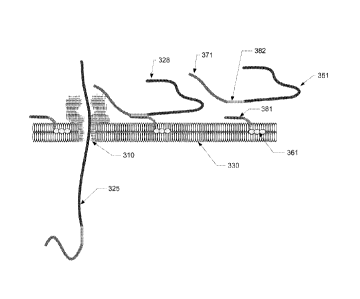
Figure 3 illustrates linking the leader and target portion of the
target molecule to the hydrophobic capture element on a surface, as well as
translocation of the leader and target portion of the target molecule (but not
the
hydrophobic capture element) through a nanopore.
Figure 4A illustrates relative event capture in a nanopore due to
end modifications of the targeted molecule. Figure 4B shows a target molecule
that has 4 duplexed regions used to pause and measure the molecule in a
nanopore. The end-modifications (3'x) are shown below the target molecule in
Figure 4B.
Figure 5 illustrates the structure of a control and a target molecule
used to assess the concentration enhancement caused by different end
modifications. Structures of five different end modifications (Y3') are shown
below the target molecule structure, which have hydrophobic groups of
different
sizes and a fixed leader length.
Figure 6 illustrates the structures of four additional end
modifications that have a fixed hydrophobic group size and leaders of
different
sizes.
Figure 7 illustrates the structures of four hydrophobic capture
elements which are designed to hybridize to the target molecule (rather than
part of the covalent structure).
6
CA 02890515 2015-05-07
WO 2014/074922 PCT/US2013/069304
Figure 8 illustrates structures of three end-adapted ds-DNA
targets used to compare different leaders.
Figure 9 illustrates concentrating a target molecule utilizing a
solid-state nanopore with supported lipid bilayers.
Figure 10A illustrates the distribution of adapted molecules that
are associated with the lipid bilayer and can freely diffuse along the plane
of the
bilayer. Figure 10B shows how a shear force, such as flow, can drive such
adapted targets to concentrate and localize at a nanopore near the edge of the
bilayer plane.
DETAILED DESCRIPTION
In brief, the invention improves the probability of interaction
between a target molecule and a nanopore by capturing the target molecule on
a surface comprising the nanopore. The captured target molecule, the
nanopore, or both, are able to move relative to each other along the surface.
In
this way, the volume occupied by the target molecule and the nanopore is
dramatically reduced compared, for example, to a target molecule in a volume
of solution that is in contact with the surface. By confining the target
molecule
and nanopore in this manner ¨ also referred to herein as "concentrating" the
target molecule - the probability of interaction between the target molecule
and
the nanopore is significantly increased. Such increased concentration leads to
significantly enhanced translocation of the target molecule, or target portion
thereof, through the nanopore.
Nanopores may be broadly classified into two types, biological
and synthetic, and both types are intended to be within the scope of this
invention. While alpha hemolysin (aHL) is perhaps the most studied biological
nanopore to date, this and other over biological nanopores may be utilized in
the context of this invention, such as mycobacterium smegmatis porin A
(MspA). More recently, synthetic nanopores have been introduced using
7
CA 02890515 2015-05-07
WO 2014/074922 PCT/US2013/069304
polymers, glass and thin solid-state membranes. Again, all such design options
are within the scope of this invention.
Nanopores are, in effect, small holes through a surface. In the
case of biological nanopores, the surface is typically a membrane such as a
lipid bilayer. However, other surfaces may also be employed, including lipid
nnonolayers or oil/water interfaces, as well as synthetic and/or inorganic
membranes. In the practice of this invention, the surface comprises the
nanopore, and also comprises a hydrophobic domain. In the case of a lipid
bilayer in aqueous media, for example, the hydrophobic domain is located in
the interior portion (i.e., where the hydrophobic tails of the phospholipids
lie). In
addition to lipid bilayers, other hydrophobic/hydrophilic interfaces can be
used
for the surface, including (for example) an oil/water interface, a tethered
lipid/water interface, an air/water interface, or a lipid-hydrophobic
substrate/water interface. In general, these surfaces exhibit differential
hydrophobicity and enable capture of the hydrophobic capture element of the
target molecule. In addition, such surfaces do not spatially fix the captured
target molecule at a given location on the surface, but instead allow the
target
molecule to diffuse along the surface.
As mentioned above, the target portion may comprise, for
example, nucleic acids or a linear polymer. In another embodiment, the target
portion may comprise a molecular bar code such as taught in Akeson et al.
(U.S. Patent No. 6,465,193), and/or an Xpandomer such as taught in Kokoris et
al. (supra).
The hydrophobic capture element of the target molecule is
associated with the hydrophobic domain of the surface. As used herein,
associated means that the hydrophobic capture element of the target molecule
and the hydrophobic domain of the surface cause the target molecule to remain
joined to the surface, while also permitting the captured target molecule to
move along the hydrophobic domain of the surface to bring the target molecule
in proximity with the nanopore. Such hydrophobic-hydrophobic interaction is
8
CA 02890515 2015-05-07
WO 2014/074922 PCT/US2013/069304
mostly an entropic effect associated with disruption of highly dynamic
hydrogen
bonds between water molecules and nonpolar substances. The strength of
hydrophobic interactions depends on temperature, as well as the shape and
number of carbon atoms on the hydrophobic compound.
As mentioned above, the target molecule comprises a target
portion, a hydrophobic capture element, and a leader. In one embodiment, the
surface is contacted with the target molecule such that the capture element of
the target molecule is associated with the hydrophobic domain of the surface,
thereby capturing the target molecule. In an alternative embodiment, the
surface having the hydrophobic capture element associated therewith is
contacted with the target portion and leader, thereby capturing the target
molecule on the surface.
Once captured by the surface, the leader portion of the target
molecule is capable of interacting with the nanopore in a manner that promotes
interaction of the target molecule (or target portion thereof) with the
nanopore.
Such interaction includes, for example, complete or partial translocation
through
the nanopore. Other interactions may involve positioning a target protein at
the
nanopore for measurement, or to position a functional protein, such as an
enzyme, proximal to the nanopore. Typically, the leader is not hydrophobic,
and in one embodiment is a hydrophilic (charged) polymer of low mass to allow
interaction with the nanopore when the nanopore and the leader of the target
molecule are in close proximity. As mentioned above, the captured target
molecule, the nanopore, or both, are capable of movement relative to each
other along the surface.
Concentrating the target molecule in this manner increases the
number of interactions of the target molecule (or target portion thereof) with
the
nanopore. As an illustrative example, one application of this invention
relates to
increasing the number of complete or partial translocations of the target
portion,
such as DNA/RNA, through a nanopore, wherein the DNA/RNA target portion is
combined with a hydrophobic capture element and an oligomer leader. In this
9
representative example, the hydrophobic capture element is captured in the
hydrophobic domain of the lipid bilayer that supports the nanopore. However,
the
target molecule still maintains lateral mobility across the lipid bilayer
surface. This
increases the probability that the oligomer leader will be drawn into the
nanopore
and increases the frequency of DNA/RNA translocation through the nanopore.
While nanopores have traditionally been developed for nucleic acid
analysis, the target portion of the target molecule may be any of a variety of
polymeric materials suitable to measurement and/or detection by the nanopore.
In one example, the target portion is an Xpandomer as disclosed in
W02008/157696 (U.S. Patent No. 7,939,259), as well as related embodiments
as disclosed in W02009/055617, W02010/088557 and W02012/003330. For
example, Xpandomers synthesized from ligation-based extension of hexamer
Xprobes have been end-adapted with C-48-polyA25 leaders and have
demonstrated translocation rates of 3 events per minute with addition of 10
fmol
of material. In this embodiment, the C-48 portion is a concatenate of 4
dodecyl
phosphodiester monomers and acts as the hydrophobic capture element, while
the polyA25 portion is a 25 base deoxyadenosine homopolymer that functions as
the leader element. Under identical conditions, the same Xpandomers adapted
to polyA25 leaders required additions of 1 pmol for the same event rate. In
both
cases the nanopore was wild-type alpha-hemolysin embedded in a 13 micron
diameter lipid bilayer.
In one embodiment, as illustrated in Figure 1, target molecule 120
comprises target portion 150, hydrophobic capture element 160 and leader 170
which, in this figure, is shown having substantially translocated through
nanopore 110 in surface 130. The direction of translocation through the
nanopore is shown by arrow 1 15. In addition to target molecule 120, Figure 1
also depicts target molecules 121 , 122 and 123 having hydrophobic capture
elements 161, 162, 163, respectively, captured by the hydrophobic domain of
Date recu/Date Received 2020-06-25
CA 02890515 2015-05-07
WO 2014/074922 PCT/US2013/069304
surface 130, which in this figure is depicted as the interior (hydrophobic)
portion
of a lipid bilayer. Captured target molecules 121, 122 and 123 further
comprise
target portions 151, 152 and 153 and leaders 171, 172 and 173, respectively.
The dots (" = = =59) shown at the ends of target portions 151, 152 and 153
represent additional length of the target portion. Captured target molecules
121, 122 and 123 are capable of movement along surface 130 (as depicted by
arrow 116), and such movement brings the leader of a captured target molecule
in proximity with the nanopore, as depicted by leader 171 of target molecule
121 being near nanopore 110. Such proximity allows the leader to interact with
the nanopore, thus drawing the target molecule into the nanopore for
translocation as depicted by target molecule 120.
In a more specific embodiment of Figure 1, the hydrophobic
capture element is a C48 aliphatic hydrophobic group and the leader is polyA24
oligonner that acts as a hydrophilic polyanionic leader. The sample reservoir
has 1 M potassium chloride in an aqueous 10 mM HEPES pH 7.4 buffer. As
the target molecule diffuses through the reservoir, it eventually interacts
with
the lipid bilayer and the hydrophobic capture element embeds into the
hydrophobic portion of the lipid bilayer core. The target molecule is now
captured by the surface and the hydrophilic leader is localized in the
reservoir
close to the surface of the lipid bilayer. Multiple target molecules
concentrate
on the lipid bilayer in this manner and diffuse along the surface until the
leader
of the target molecule is proximal to the nanopore. An electric field acting
across the pore applies a force on the negatively charged leader, drawing it
through the pore and pulling the hydrophobic capture element free of the lipid
so translocation of the remainder of the target molecule can proceed. In this
manner, the rate of capture and translocation is increased by orders of
magnitude relative to the corresponding target portion in solution interacting
with the nanopore.
In another embodiment, as illustrated in Figure 2, the target
molecule comprises more than one hydrophobic capture element. In particular,
11
CA 02890515 2015-05-07
WO 2014/074922 PCT/US2013/069304
Figure 2 illustrates a target molecule having four hydrophobic capture
elements
261, 262, 263 and 264. Hydrophobic capture elements 261, 262 and 263 are
shown as captured by the hydrophobic domain of surface 230, which in Figure
2 is depicted as a lipid bilayer. Target molecule 220 is shown in Figure 2 as
having partially translocated through nanopore 210 in the direction of arrow
215. Hydrophobic capture element 264 is depicted as having already been
dislodged from the hydrophobic domain of surface 230, and is in the process of
translocating through the nanopore.
In a more specific embodiment of Figure 2, a control molecule
was prepared having six ligated heterogeneous polymer units, with each
polymer unit having four PEG-6 (hexaethyleneglycol phosphodiester) with an
amino-modified base. One end of the polymer was adapted with a poly-A50
oligomer (forming the leader) (270). The structure on the other end of the
polymer is a hairpin loop (290) that is used to prevent backward entry into
the
pore. The hairpin loop is too large to enter the pore first, but when the
hairpin
loop is pulled through at the end, the duplex portion will open and unfold the
loop, allowing it to translocate. This control molecule was compared to a
target
molecule which is identical except that each pendant amino group (of the
amino-modified base) was conjugated with a DiBenzoCycloOctyl (DBCO)
hydrophobic moiety (forming the hydrophobic capture element). For the
resulting target molecule, the DBCO moieties interact with the hydrophobic
interior of the lipid bilayer, thus capturing the target molecule on the
surface,
and thereby increasing the concentration of the leader (the poly-A50 segment)
near the nanopore. Having the leader in close proximity with the nanopore
increases the probability that the target molecule will be translocated
through
the nanopore.
Translocation frequency through the nanopore (alpha hemolysin)
of the target molecule compared to the control molecule showed increases of
30, 15, 9, 10 and 8 times for applied potentials of 100, 110, 120, 130 and 140
mV, respectively. For these measurements, the cis and trans reservoirs had
12
2.0 M LiCI, 10 mM HEPES, pH of 7.4 at a temperature of 10 C and 15 pmol of
control or target molecule was added to the 100 pl cis reservoir. The nanopore
was a wild-type a-hemolysin (Sigma Aldrich) and the lipid bilayer was formed
on
a 13 micron diameter teflon aperture with 1,2-diphytanoyl-sn-glycero-3-
phosphoethanolamine (Avanti Polar Lipids) lipid bilayer. (Such methods follow
those described by Jetha et al., Chapter 9. Micro and Nano Technologies in
Bioanalysis, Humana Press 2009.)
In Figures 1 and 2 discussed above, capturing the target molecule
on the surface comprises contacting the surface with the target molecule,
wherein
the target molecule comprises the target portion, the hydrophobic capture
element and the leader prior to the capturing step. In another embodiment,
capturing the target molecule on the surface comprises contacting the surface
with the hydrophobic capture element and linking the hydrophobic capture
element to the target portion and leader to yield the captured target molecule
on
the surface.
Accordingly, and in another embodiment as illustrated in Figure 3,
hydrophobic capture element 361 is captured by the hydrophobic domain of
surface 330, which in this figure is depicted as a lipid bilayer. The
hydrophobic
element 361 may be captured during the formation of surface 330 or may be
captured after surface 330 is formed. Hydrophobic capture element 361 further
comprises linking element 381 which permits linkage of hydrophobic capture
element 361 to leader 371 and target portion 351 by attachment to
corresponding
linking element 382. Upon linkage of the capture element to the leader and
target
portion, target molecule 328 is both formed and captured on the surface. Once
captured, the target molecule is capable of diffusing along the surface until
the
leader of the target molecule is proximal to nanopore 310, as depicted by
target
molecule 328 in Figure 3. Such proximity allows the leader to interact with
the
nanopore, thus drawing the leader and target portion of the target molecule
(shown as leader/target portion 325) into the nanopore for translocation there
through.
13
Date recu/Date Received 2020-06-25
CA 02890515 2015-05-07
WO 2014/074922 PCT/US2013/069304
In a more specific embodiment of Figure 3, capture element 361
comprises an aliphatic group and an oligodeoxynucleotide (ODN) linker. The
aliphatic group (the hydrophobic capture element) remains embedded within
the hydrophobic domain of surface 330, which is shown as a lipid bilayer, and
the ODN linker extends out of the lipid bilayer and into the aqueous. In this
embodiment the leader and target portion are adapted with a nucleic acid
segment 382 complementary to ODN linker 381. This linker pair is used to join
(by hybridization) the leader and target portion to the hydrophobic capture
element, thereby forming the target molecule at the surface, as depicted by
target molecule 328. The aliphatic group (hydrophobic capture element)
diffuses freely throughout the plane of the lipid bilayer. This localization
to the
plane of the lipid bilayer increases the probability of interaction between
the
captured target molecule and the nanopore, resulting in the leader being
electrophoretically drawn into the nanopore. During translocation, either the
linker releases (e.g., the hybridized linkage unzips), or the linker remains
attached and the hydrophobic capture element is pulled free of the lipid
bilayer
and is stripped off at the nanopore.
A representative example of such a hydrophobic capture element
is disclosed by Chan et al. (Proceedings of the National Academy of Sciences
106(4): 979-984, 2009), which discloses the synthesis of a hydrophobic
capture element inserted into a lipid bilayer and linked to a vesicle. In this
case,
the hydrophobic portion of the capture element was one of the lipid molecules
that forms the lipid bilayer, and this lipid molecule was conjugated to an ODN
linker. The ODN linker, in turn, was used to hybridize to a complement ODN
that was conjugated to a vesicle, demonstrating capture of the vesicle. In
another example, Grenali et al. (Langmuir 22(1):292-299, 2006) showed that
bilayers where 0.5% of the lipids were head-adapted with biotin followed by
neutravidin would capture biotinylated oligonucleotides. These captured
oligonucleotides would freely diffuse along the bilayer surface with a
diffusion
constant 26% of that for the lipids themselves.
14
CA 02890515 2015-05-07
WO 2014/074922 PCT/US2013/069304
The hydrophobic capture element may be controlled in size to
facilitate diffusive capture of the target molecule with limited diffusive
release
from the surface, such as a lipid bilayer. However, it should also release
with
sufficient ease and be sized such that translocation is not interrupted. In
one
embodiment, a single length of an aliphatic element that is in-line with the
backbone of the target molecule may be utilized. If the length of the
aliphatic
element is too short, the hydrophilic portions of the target molecule (such as
the
leader) will limit its interaction with the lipid bilayer's hydrophobic core.
Thus,
the hydrophobic capture element should be large enough to resist the entropic
force that the target molecule will exert. However, if the hydrophobic capture
element is too long, translocation may be limited due to reduced target
molecule mobility in the lipid bilayer; namely, the electrophoretic force
required
to promote translocation could exceed optimum run conditions and reduce
measurement quality. In addition, excessively long hydrophobic segments may
cause target handling issues (particularly in an aqueous environment) and have
a disruptive effect on lipid bilayer stability. To increase the capture
strength of
the hydrophobic capture element while maintaining shorter lengths, the target
molecule may contain additional (i.e., more than one) hydrophobic capture
elements. Also, embodiments other than linear in-line geometries may be
utilized, such as hydrophobic capture elements pendent or branched off the
target molecule backbone.
In a further embodiment, the hydrophobic capture element may be
modified in order to selectively pause translocation through the nanopore, as
illustrated by the data presented in the bar graph of Figure 4A. In this
experiment, translocation frequencies were measured for the linear polymer of
Figure 4B with 4 nucleic acid duplexes having total contour length of ¨45 nm
(i.e., the target portion). Using the translocation control method described
by
Akeson et al. (supra) and Gundlach et al. (supra), the duplexes are used to
pause the polymer translocation for a period of time sufficient to measure a
distinct current blockage level. The blockage level is determined principally
by
CA 02890515 2015-05-07
WO 2014/074922 PCT/US2013/069304
the duplex at the nanopore entrance and the portion of polymer that threads
the
nanopore barrel. After a stochastic pause, the duplexes are stripped off the
polymer backbone and the polymer translocation proceeds until it is paused by
the next duplex portion. This polymer uses the same 14 base-pair duplex but
alternates with threaded portions DDDDAAA or DDDDD, where "D" represents
a hexaethyleneglycol phosphodiester linked monomer and "A" is an adenosine
deoxynucleotide. Translocation of the molecule can be determined from a
characteristic signature of 4 levels alternating between current blockage of
0.31
and 0.18 (relative to open pore current). Measurement was made at 20 C, 120
mV, and 1M KCl/10 mM HEPES/pH7.4 buffer. The nanopore was a wild-type
a-hemolysin (Sigma Aldrich) and the lipid bilayer was formed on a 13 micron
diameter teflon aperture with 1,2-
diphytanoyl-sn-glycero-3-
phosphoethanolamine (Avanti Polar Lipids) lipid bilayer. The methods follow
those described by Jetha et al. (Chapter 9. Micro and Nano Technologies in
Bioanalysis, Humana Press 2009).
The 3' end of the target portion was linked to one of three groups:
(1) polyA50; (2) C48-polyA25 or (3) C60-polyA25. C48 and C60 are carbon
chains of 48 and 60 carbons, respectively, synthesized from dodecyl
phosphodiester linked monomers. For example, 5 of the 12-carbon monomers
may be linked to form a C60 (the phosphate linkage between such C12
monomers is anionic and will moderate the hydrophobicity of the C12
concatenate to some degree). For polymer (1), polyA50 served as the leader to
the target portion (without hydrophobic capture element). For polymers (2) and
(3), the C48 and C60 segments, respectively, served as the hydrophobic
capture elements, while polyA25 served as the leader.
Control polymer (1) (La, target portion joined to leader without
hydrophobic capture element) and target molecules (2) and (3) were measured
for translocation frequency through a nanopore. As shown in the bar chart of
Figure 4A, target molecule (2) (C48-polyA25) and (3) (C60-polyA25) had
significantly enhanced frequency of translocation events compared to
16
CA 02890515 2015-05-07
WO 2014/074922 PCT/US2013/069304
comparative polymer (1) (polyA50). In particular, in relation to the
comparative
polymer (1), target polymer (2) increased the number of translocation
events/min/pmol by 920 times under the same experimental conditions.
It should be noted that the data presented in Figure 4A were
captured on independent runs and the effective measurement time (due to
nanopore blockages) varied between runs. Samples were introduced to the
100 microliter Cis reservoir of the nanopore in a 2 microliter aliquot loaded
with
15 femtomoles of sample. To maximize translocation rates from the small
sample size, the sample was injected directly adjacent to the nanopore,
maximizing the sample interaction with the nanopore, but results often varied
by
factors of 5 or more. Despite these variations, the concentrator method
consistently gave higher translocation rates when compared to non-
hydrophobic capture sample
To reduce sample injection variations, a control molecule was
mixed with each target molecule tested. Nanopore translocations of the target
and control could be distinguished by their unique sequence of current
blockage signals using the duplex translocation control method described
above. The results that follow utilize this approach and were derived from
measurements made at 20 C and 130 mV. The Trans well solution used for
these measurements was 2M NH4CI buffered with 10 mM HEPES/pH7.4; the
Cis well solution was 0.4M NH4CI / 0.6M Guanidine HCI buffered with 10 mM
HEPES/pH7.4. The nanopore was a wild-type a-hemolysin (Sigma Aldrich) and
the lipid bilayer was formed on a 13 micron diameter teflon aperture generally
using 1,2-diphytanoyl-sn-glycero-3-phosphoethanolamine (Avanti Polar Lipids)
lipid bilayer. In all cases duplexes are added to the target or control in
excess
of the number of binding sites by a factor of 100X and are thermally cycled.
Test molecules were synthesized on a Mermaide 12
oligonucleotide synthesizer (BioAutomation, Texas) using a variety of
phosphoamidites listed at the bottom of Figure 5. In some cases, longer
molecules were formed from two parts that are enzymatically ligated to make
17
CA 02890515 2015-05-07
WO 2014/074922 PCT/US2013/069304
the full construct. Figure 5 shows the composition of the target and control
molecules. Each had six duplexed regions that provided a different blockage
level sequence when measured in the nanopore.
Referring to Figure 5, two types of 6-base duplexes are shown
adjacent to their complementary sites along the target and control molecules
(3'AGKCKG5 and 3'ATKGKT5'); each use a modified base-type called a G-Clamp
(Glen Research, Sterling, VA, represented as "K") to provide stronger
duplexing. This
experiment compared translocation rates of the target
molecule with five different end-adaptations; namely, dA6C36dA24, dA5C48cIA24,
dA4C60dA24, C108dA24 and dA9dA24. The first
four targets had aliphatic
segments of different lengths (hydrophobic capture elements) and the latter
end-adaptation had no aliphatic segment. In each case the distal dA24 was the
leader. The control molecule was end-adapted with dA5C48dA24, and was
mixed at equal concentration with one of the target species. For each
measurement, a 2 microliter aliquot containing 150 femtomoles of each was
injected adjacent to the nanopore and measured. Translocation events were
captured and discriminated to calculate translocation rates for both. The
target
translocation rate was then normalized with the translocation rate of the
control
molecule.
Table 1 shows the normalized sample rates of these target
molecules after further normalization to the dA9dA24 rate. These are the
concentration enhancement factors that indicate the relative increase in
sample
translocation rates by incorporation of the hydrophobic capture element
compared to those without. It is noted that the concentration enhancement
factors are less in Table 1 than those shown in Figure 4A. In this regard, it
is
believed that the Table 1 data were better controlled for concentration which
likely accounts for the discrepancy. However, the data presented in both
Figure
4A and Table 1 illustrate significant enhancement in translocation rates.
18
CA 02890515 2015-05-07
WO 2014/074922 PCT/US2013/069304
Table 1
dA9dA24 dA6C36dA24 dA5C48dA24 dA4C60dA24 C108dA24
Sample Rate 1 30.9 28.8 26.5 12.5
(Normalized)
The leader length that extends beyond the hydrophobic capture
element may also be modified for interaction with the nanopore. To this end,
the leader should be of a sufficient length such that its capture in the
nanopore
exerts enough force to uncouple the target molecule from the bilayer or,
depending on the embodiment, unlink the leader/target portion from the
hydrophobic capture element. The leader should carry electrostatic charge to
promote interaction with the nanopore under an applied electric potential. A
nucleic acid is typically anionic and the leader would typically also be
anionic.
In some cases an end portion of the target portion may also function as the
leader. The leader is typically a single linear polymer, but may have two or
more linear polymer portions to help improve nanopore interaction, and should
also be able to translocate the nanopore so the target molecule can then
engage. Leader materials can be synthesized from many anionic, cationic or
neutral polymers and may be made of combinations of materials such as (but
not limited to) heterogenous or homogeneous polynucleotides, polyethylene
glycol, polyvyinyl alcohol,
polyphosphates, poly(vinylphosphonate),
poly(styrenesulfonate), poly(vinylsulfonate),
polyacrylate, abasic
deoxyribonucleic acid, abasic ribonucleic acid, polyaspartate, polyglutamate,
polyphosphates, and the like. For example, a representative leader may
comprise PEG-24 and/or poly-Al2.
The effect of leader length upon translocation rates was
compared by modifying a target with different length leaders that extend
beyond
the hydrophobic capture element (C48). The same control and target
molecules shown in Figure 5 were used with the end modifications (Y3') shown
in Figure 6. Normalized translocation rate results are shown in Table 2 and
are
19
CA 02890515 2015-05-07
WO 2014/074922 PCT/US2013/069304
identified as dA18C48dA1 1, dAi iC48dAis, dA5C48dA24 and dA5C48dA241_25. All
nanopore measurements supporting the results in Tables 1 and 2 used the
same experimental conditions and used the same control molecule so they can
be directly compared. For this reason, results in both tables are normalized
to
the dA9dA24 result of Table 1, thus referencing the enhancement in
translocation rate (also referred to as concentration enhancement herein) to a
similar molecule with no hydrophobic capture element.
Table 2
dA9dA24 dA13C48dA11 dA11C48dA18 dA5C48dA24 dA5C48dA24
Sample Rate
1.0 2.8 10.7 28.8 49.0
(Normalized)
These results indicate that the concentration enhancement factor
increases as the polyA leader increases from 11 to 24 bases. In another
measurement, using a different target molecule, the influence of end groups
dA5C48dA24 and dA5C48dA50 were compared. This showed the latter (longer)
leader to be 82% of the former indicating the enhancement effect of polyA
leaders plateaus in the range of 20 to 50 bases.
The last column of Table 2, shows the enhancement result due to
a dA24 leader that is extended with a 25 ethyl phosphodiesters (dA5C48dA241-
25).
Its concentration enhancement factor was 70% larger than dA5C48dA24 alone.
Additional leader measurements are presented in Table 4.
The hydrophobic capture element is designed to promote mobility
in the lipid bilayer and maintain the hydrophobically captured state, but
limited
enough so that the target can be released when interacting with the nanopore.
The element can extend the target backbone and be in-line with the leader or
may be pendant to the backbone or may have multiple elements pendant to the
backbone. The hydrophobic capture element can be positioned anywhere
along the target relative to the leader but can be optimized to improve
capture
CA 02890515 2015-05-07
WO 2014/074922 PCT/US2013/069304
by the nanopore. Materials that comprise the hydrophobic capture element
include, but are not limited to, linear and branched aliphatic chains, lipids,
fatty
acids, DBCO, cholesterol, fluorinated polymers, apolar polymers, steroids,
polyaromatic hydrocarbons, hydrophobic peptides, and hydrophobic proteins.
This may also include phase transition polymers that can switch from
hydrophilic to hydrophobic states under thermal or other environmental change.
In some embodiments some or all of the heads of the lipids in a bilayer are
reactive and can bind to an adapted target molecule as shown by Grenali et al.
(supra). In this case, the lipid is the hydrophobic capture element.
The method variation shown in Figure 3 was demonstrated using
the targets end-adapted with either dA9dA24 or dA5C48dA24 and the control
shown in Figure 5. In this example, a hydrophobic capture element was used
that has a C120 on one end (synthesized by linking ten, C12 monomers), and
an oligomer at the other end. This oligomer was complementary to a nucleic
acid region at the end of the target adjacent to the end modification. Figure
7
shows several different versions of this hydrophobic capture element. For each
measurement, a 2 microliter aliquot of 300 fenntonnoles of a capture element
was injected into the cis reservoir adjacent to the nanopore. The hydrophobic
C120 group on the capture element is inserted into the lipid bilayer, with its
other oligomer end remaining outside the bilayer in the aqueous buffer. The
cis
reservoir was then exchanged with fresh buffer and a 2 microliter aliquot of
sample was added containing 15 femtomoles of target and 15 fenntomoles of
control. The target molecule can hybridize to the capture element and diffuse
along the plane of the bilayer. In contrast the control shown in Figure 5 has
no
complementary region and will not hybridize to the capture elements described
in Figure 7. Nanopore translocation begins when the leader is
electrophoretically pulled through and stops at the capture element duplex.
The
duplex releases due to thermal and electrophoretic pulling forces, allowing
translocation to proceed.
21
CA 02890515 2015-05-07
WO 2014/074922 PCT/US2013/069304
Table 3 shows concentration enhancement factors for these
molecules (normalized to the target with no hydrophobic capture element;
dA9dA24). All measurements were made under the same conditions and
concentrations described above. Note that molecules adapted with dA5C48dA24
all have a second hydrophobic capture element. Comparing the two CE1
results indicates that having this second hydrophobic capture element
increases the concentration enhancement factor. Reducing the duplex length
from 16 bases (CE1) to 11 bases (CE2, CE3 and CE4), reduces the stability
and enhancement is decreased. The CE2 and CE3 capture elements had
similar structure except the C120 hydrophobic group was positioned on
opposite ends of the duplex. CE4 had 5 PEG-6 spacers between the
hydrophobic group and the hybridization site and improved the concentration
enhancement relative to both CE2 and CE3, which is believe to be due to
relaxing how tightly the duplex was held to the lipid bilayer.
Table 3
dA9dA24 dA5 C48d A24
Capture Element
None CE1 CE1 CE2 CE3 CE4
Sample Rate 1 417 890 15 100 298
(Normalized)
Additional target molecules were tested that were short ds-DNA
strands shown in Figure 8. Unlike the measurements for results in Tables 1, 2
and 3, these targets had only a single duplex and had no control added to them
for normalization. Otherwise the measurement conditions were the same.
Each was measured with a 30 femtomole sample and all were normalized to
C48A24. This indicates that dL24 enhances capture by the nanopore more than
dA24 by a factor of 2.8, and that adding longer extensions of L100 provides
even
greater enhancement.
22
CA 02890515 2015-05-07
WO 2014/074922 PCT/US2013/069304
Table 4
C48d A24 C48 L24 C48d A24L100
Sample Rate 1.0 2.8 3.7
(Normalized)
In addition, the surface can be modified to optimize performance
of the hydrophobic capture element. For example, when the surface is a lipid
bilayer, increasing mobility of the captured target molecule increases the
probability of leader interaction with the nanopore. For example, increasing
the
area of the lipid bilayer increases the probability that the target molecules
will
be captured and migrate to the nanopore. Target molecule capture in the
bilayer may also be improved by minimizing any undesired trapping on
undesired surfaces in the reservoir, such as isolated lipid or non-lipid
reservoir
walls. The use of tethered bilayers is a powerful design tool that could be
used
to control the relative mobility and capture kinetics of the bilayer surfaces.
Utilizing the characteristics of fixed lipids and lipid additives to define
these
characteristics, the target molecules can be captured and limited to diffuse
in
preferred directions along the bilayer surface. For example, by constraining
the
lipid layer to be a long thin rectangle confines any hydrophobically captured
molecules to diffuse principally along its length.
Figure 9 depicts a supported lipid bilayer (901) used in
conjunction with a solid-state nanopore (904). Figure 9 is similar to the
embodiment depicted in Figure 3, and depicted in this manner (as opposed to
the embodiment of Figures 1 or 2) for purpose of illustration only. Referring
to
Figure 9, supported lipid layers are synthesized using a tether species (906)
that covalently bond to substrate (908) at one end and imbeds into a bilayer
on
the other end (see J. Jackman et al., "Biotechnology Applications of Tethered
Lipid Bilayer Membranes," Materials 5(12):2637-2657, 2012). A common
inorganic film used for solid-state nanopores is silicon nitride which can
oxidize
to form silicon oxide on its surface. Atanasov et al. has shown supported
lipid
23
CA 02890515 2015-05-07
WO 2014/074922 PCT/US2013/069304
bilayer formation tether-stabilized with lipids adapted with silanes to bond
to a
silicon oxide surface ("Membrane on a Chip: A Functional Tethered Lipid
Bilayer Membrane on Silicon Oxide Surfaces," Biophys J., 89(3):1780-1788,
2005). These bilayers maintain the required diffusion characteristics that
enable the hydrophobically captured molecule to migrate near the nanopore.
This bilayer does not need to maintain high electrical impedance, but does
require that the bilayer integrity be sufficient near the nanopore such that
the
target molecule leader can be captured.
Additional forces can be applied to the hydrophobically associated
target molecules that will steer them in a preferred direction along the lipid
bilayer or other hydrophobic/hydrophilic interface. Graneli et al. ("Organized
Arrays of Individual DNA Molecules Tethered to Supported Lipid Bilayers,"
Langmuir 22(1):292-299, 2006) demonstrated that DNA linked to the head
group of a lipid that was in a supported lipid bilayer could be moved
laterally by
the flow of the buffer across the bilayer. Furthermore the DNA-tethered lipid
would stop at a defined diffusion barrier, fixing that end of the DNA while
the
flow remained. After flow was stopped, this lipid molecule and its tethered
DNA
would diffuse away from the barrier along the bilayer membrane.
Figures 10A and 10B show a supported lipid bilayer (1001) in a
shape defined by the diffusion barrier at its edges (1003). Arrow (1005) shows
the direction that buffer above the bilayer is flowing. This flowing buffer
applies
a shear force to the target molecules (1007) that drags them along until they
are interrupted by the diffusion barrier (Figure 10A represents the location
of
the target molecules before application of the shear force, while Figure 10B
represents location of the target molecules after application of the shear
force.)
By angling these barriers relative to the flow direction, the target molecules
(1007) are concentrated in an area in proximity to nanopore (1008) (see Figure
10B). This technique can be used to collect and concentrate target molecules
in low concentration from larger volumes near a nanopore (or each nanopore in
a nanopore array). In addition to flow, other forces can be employed to move
24
CA 02890515 2015-05-07
WO 2014/074922 PCT/US2013/069304
the target molecules along the bilayer surface,
including
electrophoretic/electroosmotic forces (C. Liu et al. "Protein Separation by
Electrophoretic¨Electroosmotic Focusing on Supported Lipid Bilayers," Anal.
Chem. 83(20):7876-7880, 2011.), and acoustic forces (J. Neumann et al.,
"Transport, Separation, and Accumulation of Proteins on Supported Lipid
Bilayers," Nano Lett. 10(8):2903-2908, 2010).
The method of this invention may be modeled with reservoir target
molecule concentration NR and rate constants for:
i) capture of the leader by the nanopore from the bilayer (kB-trans),
ii) capture of the leader by the nanopore from the reservoir
(kR-trans),
iii) capture of hydrophobic group in the bilayer (kBcapt),
iv) passive release of hydrophobic group from the bilayer (kBrel),
In this model, the reservoir may be considered infinite and NR
constant. The rate of translocations of molecules pulled directly from the
reservoir is:
NR-trans = kR-trans NR,
Along the hydrophobic capture path, the surface concentration of
molecules (associated with in the bilayer), NB, changes as:
NB = kBcapt NR (kBrel kB-trans/A) NB when kB-transNB/A < NBsaturation
Note that this simplified equation has factor of lipid area, A, that is
inserted to normalize the rates of molecule capture/release across a lipid
area
with the molecules translocating thru a single nanopore on the area. This
assumes that molecular depletion from the lipid (due to translocation) happens
uniformly across A.
At steady-state:
0 = kBcapt NR (kBrel kB-trans/A) NB
NB = kBcaptNR /(kBrel kB-trans/A)
CA 02890515 2015-05-07
WO 2014/074922 PCT/US2013/069304
Choosing area, A, sufficiently large where A >> kB-trans/ kBrei leads
to:
Ng = kBcapt NR I kBrel
A strong hydrophobic group leads to ki3c3pt/ kBrei >>1 which leads
to high surface concentration of target molecules tethered to the lipid
despite
relatively low concentration of molecules in the reservoir.
The translocation rates kB-trans and kR-trans are related but differ by
the following factors:
i) mobility of target molecule on the lipid surface vs mobility in
the reservoir.
ii) effective translocation capture cross-section of molecule end
as a function of distance from nanopore. Note that surface
tethered case has additional factors to this including position of
hydrophobic group and length of the leader.
The rate of translocation can have several regimes including:
i) Diffusion-limited: In this case the molecules must diffuse so
their capture end is within range of the nanopore.
ii) End-capture limited: In this case, many molecules are within
range (up to the maximum concentration) and translocation
rate is limited by the time it takes to capture the end of one of
these molecules.
iii) Translocation-limited: In cases where only 1 or some limited
number of molecules can enter the nanopore, other molecules
can be within range but must wait until the nanopore is
available for translocation of another molecule.
26
CA 02890515 2015-05-07
WO 2014/074922 PCT/US2013/069304
EXAMPLE 1
NANOPORE MEASUREMENT OF TARGET WITH 048
CAPTURE ELEMENT AND POLYA24 OLIGOMER LEADER
Target molecule synthesis is performed using a Mermaide 4
oligonucleotide synthesizer (BioAutomation, Texas) using commercially
available amidites (Glen Research, Sterling, VA; Chem Genes, Wilmington,
MA). The following target molecules are synthesized:
Target 1 - (dA)24(dCdGdGdGdCdAdAdTdAdA dGdCdCdC);
Target 2 - (dA)24 (Dodecyl phosphodiester)4
(dA)5 (dCdGdGdGdCdAdA dTdAdAdGdCdCdC);
Each target molecule was page purified on a 6% acrylamide TBE-
Urea gel (Life Technologies, Carlsbad, CA). Both target molecules contain a
poly dA leader portion and a stem-loop structure, which is used to control
translocation speed and direction. Target 2 includes the addition of four
dodecyl phosphodiester linked monomers, which create the 048 capture
element. Each purified target molecule is analyzed using the a-hemolysin
nanopore system described by Jetha et al. (Chapter 9. Micro and Nano
Technologies in Bioanalysis, Humana Press 2009). Targets are added to the
cis reservoir of the nanopore device that contains 100u1 2.0 M LiC1, 10 mM
HEPES, pH of 7.4. The trans reservoir contains the same solution. Event
frequencies are determined for each target across a range of target inputs (1
fmole to 1 pmole) and voltages (100-140mVolts) to determine the concentration
effect of the 048 capture element.
EXAMPLE 2
NANOPORE ARRAY CAPTURING RARE NUCLEIC ACID TARGETS WITH CONCENTRATOR
Detection and identification of nucleic acids at very low
concentration is generally not practical without molecular amplification. By
27
CA 02890515 2015-05-07
WO 2014/074922 PCT/US2013/069304
presenting a thin film of sample across a large nanopore sensor array, the
target molecules can diffuse to the sensor surface in reasonable time periods.
If the sensor surface is primarily a hydrophobic domain, target molecules
modified with at least one hydrophobic capture element associate and diffuse
along the surface. This greatly increases the likelihood of being sensed.
A microfluid flow cell is designed with a chamber through which
electrolyte with sample can pass through. The chamber is 100 microns in
height, 3 mm wide and 10 mm long with 1.0 mm diameter input and output
ports located at the ends on the top side. On the top side is glass or polymer
that is surface treated to inhibit binding to nucleic acids. The bottom side
is
sealed against a silicon chip that contains a 200 X 500 array of nanopore
cells.
The array lies on a grid with 15 micron centers. The outer dimension of the
array is 3 mm X 7.5 mm and is centered in the chamber. Each cell contains a
shallow 10 micron diameter by 3 micron deep well that has an Ag/AgCI
electrode at its base. The electrode passes current from contacting
electrolyte
to be measured by the nanopore cell's transconductance amplifier. The
current-converted voltage outputs from the array of nanopore amplifiers are
measured at bandwidths exceeding 1 ksample /s/cell.
Across the surface of the silicon chip exposed to the flow
chamber, a continuous lipid bilayer is formed in an electrolyte buffer. It is
suspended as a membrane over each cell well but is a supported lipid bilayer
over the remaining area. Hemolysin nanopores are inserted into the bilayer in
a
manner to maximize the number of wells with single nanopores. The lipid layer
that is connected to the substrate is formed so as to electrically isolate
adjacent
cell wells from current passing between the substrate and the bilayer. This
isolation is sufficient that any leakage currents can be ignored compared to
currents that pass through the single nanopore. A characteristic of the
continuous bilayer is that molecules adapted with a hydrophobic group as
described herein can associate with the bilayer from the flow chamber and will
diffuse anywhere along its surface.
28
CA 02890515 2015-05-07
WO 2014/074922 PCT/US2013/069304
A pathogen assay uses hybridization and ligation specificity to
identify DNA by using the DNA as a template to hybridize and ligate the target
shown in Figure 5 with dA5C48dA24L25 as shown in Figure 6 using methods as
taught depicted in U.S. Patent No. 8,586,301. A 3 microliter sample of this
ligation product is injected microfluidically to fill the chamber where the
ligation
products can diffuse until they contact and associate with the lipid bilayer
(due
to the C48 group). The ligation products then diffuse along the plane of the
lipid
bilayer until they are captured and measured in a nanopore. This method is
highly sensitive because for a 3 microliter sample, all volume diffusing
targets
are localized to within 100 microns of the active surface, surface-diffusing
targets are localized to within 10 microns of a nanopore and measurements
provide target specific information from a single molecule.
In alternative embodiments, the geometry described above can be
modified in a variety of ways, including (for example) the modifications noted
below.
(i) To inspect larger volumes of sample the chamber and lipid
bilayer capture surface can be extended upstream. With suitable diffusion
barriers in the lipid, flow induced concentration as described using Figure
10A
and 10B can be used to collect the targets downstream at the nanopore array.
(ii) Provided the target concentration is uniform in chamber
volume, it will collect uniformly at each well. In this case the lipid bilayer
need
only be continuous over each well. The electrical isolation of each well could
coincide with a break in the lipid layer. To maintain high collection
efficiency,
the area of the bilayers (that collect and along which target molecules can
diffuse) should be as large as possible.
(iii) By adapting the top surface of the flow chamber to have
another active silicon chip reduces the average diffusion distance that the
injected target must diffuse to reach a bilipid layer and reduces surface area
that can lead to sample loss.
29
The various embodiments described above can be combined to
provide further embodiments. Aspects of the embodiments can be modified, if
necessary to employ concepts of the various patents, applications and
publications to provide yet further embodiments.
These and other changes can be made to the embodiments in light
of the above-detailed description. In general, in the following claims, the
terms
used should not be construed to limit the claims to the specific embodiments
disclosed in the specification and the claims, but should be construed to
include
all possible embodiments along with the full scope of equivalents to which
such
claims are entitled. Accordingly, the claims are not limited by the
disclosure.
Date recu/Date Received 2020-06-25