Note: Descriptions are shown in the official language in which they were submitted.
CA 02890634 2016-06-28
APPARATUS AND METHOD FOR OPERATING A REAL TIME LARGE DIOPTER
RANGE SEQUENTIAL WAVEFRONT SENSOR
TECHNICAL FIELD OF THE INVENTION
[0001] One or more embodiments of the present invention relate generally
to
wavefront sensor(s) for use in vision correction procedures. In particular,
the invention relates
to the electronics and algorithms for driving, controlling and processing the
data of a real-time
sequential wavefront sensor and other subassemblies associated with the
wavefront sensor.
BACKGROUND OF THE INVENTION
[0002] Conventional wavefront sensors for human eye wavefront
characterization are
generally designed to take a snap shot or several snap shots of a patient's
eye wavefront with
room lighting turned down or off. These wavefront sensors generally use a CCD
or CMOS
image sensor to capture the wavefront data and need to use relatively
complicated data
processing algorithms to figure out the wavefront aberrations. Due to the fact
that a CCD or
CMOS image sensor generally has a limited number of gray scales and cannot be
operated at
a frame rate well above the 1/f noise range, these wavefront sensors therefore
cannot take full
advantage of lock-in detection scheme to provide higher signal to noise ratio.
They cannot
employ a simple algorithm to quickly derive the wavefront aberration. As a
result, when these
wavefront sensors are integrated with an ophthalmic device such as a surgical
microscope,
they generally cannot provide accurate/repeatable real time wavefront
aberration
measurement, especially with the microscope's illumination light turned on.
[0003] There is a need in the art for an apparatus and a method to not
only realize real
time wavefront measurement and display, but also address the various issues
including what
has been mentioned above.
SUMMARY OF THE INVENTION
[0004] One or more embodiments satisfy one or more of the above-
identified needs in
the art. In particular, there is provided a wavefront sensor having an
electronic control and
1
CA 02890634 2016-06-28
driving circuit together with associated algorithm and software for driving,
controlling and
processing the data collected by the sensor.
[0005] The driving circuit may include an opto-electronic position
sensing
detector/device (PSD) such as a quadrant photodiode/detector/cell/sensor or a
lateral effect
position sensing detector, a transimpedance amplifier, an Analog to Digital
(A/D) converter, a
digital amplifier with programmable gain control, a superluminescent diode
(SLD or SLED)
and its drive circuit, a wavefront scanning/shifting device and its driver
circuit, and a front-
end data processing unit (e.g. processor, microcontroller, PGA, programmable
device). In
addition, a camera may be used to provide live video images of the eye from
which the
wavefront is being measured. Furthermore, a back-end data processing unit is
employed to
convert the sequential wavefront data from the front-end processing unit to
display clinical
ophthalmic information overlaid on or side-by-side with a live image of the
patient's eye.
The circuits (frontend and/or backend) can be electronically connected to one
or more various
devices in one way or another for coordinated operation of each device,
including for
example, an eye transverse position measurement device, an eye distance
measurement
device, an accommodation enabling eye fixation target, a data storage device,
a laser based
surgical ablation device, and a display device.
[0006] In another embodiment a reference wavefront is created from either
outside or
inside of the wavefront sensor module so that a calibration/verification can
be done. A unique
feature is the creation of a reference wavefront inside the wavefront sensor
module that
simulates a wavefront from an aphakic eye. This calibration/verification can
be done before,
during and after a vision correction procedure and can be used to serve
several purposes. One
purpose is to check if the SLD beam to be directed into a patient eye for
creating the
wavefront from the eye is aligned well enough with respect to either the
wavefront sensor
optical system or to the eye pupil (such as with a desired off axis distance).
Another purpose
is to monitor the optical integrity and alignment of the optical elements
inside the wavefront
sensor module to make sure that the alignment is within allowed tolerance
range. Still another
purpose is to establish reference image spot positions on the position sensing
device/detector
(PSD) so that inherent optical system aberrations arising from slight
misalignment of the
optical elements (within certain allowable tolerance) as a result of, for
example,
2
CA 02890634 2016-06-28
environmental factors such as temperature changes, can be taken into
consideration in the
wavefront analysis/calculation. In doing so, the inherent optical system
aberration(s) can be
measured in advance and subtracted from the measured overall aberration(s).
Just as
importantly, still another purpose is to confirm or verify performance prior
to providing
guidance and/or feedback.
100071
Accordingly, there is described a wavefront sensor comprising: a light source
configured to output a light beam to illuminate a subject eye; a position
sensing detector
having a plurality of detector elements configured to output a plurality of
output signals
indicating signal strength of incident light on each detector element; a beam
deflecting
element configured to intercept a wavefront beam returned from a subject eye
when the
subject eye is illuminated by the light source and configured to direct a
portion of the
wavefront beam from the subject eye through an aperture toward the detector,
where the
portion of the wavefront beam directed through the aperture forms a spot on
the detector,
where an amplitude of deflection of a centroid of the spot from a reference
point on the
detector is approximately indicated by a ratiometric combination of the output
signal
strengths, and where the amplitude of the deflection indicates one of the
degree of tilt,
convergence and divergence of the portion of the wavefront from a plane wave;
a reference
wavefront generator disposed within the wavefront sensor, configured to
generate a
calibration wavefront equivalent to a wavefront beam returned from the subject
eye and
having a specified degree of convergence or divergence from a plane wave,
where the
deflection of the centroid of the calibration wavefront on the position
sensing detector is a real
centroid deflection for the specified convergence or divergence; and a
processing unit,
coupled to the light source, the beam deflecting element, and the position
sensing detector,
configured to control the beam deflecting element to deflect the wavefront
beam to direct
portions of an annular ring portion of the calibration wavefront on the
position sensing
detector referenced to a reference point and further configured to compare the
real centroid
deflection at the specified degree of convergence or divergence from a plane
wave to the
approximate deflection indicated by the ratiometric combination of output
signal strengths in
order to calculate correction terms to be applied to the ratiometric
combination when
calculating deflections of a wavefront beam returned from the subject eye.
3
CA 02890634 2016-06-28
[0008]
These and other features and advantages of the present invention will become
more readily apparent to those skilled in the art upon review of the following
detailed
description of the preferred embodiments taken in conjunction with the
accompanying
drawings.
3a
CA 02890634 2015-05-01
WO 2014/074598
PCT/US2013/068746
BRIEF DESCRIPTION OF THE DRAWINGS
[0010] Fig. 1 shows one example embodiment of the optical
configuration of a large
diopter range real time sequential wavefront sensor integrated with a surgical
microscope;
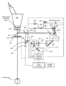
[0011] Fig. 2 shows one example embodiment of electronics interfacing with
the
optics of the wavefront sensor in Fig.1 with those potentially active devices
connected to the
electronic control circuit;
[0012] Fig. 3 shows what would happen to the wavefront sampling area
on the cornea
plane if the eye is transversely moved and there is no corresponding change
made to the
wavefront sampling scheme.
[0013] Fig. 4 shows how, by DC offsetting the wavefront beam scanner,
one can
compensate the transverse movement of the eye and hence continue to scan the
same properly
centered annular ring even though the eye is transversely moved.
[0014] Fig. 5 illustrates what happens to the wavefront or refractive
error being
measured if the eye is axially moved from the designed position.
[0015] Fig. 6 shows an overall block diagram of one example
embodiment of an
electronics system that controls and drives the sequential wavefront sensor
and the associated
devices shown in Figures 1 and 2;
[0016] Fig. 7 shows a block diagram of one example embodiment of the
front-end
electronic processing system and the live imaging camera that resides within
the sequential
wavefront sensor module and the back-end electronic processing system that
resides in the
host computer and display module shown in Fig.6;
[0017] Fig. 8 shows an example internal calibration target that can
be moved into the
wavefront relay beam path to create one or more reference wavefront(s) for
internal
calibration and/or verification.
[0018] Fig. 9A shows an embodiment of an electronics block diagram
that
accomplishes the task of automatic SLD index and digital gain control in order
to optimize
the signal to noise ratio.
[0019] Fig. 9B shows a quadrant detector with firstly a light image
spot landing at the
center and secondly landing slightly away from the center.
[0020] Fig. 9C shows a number of representative cases of planar
wavefront, defocus
and astigmatism, the associated image spot position on a quad-detector behind
a
4
CA 02890634 2015-05-01
WO 2014/074598
PCT/US2013/068746
subwavefront focusing lens, as well as the sequential movement of the
corresponding
centroid positions when displayed as a 2D data point pattern on a monitor.
[0021] Fig. 10 shows one example process flow block diagram in
optimizing the
signal to noise ratio by changing the gain of the variable gain amplifier and
the SLD output.
[0022] Fig. 11 shows one example embodiment of a composite transimpedance
amplifier with lock-in detection that can be used to amplify the signal from
any one of the
four quadrant photodiodes, as is used in the position sensing detector circuit
of Fig. 9;
[0023] Fig. 12 shows one example embodiment of the combination of a
conventional
transimpedance amplifier with a lock-in detection circuit;
[0024] Fig. 13A shows the case when the MEMS scan mirror is oriented so
that the
entire wavefront is shifted downward as the SLD pulse is fired. In this case
the aperture
samples a portion at the top of the circular wavefront section;
[0025] Fig. 13 B shows the case when the wavefront shifted leftward
as the SLD
pulse is fired so that the aperture samples a portion at the right of the of
the circular
wavefront section;
[0026] Fig. 13C shows that case when the wavefront is shifted upward
as the SLD
pulse is fired so that the aperture samples a portion at the bottom of the of
the circular
wavefront section;
[0027] Fig. 13D shows the case when the wavefront is shifted
rightward as the SLD
pulse is fired so that the aperture samples a portion at the left of the of
the circular wavefront
section;
[0028] Fig. 13E depicts the equivalence of the sequential scanning
sequence of four
pulses per cycle to sampling the wavefront section with four detectors
arranged in a ring.
[0029] Fig. 13F shows the positions of 8 SLD pulse firing relative to
the X and Y
axes of the MEMS scanner with 4 odd or even numbered pulses of the 8 pulses
aligned with
the X and Y axes of the MEMS scanner and the other 4 pulses arranged midway on
the ring
between the X and Y axes;
[0030] Fig. 14 shows an example in which the 4 SLD pulse firing
positions initially
aligned with the X and Y axes of the wavefront scanner as shown in Fig. 13F
are shifted 150
away from the X and Y axes by slightly delaying the SLD pulses;
[0031] Fig. 15 shows the collective effect of sampling a wavefront
with offset angle
at 00 on the first frame, 150 on the second frame, and 30 on the third;
5
CA 02890634 2015-05-01
WO 2014/074598
PCT/US2013/068746
[0032] Fig. 16 shows one example of a theoretically determined
relationship between
the PSD ratiometric estimate and the actual centroid displacement or position
along either the
X or the Y axis;
[0033] Fig. 17 shows an example flow diagram that illustrates how
calibration can be
performed to obtain a modified relationship and to result in more accurate
wavefront
aberration measurement;
[0034] Fig. 18 shows a graphical representation of a sequential
ellipse using
trigonometry expressions, where U(t) = a=cos(t) and V(t) = bosin(t), a>b>0,
resulting in an
ellipse that rotates counter-clockwise with the point (U(to), V(t0)) in the
first quadrant of the
U-V Cartesian coordinate;
[0035] Fig. 19 shows a corresponding graphical representation of a
similar sequential
ellipse using trigonometry expression, where U(t) = -a=cos(t), V(t) = -
bosin(t), a>b>0,
resulting in an ellipse that rotates counter-clockwise with the point (U(to),
V(t0)) in the third
quadrant of the U-V Cartesian coordinate;
[0036] Fig. 20 shows a corresponding graphical representation of a similar
sequential
ellipse using trigonometry expression, where U(t) = a=cos(t), V(t) = -
bosin(t), a>b>0,
resulting in an ellipse that rotates clockwise with the point (U(to), V(t0))
in the fourth
quadrant of the U-V Cartesian coordinate;
[0037] Fig. 21 shows a corresponding graphical representation of a
similar sequential
ellipse using trigonometry expression, where U(t) = -a=cos(t), V(t) =
bosin(t), a>b>0,
resulting in an ellipse that rotates clockwise with the point (U(to), V(t0))
in the second
quadrant of the UN Cartesian coordinate;
[0038] Fig. 22 shows an example of the sequential centroid data
points expected from
a divergent spherical wavefront and the resulting data point position and
polarity;
[0039] Fig. 23 shows another example of the sequential centroid data points
expected
from a convergent spherical wavefront and the resulting data point position
and polarity;
[0040] Fig. 24 shows the Cartesian coordinate translation and
rotation from the
original X-Y coordinate to the translated Xtr-Ytr coordinate and further
rotated to the U-V
coordinate of 8 sequentially sampled centroid data points that are fitted to a
sequential ellipse.
[0041] Fig. 25 shows the result of coordinate rotation transformation and 8
centroid
data points on the UN coordinate, with the left side corresponding to a
divergent spherical
wavefront having positive major and minor axes, and with the right side
corresponding to a
convergent spherical wavefront, having negative major and minor axes;
6
CA 02890634 2016-06-28
[0042] Fig. 26 shows the process flow diagram of one example embodiment
in
decoding the sphere and cylinder diopter values and the cylinder axis angle;
[0043] Fig. 27 shows an example process flow diagram of an eye tracking
algorithm;
[0044] Fig. 28 shows an example process flow diagram illustrating the
concept of using
the live eye image to determine the maximum wavefront sampling annular ring
diameter and to
obtain better diopter resolution for pseudo-phakic measurement;
[0045] Fig. 29 shows an example process flow diagram illustrating the
concept of using
either the live eye image and/or the wavefront sensor signal to detect the
presence of
unintended object in the wavefront relay beam path or the moving away of the
eye from a
desired position range so that the SLD can be turned off and the erroneous
"bright" or "dark"
wavefront data can be abandoned;
DETAILED DESCRIPTION OF THE INVENTION
[0046] Reference will now be made in detail to various embodiments of the
invention.
Examples of these embodiments are illustrated in the accompanying drawings.
While the
invention will be described in conjunction with these embodiments, it will be
understood that it
is not intended to limit the invention to any embodiment. On the contrary, it
is intended to
cover alternatives, modifications, and equivalents as may be included within
the scope of the
invention as defined by the appended claims. In the following description,
numerous specific
details are set forth in order to provide a thorough understanding of the
various embodiments.
However, the present invention may be practiced without some or all of these
specific details.
In other instances, well known process operations have not been described in
detail in order not
to unnecessarily obscure nor apply limitations to the present invention.
Further, each
appearance of the phrase an "example embodiment" at various places in the
specification does
not necessarily refer to the same example embodiment.
[0047] In a typical wavefront sensor used for the measurement of
wavefront aberration
of a human eye, the wavefront from the eye pupil or cornea plane is generally
relayed to a
wavefront sensing or sampling plane using the well known 4-F relay principle
once or multiple
times (see for example, J. Liang, et al. (1994) "Objective measurement of the
wave aberrations
of the human eye with the use of a Hartmann-Shack wave-front sensor," J. Opt.
Soc. Am. A 11,
1949-1957; J. J. Widiker, et al. (2006) "High-speed Shack-Hartmann wavefront
sensor design
with commercial off-the-shelf optics," Applied Optics, 45(2), 383-
7
CA 02890634 2015-05-01
WO 2014/074598
PCT/US2013/068746
395; US7654672). Such a single or multiple 4-F relay system will preserve the
phase
information of the incident wavefront while allowing it to be relayed without
detrimental
propagation effects. In addition, by configuring an afocal imaging system
using two lenses of
different focal lengths to realize the 4-F relay, the relay can allow for the
magnification or
demagnification of the incident wavefront with an associated demagnification
or
magnification of the divergence or convergence of the incident wavefront (see
for example, J.
W. Goodman, Introduction to Fourier Optics, 2nd ed. McGraw-Hill, 1996).
[0048] In recent years, it has been realized that there is a need for
a real time
wavefront sensor to provide live feedback for various vision correction
procedures such as
LRI/AK refinement, Laser Enhancement, and cataract/refractive surgery. For
these
procedures, it has been realized that any interference to a normal surgical
operation is
undesirable, especially the turning off of the surgical microscope's
illumination light and a
waiting period for wavefront data capturing and processing. Surgeons want a
real time
feedback to be provided to them as the vision correction procedure is being
normally
performed. In addition, most surgeons also prefer that the real time wavefront
measurement
results being displayed continuously is synchronized and superimposed onto or
displayed
side-by-side next to a real time video display/movie of the eye, with the
overlaid or side-by-
side-displayed wavefront measurement results shown in a qualitative or a
quantitative or a
combined qualitative/quantitative manner. Another main issue is the movement
of the eye
relative to the wavefront sensor during a vision correction surgical procedure
while the
wavefront is being measured in real time. Previous wavefront sensors do not
provide means
to compensate for eye movement; instead, they require the eye to be re-aligned
to the
wavefront sensor for meaningful wavefront measurement.
[0049] In a co-pending patent application (U520120026466) assigned to
the same
assignee of this patent application, a large diopter range sequential
wavefront sensor
especially suitable for addressing the issues encountered during a vision
correction procedure
has been disclosed. Although details of many optical design/configuration
possibilities have
been disclosed in that co-pending patent application, the electronics control
and data
processing details for operating such a large diopter range sequential
wavefront sensor have
not been disclosed. Additional measurement capabilities of different
subassemblies have not
been discussed in detail. In the present disclosure, various features of the
electronics control
and driving aspects and the associated algorithm(s) for achieving various
functions are
disclosed.
8
CA 02890634 2015-05-01
WO 2014/074598
PCT/US2013/068746
[0050] In accordance with one or more embodiments of the present
invention, a lock-
in detection electronics system associated with related algorithms for
achieving high
precision wavefront measurement is disclosed. The electronics system obtains
its electronic
signal from an opto-electronic position sensing device/detector; it amplifies
the analog signal
with a composite trans-impedance amplifier, converts the analog signal to a
digital signal via
an AID converter, amplifies the digital signal via a digital amplifier, and
processes the data
via a data processing unit. The electronics system is connected to some or all
of those
electronically active devices of the wavefront sensor module to achieve
different
functionalities. Examples of these active devices include a light source such
as a
superluminescent diode (SLD) for generating the object wavefront to be
measured, a SLD
beam focusing and/or steering module, a wavefront scanning/shifting device
such as a
MEMS scan mirror, an eye pupil transverse position and distance
sensing/measurement
device, an eye fixation target, various focus variable active lenses, one or
more data
processing and storage device(s), an end-user enabled input device(s), and a
display device.
[0051] Fig. 1 shows one example embodiment of the optical configuration of
a large
diopter range real time sequential wavefront sensor integrated with a surgical
microscope and
Fig. 2 shows the electronics connection version the wavefront sensor
configuration of Fig.1
with those potentially active devices connected to the electronics system.
[0052] In the embodiment of Fig. 1 and 2, the first lens 104/204 of
an 8-F wavefront
relay is arranged at the very first optical input port of the wavefront sensor
module. The first
lens 104/204 is shared by the surgical microscope and the wavefront sensor
module. The
benefit of arranging this first lens 104/204 of the 8-F wavefront relay as
close as possible to
the patent eye is that the designed focal length of this first lens can be the
shortest per the
requirement of an 8-F wavefront relay and accordingly the overall optical path
length of the
wavefront sensor can be made the shortest. This combined with the folding of
the wavefront
relay beam path can make the wavefront sensor module compact. In addition, a
larger diopter
measurement range of the wavefront from the eye can be achieved when compared
to a lens
of the same diameter but arranged further downstream of the optical beam path.
Furthermore,
since there is always a need for the wavefront sensor to have an optical
window at this
location, the lens therefore can serve the dual purpose of both the window and
the first lens
for the wavefront relay system as well as for the microscope. However, it
should be noted
that the first lens 104/204 can also be arranged after the dichroic or short
pass beam splitter
161/261.
9
CA 02890634 2015-05-01
WO 2014/074598
PCT/US2013/068746
[0053] The dichroic or short pass beam splitter 161/261 as shown in
Fig. 1 and 2 is
used to reflect/deflect with high efficiency the near infrared wavefront relay
beam (covering
at least the optical spectral range of the superluminescent diode or SLD
172/272) to the rest
of the wavefront sensor module while allowing most (for example ¨ 85%) of the
visible light
to pass through. The dichroic or short pass beam splitter 161/261 can be
designed to also
allow a portion of the visible and/or near infrared light outside the SLD
spectrum range to be
reflected/deflected so that a clear live image of the anterior of the patient
eye can be captured
by an image sensor 162/262.
[0054] The compensating lens 102/202 above the dichroic or short pass
beam splitter
161/261 is used to fulfill several functions. Firstly, to ensure that the
surgical view to be
formed and presented to the surgeon by the surgical microscope is not affected
because of the
use of the first lens 104/204 of the 8-F wavefront relay, this compensating
lens 102/202 can
be designed to compensate the effect of the first lens 104/204 to the
microscopic view.
Secondly, the compensating lens 102/202 can serve as the upper optical window
which can
be needed for sealing the wavefront sensor module. A third function of the
compensating lens
102/202 is to direct the illumination beam from the surgical microscope away
from the
optical axis so that when the illumination beam hits the lens 104/204,
specular reflections
from the lens 104/204 are not directed back into the two stereoscopic viewing
paths of the
surgical microscope to interfere with the surgeon's viewing of the surgical
scene. Finally, the
compensating lens 102/202 can also be coated to allow only the visible
spectrum of light to
transmit through and to reflect and/or absorb the near infrared and
ultraviolet spectrum of
light. In this manner, the near infrared spectral portion of light that
corresponds to the SLD
spectrum from the microscope illumination source will not land on the patient
eye to create
any eye returned near infrared background light that can enter the wavefront
sensor module to
either saturate the position sensing device/detector or to create background
noise. Meanwhile,
the coating can also reject or absorb any ultraviolet light from the
illumination source of the
microscope. However, it should be noted that if the first lens is arranged
after the dichroic or
short pass beam splitter 161/261, there will then be no need for the
compensation lens and a
window with certain wavelength filtering function will be sufficient.
[0055] In Fig.1 and 2, the wavefront from the eye is relayed to a wavefront
sampling
image plane 8-F downstream at which a wavefront sampling aperture 118/218 is
disposed.
The wavefront relay is accomplished using two cascaded 4-F relay stages or an
8-F wavefront
relay comprising, in addition to the first lens 104/204, a second lens
116/216, a third lens
CA 02890634 2015-05-01
WO 2014/074598
PCT/US2013/068746
140/240, and a fourth lens 142/242. The wavefront relay beam path is folded by
a
polarization beam splitter (PBS) 174/274, a mirror 152/252 and a MEMS beam
scanning/shifting/deflecting mirror 112/212 to make the wavefront sensor
module compact.
Along the wavefront relay beam path, a band pass filter 176/276 can be
arranged anywhere
between the dichroic or short pass beam splitter 161/261 and the quadrant
detector 122/222 to
filter out any light outside the SLD spectrum to reduce background noise. In
addition, an
aperture 177/277 can be arranged at the first Fourier transform plane between
the PBS
174/274 and the mirror 152/252 to serve the function of limiting the cone
angle of the light
rays from the eye and hence the diopter measurement range of the wavefront
from the eye to
a desired range as well as to prevent light from landing outside the mirror
surface area of the
MEMS scanner 112/212 that is disposed at the second Fourier transform plane.
[0056] The MEMS scan mirror 112/212 is disposed at the second Fourier
transform
plane of the 8-F wavefront relay to angularly scan the object beam so that the
relayed
wavefront at the final wavefront image plane can be transversely shifted
relative to the
wavefront sampling aperture 118/218. The wavefront sampling aperture 118/218
can be a
fixed size or an active variable aperture. The sub-wavefront focusing lens
120/220 behind the
aperture 118/218 focuses the sequentially sampled sub-wavefront onto a
position sensing
device/detector (PSD) 122/222 (such as a quadrant detector/sensor or a lateral
effect position
sensing detector). It should be noted that the electronics system can at least
be connected to
the SLD 172/272, the wavefront shifting MEMS scan mirror 112/212, and the PSD
122/222
to pulse the SLD, scan the MEMS mirror and collect the signal from the PSD in
synchronization such that lock-in detection can be realized.
[0057] At this point, it should be noted that although in Fig. 1 and
2, the first lens of
the wavefront relay is arranged at the input port location of the wavefront
sensor module or
enclosure, this does not have to be the case. The first lens 104/204 can be
arranged after the
dichroic or short pass beam splitter 161/261 and a glass window can be
arranged at the input
port location. Accordingly, the rest of the wavefront relay can be redesigned
and the optical
function of the compensating lens or window 102/202 can be modified to ensure
that good
microscopic image is presented to the surgeon.
[0058] In addition to the folded wavefront relay beam path, three more
optical beam
paths are shown in Fig. 1 and 2, one for imaging the eye, one for directing a
fixation target to
the eye, and one for launching a superluminescent diode (SLD) beam to the eye
for the
creation of the wavefront relay beam from the eye that carries the eye
wavefront information.
11
CA 02890634 2015-05-01
WO 2014/074598
PCT/US2013/068746
[0059] An imaging beam splitter 160/260 directs at least some of the
imaging light
returned from the eye and reflected by the dichroic or short pass beam
splitter 161/261 to an
image sensor 162/262, such as a 2D pixel array CCD/CMOS sensor, via a lens or
set of
lenses 168/268. The image sensor 162/262 can be a black/white or color
CMOS/CCD image
sensor connected to the electronics system. The image sensor 162/262 provides
a coplanar
video or static image of a subject eye and can be focused to image either the
anterior or the
posterior of the eye. Further, a fixation/imaging beam splitter 166/266
directs the image of a
fixation target 164/264, formed by a lens or set of lenses 170/270 together
with the first lens
104/204, along a reverse path to the patient eye. The lens 168/268 in front of
the image
sensor 162/262 can be designed to work with the first lens 104/204 to provide
a desired
optical magnification for the live image of the anterior or posterior of the
patient eye on a
display (not shown in Fig. 1 and 2) and be used to adjust focus either
manually or
automatically if needed to ensure that the image sensor plane is conjugate
with, for example,
the eye pupil plane so that a clear eye pupil image can be obtained. In the
automatic focusing
case, the lens 168/268 needs to be connected to the electronics system.
[0060] The lens 170/270 in front of the fixation target 164/264 can
be designed to
provide the patient eye with a comfortable fixation target of the right size
and brightness. It
can also be used to adjust focus to ensure that the fixation target is
conjugate with the retina
of the eye, or to fixate the eye at different distances, orientations, or even
to fog the eye. In
doing so, the lens 170/270 needs to be made active and be connected to the
electronics
system. The fixation light source 164/264 can be driven by the electronics
system to flash or
blink at a rate desired to differentiate it from, for example, the
illumination light of a surgical
microscope. The color of the fixation light source 164/264 can also change.
The fixation
target can be a micro-display with its displayed patterns or spot(s) variable
to the desire of a
surgeon/clinician. In addition, a micro-display based fixation target can also
be used to guide
the patient to gaze at different directions so that a 2D array of eye
aberration map can be
measured and generated, which can be used to assess the visual acuity of a
patient's
peripheral vision.
[0061] The fixation target 164/264 can be a red or green or yellow
(or any color) light
emitting diode (LED) with its output optical power dynamically controllable by
the
electronics system based on different background lighting conditions. For
example, when a
relatively strong illumination beam from a surgical microscope is turned on,
the brightness of
the fixation light source 164/264 can be increased to enable the patient to
easily find the
12
CA 02890634 2015-05-01
WO 2014/074598
PCT/US2013/068746
fixation target and fixate on it. A variable diaphragm or aperture (not shown
in Fig. 1 or Fig.
2) can also be arranged in front of the lens 168/268 before the image sensor
and connected to
the electronics system to control the depth of field of the live image of the
anterior or
posterior of the eye. By dynamically changing the aperture size, the degree of
blurriness of
the eye image when the eye is axially moved away from the designed distance
can be
controlled, and the relationship between the blurriness of the eye image and
the eye axial
location as a function of the diaphragm or aperture size can be used as a
signal to determine
the axial distance of the eye. As an alternative, the eye distance can also be
measured through
well known means such as triangulation based on cornea scattered/reflected
image spot
locations of one or more near infrared illumination sources. Low coherence
interferometry
based eye distance measurement as will be disclosed below can also be
employed.
[0062] A ring or multiple rings of LEDs (or arrays) (135/235) can be
arranged
encircling around the input port of the wavefront enclosure to serve multiple
functions. One
function is to simply provide flood illumination light within a wavelength
spectral range so
that eye returned light within this spectrum can reach the image sensor
(162/262). In this
way, if there is no illumination from the surgical microscope or if the
illumination light from
the surgical microscope has been filtered to only allow visible light to reach
the eye, the
contrast of the eye image as captured by the image sensor (162/262) can be
kept to within a
desired range. As one example, the image sensor is a monochrome UI-1542LE-M
which is an
extremely compact board-level camera having 1.3 Megapixel resolution
(1280x1024 pixels).
An NIR band pass filter can be disposed along the imaging path so that only
the flood
illumination light will reach the image sensor to maintain a relatively
constant contrast of the
live eye image.
[0063] A second function of the LEDs (135/235) is to create specular
reflection image
spots returned from the optical interfaces of the cornea and/or the eye lens
(natural or
artificial) so that Purkinje images of the LEDs (135/235) can be captured by
the image sensor
(162/262). Through image processing of these Purkinje images, the transverse
position of the
patient eye can be determined. In addition, the top and/or bottom surface
profile or the
topograph of the cornea and/or the eye lens (natural or artificial) can be
figured out in the
same way as a corneal topographer and/or a keratometer/keratoscope does. This
information
obtained can be used to determine change(s) in the cornea shape or even some
other eye
biometric/anatomic parameters. The measured change can then be used to set a
targeted or
expected refraction during or right after the refractive surgery so that when
the incision or
13
CA 02890634 2015-05-01
WO 2014/074598
PCT/US2013/068746
wound made in the cornea of the eye is completely healed, the final refraction
of the eye will
be as desired.
[0064]
A third function of the LEDs (135/235) can be that some can be selectively
turned on and projected onto the white of the eye to create light spots that
can be captured by
the image sensor (162/262) to realize eye distance measurement using the
principle of optical
triangulation. The change in the centroid position of the imaged light spots
can be processed
to figure out the eye distance.
[0065]
In addition to providing a live eye pupil/iris or cornea image and to image
the
flood illumination effects, the image sensor signal can also be used for other
purposes. For
example, the live image can be used to detect the size, distance from the
first lens (104/204),
and transverse position of the eye pupil. When it is found that the size of
the pupil is small,
the wavefront sampling area can be correspondingly reduced. In other words,
the pupil size
information can be used in a closed loop manner for the automatic and/or
dynamic
adjustment and/or the scaling of wavefront sensing area per the pupil size.
[0066]
One embodiment of this disclosure is the correction of wavefront
measurement error as a result of eye position change within certain position
range. The
correction can be applied to both eye transverse position change as well as
eye axial position
change. In one embodiment, when it is found that the eye or pupil is not
centered well
enough, i.e. aligned well enough with respect to the optical axis of the
wavefront sensor, the
amount of transverse movement of the eye or the pupil relative to the
wavefront sensor
module is determined and used to either correct for the measured wavefront
error that would
be introduced by such an eye or pupil position transverse movement, or to
adjust the drive
signal of the wavefront sampling scanner so that the same area on the cornea
is always
sampled.
[0067] The
transverse position of the eye or the pupil can be determined using the live
eye image or other means. For example, the limbus can provide a reference to
where eye is;
the border between the pupil and the iris can also provide the reference to
where the eye is. In
addition, specularly reflected flood illumination light from the cornea
anterior surface
captured by the live eye camera as bright light spots or detected by
additional position
sensing detectors can also be used to provide the information on the
transverse position of the
eye. Furthermore, specularly reflected SLD light from the cornea anterior
surface can also be
captured by the live eye camera as bright light spots or detected by
additional position
sensing detectors to determine the transverse position of the eye. The SLD
beam can also be
14
CA 02890634 2015-05-01
WO 2014/074598
PCT/US2013/068746
scanned in two dimensions to search for the strongest cornea apex specular
reflection and to
determine the eye transverse position.
[0068] Fig. 3 shows what would happen to the wavefront sampling area
on the cornea
plane if the eye is transversely moved and there is no corresponding change
made to the
wavefront sampling scheme. Assume that the SLD beam is coaxial with and fixed
in space
relative to the wavefront sensor optical axis and the wavefront sensor is
sampling around a
radially or rotationally symmetric annular ring with respect to the optical
axis of the
wavefront sensor on the corneal plane. When the eye is well aligned, the SLD
beam 302
would enter the eye through the apex of the cornea and the center of the
pupil, land on the
retina near the fovea. The returned wavefront would therefore be sampled
within a radially or
rotationally symmetric annular ring centered with respect to the apex of the
cornea or the
center of the eye pupil as shown by the annular ring 304 of the cross-
sectional corneal plane
view on the right. Now imagine if the eye is transversely moved downward with
respect to
the SLD beam and the wavefront sensor. The SLD beam 312 would now enter the
eye off-
centered, but still land on the retina near the fovea, although the exact
location may be
slightly different depending on the aberration of the eye. Since the wavefront
sampling area is
fixed relative to the SLD beam, on the corneal plane the sampled annular ring
would,
therefore, be shifted upward relative to the apex of the cornea or the center
of the eye pupil as
shown by the annular ring 314 of the cross-sectional corneal plane view on the
right. This
non-radially or non-rotationally symmetric wavefront sample would therefore
cause
wavefront measurement errors. In one embodiment of the present disclosure,
with the
information on the transverse position of the eye or the pupil, the wavefront
measurement
errors are corrected using software and data processing.
[0069] In one embodiment of the present disclosure, with the
information on the
transverse position of the eye or the pupil, the SLD beam can be scanned to
follow or track
the eye or the pupil so that the SLD beam will always enter the cornea from
the same cornea
location as designed (such as a position slightly off the apex of the cornea),
to, for example,
prevent specularly reflected SLD beam returned by the cornea from entering the
wavefront
sensor's PSD. The live eye image can also be used to determine the presence of
the eye, and
to turn on or off the SLD/wavefront detection system accordingly. To ensure
that the SLD
beam always enters the eye at a desired cornea location and is not blocked
partially or fully
by the iris as a result of eye transverse movement (within a certain eye
movement range), a
scan mirror 180/280 for scanning the SLD beam as shown in Fig.1 and 2 can be
positioned at
CA 02890634 2015-05-01
WO 2014/074598
PCT/US2013/068746
the back focal plane of the first wavefront relay lens 104/204. In this case,
an angular scan of
the scan mirror 180/280 will cause a transverse scan of the SLD beam with
respect to the
cornea plane. The image sensor captured live image of the eye or other eye
transverse
position detection means can be used to figure out the transverse position of
the eye center
and to provide a feedback signal to drive the scan mirror 180/280 to enable
the SLD beam to
follow the eye movement or track the eye.
[0070] In another embodiment of the present disclosure, the wavefront
beam scanner
112/212 is driven with a proper DC offset to follow the eye transverse
movement or to track
the eye so that wavefront sampling is always done over the same area of the
eye pupil. For
example, the sampling can be done over an annular ring that is radially or
rotationally
symmetric with respect to the center of the eye pupil. In order to see how
this is possible, let
us recall that the wavefront beam scanner is located at the second Fourier
transfer plane of the
8-F wavefront relay configuration. When the eye is transversely moved, at the
4-F wavefront
image plane, the image of the wavefront will also be transversely moved with a
proportional
optical magnification or de-magnification depending on the focal length ratio
of the first and
second lenses. If the wavefront beam scanner does not do any scanning and
there is no DC
offset, when this transversely moved wavefront at the intermediate wavefront
image plane is
further relayed to the final wavefront sampling image plane, it will also be
transversely
displaced with respect to the sampling aperture. As a result, when the
wavefront beam
scanner does an angular rotational scan. The effective scanned annular ring
area on the
corneal plane will be de-centered as shown by the lower portion of Fig. 3.
[0071] Fig. 4 shows how, by DC offsetting the wavefront beam scanner,
one can
compensate the transverse movement of the eye and hence continue to scan the
same properly
centered annular ring even though the eye is transversely moved. As can be
seen in Fig. 4,
when there is a transverse movement of the eye, the SLD beam 448 would enter
the eye off-
centered and the wavefront at the cornea plane as an object to be relayed by
the 8-F relay is
also off-axis. The intermediate wavefront image 402 is therefore transversely
displaced and if
there is no DC offset of the wavefront beam scanner, without the scanning of
the wavefront
beam at the second Fourier transform image plane, the intermediate wavefront
image would
be relayed to the final wavefront sampling plane as a transversely displaced
wavefront image
432 as well. In this case, if the wavefront beam scanner scans in the form of
circular angular
rotation relative to a zero DC offset angle, the sampled wavefront will then
be a non-radially
or non-rotationally symmetric annular ring with respect to the center of the
eye as shown by
16
CA 02890634 2015-05-01
WO 2014/074598
PCT/US2013/068746
the annular ring 444. However, if the wavefront beam scanner 462 as shown on
the right side
of Fig. 4 has a certain DC offset properly determined based on the transverse
displacement of
the eye, then the final wavefront image 482, when relayed to the final
wavefront sampling
image plane, can be transversely displaced to be re-centered with respect to
the wavefront
sampling aperture 458. In this case, the SLD beam 498 would still enter the
eye off-centered,
the wavefront at the cornea plane as an object to be relayed by the 8-F relay
is off-axis when
passing through the first, second and third lenses, but after the wavefront
scanner, the relay is
corrected by the wavefront scanner and is now on-axis. Accordingly, further
angular
rotational scanning of the wavefront beam scanner relative to this DC offset
angle would
result in the sampling of a radially or rotationally symmetric annular ring
494 with respect to
the center of the eye.
[0072] One embodiment of the present disclosure is therefore to
control the DC offset
of the wavefront scanner in response to the transverse movement of the eye
that can be
determined by the live eye camera or other means. Owing to the fact that along
the wavefront
relay path, the wavefront imaging is done not on-axis but off-axis along some
of the imaging
path, there can therefore be other optical aberrations introduced, including,
for example,
coma and prismatic tilt. These additional aberrations introduced as a result
of off-axis
wavefront relaying can be taken care of through calibration and be treated as
if there is
inherent aberration of an optical imaging or relay system and hence can be
subtracted using
calibration and data processing.
[0073] In another embodiment of the present disclosure, when it is
found that the eye
is not axially positioned at the designed distance from the object plane of
the wavefront
sensor, the amount of axial displacement of the eye relative to the designed
axial position is
determined and the information is used to correct for the measured wavefront
error that
would be introduced by such an eye axial movement. Fig. 5 illustrates what
happens to the
wavefront or refractive error being measured if the eye is axially moved from
the designed
position.
[0074] On the left column of Fig. 5, three emmetropic eyes are shown
with the top
one 504 moved further away from the wavefront sensor, with the middle one 506
at the
designed axial location of the wavefront sensor and the bottom one 508 moved
towards the
wavefront sensor. As can be seen, since the wavefront emerging from this
emmetropic eye is
planar, at the designed object plane 502 from which the wavefront will be
relayed to the final
wavefront sampling plane, the wavefronts 514, 516 and 518 are all planar for
the three cases.
17
CA 02890634 2015-05-01
WO 2014/074598
PCT/US2013/068746
Therefore, when the eye is emmetropic, if the eye is slightly displaced
axially from the
designed position, the wavefront measurement result will not be affected.
[0075] However, if the eye is myopic as shown by the middle column of
Fig. 5 where
the crystalline lens (525, 527, 529) of the eye is shown as thicker and the
eye (524, 526, 528)
is also drawn as longer, the wavefront emerging from the eye will converge to
a point (535,
537, 539) and the dioptric value of the wavefront at the corneal plane is
determined by the
distance from the corneal plane of the eye to the convergent point. In this
case, if the eye is
moved slightly further away from the wavefront sensor, as shown by the top
example of the
middle column, the wavefront at the object plane 522 of the wavefront sensor
is not the same
as the wavefront at the corneal plane of the eye. In fact, the convergent
radius of curvature of
the wavefront at the object plane of the wavefront sensor is smaller than that
at the corneal
plane. Therefore, when this wavefront 534 at the object plane of the wavefront
sensor is
measured by the wavefront sensor, the measured result will be different from
the wavefront
536 at the corneal plane as the radius of curvature of the wavefront 534 is
smaller than the
radius of curvature of the wavefront 536. If, on the other hand, the eye is
moved closer
towards the wavefront sensor as shown by the bottom example of the middle
column, the
wavefront 538 at the object plane 522 of the wavefront sensor is again not the
same as the
wavefront 536 at the corneal plane of the eye. In fact the radius of curvature
of the wavefront
538 at the object plane of the wavefront sensor is now larger than the
wavefront 536 at the
corneal plane. As a result, the measured wavefront result at the wavefront
object plane will
again be different from that at the corneal plane of the eye.
[0076] When the eye is hyperopic as shown by the right column of Fig.
5 where the
crystalline lens of the eye is removed and the eye (544, 546, 548) is also
drawn as shorter
than normal to simulate a short aphakic eye, the wavefront emerging from the
eye will be
divergent and by extending the divergent light rays backward, one can find a
virtual focus
point (555, 557, 559) from which the light rays originate. The hyperopic
dioptric value of the
wavefront at the corneal plane is determined by the distance from the corneal
plane of the eye
to the virtual focus point. In this case, if the eye is moved further away
from the wavefront
sensor, as shown by the top example of the right column, the wavefront 554 at
the object
plane 542 of the wavefront sensor is again not the same as the wavefront 556
at the corneal
plane of the eye. In fact, the divergent radius of curvature of the wavefront
554 at the object
plane of the wavefront sensor is now larger than the divergent radius of
curvature of the
wavefront 556 at the corneal plane. Therefore, when this wavefront 554 at the
object plane of
18
CA 02890634 2015-05-01
WO 2014/074598
PCT/US2013/068746
the wavefront sensor is measured by the wavefront sensor, the measured result
will again be
different from the wavefront 556 at the corneal plane. If, on the other hand,
the eye is moved
closer towards the wavefront sensor as shown by the bottom example of the
right column, the
wavefront 558 at the object plane 542 of the wavefront sensor will still be
different from the
wavefront 556 at the corneal plane of the eye. In fact, the radius of
curvature of the divergent
wavefront 558 at the object plane of the wavefront sensor will now be smaller
than the
wavefront 556 at the corneal plane. As a result, the measured wavefront result
at the
wavefront object plane will again be different from that at the corneal plane
of the eye.
[0077] In one embodiment of the present disclosure a real time means
to detect the
axial position of the eye under test is incorporated and in real time the
information on the
amount of axial movement of the eye relative to the wavefront sensor module's
object plane
is used to correct for the measured wavefront error that would be introduced
by such an eye
axial movement. As will be discussed later, the eye axial position measurement
means
include optical triangulation and optical low coherence interferometry as is
well known to
those skilled in the art. A calibration can be done to determine the
relationship between the
axial position of the eye, and the true wavefront aberration of the eye versus
the wavefront
aberration at the object plane of the wavefront sensor as measured by the
wavefront sensor. A
look up table can then be established and used in real time to correct for the
wavefront
measurement errors. In the case of a cataract surgery, the surgical
microscope, when fully
zoomed-out, can generally present to a surgeon a relatively sharp-focused view
of the patient
eye within an axial range of the order of about 2.5mm. So when the surgeon
focuses a
patient eye under a surgical microscope, the variation in the patient eye's
axial position
should be within a range of about 2.5mm. Therefore, the calibration can be
done over such a
range and the look-up table can be established also over such a range.
[0078] In one example embodiment of the present disclosure, when it is
found that the
eye is being irrigated with water/solution, or there are optical bubbles, or
the eye lid is in the
optical path, or facial skin, or a surgeon's hand, or a surgical tool or
instrument, is in the
image sensor's view field and is blocking the wavefront relay beam path
partially or fully, the
wavefront data can be abandoned/filtered to exclude the "dark" or "bright"
data and at the
same time, the SLD 172/272 can be turned off In another example embodiment of
the
present disclosure, the wavefront sensor is used to figure out if the eye is
dry and a reminder
in the form of video or audio signal can be sent to the surgeon or clinician
to remind him/her
when to irrigate the eye. Moreover, the signal from the image sensor 162/262
can also be
19
CA 02890634 2015-05-01
WO 2014/074598
PCT/US2013/068746
used to identify if the patient eye is in a phakic, or aphakic or pseudo-
phakic state and
accordingly, the SLD pulses can be turned on during only the needed period.
These
approaches can reduce the patient's overall exposure time to the SLD beam and
thus possibly
allow higher peak power or longer on-duration SLD pulses to be used to
increase the
wavefront measurement signal to noise ratio. Additionally, an algorithm can be
applied to
the resultant eye image to determine optimal distance to the eye through the
effective
blurriness of the resultant image, and/or in tandem with triangulation
fiducials.
[0079] In Fig. 1 and 2, a large size polarization beam splitter (PBS)
174/274 is used
for launching the SLD beam to the patient eye. The reason for using a large
window size is to
ensure that the wavefront relay beam from an eye over a desired large diopter
measurement
range is not partially, but fully, intercepted by the PBS 174/274. In the
example embodiment,
the beam from the SLD 172/272 is preferably p-polarized so that the beam
substantially
transmits through the PBS 174/274 and is launched to the eye for creating the
eye wavefront.
The SLD beam can be pre-shaped or manipulated so that when the beam enters the
eye at the
cornea plane, it can be either collimated or focused or partially defocused
(either divergently
or convergently) at the cornea plane. When the SLD beam lands on the retina as
either a
relatively small light spot or a somewhat extended light spot, it will be
scattered over a
relatively large angular range, and the returned beam thus generated will have
both the
original polarization and an orthogonal polarization. As is well known to
those skilled in the
art, for ophthalmic wavefront sensor applications, only the orthogonal
polarization
component of the wavefront relay beam is used for eye wavefront measurement.
This is
because in the original polarization direction, there exist relatively
strongly reflected SLD
light waves from the cornea and the eye's lens which can introduce errors to
the wavefront
measurement. So another function of the large PBS 174/274 is to only allow the
orthogonally
polarized wavefront relay beam to be reflected by the PBS 174/274 and to
direct the returned
light waves polarized in the original direction to be transmitted through the
PBS 174/274 and
absorbed or used for other purpose such as to monitor if there is specular
reflection of the
SLD beam by the cornea or eye lens back into the wavefront sensor module.
[0080] In Fig.1 and 2, a band pass filter 176/276 is arranged in the
wavefront relay
beam path to reject any visible light and/or ambient background light, and to
only allow the
desired relatively narrow spectrum of the wavefront relay beam light that the
SLD generates
to enter the rest of the wavefront sensor module.
CA 02890634 2015-05-01
WO 2014/074598
PCT/US2013/068746
[0081] In addition to the fact that the SLD beam can be scanned to
follow eye
transverse movement, the SLD beam can also be scanned to land over a small
scanned area
on the retina with the control from the electronics system which includes the
front end
electronic processor and the host computer. In one example embodiment, to
ensure that the
SLD beam always enters the eye at a desired cornea location and is not blocked
partially or
fully by the iris as a result of eye movement (within a certain eye movement
range), a scan
mirror 180/280 for scanning the SLD beam as shown in Fig.1 and 2 can be
positioned at the
back focal plane of the first wavefront relay lens 104/204. In this case, an
angular scan of the
scan mirror 180/280 will cause a transverse scan of the SLD beam with respect
to the cornea
plane but still allow the SLD beam to land on the same retina location if the
eye is
emmetropic. The image sensor captured live image of the eye pupil can be used
to figure out
the transverse position of the eye pupil center and to provide a feedback
signal to drive the
scan mirror 180/280 and to enable the SLD beam to follow the eye movement or
track the
eye.
[0082] In one example embodiment, to enable the SLD beam to land and also
scan
around a small area on the retina, another scan mirror 182/282 as shown in
Fig.1 and 2 can be
positioned conjugate to the cornea plane at the back focal plane of a SLD beam
shape
manipulation lens 184/284. Another lens 186/286 can be used to focus or
collimate or shape
the SLD beam from the output port of, for example, a single mode optical fiber
(such as a
polarization maintaining (PM) single mode fiber) 188/288, onto the scan mirror
182/282. The
scanning of the SLD beam over a small area on the retina can provide several
benefits; one is
to reduce speckle effects resulting from having the SLD beam always landing on
the same
retina spot area, especially if the spot size is very small; another benefit
is to divert the optical
energy over a slightly larger retinal area so that a higher peak power or
longer on-duration
pulsed SLD beam can be launched to the eye to increase the signal to noise
ratio for optical
wavefront measurement; and still another benefit is to enable the wavefront
measurement to
be averaged over a slightly larger retinal area so that wavefront measurement
errors resulting
from retinal topographical non-uniformity can be averaged out or detected
and/or quantified.
As an alternative, by controlling the focusing and de-focusing of the SLD beam
using the lens
186/286 (or 184/284), the SLD beam spot size on the retina can also be
controlled to achieve
similar goals.
[0083] It should be noted that the scanning of the SLD beam relative
to the cornea
and the retina can be performed independently, simultaneously, and also
synchronized. In
21
CA 02890634 2015-05-01
WO 2014/074598
PCT/US2013/068746
other words, the two SLD beam scanners 180/280 and 182/282 can be activated
independently of each other but at the same time. In addition, it should be
noted that a laser
beam as an eye surgery light beam (not shown In Fig. 1 and 2) can be combined
with the
SLD beam and delivered to the eye through the same optical fiber or through
another free
space light beam combiner to be delivered to the same scanner(s) for the SLD
beam or other
scanners so that the eye surgery laser beam can be scanned for performing
refractive surgery
of the eye such as limbal relaxing incision (LRI), or other corneal sculpting.
The SLD and the
eye surgery laser can have different wavelengths and be combined using optical
fiber based
wavelength division multiplexing couplers or free space dichroic beam
combiners.
[0084] An internal calibration target 199/299 can be moved into the
wavefront relay
beam path when a calibration/verification is to be made. The SLD beam can be
directed to be
coaxial with the wavefront relay optical beam path axis when the internal
calibration target is
moved in place. The calibration target can be made from a material that will
scatter light in a
way similar to an eye retina with maybe some desired attenuation so that a
reference
wavefront can be generated and measured by the sequential wavefront sensor for
calibration/verification purpose. The generated reference wavefront can be
either a nearly
planer wavefront or a typical aphakic wavefront, or a divergent or convergent
wavefront of
any other degree of divergence/convergence.
[0085] Although for eye wavefront measurement, only the beam returned
from the
retina with an orthogonal polarization is used, this does not mean that those
returned light
waves from the cornea, the eye's lens, and the retina with the original
polarization are
useless. On the contrary, these returned light waves with the original
polarization can provide
very useful information. Fig.1 and 2 show that the eye returned light waves
with the original
polarization can be used for the measurement of eye distance from the
wavefront sensor
module, the location of the eye's lens (either natural or implanted) in the
eye (i.e. effective
lens position), the anterior chamber depth, the eye length and other eye
anterior and/or
posterior biometric or anatomic parameters. In Fig.1 and 2, the returned light
waves that pass
through the PBS 174/274 is collected with a low coherence fiber optic
interferometer as is
typically employed for optical low coherence interferometry (OLCI) or optical
coherence
tomography (OCT) measurements. The SLD output fiber 188/288 can be single mode
(SM)
(and polarization maintaining (PM) if desired) and can be connected to a
normal single mode
(SM) fiber (or a polarization maintaining (PM) single mode optical fiber)
coupler so that one
portion of the SLD light is sent to the wavefront sensor and another portion
of the SLD light
22
CA 02890634 2015-05-01
WO 2014/074598
PCT/US2013/068746
is sent to a reference arm 192/292. The optical path length of the reference
arm can be
roughly matched to that corresponding to optical path length of the light
waves returned from
the eye. The light wave returned from different parts of the eye can be made
to recombine
with the reference light wave returned through the reference fiber arm 192/292
at the fiber
coupler 190/290 to result in optical low coherence interference. This
interference signal can
be detected by the detector 194/294 as shown in Fig.1 and 2. Note that
although in Fig. 1 and
2, the same fiber coupler 190/290 is used for both splitting and recombining
the light waves
in a Michelson type of optical interferometer configuration, other well known
fiber optic
interferometer configurations can all be used as well, one example is a Mach-
Zehnder type
configuration using two fiber couplers with a fiber circulator in the sample
arm to efficiently
direct the sample arm returned light wave to the recombining fiber coupler.
[0086] Various OLCl/OCT configurations and detection schemes,
including spectral
domain, swept source, time domain, and balanced detection, can be employed. In
order to
keep the wavefront sensor module (to be attached, for example, to a surgical
microscope or a
slit lamp bio-microscope) compact, the detection module 194/294, the reference
arm 192/292
(including the reference mirror plus the fiber loop), and even the SLD 172/272
and the fiber
coupler 190/290, can be located outside the wavefront sensor enclosure. The
reason for doing
this is that the detection module 194/294 and/or the reference arm 192/292
and/or the SLD
source 172/272 can be bulky depending on the scheme being used for the
OLCl/OCT
operation. The electronics for operating the OLCl/OCT sub-assembly can be
located either
inside the wavefront sensor enclosure or outside the wavefront sensor
enclosure. For
example, when a balanced detection scheme is employed as discussed in
US7815310, a fiber
optic circulator (not shown) may need to be incorporated in the SLD fiber arm.
When time
domain detection is employed, the reference arm 192/292 may need to include an
optical path
length scanner or a rapid scanning optical delay line (not shown), which needs
to be
controlled by the electronics. When spectral domain detection scheme is
employed, the
detection module may need to include an optical spectrometer and a line scan
camera (not
shown), which needs to be controlled by the electronics. When swept source
detection
scheme is employed, the light source may need to include a wavelength scanner
(not shown),
which needs to be controlled by the electronics.
[0087] In one example embodiment, in order to ensure that a
relatively strong
OLCl/OCT signal can be collected, the scan mirror(s) 180/280 (and/or 182/282)
can be
controlled by the electronics system to specifically let relatively strong
specular reflections
23
CA 02890634 2015-05-01
WO 2014/074598
PCT/US2013/068746
from, for example, the cornea, the eye's lens (natural or artificial) and the
retina, to return to
the optic fiber interferometer so that axial distance of the optical
interfaces of these eye
components with respect to the wavefront sensor module or relative to each
other can be
measured. This operation can be sequentially separated from the eye wavefront
measurement
as in the latter case, specular reflection should perhaps be avoided.
Alternatively, two
different wavelength bands can be used and spectral separation can be
employed. On the
other hand, the OLCl/OCT signal strength can be used as an indication on
whether specular
reflection is being collected by the wavefront sensor module and if yes, the
wavefront sensor
data can be abandoned.
[0088] In another example embodiment, the SLD beam can be scanned across
the
anterior segment of the eye or across a certain volume of the retina and
biometric or anatomic
structure measurement of the various parts of the eye can be made. One
particularly useful
measurement is the cornea surface and thickness profile.
[0089] In one example embodiment, the beam scanner 112/212 used for
shifting/scanning the wavefront and those (180/280, 182/282) used for scanning
the SLD
beam can also have a dynamic DC offset to bring additional benefits to the
present disclosure.
For example, the scanner 112/212 used for shifting and/or scanning the
wavefront can be
utilized to provide compensation to potential misalignment of the optical
elements as a result
of environmental changes such as temperature to ensure that wavefront sampling
is still
rotationally symmetric with respect to the center of the eye pupil. Meanwhile,
the reference
point on the position sensing device/detector (PSD) can also be adjusted if
needed per the
compensated image spot locations through a calibration. If there is any
angular DC offset of
the sampled image spots relative to the PSD reference point, this can be taken
care of through
calibration and data processing. We mentioned that the scanner 180/280 used
for scanning the
SLD beam can be employed to follow eye transverse movement within a certain
range
through a feedback signal from the image sensor 162/262. With the eye moved
relative to the
wavefront sensor module, even though the SLD beam can be made to enter the eye
through
the same cornea location at the same angle as it would when the eye is
centered well relative
to the wavefront sensor module, the returned wavefront beam from the eye will
be
transversely displaced relative to the optical axis of the wavefront sensor
module. As a result,
the relayed wavefront at the wavefront sampling image plane will also be
transversely
displaced. In this case, the DC offset of the scanner 112/212 used for
shifting the wavefront
can be employed to compensate for this displacement and still make the scanned
wavefront
24
CA 02890634 2015-05-01
WO 2014/074598
PCT/US2013/068746
beam rotationally symmetric with respect to the wavefront sampling aperture
118/218. In this
case, there can be coma or prismatic tilt or other additional aberration
introduced, these can
be taken care of through calibration and data processing. In doing so, any
wavefront
measurement error induced by the change in the eye position/location can be
compensated or
corrected.
[0090] With the combination of information provided by the image
sensor, the
wavefront sensor, the specular reflection detector and/or the low coherence
interferometer, it
is possible to combine some or all the information to realize an auto
selection of the correct
calibration curve and/or the correct data processing algorithm. Meanwhile, a
data integrity
indicator, or a confidence indicator, or a cataract opacity degree indicator,
or an indicator for
the presence of optical bubbles can be shown to the surgeon or clinician
through audio or
video or other means, or connected to other instruments in providing feedback.
The combined
information can also be used for intraocular pressure (lOP) detection,
measurement and/or
calibration. For example, a patient heart beat generated or an external
acoustic wave
generated intraocular pressure change in the anterior chamber of the eye can
be detected by
the wavefront sensor and/or the low coherence interferometer in
synchronization with an
oximeter that monitors the patient heart beat signal. A pressure gauge
equipped syringe can
be used to inject viscoelastic gel into the eye to inflate the eye and also
measure the
intraocular pressure. The combined information can also be used to detect
and/or confirm the
centering and/or tilt of an implanted intraocular lens 000 such as a multi-
focal intraocular
lens. The combined information can also be used for the detection of the eye
status, including
phakia, aphakia and pseudophakia. The wavefront sensor signal can be combined
with the
OLCl/OCT signal to measure and indicate the degree of optical scattering
and/or opacity of
the eye lens or the optical media of the ocular system. The wavefront sensor
signal can also
be combined with the OLCl/OCT signal to measure tear film distribution over
the cornea of
the patient eye.
[0091] One requirement for real time ophthalmic wavefront sensor is a
large diopter
measurement dynamic range that can be encountered during a cataract surgery,
such as when
the natural eye lens is removed and the eye is aphakic. Although the optical
wavefront relay
configuration has been designed to cover a large diopter measurement dynamic
range, the
sequential nature has eliminated the cross talk issue, and the lock-in
detection technique can
filtered out DC and low frequency 1/f noises, the dynamic range can still be
limited by the
position sensing device/detector (PSD). In one embodiment, the optics is
optimally designed
CA 02890634 2015-05-01
WO 2014/074598
PCT/US2013/068746
so that over the desired the diopter coverage range, the image/light spot size
on the PSD is
always within a certain range such that its centroid can be sensed by the PSD.
In another
embodiment, a dynamic wavefront/defocus offsetting device 178/278 as shown in
Fig. 1 and
2 is disposed at the intermediate wavefront image plane, i.e. the 4-F plane
which is conjugate
to both the cornea plane and the wavefront sampling plane. The dynamic
wavefront/defocus
offsetting device 178/278 can be a drop-in lens, a focus variable lens, a
liquid crystal based
transmissive wavefront manipulator, or a deformable mirror based wavefront
manipulator. In
the case that the PSD becomes the limiting factor for measuring a large
diopter value
(positive or negative), the electronics system can activate the
wavefront/defocus offsetting
device 178/278 to offset or partially/fully compensate some or all of the
wavefront
aberrations. For example, in the aphakic state, the wavefront from the
patient's eye is
relatively divergent, a positive lens can be dropped into the wavefront relay
beam path at the
4-F wavefront image plane to offset the spherical defocus component of the
wavefront and
therefore to bring the image/light spot landing on the PSD to within the range
such that the
PSD can sense/measure the centroid of sequentially sampled sub-wavefronts.
[0092] In other cases like high myopia, high hyperopia, relatively
large astigmatism
or spherical aberrations, the wavefront/defocus offsetting device 178/278 can
be scanned and
deliberate offsetting can be applied to one or more particular aberration
component(s) in a
dynamic manner. In this way, some lower order aberrations can be offset and
information on
other particular higher order wavefront aberrations can be highlighted to
reveal those
clinically important feature(s) of the remaining wavefront aberrations that
need to be further
corrected. In doing so, the vision correction practitioner or surgeon can fine
tune the vision
correction procedure and minimize the remaining wavefront aberration(s) in
real time.
[0093] Fig. 6 shows an overall block diagram of one example
embodiment of the
electronics system 600 that controls and drives the sequential wavefront
sensor and other
associated active devices as shown in Figures 1 and 2. In this embodiment, a
power module
605 converts AC power to DC power for the entire electronics system 600. The
wavefront
data and the images/movies of the eye can be captured and/or recorded in
synchronization in
a stream manner. The host computer & display module 610 provides back-end
processing
that includes synchronizing a live eye image with the wavefront measurement
result, and a
visible display to the user with the wavefront information overlaid on or
displayed side-by-
side with the live image of the patient eye. The host computer & display
module 610 can also
convert the wavefront data into computer graphics which are synchronized and
blended with
26
CA 02890634 2015-05-01
WO 2014/074598
PCT/US2013/068746
the digital images/movies of the eye to form a composite movie and display the
composite
movie on the display that is synchronized to real-time activity performed
during a vision
correction procedure.
[0094] The host computer & display module 610 also provides power and
communicates with the sequential wavefront sensor module 615 through serial or
parallel
data link(s) 620. The optics as shown in Fig. 1 and 2 reside together with
some front-end
electronics in the sequential wavefront sensor module 615. In one embodiment
of the present
disclosure, the host computer & display module 610 and sequential wavefront
sensor module
615 communicate through a USB connection 620. However any convenient serial,
parallel, or
wireless data communication protocol will work. The host computer & display
module 610
can also include an optional connection 625 such as Ethernet to allow
downloading of
wavefront, video, and other data processed or raw onto an external network
(not shown in
Fig. 6) for other purposes such as later data analysis or playback.
[0095] It should be noted that the display should not be limited to a
single display
shown as combined with the host computer. The display can be a built-in heads
up display, a
semi-transparent micro display in the ocular path of a surgical microscope, a
back-projection
display that can project information to overlay on the live microscopic view
as seen by a
surgeon/clinician, or a number of monitors mutually linked among one another.
In addition to
overlaying the wavefront measurement data onto the image of the patient eye,
the wavefront
measurement result (as well as the other measurement results such as those
from the image
sensor and the low coherence interferometer) can also be displayed adjacently
on different
display windows of the same screen or separately on different
displays/monitors.
[0096] Compared with prior art wavefront sensor electronics systems,
the present
electronics system is different in that the host computer & display module 610
is configured
to provide back-end processing that includes synchronizing a live eye image
with the
sequential wavefront measurement data and at the same time, displays the
synchronized
information by overlaying the wavefront information on the live eye image or
displaying the
wavefront information side-by-side next to the live eye image. In addition,
the front-end
electronics (as will be discussed shortly) inside the sequential wavefront
sensor module 615
operates the sequential real time ophthalmic wavefront sensor in lock-in mode,
and is
configured to send the front-end processed wavefront data to be synchronized
with the live
eye image data to the host computer and display module 610.
27
CA 02890634 2015-05-01
WO 2014/074598
PCT/US2013/068746
[0097] Fig. 7 shows a block diagram of one example embodiment of the
front-end
electronic processing system 700 that resides within the wavefront sensor
module 615 shown
in Fig.6. In this embodiment, a live imaging camera module 705 (such as a CCD
or CMOS
image sensor/camera) provides a live image of the patient eye, the data of
which is sent to the
host computer and display module 610 as shown in Fig. 6 so that the wavefront
data can be
overlaid on the live image of the patient's eye. A front-end processing system
710 is
electronically coupled to the SLD drive and control circuit 715 (which, in
addition to pulsing
the SLD, may also perform SLD beam focusing and SLD beam steering as has been
discussed before with regard to Fig. 1 and 2), to the wavefront scanner
driving circuit 720,
and to the position sensing detector circuit 725. Compared to prior art
wavefront sensor
electronics systems, the presently disclosed front-end electronic processing
system has a
number of features that when combined in one way or another make it different
and also
advantageous for real time ophthalmic wavefront measurement and display,
especially during
eye refractive cataract surgery. The light source used for creating the
wavefront from the eye
is operated in pulse and/or burst mode. The pulse repetition rate or frequency
is higher
(typically in or above the kHz range) than the typical frame rate of a
standard two
dimensional CCD/CMOS image sensor (which is typically about 25 to 30 Hz
(generally
referred to as frames per second)). Furthermore, the position sensing detector
is two
dimensional with high enough temporal frequency response so that it can be
operated in lock-
in detection mode in synchronization with the pulsed light source at a
frequency above the 1/f
noise frequency range. The front-end processing system 710 is at least
electronically coupled
to the SLD drive and control circuit 715, the wavefront scanner driving
circuit 720, and the
position sensing detector circuit 725. The front-end electronics is configured
to phase-lock
the operation of the light source, the wavefront scanner, and the position
sensing detector.
[0098] In addition, the front-end processing system 710 can also be
electronically
coupled to an internal fixation and LEDs driving circuit 730, and an internal
calibration target
positioning circuit 735. In addition to driving the internal fixation as
discussed before with
reference to Fig. 1 and 2, the LEDs driving circuit 730 can include multiple
LED drivers and
be used to drive other LEDs, including indicator LEDs, flood illumination LEDs
for the eye
live imaging camera, as well as LEDs for triangulation based eye distance
ranging. The
internal calibration target positioning circuit 735 can be used to activate
the generation of a
reference wavefront to be measured by the sequential wavefront sensor for
calibration/verification purpose.
28
CA 02890634 2015-05-01
WO 2014/074598
PCT/US2013/068746
[0099] The front-end and back-end electronic processing systems
include one or more
digital processors and non-transitory computer readable memory for storing
executable
program code and data. The various control and driving circuits 715-735 may be
implemented as hard-wired circuitry, digital processing systems or
combinations thereof as is
know in the art.
[00100] Fig. 8 shows an example internal calibration and/or
verification target
802/832/852 that can be moved into the wavefront relay beam path to create one
or more
reference wavefront(s) for internal calibration and/or verification. In one
embodiment, the
internal calibration and/or verification target comprises a lens (such as an
aspheric lens) 804
and a diffusely reflective or scattering material such as a piece of
spectralon 806. The
spectralon 806 can be positioned a short distance in front of or beyond the
back focal plane of
the aspheric lens 804. The aspheric lens 804 can be anti-reflection coated to
substantially
reduce any specular reflection from the lens itself.
[00101] When the internal calibration and/or verification target 802
is moved into the
wavefront relay beam path, it would be stopped by, for example, a magnetic
stopper (not
shown), such that the aspheric lens 804 is centered and coaxial with the
wavefront relay
optical axis. The SLD beam would then be intercepted by the aspheric lens with
minimum
specular reflection and the SLD beam would be focused, at least to some
extent, by the
aspheric lens to land on the spectralon 806 as a light spot. Since the
spectralon is designed to
be highly diffusely reflective and/or scattering, the returned light from the
spectralon will be
in the form of a divergent cone 812 and after travelling backward through the
aspheric lens, it
will become a slightly divergent or convergent beam of light 814.
[00102] The position of the internal calibration target as shown in
the Fig. 1 and 2 is
somewhere between the first lens 104/204 and the polarization beam splitter
174/274,
therefore a somewhat slightly divergent or convergent beam there propagating
backward
would be equivalent to a beam coming from a point source in front of or behind
the object
plane of the first lens 104/204. In other words, the internal calibration
and/or verification
target created reference wavefront is equivalent to a convergent or divergent
wavefront
coming from an eye under test.
[00103] In one embodiment, the actual axial position of the spectralon
relative to the
aspheric lens can be designed such that the reference wavefront can be made to
resemble that
from an aphakic eye. In another embodiment, the actual axial position of the
spectralon can
29
CA 02890634 2015-05-01
WO 2014/074598
PCT/US2013/068746
be designed such that the reference wavefront thus created can be made to
resemble that from
an emmetropic or a myopic eye.
[00104] It should be noted that although we used an aspheric lens
here, a spherical lens
and any other type of lens, including cylindrical plus spherical lens or even
a tilted spherical
lens can be used to create a reference wavefront with certain intended
wavefront aberrations
for calibration and/or verification. In one embodiment, the position of the
spectralon relative
to the aspheric lens can also be continuously varied so that the internally
created wavefront
can have continuously variable diopter values to enable a complete calibration
of the
wavefront sensor over the designed diopter measurement range.
[00105] In another embodiment, the internal calibration target can simply
be a bare
piece of spectralon 836. In this case, the requirement on the stop position of
the piece of
spectralon 836 can be lessened as any part of a flat spectralon surface, when
moved into the
wavefront relay beam path, can intercept the SLD beam to generate
substantially the same
reference wavefront assuming that the topographic property of the spectralon
surface is
substantially the same. In this case, the emitted beam from the bare piece of
spectralon will
be a divergent beam 838.
[00106] In still another embodiment, the internal calibration and/or
verification target
comprises both a bare piece of spectralon 866 and also a structure with an
aspheric lens 854
and a piece of spectralon 856, where the spectralon (866 and 856) can be a
single piece. The
mechanism to move the internal calibration and/or verification target 852 into
the wavefront
relay beam path can have two stops, an intermediate stop that does not need to
be very
repeatable and a final magnetic stopping position that is high repeatable. The
intermediate
stopping position can be used to enable the bare piece of spectralon to
intercept the SLD
beam and the highly repeatable stopping position can be used to position the
aspheric lens
plus spectralon structure so that the aspheric lens is centered well and
coaxial with the
wavefront relay beam optical axis. In this way, one can obtain two reference
wavefronts (864
and 868) and hence use the internal calibration target to check if the system
transfer function
behaves as designed or if there is any need to compensate for any misalignment
of the
wavefront relay optical system.
[00107] Due to the difference in the amount of light returned from a real
eye versus
that returned from a piece of spectralon, an optical attenuation means, such
as a neutral
density filter and/or a polarizer, can be included in the internal calibration
and/or verification
target and be disposed either in front of or behind the aspheric lens to
attenuate the light so
CA 02890634 2015-05-01
WO 2014/074598
PCT/US2013/068746
that it is about the same as that from a real eye. Alternatively, the
thickness of the spectralon
can be properly selected to only enable a desired amount of light to be
diffusely back
scattered and/or reflected and the transmitted light can be absorbed by a
light absorbing
material (not shown in Fig.8).
[00108] One embodiment of the present invention is to interface the front-
end
processing system 710 with the position sensing detector circuit 725 and the
SLD driver and
control circuit 715. As the position sensor detector is likely a parallel
multiple channel one in
order for it to have high enough temporal frequency response, it can be a
quadrant
detector/sensor, a lateral effect position sensing detector, a parallel small
2D array of
photodiodes, or others. In the case of a quadrant detector/sensor or a lateral
effect position
sensing detector, there are typically 4 parallel signal channels. The front-
end processing
system computes ratio-metric X and Y values based on signal amplitudes from
each of the 4
channels (A, B, C and D) as will be discussed later. In addition to the
standard practice, the
front-end processing system can (upon user discretion) automatically adjust
SLD output and
the gain of the variable gain amplifier either independently for each of the
channels or
together for all the channels so that the final amplified outputs of the A, B,
C and D values
for all sequentially sampled sub-wavefront image spots landing on the position
sensing
detector are optimized for optimal signal-to-noise ratio. This is needed
because the optical
signal returned from a patient eye can vary depending on the refractive state
(myopic,
emmetropic and hyperopic), the surgical state (phakic, aphakic and pseudo-
phakic), and
degree of cataract of the eye.
[00109] Figs. 9A and 9B show an embodiment of an electronics block
diagram that
accomplishes the task of automatic SLD index and digital gain control through
a servo
mechanism in order to optimize the signal to noise ratio, and Fig. 10 shows an
example
embodiment in the form of a process flow block diagram.
[00110] Referring to Fig. 9A, the microprocessor 901 is coupled to a
memory unit 905
that has code and data stored in it. The microprocessor 901 is also coupled to
the SLD 911
via a SLD driver and control circuit with digital to analog conversion 915,
the MEMS
scanner 921 via a MEMS scanner driving circuit with digital to analog
conversion 925, and
the PSD 931 via a composite transimpedance amplifier 933, an analog to digital
converter
935 and a variable gain digital amplifier 937.
[00111] It should be noted that the PSD in this example is a quadrant
detector with
four channels that lead to four final amplified digital outputs A, B, C, and
D, so
31
CA 02890634 2015-05-01
WO 2014/074598
PCT/US2013/068746
correspondingly, there are four composite transimpedance amplifiers, four
analog to digital
converters and four variable gain digital amplifiers, although in Fig.9A only
one of each is
drawn.
[00112] To illustrate the points, we will briefly repeat with
reference to Fig. 9B what
has been discussed in US7445335. Assume that a sequential wavefront sensor is
used for
wavefront sampling and a PSD quad-detector 931 with four photosensitive areas
of A, B, C,
and D is used to indicate the local tilt in terms of the centroid position of
the sampled sub-
wavefront image spot position as shown in Figure 9B. If the sub-wavefront is
incident at a
normal angle with respect to the sub-wavefront focusing lens in front of the
quad-detector
931, the image spot 934 on the quad-detector 931 will be at the center and the
four
photosensitive areas will receive the same amount of light, with each area
producing a signal
of the same strength. On the other hand, if the sub-wavefront departs from
normal incidence
with a tilting angle (say, pointing toward the right-upper direction), the
image spot on the
quad-detector will then be formed away from the center (moved towards the
right-upper
quadrant as shown by the image spot 938).
[00113] The departure (x, y) of the centroid from the center (x=0,
y=0) can be
approximated to a first order using the following equation:
= (B + C) ¨ (A + D)
x
A+B+C+D
(1)
= (A + B)¨ (C + D)
Y
A+B+C+D
where A, B, C and D stand for the signal strength of each corresponding
photosensitive area of the quad-detector and the denominator (A+B+C+D) is used
to
normalize the measurement so that the effect of optical source intensity
fluctuation can be
cancelled. It should be noted that Equation (1) is not perfectly accurate in
calculating the
local tilt in terms of the centroid position, but it is a good approximation.
In practice, there
may be a need to further correct the image spot position errors that can be
induced by the
equation using some mathematics and a built-in algorithm.
[00114] Referring to Fig. 10, at the beginning step 1002, the front
end microprocessor
901 preferably sets the SLD initially to an output level as much as allowed
per eye safety
document requirement. The gain of the variable gain digital amplifier 937 at
this moment can
be initially set at a value determined at the last session or at an
intermediate value as would
normally be selected.
32
CA 02890634 2015-05-01
WO 2014/074598
PCT/US2013/068746
[00115] The next step (1004) is to check the variable gain digital
amplifier final
outputs A, B, C and D. If the amplified final outputs of A, B, C and D values
are found to be
within the desired signal strength range, which can be the same for each
channel, the process
flow moves to the step 1006 at which the gain of variable gain digital
amplifier is kept at the
set value. If any or all of the final outputs are below the desired signal
strength range, the
gain can be increased as shown by step 1008 and the final outputs are then
checked as shown
by step 1010. If the final outputs are within the desired range, the gain can
be set as shown by
step 1012 at a value slightly higher than the current value to overcome
fluctuation induced
signal variations that can cause the final outputs to go outside the desired
range again. If the
final outputs are still below the desired signal strength range and the gain
has not reached its
maximum as shown to be checked by step 1014, the process of increasing the
gain per step
1008 and checking the final outputs per step 1010 can be repeated until the
final outputs fall
within the range and the gain is set as shown by step 1012. One possible
exceptional scenario
is that the final outputs are still below the desired range when the gain is
already increased to
its maximum as shown by step 1014. In this case, the gain will be set at its
maximum as
shown by step 1016 and data can still be processed, but a statement can be
presented to the
end user to inform him/her that the wavefront signal is too weak so the data
might be invalid
as shown by step 1018.
[00116] On the other hand, if any of the final outputs A, B, C and D
are above the
desired signal strength range, the gain of the variable gain digital amplifier
can be decreased
as shown by step 1020 and the final outputs are checked as shown by step 1022.
If all of the
final outputs are within the desired range, the gain can be set as shown by
step 1024 at a
value slightly lower than the current value to overcome fluctuation induced
signal variations
that can cause the final outputs to go outside the desired range again. If any
of the final
outputs is still above the desired signal strength range and the gain has not
reached its
minimum as checked at step 1026, the process of decreasing the gain per step
1020 and
checking the final outputs per step 1022 can be repeated until the final
outputs all fall within
the range and the gain is set as shown by step 1024.
[00117] However, there is a possibility that the gain has reached its
minimum when
checked at step 1026 and one or more of the final outputs A, B, C and D
is(are) still above the
desired signal strength range. In this case, the gain is kept at its minimum
as shown at step
1028 and the SLD output can be decreased as shown by step 1030. The final
outputs A, B, C
and D are checked at step 1032 after the SLD output is decreased and if it is
found that the
33
CA 02890634 2016-06-28
final A, B, C and D outputs are within the desired range, the SLD output is
then set as shown by
step 1034 at a level slightly lower than the current level to overcome
fluctuation induced signal
variations that can cause the final outputs to go outside the desired range
again. If one or more of
the final outputs A, B, C and D is(are) still above the desired range and the
SLD output has not
reached zero per the checking step of 1036, the process of decreasing the SLD
output as shown by
step 1030 and checking the final A, B, C and D outputs as shown by step 1032
can be repeated
until they reach the desired range and the SLD output is set as shown by step
1034. The only
exception is that the SLD output has reached zero and one or more of the final
A, B, C and D
outputs is(are) still above the desired range. This means that even if there
is no SLD output; there
is still a strong wavefront signal. This can only happen when there is either
electronic or optical
interference or cross talk. We can keep the SLD output at zero as shown by
step 1038 and send the
end user a message that there is strong interference signal so data is invalid
as shown by step 1040.
[001181 In addition to the above, as an alternative, the end user can also
manually control
the SLD output and the gain of the variable gain digital amplifier until
he/she feels that the real
wavefront measurement result is satisfactory.
[001191 It should be noted that the example embodiment given in Fig. 9A
and 9B and
Fig.10 is only one of many possible ways to achieve the same goal of improving
the signal to
noise ratio, so it should be considered as illustrating the concept. For
example, at the beginning
step, there is no absolute need to set the SLD output to the level as much as
allowed per eye safety
document requirement. The SLD output can be initially set at any arbitrary
level and then adjusted
together with amplifier gain until the final outputs A, B, C and D fall within
the desired range. The
advantage of setting the SLD output initially to a relatively high level is
that in the optics or
photonics domain, the optical signal to noise ratio before any opto-electronic
conversion can be
maximized. However, this does not mean that other choices would not work. In
fact, the SLD
output can even be initially set at zero and gradually increased together with
the adjustment of the
amplifier gain until the final A, B, C and D outputs fall within the desired
range. In this case, there
will be a corresponding change to the sequence and details of the process
flow. These variations
should be considered as within the scope of the present disclosure.
[00120] Another embodiment of the present disclosure is to use a composite
transimpedance amplifier to amplify the position signal of a sequential
ophthalmic wavefront
sensor. Fig. 11 shows one example embodiment of a composite transimpedance
amplifier that
34
CA 02890634 2015-05-01
WO 2014/074598
PCT/US2013/068746
can be used to amplify the signal from any one quadrant (for example, D1) of
the four
quadrant photodiodes of a quadrant detector. The circuit is used in the
position sensing
detector circuit as shown in Fig. 9A.
In this composite transimpedance amplifier, the
current-to-voltage conversion ratio is determined by the value of the feedback
resistor R1
(which, for example, can be 22 MegOhms) and is matched by resistor R2 to
balance the
inputs of the op-amp U1A. The shunt capacitors Cl and C2 could be either
parasitic
capacitance of resistors R1 and R2 or small capacitors added to the feedback
loop. The
transimpedance amplifier's stability and high-frequency noise reduction comes
from the low-
pass filter formed by resistor R3, capacitor C3 and op-amp U2A inside the
feedback loop
1150. In this circuit, +Vref is some positive reference voltage between ground
and +Vcc.
Since the output signal (Output A) is proportional to R1, but noise is
proportional to the
square root of R1, the signal-to-noise ratio therefore increases
proportionally to the square
root of R1 (since it is dominated by the Johnson noise of R1).
[00121]
Note that prior art high-bandwidth wavefront sensors generally only use
standard transimpedance amplifier(s) rather than composite transimpedance
amplifier(s) (see,
for example, S. Abado, et. al. "Two-dimensional High-Bandwidth Shack-Hartmann
Wavefront Sensor: Design Guidelines and Evaluation Testing", Optical
Engineering, 49(6),
064403, June 2010.). In addition, prior art wavefront sensors are not purely
sequential but
parallel in one way or another. Furthermore, they do not face the same weak
but
synchronized and pulsed optical signal challenge as the present sequential
ophthalmic
wavefront sensor faces. Features that, when combined in one way or another,
are uniquely
associated with the presently disclosed composite transimpedance amplifier in
terms of its
application to the amplification of the optical signal in a sequential
ophthalmic wavefront
sensor include the following: (1) In order to improve the current to voltage
conversion
precision, the selected feedback resistor value of R1 that is substantially
matched by resistor
R2 is very high; (2) In order to reduce the noise contribution from the large
resistance value
of R1 and R2 while maintaining adequate signal bandwidth, the two shunt
capacitors Cl and
C2 have very low capacitance values; (3) The low-pass filter formed by R3, C3
and U2A
inside the feedback loop substantially improves the stability and also
substantially reduces the
high-frequency noise of the transimpedance amplifier; (4) To achieve lock-in
detection, the
positive reference voltage +Vref is a properly scaled DC signal phase-locked
to the drive
signal of the SLD and the MEMS scanner, and it is between ground and +Vcc.
Furthermore,
to achieve optimal signal to noise ratio, a quadrant sensor with minimal
terminal capacitance
CA 02890634 2015-05-01
WO 2014/074598
PCT/US2013/068746
is preferably selected; and to avoid any shunt conductance between any two of
the four
quadrants, good channel isolation between the quadrants is preferred.
[00122] In addition to the above circuit, the optical signal converted
to an analog
current signal by the position sensing detector can also be AC coupled to and
amplified by a
conventional transimpedance amplifier, and then combined with a standard lock-
in detection
circuit to recover small signals which would otherwise be obscured by noises
that can be
much larger than the signal of interest. Fig. 12 shows one example embodiment
of such a
combination. The output signal from the transimpedance amplifier 1295 is mixed
at a mixer
1296 with (i.e. multiplied by) the output of a phase-locked loop 1297 which is
locked to the
reference signal that drives and pulses the SLD. The output of the mixer 1296
is passed
through a low-pass filter 1298 to remove the sum frequency component of the
mixed signal
and the time constant of the low-pass filter is selected to reduce the
equivalent noise
bandwidth. The low-pass filtered signal can be further amplified by another
amplifier 1299
for analog to digital (AID) conversion further down the signal path.
[00123] An alternative to the above lock-in detection circuit is to
activate the AID
conversion just before the SLD is illuminated to record a "dark" level, and
activate the AID
conversion just after the SLD is illuminated to record a "light" level. The
difference can then
be computed to remove the effects of interference. Yet another embodiment is
to activate the
A/D conversion just after the SLD is illuminated or record a "light" level
while ignoring the
"dark" levels if interference effects are minimal.
[00124] In addition to the optical signal detection circuit, the next
critical electronically
controlled component is the wavefront scanner/shifter. In one embodiment, the
wavefront
scanner/shifter is an electromagnetic MEMS (Micro-Electro-Mechanical System)
analog
steering mirror driven by four D/A converters controlled by the
microprocessor. In one
example, two channels of D/A converters output sinusoids 90 degrees apart in
phase, and the
other two channels output X and Y DC-offset voltages to steer the center of
the wavefront
sampling annular ring. The amplitude of the sine and cosine electronic
waveforms
determines the diameter of the wavefront sampling annular ring, which can be
varied to
accommodate various eye pupil diameters as well as to deliberately sample
around one or
more annular ring(s) of the wavefront with a desired diameter within the eye
pupil area. The
aspect ratio of the X and Y amplitude can also be controlled to ensure that a
circular scanning
is done when the mirror reflects the wavefront beam sideways.
36
CA 02890634 2015-05-01
WO 2014/074598
PCT/US2013/068746
[00125] Figs. 13A to 13F illustrate how synchronizing the MEMS scanner
and SLD
pulses create the same result as if the wavefront were sampled by multiple
detectors arrayed
in a ring.
[00126] In Fig. 13A the MEMS 1312 is oriented so that the entire
wavefront is shifted
downward when the SLD pulse is fired. In this case the aperture 1332 samples a
portion at
the top of the circular wavefront section.
[00127] In Fig. 13 B the wavefront shifted leftward so that the
aperture samples a
portion at the right of the of the circular wavefront section, in Fig. 13C the
wavefront is
shifted upward so that the aperture samples a portion at the bottom of the of
the circular
wavefront section and in Fig. 13D the wavefront is shifted rightward so that
the aperture
samples a portion at the left of the of the circular wavefront section.
[00128] Fig. 13E depicts the equivalence of the sequential scanning
sequence of four
pulses per cycle to sampling the wavefront section with four detectors
arranged in a ring.
[00129] In another example, the SLD can be synchronized with the MEMS
scanner
and 8 SLD pulses can be fired to allow 8 sub-wavefronts to be sampled per each
MEMS
scanning rotation and hence each wavefront sampling annular ring rotation. The
SLD pulse
firing can be timed such that 4 odd or even numbered pulses of the 8 pulses
are aligned with
the X and Y axes of the MEMS scanner and the other 4 pulses are arranged
midway on the
ring between the X and Y axes. Fig. 13F shows the resulting pattern of the
MEMS scanning
rotation and the relative SLD firing positions. It should be noted that the
number of SLD
pulses does not need to be restricted to 8 and can be any number, the SLD
pulses do not need
to be equally spaced in time, and they do not have to be aligned with the X
and Y axes of the
MEMS scanner.
[00130] As an alternative, for example, by changing the relative
timing and/or the
number of pulses of SLD firing with respect to the driving signal of the MEMS
scanner, we
can shift the wavefront sampling positions along the wavefront sampling
annular ring to
select the portion of the wavefront to be sampled and also to achieve higher
spatial resolution
in terms of sampling the wavefront. Fig. 14 shows an example in which the 8
wavefront
sampling positions are shifted 150 away from those shown in Fig.13F by
slightly delaying the
SLD pulses.
[00131] As another alternative, if we sample the wavefront with offset
angle at 00 on
the first frame, 150 on the second frame, and 30 on the third frame and
repeat this pattern,
we can sample the wavefront with increased spatial resolution when data from
multiple
37
CA 02890634 2015-05-01
WO 2014/074598
PCT/US2013/068746
frames are collectively processed. Fig. 15 shows such a pattern. Note that
this frame-by-
frame gradual increase in the initial firing time of the SLD can be
implemented with any
desired but practical timing precision to achieve any desired spatial
resolution along any
annular wavefront sampling ring. In addition, by combining the change in the
amplitude of
the MEMS scanner's sinusoidal and co-sinusoidal drive signals, we can also
sample different
annular rings with different diameters. In this way, sequential sampling of
the whole
wavefront can be achieved with any desired spatial resolution in both the
radial and also
angular dimension of a polar coordinate system. It should be noted that this
is only one
example of many possible sequential wavefront scanning/sampling schemes. For
example, a
similar approach can be applied to the case of raster scanning.
[00132] As described above, with reference to Fig. 9B, in terms of
interpreting the
centroid position of different sequentially sampled sub-wavefront image spots
landing on the
position sensing device/detector (PSD), standard well known ratiometric
equations can be
used. It is preferred that a quadrant detector or lateral-effect position
sensing detector is used
as the PSD and its X-Y axis is aligned in orientation to that of the MEMS
scanner so that they
have the same X and Y axis, although this is not absolutely required. In the
case of, for
example, a quadrant detector, the ratiometric X and Y values of a sequentially
sampled sub-
wavefront image spot can be expressed based on the signal strength from each
of the four
quadrants, A, B, C, and D as:
X = (A + B ¨ C ¨ D) / (A + B + C + D)
Y = (A + D ¨ B ¨ C) / (A + B + C + D)
[00133] In general, these ratiometric values of X and Y do not
directly give highly
accurate transverse displacement or position of the centroids, because the
response of, for
example, a quadrant detector is also a function of gap distance, the image
spot size which is
dependent on a number of factors, including the local average tilt and the
local
divergence/convergence of the sampled sub-wavefront, as well as the sub-
wavefront
sampling aperture shape and size. One embodiment of the present invention is
to modify the
relationship or equation so that the sampled sub-wavefront tilt can be more
precisely
determined.
[00134] In one embodiment, the relationship between the ratiometric
measurement
result and the actual centroid displacement is theoretically and/or
experimentally determined
and the ratiometric expression is modified to more accurately reflect the
centroid position.
38
CA 02890634 2015-05-01
WO 2014/074598
PCT/US2013/068746
Fig. 16 shows one example of a theoretically determined relationship between
the ratiometric
estimate and the actual centroid displacement or position along either the X
or the Y axis.
[00135] Because of this non-linearity, an approximate inverse of the
effect can be
applied to the original equation to result in a modified relationship between
the ratiometric
(X, Y) and the actual centroid position (X', Y'). Below is just one example of
such an inverse
relationship.
X' = PrimeA*X / (1 ¨ X2 / PrimeB)
Y' = PrimeB*Y / (1 ¨ Y2 / PrimeB)
where PrimeA and PrimeB are constants.
[00136] It should be noted that the relationship or equation shown above is
illustrative,
it is not intended to be a limitation to the possible approaches that can be
used to achieve the
same goal. In fact, the above modification is for the centroid position of a
sampled sub-
wavefront of a certain intensity profile when its image spot is displaced
along only the X or Y
axis. If the image spot is displaced in both X and Y, further modification
will be required,
especially if higher measurement precision is desired. In one example
embodiment, an
experimentally determined relationship in the form of data matrix or matrices
between the
quadrant detector reported ratiometric result in terms of (X, Y) and the
actual centroid
position (X', Y') can be established, and a reversed relationship can be
established to convert
each (X, Y) data point to a new centroid (X',Y') data point.
[00137] Fig. 17 shows an example flow diagram that illustrates how
calibration can be
performed to obtain a modified relationship and to result in more accurate
wavefront
aberration measurement. In the first step 1705, a wavefront can be created
using various
means such as from an eye model or from a wavefront manipulator like a
deformable mirror
that can produce different wavefront such as with different divergence and
convergence or
with different wavefront aberrations. In the second step 1710, the real
centroid position (X',
Y') of different sampled sub-wavefronts can be compared to the experimentally
measured
ratiometric values (X, Y) to obtain the relationship between (X', Y') and (X,
Y). Meanwhile,
the calibrated wavefront tilt and hence dioptric value versus the centroid
data point position
can be obtained. In the third step 1715, a measurement can be made of a real
eye and the
obtained relationship can be used to determine the centroid position and hence
the sampled
sub-wavefront tilts from the real eye. In the fourth step 1720, the determined
centroid
position or tilt of the sampled sub-wavefront can be used to determine the
wavefront
aberration or refractive errors of the real eye.
39
CA 02890634 2015-05-01
WO 2014/074598
PCT/US2013/068746
[00138] It should be noted that the first and second calibration
related steps can be
executed once for each built wavefront sensor system and the third and fourth
steps can be
repeated for as many real eye measurements as one likes. However, this does
not mean that
the calibration steps should be done only once. In fact it is beneficial to
periodically repeat
the calibration steps.
[00139] As one embodiment of the present disclosure, the calibration
steps or a partial
calibration can be repeated as often as the manufacturer or an end user
prefers using an
internal calibration target driven by the microprocessor as shown in Fig.9A.
For example, an
internal calibration target can be moved into the optical wavefront relay beam
path
temporarily every time the system is powered up or even before each real eye
measurement
automatically or manually as desired by the end user. The internal calibration
does not need
to provide all the data points as a more substantially comprehensive
calibration would or can
provide. Instead, the internal calibration target only needs to provide some
data points. With
these data points, one can experimentally confirm if the optical alignment of
the wavefront
sensor is intact or if any environmental factor such as temperature change
and/or mechanical
impact has disturbed the optical alignment of the wavefront sensor.
Accordingly, this will
determine if a completely new comprehensive calibration needs to be conducted
or if some
minor software based correction will be sufficient to ensure an accurate real
eye wavefront
measurement. Alternatively, the measured reference wavefront aberration using
an internal
calibration target can figure out the inherent optical system aberration that
the wavefront
sensor optical system has and the real eye wavefront aberration can be
determined by
subtracting the optical system induced wavefront aberration from the measured
overall
wavefront aberration.
[00140] As another embodiment of the present disclosure, a calibration
target (internal
or external) can also be used to determine the initial time delay between the
SLD firing pulse
and the MEMS mirror scanning position, or the offset angle between the sub-
wavefront
sampling position and the MEMS mirror scanning position along a certain
wavefront
sampling annular ring. The same calibration steps can also be used to
determine if the SLD
firing time is accurate enough with respect to the MEMS scan mirror position,
and if there is
any discrepancy from a certain desired accuracy, either an electronics
hardware based
correction or a pure software based correction can then be implemented to fine
tune the SLD
firing time or the MEMS scanning drive signal.
CA 02890634 2015-05-01
WO 2014/074598
PCT/US2013/068746
[00141] As still another embodiment of the present disclosure, if the
calibration
(internal or external) detects that the optical alignment is off or if in a
real eye measurement
case that the eye is found not positioned at the best position, but within a
range that wavefront
measurement can still be done with software correction, then software based
adjustment can
be performed to cater for such a misalignment as explained with reference to
Fig. 4.
[00142] In another example embodiment, if 8 sub-wavefronts are sampled
around an
annular ring of a wavefront produced from either a calibration target or from
a real eye and it
is found that there is a centroid trace center offset of the 8 measured sub-
wavefront tilts as a
result of, for example, a PSD transverse position shift or a prismatic
wavefront tilt of the
wavefront from the patient eye (X'(i), Y'(i)), where i = 0, 1, 2, ..., 7, then
a translation of the
(X', Y') Cartesian coordinate can be performed so that the 8 data points are
given a new
Cartesian coordinate (Xtr,Ytr) and are expressed as a new set of data points
(Xtr(i), Ytr(i)),
where i = 0, 1, 2, ..., 7, with the cluster center of the centroid data points
now centered at the
new origin (Xtr=0, Ytr=0). In this way, any effect that leads to the
appearance of an overall
prismatic wavefront tilt resulting, for example, from a misalignment between
the sub-
wavefront sampling aperture and the position sensing detector/device, can be
filtered out
from the measured wavefront. As a result, the rest of the data processing can
be focused on
figuring out the refractive errors and/or the higher order aberrations of the
wavefront.
[00143] Note that sequential wavefront sampling has the inherent
advantage that it can
correlate where we are sampling on an annular ring to the displacement of each
individually
sampled sub-wavefront centroid position.
[00144] As described above, the displacements of the centroids of the
sampled
wavefront portions are determined using the ratiometric X and Y values
calculated from the
output signals generated by the PSD. The positions of these output values form
geometric
patterns that can be analyzed by the front-end or backend electronic
processing system to
determine ophthalmic characteristics of a subject eye. The formation and
analysis of these
patterns are illustrated in Fig. 9C. In Fig. 9C the displacements are depicted
as if they were
displayed on monitor. However, in other example embodiments the displacements
are
processed by algorithms executed as software by the front end processing
system and are not
necessarily displayed to a user.
[00145] Figure 9C shows a number of representative cases of planar
wavefront,
defocus and astigmatism, the associated image spot position on the quad-
detector behind the
sub-wavefront focusing lens, as well as the sequential movement of the
corresponding
41
CA 02890634 2015-05-01
WO 2014/074598
PCT/US2013/068746
centroid positions when displayed as a 2D data point pattern on a monitor.
Note that instead
of drawing a number of shifted wavefronts being sampled and projected as
different sub-
wavefronts onto the same sub-wavefront focusing lens and the quad-detector, we
have taken
the equivalent representation, described above with reference to Figs. 13A-E,
such that a
number of sub-wavefronts are drawn around the same annular ring and
accordingly, a number
of quad-detectors are drawn around the same annular ring to represent the case
of scanning
different portions of a wavefront to a single sub-wavefront focusing lens and
a single quad-
detector.
[00146] Assume that we start the scan around the wavefront annular
ring from the top
sub-wavefront and move in a clockwise direction to the second sub-wavefront on
the right
and so forth as indicated by the arrow 9009. It can be seen from Figure 9C
that when the
wavefront is a plane wave 9001, all the sub-wavefronts (for example, 9002)
will form an
image spot 9003 at the center of the quad-detector 9004 and as a result, the
centroid trace
9005 on a monitor 9006 will also be always at the origin of the x-y
coordinate.
[00147] When the input wavefront is divergent as shown by 9011, the center
of the
image spot 9013 of each sub-wavefront 9012 will be on the radially outward
side from the
wavefront center with an equal amount of departure from the center of the quad-
detector
9014, and as a result, the trace 9015 on the monitor 9016 will be a clockwise
circle as
indicated by the arrow 9018 starting from the top position 9017. If, on the
other hand, the
input wavefront is convergent as shown by 9021, the center of the image spot
9023 of each
sub-wavefront 9022 will be on the radially inward side relative to the center
of the wavefront
with an equal amount of departure from the center of the quad-detector 9024.
As a result, the
centroid trace 9025 on the monitor 9026 will still be a circle but will start
from the bottom
position 9027 and will still be clockwise as indicated by the arrow 9028.
Hence when a sign
change for both the x-axis centroid position and the y-axis centroid position
is detected, it is
an indication that the input wavefront is changing from a divergent beam to a
convergent
beam or the other way round. Furthermore, the starting point of the centroid
trace can also be
used as a criterion to indicate if the input wavefront is divergent or
convergent.
[00148] It can also be seen from Figure 9C that when the input
wavefront is astigmatic,
it can happen that the wavefront can be divergent in the vertical direction as
shown by 9031a
and convergent in the horizontal direction as shown by 903 lb. As a result,
the centroid
position of the vertical sub-wavefronts 9033a will be located radially outward
with respect to
the center of the input wavefront, and the centroid position of the horizontal
sub-wavefronts
42
CA 02890634 2015-05-01
WO 2014/074598
PCT/US2013/068746
9033b will be located radially inward with respect to the center of the input
wavefront.
Consequently, the centroid trace 9035 on the monitor 9036 will start from the
top position
9037 but move anti-clockwise as indicated by arrow 9038, hence the centroid
trace rotation is
now reversed.
[00149] Using a similar argument, it is not difficult to figure out that if
the input
wavefront is astigmatic but all the sub-wavefronts are either entirely
divergent or entirely
convergent, the rotation of the centroid trace will be clockwise (i.e. not
reversed), however,
for the astigmatic case, the trace of the centroid on the monitor will be
elliptic rather than
circular since the sub-wavefronts along one astigmatic axis will be more
divergent or
convergent than those along the other axis.
[00150] For a more general astigmatic wavefront, either the centroid
trace will rotate in
the reversed direction with the trace either elliptical or circular, or the
centroid trace will
rotate in the normal clockwise rotation direction but the trace will be
elliptical. The axis of
the ellipse can be in any radial direction relative to the center, which will
indicate the axis of
the astigmatism. In such a case, 4 sub-wavefronts around an annular ring may
not be enough
in precisely determining the axis of the astigmatism and more sub-wavefronts
(such as 8, 16
or 32 instead of 4) can be sampled around an annular ring.
[00151] To summarize, for a divergent spherical wavefront versus a
convergent
spherical wavefront coming, for example, from a human eye, the sequentially
sampled sub-
wavefronts around an annular ring of the eye pupil will result in the
sequential centroid data
points being arranged around a circle, but with each data point landing at
different opposing
locations depending on whether the wavefront is divergent or convergent. In
other words, for
a divergent wavefront, for example, if we expect a certain data point (e.g.
i=0) to be at a
certain location (e.g. (Xtr(0), Ytr(0)) = (0, 0.5); then for a convergent
wavefront of the same
of spherical radius but a different sign, we expect the same data point to be
at an opposing
location (e.g. (Xtr(0), Ytr(0)) = (0, -0.5). On the other hand, if the
original wavefront has
both spherical and cylindrical component, the centroid data points will trace
out an ellipse
that can be a normal rotation ellipse, a straight line, an abnormal or reverse
rotation ellipse,
and an abnormal or reverse rotation circle. These scenarios have been
discussed in detail in
co-assigned US7445335 and co-assigned US8100530.
[00152] One embodiment of the present disclosure is to use both
positive and negative
values of major and minor axes to describe the centroid data points as an
equivalent ellipse.
For example, an overall divergent wavefront can be defined as having a
positive major and
43
CA 02890634 2015-05-01
WO 2014/074598
PCT/US2013/068746
minor axis and an overall convergent wavefront can be defined as producing a
"negative"
major and mirror axis.
[00153] Fig. 18 shows a graphical representation of a sequential
ellipse using
trigonometry expressions, where U(t) = a=cos(t), V(t) = bosin(t), a is the
radius of the bigger
circle and b is the radius of the smaller circle. As can be seen, with a>b>0
i.e. both a and b
are positive, the ellipse rotates counter-clockwise. Thus the points on the
ellipse can represent
the sequentially calculated centroid displacements of an overall divergent
wavefront with
both spherical and cylindrical refractive error components where the degree of
divergence is
different for the horizontal and vertical directions. If a = b, the ellipse
would represent a
divergent spherical wavefront where the degree of divergence is the same for
the horizontal
and vertical directions. Assume a to value of 0 < to < n/2, the point (U(to),
V(t0)) will be in the
first quadrant of the U-V Cartesian coordinate.
[00154] Note that in this particular example of Fig. 18, as well as in
Fig. 19, 20 and 21,
we have assumed that the Cartesian coordinate axes U and V are aligned with
the quadrant
detector axis x and y, and at the same time, we have also assumed that the
astigmatic axis is
along the x or y axis as well. Therefore the ellipse as shown in Figs. 18 to
21 is oriented
horizontal or vertical.
[00155] If the major and minor axes are both negative, we can express
them as ¨a and
¨b. In this case as shown in Fig. 19, the corresponding sequential ellipse is
expressed by U(t)
= -a=cos(t), V(t) = -bosin(t), with a>b>0, both -a and -b negative. This will
result in an ellipse
that still rotates counter-clockwise. This can be considered as representing
an overall
convergent wavefront with both spherical and cylindrical refractive error
components where
the degree of convergence is different for the horizontal and vertical
directions. If a = b, it
would represent a convergent spherical wavefront where the degree of
convergence is the
same for the horizontal and vertical directions. With a to value of 0 < to <
n/2, the point (U(to),
V(t0)) will now be in the third quadrant of the U-V Cartesian coordinate, on
the opposite side
of the coordinate origin as compared to that of Fig.18.
[00156] If the major axis is positive and the minor axis is negative,
we can express
them as a and ¨b. In this case as shown in Fig. 20, the corresponding
sequential ellipse is
expressed by U(t) = a=cos(t), V(t) = -bosin(t), with a>b>0, a positive, and -b
negative. This
will result in an ellipse that rotates clockwise starting from the fourth
quadrant. This can be
considered as representing a horizontally divergent and vertically convergent
wavefront with
both spherical and cylindrical refractive error components where the degree of
horizontal
44
CA 02890634 2015-05-01
WO 2014/074598
PCT/US2013/068746
divergence and vertical convergence are different. If a = b, it would
represent a horizontally
divergent and vertically convergent cylindrical wavefront where the degree of
horizontal
divergence and vertical convergence are the same. With a to value of 0 < to <
n/2, the point
(U(to), V(t0)) will now be in the fourth quadrant of the U-V Cartesian
coordinate.
[00157] If the major axis is negative and the minor axis is positive, we
can express
them as -a and b. In this case as shown in Fig. 21, the corresponding
sequential ellipse is
expressed by U(t) = -a=cos(t), V(t) = bosin(t), with a>b>0, -a negative, and b
positive. This
will result in an ellipse that rotates clockwise starting from the second
quadrant. This can be
considered as representing a horizontally convergent and vertically divergent
wavefront with
both spherical and cylindrical refractive error components where the degree of
horizontal
convergence and vertical divergence are different. If a = b, it would
represent a horizontally
convergent and vertically divergent cylindrical wavefront where the degree of
horizontal
convergence and vertical divergence are the same. With a to value of 0 < to <
n/2, the point
(U(to), V(t0)) will now be in the second quadrant of the U-V Cartesian
coordinate, on the
opposite side of the coordinate origin as compared to that of Fig.20.
[00158] Note that the assignment of divergent wavefront to "positive"
versus
"negative" axis is arbitrary and can be reversed, as long as we distinguish
between them. The
positive direction of the axes can also be swapped. For example, the U axis
can be pointing
upward instead of pointing to the right and the V axis can be pointing to the
right instead of
pointing upward. In this case, as shown in Fig. 22, the sequential centroid
data points
expected from a divergent spherical wavefront sampled at the plane represented
by the
dashed line will be a clockwise circle with the resulting data point position
and polarity as
indicated by the numbers and the arrows in Fig.22. Note that the sequential
rotation direction
is changed as compared to that of Fig. 18 due to a different assignment of the
axis polarity.
Similarly, in the same case, the sequential centroid data points expected from
a convergent
spherical wavefront sampled at the plane represented by the dashed line as
shown in Fig. 23
will be a clockwise circle with the resulting data point position and polarity
as indicated by
the numbers and the arrows in Fig.23. Note the swapping of the numbered data
points from
the original position in Fig.22 to the opposite position in Fig.23 when the
sampled wavefront
changes from being divergent to being convergent.
[00159] One embodiment of the present disclosure is to use a
calibration (internal or
external) to determine the initial offset angle of the data point vector(s)
relative to the Xtr or
Ytr axes. Another embodiment of the present disclosure is to rotate the
Cartesian coordinate
CA 02890634 2015-05-01
WO 2014/074598
PCT/US2013/068746
(Xtr, Ytr) to another Cartesian coordinate (U, V) by the offset angle so that
at least one of the
calibration centroid data point, for example, the i = 0 data point (U(0),
V(0)), is aligned on
either the U or the V axis of the new Cartesian coordinate U-V. In this
manner, the measured
sub-wavefront tilts, now expressed as data points (U(i), V(i)), where i = 0,
1, 2, ..., 7, with at
least one of the data points aligned on either the U or V axis, can be easily
correlated to an
ellipse and/or averaged as if they are on a correlated ellipse, with the
ellipse parameters
correlated to the spherical and cylindrical diopter values of the sampled
wavefront and with
the major and/or minor axis direction correlated to the cylinder axis of the
sampled
wavefront.
[00160] Fig. 24 shows the Cartesian coordinate translation and rotation
from the
original X-Y coordinate to the translated Xtr-Ytr coordinate and further
rotated to the U-V
coordinate of 8 sequentially sampled centroid data points that are fitted to a
sequential ellipse.
Note that for an overall divergent wavefront and the shown coordinate axes
selection, the
sequential rotation direction is clockwise. In this example, the center of the
8 sequentially
obtained data points is firstly determined and the X-Y coordinate is
translated to the Xtr-Ytr
coordinate where the origin of the Xtr-Ytr coordinate is the center of the 8
sequentially
obtained data points. Then the major and minor axes of the fitted ellipse
(with their
corresponding axis polarity as discussed before) are obtained through digital
data processing
and coordinate rotation is performed by aligning the major or minor axis of
the fitted ellipse
to the U or V axis of the U-V coordinate that has the same origin as the Xtr-
Ytr coordinate.
Note that in this example, the first data point (point 0) is already aligned
with or located on
the U axis. In a more general situation, this may not be the case. However, if
aligning the first
data point (point 0) with the U axis helps data processing, the firing time of
the SLD relative
to the driving signal of the MEMS scanner can be adjusted to enable this
alignment and the
phase delay between the two signals can be used for the simplification of data
processing.
[00161] The presently disclosed wavefront sampling example around an
annular ring,
the coordinate transformation, and the associated data processing have the
benefit that the
sphero-cylinder diopter values can be simply expressed analytically as a
function of the (U(i),
V(i)) data point values and as such, the data processing can be substantially
simplified and
executed extremely fast. In other words, the data points (U(i), V(i)) can now
be easily fitted
to an ellipse in canonical position (center at origin, major axis along the U
axis) with the
expression U(t) = a=cos(t) and V(t) = bosin(t), where a and b are the major
axis and the minor
axis respectively and can have positive or negative values.
46
CA 02890634 2015-05-01
WO 2014/074598
PCT/US2013/068746
[00162] This algorithm enables real time high precision measurement of
eye wavefront
over a large dynamic range. When the U, V axes are rotated to fit the ellipse
to the canonical
position the orientation of the ellipse indicates the axis of astigmatism.
Further, the
magnitudes of a and b indicate the relative magnitudes of the divergent and
convergent
astigmatic components and the direction of rotation helps identifies which
component is
divergent and which component is convergent. As a result, real time titration
of a surgical
vision correction procedure can be performed. In particular, the real time
wavefront
measurement results can be used to direct, and/or align, and/or guide the
operation of limbal
relaxing incision (LRI) and/or astigmatic keratotomy (AK), as well as toric
IOL (intraocular
lens) rotation titration.
[00163] Fig. 25 shows a special case of Fig. 24, the result of
coordinate rotation
transformation and 8 centroid data points on the U-V coordinate, with the left
side
corresponding to a divergent spherical wavefront having equal positive major
and minor
axes, and with the right side corresponding to a convergent spherical
wavefront, having equal
negative major and minor axes. Note again the swapping of the numbered data
points from
the original position to the opposite position when the sampled wavefront
changes from being
divergent to being convergent.
[00164] When there is an astigmatic component superimposed onto a
spherical
component, a number of centroid data point trace scenarios occur, depending on
the degree of
the astigmatic wavefront tilt compared to that of the spherical wavefront tilt
as has been
discussed in co-assigned US7445335 and co-assigned US8100530. With the above-
mentioned Cartesian coordinate transformations, the centroid data points can
trace out a
pattern centered at the origin of the U-V coordinate with at least one of the
data points
aligned with either the U or the V axis, but with different elliptic shapes
and orientations. The
shapes of the pattern include a normal rotation ellipse with both positive
major and positive
minor axes, a straight line with a positive or negative major axis or with a
positive or negative
minor axis, an abnormal or reverse rotation ellipse with a negative major axis
and positive
minor axis or with a positive major axis and a negative minor axis, and an
abnormal or
reverse rotation circle with either a positive major axis and a negative minor
axis or with a
negative major axis and a positive minor axis.
[00165] Since we are measuring a sequential wavefront, in the circular
trace case, we
can distinguish between three different circular trace patterns (divergent
spherical circle,
convergent spherical circle, and the astigmatic reverse rotation circle)
because axis polarity is
47
CA 02890634 2015-05-01
WO 2014/074598
PCT/US2013/068746
determined by the order in which the wavefront samples are collected. In fact,
the astigmatic
reverse rotation circle is effectively correlated to an ellipse since one axis
(major or minor)
has a different sign or polarity than the other axis (minor or major). The
orientation of the
ellipse or straight line or the reverse rotation circle can be determined from
the major or
minor axis direction and can be at any angle between 0 and 180 degree, which
is also the
practice well accepted by optometrists and ophthalmologists. It should be
noted that the
assignment of the major and/or minor axis is arbitrary so there is no need for
the absolute
length of the major axis to be longer than that of the minor axis. The
assignment is only
meant to facilitate the calculation of refractive errors associated with a
wavefront from an
eye.
[00166] It should also be noted that in addition to sampling the
wavefront around one
annular ring, multiple annular rings of different diameters or multiple
concentric annular
rings of the wavefront can be sampled. In doing so, a 2D wavefront map can be
obtained and
presented to an end user. By dynamically changing the annular ring sampling
size of the
wavefront sensor, one can also confirm the aphakic condition of a subject
throughout the
entire corneal visual field.
[00167] In yet another embodiment, the MEMS scanning mirror can be
operated to
sample sub-wavefronts in a spiral pattern or concentric rings of varying
radii, allowing the
detection of higher-order aberrations. Zernike decomposition can be performed
to extract all
the wavefront aberration coefficients, including high order aberrations such
as trefoil, coma,
and spherical aberration. For example, coma can be determined by detecting a
lateral shift of
the wavefront as the scan radius is increased or decreased. If the number of
samples per
annular ring is evenly divisible by 3, then trefoil can be detected when the
dots form a
triangular pattern that inverts when the scan radius is increased or
decreased.
[00168] The effective spacing between any two wavefront sampling points can
be
controlled by controlling the SLD firing time and the drive signal amplitude
of the MEMS
scan mirror. In addition to reducing the size of the sub-wavefront sampling
aperture which
can be achieved by the front end processing system if the aperture is
electronically variable,
higher spatial precision/resolution sampling of the wavefront can also be
achieved by
precisely controlling the SLD firing time and also reducing the SLD pulse
width as well as
increasing the precision in the control of the MEMS scan mirror amplitude or
position. In this
respect, the MEMS scan mirror can be operated in closed-loop servo mode with
the MEMS
mirror scan angle monitor signal being fed-back to the microprocessor and/or
the electronics
48
CA 02890634 2015-05-01
WO 2014/074598
PCT/US2013/068746
control system to control the scan angle drive signal to achieve better scan
angle control
precision. On the other hand, more averaging can be achieved by increasing the
size of the
sub-wavefront sampling aperture or even increasing the pulse width of the SLD.
Therefore,
another embodiment of the present disclosure is to use the electronics to
control the SLD and
the wavefront shifter/scanner to achieve either higher precision/resolution in
spatial
wavefront sampling or more averaging in spatial wavefront sampling. Higher
precision/resolution spatial wavefront sampling is desired for high order
aberration
measurement and more averaged spatial wavefront sampling is desired for
measuring the
refractive errors of the wavefront in terms of the spherical and cylindrical
dioptric values and
the axis of cylinder or astigmatism.
[00169] It should be noted that the above mentioned Cartesian
coordinate translation
and rotation is only one of many possible coordinate system transformations
that can be
employed to facilitate the calculation of refractive errors and wavefront
aberrations. For
example, non-Cartesian coordinate such as polar coordinate or non-
perpendicular axis based
coordinate transformations can be used. Therefore, the scope of the concept of
using
coordinate transformation to facilitate the calculation of wavefront
aberrations and refractive
errors should not be limited to Cartesian coordinates. The transformation can
even be
between Cartesian coordinate and polar coordinate.
[00170] In practice, a wavefront from a patient eye can contain higher
order
aberrations in addition to sphere and cylinder refractive errors. However, for
most vision
correction procedures such as cataract refractive surgery, generally only the
sphere and
cylinder refractive errors are corrected. Therefore, the need for averaging is
desired so that
the best sphere and cylinder correction dioptric values and cylinder axis
angle can be found
and prescribed. The present disclosure is extremely suitable for such an
application as by
averaging and correlating the centroid trace(s) to one or more ellipse(s) over
one or more
annular rings, together with the polarity of major and minor axis taken into
consideration
when correlating the centroid data points to the ellipse(s), the resultant
prescription given in
terms of the sphere and the cylinder dioptric values as well as the cylinder
axis has already
included averaging the effect of higher order aberrations. On the other hand,
the algorithm
and data processing can also tell the end user how much higher order
aberration there is in the
wavefront by calculating how close the correlation of the centroid data points
to the ellipse(s)
is.
49
CA 02890634 2015-05-01
WO 2014/074598
PCT/US2013/068746
[00171] Fig. 26 shows the process flow diagram of one example
embodiment in
decoding the sphere and cylinder dioptric values and the cylinder axis angle.
The calibration
steps, including the step 2605 of moving an internal calibration target into
the wavefront
relay path to calibrate the system and getting the offset angle(s), the step
2610 of obtaining
the relationship between SLD pulse delay(s) and the offset angle value(s), and
the step 2615
of moving the internal calibration target out from wavefront relay beam path,
can be
performed once for many real eye measurements such as once per day before any
measurement, or multiple times such as once before each eye measurement, as
discussed
before.
[00172] Once the offset angle information is obtained, there is an optional
step 2620 to
change or adjust the offset angle(s), which can be achieved by changing the
SLD pulse delay
or the initial phase of the sinusoidal and co-sinusoidal drive signal sent to
the MEMS scan
mirror. For example, with a spherical reference wavefront, the offset angle
can be adjusted
such that one of the centroid data point is aligned with the X or Y axis and
in this case, there
is no need to further conduct the coordinate rotation transformation. This can
reduce the
burden on data processing.
[00173] In the next step 2625, the centroid data point positions can
be computed as
discussed before from A, B, C, D values to ratiometric (X, Y) values, to
modified centroid
position values (X', Y'), and to translated centroid position values (Xtr,
Ytr). The following
step 2630, which involves coordinate rotation transformation from (Xtr, Ytr)
to (U, V), can
be optional if the SLD pulse delay relative to the MEMS mirror scanning can be
controlled so
that one of the centroid data point is already on the Xtr or Ytr axis.
[00174] In the next step 2635 in determining if the wavefront is
spherical, we can
compare the magnitude or length of some (such a perpendicular pair) or all the
centroid data
point vectors relative to the (Xtr=0, Ytr=0) or (U=0, V=0) origin in different
ways. For
example, if the standard deviation of all vector magnitudes or lengths is
below a
predetermined criteria value (for example, a value that corresponds to less
than 0.25D
cylinder), we can treat the wavefront to be spherical. Alternatively, we can
compare the
vector magnitudes of some or all the data point vectors and if their
magnitudes are
substantially equal and their difference is below a predetermined criteria
value, then the
wavefront can be considered as spherical.
[00175] In such a spherical wavefront case, as a following step 2640
as shown in
Fig.26, we can still correlate the data points to an ellipse, but in addition
to calculating the
CA 02890634 2015-05-01
WO 2014/074598
PCT/US2013/068746
major or minor axis length which will be substantially equivalent, we can
average the major
and minor axis length, and depending on the sign or polarity of the major and
minor axis
which can be both positive or negative, output an averaged positive or
negative spherical
diopter value. Note that the relationship between the dioptric value and the
major or minor
axis length can be and should have been obtained during the comprehensive
calibration stage
as has been discussed before.
[00176] An optional follow-up step 2645 is to display the computed
spherical dioptric
value quantitatively as a number and/or qualitatively as circle, with the
circle diameter or
radius representing the absolute spherical dioptric value, and with the sign
of the sphere being
shown using for example a different color or line pattern for the circle.
[00177] On the other hand, if the wavefront is found not spherical, we
can assume that
there is an astigmatic component. As a follow-up step 2650, we can correlate
the data points
to an ellipse and calculate the major and minor axis length with polarity as
the value can be
positive or negative, as well as an ellipse angle which can be either the
major or minor axis
angle. Having calculated the ellipse angle, the major and minor axis lengths,
we can compute
sphere and cylinder dioptric values using the experimentally obtained
calibration relationship
or a look-up table. It is preferred that the diopter values are monotonically
related to the
major and minor axis length (with polarity or sign information included) so
that there are
only unique solutions for a certain ellipse. As in the case of spherical
wavefront, an optional
follow-up step 2655 is to display the computed spherical and cylindrical
dioptric values and
the cylinder axis quantitatively as a set of numbers and/or qualitatively as a
circle plus a
straight line, with the circle diameter representing the sphere dioptric
value, with the straight
line length representing the cylinder dioptric value, and with the straight
line orientation
angle which can be indicated by a long thin or dashed line or an arrow,
representing the
cylinder axis angle. Alternatively, the qualitative display can also be in the
form of an ellipse
with either the major or the minor axis length representing the sphere
dioptric value, with the
difference in major and minor axis length (polarity considered) representing
the cylinder
dioptric value, and with the ellipse orientation angle representing the
cylinder axis angle.
Again, the sign of the sphere and cylinder dioptric value can be shown using,
for example, a
different color or a different line pattern for the circle-plus-straight-line
representation or for
the ellipse representation. One embodiment of the present disclosure is to
allow user selection
of an ellipse or a circle-plus-straight-line to represent the refractive
errors of a patient eye.
51
CA 02890634 2015-05-01
WO 2014/074598
PCT/US2013/068746
[00178] It should be noted that there can be many other ways to
qualitatively display
the refractive errors. The above mentioned qualitative representations are
only illustrative
rather than comprehensive. For example, the representation can also be an
ellipse with its
major axis proportional to one independent cylinder diopter value and its
minor axis
proportional to another independent and perpendicular cylinder diopter value.
In addition, the
axis angle representing one cylinder or the other cylinder angle can be the
original angle or
shifted by 900, as the cylinder axis angle can be either the major axis angle
or the minor axis
angle depending on whether the end user prefers a positive or negative
cylinder prescription.
Alternatively, the representation can also be two orthogonal straight lines
with one straight
line length proportional to one independent cylinder dioptric value and the
other orthogonal
straight line length proportional to the other independent and perpendicular
cylinder dioptric
value.
[00179] As mentioned before, one embodiment of the present disclosure
is the overlay,
on the live video image of the patient's eye, of the wavefront measurement
result in a
qualitative and/or quantitative way. The displayed ellipse or straight-line
angle can also be
dependent on the orientation of the surgeon/clinician relative to the
patient's eye (superior or
temporal), and if temporal, which of the patient's eyes is being imaged (right
or left). For
cataract surgery, it is preferred that the cylinder axis presented to a
cataract surgeon is aligned
with the steeper axis of the cornea so that the surgeon can conduct LRI
(Limbal Relaxing
Incision) based on the presented axis direction.
[00180] The live eye image can be processed with a pattern recognition
algorithm to
achieve eye registration for supine or vertical patient position and/or to
determine the axis of
an implanted toric IOL referenced to iris landmarks such as crypt. In
addition, the live image
can also be used to identity particular lens (natural or artificial)
registrations for alignment
and/or comparison of optical signals (from, for example, wavefront and/or
OLCl/OCT
measurement) to physical features of the eye lens or iris.
[00181] Also note that the conversion from the correlated ellipse
major and minor axis
length to the diopter values can be done in different ways depending on the
preference of the
end user. As is well known to those skilled in the art, there are three ways
to represent the
same refractive error prescription. The first is to represent it as two
independent
perpendicular cylinders, the second one is to represent it as sphere and a
positive cylinder,
and the third one is to represent it as a sphere and a negative cylinder. In
addition, the
representation can be with respect to either prescription or the actual
wavefront. Our
52
CA 02890634 2015-05-01
WO 2014/074598
PCT/US2013/068746
correlated ellipse actually directly provides the dioptric values of the two
independent
perpendicular cylinders. As for the conversion from one way of representation
to another, it is
well known to those skilled in the art. What needs to be emphasized is that
one embodiment
of the present disclosure is the use of both positive and negative values to
represent the major
and minor axis of the correlated ellipse and the calibration approach to
correlate the major
and minor axis length, which can be either positive or negative, to the two
independent
perpendicular cylinder dioptric values which can also be positive or negative.
[00182] Note that optometrists, ophthalmologists, and optical
engineers may represent
the same wavefront at the cornea or pupil plane of a patient eye using
different ways. For
example, an optometrist generally prefers a prescription representation which
is the lens(se)
to be used to cancel out the wavefront bending to make it planer or flat; an
ophthalmologist
tends to prefer a direct representation which is what the wavefront at the eye
cornea plane is
in terms of sphere and cylinder dioptric values and cylinder axis; while an
optical engineer
would generally not use dioptric values but a wavefront map that shows the 2D
deviation of
the real wavefront from a perfect planar or flat wavefront, or a
representation using Zernike
polynomial coefficients. One embodiment of the present disclosure is the
mutual conversion
between these different representations that can be carried out by the end
user as the
algorithm has been built in the device to do such conversion, so it is up to
the end user to
select the format of the representation.
[00183] In terms of further improving the signal to noise ratio and hence
measurement
accuracy and/or precision, the ellipse or circle-plus-straight-line
correlation can be done for
one frame (or set) of data points or multiple frames (or sets) of data points.
Alternatively, the
obtained sphere and cylinder dioptric values as well as the cylinder axis
angle can be
averaged over multiple captures. For example, the averaging can be
accomplished simply by
adding respectively a given number of sphere and cylinder dioptric values of
multiple
measurements and dividing by the given number. Similarly, the cylinder angle
can also be
averaged although it can be more involved because of the wrap-around problem
near 0 , as
we report angles from 0 to 180 . As one approach, we use trigonometric
functions to resolve
this wrap-around issue.
[00184] It should be noted that the front-end processing system as
indicated in Fig. 7
also controls the international fixation target in addition to other LEDs.
However, the internal
fixation does not need to be limited to a single LED or a single image such as
a back-
illuminated hot air balloon. Instead, the internal fixation target can be a
micro-display
53
CA 02890634 2015-05-01
WO 2014/074598
PCT/US2013/068746
combined with an eye accommodation enabling optical element such as a focus
variable lens.
The patient eye can be made to fixate at different directions by lighting up
different pixels of
the micro-display so that peripheral vision wavefront information such as a 2D
array of
wavefront maps can be obtained. In addition, the patient eye can be made to
fixate at different
distances to enable the measurement of the accommodation range or amplitude.
Furthermore,
the fixation micro-display target can be controlled to flash or blink with
various rates or duty
cycles, and the micro-display can be a colored one to enable fixation target
to change color
and to light-up pattern or spots.
[00185] As mentioned before, one embodiment of the present disclosure
is in tracking
the eye. Fig. 27 shows an example process flow diagram of an eye tracking
algorithm. The
steps involved include step 2705 of estimating the position of the eye pupil
using either the
eye pupil position information from the live eye pupil or iris image or other
means such as
detecting specular reflection from the cornea apex by scanning the SLD beam in
two
dimensions; step 2710 of adjusting the SLD beam scanner to follow the eye
movement; step
2715 of offsetting the DC drive component of the wavefront scanner/shifter in
proportion to
the SLD beam adjustment to compensate the eye pupil movement so that the same
intended
portions of the wavefront from the eye are always sampled regardless of the
eye movement;
and as an option, step 2720 of correcting the measurement of wavefront
aberration. The live
image camera provides a visual estimate of either (a) the center of the iris,
or (b) the center of
the corneal limbus. By correlating the SLD beam (X, Y) positions to the visual
field of view,
the SLD can be directed to the same position on the cornea. Typically for
wavefront sensing,
this position is slightly off the axis or apex of the cornea as in this way,
specular reflection of
the SLD beam will generally not be directly returned to the position sensing
detector/device
of the wavefront sensor. The center of the iris or the center of the limbus
can be used as a
reference point to directing the SLD beam.
[00186] Note that a unique feature of the presently disclosed
algorithm is the step of
offsetting the DC drive component of the wavefront scanner/shifter in
proportion to the SLD
beam adjustment. This is a critical step as it can ensure that the same
portions of the
wavefront (such as the same annular ring of the wavefront) from the eye are
sampled.
Without this step, as the eye is transversely moved, different portions of the
wavefront from
the eye will be sampled and this can cause significant wavefront measurement
errors. The
reason why the last step of correcting the measurement of wavefront aberration
is optional is
that with the compensation that can be provided by the wavefront
scanner/shifter in
54
CA 02890634 2015-05-01
WO 2014/074598
PCT/US2013/068746
proportion to the SLD beam adjustment, the consequence to the wavefront
measurement is
that there will be added astigmatism and/or prismatic tilt and/or other know
aberration
components to all the sampled portions of the wavefront which can be pre-
determined and
taken into consideration. We have shown that our refractive error decoding
algorithm can
automatically average the aberration to figure out compromised sphere and
cylinder and to
filter out the prismatic tilt through coordinate translation, so for
refractive error
measurements, there is no additional need for prismatic tilt correction. In
spite of the fact that
the amount of coordinate translation is already an indication of the prismatic
tilt of the
wavefront from the eye, for a complete wavefront measurement which should
include the
prismatic tilt, this additional astigmatism and/or prismatic tilt and/or other
know aberration
components caused by eye tracking should be subtracted out, so the last
correction step might
still be needed.
[00187] Another embodiment of the present disclosure is in adaptively
selecting the
diameter of the wavefront sampling annular ring so that while wavefront
sampling is only
performed within the eye pupil area, the slope sensitivity of the response
curve as a function
of the annular ring diameter can also be exploited to provide higher
measurement sensitivity
and/or resolution. In general, among all the dioptric values of different
wavefront aberrations
such as sphere, cylinder and trefoil, the sphere dioptric value generally
requires the largest
coverage range as it can vary a lot among different eyes as well as during a
cataract surgery
when the natural eye lens is removed (i.e. the eye is aphakic). On the other
hand, when a
cataract surgery is completed or near completion with an IOL (intraocular
lens) implanted in
the eye, the wavefront from the eye should be close to planar as the pseudo-
phakic eye should
in general be close to emmetropia. For a typical auto-refraction measurement,
the wavefront
from only the 3 mm diameter central area of the eye pupil is generally
sampled. A wavefront
sensor can therefore be designed to provide enough diopter measurement
resolution (e.g.
0.1D) as well as enough diopter coverage range (e.g. -30D to +30D), over an
effective
wavefront sampling annular ring area that covers for example, a diameter range
from lmm to
3mm. Meanwhile, in order confirm emmetropia with higher sensitivity and/or
wavefront
measurement resolution, we can expand the wavefront sample annular ring to a
diameter of,
for example, 5mm near the end of a cataract refractive surgery as long as the
pupil size is
large enough to more accurately measure the wavefront or refractive errors of
a pseudo-
phakic eye.
CA 02890634 2015-05-01
WO 2014/074598
PCT/US2013/068746
[00188] Fig. 28 shows an embodiment flow diagram of an algorithm that
can
implement this concept. The steps involved include the step 2805 of using the
eye pupil
information obtained from the live eye image to estimate the eye pupil size,
the step 2810 of
using the eye pupil size information to determine the maximum diameter of the
wavefront
sampling annular ring, and the step 2815 of increasing the annular ring
diameter up to the
maximum diameter as determined by step 2810 for pseudo-phakic measurement to
achieve
better diopter resolution. This "zoom in" feature could be user-selectable or
automatic. In
addition, we can also use the PSD ratiometric output to adaptively adjust the
annular ring
diameter for optimal dioptric resolution and dynamic range coverage.
[00189] One feature of the present disclosure is to combine the live eye
image, with or
without a pattern recognition algorithm, with the wavefront measurement data,
to detect the
presence of eye lids/lashes, iris, facial skin, surgical tool(s), surgeon's
hand, irrigation water
or the moving away of the eye from the designed range. In doing so, "dark" or
"bright" data
can be excluded and the SLD can be smartly turned on and off to save exposure
time, which
can enable higher SLD power to be delivered to the eye to increase the optical
or photonic
signal to noise ratio. Fig. 29 shows an example process flow diagram
illustrating such a
concept. The steps involved include the step 2905 of using either the live eye
image and/or
the wavefront sensor signal to detect the presence of unintended object in the
wavefront relay
beam path or the moving away of the eye from a desired position and/or range,
the step 2910
of abandoning the erroneous "bright" or "dark" wavefront data, the step 2915
of turning the
SLD off when the wavefront data is erroneous, and an optional step 2920 of
informing the
end user that the wavefront data is erroneous or invalid.
[00190] Another embodiment of the present disclosure is in scanning
and/or
controlling the incident SLD beam across a small area on the retina to remove
speckles, do
averaging, and also potentially allow an increase in the optical power within
the safety limit
that can be delivered into the eye, which can increase the optical signal to
noise ratio. In
addition, the SLD beam divergence/convergence and hence the size of the SLD
beam spot
size on the retina can also be dynamically adjusted using, for example, an
axially movable
lens or a focus variable lens or a deformable mirror so that the SLD spot size
on the retina
can be controlled to enable a more consistent and/or well calibrated
measurement of the
wavefront from the eye. Meanwhile, the SLD beam spot size and/or shape on the
retina can
also be monitored using, for example, the same live eye image sensor by
adjusting its focus
or a different image sensor solely dedicated to monitoring the SLD beam spot
on the retina of
56
CA 02890634 2015-05-01
WO 2014/074598
PCT/US2013/068746
an eye. With such a feedback and the incorporation of a closed loop servo
electronics system,
the static or scanned pattern of the SLD spot on the retina can be controlled.
[00191] Still another embodiment of the present disclosure is to
include a laser as a
surgery light source that can be combined with the SLD beam to be launched
through the
same optical fiber or another free space light beam combiner that can use the
same the SLD
beam scanner or a different scanner to scan the surgery laser beam for
performing refractive
correction of the eye such as LRI (limbal relaxing incision). The same laser
or a different
laser can also be used to "mark" the eye or "guide" the surgeon, i.e.
"overlaying" on the eye
so that the surgeon can see the laser mark(s) through the surgical microscope.
[00192] Another embodiment of the present disclosure is in measuring the
eye distance
while the eye wavefront is being measured and in correcting the measurement of
the
wavefront from the eye when the eye distance is changed. The information on
eye distance
from the wavefront sensor module is especially important for a cataract
refractive surgery
because when the natural lens of the eye is removed, i.e. the eye is aphakic,
the wavefront
from the eye is highly divergent, and as a result, a small axial movement of
the eye relative to
the wavefront sensor module can induce a relatively large change in the
refractive error or
wavefront aberration measurement. We have discussed how a correction to the
wavefront can
be done if the eye is transversely moved away from the designed position. A
similar
correction should also be made when the eye is axially moved away from its
designed
position. In doing the axial correction, either a low optical coherence
interferometer (LOCI)
or an optical coherence tomographer (OCT) can be included in the wavefront
sensor module
and be used to measure the eye axial distance. Alternatively, a simpler
technique of using
optical triangulation to measure the eye distance can also be employed. LOCI
and OCT are
preferred because in addition to eye distance, they can also do eye
biometric/anatomic
measurements. These measurements are especially valuable to eye refractive
surgery as they
can also reveal the effective lens (natural or artificial) position, if there
is tilt in the lens, the
anterior chamber depth, the thickness of the cornea and the lens and also the
eye length. With
transverse scanning as can be achieved by an OCT system, even the corneal
and/or eye lens
(natural or artificial) refractive power can be derived in tandem or
independently, especially
for the case of an aphakic eye.
[00193] Still another embodiment is to combine two or more of the
measurement
results obtained by the wavefront sensor, the eye imaging camera and the
LOCl/OCT for
other purposes. In one embodiment, the combined information can be used to
detect optical
57
CA 02890634 2015-05-01
WO 2014/074598
PCT/US2013/068746
scattering and/or opacity within the media of the ocular system, such as
cataract opacity and
the presence of optical bubbles in the eye, especially after the natural eye
lens has been
fractured by a femto-second laser. The combined information can also be used
to detect the
aphakic state of the eye and to calculate the IOL prescription needed for
target refraction in
real time in the operating room (OR) either on demand or right before the IOL
is implanted,
and/or to confirm the refraction, and/or to find out the effective lens
position right after the
IOL is implanted. Furthermore, the combined information can also be used to
determine the
alignment of the patient head, i.e. to determine if the eye of the patient is
normal to the
optical axis of the wavefront sensor module. In addition, the combined
information can also
be used to perform dry eye detection and to inform the surgeon when to
irrigate the eye.
Moreover, the combined information can also be displayed per the customization
by the
clinician/surgeon in order to present to him/her only the preferred
information, such as eye
refractive errors before surgery, IOL prescription at the aphakic state, and
end point indicator
to indicate for example, if a targeted eye refraction is reached at the end of
a surgery, or if a
multi-focal IOL is properly centered without significant tilt, or when a toric
IOL is implanted,
if it is centered and rotated to the correct axis angle. The display can also
show a data
integrity indicator or a confidence indicator.
[00194] The combined information can further be used to determine if
the eye is
aligned well, and if not, to include a directional guide in the display to
tell a surgeon/clinician
which way to move the patient eye or the microscope for better alignment. The
information
can also be used to indicate if the eye lid is closed, or if there is/are
optical bubble(s) or
remains of fractured/ruptured eye lens material inside the eye bag that may
affect the
wavefront measurement result, and to include confidence indicators in the
display to indicate
if the wavefront measurement is qualified.
[00195] Referring back to Fig. 2, it can be noted that the sub-wavefront
focusing lens
220 can also be controlled by the electronics system. This lens can be a focus
variable lens or
an axially movable lens or even a deformable mirror. The purpose of making
this lens active
is to dynamically adjust its focal length in either an open loop or a closed
control loop
manner so that the image/light spot size formed by the sub-wavefront focusing
lens can be
controlled based on the local divergence or convergence of the sequentially
sampled sub-
wavefront. This is especially true when wavefront sampling is performed around
an annular
ring. For example, to achieve better response slope sensitivity for better
wavefront tilt
measurement in precision and/or accuracy, the image spot can be better focused
on a the PSD
58
CA 02890634 2015-05-01
WO 2014/074598
PCT/US2013/068746
(quadrant detector or lateral effect position sensing detector) that is used
to determine the
transverse movement of the image spot. Alternatively, the image spot of the
sampled sub-
wavefront landing on the PSD (quadrant detector or lateral effect position
sensing detector)
can also be controlled to a certain desired size. For example, one choice for
the spot size is
that of a single quadrant of a quadrant detector as is well known to those
skilled in the art.
Another possible choice is a size that produces a compromised high sensitivity
and large
dynamic response range. Still another choice is an image spot size about twice
the gap size of
the quadrant detector. These different image spot sizes can be dynamically
varied depending
on the averaged local divergence or convergence of the sequentially sampled
sub-wavefront.
[00196] By dynamically compensating the wavefront or DC offsetting the
defocus of
the wavefront, the image spot can also be made to always land at or near the
center of the
quadrant detector. With this approach, one should be able to lock and null the
image spot of
each sampled sub-wavefront in size and position so that the highest
sensitivity can be
achieved. The drive signal for the wavefront compensating or defocus
offsetting device, the
wavefront shifter and the sub-wavefront focusing lens can be used to precisely
determine the
wavefront tilt of each sampled sub-wavefront.
[00197] It should be noted that the presently disclosed apparatus can
accomplish a
large number of additional tasks depending on the configuration of the host
computer that
processes the wavefront data, the eye image data, the eye distance data, the
low coherence
interferometer data, etc. For example, the host computer can be configured to
analyze the
wavefront data to obtain metrics such as refractive errors, to display the
metrics qualitatively
and/or quantitatively on the display, and to allow the surgeon/clinician to
select the manner in
which the qualitative and/or quantitative metrics is to be displayed. In terms
of how the
wavefront measurement should be displayed, the end user can opt for display of
wavefront
aberration versus refraction versus prescription, and/or positive cylinder
versus negative
cylinder, and/or end point indicator(s) such as emmetropia.
[00198] The host computer can also be configured to allow the
surgeon/clinician to flip
or rotate the live patient eye image/movie to a preferred orientation. In
addition, the
surgeon/clinician can also rewind and replay desired recorded segments of a
composite movie
that may include the eye image, the wavefront measurement result and even the
low
coherence interferometry measurement results, on demand during or after the
surgery.
[00199] Most importantly, the present disclosure can guide a surgeon
to titrate the
vision correction procedure in real time to optimize the vision correction
procedure outcome.
59
CA 02890634 2015-05-01
WO 2014/074598
PCT/US2013/068746
For example, it can guide a surgeon in adjusting the IOL position in the eye
in terms of
centration, tilt and circumferential angular orientation positioning until the
measurement
confirms optimal placement of the IOL. Moreover, it can guide a surgeon in
rotating an
implanted toric intraocular lens 000 to correct/neutralize astigmatism. It can
also guide a
surgeon in conducting limbal/corneal relaxing incision or intrastromal
lenticule laser (Flexi)
to titrate and thus neutralize astigmatism.
[00200] The presently disclosed apparatus can also be used to indicate
whether an
implanted multi-focal IOL has the desired focusing range in addition to
optimizing its
positioning. It can also be used to measure whether an implanted AIOL
(accommodating or
accommodative IOL) can provide a desired accommodation range.
[00201] On the display, a real time guide can be provided on how a
vision correction
procedure should proceed in order to facilitate removal of remaining
aberration(s), confirm
the results, and document the value and sense of the aberrations. The real
time information
displayed can also be digitally "zoomed out" or "zoomed in" automatically or
manually to
alert a surgeon or vision correction practitioner that the correction
procedure is going in the
wrong or right direction. When a certain level of correction has been reached,
the displayed
information can turn into a highlighted form in terms of, for example, font
size, boldness,
style or color, to confirm intra-operatively that a refractive endpoint goal
for a patient such as
emmetropia has been reached.
[00202] In addition to visual feedback, audio feedback can also be used
solely or in
combination with video feedback. For example, audio information can be
provided with or
without video/graphic information to indicate which direction to move an IOL
for proper
alignment or which direction to rotate a toric lens to correct/neutralize
astigmatism. Also a
real-time audio signal can be generated to indicate the type of refractive
error, magnitude of
error, and change in error. The pitch, tone and loudness of the real-time
audio signal can be
varied to indicate improvement or worsening of applied corrections during the
vision
correction procedure. A specific pitch of the real-time audio signal can be
created to identify
the error as, for example, cylinder with a tone that indicates the magnitude
of the cylinder
error.
[00203] One very important application of the present disclosure is in
helping a
cataract surgeon in determining, at the aphakic state of a patient's eye, if
the pre-surgery
selected IOL power is correct or not. The real time aphakic wavefront
measurement
(preferably together with the eye biometry measurement such as that provided
by a built-in
CA 02890634 2015-05-01
WO 2014/074598
PCT/US2013/068746
low coherence interferometer) can more accurately determine the IOL power
needed and thus
confirm whether the IOL power selected pre-surgically is correct or not,
especially for
patients with post-op corneal refractive procedures for whom the pre-surgery
IOL selection
formulas do not deliver consistent results.
[00204] Another important application of the present disclosure is in
monitoring and
recording of the changes in the cornea shape and other eye biometric/anatomic
parameters
during the whole session of a cataract surgery while the wavefront from the
patient eye is
measured. The changes can be measured before, during, and after a cataract
surgery in the
OR (operating room) and can be in corneal topography and thickness as can be
measured
with keratometry and pachymetry, anterior chamber depth, lens position and
thickness, as a
result of various factors that can cause a change in the wavefront from the
patient eye. These
factors include, for example, topical anesthesia, eye lid speculum,
incision/wound made in
the cornea, anterior chamber filling material, intra-ocular pressure,
water/solution irrigation
onto the cornea, wound sealing, even wound healing effect and surgeon induced
wavefront
change effect resulting from surgeon specific cataract surgery practice.
[00205] The data on the change in the eye biometric/anatomic
parameters can be used
to compensate for the effects induced by the various factors. The wavefront
outcome after
the healing of the incision/wound can thus be predicted and be used to set
certain desired
target eye refraction for the cataract surgery. The right-before-surgery and
right-after-surgery
cornea shape and other eye biometric/anatomic parameters can be measured using
the built-in
OCT and eye camera and a built-in or external corneal topographer/keratometer
that can be
attached either to a surgical microscope or the presently disclosed apparatus.
The right-
before-surgery measurement can be done in the OR when the patient is in the
supine position
before and after topical anesthesia is applied, before and after an eye lid
speculum is engaged
to keep the eye lids open. The during-surgery measurements can be done in the
OR after
incision(s) is(are) made in the cornea, after the cataract lens is removed and
the anterior
chamber is filled with a certain gel (OVD, Ophthalmic Viscosurgical Device)
before an
artificial intraocular lens is implanted, after an IOL is implanted but before
the incision
wound is sealed. The right-after-surgery measurement can be done in the OR as
well when
the patient is still in the supine position right after the surgeon has sealed
the incision/wound
but before the eye lid speculum is removed, and after the eye lid speculum is
removed.
[00206] The data thus obtained on the changes in the cornea shape and
other eye
biometric/anatomic parameters can be combined with the ocular wavefront
measurement data
61
CA 02890634 2015-05-01
WO 2014/074598
PCT/US2013/068746
and be saved in a data base. Another round of measurements can be done after
the
incision(s)/wound has/have completely healed weeks or months after the surgery
and the
difference or change in the ocular wavefront and the cornea shape and/or the
eye biometry
parameters can also be collected. A nominal data base can therefore be
established and
processed to figure out the target refraction right after a cataract surgery
that needs to be set
in order to result in a final desired vision correction outcome after the
wound has completely
healed. In this way, all the effects, including even surgeon-induced
aberrations such as
astigmatism resulting, for example, from a particular personalized cornea
incision habit,
would have been taken into consideration.
[00207] The presently disclosed wavefront sensor can be combined with a
variety of
other ophthalmic instruments for a wide range of applications. For example, it
can be
integrated with a femto-second laser or an excimer laser for LASIK, or eye
lens fracturing, or
for alignment and/or guidance on "incision", or for close loop ablation of eye
tissues. The
live eye image, OLCl/OCT data, and the wavefront data can be combined to
indicate if
optical bubble(s) is/are present in the eye lens or anterior chamber before,
during and after an
eye surgical operation. Alternatively, the wavefront sensor can also be
integrated with or
adapted to a slit lamp bio-microscope.
[00208] The present invention can also be integrated or combined with
an adaptive
optics system. A deformable mirror or LC (liquid crystal) based transmissive
wavefront
compensator can be used to do real time wavefront manipulation to compensate
some or all
of the wavefront errors partially or fully.
[00209] In addition, the presently disclosed wavefront sensor can also
be combined
with any other type of intra-ocular pressure (lOP) measurement means. In one
embodiment, it
can even be directly used to detect IOP by measuring the eye wavefront change
as a function
of a patient's heart beat. It can also be directly used for calibrating the
IOP.
[00210] These embodiments could also be deployed to measure optics,
spectacles
and/or glasses, IOL and/or guide the cutting/machining devices that create the
optics. These
embodiments could also be adapted to microscopes for cell and /or molecular
analysis or
other metrology applications. The present invention can also be used for lens
crafting,
spectacle confirmation, micro-biology applications etc.
[00211] Although various embodiments that incorporate the teachings of
the present
invention have been shown and described in detail herein, those skilled in the
art can readily
devise many other varied embodiments that still incorporate these teachings.
62