Note: Descriptions are shown in the official language in which they were submitted.
PROTEIN STABILIZING FACTORS APOLIPOPROTEIN A-1
OR HIGH DENSITY LIPOPROTEIN
STATEMENT OF GOVERNMENT SUPPORT
[0001] This invention was made with Government support of Grant No.
R01HL091153
and R21HL098672, awarded by the National Institutes of Health. The Government
has
certain rights in this invention.
FIELD
[0002] The present disclosure relates to the field of protein stability and
reduction of
protein loss by adsorption to surfaces. In particular, the present disclosure
provides
compositions and methods for reducing protein aggregation, denaturation, and
adsorption to
surfaces that result in protein loss. Also provided are methods to treat
diseases caused by
activation of the microvasculature.
BACKGROUND
100031 The development and production of therapeutic proteins and peptides
is rapidly
expanding in the pharmaceutical industry. Currently, there are many approved
monoclonal
antibody and protein therapeutics that have been approved or which are in
clinical studies.
This number will undoubtedly increase in the upcoming years. During the
manufacturing
process, transport, and storage, a protein therapeutic can be subjected to a
variety of
conditions that promote protein aggregation, denaturation, and adsorption that
will result in
loss of precious material. To protect against such degradation, protein
therapeutics are
usually formulated with excipients to provide the product with an acceptable
shelf life for
storage and shipping.
[0004] A related problem that can result from protein aggregation,
denaturation, and
adsorption is the "fouling" of surfaces that come into contact with blood,
such as intravenous
and intraarterial catheters. Adhesion of proteineaous material and the
formation of biofilm on
indwelling medical devices can contribute to catheter-related infections and
are a major cause
of patient morbidity and mortality, often resulting in premature catheter
removal or
replacement and an increase in costs and use of resources in this medical
setting.
[0005] A need exists in the art for improved compositions and methods for
preventing
aggregation, denaturation, and adsorption of proteins. The present invention
fulfills these and
other needs.
1
CA 2890848 2018-11-16
SUMMARY
[0006] The present disclosure provides in a first aspect, a method of
preventing loss of a
protein by adsorption to a surface or protein self-association, the method
comprising
maintaining the protein in the presence of an amount of ApoA-1 and/or HDL
sufficient to
prevent adsorption of the protein to a surface or protein self-association.
[0007] In various embodiments of the first aspect, the protein is a
recombinant purified
protein or a native protein. In some embodiments of this aspect, the protein
is VWF, Factor
VIII, Factor IX, or ADAMTS13. In further embodiments, the concentration of
ApoA-1 is at
least 40 g/mL, and the concentration of HDL is at least 80 n/mL.
[0008] In a second aspect, the present disclosure provides a method of
preventing fouling
or clogging of a medical device, the method comprising treating the surfaces
of said device
with an amount of ApoA-1 and/or HDL sufficient to prevent fouling or clogging
of the
device.
[0009] In various embodiments of the second aspect, the medical device is
an intravenous
catheter, intraarterial catheter, or ventricular assist device.
[0010] In other embodiments of the second aspect, the medical device is a
central venous
catheter, a peripheral intravenous catheter, an arterial catheter, a Swan-Ganz
catheter, a
hemodialysis catheter, an umbilical catheter, a percutaneous nontunneled
silicone catheter, a
cuffed tunneled central venous catheter or a subcutaneous central venous port.
[0011] In a third aspect, the present disclosure provides a method for
controlling the
fouling or clogging of a medical device, the method comprising providing to
said device a
composition comprising ApoA-1 and/or HDL sufficient to prevent protein
adsorption to a
surface or protein self-association.
[0012] In various embodiments of the third aspect, the composition is a
transport fluid or
flush solution.
[0013] In other embodiments of the third aspect, the medical device is an
intravenous
catheter, intraarterial catheter, or ventricular assist device.
[0014] In further embodiments of the third aspect, the medical device is a
central venous
catheter, a peripheral intravenous catheter, an arterial catheter, a Swan-Ganz
catheter, a
hemodialysis catheter, an umbilical catheter, a percutaneous nontunneled
silicone catheter, a
cuffed tunneled central venous catheter or a subcutaneous central venous port.
2
CA 2890848 2018-11-16
[0015] In a fourth aspect, the present disclosure provides a composition
comprising a
protein and an amount of ApoA-1 and/or HDL sufficient to prevent adsorption of
the protein
to a surface or loss of the protein due to self-association.
[0016] In an embodiment of the fourth aspect, the surface is a container
holding the
composition. In a further embodiment, the surface is glass or plastic.
[0017] In another embodiment of the fourth aspect, the protein is a
therapeutic protein. In
other embodiments, the protein is VWF, Factor VIII, Factor IX, or ADAMTS13.
[0018] In a fifth aspect, the present disclosure provides a medical device
coated with a
composition comprising an amount of ApoA-1 and/or HDL in an amount sufficient
to
prevent fouling or clogging of the device by proteins or the adherence of
proteins to the walls
of the device.
[0019] In an embodiment of the fifth aspect, the medical device is an
intravenous
catheter, intraarterial catheter, or ventricular assist device.
100201 In another embodiment of the fifth aspect, the medical device is a
central venous
catheter, a peripheral intravenous catheter, an arterial catheter, a Swan-Ganz
catheter, a
hemodialysis catheter, an umbilical catheter, a percutaneous nontunneled
silicone catheter, a
cuffed tunneled central venous catheter or a subcutaneous central venous port.
[0021] In a sixth aspect, the present disclosure provides a method of
treatment of a
disease caused by activation of the microvasculature comprising administration
to a subject in
need thereof an amount of Apo Al and/or HDL or a fragment thereof, in an
amount effective
to reduce or prevent the disease.
[0022] In various embodiments of the sixth aspect, the disease is TIP, HUS,
sepsis,
malaria, or sickle cell disease. In other embodiments of the sixth aspect, the
administration
decreases VWF release, modulates fiber self-association, and/or prevents
association of fluid-
phase VWF with endothelial VWF.
BRIEF DESCRIPTION OF THE FIGURES
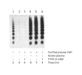
[0023] Figure 1. Macromolecules in boiled plasma stabilized and prevented
adsorption of purified plasma VWF to surfaces. Purified VWF was incubated with
boiled
plasma in 10 mM HEPES with 2 mM CaCl2 at 37 C. VWF multimers were separated on
a
SDS-agarose gel, transferred onto PVDF membrane, and visualized by an HRP-
conjugated
VWF antibody.
3
CA 2890848 2018-11-16
CA 02890848 2015-05-08
WO 2014/075033 PCT/US2013/069545
[0024] Figure 2. Macromolecules in boiled plasma stabilized and prevented
adsorption of purified recombinant A1A2A3 fragment of VWF to surfaces. Panel
A.
Purified AlA2A3 fragment of VWF remaining in solution after incubation in a
tube for
indicated duration. Panel B. Purified AlA2A3 fragment of VWF remaining in
solution after
incubation with boiled plasma in a tube for the indicated duration.
[0025] Figure 3. Macromolecules in boiled plasma stabilized and prevented
adsorption of purified recombinant biotinylated VWF to surfaces. Purified
recombinant
biotinylated VWF was incubated without (lane 1) and with (lane 2) boiled
plasma in 10 mM
HEPES, 2 mM CaCl2 at 37 C overnight. Recombinant VWF remaining in solution
(Panel A)
and VWF bound to surface and eluted by SDS (Panel B) was analyzed by reduced
SDS-
PAGE, transferred onto PVDF membrane, and visualized by a streptavidin-HRP
conjugated.
[0026] Figure 4. SDS-PAGE analysis of proteins in boiled plasma. Lane 1,
boiled
plasma, 20 big. Lane 2, al acid glycoprotein, 5 gg. Lane 3, apolipoprotein A-
1, 5 kig. Lane
4, prealbumin, 5 jug. After electrophoresis in a 4%-20% polyacrylamide gel,
separated
proteins were stained with GelCode Blue (Thermo Scientific).
[0027] Figure 5. Identification of proteins in boiled plasma by mass
spectrometry.
Shown is protein identification data using nano LC/MS-MS.
[0028] Figure 6. Apolipoprotein A-1 and MTh prevented purified recombinant
biotinylated VWF from adsorption to surfaces. Purified recombinant VWF (12
gg/mL)
was incubated without and with various proteins and VWF remaining in solution
(Panel A)
and bound to the tube surface eluted in 4% CHAPS (Panel B) were analyzed by
SDS-PAGE
after reduction and western blot onto nitrocellulose membrane probed with
streptavidin-HRF'.
Lane 1, purified recombinant VWF; lane 2, VWF incubated in absence of added
protein; lane
3, VWF with apolipoprotein A-1 (40 n/mL); lane 4, VWF with HDL (80 !..ig/mL);
lane 5,
VWF with boiled plasma (40 gg/mL); lane 6, VWF with al acid glycoprotein, (40
gg/mL);
lane 7, VWF with prealbumin (40 ilg/mL); lane 8, VWF with bovine serum albumin
(40
kt.g/mL).
[0029] Figure 7. Apolipoprotein A-1 and IIDL prevented purified recombinant
biotinylated ADAMTS13 from adsorption to surfaces. SDS-PAGE and western blot
analysis of purified recombinant ADAMTS13 after 16 hr incubation with various
proteins.
Panel A, ADAMTS13 remaining in solution; panel B, ADAMTS13 bound to tube
surfaces
4
CA 02890848 2015-05-08
WO 2014/075033
PCT/US2013/069545
and eluted in 2% SDS. Lane 1, purified ADAMTS13 before exposure to surface;
lane 2,
ADAMTS13 after exposure to surface; lane 3, ADAMTS13 with apolipoprotein A-1
(20
g/mL); lane 4, ADAMTS13 with HDL (40 g/mL); lane 5, ADAMTS13 with boiled
plasma
(20 i,t.g/mL); lane 6, ADAMTS13 with al acid glycoprotein (20 ug/mL); lane 7,
ADAMTS13
with bovine serum albumin (20 ug/mL).
[0030] Figure 8. Apolipoprotein A-1 prevented association of purified
recombinant
biotinylated A1A2A3 in the fluid phase to surface-bound recombinant
biotinylated
VWF. SDS-PAGE and western blot analysis of biotinylated proteins after
exposure to
surface. Panel A, biotinylated proteins remaining in fluid phase. Panel B,
biotinylated
proteins bound to the surface and eluted in 2% SDS. Panel C, biotinylated
proteins bound to
surface and eluted in 4% CHAPS. Lane 1, purified biotinylated AlA2A3 as
reference; lane 2,
biotinylated VWF (8 !ag/mL) immobilized to tube surface; lane 3, immobilized
biotinylated
VWF exposed to ApoA-1 (40 g/mL); lane 4, immobilized biotinylated VWF exposed
to
purified biotinylated AlA2A3 (8 g/mL); lane 5, immobilized biotinylated VWF
exposed to
ApoA-1 (40 g/mL) and AlA2A3 (8 ug/mL); lane 6, purified biotinylated VWF as
reference.
[0031] Figure 9. Fluid-phase VWF attachment to transluminal fiber. Shown is
the
process of VWF self-association in endothelialized synthetic microvessels.
[0032] Figure 10. ApoA-1 blocks soluble VWF binding to ULVWF fibers. Shown
is
the effect of Apo-Al perfusion through endothelialized synthetic microvessels
prior to the
perfusion of biotinylated soluble VWF on VWF self-association.
[0033] Figure 11. HDL decreases VWF release and modulates ULVWF assembly.
Shown is the effect of HDL on VWF release from activated endothelial cells and
on
modulation of VWF self-assembly into ULVWF strings.
DETAILED DESCRIPTION
[0034] The present disclosure was motivated by the observation that while
the blood
borne proteins, VWF and ADAMTS13, are stable in plasma, purified VWF and
ADAMTS13
are labile and readily lose activity upon dilution and exposure to surfaces.
This suggested to
the present inventors that factors present in plasma serve to stabilize these
proteins. As
disclosed herein, further investigation resulted in the identification of
factors that stabilize
proteins and reduce loss by preventing aggregation, denaturation, and
adsorption of proteins
to surfaces, using VWF and ADAMTS13 as model proteins.
[0035] In particular, the present inventors have surprisingly found that
among the factors
that have stabilizing activity are the proteins, ApoA-1 or HDL, which as shown
herein, are
able to protect VWF and other proteins from attaching to surfaces, including
plastic. In the
case of VWF, these factors also prevent self-association. Accordingly, these
factors can be
used to stabilize VWF, or other highly purified recombinant proteins useful as
drugs, such as
factors VIII and IX, and ADAMTS13, and any other proteins that can be shown to
be
stabilized by ApoA-1 and/or HDL.
[0036] Additionally, because of these properties, ApoA-1 and HDL may be
used as
additives to prevent the "fouling" of surfaces that come in contact with
blood, such as
intravenous and intraarterial cathethers, and other devices, such as
ventricular assist devices.
[0037] Moreover, the present inventors have investigated the effect of ApoA-
1 and HDL
on the interaction of VWF with endothelial microvessel surfaces. Based on
these studies,
methods for the treatment of diseases caused by activation of the
microvasculature are also
presented.
[0038] The descriptions of various aspects of the invention are presented
for purposes of
illustration, and are not intended to be exhaustive or to limit the invention
to the forms
disclosed. Persons skilled in the relevant art can appreciate that many
modifications and
variations are possible in light of the aspect teachings.
[0039] It must be noted that, as used in the specification and the appended
claims, the =
singular forms "a," "an" and "the" include plural referents unless the context
clearly dictates
otherwise. The definition of standard terminology can be found in reference
works, including
Sambrook et al., Molecular Cloning, A Laboratory Manual (1989) and Ausubel et
al., Short
Protocols in Molecular Biology (1999) 4th Ed., John Wiley & Sons, Inc. (as
well as the
complete version of Current Protocols in Molecular Biology). The practice of
the present
disclosure will employ, unless otherwise indicated, conventional methods of
mass
spectroscopy, protein chemistry, biochemistry, recombinant DNA techniques and
pharmacology, all of which are within the skill of those in the art.
6
CA 2890848 2018-11-16
CA 02890848 2015-05-08
WO 2014/075033 PCT/US2013/069545
Von Willebrand Factor (VWF)
[0040] VWF is a plasma glycoprotein required for primary hemostasis. As an
extracellular adapter molecule it mediates the adhesion of platelets to
subendothelial collagen
of a damaged blood vessel and platelet-platelet interactions in high shear-
rate conditions.
The concentration of mature VWF in plasma is approximately 10 jAg/mL, and its
half life is
about 12 hours (Tomokiyo et al., Blood, 105:1078-1084 (2005); Nossent et al.,
J Thromb
Hacmost, 4:2556-2562 (2006)). VWF is synthesized in endothelial cells, where
it is either
secreted constitutively or stored in Weibel-Palade bodies for secretion upon
stimulation, as
well as in megakaryocytes, where it is stored in a-granules that later are
partitioned into
platelets (Ono et al., Blood, 107:528-534 (2006)). Subsequent to the synthesis
in the form of
a precursor protein, VWF undergoes a number of intracellular processing steps.
Building
blocks of the VWF multimer are initially generated in a dimeric form by
formation of a
disulfide bond near the C-terminus. By generation of disulfide bonds near the
N-termini, the
protein multimerizes to a gigantic protein with a molecular mass ranging over
3 orders of
magnitude to more than 20,000 kDa (Sadler, Annu Rev Biochem, 67:395-424
(1998)).
Through the process of self-association subsequent to its secretion, VWF can
show the
extraordinary length of several millimeters.
[0041] The pro-coagulant activity of VWF exhibits a non-linear function of
size, since
the larger the multimer, the more effective it is in promoting platelet
adhesion exhibiting a
critical effect on its function (Furlan, Ann Hematol, 72:341-348 (1996)).
However, under
shear stress conditions in the circulation the protein emerges more vulnerable
to proteolytic
digestion by ADAMTS13 (Lopez et al., Blood Coagul Fibrinolysis, 16 Suppl 1:S11-
6
(2005)).
[0042] Regulation of VWF multimer composition in plasma is mediated by two
major
cleaving events: first, ADAMTS13 proteolytically cleaves the A2 domain of each
VWF
monomer and second, thrombospondin-1 reduces the disulfide bonds interlinking
VWF
multimers (Tsai, Semin Thromb Hemost, 30:549-557 (2004)). In contrast to an
irreversible
fragmentation of VWF by ADAMTS13, the activity of thrombospondin-1 can
regulate VWF
size reversibly employing a reductase activity. Thrombospondin-1 is crucially
involved in
the predominant VWF cleavage by ADAMTS13 due to competition with ADAMTS13 for
binding to the VWF A3 domain (Bonnefoy et al., Blood, 107:955-964 (2006)).
7
CA 02890848 2016-11-15
[0043] The term "VWF" or "recombinant VWF" or "rVWF" can be used
interchangeably
herein and refers to the von Willebrand factor polypeptide and multimers.
[0044] The term "VWF cleavage fragment" or -VWF fragments- or "VWF cleavage
products" are used interchangeably herein and refer to fragments of VWF which
are derived
from VWF, including those generated by protease cleavage. In various aspects,
the protease
cleaving VWF is ADAMTS13. ADAMTS13, also called VWF-cleaving protease (VWFCP),
is a zinc-containing metalloprotease enzyme that cleaves VWF. ADAMTS13 is
secreted into
blood and degrades large VWF multimers, decreasing their hemostatic activity.
ADAMTS13
contains of multiple structural and functional domains, and these domains can
participate in
the recognition and binding of ADAMTS13 to VWF.
100451 The terms "VWF multimers." "multimers," or "multimer forms" are used
interchangeably herein. The ultra large VWF (ULVWF) multimers are cleaved by
ADAMTS13 as they are secreted from endothelial cells. Thus, the terms
"ADAMTS13 and
"VWFCP- are used interchangeably.
ADAMTS13
[00461 The terms "ADAMTS13," "recombinant ADAMTS13," and "rADAMTS13" can
be used interchangeably and refer to a protein encoded by ADAMTS13, a gene
responsible
for the familial form of thrombotic thrombocytopenic purpura (TTP). Structural
details and
sequence information on ADAMTS13 can be found in Zheng etal. (Zheng et al.. J
Biol
Chem. 276(44):41059-41063 (2001. ADAMTS13 has been identified as a unique
member of
the metalloproteinase gene family. ADAM (a disintegrin and metalloproteinase).
ADAMTS
family members are distinguished from ADAMs by the presence of one or more
thrombospondin 1-like (TSP1) domain(s) at the C-terminus and the absence of
the EGF
repeat, transmembrane domain and cytoplasmic tail typically observed in ADAM
metalloproteinases. ADAMTS13 is known to possess VWF-cleaving protease
activity.
[00471 The plasma ADAM1 SI3 in healthy individuals ranges from 0.5 mg to 1
mg per
liter (Grunewald et al., Platelets, 13:451-458 (2002); Rock et al.. Br J
Haematol, 93:684-687
(1996)). ADAMTS13 consists of metalloprotease, disintegrin, first
thrombospondin type 1
(TSP-1) repeat, Cys-rich and spacer domains (Zheng et al., J Biol Chem,
276(44):41059-
41063 (2001); Levy etal., Nature, 413:488-494 (2001)). The C-terminus of
ADAMTS13 has
additional TSPI repeats and two CUB domains. Previous studies have shown that
the N-
- 8 -
CA 02890848 2015-05-08
WO 2014/075033 PCT/US2013/069545
terminus of ADAMTS13 is necessary and sufficient for recognition and cleavage
of
denatured multimeric VWF (Fay et at., J Bioi Chem, 266:8957-8962 (1991);
Horton et al.,
Gene, 77:61-68 (1989); Kaufman et al., Nucl Acids Res, 19:4485-4490 (1991)) or
peptide
substrate (GST-VWF73 or FRETS-VWF73) (Fay et al., J Biol Chem, 266:8957-8962
(1991);
Kokame et al., Br J Haematol, 129:93-100 (2005)). More recent studies have
demonstrated
that the spacer domain of ADAMTS13 binds the exosite (E-1660 APDLVLQR-1668)
near
the C-terminus of the VWF-A2 domain (Toso et al., J Biol Chem, 279: 21643-
21650 (2004);
Lankhof et al., Thromb Haemost, 81:976-983 (1999)). However, the role of the
middle and
distal C-terminal domains of ADAMTS13 in substrate recognition remains
controversial.
Apolipoprotein Al (ApoA-l)
[0048] Apolipoproteins are proteins that bind lipids to form lipoproteins.
Apolipoprotein
A-1 is the major protein component of high density lipoprotein (HDL) in
plasma, with a
molecular weight of approximately 28 kDa. Human ApoA-1 is a 243 amino acid
protein.
The sequence of ApoA-1 has been determined in a number of species and was
found to be
highly conserved, especially at the N-terminus. The cystal structure of Apo-Al
in a lipid-free
state reveals an N-terminal anti-parallel four-helix bundle domain and a
separate two-helix C-
terminal domain. See, e.g., Davidson and Thompson, J. Biol. Chem., 282: 22249-
22253
(2007) for a review of ApoA-1 structure and function.
[0049] An exemplary sequence of a human ApoA-1 (NCBI Reference Sequence:
NM 000039.1) is shown below.
cDNA
1 agagactgcg agaaggaggt cccccacggc ccttcaggat gaaagctgcg gtgctgacct
61 tggccgtgct cttcctgacg gggagccagg ctcggcattt ctggcagcaa gatgaacccc
121 cccagagccc ctgggatcga gtgaaggacc tggccactgt gtacgtggat gtgctcaaag
181 acagcggcag agactatgtg tcccagtttg aaggctccgc cttgggaaaa cagctaaacc
241 taaagctcct tgacaactgg gacagcgtga cctccacctt cagcaagctg cgcgaacagc
301 tcggccctgt gacccaggag ttctgggata acctggaaaa ggagacagag ggcctgaggc
361 aggagatgag caaggatctg gaggaggtga aggccaaggt gcagccctac ctggacgact
421 tccagaagaa gtggcaggag gagatggagc tctaccgcca gaaggtggag ccgctgcgcg
481 cagagctcca agagggcgcg cgccagaagc tgcacgagct gcaagagaag ctgagcccac
541 tgggcgagga gatgcgcgac cgcgcgcgcg cccatgtgga cgcgctgcgc acgcatctgg
601 ccccctacag cgacgagctg cgccagcgct tggccgcgcg ccttgaggct ctcaaggaga
661 acggcggcgc cagactggcc gagtaccacg ccaaggccac cgagcatctg agcacgctca
721 gcgagaaggc caagcccgcg ctcgaggacc tccgccaagg cctgctgccc gtgctggaga
781 gcttcaaggt cagcttcctg agcgctctcg aggagtacac taagaagctc aacacccagt
841 gaggcgcccg ccgccgcccc ccttcccggt gctcagaata aacgtttcca aagtggg
9
CA 02890848 2015-05-08
WO 2014/075033 PCT/US2013/069545
Protein
MKAAVLTLAVLFLTGSQARHFWQQDE PPQS PWDRVKDLATVYVDVLKD SGRDYVSQFEG
SALGKQLNLKLLDNWD
SVTS TFSKLREQLGPVTQEFTAIDNLEKETEGLRQEMSKDLEEVYAKVQPYLDDFQKFWQEEMELYRQKVE
PLRAEL
QEGARQKLHELQEKLS PLGEEMRDRARAHVDALRTHLAPYS DE LRQRLAARLEALKENGGARLAEYHAKATEHL
S
TLSEKAKPALEDLRQGLLPVLE SFKVSFLSALEEYTKKLNTQ
[0050] As used herein "ApoA-1" or "recombinant ApoA-1" or "r ApoA-1" can be
used
interchangeably and refers to Apolipoprotein A-1 polypeptide. Also included in
the
invention are fragments and peptides derived from ApoA-1, as well as drugs
that mimic the
function of ApoA-1.
High-density lipoprotein (HDL)
[0051] High-density lipoprotein (HDL) is the smallest of the five major
groups of
lipoproteins, which enable lipids like cholesterol and triglycerides to be
transported within
the bloodstream. In healthy individuals, about thirty percent of blood
cholesterol is carried
by HDL.
[0052] The conformation of ApoA-1 in discoidal and spherical HDL particles
has been
modeled to be organized as a double-belt in discoidal particles and as a
trefoil in speherical
particles. Sec, e.g., Lund-Katz and Phillips, Subcell. Biochcm. 51: 183-227
(2010) for a
review of HDL structure and function.
EXAMPLARY ASPECTS
[0053] Below are examples of specific aspects for carrying out the present
disclosure.
The examples are offered for illustrative purposes only, and are not intended
to limit the
scope of the present disclosure in any way. Efforts have been made to ensure
accuracy with
respect to numbers used (e.g., amounts, temperatures, etc.), but some
experimental error and
deviation should, of course, be allowed for.
EXAMPLE 1
Methods and Materials
[0054] The following reagents were purchased from Sigma-Aldrich: high
density
lipoprotein (HDL), al acid glycoprotein (AGP), prealbumin, CHAPS,
dithiothreitol (DTT),
ethylenediamine tetraacetic acid (EDTA), HEPES, protease inhibitor cocktail.
Apolipoprotein
A-1 was from Molecular Innovations. Bovine serum albumin (BSA) was from
Equitech-Bio
Inc. Sodium dodecyl sulfate (SDS) was from BDH Biochemicals. Urea was from
Gibco-
CA 02890848 2015-05-08
WO 2014/075033 PCT/US2013/069545
BRL. Calcium chloride was from Baker. PVDF and Nitrocellulose membranes,
gradient (4%-
20%) polyacrylamide gels were from Bio-Rad. HRP conjugated antibody to human
VWF
was from Dako. Streptavidin-horse radish peroxidase conjugate (SA-HRP), and
GelCode
Blue were from Thermo Scientific. Immobilon Western HRP substrate peroxide
solution, and
Ultraccl 10K centrifugal filter were from Millipore. HPC4-agarose was from
Roche. The
BCA protein determination kit was from Thermo Sicentific. Serum-free FreeStyle
293 culture
medium was from Life Technologies Corp.
Preparation of purified human plasma VWF
[0055] Human plasma VWF was purified from cryoprecipitate according to the
method
of Thorell and Blomback (Thromb Res, 35:431-450, (1984)). Briefly, human
cryoprecipitate
was dissolved in citrate buffer (55 mM Na-citrate, pH 6.8), and fibrinogen was
precipitated
from the preparation by 2M glycine at 37 C for 30 min. After removal of the
precipitated
fibrinogen by centrifugation (2,500 x g, 30 min at 4 C), the VWF in the
supernatant was
precipitated by the addition of Nan to a final concentration of 1.55M. The
precipitated VWF
was collected by centrifugation (2,500 x g, 30 min at 4 C), dissolved in 2.5
mL of buffer (10
mM HEPES, 50 mM NaCl, pH 6.8), and chromatographed over a column of Sephacryl
S500
(2.6 cm x 93 cm, GE Healthcare) equilibrated in 10 mM HEPES, 50 mM NaC1, pH
6.8.
Column fractions containing purified VWF were identified by ELISA, SDS-agarose
gel
electrophoresis, SDS-PAGE, and/or western blotting. The purified VWF was
further
concentrated by binding to Q Sepharose (1.5 cm x 3 cm, GE Healthcare) in
buffer containing
25 mM HEPES, 25 mM NaC1, 10 mM EDTA, pH 6.8. VWF was eluted in a concentrated
form with 25 mM HEPES, 0.5M NaCI, pH 6.8. Purified VWF was stored at -80 C
until use.
Preparation of boiled human plasma
[0056] Ten milliliters of normal human plasma anticoagulated with citrate
was heated at
100 C for 10 min. The heated plasma was frozen at -20 C for 16 hr, thawed,
broken in small
pieces by a spatula, and centrifuged at 12,000 x g for 20 min at 4 C. The
supernatant (6 mL)
was desalted over a column of Sephadex G25 (GE Healthcare) equilibrated in 10
mM
HEPES, 2 mM CaCl2, pH 7.4. The macromolecular fraction of boiled plasma (9 mL)
devoid
of material <10,000 Daltons was stored at -20 C until use. The protein
concentration of the
desalted boiled plasma, determined with the BCA reagent using bovine serum
albumin as
standard, was 0.9 mg/mL.
11
CA 02890848 2015-05-08
WO 2014/075033 PCT/US2013/069545
Expression of recombinant VWF
[00571 Recombinant human VWF, containing a C-terminal protein C epitope tag
(PC-
tag), and a biotin tag, was expressed as secreted multimers in stably
transfected HEK293 cells
that were also stably transfected and co-expressing human furin. Briefly, cDNA
encoding
residues Metl to Lys2813 of VWF was inserted in frame in the vector and
followed by an
epitope tag derived from human protein C (PC tag) and a 13-residue biotin
acceptor sequence
(BioTag) (Mize et al., Protein Expr Purif, 57:280-289 (2008)) at the 3' end.
Expression of
recombinant VWF was under the control of a bidirectional doxycycline-inducible
promoter,
which also drives the expression of a bicistronic expression cassette encoding
a secreted form
of E. coli biotin ligase (BirA), and an enhanced green fluorescent protein
(EGFP) reporter.
The secreted form of biotin ligase enabled sequence-specific biotinylation of
the BioTag in
the secreted VWF, while cytoplasmic expression of EGFP, monitored by flow
cytometry, and
selected by fluorescence-activated cell sorting (FACS), enabled automated
selection of
transfected cells expressing biotinylated VWF. In order to obtain fully
processed VWF
without the propeptide, we co-transfected the VWF expression vector with a
second vector
that encoded human furin into HEK293 Tet-On cells using lipofactamine and
stably
transfected cells were selected and clonally expanded in the presence of
puromycin.
Preparation of purified recombinant biotinylated VWF
[00581 Stably transfected cells expressing recombinant VWF were grown to
confluency,
and expression of VWF was induced by the addition of doxycycline to a final
concentration
of 2 rig/ml. Serum-free FreeStyle 293 culture medium containing the secreted
recombinant
VWF multimers was collected and examined on SDS-agarose gels and by western
blotting.
The concentration of VWF antigen was typically 13-18 ug/mL measured by an
ELISA, in
which a polyclonal antibody to human VWF is coated on an ELISA plate as a
capture
antibody and an HRP-conjugated VWF antibody is used as a detection antibody.
Recombinant VWF was purified by affinity chromatography over a monoclonal
antibody
(HPC4) column in the presence of calcium chloride. Cell culture medium
containing
recombinant VWF was thawed and calcium was added to a final concentration of 2
mM. One
mL of culture medium was mixed with 0.2 mL of washed HPC4-agarose in a tube,
and the
mixture was mixed end-over-end at 4 C for 16 hr, during which the biotinylated-
VWF bound
to the HPC4-agarose. The HPC4-agarose suspension was packed into a column, and
washed
with 5 mL of 10 mIVI HEPES, 2 mM CaCl2, 100 mM NaC1, pH 7.4, followed by 10 mL
of 10
12
CA 02890848 2015-05-08
WO 2014/075033 PCT/US2013/069545
mM HEPES, 2 mM CaC12, pH 7.4. The bound VWF was eluted with 1 mL of 10 mM
HEPES, 10 mM EDTA, pH 7.4. Calcium was added to a final concentration of 12 mM
and
the purified biotinylated-VWF was used in stabilization studies within 1 hour
of its
purification. The concentration of purified recombinant VWF was ¨12-13 ,ug/mL.
Preparation of purified VWF A1A2A3 fragment
[0059] The A1A2A3 region of VWF, encompassing Asp1261-11e1878 of VWF, was
expressed with an N-terminal biotin tag and a C-terminal PC tag with the
pNBioSec (2)
vector in stably transfected HEK293 Tet-On cells. Recombinant VWF A1A2A3
fragment
was purified from cell culture medium by binding to and elution from HPC4-
agarose as
described above for recombinant VWF. Purified A1A2A3 fragment was used in
stability
studies within 1 hr of its purification.
Preparation of purified recombinant human ADAMTS13
[0060] Recombinant ADAMTS13 was expressed with a N-terminal biotin tag and
a C-
terminal PC tag with the pNBioSec vector (Mize et al., Protein Expr Purif,
57:280-289
(2008)) in stably transfected HEK293 Tet-On cells. Serum-free FreeStyle 293
medium
containing recombinant ADAMTS13 was concentrated tenfold by centrifugation in
an
Ultracel 10K centrifugal filter, and desalted over Sephadex G-25 to remove
biotin and low
molecular weight molecules. The concentrated recombinant ADAMTS13 preparation
was
treated with a mixture of protease inhibitors (Protease Inhibitor Cocktail, l
% v/v) and stored
at -80 C. Recombinant ADAMTS13 was purified by chromatographed over a
Superdex 200
HR column (1 cm x 30 cm, GE Healthcare) equilibrated in 10 mM HEPES, 2 niM
CaCl2, pH
7.4. Purified ADAMTS13 was used in stabilization studies within 1 hour of its
purification
from the column.
Identification of proteins by mass spectrometry
[0061] Proteins in boiled plasma were identified by nano- liquid
chromatography -tandem
mass spectrometry (nano-LC-MS/MS). Briefly, 5 ug proteins were reduced with
5mM DTT,
alkylated with 12.5 mM iodoacetamide, and digested overnight at 37 C with
trypsin (1:20,
wt/wt, trypsin/total protein) in a buffer containing 50 mM ammonium
bicarbonate and 5%
acetonitrile. The resultant peptides were analyzed using nano-LC-MS/MS in the
positive ion
mode with a Thermo Scientific LTQ Orbitrap Velos mass spectrometer coupled to
a Waters
nanoACQUITY Ultra Performance LC system. MS/MS spectra were searched against
the
13
CA 02890848 2015-05-08
WO 2014/075033 PCT/US2013/069545
human protein database using Thermo Proteome Discoverer software with the
SEQUEST
search engine. Protein ID, sequence coverage, number of unique peptides and
score listed in
Table 1. Three major proteins in boiled plasma (Figure 4, lane 1) are
identified as al acid
glycoprotein, apolipoprotein A-1, and transthyretin (prealbumin).
Protein adsorption assay
[00621 Purified plasma VWF, purified recombinant biotinylated VWF, purified
recombinant biotinylated VWF AlA2A3 fragment, and purified recombinant
biotinylated
ADAMTS13 were diluted in 10 mM HEPES, 2 mM CaCl2, and were incubated at 22 C
or
37 C for 4 to 16 hr in 1.5 mL microfuge tubes. In a typical assay with VWF, 50
.1, of VWF
at 10 jug/mL was used. Proteins that were not adsorbed to tube surface and
remained in
solution were heated in 2% SDS at 100 C, and 10-12% of the samples were
analyzed by
SDS-1% agarose gel electrophoresis without reduction or by SDS-4%-15% gradient
polyacrylamide gel electrophoresis after reduction with mercaptoethanol. The
separated
proteins on gels were transferred to PVDF or nitrocellulose membranes and the
blots were
blocked with 1% bovine serum albumin in TBST (50 mM Tris-HC1, pH 7.5, 150 mM
NaCl,
0.1% Tween-20) for 30 minutes at room temperature, and probed with either an
HRP-
conjugated antibody to VWF or with a streptavidin-HRP conjugate diluted
1:10,000 in TBST
containing 1% albumin. The membranes were washed for 15 min in three changes
of TBST
and incubated with the chemiluminescent HRP substrate Immobilon Western HRP
substrate
peroxide solution. The intensity of chemiluminescence was either recorded by
exposure to X-
ray films or captured on an ImageQuant 350 imaging system (GE Healthcare,
Piscataway,
NJ) and quantitatively analyzed with the ImageQuant software. Proteins
adsorbed to the tube
surfaces were eluted either with 2% SDS or 4% CHAPS. Ten to twelve percent of
the eluted
fractions were analyzed by SDS-PAGE after reduction by mercaptoethanol and
western
blotting as described for the nonadsorbed fractions.
EXAMPLE 2
Macromolecules in boiled plasma stabilized and prevented adsorption of
purified
plasma VWF to surfaces
[00631 Purified VWF from human plasma was diluted in 10 mM HEPES, 2 mM
CaCl2,
pH 7.4, and incubated at 37 C for 4 hours. VWF multimers remaining in solution
was
analyzed by SDS-1% agarose gel electrophoresis, western blotting and
visualization with an
HRP-conjugated antibody to VWF. VWF multimers were nondetectable in solution
(Figure
14
CA 02890848 2015-05-08
WO 2014/075033 PCT/US2013/069545
1, lane 2), suggesting that the VWF had adsorbed to the tube surface. In
contrast, when boiled
plasma containing 90 g/mL of protein was added to the purified plasma VWF
during
incubation, a substantial amount of the VWF remained in solution (Figure 1,
lane 5). These
results showed that soluble macromolecules in boiled plasma stabilized VWF in
solution and
prevented its adsorption to the tube surfaces. Stabilization of VWF in
solution or its
adsorption to the tube surface was unaffected by the presence or absence of
calcium ions
(Figure 1, lanes 3 and 6).
EXAMPLE 3
Macromolecules in boiled plasma stabilized and prevented adsorption of
purified
recombinant A1A2A3 fragment of VWF to surfaces
[0064] Purified VWF A1A2A3 fragment was diluted in 10 mM HEPES, 2 mM CaC12,
pH
7.4, and incubated at 22 C for durations up to 20 hours. The A1A2A3 fragment
is monomeric
and does not multimerize into a collection of multimers as plasma VWF. As
shown in Figure
2, panel A, the amount of Al A2A3 remaining in solution decreased with time.
In contrast,
the presence of macromolecules from boiled plasma (90 iig/mL) prevented the
time-
dependent decrease of AlA2A3 in solution (Figure 2, panel B). These results
showed that
soluble macromolecules in boiled plasma also stabilized the monomeric A1A2A3
fragment
of VWF in solution and prevented its time-dependent adsorption to tube
surfaces.
EXAMPLE 4
Macromolecules in boiled plasma stabilized and prevented adsorption of
purified
recombinant biotinylated VWF to surfaces
[0065] Purified recombinant VWF multimers, enzymatically biotinylated at
the C-
terminus of each subunit was diluted to 10 jag/mL in 10 mM HEPES, 2 mM CaCl2,
pH 7.4
and incubated at 22 C for 16 hr in the presence or absence of boiled plasma.
Ten percent of
the VWF solutions were analyzed by SDS-4%-15% gradient polyacrylamide gel
electrophoresis after reduction with mercaptoethanol. As shown in Figure 3,
panel A, lane 1,
absence of boiled plasma led to a substantial decrease of VWF in solution.
However, a
substantial amount of VWF remained in solution when boiled plasma (90 gg/mL)
was
present during the incubation (Figure 3, panel A, lane 2). Surface-bound VWF
multimers
were eluted from the tube surface by heating at 100 C in 2% SDS for 2 min, and
10% of the
eluted material was analyzed by SDS-4%-15% gradient polyacrylamide gel
electrophoresis
after reduction with mercaptoethanol. These results confirmed that a
substantial amount of
VWF had bound to and was recovered from the tube surface in the absence of
boiled plasma
CA 02890848 2015-05-08
WO 2014/075033 PCT/US2013/069545
(Figure 3, panel B, lane 1), while a small amount of VWF was recovered from
the tube
surface when boiled plasma was present during the incubation (Figure 3, panel
B, lane 2).
These results showed that similar to purified plasma VWF, purified recombinant
biotinylated
VWF multimers also adsorbed to the tube surface in the absence of boiled
plasma, and at
least a portion of the adsorbed material could be eluted from the tube surface
by heating in
2% SDS.
EXAMPLE 5
SDS-PAGE analysis of proteins in boiled plasma
[00661 A sample of
boiled plasma (20 jig) was analyzed by SDS-4%-20% gradient
polyacrylamide gel electrophoresis, and the separated proteins were visualized
by staining
with GelCode Blue. Boiled plasma contains three major proteins, designated as
component A
(-50 kD), component B (-29 kD) and component C (-16 kD), respectively (Figure
4, lane 1).
Commercial preparations of purified al acid glycoprotein (5 jig, Figure 4,
lane 2),
apolipoprotein A-1 (5 jig, Figure 4, lane 3), and prealbumin (5 jig, Figure 4,
lane 4) show
identical electrophoretic mobility with the three major components in boiled
plasma.
EXAMPLE 6
Identification of proteins in boiled plasma by mass spectrometry
[00671 Boiled plasma
was digested with trypsin (weight ratio 1:20) overnight in the
buffer containing 50 mM ammonium bicarbonate and 5% acetonitrile. The
resultant peptides
were analyzed by nano LC-MS/MS and identified as al acid glycoprotein,
apolipoprotein A-
1, and prealbumin (Table 1). Consistent with these identifications, commercial
purified
preparations of these three proteins migrated with mobilities similar to
components A, B and
C in boiled plasma (Figure 4).
Table 1. Identification of proteins in boiled plasma by mass spectrometry
(nano
LC/MS-MS).
Accession Description Coverage # Peptides Score"
IPLIP100022429.3 Alpha-1-acid glycoprotein 42.79 12 1165.02
1
IP1:1P100021841.1 Apolipoprotein A-I 81.27 38 6664.51
IP1:1P100022432.1 Transthyretin 69.39 10 889.50
*Score: this is a probability-based score, which can independently rank the
peptides and
proteins. The higher the score, the more confidence there is in the protein
identification.
16
CA 02890848 2015-05-08
WO 2014/075033
PCT/US2013/069545
EXAMPLE 7
Apolipoprotein A-1 and HDL prevented purified recombinant biotinylated VWF
from
adsorption to surfaces
[00681 Purified recombinant biotinylated VWF (6 p.g/mL) was incubated for
16 h at 22 C
without and with various proteins and the extent of VWF surface adsorption
compared
(Figure 6). Twelve percent of the fluid phase (nonadsorbed) and 12% of the
CHAPS-eluted
samples (adsorbed)were analyzed by SDS-PAGE and western blotting probed with
streptavidin-HRP. Results showed that ApoA-1 (40 ps/mL), HDL (80 i.tg/m1), and
boiled
plasma (40 gimp, prevented surface-adsorption of VWF (Figure 6, panel A, lanes
2-5),
while al acid glycoprotein (401,tg/m1), prealbumin (40 ,ug/m1), and bovine
albumin (40
jAg/m1), did not prevent surface-adsorption. These studies showed that ApoA-1
and HDL
from commercial sources, purified without the use of heat, were effective in
preventing VWF
surface adsorption at low concentrations. These results also confirmed that
two of the major
components in boiled plasma, al acid glycoprotein and prealbumin, did not
contribute to
VWF stabilization, consistent with the hypothesis that ApoA-1 in boiled plasma
was the
component responsible for stabilizing VWF in solution and preventing its
adsorption to
surfaces.
EXAMPLE 8
Apolipoprotein A-1 and HDL prevented purified recombinant biotinylated
ADAMTS13
from adsorption to surfaces
[00691 Purified recombinant ADAMTS13 was diluted to 2.6 i.igimL and
incubated in
microfuge tubes at 22 C without and with various proteins for 16 hr. Ten
percent of the
solution was analyzed by SDS-PAGE, western blot, and probed with streptavidin-
HRP
conjugate. As shown in Figure 7, purified ADAMTS13 adsorbed to the tube
surface in the
absence of added protein (Lane 2, panels A and B). Addition of ApoA-1, HDL,
and boiled
plasma prevented surface-adsorption of ADAMTS13 (Lanes 3-5, panels A and B),
consistent
with the interpretation that ApoA-1 in these three preparations prevented
surface-adsorption.
In comparison, comparable concentrations of al acid glycoprotein and albumin
failed to
prevent surface-adsorption (Figure 7, lanes 6, 7, panels A and B).
17
CA 02890848 2015-05-08
WO 2014/075033 PCT/US2013/069545
EXAMPLE 9
Apolipoprotein A-1 prevented association of purified recombinant biotinylated
AlA2A3
in the fluid phase to surface-bound recombinant biotinylated VWF
[00701 The substantial amount of VWF lost to surface adsorption suggests
that as fluid-
phase VWF molecules are recruited and bind to the immobilized layers of VWF
molecules on
a limited area on the surface, the newly immobilized molecules must have
exposed additional
self-association sites to perpetuate the continued capture of VWF molecules in
the fluid phase
onto the surface. To show that the immobilized VWF surface was able to capture
VWF
molecules in the fluid phase, we immobilized purified biotinylated VWF to a
surface for 6 hr
and removed the unbound material from the tubes (Stage I) to produce an
immobilized
multilayered VWF surface. We confirmed that the immobilized VWF multimers did
not
dissociate and desorb from the surface when exposed to buffer or ApoA-1 (Stage
II) (Fig. 8A,
lanes 2, 3). We then exposed the immobilized VWF to a preparation of purified
monomeric
biotinylated Al A2A3 fragment of VWF (Mr 90 kDa) in the presence or absence of
ApoA-1
(Stage II). Subsequent elution with SDS and CHAPS showed that a portion of the
monomeric biotinylated Al A2A3 fragment in the fluid phase had bound to the
immobilized
VWF exclusively via protein-protein (i.e. VWF-A1A2A3) interactions (Fig. 8B,
lane 4),
while the presence of ApoA-1 blocked this interaction (Fig. 8B, lane 5). These
results
showed that the biotinylated VWF-coated surface formed in Stage I was able to
further
recruit and bind a monomeric VWF fragment in the fluid phase during stage 11.
No
biotinylated AlA2A3 was eluted by 4% CHAPS (Fig. 8C, lanes 4, 5), indicating
that the
biotinylated Al A2A3 did not bind directly to the tube surface through
hydrophobic
interactions. These results confirmed that after a fluid-phase VWF molecule
has bound to
and become a part of the immobilized VWF, it changed its conformation and
exposed new
self-association site(s), and continued to recruit other fluid-phase VWF
molecules, including
a monomeric VWF fragment, onto the immobilized VWF and this process of self-
association
was interrupted by the presence of ApoA-1.
EXAMPLE 10
Apolipoprotein A-1 prevented the association of VWF in solution to immobilized
ULVWF fibers
[00711 We studied the effect of ApoA-1 in VWF self-association in
endothelialized
synthetic microvessels according to the method of Zheng et al (PNAS 109:9342-
9347, 2012).
When we stimulated the endothelial cells in the synthetic microvessels with
phorbol myristatc
18
CA 02890848 2015-05-08
WO 2014/075033 PCT/US2013/069545
acetate (PMA), endothelial cells secreted VWF molecules from the Weibel-Palade
bodies.
The secreted VWF molecules form macroscopic transluminal ULVWF fibers (Figure
9A).
When we perfused soluble biotinylated VWF multimers through these
microvessels, we
observed that the biotinylated VWF molecules bound to the ULVWF fibers (Figure
9B).
However, when Apo-Al was perfused through the vessels prior to the
biotinylated soluble
VWF, no association of soluble VWF with the ULVWF fibers was observed (Figure
10B).
This result shows that ApoA-1 can inhibit the recruitment of circulating VWF
to the
transluminal ULVWF fibers under flow. We immuno-stained the fixed microvessels
after
perfusion of ApoA-1, and verified that ApoA-1 had bound to the ULVWF fibers
(Figure
10C). This result confirmed that ApoA-1 binding to the ULVWF fibers completely
prevented
the association of soluble VWF to the ULVWF fibers. Since the transluminal
ULVWF fibers
have been exposed to 6% albumin prior to perfusion of ApoA-1 or soluble VWF,
the binding
of ApoA-1 or soluble VWF to the ULVWF fibers was specific. The lack of soluble
VWF
associating with ApoA-1-saturated ULVWF fibers also showed soluble VWF was
unable to
displace the ApoA-1 that had bound to the ULVWF fibers, suggesting that ApoA-1
had a
higher affinity for the ULVWF fibers than soluble VWF.
EXAMPLE 11
HDL decreases VWF release and modulates VWF self-association into ULVWF
strings
[0072] We stimulated endothelial cells grown in flow chambers with PMA in
the
presence or absence of HDL. The secreted VWF was detected by binding of fixed
platelets.
There were fewer and shorter platelet-decorated ULVWF strings on the
endothelial cells
stimulated in the presence of HDL than those stimulated in the absence of HDL
(Figure 11).
This result shows that HDL reduces VWF release from activated endothelial
cells and
consequently modulates VWF self-assembly into ULVWF strings.
[0073] VWF secreted from the endothelium can form large fibers that self-
associate, trap
platelets, and can cause microvascular occlusion. Apo-Al decreases VWF
release, modulates
fiber self-association, and prevents association of fluid-phase VWF with
endothelial VWF.
We postulate that the apoAl level determines the extent of pathology caused by
syndromes
with activation of the microvaseulature: TTP, HUS, sepsis, malaria, sickle
cell disease. HDL,
ApoAl or synthetic peptides derived from it could be used as an adjunct to
treat these
disorders.
19
CA 02890848 2016-11-15
[0074] It will be readily apparent to one of ordinary skill in the relevant
arts that other
suitable modifications and adaptations to the methods and applications
described herein are
suitable and can be made without departing from the scope of the invention or
any aspect
thereof. While the invention has been described in connection with certain
aspects, it is not
intended to limit the invention to the particular forms set forth, but on the
contrary, it is
intended to cover such alternatives, modifications and equivalents as can be
included within
the spirit and scope of the invention as defined by the following claims.
[0075]
- 20 -