Note: Descriptions are shown in the official language in which they were submitted.
CA2891142
A Method of Nucleic Acid Sequencing Comprising First and Second Flow Cells
Field
The present disclosure is related to devices, methods for making devices and
methods of
using devices, including devices for detecting fluorescence. One embodiment
contemplates an
optical system, for exciting and measuring fluorescence on or in samples
comprising
fluorescent materials (e.g. fluorescent labels, dyes or pigments). In one
embodiment, a device is
used to detect fluorescent labels on nucleic acid. In a preferred embodiment,
the device is
configured such that fluorescent labels in a plurality of different DNA
templates are
simultaneously detected.
Background
Scanning light microscopes have been known for several decades. Their
functional
principal is based on a light beam being concentrated to a small point of
light (the first focal
point) on a sample. The sample and this point of light are mutually moved in
such a way that a
specific area of the sample is scanned by the point of light. The light which
penetrates the
sample or is reflected by it and/or the fluorescence triggered on or in the
sample during the
scanning is therefore referred to as "light originating from the sample" and
is measured by one
or more photodetectors. An enlarged image is produced in that an original
measurement point
is assigned a specific area on an image of the sample. In principle, such a
scanning light
microscope therefore includes: a light source, such as a laser, which produces
a light beam; a
sample holder for holding the sample; an optic for producing a first focal
point on the sample;
an optical arrangement for imaging a second focal point using the light which
shines through
the sample and/or is reflected by the sample and/or which represents
fluorescence triggered on
or in the sample; a photodetector for measuring the intensity of the second
focal point; and a
scanning mechanism for mutual movement of the sample and first focal point.
The approach has a number of disadvantages. First, the small focal point means
that
only a very small portion of the sample can be addressed at one time. Second,
the necessity for
moving the light creates significant engineering issues and increased cost.
1
CA 2891142 2018-03-07
CA 02891142 2015-05-11
81344-126D
Summary
The present disclosure is related to devices, methods for making devices and
methods of
using devices, including devices for detecting fluorescence. The present
disclosure
contemplates an optical system, for exciting and measuring fluorescence on or
in samples
comprising fluorescent materials (e.g. fluorescent labels, dyes or pigments).
In one
embodiment, a device is used to detect fluorescent labels on nucleic acid.
The device may be configured such that fluorescent labels in a plurality of
different
DNA templates are simultaneously detected. In other words, rather than using a
light source
which creates a small focal point (such as a laser), the preferred light
source of the present
invention (preferably a non-lasing light source) illuminates a large area of a
sample (e.g. at
least 10% of the area defined by a conventional microscope slide, more
preferably greater than
20% of the area defined by a conventional microscope slide, still more
preferably, greater than
50% of the area defined by a conventional microscope slide, still more
preferably, greater than
70% of the area defined by a conventional microscope slide). The light source
(i.e., a non-
lasing light source) illuminates a defined area of a chip (e.g. at least 10%
of the area of the chip
or 14.9 X 10 mm field of view). In still another embodiment, the light source
illuminates a
larger area of a chip (e.g. up to and including an image area of 22 mm X 22
mm, and more
preferably, 22 mm X 66 mm). The system may include a light collection means
such as a
digital camera which is capable of capturing images (capable of recording 120
um features, and
more preferably, 10 micron features or less).
With conventional devices, moreover, it is also difficult to perform
concurrent
measurements of a number of different fluorescent labels that may be present
in a sample (or in
different samples). There may be multiple fluorescent labeling agents that
have different
excitation and/or emission wavelengths. Existing fluorometers, however, do not
facilitate such
multiple-label experiments. Many fluorometers are designed for a single
combination of
excitation and emission wavelengths. By contrast, in a preferred embodiment,
the imaging
system of the present invention is designed for multiple excitation and
emission wavelengths.
The present disclosure contemplates an imaging system, comprising: a non-
lasing light
source configured such that the emitted light from said source illuminates
(and preferably
converges on) a flow cell (or portion thereof), said emitted light suitable
for causing visible
2
CA 02891142 2015-05-11
81344-126D
fluorescence of fluorescent compounds; a lens positioned to collect at least a
portion of said
visible fluorescence; and a light collection means (e.g. a light
imaging/recording means such as
a charge coupled device, a CMOS device, or other type of cameras) positioned
such that said
portion of said visible fluorescence collected by said lens passes through
toward the light
collection means. In one embodiment, the imaging system is conveniently
contained within a
housing (portions of which may be opaque or transparent). In one embodiment,
the flow cell is
mounted on a platform or other support structure. In another embodiment, the
flow cell is
attached to said housing (e.g. to a wall of the housing, or to a mount which
is attached to the
housing).
Various embodiments of the imaging system disclosed herein can be complemented
with hardware (e.g. a computer) or with software. Thus, in one embodiment, the
imaging
system further comprises a processor in communication with said light
collection device (e.g.
CCD or other digital camera), said processor capable of recording and
(optionally) optimizing
images from said system. With respect to optimizing, it may be practical and
convenient to
carry out optimization of the image noise in addition to the compensation of
the brightness of
the individual partial images. Corresponding methods for adaptive. noise-
optimized filtering
are known, for example, from the text of William A. Pratt entitled "Digital
Imaging
Processing", 1978, John Wiley & Sons, Inc., New York.
It is not intended that the imaging system be limited by its arrangement. In
one
embodiment, the flow cell is on the bottom of the system and the other
elements are positioned
above it. In another embodiment, the flow cell is positioned to one side of
the other elements,
with the other elements positioned in a train or train-like manner. In one
embodiment, the flow
cell can be considered to occupy two spatial axes X and Y, with at least some
of the other
elements (e.g. the light source) positioned in the Z axis to illuminate the
flow cell (or sample
therein). On the other hand, the light source can be positioned differently,
with the emitted
light directed by mirrors into the Z axis. In one embodiment, it has been
found convenient to
position the flow cell such that the draining of the flow cell (e.g. the
removal of fluids, such as
solutions containing reagents, or wash buffers and the like) is achieved in
part by gravity.
The flow cell may be connected to a fluidics system, comprising various
reagent and
solution reservoirs in fluidic communication with said flow cell (e.g. via
tubing). The fluidic
3
CA 02891142 2015-05-11
81344-126D
system, in one embodiment, is pressurized and different reagents and solutions
are introduced
by controlled valving (described in more detail below). In one embodiment,
said flow cell
comprises one or more tubing connection ports.
It is not intended to limit the nature of the fluorescent compound(s)
detected. Devices
and systems disclosed herein can be utilized with a variety of compounds,
including but not
limited to, dyes, inorganic molecules, multi-molecular mixtures of organic
and/or inorganic
molecules. crystals, heteropolymers, and the like. For example, CdSe--CdS core-
shell
nanocrystals enclosed in a silica shell may be easily derivatized for coupling
to a biological
molecule (Bruchez et al. (1998) Science, 281: 2013 2016). Similarly, highly
fluorescent
quantum dots (zinc sulfide-capped cadmium selenide) have been covalently
coupled to
biomolecules for use in ultrasensitive biological detection (Warren and Nie
(1998) Science,
281: 2016 2018). Fluorescent oligonucleotides (primers or probes) containing
base-linked or
terminally-linked fluors and quenchers are well-known in the art. They can be
obtained, for
example, from Life Technologies (Gaithersburg, Md.), Sigma-Genosys (The
Woodlands, Tex.),
Genset Corp. (La Jolla, Calif.), or Synthetic Genetics (San Diego, Calif.).
One of skill in the art
will recognize that a large number of different fluorophores are available,
including from
commercial sources such as Molecular Probes, Eugene, Oreg. and other
fluorophores are
known to those of skill in the art. Useful fluorophores include: fluorescein,
fluorescein
isothiocyanate (FITC), carboxy tetrachloro fluorescein (TET), NHS-fluorescein,
5 and/or 6-
carboxy fluorescein (FAM), 5- (or 6-) iodoacetamidofluorescein, 5-{[2(and 3)-5-
(Acetylmercapto)-succinyljamino1 fluorescein (SAMSA-fluorescein), and other
fluorescein
derivatives, rhodamine, Lissamine rhodamine B sulfonyl chloride, Texas red
sulfonyl chloride,
5 and/or 6 carboxy rhodamine (ROX) and other rhodamine derivatives, coumarin,
7-amino-
methyl-coumarin, 7-Amino-4-methylcoumarin-3-acetic acid (AMCA), and other
coumarin
derivatives, BODIPY.TM. fluorophores, Cascade Blue.TM. fluorophores such as 8-
methoxypyrene-1,3,6-trisulfonic acid trisodium salt, Lucifer yellow
fluorophores such as 3,6-
Disulfonate-4-amino-naphthalimide, phycobiliproteins derivatives, Alexa fluor
dyes (available
from Molecular Probes, Eugene, Oreg.) and other fluorophores known to those of
skill in the
art. For a general listing of useful fluorophores, see also Hermanson, G. T.,
BIOCONJUGATE
4
CA 02891142 2015-05-11
81344-126D
TECHNIQUES (Academic Press, San Diego, 1996). All such fluorescent materials
are
contemplated herein.
The flow cell may comprise an array of nucleic acid (e.g. the array is
contained within
the flow cell), at least a portion of said nucleic acid comprising fluorescent
dyes (e.g.
fluorescent labels covalently attached to a nucleotide incorporated in said
nucleic acid). The
flow cell may comprise means for introducing reagents in solution (such that
biological
reactions can take place on or in the array), said reagents selected from the
group consisting of
labeled nucleotides and enzymes (typically introduced in solution, such as
buffers; the buffers
also being useful alone for washing the array free of reactants).
It is not intended to limit the nature of the non-lasing light source. A
variety of non-
laser type light sources are contemplated, including but not limited to light
emitting diodes
(LEDs). The present disclosure contemplates an imaging system, wherein said
non-lasing light
source comprises a plurality of light emitting diodes. Said plurality of light
emitting diodes
may comprise four different sets of light emitting diodes, each of which emit
a different
wavelength of light (e.g. 488 nm, 530 nm, 585 nm, and 615 nm). The light
emitting diodes can
be configured in an array (e.g. linear or circular) such that the emitting
light illuminates (and
preferably converges on) a sample (e.g. material on a microscope slide, an
array, an array
contained within a flow cell, a flow cell, etc.). It is not intended to limit
the number of light
emitting diodes. One embodiment contemplates the simple case where just four
different LEDs
are used (as distinct from four different sets of LEDs), each emitting a
different wavelength.
Even where four different sets are used, the present disclosure contemplates
embodiments
wherein there are equal numbers within each set, and embodiments where some or
all sets have
different numbers of light emitting diodes. Thus, for example, in a circular
array of 20 LEDs, 7
may emit at one particular wavelength, while 3 may emit at another, with the
remaining 10
comprising two sets of 5 LEDs, each set emitting at yet other wavelengths.
Optionally, in order
to further limit or narrow the wavelengths emitted by the LEDs, they may be
combined with
narrow bandpass filters placed between the LEDs and the sample (e.g. the flow
cell containing
the array on a chip). Further embodiments may optionally include additional
elements used to
shape the light (e.g. a shaping lens and/or collimating lens) from the light
source.
5
CA 02891142 2015-05-11
81344-126D
The imaging system may comprise filters positioned in front of the lens,
within the lens,
or between the lens and the light collection means. Preferably, the filters
are optical bandpass
filters which can be positioned in a linear or circular manner. In a
particular embodiment of the
imaging system described above, the system further comprises a filter wheel
comprising a hub
and a plurality of radially extending mounts, each of said mounts containing
an optical
bandpass filter. In one embodiment, four such filters are employed, each
selected for different
preferred wavelengths. In one embodiment, four 50 mm interference filters are
employed to
allow the measurement of the fluorescent emissions of four different
fluorophores.
The filters can be stationary or can be movable. In an embodiment of the
imaging
system described above, the system further comprises a motor engaged (either
directly or
through transmission elements) with said hub of said filter wheel, wherein the
motor is adapted
to rotate said filter wheel to position any one of the plurality of filters
between the light
collection means (e.g. a charge coupled device) and the sample (e.g. the flow
cell). Other
means of limiting the bandwidth of light such as dichroic mirrors may also be
used as a kind of
filter.
In another embodiment, the present disclosure contemplates an imaging system,
comprising: an array of light emitting diodes configured such that the emitted
light illuminates
(and preferably converges on) a sample comprising fluorescent materials, said
emitted light
suitable for causing visible fluorescence of fluorescent materials; a lens
positioned to collect at
least a portion of said visible fluorescence; and a charge coupled device
positioned such that
said portion of said visible fluorescence collected by said lens passes
through toward the charge
coupled device. In one embodiment of the imaging system, said array of light
emitting diodes
comprises four different light emitting diode sets, each of which emit a
different wavelength of
light (e.g. 488 nm, 530 nm, 585 nm, and 615 rim). The light emitting diodes
can be configured
in an array (e.g. linear or circular) such that the emitting light illuminates
(and preferably
converges on) a sample (e.g. material on a microscope slide, an array, an
array contained within
a flow cell, a flow cell, etc.). It is not intended to limit the number of
light emitting diodes.
Even where four different sets are used, the present disclosure contemplates
embodiments
wherein there are equal numbers within each set, and embodiments where some or
all sets have
different numbers of light emitting diodes. Thus, for example, in a circular
array of 20 LEDs, 7
6
CA 02891142 2015-05-11
81344-126D
may emit at one particular wavelength, while 3 may emit at another, with the
remaining 10
comprising two sets of 5 LEDs, each set emitting at yet other wavelengths.
Optionally, in order
to further limit or narrow the wavelengths emitted by the LEDs, they may be
combined with
narrow bandpass filters placed between the LEDs and the sample (e.g. the flow
cell containing
the array on a chip). Further embodiments may optionally include additional
elements used to
shape the light (e.g. a shaping lens and/or collimating lens) from the light
source.
In one embodiment of the imaging system, said sample comprises nucleic acid,
at least
a portion of said nucleic acid comprising fluorescent dyes. In one embodiment,
said sample is
contained within a flow cell. In one embodiment, said flow cell comprises
means for
introducing reagents (typically in solution to said sample). In one
embodiment, said reagents
are selected from the group consisting of labeled nucleotides and enzymes
(e.g. polymerases).
As discussed above, in one embodiment, the flow cell is in fluidic
communication with a
fluidics system (via tubing and connection ports).
The imaging system may comprise filters positioned in front of the lens,
within the lens,
or between the lens and the light collection means. Preferably, the filters
are optical bandpass
filters which can be positioned in a linear or circular manner. In one
embodiment of the
imaging system described above, the system further comprises a filter wheel
comprising a hub
and a plurality of radially extending mounts, each of said mounts containing
an optical
bandpass filter. In one embodiment, four such filters are employed, each
selected for different
preferred wavelengths. In one embodiment, four 50 mm interference filters are
employed to
allow the measurement of the fluorescent emissions of four different
fluorophores.
The filters can be stationary or can be movable. In one embodiment of the
imaging
system described above, the system further comprises a motor engaged (either
directly or
through transmission elements) with said hub of said filter wheel, wherein the
motor is adapted
to rotate said filter wheel to position any one of the plurality of filters
between the light
collection means (e.g. a charge coupled device) and the sample (e.g. the flow
cell).
The present disclosure also contemplates manufacturing an imaging system,
comprising
assembling: a non-lasing light source configured such that the emitted light
from said source
illuminates (and preferably converges on) a flow cell (or portion thereof),
said emitted light
suitable for causing visible fluorescence of fluorescent compounds; a lens
positioned to collect
7
CA 02891142 2015-05-11
81344-126D
at least a portion of said visible fluorescence; and a light collection means
(e.g. a charge
coupled device, a CMOS device, or other type of cameras) positioned such that
said portion of
said visible fluorescence collected by said lens passes through toward the
light collection
means. In one embodiment, said light source comprises LEDs (e.g. a circular
array of LEDs).
In one embodiment, the present disclosure contemplates a method comprising: a)
providing an imaging system, said imaging system comprising a non-lasing light
source
configured such that the emitted light from said source illuminates (and
preferably converges
on) a flow cell (or portion thereof) comprising an array of biomolecules, said
emitted light
suitable for causing visible fluorescence of fluorescent compounds; a lens
positioned to collect
at least a portion of said visible fluorescence; and a light collection means
(e.g. a charge
coupled device, a CMOS device, or other type of camera) positioned such that
said portion of
said visible fluorescence collected by said lens passes through toward the
light collection
means; b) introducing a solution into said flow cell, said solution comprising
one or more
fluorescent compounds, under conditions such that at least a portion of said
fluorescent
compounds attaches to at least a portion of said array of biomolecules, so as
to create treated
biomolecules, and c) imaging said treated biomolecules with said imaging
system. In one
embodiment of this method, the biomolecules comprises nucleic acid. In one
embodiment of
this method, the solution comprises oligonucleotides comprising fluorescent
tags, wherein a
portion of said oligonucleotides hybridize with a portion of said nucleic acid
biomolecules of
said array.
In another embodiment, said biomolecules comprise nucleic acid and said
solution
comprises fluorescently-labeled nucleotides and an enzyme capable of causing
at least a portion
of said nucleotides to be incorporated into at least a portion of said nucleic
acid biomolecules of
said array. In one embodiment, said nucleotides are BODIPY-labeled
nucleotides. In another
embodiment, a second solution is employed comprising one or more enzymes (or
chemicals)
capable of removing said fluorescent labels. In one embodiment, first and
second solutions are
used stepwise whereby labels are introduced, imaged, and subsequently removed
(the cycle
being repeated two times, more preferably 10 times or more).
In another embodiment, the present disclosure contemplates a method,
comprising a)
constructing a crosstalk matrix from measurement of pure dyes, b) inverting
the matrix and c)
8
CA2891142
using it to separate subsequent measurements using imaging system (such as the
LED
illumination-based detector system describe above). This crosstalk matrix can
be constructed
for a four color system (but is not limited to four colors).
This disclosure provides a method comprising: a) providing an imaging system
for
visible fluorescence, said imaging system comprising i) first and second non-
lasing light
sources each emitting a different wavelength of visible light, each configured
such that the
emitted light from said source illuminates a portion of ii) a flow cell
comprising an array of
biomolecules on a first surface of a fluid channel, said flow cell in fluidic
communication with
a reservoir so that reagents can be introduced in solution into said fluid
channel and can contact
said biomolecules, iii) a lens positioned to collect at least a portion of
said visible fluorescence;
and iv) a camera; b) introducing a solution from said reservoir into said
fluid channel of said
flow cell, said solution comprising one or more fluorescently-labeled
nucleotides, under
conditions such that at least a portion of said fluorescent nucleotides
attaches to at least a
portion of said array of biomolecules, so as to create treated biomolecules,
and c) imaging said
treated biomolecules with said camera. In some embodiments, the flow cell is
connected to a
fluidic system comprising a plurality of reservoirs. Such a fluidic system may
be pressurized. In
some embodiments, the light sources are light emitting diodes. In some
embodiments, the flow
cell is moved prior to step (c).
The invention disclosed and claimed herein relates to a method of nucleic acid
sequencing, comprising: a) providing nucleic acid to be sequenced in a first
flow cell, nucleic acid
to be sequenced in a second flow cell, nucleic acid sequencing reagents, and a
camera, wherein said
nucleic acids are arrayed on a surface within said flow cells; b) introducing
the nucleic acid
sequencing reagents into the first and second flow cells such that, while the
nucleic acid in the first
flow cell is undergoing one or more reaction steps, the nucleic acid in a
second flow cell is being
scanned and imaged with the camera; and c) scanning and imaging the nucleic
acid in the first flow
cell with the camera.
Description of the Figures
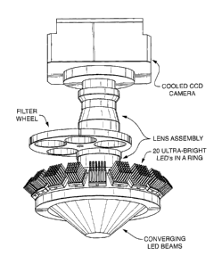
Figure 1 schematically shows one embodiment of the imaging system of the
present
invention, said embodiment comprising a) a circular array of LEDs configured
such that the
emitted light converges on a region or platform (e.g. a position for a sample,
flow cell, etc.) so
9
CA 2891142 2018-03-07
CA 02891142 2015-05-11
81344-126D
as to excite fluorescence of fluorescent material, b) a lens assembly
positioned above the region
so as to capture at least a portion of said fluorescence, c) a filter wheel
comprising bandpass
filters, and d) light collection means (in this case a cooled CCD camera),
wherein said filter
wheel is positioned between the region where the light converges and the light
collection
means.
Figure 2 schematically shows one embodiment of a flow cell. Figure 2A shows a
three
dimensional translucent view of a flow cell, comprising fluid tubing
connections, cartride
heaters, and 0-ring seal. Figure 2B is a two dimensional drawing of a side
view of a flow cell,
showing an array or slide with spaced spots on the surface (representing
positions for
biomolecules and/or anchoring molecules), said array positioned in a fluid
channel such that
solutions of buffers and/or reagents can be introduced over the surface under
conditions
whereby reactions and/or washing can be achieved. The arrows show one
preferred direction
of fluid flow, with entrance and exit ports, as well as one preferred method
of sealing (0-ring
seal).
Figure 3 schematically shows one embodiment of a fluidics system, comprising a
variety of illustrative reagent and buffer reservoirs in communication (via
tubing or other
channeling into a manifold comprising valves) with one embodiment of a flow
cell (comprising
a side entrance port and one or more heaters), wherein the array or chip is
inverted and the exit
port is on the bottom, thereby permitting the fluid channel to be drained at
least in part by
gravity so that waste can be readily collected into a reservoir.
Figure 4 schematically shows another embodiment of an imaging system, wherein
two
flow cells and two cameras are employed to increase capacity and efficiency
(e.g. while one
chip in a first flow cell is undergoing one or more reaction steps, a second
chip in a second flow
cell is being scanned and imaged).
Figure 5 shows an illustrative excitation and emission filter selection (grey
rectangles)
for four illustrative dyes, relative to the dye's excitation (dashed) and
emission (solid) spectra.
Figure 6 shows the raw data (6A) and crosstalk adjusted data (6B) for four
illustrative
dyes.
CA 02891142 2015-05-11
81344-126D
Detailed Description
The present disclosure contemplates a fluorescent detection system and a flow
cell for
processing biomolecules (e.g. nucleic acid samples) arrayed on a "chip" or
other surface (e.g.
microscope slide, etc.). The flow cell permits the user to perform biological
reactions,
including but not limited to, hybridization and sequencing of nucleic acids.
It is not intended for this subject matter to be limited to particular light
sources. By way
of example only, the system can employ ultra-bright LEDs (such as those
available from
Philips Lumileds Lighting Co., San Jose, CA) of different colors to excite
dyes attached to the
arrayed nucleic acids. These LEDs are more cost effective and longer life than
conventionally
used gas or solid state lasers. Other non-lasing sources of lights such as
incandescent or
fluorescent lamps may also be used.
Figure 1 shows a useful configuration of the LEDs, whereby the emitted light
converges
on a region or platform (e.g. suitable for positioning the flow cell or
sample). However, linear
arrays of LEDs can also be used.
The system may employ a high sensitivity CCD camera (such as those available
from
Roper Scientific, Inc., Photometric division, Tucson AZ or those available
from Apogee
Instruments, Roseville, CA) to image the fluorescent dyes and make
measurements of their
intensity. The CCD cameras may also be cooled to increase their sensitivity to
low noise level
signals. These may also be CMOS, vidicon or other types of electronic camera
systems.
Since LED illumination light is not a collimated beam as from lasers, it is
therefore an
appropriate choice for imaging a larger area of many nucleic acid spots. To
get sufficient light
and therefore fluorescent signals over the larger area, the area seen by each
pixel of the camera
must be of sufficient size to allow enough fluorescent dye molecules to create
a sufficient
signal (for example, an Apogee U13 CCD available has 1.3 megapixels of 16
microns in size,
while the Apogee U32 has 3.2 megapixels of 6.8 microns in size).
To increase capacity and efficiency, the present invention contemplates in one
embodiment, a two flow cell system (e.g. while one chip in a first flow cell
is undergoing one
or more reaction steps, a second chip in a second flow cell is being scanned
and imaged) with a
single camera. In yet another embodiment of an imaging system, two flow cells
and two
cameras are employed (Figure 4).
11
CA 02891142 2015-05-11
81344-126D
In one embodiment, the chip containing the array of nucleic acid spots is
processed in a
transparent flow cell incorporated within the instrument, which flows reagent
past the spots and
produces the signals required for sequencing (see Figures 2A and 2B). In a
preferred
embodiment, the chip remains in the flow cell while it is imaged by the LED
detector. The flow
cell and associated reagents adds the nucleic acids, enzymes, buffers, etc.
that are required to
produce the fluorescent signals required for each sequencing step, then the
flow cell delivered
the required reagents to remove the fluorescent signals in preparation for the
next cycle.
Measurement by the detector occurs between these two steps. In order for
reactions to take
place, the flow channels need to be of sufficient dimensions. For example, the
channel by the
array should be at least 0.1 mm in depth (more preferably 0.5 mm in depth) and
the volume
formed by the chip, the block and the seal should be at least 100 microliters
in volume (more
preferably, between 100 and 700 microliters, and still more preferably,
between 150 and 300
microliters, e.g. 200 microliters, in volume).
The flow cell is preferably motionless (i.e. not moved during reactions or
imaging). On
the other hand, the flow cell can readily be mounted on a rotary or one or
more linear stages,
permitting movement. For example, in a two flow cell embodiment, the two flow
cells may
move up and down (or side to side) across the imaging system. Movement may be
desired
where additional processes are desired (e.g. where exposure to UV light is
desired for
photochemical reactions within the flow cell, such as removal of
photocleavable fluorescent
labels), when multiple flow cells share a single camera, or when the field of
view of the
detection system is smaller than the desired area to be measured on the flow
cell. The detector
system may also be moved instead of the flow cell.
The flow cell is preferably in fluid communication with a fluidics system (see
illustrative system shown in Figure 3. In one embodiment, each bottle is
pressurized with a
small positive gas pressure. Opening the appropriate valve allows reagent to
flow from the
source bottle through the flow cell to the appropriate collection vessel(s).
In one embodiment,
the nucleotides and polymerase solutions will be recovered in a separate
collection bottle for re-
use in a subsequent cycle. In one embodiment, hazardous waste will be
recovered in a separate
collection bottle. The bottle and valve configuration allow the wash fluid to
flush the entire
valve train for the system as well as the flow cell. In one embodiment, the
process steps
12
CA 02891142 2015-05-11
81344-126D
comprise: 1) flushing the system with wash reagent, 2) introducing nucleotides
(e.g. flowing a
nucleotide cocktail) and polymerase, 3) flushing the system with wash reagent,
4) introducing
de-blocking reagent (enzyme or compounds capable of removing protective groups
in order to
permit nucleic acid extension by a polymerase), 5) image, 6) introduce label
removing reagent
(enzyme or compounds capable of removing fluorescent labels), and 7) flushing
the system
with wash reagent.
The system can be made to include a user interface system. The Labview
(National
Instruments, Austin, TX) system is available and provides relatively simply
software for
computer controlled systems. Galil Motion Control (Rocklin, CA) provides
motion control
systems that can be interfaced to control the instrument.
Example: Method for removing crosstalk between detected fluorescent signals
for a multicolor
system. Previous sequencing systems utilizing lasers have attempted to
minimize the number of
lasers in order to reduce costs (for example ABI Prism sequencers). For a four
color detection
system using LEDs, the light sources are fairly inexpensive and it is
desirable to have four
separate color light sources in order to reduce crosstalk between colors as
follows.
To determine actual fluorescent intensities for the four colors, A, B, C and D
from
measured detector outputs, MA, MB, Mc, MD in corresponding channels, you need
to know all
of the crosstalk factors: RAB, RBA, Rgc, RcB, RcD, RDc. Six crosstalk factors
are used for
illustrative purposes. There may be more or fewer factors which may be
incorporated into the
analysis.
For example, RAB is the ratio between the portion of the signal in the A
channel coming
from the B dye and the actual intensity of the B dye. If for instance RAB is
20%, then the A
channel will have an additional signal equal to .2 times the actual B dye
intensity in the B
channel. Thus for channel B, the observed measurement, MB, is the direct
measurement of B
and the two contributions from the adjacent channels (if any): MB = B RBAA R
BcC (1)
For the four channels, this may be written in matrix form:
13
CA 02891142 2015-05-11
81344-126D
M -A -
A
M B
=K (2)
Mc
_ D _
where
1 RAH 0 0
K R 1 RBC 0
0 Rc, 1 Rcp
0 0 Rpc 1
Each of the six crosstalk factors may be determined through a simple
experiment with pure
dyes. Some may be zero and they might vary with intensity, so we may need a
table of a
number of values for each depending on the measured intensity range. We want
to solve for the
actual fluorescent signals, A, B, C and D given the detector measurements, MA,
MB, MC, MD.
Thus, we want to solve the above matrix equation (2). This is:
A MA
= B (3)
Mc
_ D _
where K' is the inverse of matrix K. Although this may be written out in terms
of the six
crosstalk factors, it is somewhat complex and is best performed by plugging in
the numbers and
letting the computer take the inverse. Figure 6 shows the raw data (6A) and
crosstalk adjusted
data (6B) for four illustrative dyes.
14