Note: Descriptions are shown in the official language in which they were submitted.
INTRAVASCULAR ARTERIAL TO VENOUS ANASTOMOSIS AND TISSUE
WELDING CATHETER
Technical Field
The present invention relates to manipulations of circulatory vessels and in
particular to devices usable in creating arteriovenous (AV) fistulas to
connect arteries and
veins.
Background of the Invention
In the body, various fluids are transported through conduits throughout the
organism to perform various essential functions. Blood vessels, arteries,
veins, and
capillaries carry blood throughout the body, carrying nutrients and waste
products to
different organs and tissues for processing. Bile ducts carry bile from the
liver to the
duodenum. Ureters carry urine from the kidneys to the bladder. The intestines
carry
nutrients and waste products from the mouth to the anus.
In medical practice, there is often a need to connect conduits to one another
or to
a replacement conduit to treat disease or dysfunction of the existing
conduits. The
connection created between conduits is called an anastomosis.
In blood vessels, anastomoses are made between veins and arteries, arteries
and
armies, or veins and veins. The purpose of these connections is to create
either a high
flow connection, or fistula, between an artery and a vein, or to carry blood
around an
obstruction in a replacement conduit, or bypass. The conduit for a bypass is a
vein,
artery, or prosthetic graft.
An anastomosis is created during surgery by bringing two vessels or a conduit
into direct contact. The vessels are joined together with suture or clips. The
anastomosis
can be end-to-end, end-to-side, or side-to-side. In blood vessels, the
anastomosis is
elliptical in shape and is most commonly sewn by hand with a continuous
suture. Other
methods for anastomosis creation have been used including carbon dioxide
laser, and a
number of methods using various connecting prosthesis, clips, and stents.
An arterio-venous fistula (AVF) is created by connecting an artery to a vein.
1
CA 2891257 2019-08-16
CA 02891257 2015-05-11
WO 2014/078601
PCT/US2013/070200
This type of connection is used for hemodialysis, to increase exercise
tolerance, to
keep an artery or vein open, or to provide reliable access for chemotherapy.
An alternative is to connect a prosthetic graft from an artery to a vein for
the
same purpose of creating a high flow connection between artery and vein. This
is
called an arterio-venous graft, and requires two anastomoses. One is between
artery
and graft, and the second is between graft and vein.
A bypass is similar to an arteriovenous graft. To bypass an obstruction, two
anastomoses and a conduit are required. A proximal anastomosis is created from
a
blood vessel to a conduit. The conduit extends around the obstruction, and a
second
distal anastomosis is created between the conduit and vessel beyond the
obstruction.
As noted above, in current medical practice, it is desirable to connect
arteries
to veins to create a fistula for the purpose of hemodialysis. The process of
hemodialysis requires the removal of blood from the body at a rapid rate,
passing the
blood through a dialysis machine, and returning the blood to the body. The
access to
the blood circulation is achieved with (1) catheters placed in large veins,
(2)
prosthetic grafts attached to an artery and a vein, or (3) a fistula where an
artery is
attached directly to the vein.
Hemodialysis is required by patients with kidney failure. A fistula using
native blood vessels is one way to create high blood flow. The fistula
provides a
high flow of blood that can be withdrawn from the body into a dialysis machine
to
remove waste products and then returned to the body. The blood is withdrawn
through a large access needle near the artery and returned to the fistula
through a
second large return needle. These fistulas are typically created in the
forearm, upper
arm, less frequently in the thigh, and in rare cases, elsewhere in the body.
It is
important that the fistula be able to achieve a flow rate of 500 ml per minute
or
greater, in order for the vein to mature or grow. The vein is considered
mature once
it reaches > 4 mm and can be accessed with a large needle. The segment of vein
in
2
CA 02891257 2015-05-11
WO 2014/078601 PCT/1JS2013/070200
which the fistula is created needs to be long enough (> 6 cm) to allow
adequate
separation of the access and return needle to prevent recirculation of
dialysed and
non-dialysed blood between the needles inserted in the fistula.
Fistulas are created in anesthetized patients by carefully dissecting an
artery
and vein from their surrounding tissue, and sewing the vessels together with
fine
suture or clips. The connection thus created is an anastomosis. It is highly
desirable
to be able to make the anastomosis quickly, reliably, with less dissection,
and with
less pain. It is important that the anastomosis is the correct size, is
smooth, and that
the artery and vein are not twisted.
Summary of the Invention
The present invention comprises a device for creating an arteriovenous (AV)
fistula, which comprises a proximal base having a distal tapered end surface
and a
distal tip connected to the proximal base and movable relative to the proximal
base.
The distal tip has a proximal tapered end surface. A first heating assembly,
comprising an energized heating element, is disposed on at least one of the
distal
tapered end surface and the proximal tapered end surface. A second heating
assembly, comprising a passive non-energized heat spreader, is disposed on the
other
one of the distal tapered end surface and the proximal tapered end surface.
The
distal tapered end surface and the proximal tapered end surface are adapted to
contact opposing sides of a tissue portion to create the fistula. The distal
tapered end
surface is oriented at an angle of 15-90 degrees relative to a longitudinal
axis of the
device, and more advantageously at an angle of 15-50 degrees relative to the
longitudinal axis. In one particularly optimal configuration, the distal
tapered end
surface is oriented at an angle of approximately 23 degrees relative to the
longitudinal axis. The taper of the proximal tapered end surface matches the
taper of
3
CA 02891257 2015-05-11
WO 2014/078601
PCT/US2013/070200
the distal tapered end surface, so that the two surfaces match one another and
fully
engage with one another when engaged.
A shaft is provided for connecting the distal tip to the proximal base, the
shaft being extendable and retractable to extend and retract the distal tip
relative to
the proximal base.
The tapered end surface on which the heating assembly is disposed preferably
has a second passive non-energized heat spreader disposed thereon. The
energized
heating element optimally comprises a serpentine configuration. A temperature
sensor is disposed near the energized heating element, for providing closed
loop
temperature control to the heating assembly.
The second heat spreader comprises a thermally conductive material which
extends across a substantial portion of the tapered end surface on which it is
disposed, the second heat spreader being in thermal contact with the energized
heating element to draw heat from the heating element and spread the heat
across the
tapered end surface. It is constructed so that it has a thickness
approximately equal
to a thickness of a vessel in which the device is deployed, this thickness
falling
within a range of 0.010 inches to 0.060 inches.
In one configuration, the heat spreader comprises a plurality of raised
segments forming a segmented rib, for creating a focused heat conduction path
through tissue. The segmented rib further comprises gaps between the segments,
which gaps provide an insulative barrier that limits tissue dessication to
promote
adhesion without cutting. In another configuration, the heat spreader
comprises a
raised outer rib along its circumference, the raised outer rib forming a
pocket in a
center portion thereof for capturing and removing tissue removed. An outer
circumference of the rib comprises a radius for creating a transition between
a weld
band outside of a cut zone formed during a procedure and native tissue.
In other embodiments, the heat spreader comprises a domed surface, or
4
CA 02891257 2015-05-11
WO 2014/078601
PCT/US2013/070200
comprises a raised center surface and a lower profile outer surface.
The distal tip comprises a tapered outer surface, tapering down from the
proximal tapered end surface toward a distal end thereof, the distal end of
the distal
tip comprising an aperture for a through lumen for receiving a guidewire,
wherein a
width of the distal tip at the lumen aperture is approximately equal to a
diameter of a
guidewire.
The energized heating element comprises separate elliptical elements that
provide independent power delivery for heating and cutting. The separate
elliptical
elements comprise an outer element and an inner element, the outer element
being
configured to deliver reduced heat to promote controlled dessication and
adhesion in
a weld zone without cutting through tissue and the inner element being
configured to
deliver increased heat to promote rapid cutting through tissue in a cutting
zone.
In illustrated embodiments, the first heating assembly is disposed on the
distal tapered end surface and the second heating assembly is disposed on the
proximal tapered end surface.
A second active energized heating element is provided on the proximal
tapered end surface in some embodiments, which is embedded into the heat
spreader.
Each of the first and second heating assemblies preferably comprise non-
stick surfaces, and the shaft also preferably comprises a non-stick surface.
The non-
stick surfaces have a surface finish of less than 16 Ra.
A position sensor is provided for monitoring movement of the distal tip.
In another aspect of the invention, there is provided a method for creating an
arteriovenous (AV) fistula, which comprises steps of selecting an appropriate
procedural site having each of a primary blood vessel and a secondary blood
vessel
in close proximity to one another, inserting a piercing device into the
primary vessel
to pierce the vessel walls and creating an opening so that the piercing device
extends
into the adjacent secondary vessel, and advancing a guidewire until the
guidewire is
5
CA 02891257 2015-05-11
WO 2014/078601
PCT/US2013/070200
positioned in a blood flow path of the secondary vessel sufficiently to allow
the
piercing device to be removed. The piercing device is then withdrawn. A
proximal
end of the guidewire is loaded into a lumen of a distal tip of a device for
creating the
AV fistula, and the device is advanced over the guidewire until a tapered
dilating
distal tip of the device comes into contact with the selected anastomosis
site. The
distal tip of the device is advanced relative to a proximal base of the device
to
thereby dilate the opening in the second vessel, so that the distal tip is in
the second
vessel and the proximal base is in the first vessel.
At this juncture, a heat spreader on an angled distal surface of the proximal
base is seated against an inner wall of the first vessel surrounding the
opening. The
distal tip is retracted so that a heat spreader on an angled proximal surface
of the
distal tip seats against an inner wall of the second vessel surrounding the
opening,
thereby capturing the walls of the first and second vessel between the facing
angled
surfaces of each of the distal tip and the proximal base, respectively.
A controlled tension is maintained between the distal tip and the proximal
base, and at the same time energy is applied to a heating element on the
distal angled
surface of the proximal base. The resultant applied heat and pressure forms a
fistula
with welded edges defining the fistula opening. The device is then withdrawn
from
the procedural site.
The invention, together with additional features and advantages thereof, may
best be understood by reference to the following description taken in
conjunction
with the accompanying illustrative drawings.
Brief Description of the Drawings
Fig. la is an elevational view of the handle portion of a device constructed
in
accordance with one embodiment of the present invention;
6
CA 02891257 2015-05-11
WO 2014/078601
PCT/US2013/070200
Fig. lb is an elevational enlarged view of the circled distal working portion
of the device of Fig. la;
Fig. 2a is an elevational view of an embodiment like that shown in Figs. la-
lb, with the distal end in a first working configuration;
Fig. 2b is an elevational view similar to Fig. 2a, with the distal end in a
second working configuration;
Fig. 3a is an isometric view of one embodiment of the device shown in Figs.
la-2b;
Fig. 3b is an isometric view similar to Fig. 3a illustrating a modified
embodiment of the heating mechanism;
Fig. 4a is an exploded isometric view illustrating an embodiment of the
proximal base and particularly showing the assembly of the heating element and
proximal heat spreader;
Fig. 4b is an isometric view showing the assembled heating element and
proximal heat spreader;
Fig. 4c is an exploded isometric view similar to Fig. 4a showing a modified
embodiment of the proximal heating assembly;
Fig. 5 is an exploded isometric view of another embodiment of the proximal
base and heating assembly;
7
CA 02891257 2015-05-11
WO 2014/078601
PCT/US2013/070200
Fig. 6 is an exploded isometric view of an embodiment of the distal tip and
distal heat spreader;
Fig. 7a is an isometric view of one embodiment of the distal tip and heating
assembly of the present invention;
Fig. 7b is an isometric view similar to Fig. 7a of a modified embodiment of
the distal tip and heating assembly of the present invention;
Fig. 7c is an isometric view similar to Figs. 7a-7b of still another modified
embodiment of the distal tip and heating assembly of the present invention;
Fig. 8a is an isometric view similar to Figs. 7a-7c of yet another modified
embodiment of the distal tip and heating assembly of the present invention;
Figs. 8b-8f are cross-sectional views of different embodiments of the distal
tip and heating assembly of the present invention;
Fig. 9 is a cross-sectional view showing an application and method of using
the device and system of the present invention;
Fig. 10 is a diagram of an anastomosis creating using the devices and
methods disclosed in the present application;
Fig. lla is an elevational view similar to Fig. la illustrating a modified
embodiment of the device of Fig. la, but having an active distal heater rather
than a
passive heat spreader; and
8
CA 02891257 2015-05-11
WO 2014/078601
PCT/US2013/070200
Fig. lib is an elevational enlarged view of the circled portion of Fig. ha.
Description of the Preferred Embodiment
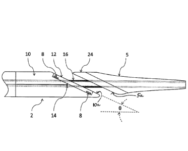
Referring now more particularly to the drawings, as illustrated in Figs. la
and
lb, one embodiment of the inventive intraluminal anastomotic device 1
comprises
four main components, including a proximal heating assembly 2, a proximal
shaft 3,
a distal heating assembly 4, and a handpiece 6. The distal heating assembly 4
comprises a distal tip 5 and heat spreader 24. The handpiece 6 comprises a tip
actuation button 7 and a release button 13. The proximal heating assembly 2 is
constructed of a proximal base 10 that is cut at an angle U at the distal end.
In one
embodiment, the proximal base 10 is cut at an angle 6 of 23 degrees, forming
an
angled distal tapered end surface 10a. However, the angle 0 can be adjusted
depending on the particular anatomy of a procedural site and desired
anastomosis
length. The inventors have found that the angle 0 provides advantageous
outcomes
within a range of about 15-90 degrees, and more particularly within a range of
15-50
degrees, keeping in mind that approximately 23 degrees is presently a
particularly
preferred angle within that range. These preferred angles/angle ranges result
in an
optimized oval configuration for the anastomosis which maximizes the cutting
surface while also efficiently utilizing available heating energy to create an
effective
cut and welding zone.
On the angled surface 10a of the proximal base 10, a heating element 8 is
embedded. The proximal base 10 is typically constructed of a thermally
insulating
material that is resistive to high temperatures. Materials known to work well
for this
application include Vespel, Celazol, Teflon, Polyimide, Ultem, and ceramics. A
proximal heat spreader 12 is used to compress and heat the tissue to create
coaptation of vessel tissues. This process is known as tissue welding or
tissue
9
CA 02891257 2015-05-11
WO 2014/078601
PCT/US2013/070200
fusion. In one embodiment, the proximal heat spreader 12 is constructed of a
thermally conductive material with the resistive heating element embedded
therein.
Some examples of thermally conductive material suitable for this purpose
include
aluminum, stainless steel, aluminum nitride, or other metal or ceramic
materials
known to those skilled in the art. The position, size, and shape of the
proximal heat
spreader 12 can be adjusted to control where the heat is applied to tissue
(see Figs.3a
and 3b for exemplary alternative embodiments). For example, it may be
beneficial
to place the proximal heat spreader 12 toward the center of the long axis of
the
device body (Fig. 3b), such that a heat gradient is created across the face of
the
angled surface of the proximal base 10. This provides the tissue near the
center of
the cutting region with the most heat, which completely denatures the tissue,
and less
heat radially outwardly of the center, to limit the amount of necrosis, while
still
providing strong coaptation or welding of the tissues. The proximal base 10 is
configured with at least one thermocouple or temperature sensor 14 to monitor
the
temperature near the active heating element 8, and provides a means for closed
loop
temperature control to optimize tissue welding and cutting.
As illustrated particularly in Figs. 6 and 7a-7c, the distal tip 5 comprises a
uniform conical tapered outer surface, though it can have a variable tapered,
sloped
outer surface as illustrated in Figs. 8a-8f, wherein the outer surface tapers
down to
the approximate diameter of a guidewire to provide an atraumatic method for
passing through the vessel wall. A guidewire lumen 18 extends through the
center
of the distal tip 5, as shown in Figs. 3a and 3b. In one particular
embodiment, the
lumen 18 is sized to receive a 0.014 inch guidewire, but may be sized to
receive
guidewires of various diameters. The intraluminal anastomotic device 1 is
tracked
over a guidewire 17 (Fig. 9) and the tapered outer surface of the distal tip 5
dilates
through the tissue into the adjacent vessel. Once the distal heating assembly
4 is
completely disposed within the adjacent vessel, the distal tip 5 is retracted
to bring
CA 02891257 2015-05-11
WO 2014/078601
PCT/US2013/070200
the tip toward the proximal heating assembly 2, thereby capturing vessel wall
tissue
between the two components 5 and 10, and bringing the adjacent walls of a
first
vessel 20 and a second vessel 22 together. A proximal end surface 5a of the
distal
heating assembly 4 is angled to precisely match the angle 13 of the proximal
heating
assembly 2.
In one embodiment, the proximal base 10 is configured as shown in Figs. 3a
and 3b. The proximal base 10 is configured to receive the first heating
element 8
(Figs. 4a-4c), which is covered by the proximal heat spreader or heating
surface 12.
The heating surface 12 is comprised of a thermally conductive material which
draws
heat from the first heating element 8. Power attachment points 11 ensure that
the
heating element 8, in whichever illustrated configuration is selected, may be
energized. The heating surface 12 transfers heat into the adjoining vessels to
create a
weld band 21 (Figs. 9 and 10) and to cut tissue to create an anastomosis or
fistula 25
(Figs. 9 and 10). The size and shape of the weld zone and anastomosis can be
altered by adjusting the shape of the heating surface 12. The geometry can
also be
altered such that the temperature is equal in the passive and active heated
surfaces.
In one preferred embodiment, the heating surface or proximal heat spreader 12
comprises an aluminum plate, although alternative thermally conductive
materials
such as aluminum nitride, ceramics, tungsten, steel, or beryllium may be used.
The
thickness of the heating surface 12 is approximately the thickness of the
vessel in
which the weld is being created. However, the thickness may be increased or
decreased to control the amount of heat that is conducted into the surrounding
tissue.
Typical thickness of the heating surface ranges from 0.010 inches to 0.060
inches
(Figs. 3a-3b, 4a-4c).
In one embodiment as illustrated in Fig. 7b, a distal heat spreader 24 on the
distal tip 5 has a plurality of raised segments 29 for forming a segmented rib
30. The
segmented rib 30 creates a focused heat conduction path through the tissue,
while
11
CA 02891257 2015-05-11
WO 2014/078601
PCT/US2013/070200
gaps 31 between the segments 29 provide an insulative barrier that limits
tissue
dessication to promote adhesion without cutting. The size and number of
segments
29 can be adjusted to control the rate of tissue dessication that may
accommodate
variable tissue thickness.
In another embodiment, as illustrated in Figs. 7a and 8c, the passive heating
element 24 has a raised outer rib 28 along its circumference. The raised outer
rib 28
creates a pocket 26 in the center where tissue is captured and removed during
the
welding process. The outer circumference of the rib has a radius to create a
transition between the weld band outside of the cut zone and the native
tissue. A
radius allows for minimal compression at the edge of the weld. This
configuration
provides a focused heat conduction path through the tissue between the active
and
passive heating assemblies to promote tissue cutting while the step gap
provides an
offset that limits tissue compression and dessication in the inner and outer
regions to
promote tissue adhesion without cutting in the adjacent zone.
In still another embodiment as illustrated in Fig. 8f, the distal heating
assembly 4 has a domed surface 33. The domed shape of the surface 33 creates a
higher compression zone in the center to promote tissue cutting, while
tapering off at
the perimeter to promote tissue dessication and adhesion without cutting.
In another embodiment, as illustrated in Figs. 7c and 8d, a raised surface 32
is designed to increase the compression force on the tissue in the center,
while
creating a wider weld band 21 (Fig. 9) around the perimeter. The wider weld
band
creates a stronger weld. The width of the raised center section may be
adjusted to be
narrower or wider in order to achieve the desired weld strength or anastomosis
opening geometry. As illustrated in Fig. 8e, a slit between the two vessels
can be
created by making the raised surface 32 extremely narrow. As the surface area
of the
mating section of the distal heating assembly 4 is decreased, the amount of
heat
transferred from the active heater will decrease. This can be useful if less
heat is
12
CA 02891257 2015-05-11
WO 2014/078601
PCT/US2013/070200
needed between two different anatomical structures that are being welded.
Another
feature of a narrow raised section is a temperature gradient across the distal
heating
assembly 4 that increases radially from the raised section. A temperature
gradient
allows the heat to be the highest at the center, which completely denatures
and cuts
through tissue, creating an anastomosis. As the temperature decreases
radially, the
tissue has less necrosis, yet the proteins are denatured, which leads to a
strong weld
and long term healing.
The shape of the distal heating assembly 4, in combination with compression
force, influences the rate at which the passive heating element cuts through
the
tissue. If too much heat or pressure is applied abruptly, distal heating
assembly 4
may quickly cut through the tissue without transferring enough heat to the
surrounding tissue to achieve a satisfactory weld. Instead, a balance of heat
and
pressure is required to dessicate and denature the protein in the tissue
surrounding
the cut to promote adhesion prior to cutting. In order to best achieve this
balance,
the temperature and position of the distal tip are monitored during the
welding
process and the heat and/or pressure being applied is adjusted to achieve the
desired
rate and to ensure that the distal heating assembly 4 and proximal heating
assembly 2
are directly opposed to ensure complete weld fusion and aperture cutting.
Different
heat profiles may also be designated, based upon the starting tissue thickness
prior to
joining the tissue. In an embodiment as illustrated in Fig. 4b, heating
element 8 is
embedded in the conductive proximal heat spreader 12 that is a component of
the
proximal heating assembly 2 for tissue compression. Heating element 8 has a
serpentine shape to increase the surface area in contact with the proximal
heat
spreader 12 to provide more effective heat transfer to the tissue to promote
controlled dessication and adhesion without cutting through the tissue too
rapidly.
In another embodiment, as illustrated in Fig. 4c, the active heating element
within the proximal heating assembly 2 may be configured to have separate
elliptical
13
CA 02891257 2015-05-11
WO 2014/078601 PCT/US2013/070200
elements that provide independent power delivery for heating and cutting. The
outer
element can be configured to deliver reduced heat to promote controlled
dessication
and adhesion in the weld zone without cutting through the tissue, while the
inner
element can be configured to deliver increased heat to promote rapid cutting
through
the tissue in the cutting zone.
In a modified embodiment of the intraluminal anastomotic device l', as
illustrated in Fig. 11b, wherein like elements to those in the embodiment of
Figs. 1 a
and lb are denoted by like reference numerals, primed, an active distal
heating
element 9 is embedded into the distal heat spreader 24', rather than the
passive heat
spreader 24 employed in the Fig. 1 embodiment. This is beneficial if separate
heating profiles are required for different tissue types. For example,
ifjoining a
thick artery to a vein, it may be beneficial to apply more heat to the thick
artery
because it dissipates more heat and requires more energy to denature the
tissue.
Distal heating element 9 may be constructed similarly to the heating element
8'
within the proximal heating assembly 2', and may have a closed loop
temperature
control so that temperature may be precisely controlled independently from
heating
element 8'. Alternatively, the distal heating element 9 can also be heated
using
electrodynamic inductive energy. In this case, a primary coil is integrated
into the
proximal heating assembly 2' and a secondary coil, which can be tuned to the
same
natural frequency, is embedded in the distal heating assembly 4'. As the
proximal
heating assembly 2' heats, current passes through the primary coil, creating a
magnetic field which acts on the embedded coil in distal heating assembly 4',
creating a current that heats the resistive element.
It is important for the proximal and distal heating assemblies 2, 2' and 4, 4'
in
both embodiments to have a non-stick surface to prevent denatured tissue from
bonding to the device. If tissue bonds to the device, the weld between vessels
can be
damaged or weakened during removal of the device. Multiple different coatings
or
14
CA 02891257 2015-05-11
WO 2014/078601
PCT/US2013/070200
surface modifications can be applied to the components to create a non-stick
surface.
In one preferred embodiment, components of the device have a surface finish of
<16
Ra and are coated using a high temperature Parylene. Other non-stick coatings,
such
as Poly Tetra Fluoro Ethylene (PTFE), Titanium Nitride (TiN), Chromium Nitride
(CrN), Dicronite, silicone, or other similar coatings known to those skilled
in the art
may be used to prevent tissue adherence.
In the embodiments of Figs. 3a and 3b, it is important that an inner tube 16
also have a non-stick surface to prevent coagulated blood and tissue from
bonding to
the surface and obstructing the annular gap between the outside diameter of
the inner
tube 16 and the inside diameter of the proximal heating assembly 2. If blood
or
tissue bonds to or obstructs this annular gap, this may prevent effective
compressive
force transmission to the distal heating assembly 4 and compromise tissue weld
fusion or tissue cutting. In one preferred embodiment, the outside diameter of
the
inner tube 16 and inside diameter of the proximal heating assembly 2 1) have a
surface finish of <16 Ra, 2) have an annular gap of .0005-.0002 inches, and 3)
are
coated with a high temperature non-stick material as previously discussed.
The embodiment illustrated in Figs. 2a and 2b provides distal tip feedback,
wherein movement of the distal heating assembly 4 is converted to a signal by
a
position sensor 36 within the handpiece 6, or, alternatively, outside of the
handpiece
6. This movement can then be displayed and/or utilized for a control
algorithm. A
signal that relays the absolute position of the distal heating assembly 4 from
the
position sensor 36 to a display device (not shown) of some type, through an
output
signal cable 34 is valuable for verifying the tip position throughout the
procedure
and for determining the thickness of the tissue between the tip and base of
the
catheter before, during, and after the formation of the fistula 25 (Fig. 10).
The tissue
thickness is related to the distance measurement by the equation T = dsine.
The
tissue thickness before the procedure can be correlated to the length of the
fistula
CA 02891257 2015-05-11
WO 2014/078601
PCT/US2013/070200
post-procedure. The relative position of the distal heating assembly 4 during
the
formation of the fistula 25 is also valuable and can be related to the rate of
tissue
dessication, cutting and welding. This signal may be used as an input to
control heat
application. For example, in Fig. 2a, the proximal heating assembly 2 and
distal
heating assembly 4 are spaced by a distance d1, prior to the procedure. Based
upon
the type and thickness of the tissue through which the anastomosis is being
created,
and other factors related to functionality and durability of the fistula, tip
position
after the procedure can provide confirmation that the tissue was properly
desiccated
and both vessel walls have been cut. After the procedure, the tip is moved
forward
to a spaced position d2 (Figs. 2b) for device extraction and the position of
the tip can
be verified using the sensor(s) 36.
In Fig. 5, there is illustrated an embodiment of the proximal heating assembly
2 wherein the heating element 8 is comprised of tungsten, and that tungsten
heating
element is sandwiched between two ceramic layers, comprising together the
proximal heat spreader 12.
Referring now particularly to Figs. 9 and 10, a method for using the device 1,
l' will be discussed. To begin the inventive method of intravascular access
and
communication, the practitioner selects an appropriate procedural site having
each of
a primary blood vessel 20 and a secondary blood vessel 22 in close proximity
to one
another. In currently preferred approaches, the primary blood vessel 20
comprises a
vein, and the secondary blood vessel 22 comprises an artery, but the invention
is not
limited to this arrangement. Initially, a piercing device is inserted into the
primary
vessel 20 and actuated to pierce the vessel walls and extend into the adjacent
secondary vessel 22. Once penetration from primary blood vessel 20 to
secondary
blood vessel 22 has been achieved, the guidewire 17, preferably having a
diameter of
.014" or less, is advanced until the guidewire is positioned in the blood flow
path of
blood vessel 22 sufficiently to allow the piercing device to be removed while
16
retaining the guidewire's position in blood vessel 22.
Once guidewire 17 is sufficiently in position as previously described, the
practitioner withdraws the piercing device completely from the body, thus
leaving the
guidewire in the desired position and crossing from primary vessel 20 to
secondary vessel
22. One exemplary piercing system and methods is disclosed in co-pending U.S.
Application Serial No. 13/668,190 but any suitable piercing system and method
may be
used within the scope of the present invention.
Now, as disclosed, for example, in a manner similar to those disclosed in
prior
pending Provisional U.S. Application Serial No. 61/596,670, the anastomosis
using the
embodiments of the present invention may be created. The guidewire 17 creates
an
access path for the device 1, 1'. The device 1, l' is inserted into the
patient by loading a
proximal end of the guidewire 17 into the lumen 18 of tip 5. The device 1, l'
is advanced
further into the patient, tracking over the guidewire 17, until the tapered
dilating distal tip
5 comes into contact with the selected anastomosis site. The device 1, l' can
be tracked
over the guidewire with the distal tip extended (as shown in Fig. 2a) or
retracted (as
shown in Fig. 2b). The distal heating assembly 4 is extended and further
advanced into
the second vessel 22 by advancing the inner tube 16 distally, thereby dilating
the opening
in the vessel, so that the distal tip 5 is in the second vessel 22, and the
proximal base
20 10 is in the first vessel 20, with its heat spreader surface 12
contacting the inner wall of
the first vessel 20. At this juncture, the opening 25 formed in the adjoined
walls of
vessels 20 and 22 has recovered back to a smaller diameter and fits tightly
around the
device.
After the distal tip 5 is advanced into the second vessel 22, as illustrated
in Fig. 9,
25 a slight tension, or alternatively a slight pressure, is applied to the
proximal heat spreader
12 to seat it against the vessel wall and promote vessel apposition. The
17
CA 2891257 2019-08-16
CA 02891257 2015-05-11
WO 2014/078601
PCT/US2013/070200
blunt shape of the proximal end 24 of the distal tip 5 prevents the distal tip
from
inadvertently retracting back through the vessel wall. The proximal end 24 of
the
distal heating assembly 4 is then retracted to close the spacing between the
respective proximal and distal heating assemblies, until the walls of the
first and
second vessels 20 and 22 respectively, are captured between the facing blunt
surfaces of each of the proximal heat spreader 12 and the distal heat spreader
24.
A controlled tension is maintained between the distal tip 5 and the proximal
base 10, and at this juncture, with the vessels securely clamped, energy is
applied to
the proximal heating element 8, as well as to the distal heating element 9 in
the case
of the modified embodiment V. As the heat elements weld and cut the vessels,
the
heat elements will move closer to one another. When fully retracted, the
system is
designed so that the two heat elements come into direct contact with one
another to
ensure a complete cut and capture of the vessel tissue to be removed. A
variety of
DC resistive energy profiles may be used to achieve the desired coaptation and
cutting. For example, a rapidly stepped or ramped increase to achieve and
maintain a
desired temperature setting of 150 C -350 C may be applied to maximize welding
prior to cutting. Energy may be modulated based upon the impedance of the
tissue or
temperature feedback. Different energy application durations, or cyclic pulses
may
be used to maximize welding and cutting, while minimizing heat transfer to
adjacent
tissues. The distal tip 5 is configured to have insulating properties to
minimize heat
transfer to adjacent tissues and/or fluids. The active heat element is a
generally oval
shape and cuts an anastomosis larger that the diameter of the proximal base
10.
Within the oval shape of the cutting elements, there may be provided, if
desired, a
cavity for capturing the tissue that has been cut. As noted above, the entire
surface
of the proximal and distal heat elements is configured to have a non-stick
coating,
such as PTFE, to limit tissue adhesion.
Regarding the tissue welding process, more particularly, the DC resistive
18
CA 02891257 2015-05-11
WO 2014/078601
PCT/US2013/070200
energy functions to fuse or weld the vessels together, creating an elongate
aperture
25 (Fig. 10) through the opposing walls of each of the first and second
vessels, as
well as any intervening tissue. As formed, the elongate aperture may typically
resemble a slit. However, as pressurized flow begins to occur through aperture
25,
which creates a communicating aperture between the first and second blood
vessels,
the aperture widens in response to the pressure, taking the shape of an
ellipse as it
opens to form the desired fistula. The effect is illustrated in Fig. 10. The
edges 21 of
the aperture are cauterized and welded. Outwardly of the weld band 21 is a
coaptation area 23. As shown, the cut area corresponds to the shape of the
heating or
cutting element. It can be of multiple shapes, such as round, oval, a slit, or
a
combination as shown. The area adjacent to the cut has been approximated and
welded due to the flat face of the catheter in the vein (first vessel) being
larger than
the heating surface 12. The heat from the heating surface 12 is also
preferably spread
over this area by a conductive material that can be above, below or within the
heating surface 12 or base 10. This creates a temperature gradient, which is a
particularly advantageous feature of the present invention.
Once the fistula 25 has been fully formed, the entire instrument 1, l' and
guidewire 17 are withdrawn.
Other embodiments and approaches are contemplated, but not fully
illustrated herein. For example, in certain applications, it may be
advantageous to
provide an outer lumen surrounding the proximal base 10 and tapered at the
same
angle. After the creation of the anastomosis 25, the outer lumen may be
advanced
until it comes into contact with the wall of the primary vessel 20. With
slight
forward pressure on the outer lumen, the proximal base and distal tip are
retracted
into the outer lumen. The outer lumen provides support to the surrounding
tissue,
and prevents the weld area from being damaged during the removal step. The
outer
lumen may be utilized in conjunction with any of the previously disclosed
19
CA 02891257 2015-05-11
WO 2014/078601
PCT/US2013/070200
embodiments.
In an alternative method, after welding, the distal heating assembly 4 may be
advanced to separate it and the proximal heating assembly 2. Prior to
retracting the
distal heating assembly 4 through the fistula 25, the distal heating assembly
4 is
rotated 45-180 degrees such that the taper of the assembly is oriented to
create a
ramp when being retracted through the fistula.
In yet another alternative method, the tip can be retracted by keeping the
distal and proximal heating assemblies 4 and 2, respectively, together,
applying heat,
and applying a retraction force to the device 1, V. The heat will cause the
tissue to
expand away from the catheter as it is removed.
Other optional alternative configurations are as follows:
1) External Inductive Activation Energy
An alternative embodiment may be constructed wherein inductive activation
energy is supplied from outside, or external to, the body and does not have a
direct
electrical connection to the catheter. An emitter is placed in close proximity
to the
desired fistula location, adjacent to the catheter tip. The activation energy
then
travels through the skin and surrounding tissue without effect, but creates
heat
through reactive elements in the catheter tip and base.
2) Distal Tip Angle
Another alternative embodiment is contemplated wherein the catheter, with
cylindrical shape, is comprised of a stationary base with movable tip, wherein
the
interface between the base and tip have a coplanar interface, and further
wherein the
angle () of the interface is between 15 and 50 degrees.
3) Expandable Distal Tip
Another alternative embodiment may be provided wherein the distal tip is
expandable to allow for a reduced area profile of the distal tip for entry
into and exit
from the adjacent vessel and an expanded area profile to increase the area of
CA 02891257 2015-05-11
WO 2014/078601
PCT/US2013/070200
compression for vessel wall welding and cutting. It remains in the closed, or
reduced area profile position as the catheter is advanced to the target site
for the
anastomosis and the distal tip enters the artery which limits potential trauma
as the
distal tip dilates through the vessel wall. Once the catheter is in place at
the target
site for the anastomosis, the distal tip is retracted toward the proximal tip
and a
compressive counter force from the proximal tip is applied to the rigid
spreader faces
of the distal tip, which cause them to pivot to the open position and apply a
greater
surface area of compression to the adjacent vessel walls captured between the
proximal and distal tip.
Still another embodiment is contemplated wherein the distal tip is
expandable to allow for a reduced area profile of the distal tip for entry
into and exit
from the adjacent vessel and an expanded area profile to increase the area of
compression for vessel wall welding and cutting. The distal tip is comprised
of a
flexible elastomeric material such as silicone, though other materials may be
used.
In a manner similar to the previous embodiment, the catheter is positioned at
the
target site for the anastomosis in the reduced area profile position and the
distal tip is
retracted toward the proximal tip and a compressive counter force from the
proximal
tip is applied to the elastomeric material of the distal tip, which causes the
distal tip
to expand radially outward and apply a greater surface area of compression to
the
adjacent vessel walls captured between the proximal and distal tip.
4) Cooling Methods
An approach for cooling the proximal heating assembly 2 near the active heat
element may be desired to prevent unintended heat transfer and necrosis to
adjacent
vascular tissue. To achieve this, it is desired to keep the surface
temperature of the
catheter components near the active and passive heat elements below 150 F. An
embodiment is contemplated wherein an inner infusion lumen, which may be
auxiliary lumen 15 shown in Figs. 4 and 5, is employed in the catheter shaft
that
21
CA 02891257 2015-05-11
WO 2014/078601
PCT/US2013/070200
allows room temperature sterile saline to be infused through the inner lumen
and
exits the proximal tip near the active heat element. In this contemplated
embodiment, the exit is within 2 mm of the active heat element, though the
position
can be up to 10 mm spaced from the active heat element. In one particular
method,
the saline flow rate is 3 cc/min, though the rate can be variable from 2 - 5
ce/min.
Another embodiment is contemplated wherein an outer infusion lumen is
employed that allows room temperature sterile saline to be infused through the
annular space between the catheter shaft and outer lumen and exit near the
active
heat element on the proximal tip. The lumen can be incorporated into the
vascular
access sheath, or can be incorporated separately. Like the previous
embodiment, the
exit is within 2 mm of the active heat element, though the position can be up
to 10
mm away from the active heat element. In this method, the saline flow rate is
3
cc/min, though the rate can be variable from 2 5 cc/min.
Yet another embodiment utilizes a passive thermal conductive element,
which is embedded in the proximal heating assembly 2 and provides a heat sink
to
shunt unintended heat from the active heat element and the plastic material of
the
proximal heating assembly 2, conducting it proximally in the catheter. The
passive
heat conductive element can be fabricated of aluminum, copper, stainless
steel,
ceramics and many other thermally conductive materials.
Accordingly, although an exemplary embodiment and method according to
the invention have been shown and described, it is to be understood that all
the terms
used herein are descriptive rather than limiting, and that many changes,
modifications, and substitutions may be made by one having ordinary skill in
the art
= without departing from the spirit and scope of the invention.
22