Note: Descriptions are shown in the official language in which they were submitted.
Method for Treating a Disease Associated With Soluble,
Oligomeric Species of Amyloid Beta 1-42
Introduction
[0001]
Background of the Invention
[0002] Alzheimer's disease (AD) is characterized by the
progressive loss of cognitive function and the accumulation
of amyloid beta (Ap) plaques in regions associated with
learning and memory. While Ap plaques were once thought to
play a central role in the pathogenesis of AD, a growing
body of evidence suggests that soluble oligomeric species
of Ap may be responsible for the disease-associated
neuronal dysfunction and cognitive decline (Walsh & Selkoe
(2004) Protein Pept. Lett. 11:213-228; Selkoe (2008)
Behavioral Brain Res. 192:106-113; Sakano & Zako (2010)
FEBS J. 277:1348-58). Soluble, globular, non-fibrillar
oligomeric species of Ap, also referred to 4-derived
diffusible ligands (ADDLs; Lambert et al. (1998) Proc.
Natl. Acad. Sci. USA 95:6448-53) or toxic soluble Ap
oligomers (Walsh, et al. (2002) Nature 416:535-539; Selkoe
(2008) Handb. Clin. Neurol. 89:245-60), are abundant in AD,
but not normal brains (McLean, et al. (1999) Ann. Neurol.
46:860-866; Gong, et al. (2003) Proc. Natl. Acad. Sci. USA
100:10417-10422). In vitro studies have shown that ADDLs,
isolated from AD brain or synthetic preparations, bind to a
subpopulation of cortical and hippocampal neurons (Gong, et
al. (2003) supra; Klein, et al. (2004) Neurobiol. Aging
25:569-580; Lacor, et al. (2004) J. Neurosci. 24:10191-
-1-
CA 2891627 2020-02-14
CA 02891627 2015-05-13
WO 2014/089149 PCT/US2013/072991
10200; Shughrue, et al. (2010) Neurobiol. Aging 31:189-
202), while little or no binding is detected with fibrillar
or monomer Ap preparations (Lacor, et al. (2004) supra;
Hepler, et al. (2006) Biochemistry 45:15157-15167). More
specifically, ADDL binding has been demonstrated to be
localized to the synapses of hippocampal neurons (Rammes,
et al. (2011) Neuropharmacol. 60:982).
[0003] Furthermore, ADDL binding to neurons can be
attenuated with both polyclonal (Gong, et al. (2003) supra)
and monoclonal antibodies (Lee, et al. (2006) J. Biol.
Chem. 281:4292-4299; De Felice, et al. (2007) Neurobiol.
Aging 29:1334-1347; Shughrue, et al. (2010) supra)
generated against ADDLs.
[0004] In rodent models, the central administration of
ADDLs induces deficits in rodent long-term potentiation
(LTP) and memory formation (Walsh, et al. (2002) supra;
Cleary, et al. (2004) Nat. Neurosci. 8:79-84; Klyubin, et
al. (2005) Mdl. Pled. 11:556-561). The effeoL of oligomers
on LTP was attenuated when ADDLs were co-administered with
an anti-A13 antibody or administered to animals that were
vaccinated with the Ap peptide (Rowan, et al. (2004) Exp.
Gerontol. 39:1661-1667). In a transgenic model of AD, such
as transgenic mice that produce human amyloid precursor
protein (hAPP), age-associated cognitive deficits have been
observed with elevated ADDL levels (Westerman, et al.
(2002) J. Neurosci. 22:1858-1867; Ashe (2005) Biochem. Soc.
Trans. 33:591-594; Lee, et al. (2006) supra; Lesne, et al.
(2006) supra). When hAPP mice were treated with an anti-Ap
oligomer antibody, a significant improvement in cognitive
performance was observed without a concomitant decrease in
Ap plaque load (Lee, et al. (2006) supra). Together these
findings suggest that ADDLs, and not Ap plaques, are
primarily responsible for cognitive impairment and that the
2
CA 02891627 2015-05-13
WO 2014/089149 PCT/US2013/072991
use of anti-ADDL antibodies may prove efficacious in the
treatment of AD. See also, US 7,731,962, 7,780,963; WO
2007/050359; US 2007/0218499, WO 2006/014478; US 7,700,099;
US 2008/01758835, WO 2006/055178; and US 7,811,563.
[0005] Accordingly, there is a need for ADDL-selective
therapeutic antibodies for the prevention and treatment of
AD. The present invention meets this need.
Summary of the Invention
[0006] This invention is a method for treating a disease
associated with or resulting from the accumulation of
soluble oligomer amyloid beta 1-42 by administering to a
subject in need thereof (a) a dose of less than 10 mg/kg
body weight of an antibody, or antibody fragment thereof,
that has a higher affinity for amyloid beta 1-42 oligomers
than for amyloid beta 1-42 monomer, amyloid beta 1-40
monomer, plaques and amyloid beta fibrils and (b) a second
therapeutic such as a beta-secretase or gamma-secretase
inhibitor, an amyloid beta aggregation inhibitor, a tau
therapeutic, or a combination thereof. In additional
embodiments, the antibody also exhibits an affinity for
amyloid beta 1-42 oligomers compared to amyloid beta 1-40
monomers in a competitive binding assay of at least 500:1;
blocks binding of amyloid beta 1-42 oligomers to neurons;
blocks incorporation of amyloid beta 1-42 oligomers into
amyloid plagues; reverses acute amyloid beta 1-42 oligomer-
mediated impairment of long-term potentiation; and/or
provides improvement in cognitive testing as compared to a
subject not receiving the antibody or antibody fragment. In
certain embodiments, the antibody, or antibody fragment
thereof, has
(a) a light chain variable region comprising,
3
(i) a CDR1 having the sequence Arg-Ser-Ser-Gln-
Ser- lie -Val -His -Ser-Xaai-Gly-Xaa2-Thr-Thy-Leu-Glu (SEQ ID
NO:1), wherein Xaal is Asn, Ser, Thr, Ala, Asp or Glu and
Xaa2 is Asn, His, Gin, Ser, Thr, Ala, or Asp,
(ii) a CDR2 having the sequence Lys-Ala-Ser-Xaal-
Arg-Phe-Ser (SEQ ID NO:2), wherein Xaal is Asn, Gly, Ser,
Thr, or Ala, and
(iii) a CDR3 having the sequence Phe-Gln-Gly-Ser-
Xaal-Xaa2-Xaa3-Xaa4-Xaa5 (SEQ ID NO:3), wherein Xaal is Arg,
Lys or Tyr, Xaa2 is Val, Ala, or Leu, Xaa3 is Pro, His, or
Gly, Xaa4 is Ala, Pro, or Val, and Xaa5 is Ser, Gly, Arg or
Phe; and
(b) a heavy chain variable region comprising,
(i) a CDR1 of SEQ ID NO:4,
(11) a CDR2 of SEQ ID NO:5, and
(iii) a CDR3 of SEQ ID NO:6.
[0007] In yet other embodiments, the beta-secretase or
gamma-sccrctasc inhibitor is CTS-21166, PosiphcnTM, ACI-91,
MK-8931, TAK-070, MK-0752, G5K188909, 0M99-2, KMI-429, GRL-
8234 a statine inhibitor, a phenylnorstatine inhibitor, a
hydroxyethylamine inhibitor, a carbinamine inhibitor, an
acylguanidine inhibitor, an aminoimidazole inhibitor, an
aminohydantoin inhibitor, an aminoquinazoline inhibitor,
semagacestat, avagacestat, or EVP-0962; the amyloid beta
aggregation inhibitor is PBT2, ELND0005, N-(2-methoxy-
phenyl)-2-oxo-2-{N'-[3-oxo-3-thiophen-2-y1-1-
trifluoromethyl-prop-(Z)-ylidene]-hydrazinol-acetamide, 2-
1[1-(2-hydroxy-3-methoxy-phenyl)-meth-(E)-ylidene-
hydrazinooxaly1]-aminol-6-methyl-4,5,6,7-tetrahydro-
benzo[b]thiophene-3-carboxylic acid ethyl ester, 2-1[1-(2-
hydroxynaphthalen-l-y1)-meth-(E)-ylidene-hydrazinooxaly1]-
aminol-6-methyl-4,5,6,7-tetrahydro-benzo[b]thiophene-3-
carboxylic acid ethyl ester, 2-1[1-(2-hydroxypheny1)-meth-
-4-
Date Recue/Date Received 2020-11-17
CA 02891627 2015-05-13
WO 2014/089149 PCT/US2013/072991
(E)-ylidene-hydrazinooxaly1)-amino1-6-methy1-4,5,6,7-
tetrahydro-benzo[b]thiophene-3-carboxylic acid ethyl ester,
2-(5-hydroxy-3-isobuty1-5-(trifluoromethyl)-4,5-dihydro-1H-
pyrazol-1-y1)-N-(2-methoxypheny1)-2-oxoacetamide or (E)-2-
hydroxy-Nr-((l-hydroxynaphthalen-2-
yl)methylene)benzohydrazide and the tau therapeutic is EC-
102, davunetide, methylthioninium chloride or tideglusib.
[0008] This invention also features a kit, which includes
an antibody, or antibody fragment thereof, that has a
higher affinity for amyloid beta 1-42 oligomers than for
amyloid beta 1-42 monomer, amyloid beta 1-40 monomer,
plaques and amyloid beta fibrils; and a second therapeutic,
such as a beta-secretase and/or gamma-secretase inhibitor,
an amyloid beta aggregation inhibitor, a tau therapeutic,
or a combination thereof.
Brief Description of the Drawings
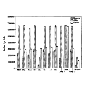
[0009] Figure 1 is a graphic representation of the ELISA
binding of a panel of humanized (h3B3) and affinity matured
anti-ADDL (14.2, 7.2, 11.4, 9.2, 13.1, 17.1, and 19.3)
antibodies and three comparator antibodies (Comp 1, 2, and
3) to monomer Ap, ADDLs and fibrillar A. The background of
this assay was determined by removing the capture antibody
from the ELISA (no mAb). Error bars represent standard
error of the mean.
[0010] Figure 2 is a graphic representation of the ELISA
binding of anti-ADDL antibody 19.3 and antibody 3B3 to
ADDLs or monomer Ap (A131-40) evaluated with an 11 point
titration curve.
[0011] Figure 3 is a graphic representation of the ability
of anti-ADDL antibody 19.3 and 3B3 to block ADDL binding to
primary hippocampal neuronal cells after pre-incubation
with increasing concentration of the antibody. The ability
CA 02891627 2015-05-13
WO 2014/089149 PCT/US2013/072991
of anti-ADDL antibody 19.3 to block ADDL binding to neurons
was attenuated after heat denaturing of the antibody. Error
bars represent standard error of the mean.
[0012] Figures 4A-40 are graphic representations of the
ELISA binding to ADDLs of the anti-ADDL antibody 19.3
(designated as WT in Figure 4A) and two 19.3-derived anti-
ADDL antibodies (Figures 4B and 40) after incubation up to
one month at varying temperatures to evaluate antibody
stability. The 19.3-derived anti-ADDL antibodies had a
single amino-acid substitution of Asn33 within light chain
CDR1 to either Ser33 (19.3S33) or Thr33 (19.3T33) (SEQ ID
NOS: 42 and 43, respectively). Substitution of Asn33 with
either S33 (Figure 4B) or T33 (Figure 40) resulted in
improved antibody stability versus the parental 19.3
antibody.
[0013] Figure 5 is a graphic representation of the binding
and dissociation of anti-ADDL antibodies to immobilized
human FcRn when assessed with BIACORE'1' (GE Healthcare,
Piscataway, NJ). The adjusted sensorgram shows initial
binding at pH 6.0 and then the dissociation of antibodies
at pH 7.3 from 180 seconds. A report point (Stability) was
inserted at 5 seconds after the end of pH 6.0 binding and
the "% bound" was calculated as RUstability/RUBinding(%).
[ 0014 ] Figure 6 shows the alignment of the heavy and light
chain variable regions for anti-ADDL antibody 19.3 with a
human germ line with the complementary determining regions
(CDRs) indicated in bold type face. Antibody 19.3 heavy
chain variable region (SEQ ID NO:7), antibody 3-66 human
heavy chain variable region (SEQ ID NO:8), Antibody 19.3
light chain variable region (SEQ ID NO:9), antibody 3-66
human light chain variable region (SEQ ID NO:10).
[0015] Figure 71\ shows a one-sided ELISA with plates coated
with either Ap oligomer (triangles) or Ap monomer
6
CA 02891627 2015-05-13
WO 2014/089149 PCT/US2013/072991
(squares), demonstrating the relative affinities and
maximum binding characteristics of the humanized antibody
19.3.
[0016] Figure 7B shows a competitive ELISA and the relative
affinities of 19.3 for Ap oligomers (triangles) and Ap
monomer (squares) coated on an ELISA plate in the presence
of the competing species in solution.
[0017] Figures 8A and 8B are graphic representations of the
levels of Ap oligomers detected in human cerebrospinal
fluid (CSF) samples. Figure 8A shows that the Ap oligomers
levels were four-fold higher in AD patients as compared to
age-matched control, i.e., non-AD, patients in a blinded
evaluation. The differences were statistically significant
to p < 0.0004 as determined using a two-way t-test and Mann
Whitney analysis of ranks, assuming the population was non-
Gaussian. Figure 8B shows that the Ap oligomer levels were
eight-fold higher in AD patients as compared to young
control, i.e., non-AD, patients in a blinded evaluation.
The differences were also statistically significant between
these groups using the same statistical method as in Figure
8A to a p-value < 0.0021.
[0018] Figures 9A and 9B are graphic representations of Ap
monomer levels in the CSF of either clinically confirmed AD
or young control, i.e., non-AD, patients, with a
corresponding decrease in the levels of Ap1-42 monomer and
unchanged levels of A131-40 monomer in the AD samples. This
is representative of the general pattern observed for AD
patients and confirmed the disease state of the samples
evaluated in Figure 8B. Figure 9A shows the reduced levels
of Ap1-42 monomer in the AD CSF samples. The differences
were statistically significant to p < 0.002 as determined
using a two-way t-test and Mann Whitney analysis of ranks,
assuming the population was non-Gaussian. Figure 9B shows
7
CA 02891627 2015-05-13
WO 2014/089149 PCT/US2013/072991
the unchanged levels between the two groups of A131-40
monomer.
[0019] Figure 10 is a graphical representation of the
pharmacokinetic (PK) profile of anti-ADDL antibodies 19.3
and 3B3 evaluated in heterozygous 276 human FcRn mice
(Jackson Laboratory (Bar Harbor, ME) following a single 10
mg/kg intravenous (IV) administration. The concentration of
antibody was measured at various time intervals to
determine the half-life (t1/2) of free antibody (19.3: 77 6
hours; 3B3: 29 9 hours).
[0020] Figure 11 is a graphical representation of the PK of
anti-ADDL antibody 19.3 (in serum) assessed in six rhesus
monkeys following administration of a bolus intravenous
(IV) or subcutaneous (SC) dose of 5 mg/kg. A half-life
(t1/2) of 254 28 (274 9) hours was determined after IV
administration and 204 49 (219 52) hours after SC dosing.
[0021] Figure 12 is a graphical representation of the PK of
anti-ADDL antibody 19.3 assessed in primate (three male
rhesus monkeys) cerebrospinal fluid (CSF) using a cisterna
magna ported rhesus model following administration of a
bolus IV dose of 5 mg/kg. At about 48 hours post-dose, the
anti-ADDL antibody 19.3 was present in the CSF at 0.1% of
the concentration in serum.
[0022] Figures 13A-13D are representations of the ability
of anti-ADDL antibody 19.3, versus two comparator
antibodies (Comp 1 and Comp2), to cross the blood-brain-
barrier in a transgenic mouse model that over-expresses
human amyloid precursor protein (hAPP). Mice were injected
intravenously (IV) with 1251 -labeled anti-ADDL antibody
19.3, or a comparator antibody, and the blood, CSF and
brain samples were collected two hours post-dose. Upon
assessment of the radioactivity distribution, 0.02% of
anti-ADDL antibody 19.3 was present in the CSF (Figure
8
CA 02891627 2015-05-13
WO 2014/089149 PCT/US2013/072991
13A), while 0.19% was seen in the brain (Figure 13B).
Similar levels were seen with the two comparator
antibodies. Immunocytochemical analysis demonstrated that
anti-ADDL antibody 19.3 is moving from plasma to the brain
and is concentrated after dosing (Figure 130, arrows), and
that some anti-ADDL antibody 19.3 is associated near the
periphery of plaques (Figure 13D). This shows that anti-
ADDL antibody 19.3 is able to penetrate into the brain and
bind ADDLs.
[0023] Figures 14A-140 show that anti-ADDL antibody 19.3
blocks the deposition of ADDLs into growing plaques in a
transgenic mouse model that over-expresses hAPP.
Biotinylated ADDLs (bADDLs) infused into the hippocampus of
12-month-old mice for four weeks (one injection per week)
(Figure 14A) labeled existing plaques (vehicle alone:
Figure 14B; antibody 19.3: Figure 140, ring).
Immunocytochemical analysis was used to assess the
deposition of new material (ADDLs) (Figures 14B and 140).
[0024] Figure 15 shows blood-brain-barrier penetration and
target engagement of antibody 19.3 in the brain. Levels of
antibody 19.3:ADDL complexes in the brain of female (left
panel) and male (right panel) Tg2576 mice 24 hours
following IV injection of antibody 19.3 were determined.
The asterisks indicate a statistically significant
difference from vehicle control levels. (RLU, relative
light units).
[0025] Figure 16 shows that parental anti-ADDL antibody 3B3
reverses acute Ap impairment of long term potentiation
(LTP) in murine hippocampal slices. The magnitude of LTP is
shown as a normalized potentiation of the fEPSP (field
excitatory postsynaptic potential) slope values averaged
from the last 10 minutes of recordings.
9
CA 02891627 2015-05-13
WO 2014/089149 PCT/US2013/072991
[0026] Figure 17 shows the behavioral effects of antibody
19.3. Shown is a comparison of locomotor activity in Tg2576
and non-transgenic control mice at Days 7, 14, and 21
expressed as percent change relative to baseline activity
prior to treatment with antibody 19.3 (30 mg/kg) and
vehicle, respectively. A significant decrease in locomotor
activity was observed 14 and 21 days post-treatment with
antibody 19.3. W-Veh = non-transgenic mice; T-Veh: Tg2576
mice treated with control IgG; T-30mpk 19.3 = Tg2576 mice
treated with antibody 19.3 at 30 mg/kg.
Detailed Description of the Invention
[0027] The present invention is directed to the treatment
of a disease caused by, resulting from, or associated with
the neurotoxic effects of soluble, oligomeric species of
amyloid 131-42 (A131-42) using an antibody that selectively
and specifically binds soluble, oligomeric species of A131-
42 with high affinity in vivo and blocks binding of the
same to neurons and amyloid plagues in vivo. The method of
this invention can be used to provide acute behavioral
benefits (e.g., within 1, 2 or 3 months of administration)
and chronic disease modification. Moreover, in accordance
with the method herein, the antibodies can be used as a
stand-alone therapy, or can be used in combination with
other Ap- and/or Tau-directed therapies.
[0028] The antibodies of this invention have the added
advantage of being capable of distinguishing between
Alzheimer's disease (AD) and control human brain extracts,
identifying endogenous oligomers in AD brain slices and on
hippocampal cells, and neutralizing endogenous and
synthetic ADDLs in solution. Antibodies of the invention
specifically bind one or more multi-dimensional
conformations of ADDLs, bind particular ADDLs derived from
CA 02891627 2015-05-13
WO 2014/089149 PCT/US2013/072991
the oligomerization of A131-42, while having significantly
lower affinity or substantially no affinity for other Ap
species, including amyloid beta 1-42 monomer, amyloid beta
1-40 monomer, plagues and amyloid beta fibrils.
[0029] This invention is particularly directed to the use
of antibodies 17.1, 14.2, 13.1, 19.3, 7.2, 9.2, 11.4, and
derivatives thereof, that preferentially bind ADDLs and
that have been characterized as to their specificity and
selectivity for ADDLs. Importantly, the specificity and
selectivity of the antibodies of this invention was not
predictable from the linear epitope of Ap to which they
bound, nor was this activity predictable from their ability
to detect ADDLs by western blot analysis, or from their
ability to detect immuno-stained ADDLs bound to neurons.
Moreover, the differential ability of the anti-ADDL
antibodies of the invention to neutralize ADDLs and block
binding to primary hippocampal neurons supports the belief
that the antibodies of this invention act through binding
to a more relevant, conformational epitope, which prevents
soluble oligomeric species of Ap1-42 from binding to
neurons and amyloid plaques. One embodiment of the present
invention, antibody 19.3, not only blocked the binding of
ADDLs to primary neurons, but also abated ADDL-induced
changes to hippocampal spine morphology, an indication that
the impedance of ADDL-neural binding has significant
physiological ramifications, for example, neuronal
survival, neuronal connectivity and signal transduction.
Antibody 19.3 also had an improved pharmacokinetic (PK)
profile, as compared with a previously known anti-ADDL
antibody, 3B3, when assessed in both in vitro and in vivo
models. In addition, when administered to transgenic mice
that over-express a human form of amyloid precursor protein
(hAPP), antibody 19.3 was shown to penetrate the blood-
11
CA 02891627 2015-05-13
WO 2014/089149 PCT/US2013/072991
brain-barrier, concentrate in the brain, and block
incorporation of ADDLs into amyloid plaques. Since ADDLs
are localized in the brain and act there to adversely
affect neuronal function, one of skill in the art would
appreciate and recognize that the penetration and
concentration of antibody in the brain would be beneficial
for immunotherapy. Taken together, these data demonstrate
that selective anti-ADDL antibodies, such as antibody 19.3,
can block the binding of ADDLs to hippocampal neurons,
which are critically involved in learning and memory.
[0030] The method of treatment herein is based on a body of
evidence that indicates that ADDLs, and not amyloid plaques
per se, play a fundamental role in the cognitive decline
associated with this disease (Walsh & Selkoe (2004) Protein
Pept. Lett. 11:213-228). ADDLs are elevated in the AD brain
and induce deficits in behavioral and electrophysiological
endpoints when centrally administered to rodents (Walsh, et
al. (2002) Nature 416:535-539; Cleary, et al. (2004) Nat.
Neurosci. 8:79-84; Klyubin, et al. (2005) Nat. Med. 11:556-
561; Balducci, et al. (2010) Proc. Natl. Acad. Sci. USA
107:2295-2300). Deficits in learning and memory have also
been observed in a hAPP expressing mouse model, with the
onset of impairment associated with elevated ADDL levels
(Westerman, et al. (2002) J. Neurosci. 22:1858-1867; Ashe
(2005) Biochem. Soc. Trans. 33:591-594; Lee, et al. (2005)
J. Biol. Chem. 281:4292-4299; Lesne, et al. (2006) Nature
440:352-357). While the cellular and sub-cellular events
that mediate these effects on cognition are not fully
understood, it is clear that ADDLs bind to the synaptic
terminals localized on the dendritic processes of
hippocampal neurons (Lacore, et al. (2004) J. Neurosci.
24:10191-1022) and alter the morphology and number of
dendritic spines (Lacor, et al. (2007) J. Neurosci. 27:796-
12
CA 02891627 2015-05-13
WO 2014/089149 PCT/US2013/072991
807; Shankar, et al. (2007) J. Neurosci. 27:2866-2875;
Shughrue, et al. (2010) Neurobiol. Aging 31:189-202). The
finding that ADDLs bind to both GABAergic and glutamate
neurons in the hippocampus (Shughrue, et al. (2010) supra),
neurons critically involved in learning and memory, which
results in the internalization of AMPA receptors (Zhao, et
al. (2010) J. Biol. Chem. 285:7619-7632), further supports
the indication that ADDLs directly or indirectly modulate
these neurotransmitter systems (see, for example,
Venkitaramani, et al. (2007) J. Neurosci. 27:11832-11837).
[0031] As described herein, a panel of anti-ADDL antibodies
derived from anti-ADDL antibody, 3B3 (US 7,780,963 and US
7,811,563), were assessed for their ability to block ADDL
binding to primary hippocampal neurons. Selected monoclonal
antibodies were then humanized and affinity matured for
further characterization. Lead antibodies, selected for
their ability to bind to ADDLs, were further assessed at a
single concentration using a three-pronged ELISA to
determine antibody binding to monomer Ap, ADDLs, and
fibrillar Ap. As shown in Figure 1, six of the seven
affinity matured anti-ADDL antibodies, specifically
antibodies 14.2, 7.2, 1 1.4, 13.1, 17.1, and 19.3 were ADDL
preferring, when compared with monomer Ap and fibrillar Ap.
[0032] Subsequently, an eleven point titration curve and
ELISA were used to ascertain the binding affinity of anti-
ADDL antibodies to ADDLs and monomer Ap (A131-40) over a
broad range of concentrations. As shown in Figure 2, the
anti-ADDL antibodies 3B3 and 19.3 were highly ADDL
selective. In addition, antibodies were compared in a cell-
based binding assay to determine the ability of antibodies
to block ADDL binding to neurons. As shown in Figure 3,
ADDLs, pre-incubated with increasing concentrations of
anti-ADDL antibodies 3B3 and 19.3, were added to primary
13
CA 02891627 2015-05-13
WO 2014/089149 PCT/US2013/072991
hippocampal neurons, and a titration curve was used to show
quantitatively the ability of the antibody to block ADDL
binding to neurons. Taken together, these results show that
anti-ADDL antibodies profoundly attenuate neuronal binding
in a cell-based format.
[0033] An assessment of the amino acid sequence was
conducted to identify potential sites of deamidation.
Asparagine and aspartic acid residues present in the CDRs
of therapeutic antibodies can undergo deamidation and
isoaspartate formation (Valsak & Ionescu (2008) Curr.
Pharm. Biotech. 9:468-481; Aswad, et al. (2000) J. Pharm.
Biomed. Anal. 21:1129-1136), the formation of which can
alter the binding potency of an antibody and, in turn,
reduce antibody effectiveness for use as a therapeutic.
Thus, those of skill in the art would recognize and
appreciate that the presence of an asparagine or an
aspartic acid within the CDRs for the 19.3 antibody would
not be desirable. Accordingly, the asparagine residue at
position 33 of the light chain CDR1 was altered to optimize
the stability of the anti-ADDL antibody 19.3. Derivatives
of the 19.3 antibody were produced with the substitution of
serine (SEQ ID NO:42), threonine (SEQ ID NO:43), or
glutamic acid (SEQ ID NO:45) for the asparagine at position
33 in CDR1. The substitution of aspartic acid (SEQ ID
NO:46) for the asparagine as position 33 was also generated
as a control. These changes remove the possibility of
deamidation of asparagine at position 33 in CDR1. The 19.3
derivatives were generated and characterized as described
in the Examples. As shown in Figures 4B and 4C,
respectively, two representative derivatives, 19.3S33 (SEQ
ID NO:42) and 19.3T33 (SEQ ID NO:43), had enhanced binding
stability following a one-month incubation at varying
temperatures. Other amino acid substitutions in the light
14
CA 02891627 2015-05-13
WO 2014/089149 PCT/US2013/072991
chain CDR1 for the asparagine at positions 33 and 35 (SEQ
ID NOs:47-50) and in the light chain CDR2 for the
asparagine at position 58 position (SEQ ID NOs:52-55) are
listed in Tables 7 and 8, respectively, for further
evaluation.
[0034] To determine the pharmacokinetics of the affinity
matured anti-ADDL antibodies of this invention, a series of
in vitro and in vivo studies were conducted. The binding of
antibodies to the FcRn receptor at pH 6.0 has been shown to
be predictive of antibody half-life in humans (Zalevsky, et
al. (2010) Nat. Biotech. 28(2):157-159) and at pH 7.3 (US
61/307,182) The binding and dissociation of the anti-ADDL
antibodies of the invention to immobilized human FcRn was
assessed via label-free interaction analysis, such as that
offered by BIACORETM Life Sciences, BIACOREm T-100 (GE
Healthcare, Piscataway, NJ). An adjusted sensorgram is used
to show the initial binding at pH 6.0 and then the
dissociation of antibodies at pH 7.3 from 180 seconds. A
report point (Stability) was inserted at 5 seconds after
the end of pH 6.0 binding and the "% bound" was calculated
as RU5Labi1iLy/RUBincung (%). As shown in Figure 5, the off-rate
for humanized 3B3 was markedly slower than the seven anti-
ADDL antibodies of this invention, which included antibody
19.3, =and three comparator antibodies. In that a slow off-
rate is thought to be an indicator of poor in vivo PK, an
additional in vivo study was conducted in transgenic FcRn
mice (heterozygous 276 human FcRn mice, Jackson
Laboratories, Bar Harbor, ME). When the transgenic FcRn
mice were given 10 mg/kg intravenously (IV) of either anti-
ADDL antibody 3B3 or 19.3, a significant difference in
pharmacokinetics was determined. As shown in Figure 10, the
half-life (t1/2) of anti-ADDL antibody 333 was relatively
short (29 9 hours), which was consistent with the
CA 02891627 2015-05-13
WO 2014/089149 PCT/US2013/072991
prediction from the in vitro BIACORE'm data, while the half-
life for anti-ADDL antibody 19.3 was significantly longer
(77 6 hours). Given its more desirable PK, 19.3 is of use
as a therapeutic due to its bioavailability.
[0035] To confirm the predicted half-life of anti-ADDL
antibody 19.3 in primates, a primate pharmacokinetics study
was conducted for the antibody in a cohort of cisterna
magna ported rhesus monkeys. The animals were dosed with a
single intravenous (IV) bolus or subcutaneous (SC)
injection of anti-ADDL antibody 19.3 (5 mg/kg) and blood
samples collected after antibody administration.
Concurrently, CSF samples were collected from the cisterna
magna port at timed intervals and the concentration of
anti-ADDL antibody 19.3 in serum and CSF was determined
with an anti-human IgG ELISA assay. When the animals were
administered anti-ADDL antibody 19.3 by a single IV bolus
injection a t1/2 of 254 28 hours was observed (Figure 11),
while a t1/2 of 204 49 hours was observed for the
subcutaneous administration. In addition, it was found that
anti-ADDL antibody 19.3 was able to cross into the primate
CSF, where it increased in concentration during the first
48 hours and peaked at about 0.1% of the antibody dosed
(Figure 12).
[0036] To ascertain the amount of Ap oligomeric species
present in the brain of AD patients, Ap oligomeric species
were determined in AD brain as compared to age-matched
(Figure 8A) and young (Figure 8B) controls. The absolute
levels of Ap oligomers observed were -2 pg/mL in AD and 0.2
pg/mL in control CSF samples. To compare the levels of Ap
oligomeric species to the amount of antibody that crosses
the blood-brain barrier, anti-ADDL antibody 19.3 and two
comparator antibodies (Comp 1 and Comp 2) were 125I-labeled
and administered to aged (twelve-month old) mice that over-
16
CA 02891627 2015-05-13
WO 2014/089149 PCT/US2013/072991
express hAPP, a rodent model for AD. Two hours after IV
dosing, about 0.02% of antibody 19.3 was seen in the CSF
(Figure 13A), while about 0.19% of antibody 19.3 was seen
in the brain (Figure 13B). Similar levels were seen for the
two comparator antibodies (Figure 13A and 13B). When
immunocytochemical analysis was carried out on brain
sections of the dosed mice and the localization of anti-
ADDL antibody 19.3 was determined (arrow in Figure 130), a
concentration of the antibody associated with the
deposition of Ap into plaques was observed (Figure 13D).
Recently, it was shown that exogenous ADDLs were deposited
into plaques when administered to mice that overexpress
hAPP (Gaspar, et al. (2010) Exp. Neurol. 223:394-400).
Thus, the findings herein confirmed that the localized
anti-ADDL antibody 19.3 bound to circulating ADDLs that
became associated with plaques. Overall, this analysis
demonstrated that the anti-ADDL antibody 19.3 penetrated
into the CSF and brain at a level sufficient to bind the
soluble oligomeric species of Ap present in the brain.
Moreover, the animal model studies indicated that the
minimal efficacious dose to significantly elevate antibody
19.3:ADDL complexes in the brain was 10 mg/kg (Figure 15).
[0037] To further evaluate the in vivo efficacy of the
antibodies of this invention, the ability of antibody 19.3
to block the deposition of ADDLs into growing plaques was
assessed in hAPP transgenic mice following four weekly
infusions of biotinylated ADDLs (bADDLs) into the
hippocampus of 12-month old mice to label existing plaques
(Figure 14A). The animals then received four weekly
intravenous infusions of antibody 19.3 (Figure 14A). The
deposition of new material (ADDLs) into growing plaques was
assessed by immunocytochemical analysis. As seen in Figures
14B and 140, anti-ADDL antibody 19.3 significantly reduced
17
CA 02891627 2015-05-13
WO 2014/089149 PCT/US2013/072991
the deposition of ADDLs into the periphery of existing
plaques (Figure 14C) as compared to mice treated with
vehicle alone (Figure 14B), but did bind vascular plaques.
Taken together, these results demonstrated that an anti-
ADDL antibody, specifically the 19.3 antibody, was able to
cross the blood-brain-barrier, bind ADDLs, and block the
deposition of new material into growing plagues.
[0038] ADDL binding may also have long-term effects on
neurons. Recent studies have shown that ADDL binding to
hippocampal neurons can initiate a signaling cascade that
results in the phosphorylation of tau (De Felice, et al.
(2006) Neurobiol. Aging 29:394-400). One component of this
signaling cascade, GSK-313, has also been shown to be
modulated by ADDL binding in vivo and in vitro (Ma, et al.
(2006) J. Neurosci. Res. 83:374-384). In this study, it was
observed that passive immunization of hAPP mice with an
antibody that reduced ADDLs also reduced GSK-313 levels and
phosphorylation of tau in the cortex. This finding supports
a link between Ap and phosphorylated tau and suggests that
ADDL binding may trigger events that lead to the
intracellular aggregation of tau. Further, the data
indicates that antibodies that prevent the binding of ADDLs
to neurons and the associated loss of synaptic spines, such
as the antibodies of this invention, would ameliorate the
cognitive and/or pathological outcomes associated with
Alzheimer's disease and related diseases. In this respect,
it was demonstrated that an anti-ADDL antibody can reverse
acute ADDL impairment of LTP in murine hippocampal slices
(Figure 16) and alter behavioral activity by reverting
increases in locomotor activity in the Tg2576 mouse model
of AD (Figure 17).
[0039] Accordingly, this invention includes the use of an
anti-ADDL antibody or antibody fragment to prevent or treat
18
CA 02891627 2015-05-13
WO 2014/089149 PCT/US2013/072991
a disease associated with, caused by, or resulting from the
accumulation of ADDLs (for example, Alzheimer's disease or
similar memory-related disorders). Evidence in the art
indicates that elevated levels of Ap, but not necessarily
aggregated plague, cause Alzheimer's disease-associated
dementia and subsequent tau abnormalities. 1M3-derived
diffusible ligands are directly implicated in neurotoxicity
associated with Alzheimer's disease. The art indicates that
ADDLs are elevated in transgenic mice and Alzheimer's
disease patients and modulate functional activity
associated with mnemonic processes in animal models. Thus,
removing this form of Ap would provide relief from the
neurotoxicity associated with Alzheimer's disease. As such,
treatment with an antibody of the present invention that
reduces central nervous system ADDL load could prove
efficacious for the treatment of Alzheimer's disease.
[0040] Patients amenable to treatment include individuals
at risk of disease but not exhibiting symptoms, as well as
patients presently exhibiting symptoms. In the case of
Alzheimer's disease, virtually anyone is at risk of
suffering from Alzheimer's disease if he or she lives long
enough. Therefore, the antibody or antibody fragments of
the present invention can be administered prophylactically
to the general population without the need for any
assessment of the risk of the subject patient. The present
methods are especially useful for individuals who have a
known genetic risk of Alzheimer's disease. Such individuals
include those having relatives who have been diagnosed with
the disease, and those whose risk is determined by analysis
of genetic or biochemical markers. Genetic markers of risk
for Alzheimer's disease include mutations in the APP gene,
particularly mutations at position 717 and positions 670
and 671 referred to as the Hardy and Swedish mutations,
19
CA 02891627 2015-05-13
WO 2014/089149 PCT/US2013/072991
respectively. Other markers of risk are mutations in the
presenilin genes, PS1 and PS2, and ApoE4, family history of
Alzheimer's disease, hypercholesterolemia or
atherosclerosis. Individuals presently suffering from
Alzheimer's disease can be recognized from characteristic
dementia, as well as the presence of risk factors described
above. In addition, a number of diagnostic tests are
available for identifying individuals who have Alzheimer's
disease. These include measurement of CSF tau and Al-42
levels. Individuals suffering from Alzheimer's disease can
also be diagnosed by ADRDA criteria or the method disclosed
herein.
[0041] In asymptomatic patients, treatment can begin at any
age (for example, 10, 20, 30 years of age). Usually,
however, it is not necessary to begin treatment until a
patient reaches 40, 50, 60 or 70 years of age. Treatment
typically entails multiple dosages over a-period of time.
Treatment can be monitored by assaying for the presence of
ADDLs over time.
[0042] In therapeutic applications, a pharmaceutical
composition or medicament containing an antibody or
antibody fragment of the invention is administered to a
patient suspected of, or already suffering from such a
disease associated with the accumulation of ADDLs in an
amount sufficient to cure, or at least partially arrest,
the symptoms of the disease (biochemical, histologic and/or
behavioral), including its complications and intermediate
pathological phenotypes in development of the disease. In
prophylactic applications, a pharmaceutical composition or
medicament containing an antibody or antibody fragment of
the invention is administered to a patient susceptible to,
or otherwise at risk of, a disease associated with the
accumulation of ADDLs in an amount sufficient to achieve
CA 02891627 2015-05-13
WO 2014/089149 PCT/US2013/072991
passive immunity in the patient thereby eliminating or
reducing the risk, lessening the severity, or delaying the
onset of the disease, including biochemical, histologic
and/or behavioral symptoms of the disease, its
complications and intermediate pathological phenotypes
present during development of the disease. In some methods,
administration of agent reduces or eliminates myocognitive
impairment in patients that have not yet developed
characteristic Alzheimer's pathology. In particular
embodiments, an effective amount of an antibody or antibody
fragment of the invention is an amount which achieves at
least a 15%, 20%, 30%, 40%, 50%, 60%, 70%, 80%, 90%, 95%,
or 97% decrease in the binding of ADDLs to neurons in the
patient as compared to binding of ADDLs in the absence of
treatment so that impairment of .. long-term
potentiation/memory formation is decreased.
[0043] Effective doses of the compositions of the present
invention, for the treatment of the above described
conditions vary depending, upon many different factors,
including means of administration, physiological state of
the patient, whether the patient is human or an animal,
other medications administered, and whether treatment is
prophylactic or therapeutic. Usually, the patient is a
human but nonhuman mammals such as dogs or transgenic
mammals can also be treated.
[0044] Treatment dosages are generally titrated to optimize
safety and efficacy. For passive immunization with an
antibody or antibody fragment, dosage ranges from about
0.0001 to 100 mg/kg, and more usually 0.01 to 20 mg/kg, of
the host body weight are suitable. For example, dosages can
be 0.5 mg/kg body weight or 10 mg/kg body weight or within
the range of 0.5-10 mg/kg are particularly contemplated. In
one embodiment, the dose is at or about 10 mg/kg (i.e., 5
21
CA 02891627 2015-05-13
WO 2014/089149 PCT/US2013/072991
mg/kg). In another embodiment, the dose is at or about 1
mg/kg (i.e., 0.5 mg/kg). In some methods, two or more
antibodies of the invention with different binding
specificities are administered simultaneously, in which
case the dosage of each antibody administered falls within
the ranges indicated. Antibodies are usually administered
on multiple occasions, wherein intervals between single
dosages can be weekly, monthly or yearly. An exemplary
treatment regime entails subcutaneous dosing, once biweekly
or monthly. Intervals can also be irregular as indicated by
measuring blood levels of antibody to ADDLs in the patient.
In some methods, dosage is adjusted to achieve a plasma
antibody concentration of 1-1000 pg/mL and in some methods
25-300 pg/mL. Alternatively, the antibody or antibody
fragment can be administered as a sustained-release
formulation, in which case less frequent administration is
required.
[0045] Dosage and frequency vary depending on the half-life
of the antibody in the patient. In general, human and
humanized antibodies have longer half-lives than chimeric
antibodies and nonhuman antibodies. As indicated above,
dosage and frequency of administration can vary depending
on whether the treatment is prophylactic or therapeutic. In
prophylactic applications, a relatively low dosage is
administered at relatively infrequent intervals over a long
period of time. Some patients continue to receive treatment
for the rest of their lives. In therapeutic applications, a
relatively high dosage at relatively short intervals is
sometimes required until progression of the disease is
reduced or terminated, and preferably until the patient
shows partial or complete amelioration of symptoms of
disease. Thereafter, the patient can be administered a
prophylactic regime.
22
CA 02891627 2015-05-13
WO 2014/089149 PCT/US2013/072991
[0046] Antibody and antibody fragments of the present
invention can be administered as a component of a
pharmaceutical composition or medicament. Pharmaceutical
compositions or medicaments generally contain the active
therapeutic agent and a variety of other pharmaceutically
acceptable components. See, Remington: The Science and
Practice of Pharmacy, Alfonso R. Gennaro, editor, 20th ed.
Lippincott Williams & Wilkins: Philadelphia, PA, 2000. The
preferred form depends on the intended mode of
administration and therapeutic application. Pharmaceutical
compositions can contain, depending on the formulation
desired, pharmaceutically-acceptable, non-toxic carriers or
diluents, which are defined as vehicles commonly used to
formulate pharmaceutical compositions for animal or, human
administration. Diluents are selected so as not to affect
the biological activity of the combination. Examples of
such diluents are distilled water, physiological phosphate-
buffered saline, Ringer's solutions, dextrose solution, and
Hank's solution.
[0047] Pharmaceutical compositions can also contain large,
slowly metabolized macromolecules such as proteins,
polysaccharides such as chitosan, polylactic acids,
polyglycolic acids and copolymers (such as latex-
functionalized SEPHAROSE"4, agarose, cellulose, and the
like), polymeric amino acids, amino acid copolymers, and
lipid aggregates (such as oil droplets or liposomes).
[0048] Administration of a pharmaceutical composition or
medicament of the invention can be carried out in a variety
of routes including, but not limited to, oral, topical,
pulmonary, rectal, subcutaneous, intradermal, intranasal,
intracranial, intramuscular, intraocular, or intrathecal or
intra-articular injection, and the like. The most typical
route of administration is intravenous followed by
23
CA 02891627 2015-05-13
WO 2014/089149 PCT/US2013/072991
subcutaneous, although other routes can be equally
effective.
[0049] Intramuscular injection can also be performed in the
arm or leg muscles. In some methods, agents are injected
directly into a particular tissue where deposits have
accumulated, for example, intracranial or intrathecal
injection. In some embodiments, an antibody or antibody
fragment is injected directly into the cranium or CSF. In
other embodiments, antibody or antibody fragment is
administered as a sustained-release composition or device,
such as a MEDIPADTM device.
[0050] For parenteral administration, antibody or antibody
fragments of the invention can be administered as
injectable dosages of a solution or suspension of the
substance in a physiologically acceptable diluent with a
pharmaceutical carrier that can be a sterile liquid such as
water, oils, saline, glycerol, or ethanol. Additionally,
auxiliary substances, such as wetting or emulsifying
agents, surfactants, pH buffering substances and the like
can be present in compositions. Other components of
pharmaceutical compositions are those of petroleum, animal,
vegetable, or synthetic origin, for example, peanut oil,
soybean oil, and mineral oil. In general, glycols such as
propylene glycol or polyethylene glycol are suitable liquid
carriers, particularly for injectable solutions. Antibodies
can be administered in the form of a depot injection or
implant preparation which can be formulated in such a
manner as to permit a sustained-release of the active
ingredient.
[0051] An exemplary, composition contains an isolated
antibody, or antibody fragment thereof, of the present
invention formulated as a sterile, clear liquid at a
concentration of at least 10 mg/ml in isotonic buffered
24
saline (10 mM histidine, 150 mM sodium chloride, 0.01%
(w/v) POLYSORBATE 80, pH 6.0). An exemplary antibody
formulation is filled as a single dose, 0.6 ml glass vials
filled with 0.3 ml of solution per vial. Each vial is
stopped with a TEFLONTm-coated stopper and sealed with an
aluminum cap.
[0052] Typically, compositions are prepared as injectables,
either as liquid solutions or suspensions; solid forms
suitable for solution in, or suspension in, liquid vehicles
prior to injection can also be prepared. The preparation
also can be emulsified or encapsulated in liposomes or
micro particles such as polylactide, polyglycolide, or
copolymer for enhanced delivery.
[0053] For suppositories, binders and carriers include, for
example, polyalkylene glycols or triglycerides; such
suppositories can be formed from mixtures containing the
active ingredient in the range of 0.5% to 10%, or more
desirably l90-29..
[0054] Oral formulations include excipients, such as
pharmaceutical grades of mannitol, lactose, starch,
magnesium stearate, sodium saccharine, cellulose, and
magnesium carbonate. These compositions take the form of
solutions, suspensions, tablets, pills,
capsules,
sustained-release formulations or powders and contain 10%-
95% of active ingredient, or more suitably 25%-70%.
[0055] Topical application can result in transdermal or
intradermal delivery. Topical administration can be
facilitated by co-administration of the agent with cholera
toxin or detoxified derivatives or subunits thereof or
other similar bacterial toxins (see Glenn, et al. (1998)
Nature 391:851). Co-administration can be achieved by using
the components as a mixture or as linked molecules obtained
by chemical crosslinking or expression as a fusion protein.
-25-
CA 2891627 2020-02-14
CA 02891627 2015-05-13
WO 2014/089149 PCT/US2013/072991
[0056] Alternatively, transdermal delivery can be achieved
using a skin path or using transferosomes (Paul, et al.
(1995) Eur. J. Immunol. 25:3521-3524; Cevc, et al. (1998)
Biochem. Biophys. Acta 1368:201-215).
[0057] To provide prophylactic or therapeutic treatment of
diseases such as AD, monoclonal antibodies that
differentially recognize multi-dimensional conformations of
AP-derived diffusible ligands, i.e., ADDLs, were generated.
These antibodies were humanized and, in some embodiments,
affinity-matured. The antibodies advantageously distinguish
between Alzheimer's disease and control human brain
extracts, and identify endogenous A131-42 oligomers in
Alzheimer's disease brain slices and in cultured
hippocampal cells. Further, the antibodies of the present
invention neutralize endogenous and synthetic ADDLs in
solution. So-called "synthetic" ADDLs are produced in vitro
by mixing purified A131-42 under conditions that generate
ADDLs. See, US 6,218,506. The antibodies disclosed herein
exhibit a high degree of selectivity for ADDLs, with
minimal detection of monomer Ap species. Moreover, these
antibodies differentially block the ability of ADDL-
containing preparations to bind primary cultures of rat
hippocampal neurons and immortalized neuroblastoma cell
lines, and also block ADDL incorporation into amyloid
plagues. These findings demonstrate that these antibodies
possess a differential ability to recognize a multi-
dimensional conformation of ADDLs despite similar linear
sequence recognition and affinities. Since ADDLs are known
to associate with a subset of neurons and disrupt normal
neuronal function, the antibodies of this invention find
use in the prevention of ADDL binding to neurons and the
assembly of ADDLs into plagues and, in turn, can be used
26
CA 02891627 2015-05-13
WO 2014/089149 PCT/US2013/072991
for the treatment of ADDL-related diseases including
Alzheimer's disease.
[0058] Accordingly, one embodiment of the present invention
is an isolated antibody that differentially recognizes one
or more multi-dimensional conformations of ADDLs. An
"isolated" antibody of the present invention refers to an
antibody which is substantially free of other antibodies.
However, the molecule may include some additional agents or
moieties which do not deleteriously affect the basic
characteristics of the antibody (for example, binding
specificity, neutralizing activity, etc.).
[0059] An antibody which is capable of specifically and
selectively binding one Or more multidimensional
conformations of ADDLs, binds particular ADDLs derived from
the oligomerization of A131-42, but does not cross-react
with other Ap peptides, namely monomeric Al-12, A131-28,
A131-40, and A1312-28 as determined by western blot analyses
as disclosed herein, and preferentially binds .ADDLs in
solution. Specific binding between two entities generally
refers to an affinity of at least 106, 107, 108, 109, or 1010
M. Affinities greater than 108 M-1 are desired to achieve
specific binding.
[0060] In particular embodiments, an antibody that is
capable of specifically binding a multi-dimensional
conformation of one or more ADDLs is also raised against,
i.e., an animal is immunized with, multi-dimensional
conformations of ADDLs. In other embodiments, an antibody
that is capable of specifically binding a multi-dimensional
conformation of one or more ADDLs is raised against a low
n-mer-forming peptide such as Ap1-42[Nle35-Dpro37].
[0061] The term "epitope" refers to a site on an antigen to
which B and/or T cells respond or a site on a molecule
against which an antibody will be produced and/or to which
27
CA 02891627 2015-05-13
WO 2014/089149 PCT/US2013/072991
an antibody will bind. For example, an epitope can be
recognized by an antibody defining the epitope.
[0062] A linear epitope is an epitope wherein an amino acid
primary sequence comprises the epitope recognized. A linear
epitope typically includes at least 3, and more usually, at
least 5, for example, about 6 to about 10 amino acids in a
unique sequence.
[0063] A conformational epitope, in contrast to a linear
epitope, is an epitope wherein the primary sequence of the
amino acids comprising the epitope is not the sole defining
component of the epitope recognized (for example, an
epitope wherein the primary sequence of amino acids is not
necessarily recognized by the antibody defining the
epitope). Typically a conformational epitope encompasses an
increased number of amino acids relative to a linear
epitope. With regard to recognition of conformational
epitopes, the antibody recognizes a three-dimensional
structure of the peptide or protein. For example, when a
protein molecule folds to form a three-dimensional
structure, certain amino acids and/or the polypeptide
backbone forming the conformational epitope become
juxtaposed enabling the antibody to recognize the epitope.
[0064] Methods of determining conformation of epitopes
include, but are not limited to, for example, x-ray
crystallography, two-dimensional nuclear magnetic resonance
spectroscopy and site-directed spin labeling and electron
paramagnetic resonance spectroscopy. See, for example,
Epitope Mapping Protocols in Methods in Molecular Biology
(1996) Vol. 66, Morris (Ed.).
[0065] The term "A131-40 monomer" or "41-42 monomer" as
used herein refers to the direct product of the enzymatic
cleavage, i.e., aspartic protease activity, by S-secretase
and y-secretase on the amyloid protein precursor (APP) in a
28
CA 02891627 2015-05-13
WO 2014/089149 PCT/US2013/072991
cell-free or cellular environment. Cleavage of APP by p-
secretase generates the Ap species beginning at Asp 1
(numbering as to Ap peptide sequence after cleavage), while
y-secretase liberate the C-terminus of Ap, predominantly
either at residues 40 or 42.
[0066] Amyloid p-derived diffusible ligands or ADDLs refer
to neurotoxic, soluble, globular, non-fibrillar oligomeric
structures that are desirably composed of aggregates of
Ap1-42 peptides (e.g., eight or nine 1)01-42 peptides) and
are found associated with Alzheimer's disease. See US
Patent No. 6,218,506 and WO 01/10900. This is in contrast
to high molecular weight aggregation intermediates, which
form strings of micelles leading to fibril formation. The
term -Ap fibrils" or "fibrils" or "fibrillar amyloid" as
used herein refers to insoluble species of Ap that are
detected in human and transgenic mouse brain tissue because
of their birefringence with dyes such as thioflavin S. Ap
species that form fiber-like structures composed of Ap
monomers include 3-pleated sheets. These species are
believed to be immediate precursors to the extracellular
amyloid plaque structures found in AD brain.
[0067] As exemplified herein, the antibodies of this
invention specifically bind to or recognize at least one
multi-dimensional conformation of an ADDL. In particular
embodiments, the antibodies bind at least two, at least
three, or at least four multi-dimensional conformations of
an ADDL. Multi-dimensional conformations of ADDLs are
intended to encompass dimers, trimers, tetramers,
pentamers, hexamers, heptamers, octamers, nonamers,
decamers, etc. as defined by analysis via SDS-PAGE. Because
trimer, tetramer, etc. designations can vary with the assay
method employed (see, e.g., Bitan, et al. (2005) Amyloid
12:88-95), the definition of trimer, tetramer, and the
29
CA 02891627 2015-05-13
WO 2014/089149 PCT/US2013/072991
like, as used herein, is according to SDS-PAGE analysis. To
illustrate the differential binding capabilities of the
antibodies herein, it has been found that certain
antibodies will recognize one multi-
dimensional
conformation, for example, tetramers of ADDLs (US
7,780,963, murine antibodies 206 and 4E2), while other
antibodies recognize several
multidimensional
conformations, for example, trimers and tetramers of ADDLs
(US 7,780,963, murine antibodies 2A10, 2B4, 5F10, and 20C2
and humanized antibody 20C2). As such, the antibody of this
invention has oligomer-specific characteristics. In
particular embodiments, a multi-dimensional conformation of
an ADDL is associated with a specific polypeptide structure
which results in a conformational epitope that is
recognized by an antibody of the present invention. In
other embodiments, an antibody of the invention
specifically binds a multi-dimensional conformation ADDL
having a size range of approximately a trimer or tetramer,
which have molecular weights in excess of >50 kDa.
[0068] Preferably, an antibody of this invention is
selective for Ap oligomer, i.e., the antibody has a higher
affinity for 41-42 oligomers or ADDLs than for 41-42
monomer, 41-40 monomer, plagues and/or amyloid beta
fibrils. As demonstrated herein, selectivity can be
assessed using a variety of methods including, but not
limited to competitive binding assays such as one-sided
ELISA, sandwich ELISA or competitive ELISA assays. Based
upon this analysis, an antibody of this invention is
defined as being specific for Ap oligomers if it exhibits
at least a 2-fold, 3-fold, 4-fold, 5-fold higher affinity
for Ap oligomers compared to one or more of Ap1-42 monomer,
Al-40 monomer, plaques or amyloid beta fibrils when
assessed in a conventional assay, e.g., BIACORE, KINEXA, or
CA 02891627 2015-05-13
WO 2014/089149 PCT/US2013/072991
one-sided ELISA. In particular embodiments, the affinity of
the capture antibody for Ap1-42 oligomers compared to Ap1-
40 monomers in a competitive binding assay is at least
500:1. In other embodiments, the affinity of the antibody
for amyloid beta 1-42 oligomers compared to amyloid beta 1-
42 monomers in a sandwich ELISA assay is at least 500:1, at
least 600:1, at least 700:1, at least 800:1, at least 900:1
or more preferably at least 1000:1.
[0069] While antibodies of the invention may have similar
linear epitopes, such linear epitopes are not wholly
indicative of the binding characteristics of these
antibodies, i.e., ability to block ADDL binding to neurons,
prevent tau phosphorylation and inhibit ADDL incorporation
into plaques, because, as is well-known to the skilled
artisan, the linear epitope may only correspond to a
portion of the antigen's epitope (see, for example,
Breitling and Dubel (1999) Recombinant Antibodies, John
Wiley & Sons, Inc., NY, pg. 115). The antibodies of the
invention can be distinguished from those of the art as
being capable of differentially
recognizing
multidimensional ADDLs and accordingly differentially
blocking ADDL binding to neurons, differentially preventing
tau phosphorylation and differentially inhibiting
incorporation of ADDLs into amyloid plaques.
[0070] An antibody, as used in accordance with the
invention includes, but is not be limited to, polyclonal or
monoclonal antibodies, and chimeric, human (for example,
isolated from B cells), humanized, neutralizing, bispecific
or single chain antibodies thereof. In one embodiment, an
antibody of the invention is monoclonal. For the production
of antibodies, various hosts including goats, rabbits,
chickens, rats, mice, humans, and others, can be immunized
by injection with synthetic or natural ADDLs. Methods for
31
CA 02891627 2015-05-13
WO 2014/089149 PCT/US2013/072991
producing antibodies are well-known in the art. See, for
example, Kohler & Milstein (1975) Nature 256:495-497;
Harlow & Lane (1988) Antibodies: A Laboratory Manual, Cold
Spring Harbor Laboratory, New York.
[0071] Depending on the host species, various adjuvants can
be used to increase the immunological response. Adjuvants
used in accordance with the invention desirably augment the
intrinsic response to ADDLs without causing conformational
changes in the immunogen that affect the qualitative form
of the response. Particularly suitable adjuvants include 3
De-O-acylated monophosphoryl lipid A (MPL"; RIBI ImmunoChem
Research Inc., Hamilton, MT; see GB 2220211) and oil-in-
water emulsions, such as squalene or peanut oil, optionally
in combination with immune stimulants, such as
monophosphoryl lipid A (see, Stoute, et al. (1997) N. Engl.
J. Bed. 336:86-91), muramyl peptides (for example, N-
acetylmuramyl-L-threonyl-D-isoglutamine (thr-MDP), N-
acetyl-normuramyl-L-alanyl-D-isoglutamine (nor-MDP), N-
acetylmuramyl-L-alanyl-D-isoglutaminyl-L-alanine-2-(1'-2'
dipalmitoyl-sn-glycero-3-hydroxyphosphoryloxy) ethylamine
(E-PE), N-
acetylglucsaminyl-N-acetylmuramyl-L-Al-D-isoglu-
L-Ala-dipalmitoxy propylamide (DTP-DPP)), or other
bacterial cell wall components. Specific examples of oil-
in-water emulsions include MF59 (WO 90/14837), containing
5% Squalene, 0.5% TWEEN' 80, and 0.5% SPAN 85 (optionally
containing various amounts of MTP-PE) formulated into
submicron particles using a microfluidizer such as Model
110Y microfluidizer (Microfluidics, Newton, MA); SAF
containing 10% Squalene, 0.4% TWEEN' 80, 5% PLURONI00-
blocked polymer L121, and thr-MDP, either microfluidized
into a submicron emulsion or vortexed to generate a larger
particle size emulsion; and RIBITM adjuvant system (RAS)
(Ribi ImmunoChem, Hamilton, MT) containing 2% squalene,
32
CA 02891627 2015-05-13
WO 2014/089149 PCT/US2013/072991
0.2% TWEENTm 80, and one or more bacterial cell wall
components such as monophosphoryllipid A, trehalose
dimycolate (TDM), and cell wall skeleton (CWS).
[0072] Another class of adjuvants is saponin adjuvants,
such as STIMOLONTm (QS-21, Aquila, Framingham, MA) or
particles generated therefrom such as ISCOMs
(immunostimulating complexes) and ISCOMATRIV) (CSL Ltd.,
Parkville, Australia). Other suitable adjuvants include
Complete Freund's Adjuvant (CFA), Incomplete Freund's
Adjuvant (IFA), mineral gels such as aluminum hydroxide,
and surface-active substances such as lysolecithin,
PLURONICIO polyols, polyanions, peptides, CpG (WO 98/40100),
keyhole limpet hemocyanin, dinitrophenol, and cytokines
such as interleukins (IL-1, IL-2, and IL-12), macrophage
colony stimulating factor (M-CSF), and tumor necrosis
factor (TNF). Among adjuvants used in humans, BCG (bacilli
Calmette-Guerin) and Corynebacterium parvum are
particularly suitable.
[0073] An antibody to a multi-dimensional conformation ADDL
is generated by immunizing an animal with ADDLs. Generally,
ADDLs can be generated synthetically or by recombinant
fragment expression and purification. Synthetic ADDLs can
be prepared as disclosed herein, or in accordance with the
methods disclosed in US 6,218,506 and US 7,811,563.
Further, ADDLs can be fused with another protein such as
keyhole limpet hemocyanin to generate an antibody against
the chimeric molecule. The ADDLs can be conformationally
constrained to form an epitope useful as described herein
and furthermore can be associated with a surface for
example, physically attached or chemically bonded to a
surface in such a manner so as to allow for the production
of a conformation which is recognized by the antibodies of
the present invention.
33
CA 02891627 2015-05-13
WO 2014/089149 PCT/US2013/072991
[0074] Monoclonal antibodies to multi-
dimensional
conformations of ADDLs can be prepared using any technique
the provides for the production of antibody molecules by
continuous cell lines in culture. These include, but are
not limited to, the hybridoma technique, the human B-cell
hybridoma technique, and the EBV-hybridoma technique
(Kohler, et al. (1975) Nature 256:495-497; Kozbor, et al.
(1985) J. Immunol. Methods 81:31-42; Cote, et al. (1983)
Proc. Natl. Acad. Sci. USA 80:2026-2030; Cole, et al.
(1984) Mol. Cell Biol. 62:109-120).
[0075] In particular embodiments, the antibodies of the
invention are humanized. Humanized or chimeric antibodies
can be produced by splicing of mouse antibody genes to
human antibody genes to obtain a molecule with appropriate
antigen specificity and biological activity (see, Morrison,
et al. (1984) Proc. Natl. Acad. Sci. USA 81:6851-6855;
Neuberger, et al. (1984) Nature 312:604-608; Takeda, et al.
(1985) NdLuie 314:452-454; Queen, et al. (1989) Proc. NdLl.
Acad. Sci. USA 86:10029-10033; WO 90/07861). For example, a
mouse antibody is expressed as the Fv or Fab fragment in a
phage selection vector. The gene for the light chain (and
in a parallel experiment, the gene for the heavy chain) is
exchanged for a library of human antibody genes. Phage
antibodies that still bind the antigen are then identified.
This method, commonly known as chain shuffling, provided
humanized antibodies that should bind the same epitope as
the mouse antibody from which it descends (Jespers, et al.
(1994) Biotechnology NY 12:899-903). As an alternative,
chain shuffling can be performed at the protein level (see,
Figini, et al. (1994) J. Mol. Biol. 239:68-78).
[0076] Human antibodies can also be obtained using phage-
display methods. See, for example, WO 91/17271 and WO
92/01047. In these methods, libraries of phage are produced
34
CA 02891627 2015-05-13
WO 2014/089149 PCT/US2013/072991
in which members display different antibodies on their
outer surfaces. Antibodies are usually displayed as Fy or
Fab fragments. Phage displaying antibodies with a desired
specificity are selected by affinity enrichment to ADDLs.
Human antibodies against ADDLs can also be produced from
non-human transgenic mammals having transgenes encoding at
least a segment of the human immunoglobulin locus and an
inactivated endogenous immunoglobulin locus. See, for
example, WO 93/12227 and WO 91/10741. Human antibodies can
be selected by competitive binding experiments, or
otherwise, to have the same epitope specificity as a
particular mouse antibody. Such antibodies generally retain
the useful functional properties of the mouse antibodies.
Human polyclonal antibodies can also be provided in the
form of serum from humans immunized with an immunogenic
agent. Optionally, such polyclonal antibodies can be
concentrated by affinity purification using ADDLs as an
dffiniLy LeagenL.
[0077] As exemplified herein, humanized antibodies can also
be produced by veneering or resurfacing of murine
antibodies. Veneering involves replacing only the surface
fixed region amino acids in the mouse heavy and light
variable regions with those of a homologous human antibody
sequence. Replacing mouse surface amino acids with human
residues in the same position from a homologous human
sequence has been shown to reduce the immunogenicity of the
mouse antibody while preserving its ligand binding. The
replacement of exterior residues generally has little, or
no, effect on the interior domains, or on the inter-domain
contacts. See, for example, US 6,797,492.
[0078] Human or humanized antibodies can be designed to
have IgG, IgD, IgA, IgM or IgE constant regions, and any
isotype, including IgGl, IgG2, IgG3 and IgG4. In particular
CA 02891627 2015-05-13
WO 2014/089149 PCT/US2013/072991
embodiments, an antibody of the invention is IgG or IgM, or
a combination thereof. In one specific embodiment the
antibodies of the present invention are IgG2. Those of
skill in the art would understand that other isoforms can
be utilized herein. Exemplary sequences for these isoforms
are given in SEQ ID NOS:56-58. Other embodiments of the
present invention embrace a constant region formed by
selective incorporation of human IgG4 sequences into a
standard human IgG2 constant region. An exemplary mutant
IgG2 Fc is IgG2m4, set forth herein as SEQ ID NO:59.
Antibodies can be expressed as tetramers containing two
light and two heavy chains, as separate heavy chains and
light chains or as single chain antibodies in which heavy
and light chain variable domains are linked through a
spacer. Techniques for the production of single chain
antibodies are well-known in the art.
[0079] Exemplary humanized antibodies produced by CDR
grafting and veneering are disclosed in US 7,780,963; US
7,731,962 and US 7,811,563.
[0080] Diabodies are also contemplated. A diabody refers to
an engineered antibody construct prepared by isolating the
binding domains (both heavy and light chain) of a binding
antibody, and supplying a linking moiety which joins or
operably links the heavy and light chains on the same
polypeptide chain thereby preserving the binding function
(see, Holliger, et al. (1993) Proc. Natl. Acad. Sci. USA
90:6444: Poljak (1994) Structure 2:1121-1123). This forms,
in essence, a radically abbreviated antibody, having only
the variable domain necessary for binding the antigen. By
using a linker that is too short to allow pairing between
the two domains on the same chain, the domains are forced
to pair with the complementary domains of another chain and
create two antigen-binding sites. These dimeric antibody
36
CA 02891627 2015-05-13
WO 2014/089149 PCT/US2013/072991
fragments, or diabodies, are bivalent and bispecific. The
skilled artisan will appreciate that any method to generate
diabodies can be used. Suitable methods are described by
Holliger, et al. (1993) supra; Poljak (1994) supra; Zhu, et
al. (1996) Biotechnology 14:192-196, and US 6,492,123.
[0081] Fragments of an isolated antibody of the invention
are also expressly encompassed by the present invention.
Fragments are intended to include Fab fragments, F(ab')2
fragments, F(ab') fragments, bispecific scFv fragments, Fv
fragments, single domain antibodies and fragments produced
by a Fab expression library, as well as peptide aptamers.
For example, F(abv)2 fragments are produced by pepsin
digestion of the antibody molecule of the invention,
whereas Fab fragments are generated by reducing the
disulfide bridges of the F(ab')2 fragments. Alternatively,
Fab expression libraries can be constructed to allow rapid
and easy identification of monoclonal Fab fragments with
Lhe desired specificity (see, Huse, et al. (1989) Science
254:1275-1281). In particular embodiments, antibody
fragments of the present invention are fragments of
neutralizing antibodies which retain the variable region
binding site thereof, i.e. antigen binding fragment.
Exemplary are F(ab')2 fragments, F(ab') fragments, and Fab
fragments. See, generally, Immunology: Basic Processes
(1985) 2nd edition, J. Bellanti (Ed.) pp. 95-97.
[0082] Single domain antibodies or nanobodies are also
encompassed by this invention. Nanobodies are prepared by
splitting the dimeric variable domains from common human or
mouse IgG into monomers and camelizing a few key residues.
See, e.g., Davies & Riechmann (1994) FEBS Lett. 339:285-290
and Reichman & Muyldermans (1999) J. Immunol. Meth. 231:25-
38.
37
CA 02891627 2015-05-13
WO 2014/089149 PCT/US2013/072991
[0083] Peptide aptamers that differentially recognize
multi-dimensional conformations of ADDLs can be rationally
designed or screened for in a library of aptamers (for
example, provided by Aptanomics SA, Lyon, France). In
general, peptide aptamers are synthetic recognition
molecules whose design is based on the structure of
antibodies. Peptide aptamers are composed of a variable
peptide loop attached at both ends to a protein scaffold.
This double structural constraint greatly increases the
binding affinity of the peptide aptamer to levels
comparable to that of an antibody (nanomolar range).
[0084] Exemplary nucleic acid sequences encoding light and
heavy chain variable regions for use in producing antibody
and antibody fragments of the present invention are
disclosed herein in SEQ ID NOs: 60 and 61, respectively. As
will be appreciated by the skilled artisan, the heavy chain
variable regions disclosed herein, such as that shown in
SEQ ID NO:61, can be used in combination with any one of
the light chain variable regions disclosed herein to
generate antibodies with modified affinities, dissociation,
epitopes, and the like.
[0085] Antibodies or antibody fragments of the present
invention can have additional moieties attached thereto.
For example, a microsphere or microparticle can be attached
to the antibody or antibody fragment, as described in US
4,493,825.
[0086] Moreover, particular embodiments embrace antibody or
antibody fragments that are mutated and selected for
increased antigen affinity, neutralizing activity (i.e.,
the ability to block binding of ADDLs to neuronal cells or
the ability to block ADDL assembly or incorporation into
amyloid plagues), or a modified dissociation constant.
Mutator strains of E. coli (Low, et al. (1996) J. Mo1.
38
CA 02891627 2015-05-13
WO 2014/089149 PCT/US2013/072991
Biol. 260:359-368), chain shuffling (Figini, et al. (1994)
supra), and PCR mutagenesis are established methods for
mutating nucleic acid molecules encoding antibodies. By way
of illustration, increased affinity can be selected for by
contacting a large number of phage antibodies with a low
amount of biotinylated antigen so that the antibodies
compete for binding. In this case, the number of antigen
molecules should exceed the number of phage antibodies, but
the concentration of antigen should be somewhat below the
dissociation constant. Thus, predominantly mutated phage
antibodies with increased affinity bind to the biotinylated
antigen, while the larger part of the weaker affinity phage
antibodies remains unbound. Streptavidin can then assist in
the enrichment of the higher affinity, mutated phage
antibodies from the mixture (Schier, et al. (1996) J. Plol.
Biol. 255:28-43).
[0087] In particular embodiments of this invention,
variants of antibody h3B3 (i.e., 14.2, 7.2, 11.4, 13.1,
17.1, 19.3), or variants of antibody 19.3 (i.e., 19.3 N33S,
19.3 N33T, 19.3 N33A, 19.3 N33E, 19.3 N33D, 19.3 N33S-N35Q,
19.3 N33S-N35S, 19.3 N335-N35T, 19.3 N335-N35A, 19.3 N58Q,
19.3 N585, 19.3 N58T, 19.3N35A) are used in the method of
this invention. Accordingly, in some embodiments, an
antibody of the invention has a light chain variable region
with a GDR1 having the sequence Arg-Ser-Ser-Gln-Ser-Ile-
Val-His-Ser-Xaal-Gly-Xaa2-Thr-Thy-Leu-Glu (SEQ ID NO:1).
wherein Xaal is Asn, Ser, Thr, Ala, Asp or Glu and Xaa2 is
Asn, His, Gin, Ser, Thr, Ala, or Asp, a CDR2 having the
sequence Lys-Ala-Ser-Xaai-Arg-Phe-Ser (SEQ ID NO:2), wherein
Xaal is Asn, Gly, Ser, Thr, or Ala, and a CDR3 having the
sequence Phe-G1n-Gly-Ser-Xaal-Xaa2-Xaa2-Xaa4-Xaa5 (SEQ ID
NO:3), wherein Xaal is Arg, Lys or Tyr, Xaa2 is Val, Ala, or
Leu, Xaa3 is Pro, His, or Gly, Xaa4 is Ala, Pro, or Val, and
39
CA 02891627 2015-05-13
WO 2014/089149 PCT/US2013/072991
Xaa5 is Ser, Gly, Arg or Elie; and a heavy chain variable
region with a CDR1 having the sequence Gly-Phe-Thr-Phe-Ser-
Ser-Phe-Gly-Met-His (SEQ ID NO:4), a CDR2 having the
sequence Tyr-Ile-Ser-Arg-Gly-Ser-Ser-Thr-Ile-Tyr-Tyr-Ala-
Asp-Thr-Val-Lys-Gly (SEQ ID NO:5), and a CDR3 having the
sequence Gly-Ile-Thr-Thr-Ala-Leu-Asp-Tyr (SEQ ID NO:6).
Accordingly, in some embodiments, an antibody of the
invention has a light chain variable region with a CDR1
having the sequence Arg-Ser-Ser-Gln-Ser-Ile-Val-His-Ser-
Xaal-Gly-Xaa2-Thr-Thy-Leu-Glu (SEQ ID NO:1), wherein Xaal is
Thr, Ala, Asp or Glu and Xaa2 is Asn, His, Gin, Ser, Thr,
Ala, or Asp or wherein Xaal is Asn, Ser, Thr, Ala, Asp or
Glu and Xaa2 is Thr, a CDR2 having the sequence Lys-Ala-Ser-
Xaal-Arg-Phe-Ser (SEQ ID NO:2), wherein Xaal is Thr, and a
CDR3 having the sequence Phe-Gln-Gly-Ser-Xaa1-Xaa2-Xaa3-Xaa4-
Xaa5 (SEQ ID NO:3), wherein Xaal is Arg, Lys or Tyr, Xaa2 is
Val, Ala, or Leu, Xaa3 is Pro, His, or Gly, Xaa4 is Ala,
Pro, or Val, and Xaa5 is Ser, Gly, Arg or She; and a heavy
chain variable region with a CDR1 having the sequence Gly-
Phe-Thr-Phe-Ser-Ser-Phe-Gly-Met-His (SEQ ID NO:4), a CDR2
having the sequence Tyr-Ile-Ser-Arg-Gly-Ser-Ser-Thr-Ile-
Tyr-Tyr-Ala-Asp-Thr-Val-Lys-Gly (SEQ ID NO:5), and a CDR3
having the sequence Gly-Ile-Thr-Thr-Ala-Leu-Asp-Tyr (SEQ ID
NO: 6).
[0088] In some embodiments, the antibody of the method of
the invention is a variant of antibody h353 (i.e., 14.2,
7.2, 11.4, 13.1, 17.1, 19.3). In accordance with this
embodiment, the antibody has a light chain variable region
with a CDR1 having the sequence Arg-Ser-Ser-Gln-Ser-Ile-
Val-His-Ser-Asn-Gly-Asn-Thr-Tyr-Leu-Glu (SEQ ID NO:41), a
CDR2 having the sequence Lys-Ala-Ser-Asn-Arg-Phe-Ser (SEQ
ID NO:51), and a CDR3 of Phe-Gln-Gly-Ser-Xaal-Xaa2-Xaa3-Xaa4-
Xaa5 (SEQ ID NO:3), wherein Xaal is Arg, Lys or Tyr, Xaa2 is
CA 02891627 2015-05-13
WO 2014/089149 PCT/US2013/072991
Val, Ala, or Leu, Xaa3 is Pro, His, or Gly, Xaa4 is Ala,
Pro, or Val, and Xaa5 is Ser, Gly, Arg or Phe; and a heavy
chain variable region with a CDR1 of SEQ ID NO:4, a CDR2 of
SEQ ID NO:5, and a CDR3 of SEQ ID NO:6.
[0089] In other embodiments, the antibody of the method of
the invention is a variant of antibody 19.3, wherein the
CDR1 of the light chain variable region has been mutated
(i.e., 19.3 N33S, 19.3 N33T, 19.3 N33A, 19.3 N33E, 19.3
N33D, 19.3 N33S-N35Q, 19.3 N33S-N35S, 19.3 N33S-N35T, 19.3
N33S-N35A). In accordance with this embodiment, the
antibody has a light chain variable region with a CDR1 of
SEQ ID NO:1, a CDR2 of SEQ ID NO:2, and a CDR3 having the
sequence Phe-Gln-Gly-Ser-Arg-Leu-Gly-Pro-Ser (SEQ ID
NO:18); and a heavy chain variable region with a CDR1 of
SEQ ID NO:4, a CDR2 of SEQ ID NO:5, and a CDR3 of SEQ ID
NO:6.
[0090] In still other embodiments, the antibody of the
meLhod of the invention is a variant of antibody 19.3,
wherein the CDR2 of the light chain variable region has
been mutated (i.e., 19.3 N58Q, 19.3 N585, 19.3 N58T,
19.3N35A). In accordance with this embodiment, the antibody
has a light chain variable region with a CDR1 of SEQ ID
NO:41, a CDR2 of SEQ ID NO:2, a CDR3 of SEQ ID NO:17; and a
heavy chain variable region with a CDR1 of SEQ ID NO:4, a
CDR2 of SEQ ID NO:5, and a CDR3 of SEQ ID NO:6.
[0091] In certain embodiments, the CDR1 of the light chain
variable region of the antibody has the sequence Arg-Ser-
Ser-Gln-Ser-Ile-Val-His-Ser-Xaal-Gly-Xaa2-Thr-Thy-Leu-Glu
(SEQ ID NO:1), wherein Xaal is Thr, Ala, Asp or Glu and Xaa2
is Thr. In other embodiments, the CDR2 of the light chain
variable region of the antibody has the sequence Lys-Ala-
Ser-Xaal-Arg-Phe-Ser (SEQ ID NO:2), wherein Xaal is Thr.
41
CA 02891627 2015-05-13
WO 2014/089149 PCT/US2013/072991
[0092] An exemplary antibody of use in this invention is
antibody 19.3 having a heavy chain variable region sequence
as in SEQ ID NO:7 (i.e., CDR1, CDR2, and CDR3 of SEQ ID
NOs:4, 5 and 6, respectively) and light chain variable
region sequence as in SEQ ID NO:9 (i.e., CDR1, CDR2, and
CDR3 of SEQ ID NOs:41, 51, 18). See Figure 6. In certain
embodiments, the antibody used in the method of this
invention is not 3B3.
[0093] To facilitate production and enhance storage and use
of the antibody in the method of this invention, certain
embodiments include the use of an antibody that exhibits
less than a 10-fold decrease in EC50, in an ELISA-based
assay with Ap oligomers, when stored at 40 C for 1 month.
More preferably, the antibody exhibits less than a 6-fold,
5-fold, 4-fold, 3-fold, or 2-fold decrease in EC50 when
stored at 40 C for 1 month. Antibody stability can be
assessed as described in the Examples herein. Antibodies
having such stability at elevated temperatures are provided
in Example 7.
[0094] For some therapeutic applications it may be
desirable to reduce the dissociation of the antibody from
the antigen. To achieve this, phage antibodies are bound to
biotinylated antigen and an excess of unbiotinylated
antigen is added. After a period of time, predominantly the
phage antibodies with the lower dissociation constant can
be harvested with streptavidin (Hawkins, et al. (1992) J.
Mol. Biol. 226:889-96).
[0095] Various immunoassays including those disclosed
herein can be used for screening to identify antibodies, or
fragments thereof, having the desired specificity for
multi-dimensional conformations of ADDLs. Numerous
protocols for competitive binding (for example, ELISA),
latex agglutination assays, immunoradiometric assays,
42
CA 02891627 2015-05-13
WO 2014/089149 PCT/US2013/072991
kinetics (for example, BIACORETM analysis) using either
polyclonal or monoclonal antibodies, or fragments thereof,
are well-known in the art. Such immunoassays typically
involve the measurement of complex formation between a
specific antibody and its cognate antigen. A two-site,
monoclonal-based immunoassay utilizing monoclonal
antibodies reactive to two non-interfering epitopes is
suitable, but a competitive binding assay can also be
employed. Such assays can also be used in the detection of
multi-dimensional conformations of ADDLs in a sample.
[0096] An antibody or antibody fragment can also be
subjected to other biological activity assays, e.g.,
displacement of ADDL binding to neurons or cultured
hippocampal cells or blockade of ADDL assembly or ADDL
incorporation into amyloid plaques, in order to evaluate
neutralizing or pharmacological activity and potential
efficacy as a prophylactic or therapeutic agent. Such
assays are described herein and are well-known in the art.
[0097] Antibodies and fragments of antibodies can be
produced and maintained as hybridomas or, alternatively,
recombinantly produced in any well-established expression
system including, but not limited to, E. coli, yeast (e.g.,
Saccharomyces spp. and Pichia spp.), baculovirus, mammalian
cells (e.g., myeloma, CHO, COS), plants, or transgenic
animals (Breitling & Dubel (1999) Recombinant Antibodies,
John Wiley & Sons, Inc., NY, pp. 119-132). Antibodies and
fragments of antibodies can be isolated using any
appropriate methods including, but not limited to, affinity
chromatography, immunoglobulins-binding molecules (for
example, proteins A, L, G or H), tags operatively linked to
the antibody or antibody fragment (for example, His-tag,
FLAG -tag, Strep tag, c-myc tag) and the like. See,
Breitling & Dubel (1999) supra.
43
CA 02891627 2015-05-13
WO 2014/089149 PCT/US2013/072991
[0098] To assess prophylactic or therapeutic treatment of a
disease associated with ADDLs, the activity of the
antibodies and antibody fragments of this invention can be
analyzed for the ability to block or inhibit binding of
ADDLs to neuronal cells, inhibit assembly of higher order
oligomers, block ADDL incorporation into amyloid plaques,
and/or prevent the phosphorylation of tau protein at
Ser202/Thr205.
[0099] The ability of an antibody or antibody fragment to
block or inhibit binding of ADDLs to neuronal cells is
determined by measuring whether ADDLs are bound to neurons
in the presence of the antibody or antibody fragment. The
degree to which an antibody can block the binding of ADDLs
to a neuron can be determined in accordance with the
methods disclosed herein, i.e., immunocytochemistry, or
cell-based alkaline phosphatase assay, or any other
suitable assay. In particular embodiments, an antibody or
antibody fragment of the present invention achieves at
least a 15%, 20%, 30%, 40%, 50%, 60%, 70%, 80%, 90%, 95%,
or 97% decrease in the binding of ADDLs as compared to
binding of ADDLs in the absence of the antibody or antibody
fragment.
[00100] The ability of an antibody or antibody fragment to
block or inhibit assembly of ADDLs can be determined by
measuring whether assembly of larger oligomeric species of
Ap1-42, e.g., octamers or decamers, is inhibited in the
presence of the antibody or antibody fragment. The degree
to which an antibody can block the assembly of larger
oligomeric species of ADDLs can be determined by, e.g.,
FRET or fluorescence polarization or any other suitable
assay.
[00101] The ability of an antibody or antibody fragment to
prevent the phosphorylation of tau protein at Ser202/Thr205
44
CA 02891627 2015-05-13
WO 2014/089149 PCT/US2013/072991
can be determined by measuring whether tau protein is
phosphorylated in the presence of the antibody or antibody
fragment. The degree to which an antibody can prevent the
phosphorylation of tau protein at Ser202/Thr205 can be
determined in accordance with the methods disclosed herein
or any other suitable assay.
[00102] Blocking or decreasing binding of ADDLs to neurons,
inhibiting assembly of larger oligomeric species of ADDLs,
and/or preventing the phosphorylation of tau protein at
Ser202/Thr205 can be used as an indication that a disease
associated with the accumulation of ADDLs is
prophylactically or therapeutically being treated.
[00103] In accordance with the method herein, an antibody or
antibody fragment of the invention can optionally be
administered in combination with other agents that are at
least partly effective in treatment of amyloidogenic
disease. For example, the present antibody can be
administered with exisLing palliative treatments for
Alzheimer's disease, such as
acetylcholinesterase
inhibitors such as ARICEPTu4 (Donepezil Hydrochloride),
EXELOW4 (Rivastigmine Tartrate), and REMIYLm (galantamine
hydrohromide) and, the NMDA antagonist, NAMENDArm (memantine
HC1). In addition to these known treatments, particular
embodiments feature the use of one or more antibodies of
this invention in combination with an inhibitor of Ap
production and aggregation (e.g., a p-secretase inhibitor,
y-secretase inhibitor, A3-monomer aggregation inhibitor,
Pan-A13 immunotherapy, and/or fibrillic or amyloid plaque
immunotherapy) and/or tau therapy.
[00104] Secretase Enzyme Modulation. One approach for
reducing the levels of Ap involves modulating the activity
of the p- and y-secretase cleaving enzymes to inhibit the
production of AP. The p- and y-secretase enzymes are
CA 02891627 2015-05-13
WO 2014/089149 PCT/US2013/072991
aspartyl proteases that convert APP to Ap; treatment
strategies that involve the inhibition of these two enzymes
aim to reduce the levels of cerebral amyloid (Marlatt, et
al. (2005) Curr. Med. Chem. 12(10):1137-47; Lundkvist &
Naslund (2007) Curr. Opin. Pharmacol. 7(1):112-8). Several
agents targeting these enzymes are known including the p-
secretase inhibitors CTS-21166 (CoMentis, Inc.), Posiphen
(QR Pharma Inc.; Sabbagh (2009) Am. J. Geriatr.
Pharmacother. 7:167-185; Neugroschl & Sano (2009) Curr.
Neurol. Neurosci. Rep. 9:368-376), ACT-91 (AC Immune SA),
MK-8931 (Merck & Co. Inc.), LY2811376 (Eli Lilly & Co.),
TAK-070 (Takeda Pharmaceutical Company Limited), GSK188909
(Hussain, et al. (2007) J Neurochem. 100:802-809), KMI-429
(Asai, et al. (2006) J. Neurochem. 96:533-40), GRL-8234
(Chang, et al. (2010) FASEB J. 25:775-784), and 0M99-2 and
statine-, phenylnorstatine-,
hydroxyethylamine-,
carbinamine-, acylguanidine-,
aminoimidazole-,
aminohydantoin-, and aminoquinazoline-based inhibitors
(Ghosh, et al. (2012) J. Neurochem. 120:71-83). Gamma-
secretase inhibitors include, but are not limited to, MK-
0752 (Merck & Co Inc.), semagacestat (LY-450139; Eli Lilly
& Co; NCT00762411 and NCT00594568) (Lundkvist & Naslund
(2007) supra; Barten, et al. (2005) J. Pharmacol. Exp.
Ther. 312(2):635-643; Wong, et al. (2004) J. Biol. Chem.
279(13):12876-82), avagacestat (BMS-708163; Bristol-Myers
Squib), EVP-0962 (EnVivo Pharmaceuticals).
[00105]Modulation of Beta-AmyJoid Aggregation. Several
inhibitors of beta-amyloid aggregation have been developed
including PBT2 (Prana Biotechnology Ltd.), which is a zinc
and copper ionophore that prevent beta-amyloid toxicity;
and ELND0005 (scyllo-inositol; Elan Corporation, PLC).
Additional compounds for inhibiting the formation of
neurotoxic beta-amyloid species are described in WO
46
CA 02891627 2015-05-13
WO 2014/089149 PCT/US2013/072991
2009/008891 and include, but are not limited to, 2-hydroxy-
benzoic acid[3-oxo-
3-(1-methy1-1H-pyrazol-5-y1)-1-
trifluoromethyl-prop-(Z)-ylidene]-hydrazide, 2-hydroxy-
benzoic acid[3-oxo-3-thiophen-2-y1-1-trifluoromethyl-prop-
(Z)-ylidene]-hydrazide, 2-hydroxy-benzoic acid[3-oxo-
3-
furan-2-y1-1-trifluoromethyl-prop-(Z)-ylidene]-hydrazide,
N-(2-methoxy-pheny1)-2-oxo-2-{N'-[3-oxo-3-thiophen-2-y1-1-
trifluoromethyl-prop-(Z)-ylidene]-hydrazinol-acetamide, 2-
([1-(2-hydroxy-3-methoxy-pheny1)-meth-(E)-ylidene-
hydrazinooxaly1]-amino}-6-methy1-4,5,6,7-tetrahydro-
benzo[b]thiophene-3-carboxylic acid ethyl ester, 2-([1-(2-
hydroxynaphthalen-1-y1)-meth-(E)-ylidene-hydrazinooxaly1]-
amino]-6-methy1-4,5,6,7-tetrahydro-benzo[b]thiophene-3-
carboxylic acid ethyl ester, 2-{[l-(2-hydroxypheny1)-meth-
(E)-ylidene-hydrazinooxaly1]-amino1-6-methy1-4,5,6,7-
tetrahydro-benzo[b]thiophene-3-carboxylic acid ethyl ester,
2-(3-ethy1-5-hydroxy-5-(trifluoromethyl)-4,5-dihydro-1H-
pyrazol-1-y1)-N-(2-methoxypheny1)-2-oxoacetamide, 2-(5-
hydroxy-3-propy1-5-(trifluoromethyl)-4,5-dihydro-1H-
pyrazol-1-y1)-N-(2-methoxypheny1)-2-oxoacetamide, 2-(5-
hydroxy-3-isopropy1-5-(trifluoromethyl)-4,5-dihydro-1H-
pyrazol-1-y1)-N-(2-methoxypheny1)-2-oxoacetamide, 2-(5-
hydroxy-3-isobuty1-5-(trifluoromethyl)-4,5-dihydro-1H-
pyrazol-1-y1)-N-(2-methoxypheny1)-2-oxoacetamide, (5-
hydroxy-3-propy1-5-(trifluoromethyl)-4,5-dihydro-1H-
pyrazol-1-y1)(2-hydroxyphenyl)methanone, N-(4-bromopheny1)-
2-(3-ethy1-5-hydroxy-5-(trifluoromethyl)-4,5-dihydro-1H-
pyrazol-1-y1)-2-oxoacetamide, 2-(5-
hydroxy-3-propy1-5-
(trifluoromethyl)-4,5-dihydro-1H-pyrazol-1-y1)-2-oxo-N-
phenethylacetamide, N-(4-
bromopheny1)-2-(5-hydroxy-3-
propy1-5-(trifluoromethyl)-4,5-dihydro-1H-pyrazol-1-y1)-2-
oxoacetamide, 2-(3-buty1-5-hydroxy-5-(trifluoromethyl)-4,5-
dihydro-1H-pyrazol-1-y1)-2-oxo-N-phenethylacetamide, 2-(5-
47
CA 02891627 2015-05-13
WO 2014/089149 PCT/US2013/072991
hydroxy-3-isobuty1-5-(trifluoromethyl)-4,5-dihydro-1H-
pyrazol-1-y1)-2-oxo-N-phenethylacetamide, N-(4-
bromopheny1)-2-(5-hydroxy-3-penty1-5-(trifluoromethyl)-4,5-
dihydro-1H-pyrazol-1-y1)-2-oxoacetamide, 2-(5-
hydroxy-3-
penty1-5-(trifluoromethyl)-4,5-dihydro-1H-pyrazol-1-y1)-2-
oxoacetamide, 2-(5-hydroxy-3-isopenty1-5-(trifluoromethyl)-
4,5-dihydro-1H-pyrazol-1-y1)-2-oxo-N-phenethylacetamide, 2-
(5-hydroxy-3-isopenty1-5-(trifluoromethyl)-4,5-dihydro-1H-
pyrazol-1-y1)-N-(4-methoxypheny1)-2-oxoacetamide, 2-(3-
hexy1-5-hydroxy-5-(trifluoromethyl)-4,5-dihydro-1H-pyrazol-
1-y1)-N-(4-methoxypheny1)-2-oxoacetamide, 2-(3-
cyclohexy1-
5-hydroxy-5-(trifluoromethyl)-4,5-dihydro-1H-pyrazol-1-y1)-
N-(4-methoxypheny1)-2-oxoacetamide, (E)-2-
hydroxy-N'-((2-
hydroxynaphthalen-1-yl)methylene)benzohydrazide, (E)-2-
hydroxy-N'-((l-hydroxynaphthalen-2-
yl)methylene)benzohydrazide, (E)-N'-
(3,5-dibromo-2-
hydroxybenzylidene)-2-hydroxybenzohydrazide, or (E)-N'-(5-
bromo-2-hydroxybenzylidene)-2-hydroxybenzohydrazide. In
particular embodiments, the inhibitor of amyloid beta
aggregation is N-(2-methoxy-pheny1)-2-oxo-2-{N'-[3-oxo-3-
thiophen-2-y1-1-trifluoromethyl-prop-(Z)-ylidenel-
hydrazinol-acetamide, 2-{[1-(2-
hydroxy-3-methoxy-pheny1)-
meth-(E)-ylidene-hydrazinooxaly1]-amino)-6-methy1-4,5,6,7-
tetrahydro-benzo[h]thiophene-3-carboxylic acid ethyl ester,
2-{[1-(2-hydroxynaphthalen-l-y1)-meth-(E)-ylidene-
hydrazinooxaly1]-aminol-6-methy1-4,5,6,7-tetrahydro-
benzo[b]thiophene-3-carboxylic acid ethyl ester, 2-{[1-(2-
hydroxypheny1)-meth-(E)-ylidene-hydrazinooxaly1]-amino1-6-
methy1-4,5,6,7-tetrahydro-benzo[b]thiophene-3-carboxylic
acid ethyl ester, 2-(5-
hydroxy-3-isobuty1-5-
(trifluoromethyl)-4,5-dihydro-1H-pyrazol-1-y1)-N-(2-
methoxypheny1)-2-oxoacetamide or (E)-2-
hydroxy-N'-((1-
hydroxynaphthalen-2-yl)methylene)benzohydrazide.
48
CA 02891627 2015-05-13
WO 2014/089149 PCT/US2013/072991
[00106] Tau-Based Therapies. Another significant aspect of
AD pathology that provides a target for therapeutic
intervention is the hyperphosphorylated form of the
microtubule-associated protein tau. Tau
hyperphosphorylation and the presence of this protein in an
aggregated form in neurofibrillary tangles are correlated
with cognitive decline in patients with AD (Castellani, et
al. (2006) Acta Neuropathol. 111:503-509; Nunomura, et al.
(2006) Sci. Aging Knowledge Environ. 2006:e10). Thus,
therapeutic strategies that target hyperphosphorylated tau
proteins are potentially relevant for the treatment of AD.
[00107]The disruptive effects of
aggregated,
hyperphosphorylated tau can also be eliminated by the
upregulation of the intracellular degradation of the
protein through the ubiquitin proteosome system or through
macroautophagy (Brunden, et al. (2009) Nat. Rev. Drug
Discov. 8:783-93). In the ubiquitin proteosome degradation
pathway, a LalyeLed pLuLeiu is tagged with ubiquitin and
subsequently recognized and degraded by the proteosome
complex (Ravikumar, et al. (2003) Clin. Neurosci. Res.
3:141-148). As the uhiquitin protposome system requires
that the target protein is threaded through the narrow
opening of the proteosome, the activation of this system
degrades only the non-fibrillar phosphorylated tau.
Nevertheless, the Hsp90 inhibitor-mediated degradation of
the smaller non-fibrillar phosphorylated tau. Because Hsp90
is primarily responsible for the ATP-driven refolding of
denatured proteins, the inhibition of this protein halts
the attempted preservation of phosphorylated tau by this
chaperone effectively, thereby enhancing tau degradation
(Dickey, et al. (2007) J. Clin. Invest. 117:648-658). For
example, the Hsp90 inhibitor EC-102, which was administered
to human tau-expressing Tg mice for 7 days, reduced the
49
levels of hyperphosphorylated tau in the brain (Dickey, et
al. (2007) supra; Luo, et al. (2007) Proc. Natl. Acad. Sci.
USA 104:9511-16). Moreover, EC-102 inhibited the formation
of Hsp90/non-fibrillar phosphorylated tau complexes in
cortical homogenates from the brains of patients with AD
effectively, at a concentration that was 1000-fold lower
than for control homogenates (Dickey, et al. (2007) supra);
thus, clinically safe doses of EC-102 are a possibility.
[00108] Additional agents that target tau include davunetide
(AL-108; NAPVSIPQ, SEQ ID NO:62; Allon Therapeutics),
methylthioninium chloride (REMBERm; TauRx Pharmaceuticals
Ltd.), and tideglusib (NYPTAm/ZENTYLORm; Noscira), which is
a glycogen synthetase kinase-3 inhibitor. Still another
embodiment of the present invention is a kit for detecting
ADDLs comprising an isolated anti-ADDL antibody, or an
antigen binding fragment thereof, that binds ADDLs.
[00109] In accordance with such combination treatments, this
invention also includes a kit containing one or more
antibodies that selectively and specifically bind soluble
oligomers of A31-42 in combination with an inhibitor of Ap
production and aggregation and/or a tau therapeutic. Such a
kit can contain various containers, already containing the
doses of the individual active ingredients, in a single
package (kit) bearing the instructions for the modes of
administration.
[00110] In addition to treatment, antibody and antibody
fragments of the present invention also find application in
the identification of therapeutic agents that prevent the
binding of ADDLs to neurons (e.g., a hippocampal cell)
thereby preventing downstream events attributed to ADDLs.
Such an assay is carried out by contacting a neuron with
ADDLs in the presence of an agent and using an antibody or
antibody fragment of the invention to determine binding of
-50-
CA 2891627 2020-02-14
CA 02891627 2015-05-13
WO 2014/089149 PCT/US2013/072991
the ADDLs to the neuron in the presence of the agent. As
will be appreciated by the skilled artisan, an agent that
blocks binding of ADDLs to a neuron will decrease the
amount of ADDLs bound to the neuron as compared to a neuron
which has not been contacted with the agent; an amount
which is detectable in an immunoassay employing an antibody
or antibody fragment of the present invention. Suitable
immunoassays for detecting neuronal-bound ADDLs are
disclosed herein.
[00111]Agents which can be screened using the method
provided herein encompass numerous chemical classes,
although typically they are organic molecules, preferably
small organic compounds having a molecular weight of more
than 100 and less than about 2,500 daltons. Agents
encompass functional groups necessary for structural
interaction with proteins, particularly hydrogen bonding,
and typically include at least an amine, carbonyl, hydroxyl
or carboxyl group, preferably at least two of the
functional chemical groups. The agents often contain
cyclical carbon or heterocyclic structures and/or aromatic
or polyaromatic structures substituted with one or more of
the above functional groups. Agents can also be found among
biomolecules including peptides, antibodies, saccharides,
fatty acids, steroids, purines, pyrimidines, derivatives,
structural analogs or combinations thereof. Agents are
obtained from a wide variety of sources including libraries
of natural or synthetic compounds.
[00112]A variety of other reagents such as salts and
neutral proteins can be included in the screening assays.
Also, reagents that otherwise improve the efficiency of the
assay, such as protease inhibitors, nuclease inhibitors,
anti-microbial agents, and the like can be used. The
51
mixture of components can be added in any order that
provides for the requisite binding.
[00113] Agents identified by the screening assay of the
present invention will be beneficial for the treatment of
amyloidogenic diseases and/or tauopathies. In addition, it
is contemplated that the experimental systems used to
exemplify these concepts represent research tools for the
evaluation, identification and screening of novel drug
targets associated with amyloid beta induction of tau
phosphorylation.
[00114] The invention is described in greater detail by the
following non-limiting examples.
Example 1: Materials and Methods
[00115] Generation of ADDL-Selective Monoclonal Antibodies.
Soluble AP oligomers, a species of which is referred to
herein as "synthetic" ADDLs, were mixed 1:1 with complete
Freund's adjuvant (first and second vaccination) or
incomplete Freund's adjuvant (all subsequent vaccinations)
and were given by subcutaneous (first two vaccinations) or
intraperitoneal injection into three mice in a total volume
of 1 mL/mouse. Each injection included purified ADDLs
equivalent to 194 25 pg total protein. Mice were injected
approximately every three weeks. After six injections, one
mouse died and its spleen was frozen. The spleen from the
mouse with the highest titer serum was then fused with
SP2/0 myeloma cells in the presence of polyethylene glycol
and plated out into six 96-well plates. The cells were
cultured at 37 C with 5% CO2 for 10 days in 200 pL of
hypoxanthine-aminopterin-thymidine (HAT) selection medium,
which is composed of an enriched synthetic medium, such as
Iscove's Modified Dulbecco's Medium (IMDM), (Sigma-Aldrich,
-52-
Date Recue/Date Received 2021-09-09
St. Louis, MO), supplemented with 10% fetal bovine serum
(FBS), 1 pg/mL HYBRI-MAX (azaserine-hypoxanthine; Sigma-
Aldrich, MO), and 30% conditioned media collected from
SP2/0 cell culture. The cultures were fed once with IMDM
(Sigma-Aldrich, St. Louis, MO) supplemented with 10% FBS on
day 10, and the culture supernatants were removed on day 14
to screen for positive wells in ELISA. The positive
cultures were further cloned by limiting dilutions with the
probability of 0.3 cells per well. The positive clones were
confirmed in ELISA and further expanded. Monoclonal
antibodies were then produced and purified for use (QED
Bioscience, San Diego, CA).
[00116] Preparation of ADDLs and bADDLs. ADDLs were prepared
using previously described methods (Hepler, et al. (2006)
Biochemistry 45:15157-15167; Shughrue, et al. (2010)
Neurobiol. Aging 31:189-202). Briefly, synthetic A131-42
peptide (American Peptide, Sunnyvale, CA) was dissolved in
hexafluoro-2-propanol (HFIP) at a concentration of 10
mg/ml, and incubated at room temperature (RT) for one hour.
The peptide solution was dispensed into 50 pl aliquots in
polypropylene 1.5 ml microcentrifuge tubes. The HFIP was
removed using a SPEEDVACO (Thermo-Fisher Scientific,
Waltham, MA), and the resulting peptide films were stored
desiccated at -70 C until needed. A 0.5 mg dried HFIP film
was dissolved in 22 pl of anhydrous dimethyl sulfoxide
(DMSO) with agitation for 10 minutes on a vortex mixer.
Subsequently, 1 ml of cold Ham's F12 media without phenol
red (United Biosource, San Francisco, CA) was added rapidly
to the DMSO/peptide mixture. The tube was capped, inverted
to insure complete mixing and incubated overnight at 4 C.
The next morning, the samples were centrifuged for ten
minutes at 12,000 x g in a Beckmanm microcentrifuge (Beckman
Coulter, Brea, CA) operated at 2-8 C. The supernatant was
-53-
CA 2891627 2020-02-14
CA 02891627 2015-05-13
WO 2014/089149 PCT/US2013/072991
collected and filtered through YM50 (50,000 kDa molecular
cutoff) CENTRICONO centrifugal filter (Millipore,
Billerica, MA) to enrich the oligomeric species.
Biotinylated ADDLs (bADDLs) were prepared using the same
methods, but starting with N-terminal biotinylated A131-42
peptide (American Peptide, Sunnyvale, CA). Such
preparations of bADDLs have been shown via
immunocytochemistry analysis to bind to mature synapses of
rat hippocampal neurons in the same manner as ADDLs
(Shughrue, et al. (2010) supra).
[00117] Monomer and Fibril Preparations. To generate monomer
preparations, room temperature A31-40 or A131-42 peptide
film was dissolved in 2 mL of 25 mM borate buffer (pH 8.5)
per mg of peptide, divided into aliquots, and frozen at -
70 C until used. The fibril preparations were made by
adding 2 mL of 10 mM hydrochloric acid per mg of Ap1-42
peptide film. The solution was mixed on a vortex mixer at
the lowest possible speed for five to ten minutes and the
resulting preparation was stored at 37 C for 18 to 24 hours
before use.
[00118] Primary Neurons. Primary neuronal cultures were
prepared from rat hippocampal and/or cortical tissues
purchased from BrainBits (Springfield, IL). After
dissociation, cells were plated at a 35,000 cells/well in
96-well plates pre-coated with laminin and poly-D-lysine
(Corning Life Sciences, Lowell, MA). Cells were maintained
at 37 C with 5% 002 in media (Neurobasal supplemented with
2% B27, 1% L-glutamine, and 1% pen/strep; Invitrogen,
Carlsbad, CA) for two-three weeks and then used for binding
studies.
[00119] Cell-based ADDL Binding Assay. To measure the effect
of anti-ADDL antibodies on blocking ADDL binding, anti-ADDL
antibodies were mixed with 500 nM bADDLs, with the final
54
CA 02891627 2015-05-13
WO 2014/089149 PCT/US2013/072991
antibody concentrations ranging from 1.8 nM to 450 nM. As a
control, the same concentration of heat-denatured antibody
(98 C for 30 minutes) was mixed with bADDLs. The antibody-
bADDL mixtures were incubated in
siliconized
microcentrifuge tubes (Fischer Scientific, Pittsburgh, PA)
at 37 C for one hour with constant end-to-end rotation at a
low speed. The mixtures were then applied to primary
hippocampal and/or cortical cultures and incubated at 37 C
for one hour. The incubation was terminated by removing the
culture medium. Cells were subjected to fixation and post-
fixation treatments using known methods Cells were then
incubated with streptavidin conjugated with alkaline
phosphate (AP) at 4 C overnight, washed five times with PBS
and reacted with the TROPIXO CDPO-Star chemiluminescent
substrate (LIFE TECHNOLOGIES2m, Carlsbad, CA) at room
temperature for 30 minutes. The bADDL binding intensity was
measured and recorded with an ENVISION microplate reader
(PerkinElmer, Waltham, NA).
[00120] ELISA. Biotinylated ADDLs (bADDLs) or monomer Ap1-40
or Al-42 was added to a high-capacity streptavidin-coated
plate (Sigma-Aldrich, St. Louis, MO) with 100 pl per well
of coating reagent in PBS at 1 pM and incubated for two
hours at room temperature. The plates were washed in PBS
with 0.05% TWEEN (six times) and then PBS alone (three
times) prior to blocking the wells with 5% non-fat dry milk
in PBS for one hour at room temperature. The wells were
then washed and a serial dilution of antibody samples was
added to the plates and allowed to bind for two hours at
room temperature. After incubation and washing, the
antibody binding was detected with a goat anti-human IgG-Fc
secondary antibody conjugated to horse radish peroxidase
(HRP) (1:1000; one hour at room temperature). The HRP label
was visualized with tetramethyl benzidine (Virolabs,
CA 02891627 2015-05-13
WO 2014/089149 PCT/US2013/072991
Chantilly, VA) as a substrate and read at 450 nm on a
microplate reader.
Example 2: Selection of Anti-ADDL Antibodies
[00121] Panning Humanized Antibody Library. An affinity
mature library of a humanized anti-ADDL antibody, h3B3,
(See, US 2006/0228349 and US 2008/0175835) was constructed
in which part of the light chain CDR3 amino acid sequences
was subject to random mutagenesis. To cover the entire CDR3
region, two sub-libraries were built. One library was
composed of the parental heavy chain variable region and
mutated amino acids in the left half of the light chain
CDR3 and the other in the right half of the light chain
CDR3. A similar strategy was used for heavy chain CDRs
random mutagenesis with three sub-libraries.
[00122] Humanized 3B3 (h3B3) was subject to affinity
maturation using methods known in the art. The h3B3
variable regions were cloned in a Fab display vector
(pFab3D). In this vector, the variable regions for heavy
and light chains were in-frame inserted to match the CH1
domain of the constant region and the kappa constant
region, respectively. In Fab3D, myc epitope and six
consecutive histidine amino acids follow the CH1 sequence,
which is then linked to the phage pIII protein for display.
All positions in the heavy and light chain CDR3s were
randomly mutagenized using degenerate oligonucleotide
sequences built in the PCR primers. To accommodate the
physical size, the sub-libraries were constructed with each
focusing on 5-6 amino acids. The vector DNA of human 3B3
(h3B3) was used as template DNA to amplify both heavy and
light chains with the mutated PCR primers (Table 1). After
PCR amplification, the synthesized DNA fragments were
separated on a 1.3% agarose gel, the primers removed and
56
CA 02891627 2015-05-13
WO 2014/089149 PCT/US2013/072991
the variable fragments digested with restriction enzymes,
BsiWI and XbaI cloning sites for light chain variable
cloning, and XhoI and ApaI for heavy chain variable
cloning.
TABLE 1
383 Affinity
SEQ ID
Maturation Primer Primer Sequence
NO:
Library
Forward tatggcttctagagatgtggtgatg 11
tgcagccaccgtacgcttgatctcca
gcttggtgccctggccaaaggtgggg
Light Chain 12
ggcacmnnmnnmnnmnnmnngcagta
Libraries Reverse gtag
tgcagccaccgtacgcttgatctcca
gcttggtgccctggccaaamnnmnnm 13
nnmnnmnngctgccctgg
Forward aggcggccctcgaggaggtgcagc 14
agaccgatgggcccttagtggaggcg
ctggacacggtcaccagggtgccctg
gccccamnnmnnmnnmnnmnnggtga
Heavy Chain
tgccc
Libraries Reverse
agaccgatgggcccttggtggaggcg
ctggacacggtcaccagggtgccctg
16
gccccagtagtccagmnnmnnmnnmn
nmnnccgggcacag
M - A/C, N = A/C/G/T.
[00123] To construct an affinity maturation library in
pFab3D phage display vector, pFab3D-333 DNA was digested
with the same pair of the restriction enzymes, purified and
the PCR fragments for heavy or light chain variables
ligated with T4 ligase (Invitrogen) overnight at 16 C. The
ligation products were then transfected into E. co1i TGI
electroporation-competent cells (Stratagene, Agilent
Technologies, Santa Clara, CA) and aliquots of the
bacterial culture plated on LB agar-carbenicillin (50
pg/mL) plates to titer library size. The remaining cultures
were either plated on a large plate with carbenicillin and
incubated at 30 C overnight for E. co1i library stock or
infected with helper phage M13K07 (Invitrogen, Carlsbad,
57
CA 02891627 2015-05-13
WO 2014/089149
PCT/US2013/072991
CA, 10" pfu/mL) by incubating at room temperature and 37 C
for ten minutes. Then 21T medium with carbenicillin (50
pg/mL) was added and incubated at 37 C for one hour with
shaking. Kanamycin (70 pg/mL) was then added and the
cultures grown overnight at 30 C with shaking. The phage
culture supernatant was titered and concentrated by
precipitation with 20% (v/v) PEG (polyethylene glycol/NaCl,
resuspended in PBS, sterilized with a 0.22 pm filter, and
aliquots made for phage library panning.
[00124] Phage library panning was then conducted as
summarized in Table 2.
TABLE 2
Panning
Round 1 Round 2 Round 3 Round 4
Rounds
Antigen
100 nM 60 nM 20 nM 10 nM
Concentration
[00125] Input phages from the Fab display phage libraries
(100 pl, about 1011-12 pfu) were blocked with 900 pl, of
blocking solution (3% non-fat dry milk in PBS) to reduce
nonspecific binding to the phage surface. Streptavidin-
coated beads were prepared by collecting 200 pT of the bead
suspension in a magnetic separator and removing
supernatants. The beads were then suspended in 1 mL of
blocking solution and put on a rotary mixer for 30 minutes.
To remove non-specific streptavidin binding phage, the
blocked phage library was mixed with the blocked
streptavidin-coated beads and placed on a rotary mixer for
thirty minutes. Phage suspensions from the deselection
process were transferred to a new tube and 200 pl of
antigen, 10% bADDL was added and incubated for two hours
for antibody and antigen binding. After the incubation, the
mixture was added into the blocked Streptavidin-coated
beads and incubated on a rotary mixer for one hour to
58
CA 02891627 2015-05-13
WO 2014/089149 PCT/US2013/072991
capture the antibody/antigen complex on streptavidin beads.
The beads with captured 10% bADDL/phage complexes were
washed five times with PBS/0.05% TWEEN 20 and then twice
with PBS alone. The bound phages were eluted from the bADDL
with 200 pl. of 100 mM TEA (Sigma Aldrich, St. Louis, MO)
and incubated for twenty minutes. The eluted phage were
then transferred to a 50 mL tube, neutralized with 100 pl
of 1M Tris-HC1, pH 7.5, and added to 10 mL of E. con. TGI
cells with an OD 600 nm between 0.6-0.8. After incubation
at 37 C with shaking for one hour, culture aliquots were
plated on LB agar-carbenicillin (50 pg/mL) plates to titer
the output phage number, and the remaining bacteria
centrifuged and suspended with 500 pl 2xYT medium (Teknova,
Hollister, CA), plated on bioassay YT agar plates (Teknova,
Hollister, CA) containing 100 pg/ml ampicillin and 1%
glucose. The bioassay plates were grown overnight at 30 C.
After each round of panning, single colonies were randomly
picked to produce Linage in 96-well plates. =The procedures
for phage preparation in 96-well plate were similar to that
described above except no phage precipitation step was
used. Culture plates containing colonies growing in 120 pl
of 2xTY medium with 100 pg/ml ampicillin and 0.1% glucose
were incubated overnight in a HIGRO10 shaker (Genomic
Solutions, Ann Arbor, MI) at 30 C with shaking at 450 rpm.
The phage supernatants (about 100 pl) were directly used
for analysis in the ADDL binding ELISA described above. One
difference is that the binding of phage to ADDLs was
detected with an anti-M13 antibody conjugated to HRP
(Amersham Bioscience, GE Healthcare, Waukesha, WI).
Example 3: Identification of Anti-ADDL Antibodies
[00126] From the light chain affinity maturation effort, a
panel of seven clones (11.4, 17.1, 14.2, 13.1, 19.3, 7.2
59
CA 02891627 2015-05-13
WO 2014/089149 PCT/US2013/072991
and 9.2) showed strong binding activities to ADDLs when
compared with h3B3 in a phage/Fab ELISA. Table 3 shows the
amino acid similarity for the clones selected from the
light chain affinity maturation library relative to
parental antibody, h3B3.
TABLE 3
h3B3-
Antibody 11.4 17.1 14.2 13.1 19.3 7.2 9.2 humanized
LC
11.4 - 98 98 96 96 96 97 97
17.1 - - 98 96 97 96 97 97
14.2 - - - 96 97 98 98 98
13.1 - - - - 97 97 97 96
19.3 - - _ - - 96 97 96
7.2 - - - - - - 98 96
9.2 - - - - - - - 97
[00127] Table 4 summarizes the amino acid sequences in CDR3
of the light chain (LC) of the selected clones compared to
the CDR3 of the light chain for the parental antibody,
h3B3.
TABLE 4
Antibody LC-CDR3 Sequence SEQ ID NO:
h3B3 (parental) FQGSHVPPT 17
19.3 FQGSRLGPS 18
17.1 FQGSRVPAS 19
14.2 FQGSRVPPG 20
13.1 FQGSKAHPS 21
7.2 FQGSYAPPG 22
9.2 FQGSRAPPF 23
11.4 FQGSRVPVR 24
[00128] Table 5 provides the sequence of a portion
(positions 21-117) of the light chain variable regions
(LCVR) for the selected clones and the parental antibody,
h3B3. The CDR3 of each clone is shown in bold.
CA 02891627 2015-05-13
WO 2014/089149 PCT/US2013/072991
TABLE 5
Ab LCVR Sequence SEQ
NO:ID
PASISCRSSQSIVHSNGNTYLEWYLQKPGQSPQLLIYKASNRF
h3B3 SGVPDRFSGSGSGTDFTLKISRVEAEDVGVYYCFQGSHVPPTF 25
GQGTKLEIK
PASISCRSSQSIVHSNGNTYLEWYLQKPGQSPQLLIYKASNRF
19.3 SGVPDRFSGSGSGTDFTLKISRVEAEDVGVYYCFQGSRLGPSF 26
GQGTKLEIK
PASISCRSSQSIVHSNGNTYLEWYLQKPGQSPQLLIYKASNRF
17.1 SGVPDRFSGSGSGTDFTLKISRVEAEDVGVYYCFQGSRVPASF 27
GQGTKLEIK
PASISCRSSQSIVHSNGNTYLEWYLQKPGQSPQLLIYKASNRF
14.2 SGVPDRFSGSGSGTDFTLKISRVEAEDVGVYYCFQGSRVPPGF 28
GQGTKLEIK
PASISCRSSQSIVHSNGNTYLEWYLQKPGQSPQLLIYKASNRF
13.1 SGVPDRFSGSGSGTDFTLKISRVEAEDVGVYYCFQGSKAHPSF 29
GQGTKLEIK
PASISCRSSQSIVHSNGNTYLEWYLQKPGQSPQLLIYKASNRF
7.2 SGVPDRFSGSGSGTDFTLKISRVEAEDVGVYYCFQGSYAPPGF 30
GQGTKLEIK
PASISCRSSQSIVHSNGNTYLEWYLQKPGQSPQLLIYKASNRF
9.2 SGVPDRFSGSGSGTDFTLKISRVEAEDVGVYYCFQGSRAPPFF 31
GQGTKLEIK
PASISCRSSQSIVHSNGNTYLEWYLQKPGQSPQLLIYKASNRF
11.4 SGVPDRFSGSGSGTDFTLKISRVEAEDVGVYYCFQGSRVPVRF 32
GQGTKLEIK
Example 4: IgG Conversion of Affinity Matured 3E3
Antibodies
[00129] The seven leading Fab clones (11.4, 17.1, 14.2,
13.1, 19.3, 7.2 and 9.2) were selected for IgG conversion.
The converted IgGs were expressed using plasmid-based
vectors. The expression vectors were built such that they
contained all the necessary components except the variable
regions. In the basic vectors, the expression of both light
and heavy chains was driven by human CMV promoter and
bovine growth hormone polyadenylation signal. For the seven
clones selected for IgG conversion, the heavy chain
variable region was in-frame fused with a human IgG2 heavy
chain constant region (SEQ ID NOs:33 and 34), while the
61
light chain variable region was in-frame fused with the
kappa light chain constant region (SEQ ID NOs:35 and 36).
The heavy (SEQ ID NOs:37 and 38) and light (SEQ ID NOs:39
and 40) chain leader sequences, which mediate the secretion
of the antibodies into the culture media, were also in-
frame fused with the variable regions accordingly. For the
heavy chain expression vectors, the constant region could
be selected from a different subclass isotype, e.g., IgG1
or IgG2. Between the leader sequence and the constant
region, the intergenic sequences contain cloning sequences
for seamless in-frame fusion of the incoming variable
region with the leader sequence at its 5'-end and the
constant region at its 3'-end using an N-FUSIONTM cloning
strategy (Clontech, Mountain View, CA). The IN-FUSION Dry-
Down PCR Cloning Kit (Clontech, Mountain View, CA) was used
for PCR amplification of the variable regions. The dry-down
cloning kit contained all the necessary components for PCR
reaction. PCR primers and template DNAs were added. The
expression vectors carry oriP from the EBV viral genome.
The oriP/EBNA1 pair is often used to prolong the presence
of the expression vector inside the transfected cells and
widely used for the extension of the expression duration
(Lindner, et al. (2007) Plasmid 58:1-12) for prolonged
expression in 293EBNA cells, bacterial sequences for a
kanamycin selection marker, and a replication origin in E.
coli. When the variable regions were inserted, the IgGs
were directly expressed in mammalian cells. All heavy chain
variable regions herein were cloned into an IgG1 expression
vector (pV1 JNSA-BF-HCG1) and the light chain variable
regions were cloned into a matching kappa or lambda
expression vector (pV1 JNSA-GS-FB-LCK).
-62-
CA 2891627 2020-02-14
Example 5: Affinity Matured 3B3 Antibody Cloning and
Expression
[00130] The seven leading clones (11.4, 17.1, 14.2, 13.1,
19.3, 7.2 and 9.2) were produced as monoclonal antibodies
and purified for further characterization. The cloning
procedure for the resulting antibody expression vectors was
as follows. The variable regions were PCR-amplified,
wherein the PCR reactions were carried out in a volume of
25 pL containing high fidelity PCR master mix, template (1
pL), and forward and reverse primers (1 pL each). PCR
conditions: 1 cycle of 94 C, 2 minutes; 25 cycles of 94 C,
1.5 minutes; 60 C, 1.5 minutes; 72 C, 1.5 minutes and 72 C,
7 minutes; 4 C until removed. The PCR products were then
digested with DpnI and purified with QIAQUICKTM plate kit
(Qiagen, Venlo, The Netherlands). One hundred nanograms of
the corresponding previously linearized heavy chain or
light chain vectors was annealed to 10 ng of the PCR
fragment with an IN-FUSION reaction (IN-FUSION Dry-Down
Cloning Kit, Clontech, Mountain View, CA). The reaction
mixture was transformed to XL2m Blue MRF' competent cells
and plated overnight on agar plates containing 50 pg/mL
kanamycin. Light chain constructs were digested with
HindIII + NotI and heavy chain constructs were digested
with AspI + HindIII to check structure by restriction
analysis. The DNA sequences for all the clones were
confirmed by sequence analysis.
[00131] Sequencing confirmed constructs of light chain and
heavy chain DNA were transfected in 293 FREESTYLEm cells
(Invitrogen, Carlsbad, CA). The 293 FREESTYLE cells were
transfected using 293 Transfectinm (Invitrogen, Carlsbad,
CA). EBNA monolayer cells were transfected using
polyethylenimine-based transfection reagents. Transfected
cells were incubated at 37 C/5% CO2 for seven days in OPTI-
-63-
CA 2891627 2020-02-14
MEMm serum-free medium (Invitrogen, Carlsbad, CA). The
medium was collected, centrifuged, filtered through 0.22 um
filtration system (Millipore, Billerica, MA), and then
concentrated by a CENTRICONm centrifuge filter (Millipore,
Billerica, MA). Concentrated medium was mixed 1:1 with
binding buffer (Pierce, Thermo Fisher Scientific, Rockford,
IL), and subsequently loaded onto a pre-equilibrated
protein A/G column (Pierce, Thermo Fisher Scientific,
Rockford, IL) or HI-TRAPm rProtein A FF (GE Healthcare,
Waukesha, WI). The loaded column was washed with binding
buffer and eluted with elution buffer (Pierce, Thermo
Fisher Scientific, Rockford, IL). Eluted antibody was
neutralized immediately and dialyzed against PBS buffer for
overnight. Dialyzed antibody was concentrated with an
ANICONTM centrifuge filter (Pierce, Thermo Fisher
Scientific, Rockford, IL) and protein concentration was
determined at 0D280 nm with the extinct coefficient of 1.34
mg/mL. Purified antibody was analyzed using SDS-PAGE
(Invitrogen, Carlsbad, CA), or protein LABCHIPTM (Caliper
LifeSciences, Hopkinton, MA). SDS-PAGE was run under non-
reducing conditions.
Example 6: Characterization of Affinity Matured 3E3
Antibodies
[00132] ELISA. The selected anti-ADDL antibodies, i.e.,
those derived from the parental antibody, h3B3, where first
assessed in a three-pronged AP ELISA to evaluate binding of
the antibody to monomer Ap, ADDLs, and fibrillar A.
Polyclonal anti-ADDLs IgG (M90/1; Bethyl Laboratories,
Inc., Montgomery, TX) was plated at 0.25 mg/well on
IMMULONm 3 REMOVAWELLm strips (Dynatech Labs, Chantilly, VA)
for 2 hours at room temperature and the wells blocked with 2%
BSA in TBS. Samples (monomeric Ap, ADDLs, or fibrillar Ap)
-64-
CA 2891627 2020-02-14
diluted with 1% BSA in F12 were added to the wells, allowed
to bind for 2 hours at 4 C, and washed 3X with BSA/TBS at
room temperature. Monoclonal antibodies diluted in BSA/TBS
were incubated for 90 minutes at room temperature and
detected with a VECTASTAIWO ABC kit to mouse IgG. The HRP
label was visualized with BIO-RADTM peroxidase substrate and
read at 405 nm on a DynexTM MRX-TC microplate reader.
[00133] As shown in Figure 1, with the exception of antibody
9.2, all of the anti-ADDL antibodies showed preferential
binding to ADDLs relative to h3B3, selective (Comp 1 and 3:
bind only ADDLs), non-selective (Comp 2: bind all forms of
Ap evaluated) comparators, and a control (no antibody).
Antibody 9.2 showed low binding to all forms of Ap, which
suggested that its binding affinity was adversely affected
during IgG conversion and/or antibody production. A summary
of the ratio of ADDL:monomer and ADDL:fibrillar binding of
the antibodies in this assay is presented in Table 6.
TABLE 6
Antibody ADDL:Monomer ADDL:Fibrillar
h3B3 3.2 2.2
14.2 4.2 2.3
7.2 3.2 2.1
11.4 2.4 2.4
9.2 4.0 0.5
13.1 2.4 2.0
17.1 3.2 2.1
19.3 2.5 2.0
[00134] Cell-Based Binding Assay. It has been shown that
some anti-ADDL antibodies having preferential binding to
ADDLs but cannot prevent ADDL binding to primary
hippocampal neurons (Shughrue, et al. (2010) Neurobiol.
Aging 31:189-202). In that preferential binding to ADDLs
alone may not be an accurate predictor of effectiveness, it
was desirable to identify anti-ADDL antibodies that also
-65-
CA 2891627 2020-02-14
block ADDL binding to neurons, which was evaluated in a
cell-based binding assay as follows. Anti-ADDL antibodies
were mixed with 500 nM bADDLs, with the final antibody
concentrations ranging from 1.8 nM to 450 nM. As a control,
the same concentration of heat-denatured antibody (98 C for
30 minutes) was mixed with bADDLs. The antibody-bADDL
mixtures were incubated in siliconized microcentrifuge
tubes (Fischer Scientific, Pittsburgh, PA) at 37 C for one
hour with constant end-to-end rotation at a low speed. The
mixtures were then applied to primary hippocampal and/or
cortical cultures and incubated at 37 C for one hour. The
incubation was terminated by removing the culture medium.
Cells were subjected to fixation and post-fixation
treatments. Cells were then incubated with streptavidin
conjugated with alkaline phosphate (AP) at 4 C overnight,
washed five times with PBS and reacted with the TROPIXI"
CDP-Star chemiluminescent substrate (Life Technologies,
Carlsbad, CA) at room temperature for 30 minutes. The bADDL
binding intensity was measured and recorded with an
ENVISIONTM microplate reader (PerkinElmer, Waltham, MA).
[00135] The results of this study showed that the anti-ADDL
antibodies herein, specifically antibody 19.3, dramatically
reduced ADDL binding to neurons (Figure 3). However, a
marked reduction in antibody activity in this assay was
observed when the antibodies were heat-denatured (Figure
3).
[00136] In the same cell-based assay, it was determined
whether excess Ap monomer could reduce the ability of the
19.3 antibody to block ADDL binding to neurons. This
analysis indicated that excess Ap monomer did not reduce
the in vitro efficacy of antibody 19.3. The IC50 of antibody
19.3 alone was 15.4 nM, whereas the IC50 of antibody 19.3 in
the presence of excess monomer was 15.3 nM.
-66-
CA 2891627 2020-02-14
CA 02891627 2015-05-13
WO 2014/089149 PCTMS2013/072991
[00137] termination of EC50. High protein binding plates
(Costar, Corning, Lowell, MA), were coated with target
ligand in PBS overnight at 4 C. The concentration of
coating protein was 100 pmol/well for A1340 (American
Peptide, Sunnyvale, CA) and 50 pmol/well for ADDLs. ADDLs
were generated as described in Example 1. Next day, plates
were washed five times with PBS + 0.05% TWEEN-20 (Sigma
Aldrich, St. Louis, MO) and blocked overnight with casein
blocking buffer (Thermo Scientific, Waltham, MA) and 0.05%
TWEEN-20. Three representative antibodies, 19.3 (Figure
4A), 19.3S33 (Figure 4B), and 19.3T33 (Figure 4C),
generated as described in Example 3, were tested at 15
pg/ml to 0 pg/ml in a 12-point three-fold dilution series.
After 2 hours at room temperature incubation, the plates
were washed and alkaline phosphatase conjugated anti-human
IgG (ThermoScientific, Waltham, MA) was added at 0.08
pg/ml. After 45 minutes at room temperature incubation, the
plates were washed and TROP1X0 CUP -Star chemiluminesueuL
substrate (LIFE TECHNOLOGIESu4, Carlsbad, CA) was added.
Luminescence was detected after 30 minutes on an ENVISION
microplate reader (PorkinElmor, Waltham, MA). Curve fits
were completed using GRAPHPAD PRISM (GraphPad Software,
Inc., San Diego, CA) software.
Example 7: Preparation of 19.3 Variants
[00138] An assessment of the amino acid sequence of the 19.3
antibody was conducted to identify potential sites of
deamidation. Asparagine and aspartic acid residues present
in the CDRs of therapeutic antibodies can undergo
deamidation and isoaspartate formation (Valsak & Ionescu
(2008) Curr. Pharm. Biotech. 9:468-481; Aswad, et al.
(2000) J. Pharm. Biomed. Anal. 21:1129-1136), the formation
of which can alter the binding potency of an antibody and,
67
in turn, reduce antibody effectiveness for use as a
therapeutic. Therefore, the asparagine residue at position
33 of the light chain CDR1 of antibody 19.3 was altered.
Variants of the 19.3 antibody were produced (Table 7) with
the substitution of serine, threonine or glutamic acid for
the asparagine at position 33 in CDR1. The substitution of
aspartic acid for the asparagine as position 33 was also
generated as a control.
[00139] The mutagenesis of the asparagine at position 33
(N33) of the light chain CDR1 for the antibody 19.3 into
N33S, N33T, N33E, or N33D was carried out by site-directed
mutagenesis from the wild-type expression vector of pV1
JASN-GS-19.3-LCK using QUIKCHANGETm II XL Site-Directed
Mutagenesis Kit (Agilent Technologies, La Jolla, CA). The
codon AAT for N was mutated to AGT for S in 19.3 N335, ACT
for T in 19.3 N33T, GAA for E in 19.3 N33E, or GAT for D in
19.3 1\133D. Additional mutations at the asparagine at
position 35 (N35) of CDR1 were also generated and combined
with the N335 mutation (Table 7). Furthermore, mutations at
the asparagine at position 58 in the CDR2 of antibody 19.3
were prepared (Table 8). All new codons in were confirmed
by DNA sequence analysis. To generate full-length IgG .
antibodies for these variants, the respective light chain
plasmids were paired with the cognate heavy chain plasmid,
pV1JNSA-19.3-HCG2, for transient transfection in 293
FREESTYLE cells (Invitrogen, Carlsbad, CA). The expression
and purification methods were described above.
[00140] Table 7 summarizes the amino acid sequence of CDR1
of the light chain of the variants compared to the CDR1 of
the light chain for the parental antibody, 19.3. The
present invention provides the variants of 19.3 whose light
chain CDR1 is as set out in Table 7 below and whose CDR2
-68-
CA 2891627 2020-02-14
and CDR3 light chains and all heavy chains are as set for
19.3 itself.
TABLE 7
Antibody LC-CDR1 Sequence SEQ ID
NO:
19.3 (parental) RSSQSIVHSNGNTYLE 41
19.3 N33S RSSQSIVHSSGNTYLE 42
19.3 N33T RSSQSIVHSTGNTYLE 43
19.3 N33A RSSQSIVHSAGNTYLE 44
19.3 N33E RSSQSIVHSEGNTYLE 45
19.3 N33D RSSQSIVHSDGNTYLE 46
19.3 N335-N35Q RSSQSIVHSSGQTYLE 47
19.3 N33S-N355 RSSQSIVHSSGSTYLE 48
19.3 N335-N35T RSSQSIVHSSGTTYLE 49
19.3 N33S-N35A RSSQSIVHSSGATYLE 50
[00141] Table 8 summarizes the amino acid sequence of CDR2
of the light chain of the variants compared to the CDR2 of
the light chain for the parental antibody, 19.3. The
present invention provides the variants of 19.3 whose light
chain CDR2 is as set out in Table 8 below and whose CDR1
and CDR3 light chains and all heavy chains are as set for
19.3 itself.
TABLE 8
Antibody LC-CDR2 Sequence SEQ ID
NO:
19.3 (parental) KASNRFS 51
19.3 N58Q KASQRFS 52
19.3 N585 KASSRFS 53
19.3 N58T KASTRFS 54
19.3 N58A KASARFS 55
[00142] The 19.3 variants were subsequently evaluated to
determine whether the mutations had any effect on the
stability of the antibody. Aliquots of purified variant
antibodies, along with the 19.3 parental antibody, were
incubated under various conditions at 4 C, 25 C or 40 C for
a month before being subjected to ELISA analysis. High
protein binding plates (CostarTM, Corning, Lowell, MA), were
coated with target ligand in PBS overnight at 4 C. The
-69-
CA 2891627 2020-02-14
concentration of coating protein was 50 pmol/well for
ADDLs. ADDLs were generated as described in Example 1. One
the next day, plates were washed five times with PBS +
0.05% TWEEN 20 (Sigma Aldrich, St. Louis, MO) and blocked
overnight with casein blocking buffer (Thermo Scientific,
Waltham, MA) and 0.05% TWEEN 20. Three representative
antibodies, 19.3, 19.3 N335, and 19.3 N33T were tested at
15 pg ml to 0 pg/ml in a 12-point three-fold dilution
series. After 2 hours at room temperature incubation, the
plates were washed and alkaline phosphatase-conjugated
anti-human IgG (ThermoScientific, Waltham, MA) was added at
0.08 pg/ml. After 45 minutes at room temperature
incubation, the plates were washed and TROPIXTm CDP-Star
chemiluminescent substrate (LIFE TECHNOLOGIES, Carlsbad,
CA) was added. Luminescence was detected after 30 minutes
on an ENVISION microplate reader (PerkinElmer, Waltham,
MA). Curve fits were completed using GRAPHPAD PRISM
coftware (CraphPad Software, Inc., San Diego, CA).
[00143] As shown in Figures 4B and 4C, antibodies 19.3 N335
and 19.3 N33T had enhanced binding stability compared to
the 19.3 parent (WT, Figure 4A) following a one-month
incubation at varying temperatures. A summary of the ECsos
of these antibodies at the various incubation temperatures
is provided in Table 9.
-70-
CA 2891627 2020-02-14
CA 02891627 2015-05-13
WO 2014/089149
PCT/US2013/072991
TABLE 9
Antibody EC50 (nM)
Antigen Incubation
19.3 19.3 N33T 19.3 N33S
0 timepoint 1.1 15.5 7.8
bADDL 4 , 1 month 1.7 11.6 8.6
25 , 1 month 2.1 15.7 12.8
40 , 1 month 5.9 23.5 10.1
0 timepoint 10.1 332.1 55.1
Ap1-40 4 , 1 month 16.3 306.8 59.1
25 , 1 month 22.1 ND 24.3
400, 1 month 88.8 96.3 29.9
[00144] EC50s of several of the 19.3 variants were determined
and it was found that the variants maintained specificity
for ADDLs in an ELISA assay (Table 10)
TABLE 10
Antibody EC50 (nM)
ADDL Ap1-42
19.3 0.8 18
19.3 N33S 1.7 150
19.3 N33T 3.1 244
19.3 N33D 0.82 28
All antibodies were IgG2.
Example 8: In Vitro FcRn Binding of Anti-ADDL Antibodies
[00145] To characterize the ability of anti-ADDL antibodies
to bind and to dissociate immobilized human FcRn, the seven
h3B3 variant anti-ADDL antibodies were evaluated in a
BIACORE FcRn binding assay, a surrogate system used to
evaluate antibody PK and predict the terminal half life
(t1/2) of antibodies in non-human primates. Briefly,
purified human FcRn protein was immobilized onto a BIACORE
CM5 biosensor chip and PBSP (50 mM NaPO4, 150 mM NaCl and
0.05% (v/v) TWEEN 20) pH 7.3 was used as running buffer.
The monoclonal antibodies were diluted with PBSP, pH 6.0,
to 100 nM, allowed to bind FcRn for 3 minutes to reach
equilibrium and dissociated in pH 7.3 running buffer. A
report point (Stability) was inserted at 5 seconds at the
71
CA 02891627 2015-05-13
WO 2014/089149 PCT/US2013/072991
end of monoclonal antibody binding and the "% bound" was
calculated as RU stability /RUbinding (%). This analysis indicated
that monoclonal antibodies (mAbs) with identical Fc
sequences but different Fab domains can bind and dissociate
from FcRn with considerable differences. Moreover, an
apparent correlation between dissociation at neutral pH and
in vivo pharmacokinetics was observed, in which mAbs with
slow-dissociation fractions (i.e., higher "% hound") tended
to exhibit shorter tln in vivo. This feature was used as an
in vitro screening tool for antibody pharmacokinetics.
[00146] h3B3 variant anti-ADDL antibodies, along with h3B3,
two ADDL preferring antibodies (Comp 1 and 3) and a non-
selective (Comp 2: binds all Ap forms evaluated) comparator
in the FcRn binding assay. A sensorgram was generated
(Figure 5) showing the initial binding of the antibody at
pH 6.0 and then the dissociation of the antibody at pH 7.3
from 180 seconds. As shown in Figure 5, there was a
noticeable difference between h3B3 and the other antibodies
assessed. While h3B3 had a high percent bound to FcRn, the
seven anti-ADDL antibodies of the present invention, as
well as the two comparator antibodies exhibited
considerably lower binding.
Example 9: Binding Affinity of Anti-ADDL Antibody 19.3
(00147]Affinity matured antibody 19.3 was selected for
further characterization. The complete DNA sequence and the
deduced amino acid sequence for the variable region of the
light chain was determined, SEQ ID NOs:14 and 15,
respectively. Alignment of the heavy (SEQ ID NO:17) and
light (SEQ ID NO:15) chain variable regions is shown in
Figure 6, together with the closest germ line sequence (SEQ
ID NO:47).
72
[00148] BIACORETM (GE Healthcare, Waukesha, WI) and KINEXATM
(Sapidyne, Boise, ID) analyses were carried out to
ascertain the binding affinity of anti-ADDL antibody 19.3
for ADDLs and determine the sqlectivity of 19.3 for ADDLs
versus monomer Ap. BIACORETM and KINEXA-based technologies
are widely used for the measurement of binding affinity
between macromolecules such as antibody and protein target.
[00149] BIACORErTM. In the Surface Plasmon Resonance (SPR)
technology on which BIACORETM is based, quantitative
measurements of the binding interaction between one or more
molecules are dependent on the immobilization of a target
molecule to the sensor chip surface. Binding partners to
the target can be captured as they pass over the chip. SPR
detects changes in mass in the aqueous layer close to the
sensor chip surface by measuring changes in refractive
index. When molecules in the test solution bind to a target
molecule, the mass increases (ka), when they dissociate the
mass falls (kc1). This simple principle forms the basis of
the sensorgram, i.e., a continuous, real-time monitoring of
the association and dissociation of the interacting
molecules. The sensorgram provides quantitative information
in real-time on specificity of binding, active
concentration of molecule in a sample, kinetics and
affinity.
[00150] KINEXA. The KINEXA technology (Sapidyne Instruments,
Boise, Idaho) measures binding constants to characterize
biomolecular binding events in the solution phase, not
binding events between a solution phase and a solid phase.
In solution, the binding partners reach equilibrium after
sufficient incubation. The unbound molecules are quantified
with a titration, which reflects the portion of molecules
bound to the partners. The KINEXA method does not require
modification of molecules under study. With KINEXA, the
-73-
CA 2891627 2020-02-14
CA 02891627 2015-05-13
WO 2014/089149 PCT/US2013/072991
reaction being measured occurs between unmodified molecules
in solution. Therefore, concerns of how modification alters
"native" binding reactions are eliminated. The KINEXA
method allows a wider range of binding constants as tight
as 10-13 M. The KINEXA software performs data analyses,
which are based on exact solutions to classic binding
equations (Kd mathematics), not pseudo first-order
approximations. KINEXA does not require arbitrary data
manipulations or range selections.
[00151] As shown in Table 11, antibody 19.3 had a 4.8 nM
affinity for ADDLs as compared to a 150 nM affinity for
monomer Ap in the BIACORETM assay. The thirty-fold
selectivity of antibody 19.3 for ADDLs over Ap monomer was
markedly better than that seen for the parental antibody,
h3B3, which exhibited only a 10-fold preference for ADDLs
versus Ap monomer.
TABLE 11
Antibody ADDLs (nM) Ap1-40 (nM) Ratio (Ap
monemer/ADDL)
3B3 10.0 104.6 10
19.3 4.8 150.0 31
[00152] Similarly, antibody 19.3 was evaluated in a KINEXA-
based equilibrium constant measurement. As shown in Table
12, antibody 19.3 had an equilibrium constant of 2.7 nM,
which represents more than a six-fold preference for ADDL
oligomers versus A1340 monomer binding in the same assay.
TABLE 12
Antibody ADDLs (nM) Al-40 (nM) Ratio (Ap
monomer/ADDL)
3B3 3.3 45.0 13.6
19.3 2.7 16.7 6.2
[00153] EC50 of 19.3 for Ap 01igomers and A01-40 in One-Sided
ELISA Assay. E050 represents the half-maximal total Ap
74
CA 02891627 2015-05-13
WO 2014/089149 PCT/US2013/072991
oligomer binding. High protein binding plates were coated
at either 100 pmol/well Ap1-40 or 50 pmol/well Ap oligomers
in PBS, overnight at 4 C. Next day, plates were washed five
times with PBS + 0.05% TWEEN 20 and blocked overnight with
casein blocking buffer (Thermo Scientific, Waltham, MA) and
0.05% TWEEN 20. The 19.3 antibody was tested at 0 to 15
pg/ml in a 12-point three-fold dilution series. After two
hours at room temperature incubation, the plates were
washed and alkaline phosphatase-conjugated anti-human IgG
(ThermoScientific, Waltham, MA) was added at 0.08 pg/ml.
After incubation for 45 minutes at room temperature, the
plates were washed and TROPIX CDP star (Applied Biosystems,
Foster City, CA) was added. Luminescence was detected after
30 minutes on an ENVISION plate reader (PerkinElmer,
Waltham, MA). Curve fits were completed using GraphPad
Prism (GraphPad Software, Inc., San Diego, CA) software.
This analysis indicated that the 19.3 antibody (IgG2
isotype) has an EC50 of approximately 1.7 nM and 4.3 nM for
Ap oligomers and Al-40 monomer, respectively, in the one-
sided ELISA assay (Figure 7A). In this format the 19.3
antibody demonstrated approximately three-fold greater
maximum binding for Ap oligomers as compared to AP40
monomer, while the potency was approximately 3.7-fold
greater.
[00154] Competitive Binding Assays with Ap Oligomers and Ap
Monomer. In an ELISA assay that measures binding of
antibody 19.3 to ADDLs and Ap monomer captured on plates,
ED50 values for ADDLs and Ap monomer were 1.7 nM and 4.3 nM,
respectively. The numbers generated by the BIACORE and the
plate-based ELISA assays represent an underestimation of
the true affinity and selectivity of the 19.3 antibody,
because the values are calculated based on the monomeric
concentration of A131_42 in the ADDL preparations. ADDL
CA 02891627 2015-05-13
WO 2014/089149 PCT/US2013/072991
preparations contain a mixture of soluble Ap oligomers of
various sizes, ranging from dimers to 24-mers, and possibly
larger aggregates, thus the epitope concentration of ADDLs
is not known. Moreover, to more accurately represent an in
vivo CSF sample, where both Ap oligomers and Ap monomers
would be present, the affinity of 19.3 for Ap oligomers in
the presence of A131-40 monomer was tested in a competitive
ELISA format.
[00155]The ELISA plate was prepared by first coating with a
preparation of Ap oligomers at 50 pmol per well and then
adding the 19.3 antibody at a final concentration of 2 nM
to each well. This concentration of 19.3, i.e., 2 nM,
represents the ECH concentration for Ap oligomers binding
determined in the one-sided ELISA (Figure 7A). Adding A131-
40 monomer in a titration curve to competitively remove
19.3 from the Ap oligomer-coated surface resulted in an EC50
of 5.5 pM. A131-40 monomer-coated plates were prepared in
the same way, using 100 pmol/well. The 19.3 antibody was
applied at 4 nM to each well in the casein blocking buffer
matrix and allowed to interact with Ap oligomers or A131-40
for 30 minutes at room temperature with shaking. A 12-
point, three-fold concentration curve starting at 10 pM,
for either Ap oligomers or AS1-40, was applied to the
antibody containing wells. For plates coated with AS
oligomers, AS1-40 was added to the wells; for A131-40
plates, Ap oligomers were added to the wells. The plates
were incubated for one and half hours at room temperature.
Both detection of residual antibody binding and the EC50
calculations were determined as in the one-sided ELISA
assay.
[00156] This analysis indicated that adding AS1-40 monomer
in a titration curve to competitively remove 19.3 from AS
oligomer-coated surface resulted in an EC50 of 5.5 pM
76
CA 02891627 2015-05-13
WO 2014/089149 PCT/US2013/072991
(Figure 7B). When 100 pmol per well of Al-40 monomer was
used to coat the ELISA plate and Ap oligomers were used to
compete for antibody binding, the EC50 was 8.7 nM. This
indicated that 19.3 had an affinity for A131-42 oligomers
compared to Ap1-40 monomers of -630:1 in a competitive
binding assay. Alternatively stated, the concentration of
Ap1-40 required to displace 50% of 19.3 from Ap oligomers
was approximately 600-fold higher than the concentration of
Ap oligomers required to displace 19.3 binding to A81-40.
Concentrations up to 0.2 pM of Ap oligomers have been
reported in CSF from AD patients (Georganopoulou, et al.
(2005) Proc. Natl. Acad. Sci. USA 102:2273-2276) as
compared to 1500 pM of Ap monomer. Thus, the sensitivity
and selectivity of 19.3 for Ap oligomers indicates the use
of this antibody in the treatment of a disease mediated by
the effects of soluble, oligomers of A131-42.
[00157] To determine the calculated affinity of antibody
19.3 for monomer Ap1-40 versus soluble oligomers of Ap1-42,
the average molecular weight of ADDLs was taken into
consideration (Hepler, et al. (2006) Biochemistry 45:15157-
15167). With a measured EC50 for ADDLs of 9 nM, antibody
19.3 exhibits approximately a 600-fold selectivity for
ADDLs as compared to monomeric A131-40. When including the
molecular weight of ADDLs (175 kDa), as compared to the
molecular weight of monomeric A131-42 (4.5 kDa), the ICso
value of antibody 19.3 for ADDLs was calculated to be 0.28
nM, with a selectivity versus monomer of >17,000.
[00158] ALPHALISA Assay. The ALPHALISA technology
(PerkinElmer) is a bead-based immunoassay designed for the
detection of analytes in biological samples. This
chemiluminescent assay exhibits remarkable sensitivity,
wide dynamic range and robust performance that compares
advantageously with conventional ELISA. The selectivity and
77
CA 02891627 2015-05-13
WO 2014/089149 PCT/US2013/072991
sensitivity the 19.3 antibody for ADDLs versus monomeric Ap
(Ap1-40) in the ALPHALISA assay was determined. This
analysis indicated that a signal at 0.2 pM of ADDLs was
greater than a signal at 1000 pM of Ap1-40, indicating an
ADDL versus monomeric Ap selectivity of approximately 5000
in this assay.
[00159] Oligomer Selectively. Synthetic ADDLs or ADDLs
extracted from Tg2576 mouse brains were prepared and cross-
linked using the photo-induced cross-linking of unmodified
proteins (PIC(JP) method (Bitan & Teplow (2004) Acc. Chem.
Res. 37:357-64). Antibody 19.3 was added and antibody:ADDL
complexes were cross-linked with an amine-reactive
crosslinker (CovalX technology; Bich & Zenobi (2009) Curr.
ppin. Struct. Biol. 19:632-39) and then separated by size
exclusion chromatography (SEC). The cross-linked complexes
of antibody 19.3 with ADDLs where detected with an ELISA
using a second anti-A13 antibody, 82E1, and an anti-human
kappa antibody. 19.3:ADDL complexes eluted at retention
times corresponding to soluble AP oligomers; monomeric Ap
eluted in later eluting fractions. Wild-type mouse brain
extracts showed no signal. These results showed that
antibody 19.3 binds synthetic and endogenous ADDLs, and
that synthetic and endogenous ADDLs have a similar size
distribution. This analysis indicated that antibody 19.3
had an affinity for a spectrum of soluble Ap oligomer
species separated by SEC from AP monomers.
[00160)Binding of Antibody 19.3 to p-Amy1oid in Brain
Tissue. To assess whether antibody 19.3 binds to p-amyloid
plague deposits, 8-9 month old female Tg2576 transgenic
mice, with existing p-amyloid aggregates in the brain were
injected IV with 2, 20, or 50 mg/kg of the antibody and
brain sections were collected and evaluated
microscopically. Co-localization studies with the amyloid
78
CA 02891627 2015-05-13
WO 2014/089149 PCT/US2013/072991
marker Thioflavin-S showed no preferential staining of p-
amyloid. The results of this analysis showed that antibody
19.3 was present in the brain and typically did not co-
localize with plagues in Tg2576 brain at 24 hours after a
50 mg/kg IV dose. However, there was occasional co-
localization of antibody 19.3 with fibrillic plagues. These
results indicate that antibody 19.3 has very low, non-
selective affinity for fibrillar A13-species. In addition,
in all of these studies there was no evidence of antibody
19.3-mediated plague dissolution or microhemorrhage.
[00161] The ability of antibody 19.3 to bind to vascular p-
amyloid plaque deposits was assessed in the same study. The
results of this analysis indicated that there was no 19.3
antibody staining of blood vessels or of P-amyloid
associated with blood vessels. Therefore, there is a
reduced potential for vasogenic edema using the antibody of
this invention in the treatment of subjects with AD.
Example 10: Biophysical Characterization of Anti-ADDL
Antibody 19.3
[00162] Biophysical characterization to assess the potential
for antibody aggregate formation was carried out to show
that the anti-ADDL antibodies herein are stable under
stressed conditions and suitable for use as a therapeutic.
Anti-ADDL antibody 19.3 was concentrated to >50 mg/mI, and
placed in a number of formulations with a pH ranging from
5.0 to 8Ø Two sets of samples were incubated at 37 C and
45 C for one week. A third set of samples was placed at -
70 C to initiate a series of five freeze/thaw cycles. Size
exclusion chromatography analysis indicated that the
antibody preparations were predominantly (>95%) in the
monomer state, with small amount of dimers, which were
typical for monoclonal antibody preparations, The amount of
79
CA 02891627 2015-05-13
WO 2014/089149 PCT/US2013/072991
dimers and higher molecular weight oligomers did not
increase after the temperature stress across all buffers
and no fragmentation was observed. As summarized in Table
13, the near ultraviolet turbidity analysis also indicated
lack of aggregation.
TABLE 13
Initial Initial
Fragments
Antibody
Aggregation (%) (%)
19.3 2.2 0.0
Control 1 1.6 0.4
Control 2 2.6 0.0
[00163] The freeze/thaw stressed samples showed buffer-
dependent increase in turbidity, which was comparable to
other monoclonal antibodies. Viscosity at 50 mg/mL was
below 2 centipoise, indicating an acceptable injection
viscosity, as the 20 centipoise level is generally
considered to be a practical limit for subcutaneous
injections. Differential scanning calorimetry also revealed
acceptable thermal stability, with Fab unfolding at about
72 C and the least stable CH2 domain unfolding above 65 C.
Taken together, antibody 19.3 demonstrated very good
structural stability with biophysical properties compatible
with subcutaneous delivery.
Example 11: Ap Oligomers in Human CSF and Brain
[00164] Data provided in a number of publications (Mayeux,
et al. (2003) Neurology 61:1185-1190; Mechta, et al. (2000)
Arch. Ncurol. 57:100-105; Fukumoto, et al. (2010) FASEB J.
24:2716-2726; Karran, et al. (2011) NaLure 10:698-712;
Delacourte, et al. (2002) Neurology 59:398-407) were
analyzed to determine the level of various species of Ap
that are present in the brain and CSF of AD and healthy
subjects. This analysis (Table 14) indicated that soluble
CA 02891627 2015-05-13
WO 2014/089149 PCT/US2013/072991
oligomeric Ap are the least prevalent species of Ap in the
brain and CSF of subjects with AD.
TABLE 14
Ap Species AD Non-AD
A340 Plague in 7.80E+07 pg/g 1.00E+05 pg/g
brain brain tissue brain tissue
442 Plague in 3.44E+08 pg/g 7.10E+06 pg/g
brain brain tissue brain tissue
CSF A340 monomer 6.04E+03
pg/mL CSF 6.39E+03 pg/mL CSF
CSF A342 monomer 2.38E+02
pg/mL CSF 4.03E+02 pg/mL CSF
CSF Ap oligomer 2.20E+00
pg/mL CSF 3.00E-01 pg/mL CSF
[00165] A combination of antibody 19.3 and 82E1
(Immunobioiogical Laboratories (IBL), Inc., Minneapolis,
MN) were used in an Ap oligomer-selective sandwich ELISA to
further determine endogenous levels of Ap oligomers in
human CSF samples (Figures 8A and 8B). In two separate
sample cohorts, the fluorescent signal, generated by the
presence of Ap oligomers, was significantly elevated in AD
(clinically diagnosed using a MMSE score below 25 as
probable AD) CSF as compared to either young or healthy age
matched controls. The absolute levels of Ap oligomers
observed were 2.1 0.61 pg/mL in AD (n=20) and 0.53 0.26
pg/mL in age-matched control (n=10) in CSF samples from
Precision Medicine (Solana Beach, CA) with a t-test, two
way Mann-Whitney score of p<0.0004 (Figure 8A). The
absolute levels of Ap oligomers observed were 1.66 0.5
pg/mL in AD (n=10) and 0.24 0.05 pg/mL in control (n =
10) in CSF samples from Bioreclamation (Hicksville, NY),
with a t-test, two way Mann-Whitney score of p<0.0021
(Figure 8B). Combining the two cohorts, 90% of the
diagnosed AD CSF samples were above the LLoRQ of 0.42
pg/mL, while only 20% of the age-matched control or 10% of
the young controls were above this limit. All values were
above the LoD of 0.04 pg/mL. A340 and A342 monomer levels
81
CA 02891627 2015-05-13
WO 2014/089149 PCT/US2013/072991
were measured in the CSF samples obtained from
Bioreclamation (Figures 9A and 9B, respectively) and were
comparable between the AD and control CSF for Al-40
(Figure 9A), while they were significantly reduced in the
AD samples for Ap1-42 (Figure 9B). This has been previously
reported as a feature of AD CSF (De Meyer, et al. (2010)
Arch. Neurol. 67:949-956; Jack, et al. (2010) Lancet
Neurol. 9:119-128) and confirmed the correct diagnosis of
these samples. Without wishing to be bound to any theory,
it is believed that the lower levels of 41-42 in the AD
CSF samples is due to retention of 41-42 in the amyloid
deposits of the AD brain.
[00166] Given the specifically and selectively of the
antibodies of this invention for soluble oligomeric Ap, the
instant antibodies can provide a therapeutic benefit at
relatively low doses because once the anti-ADDL antibody of
the invention reaches the brain it will not be diluted by
the more abundant species of AP (Table 14). Not wishing to
be bound by theory, it is believed that the lack of
efficacy of other anti-AP immunotherapies may be attributed
to their lack of specificity. In particular, given that
these other antibodies bind to the highly abundant AP
monomer and/or AP plague species (Table 15), an efficacious
dose will likely be difficult to attain.
TABLE 15
mAb Ap Monomer Ap Oligomers pip Plague
19.3 +++
mAb1581 +++ ++
BiiB0372 +++
Gantenerumab3 ++ +++
Crenezumab4 ++ +++ ++
Bapineuzumab5 +++ +++ +++
Ponezumab6 +++ +++
Solanezumab7 +++ ++
-, no detectable affinity; +, low affinity; ++, medium
affinity; +++, high affinity.
82
CA 02891627 2015-05-13
WO 2014/089149 PCT/US2013/072991
Lord, et al. (2009) Neurobiol. Dis. 36:425-34).
2Dunstan, et al. (2011) Alz. Dement. 7:S457 and S700.
3Bohrmann, et al. (2012) J. Alzheimers Dis. 28:49-69.
4U5 2010/0098707.
5Bonda, et al. (2010) Curr. Opin. Drug Discov. Devel.
13:235-246; Salloway, et al. (2012) Verbal presentation,
Annual Meeting European Federation of Neurological '
Societies, Stockholm, Sweden.
6Freeman, et al. (2012) J. Alzheimers Dis. 28:531-41.
7Farlow, et al. (2012) Alzheimers Dement. 8:261-71; Doody
(2012) Verbal presentation, American Neurological
Association Annual Conference, Boston.
Example 12: Pharmacokinetic Analysis of 19.3 and Efficacy
in a Model of AD
[00167] Pharmacokinetics Study in Human FcRn Mice. Human
FcRn mice (heterozygous Tg2576) (Jackson Laboratories, Bar
Harbor, ME) have been shown to be a valuable surrogate
system for evaluating monoclonal antibody pharmacokinetics.
To characterize the pharmacokinetics of the anti-ADDL
antibody 19.3 in human FcRn mice, three animals received a
single intravenous injection of antibody 19.3 at 10 mg/kg
via tail vein. A series of 10 blood samples were then
collected at time points 0, 25, 50, 75, 100, 150, 250 and
350 hours after IV administration of antibody 19.3 or h3B3
and a validated anti-human IgG immunoassay was used to
determine blood levels of antibody. As shown in Figure 10,
blood levels for antibody 19.3 declined in a biphasic
manner with an apparent t1/2 77 6 hours, which was
considerably longer than the half-life for the parental
antibody, h3B3, of about 29 9 hours. These half lives were
in agreement with the difference predicted by the in vitro
FcRn binding assay (Figure 5). The elimination phase
terminal half-life was determined using non-compartmental
model (WINNONLINC1, Pharsight, Sunnyvale, CA) and data
points between day 3 and day 15 post-dose.
83
CA 02891627 2015-05-13
WO 2014/089149 PCT/US2013/072991
[00168] Pharmacokinetics of Antibody 19.3 in Rats. Male rats
were injected IV with 2, 10, or 50 mg/kg of the antibody
19.3. Plasma, CSF, and brain levels were measured 24 hours
after the injection. There were linear, dose-dependent
increases of antibody levels in all 3 compartments. CSF and
brain levels were roughly, 0.1% and 0.03%, respectively, of
the plasma concentration.
[00169] Pharmacokinetics of Antibody 19.3 in Dogs_ Two male
beagle dogs were injected IV with 10 mg/kg of antibody
19.3. Serum, CSF, and brain levels were measured 48 hours
after the injection. The injections produced significant
concentrations of the antibody in the serum and measureable
concentrations in CSF and brain of dogs (Table 16).
TABLE 16
Sample Antibody 19.3 (pg/ml)
Serum 31200
CSF 45
Brain 6
[00170]CSF and brain levels were 0.15% and 0.02%,
respectively, of serum concentrations at 48 hours.
[00171] Pharmacokinetics Study in Non-Human Primates. To
confirm the predicted tln of 19.3 in primates, a primate
pharmacokinetics study was conducted for anti-ADDL antibody
19.3 in a cohort of cisterna magna ported rhesus monkeys.
Six animals (three male/three female) were dosed with a
single intravenous bolus or subcutaneous injection of
antibody 19.3 (5 mg/kg) and blood samples collected after
antibody administration. Concurrently, CSF samples were
collected from the cisterna magna port at 0, 2, 4, 8, 12,
24, 30, 48, 54 and 72 hours and the concentration of
antibody 19.3 in the serum and CSF was determined with an
anti-human IgG ELISA assay. When the animals were
administered a single IV bolus injection of antibody 19.3,
84
CA 02891627 2015-05-13
WO 2014/089149 PCT/US2013/072991
a t1/2 of 254 28 hours was observed, while a t1/2 of 204 49
hours was seen after subcutaneous administration (Figure
11). In addition, it was observed that antibody 19.3 was
able to cross into the primate CSF, where it increased in
concentration during the first 48 hours and peaked at about
0.1% of the antibody dosed (Figure 12).
[00172] In a second study, 20 mg/kg of the anti-ADDL
antibody 19.3 was given IV to 6 male Rhesus monkeys on
Study Day 1 and Day 7. Plasma and CSF were sampled at
multiple time points after dosing. Dosing resulted in
significant plasma concentrations of antibody. There were
no significant differences in the values measured for the
first and the second dosing. The values of the 2 doses were
thus combined for the quantitative analysis. The terminal
plasma half-life was measured at 10.8 days and the
clearance at 0.75 mL/hr/kg. Antibody dosing resulted in
measurable levels in the CSF of the animals with a similar
time course as in the plasma CSF concentrations of the
antibody were approximately 0.05% of the corresponding
plasma levels.
[00173] In additional repeat-dose studies, 3 male and 3
female rhesus monkeys received three weekly doses of
antibody 19.3 (100 mg/kg). Clinical and serological
endpoints were measured after the last dose and after 28
day recovery period there were no deaths, physical
indications, or changes in body weight or food consumption
in any animals. Moreover, there were no relevant
hematological findings. Therefore, the No Observed Adverse
Effect Level (NOAEL) in Rhesus monkeys was up to 100 mg/kg.
Telemetric measurements of cardiovascular, respiratory
function and body temperature in conscious animals
indicated that there was increased systolic and diastolic
blood pressure and heart rate in 2 monkeys with a
CA 02891627 2015-05-13
WO 2014/089149 PCT/US2013/072991
coinciding decreased uncorrected QT interval (-8%) and no
effects on the HR-corrected QT interval (QTci interval). In
addition, there was a slight increase in body temperature,
but no effects on rate and depth of respiration.
[00174] BiodisLribution of Antibody 19.3 in Rhesus Monkeys.
A specific study assessed the in vivo distribution of
antibody 19.3 in comparison with antibody 3D6 (murine
precursor of bapineuzumab) in Rhesus monkey using PET
combined with computerized tomography (CT). Three male
adult monkeys were used in the study. Antibodies 19.3 and
3D6 were radiolabeled with "Cu. Blood and plasma counts
were measured and whole body PET/CT scans were obtained at
various time intervals over 48 hours. PET/CT imaging
demonstrated no significant off-target binding uptake
difference between antibodies for non-
excretory
organs/tissues. The highest signals were observed in the
heart reflecting the blood pool. Liver and kidneys were
considered excretion organs for labeled antibodies and
smaller protein fragments. Inspection of the PET images
revealed higher uptake in the sacral region for the 19.3
antibody at the 24- and 48-hour time points as compared to
3D6. The similar shape of the curve for both antibodies in
liver did not point to off-target binding, but a difference
in non-specific hepatic clearance rate between the two
antibodies.
[00175] Distribution of 1-251-Labeled Anti-ADDL Antibody 19.3
in Mouse Brain. To determine the concentration of antibody
that reached the brain, twelve-month-old male Tg2576 mice
(line B6; SJL-TgN APPSWE) were injected (tail vein) with
200 pg of 'I-labeled 19.3 antibody (-8 mg/kg), or one of
two comparator antibodies, and the blood and CSF collected
two hours later. The residual radioactivity was cleared
from the vessels of the brain via cardiac perfusion with
86
CA 02891627 2015-05-13
WO 2014/089149 PCT/US2013/072991
PBS prior to the removal of the brain. A sample of blood,
CSF and the whole brain was then placed in a gamma counter
to determine the amount of radio-labeled antibody present
in each sample. After counting, the brains were fixed in 4%
paraformaldehyde for 48 hours and then processed for free-
floating immunocytochemistry. The localization of antibody
19.3 in the mouse brain was detected with an anti-human
secondary antibody and a standard ABC detection method.
This immunoreactivity was then combined with thioflavin S
staining (a stain that detects plagues) to determine the
colocalization of antibody with plagues in the mouse brain.
[00176]As shown in Figures 13A and 13B, radiolabeled
antibody 19.3 was able to penetrate the blood-brain-barrier
into the mouse CSF and brain. Moreover, the data indicated
that antibody 19.3 was enriched in the brain (0.19%) when
compared with levels seen in the CSF (0.02%). To determine
if this concentration in the brain was due to the
association of antibody 19.3 with Ap, the brains were fixed
and processed for immunocytochemistry. Analysis of antibody
distribution in the aged Tg2576 mouse brain revealed that
antibody 19.3 was associated with thioflavin S-positive
amyloid plagues in the brain (Figure 13C and 13D). These
data provided the first evidence that antibody 19.3 was
able to penetrate into the transgenic mouse brain and bind
to AP species of interest.
[00177] Plaque Deposition Model. To further assess the
ability of anti-ADDL antibody 19.3 to abate ADDL deposition
into amyloid plagues in the brain, twelve month-old male
Tg2576 mice (Taconic, NY) were unilaterally cannulated
weekly and bADDLs (50 pmol/pl) infused weekly for four
weeks into the hippocampus (Figure 14A). One week after the
last bADDL treatment, half of the mice (n=5/treatment) were
dosed (tail vein) weekly, for four weeks with PBS, while
87
CA 02891627 2015-05-13
WO 2014/089149 PCT/US2013/072991
the remaining animals were dosed weekly with 200 pg of
anti-ADDL antibody (about 8 mg/kg). All animals were
euthanized one week after the last treatment and their
brains processed for immnuno-cytochemistry. For the
detection of bADDL and plaques, brain sections were
incubated with Streptavidin ALEXA FLUOR 594 (Invitrogen,
Carlsbad, CA), mounted onto slides and the plaques stained
with thioflavin S. Fluorescent images of the plagues were
then captured with a PERKINELMER Rapid Confocal Imager with
ULTRAVIEW ERS software and the difference in plague growth
quantified. The details of this model have been described
(Gaspar, et al. (2010) Exp. Neurol. 223:394-400). After one
month of treatment, a significant reduction in the
deposition of new ADDLs into existing plagues was seen in
animals treated with antibody 19.3 (Figure 14C), when
compared to animals treated with vehicle alone (Figure 14B;
Table 16).
[00178] In a second series of experiments animals wele
treated with bADDLs as above, however beginning one week
after the last bADDL injection, animals received 4 weekly
IV injections of an anti-ADDL antibody (3B3, the murine
progenitor of antibody 19.3). The effects of 3B3 were
compared with that of antibody m266, an Ap monomer
selective antibody (Yamada, et al. (2009) J. Neurosci.
29:11393-8), or vehicle. Animals were euthanized one week
following the last injection of antibody and brain tissue
was analyzed for p-amyloid plaque as above. The results of
this study demonstrate that an anti-ADDL antibody can
penetrate the brain, prevent bADDL deposition around
plaques, and suppress accumulation of new p-amyloid plaque
growth deposits surrounding the bADDL halos, while
treatment with an Ap monomer selective antibody did not
suppress P-amyloid plague growth (Table 17).
88
CA 02891627 2015-05-13
WO 2014/089149 PCT/US2013/072991
TABLE 17
Treatment Growth Relative to Baseline
anti-ADDL antibody 0.46
m266 1.27
PBS 1.19
[00179] In the mice receiving only bADDL infusions, without
anti-ADDL antibody treatment, a positive 3B3 signal
reflecting the presence of biotinylated Ap appeared as a
halo surrounding thioflavin-S positive dense core plaques
or as separate bADDL deposits not associated with existing
plagues at Week 4. At Week 8, additional ThioS-positive
deposition was observed surrounding Week 4 biotinylated Ap
coated dense core plaques or bADDL deposited plaques. These
results showed further accumulation of growth of p-amyloid
plaques during the one-month bADDL treatment period, and
continued growth of p-amyloid plaques following termination
of biotinylated Ap treatment due to endogenously produced
Ap. Treatment of the mice with anti-ADDL antibodies
significantly reduced the halo surrounding biotinylated AP
positive plagues, indicating that antibody treatment
effectively prevented further growth of the p-amyloid
plaques. This data demonstrate that IV treatment with an
anti-ADDL antibody is effective in reducing biological
effects of ADDLs within the brain.
[00180]One month of treatment with anti-ADDL antibody 3B3
significantly reduced the further growth of p-amyloid
plaques when compared to animals treated with vehicle alone
(Table 17) or an Ap monomer selective antibody. These
results show that an anti-ADDL antibody can penetrate into
the brain, sequester ADDLs, and abate the further growth of
p-amyloid plaques.
(00181] Minimal Efficacious Dose. To determine a minimal
efficacious dose, Tg2576 mice (7 month old) received a
89
CA 02891627 2015-05-13
WO 2014/089149 PCT/US2013/072991
single IV injection of antibody 19.3 and were taken for
analysis of antibody 19.3:ADDL complexes in the brain after
24 hour using the sandwich ELISA assay described in Example
11. Single IV injections of antibody 19.3 in male and
female mice resulted in a dose-dependent increase in the
level of antibody 19.3:ADDL complexes in the brain (Figure
15) and direct evidence for target engagement. In addition,
this study identified 10 mg/kg as minimal effective dose
(MED) to significantly elevate antibody 19.3:ADDL complexes
in the brain. As such, a dose of 0.8 mg/kg (56 mg for an
individual weighing 70 kg) would be the Human Equivalent
Dose (RED) of the MED based on allometric scaling
(conversion factor 0.08 from mouse to human; FDA Guidance
Document UCM078932).
[00182] The methodology used in this study provided an
opportunity to measure the levels of ADDLs in the brain.
Levels of antibody 19.3:ADDL complexes in brain extracts
were compared with a standard curved based on such
complexes formed in vitro. Concentrations of approximately
2 nM ADDLs were present in the brains of 7-9 months old
Tg2576 mice. Given the levels of antibody that can be
achieved via IV and SC injection, the amount of anti-ADDL
antibodies are approximately an order of magnitude higher
than the levels of ADDLs in the brain. Therefore, the
antibodies of this invention are of use in the treatment of
a disease associated with or resulting from the
accumulation of soluble oligomer amyloid beta 1-42.
Example 13: Activity of 19.3 Antibody in Hippocampal Slices
[00183] In rodent hippocampal slice preparations, synaptic
binding of ADDLs leads to rapid blockage of long-term
potentiation (LTP) (Rammes, et al. (2011) Neuropharmacol.
60:982-990), and injection of various soluble Ap oligomer
CA 02891627 2015-05-13
WO 2014/089149 PCT/US2013/072991
preparations directly into the rodent brain leads to
impaired cognitive function (Reed, et al. (2011) Neurobiol.
Aging 32:1784-1794). Therefore, it was determined whether
murine 3B3 could reverse ADDL impairment of LTP in this
model. Antibody 3B3, the parent of 19.3, was used in this
analysis as it is the murine version of humanized 19.3.
Electrophysiological recordings were carried out as
described by Rammes, et al. ((2011) supra). Briefly, murine
hippocampal slices were perfused with oligomeric A31-42 (50
nM), 3B3 antibody (500 pM) or oligomeric A131-42 + 3B3
antibody. Twenty minutes after perfusion, high frequency
stimulation (100 Hzils) was used to induce LTP and field
excitatory postsynaptic potential (fEPSPs) slopes were
recorded. This analysis indicated that murine 3B3 reversed
acute ADDL impairment of LTP in murine hippocampal slices
(Figure 16).
Example 14: Effect of Antibody 19.3 on Behavior in a Model
of AD
[00184] Studies in animals and the theoretical
considerations based on known functions of soluble AP
oligomers indicate that behavioral benefits should manifest
acutely with measurable impact within days and weeks of
initial treatment. To analyze the acute effect of antibody
19.3 in a mouse model of AD, in vivo efficacy was evaluated
using a locomotor activity behavioral assay. Increases in
open-field locomotor activity in Tg2576 mice relative to
control animals has been previously described as a
behavioral readout of Alzheimer pathology in these animals
(Gil-Bea, et al. (2007) Behavioral Neurosci. 121:340-4;
King & Arendash (2002) Physiol. Behavior 75:627-42). In
this study Tg2576 (8-9 month old) and wild-type mice were
treated with a single dose of antibody 19.3 (30 mg/kg) or
91
CA 02891627 2015-05-13
WO 2014/089149 PCT/US2013/072991
vehicle control, and locomotor activity (LMA) was tested at
baseline prior to dosing and again at Days 7, 14, and 21
post-dosing. The locomotor activity (total distance
travelled) was measured as an average of 10 minute time
intervals over a 30 minute time period using a video
tracking system. Tg2576 mice treated with IgG vehicle
showed a significant increase of locomotor activity after
14 days post-injection as measured by the distance traveled
over a 30 minute period. This increase was not observed in
wild-type mice. Treatment of Tg2576 mice with antibody 19.3
reduced the LMA at Day 14 and Day 21 post-injection
relative to the vehicle control group and reverted it to
the level seen in non-transgenic control animals. The
results are shown in Figure 17. The data of the behavioral
study provide evidence that IV administration of antibody
19.3 is able to alter behavior of the Tg2576 transgenic
animals and is therefore useful in the treatment of AD.
(00185]A contextual fear conditioning model can also be
used to assess behavior. Contextual fear conditioning is
the most basic of the conditioning procedures. It involves
taking an animal (e.g., a Tg2576 mouse) and placing it in a
novel environment, providing an aversive stimulus, and then
removing it. When the animal is returned to the same
environment, it generally will demonstrate a freezing
response if it remembers and associates that environment
with the aversive stimulus. Freezing is a species-specific
response to fear, which has been defined as "absence of
movement except for respiration." This may last for seconds
to minutes depending on the strength of the aversive
stimulus, the number of presentations, and the degree of
learning achieved by the subject. See Curzon, et al. (2009)
Methods of Behavior Analysis in Neuroscience, 2'd Ed.,
Buccafusco (Ed.), Boca Raton: CRC Press.
92
CA 02891627 2015-05-13
W02014/089149 PCT/1JS2013/072991
Example 15: Use of Antibody 19.3 in the Treatment of AD
[00186]A randomized, double blind, placebo controlled study
can be carried out in subjects with mild to moderate AD
(PET confirmed) with detectable levels of ADDLs in the CSF.
The study population can be composed of men and women aged
45-90 who fulfill clinical criteria for Alzheimer's disease
using the McKhann criteria (McKhann, et al. (2011)
Alzheimers Dement. 7:263-9). The population can be enriched
for people with disordered Ap metabolism by requirements
for a florbetapir F18 PET scan demonstrating Ap deposition;
CSF with decreased AP1-42, and detectable CSF soluble Ap
oligomers. Throughout the study, treatment can include
three IV infusions administered at monthly intervals.
Subjects in the study are provided a dose of 0.1, 0.3, 1.0,
3.0 or 10 mg/kg and are monitored for signs and/or symptoms
of AD. In particular, CSF samples are taken and
ADDL:Antibody 19.3 conjugates and CSF biomarkers (e.g.,
Al-40, Ap1/42, tau, and phosphotau) are measured.
Moreover, cognitive tests such as CANTAB (i.e., Paired
Associates Learning, Pattern Recognition Memory, Spatial
Working Memory, Delayed Matching to Sample, Reaction Time
and Rapid Visual Information Processing), Cogstate
computerized tests, ADAS-Cog (Alzheimer's Disease
Assessment Scale-cognitive subscale), MMSE (Mini-Mental
State Examination), Repeatable Battery for the Assessment
of Neuropsychological Status (RBANS), and/or NPI
(Neuropsychiatric Inventory) are performed. Given the
excellent pharmacokinetic and pharmacodynamic, blood-brain
barrier penetration, and safety properties demonstrated
herein, the results of human administration are expected to
deliver acute symptomatic benefit to the subjects and
chronic disease modification. Specifically, it is expected
that reversal of ADDL-mediated disruption of synaptic
93
CA 02891627 2015-05-13
WO 2014/089149 PCT/US2013/072991
activities will permit improved hippocampal activity, which
will manifest clinically as improved memory and cognition.
Therefore, unlike other anti-Ap monoclonal antibodies, the
anti-ADDL antibodies of this invention are anticipated to
produce measurable symptomatic improvement after short term
treatment and are therefore useful in the treatment of a
disease associated with or resulting from the accumulation
of soluble oligomer amyloid beta 1-42.
94