Note: Descriptions are shown in the official language in which they were submitted.
CA 02892197 2015-05-21
WO 2014/113221 PCT/US2014/010095
APPARATUS AND METHOD FOR DELIVERING INTRAIAJMINAL
THERAPY
CROSS-REFERENCE TO RELATED APPLICATIONS
[0001] This application claims the benefit of priority of U.S. Provisional
Patent Application
Ser. No. 61/752,902, filed January 15, 2013, and U.S. Utility Patent
Application No.
14/084,518, filed November 19, 2013, the entire contents of which are
incorporated herein by
=
reference.
FIELD OF THE INVENTION
100021 The present invention relates generally to the delivery of intraluminal
therapy, such
as treatment of vascular lesions. In some preferred embodiments, apparatus and
methods are
provided for treating calcified lesions in peripheral vasculature to prevent
arterial dissections,
atheroembolizations, perforations and restenosis following an angioplasty
and/or stent
procedures.
BACKGROUND OF THE INVENTION
[0003] A need exists for simple and efficacious delivery of intraluminal
therapies. Such
therapies range from delivery of anti-mitotic agents to reduce the restenosis
following
angioplasty, to delivery of angiogenic factors, delivery of therapeutic agents
to reduce
intravascular thrombus, delivery of therapeutic agents to improve arterial
compliance through
the structural alteration of intimal and medial calcification, delivery of
fluent cross-linkable
materials that may be hardened in situ to provide support for a vessel (e.g.,
as is described in
U.S. Patent No. 5,749,915 to Slepian, the entire contents of which is
incorporated herein by
reference), or to exclude or reduce the development of a nascent vascular
aneurysms.
Previously-known methods and apparatus typically involve use of multiple
catheters and
devices to accomplish such treatments, which adds time, cost and complexity,
increased
exposure to ionizing radiation and risk of morbidity to previously-known
therapeutic
procedures. It therefore would be advantageous to provide methods and
apparatus that
simplify such previously-known procedures, reduce time, cost and complexity,
and improve
acute procedural success and long-term patient outcomes.
CA 02892197 2015-05-21
WO 2014/113221 PCT/US2014/010095
100041 Percutaneous transluminal angioplasty of coronary and peripheral
arteries (PTCA
and PTA, respectively) are widely accepted as the revascularization procedures
of choice in
patients with ischemic cardiovascular syndromes (i.e., chronic and acute
coronary ischemic
syndromes and chronic limb ischemia, including claudication and critical limb
ischemia).
However, use of these conventional percutaneous treatments has an important
limitation:
restenosis -- the exuberant proliferation of smooth muscle cells that grow to
re-occlude the
treated vessel segment, causing the reoccurrence of symptoms and necessitating
potential
reintervention.
100051 Various adjuncts to angioplasty seek to reduce restenosis; these
include atherectomy
(e.g., extractional, rotational, orbital, laser), bare metal and bare nitinol
stents and, more
recently, drug eluting gents (DES). The latter technology has been
demonstrated to
significantly reduce coronary artery restenosis when compared to angioplasty
or bare metal
stents, however, its use requires chronic administration of adjunct
pharmacotherapies to
prevent subacute stent thrombosis, the sudden and life threatening clotting of
the stent.
Unfortunately, not all patients tolerate these essential pharmacotherapies due
to impaired
tolerance, allergic reactions or contraindication to such drug use (i.e.,
history of previous
bleeding) and/or their associated expense.
100061 In peripheral arteries, the use of bare nitinol stents have been shown
to be superior
to balloon angioplasty alone and has emerged as the "default" percutaneous
strategy for the
treatment of chronic limb ischemic syndromes, particularly in complex disease
patterns
involving the femoropopliteal artery. Despite their common use, nitinol stents
present a
substantial concern of in-stent restenosis (ISR), the proliferation of smooth
muscle cells
within the stent leading to occlusion of the stent lumen. ISR poses additional
risk to the
patient by necessitating additional vessel reintervention to re-establish
vessel blood flow.
100071 Currently, there is no established treatment for the vexing problem of
ISR, which
occurs in about 30%- 50% of nitinol stents over a 1-2 year follow-up period, a
rate that may
increase depending on the patient demographic (i.e., diabetics) and vessel
morphology (small
vessel diameter, length of diseased vessel treated and the presence of vessel
wall
calcification). Importantly, there are presently no recognized effective and
durable therapies
to treat ISR; as such, emerging technologies focus on preventing restenosis
through the
CA 02892197 2015-05-21
WO 2014/113221 PCT/US2014/010095
application of anti-restenotic therapeutic agents into the diseased vessel
wall layers via the
vessel's luminal surface.
[00081 Anti-proliferative drugs (i.e., paclitaxel, sirolimus) retard smooth
muscle migration
into an area of angioplasty-induced vessel injury and reduce restenosis. Drug
delivery
catheters have been designed to facilitate the delivery of such therapeutic
agents into the
vessel wall via its luminal surface. For example, U.S. Patent No. 5,112,305 to
Barath et al.
describes a catheter having a single balloon including a multiplicity of
protrusions. The
protrusions include apertures that enable a drug to be introduced into the
balloon and infused
through the apertures into the vessel wall. U.S. Patent No. 5,049,132 to
Shaffer et al. and
U.S. Patent No. 6,733,474 to Kusleika each describe a catheter having an
impermeable inner
balloon and an outer balloon having pores through which a drug may be infused
into the
vessel wall. U.S. Patent No. 5,681,281 to Vigil et al. similarly shows a
catheter having an
impermeable inner balloon and an outer balloon having a multiplicity of
apertured
protrusions for injecting a drug into a vessel wall. U.S. Patent No. 5,213,576
to Abiuso et al.
describes a catheter having nested balloons with offset apertures, to reduce
jetting and
provide more uniform distribution of a drug infused into a vessel through the
catheter.
100091 All of the previously-known systems described in the foregoing patents
have had =
drawbacks that have prevented commercialization of those designs. For example,
catheters
having a single apertured balloon, such as described in the above patent to
Shaffer et al.,
cannot provide uniform distribution of a drug or other material around the
circumference or
along the axis of the vessel due to jetting through the apertures. Catheters
with apertured
protrusions, such as described in the above patents to Barath et al. and Vigil
et al, are difficult
to manufacture and are believed to be prone to having the apertures clogged
with debris when
the balloon is embedded into the plaque lining the vessel wall. Also, the use
of excessively
high pressures within the balloon to clear the apertured protrusions may lead
to excessively
non-uniform drug infusion and potential vessel dissection.
[00101 On the other hand, in a catheter such as described in Abiuso et al.,
nested balloons
having offset apertures cause the inner balloon to serve as a baffle that
reduces jetting
through the apertures in the outer balloon, thereby providing a much more
uniform infusion
through the outer balloon. However, as the Abiuso catheter lacks an inner
impermeable
balloon to move the drug infusing layers into apposition with the vessel wall,
there is the
-1-
CA 02892197 2015-05-21
WO 2014/113221 PCT/US2014/010095
potential for much of the drug to be washed into systemic circulation during
deployment of
the nested balloons. Moreover, because Abiuso lacks a dilatation balloon, it
has no ability to
disrupt calcified plaque, and accordingly, must be used with a separate
dilatation balloon
requiring additional catheter exchanges, contrast and radiation exposure and
vessel irritation.
100111 Recent clinical data has identified a variety of atherosclerotic plaque
morphologies
in coronary and peripheral vessels, which prevent the effective penetration of
drug therapies
into the various vessel layers. Specifically, the presence of dense fibro-
calcific and calcified
intimal and medial plaques, are associated with pen-procedural failure (due to
vessel recoil
and/or vessel wall dissection) and subsequent restenosis as these plaques are
effective barriers
to the penetration and uptake of therapeutic drugs delivered luminally. As
such, the
instructions for use (FEU) of many current approved devices and
inclusion/exclusion
angiographic criteria of on-going regulatory trial designs specifically
exclude patients from
device treatment with angiographic evidence of severely calcified vessels.
Given the large
and growing patient population with diabetes and chronic kidney disease and
conditions
associated with heavy vessel wall calcification, this represents a substantial
patient population
in which emerging therapies may be ineffective.
100121 In view of the many drawbacks of previously-known systems and methods,
it would
be desirable to provide apparatus and methods that overcome such drawbacks. In
particular,
it would be desirable to provide devices suitable for intraluminal therapies,
such as
intravascular drug infusion systems and methods, which reduce the number of
equipment
exchanges needed to both disrupt intravascular plaque and to infuse an anti-
stenotic agent
into a vessel wall to reduce occurrence of restenosis.
100131 It further would be desirable to provide devices and methods suitable
for
intraluminal therapies, such as intravascular drug infusion systems and
methods, that permit a
clinician to dilate a vessel to disrupt calcified plaque and then to infuse an
anti-mitotic agent
into the vessel wall through the disrupted plaque.
[0014] It still further would be desirable to provide devices and methods
suitable for
intraluminal therapies, such as intravascular drug infusion systems and
methods, wherein a
balloon of the catheter may include a multiplicity of apertures, such that the
apertures are
resistant to clogging during use of the balloon to dilate the vessel and
disrupt the plaque.
-4-
CA 02892197 2015-05-21
WO 2014/113221 PCT/US2014/010095
100151 Previously known systems also describe the use of various energy
sources to deliver
energy to fluent material infused into a vessel to pave a vessel or create an
in situ stent. Such
systems are described, for example, in U.S. Patent No. 5,662,712 to Pathak et
al. and U.S.
Patent No. 5,899,917 to Edwards et al. A drawback of these systems, however,
is that each
forms a new mechanical structure disposed within the vessel that is separate
and distinct from
the vessel wall. Because the arteries, and to a lesser extent, the veins,
expand and contract
during each cardiac cycle due to pressure pulsations, such attempts to form a
rigid
mechanical support that is not integrated with the vessel wall are inherently
problematic.
100161 It therefore further would be desirable to use existing vasculature
structure to
enhance or perpetuate the anti-mitotic effect of drugs infused via an
intravascular route. In
particular, it would be desirable to employ application of energy, e.g., such
as ultraviolet
(UV) light energy, monopolar or bipolar generated radiofrequency (RF)
generated heat, or
focused or unfocused ultrasonic energy, to potentiate the delivery and
effectiveness of anti
mitotic agents when administered from the luminal surface into the media and
adventitial
layers in the presence of vascular calcification.
SUMMARY OF THE INVENTION
100171 In view of the aforementioned drawbacks of previously-known systems and
methods, the present invention provides apparatus and methods that reduce the
number of
equipment exchanges needed to both disrupt intravascular plaque and to infuse
therapeutic
agents, such as anti-proliferative drugs or regenerative therapy agents, into
a vessel wall to
reduce occurrence of restenosis and/or promote angiogenesis, or to exclude a
weakened
vessel portion or reduce enlargement of a nascent aneurysm.
100181 The present invention further provides devices and methods suitable for
intraluminal
therapies, such as intravascular drug infusion systems and methods, that
permit a clinician to
dilate a vessel to disrupt calcified plaque and then to infuse therapeutic
agents into the vessel
wall through the disrupted plaque without the need to exchange catheters.
[0019] In accordance with another aspect of the present invention, a balloon
catheter is
provided including an outer balloon having a multiplicity of apertures for
infusing one or
more therapeutic agents into the vessel wall, an intermediate balloon having a
multiplicity of
apertures offset from the apertures of outer balloon to serve as a baffle that
reduces jetting
-5-
CA 02892197 2015-05-21
WO 2014/113221 PCT/US2014/010095
and promotes uniform distribution of therapeutic agents through the outer
balloon, and an
impermeable inner balloon disposed within the intermediate balloon that
enables the
intermediate and outer balloons to be forced into engagement with the vessel
wall to dilate
the vessel and disrupt plaque lining the vessel wall.
100201 The intermediate balloon optionally may include a texture, ribs or
protrusions on its
outer surface that contacts the inner surface of the outer balloon to prevent
the intermediate
and outer balloons from adhering to one another during dilation of the vessel.
Such a feature
ensures that an annular space is maintained between the intermediate and outer
balloons to
facilitate uniform distribution of therapeutic agents during use of the
catheter to perform
therapy.
[0021] The outer balloon also may include bumpers at its proximal and distal
ends to
facilitate delivery of therapeutic agents. The outer balloon optionally may
include a
multiplicity of protrusions and apertures, such that the apertures are
interposed between the
protrusions so as to reduce the risk that the apertures become clogged during
use of the
balloon to dilate the vessel and disrupt the plaque.
[0022] In accordance with yet another aspect of the present invention, a
catheter of the
present invention is constructed to include a central lumen that accommodates
not only a
conventional guide wire for positioning the catheter, but also permits a wire
carrying an
energy source, such as an ultraviolet light source ("UV"), ultrasound
transducer,
electrically-powered resistive heater, or monopolar or bipolar radiofrequency
(RF) heating
element, to be substituted for the guide wire to deliver energy to the vessel
wall segment
where the therapeutic agent was infused. In a preferred embodiment, the
material comprising
the distal end region of the catheter shaft, and preferably also the materials
comprising the
inner, intermediate and outer balloons, are selected to reduce absorption
energy delivered to
the material infused into the vessel wall.
[0023] Methods of using the apparatus of the present invention also are
provided, wherein
the inventive catheter is first used, by inflating the inner balloon with a
conventional balloon
inflation system, to dilate a vessel and disrupt calcified plaque disposed on
the lumina! lining.
The inner balloon is then depressurized, and one or more suitable fluent
therapeutic agents
are infused into a space between the inner balloon and the intermediate
balloon. The
-6-
CA 02892197 2015-05-21
WO 2014/113221 PCT/US2014/010095
therapeutic agent passes through the multiplicity of apertures, designed of
specific variable
diameters and positioned in specific patterns along the inner-most and outer-
most balloons,
into the annular space between the intermediate and outer balloons, and then
through the
apertures in the outer balloon to uniformly contact the disrupted plaque.
Immediately, or
after a predetermined interval, an energy delivery source, (e.g., a wire
delivering a UV light
source, ultrasound transducer or resistive heater), may be exchanged for the
guide wire in the
central lumen of the catheter. The energy source is activated to enhance
uptake of the
therapeutic agent through plaque, intima, media of the vessel wall so that the
therapeutic
agent becomes deposited in the media, adventitia and/or vase vasorum of the
vessel wall, or
to activate a property of the fluent material to cause it to harden or
otherwise transition to
effectuate a therapeutic or diagnostic purpose.
100241 In accordance with one aspect of the present invention, the application
of energy
from the energy source to the therapeutic agent infused into the vessel wall
causes the agent
to polymerize in the adventitia or vaso vasorum, thereby reducing washout of
the drug caused
by circulation through the va.so vasorum. In this manner, the therapeutic
agent will be
localized within the vessel wall, and serve as a reservoir that prolongs the
therapeutic effect
of the agent, for example, by reducing occurrence of late-term restenosis of
the vessel.
Alternatively, the agent may polymerize to form a durable rigid or semi-rigid
support within
the vessel wall, that serves as an in situ stent that reduces reduction
(restenosis) or
enlargement (growth of an aneurysm) of the vessel diameter, as suited for a
particular
therapy. Alternatively, energy from the energy source may be delivered to the
vessel media,
adventitia and/or vase vasorum prior to the application of the therapeutic
agent or substance.
100251 The apparatus and methods of the present invention therefore facilitate
ease of use
by reducing the number of catheters required for the effective pre-dilatation
of a diseased
vessel segment and facilitates the penetration and controlled, uniform
delivery of one or more
therapeutic agents into the vessel layers using a baffled balloon, which may
include a
multiplicity of bumpers or protrusions configured to disrupt calcified plaque
while avoiding
clogging of the infusion apertures. Finally, the catheter provides a central
lumen
dimensioned to accept an externally powered energy source, and the distal
region of the
catheter preferably comprises materials that facilitate transmission of such
energy to the
therapeutic agent while reducing absorption by the catheter materials.
-7-
CA 02892197 2015-05-21
WO 2014/113221 PCT/US2014/010095
BRIEF DESCRIPTION OF THE DRAWINGS
[00261 Further features of the invention, its nature and various advantages
will be apparent
from the accompanying drawings and the following detailed description of the
preferred
embodiments, in which:
100271 FIG. I is a plan view of the illustrative catheter constructed in
accordance with the
principles of the present invention.
[00281 FIGS. 2A and 2B are, respectively, detailed plan and sectional views of
the distal
region of the catheter of FIG. I.
[00291 FIGS. 3A and 38 are, respectively, detailed plan and sectional views of
the distal
region of an alternative catheter constructed in accordance with the
principles of the present
invention.
[00301 FIGS. 4A and 4B are, respectively, detailed plan and sectional views of
the distal
region of another alternative catheter constructed in accordance with the
principles of the
present invention.
[00311 FIGS. 5A to 5C illustrate steps of the using the catheter of FIG. l to
dilate a plaque-
lined vessel and to infuse an anti-mitotic or other therapeutic agent or drug.
[00321 FIG. 6 is a detailed sectional view of the balloons described in FIG.
5.
100331 FIG. 7 is a detailed sectional view corresponding to encircled region 7
in FIG. 5B.
[00341 FIG. 8 is a detailed sectional view corresponding to encircled region 8
in FIG. 5C,
[0035] HG. 9 illustrates a step of inserting an energy delivery wire into the
central lumen of
the catheter of the present invention during or after the step illustrated in
FIG. 5C.
[0036] FIGS. 10A and 1013 are, respectively, plan and sectional views of an
alternative
embodiment of the catheter of the present invention.
[00371 FIG. 11 is a detailed sectional view corresponding to encircled region
ii in FIG.
I 013.
-8-
CA 02892197 2015-05-21
WO 2014/113221 PCT/US2014/010095
DETAILED DESCRIPTION OF THE PREFERRED EMBODIMENTS
[0038] Referring to FIG. 1, balloon catheter 20 constructed in accordance with
the
principles of the present invention is described. Catheter 20 includes
proximal end 21, distal
region 22 and elongated shaft 23. Proximal end 21, which is manipulated by the
clinician,
preferably includes hemostatic port 24 that permits conventional guide wire 25
to be
extended through a lumen of catheter 20, balloon inflation port 26 and
infusion agent port 27.
Catheter preferably has a length and diameter suitable for use in the desired
cardiac or
peripheral vessel, e.g., 130 to 150 cm in length with a diameter of 2.5 mm to
60 mm, in the
case of an abdominal aortic or thoracic aneurysm and balloon lengths from 2 cm
to 20 cm.
Ports 24, 26 and 27 are conventional elements, and together with proximal end
21 of catheter
20 may comprise materials conventionally used in the construction of
intravascular catheters,
e.g., polyethylene or polyterephthalate. Although catheter 20 is depicted as
an over-the-wire
("OTW") catheter, it is to be understood that the inventive aspects of the
catheter of the
present invention readily may be employed in a rapid exchange ("RX") catheter
or in a
catheter having a working lumen and an auxiliary lumen for guidewire insertion
such as that
described in U.S. Patent No. 7,018,358 to Joergensen, the entire contents of
which is
incorporated herein by reference.
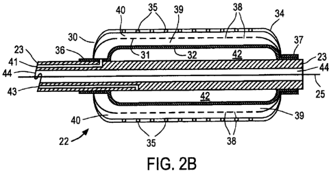
[0039j Referring now to FIGS. 2A and 2B, distal region 22 of one embodiment of
catheter
20 of the present invention is described. FIG. 2A depicts the exterior of
distal region 22 with
outer balloon 30 in an expanded state suitable for dilating a vessel, while
for purposes of
clarity, FIG. 2B depicts a sectional view of the inner components of distal
region 22 with
intermediate balloon 31 and inner balloon 32 in partially expanded states
suitable for infusing
a therapeutic agent into a vessel wall. Outer balloon 30 preferably comprises
a noncompliant
or semi-compliant material such as polyethylene or polyterephthalate. Outer
balloon 30 is
sized and shape for insertion as appropriate for the intended therapy and
bodily lumen. For
example, outer balloon 30 may have a diameter in an expanded state of about
2.5- 4.0 mm for
insertion in smaller lumens, such as coronary vessels, about 4-7 mm for
insertion in larger
lumens such as peripheral vessels, or as much as 4-6 cm if the catheter is
designed for use in
providing therapy in the thoracic or abdominal aorta. Intermediate balloon 31
and inner
balloon 32 preferably comprise a semi-compliant or compliant material such as
polyterephthalate or nylon. As described in further detail below, in a
preferred embodiment,
-9-
CA 02892197 2015-05-21
WO 2014/113221 PCT/US2014/010095
inner balloon 32 is configured to expand intermediate balloon 31 and outer
balloon 30 until
outer balloon 30 reaches its maximum designed diameter. In an alternative
embodiment,
outer balloon 30 also may comprise a compliant material, while intermediate
balloon 31 and
inner balloon 32 also may comprise a non-compliant material.
100401 Still referring to FIGS. 2A and 2B, outer balloon 30 has exterior
surface 34 and
multiplicity of through-wall apertures 35. In the embodiment depicted in FIG.
2A, apertures
35 illustratively are arranged in a pattern where each row is offset by a
predetermined angle,
e.g., about 45 ", from an adjacent row; however, other patterns will readily
occur to a person
of ordinary skill in the design of balloon catheters. For example, each row or
pattern of
apertures on the outer balloon may be aligned uniformly with adjacent rows;
there may be a
single row of apertures on the outer balloon; there may be two rows of
apertures on opposite
sides of the outer balloon, etc. In addition, apertures 35 are depicted as
being circular in
shape which may vary in diameter along the length of the balloon, but could
have any other
desired shapes, such as rectangular, triangular or elliptical. Outer balloon
30, intermediate
balloon 31 and inner balloon 32 preferably are affixed to catheter shaft 23 at
shoulders 36 and
37 via thermal bonds or glue welds.
100411 As best shown in FIG. 2B, intermediate balloon 31 includes multiplicity
of through-
wall apertures 38 which may have varying diameters along the balloon length,
and which
preferably are offset from apertures 35 in outer balloon 30. In this manner, a
fluent
therapeutic agent introduced into annular space 39 between the exterior of
inner balloon 32
and interior surface of intermediate balloon 31 will pass into annular space
40 between the
exterior of intermediate balloon 31 and the interior surface of outer balloon
30 without
directly exiting through apertures 35 in the outer balloon. Accordingly, when
a therapeutic
agent is introduced into annular space 39 via infusion lumen 41 and infusion
port 27 on
proximal end 21 (see FIG. 1), the agent passes from annular space 39 to
annular space 40,
from which it uniformly exits outer balloon 30 via apertures 35. Inflation
port 26 on
proximal end 21 (see FIG. 1) is coupled to interior space 42 of inner balloon
32 via inflation
lumen 43 that extends through catheter shaft 23. Apertures 35 may be the same
size or a
different size than apertures 38. Preferably, apertures 35 and 38 are laser
drilled and have a
diameter between about 5 um and about 50 um. In one embodiment, apertures 35
have a
diameter of about 5 tm and apertures 38 have a diameter of about 10 um. In
addition, a
-10-
CA 02892197 2015-05-21
WO 2014/113221 PCT/US2014/010095
subset of the multiplicity of apertures 35 or 38 may be differently sized from
another subset
of the multiplicity. For example, a distal portion of a row of apertures 35
each may have a
first diameter and a proximal portion of the row each may have a second
diameter, different
from the first. In one embodiment, in a row of sixteen apertures, eight distal
apertures each
has a diameter of about 15-25 p.m and eight proximal apertures each has a
diameter of about
7-17 p.m.
[0042] As depicted in FIG. 2B, after use of catheter 20 for dilating the
vessel wall, inner
balloon 32 may be inflated to any lower desired pressure to reduce the volume
of therapeutic
agent delivered into annular space 39 and to facilitate the rate of delivery
to the vessel wall.
Alternatively, inner balloon 32 may be deflated entirely after the vessel
dilatation step.
[0043] Still referring to FIG. 213, catheter shaft 23 includes lumen 44,
preferably centrally
located in catheter shaft 23, to permit guide wire 25 to be extended through
catheter 20 to
facilitate positioning of distal region 22 at a desired location in a
patient's vasculature or
organ. Distal region 22 also may include radiopaque markers disposed along
catheter shaft
23, for example, in the vicinity of shoulders 36 and 37, to facilitate
positioning of the catheter
under fluoroscopic imaging. In accordance with one aspect of the present
invention, lumen
44 preferably is sized to permit a wire containing an energy source, e.g., an
ultraviolet light
source (or light fiber), ultrasound transducer, or resistive heater, to be
advanced into distal
region 22 to deposit energy into the therapeutic agent or drug, to facilitate
uptake by the
vessel wall or provide another therapeutic effect, as described herein below.
For such
embodiments, balloons 30-31 and catheter shaft 23 preferably comprise
materials that permit.
light energy of selected frequencies to pass through the catheter without
significant
absorption or loss of energy.
[0044] Referring now to FIGS. 3A and 3B, distal region 22' of an alternative
balloon
catheter is constructed similarly to distal region 22 of FIGS. 2A and 2B,
wherein like
components are identified by like-primed reference numbers. Thus, for example,
apertures
35' in FIGS. 3A and 3B correspond to apertures 35 of FIGS. 2A and 2B, etc. As
will be
observed by comparing FIGS. 2A, 2B and 3A, 313, outer balloon 30' includes
proximal
bumper 45 around the circumference of its proximal end and distal bumper 46
around the
circumference of its distal end, and apertures 35' are aligned in uniform
rows. Bumpers 45,
46 extend from exterior surface 34' so as to create a pocket between bumpers
45 and 46 and
-11-
CA 02892197 2015-05-21
WO 2014/113221 PCT/US2014/010095
between exterior surface 34' and the luminal surface when bumpers 45,46 are
urged into
contact with the luminal surface. In this manner, bumpers 45, 46 facilitate
delivery of
therapeutic agents to the luminal surface via the pocket such that the agents
are delivered
uniformly along the length of the balloon, reduce clogging of the apertures
when the bumpers
are urged into contact with the luminal surface, and reduce the risk that
fluent material
delivered to the vessel surface will be washed into systemic circulation.
100451 Referring now to FIGS. 4A and 4B, distal region 22" of yet another
alternative
balloon catheter is constructed similarly to distal region 22 of FIGS. 2A and
2B except that
outer balloon 30" further includes multiplicity of solid protrusions 47
extending from exterior
surface 34" and interposed between multiplicity of through-wall apertures 35".
In the
embodiment depicted in FIG. 4A, protrusions 47 and apertures 35"
illustratively are arranged
in a regular pattern; however, other patterns will readily occur to a person
of ordinary skill in
the design of balloon catheters. Preferably, apertures 35" are offset from
protrusions 47 so as
to reduce clogging of the apertures when the protrusions are urged into
contact with the
luminal surface. In addition, while protrusions 47 are illustratively depicted
as substantially
circular cylinders having rounded extremities, other configurations, such as
rectangular,
conical or pyramidal structures also could be used. Protrusions 47 extend from
exterior
surface 34" so as to create a pocket between exterior surface 34" and the
luminal surface
when protrusions 47 are urged into contact with the luminal surface. In this
manner,
protrusions 47 facilitate delivery of therapeutic agents to the luminal
surface via the pocket,
and reduce the risk that fluent material delivered to the vessel surface will
be washed into
systemic circulation.
100461 Referring now to FIGS. 5A to 5C, a method of using the catheter of
FIGS. I and 2
to perform an interventional procedure is described. As will be readily
understood to one of
ordinary skill in the art, while the method is described for use with the
catheter of FIGS. 1
and 2, the alternative catheters of FIGS. 3 and 4 may be used in a similar
manner to that
described below.
100471 In FIG. 5A, guide wire 25 is placed in the vessel at the location of a
lesion or plaque
P, or nascent aneurysm, as determined using fluoroscopic imaging, contrast
agents and
conventional interventional techniques. Catheter 20 then is backloaded onto
guide wire 25
by inserting the proximal end of the guide wire into the distal opening of
lumen 44. Catheter
-12-
CA 02892197 2015-05-21
WO 2014/113221 PCT/US2014/010095
20 is advanced through the patient's vasculature until distal region 22 is
disposed in the
region of interest, as determined using radiopaque markers on catheter shaft
23 and
fluoroscopic imaging. When so disposed in patient's vessel V, distal end 22 of
catheter 20
will appear as depicted in FIG. 5A. In embodiments protrusions (FIG. 4),
during
manufacture of the catheter, outer balloon 30 or 30" of the catheter may be
wrapped or folded
so that protrusions 47 are substantially flush with the remainder of the
balloon material, thus
preventing the protrusions from snagging or abrading the vessel intima during
advancement
along guide wire 25 to the location of interest. Alternatively, a delivery
sheath (not shown)
may be disposed over distal region 22, 22', or 22" of the catheter to present
a smooth outer
surface for the catheter, and the sheath then may be retracted proximally to
expose the distal
region once it is at the desired location in vessel V.
(0048) Referring now to FIGS. 5B, 6, and 7, a conventional inflator is coupled
to inflation
port 26 and an inflation medium, such as saline or a saline diluted iodinated
contrast agent, is
delivered via inflation lumen 43 to inner balloon 32 to cause inner balloon 32
to expand
intermediate balloon 31 and outer balloon 30. As shown in FIG. 6, inner
balloon 32 may
expand intermediate balloon 31 and outer balloon 30 so that pocket 48 is
created between
outer balloon 30 and plaque P. In such an embodiment, pocket 48 may extend
between
bumpers 45 and 46 (FIG. 3) or protrusions 47 (FIG. 4) contact plaque P and the
intima of the
vessel V to dilate the vessel V and crack or disrupt plaque P. In addition, as
shown in FIG. 7,
inner balloon 32 may expand intermediate balloon 31 and outer balloon 30 into
contact with
plaque P and the intima of vessel V to dilate the vessel V and disrupt or
cause cracks C in the
plaque P. As inner balloon 32 expands, it contacts intermediate balloon 31
which contacts
outer balloon 30 and causes outer balloon 30 to contact and crack or disrupt
plaque P.
[0049] In embodiments where the outer balloon includes protrusions (FIG. 4),
the
protrusions engage plaque at discrete locations and place the plaque in
tension, causing it to
fracture. One or more therapeutic agents are infused through apertures 35,
35', 35" in outer
balloon 30, 30', 30" and contacts the plaque along fracture zones that enable
the therapeutic
agent to be rapidly taken up by the vessel intima. Because apertures 35" are
interposed
between the protrusions instead of extending through the protrusions as in
prior art systems,
compressed plaque at the point of contact of the protrusions is expected not
to clog the
apertures. It is expected that the foregoing arrangement of solid protrusions
and interposed
-13-
CA 02892197 2015-05-21
WO 2014/113221 PCT/US2014/010095
apertures will enable better uptake of therapeutic agents in calcified lesions
than has
heretofore been achieved.
100501 Referring to FIGS. 6 and 7, it is observed that vessel V comprises
three layers:
intima I, medial M, and adventitial A, which is supplied by vaso vasorum VV.
It is known
that the vaso vasorum VV supplies nourishment to vessel V and removes
metabolic
byproducts resulting from activity of the cells making up the vessel wall. In
accordance with
one aspect of the present invention, a therapeutic agent is infused into the
wall of a vessel V,
and preferably into the adventitia A and/or vaso vasorum VV, while also
locally reducing
flow in the vaso vasorum VV to reduce washout of the therapeutic agent from
the adventitia
A and vaso vasorum VV. In this manner, the vessel wall serves as a reservoir
for the
therapeutic agent, so that the infused therapeutic agent or drug is released
from the adventitia
A back into the medial M and intimal portions I of the vessel wall over a
period of months to
years, thereby prolonging the therapeutic effect of the infused agent or drug.
100511 The foregoing benefits may be achieved by a number of modes. In one
embodiment, the therapeutic agent or drug may be designed so that when
activated by supply
of energy, e.g., irradiated by ultraviolet light, insonicated with ultrasound
energy of a desired
frequency, or heated by a resistive or other type of heater, the drug
transitions from a fluent
form to a gel-like or solid form. In this case, the therapeutic agent will
assist in blocking or
reducing flow through the vaso vasorum, and reduce the rate at which the
therapeutic agent or
drug is removed from the selected portion of the vessel wall. Alternatively or
in addition, if
the therapeutic agent transforms to a gel-like or solid form, it will be less
susceptible to
erosion. In an alternative embodiment, the deposited energy may cause a
component of the
therapeutic agent to heat up to cause polymerization or cross-linking of
fluent bioactive
materials and/or remodel or partially necrose portions of the adventitia or
vaso vasorum,
thereby locally blocking or reducing flow through the vaso vasorum and
producing a
reservoir of the therapeutic agent that provides prolonged release. As a
further alternative
embodiment, the deposited energy may function to enhance uptake of the
therapeutic agent
through the layers of the vessel wall. As a still further alternative
embodiment, the energy
may directly cause partial remodeling or necrosis of the adventitia and/or
vaso vasorum to
produce the reservoir effect noted above.
-14-
CA 02892197 2015-05-21
WO 2014/113221 PCT/US2014/010095
[00521 Referring now to FIGS. 5C and 8, after inner balloon 32 has been
expanded to drive
intermediate balloon 31, and outer balloon 30 (and, if present, optional
bumpers or
protrusions) into contact with the vessel wall, inner balloon 32 is partially
or completely
deflated. Next, a vial or syringe containing a desired fluent therapeutic
agent or drug, (e.g.,
an anti-mitotic drug such as paclitaxel or sirolimus, angiogenic vector, or
stem cells), is
coupled to infusion port 27 on proximal end 21 and activated to inject the
agent through
infusion lumen 41 into annular space 39 between inner balloon 32 and
intermediate balloon
31 (see FIG. 2B). As indicated by the arrows in FIG. 5C, the agent passes
through apertures
38 in intermediate balloon 31 and into annular space 40 between intermediate
balloon 31 and
outer balloon 30. Inner balloon 32 may be partially or completely reinflated
to cause the
therapeutic agent to pass through apertures 38 and into annular space 40
between
intermediate balloon 31 and outer balloon 30 before exiting through apertures
35. Because
apertures 38 are offset from apertures 35 in outer balloon 30, the agent
circulates within
annular space 40 before passing through apertures 35 and exiting outer balloon
30.
Additionally, because agent moves laterally towards apertures 35, it will be
more uniformly
distributed around the circumference and along the axial length of the vessel
than previously-
known single balloon systems. This baffling effect provided by intermediate
balloon 31 is
expected to reduce jetting of therapeutic agent exiting through apertures 35
of outer balloon
30, thus reducing the potential for vessel dissection.
[00531 As depicted in further detail in FIG. 8, the therapeutic agent exits
outer balloon into
pockets 48 formed between cracks C in plaque and/or between bumpers, if
provided. The
therapeutic agent exits apertures 35 into pockets 48, where it is expected to
gain ready access
to the vessel intima through cracks and fractures formed in plaque P during
the dilatation step
illustrated in FIG. 5B.
[00541 As will be apparent to one of ordinary skill in interventional
procedures, the rate of
infusion of therapeutic agent can be adjusted by varying the pressure at which
the agent is
supplied from the syringe or vial through infusion port 27, or alternatively
by adjusting the
degree of inflation of inner balloon 32. By adjusting the latter, the
clinician can reduce the
volume of annular space 39, reducing the volume of therapeutic agent that must
be used
during the procedure. In addition, after infusing the therapeutic agent into
annular space 39,
the clinician may increase the pressure in inner balloon 32 to pressurize
annular spaces 39
-15-.
CA 02892197 2015-05-21
WO 2014/113221 PCT/US2014/010095
and 40 and enhance the rate at which therapeutic agent exits apertures 35 and
is infused into
the vessel wall. Therapeutic agent deposited in pockets 48 preferably is taken
up by the cells
in the various layers of the wall of vessel V by normal cellular processes, as
opposed to
traumatically (e.g., by cleaving intercellular connections).
100551 In addition, as will be readily understood to one of ordinary skill in
the art, while the
balloon catheter is generally described as delivering a therapeutic agent,
such as an anti-
mitotic drug, to plaque, the disclosure is not limited thereto. The
therapeutic agent may be
selected to treat any condition where subintimal injection would be
beneficial. For example,
the therapeutic agent may be selected for treating a nascent or existing
aneurysm when the
balloon catheter is delivered proximate to an aneurysm. As another example,
the therapeutic
agent may be selected to induce angiogenesis, delivered either transluminally
or into the sub-
intimal space. The therapeutic agent may comprise, for example, one or more
regenerative
agents, anti-inflammatory agents, anti-allergenic agents, anti-bacterial
agents, anti-viral
agents, anticholinergic agents, antihistamines, antithrombotic agents, anti-
scarring agents,
antiproliferative agents, antihypertensive agents, anti-restenosis agents,
healing promoting
agents, vitamins, proteins, genes, growth factors, cells, stem cells, vectors,
RNA, or DNA.
[00561 FIG. 9 illustrates a final optional step in accordance with the method
of present
invention for infusing one or more therapeutic agents into the wall of vessel
V. FIG. 9 is
similar to FIG. 5C, except that in this step guide wire 25 is removed or
retracted, and energy
delivery device 50 carrying an energy deposition element is advanced through
lumen 44 of
catheter 20 and disposed in distal region 22. The energy delivery element,
located in the
distal region of energy delivery device 50, preferably includes one or more
radiopaque
markers to indicate positioning of the distal region under fluoroscopic
imaging. Energy
delivery device 50 preferably has a diameter between 0.018" to 0.035" and may
comprise an
optical fiber or source for delivering ultraviolet light, ultrasonic energy,
or heat. Such
devices, and the energy sources that are coupled to the proximal ends of such
devices, are
known in the art and accordingly are not described in detail here. Of
particular importance,
however, if a UV light or ultrasonic energy delivery device 50 is employed,
catheter 20
preferably is constructed so that a substantial part of the energy is
delivered to the vessel wall
without being absorbed by the catheter material, and the energy absorbed by
the vessel wall
-16-
CA 02892197 2015-05-21
WO 2014/113221 PCT/US2014/010095
has some therapeutic benefit, e.g., activates the therapeutic agent. Energy
emitted by energy
delivery device 50 and absorbed by vessel V is represented by the solid arrows
in FIG. 9.
100571 As discussed above with respect to FIGS. 6 and 7, energy delivery
device 50 may
provide a therapeutic effect either by facilitating uptake of the therapeutic
agent by the vessel
wall; by activating the therapeutic agent; by heating the therapeutic agent to
effect a change
to the vessel wall structure; or by directing delivering energy to selected
layers of the vessel
wall to cause polymerization or cross-linking of fluent therapeutic agents
(e.g., as described
in U.S. Patent No. 5,749,915 to Slepian localized necrosis or remodeling of
collagen
contained within the vessel wall.
100581 In one embodiment, the deposited energy enhances uptake of the
therapeutic agent
through the layers of the vessel wall, for example, by activating moieties
bound to the
effective portion (e.g., anti-proliferative portion) of the therapeutic agent,
(e.g., as described
in U.S. Patent No. 4,590,211 to Vorhees). Alternatively, the therapeutic agent
or drug may
be designed so that when irradiated by ultraviolet light, or insonicated with
ultrasound energy
of a desired frequency, the drug transitions from a fluent form to a gel-like
or solid form. In
this case, the therapeutic agent will assist in blocking or reducing flow
through the vaso
vasorum, and reduce the rate at which the therapeutic agent or drug is removed
from the
selected portion of the vessel wall. Alternatively or in addition, if the
therapeutic agent
transforms to a gel-like or solid form, it will be less susceptible to
erosion, thereby locally
prolonging the therapeutic effect of the agent.
100591 In a further alternative embodiment, the energy deposited by delivery
device 50
may cause a component of the therapeutic agent to heat up and remodel collagen
of, or
partially necrose portions of, the adventitia or vaso vasorum. This effect
also may cause a
localized blockage that stops or reduces flow through the vaso vasorum and act
to produce a
localized reservoir of the therapeutic agent that provides prolonged release.
As yet another
alternative embodiment, the UV or ultrasonic energy may directly cause partial
remodeling or
necrosis of the adventitia and/or vaso vasorum to create localized blockage of
the vaso
vasorum to produce the reservoir effect noted above.
100601 Referring again to FIG. 9, energy delivery device 50 may be configured
to deliver
energy to vessel V during and after, or alternatively only a predetermined
interval after, the
-17-
CA 02892197 2015-05-21
WO 2014/113221 PCT/US2014/010095
therapeutic agent is delivered by catheter 20. Once the process of delivering
the therapeutic
agent into the vessel wall is completed, and the appropriate amount of energy
has been
delivered to enhance or prolong the therapeutic effect of the therapeutic
agent, energy
delivery device 50 may be withdrawn. Next, suction may be drawn on infusion
lumen 41 to
remove any excess therapeutic agent from annular spaces 39 and 40 to collapse
intermediate
balloon 31 and retract outer balloon 30 away from the vessel wall. In an
embodiment where
the outer balloon includes protrusions, the outer balloon may be constructed
so that, when
deflated, the balloon preferentially will fold to enclose the protrusions and
reduce the risk of
abrading the vessel wall during removal. Alternatively, or in addition, an
open-ended sheath
(not shown) may be advanced over the exterior surface of catheter shaft 23 and
the exterior of
outer balloon 30 to facilitate removal of catheter 20. Once catheter 20 is
removed from the
patient's vasculature, the access site may be closed using standard
interventional techniques.
[00611 Referring now to FIGS. I OA, 10B and II, an alternative embodiment of
apparatus
constructed in accordance with the principles of the present invention is
described. Catheter
60 includes elongated catheter shaft 61 having distal region 62 and outer
balloon 63. The
proximal end of catheter shaft 61 is similar in construction to catheter 20
and preferably
includes a hemostatic guide wire port, balloon inflation port and infusion
port. As shown in
FIG. 10B (which corresponds to an inflation state similar to FIG. 2B), distal
region 62
includes outer balloon 63, intermediate balloon 64 and inner balloon 65. As
for catheter 20
of the preceding embodiment, inner balloon 65 is fluid impermeable and is
coupled via an
inflation lumen to an inflation port on the proximal end. Likewise,
intermediate balloon 64
includes a multiplicity of through-wall apertures 66 (see FIG. 11) and is
coupled via an
infusion lumen to an infusion port disposed on the proximal end of the
catheter. Outer
balloon 63 includes one or more spiral protrusions 67 and a multiplicity of
through-wall
apertures 68.
[00621 Catheter 60 differs from the embodiment of FIG. I in that the exterior
surface of
outer balloon 63 includes protrusions 67 arranged as a spiral ridge. In
addition, whereas
intermediate balloon 64 of the embodiment of FIG. 1 may contain a textured
surface to
ensure that intermediate balloon 64 does not adhere to outer balloon 63,
intermediate balloon
64 in the embodiment of FIGS. 10 and 11 includes a macroscopic feature to
prevent such
adhesion. In particular, intermediate balloon 64 includes spiral rib 69,
preferably comprised
-18-
CA 02892197 2015-05-21
WO 2014/113221 PCT/US2014/010095
of the same material and potentially integrally formed with intermediate
balloon 64, disposed
on its exterior-facing surface of the intermediate balloon. In this manner,
spiral rib 69
contacts the inner surface of outer balloon to ensure that annular space 70 is
maintained
between intermediate balloon 64 and outer balloon 63 when inner balloon 65 is
inflated to
urge intermediate balloon 64 and outer balloon 63 into contact with a vessel
wall to dilate the
vessel and disrupt plaque.
100631 While in the embodiment of FIGS. 10 and 11 protrusions 67 are
configured as a
spiral ridge having a rounded extremity, it should be understood that other
patterns will
readily occur to a person of ordinary skill in the design of balloon
catheters, such as
structures having rectangular, conical or pyramidal cross-sections, as may be
desirable to
fracture severe calcifications. Similarly, while apertures 68 are depicted as
being circular,
they may have any other desired shape, such as rectangular, triangular or
elliptical.
Preferably, apertures 68 are offset from protrusions 67 so as to reduce
clogging of the
apertures when the protrusions are urged into contact with the lumina!
surface. Likewise,
apertures 66 in intermediate balloon 64 may be offset from apertures 68 in
outer balloon 63 to
achieve the benefits described above.
100641 Finally, although the macroscopic feature in intermediate balloon 64 is
illustratively
depicted as comprising spiral rib 69 having a substantially circular cross-
section, this feature
could have other cross-sections, such as rectangular, elliptical or
triangular. In addition,
spiral rib 69 need not form a continuous structure, but instead could comprise
a multiplicity
of discrete structures, similar in shape to protrusions 47 disposed on outer
balloon 30" of the
embodiment of FIG. 4. For example, intermediate balloon 64 and outer balloon
63 may
comprise the same material having the same protrusions disposed on their
respective exterior
surfaces. In this manner, construction of the distal end of the catheter of
the present invention
could be simplified, so long as the apertures in the intermediate and outer
balloons are
staggered or offset to provide the baffle action discussed above.
[0065.1 While preferred illustrative embodiments of the invention are
described above, it
will be apparent to one skilled in the art that various changes and
modifications may be made
therein without departing from the invention. The appended claims are intended
to cover all
such changes and modifications that fall within the true spirit and scope of
the invention.
-19-