Note: Descriptions are shown in the official language in which they were submitted.
CA 02892308 2015-05-25
WO 2014/058987
PCT/US2013/064081
SYSTEMS AND METHODS FOR TUMOR CLONALITY ANALYSIS
[0001] This application claims priority to our copending U.S. provisional
application with the
serial number 61/711467, which was filed October 9, 2012.
Field of the Invention
[0002] The field of the invention is computational analysis of genomic data,
particularly as it
relates to identification of clonality status of a mixed cell population.
Back2round of the Invention
[0003] With the increasing availability of whole genome data and the ever-
increasing speed
of whole genome sequencing, enormous quantities of data are now available that
demand a
meaningful analysis to so provide a clinician or scientist with information to
enable more
effective treatment or drug development.
[0004] For example, multiple tumor and matched normal whole genome sequences
are now
available from projects like The Cancer Genome Atlas (TCGA), and extraction of
relevant
information is difficult. This is further compounded by the need for high
genome sequencing
coverage (e.g., greater than 30-fold) to obtain statistically relevant data.
Even in compressed
form, such genomic information can often reach hundreds of gigabytes, and a
meaningful
analysis comparing multiple of such large datasets is in many cases very slow
and difficult to
manage, however, absolutely necessary to discover the many genomic changes
that occurred
in any given sample relative to a second sample. More recently, systems and
methods have
been developed to allow for rapid generation of information in a format that
avoids massive
output files as is described in W02013/074058. This publication and all other
publications
identified herein are incorporated by reference to the same extent as if each
individual
publication or patent application were specifically and individually indicated
to be
incorporated by reference. Where a definition or use of a term in an
incorporated reference is
inconsistent or contrary to the definition of that term provided herein, the
definition of that
term provided herein applies and the definition of that term in the reference
does not apply.
[0005] While the system of the '048 application provides a significant
improvement over
other known systems, various difficulties nevertheless are present. For
example, most breast
cancer is clinically and genomically heterogeneous and is composed of several
pathologically
1
CA 02892308 2015-05-25
WO 2014/058987
PCT/US2013/064081
and molecularly distinct subtypes, which often complicates genomic analysis.
Moreover,
currently known methods do not allow for deconvolution of such genomic
diversity to so gain
insight into possible tumor cell evolution and resulting clonality among tumor
cells in a
tissue.
[0006] Thus, even though numerous methods of genomic analysis are known in the
art, all or
almost all of them suffer from several disadvantages. Most significantly,
heretofore known
methods fail to allow identification of tumor progression on a molecular
level, and with that
fail to provide insight into clonality and potential treatment efficacies.
Viewed from another
perspective, heretofore known methods failed to allow identification of
clonality and clonal
relationship of cell populations within a sample containing multiple non-
homogenous cells.
Consequently, there is still a need to provide improved systems and methods
for genomic
analysis, and especially systems and methods that provide information on
clonality, clonal
fraction, molecular tumor progression, and/or treatment options based on such
information.
Summary of The Invention
[0007] The present invention is directed to various systems, devices, and
methods for genetic
analysis, and especially genomic analysis to identify presence and
distribution of distinct cell
clones within a sample containing one or more clonal populations of cells
based on genomic
data obtained from the sample. In especially preferred aspects, analysis is
based on genomic
DNA from a tumor or otherwise abnormal cell population, and allows not only
determination
of multiple clones within the tumor or cell population but also allows
identification of likely
clonal evolution and/or clonal relationships.
[0008] In one aspect of the inventive subject matter, a method of ex-vivo
determining
clonality of a tumor from sequencing data obtained from the tumor includes a
step of
determining from the sequencing data copy number and allele fraction for an
allele within the
sequencing data, and another step of calculating an allelic state for the
allele based on the
determined copy number and the determined allele fraction. The allelic state
is then used to
determine the clonality of the tumor. While not limiting to the inventive
subject matter, it is
generally preferred that the allelic state is plotted or displayed in an
allelic state diagram
(which may be a single or dual allelic state diagram).
[0009] In at least some embodiment of the inventive subject matter
determination of the copy
number and allele fraction is performed by a sequence analysis program that
produces local
2
CA 02892308 2015-05-25
WO 2014/058987
PCT/US2013/064081
alignments by incremental synchronization of sequence strings (e.g., BAMBAM).
Among
other states, contemplated allelic states include normal copy number, single
copy
amplification, single copy/hemizygous deletion, copy-neutral loss of
heterozygosity, and
amplification of both alleles.
[0010] In further contemplated embodiments of the inventive subject matter,
the allelic state
calculation comprises a correction for normal contamination, uses majority and
minority
allelic states for tumor and normal, and/or includes an identification of a
mixture fraction Mb
for an allele (which is either 0 or 1 for a monoclonal tumor, or greater than
0 and smaller than
1 when the tumor is polyclonal). It is still further contemplated that the
calculation of the
allelic state may also comprises a correction for sequencing coverage level,
particularly
where the coverage level for the tumor is higher than the coverage level for a
corresponding
non-tumor (e.g., healthy) sample of the same patient.
[0011] Where desired, contemplated methods will further include a step of
determination of
an allelic state landmark, which is preferably used to determine a number of
distinct (related
or unrelated) clones in the tumor and/or a proportion of clones in the tumor.
Additionally, or
alternatively, it is still further contemplated that a mutation can be linked
to a majority allele
or a minority allele, and that the mutational allele fraction can be employed
for determination
of timing of the mutation relative to a change in allelic state.
[0012] In another aspect of the inventive subject matter, a method of ex-vivo
visualization of
allelic states in a tumor includes a step of determining a copy number and an
allele fraction
for an allele within sequencing data, and a step of calculating the allelic
state for the allele
based on the determined copy number and the determined allele fraction. In a
still further
step, the allelic state of the allele is mapped in an allelic state diagram
that plots copy number
versus allele fraction (typically majority allele fraction).
[0013] Most typically, the allelic state diagram is presented such that each
vertex in the
allelic state diagram corresponds to a tumor allelic state, that clones with
loss or gain of an
allele in a polyclonal tumor map along edges drawn between vertices, and/or
that clones with
changes other than loss or gain of an allele in a polyclonal tumor map between
edges drawn
between vertices. It is still further contemplated that the allelic state
diagram is adjusted for
normal contamination. Of course, it should be appreciated that the allelic
state diagram may
be a dual allele state diagram.
3
CA 02892308 2015-05-25
WO 2014/058987
PCT/US2013/064081
[0014] Therefore, and viewed from a different perspective, the inventors also
contemplate a
method of analyzing genomic sequence data in which a BAM server receives a
plurality of
genomic sequence reads, wherein the plurality of genomic sequence reads are
obtained from
a genome of a tumor sample and a genome of a normal sample of a patient. The
BAM server
then processes the genomic sequence reads to produce a plurality of
differential sequence
objects that comprise a copy number and an allele fraction for an allele
within the tumor
genome. An analytics engine (that is coupled to the BAM server) then processes
the copy
number and the allele fraction for the allele to so determine an allelic state
for the allele.
[0015] In a typical embodiment of such methods, a differential sequence
database is coupled
to the BAM server and the analytics engine such that the BAM server provides
the
differential sequence object to the differential sequence database and such
that the differential
sequence database provides the differential sequence object to the analytics
engine.
Furthermore, it is contemplated that a graphic output is generated by the
analysis engine that
plots the allelic state for the allele in an allelic state diagram.
[0016] In a further contemplated aspect of the inventive subject matter, a
method of ex-vivo
characterizing genomic information from a tumor includes a step of determining
an allelic
state for an allele in the tumor genome, and a further step of using the
determined allelic state
to identify the tumor as being a monoclonal tumor or as comprising at least
two distinct
tumor clones.
[0017] In such methods, it is further contemplated to use the determined
allelic state to
identify a relationship of the tumor clones (e.g., as being distinct and
unrelated or as being
related). Where the clones a related, it is contemplated that the determined
allelic state can be
employed to identify a clonal history for the distinct tumor clones.
[0018] Thus, the inventors also contemplate a method of ex-vivo characterizing
a tumor clone
in a tumor mass in which in one step genomic sequence information from the
tumor mass is
obtained (e.g., from a BAM server). In another step, the genomic information
is used to
determine an allelic state for an allele in the tumor genomic sequence
information. In a
further step, the location of the allelic state for the allele in an allele
state diagram is
determined (e.g., in a graphic display or in silico, or numerically), and the
location is used to
identify the clone as monoclonal or polyclonal. For example, a clone is
monoclonal when the
location of the allelic state is on a vertex of the allelic state diagram.
4
CA 02892308 2015-05-25
WO 2014/058987
PCT/US2013/064081
[0019] In yet another aspect of the inventive subject matter, the inventors
contemplated a
method of providing treatment information for treatment of a tumor. In such
method, allelic
state information for the tumor is ascertained, and presence or emergence of
(a) a clone or (b)
an evolutionary pattern of clones is ascertained within the tumor that is
indicative of at least
one of susceptibility of the tumor to treatment with a drug, and an increased
risk of drug
resistance or metastatic potential. Most typically, the step of identifying
presence or
emergence is based on prior treatment data or a priori known data.
[0020] Various objects, features, aspects and advantages of the inventive
subject matter will
become more apparent from the following detailed description of preferred
embodiments,
along with the accompanying drawing figures in which like numerals represent
like
components.
Brief Description of The Drawing
[0021] Fig.1A is an exemplary illustration of evolution of a tumor starting
from a germline
cell, to an initial tumor cell, to a population of major and minor clones that
are sampled by
the tumor biopsy.
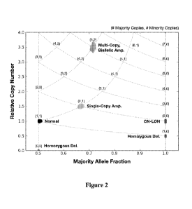
[0022] Fig. 2 is an exemplary allelic state diagram (ASD) of simulated data
for a monoclonal
tumor sample with zero normal contamination, a = O. Chromosomal regions
exhibiting
different copy number alterations are plotted in different shades. This
simulated tumor
genome exhibits 6 allelic states: normal, single-copy amplification,
hemizygous deletion,
homozygous deletion, copy-neutral LOH, and multi-copy biallelic amplification.
[0023] Figs. 3A-3D depict exemplary an set of allelic state diagrams for the
simulated
monoclonal tumor genome of Fig.2 with different levels of normal contaminant:
Fig. 3A
illustrates 0% normal contaminant, Fig. 3B illustrates 10% normal contaminant,
Fig. 3C
illustrates 50% normal contaminant, and Fig. 3D illustrates 90% normal
contaminant,
indicating the difference is resolution as a function of level of normal
contamination.
[0024] Fig. 4 is the allelic state diagram of Fig. 2 showing some possible
bidirectional and
unidirectional transitions between allelic states where unidirectional
transitions are those that
involve irreversible loss of a parental chromosome.
CA 02892308 2015-05-25
WO 2014/058987
PCT/US2013/064081
[0025] Fig. 5 is an allele state diagram for a tumor genome transitioning from
the allelic
states presented in the previous figures to new allelic states that differ
only by a single copy
loss or gain. Here, transitional allelic states are created when the tumor
comprises a mixture
of two different clones/subclones: Clone A is defined by the original allelic
states: (2,1),
(5,2), and (1,0). Clone B alters these states through amplifications and a
deletion to produce
the allelic states: (2,2), (4,2), and (2,0). The percentages denote the
percentage of clone B
present in the tumor population, where 0% describes a monoclonal population of
clone A,
and 100% is a monoclonal population of clone B.
[0026] Fig. 6 is an exemplary illustration of an allelic state diagram of the
Fig. 2 showing the
transitional allelic states produced when allelic states are "skipped", which
can occur when
the tumor consists of two or more unrelated, or distantly-related clones. In
that case, the
transitional states are not found on the edges connecting allelic states if
the allelic states of
the two major clones differ in both majority and minority alleles.
[0027] Figs. 7A and 7B are exemplary illustrations of allelic state diagrams
for two GBM
tumors: GBM-06-0145 (Fig. 7A) and GBM-06-0185 (Fig. 7B). The fitted parameters
found
normal contamination at 21.5% and 14.6%, respectively. Fig. 7A depicts only
clonal allelic
states and no evidence of transitional allelic states, indicating that GBM-06-
0145 is a
monoclonal tumor, while Fig. 7B depicts both clonal states and multiple
transitional allelic
states. Since the transitional allelic states (marked with (*)) feature three
different mixture
percentages, this polyclonal tumor must consist of at least three sub-clones.
The black X's
plotted in Fig. 7B represent "landmark" allelic states suitable for use to
determine the clonal
mixture of GBM-06-0185.
[0028] Fig. 8 is an exemplary plot depicting the monoclonal karyotype for GBM-
06-0145.
The "Relative Coverage" and "Allele Fraction" displayed at the top of the plot
shows both
the observed results output by BamBam and the computed coverage and allele
fraction
generated by modeling the mixture of the single clone and normal
contamination. The
comparison of real versus modeled data shows very strong agreement. The
clone's karyotype
below shows the majority and minority allelic states for the tumor genome,
showing
amplification of one copy of entire chr7 and chr19, complete loss of one copy
of chr10, and
arm-level loss of chr9p.
6
CA 02892308 2015-05-25
WO 2014/058987
PCT/US2013/064081
[0029] Fig. 9 is an exemplary plot depicting the polyclonal karyotypes for GBM-
06-0185. A
total of 4 distinct clones were identified in this tumor, with clone D
determined to be the
dominant clone of the population, comprising 42.7% of the tumor sample. All
clones have
single-copy amplifications of chr7, chr19 & chr20, single copy loss of chr10
and chr22, and
loss of chr9p in common. Clones B, C, & D all have deletions in chr6, but
clone B's deletions
are focal while clones C & D display arm-level loss of chr6q. Clone D is
further
distinguished by amplification of the intact copy of chr9.
[0030] Fig. 10 is an exemplary plot depicting the polyclonal karyotypes for
GBM-06-0152.
Three clones were identified in this tumor sample that has an estimated 24.1%
normal
contamination. All clones share amplifications of chrl, chr19 & chr20,
deletions of chr10 &
chr22, and focal losses of chr12 related to the chromothripsis-like event that
created two DMs
described in the previous chapter. Clones B & C exhibit amplification of chr7
and deletions
of the nonamplified copy of chrl as well as chr2, chr3, chr4, chr8, chr13, and
chr17. Clone C
further amplifies the remaining copy of chr8.
[0031] Fig. 11 is an exemplary plot depicting the polyclonal karyotypes for
GBM-06-1086.
Four clones were identified in this tumor sample that has an estimated 7.5%
normal
contamination. All clones share amplification of chr21 and deletions of chr9 &
chrl1p.
Clones C & D exhibit significant chromosomal loss, deleting chrl, chr3, chr4,
chr5, chr6,
chr8, chr10, chr13, chr14, chr15, chr17, chr18, and chr20. The dominant clone
D, making up
41.6% of the tumor sample, further deletes the sole remaining copy of chrl 8
and amplifies
chr19. The black arrows indicate the position of CD1(2NA in clones A & B,
highlighting the
arrival of the focal deletion of CDI(1\12A in the latter clone.
[0032] Fig. 11 is an exemplary plot depicting the polyclonal karyotypes for
GBM-06-1086.
Four clones were identified in this tumor sample that has an estimated 7.5%
normal
contamination. All clones share amplification of chr21 and deletions of chr9 &
chrl1p.
Clones C & D exhibit significant chromosomal loss, deleting chrl, chr3, chr4,
chr5, chr6,
chr8, chr10, chr13, chr14, chr15, chr17, chr18, and chr20. The dominant clone
D, making up
41.6% of the tumor sample, further deletes the sole remaining copy of chrl 8
and amplifies
chr19. The black arrows indicate the position of CD1(2NA in clones A & B,
highlighting the
arrival of the focal deletion of CDI(1\12A in the latter clone.
7
CA 02892308 2015-05-25
WO 2014/058987
PCT/US2013/064081
[0033] Fig. 12 is an exemplary illustration of phased mutations on the dual
ASD. A
representative region on the tumor genome is shown, consisting of a region in
the single copy
gain allelic state, a region in the "normal" allelic state, a region
exhibiting CN-LOH, and a
region exhibiting LOH. Three mutations are found in the amplified region, two
majority-
phased (red stars) and one minority-phased (blue star). Two mutations are
found on the
"normal" allelic state, one majority-phased and another minority-phased. Both
regions
exhibiting LOH have one mutation each phased to the sole remaining allele, and
are thus
majority-phased. The dual ASD below shows where each of these mutations would
be found,
using each mutation's corrected allele fraction, MAFc, to determine its
location along the x-
axis. Note the different placement of the two majority-phased mutations in the
single-copy
gain allelic state, where only the mutant allele that exists on both majority
alleles (i.e.
mutated before amplification) is found near the single copy gain majority
allelic state. The
other is found near the single copy gain minority allelic state, correctly
identifying that the
mutation exist on only one copy of the majority allele. Finally, note that the
minority-phased
mutations in blue are all found towards the left-half of the dual ASD.
[0034] Fig. 13 is an illustration of phased mutations on the dual ASD for
tumor GBM-06-
0145. 7 regions are encircled on these plots: (a) majority-phased to an
amplified allelic state
yet presents with MAFc suggesting the mutation is only on one of the two
copies, (b)
majority-phased to an amplified allelic state with MAFc suggesting mutation is
present on
both amplified copies, (c) majority-phased with allele fraction consistent
with the LOH allelic
state, (d) minority-phased in the majority-amplified allelic state with MAFc
consistent with
single copy, (e) unphased mutations with MAFc consistent with amplified
allelic state, and
(f) unphased mutations with MAFc consistent with LOH allelic state
[0035] Fig. 14 is an illustration of phased mutations on the dual ASD for
tumor LUSC-34-
2596. The two encircled regions, (a) and (b), show a number of mutations
phased to the
majority and minority alleles, respectively, in the balanced amplified allelic
state (2,2). One
majority-phased mutation in NDRG1 is found in a transitional allelic state
with matching
MAFc. The locations of two missense mutations, in BRAF & DNMT3A, and one
nonsense
mutation in TP53 are shown in the unphased plot, placing BRAF in a highly
amplified allelic
state, DNMT3A in the "normal" allelic state, and TP53 in the CN-LOH state.
8
CA 02892308 2015-05-25
WO 2014/058987
PCT/US2013/064081
Detailed Description
[0036] The inventors have discovered that clonality of a genetically
heterogeneous sample
can be readily resolved using an approach that uses an allelic state model
(e.g., expressed as
allelic state diagram), and that the so obtained clonality information can be
used for various
purposes, including analytic, prognostic, and diagnostic uses.
[0037] For example, the methods and systems contemplated herein provide the
ability to
computationally dissect a tumor's population using whole genome sequencing
data, and
where desired, to visually assess a tumor sample's clonality using an allelic
state diagram.
Viewed from a different perspective, the clonal mixture of a tumor can now be
determined by
decomposition of the tumor population into the major clones of the tumor cell
population and
by estimation of normal contamination to account for the copy number and
allele fraction
(which is preferably performed using BamBam, as described in W02013/074058).
Still
further, contemplated systems and methods all for a determination and phasing
of whole
genome karyotypes of all major clones, which in turn allows inferring the
phylogenetic tree
of polyclonal tumor genomes to time the emergence of clone-specific copy
number
alterations. Finally, by using phasing and mutant allele fraction, emergence
of mutations can
be timed with respect to their encompassing copy number alterations.
[0038] Therefore, in one aspect of the inventive subject matter, it should be
appreciated that
clonality and timing information will help better understand the dynamic
nature of individual
tumors, which may be reflective of a tumor type, or an individual's or
tissue's response to the
presence or development of the tumor. Remarkably, all of this information can
be discovered
from just a single tumor biopsy, making contemplated systems and methods
particularly
useful in an ex vivo diagnostic approach.
[0039] In another aspect of the inventive subject matter, it should be
appreciated that the
phylogenetic-based mutation models contemplated herein can be employed to
analyze the
mutations of related samples (e.g., primary tumor and its metastases) to so
reconstruct the
mutational history of a cancer as it spread. The ability to determine a
tumor's clonality and
identify all of the major clones that comprise the growing tumor mass, all
from the whole
genome sequencing data of a single biopsy, opens up a wide variety of
potential clinical
applications. For example, in a scenario where a newly-diagnosed patient's
tumor is biopsied
and all of the major clones are identified via clonal analysis. A clinician
could then use this
9
CA 02892308 2015-05-25
WO 2014/058987
PCT/US2013/064081
clonality analysis to tailor the patient's treatment according to the
alterations specific to the
clone furthest up the evolutionary tree, the progenitor tumor cell, with the
hope that treating
the initial tumor mass, derivative clones will also be targeted. On the other
hand, in a scenario
where a patient is diagnosed with a slow-growing tumor that can be safely
monitored for a
longer period of time before surgery or the beginning of chemotherapy, clonal
analysis of a
series of biopsies could be performed and, by tracking the clonal composition
of the tumor
through time, a clinician can identify the clones that are growing most
rapidly. By designing
a treatment that targets not what is currently the dominant clone, but the
clone that is set to
become the dominant clone, might more effectively treat the cancer.
[0040] Clonality analysis is also contemplated to prove useful in better
understanding the
metastatic spread of cancers. In such scenario, clonal analysis of a primary
tumor and a series
of metastases is used to determine all of the major clones present in the
spreading tumor. By
inspecting the clonal composition of the primary and each metastasis, one can
determine how
each clone spreads and discover if one or more particular clones exist that
show increased
metastatic potential. By determination of the characteristics unique to the
metastatic clones,
the inventors contemplate that identification of emergence of these
characteristics in minor
clones of another patient's primary tumor an "early warning" signal may be
developed for
determination of likelihood of imminent metastasis.
[0041] With respect to methods of data acquisition for clonality analysis, it
is preferred that
genomic analysis to identify copy number and allele fractions are determined
using systems
and methods in which multiple relatively small genomic sequence sub-strings
(e.g., short
reads from sequencing runs) of respective larger genetic sequence strings from
a first and
second tissue sample (e.g., healthy and diseased tissue) are obtained. The
genetic sequence
strings are then incrementally synchronized using one or more known positions
of at least one
of corresponding sub-strings to so produce a local alignment. The so generated
local
alignment is then analyzed (typically using a reference genomic sequence) to
generate a local
differential string between the first and second sequence strings within the
local alignment
that thus contains significant differential information (typically relative to
the reference
genomic sequence). A differential genetic sequence object for a portion or
even the entire
genome is then created using the local differential string, and most typically
a plurality of
local differential strings. It should be noted that incremental
synchronization to produce local
alignments and differential information provides various technical advantages,
including a
CA 02892308 2015-05-25
WO 2014/058987
PCT/US2013/064081
significant increase in processing speed of an entire genome, as well as the
capability to
produce allele specific information (e.g., copy number, allele fraction, etc.)
[0042] In such systems and methods, it should be appreciated that instead of
processing two
extremely large files to generate another extremely large intermediate (or
even output) file,
genome wide analysis can be achieved in multiple significantly smaller
portions wherein the
smaller portions are aligned to a reference genome using known positions
within the genome
of one or more sub-strings. Viewed from another perspective, alignment is
performed by
incremental synchronization of sequence strings using known positions of
substrings and a
reference genome sequence, and an output file can be generated that comprises
only relevant
changes with respect to a reference genome. Thus, the processing speed is
significantly
improved and the amount of data required for production of a meaningful output
is
dramatically reduced. Still further, it should be noted that such systems and
methods allow,
inter alia, haplotyping/somatic and germline variant calling, and
determination of allele-
specific copy numbers. Moreover, the systems and methods presented herein are
suitable for
use with sequence information in SAM/BAM-format.
[0043] For example, multiple sequencing fragments (e.g., short reads from a
tumor sample of
a donor and corresponding non-tumor sample of the same donor) are aligned to
the same
reference genome, which is employed to organize the sequencing fragments from
the
samples. Thus, such methods use two sequencing fragment datasets (one from the
tumor, the
other from corresponding normal "germline" tissue) from the same patient and
the reference
genome, and reads the datasets such that all sequences in both datasets
overlapping the same
genomic position (based on the reference genome and annotation in sub-strings)
are
processed at the same time. This is the most efficient method for processing
such data, while
also enabling complex analyses that would be difficult or impossible to
accomplish in a
serialized manner, where each dataset is processed by itself, and results are
only merged
afterwards. A particular suitable system is described in W02013/074058,
incorporated by
reference herein.
Fundamental Considerations
[0044] At first approximation, a tumor growth is a population of cancer cells.
This population
may homogenous, where all tumor cells share substantially the same genetic
characteristics.
Such tumors are said to be monoclonal since all tumor cells feature
substantially the same
11
CA 02892308 2015-05-25
WO 2014/058987
PCT/US2013/064081
genetic variants (e.g., copy number aberrations, structural variants,
mutations) as compared to
the progenitor tumor cell from which the tumor cells propagated. This
progenitor tumor cell
may be the first cancerous cell that initiated the tumor, or may be a
subsequent tumor cell that
gained an advantageous mutation that aided a complete sweep of the tumor
population.
[0045] On the other hand, polyclonal tumor growths are viewed as tumors
composed of at
least two genetically distinct clonal populations of tumor cells. In
polyclonal tumors, each
clonal population arose from a respective progenitor clone, but each
progenitor clone differs
from the other by some observable alteration. Thus, the multiple clonal
populations may be
significantly different from each other, or (as is more often the case), the
clonal populations
are related, sharing a set of variants that are found in all or a large subset
of tumor cells. For
example, a polyclonal tumor may comprise multiple major clones, where a major
clone
represents a computationally detectable clone (typically representing 10% of
the tumor
population), while the same polyclonal tumor may comprise further numerous
minor clones
that are undetectable with any given method.
[0046] In addition, it should be noted that individual mutations may be
classified as either
clonal or subclonal. In that context, when the dominant clones of a particular
tumor are
found, clonal variants are those shared by all tumor cells of any or all
dominant clones.
Viewed from a different perspective, clonal variants achieved full penetrance
in the entire
population or polyclonal subpopulation of cells. Subclonal variants are those
that exist in only
a small proportion of the cells belonging to a clonal population.
[0047] An example for the above model of tumor and its evolution is provided
in Fig. 1 in
which an initial germline cell acquired a nonsense mutation in a key tumor
suppressor (M1)
and amplified an oncogene (A1) that supported the initial growth of a tumor.
Early on in this
tumor's development, another tumor suppressor was deleted (D1) that caused the
tumor cell
to grow even more rapidly, enabling cells with this deletion to rapidly
overtake the entire
tumor population. Soon after acquiring deletion D1, a cell also acquires a set
of neutral
mutations (M2, M3), amplifications (A2, A3), deletions (D2, D3). Since these
variants
occurred early during the clonal expansion of this tumor cell variant, but do
not provide any
selective advantage, the population of tumor cells are split into two "major
clones," where
25% of tumor cells have the neutral variants (M2, M3, A2, A3, D2, and D3) and
75% of
tumor cells do not. Much further during this tumor's development, additional
mutations (M4,
12
CA 02892308 2015-05-25
WO 2014/058987
PCT/US2013/064081
M5) appear on one of the two major clones, but do not have a chance to spread
through the
population prior to the patient's death and/or tissue biopsy.
[0048] In the example of Fig. 1, the tumor population is polyclonal, with its
two major clones
defined such that: clone (1) has variants Ml, Al, and D1, and clone (2) shares
the variants of
clone (1), but in addition has variants M2, M3, A2, A3, D2, and D3. The clonal
mixture is
determined as 75% clone (1) and 25% clone (2). Mutations Ml, M2, and M3 would
all be
classified as "clonal" since they all achieved full penetrance in their
respective clones, while
M4 and M5 would be classified as "subclonal" mutations. Moreover, as can be
seen from
Fig. 1, a biopsy will typically include normal tissue in addition to tumor
heterogeneous tissue.
Data Extraction and Synthesis
[0049] The following presents various systems and methods to extract and
synthesize data to
reconstruct the clonal evolution of a tumor from whole genome sequencing data
of a single
tumor biopsy. These systems and methods provide a powerful framework to
determine the
clonality of a tumor, the number and proportion of all major clones in the
tumor, and possible
variants that distinguish the major clones. Furthermore, systems and methods
are presented to
phase mutations to parental alleles to thereby time their emergence within the
population. In
addition, contemplated systems and methods will provide an accurate estimate
of the amount
of contaminating normal tissue that was present in a tumor biopsy.
Copy number alterations, allele fraction, and the allelic state diagram
[0050] To discover and describe the major clones of a population, relative
copy number and
allele fraction estimates are utilized. Such data can be obtained using
algorithms and methods
as described in W02013/074058. Underlying the method to determine both
clonality and
estimate normal contamination is the "allelic state diagram" (ASD), which is
described in
more detail below. It should be especially appreciated that the ASD describes
the positions of
clonal positions of allele-specific copy number variants using both relative
copy number and
allele fraction of copy number alterations, thus demonstrating the
relationship between copy
number and allele fraction for all allelic states. The positions of clonal
allelic states in the
ASD are determined by the following Equations I and II:
13
CA 02892308 2015-05-25
WO 2014/058987
PCT/US2013/064081
¨ (tr;d6
CAT (trnaj train nnzal Irwin, a) =
masj min; Eq. I
k -
AF(trnt-ej, 7111Z.11,i 'a min 7 CO
( 1 ¨ e:),) (tma t õtin (Ityn
Eq. //
where CN is the relative tumor copy number compared to a matched-normal, AF is
allele
fraction in the tumor, a is the fraction of normal contamination in the tumor
sample, and tmai,
tmin, ilmaj, and nn are the majority and minority allelic states in the tumor
and normal,
respectively. Since individual genomes can only have discrete allelic states,
such that they
have 0, 1, 2, or more copies of a given chromosomal segment, the possible
values for tmai and
tmin are constrained to the set of positive integers, ti c (0, 1, 2, ...,n).
Furthermore, the majority
and minority allelic states for the normal are set to one, ni = 1, which is
true for all of the
autosomes in a normal human genome. The sex chromosomes, X and Y, are ignored
in the
ASD. Note that since the above formulae necessarily require two alleles, only
heterozygous
sites in the matched normal genome are considered for the ASD.
[0051] In the following figures, particularly significant allelic states are
normal copy number,
single-copy amplification, single-copy/hemizygous deletion, homozygous
deletion, copy-
neutral loss of heterozygosity (CN-LOH), and amplification of both parental
alleles. For
example, Fig. 2 shows exemplary copy number and allele fraction data for the
above allelic
states with no normal contamination, demonstrating how the ASD can be used to
determine
the allelic state of each cluster of points. Here, each vertex in the ASD's
grid is labeled with
its tumor allelic state, (tmai, tmin), and the position is determined by the
equations above. Fig. 3
demonstrates how the locations of allelic states are affected by increasing
amounts of normal
contamination, a. Fig. 3A has no normal contamination (a =0), with Figs. 3B-D
having
increased normal contamination (3B: a =0.1; 3C: a =0.5; 3D: a =0.9). As is
readily apparent,
as normal contamination increases, the allelic state positions grow closer
together, reducing
the ability to resolve different allelic states. It should be especially noted
that plotting the
copy number versus allele fraction to produce an ASD provides various
technical advantages,
including the capability to observe and identify clonality status of a tumor
and the capability
to observe and identify (unidirectional and bidirectional) changes in the
clonality status of a
tumor.
[0052] It should be noted that the example of Fig. 3 depicts a static snapshot
of a monoclonal
tumor. However, it is well known that the tumor genome can be very dynamic,
with gains
14
CA 02892308 2015-05-25
WO 2014/058987
PCT/US2013/064081
and losses of small and large chromosomal segments. Fig. 4 exemplarily
illustrates some of
the possible transitions between the allelic states described in previous
figures. It should be
appreciated that some transitions are "one-way" since they involve the
irreversible loss of
chromosomal segments. For example, the transition between the normal allelic
state (1,1) and
the hemizygous deletion state (1,0) is "one-way" because that deleted allele
can never be
restored. However, the retained allele in this case can be amplified,
permitting transitions to
the copy-neutral LOH (CN-LOH) state and beyond (2+,0). Notice that the
deletions necessary
for the transition between other allelic states are not deemed "one way"
because at least one
copy remains of each allele remains in the genome.
[0053] Based on the above, it should be recognized that allelic states can now
be identified in
a relatively simple manner. For example, Fig. 5 displays the ASD of a tumor
genome
transitioning from the allelic states presented in the previous figures to new
allelic states that
differ only by a single copy loss or gain. During such a transition, the
population of tumor
cells will be a mixture of tumor cells having tumor cells with the original
allelic states and
tumor cells with the new allelic states. For the example presented in Fig. 5,
one could view
this "transitional" tumor as a population divided between two major clones, A
and B, where
clone A is defined by the original allelic states, and clone B is defined by
the new allelic
states. The mixture fractions shown on this figure, Mb, represents the
fraction of clone B
within the population, such that the tumor population solely consists of clone
A when Mb = 0,
and the population consists only of clone B when Mb = 1. It is important to
note that the
allelic states for both clones, ti,a and to, are still constrained to the set
of positive integers.
[0054] From Fig. 5, when the mixture fraction Mb is such that the tumor is a
heterogenous
population of cells, Mb = 0.25, 0.5, 0.75, the allelic states do not lie on
the vertices of the
ASD, but rather on the edge connecting two vertices. A tumor population in
such a state
would be classified as polyclonal. Take, for example, the cluster of points in
Fig. 4. In this
region of the genome, clone A has the allelic state of hemizygous deletion, or
(1, 0), whereas
clone B has amplified clone A's retained allele, altering its allelic state in
this region to copy-
neutral LOH, or (2, 0). When Mb = 0, the allelic state of the red points are
found clustered on
the ASD vertex representing the hemizygous deletion allelic states. As M
increases (i.e. with
increasing amounts of clone B in the population), the cluster of points
progresses along the
edge towards the CN-LOH state. At Mb = 0.5, where there are equal amounts of
clone A and
CA 02892308 2015-05-25
WO 2014/058987
PCT/US2013/064081
B in the population, the cluster of points is found precisely in the middle of
the edge between
the LOH and CN-LOH allelic states.
[0055] If the tumor population comprises non-derivative clones, or clones that
are distantly
related to one another such that their allelic states do not differ by single
copy gains or losses,
the position of the mixture of allelic states will not lie along the edges of
the ASD, as shown
in Fig. 6. As will be discussed in more detail below, such abnormal allelic
states can also
occur when more than 2 major clones exist in a polyclonal tumor. Thus, it
should be
recognized that the ASD can, at a glance, indicate the presence of one or more
major clones
in a tumor sample, help determine the allelic states of the major clones, and
provide a visual
estimate of the proportion of each major clone in the tumor population,
rendering the ASD a
powerful diagnostic tool for determining clonality of a tumor sample.
Moreover, it should be
appreciated that plotting the copy number versus allele fraction to produce an
allelic state
diagram advantageously allows determination of mixture fractions in non-
monoclonal
related/derivative or unrelated/non-derivative tumors.
Fitting Sequence Data to the ASD
[0056] The mathematical construct behind the ASD is expressed in Equation I
and II above is
modeling the ideal case where the relative copy number is 1.0 and the majority
allele fraction
is 0.5 for normal (1, 1) allelic states. However, the results produced by
sequence analysis on
real world data do often not precisely fit this idealized case. To estimate
the relative copy
number, sequence analysis (e.g., as described in W02013/074058) calculates the
relative
coverage between tumor and normal. If the tumor and normal samples are
sequenced at the
same coverage level, relative coverage is an accurate measure of relative copy
number.
However, this will not be the case if the tumor sample is sequenced at a much
higher
coverage than its matched-normal, in an attempt to improve detection of
mutations,
particularly subclonal mutations, in the tumor sample.
[0057] For example, and assuming no normal contamination, if a tumor is
sequenced at twice
the coverage of its matched-normal, then a region with a "normal" allelic
state will have
twice as many reads in the tumor as it has in the normal. Thus, this region
has a relative
coverage of 2.0 and a relative copy number of 1.0, and the so determined
relative coverage
will not fit the ASD. Unfortunately, the precise coverage level of a given
sequencing dataset
is unknown, as the sequencing services often only target the desired coverage
level, but have
16
CA 02892308 2015-05-25
WO 2014/058987
PCT/US2013/064081
no guarantee of achieving it. Using the raw number of reads found in the tumor
and matched-
normal datasets as an estimate of overall coverage level can help correct the
imbalance, but is
complicated by the ploidy of the tumor sample. If a tetraploid tumor (ploidy =
4.0) and its
matched-normal (ploidy = 2.0) are sequenced at the same physical coverage, the
tumor will
have two times the number of raw reads than the matched-normal. So, using the
ratio of their
raw numbers of reads to scale local relative coverage estimates would this
tetraploid tumor to
appear to have normal copy number.
[0058] The error in the estimate of allele fraction that sequence analysis
(e.g., as described in
W02013/074058) produces is caused by a limitation in how the majority allele
is selected in
regions of allelic balance, such as the "normal" allelic state. Ideally, the
allele fraction for
such regions should be approximately 0.5, but this only occurs when both
alleles have equal
read depths. More often, due to the stochastic nature of how heterozygous
alleles are sampled
from a pool of genomic DNA, one of the two alleles will likely have a slightly
higher read
depth than the other, causing a slight increase in the majority allele
fraction that is estimated.
[0059] For example, assuming no normal contamination, a whole genome with 30x
coverage
would ideally produce 15 of both alleles at heterozygous "normal" allelic
states. However, if
one allele's read depth was shifted by just a single read, such that allele A
has read support of
16, the sequence analysis (e.g., as described in W02013/074058) would estimate
the majority
allele fraction to be 16/30 = .53, a deviation of 0.03 from the actual allele
fraction. Usually
averaging over multiple positions can reduce the effect of such errors, the
error in majority
allele fraction for these balanced allelic states cannot be averaged out
because majority allele
fraction, by its definition, can never dip below 0.5. Fortunately, sampling
error has a much
less pronounced effect on amplified and deleted allelic states. In these
cases, the majority
allele is readily identifiable and the sampling error can be averaged out over
multiple
positions.
[0060] In order to fit sequence analysis results (e.g., as described in
W02013/074058) onto
the idealized ASD, the above errors can be modeled and corrected out from the
data. The
model has four parameters: normal contamination a, allele fraction delta AFd,
coverage delta
COVd, and coverage scaling factor COY,. The a parameter only affects grid
layout of the
ASD, as shown in Figs. 3A-D. The latter three parameters transform the
sequence analysis
results. The parameters COVd and COV, affect the y-axis shift of the copy
number data and
17
CA 02892308 2015-05-25
WO 2014/058987
PCT/US2013/064081
scale of copy number data from the "normal" allelic state according to the
following
equation:
eNxõ4. (IC , C 017d.C 01/,) = COV g(C N ¨. H- d) + 1 Jill
where CN is the relative copy number estimate produced by the sequence
analysis and CNcon
is the corrected copy number used to compare against the ASD. The final
parameter, AFd has
its strongest effect on the allele fraction estimates of the allelic balanced
states. It does this
with the following equation:
A. ,0 ¨ A IL" ¨
A 17õõ,, ( A T2, A I'd,. C .Arõ _________ õ,., = 21.F
,5
where x is set to a large integer (e.g. x = 20) to rapidly reduce the degree
to which allele
fraction estimates are corrected as they diverge from balanced allelic states.
It should be
noted that the allele fraction estimates at deleted states should not be
appreciably altered as
they are the determining factor for estimating normal contamination.
[0061] The optimal values for these four parameters are discovered using
gradient steepest
descent search, optimizing the RMSD of the corrected copy number and allele
fraction
estimates, CN. and AFcorr, to the ASD defined by the normal contamination
parameter, a.
The search begins with a set of initial values for each parameter, and a set
of increments for
each parameter, COVid, COViõAFid and ai. For each parameter, p, and parameter
increment,
pi, the RMSD from the ASD is calculated for p, p + pi and p¨pi. The parameter
value that
yields the greatest reduction in RMSD among all four parameters is chosen as
the new
current value for that parameter, and the cycle repeats. If no reduction in
RMSD is possible
with the current parameter increments, the increments are divided by half and
the search
resumes. Once three rounds of divisions have occurred, the search is concluded
and the best
fit parameters are reported. Since gradient descent can often get stuck in a
local minimum,
gradient search is performed with a number of different initial parameters
until a consistent
set of fit parameters is found. Therefore, it should be noted that by taking
onto account the
actual coverage of the sequence reads (e.g., tumor reads versus normal reads)
as described
above will allow to identify allelic states even where coverage between tumor
and normal is
not identical (or even unclear).
18
CA 02892308 2015-05-25
WO 2014/058987
PCT/US2013/064081
Modeling the clonal mixture of a tumor sample
[0062] The ASD can then be used to determine a set of allelic state
"landmarks" that help
define the number of distinct clones and their proportions within the tumor
population, Li =
AFcorrt). The landmarks used in this analysis will be defined by the large
clusters of
points on the ASD, as they indicate major portions of the tumor that have
undergone a copy
number change in a significant fraction of the overall tumor population. See
Fig.7B for the
landmark allelic states used to analyze GBM-06-0185. For each landmark on the
ASD, all
plausible clonal mixtures that would result in its observed copy number and
allele fraction are
considered, then the optimal clonal mixture is chosen such that it can account
for all ASD
landmarks most parsimoniously.
[0063] As observed in Fig. 5, for monoclonal tumor populations, one would
expect the
landmarks to all lie on ASD vertices. However, in polyclonal tumors comprising
two major
clones, where clone B inherits all of clone A's allelic states and has
additional allelic states
distinct from clone A, one would expect to find landmarks on both the vertices
and edges of
the ASD. The landmarks that lie on vertices are those that represent the
allelic states shared
by both clone A & B, while the landmarks on ASD edges represent the mixture of
the
different allelic states. The position along this connecting edge determines
the proportions of
clone A and B in the mixture. If multiple landmarks are found on edges and not
on vertices,
then the variety of positions along their respective edges will determine the
number of clones.
[0064] For example, if all landmarks are found halfway between two allelic
states, the
example is most simply explained by two major clones in equivalent proportion
within the
population. If, however, one landmark is located at the halfway mark and
another is found
25% along the way towards an allelic state, there must be more than two clones
in the
population. One simple explanation for this is that there are three clones, A,
B, & C, where A
makes up 50% of the tumor population and clones B & C make up 25% each.
Assuming
clones B & C both exhibit a single-copy allelic state change from clone A,
explaining the
halfway landmark. The 25% landmark is then explained if, in that chromosomal
segment,
clone B (or C) experienced a single-copy allelic state change not found in
clones A and C (or
B). Thus, the problem at hand is determining the least number of major clones
that explain n
observed landmarks, which can be expressed as:
19
CA 02892308 2015-05-25
WO 2014/058987
PCT/US2013/064081
Lobs (- f y das T OINS r ObSA
"1-'' 0 , -L.': 1 7 = = = 7 -Lin. /
where Lobs = (cNobsi ,AFobs) i ,.
One can then assume a mixture of m clones, each with k
integral majority and minority allelic states Ci = 1-0
,, - maj ,i 9 Omin,i) 9 (tlmaj ,i 9 t imin,i) 9 = = = 9 (tkmaj ,i 9
tkmin,i)1 9 and mixture proportions, Mi, such that E Mi = 1.0 ¨ a. The
relative copy number and
allele fraction of each landmark, Lmixi , is a linear combination of the
allelic states indexed by
i across the clonal mixture:
1-1- Azrrtia7 ¨.4 ,:
'`= = T t 9,
Y.17-''' Ilk. f tk . 1
,A pplix = .4...-, h: - - .rnta3,z ,
" t
-'":, 'trta.j,i
where the normal allelic states for all clones are assumed to be nkmai ,i =
nkmin ,i = 1. The
optimal solution is the one that most closely approximates the observed
landmarks with a
simplest mixture of major clones, or optimizing the objective function:
N
0 (Loba , urekx ) (c.iv.:,.)tis _ cNienix )2 + ( A Fes _ AFIrnix)2 mx
1
\
f b .
which is the RMSD from the observed data plus a penalty for the number of
clones in the
population, controlled by the strength parameter x.
[0065] The method is performed after finding the best fit parameters. It
begins by identifying
all "shared" landmark allelic states, which every clone in the mixture must
exhibit. If we
assume a tumor is step-wise evolving, these shared allelic states represent
the "root" of the
tumors evolutionary tree. If there are no landmarks on ASD edges, the
procedure is complete
and the tumor population is classified as monoclonal.
[0066] If landmarks exist along ASD connecting edges, between two bounding
allelic states,
then additional clones are necessary. The procedure adds one additional
"daughter" clone to
the mixture, which inherits all of the shared allelic states and gains an
allelic state and
mixture proportion necessary to explain the edge-bound landmark. If more than
one edge-
bound landmark can be explained with the same mixture proportion, then those
new allelic
CA 02892308 2015-05-25
WO 2014/058987
PCT/US2013/064081
states are added to the new clone. This process is repeated until all non-
vertex landmarks are
explained by the clonal mixture, wherein each additional "daughter" clone can
derive from
any current clone in the mixture that bounds one side of an unexplained
landmark. Once all
landmarks can be reasonably explained, the clones' allelic states and mixture
proportions are
reported.
[0067] It should be noted that from the equations above the combination of
allelic states that
uniquely determine each landmark's position on the ASD can also determine the
phased set
allelic states for all positions in the genome that correspond to the
landmark. This can only
work when the mixture proportions are unique for each clone, i.e. the major
clones must
unevenly split the tumor population. In such cases, this enables whole genome,
clone-specific
karyotypes to be inferred for each clone in the tumor population.
Consequently, using an
allelic landmark will provide a technical advantage in that it is now possible
to define the
number of distinct clones and their proportions within the tumor population.
Linkin2 mutations to clone-specific allelic states
[0068] To achieve an even greater understanding of the evolution of a tumor,
one need not be
constrained to exclusive analysis of copy number changes. By integrating
somatic mutations
into the above discussed framework, it is now possible to determine when a
mutation arose
during the tumor's development. To do this, one or more mutations will be
directly linked to
the majority or minority allele in the encompassing chromosomal region on the
ASD. Then
the mutation's allele fraction is used to determine whether the mutation
occurred prior to the
change in allelic state, soon after the allelic state change, or much later.
Such analysis can be
performed in two different manners.
[0069] Via direct phasing: For every mutation discovered by sequence analysis,
all nearby
germline heterozygous variants can be identified to identify paired reads that
physically
connect, or "phase," the mutation allele to a specific germline allele.
"Nearby" is defined in
this context as being separated by no more than double the insert size of the
paired read
library, typically 1,000bp for these whole genome libraries, as that is well
outside the
expected distance that would separate two paired reads.
[0070] All read pairs that overlap the positions of both the mutation and the
germline variant
are collected and the number of times the mutation is phased to either
germline variant alleles
is recorded. If the mutation is found linked to the same germline variant
allele more than
21
CA 02892308 2015-05-25
WO 2014/058987
PCT/US2013/064081
once, and is not found also phased to the other allele of that germline
variant, it is considered
to be directly phased to that germline variant allele. Phasing can be made
either within a
single read if the mutation and germline variant are separated by less than a
read's length, or
can occur across mates of a read pair. Mutations can also be phased to
multiple germline
variant positions.
[0071] For every mutation that can be directly phased to a germline variant,
the germline
variant's allele fraction is used to determine if the mutation is phased to
the majority or
minority allele. If the germline variant's allele fraction is determined to be
greater than or
equal to 0.5, then the mutation is deemed "majority-phased," otherwise it was
phased to the
minority allele, or "minority-phased." Note that in the cases of when the two
allelic states are
equal, such as normal (1, 1) or bi-allelic, balanced amplifications (2, 2),
the mutation's
assignment to "majority" or "minority" allele depends on whichever allele was
sampled
slightly deeper in the sequencing data. Thus, classifying mutations as
"majority-phased" or
"minority-phased" in such cases is not meaningful.
[0072] Via amplified allele fraction: When direct phasing cannot be made, the
ability to
determine which allele the mutation is linked to is severely limited. However,
when
mutations are found within an amplified chromosomal segment, one can use the
mutation's
allele fraction to determine to which allele the mutation may be linked. When
the mutation's
allele fraction is approximately equal to the majority allele fraction, this
can only have
occurred if the mutation was present in the amplified allele prior to the
amplification. If the
mutation was instead on the un-amplified allele, the mutation's allele
fraction would
necessarily be much lower.
[0073] However, low mutation allele fractions do not necessarily indicate that
they are not
"majority-phased," since mutations can occur post-amplification. For example,
if a region
was amplified by a single copy, allelic state (2, 1), a post-amplification
mutation could be, at
most, present on one copy of the majority allele, with a maximal allele
fraction of 1/2+1 =
0.33, compared to the expected allele fraction of a pre-amplification
mutation, 2/2+1 = 0.67.
[0074] Thus, one is limited to linking un-phased mutations to amplified
segments when the
mutations occur prior to the amplification. Nevertheless, this can be still be
useful, as one
would expect oncogenic mutations to occur early in the tumor's development, as
they are
likely to drive tumor growth. If multiple copies of these oncogenic mutations
are selectively
22
CA 02892308 2015-05-25
WO 2014/058987
PCT/US2013/064081
advantageous for the tumor cell, then one would expect the requisite increase
in mutation
copy number and allele fraction to enable the user to employ this method.
Comparing allele fractions to infer mutation timing
[0075] After assigning mutations to the majority or minority alleles, one can
then compare
the allele fraction of the mutation to the allele fraction of the majority or
minority allele
fraction of the chromosomal segment that encompasses the germline variant
allele. It is
generally preferred to use the allele fraction of the chromosomal segment
instead of the
germline variant allele because the estimate of the chromosomal segment's
allele fraction is
more accurate due to averaging over all germline heterozygous positions within
the segment.
To accurately compare a mutation's allele fraction to the majority or minority
allele fraction
of heterozygous positions, one must add in some "normal" contamination to the
mutated
allele. Note that majority allele fraction, AF, features normal contamination
in both its
numerator and denominator. This is due to the fact that the positions
considered in these
equations are heterozygous in the normal, and thus one expects to get normal
contamination
from both alleles. However, for a somatic mutation, there is no normal
contamination of the
mutant allele, as the mutation does not exist in the normal:
¨
- -
where MAF is mutant allele fraction, m is the fraction of copies of the tumor
allele tmaj that
are mutated, and tmaj, tmin, nmaj , and nmin represent the same homozygous
allele. To fairly
compare MAF to allele fractions estimated at heterozygous positions, the
following
correction is employed:
MAE,: = ,-ranj
k, ¨ Latin) A ( + )
¨
¨ ( (i i1laj ) ():(nrfaCi
krt,,
where MAFc is the corrected mutant allele fraction. Note that while m is
allowed to be any
fraction less than or equal to zero in the above equations, there are some
values of m that
have special meaning. If m = 1, then all of the tmaj alleles were mutated, and
in the cases
23
CA 02892308 2015-05-25
WO 2014/058987
PCT/US2013/064081
where finai represents an amplified allele, when m = 1 the mutation must have
occurred prior
to the amplification. When m = 1 tmai, where tmai represents the number of
copies of the
amplified allele, then one knows that the mutation must have occurred soon
after the
amplification, since it exists on a single copy of the amplified allele but is
found in this state
in the majority of tumor cells. If, however, m << 1/tmai, then the mutation
must have occurred
after the amplification, likely very late during the tumor's growth, as its
very low allele
fraction indicates it's only found in a small fraction of tumor cells.
[0076] If the mutation is phased to the minority allele, tmin, one expects to
find the maximum
mutation fraction to be m = tmidtmai as that indicates that all copies of the
minority allele were
converted. So, when the minority allele state exists in single copy and all
copies of it were
mutated, m = 1 tmai, precisely the same mutant fraction one would calculate
for a "majority-
phased" mutation present at single copy. Thus, only with the direct phasing
one can
distinguish between an early "minority-phased" mutation and mutations that
occur post-
amplification.
Examples
[0077] GBM (glioblastoma multiforme): 12 whole genome GBM samples were
processed
with the above methods to determine the level of normal contamination and the
clonality
present in each tumor biopsy. The relative coverage and allele fraction
produced by BamBam
for the other 5 whole genome GBM samples discussed in previous sections
possessed too
much variability to be analyzed by these methods. The results of the clonality
analysis are
summarized in Table 1.
pl Clonality Normal Cont. . (a) # Major Clones
G1uM-06-0145 monoclonal 22.5`;:i 1
Gi15 moncsclonal 24.5% 1
CAM-OG-0877 monoclonal 29.8% 1
GB M-06-064 11(1I 12.5% .9.
G B M-06-0152 polydonal 24.1% 3
GEM-06-1086 polyclonal 7.5% 4
GEM-06-0185 polydonal 14.6% 4
GEM-14-1454 polycloTial 5.9% > 1
GI3M-144i786 p *clonal 13.9% > 1
GB1-06-0188 polydonal 20.62' > 1
GEM-06-0214 po13rdona1 33.6% > 1
GBM-21438 polyclonal 50.0% 1
24
CA 02892308 2015-05-25
WO 2014/058987
PCT/US2013/064081
[0078] Surprisingly, only 3 GBM tumor samples were found to be monoclonal,
while the
other 9 samples included at least two major clones. For 7 GBM tumors, the
precise mixture of
clones was determined, while the remaining 5 tumor were inspected visually to
determine
their clonality.
[0079] Results of two tumors, GBM-06-0145 and GBM-06-0185, are shown in Figs.
7A and
7B. The relative coverage and allele fraction data of these two samples were
transformed
using the best fit parameters as described above, demonstrating close fit to
the ASD with
estimated normal contamination levels of 21.5% and 14.6%, respectively. By
inspecting the
location of the data cluster, whether on vertices or edges, one can visually
determine the
clonality of these tumors. Since all of GBM-06-0145's (Fig. 7A) data cluster
around ASD
vertices, it is likely this tumor is monoclonal. On the other hand, GBM-06-
0185 (Fig. 7B) is
clearly polyclonal, since the several large clusters along the ASD edges
indicate the presence
of at least two major clones in this tumor. In fact, since the edge-bound
clusters are found at
different positions along their edges (e.g. some clusters are at the halfway
mark, while other
clusters are approximately .75 and .80 of the way towards the single-copy
deletion state,
respectively), this can only occur from a mixture of at least three major
clones.
[0080] To determine precisely the number of clones in these samples, the
inventors used the
methods described above to determine the number of clones and their allelic
states. For each
inferred clonal mixture, the inventors computationally determined relative
copy number and
allele fraction for every position in the genome given the derived clonal
mixture and
compared it against the results produced by the sequence analysis. This
provides a metric to
determine how well the clonal mixture models the observed data.
[0081] As shown in Fig. 8, the inventors found a single clone for GBM-06-0145,
as
expected. The computationally-derived relative copy number and allele fraction
data shows a
very good fit to the observed data. A total of four major clones were found
for GBM-06-
0185, whose clonal allelic state is presented in Fig. 9. There are two
important things to note
from the four clones presented here. Firstly, as described before, the fact
that each clone's
mixture proportion is different from all others helps to phase the allelic
states across the
whole genome into clone-specific karyotypes. Secondly, all clones appear to
have derived
from clone A. Each derivative clone shares all of the events found in clone A,
suggesting that
clone A is the progenitor of clones B, C, and D. It is unclear, however, if
this set of clones
evolved linearly, in a stepwise progression, or if clone B and clones C & D
represent different
CA 02892308 2015-05-25
WO 2014/058987
PCT/US2013/064081
lineages. These latter three clones differ by the deletions in chr6q, where
clone B features a
set of focal deletions while clones C & D have lost all of chr6q. These are
not mutually
exclusive events, so it is possible that clone C had derived from B,
inheriting its focal
deletions and subsequently deleting the remainder of chr6q. However, nothing
precludes
clones B and C from deriving directly from clone A and independently deleting
parts of
chr6q. It is interesting that it is clone D, the last clone of the tree, that
becomes the dominant
clone in the tumor population according to mixture proportion, suggesting that
the events
unique to this clone (e.g. amplification of chr9) may have provided a growth
advantage to
this clone.
[0082] The clonal karyotypes for GBM-06-0152 is shown in Fig.10. This tumor is
interesting
because the amplification of chr7, a characteristic of approximately 40% of
GBM tumors,
does not occur until clone B. It should be noted that this sample was also
shown in an
independent analysis to have two double minute chromosomes, one with MDM2 &
CDK4
and another containing EGFR, that were borne out of a chromothripsis-like
event. While
extremely amplified genomic regions are difficult to model in these clonal
karyotypes,
evidence of the deletions related to these events on chr12 in clone A can be
seen, suggesting
that these double minutes occurred early in the tumor development. It is
possible that the
early focal amplification of EGFR may have played a role in the later
emergence of chr7
amplification.
[0083] The clonal evolution presented by the karyotypes for sample GBM-06-1086
has a few
interesting aspects that are worth describing here. The first subtle thing to
notice in its
karyotype, shown in Fig.11, is that the focal deletion of CDKN2A occurs does
not occur until
clone B, suggesting it occurred after the complete loss of chr9 observed first
in clone A. This
is strong evidence supporting the hypothesis that focal losses of CDKN2A
likely occur after
arm-level or entire chromosomal losses of chr9. The second interesting aspect
is that clones C
and D have losses of 13 different entire chromosomes. Clone D takes this one
step further by
deleting its last copy of chr18, as well as amplifying chr19. This reduces the
ploidy of both
clones C and D to 1.31, from the approximately normal ploidy shared by the
other two clones
(ploidy = 1.95). It is remarkable how cells that have lost almost 30% of their
genomic content
can not only survive but, given the 41.8% mixture proportion of clone D,
apparently thrive in
a population of tumor cells.
26
CA 02892308 2015-05-25
WO 2014/058987
PCT/US2013/064081
[0084] Lung Squamous cell carcinoma (LUSC): Whole genome data for nine
squamous cell
carcinomas of the lung (LUSC) sequenced by TCGA were analyzed by these methods
to infer
clonality. The allelic state diagrams of two tumors are shown in Fig.7C and
7D. From the
greater number of transitional allelic states evident in these two samples, it
appears that these
LUSC tumors exhibit a much higher degree of clonality compared to the GBM
tumors
described above.
[0085] The tumor sample LUSC-66-2756 shown in Fig. 7D exhibits numerous highly
amplified states at ASD vertices (states common among all major clones) and
ASD edges
(states shared by only a subset of major clones). A wide variety of mixture
proportions is
evident from the almost continuous set of different positions of point
clusters along, and in
between, ASD edges, suggesting that this sample is highly polyclonal. Another
interesting
feature of this sample is that none of its genome is found in the single copy
loss allelic state
(1,0). This may have occurred via a genome doubling event where the tumor
genome was
briefly tetraploid (N=4), then a series of chromosomal deletions led to either
single copy gain,
"normal," or CN-LOH allelic states. Genome doubling events are believed to
often occur in
serous ovarian carcinomas to explain how large portions of their genomes are
observed in the
CN-LOH allelic state.
[0086] Phased mutations to allelic states: To visualize the phased mutations
onto allelic
states, the inventors used a slightly modified allelic state diagram, the dual
allelic state
diagram (dual ASD). Noting from the equations above that since minority allele
fraction is
the complement of majority allele fraction (AFmir, = 1.0 ¨ AFmaj), one can
construct a dual
ASD by placing a mirror image of the ASD to display the location of the
minority allelic
states. Mutations phased to germline variants corresponding to the majority
allele, minority
allele, or neither, are plotted on the dual ASD. By determining which allelic
state (majority or
minority) the mutations are nearest and using their phased status (if any),
one can infer the
timing of the mutations.
[0087] An exemplary dual ASD is shown in Fig.12, which presents a series of
mutations that
are phased to germline variants belonging to either the majority or minority
alleles in two
different allelic states. Each mutation's allele fraction is corrected as
noted above and placed
onto the dual ASD. Based on their phase and mutant allele fraction, the dual
ASD assists in
the identification on how many copies of the majority (or minority) alleles
the mutation is
present. In the case of the amplification presented in Fig. 12, mutations
present on both
27
CA 02892308 2015-05-25
WO 2014/058987
PCT/US2013/064081
alleles versus those on just one of the amplified copies is readily
distinguished, allowing
visual determination whether a mutation occurred before or after the
amplification. Similarly,
for mutations phased to the minority allele, one would see them having MAFc <
0.5 for all
but the "normal" allelic state where their phased assignment is not
meaningful.
[0088] The dual ASDs for tumor GBM-06-0145 are shown in Fig.13. 6 regions on
these
diagrams are highlighted to help with interpretation of these diagrams on real
data. Regions
(a) and (b) show mutations that were directly phased via nearby germline
variants to the
majority allele, but only 2 mutations in (b) are found to have the MAFc
corresponding to an
amplified mutation. Most majority-phased mutations are found in region (a),
corresponding
to mutations at single copy number, discovering that these mutations occurred
post-
amplification. An unphased, missense mutation in DOCK8 is found in the single
copy loss
allelic state, meaning that the only copy of DOCK8 remaining in this tumor is
in mutated
state. Inactivation of DOCK8 through homozygous deletion has been linked to
progression of
lung cancers, so the lack of wildtype DOCK8 in this GBM tumor may have played
a role in
its tumorigenesis. Fig.13 also demonstrates the high degree of variation in
estimates of MAFc
from these average coverage whole genomes.
[0089] The most striking thing about the dual ASD for tumor LUSC-34-2596,
shown in
Fig.14, is the sheer number of mutations, phased or unphased, across all
expected allelic
states. Compared to the previous GBM tumor, it is clear that the mutation rate
of LUSC-34-
2596 is significantly higher. This is expected since lung tumors exhibit some
of the highest
mutation rates among the cancers studied thus far by TCGA.
[0090] The inventors observed a great number of both majority- and minority-
phased
mutations in the balanced-amplified allelic state (2,2) at the expected MAFc
0.5, labeled
(a) and (b) in Fig.14. The inventors also observed a cluster of mutations to
the left of these
regions that corresponding to mutations at single copy number. The location of
a majority-
phased missense mutation in NDRG1, a gene recently discovered to be up-
regulated in
squamous cell lung cancer, is found in a genomic region in between the
"normal" and single
copy loss allelic states. Its MAFc is approximately equal to the allele
fraction of the genomic
region, suggesting that the mutation exists on both clones (i.e. the clone
with "normal" allelic
state and the clone with single copy loss allelic state). This is evidence
that the mutation
occurred early, prior to the emergence of the second clone featuring the new
deletion, and
that the deletion contained the wildtype version of NDRG1.
28
CA 02892308 2015-05-25
WO 2014/058987
PCT/US2013/064081
[0091] The location of three unphased mutations, BRAF, DNMT3A, and TP53, are
also
highlighted in Fig.14. The nonsense mutation in TP53 is found in a CNLOH state
and has a
mutant allele fraction that precisely corresponds to the CN-LOH allele
fraction, meaning that
this tumor has deleted one copy of TP53, knocked out the remaining copy via
mutation, and
then amplified the mutant allele. The region encompassing BRAF was highly
amplified, and
it is clear from BRAF's MAFc that the mutation occurred prior or early in its
amplification.
BRAF mutations occur frequently in melanomas but have been recently discovered
in a small
percentage of non-small cell lung carcinomas. Since more than half of the
copies are mutated,
the mutation could not have occurred after the amplification process had
finished unless
BRAF was independently and identically mutated on multiple copies, a highly
improbable
event. DNMT3A, a gene whose loss is implicated in lung cancer and other tumor
types, is
found in the "normal" allelic state and has the expected MAFc;--,' 0.5. In all
of these cases,
mutations to these genes must have occurred early during tumorigenesis as they
are present in
all (or, in the case of BRAF, at least the majority) of the tumor's major
clones. Coupled with
the fact that these are genes known to be implicated in multiple tumor types
raise the
possibility that one or more of these mutations are drivers of this particular
tumor.
[0092] Table 2 below summarizes the phaseable mutations for 12 GBM and 8 LUSC
tumors.
Again, the higher overall rate of mutations in the LUSC tumors relative to the
GBM tumors
should be noted. Also, it is clear that significantly more mutations are found
at single copy
within the amplified regions of the GBM tumors, whereas one can find mutations
uniformly
distributed across the amplified allelic states in LUSC tumors.
29
CA 02892308 2015-05-25
WO 2014/058987
PCT/US2013/064081
Sample Total 1\4aj-Pliased Amp. State Single Copy Min-Phaseti
G1EM-06-0188 1,595 67 2. 1 (50(4) 19
G11M-06-0648 1,600 68 9 5 (56%) 34
GUM-06-0877 4,792 367 139 84 (65%) 145
GUM-14-1454 2,539 991 06r -
38 (69%) 89
GB111-00-0152 2,896 269 47 35 (74%) 80
GBM-26-1438 2,820 939 36 32 (89%) 86
GBM-06-0155 7,167 1,108 09 93 (04%) 440
GBM-06-0914 2,827 937 31 29 (94%) 83
GBM-06-0145 3,800 511 56 53 (95%) 975
GBM-14-0780 5,8(33 783 117 114 (97%) 241
GBM-06-1080 3,993 419 10 10 (199%) 129
GBM-06-0185 1,195 20 2. 2 (100) 14
LUSC-06-2756 45,995 7,336 6,.8(i9 2,107 (3150 2,104
LUSO-56-1622 19,343 3,159 286 99 (35%) 1,016
LUSC-69-2695 6,265 830 408 148 (36%) 302
LUSC-43-3394 18,905 4,162 694 267 (38%) 1,184
LUSC-66-2757 11,400 1,477 374 177 (47%) 498
LUSC-60-2713 2E337 4,290 503 262 (52%) 1,207
LUSC-34-2590 39,794 7,166 663 349 (53%) 1,410
LUSC-00-2722 ),'2,485 5,101 504 319 (63%) 1,082
"Total" = # phaakt and rinpliased mutations called, "1Iaj-Phased" = # majority-
phased mutations, "Amp. State" = # majority-phased mutations in regions 4
amplifii-xl
:Allelic state, "Single Copy" = 4 majority-phased mutations in regions of
amplified allelic
state, but has 31,4F, corresponding to single copy, "Min-Phased = # minority-
phased
mut at ions.
[0093] Assuming the mutation rate remained constant throughout the development
of these
tumors, then the amplifications occurred early in the development of the GBM
tumors, before
most mutations occurred. Using this same reasoning, mutations and copy number
alterations
are frequent occurrences of LUSC tumor development, with large numbers of
mutations
occurring prior to and after amplification events.
[0094] Another possibility explaining the difference in mutation patters is
that the mutation
rate did not remain constant during development. Suppose that amplification of
the growth
factor EGFR, a common event in these GBM tumors, increases the cell's rate of
growth and
subsequently reduces the cell's ability to correct mistakes made during genome
replication,
thereby increasing the mutation rate per cellular division. This could explain
the enrichment
of mutations present in single copy within amplified allelic states. However,
without
CA 02892308 2015-05-25
WO 2014/058987
PCT/US2013/064081
knowledge of the number of generations that occurred before and after EGFR
amplification,
one cannot determine if the mutation rate increased. Nevertheless, it should
be appreciated
that using the ASD and dual ASD methods presented herein, significant and
clinically
relevant information can be drawn from sequence analysis output that in an
unprecedented
manner.
[0095] It should be apparent to those skilled in the art that many more
modifications besides
those already described are possible without departing from the inventive
concepts herein.
The inventive subject matter, therefore, is not to be restricted except in the
spirit of the
appended claims. Moreover, in interpreting both the specification and the
claims, all terms
should be interpreted in the broadest possible manner consistent with the
context. In
particular, the terms "comprises" and "comprising" should be interpreted as
referring to
elements, components, or steps in a non-exclusive manner, indicating that the
referenced
elements, components, or steps may be present, or utilized, or combined with
other elements,
components, or steps that are not expressly referenced. Where the
specification claims refers
to at least one of something selected from the group consisting of A, B, C
.... and N, the text
should be interpreted as requiring only one element from the group, not A plus
N, or B plus
N, etc.
31