Note: Descriptions are shown in the official language in which they were submitted.
CA 02892747 2015-05-27
WO 2014/083208 PCT/EP2013/075290
Binding proteins comprising at least two repeat domains against HER2.
Field of the invention
The present invention relates to binding proteins comprising at least two
repeat domains
with binding specificity for human epidermal growth factor receptor 2 (HER2),
as well as
nucleic acids encoding such HER2 binding proteins, pharmaceutical compositions
comprising such proteins and the use of such proteins in the treatment of
diseases.
Background of the invention
Human epidermal growth factor receptor 2 (HER2; human HER2 has the
UniProtKB/Swiss-Prot number P04626) also known as ErbB2 is a protein that in
humans
is encoded by the ERBB2 gene. Amplification or over-expression of this gene
has been
shown to play an important role in the pathogenesis and progression of certain
types of
cancer and in recent years it has evolved to become an important biomarker and
target of
disease therapy. HER2 is a trans-membrane receptor tyrosine kinase (RTK)
belonging to
the wider family of ErbB receptors (Bublil, E.M. and Yarden, Y. Curr. Opin.
Cell Biol. 19(2),
124-34, 2007). The ErbB receptor family is conserved across vertebrates and
also
.. includes the family founder ErbB1 (also named epidermal growth factor
receptor (EGFR)
or HER1; P00533 number in UniProKB/Swiss-Prot for the human protein) and the
more
recently identified receptors HER3 (also named ErbB3; P21860 number in
UniProKB/Swiss-Prot for the human protein) and HER4 (also named ErbB4; 015303
number in UniProKB/Swiss-Prot for the human protein). All ErbB receptors share
extensive sequence and domain homologies, and form functional homodimers (e.g.
ErbB1-ErbB1, HER2-HER2 and HER4-HER4) and heterodimers in all combinations.
Receptor homo- and heterodimerization occurs upon ligand binding or receptor
overexpression, and in turn activates intracellular receptor kinase domains by
autophosphorylation. This then triggers downstream intracellular signaling and
biological
responses. In contrast to the other ErbB-receptors, HER2 does not have any
known
ligand and is able to dimerize, which is strongly pronounced after its
overexpression and
is thereby activated without previous ligand binding. Importantly, HER3 has no
active
intracellular kinase domain and is activated through heterodimerization with
other ErbB
receptor family members leading to very potent downstream signaling. Such
heterodimerization and activation of HER3 occurs upon ligand binding to HER3
or if a
partnering receptor, such as HER2, is strongly overexpressed.
CA 02892747 2015-05-27
WO 2014/083208 PCT/EP2013/075290
2
HER2 as well as all the other ErbB receptor family members are composed of
four
extracellular domains, which are sequentially named I, II, Ill and IV; where
domain IV is
the closest to the extracellular cell membrane and domain I the most distal.
In ligand-
deprived conditions, domains I and III in ErbB receptors share an
intramolecular
interaction that occludes domain II. This prevents receptor homo-
/heterodimerization and
signaling, since interaction between domains II of two neighboring ErbB
receptors is
required for dimerization (Burguess A.W., et al., Mol. Cell 12(3), 541-552,
2003). Ligand
binding disrupts the interaction between domains I and III, which then causes
a tethered-
to-extended receptor conformational change and leaves domain II exposed. This
makes
the receptor promiscuous to dimerize with other extended ErbB receptors and
initiate
signaling. Interestingly, HER2 is the only ErbB receptor family member that is
constitutively found in an extended conformation; hence domain II is
continuously
exposed and accessible for homo- and heterodimerization.
ErbB receptor dimerization and autophosphorylation leads to the activation of
a plethora
of key downstream signaling molecules involved in normal physiology as well as
in
disease. The nature of such activated signaling molecules depends to some
extend on the
composition of the active ErbB receptor dimers. For instance, HER1-HER1 and
HER2-
HER2 homodimers preferentially activate downstream extracellular-signal-
regulated
kinase (ERK) signaling and proliferation, whereas HER2-HER3 heterodimers also
activate
the PI3K-signaling pathway (including activation of the downstream kinase AKT)
and
thereby cell survival. In fact, AKT activation by HER2-HER3 signaling in tumor
cells
promotes survival and makes tumor cells resistant to HER2 targeting drugs,
such as the
monoclonal antibody trastuzumab (Berns K. et al., Cancer Cell 12, 395-402,
2007).
Interestingly, inhibition of HER2-HER3 mediated PI3K-AKT signaling in these
cells
becomes rate-limiting and results in cell death. Apart from cell proliferation
and survival,
HER2 signaling has been also causally involved in other processes such as
angiogenesis
and migration.
HER2 is overexpressed in approximately 20% of all breast cancers. Due to its
clinical
relevance, HER2 became the first RTK against which a targeted biological was
developed, namely trastuzumab (HerceptinQ Genentech). This antibody binds to
domain
IV of HER2 and inhibits HER2 signaling by several mechanisms that are not yet
completely understood. These include induction of receptor internalization in
tumor cells,
which results in reduced HER2 expression levels and signaling and leads to an
attenuated
CA 02892747 2015-05-27
WO 2014/083208 PCT/EP2013/075290
3
tumorigenic phenotype. Trastuzumab has changed the life of tens of thousands
of breast
cancer women, expanding their lifetime and quality of life. However,
trastuzumab has
mainly an anti-proliferative effect and tumors may escape from such treatment
in
advanced disease stages. In an attempt to develop more efficacious treatments,
a new
antibody was generated that recognized domain II or HER2, namely pertuzumab
(Omnitarg , Perjetae; Genentech). In contrast to trastuzumab, this antibody
was not
developed to reduce the membrane expression levels of HER2, but to interfere
with HER2
homo- and heterodimer formation by binding to and occluding the dimerization
domain II
of the receptor. Pertuzumab treatment has an unexpected low therapeutic
efficacy in vitro
and in vivo as single agent; nevertheless, its combination with trastuzumab
shows
synergistic effects. Therefore, the combination of both antibodies may become
a standard
of care therapy for breast cancer patients (Capelan M., et al., Ann. Oncol.,
24, 273-82,
2013).
The preclinical and clinical success of the combination of trastuzumab and
pertuzumab
has led to the concept that dual targeting of domains ll and IV in HER2 is
required for
superior anti-tumor efficacy. This is aligned with other molecules more
recently generated
to simultaneously target HER2 on domains II and IV. For instance, the Danish
company
Symphogen is developing antibody mixes against domains II and IV of HER2 that
have
shown some higher efficacy (i.e. superior to trastuzumab alone) in preclinical
mouse
tumor models.
Similarly, US2011/033460 describes that the combination of antibodies that
bind domain I
and domain IV of HER2 exhibits synergistic effects on DNA synthesis and
viability of
BT474 cells. Furthermore, US2011/033460 also describes bispecific antibodies
that bind
two different epitopes of HER2, one epitope located on domain I of HER2 and
the other
epitope located on domain IV of HER2.
WO 2009/068625 covers the development of biparatopic antibody constructs
comprising a
first antibody domain, which competes with trastuzumab for binding to HER2,
and a
second antibody domain, which binds to a different epitope or part of HER2.
Interestingly,
some constructs had an antagonistic effect of SKBR3 cell proliferation,
whereas others
had an agonistic effect. Especially, WO 2009/068625 covers the development of
biparatopic antibody constructs comprising a first antibody domain, which
competes with
trastuzumab for binding to HER2 (i.e. binding domain IV of Her2) and a second
antibody
domain, which competes with pertuzumab for binding to HER2 (i.e. binding
domain II of
CA 02892747 2015-05-27
WO 2014/083208 PCT/EP2013/075290
4
HER2). Constructs where the domain IV binding antibody domain was cloned N-
terminally
to the domain II binding antibody domain showed blocking of map kinase
activation,
whereas such a blocking was not observed with the other orientation (i.e.,
having the
domain ll binding antibody domain at the N-terminus). Overall, WO 2009/068625
describes a variety of biparatopic antibody constructs targeting HER2, which
have to
variable extends effects on SKBR3 cell proliferation (agonistic or
antagonistic) or cell
signaling, but no cytotoxic nor apoptotic effects were described.
Bivalent binding proteins, such as bivalent diabody molecules or bivalent
affibodies
.. targeting HER2, are described also (Nielsen, U.B., et al., Cancer Res., 60,
6434-6440,
2000; Steffen, A-C., Cancer Biother. Radiopharmaceut. 20, 239-248, 2005). Such
molecules combine two times the same binding domain and thus are different to
biparatopic molecules that comprise two binding domains each of which binds to
a
different epitope on the same target molecule.
As an alternative to antibody-derived therapeutics and SMIs, there are novel
binding
proteins or binding domains that can be used to specifically bind a target
molecule (e.g.
Binz, H.K., Amstutz, P. and Pluckthun, A., Nat. Biotechnol. 23, 1257-1268,
2005) and
thereby act as an antagonist. One such novel class of binding proteins or
binding domains
not possessing an Fc are based on designed repeat proteins or designed repeat
domains
(WO 2002/020565; Binz, H.K., Amstutz, P., Kohl, A., Stumpp, M.T., Briand, C.,
Forrer, P.,
Grutter, M.G., and Pluckthun, A., Nat. Biotechnol. 22, 575-582, 2004; Stumpp,
M.T., Binz,
H.K and Amstutz, P., Drug Discov. Today 13, 695-701, 2008).
WO 2002/020565 describes how large libraries of repeat proteins can be
constructed and
their general application. Such designed repeat domains harness the modular
nature of
repeat proteins and may possess N-terminal and C-terminal capping modules to
prevent
the designed repeat domains from aggregation by shielding the hydrophobic core
of the
domain (Forrer, P., Stumpp, M.T., Binz, H.K. and Pluckthun, A., FEBS letters
539, 2-6,
2003). This novel class of binding proteins includes designed ankyrin repeat
proteins
(DARPins). The generation of monospecific DARPins binding to HER2 were
previously
described (e.g. Steiner, D., Forrer, P. and Pliickthun, A., J. Mol. Biol. 382,
1211-1227,
2008; Zahnd, C., Pecorari, F., Straumann, N., Wyler, E. and Pluckthun, A., J.
Biol. Chem.
281(46), 35167-35175, 2006).
CA 02892747 2015-05-27
WO 2014/083208 PCT/EP2013/075290
Recently, a bispecific designed ankyrin repeat protein was described, which
targets HER2
(Jost, Ch., et al., Structure 21, 1-13, 2013). The authors show that binding
of two ankyrin
repeat domains connected by a short linker (longer linkers do not work as
well), one
targeting domain I of Her2 and the other domain IV of Her2, causes stronger
cytotoxic
5 effects on BT474 cells as compared to trastuzumab alone, which targets
domain IV of
Her2. This biparatopic repeat protein works by intra-molecular cross-linking
of two Her2
molecules; i.e., it connects two membrane-bound HER2 molecules, distorting
them such
that they cannot form signaling-competent dimers with any EGFR family member,
preventing any kinase dimerization, and thus leading to the observed cytotoxic
effects.
Even though the prior art indicates that targeting of HER2 is beneficial for
the therapy of
diseases, such as cancer, there is a clear need to generate binding proteins
targeting
HER2 with higher efficacy.
Object of the present invention
It is an object of the present invention to provide new antagonists to Her2.
It is another object of the present invention to provide a new mechanism of
inhibiting
HER2-related cell signaling.
It is another object of the present invention to provide a novel approach to
inhibit HER2-
mediated cell proliferation and/or to induce apoptosis in a cell (e.g. tumor
cell), tissue,
organ or patient.
It is another object of the present invention to provide a monotherapeutic
approach that
addresses two domains of Her2 by using biparatopic repeat proteins.
It is another object of the present invention to provide new therapeutic
options for cancer.
It is another object of the present invention to provide a treatment against a
neoplastic
disease, which has good efficacy and/or little side effects.
It is another object of the present invention to provide an alternative
treatment against
neoplastic diseases which do not (or only partially) respond, or are
resistant, to, therapies
from the prior art.
CA 02892747 2015-05-27
WO 2014/083208 PCT/EP2013/075290
6
Summary of the invention
These objects are achieved by the subject matter of the independent claims,
while the
dependent claims as well as the specification disclose further preferred
embodiments.
While the invention has been illustrated and described in detail in the
drawings and
foregoing description, such illustration and description are to be considered
illustrative or
exemplary and not restrictive; the invention is not limited to the disclosed
embodiments.
Other variations to the disclosed embodiments can be understood and effected
by those
skilled in the art in practicing the claimed invention, from a study of the
drawings, the
disclosure, and the appended claims. In the claims, the word "comprising" does
not
exclude other elements or steps, and the indefinite article "a" or "an" does
not exclude a
plurality. The mere fact that certain measures are recited in mutually
different dependent
claims does not indicate that a combination of these measures cannot be used
to
advantage. Any reference signs in the claims should not be construed as
limiting the
scope.
Brief Description of the Figures
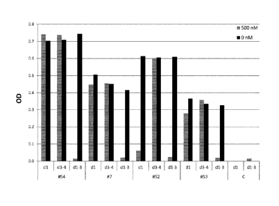
Figure 1. Binding of DARPin domains to HER2
The binding of monovalent DARPins to the HER2 extra cellular domain (domain I-
IV) was
tested by competition ELISA using purified HER2 domains (domain I, domain III-
IV or
domain I-III) as competitors, as depicted in Figure 1A and 1B. In presence of
500nM of
Her2 domain I, the DARPin #51 and DARPin #52 cannot bind HER2 (domain I-IV)
anymore, indicating that they bind an epitope located on domain I. DARPin #7,
DARPin
#53 and DARPin #54 are binding domain ll as neither 500nM of Her2 domain I nor
500nM
of Her2 domain III-IV can prevent their binding to the full length Her2
(domain I-IV). Figure
1.0 shows that the monovalent DARPins can bind on the preformed HER2-
pertuzumab
complex and are thus binding a different epitope than pertuzumab on the HER2
domain II.
See below for the definitions of the DARPins. OD, optical density at 450 nM
minus OD at
620 nm; C, a control DARPin, which is not binding HER2; dl, domain I of HER2;
d1-3;
domain I-Ill of HER2; d3-4, domain III-IV of HER2.
Figure 2. Inhibition of BT474 cell proliferation by monovalent and biparatopic
binding
proteins
CA 02892747 2015-05-27
WO 2014/083208 PCT/EP2013/075290
7
The inhibition of BT474 proliferation by monovalent DARPins (i.e. DARPin #
land DARPin
#18), a non-covalent mixture of these monovalent DARPins and biparatopic
binding
proteins comprising these monovalent DARPins in different orientations (DARPin
# 41 and
DARPin #49) was tested. Figure 2A shows the inhibition of proliferation by
various
concentrations of biparatopic DARPins and the corresponding fitted inhibition
curves are
shown for a distinct single experiment. The 1 050 value for DARPin #41 was
then calculated
to be about 2 nM. The 1050 values for distinct DARPins are listed in Table 2.
The graph in
Figure 2A shows OD, optical density at 450 nm minus OD at 620 nm plotted
against C,
concentration of DARPins in nM. The X axis is shown in logarithmic scale.
Figure 2B
shows inhibition of proliferation at a concentration of 100nM for biparatopic
DARPins, a
mixture of both monovalent DARPins and the individual corresponding monovalent
DARPins. The OD is plotted on the Y-axis. Inhibition of proliferation is
reflected by a low
OD. See below for the definitions of the DARPins. #41, DARPin #41; #49, DARPin
#49;
#18, DARPin #18; #1, DARPin #1; n.c., negative control.
Figure 3. Inhibition of BT474 cell proliferation by various biparatopic
DARPins
Inhibition of BT474 proliferation by a subset of biparatopic DARPins (#23,
#24, #33, #37,
#43, #44 and #41) comprising different N-terminal and/or C-terminal ankyrin
repeat
domains is shown. The inhibition of proliferation by various concentrations of
DARPins
and the corresponding fitted inhibition curves are shown for a distinct single
experiment
each. The IC50 values for distinct DARPins are listed in Table 2. Figure 3A
shows
inhibition of biparatopic DARPins having DARPin #15 and Figure 3B shows
inhibition of
biparatopic DARPins having DARPin #18 at the C-terminus. Figure 30 and 3D show
inhibition of biparatopic DARPins having DARPin #51 at the N-terminus and
DARPin #18
on the C-terminus and Figure 3D shows inhibition of biparatopic DARPins having
DARPin
#51 at the N-terminus and DARPin #21 at the C-terminus. Graph show OD, optical
density
at 450nm minus OD at 620nm plotted against C, concentration of DARPins in nM.
The X
axis is shown in logarithmic scale. See below for the definitions of the
DARPins. #23,
DARPin #23; #24, DARPin #24; .#33, DARPin #33; #37, DARPin #37; #41, DARPin
#41;
.. #43, DARPin #43; #44, DARPin #44.
Figure 4. Inhibition of cell proliferation by biparatopic DARPin #41/n
different cell lines
Inhibition of proliferation of NCI-N87 (Figure 4A) and ZR75-30 (Figure 4B) and
MDA-
MB175 (Figure 40) by DARPin #41 and trastuzumab was tested. The inhibition of
.. proliferation by various concentrations of DARPins and the corresponding
fitted inhibition
curves are shown for a distinct single experiment each. The IC 50 values for
distinct cell
CA 02892747 2015-05-27
WO 2014/083208 PCT/EP2013/075290
8
lines are listed in Table 3. Graph shows OD, optical density at 450 nm minus
OD at 620
nm plotted against C, concentration of DARPins in nM. The X axis is shown in
logarithmic
scale. See below for the definitions of the DARPins and reference molecules.
#41,
DARPin #41; T, trastuzumab.
Figure 5. Induction of apoptosis by biparatopic DARPin #41 in different cell
lines
Induction of apoptosis in BT474 cells (Figure 5A) and NCI-N87 cells (Figure
5B) and
MDA-MB175 (Figure 5C) by DARPin #41 and trastuzumab was tested. The induction
of
apoptosis by various concentrations of DARPins and the corresponding fitted
inhibition
curves are shown for a distinct single experiment each. The EC50 values for
distinct cell
lines are listed in Table 3. Graph in Figure 5A shows OD, optical density at
at 450 nm
minus OD at 490 nm plotted against C, concentration of DARPins of trastuzumab
in nM.
Graph in Figure 5B and 5C shows RLU, relative light units plotted against C,
concentration of DARPins or trastuzumab in nM. The X axis is shown in
logarithmic scale.
See below for the definitions of DARPins. T, trastuzumab; #41, DARPin #41.
Figure 6. Comparison of efficacy of DARPin #41 with benchmarks in inhibition
of cell
proliferation and induction of apoptosis.
Inhibition of proliferation (Figure 6A) and induction of apoptosis (Figure 6B)
in BT474 cells
was tested for DARPin #41 and the benchmarks trastuzumab and pertuzumab and a
combination of 100 nM trastuzumab and a titration of pertuzumab. Figure 6A
shows
inhibition of proliferation by various concentrations of DARPin, respectively
benchmark
concentrations and the corresponding fitted inhibition curves are shown for a
distinct
single experiment each. The IC50 values for distinct cell lines are listed in
Table 3. The
Graph shows OD, optical density at at 450 nm minus OD at 620 nm plotted
against C,
concentration of DARPin / benchmarks in nM. The X axis is shown in logarithmic
scale.
Figure 6B shows induction of apoptosis by various concentrations of DARPin,
respectively
benchmark concentrations and the corresponding fitted activation curves are
shown for a
distinct single experiment each. The EC50 values for distinct cell lines are
listed in Table 3.
The Graph shows relative light units (RLU) plotted against C, concentration of
DARPin /
benchmarks in nM. The X axis is shown in logarithmic scale. See below for the
definitions
of DARPins. T, trastuzumab; P, pertuzumab; #41, DARPin #41.
Figure 7. Inhibition of BT474 cell proliferation by different formats of
biparatopic binding
proteins
CA 02892747 2015-05-27
WO 2014/083208 PCT/EP2013/075290
9
The inhibition of B1474 proliferation by different formats of biparatopic
DARPins
composed DARPin #1 at the N-terminus and DARPin #18 at the C-terminus is
shown.
Figure 7A shows the inhibition of proliferation by various concentrations of
biparatopic
DARPins, which were engineered to have a long serum half live, and the
corresponding
fitted inhibition curves are shown for a distinct single experiment. The
biparatopic DARPin
#63 is PEGylated at its C-terminal Cys residue, whereas the biparatopic
DARPins #64
and #65 comprise an ankyrin repeat domain binding to serum albumin. Figure 7B
shows
the inhibition of proliferation by various concentrations of biparatopic
DARPins comprising
different linkers between the repeat domains binding HER2 and the
corresponding fitted
inhibition curves are shown for a distinct single experiment. The IC50 values
for DARPins
are listed in Table 2. Graph shows OD, optical density at 450 nm minus OD at
620 nm
plotted against C, concentration of DARPins in nM. The X axis is shown in
logarithmic
scale. See below for the definitions of the DARPins. #66, DARPin #66, which
comprises a
short two amino acid long GS-linker between the two repeat domains; #67,
DARPin #67,
which comprises a five amino acid long GS-linker between the two repeat
domains; #41,
DARPin #41, which comprises a ten amino acid long GS-linker between the two
repeat
domains; #68, DARPin #68, which comprises a 24 amino acid long PT-linker
between the
two repeat domains.
Detailed description of the invention
According to one embodiment of the invention, a recombinant binding protein
comprising
at least a first and a second repeat domain, wherein each of said two repeat
domains
binds the extracellular region of HER2 and wherein said repeat domains are
covalently
linked.
It has surprisingly turned out that binding of the extracellular part of HER2
with a
recombinant binding protein comprising at least two covalently linked repeat
domains,
each with specificity for the extracellular region of HER2, has advantageous
and
unexpected effects over prior art approaches as outlined above, which bind
HER2 with
distinct and individual binders (e.g., a combination of trastuzumab and
pertuzumab; Figure
6).
Human HER2 consists of 1255 amino acids with a 21 amino acid signal sequence,
a 631
.. amino acid extracellular region (e.g. the ectodomain comprising domains I
to IV), a 23
amino acid transmembrane region, and a 580 amino acid cytoplasmic domain.
CA 02892747 2015-05-27
WO 2014/083208 PCT/EP2013/075290
Preferably, said binding of the extracellular region of HER2 by said
recombinant binding
protein is a simultaneous or concurrent binding of said repeat domains to said
extracellular region of HER2. Also preferably, said repeat domains bind to two
different
5 .. epitopes of the extracellular region of HER2. Also preferably, said
repeat domains bind to
two different and non-overlapping epitopes of the extracellular region of
HER2.
One reason for this increased efficacy could be that a recombinant binding
protein
according to the invention induces a so far not described tethered
conformation of the
10 extracellular region of HER2, which seems to be the consequence of an
intramolecular
interaction of the biparatopic binding protein of the invention with two
different epitopes on
the extracellular region of HER2 (Example 8); i.e. both repeat domains of the
binding
protein seem to bind simultaneously to different epitopes on the same HER2
molecule
and thereby forcing the extracellular region of HER2 in this new tethered
conformation.
.. Such a tethered conformation is not described by the prior art.
Importantly, these two
repeat domains need to be linked by being present in the same binding protein;
i.e. a
simple mixture of the two repeat domains does not show efficacy (Fig. 2B).
Furthermore,
the bivalent binding of such a binding protein to the extracellular region of
HER2 could
develop synergistic binding effects by exhibiting increased avidity, i.e., a
combined
.. strength of synchronous binding to different epitopes of the target.
Avidity is distinct from
affinity, which corresponds to the strength of a single binding interaction.
Overall, this
specific interaction of the binding protein with HER2 may explain the very
effective
inhibition of proliferation and induction of apoptosis by such molecules as
shown in the
examples.
According to this theory the two different repeat domains in the same protein
synergistically support each other in binding their respective epitope, thus
leading to an
increase in overall affinity to the target.
.. Binding of the first repeat domain to its epitope on HER2 brings the second
repeat domain
into an energetically and/or sterically favorable position which facilitates
it's binding to its
respective epitope on HER2.
As shown in the examples the covalent linkage of the first and the second
repeat domain
.. seems to potentiate their biological activity.
CA 02892747 2015-05-27
WO 2014/083208 PCT/EP2013/075290
11
In a preferred embodiment of the recombinant binding protein according to the
invention a
first repeat domain binds domain ll of HER2 and a second repeat domain binds
domain IV
of HER2.
It is important to understand that the term "binds domain II" means that the
respective
repeat domain binds primarily domain ll of HER2. This definition, however,
does not
exclude that the parts of said repeat domain can bind, or overlap, to other
domains. The
same applies for the term "binds domain IV".
A simultaneous targeting of domains II and IV of HER2 by a biparatopic binding
protein
according to the present invention has particular unexpected effects over what
was known
from the prior art. Cell responses in terms of inhibition of proliferation and
induction of cell
apoptosis by such binding proteins were much more dramatic when compared to
effects
obtained by state of the art antibodies. For example, the extent of such
responses has
proved to be superior to that induced by clinical antibody benchmarks, such as
the
combination of trastuzumab and pertuzumab targeting domain IV and ll of HER2,
respectively (Fig. 4, 5 and 6). Interestingly, some biparatopic binding
proteins binding to
domain I and domain IV of HER2 do not show such unexpected effects (Fig. 3C
and 3D).
Methods to determine the domain of the extracellular region of HER2 to which a
repeat
domain binds, e.g. as shown in Example 3, are well known to the person skilled
in the art
(e.g. Jost et al., loc. cit.).
Applicant's findings have important implications for the treatment of HER2-
driven human
cancers, in the sense that simultaneous targeting of domains II and IV of HER2
with a
biparatopic binding protein according to the present invention could be a more
efficacious
alternative to current antibody targeting approaches.
The binding protein according to the present invention is thus preferably a
biparatopic
binding protein, i.e., it comprises two antigen repeat domains recognizing two
different
epitopes, or domains (e.g. domains II and IV) on the same protein target
(namely HER2).
However, polypeptides which are multiparatopic, i.e, containing antigen repeat
domains
recognizing three, four or more epitopes on the same target protein, are
encompassed
within the scope invention, as are polypeptides which are both bi- or
multiparatopic and
multivalent, i.eõ having also antigen repeat domains recognizing one or more
other target
proteins.
CA 02892747 2015-05-27
WO 2014/083208 PCT/EP2013/075290
12
HER2, as used herein, relates to Human Epidermal Growth Factor Receptor 2,
also
known as Neu, ErbB-2, CD340 (cluster of differentiation 340) or p185. HER2 is
a member
of the epidermal growth factor receptor (EGFR/ErbB) family. HER2 is, in
humans,
encoded by ERBB2, a known proto-oncogene located at the long arm of human
chromosome 17 (17q12). HER2 has the UniProtKB/Swiss-Prot number P04626.
According to a preferred embodiment of the invention, the first and second
repeat
domains are located on the same polypeptide, while the repeat domain targeting
domain II
of HER2 is located N-terminally to the repeat domain targeting domain IV of
HER2.
These embodiments are for example shown in Fig. 2A, and the corresponding
description.
The inventors have, surprisingly, shown that a binding protein in which the
repeat domain
targeting domain II of HER2 is located C-terminally to the repeat domain
targeting domain
IV of HER2 is significantly less efficacious than a binding protein in which
the repeat
domain targeting domain II of HER2 is located N-terminally to the repeat
domain targeting
domain IV of HER2.
Preferably, said first repeat domain binding domain II of HER2 is not
competing for
binding to HER2 with pertuzumab. For example, Fig. 1C shows such repeat
domains not
competing for binding to HER2 with pertuzumab. Likewise preferably, said
second repeat
domain binding domain IV of HER2 is not competing for binding to HER2 with
trastuzumab. For example, the repeat domains of DARPins #18 to 20 do not
compete for
binding to HER2 with trastuzumab. Methods to determine if a repeat domain does
not
compete for binding to HER2 with trastuzumab or pertuzumab, e.g. as shown in
Example
3, are well known to the person skilled in the art.
This means that, in the first preferred embodiment, the first repeat domain
binds a
different epitope of domain II of HER2 than pertuzumab. Likewise, in the
second preferred
embodiment, the second repeat domain binds a different epitope of domain IV of
HER2
than trastuzumab. Without being bound to theory, the inventors attribute at
least some of
the effects shown in the experimental section to these facts.
According to another preferred embodiment of the invention said first repeat
domain is an
ankyrin repeat domain, or a designed ankyrin repeat domain, and said second
repeat
domain is an ankyrin repeat domain, or a designed ankyrin repeat domain.
CA 02892747 2015-05-27
WO 2014/083208 PCT/EP2013/075290
13
Preferably, said ankyrin repeat domains or designed ankyrin repeat domains
comprise
between 70 and 300 amino acids, in particular between 90 and 200 amino acids.
Also preferably, a repeat domain of the invention is an ankyrin repeat domain
or a
designed ankyrin repeat domain as described in WO 2002/020565. Examples of
designed
ankyrin repeat domains with biparatopic binding specificity for different
domains of Her2
are shown in the Examples.
According to a preferred embodiment of the invention, the first repeat domain
binds the
extracellular region of HER2 in PBS with a Kd smaller than 10-7M and said
second repeat
domain binds the extracellular region of HER2 in PBS with a Kd smaller than 10-
7M.
Kd is the dissociation constant and will further be defined in the text below.
A Kd smaller
than 10-7M is required to provide sufficient affinity of the repeat domain to
its target.
Preferably, the repeat domains bind their target domains in PBS with a Kd
smaller than
10_8m, 10-9m, m or, most preferably smaller than 10-11M.
Recombinant binding proteins comprising proteins binding domain II and/or
domain IV of
Her2 with a Kd in PBS below 10-7M are shown in Example 2.
According to a preferred embodiment, said binding protein inhibits stimulated
proliferation
of BT474 cells with an half maximal inhibitory concentration (I050) value of
smaller than
100 nM. Preferably, said binding protein inhibits stimulated proliferation of
BT474 cells
with an I050 value of smaller than 90, 80, 70, 60, 50, 40, 30, 20 or 10 nM.
Also preferably,
said binding protein inhibits stimulated proliferation of B1474 cells by at
least 100%, 90%,
80%, 70%, 60%, 50%, 40%, 30%, 20% or 10%.
BT474 cells can be used to measure the functional capability of the binding
proteins of the
invention to inhibit proliferation by standard means well known to the person
skilled in the
art, e.g. as shown in Example 4. Preferably, B1474, SKBR-3, NCI-N87, ZR75-30,
HCC1419 or MDA-MB175 cells can be used to measure the functional capability of
the
compounds of the invention to inhibit proliferation, e.g. as shown in Example
5.
Recombinant binding proteins which inhibit stimulated proliferation of B1474
cells with an
1050 value of smaller than 100 nM are disclosed, and discussed, in Example 4.
CA 02892747 2015-05-27
WO 2014/083208 PCT/EP2013/075290
14
According to another preferred embodiment, said binding protein induces
apoptosis in
BT474 cells with an half maximal effective concentration (EC50) value of
smaller than 100
nM. Preferably, said binding protein induces apoptosis in BT474 cells with an
EC50 value
of smaller than 90, 80, 70, 60, 50, 40, 30, 20 or 10 nM.
BT474 cells can be used to measure the functional capability of the binding
proteins of the
invention to induce apoptosis by standard means well known to the person
skilled in the
art, e.g. as shown in Example 5. Preferably, B1474, SKBR-3, NCI-N87, ZR75-30,
HCC1419 or MDA-MB175 cells can be used to measure the functional capability of
the
compounds of the invention to induce apoptosis, e.g. as shown in Example 5.
Recombinant binding proteins which induce apoptosis in BT474 cells with an
EC50 value
of smaller than 100 nM are disclosed, and discussed, in Examples 5.
According to a preferred embodiment, said first and second repeat domains are
connected by a polypeptide linker.
Such polypeptide linker may, for example, be accomplished by mere genetic
fusion of the
encoding cDNAs of the respective domains to be fused. Such type of embodiment
qualifies as a fusion peptide protein with two different repeat domains.
The linker can for example consist of an oligopeptide comprising the amino
acids G and
S, or P and T, respectively, as set forth in SEQ ID Nos: 7 to 12. According to
another
preferred embodiment, a "multimerization moiety" as described below can be
used.
Alternatively, the two repeat domains can be linked to one another, e.g., by
means of non-
peptide based chemical linkers.
Preferably, the recombinant binding protein and/or repeat domain has a
midpoint
denaturation temperature (Tm) above 45 C, more preferably above 50 C, more
preferably
above 55 C, and most preferably above 60 C upon thermal unfolding in PBS at pH
7.4. A
binding protein or a repeat domain of the invention possesses a defined
secondary and
tertiary structure under physiological conditions. Thermal unfolding of such a
polypeptide
results in a loss of its tertiary and secondary structure, which can be
followed, for
example, by circular dichroism (CD) measurements. The midpoint denaturation
temperature of a binding protein or repeat domain upon thermal unfolding
corresponds to
CA 02892747 2015-05-27
WO 2014/083208 PCT/EP2013/075290
the temperature at the midpoint of the cooperative transition in physiological
buffer upon
heat denaturation of said protein or domain by slowly increasing the
temperature from
10 C to about 100 C. The determination of a midpoint denaturation temperature
upon
thermal unfolding is well known to the person skilled in the art. This
midpoint denaturation
5 temperature of a binding protein or repeat domain upon thermal unfolding
is indicative of
the thermal stability of said polypeptide.
Also preferred is a recombinant binding protein and/or ankyrin repeat domain
forming less
than 5% (w/w) insoluble aggregates at concentrations up to 20 g/L, preferably
up 40 g/L,
10 more preferably up to 60 g/L, even more preferably up to 80 g/L, and
most preferably up
to 100 g/L when incubated for over 5 days, preferably over 10 days, more
preferably over
days, more preferably over 40 days, and most preferably over 100 days at 37 C
in
PBS. The formation of insoluble aggregates can be detected by the appearance
of visual
precipitations, gel filtration or dynamic light scattering, which strongly
increases upon
15 formation of insoluble aggregates. Insoluble aggregates can be removed
from a protein
sample by centrifugation at 10000 x g for 10 minutes. Preferably, a
recombinant binding
protein and/or ankyrin repeat domain forms less than 2%, more preferably less
than 1%,
0.5%, 0.2%, 0.1%, or most preferably less than 0.05% (w/w) insoluble
aggregates under
the mentioned incubation conditions at 37 C in PBS. Percentages of insoluble
aggregates
20 can be determined by separation of the insoluble aggregates from soluble
protein,
followed by determination of the protein amounts in the soluble and insoluble
fraction by
standard quantification methods.
Also preferred is a recombinant binding protein and/or ankyrin repeat domain
that does
not lose its native three-dimensional structure upon incubation in PBS
containing 100 mM
dithiothreitol (OTT) for 1 or 10 hours at 37 C.
In one particular embodiment the invention relates to a recombinant binding
protein
comprising two ankyrin repeat domains, specifically binding to HER2 and having
the
indicated or preferred midpoint denaturation temperature and non-aggregating
properties
as defined above.
According to other preferred embodiments of the invention, it is provided that
CA 02892747 2015-05-27
WO 2014/083208 PCT/EP2013/075290
16
= said first repeat domain competes for binding to HER2 with an ankyrin
repeat
domain selected from the group consisting of SEQ ID NOs: 62 to 68, 72 and 114
to 121 and/or
= said second repeat domain competes for binding to HER2 with an ankyrin
repeat
domain selected from the group consisting of SEQ ID NOs: 74 to 82.
The inventors have evidence that, out of these repeat domains, the first
repeat domain
binds domain ll of HER2, whereas the second repeat domain binds domain IV of
HER2
Preferably, said first repeat domain competes for binding to HER2 with an
ankyrin repeat
domain selected from the group consisting of SEQ ID NOs: 62 to 67 and 115 to
121. More
preferably, said first repeat domain competes for binding to HER2 with an
ankyrin repeat
domain selected from the group consisting of SEQ ID NOs: 62, 115, 120, and
121, in
particular SEQ ID NO: 115 and 120. Also preferably, said first repeat domain
competes for
binding to HER2 with a binding protein selected from the group of DARPins #1
to 6 and 54
to 60; more preferably, with a binding protein from the group of DARPins #1,
54, 59 and
60; in particular, with a binding protein from the group of DARPins #54 and
60.
Further preferred, said second repeat domain competes for binding to HER2 with
an
ankyrin repeat domain selected from the group consisting of SEQ ID NOs: 79 to
81, in
particular SEQ ID NO: 80 and 81. Also preferably, said second repeat domain
competes
for binding to HER2 with a binding protein selected from the group of DARPins
#18 to 20;
in particular, with a binding protein from the group of DARPins #19 and 20.
According to still other preferred embodiments of the invention, it is
provided that
= a first repeat domain comprises an amino acid sequence that has at least
70%
amino acid sequence identity with one ankyrin repeat domain selected from the
group consisting of SEQ ID NOs: 62 to 68, 72 and 114 to 121,
= a second repeat domain comprises an amino acid sequence that has at least
70%
amino acid sequence identity with one ankyrin repeat domain selected from the
group consisting of SEQ ID NOs: 74 to 82,
and wherein further,
= G at position 1 and/or S at position 2 of said ankyrin repeat domain are
optionally
missing; and
CA 02892747 2015-05-27
WO 2014/083208 PCT/EP2013/075290
17
= L at the second last position and/or N at the last position of said
ankyrin repeat
domain are optionally exchanged by A.
Preferably, said first repeat domain comprises an amino acid sequence that has
at least
70% amino acid sequence identity with one ankyrin repeat domain selected from
the
group consisting of SEQ ID NOs: 62 to 67 and 115 to 121. More preferably, said
first
repeat domain comprises an amino acid sequence that has at least 70% amino
acid
sequence identity with one ankyrin repeat domain selected from the group
consisting of
SEQ ID NOs: 62, 115, 120, and 121, in particular SEQ ID NO: 115 and 120. Also
preferably, said first repeat domain comprises an amino acid sequence that has
at least
70% amino acid sequence identity with a binding protein selected from the
group
consisting of DARPins #1 to 6 and 54 to 60; more preferably, with a binding
protein from
the group of DARPins #1, 54, 59 and 60; in particular, with a binding protein
from the
group of DARPins #54 and 60.
Further preferred, said second repeat domain comprises an amino acid sequence
that has
at least 70% amino acid sequence identity with one ankyrin repeat domain
selected from
the group consisting of SEQ ID NOs: 79 to 81, in particular SEQ ID NO: 80 and
81. Also
preferably, said second repeat domain comprises an amino acid sequence that
has at
least 70% amino acid sequence identity with a binding protein from the group
consisting of
of DARPins #18 to 20; in particular, with a binding protein from the group of
DARPins #19
and 20.
Preferably, the first ankyrin repeat domain comprises an amino acid sequence
that has at
least 70, 71, 72, 73, 74, 75, 76, 77, 78, 79, 80, 81, 82, 83, 84, 85, 86, 87,
88, 89, 90, 91,
92, 93, 94, 95, 96, 97, 98, 99, or 100 % amino acid sequence identity with one
ankyrin
repeat domain selected from the group consisting of SEQ ID NOs: 62 to 68, 72
and 114 to
121.
Preferably, the second ankyrin repeat domain comprises an amino acid sequence
that
has at least 70, 71, 72, 73, 74, 75, 76, 77, 78, 79, 80, 81, 82, 83, 84, 85,
86, 87, 88, 89,
90, 91, 92, 93, 94, 95, 96, 97, 98, 99, or 100 % amino acid sequence identity
with one
ankyrin repeat domain selected from the group consisting of SEQ ID NOs: 74 to
82.
Also preferably, the first ankyrin repeat domain comprises an amino acid
sequence that
has at least 70, 71, 72, 73, 74, 75, 76, 77, 78, 79, 80, 81, 82, 83, 84, 85,
86, 87, 88, 89,
CA 02892747 2016-03-04
18
90, 91, 92, 93, 94, 95, 96, 97, 98, 99, or 100 % amino acid sequence identity
with one,
two or three ankyrin repeat modules present between the N-terminal and C-
terminal
capping modules of an ankyrin repeat domain selected from the group consisting
of SEQ
ID NOs: 62 to 68,72 and 114 to 121.
Also preferably, the second ankyrin repeat domain comprises an amino acid
sequence
that has at least 70, 71, 72, 73, 74, 75, 76, 77, 78, 79, 80, 81, 82, 83, 84,
85, 86, 87, 88,
89, 90, 91, 92, 93, 94, 95, 96, 97, 98, 99, or 100 % amino acid sequence
identity with
one, two or three ankyrin repeat modules present between the N-terminal and C-
terminal
capping modules of an ankyrin repeat domain selected from the group consisting
of SEQ
ID NOs: 74 to 82.
According to yet other preferred embodiments of the invention, it is provided
that
= said first repeat domain is selected from the group consisting of SEQ ID
NOs: 62 to
68,72 and 114 to 121,
= said second repeat domain is selected from the group consisting of SEQ ID
NOs:74 to 82
and wherein further
= G at position 1 and/or S at position 2 of said ankyrin repeat domain are
optionally
missing; and
= L at the second last position and/or N at the last position of said
ankyrin repeat
domain are optionally exchanged by A.
Preferably, the first ankyrin repeat domain is selected from the group
consisting of SEQ ID
NOs: 62 to 67 and 115 to 121; more preferably, 115, 120, and 121; in
particular, SEQ ID
NO: 115 and 120.
Preferably, the second ankyrin repeat domain is selected from the group
consisting of
SEQ ID NOs: 79 to 81, in particular SEQ ID NO: 80 and 81.
According to yet other preferred embodiments of the invention, it is provided
that
= said first repeat domain comprises an ankyrin repeat module having an
amino acid
sequence selected from the group consisting of SEQ ID NO: 15 to 18, 21 to 23,
37, 38, 125, 126, 129, 130, 133 and 134 and sequences, wherein up to 9 amino
acid residues in SEQ ID NO: 15 to 18, 21 to 23, 37, 38, 125, 126, 129, 130,
133
and 134 are replaced by any other amino acid residues, and/or
CA 02892747 2015-05-27
WO 2014/083208 PCT/EP2013/075290
19
= said second repeat domain comprises an ankyrin repeat module having an
amino
acid sequence selected from the group consisting of SEQ ID NO: 46, 47, 51, 52,
55 and 56, and sequences, wherein up to 9 amino acid residues in SEQ ID NO:
46, 47, 51, 52, 55 and 56 are replaced by any other amino acid residues.
Preferably, such an ankyrin repeat module of the first ankyrin repeat domain
is selected
from the group consisting of SEQ ID NO: 15 to 18, 125, 126, 129, 130, 133 and
134; more
preferably, 15, 125, 129 and 133; and even more preferably, 125 and 133.
Preferably, such an ankyrin repeat module of the second ankyrin repeat domain
is
selected from the group consisting of SEQ ID NO: 46, 47, 55 and 56; more
preferably, 55
and 56.
Also preferably, up to 8 amino acids in the repeat modules of SEQ ID NO: 15 to
18, 21 to
23, 37, 38, 46, 47, 51, 52, 55, 56, 125, 126, 129, 130, 133 and 134 are
exchanged by
another amino acid, more preferably up to 7 amino acids, more preferably up to
6 amino
acids, more preferably up to 5 amino acids, even more preferably up to 4 amino
acids,
more preferably up to 3 amino acids, more preferably up to 2 amino acids, and
most
preferably 1 amino acid.
Preferably, when amino acids are exchanged in capping modules, repeat modules
or
repeat domains, repeat domains, or binding proteins, these amino acids are
replaced by
an amino acid selected from the group consisting of A, D, E, F, H, I, K, L, M,
N, Q, R, S, T,
V, W and Y; more preferably from the group consisting of A, D, E, H, I, K, L,
Q, R, S, T, V,
and Y. Also preferably, an amino acid is exchanged by a homologous amino acid;
i.e. an
amino acid is exchanged by an amino acid having a side chain with similar
biophysical
properties. For example, the negative charged amino acid D may be replaced by
the
negative charged amino acid E, or a hydrophobic amino acid such as L may be
replaced
by A, I or V. The techniques of exchanging an amino acid by another amino acid
in a
polypeptide are well known to the person skilled in the art.
Preferably, the repeat module according to the invention has an amino acid
sequence
selected from the group consisting of KDFQGITPLHIAATSGHLEIVEVLLKAGADVNA
(SEQ ID NO: 16 and sequences, in which up to 9 amino acid residues in SEQ ID
NO: 16
are replaced by any other amino acid residues, and wherein
= F at position 3 is optionally exchanged by A
CA 02892747 2016-03-04
= Q at position 4 is optionally exchanged by E;
= G at position 5 is optionally exchanged by S;
= I at position 6 is optionally exchanged by V;
= I at position 11 is optionally exchanged by L;
= T at position 14 is optionally exchanged by Q; and/or
= S at position 15 is optionally exchanged by an amino acid selected from
the group
consisting of N and W.
One very preferred repeat module of this group has an amino acid sequence
consisting of
KDFQGVTPLHIAAQSGHLEIVEVLLKAGADVNA (SEQ ID NO: 125), SEQ ID NO: 12901
SEQ ID NO: 133.
Also preferably, the ankyrin repeat module according to the invention has an
amino acid
sequence selected from the group consisting of
KDITGETPLHHAADSGHLEIVEVLLKAGADVNA (SEQ ID NO: 18) and sequences, in which
up to 9 amino acid residues in SEQ ID NO: 18 are replaced by any other amino
acid
residues, and wherein
= I at position 3 is optionally exchanged by V;
= E at position 6 is optionally exchanged by D;
= H at position 11 is optionally exchanged by L;
= D at position 14 is optionally exchanged by Q;
= S at position 15 is optionally exchanged by H; and/or
= E at position 19 is optionally exchanged by V.
One very preferred repeat module of this group has an amino acid sequence
consisting of
KDVTGDTPLHLAAQHGHLEIVEVLLKAGADVNA (SEQ ID NO: 126), SEQ ID NO: 130 or
SEQ ID NO: 134.
Also preferably, the ankyrin repeat module according to the invention has an
amino acid
sequence selected from the group consisting of
KDWEGTTPLHLAAHTGHLEIVEVLLKAGADVNA (SEQ ID NO: 21) and sequences, in which
up to 9 amino acid residues in SEQ ID NO: 21 are replaced by any other amino
acid
residues, and wherein
= W at position 3 is optionally exchanged by F;
= W at position 4 is optionally exchanged by Q;
CA 02892747 2015-05-27
WO 2014/083208 PCT/EP2013/075290
21
= T at position 6 is optionally exchanged by an amino acid selected from
the group
consisting of I, Y and V; preferably T;
= L at position 11 is optionally exchanged by an amino acid selected from
the group
consisting of I and V; preferably I and V;
= H at position
14 is optionally exchanged by an amino acid selected from the group
consisting of H, Q, Y and W; preferably H; and/or
= T at position 15 is optionally deleted or exchanged by an amino acid
selected from
the group consisting of A and D.
Also preferably, the ankyrin repeat module according to the invention has an
amino acid
sequence selected from the group consisting of
KDTVGTTPLHYAAEDGHLEIVEVLLKAGADVNA (SEQ ID NO: 22) and sequences, in
which up to 9 amino acid residues in SEQ ID NO: 22 are replaced by any other
amino
acid residues, and wherein
= T at position 3 is optionally exchanged by an amino acid selected from the
group
consisting of S, K, E and I; equal amino acid distribution;
= V at position 4 is optionally exchanged by an amino acid selected from
the group
consisting of Q, I and Y; preferably Y;
= T at position 6 is optionally exchanged by an amino acid selected from
the group
consisting of Q, F, R and W;
= Y at position 11 is optionally exchanged by an amino acid selected from
the group
consisting of L, E and S; preferably S;
= E at position 14 is optionally exchanged by an amino acid selected from
the group
consisting of S, Q, Y and V; and/or
= D at position 15 is optionally exchanged by an amino acid selected from the
group
consisting of S, F and Y.
= G at position 16 is optionally exchanged by D.
Also preferably, the ankyrin repeat module according to the invention has an
amino acid
sequence selected from the group consisting of
KDVEGWTPLHYAASSGHLEIVEVLLKAGADVNA (SEQ ID NO: 38) and sequences, in
which up to 9 amino acid residues in SEQ ID NO: 38 are replaced by any other
amino
acid residues, and wherein
= W at position 6 is optionally exchanged by Q;
= Y at position 11 is optionally exchanged by L; and/or
CA 02892747 2015-05-27
WO 2014/083208 PCT/EP2013/075290
22
= S at position 15 is optionally exchanged by Y.
Also preferably, the ankyrin repeat module according to the invention has an
amino acid
sequence selected from the group consisting of
KDWRGFTPLHYAAYLGHLEIVEVLLKAGADVNA (SEQ ID NO: 46) and sequences, in
which up to 9 amino acid residues in SEQ ID NO: 46 are replaced by any other
amino
acid residues, and wherein
= W at position 3 is optionally exchanged by an amino acid selected from
the group
consisting of W, T, V and R; preferably, T and R;
= R at position 4 is optionally exchanged by an amino acid selected from the
group
consisting of R, T and I; preferably, I;
= F at position 6 is optionally exchanged by F or H; preferably F;
= Y at position 11 is optionally exchanged by R;
= Y at position 14 is optionally exchanged by F;
= L at position 15 is optionally exchanged by V; and/or
= H at position 17 is optionally exchanged by Q.
Preferably, 9, 8, 7, 6, 5, 4, 3, 2, or 1 amino acid residues in SEQ ID NOs:16,
18, 28, 31.
21, 22, 38 and/or 46 are replaced by any other amino acid residues.
Furthermore, it is particularly preferred that said binding protein comprises
a polypeptide,
wherein said polypeptide comprises said first and second ankyrin repeat
domains and
wherein said polypeptide has at least 70% amino acid sequence identity with a
polypeptide selected from the group consisting of SEQ ID NO: 83 to 98, 102,
103, 122,
123 and 136 to 141.
Preferably, said polypeptide comprises an amino acid sequence that has at
least 70, 71,
72, 73, 74, 75, 76, 77, 78, 79, 80, 81, 82, 83, 84, 85, 86, 87, 88, 89, 90,
91, 92, 93, 94, 95,
96, 97, 98, 99, or 100 % amino acid sequence identity with a polypeptide
selected from
the group consisting of SEQ ID NOs: 83 to 98, 102, 103, 122,123 and 136 to
141.
Also preferably, such polypeptide is selected from the group consisting of SEQ
ID NO: 84,
85, 86, 87, 90, 91, 92, 98, 102, 103, 122 and 123; more preferably, 85, 86,
87, 90, 91, 92,
102, 103, 122 and 123; even more preferably, 86, 87, 91 and 92; and most
preferably, 86
and 87.
CA 02892747 2015-05-27
WO 2014/083208 PCT/EP2013/075290
23
According to yet other preferred embodiment, one or more of the amino acid
residues of
the ankyrin repeat modules of said first and second ankyrin repeat domains are
exchanged by an amino acid residue found at the corresponding position on
alignment of
an ankyrin repeat unit.
Another embodiment of the invention provides a nucleic acid molecule encoding
at least
one binding protein or a particular ankyrin repeat domain according to the
above
description. Further, a vector comprising said nucleic acid molecule is
considered.
Not all binding compositions according to the present invention comprise
polypeptides or
proteins. The latter embodiment only relates to those who do. For these,
applicant refrains
from disclosing herein all nucleic acid molecules capable of encoding them
because, due
to the Degeneracy of the genetic code, many nucleic acid molecules can encode
for one
and the same polypeptide or protein.
However, it can unequivocally and unambiguously determined whether a given
nucleic
acid encodes for a given polypeptide or protein. Thus, the present embodiment
is clear for
the skilled person, and its scope is easily determined.
Another embodiment of the invention provides the use of a binding protein
according to
the above description to inhibit at least one of
= HER2-receptor dimerization,
= HER2/HER3-heterodimerization,
= HER2-receptor autophosphorylation
= HER-receptor mediated signal transduction
= HER3-receptor ligand induced phosphorylation, and/or
= HER3-receptor mediated signal transduction.
HER2-receptor dimerization (also called "homodimerization") occus in tissues
overexpressing HER2 independent of a ligand. Said homodimerization leads to an
intracellular autophosphrylation which can eventually lead, for example, to
increased cell
proliferation.
Because HER3 lacks intrinsic kinase activity, HER3 is phosphorylated in HER2-
overexpressing breast cancer after formation of HER2/HER3 heterodimers, which
may
eventually result, for example, in apoptosis inhibition.
CA 02892747 2015-05-27
WO 2014/083208 PCT/EP2013/075290
24
Said use can either take place in vitro or in vivo. As set forth above, all
these processes
can result in pathogenic consequences, namely by activating respective signal
transduction pathways. Signal transduction pathways activated by HER2
dimerization
and/or HER2/HER3-heterodimerization include mitogen-activated protein kinase
(MAPK),
phosphoinositide 3-kinase (PI3K/Akt), phospholipase C y, protein kinase C
(PKC), Signal
transducer and activator of transcription (STAT), the Ras-Map kinase pathway
and the
mTOR pathway.
The phosphoinositide 3-kinase (PI3K/Akt) pathway is for example considered to
be one of
the critical pathways that is maintaining cell survival by blocking apoptosis.
Pathologic
activation thereof, e.g., by HER2/HER3-heterodimerization, may thus lead to
malignant
proliferation (e.g. see Examples)
Pathologic activation of HER2, e.g. by HER2-homodimerization, may lead to
malignant
cell migration, invasion or proliferation (e.g. see Examples; Hynes NE. and
Lane HA., Nat.
Rev. Cancer., 5,341-54, 2005).
Yet another embodiment of the invention provides a pharmaceutical formulation
comprising a binding protein or a composition according to the above
disclosure, and
optionally a pharmaceutical acceptable carrier and/or diluent.
Pharmaceutical acceptable carriers and/or diluents are known to the person
skilled in the
art and are explained in more detail below. Even further, a diagnostic
composition
comprising one or more of the above mentioned recombinant binding proteins, in
particular binding proteins comprising repeat domains, is considered.
A pharmaceutical formulation comprises recombinant binding proteins as
described above
and a pharmaceutically acceptable carrier, excipient or stabilizer, for
example as
described in Remington's Pharmaceutical Sciences 161h edition, Osol, A. Ed.
[1980].
Suitable carriers, excipients or stabilizers known to the skilled man are
saline, Ringer's
solution, dextrose solution, Hank's solution, fixed oils, ethyl oleate, 5%
dextrose in saline,
substances that enhance isotonicity and chemical stability, buffers and
preservatives.
Other suitable carriers include any carrier that does not itself induce the
production of
antibodies harmful to the individual receiving the composition such as
proteins,
CA 02892747 2015-05-27
WO 2014/083208 PCT/EP2013/075290
polysaccharides, polylactic acids, polyglycolic acids, polymeric amino acids
and amino
acid copolymers.
The formulations to be used for in vivo administration must be aseptic or
sterile. This is
5 readily accomplished by filtration through sterile filtration membranes.
The pharmaceutical
formulation may be administered by any suitable method within the knowledge of
the
person skilled in the art.
Further, in another embodiment of the present invention the use of at least
one binding
10 protein, composition or pharmaceutical formulation according to the
above disclosure as a
medicament is provided. Likewise, a process comprising administering a binding
protein,
composition or pharmaceutical formulation according to the aforementioned
claims to a
patient is provided. In both cases, it is preferred that the disease to be
treated is a
neoplastic disease, preferably cancer.
In each case, an effective amount of the binding protein, composition or
pharmaceutical
formulation according to the aforementioned claims is preferably administered
to a patient
for treating the disease.
The term "neoplastic disease", as used herein, refers to an abnormal state or
condition of
cells or tissue characterized by rapidly proliferating cell growth or
neoplasm. In a more
specific meaning, the term relates to cancerous processes, e.g., tumors and/or
leukemias.
The binding proteins according to the invention demonstrated apoptotic and
anti-
.. proliferative effects (see experimental section). As neoplastic diseases
are often
characterized by suppression of apoptosis and/or increased proliferation, it
is plausible to
deduce, from these experiments, that the binding proteins according to the
present
invention can be used in the treatment of neoplastic diseases. .
Preferably, said neoplastic disease is a disease characterized by at least one
selected
from the group consisting of
= Amplification of the HER2 encoding gene
= Overexpression of the HER2 encoding gene,
= Expression of a mutated form of the HER2 encoding gene, and/or
= Overexpression of the Her3 encoding gene in trastuzumab resistant tumors.
CA 02892747 2015-05-27
WO 2014/083208 PCT/EP2013/075290
26
In humans, HER2 is encoded by the ERBB2 gene. The above options can be
ascribed to
mutations in the ERBB2 gene which can be detected by means of modern molecular
diagnostics, as are currently on the market.
As used herein, the term õexpression of the HER2 encoding gene" is related to
cells,
tissues or organs which express the HER2 receptor protein, as for example
detected by
immunohistochemistry (INC). As used herein, the term "amplification or
overexpression of
the HER2 encoding gene" is related to indicate an abnormal level of expression
of the
HER2 receptor protein in a cell, tissue or organ, relative to the level of
expression in a
normal cell, tissue or organ, as for example detected by Immunohistochemistry
(INC).
Such IHC detection assays are known in the art and include the Clinical Trial
Assay
(CTA), the commercially available LabCorp 4D5 test, and the commercially
available
DAKO HercepTestO (DAKO, Carpinteria, Calif.). The latter assay uses a specific
score
.. range of 0 to 3+ cell staining (0 being normal expression, 3+ indicating
the strongest
positive expression) to identify cancers having overexpression of the HER2
protein. Thus,
patients having a cancer characterized by overexpression of the HER2 protein
in the
range of 1+, 2+, or 3+, preferably 2+ or 3+, more preferably 3+ would benefit
from the
methods of therapy of the present invention.
Alternatively, Her2 expression and/or overexpression scores can also be
detected by In
Situ hybridization (ISH), RT-PCT and other methods.
According to a particularly preferred embodiment, said neoplastic disease is
at least one
.. selected from the group consisting
= breast cancers
= ovarian cancer,
= gastric cancer,
= stomach cancer, and/or
= uterine cancer.
= colorectal cancer.
Furthermore, said use is preferably complemented, in a coordinated fashion, by
the
administration of at least one active substance selected from the group
consisting of
CA 02892747 2015-05-27
WO 2014/083208 PCT/EP2013/075290
27
= an antineoplastic agent
= an endocrine drug,
= a tumor vaccine,
= immunotherapy, and/or
= cellular therapy.
The term "complemented, in a coordinated fashion", as used herein, shall refer
to a co-
administration, which is carried out under a given regimen. This includes
synchronous
administration of the different compounds as well as time-shifted
administration of the
different compounds (e.g., compound A is given once and compound B is given
several
times thereafter, or vice versa, or both compounds are given synchronously and
one of
the two is also given at later stages).
As used herein, the term "antineoplastic agent" relates to a drug, or a
combination of
drugs, which have antineoplastic or anticancer effects. This applies, above
all, to
chemotherapeutic agents, which work by impairing mitosis, effectively
targeting fast-
dividing cells, or by causing cells to undergo apoptosis. The majority of
chemotherapeutic
drugs can be divided into alkylating agents, antimetabolites, anthracyclines,
plant
alkaloids, topoisomerase inhibitors, and other antitumour agents.
Preferred antineoplastic agents are 5-fluorouracil, actinomycin, adriamycin,
amsacrine,
anthracyclines, azathioprine, bendamustine, bleomycin, carboplatin,
chlorambucil,
cisplatin, cyclophosphamide, daunorubicin, docetaxel, doxorubicin, epirubicin,
etoposide,
idarubicin, ifosfamide, irinotecan, mechlorethamine, mercaptopurine,
methotrexate,
mitomycin, oxaliplatin, paclitaxel, plicamycin, podophyllotoxin, teniposide,
topotecan.,
valrubicin, vinblastine, vincristine, vincristine, vindesine, and/or
vinorelbine.
Immunotherapy involves the isolation of proteins from cancer cells and
subsequent
immunization of cancer patients against those proteins, in the hope of
stimulating an
immune reaction that would kill the cancer cells. Another approach to
therapeutic anti-
cancer vaccination is to generate the immune response in situ in the patient.
This
enhances the anti-tumor immune response to tumor antigens released following
lytic virus
replication providing an in situ, patient specific anti-tumor vaccine as a
result. Yet another
approach is to immunize the patient with a compound that plays a physiological
role in
cancer genesis, so that the human body eliminates said compound.
CA 02892747 2015-05-27
WO 2014/083208 PCT/EP2013/075290
28
Targeted drugs are a type of medication that blocks the growth of cancer cells
by
interfering with specific targeted molecules needed for carcinogenesis and
tumor growth,
rather than by simply interfering with rapidly dividing cells (e.g. with
traditional
chemotherapy). The main categories of targeted therapy are small molecules and
.. monoclonal antibodies.
Small molecules falling under this definition encompass, but are not limited,
to Lapatinib,
Neratinib, Afatinib, Imatinib, Gefitinib, Erlotinib, Bortezomib, BcI-2
inhibitors (e.g.
Obatoclax, ABT-263, and Gossypol), PARP inhibitors (e.g. lniparib, Olaparib),
Janus
kinase inhibitors, PI3K inhibitors, Apatinib, mTOR inhibitors (Everolimus), AN-
152, AKT-
inhibitors, HDAC inhibitors, proteasome inhibitors, Doxorubicin linked to [D-
Lys(6)]- LHRH,
Pegaptanib, Sunitinib, Sorafenib, Tivozanib and Pazopanib. Monoclonal
antibodies falling
under this definition encompass, but are not limited, to Rituximab,
trastuzumab,
trastuzumab-TDM1, pertuzumab, cetuximab and bevacizumab.
Endocrine drugs, as used herein, are drugs that are antagonistic to hormones
or hormone
receptors and thus interfere with cancer types that require hormones to grow.
One
example for such Endocrine drug is Tamoxifen, which is an antagonist of the
estrogen
receptor in breast tissue.
The term "cellular therapy", as used herein, shall relate to cell-based
therapies such as
adoptive transfer of modified, or unmodified, cytotoxic lymphocytes or
dendritic cells.
The term "tumor vaccine", as used herein, refers to vaccines that either a)
prevent
.. infections with cancer-causing viruses (mode of action is similar to other
vaccines against
viral infections), b) treat existing cancer (therapeutic cancer vaccines) or
c) prevent the
development of cancer, or ameliorate its effects (prophylactic cancer
vaccines).
In addition or alternatively thereto, said use is preferably complemented, in
a coordinated
fashion, by at least one other treatment selected from the group consisting of
= radiotherapy
= surgery, and/or
= laser ablation
CA 02892747 2015-05-27
WO 2014/083208 PCT/EP2013/075290
29
Furthermore, a method of treatment of a human or animal subject is provided
which
method comprises the use according to the above disclosure. Preferably, said
method of
treatment relates to an indication as set forth in the above disclosure. The
method
comprises administering, to a human or animal in need thereof, a
therapeutically effective
amount of a recombinant binding protein of the invention.
The recombinant binding protein or ankyrin repeat domain according to the
invention may
be obtained and/or further evolved by several methods such as display on the
surface of
bacteriophages (WO 1990/002809, WO 2007/006665) or bacterial cells (WO 1993/
010214), ribosomal display (WO 1998/048008), display on plasmids (WO
1993/008278)
or by using covalent RNA-repeat protein hybrid constructs (WO 2000/032823), or
intracellular expression and selection / screening such as by protein
complementation
assay (WO 1998/341120). Such methods are known to the person skilled in the
art.
A library of ankyrin repeat proteins used for the selection/screening of a
recombinant
binding protein or ankyrin repeat domain according to the invention may be
obtained
according to protocols known to the person skilled in the art (WO 2002/020565,
Binz,
H.K., et al., J. Mol. Biol., 332, 489-503, 2003, and Binz et al., 2004, loc.
cit). The use of
such libraries for the selection of ankyrin repeat domains with specificity
for the
extracellular region of HER2 is exemplified in Example 1. Furthermore, ankyrin
repeat
domains of the present invention may be modularly assembled from ankyrin
repeat
modules according to the current invention and appropriate capping modules or
capping
repeats (Forrer, P., et al., FEBS letters 539, 2-6, 2003) using standard
recombinant DNA
technologies (e.g. WO 2002/020565, Binz et al., 2003, loc. cit. and Binz et
al., 2004, loc.
cit).
The invention is not restricted to the particular embodiments described in the
Examples.
Other sources may be used and processed following the general outline
described below.
Definitions
The term "protein" refers to a polypeptide, wherein at least part of the
polypeptide has, or
is able to acquire a defined three-dimensional arrangement by forming
secondary, tertiary,
or quaternary structures within and/or between its polypeptide chain(s). If a
protein
comprises two or more polypeptides, the individual polypeptide chains may be
linked non-
covalently or covalently, e.g. by a disulfide bond between two polypeptides. A
part of a
CA 02892747 2015-05-27
WO 2014/083208 PCT/EP2013/075290
protein, which individually has, or is able to acquire, a defined three-
dimensional
arrangement by forming secondary or tertiary structures, is termed "protein
domain". Such
protein domains are well known to the practitioner skilled in the art.
5 The term "recombinant" as used in recombinant protein, recombinant
protein domain,
recombinant binding protein and the like, means that said polypeptides are
produced by
the use of recombinant DNA technologies well known by the practitioner skilled
in the
relevant art. For example, a recombinant DNA molecule (e.g. produced by gene
synthesis) encoding a polypeptide can be cloned into a bacterial expression
plasmid (e.g.
10 pQE30, Qiagen), yeast expression plasmid or mammalian expression
plasmid. When, for
example, such a constructed recombinant bacterial expression plasmid is
inserted into an
appropriate bacteria (e.g. Escherichia coil), this bacteria can produce the
polypeptide
encoded by this recombinant DNA. The correspondingly produced polypeptide is
called a
recombinant polypeptide.
In the context of the present invention, the term "polypeptide" relates to a
molecule
consisting of one or more chains of multiple, i.e. two or more, amino acids
linked via
peptide bonds. Preferably, a polypeptide consists of more than eight amino
acids linked
via peptide bonds.
The term "polypeptide tag" refers to an amino acid sequence attached to a
polypeptide/protein, wherein said amino acid sequence is useful for the
purification,
detection, or targeting of said polypeptide/protein, or wherein said amino
acid sequence
improves the physicochemical behavior of the polypeptide/protein, or wherein
said amino
acid sequence possesses an effector function. The individual polypeptide tags,
moieties
and/or domains of a binding protein may be connected to each other directly or
via
polypeptide linkers. These polypeptide tags are all well known in the art and
are fully
available to the person skilled in the art. Examples of polypeptide tags are
small
polypeptide sequences, for example, His (e.g. the His-tag of SEQ ID NO: 6),
myc, FLAG,
or Strep-tags or moieties such as enzymes (for example enzymes like alkaline
phosphatase), which allow the detection of said polypeptide/protein, or
moieties which can
be used for targeting (such as immunoglobulins or fragments thereof) and/or as
effector
molecules.
The term "polypeptide linker" refers to an amino acid sequence, which is able
to link, for
example, two protein domains, a polypeptide tag and a protein domain, a
protein domain
CA 02892747 2015-05-27
WO 2014/083208 PCT/EP2013/075290
31
and a non-polypeptide moiety such as polyethylene glycol or two sequence tags.
Such
additional domains, tags, non-polypeptide moieties and linkers are known to
the person
skilled in the relevant art. A list of example is provided in the description
of the patent
application WO 2002/020565. Particular examples of such linkers are glycine-
serine-
linkers and proline-threonine-linkers of variable lengths; preferably, said
linkers have a
length between 2 and 24 amino acids; more preferably, said linkers have a
length
between 2 and 16 amino acids. Examples of glycine-serine-linkers are provided
in SEQ ID
NO: 7 to 10 and examples of a proline-threonine-linkers are provided in SEQ ID
NO: 11
and 12. Preferably, the proline-threonine-linker of SEQ ID NO: 11 is preceded
by GS
and/or followed by GS.
The term "polymer moiety" refers to either a proteinaceous polymer moiety or a
non-
proteinaceous polymer moiety. A "proteinaceous polymer moiety" preferably is a
polypeptide that does not form a stable tertiary structure. Examples of
proteinaceous
.. polymer moieties are XTENO (a registered trademark of Amunix; WO
2007/103515)
polypeptides, or polypeptides comprising proline, alanine and serine residues
as
described in WO 2008/155134. Such proteinaceous polymer moieties can be
covalently
attached to, for example, a repeat domain of the invention by the generation
of genetic
fusion polypeptides using standard DNA cloning technologies, followed by their
standard
expression and purification. A "non-proteinaceous polymer moiety" is a polymer
moiety
not built from polypeptides. Examples of non-proteinaceous polymer moieties
are
hydroxyethyl starch (HES), polyethylene glycol (PEG), polypropylene glycol, or
polyoxyalkylene. The term "PEGylated" means that a PEG moiety is covalently
attached
to, for example, a polypeptide of the invention. A polymer moiety of the
invention may vary
widely in molecular weight. Preferably, said polymer moiety is connected by a
polypeptide
linker to a repeat domain.
In a specific embodiment, a PEG moiety or any other non-proteinaceous polymer
can,
e.g., be coupled to a cysteine thiol via a maleimide linker with the cysteine
being coupled
via a peptide linker to the N- or C-terminus of a repeat domain as described
herein.
The term "binding protein" refers to a protein comprising one or more binding
domains,
one or more bioactive compounds and one or more polymer moieties as further
explained
below. Preferably, said binding protein comprises up to four binding domains.
Furthermore, any such binding protein may comprise additional protein domains
that are
CA 02892747 2015-05-27
WO 2014/083208 PCT/EP2013/075290
32
not binding domains, multimerization moieties, polypeptide tags, polypeptide
linkers
and/or a single Cys residue.
Examples of "multimerization moieties" are immunoglobulin heavy chain constant
regions
which pair to provide functional immunoglobulin Fc domains, and leucine
zippers or
polypeptides comprising a free thiol which forms an intermolecular disulfide
bond between
two such polypeptides. The single Cys residue may be used for conjugating
other
moieties to the polypeptide, for example, by using the maleimide chemistry
well known to
the person skilled in the art. Preferably, said binding protein is a
recombinant binding
protein. Also preferably, the binding domains of binding protein possess
different target
specificities.
The term "compete for binding" means the inability of two different binding
domains of the
invention to bind simultaneously to the same target, while both are able to
bind the same
target individually. Thus, such two binding domains compete for binding to
said target.
Preferably, said two competing binding domains bind to an overlapping or the
same
binding epitope on said target. Methods, such as competition Enzyme-Linked
lmmuno
Sorbent Assay (ELISA) or competition SPR measurements (e.g. by using the
Proteon
instrument from BioRad), to determine if two binding domains compete for
binding to a
target, are well known to the practitioner in the art.
The term "multiparatopic binding protein" means a binding protein directed
against two or
mpre different epitopes located on the same target protein. For example, a
multiparatopic
binding protein targeting HER2 comprises at least a first binding domain
targeting a first
epitope on HER2, a second binding domain targeting a different second epitope
on HER2,
and optionally further binding domain targeting further epitopes on HER2.
The term "biparatopic binding protein" means a binding protein directed
against two
different epitopes located on the same target protein. For example, a
biparatopic binding
protein targeting HER2 comprises at least a first binding domain targeting a
first epitope
on HER2 and a second binding domain targeting a different second epitope on
HER2.
Correspondingly, a "biparatopic DARPin" comprises a first binding domain
against a first
epitope and a second binding domain against a different second epitope on the
same
target molecule.
CA 02892747 2015-05-27
WO 2014/083208 PCT/EP2013/075290
33
The term "bioactive compound" refers to a compound that is disease modifying
when
applied to a mammal having said disease. A bioactive compound may have
antagonistic
or agonistic properties and can be a proteinaceous bioactive compound or a non-
proteinaceous bioactive compound. Such proteinaceous bioactive compounds can
be
covalently attached to, for example, a binding domain of the invention by the
generation of
genetic fusion polypeptides using standard DNA cloning technologies, followed
by their
standard expression and purification. Such non-proteinaceous bioactive
compounds can
be covalently attached to, for example, a binding domain of the invention by
chemical
means, e.g., by coupling to a cysteine thiol via a maleimide linker with a
cysteine being
coupled via a peptide linker to the N- or C-terminus of a binding domain as
described
herein. Examples of proteinaceous bioactive compounds are binding domains
having a
distinct target specificity (e.g. neutralizing a growth factor by binding to
it), cytokines (e.g.
interleukins), growth factors (e.g. human growth hormone), antibodies and
fragments
thereof, hormones (e.g. GLP-1) and any possible proteinaceous drug. Examples
of non-
proteinaceous bioactive compounds are, toxins (e.g. DM1 from ImmunoGen), small
molecules targeting GPCRs, antibiotics and any possible non-proteinaceous
drug.
The term "binding domain" means a protein domain exhibiting the same "fold"
(three-
dimensional arrangement) as a protein scaffold and having a predetermined
property, as
defined below. Such a binding domain may be obtained by rational, or most
commonly,
combinatorial protein engineering techniques, skills which are known in the
art (Binz et al.,
2005, loc. cit.). For example, a binding domain having a predetermined
property can be
obtained by a method comprising the steps of (a) providing a diverse
collection of protein
domains exhibiting the same fold as a protein scaffold as defined further
below; and (b)
screening said diverse collection and/or selecting from said diverse
collection to obtain at
least one protein domain having said predetermined property. The diverse
collection of
protein domains may be provided by several methods in accordance with the
screening
and/or selection system being used, and may comprise the use of methods well
known to
the person skilled in the art, such as phage display or ribosome display.
Preferably, said
binding domain is a recombinant binding domain. Also preferably, said binding
domain is
a repeat protein or a designed repeat protein.
Accordingly, the term "binds", as used herein, relates to a binding domain
that recognizes
and binds a given target, but does not substantially recognize or bind other
targets.
Preferably, a dissociation constant in PBS of smaller than 10-7M is required
for a
candidate to qualify as a binding domain in the meaning of the present
invention.
CA 02892747 2015-05-27
WO 2014/083208 PCT/EP2013/075290
34
The term "Kd" relates to the dissociation constant, which is a specific type
of equilibrium
constant that measures the propensity of a larger object to separate
(dissociate) reversibly
into smaller components, as when a complex falls apart into its component
molecules.
Methods to determine dissociation constants of protein-protein interactions,
such as
surface plasmon resonance (SPR) based technologies (e.g. SPR equilibrium
analysis) or
isothermal titration calorimetry (ITC) are well known to the person skilled in
the art. The
measured Kd values of a particular protein-protein interaction can vary if
measured under
different conditions (e.g., salt concentration, pH). Thus, measurements of Kd
values are
preferably made with standardized solutions of protein and a standardized
buffer, such as
PBS.
The term "PBS" means a phosphate buffered water solution containing 137 mM
NaCI,
10 mM phosphate and 2.7 mM KCI and having a pH of 7.4.
The term "protein scaffold" means a protein with exposed surface areas in
which amino
acid insertions; substitutions or deletions are highly tolerable. Examples of
protein
scaffolds that can be used to generate binding domains of the present
invention are
antibodies or fragments thereof such as single-chain Fv or Fab fragments,
protein A from
Staphylococcus aureus, the bilin binding protein from Pieris brassicae or
other lipocalins,
ankyrin repeat proteins or other repeat proteins, and human fibronectin.
Protein scaffolds
are known to the person skilled in the art (Binz et al., 2005, loc. cit.; Binz
et at., 2004, loc.
cit.).
The term "target" refers to an individual molecule such as a nucleic acid
molecule, a
polypeptide or protein, a carbohydrate, or any other naturally occurring
molecule,
including any part of such individual molecule, or complexes of two or more of
such
molecules. The target may be a whole cell or a tissue sample, or it may be any
non-
natural molecule or moiety. Preferably, the target is a naturally occurring or
non-natural
polypeptide or a polypeptide containing chemical modifications, for example
modified by
natural or non-natural phosphorylation, acetylation, or methylation. In the
particular
application of the present invention, the target is the extracellular region
of HER2.
The term "predetermined property" refers to a property such as binding to a
target,
blocking of a target, activation of a target-mediated reaction, enzymatic
activity, and
related further properties. Depending on the type of desired property, one of
ordinary skill
CA 02892747 2015-05-27
WO 2014/083208 PCT/EP2013/075290
will be able to identify format and necessary steps for performing screening
and/or
selection of a binding domain with the desired property. Preferably, said
predetermined
property is binding to a target.
5 The definitions hereinafter for repeat proteins are based on those in
patent application
WO 2002/020565. Patent application WO 2002/020565 further contains a general
description of repeat protein features, techniques and applications.
The term "repeat protein" refers to a protein comprising one or more repeat
domains.
10 Preferably, each of said repeat proteins comprises up to four repeat
domains. More
preferably, each of said repeat proteins comprises up to two repeat domains.
Most
preferably, each of the repeat proteins comprises only one repeat domain.
Furthermore,
said repeat protein may comprise additional non-repeat protein domains,
polypeptide tags
and/or polypeptide linkers.
The term "repeat domain" refers to a protein domain comprising two or more
consecutive
repeat units (modules) as structural units, wherein said structural units have
the same
fold, and stack tightly to create a superhelical structure having a joint
hydrophobic core.
Preferably, a repeat domain further comprises an N-terminal and/or a C-
terminal capping
unit (or module). Even more preferably, said N-terminal and/or C-terminal
capping units
(or modules) are capping repeats.
The term "designed repeat protein" and "designed repeat domain" refer to a
repeat protein
or repeat domain, respectively, obtained as the result of the inventive
procedure explained
in patent application WO 2002/020565. Designed repeat proteins and designed
repeat
domains are synthetic and not from nature. They are man-made proteins or
domains,
respectively, obtained by expression of correspondingly designed nucleic
acids.
Preferably, the expression is done in eukaryotic or prokaryotic cells, such as
bacterial
cells, or by using a cell-free in vitro expression system. Accordingly, a
designed ankyrin
repeat protein (i.e. a DARPin) corresponds to a recombinant binding protein of
the
invention comprising at least one ankyrin repeat domain.
The term "structural unit" refers to a locally ordered part of a polypeptide,
formed by three-
dimensional interactions between two or more segments of secondary structure
that are
near one another along the polypeptide chain. Such a structural unit exhibits
a structural
motif. The term "structural motif" refers to a three-dimensional arrangement
of secondary
CA 02892747 2015-05-27
WO 2014/083208 PCT/EP2013/075290
36
structure elements present in at least one structural unit. Structural motifs
are well known
to the person skilled in the art. Structural units alone are not able to
acquire a defined
three-dimensional arrangement; however, their consecutive arrangement, for
example as
repeat modules in a repeat domain, leads to a mutual stabilization of
neighboring units
resulting in a superhelical structure.
The term "repeat unit" refers to amino acid sequences comprising repeat
sequence motifs
of one or more naturally occurring repeat proteins, wherein said "repeat
units" are found in
multiple copies, and which exhibit a defined folding topology common to all
said motifs
determining the fold of the protein. Such repeat units correspond to the
"repeating
structural units (repeats)" of repeat proteins as described by Forrer et al.,
2003, loc. cit. or
the "consecutive homologous structural units (repeats)" of repeat proteins as
described by
Binz et al, 2004, loc. cit.. Such repeat units comprise framework residues and
interaction
residues. Examples of such repeat units are armadillo repeat units, leucine-
rich repeat
units, ankyrin repeat units, tetratricopeptide repeat units, HEAT repeat
units, and leucine-
rich variant repeat units. Naturally occurring proteins containing two or more
such repeat
units are referred to as "naturally occurring repeat proteins". The amino acid
sequences of
the individual repeat units of a repeat protein may have a significant number
of mutations,
substitutions, additions and/or deletions when compared to each other, while
still
substantially retaining the general pattern, or motif, of the repeat units.
Accordingly, the term "ankyrin repeat unit" shall mean a repeat unit, which is
an ankyrin
repeat as described, for example, by Forrer et al., 2003, loc. cit.. Ankyrin
repeats are well
known to the person skilled in the art. The term "ankyrin repeat domain"
refers to a repeat
.. domain comprising two or more consecutive ankyrin repeat units (modules) as
structural
units, and, preferably, an N-terminal and/or a C-terminal capping unit (or
module).
The term "framework residues" relates to amino acid residues of the repeat
units, or the
corresponding amino acid residues of the repeat modules, which contribute to
the folding
topology, i.e. which contribute to the fold of said repeat unit (or module) or
which
contribute to the interaction with a neighboring unit (or module). Such
contribution might
be the interaction with other residues in the repeat unit (or module), or the
influence on the
polypeptide backbone conformation as found in a-helices or I3-sheets, or amino
acid
stretches forming linear polypeptides or loops.
CA 02892747 2015-05-27
WO 2014/083208 PCT/EP2013/075290
37
The term "target interaction residues" refers to amino acid residues of the
repeat units, or
the corresponding amino acid residues of the repeat modules, which contribute
to the
interaction with target substances. Such contribution might be the direct
interaction with
the target substances, or the influence on other directly interacting
residues, e.g. by
stabilizing the conformation of the polypeptide of a repeat unit (or module)
to allow or
enhance the interaction of directly interacting residues with said target.
Such framework
and target interaction residues may be identified by analysis of the
structural data
obtained by physicochemical methods, such as X-ray crystallography, NMR and/or
CD
spectroscopy, or by comparison with known and related structural information
well known
to practitioners in structural biology and/or bioinformatics.
Preferably, the repeat units used for the deduction of a repeat sequence motif
are
homologous repeat units, wherein the repeat units comprise the same structural
motif and
wherein more than 70% of the framework residues of said repeat units are
homologous to
.. each other. Preferably, more than 80% of the framework residues of said
repeat units are
homologous. Most preferably, more than 90% of the framework residues of said
repeat
units are homologous. Computer programs to determine the percentage of
homology
between polypeptides, such as Fasta, Blast or Gap, are known to the person
skilled in the
art. Further preferably, the repeat units used for the deduction of a repeat
sequence motif
are homologous repeat units obtained from repeat domains selected on a defined
target.
The term "repeat sequence motif" refers to an amino acid sequence, which is
deduced
from one or more repeat units or repeat modules. Preferably, said repeat units
or repeat
modules are from repeat domains having binding specificity for the same
target. Such
repeat sequence motifs comprise framework residue positions and target
interaction
residue positions. Said framework residue positions correspond to the
positions of
framework residues of the repeat units (or modules). Likewise, said target
interaction
residue positions correspond to the positions of target interaction residues
of the repeat
units (or modules). Repeat sequence motifs comprise fixed positions and
randomized
positions. The term "fixed position" refers to an amino acid position in a
repeat sequence
motif, wherein said position is set to a particular amino acid. Most often,
such fixed
positions correspond to the positions of framework residues and/or the
positions of target
interaction residues that are specific for a certain target. The term
"randomized position"
refers to an amino acid position in a repeat sequence motif, wherein two or
more amino
acids are allowed at said amino acid position, for example, wherein any of the
usual
twenty naturally occurring amino acids are allowed, or wherein most of the
twenty
CA 02892747 2015-05-27
WO 2014/083208 PCT/EP2013/075290
38
naturally occurring amino acids are allowed, such as amino acids other than
cysteine, or
amino acids other than glycine, cysteine and proline. Most often, such
randomized
positions correspond to the positions of target interaction residues. However,
some
positions of framework residues may also be randomized.
The term "folding topology" refers to the tertiary structure of said repeat
units or repeat
modules. The folding topology will be determined by stretches of amino acids
forming at
least parts of a-helices or 13-sheets, or amino acid stretches forming linear
polypeptides or
loops, or any combination of a-helices, I3-sheets and/or linear
polypeptides/loops. For
example, an ankyrin repeat unit/module consists of a 13-turn, followed by two
antiparallel
a-helices and a loop that reaches the turn of the next repeat unit/module.
The term "consecutive" refers to an arrangement, wherein the repeat units or
repeat
modules are arranged in tandem. In designed repeat proteins, there are at
least 2, usually
about 2 to 6, in particular at least about 6, frequently 20 or more repeat
units (or modules).
In most cases, repeat units (or modules) of a repeat domain will exhibit a
high degree of
sequence identity (same amino acid residues at corresponding positions) or
sequence
similarity (amino acid residues being different, but having similar
physicochemical
properties), and some of the amino acid residues might be key residues being
strongly
conserved. However, a high degree of sequence variability by amino acid
insertions
and/or deletions, and/or substitutions between the different repeat units (or
modules) of a
repeat domain may be possible as long as the common folding topology of the
repeat
units (or modules) is maintained.
Methods for directly determining the folding topology of repeat proteins by
physico-
chemical means such as X-ray crystallography, NMR or CD spectroscopy, are well
known
to the practitioner skilled in the art. Methods for identifying and
determining repeat units or
repeat sequence motifs or for identifying families of related proteins
comprising such
repeat units or motifs, such as homology searches (BLAST etc.), are well
established in
the field of bioinformatics, and are well known to the practitioner in the
art. The step of
refining an initial repeat sequence motif may comprise an iterative process.
The term "repeat modules" refers to the repeated amino acid sequences of the
designed
repeat domains, which are originally derived from the repeat units of
naturally occurring
repeat proteins. Each repeat module comprised in a repeat domain is derived
from one or
more repeat units of the family or subfamily of naturally occurring repeat
proteins, e.g. the
CA 02892747 2015-05-27
WO 2014/083208 PCT/EP2013/075290
39
family of armadillo repeat proteins or ankyrin repeat proteins. Further
preferably, each
repeat module comprised in a repeat domain comprises a repeat sequence motif
deduced
from homologous repeat units obtained from repeat domains selected on a
target, for
example as described in Example 1 and having the same target specificity.
Accordingly, the term "ankyrin repeat module" shall mean a repeat module,
which is
originally derived from the repeat units of naturally occurring ankyrin repeat
proteins.
Ankyrin repeat proteins are well known to the person skilled in the art.
"Repeat modules" may comprise positions with amino acid residues present in
all copies
of corresponding repeat modules ("fixed positions") and positions with
differing or
"randomized" amino acid residues ("randomized positions").
The term "capping module" refers to a polypeptide fused to the N- or C-
terminal repeat
module of a repeat domain, wherein said capping module forms tight tertiary
interactions
(i.e. tertiary structure interactions) with said repeat module thereby
providing a cap that
shields the hydrophobic core of said repeat module at the side not in contact
with the
consecutive repeat module from the solvent. Said N- and/or C-terminal capping
module
may be, or may be derived from, a capping unit or other structural unit found
in a naturally
occurring repeat protein adjacent to a repeat unit. The term "capping unit"
refers to a
naturally occurring folded polypeptide, wherein said polypeptide defines a
particular
structural unit which is N- or C-terminally fused to a repeat unit, wherein
said polypeptide
forms tight tertiary structure interactions with said repeat unit thereby
providing a cap that
shields the hydrophobic core of said repeat unit at one side from the solvent.
Preferably,
capping modules or capping units are capping repeats. The term "capping
repeat" refers
to capping module or capping unit having a similar or the same fold as said
adjacent
repeat unit (or module) and/or sequence similarities to said adjacent repeat
unit (or
module). Capping modules and capping repeats are described in WO 2002/020565
and
by Interlandi et al., 2008 (loc. cit.).
Examples of N-terminal ankyrin capping modules (i.e. N-terminal capping
repeats) are
SEQ ID NO: 1, 2, 3, 13, 14, 20, 26, 27 36, 40, 44, 45, 50, 54, 124, 128 and
132 and
examples of ankyrin C-terminal capping modules (i.e. C-terminal capping
repeats) are
SEQ ID NO: 4, 5, 19, 24, 25, 33, 34, 35, 39, 43, 48, 49, 53 ,57, 127, 131 and
135.
CA 02892747 2015-05-27
WO 2014/083208 PCT/EP2013/075290
For example, the N-terminal ankyrin capping module of SEQ ID NO: 13 is encoded
by the
amino acids from position 1 to 32 and the C-terminal capping module of SEQ ID
NO: 19 is
encoded by the amino acids from position 99 to 126.
5 A recombinant binding protein according to the invention comprises at
least one ankyrin
repeat domain, wherein said ankyrin repeat domain has binding specificity for
mammalian
extracellular region of HER2.
The term "has binding specificity for a target", "specifically binding to a
target" or "target
10 specificity" and the like means that a binding protein or binding domain
binds in PBS to a
target with a lower dissociation constant than to an unrelated protein such as
the E. coli
maltose binding protein (MBP). Preferably, the dissociation constant in PBS
for the target
is at least 10, more preferably at least 102, even more preferably at least
103, or most
preferably at least 104 times lower than the corresponding dissociation
constant for MBP.
The term "consensus sequence" refers to an amino acid sequence, wherein said
consensus sequence is obtained by structural and/or sequence aligning of
multiple repeat
units. Using two or more structural and/or sequence aligned repeat units, and
allowing for
gaps in the alignment, it is possible to determine the most frequent amino
acid residue at
each position. The consensus sequence is that sequence which comprises the
amino
acids which are most frequently represented at each position. In the event
that two or
more amino acids are represented above-average at a single position, the
consensus
sequence may include a subset of those amino acids. Said two or more repeat
units may
be taken from the repeat units comprised in a single repeat protein, or from
two or more
different repeat proteins.
Consensus sequences and methods to determine them are well known to the person
skilled in the art.
A "consensus amino acid residue" is the amino acid found at a certain position
in a
consensus sequence. If two or more, e.g. three, four or five, amino acid
residues are
found with a similar probability in said two or more repeat units, the
consensus amino acid
may be one of the most frequently found amino acids or a combination of said
two or
more amino acid residues.
CA 02892747 2015-05-27
WO 2014/083208 PCT/EP2013/075290
41
Further preferred are non-naturally occurring capping modules, repeat modules,
binding
proteins or binding domains.
The term "non-naturally occurring" means synthetic or not from nature, more
specifically,
the term means made from the hand of man. The term "non-naturally occurring
binding
protein" or "non-naturally occurring binding domain" means that said binding
protein or
said binding domain is synthetic (i.e. produced by chemical synthesis from
amino acids) or
recombinant and not from nature. "Non-naturally occurring binding protein" or
"non-
naturally occurring binding domain" is a man-made protein or domain,
respectively,
obtained by expression of correspondingly designed nucleic acids. Preferably,
the
expression is done in eukaryotic or bacterial cells, or by using a cell-free
in vitro
expression system. Further, the term means that the sequence of said binding
protein or
said binding domain is not present as a non-artificial sequence entry in a
sequence
database, for example in GenBank, EMBL-Bank or Swiss-Prot. These databases and
other similar sequence databases are well known to the person skilled in the
art.
General modifications and derivatives of the ankyrin repeat domains according
to the
invention; particularly of the ankyrin repeat modules and capping modules
according to
the invention:
Further preferred is a N-terminal or C-terminal ankyrin capping module
comprising an N-
terminal or C-terminal ankyrin capping repeat, respectively, wherein one or
more of the
amino acids residues in said capping repeat are replaced by an amino acid
residue found
at the corresponding position on alignment of a corresponding ankyrin capping
unit or
ankyrin repeat unit.
The replacement of amino acids can be by any of the 20 most often naturally
occurring
amino acids, preferably by amino acids selected from the group consisting of
A, D, E, F,
H, I, K, L, M, N, Q, R, S, T, V, W and Y; and more preferably from the group
consisting of
A, D, E, H, I, K, L, Q, R, S, T, V, and Y. Also preferably, the replacement of
amino acids is
by a homologous amino acid; i.e. an amino acid is replaced by an amino acid
having a
side chain with similar biophysical properties. For example, the negative
charged amino
acid D may be replaced by the negative charged amino acid E, or a hydrophobic
amino
acid such as L may be replaced by A, I or V. The replacement of an amino acid
by a
homologous amino acid is well known to the person skilled in the art.
CA 02892747 2015-05-27
WO 2014/083208 PCT/EP2013/075290
42
Also preferred is a C-terminal ankyrin capping module comprising the amino
acid A at
position 27 and 28 of any of the above C-terminal capping modules based on SEQ
ID NO:
4, 5, 19, 24, 25, 33, 34, 35, 39, 43, 48, 49, 53, 57, 127, 131 or 135
Also preferred is a C-terminal capping module comprising the amino acids from
position 1
to 26 or from position 1 to 27 of any of the above C-terminal capping modules
based on
SEQ ID NO: 4, 5, 19, 24, 25, 33, 34, 35, 39, 43, 48, 49, 53, 57, 127, 131 or
135.
Amino acids G at position 1 and/or S at position 2 of SEQ ID NO: 1, 2, 3, 13,
14, 20, 26,
.. 27, 36, 40, 44, 45, 50, 54, 124, 128 or 132 can be removed from N-terminal
ankyrin
capping modules without any apparent influence on the properties. These two
amino
acids serve as linkers to connect the ankyrin repeat domain to further amino
acids and
proteins. The invention also comprises such ankyrin repeat domains comprising
N-
terminal ankyrin capping modules wherein G at position 1 and/or S at position
2 are
removed. It is understood that the amino acid positions (e.g. "position 33")
in an ankyrin
repeat domain as defined herein are adapted accordingly, resulting in a number
shift, e.g.
"position 33" will become "position 32", if one amino acid is missing, or
"position 33" will
become "position 31", if two amino acid are missing.
An ankyrin capping module of an ankyrin repeat domain of the invention can be
exchanged by an ankyrin capping module by combining techniques, such as
alignment of
amino acid sequences, mutagenesis and gene synthesis, known to the person
skilled in
the art. For example, the C-terminal capping repeat of SEQ ID NO: 79 can be
replaced by
the C-terminal capping repeat of SEQ ID NO: 5 by (i) determination of the C-
terminal
capping repeat of SEQ ID NO: 79 (i.e. sequence position 99 to 126) by sequence
alignment with SEQ ID NO: 5, (ii) replacing the sequence of the determined C-
terminal
capping repeat of SEQ ID NO: 79 with the sequence of SEQ ID NO: 5, (iii)
generation of a
gene encoding the repeat domain encoding the exchanged C-terminal capping
module,
(iv) expressing of the modified repeat domain in the cytoplasm of E. coli and
(v)
purification of the modified repeat domain by standard means. As a further
example, the
N-terminal capping repeat of SEQ ID NO: 79 can be replaced by the N-terminal
capping
repeat of SEQ ID NO: 3 by (i) determination of the N-terminal capping repeat
of SEQ ID
NO: 79 (i.e. sequence position 1 to 32) by sequence alignment with SEQ ID NO:
3, (ii)
replacing the sequence of the determined N-terminal capping repeat of SEQ ID
NO: 79
.. with the sequence of SEQ ID NO: 3, (iii) generation of a gene encoding the
repeat domain
encoding the exchanged N-terminal capping module, (iv) expressing of the
modified
CA 02892747 2015-05-27
WO 2014/083208 PCT/EP2013/075290
43
repeat domain in the cytoplasm of E. coil and (v) purification of the modified
repeat
domain by standard means.
Furthermore, an ankyrin repeat domain of the invention can be constructed
genetically by
assembling a N-terminal ankyrin capping module (e.g. the N-terminal capping
repeat of
SEQ ID NO: 3) followed by one or more repeat modules (e.g. the two ankyrin
repeat
modules comprising the amino acid residues from position 33 to 99 of SEQ ID
NO: 79)
and a C-terminal capping module (e.g. the C-terminal capping repeat of SEQ ID
NO: 5) by
means of gene synthesis. The genetically assembled repeat domain gene can then
be
expressed in E. coil as described above.
Further preferred is a recombinant binding protein, repeat domain, repeat
module, N-
terminal capping module or C-terminal capping module having an amino acid
sequence
devoid of amino acids C, M or N.
Further preferred is a recombinant binding protein, repeat domain, repeat
module, N-
terminal capping module or C-terminal capping module having an amino acid
sequence
devoid of amino acid N followed by G.
Further preferred is a recombinant binding protein or repeat domain comprising
any such
N-terminal or C-terminal capping module.
In a further preferred embodiment of a recombinant binding protein comprising
an ankyrin
repeat domain according to the present invention, one or more of the amino
acid residues
of the N-terminal capping module of said repeat domain is exchanged by an
amino acid
residue found at the corresponding position on alignment of an N-terminal
capping unit.
Preferably, up to 30% of the amino acid residues are exchanged, more
preferably, up to
20%, and even more preferably, up to 10% of the amino acid residues are
exchanged.
Most preferably, such an N-terminal capping unit is a naturally occurring N-
terminal
capping unit.
In a further preferred embodiment of a recombinant binding protein comprising
an ankyrin
repeat domain according to the present invention, one or more of the amino
acid residues
of the C-terminal capping module of said repeat domain is exchanged by an
amino acid
residue found at the corresponding position on alignment of a C-terminal
capping unit.
Preferably, up to 30% of the amino acid residues are exchanged, more
preferably, up to
CA 02892747 2015-05-27
WO 2014/083208 PCT/EP2013/075290
44
20%, and even more preferably, up to 10% of the amino acid residues are
exchanged.
Most preferably, such a C-terminal capping unit is a naturally occurring C-
terminal capping
unit.
In still another particular embodiment, up to 30% of the amino acid residues,
more
preferably, up to 20%, and even more preferably, up to 10% of the amino acid
residues
are exchanged with amino acids which are not found in the corresponding
positions of
repeat units, N-terminal capping units or C-terminal capping units.
In a further preferred embodiment of a recombinant binding protein comprising
an ankyrin
repeat domain according to the present invention, one or more of the amino
acid residues
of the repeat modules of said ankyrin repeat domain are exchanged by an amino
acid
residue found at the corresponding position on alignment of a repeat unit.
Preferably, up
to 30% of the amino acid residues are exchanged, more preferably, up to 20%,
and even
more preferably, up to 10% of the amino acid residues are exchanged. Most
preferably,
such a repeat unit is a naturally occurring repeat unit.
In still another particular embodiment, up to 30% of the amino acid residues,
more
preferably, up to 20%, and even more preferably, up to 10% of the amino acid
residues
are exchanged with amino acids which are not found in the corresponding
positions of
repeat units.
In further embodiments, any of the recombinant HER2 binding proteins or
domains
described herein may be covalently bound to one or more additional moieties,
including,
for example, a moiety that binds to a different target to create a dual-
specificity binding
agent, a bioactive compound, a labeling moiety (e.g. a fluorescent label such
as
fluorescein, or a radioactive tracer), a moiety that facilitates protein
purification (e.g. a
small peptide tag, such as a His- or strep-tag), a moiety that provides
effector functions for
improved therapeutic efficacy (e.g. the Fc part of an antibody to provide
antibody-
dependent cell-mediated cytotoxicity, a toxic protein moiety such as
Pseudomonas
aeruginosa exotoxin A (ETA) or a small molecular toxic agent such as
maytansinoids or
DNA alkylating agents) or a moiety that provides improved pharmacokinetics.
Improved
pharmacokinetics may be assessed according to the perceived therapeutic need.
Often it
is desirable to increase bioavailability and/or increase the time between
doses, possibly
by increasing the time that a protein remains available in the serum after
dosing. In some
instances, it is desirable to improve the continuity of the serum
concentration of the
CA 02892747 2015-05-27
WO 2014/083208 PCT/EP2013/075290
protein over time (e.g., decrease the difference in serum concentration of the
protein
between the concentration shortly after administration and the concentration
shortly
before the next administration). Moieties that tend to slow clearance of a
protein from the
blood include hydroxyethyl starch (HES), polyethylene glycol (PEG), sugars
(e.g. sialic
5 .. acid), well-tolerated protein moieties (e.g. Fc fragments or serum
albumin), and binding
domains or peptides with specificity and affinity for abundant serum proteins,
such as
antibody Fc fragments or serum albumin. Examples of such binding domains or
repeat
domains with affinity for serum albumin are provided in WO 2012/069654. The
recombinant binding protein of the invention may be attached to a moiety that
reduces the
10 .. clearance rate of polypeptides in a mammal (e.g. in mouse, rat, or
human) by greater than
three-fold relative to the unmodified polypeptides.
In one particular embodiment the invention relates to a recombinant binding
protein
comprising the first repeat domain binding to HER2, the second repeat domain
binding to
15 HER2 and further comprising one or more ankyrin repeat domains
specifically binding to
human serum albumin. Examples of repeat domains with specificity for HER2 are
given
herein and examples of ankyrin repeat domains with specificity to human serum
albumin
are described in WO 2012/069654. Such domains can be linked by a polypeptide
linker by
genetic means by methods known to the person skilled in the art.
Another preferred embodiment is a recombinant binding protein wherein the
first repeat
domain and the second repeat domain are ankyrin repeat domains with binding
specificity
for HER2 comprising one, two, three or more internal repeat modules that will
participate
in binding to HER2. Preferably, such ankyrin repeat domains comprise an N-
terminal
capping module, one to four internal repeat modules, and a C-terminal capping
module.
Preferably, said capping modules are capping repeats. Also preferably, said
capping
modules will participate in binding to HER2.
Further, any of the above mentioned pharmaceutical composition is considered
for the
treatment of a disorder.
The invention further provides methods of treatment. The method comprises
administering, to a patient in need thereof, a therapeutically effective
amount of a
recombinant binding protein of the invention.
CA 02892747 2015-05-27
WO 2014/083208 PCT/EP2013/075290
46
Further, a method of treating a pathological condition in a mammal including
man,
comprising administering to a patient in need thereof an effective amount of
the above
mentioned pharmaceutical composition is considered.
Examples
All of the starting materials and reagents disclosed below are known to those
skilled in the
art, and are available commercially or can be prepared using well-known
techniques.
Materials
Chemicals were purchased from Fluka (Switzerland). Oligonucleotides were from
Microsynth (Switzerland). Unless stated otherwise, DNA polymerases,
restriction enzymes
and buffers were from New England Biolabs (USA) or Fermentas (Lithuania). The
cloning
and protein production strain was E. coli XL1-blue (Stratagene, USA) or BL21
(Novagen,
USA). Recombinant human HER2 ectodomain (ErbB2 S22-N530-Flag and ErbB2 S22-
E645-Flag produced in CHO cells by standard means) was purchased from CSIRO
Enquiries (Australia). Biotinylated Her2 ectodomain was obtained chemically
via coupling
of the biotin moiety to primary amines of the protein using standard
biotinylation reagents
and methods (Pierce, USA). Cell lines were purchased from LGC/ATCC
(France/USA;
Cat. No: BT474 -HTB-20, SKBR-3 ¨HTB-30, NCI-N87 ¨ CRL5822, ZR75-30 ¨CRL1504,
HCC1419 -CRL2326, MDA-MB175 VII¨HTB-25). Cell culture media were from
Invitrogen /
Lubio (Switzerland). Fetal calf serum was from PAA. Assay reagent for
detection of cell
proliferation, Cell Proliferation ELISA, BrdU (colorimetric) (Cat. No.
1164722900) was from
Roche, Switzerland and the assay reagent for detection of apoptosis, Caspase
Glo 3/7
(Cat. No. G8091) was from Promega and the Switzerland and the Cell Death
Detection
ELISAPLUS system (11 774 425 001) from Roche, Switzerland. Cell transfection
reagent,
Lipofectamin 2000 (11668027) was from Invitrogen Switzerland. FACS analyses
were
performed using the FACS Canto II System from Becton-Dickinson (Switzerland).
The
binding of DARPins to Her2 was detected using an anti-Penta-His Alexa Fluor
647
conjugate (Cat. No. A21445; Lubio Switzerland). Accutase (Cat. No: L-11-007)
was from
PAA. Trastuzumab was purchased from Kantonal Apotheke Zurich and pertuzumab
was
synthesized by Evitra (Switzerland). The expression vector for GFP-tagged Her2
(Cat. No.
RG212583) was from Origene USA.
CA 02892747 2015-05-27
WO 2014/083208 PCT/EP2013/075290
47
Molecular Biology
Unless stated otherwise, methods are performed according to described
protocols
(Sambrook J., Fritsch E.F. and Maniatis T., Molecular Cloning: A Laboratory
Manual, Cold
Spring Harbor Laboratory 1989, New York).
Proliferation analysis
Effects of DARPins on cell proliferation were determined by measuring DNA
synthesis
using BrdU-labeling (BrdU, Cell Proliferation ELISA, Roche). Briefly, 10000
BT474 cells
were seeded per well in a 96 well plate in 100 ul complete medium and
incubated for 24h.
DARPins and benchmarks were added for an additional 72h. BrdU for cell
labeling was
added for the last 24h. Labeled (proliferating) cells were detected according
to the
manufactures protocol. The data were analyzed using the GraphPad prism
software,
plotting log [c] on the x-axis against 0D450-602 nm on the y-axis. Data were
fitted using a
non-linear regression fit (log(antagonist) vs. response -- Variable slope
(four parameters)).
Apoptosis analysis
Induction of apoptosis by DARPins was determined by measuring Caspase3/7
activation
using the Caspase 3/7-Glo systems (Promega, Switzerland). Briefly, 10000 BT474
cells
were seeded per well in a 96 well plate in100u1 complete medium and incubated
for 24h.
DARPins and benchmarks were added for an additional 24h. Caspase Glo reagent
was
added according to the manufactures protocol for 1h. Caspase 3/7 activation
was
monitored by measuring luciferase activity.
Alternatively induction of apoptosis was determined using the Cell Death
Detection
ELISAPLUS system (Roche, Switzerland). The assay was performed according to
the
manufactures protocol. Cell number and incubations times were similar to the
Caspase
Glo readout.
The data were analyzed using the GraphPad prism software, plotting
concentration on the
x-axis against 0D405/490 nm or RLU on the y-axis. Data were fitted using a non-
linear
regression fit (log(agonist) vs. response - Variable slope (four parameters)).
Designed ankyrin repeat protein libraries
Methods to generate designed ankyrin repeat protein libraries are described
(WO
2002/020565; Binz et at. 2003, loc. cit.; Binz et at. 2004, loc. cit.). By
such methods
designed ankyrin repeat protein libraries having randomized ankyrin repeat
modules
and/or randomized capping modules can be constructed. For example, such
libraries
CA 02892747 2015-05-27
WO 2014/083208 PCT/EP2013/075290
48
could accordingly be assembled based on a fixed N-terminal capping module
(e.g. the N-
terminal capping module of SEQ ID NO: 2) or a randomized N-terminal capping
module
according to the sequence motif of SEQ ID NO: 60, one or more randomized
repeat
modules according to the sequence motif of SEQ ID NO: 58 or 59, and a fixed C-
terminal
capping module (e.g. the C-terminal capping module of SEQ ID NO: 5) or a
randomized
C-terminal capping module according to the sequence motif of SEQ ID NO: 61.
Preferably, such libraries are assembled to not have the amino acids C, G, M,
N (in front
of a G residue) or P at randomized positions of repeat or capping modules. In
addition,
randomized repeat modules according to the sequence motif of SEQ ID NO: 58 or
59
.. could be further randomized at position 10 and/or position 17; the
randomized N-terminal
capping module according to the sequence motif of SEQ ID NO: 60 could be
further
randomized at position 7 and/or position 9; and the randomized C-terminal
capping
modules according to the sequence motif of SEQ ID NO: 61 could be further
randomized
at positions 10, 11 and/or 17.
Furthermore, such randomized modules in such libraries may comprise additional
polypeptide loop insertions with randomized amino acid positions. Examples of
such
polypeptide loop insertions are complement determining region (CDR) loop
libraries of
antibodies or de novo generated peptide libraries. For example, such a loop
insertion
could be designed using the structure of the N-terminal ankyrin repeat domain
of human
ribonuclease L (Tanaka, N., Nakanishi, M, Kusakabe, Y, Goto, Y., Kitade, Y,
Nakamura,
K.T., EMBO J. 23(30), 3929-3938, 2004) as guidance. In analogy to this ankyrin
repeat
domain where ten amino acids are inserted in the beta-turn present close to
the boarder
of two ankyrin repeats, ankyrin repeat proteins libraries may contain
randomized loops
(with fixed and randomized positions) of variable length (e.g. 1 to 20 amino
acids) inserted
in one or more beta-turns of an ankyrin repeat domain.
Any such N-terminal capping module of an ankyrin repeat protein library
preferably
possesses the RELLKA or RILKAA motif instead of the RILLAA motif (e.g. present
from
position 21 to 26 in SEQ ID NO: 65) and any such C-terminal capping module of
an
ankyrin repeat protein library preferably possesses the KAA or KLA motif
instead of the
KLN motif (e.g. the last three amino acids in SEQ ID NO: 65).
The design of such an ankyrin repeat protein library may be guided by known
structures of
an ankyrin repeat domain interacting with a target. Examples of such
structures, identified
CA 02892747 2015-05-27
WO 2014/083208 PCT/EP2013/075290
49
by their Protein Data Bank (PDB) unique accession or identification codes (PDB-
IDs), are
1WDY, 3V31, 3V30, 3V2X, 3V20, 3UXG, 3TWQ-3TWX, 1N11, 1S70 and 2ZGD.
Examples of designed ankyrin repeat protein libraries, such as the N2C and N3C
designed ankyrin repeat protein libraries, are described (WO 2002/020565; Binz
et al.
2003, loc. cit.; Binz et al. 2004, loc. cit.). The digit in N2C and N3C
describes the number
of randomized repeat modules present between the N-terminal and C-terminal
capping
modules.
The nomenclature used to define the positions inside the repeat units and
modules is
based on Binz et al. 2004, loc. cit. with the modification that borders of the
ankyrin repeat
modules and ankyrin repeat units are shifted by one amino acid position. For
example,
position 1 of an ankyrin repeat module of Binz et al. 2004 (loc. cit.)
corresponds to position
2 of a ankyrin repeat module of the current disclosure and consequently
position 33 of a
ankyrin repeat module of Binz et al. 2004, loc. cit. corresponds to position 1
of a following
ankyrin repeat module of the current disclosure.
All the DNA sequences were confirmed by sequencing, and the calculated
molecular
weight of all described proteins was confirmed by mass spectrometry.
Example 1: Selection of binding proteins comprising ankyrin repeat domains
with binding
specificity for HER2
Using ribosome display (Hanes, J. and Pliickthun, A., PNAS 94, 4937-42, 1997)
many
designed ankyrin repeat proteins (DARPins) with binding specificity for the
ectodomain of
HER2 were selected from DARPin libraries as described by Binz et al. 2004
(loc. cit.).
Their binding specificity was assessed by crude extract ELISA (see below)
indicating that
hundreds of HER2-specific binding proteins were selected. HER2-specififc
inhibition of
proliferation and induction of apoptosis of the selected clones was measured
by testing
biparatopic DARPins for their ability to inhibit proliferation of BT474 cells.
For example, the ankyrin repeat domains of SEQ ID NO: 62 to 82, 112 to 121
constitute
amino acid sequences of selected binding proteins comprising an ankyrin repeat
domain
with binding specificity for HER2. Individual ankyrin repeat modules from such
ankyrin
repeat domains with binding specificity to HER2 are provided in SEQ ID NO: 15
to 18, 21
to 23, 28 to 32, 37, 38, 41, 42, 46, 47, 51, 52, 55, 56, 125, 126, 129, 130,
133 and 134.
CA 02892747 2015-05-27
WO 2014/083208 PCT/EP2013/075290
Individual capping modules of such ankyrin repeat domains with binding
specificity to
HER2 are provided in SEQ ID NO: 13, 14, 19, 20, 24 to 27, 33 to 36, 39, 40, 43
to 45, 48
to 50, 53, 54, 57, 124, 127, 128, 131, 132 and 135.
5 Selection of HER2 specific ankyrin repeat proteins by ribosome display
The selection of HER2 specific ankyrin repeat proteins was performed by
ribosome
display (Hanes and Pluckthun, loc. cit.) using human HER2 as target proteins,
libraries of
designed ankyrin repeat proteins as described above and established protocols
(Zahnd,
C., Amstutz, P. and Pluckthun, A., Nat. Methods 4, 69-79, 2007). The number of
reverse
10 transcription (RT)-PCR cycles after each selection round was constantly
reduced from 45
to 30, adjusting to the yield due to enrichment of binders. The first four
rounds of selection
employed standard ribosome display selection, using decreasing target
concentration and
increasing washing stringency to increase selection pressure from round 1 to
round 4
(Binz et al. 2004, loc. cit.). To enrich high affinity anti-HER2 DARPins, the
output from the
15 fourth round of standard ribosome display selection (above) was
subjected to an off-rate
selection round with increased selection stringency (Zahnd, 2007, loc. cit.).
A final
standard selection round was performed to amplify and recover the off-rate
selected
binding proteins.
20 .. Selected clones bind specifically to HER2 as shown by crude extract
ELISA
Individual selected DARPins specifically binding the ectodomain of HER2 were
identified
by enzyme-linked immunosorbent assay (ELISA) using crude Escherichia coli
extracts of
DARPin expression cells using standard protocols. DARPins selected by ribosome
display
were cloned into the pQE30 (Qiagen) expression vector, transformed into E.
coli XL1-Blue
25 (Stratagene) and then grown overnight at 37 C in a 96-deep-well plate
(each clone in a
single well) containing 1 ml growth medium (2YT containing 1% glucose and 100
pg/ml
ampicillin). 1 ml of fresh 2YT containing 50 pg/ml ampicillin was inoculated
with 100 pl of
the overnight culture in a fresh 96-deep-well plate. After incubation for 2 h
at 37 C,
expression was induced with IPTG (1 mM final concentration) and continued for
3 h. Cells
30 were harvested, resuspended in 100 pl B-PERII (Pierce) and incubated for
15 min at room
temperature with shaking. Then, 900 pl PBS-TC (PBS supplemented with 0.25%
Casein
hydrolysate, 0.1% Tween 200, pH 7.4) were added and cell debris were removed
by
centrifugation. 100 pl of each lysed clone were applied to a well of a
Neutravidin coated
MaxiSorp plate containing either HER2 or the unrelated MBP immobilized via
their biotin
35 moiety and incubated for 1 h at RT. After extensive washing with PBS-T
(PBS
supplemented with 0.1% Tween 20O, pH 7.4) the plate was developed using
standard
CA 02892747 2015-05-27
WO 2014/083208 PCT/EP2013/075290
51
ELISA procedures using the monoclonal horse-radish-labeled anti-RGS(His)4
antibody
(34650, Qiagen) Binding was then detected by POD substrate (Roche). The color
development was measured at 405 nm. Screening of several hundred clones by
such a
crude cell extract ELISA revealed more than hundred different DARPins with
specificity for
HER2. These binding proteins were chosen for further analysis. Examples of
amino acid
sequences of selected ankyrin repeat domains that specifically bind to the
ectodomain
HER2 are provided in SEQ ID NO: 62 to 82 and 112 to 121.
These ankyrin repeat domains with binding specificity for HER2 and a negative
control
ankyrin repeat domain with no binding specificity for HER2 (i.e. SEQ ID NO:
111) were
cloned into a pQE (QIAgen, Germany) based expression vector providing an N-
terminal
His-tag to facilitate simple protein purification as described below. Thus,
expression
vectors encoding the following DARPins were constructed:
DARPin #1 (SEQ ID NO: 62 with a His-tag (SEQ ID NO: 6) fused to its N-
terminus);
DARPin #2 (SEQ ID NO: 63 with a His-tag (SEQ ID NO: 6) fused to its N-
terminus);
DARPin #3 (SEQ ID NO: 64 with a His-tag (SEQ ID NO: 6) fused to its N-
terminus);
DARPin #5 (SEQ ID NO: 66 with a His-tag (SEQ ID NO: 6) fused to its N-
terminus);
DARPin #6 (SEQ ID NO: 67 with a His-tag (SEQ ID NO: 6) fused to its N-
terminus);
DARPin #7 (SEQ ID NO: 68 with a His-tag (SEQ ID NO: 6) fused to its N-
terminus);
DARPin #8 (SEQ ID NO: 69 with a His-tag (SEQ ID NO: 6) fused to its N-
terminus);
DARPin #9 (SEQ ID NO: 70 with a His-tag (SEQ ID NO: 6) fused to its N-
terminus);
DARPin #10 (SEQ ID NO: 71 with a His-tag (SEQ ID NO: 6) fused to its N-
terminus);
DARPin #11 (SEQ ID NO: 72 with a His-tag (SEQ ID NO: 6) fused to its N-
terminus);
DARPin #12 (SEQ ID NO: 73 with a His-tag (SEQ ID NO: 6) fused to its N-
terminus);
DARPin #13 (SEQ ID NO: 74 with a His-tag (SEQ ID NO: 6) fused to its N-
terminus);
DARPin #14 (SEQ ID NO: 75 with a His-tag (SEQ ID NO: 6) fused to its N-
terminus);
DARPin #15 (SEQ ID NO: 76 with a His-tag (SEQ ID NO: 6) fused to its N-
terminus);
DARPin #16 (SEQ ID NO: 77 with a His-tag (SEQ ID NO: 6) fused to its N-
terminus);
DARPin #17 (SEQ ID NO: 78 with a His-tag (SEQ ID NO: 6) fused to its N-
terminus);
DARPin #18 (SEQ ID NO: 79 with a His-tag (SEQ ID NO: 6) fused to its N-
terminus);
DARPin #19 (SEQ ID NO: 80 with a His-tag (SEQ ID NO: 6) fused to its N-
terminus);
DARPin #20 (SEQ ID NO: 81 with a His-tag (SEQ ID NO: 6) fused to its N-
terminus);
DARPin #21 (SEQ ID NO: 82 with a His-tag (SEQ ID NO: 6) fused to its N-
terminus);
DARPin #50 (SEQ ID NO: 111 with a His-tag (SEQ ID NO: 6) fused to its N-
terminus).
DARPin #51 (SEQ ID NO: 112 with a His-tag (SEQ ID NO: 6) fused to its N-
terminus);
CA 02892747 2015-05-27
WO 2014/083208 PCT/EP2013/075290
52
DARPin #52 (SEQ ID NO: 113 with a His-tag (SEQ ID NO: 6) fused to its N-
terminus);
DARPin #53 (SEQ ID NO: 114 with a His-tag (SEQ ID NO: 6) fused to its N-
terminus);
DARPin #54 (SEQ ID NO: 115 with a His-tag (SEQ ID NO: 6) fused to its N-
terminus);
DARPin #55 (SEQ ID NO: 116 with a His-tag (SEQ ID NO: 6) fused to its N-
terminus);
DARPin #56 (SEQ ID NO: 117 with a His-tag (SEQ ID NO: 6) fused to its N-
terminus);
DARPin #57 (SEQ ID NO: 118 with a His-tag (SEQ ID NO: 6) fused to its N-
terminus);
DARPin #58 (SEQ ID NO: 119 with a His-tag (SEQ ID NO: 6) fused to its N-
terminus);
DARPin #59 (SEQ ID NO: 120 with a His-tag (SEQ ID NO: 6) fused to its N-
terminus);
DARPin #60 (SEQ ID NO: 121 with a His-tag (SEQ ID NO: 6) fused to its N-
terminus);
Examples of amino acid sequences of selected biparatopic ankyrin repeat
proteins are
provided in SEQ ID NO: 83 to 110, 122, 123, and 136 to 141. These biparatopic
DARPins
were cloned into a pQE (QIAgen, Germany) based expression vector providing an
N-
terminal His-tag to facilitate simple protein purification as described below.
Thus,
expression vectors encoding the following DARPins were constructed:
DARPin #22 (SEQ ID NO: 83 with a His-tag (SEQ ID NO: 6) fused to its N-
terminus);
DARPin #23 (SEQ ID NO: 84 with a His-tag (SEQ ID NO: 6) fused to its N-
terminus);
DARPin #24 (SEQ ID NO: 85 with a His-tag (SEQ ID NO: 6) fused to its N-
terminus);
DARPin #25 (SEQ ID NO: 86 with a His-tag (SEQ ID NO: 6) fused to its N-
terminus);
DARPin #26 (SEQ ID NO: 87 with a His-tag (SEQ ID NO: 6) fused to its N-
terminus);
DARPin #27 (SEQ ID NO: 88 with a His-tag (SEQ ID NO: 6) fused to its N-
terminus);
DARPin #28 (SEQ ID NO: 89 with a His-tag (SEQ ID NO: 6) fused to its N-
terminus);
DARPin #29 (SEQ ID NO: 90 with a His-tag (SEQ ID NO: 6) fused to its N-
terminus);
DARPin #30 (SEQ ID NO: 91 with a His-tag (SEQ ID NO: 6) fused to its N-
terminus);
DARPin #31 (SEQ ID NO: 92 with a His-tag (SEQ ID NO: 6) fused to its N-
terminus);
DARPin #32 (SEQ ID NO: 93 with a His-tag (SEQ ID NO: 6) fused to its N-
terminus);
DARPin #33 (SEQ ID NO: 94 with a His-tag (SEQ ID NO: 6) fused to its N-
terminus);
DARPin #34 (SEQ ID NO: 95 with a His-tag (SEQ ID NO: 6) fused to its N-
terminus);
DARPin #35 (SEQ ID NO: 96 with a His-tag (SEQ ID NO: 6) fused to its N-
terminus);
DARPin #36 (SEQ ID NO: 97 with a His-tag (SEQ ID NO: 6) fused to its N-
terminus);
DARPin #37 (SEQ ID NO: 98 with a His-tag (SEQ ID NO: 6) fused to its N-
terminus);
DARPin #38 (SEQ ID NO: 99 with a His-tag (SEQ ID NO: 6) fused to its N-
terminus);
DARPin #39 (SEQ ID NO: 100 with a His-tag (SEQ ID NO: 6) fused to its N-
terminus);
DARPin #40 (SEQ ID NO: 101 with a His-tag (SEQ ID NO: 6) fused to its N-
terminus);
DARPin #41 (SEQ ID NO: 102 with a His-tag (SEQ ID NO: 6) fused to its N-
terminus);
CA 02892747 2015-05-27
WO 2014/083208 PCT/EP2013/075290
53
DARPin #42 (SEQ ID NO: 103 with a His-tag (SEQ ID NO: 6) fused to its N-
terminus);
DARPin #43 (SEQ ID NO: 104 with a His-tag (SEQ ID NO: 6) fused to its N-
terminus);
DARPin #44 (SEQ ID NO: 105 with a His-tag (SEQ ID NO: 6) fused to its N-
terminus);
DARPin #45 (SEQ ID NO: 106 with a His-tag (SEQ ID NO: 6) fused to its N-
terminus);
DARPin #46 (SEQ ID NO: 107 with a His-tag (SEQ ID NO: 6) fused to its N-
terminus);
DARPin #47 (SEQ ID NO: 108 with a His-tag (SEQ ID NO: 6) fused to its N-
terminus);
DARPin #48 (SEQ ID NO: 109 with a His-tag (SEQ ID NO: 6) fused to its N-
terminus);
DARPin #49 (SEQ ID NO: 110 with a His-tag (SEQ ID NO: 6) fused to its N-
terminus)
DARPin #61 (SEQ ID NO: 122 with a His-tag (SEQ ID NO: 6) fused to its N-
terminus);
DARPin #62 (SEQ ID NO: 123 with a His-tag (SEQ ID NO: 6) fused to its N-
terminus);
DARPin #63 (SEQ ID NO: 136 with a His-tag (SEQ ID NO: 6) fused to its N-
terminus);
DARPin #64 (SEQ ID NO: 137 with a His-tag (SEQ ID NO: 6) fused to its N-
terminus);
DARPin #65 (SEQ ID NO: 138 with a His-tag (SEQ ID NO: 6) fused to its N-
terminus);
DARPin #66 (SEQ ID NO: 139 with a His-tag (SEQ ID NO: 6) fused to its N-
terminus);
DARPin #67 (SEQ ID NO: 140 with a His-tag (SEQ ID NO: 6) fused to its N-
terminus);
DARPin #68 (SEQ ID NO: 141 with a His-tag (SEQ ID NO: 6) fused to its N-
terminus).
High level and soluble expression of monovalent DARPins
For further analysis, DARPins #1 to 50 were expressed in E. coli BL21 or XL1-
Blue cells
and purified using their His-tag using standard protocols. 25 ml of stationary
overnight
cultures (LB, 1% glucose, 100 mg/I of ampicillin; 37 C) were used to inoculate
1 I cultures
(same medium). At an absorbance of 0.7 at 600 nm, the cultures were induced
with 0.5
mM IPTG and incubated at 37 C for 4-5 h. The cultures were centrifuged and the
resulting
pellets were resuspended in 40 ml of TBS500 (50 mM Tris¨HCI, 500 mM NaCI, pH
8) and
sonicated. The lysate was recentrifuged, and glycerol (10% (v/v) final
concentration) and
imidazole (20 mM final concentration) were added to the resulting supernatant.
Proteins
were purified over a Ni-nitrilotriacetic acid column (2.5 ml column volume)
according to the
manufacturer's instructions (QIAgen, Germany). Alternatively, DARPins or
selected repeat
domains devoid of a 6xHis-tag were purified by anion exchange chromatography
followed
by size exclusion chromatography according to standard resins and protocols
known to
the person skilled in the art. Up to 200 mg of highly soluble DARPins with
binding
specificity to HER2 can be purified from one liter of E. coli culture with a
purity > 95% as
estimated from SDS-15% PAGE. Such purified DARPins are used for further
characterizations.
CA 02892747 2015-05-27
WO 2014/083208 PCT/EP2013/075290
54
Example 2: Characterization of the DARPins with binding for specificity for
HER2 by
Surface Plasmon Resonance Analysis
Protein binding kinetics of interesting purified HER2-binding DARPins were
assayed by
Surface Plasmon Resonance (SPR) analysis with a ProteOn array system (BioRad)
using
a setup, where biotinylated human HER2 was immobilized via neutravidin and the
interaction was measured by adding free monovalent DARPin. The determination
of Kd
values was performed according to standard procedures.
Biotinylated ectodomain of human HER2 molecule was immobilized in a flow cell
through
binding to coated Streptavidin and the interaction with various selected
DARPins was
analyzed.
Surface Plasmon Resonance (SPR) analysis
SPR was measured using a PrateOn instrument (BioRad) and measurement was
performed according standard procedures known to the person skilled in the
art. The
running buffer was PBS, pH 7.4, containing 0.005% Tween 20 . Neutravidin was
covalently immobilized on a GLC chip (BioRad) to a level of about 8000
resonance units
(RU). Immobilization of HER2 on the neutravidin coated chip was then
performed. The
interaction of DARPin HER2 was then measured by injecting 100 pl running
buffer (PBS
containing 0.005% TweenO) containing serial dilutions of DARPins of
concentration of 50,
25, 12.5, 6.25 and 3.125 nM (on-rate measurement), followed by a running
buffer flow for
between 10 minutes and up to 3 hours at a constant flow rate of 100 pl/min
(off-rate
measurement). The signals (i.e. resonance unit (RU) values) of an uncoated
reference
cell and a reference injection (i.e. injection of running buffer only) were
subtracted from
the RU traces obtained after injection of HER2 (double-referencing). From the
SRP traces
obtained from the on-rate and off-rate measurements the on- and off-rate of
the
corresponding DARPin HER2 interaction can be determined.
The results are summarized in Table 1. Dissociation constants (Kd) were
calculated from
the estimated on- and off-rates using standard procedures known to the person
skilled in
the art.
CA 02892747 2015-05-27
WO 2014/083208
PCT/EP2013/075290
Table 1: Dissociation constants of selected DARPins for human HER2
as determined by SPR
DARPin# Kd [M]
1 7.81E-11
2 8.75E-10
3 1.31E-11
4 1.86E-10
5 7.08E-11
6 2.92E-11
7 1.03E-09
8 4.83E-10
9 4.17E-10
10 1.03E-09
11 2.56E-10
12 1.41E-09
13 n.d.
14 1.88E-09
15 4.68E-10
16 2.67E-09
17 2.30E-09
18 3.35E-10
19 9.44E-10
20 2.58E-10
21 1.65E-09
51 1.3E-09
52 1.37E-10
53 1.46E-09
54 9.27E-12
55 8.73E-11
56 2.00E-09
57 6.04E-11
58 4.13E-11
59 3.33E-11
1.17E-11
n.d.: not determined.
CA 02892747 2015-05-27
WO 2014/083208 PCT/EP2013/075290
56
Example 3: Mapping repeat domain binding to specific extracellular HER2
epitopes
The interaction of the repeat domains with the extracellular HER2 domains was
analyzed
by standard methods known to the person skilled in the art, such as quaternary
structure
analysis of the complexes by X-ray crystallography or NMR spectroscopy, or
epitope
mapping by using alanine mutagenesis of potential interaction residues or by
using mass
spectrometry and covalent tagging. Furthermore, various competition assays,
such as
competition enzyme-linked immunosorbent assays (ELISAs) know to the
practitioner in
the art were performed to identify the extracellular domains to which selected
repeat
protein bind or if they have overlapping epitopes on the extracellular domains
of HER2
with other binding proteins, for example antibodies such as trastuzumab or
pertuzumab.
The extracellular domains of HER2 were either purchased or produced as
described (Jost
et. al., loc. cit.)
Competition of interesting purified HER2-binding DARPins was performed by
Surface
Plasmon Resonance (SPR) analysis with a ProteOn array system (BioRad) using a
setup,
where biotinylated human ErbB2 S22-N530 and ErbB2 S22-E645 was immobilized via
neutravidin and the competition was measured by adding the first monovalent
DARPin at
saturation (1 uM), followed by a 1:1 mixture of the first and the second
DARPin (100 nM
each). If the second DARPin bound, despite the presence of the first DARPin,
the second
DARPin was considered to bind a different epitope.
For example, competition ELISA (Fig 1A and 1B) data suggest that DARPin #54
binds to
domain ll in Her2 and DARPin #51 binds to domain I of HER2. Previously it was
shown
that DARPin #18 binds to domain IV of HER2 (Jost et al., loc. cit.). The
DARPins (20nM)
were preincubated with HER2 domain I, domain I-Ill or domain III-IV (in each
case at a
domain concentration of 500nM) in PBS for 45min at room temperature. The
mixture was
added to 20nM of full length Her2 coated on a F96 MaxiSorb Nunc (Cat. 442404)
plate.
Bound DARPins were specifically detected using a monoclonal mouse anti RGS-His
antibody (Qiagen Cat.34650) as primary antibody and an anti-mouse antibody
labeled
with horse radish peroxidase (Pierce, Cat.31438) as secondary antibody. The
primary
antibody (mouse anti RGS-His antibody) was replaced by a monoclonal mouse anti-
DARPin antibody for the ELISA depicted in Figure 1B.
CA 02892747 2015-05-27
WO 2014/083208 PCT/EP2013/075290
57
The read out was made at 450 nm. All the incubations steps were performed in
PBS at pH
7.4 containing 0.1% Tween 20 and 0.25% Casein at room temperature for 2h on a
Heidolph Titramax 1000 shaker at 450 rpm except the plate coating, which was
performed
over-night at 4 C using PBS at pH 7.4.
These findings were confirmed by competing binding of these DARPins to Her2
overexpressing cells (BT474) with recombinant domain 1, domain 1-11-111 and
domain 111-1V
of Her2 by Flow Cytometry (FACS). DARPins (100 nM) were preincubated with the
individual Her2 constructs (1 uM) at 25 C for 30 minutes. The mixture was
applied to cells
(100.000 cells in 100 ul) for 20 minutes on ice. DARPin binding to cells was
monitored
using an Alexa 647 labeled anti-Penta-His antibody (Qiagen Cat. No: 35370).
The
analyses confirmed the binding of DARPin #51 to domain 1 of HER2 and DARPin #1
to
domain II in HER2 and DARPin#18 to domain IV of HER2.
Competition of DARPin #1 with pertuzumab and DARPin #18 with trastuzumab was
also
tested using Flow Cyotmetry. To this end BT474 cells were preincubated with
pertuzumab, respectively trastuzumab (both 1 uM) before incubation with the
respective
DARPin (1 uM). Binding of DARPin to the cells was monitored using an Alexa 647
labeled
anti-Penta-His antibody (Qiagen Cat. No: 35370) and binding of pertuzumab or
trastuzumab was monitored using an Alexa 546 labeled anti-human-IgG antibody
(Invitrogen Cat. No: A-21089). The experiment showed that none of the DARPins
competes with binding of pertuzumab or trastuzumab to HER2 expressed by BT474
cells.
This finding was also observed by EL1SA (Fig. 1C), where pertuzumab (coated on
a F96
MaxiSorb Nunc (Cat. 442404) at 20nM) was preincubated with 20nM Her2 (domain 1-
1111)
before incubation with the respective DARPins (20nM). The specific binding of
the
DARPin on the Her2-Pertuzumab complex was detected using a monoclonal mouse
anti
RGS-His antibody (Qiagen, Cat.34650) and an anti-mouse antibody labeled with
horse
radish peroxidase (Pierce, Cat.31438) (premixed for 45min at room
temperature). All the
incubations steps were performed at room temperature for 2h on a Heidolph
Titramax
1000 shaker at 450 rpm except the plate coating, performed over-night at 4 C.
PBS, 0.1%
Tween 20 pH7.4, 0.25% Casein was used a blocking agent. All the N-terminal
DARPins
tested in this assay (DARPin #7, DARPin #52, DARPin #53, and DARPin #54) are
binding
Her2 in presence of pertuzumab, showing that they all bind a different epitope
than the
antibody.
CA 02892747 2015-05-27
WO 2014/083208 PCT/EP2013/075290
58
Overall such experiments showed that the monovalent repeat domains encoded by
SEQ
ID NO: 62 to 68, 72, and 114 to 121_bind to domain ll of HER2, the monovalent
repeat
domains encoded by SEQ ID NO: 69-71, 73, 112 and 113 bind to domain I of HER2
and
the monovalent repeat domains encoded by SEQ ID NO: 74 to 82 bind to domain IV
of
HER2. None of the monovalent repeat domains binding to domain II of HER2 (SEQ
ID
NO: 62 to 68, 72, and 114 to 121 compete with pertuzumab on binding to HER2.
Among
the monovalent repeat domain binding to domain IV of HER2, the repeat domains
encoded by the SEQ ID NO: 77, 78 and 82 compete with trastuzumab for binding
to HER2
whereas the repeat domains encoded by the SEC ID NO: 74 to 76 and 79 to 81 do
not
compete with trastuzumab.
Example 4: Biparatopic Her2-binding DARPins block growth of Her2-
overexpressing
tumor cells.
Monovalent DARPins, mixtures of DARPins and biparatopic Her2-binding DARPins
were
tested for inhibition of BT474 cell proliferation. Figure 2 shows that
monovalent DARPins
and mixtures of monovalent DARPins are not capable to block BT474
proliferation. In
contrast, a subset of biparatopic DARPins induce proliferation inhibition
(Figure 2, and
Table 2). Interestingly, DARPins repeat domain IV of HER2 have to be located
at the C-
terminus of the molecule (Figure 2). Multiple combinations of monovalent
DARPins in a
biparatopic format resulted in proliferation inhibiting biparatopic DARPins.
However, not all
combinations are capable to block BT474 proliferation to 90-100% (Figure 3),
which
allows ranking of certain DARPin combinations. These findings indicate that
targeting a
distinct subset of certain epitopes in HER2 in a biparatopic format is key for
achieving
potency. Induction of HER2 receptor internalization and degradation as
reported by
trastuzumab is not sufficient to induce potent inhibition of tumour cell
proliferation (Figure
3 and 5). Both DARPin #41 and DARPin #43 induce degradation of Her2 similar to
trastuzumab, but only DARPins such as DARPin #41 inhibits tumour cell
proliferation.
Experiments were performed as described in the Methods section. Example
results are
summarized in Table 2. IC50 values were calculated from the titration curves
obtained as
described above using standard procedures known to the person skilled in the
art.
Example titration curves are given for DARPin #41 in Figure 2 and 3.
CA 02892747 2015-05-27
WO 2014/083208 PCT/EP2013/075290
59
Table 2: Inhibition potency by various DARPins of BTB474 cell
proliferation
DARPin # or antibody IC50[nM] `)/0 activity vs. DARPin # 41
32 3.29 48.0
22 4.03 60.1
27 4.57 37.8
35 4.63 63.0
38 3.30 99.3
33 4.47 65.3
23 2.99 97.3
28 5.15 82.5
36 2.56 68.8
34 3.88 95.1
24 1.97 99.9
29 1.33 95.0
37 2.19 94.8
40 2.76 91.2
42 3.77 100
45 1.55 100
46 3.34 100
41 4.01 100
47 n.i. 6.8
43 n.i. n.i.
44 n.i. n.i.
48 n.i. n.i.
49 n.i n.i
21 n.i. n.i.
12 n.i. n.i.
1 n.i. n.i.
18 n.i. n.i.
64 2.31 100
65 4.07 100
63 1.77 100
68 5.35 100
67 4.87 100
66 4.06 100
64 2.31 100
trastuzunnab 3.05 52
pertuzunnab n.i n.i
CA 02892747 2015-05-27
WO 2014/083208 PCT/EP2013/075290
n.i.: no inhibition observed
Example 5: Biparatopic Her2-targeting DARPins inhibit proliferation of various
Her2
5 overexpressind cell lines and induces apoptosis
The potency of the biparatopic DARPin #41 was tested The DARPin inhibited
proliferation
in cell lines overexpressing Her2 in the range from Her2 IHO 3+ to 1+ and not
in cells
expressing wild type HER2 levels (Figure 4; Table 3). Moreover the DARPin
induces
10 robustly apoptosis within 24h of incubation in the listed cell lines
(Figure 5, Table 3).
Experiments were performed as described in the Methods section. Example
results are
summarized in Table 3. IC50 and EC50 values were calculated from the titration
curves
obtained as described above using standard procedures known to the person
skilled in
15 the art. Example titration curves are given for DARPin #41 on three
different cell lines in
Figure 4 and 5. The I050 and EC50 values ranges between 0.2 ¨ 10 nM, depending
on
the tested DARPin and the cell line. For example, it was shown that DARPin
#41, #45 and
#46 induce apoptosis in BT474, MDA-MB175 and NCI-N87 cells (Table 3). Similar
results
were obtained using other biparatopic binding proteins of the inventions.
CA 02892747 2015-05-27
WO 2014/083208 PCT/EP2013/075290
61
Table 3: Potency of DARPin #41 on various different cell lines
Cell line Her2 Inhibition of proliferation Induction of
apoptosis
status IC50[nM] EC50 [nM]
BT474 IHC 3+ 0.98 0.69
SKBR-3 IHC3+ 1.75 n.a.
NCI-N87 IHC2+ 0.94 0.26
ZR75-30 IHC3+ 0.60 n.a.
HCC1419 IHC 3+ 3.17 n.a.
MDA-MB175 IHC 1+ 3.42 5.94
MCF7 IHC 0 / wt n.i. n.i.
n.a.: not analyzed
n.i.: no inhibition
Example 6: Biparatopic Her2 -targeting DARPins inhibit proliferation and
induces apoptosis
in BT474 cells in contrast to the current standard of care therapies
The potency of the biparatopic DARPin #41 was compared to drugs approved for
the
treatment of Her2 positive breast cancers, trastuzumab and pertuzumab. The
DARPin
efficiently inhibits proliferation and is inducing apoptosis in contrast to
trastuzumab,
Pertuzumab or a combination of trastuzumab and pertuzumab (Figure 6).
Experiments were performed as described in the Methods section. Example
results are
shown in Figure 6. IC50 and EC50 values (Table 3) were calculated from the
titration curves
obtained as described above using standard procedures known to the person
skilled in
the art. Similar results were obtained using other biparatopic binding
proteins of the
inventions.
Example 7: Generation of various DARPin formats
As an example, the potency of different formats of the biparatopic DARPin #41
were
compared to DARPin #41 in inhibition of BT474 cell proliferation (Figure 7,
Table 2).
PEGylation or fusion to a human serum albumin binding DARPin (DARPin #41, #63,
#64,
#65) to the N- or C-terminus did not affect potency (Figure 7A). Moreover
variation of the
linkers between the DARPin moieties did not affect potency (Figure 7B). The
IC50 values
CA 02892747 2015-05-27
WO 2014/083208 PCT/EP2013/075290
62
range between 1.5 ¨ 5.5 nM. Corresponding results were obtained using
corresponding
formats of the biparatopic DARPins #41, #66, #67, #68 was obtained. Overall,
this clearly
suggests that the biparatopic DARPins can be modified (by methods known to the
person
skilled in the art, such as PEGylation or fusion to serum albumin binding
domains) to
increase their in vivo half-life without the loss of potency. Furthermore,
these experiments
suggest that the linker between the two repeat domains binding to HER2 in a
biparatopic
construct can be varied at least from two to 24 amino acids without
significantly
influencing the efficacy of the biparatopic construct.
Example 8: DARPin/Her2 interaction mapping
The interaction of the biparatopic DARPins of the inventions with the HER2
ectodomain
was further analyzed by chemical crosslinking of the complex formed by these
two
molecules in solution (i.e. in PBS pH 7.4), followed by a digest of the
complex with a
protease, and analysis of the resulting peptides by mass spectroscopy. In such
an
experiment regions of the DARPin can be covalently crosslinked to regions of
HER2 only
if they are in close proximity to the latter. The detection of peptides from
the DARPin that
are covalently crosslinked to a corresponding peptide of HER2 by such a mass
spectroscopy analysis indicates that those peptides are in close proximity in
the
HER2/DRAPin complex. Such proximity analysis methods are well known to the
person
skilled in the art (e.g., Birch, C., et al., Anal. Chem., 82, 172-179, 2010)
and are offered
by various companies as a service (e.g., CovaIX AG, Zurich, Switzerland).
For example, in such experiments it was found that the biparatopic DARPin #41,
which
binds domain II and domain IV of HER2, can form a 1 to 1 complex with HER2.
Surprisingly, covalent crosslinks between the C-terminal repeat domain
(binding to
domain IV of HER2) and domain I of HER2 were observed, indicating close
proximity of
this repeat domain with domain I of HER2 in the complex, even though it binds
to domain
IV. Such crosslinks would not be expected to be seen if HER2 would be in a
conformation
as described in the prior art (e.g., Bublil and Yarden, loc. cit).
Importantly, when the HER2
ectodomain was analyzed in complex with this C-terminal repeat domain binding
to
domain IV alone then no such crosslinks to domain I of HER2 could be observed,
indicating that in the case of the complex formed by HER2 and the monomeric
repeat
domain binding to domain IV, no proximity of this repeat domain to domain I
exists. Thus,
the three dimensional domain arrangements for HER2 must be different in the
complex
CA 02892747 2015-05-27
WO 2014/083208 PCT/EP2013/075290
63
formed with the biparatopic binding protein of the invention compared to the
complex
formed with the individual repeat domain binding domain IV of HER2.
Interestingly, the known structures of the ectodomain of HER2 would not allow
the
simultaneous binding of both repeat domains of a biparatopic binding protein
of the
invention to the same HER2 molecule, when considering the short linkers in the
range of 2
to 24 amino acids between two repeat domains. This indicates that HER2 may be
in a yet
unknown conformation allowing the simultaneous binding of both repeat domains.
Overall, such experiments indicate that the biparatopic binding proteins of
the invention
may be able to intramolecularly interact with the ectodomain of HER2, and that
they
thereby fix the HER2 ectodomain in a novel conformation not known in the prior
art,
namely by bringing domain I and domain IV in a steric arrangement that allows
the
observed crosslink between the repeat domain (binding to domain IV of HER2)
and
domain I to occur. Thus, this novel conformation of HER2 seems to be
stabilized by a
biparatopic binding protein of the invention by simultaneously binding domain
II and
domain IV of HER2 in an intramolecular manner.