Note: Descriptions are shown in the official language in which they were submitted.
CA 02894370 2015-06-15
WO 2014/150609 PCT/US2014/023777
METADICHOLO LIQUID AND GEL NANOPARTICLE FORMULATIONS
PRIORITY CLAIM
[00011 This application claims priority to United States Patent Application No
61/794,490,
filed March 15, 2013.
FIELD OF THE INVENTION
100021 The present invention relates to a novel formulation of Metadichol a
Nano particulate
in liquid oral and in polymeric Nano gel formulation. The Metadichol
formulation
(previously described in US patent application (12/691,706)) behaves as an
inverse agonist
against the Vitamin D receptor (VDR). This application relates to methods of
treating, and
preventing diseases and extends the therapeutic effects beyond what is
observed with 1,25
dihydroxy Vitamin D3 (Calcitriol) the active form of Vitamin D in the human
body.
BACKGROUND OF THE INVENTION
100031 Vitamin D, acquired either from dietary sources or via ultraviolet
irradiation of 7-
dehydrocholesterol in the epidermis, is metabolized to its hormonal form. The
keratinocytes of
the skin are unique in being not only the primary source of vitamin D for the
body, but also
possessing the enzymatic machinery to metabolize vitamin D to active
metabolites. Many
functions of the skin are regulated by vitamin D and/or its receptor: these
include inhibition of
proliferation, stimulation of differentiation including formation of the
permeability barrier,
promotion of innate immunity, regulation of the hair follicle cycle, and
suppression of tumors.
100041 When exposed to ultraviolet radiation, cells in the epidermis convert a
cholesterol related
steroid to vitamin D or cholecalciferol. Vitamin D is essential for proper
development of the
bones. The ultraviolet radiation necessary for vitamin D synthesis
(specifically, UV-B) only
reaches the Earth's surface in much abundance for a few hours a day when the
sun is high. Much
less of it reaches the Earth's surface at high latitudes than at low
latitudes, and very little reaches
the Earth's surface on cloudy days or d'iring the winter. Even so, the average
fair-skinned
1
CA 02894370 2015-06-15
WO 2014/150609
PCT/US2014/023777
person can make and store several days' worth of vitamin D with just one
hour's exposure to the
midday sun. Dark-skinned people living at high latitudes are much more likely
to suffer from
vitamin D deficiency than are light-skinned people.
[0005] The most important source of Vitamin D is through the action of sun on
cholesterol on
the skin. Vitamin D modulates T-cell responses and has anti-inflammatory
properties, and
boosts innate immune responses by induction of the human gene for
cathelicidin. Cathelicidin's
and defensins are small peptides with amphipathic structures that allow them
to disrupt the
integrity of the pathogen cell membrane, resulting in its death. Most immune
cells or those
epithelial cells that are in contact with the environment express these
proteins. Deficiency in
these peptides results in increased susceptibility to infection.
[0006] Vitamin D enters the circulation and is transported to the liver, where
it is hydroxylated to
form 25-hydroxyvitamin D3 (calcidiol; the major circulating form of vitamin
D). In the kidneys,
the 25-hydroxyvitamin D3-1-hydroxylase enzyme catalyzes a second hydroxylation
of 25-
hydroxyvitamin D, resulting in the formation of 1,25-dihydroxyvitamin D3
(calcitriol, lalpha,
25-dihydroxyvitamin D]¨the most potent form of vitamin D. Most of the
physiological effects
of vitamin D in the body are related to the activity of 1,25-dihydroxyvitamin
D. Keratinocytes in
the epidermis possess hydroxylase enzymes that locally convert vitamin D to
1,25
dihydroxyvitamin D3, (Bikle, et al. 1986. Biochemistry.25 (7): 1545-15480) the
form that
regulates epidermal proliferation and differentiation. In skin, the vitamin D
receptor (VDR)
appears to have other roles that are independent of its association with 1,25
dihydroxyvitamin
D3. For instance, the VDR is important in regulating the growth cycle of
mature hair follicles.
(Bikle, et at., J. Bone. Miner. Metab, (2010) 28:117-130). Certain mutations
in the VDR lead to
misregulated gene expression resulting in aberrant hair follicle cycling and
alopecia (hair loss) in
mice (28, 29) and in humans (30). The VDR also functions as a tumor suppressor
in skin. The
VDR is one of several factors that control these two diverse roles. Moreover,
1,25-
dihydroxyvitamin D3 is a potent immune modulator in skin.
Functions in Healthy Skin
[0007] Photo protection photo damage refers to skin damage induced by
ultraviolet (UV) light.
Depending on the dose, UV light can lead to DNA damage, inflammatory
responses, skin cell
apoptosis (programmed cell death), skin aging, and skin cancer. Some studies,
mainly in vitro
2
CA 02894370 2015-06-15
WO 2014/150609
PCT/US2014/023777
(cell culture) studies (Dixon, KM, et al. 2005, JSteroid Biochem Mol Biol,
97(1-2):137-143) and
mouse studies where 1,25-dihydroxyvitamin D3 was topically applied to skin
before or
immediately following irradiation, have found that vitamin D exhibits photo
protective effects
(Gupta, et at., 2007, .1 Invest Dermatol. 127(3):707-715) Documented effects
in skin cells
include decreased DNA damage, reduced apoptosis, increased cell survival, and
decreased
erythema. The mechanisms for such effects are not known, but one mouse study
found that 1,25-
dihydroxyvitamin D3 induced expression of metallothionein (a protein that
protects against free
radicals and oxidative damage) in the stratum basale. It has also been
postulated that non-
genomic actions of vitamin D contribute to the photo protection; such effects
of vitamin D
involve cell-signaling cascades that open calcium channels. 1,25-
dihydroxyvitamin D3 regulates
the expression of cathelicidin (LL-37/hCAP18) (40, 41), an antimicrobial
protein that appears to
mediate innate immunity in skin by promoting wound healing and tissue repair.
One human
study found that cathelicidin expression is up regulated during early stages
of normal wound
healing (Gombart AF, Faseb J., 2005, 19(9):1067-1077). Other studies have
shown that
cathelicidin modulates inflammation in skin Kratz, G, et at., 2003, J Invest
Dermatol.
120(3):379-389), induces angiogenesis (Kupatt C, et at., J Clin Invest., 2003,
111(11):1665-
1672), and improves re-epithelialization (the process of restoring the
epidermal barrier to re-
establish a functional barrier that protects underlying cells from
environmental exposures). The
active form of vitamin D and its analogs have been shown to up regulate
cathelicidin expression
in cultured keratinocytes (Sthale M, 2005, J Invest Dermatol., 124(5):1080-
1082).
[0008] Diseases such as rosacea may require lower levels of vitamin D or even
locally active
serine proteases inhibitors and vitamin D antagonists to prevent harm. In
rosacea, patients might
benefit from therapies blocking cathelicidin expression and processing.
Polymorphisms in the
vitamin D receptor gene have been described in patients with severe rosacea
indicating that
vitamin D3 signaling is involved in pathogenesis (Jansen T, et.al, J Dermatol
2004; 31:244-
246.). Blocking cathelicidin expression by targeting the vitamin D3 pathway
might represent a
novel therapeutic approach in rosacea. As an example, vitamin D3 analogues
without intrinsic
activity at the vitamin D receptor have been shown to inhibit 1,25D3-induced
cathelicidin in
keratinocytes in vitro (Liu P.T, et.al, Science 2006; 311:1770-1773).
3
CA 02894370 2015-06-15
WO 2014/150609
PCT/US2014/023777
[0009] In psoriasis, blocking cathelicidin peptide could break the vicious
cycle of increased LL-
37 expression, dendritic cell activation and cutaneous inflammation. Again
strategies to decrease
cathelicidin in keratinocytes could target vitamin D3 signaling.
Paradoxically, for a long time
vitamin D3 analogues have been used in the therapy of psoriasis. Vitamin D3
analogues bind to
and activate the vitamin D receptor and should therefore increase cathelicidin
in keratinocytes
presumably worsening inflammation in psoriasis. However, the opposite is true:
vitamin D
analogues resemble one of the pillars of topical psoriasis treatment. They
ameliorate cutaneous
inflammation and reverse morphological changes within skin lesions. (Lebwohl
M, et.al, J Am
Acad Dermatol 2004; 50:416-430). Understanding the molecular effects of
vitamin D3
analogues on cutaneous innate immune function will eventually also lead to
better treatment. In
summary, influencing cathelicidin expression via vita-min D3 signaling might
offer a new
treatment angle in the therapy of very common skin diseases. However, until
the 'sunshine
vitamin' can be targeted additional experimental work and clinical studies
have to be performed
to prove its safety and benefits. Overall, current data overwhelmingly support
the importance of
AMPs to healthy human skin but the key steps to put this information to
therapeutic use remain
to be done.
[0010] Eczema (Bjorn Hartmann, et.al, Journal of Investigative Dermatology
(2012), Volume
132) is a chronic inflammatory skin disease that has reached nearly epidemic
proportions in
childhood. Moreover, it is a difficult disease to control and, with its onset
in childhood, is often
the first manifestation of atrophy. The clinical features of eczema include
itchy red skin
accompanied by dryness and lichenification. In the past, treatment options
consisted primarily of
avoidance of soap and water. These options have considerably improved with
both non-
pharmacologic and pharmacologic approaches. However, eczema is still a
treatment challenge.
Part of the problem in developing new treatment options has been the relative
failure in
translating basic science information into clinical application. It is hoped
that the newer biologics
will help bridge this gap and lead to greater success rates.
[0011] Atopic dermatitis (AD) is a common chronic inflammatory skin disease
that has
increased in prevalence over the last several decades in industrialized
countries. AD is a
multifactorial, heterogeneous disease with a variety of defects in the immune
system, in
antimicrobial defense mechanisms and epidermal barrier integrity, which
collectively contribute
to the risk and severity of AD development. (J Innate Immun 2011; 3:131-141).
4
CA 02894370 2015-06-15
WO 2014/150609
PCT/US2014/023777
[0012] Topical corticosteroids have been the gold standard for the treatment
of atopic dermatitis
for many decades. The emergence of the immuno-modulatory drugs Tacrolimus and
Picrolimus
represented the first major advance in the treatment of this disease in 40
years. Numerous other
therapeutic modalities have been studied and whereas some have been found to
have beneficial
effects, none have exceeded the efficacy of topical corticosteroids. Less
severe forms of eczema
are generally treated successfully with topical steroids or immuno-modulatory
drugs, however,
[0013] Steroid-resistant eczema presents a problem because most of the other
adjunctive
treatments do not completely resolve the condition.
[0014] Warts are a benign proliferation of the skin and mucosa caused by
infection with human
papillomavirus (HPV). HPV is ubiquitous, and renal transplant recipients
(RTRs) may never
totally clear HPV infections, which are the most frequently recurring
infections. This infection is
important because of its link to the development of certain skin cancers, in
particular, squamous
cell carcinoma. Regular surveillance, sun avoidance, and patient education are
important aspects
of the management strategy. Warts are usually treated by traditional
destructive modalities such
as cryotherapy with liquid nitrogen, local injection of bleomycin,
electrocoagulation, topical
application of glutaraldehyde, and local and systemic interferon-13 therapy
[S. Gibbs, et.al,
British Medical Journal, vol. 325, no. 7362, pp. 461-464, 2002. However, the
tolerance of
patients to these treatment modalities is poor, because they often cause pain,
especially in
children, and sometimes scarring or pigmentation after treatment. No treatment
has been
uniformly effective, and warts are often refractory, especially in immuno-
compromised patients
where their quality of life is threatened. Researchers have reported an RTR
with a right index
finger wart, which was successfully treated with a topical activated vitamin
D. (Luciano
Moscarelli, et.al, Case Reports in Transplantation Volume 2011, Article ID
368623).
[0015] Hair loss (alopecia) is a much-feared side effect of many chemotherapy
protocols and is
one of the most psychological devastating aspects of cancer therapy. So far,
no satisfactory
strategy for suppressing chemotherapy-induced alopecia is at hand. During the
last decade, some
progress in understanding molecular mechanisms of chemotherapy-induced hair
loss has been
achieved using rodent models. However, the pathobiology of the response of
human hair follicle
to chemotherapy remains largely unknown. (Vladimir A Botchkarev, Journal of
Investigative
Dermatology Symposium Proceedings (2003) 8, 72-75).
5
CA 02894370 2015-06-15
WO 2014/150609
PCT/US2014/023777
[0016] Androgenetic alopecia is the most common hair loss disorder in men and
is largely
determined by genetic factors and the peripheral action of androgens. Others
mechanisms such as
chronic inflammation and several hormones or vitamins like aldosterone,
insulin or vitamin D
have been implicated in the pathogenesis of Androgenetic alopecia. The
diagnosis of
Androgenetic alopecia is made by clinical history and clinical examination.
Minoxidil and
finasteride are the main drugs approved for the treatment of Androgenetic
alopecia.
Androgenetic alopecia has been associated with cardiovascular risk factors and
benign prostatic
hyperplasia. Alopecia is a feature of vitamin D receptor (VDR) mutations in
humans and in VDR
null mice. This alopecia results from an inability to initiate the anagen
phase of the hair cycle
after follicle morphogenesis is complete.
[0017] Thus, once the initial hair is shed it does not regrow. VDR expression
in the epidermal
component of the hair follicle, the keratinocyte, is critical for maintenance
of the hair cycle. To
determine which functional domains of the VDR are required for hair cycling,
mutant VDR
transgenes were targeted to the keratinocytes of VDR null mice. Keratinocyte-
specific
expression of a VDR transgene with a mutation in the hormone binding domain
that abolishes
ligand binding restores normal hair cycling in VDR null mice, whereas a VDR
transgene with a
mutation in the activation function 2 domain that impairs nuclear receptor co-
activator
recruitment results in a partial rescue. Mutations in the nuclear receptor co-
repressor Hairless are
also associated with alopecia in humans and mice. Hairless binds the VDR,
resulting in
transcriptional repression. Neither VDR mutation affects Hairless interactions
or its ability to
repress transcription. These studies demonstrate that the effects of the VDR
on the hair follicle
are ligand independent and point to novel molecular and cellular actions of
this nuclear receptor
(Kristi Skorija, et.al, Molecular Endocrinology 19: 855-862,2005).
[0018] Previous reports have described the effects of vitamin D on hair
follicles. Topical
pretreatment of VD3 enhanced hair regrowth in a mouse model of chemotherapy-
induced
alopecia [Paus R, et.al, Cancer Res 1996; 56:4438¨ 4443). Nuclear vitamin D
receptor (VDR)-
null-mutant mice were reported to develop alopecia and poor whiskers, although
they had normal
hair until weaning after birth [Li YC, et.al, Proc Natl Accad Sci USA 1997;
94: 9831-9835).
This suggests that such mice can develop a normal first coat of hair but
cannot regulate postnatal
hair cycles. In humans, mutations in the VDR coding gene are known to cause
hereditary vitamin
D resistant rickets with alopecia [Miller J, et.al, J Invest Dermatol 2001;
117:612¨ 617: Zhou Y,
6
CA 02894370 2015-06-15
WO 2014/150609
PCT/US2014/023777
et.al, J Bone Miner Res 2009; 24: 643¨ 651]. Furthermore, it has been shown
that VDR
maintains hair follicle homeostasis that is ligand-independent and suggest
that recruitment of
novel nuclear receptor co-modulators by the VDR is required for maintenance of
hair follicle
homeostasis (K. Skorija, et.al, Mol. Endocrinol. 19 (4) (2005) 855-862).
[0019] A phase I trial of 14 patients failed to show efficacy for topical
calcitriol in the
prevention of chemotherapy-induced alopecia (Hidalgo M, et.al, et.al:
Anticancer Drugs 10:393-
395, 1999 A Phase I trial of topical topitriol (calcitriol, 1, 25-
dihydroxyvitamin D3) to prevent
chemotherapy-induced alopecia. It has been suggested vitamin D3 resistance may
also play a role
in alopecia. (Hochberg Z, et.al, Am J Med 77:805-811, 1984)
[0020] The Global Burden of Disease (GBD) Study 2010 ((Roderick J Hay, ET.AL,
Journal of
Investigative Dermatology (28 October 2013) estimated the GBD attributable to
15 categories of
skin disease from 1990 to 2010 for 187 countries. At the global level, skin
conditions were the
fourth leading cause of nonfatal disease burden. Using more data than has been
used previously,
the burden due to these diseases is enormous in both high- and low-income
countries. These
results argue strongly to include skin disease prevention and treatment in
future global health
strategies as a matter of urgency.
[0021] Circulating 1,25 dihydroxy D3, bound by DBP (D binding protein), can be
delivered
systemically to vitamin D target cells that retain the hormone through
expression of the nuclear
vitamin D receptor (VDR). Intestinal epithelial cells and osteoblasts
represent primary sites of
VDR (Vitamin D Receptor) expression, where the receptor mediates the actions
of 1,25
dihydroxy D3 to promote intestinal calcium and phosphate absorption, and to
remodel skeletal
mineral, respectively (Haussler, et at., 2013, Calcif Tissue Int, 92:77-98).
When occupied by
1,25D, VDR interacts with the retinoid X receptor (RXR) to form a hetero dimer
that binds to
vitamin D responsive elements in the region of genes directly controlled by
1,25D. By recruiting
complexes of either co-activators or co-repressors, ligand-activated VDR-RXR
modulates the
transcription of genes encoding proteins that promulgate the traditional
functions of vitamin D,
including signaling intestinal calcium and phosphate absorption to effect
skeletal and calcium
homeostasis. The disease targets, envisioned for vitamin D analogs,
appropriately, include
osteoporosis by bone-mineral mobilization, secondary hyperparathyroidism to
reduce PTH gene
transcription and blocking chief cell hyperplasia, autoimmune diseases such as
psoriasis and
7
CA 02894370 2015-06-15
WO 2014/150609
PCT/US2014/023777
asthma, organ-transplant rejection, benign prostate hyperplasia, involuntary
bladder control,
blood pressure control by suppressing renin biosynthesis, type 1 diabetes and
insulin secretion by
affecting pancreatic cell function, anti-inflammatory events via
cyclooxygenase-2 (COX-2)
inhibition, and cancer via the established anti proliferative and pro
differentiating effects on a
variety of cell lines, such as breast, prostate and colon. (Pike, et at.,
2012, Rev Endocr liletab
Di õsord, 13:45-55).
[0022] In this fashion, 1,25 dihydroxy D3 elicits its two major functions of
preventing rickets in
children and osteomalacia in adults, as well as strengthening bone via
remodeling. Thus,
although vitamin D has no direct role in bone calcification it is responsible
for supplying
adequate amounts of calcium and phosphorus minerals.
[0023] Extra renal 1,25 dihydroxy Vitamin D3 can also be produced locally in a
number of cell
types that express VDR, notably skin, cells of the immune system, colon,
pancreas, and the
vasculature. The significance of local effects of 1,25 dihydroxy D3-VDR is not
defined fully, but
it appears that vitamin D, likely cooperating with other regulators, exerts
immuno regulation,
antimicrobial defense, xenobiotic detoxification, anti-cancer actions, control
of insulin secretion
and cardiovascular benefits. (Hussler, et at. 2013, Calcif Tissue Int, 92:77-
98).
[0024] 1,25-dihydroxycholecalciferol, i.e., calcitriol, the biologically
active form of vitamin D is
a secosteroid that acts through binding to the VDR inside cells. VDRs have
been suggested to
reside in the cytoplasm and in the nucleus without hormone in an unbound
state. The VDR binds
several forms of cholecalciferol; however, its affinity for 1,25-
dihydroxycholecalciferol is
roughly 1,000 times that of 25-hydroxycholecalciferol. (Hewisson, et at.,
2012, Plos One,
Volume 7, Issue 1,e30773).
[0025] Calcitriol, the active hormonal form of vitamin D, also acts through
the VDR to regulate
important functions, such as cellular proliferation and differentiation and
immune functions.
Calcitriol has biphasic effects on cell growth, where physiological doses
stimulate cell
proliferation, and high pharmacological doses inhibit cell growth. Calcitriol
and its derivatives
are thought to have utility in the treatment of cancers by retarding tumor
growth, inducing
apoptosis, and stimulating the differentiation of malignant cells. Current
calcitriol derivatives are
administered in large dosages to inhibit cancer growth. Unfortunately, such
large dosages result
in toxic levels of serum calcium.
8
CA 02894370 2015-06-15
WO 2014/150609
PCT/US2014/023777
[0026] Further, the therapeutic possibilities of 1,25-dihydroxycholecalciferol
are severely limited
by the potent effect of this hormone on calcium metabolism, since serious side
effects due to
hypercalcemia will result from the high doses necessary to obtain a
therapeutic effect on, for
example, psoriasis, cancer or immunological disorders. To inhibit cell growth,
current
methodologies utilize combinations of vitamin D derivatives and therapies that
specifically
alleviate calcemic toxicities incurred by such high pharmacological dosages.
[0027] Vitamin D and its analogues, while potentially useful in retarding
abnormal cellular
proliferation or tumor growth, have the disadvantage of being potent calcemic
agents that cause
elevated blood calcium levels by stimulating intestinal calcium absorption and
bone calcium
resorption. Accordingly there is a need for a selective molecular
modifications of vitamin D to
balance the potential function as a nuclear receptor agonist, antagonist or
reverse agonist, and at
the same time maintain tissue specificity and sufficient metabolic stability
with a constant look
out for hypercalcemia and hypophoosphatemia. Therefore, the current focus is
directed toward
new vitamin D derivatives or analogs with weak calcemic effects and a wide
therapeutic
window. This and other objects and advantages, as well as additional inventive
features, will be
apparent from the detailed description provided herein to one of skill in the
art.
[0028] Compounds which have VDR like activity are known in the art, and are
described in
numerous United States patents and in scientific publications as agonists and
as antagonists;
Cited patents include as Agonist (U.S. Patent No. 6,689,922, U.S. Patent No.
7,101,865, U.S.
Patent No. 7,595,345, U.S. Patent No. 7,566,803, U.S. Patent No. 7,659,296,
U.S. Patent No.
7,750,184) and as Antagonists U.S. Pat. Application No. 10/481,052, U.S.
Patent No. 7,361,664,
U.S. Patent No. 7,915,242, U.S. Pat. Application No. 12/266,513, U.S. Pat.
Application No.
10/774,843). It is generally known and accepted in the art that VDR like
activity is useful for
treating mammals, including humans, to cure or alleviate the symptoms
associated with
numerous diseases and conditions.
[0029] VDR ligands (vitamin D and its derivatives) are known to have broad
activities, including
effects on cell proliferation and differentiation, in a variety of biological
systems. This activity
has made Vitamin D derivatives useful in the treatment of a variety of
diseases, including
dermatological disorders and cancers. The prior art has developed a large
number of chemical
9
CA 02894370 2015-06-15
WO 2014/150609
PCT/US2014/023777
compounds that have Vitamin D-like biological activity, and voluminous patent
and chemical
literature exists describing such compounds. (Schrager, et at. 2009, JABFM, 9,
Vol. 22, No. 6).
[0030] The importance of VDR has been shown by Ramagopalan et at. (2010:
Genome
Research, 20:1351) by isolating fragments of genomic DNA bound to the VDR
before and after
treatment of cells with calcitriol, and then sequenced the DNA fragments. By
mapping the
sequences back to the genome, they identified more than 2,700 sites of VDR
binding, a number
that shows just how important vitamin D is to humans, and the wide variety of
biological
pathways that vitamin D plays a role in.
[0031] While the discovery of agonist or even "super agonist" activity is
known, ligands that
selectively stabilize an antagonistic conformation of the VDR LBD within the
VDR-RXR-
DVRE construct, to prevent induction of transactivation, are also of potential
therapeutic value.
The degree of affinity of a vitamin-D analog to the VDR appears to be of
lesser importance than
its alignment with specific contact sites in the ligand binding domain to
produce different VDR
conformations with modified transcriptional consequences. There are a reported
3000 vitamin D
related compounds that have already beensynthesized, with the goal to minimize
or eliminate
hyper calcemic side effects while maintaining sustained plasma levels, the
desired transactivation
potencies and cell specificities. Many vitamin D analogs that circumscribe the
binding affinity
and determine its function as agonist or antagonist, the disease modifying
potential and,
eventually, the inherent clinical value.
[0032] Unfortunately, compounds having Vitamin D like activity (e.g.;
calcitriol) also cause a
number of undesired side effects at therapeutic dose levels, including
hypercalcemia. These side
effects limit the acceptability and utility of vitamin D for treating
diseases. There are no known
inverse agonists of VDR and Metadichol0 is the first of its kind.
SUMMARY OF THE INVENTION
[0033] The compounds of the present invention are useful for preventing
certain undesired side
effects of Vitamin D which are administered for the treatment or prevention of
certain diseases
or conditions.
CA 02894370 2015-09-28
P1086-1CA
[0033a] The present disclosure refers to a liquid Nano particle of
policosanol described in
US patent application 12/691706.
10033b] According to US patent application 12/691706, a representative
nanoparticle of
policosanol includes a policosanol fraction comprising about 60% to about 95%,
e.g. from about
70% to about 95% octacosanol; and a stabilizer fraction. In an exemplary
embodiment, the
stabilizer fraction includes a poly (ethylene glycol) ester. In various
embodiments, the stabilizer
fraction includes a tocopheryl ester. Exemplary components of the stabilizer
fraction include
tocopheryl poly (ethylene glycol) esters, e.g. tocopheryl polyethylene glycol
(1000) succinate
("TPGS"). Exemplary nanoparticles described in US patent application 12/691706
have a
diameter of less than about 100 nm. US patent application 12/691706 also
describes formulations
incorporating a plurality of the nanoparticles of the invention, including
pharmaceutical
formulations.
10033c] According to US patent application 12/691706, in various
embodiments, the
nanoparticles include a policosanol fraction that includes at least about 70%
octacosanol, at least
about 71% octacosanol, at least about 72% octacosanol, at least about 73%
octacosanol, at least
about 74% octacosanol, at least about 75% octacosanol, at least about 76%
octacosanol, at least
about 77% octacosanol, at least about 78% octacosanol, at least about 79%
octacosanol, at least
about 80% octacosanol, at least about 81% octacosanol, at least about 82%
octacosanol, at least
about 83% octacosanol, at least about 84% octacosanol, at least about 85%
octacosanol, at least
about 86% octacosanol, at least about 87% octacosanol, at least about 88%
octacosanol, at least
about 89% octacosanol, or at least about 90% octacosanol.
[0033d] According to US patent application 12/691706, in various
embodiments, the
nanoparticles include a policosanol fraction that includes not more than about
90% octacosanol,
not more than about 89% octacosanol, not more than about 88% octacosanol, not
more than
about 87% octacosanol, not more than about 86% octacosanol, not more than
about 85%
octacosanol, not more than about 84% octacosanol, not more than about 83%
octacosanol, not
more than about 82% octacosanol, not more than about 81% octacosanol, not more
than about
80% octacosanol, not more than about 79% octacosanol, not more than about 78%
octacosanol,
not more than about 77% octacosanol, not more than about 76% octacosanol, not
more than
10a
CA 02894370 2015-09-28
P1086-1CA
about 75% octacosanol, not more than about 74% octacosanol, not more than
about 73%
octacosanol, not more than about 72% octacosanol, or not more than about 71%
octacosanol.
[0033e] According to US patent application 12/691706, in an exemplary
embodiment, the
nanoparticles include a policosanol fraction having octacosanol in the range
from about 70% to
about 90%, from about 71% to about 89%, from about 72% to about 88%, from
about 73% to
about 87%, from about 74% to about 86%, from about 75% to about 85%, from
about 76% to
about 84%, from about 77% to about 83%, from about 78% to about 82%, from
about 79% to
about 81%, or about 80%.
[0033f]According to US patent application 12/691706, in various embodiments,
the policosanol
fraction includes both octacosanol and triacontanol. In an exemplary
embodiment, the
policosanol used has an octacosanol ¨triacontanol ratio from about 8:1 to
about 17:1, from about
9:1 to about 16:1, from about 10:1 to about 15:1, from about 11:1 to about
14:1, from about 12:1
to about 13:1, from about 8:1 to about 15:1, from about 8:1 to about 13:1,
from about 8:1 to
about 11:1, or from about 8:1 to about 9:1.
[0033g] According to US patent application 12/691706, in various
embodiments, the
policosanol fraction includes both octacosanol and hexacosanol. In an
exemplary embodiment,
the policosanol used has an octacosanol ¨ hexacosanol ratio from about 16:1 to
about 50:1; from
about 18:1 to about 45:1; from about 19:1 to about 40:1; from about 19:1 to
about 35:1; from
about 19:1 to about 30:1; from about 19:1 to about 25:1; from about 19:1 to
about 22:1; or from
about 19:1 to about 20:1.
[0033h] According to US patent application 12/691706, in various
embodiments, the
policosanol fraction includes both triacontanol and hexacosanol. In an
exemplary embodiment,
the policosanol user has a triacosanol:hexacosanol ratio of at most about
1.5:1, at most about
1.3:1; at most about 1:1; at most about 0.8:1; at most about 0.6:1; at most
about 0.4:1; or at most
about 0.2:1.
[0033i]According to US patent application 12/691706, in an exemplary
embodiment, the
nanoparticles include a policosanol fraction that includes from 70% to about
95% octacosanol in
admixture with triacontanol at a ratio of about 9:1 to about 16:1 and a
stabilizer fraction that is
essentially completely formed from TPGS (e.g., at least about 90%, 91%, 92%,
93%, 94%, 95%,
10b
CA 02894370 2015-09-28
P1086-1CA
96%, 97%, 98% or 99% TPGS). In various embodiments, the policosanol fraction
and TPGS are
in a ratio of about 1:2.8.
[0033j]According to US patent application 12/691706, various exemplary
surfactants of use as a
stabilizer fraction in the nanoparticle of the invention and its formulations
include vitamin E
TPGS (tocopherol propylene glycol succinate, a water-soluble form of vitamin
E), sorbitan
monolaurate (Span 20), sorbitan monopalmitate (Span 40), poloxamer, sorbitan
monostearate
(Span 60), sorbitan monooleate (Span 80), polyoxyethylene (20) sorbitan
monolaurate (Tween
20, polysorbate 20), polyoxyethylene (20) monopalmitate (Tween 40, polysorbate
40),
polyoxyethylene (20) monostearate (Tween 60, polysorbate 60), polyoxyethylene
(20) tri-
stearate (Tween 65, polysorbate 65), polyoxyethylene (20) monooleate (Tween
80, polysorbate
80), sucrose monomyristate, sucrose palmitate/stearate, sucrose stearate,
dioctylsulfosuccinate
sodium salt, monoglyceride monooleate, monoglyceride monolaurate,
monoglyceride
monopalmitate, lecithin, diglyceride mixtures, citric acid esters of
monoglycerides, acetic acid
esters of monoglycerides, lactic acid esters of monoglycerides, diacetyl
tartaric esters of
monoglycerides, polyglycerol esters of fatty acids, cyclodextrins, propylene
glycol esters of fatty
acids, stearoyl lactylates, C8-18 free fatty acids, PTS (U.S. Pat No. 6045826)
or combinations
thereof. In various embodiments, the stabilizer fraction does not include a
cyclodextrin. In other
embodiments, the stabilizer fraction does not include a polyoxyethylene
sorbitan fatty acid ester.
[0033k]
According to US patent application 12/691706, the nanoparticles of the
invention
can include any useful ratio of policosanol fraction to stabilizer fraction
that provides a
nanoparticle having a diameter of less than or equal to about 100 nm. In an
exemplary
embodiment, the ratio of policosanol fraction : stabilizer fraction is from
about 1:1 to about 1:4,
for example, from about 1:2 to about 1:3.5. Similarly, in various embodiments,
the ratio of
octacosanol : stabilizer ranges from about 1:1.6 to about 1:2.8, for example,
from about 1:2 to
about 1:2.5. In an exemplary embodiment, the ratio is about 1:2.25. The ratio
of triacontanol :
stabilizer in exemplary nanoparticles of the invention ranges from about 1:10
to about 1:40, for
example, from about 1:11 to about 1:35. In an exemplary embodiment, the
stabilizer is an ester
of vitamin E, such as TPGS (d-alpha-tocopheryl polyethylene glycol 1000
succinate).
10c
CA 02894370 2015-06-15
WO 2014/150609
PCT/US2014/023777
[0034] The present invention additionally relates to the use of vitamin D
receptor (VDR) inverse
agonists in binding to receptor sites in biological systems, including
mammals, to maintain a
basal level of activity on said receptor sites.
[0035] In one particular aspect of the present invention, there is provided a
method of treating a
pathological condition in humans. The conditions treated are associated with a
VDR receptor
activity. This method involves administering to humans a VDR inverse agonist
capable of
binding to a VDR receptor or its subtypes: The inverse agonist is administered
in an amount
pharmaceutically effective to provide a therapeutic benefit against the
pathological condition in
the mammal.
[0036] The VDR inverse agonist can be administered to humans internally, i.e.,
Intra-gastric
intubation or food or water admixture, or parentally, e.g., intra-peritoneal,
intra-muscularly,
subcutaneously, and in addition as a gel which can be applied topically to
treat various skin
ailments and diseases.
[0037] In one particular aspect of the invention for topical applications a
clear gel was made by
treating Metadichol0 with any one of one of the commercial available like,
Carbopol0 Polymer
( Registered trade mark of Lubrizol) polyethylene oxide (PEO) (polyvinyl
pyrollidone (PVP) ),
polylactic acid (PLA) (, polyacrylic acid (PAA) polymethacrylate (PMA)
polyethylene glycol
(PEG) or natural biopolymers , such as alginate, chitosan, carrageenan,
hyaluronan, and
carboxymethyl cellulose (CMC).
[0038] The gel in this example and its applications was made by rapid stirring
and mixing at 30-
35 C, 0.5% -1% of the liquid Nano particle described earlier ( US patent
application 12/691706)
with 0.5% to 1 % by weight of Carbopol0 a(Lubrizol Corporation Pharmaceutical
bulletin 22
Edition: May 31, 2011) and resulted in a clear gel containing 0.5-1% of active
Nano particle
ingredient ready for topical use.
[0039] The only requirement for the route of administration is that it must
allow delivery of the
agonist to the target tissue through Oral or topically using the gel. The VDR
inverse agonist is
formulated in combination with excipients.
11
CA 02894370 2015-06-15
WO 2014/150609
PCT/US2014/023777
BRIEF DESCRIPTION OF THE DRAWINGS
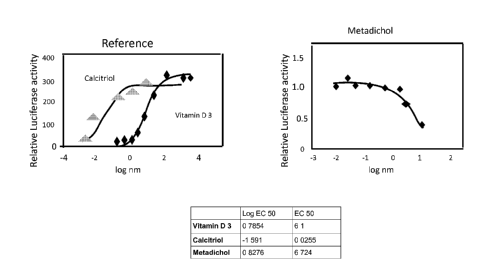
[0040] FIGURE 1 shows the results of the VDR transactivation assay of the test
compound
relative to all 1,25, dihydroxy vitamin D3 ( the natural agonist) and Vitamin
D for certain
exemplary compound of the invention.
[0041] FIGURE 2 shows results of the malaria assay.
[0042] FIGURE 3 shows results of treatment of gel treatment in an acne
patient.
[0043] FIGURE 4 shows the results of treatment of an eczema patient with
Metadichol gel. The
eczema is on the patient's head. The number on the picture shows condition of
skin on said day
or week.
[0044] FIGURE 5 shows the results of treatment of an eczema patient with
Metadichol gel. The
eczema is on the patient's arm. The number on the picture shows condition of
skin on said day or
week.
[0045] FIGURE 6 shows results of Gel treatment of patient with ganglion
[0046] FIGURE 7 shows results of treatment of gel on the leg of a patient with
an outbreak of
rash of unknown etiology.
[0047] FIGURE 8 shows treatment of gel on a dog with lipoma that developed
into a open skin
tumor infection.
[0048] FIGURE 9 shows treatment of a finger infection of a MRSA infected
patient treated with
Metadichol Gel.
[0049] FIGURE 10 shows skin treatment of a patient with gel under eye lids and
face.
[0050] FIGURE 11 shows Metadichol gel treatment of a patient with a deep wound
on the hand.
[0051] FIGURE 12 shows Metadichol gel treatment of a patient suffering from a
fungal
infection of tongue.
[0052] FIGURE 13 shows Metadichol gel treatment of a patent with psoriasis.
[0053] FIGURE 14 shows Metadichol gel application of on a patient with an skin
eruption.
12
CA 02894370 2015-06-15
WO 2014/150609
PCT/US2014/023777
[0054] FIGURE 15 shows Metadichol gel application on a balding person and hair
growth @ 4
months.
DETAILED DESCRIPTION OF THE INVENTION AND THE
PREFERRED EMBODIMENTS
Introduction
[0055] The present invention provides Metadichol0 a novel VDR inverse agonist.
This type of
receptor is one of the most important targets of the pharmaceutical industry,
and many of the
drugs with significant therapeutic action have been shown to be inverse
agonists. The VDR
inverse agonists provided herein have been demonstrated to bind to VDRs with
good affinity.
Further, the inverse agonists can, by binding with Vitamin D receptor,
regulate gene
transcription and cell growth.
[0056] Metadichol0 is more likely based on the results that we describe as a
protean agonist.
The word protean is derived from the Proteas the Greek who could change shape.
This concept
was proposed by Kenakin, et.al, (FASEB J. 2001, Mar: 15(3): 598-611) and was
demonstrated
by Gbahou. et.al, (Proc. Natl. Acad. Sci. (USA) 2003, 100, 11086-11091) with
H3 histaminic
receptors. According to this concept GPCRs are allosteric Proteins that adopt
inactive and active
conformations. In equilibrium the active form of receptor can occur
spontaneously leading to
constitutive activity. An agonist may also promote it. Inverse agonists
promote inactive form of
receptors and decrease constitutive activity. The rationale behind protean
agonism is that if an
agonist produces an active state of lower efficacy than the constitutively
formed one, the ligand
would act as an inverse agonist. However, in the absence of constitutive
activity, the ligand
would be converted to an agonist.
[0057] In the unbound state a receptor is functionally silent, and this is
true in most cases.
However, some receptor systems display constitutive activity, either
experimentally as a result of
over expression or as a result of mutation. These receptors are active in
absence of agonist. An
inverse agonist would inhibit this constitutive activity. Recent studies have
demonstrated
intriguing actions of inverse agonists. (llzerman, et at., 2000, British
Journal of Pharmacology,
130:1-12). They have been shown not only to block constitutive responses of
receptors but also
to activate and regulate seven-trans membrane receptor signaling and
trafficking. (Dupre, et al.,
13
CA 02894370 2015-06-15
WO 2014/150609
PCT/US2014/023777
2004, Biochem. Cell Biol., 82(6):676-680. A receptor is said to be
constitutive active, if the
receptor activates and functions by itself without a ligand.
[0058] Basal receptor signaling denotes a state of constant low-level activity
of a receptor. It is
mainly seen in case of receptors that enable survival. For example, growth
hormone receptor has
a basal level of activity that depends on the presence of a low level of
ligand, like the growth
hormone and insulin like growth hormone, in the blood. The removal of this
activity causes cell
death. Thus there should be a basal level of receptor activity for the cells
to survive. The
advantage of basal receptor activity and constitutive activity is that the
control of cell having
such receptors can be more precise.
[0059] For example, if the function of a cell has to be decreased, the body
can secrete an inverse
agonist, in case of constitutively active receptor, or decrease the production
of the ligand for
basally active receptors. Because the receptors are basally active, the
control can be either
negative and/or positive i.e. in both directions. (Ijzerman, 2006, Trends
Pharmacol Sci, 27:92-
6).
[0060] These inverse agonists can be used for the treatment of abnormal cell
growth, such as
cancer, and the prevention of recurrent cancers. One preferred embodiment of
the invention
utilizes the VDR inverse agonist for the therapeutic treatment of elevated PSA
levels, in reducing
ferritin levels, in reducing RDW red cell distribution width, in increasing
Apo (a) and reducing
APO (b) protein levels in hyperlipidemia, treating MDS (myelodysplacia
syndrome) patients,
increasing Hemoglobin and platelet counts and normalizing Neutrophils,
lymphocytes, and
monocytes, and in reducing uric acid and Lipoprotein (a) levels, and reducing
TSH levels
(thyroid stimulating hormone) (and thyroid globulin antibody (TgAb) and
thyroid peroxidase
antibody (TP0Ab) levels that are seen in Graves and Hashimoto diseases and
other autoimmune
diseases) and in high reducing high levels of bilirubin and in regulating
parathyroid hormone and
normalizing Calcium and Phosphorus and Potassium levels in kidney patients.
The VDR inverse
agonists of the invention can also be used for the therapeutic treatment
and/or prophylactic
prevention of other types of conditions or diseases, such as, but not limited
to, rheumatoid
arthritis, bone marrow disease, prostate cancer, colorectal cancer, leukemia,
brain cancer,
primary or metastatic melanoma, glioma, primary hyperparathyroidism,
psoriasis, kidney stones,
and infections diseases (e.g., malaria). Furthermore, since these derivatives
inhibit parathyroid
14
CA 02894370 2015-06-15
WO 2014/150609
PCT/US2014/023777
hormone secretion, they are contemplated to be effective for the treatment of
secondary
hyperparathyroidism that causes bone disease and vascular calcification in
patients suffering
from renal failure.
Definitions
[0061] Unless defined otherwise, all technical and scientific terms used
herein generally have the
same meaning as commonly understood by one of ordinary skill in the art to
which this invention
belongs.
[0062] The methods and formulations may be used for prophylactic or
therapeutic purposes. In
some embodiments, the terms "treating" or "treatment" of any disease or
disorder refers to
ameliorating the disease or disorder (i.e., arresting or reducing the
development of the disease or
at least one of the clinical symptoms thereof). In other embodiments,
"treating" or "treatment"
refers to ameliorating at least one physical parameter, which may not be
discernible by the
subject. In yet other embodiments, "treating" or "treatment" refers to
inhibiting the disease or
disorder, either physically, (e.g., stabilization or eradication of a
discernible symptom),
physiologically, (e.g., stabilization or eradication of a physical parameter)
or both. In still other
embodiments, "treating" or "treatment" refers to delaying the onset of the
disease or disorder.
[0063] Therapeutically effective amount" is used interchangeably herein with
"an amount
effective to," when referring to a method of the invention. When used in
reference to a
Metadichol0 dosage, these terms refer to a dosage that provides the specific
pharmacological
response for which the policosanol is administered in a significant number of
subjects in need of
such treatment. It is emphasized that "therapeutically effective amount,"
administered to a
particular subject in a particular instance may not be effective for 100% of
patients treated for a
specific disease, and will not always be effective in treating the diseases
described herein, even
though such dosage is deemed a "therapeutically effective amount" by those
skilled in the art. It
is to be further understood that policosanol dosages are, in particular
instances, measured as oral
dosages, or with reference to drug levels as measured in blood. As used
herein, the terms
"individual," "subject," and "patient," is used interchangeably to refer to an
animal, e.g. a
mammal, e.g., a human.
The Compositions
Metadichol0 Nano gel
CA 02894370 2015-06-15
WO 2014/150609
PCT/US2014/023777
[0064] An aging population in the developing world has led to an increase in
musculoskeletal
diseases such as osteoporosis and bone metastases. Left untreated, many bone
diseases cause
debilitating pain and in the case of cancer, death. Many potential drugs are
effective in treating
diseases but result in side effects preventing their efficacy in the clinic.
Bone, and skin however,
provides a unique environment of inorganic solids, which can be exploited to
effectively target
drugs to diseased tissue. By integration of bone targeting moieties to drug-
carrying water-
soluble polymers, the payload to diseased area can be increased while side
effects decreased.
[0065] Nanometer-sized polymeric hydrogels, Nano gels, or hydrogel Nano
particles (NPs; size
from 1 to 1000 nm) are swollen networks composed of amphiphilic or hydrophilic
poly ionic
polymers, either natural or synthetic. Nano gels are promising multifunctional
polymeric NPs
with potential as delivery systems because of their unique properties. These
include tunable
chemical and physical structures, flexible Nano size, large surface area for
multivalent
conjugation, high water content, biocompatibility, loading capacity,
stability, ability to target
specific cells and specific cell compartments, immune modulatory properties,
and responsiveness
to environmental factors. (Oh JK, et at., Prog Polym Sci, 2009, 34:1261-82; Oh
JK., et at. 2010,
Can J Chem, 88:173-84; Hubbell JA, et at., Nature, 2009, 462:449-60).
[0066] As Nano carriers must be delivered to specific sites upon injection
into body fluids, the
possibility of modulating the chemical and physical properties of NPs could be
most helpful in
overcoming major biological barriers such as the reticuloendothelial system,
clearance through
kidney glomeruli, and nonspecific accumulation in different organs. Nano gels
are still a new and
rapidly developing group of materials, gaining wide application in many
fields, especially
pharmacy, medicine and agriculture. An exemplary hydrogel is a material made
when a water-
insoluble polymer absorbs a large amount of water, or it is simply a water-
swollen polymer
network.
[0067] The terms gels and hydrogels are used interchangeably by food and
biomaterials
scientists to describe polymeric cross-linked network structures. Although the
water content in
hydrogels may be as little as a few percent to over 99%, hydrogels retain the
properties of solids
(Truong N., et at., 2002, Biomaterials, 23:4307, Glyn 0 Phillips, et at.,
2011, Hydrogels:
Methods of Preparation, Characterization and Applications, Progress in
Molecular and
16
CA 02894370 2015-06-15
WO 2014/150609
PCT/US2014/023777
Environmental Bioengineering - From Analysis and Modeling to Technology
Applications,
Angelo Carpi (Ed.), ISBN: 978-953-307-268-5, InTech).
[0068] Due to their high water absorption capacity and biocompatibility these
gels have been
used in wound dressing, drug delivery, agriculture, sanitary pads as well as
trans-dermal systems,
dental materials, implants, injectable polymeric systems, ophthalmic
applications, hybrid-type
organs (encapsulated living cells (Table lbelow) They are used in wound care,
in drug delivery,
dental materials, tissue engineering implants and injectable polymeric systems
to name a few.
(Vinogradov SV, et al., 2009, Angew. Chem. Intern. Ed., 48:5418-5429).
[0069] Table 1
Therapeutic Polymer
Moieties
Insulin Tri polymer of N-vinyl pyrrolidone methacrylamide and
itaconic acid
Caffeine Poly dimethyaminoethylmethyacrylate
Camptothecin polyethylene glycol
Calcitonin copolymer of polymethylacrylic acid and polyethylene glycol
Ketoprofen Copolymer of cationic guar gum and acrylic acid polymer
Human Poly organophosphazene with alpha-amino omega methyl
polyethylene
Growth glycol
Hormone
Adenochrome Copolymer of poly-PNIPA and poly PNIPA-Co-AA
(Blood
coagulating
agent)
Proteins and Polyepsilon caprolactone-co-lactide-polyethylene glycol:
Chitosan
peptide
5-Fluouracil Co-polymer of poly-PNIPA and poly-PNIPA-Co-AA
Insulin NIPAAm-Co-AAm
Vaginal NIPAAm-Co-AAm
Microbicide
[0070] Nano gels have found applications in several fields such as sensing
diagnostics and
bioengineering, but its greatest impact has been in the area of drug delivery.
Nano gels and other
Nano-sized drug delivery systems have several advantages over macro-sized
ones. Nano gels can
also be inherently useful in systems that require a burst release. Nano
systems, unlike bulk drug
17
CA 02894370 2015-06-15
WO 2014/150609
PCT/US2014/023777
delivery systems, can enter cells to deliver drugs and can be designed to
respond to intracellular
cues. (S. Thayumanavan, et at. 2912, Advanced Drug Delivery Reviews, 64, 836-
851).
[0071] The tables below show some applications of hydrogel in therapeutics.
(Kohli, et at.,
Scientific Research and Essay, 2009, 3(11):1175-1183).
[0072] Dispersed in aqueous media, swollen Nano gel networks are soft and can
encapsulate a
considerable volume of water. Biological agents and drugs can be loaded into
Nano gels via a
spontaneous process including interactions between the agent and the polymer
matrix, forming
hydrophilic particles with high dispersion stability. Nano gels are able to
physically protect
biological molecules from degradation in vivo and have been pre-clinically
investigated for
many types of active molecules, ranging from small drugs to bio-
macromolecules. The water
holding capacity and permeability are the most important characteristic
features of a hydrogel
(S.Vinogradov, et at., Nanomedicine, 2010, 166, 5(2)).
[0073] Accordingly, the present invention provides a compound or formula as
described in US
patent application, 12/691,706 published Aug 26th 2010 and as a polymeric
hydrogel formulation
for skin and topical applications. Described herein are compositions and
methods for preventing
and/or treating skin diseases including, but not limited to, psoriasis MRSA
infections, and atopic
dermatitis as well as providing anti-aging benefits which results in reduced
appearance of
wrinkles and aged skin, improved skin color, treatment of photo damaged skin,
growing hair,
improvement in skin's radiance and clarity and finish, and an overall healthy
and youthful
appearance of the skin, involving aberrant angiogenesis and hyperplasia
employing the said
formulation.
Biological Activity, Modes of Administration
[0074] As noted above, the compounds of the present invention are inverse
agonists of the VDR
receptor. This means that the compounds of the invention bind to the same site
as the natural
receptor 1,25 dihydroxy vitamin D3. Depending on the site and nature of
undesirable side
effects, which are ideally suppressed or ameliorated, compounds used in
accordance with the
invention may be inverse agonists of VDR receptor.
[0075] A compound should not cause significant activation of a reporter gene
through a VDR
receptor in the transactivation assay in order to qualify as a VDR inverse
agonist with utility in
18
CA 02894370 2015-06-15
WO 2014/150609
PCT/US2014/023777
the present invention. Last, but not least, a compound should bind to VDR
receptor subtypes in
the ligand binding assay in order to be capable of functioning as an inverse
agonist of the bound
receptor subtype, provided the same receptor is not significantly activated by
the compound.
VDR transactivation assay
[0076] The VDR assay was carried out as described in the VDR assay kit
supplied by Indigo
Biosciences Inc. College Town PA) All appropriate controls and standards as
specified by the
manufacturer's kit were used. The procedures are described by Vanden Heuve, et
at., PPAR Res,
2006, 69612; Vanden Heuve, Toxicol Sci, 92:476-489.
[0077] The Antimalarial activity was carried using an adaptation of the
procedures described by
Desjardins et al. (Antimicrob. Agents Chemother. 16, 710-718, 1979); Matile
and Pink (In:
Lefkovits, I. and Pernis, B. (Eds.) Immunological Methods Vol. IV, Academic
Press, San Diego,
pp. 221-234, 1990).
[0078] FIGURE 1 shows the results of the VDR transactivation assay of the test
compound
relative to all 1,25 dihydroxy vitamin D3 (the natural agonist) and Vitamin D
for certain
exemplary compound of the invention. FIGURE 2 shows the results of in vitro
assay of
Metadichol0 against A strain of P. falciparum used in this experiment is the
drug-sensitive
NF54 (an airport strain of unknown origin) VDR transactivation assay.
Procedure
[0079] Plasmids. The ligand-binding domain of the nuclear receptors was fused
to the DNA-
binding domain of the yeast transcription factor Ga14 under the control of the
5V40 promoter. A
reporter plasmid encodes the firefly luciferase gene under the control of the
Ga14 DNA response
element (UAS). A transfection efficiency control vector is included in most
assays (pRL-
luciferase, Promega, Madison, WI). All plasmids were verified by sequencing
and through
examination of positive controls. (Tien, et at., 2006; Vanden Heuvel, et at.,
2006) Full.
[0080] Length System. The full-length cDNA of the nuclear receptor is under
the control of the
5V40 promoter. A reporter plasmid encodes the luciferase reporter under the
control of the
MMTV response element. All plasmids were verified by sequencing and through
examination of
positive controls.
Cell culture and transactivation assays
19
CA 02894370 2015-06-15
WO 2014/150609
PCT/US2014/023777
[0081] HEK 293-T fibroblasts (ATCC, Manassas, VA) were cultured in high
glucose
Dulbecco's Minimal Essential Medium (DMEM) supplemented with 10% fetal bovine
serum
(FBS, Sigma), 0.2 mg/ml streptomycin and 200 U/ml penicillin (Gibco, Grand
Island, NY). For
transient transfection reporter assays, HEK 293-T ells were transfected with
plasmid DNA using
Lipofectamine reagent (Invitrogen, Carlsbad, CA) and following the
manufacturer's
recommended procedures, using HEK 293-T cells at approximately 80% confluence
in 10 cm
culture dishes. After 6 h, the DNA-Lipofectamine complex was removed Following
overnight
culture, the media was replaced 4 h after repeating with DMEM (10% FBS)
containing test
compounds in DMSO (0.1% final concentration). Concentrations of the chemicals
are given in
the figure legends. Sixteen hours after treatment, the cells were lysed with
passive lysis buffer
(Promega, Madison, WI) for 30 min; luciferase activity was measured using the
Luciferase dual
reporter assay kit (Promega, Madison, WI) and a Tecan GeniosPro (Research
Triangle Park, NC)
and manufacturer's recommended procedures. The fold induction of normalized
luciferase
activity was calculated relative to vehicle-treated cells, and represents the
mean of three
independent samples per treatment group.
Malaria: in vitro screening procedure
[0082] Parasite cultures; A strain of P. falciparum used in this experiment is
the drug-sensitive
NF54 (an airport strain of unknown origin) The strains are maintained in RPMI
1640 medium
with 0.36 mM hypoxanthine, supplemented with 25 mM N-2-hydroxyethylpiperazine-
N'-2-
ethane-sulphonic acid (HEPES), 25 mM NaHCO3, neomycin (100 U/ml) and 5 g/1 of
AlbumaxR
II (lipid-rich bovine serum albumin, GIBCO, Grand Island, NY, USA), together
with 5% washed
human A+ erythrocytes. All cultures and assays are conducted at 37 C under an
atmosphere of
4% CO2, 3% 02 and 93% N2. Cultures are kept in incubation chambers filled with
the gas
mixture. Subcultures are diluted to a parasitemia of between 0.1 and 0.5% and
the medium is
changed daily.
Drug sensitivity assays
[0083] Antimalarial activity is assessed using an adaptation of the procedures
described by
Desjardins et al. (Antimicrob. Agents Chemother. 16, 710-718, 1979), and
Matile and Pink (In:
Lefkovits, I. and Pernis, B. (Eds.) Immunological Methods Vol. IV, Academic
Press, San Diego,
pp. 221-234, 1990).
CA 02894370 2015-06-15
WO 2014/150609
PCT/US2014/023777
[0084] Stock drug solutions are prepared in 100% dimethyl-sulfoxide (DMSO)
(unless otherwise
suggested by the supplier) at 10 mg/ml, and heated or sonicated if necessary
to dissolve the
sample. After use the stocks are kept at ¨20 C. For the assays, the compound
is further diluted in
serum-free culture medium and finally to the appropriate concentration in
complete medium
without hypoxanthine. The DMSO concentration in the wells with the highest
drug concentration
does not exceed 1%.
[0085] Assays are performed in sterile 96-well micro titer plates, each well
containing 200 IA of
parasite culture (0.15% parasitemia, 2.5% hematocrit) with or without serial
drug solutions.
Seven 2-fold dilutions are used, covering a range from 5 g/m1 to 0.078 g/ml.
For active
compounds the highest concentration is lowered (e.g. to 100 ng/ml); for plant
extracts the highest
concentration is increased to 50 g/ml. Each drug is tested in duplicate and
the assay is repeated
for active compounds showing an IC50 below 1.0 g/ml. After 48 hours of
incubation at 37 C,
0.5 Ci '3H- hypoxanthine is added to each well. Cultures are incubated for a
further 24 h before
being harvested onto glass-fiber filters and washed with distilled water. The
radioactivity is
counted using a BetaplateTm liquid scintillation counter (Wallac, Zurich,
Switzerland). The
results are recorded as counts per minute per well at each drug concentration
and expressed as
percentage of the untreated controls. IC50 values are calculated from the
sigmoidal inhibition
curves using Microsoft EXCEL).
The Compositions
[0086] In various embodiments, the invention provides Metadichol0 as a liquid
or as gel which
is a Nano formulation of Policosanol described in US Patent Application
12/691,706.
Composition of gel
[0087] The polymer used is derived from one of the following: Carbopol 0
Polymer ( Registered
trade mark of Lubrizol) polyethylene oxide (PEO) (polyvinyl pyrollidone (PVP)
), polylactic
acid (PLA) (polyacrylic acid (PAA) polymethacrylate (PMA) polyethylene glycol
(PEG) (Singh
et al.), or natural biopolymers, such as alginate, agar, chitosan,
carrageenan, hyaluronan, and
carboxymethyl cellulose (CMC).
[0088] In an exemplary embodiment, the unit dosage gel formulation is a
formulation of Nano
particleNano particles containing Metadichol0 and a stabilizer fraction and
the unit dosage
21
CA 02894370 2015-06-15
WO 2014/150609 PCT/US2014/023777
formulation includes from about 10 mg to about 100 mg, for example from about
1 mg to about
20 mg per mL or from about 10 mg to about 30 mg per mL. In various
embodiments, the unit
dosage is a daily dosage. One of ordinary skill will appreciate that
therapeutically effective
amounts of Metadichol0 gel can be determined empirically and can be employed
in pure form
or, where such forms exist, in pharmaceutically acceptable salt, ester, or pro-
drug form. Actual
dosage levels of Metadichole in the Nano particulate compositions of the
invention may be
varied to obtain an amount of Metadichol0 that is effective to obtain a
desired therapeutic
response for a particular composition and method of administration. The
selected dosage level
therefore depends upon the desired therapeutic effect, the route of
administration, the potency of
the administered policosanol, the desired duration of treatment, and other
factors.
[0089] Dosage unit compositions may contain such amounts of such submultiples
thereof as may
be used to make up the daily dose. It will be understood, however, that the
specific dose level
for any particular patient will depend upon a variety of factors: the type and
degree of the
cellular or physiological response to be achieved; activity of the specific
agent or composition
employed; the specific agents or composition employed; the age, body weight,
general health,
sex, and diet of the patient; the time of administration, route of
administration, and rate of
excretion of the agent; the duration of the treatment; drugs used in
combination or coincidental
with the specific agent; and like factors well known in the medical arts.
The Methods
[0090] The present invention provides methods of using these Nano particles of
Metadichol0 in
liquid or gel form and prevent disease and to regulate metabolism. In various
embodiments, the
Nano particleNano particles of the invention in liquid form are of use to
regulating, Lp (a), Apo
(a) and Apo (b) protein levels, Uric acid, Parathyroid hormone levels,
decreasing bun ratios in
kidney patients, Regulating Phosphorous and Calcium levels, Potassium levels
in hypertension
patients, Ferritin levels, TSH levels, neutropenia, modulating aspartate
aminotransferase (AST)
or serum glutamic-oxaloacetic transaminase [SGOT]), alanine aminotransferase
(ALT) or serum
glutamate pyruvate transaminase [SGPT]), levels, modulating absolute
neutrophil and
Lymphocyte ratios and modulating albumin in hyperlipidemia patients.
[0091] The Metadichole gel formulations is topically used in treating various
skin diseases like
acne, MRSA infection, Eczema, Psoriasis, and preventing and/or treating skin
diseases
22
RECTIFIED (RULE 91) - ISA/US
CA 02894370 2015-06-15
WO 2014/150609 PCT/US2014/023777
including, but not limited to, psoriasis and atopic dermatitis as well as
providing anti-aging
benefits which results in reduced appearance of wrinkles and aged skin,
improved skin color,
treatment of photo damaged skin, improvement in skin's radiance and clarity
and finish, and an
overall healthy and youthful appearance of the skin, involving aberrant
angiogenesis and
hyperplasia.
[0092] In an exemplary embodiment, the formulations are administered in a
therapeutically
effective amount to a subject to treat a particular disease or disorder and
wherein the subject is
not otherwise in need of treatment with Metadichola In various embodiments,
the Metadichol0
is administered to treat a single disease or regulate a single metabolic
factor. Thus, in an
exemplary embodiment, the invention provides a method to treat Lipoprotein (a)
in a subject not
in need of treatment for hyperlipidemia, hypercholesterolemia, hypertension,
etc. In an
exemplary embodiment, the invention provides a method of regulating
parathyroid hormones
levels in a subject not in need of treatment for kidney disease,
hyperlipidemia,
hypercholesterolemia, etc. In various embodiments, the invention provides a
method of treating
Potassium levels in a subject not in need of treatment for hypertension,
diabetes etc. In various
embodiments, the invention provides a method to decrease or prevent
neutropenia in a subject
who is not in need of treatment for treatment for kidney diseases,
Inflammation etc., In an
exemplary embodiment, the invention provides a method of increasing Apo
Protein (a) levels in
a subject not in need of treatment for hyperlipidemia, hypercholesterolemia,
etc. In other
embodiments, the invention provides a method of modulating AST and ALT levels
in a subject
not in need of treatment for hyperlipidemia, hypercholesterolemia, etc.
[0093] In various embodiments, the Metadichol0 gel is used in Eczema,
Psoriasis, Lipoma
tumors on removing wrinkles and warts and in MRSA and other skin infections.
[0094] Non-limiting examples of methods of the invention are set forth below:
URIC ACID
[0095] The invention provides a method of decreasing Uric acid levels in a
subject and,
therefore, reducing the deleterious consequences of this oxidation. The method
includes
administering to a subject a therapeutically effective amount of Metadichol0
to decrease protein
oxidation in a subject.
23
RECTIFIED (RULE 91) - ISA/US
CA 02894370 2015-06-15
WO 2014/150609
PCT/US2014/023777
[0096] Uric acid is present in small amounts in everyone's bodies and is made
from the
breakdown of purines, which are released as part of the body's normal
functioning and can be
absorbed by the body from certain types of food (such as meat or seafood). In
addition a number
of epidemiological studies have reported a relation between serum uric acid
levels and a wide
variety of cardiovascular conditions, including hypertension, (Acosta, et al.
2005, J Am Soc
Nephrol, 16,909-1919; Heinig M, et at. 2006, Cleveland Clinic Journal of
Medicine,
73(12):1059-64).
[0097] Uric acid may play a role in the metabolic syndrome. Historically, the
elevated level of
uric acid observed in the metabolic syndrome has been attributed to
hyperinsulinemia, since
insulin reduces renal excretion of uric acid. Hyperuricemia, however, often
precedes the
development of hyperinsulinemia, obesity, and diabetes. Hyperuricemia may also
be present in
the metabolic syndrome in people who are not overweight or obese. (Tyagi, et
at., Nutrition &
Metabolism, 2004, 1:10). Hyperuricemia is strongly associated with peripheral,
carotid, and
coronary vascular disease, with the development of stroke, with preeclampsia,
and with vascular
dementia. The relationship of uric acid with cardiovascular events is
particularly strong,
especially in patients at high risk for heart disease and in women. In
coronary artery disease,
(Viazzi, et at., 2006, The Journal of Clinical Hypertension, 8(7):510).
[0098] Serum uric acid is a strong predictor of stroke in patients with non-
insulin dependent
diabetes mellitus ND cerebrovascular disease, (Puig, et.al., 2007, Nutrition,
Metabolism &
Cardiovascular Diseases, 17, 409e414).
[0099] Gout is a type of arthritis caused by uric acid build-up in the joints
(the places where two
or more bones come together). When uric acid levels in the body are high
(known as
hyperuricemia), uric acid crystals can form in the fluid around the joints. If
there is too much uric
acid around the joints, inflammation (a condition in which a part of your body
can become red,
swollen, and painful) can occur, which may lead to a gout attack (Tausche AK,
et at., 2006, Der
Internist, 47(5):509-20).
Lesch-Nyhan Syndrome
[00100] Lesch-Nyhan syndrome, an extremely rare inherited disorder, is
also associated
with very high serum uric acid levels. Spasticity, involuntary movement and
cognitive
24
CA 02894370 2015-06-15
WO 2014/150609
PCT/US2014/023777
retardation as well as manifestations of gout are seen in cases of this
syndrome. (Nyhan W.L,
2005, Journal of the History of the Neurosciences, 14(1):1-10).
Uric acid stone formation
[00101] Saturation levels of uric acid in blood may result in one form
of kidney stones
when the urate crystallizes in the kidney. Uric acid stones, which form in the
absence of
secondary causes such as chronic diarrhea, vigorous exercise, dehydration, and
animal protein
loading, are felt to be secondary to obesity and insulin resistance seen in
metabolic syndrome.
Increased dietary acid leads to increased endogenous acid production in the
liver and muscles,
which in turn leads to an increased acid load to the kidneys. This load is
handled more poorly
because of renal fat infiltration and insulin resistance, which are felt to
impair ammonia
excretion (a buffer). The urine is therefore quite acidic, and uric acid
becomes insoluble,
crystallizes and stones form. In addition, naturally present promoter and
inhibitor factors may be
affected. This explains the high prevalence of uric stones and unusually
acidic urine seen in
patients with type 2 diabetes. Uric acid crystals can also promote the
formation of calcium
oxalate stones, acting as "seed crystals" (heterogeneous nucleation). (Pak
C.Y, et al. 2008; The
Journal of Urology, 180(3):813-9).
[00102] Hyperuricemia has been shown to be associated with
histological liver damage in
patients with non-alcoholic fatty liver disease (NAFLD). Its clinical
relevance arises from the
fact that a considerable proportion of subjects (20-30%) develop a condition
namely non-
alcoholic steatoheppatitis (NASH) that is a potentially progressive hepatic
disorder leading to
end-stage liver disease and hepatocellular carcinoma. In addition NAFLD is
considered the
hepatic manifestation of insulin resistance (IR), and is therefore strongly
associated with
metabolic syndrome, obesity, type II diabetes, dyslipidemia and hypertension,
also representing,
together with the above cited conditions, an independent cardiovascular risk
factor. In these
patients. (Non-alcoholic fatty liver disease (NAFLD) is a leading cause of
chronic liver disease
worldwide. (S. Petta, et al., Aliment Pharmacol Ther, 2011,34:757-766).
Uric Acid and Parkinson's Disease (PD)
[00103] PD is the second most common age related neurodegenerative
condition in the
US, affecting approximately one percent of the population over the age of 65
in North America
and Europe. The symptoms of PD are characterized by loss of dopaminergic
neurons in the
CA 02894370 2015-06-15
WO 2014/150609
PCT/US2014/023777
substantia nigra. While the cause of this loss is thought to be
multifactorial, there is evidence to
support oxidative stress as a factor in neurodegeneration. Researchers have
proposed that
elevated levels of uric acid yield a protective effect against the development
and progression of
PD on the basis of these principles. There is also evidence to support the
association between
high dietary urate and a decreased incidence of PD. It should be noted that
there are numerous
risks associated with hyperuricemia diets, such as gout, stroke and
hypertension. At this point,
the data we have suggests an association, but not a causal relationship
between low serum urate
and the incidence of PD. This association is intriguing as there is the
potential that in the future
physicians could modify a patient's risk for PD by suggesting dietary changes
or using
pharmacological supplement. Overall, recent research supports an inverse
relationship between
serum urate levels and the incidence of PD in men. (Mandel, et at., 2009,
Practical Neurology,
p21).
Ferritin
[00104] Ferritin is a ubiquitous and specialized protein involved in
the intracellular
storage of iron; it is also present in serum and other biological fluids,
although its secretion
processes are still unclear. Ferritin is nature's unique and conserved
approach to controlled, safe
use of iron and oxygen, with protein synthesis in animals adjusted by dual,
genetic DNA and
mRNA sequences that selectively respond to iron or oxidant signals and link
ferritin to proteins
of iron, oxygen and antioxidant metabolism. (Cairo, et at. 2008, Journal of
Autoimmunity 30, 84-
89).
[00105] Ferritin is a key protein of iron metabolism that is capable
of sequestering large
amounts of iron, and thus serves the dual function of iron detoxification and
iron storage. The
importance of these functions is underlined by its ubiquitous distribution in
many living species.
The structural properties of the ferritins are largely conserved from bacteria
to man, although
their role in the regulation of iron trafficking varies substantially.
[00106] Ferritin synthesis is regulated by cytokines (TNFa and IL-1a)
at various levels
(transcriptional, post-transcriptional, and translational) during development,
cellular
differentiation, proliferation, and inflammation. The cellular response by
cytokines to infection
stimulates the expression of ferritin genes. (Hintze KJ, et at., 2006, Cell
Mol Life Sci; 63:591-
600).
26
CA 02894370 2015-06-15
WO 2014/150609
PCT/US2014/023777
[00107] Ferritin has been reported to exhibit different immunological
activities including
binding to T lymphocytes, suppression of the delayed type of hypersensitivity,
suppression of
antibody production by B lymphocytes, and decreased phagocytosis of
granulocytes (Marikina,
K, et al., 999, Blood, 483:737-43).
[00108] Ferritin and iron homeostasis have been implicated in the
pathogenesis of many
diseases, including diseases involved in iron acquisition, transport and
storage (primary
hemochromatosis) as well as in atherosclerosis, Parkinson's disease, Alzheimer
disease, and
restless leg syndrome. (Zandman, et at., 2007, Autoimmunity Reviews, 6:457-
463).
[00109] Mutations in the ferritin gene cause the hereditary
hyperferritinemia-cataract
syndrome and neuroferritinopathy. Hyperferritinemia is associated with
inflammation,
infections, and malignancies. Thyroid hormone, insulin and insulin growth
factor-1 have also
been implicated in regulation of ferritin at the mRNA level (Beaumont C, et
at., 1995, Nat
Genet, 11:444-6; Nishiya K, et at., 1997, Clin Exp Rheumatol, 15:39-44).
[00110] Some evidence points to the importance of hyperferritinemia in
RA, MS, and
thyroiditis, Ferritin and iron homeostasis have been implicated in the
pathogenesis of many
diseases, including diseases involved in iron acquisition, transport and
storage (primary
hemochromatosis) as well as in atherosclerosis (You S.A, et at., 2005, Clin
Chim Acta, 357:1-
16). Genetic mutations of the ferritin IRE region as well as coding regions of
ferritin cause some
hereditary human diseases. Ferritin IRE mutations cause the hereditary
hyperferritinemia-
cataract syndrome (Beaumont C, et at., 1997; Nat Genet, 11:444-446) which is
an autosomal
dominant disease characterized by elevated ferritin levels and early-onset
bilateral cataracts.
[00111] Neuroferritinopathy, a dominantly inherited movement disorder
characterized by
decreased levels of ferritin and abnormal deposition of ferritin and iron in
the brain, is caused by
a mutation in the C-terminus of the ferritin L gene (Curtis ARJ, et at., 2001,
Nat Genet, 28:350-
4). It has been shown that Serum ferritin concentration was found to be
related with
dyslipidemia, hypertension and abdominal adiposity. Ferritin is also known to
be a marker of
inflammation and increases in cardiovascular disease and it has been suggested
that serum
ferritin levels can be used as a risk marker for atherosclerotic disease
(Faruk, et at. 2006;
Endocrine Abstracts, ii P323).
27
CA 02894370 2015-06-15
WO 2014/150609
PCT/US2014/023777
[00112] Thalassemia will remain to be the one of the major health
problem for at least the
next few decades, particularly in developing countries. Although the survival
of thalassaemics is
steadily increasing, the prevalence of complications due to Serum ferritin
being high. This
overload is the life limiting complication commonly found in thalassaemics
(Wangruangsattit S,
et at., 1999, J. Med Assoc Thai, 82(1):74 ¨76). The iron that exceeds the iron
binding capacity of
transferrin appears in the plasma as non-transferrin bound iron, which is
highly toxic to tissues.
The accumulation of iron results in progressive dysfunction of the heart,
liver and endocrine
glands (Hathirat P., et at., 1001, Hematol, 2001, 38:360 ¨366).
[00113] Ferritin may act as an immune regulator by binding to subsets
of lymphocytes and
myeloid cells contrasts with its well-known function as an intracellular iron
storage protein.
Ferritin, which may therefore not only be the major iron storage protein, but
also an important
regulator of the immune system playing a possible role in autoimmune diseases.
(Recalcati, et
at., 2008, Journal of Autoimmunity, 30:84-89).
[00114] Ferritin has been suggested to be a circulating tumor-
associated antigen in
Hodgkin's disease, Bieber, C. P., et al., 1973, Nat. Cancer Inst. Monogr.,
36:147-157; Jones, P.
A, et at. (1973, Brit. J. Cancer 27, 212-217).
[00115] Although ferritin occurs intracellularly in a wide variety of
tissues and its level in
the serum of healthy humans is low, it was found in elevated amounts in the
serum of patients
with Hodgkin's disease as well as in' other malignancies and diseases with
liver involvement
(Aungst, C. W., 1968, J. Lab. Clin. Med., 71:517-522; Reissman, K. R., 1956,
J. Clin. Invest.,
35:588-595).
[00116] Antigens which exist in high frequency in tumor tissues of
patients with
Hodgkin's disease have been obtained in relatively concentrated form by gel
chromatography
procedures. Further purification and analysis of these antigens (Eshhar, et
at., 1974, Proc. Nat.
Acad. Sci. USA, 71(10):3956-3960), have demonstrated that the antigen of fast
electrophoretic
mobility (F-antigen) is normal tissue ferritin.
Lipoprotein A (Lp (a))
[00117] First described in 1963 (Berg, 1963, Acta Pathol Microbiol
Scand, 59:369-82)
that Lp(a) is a modified LDL particle. Berg and his colleagues in Sweden later
determined that
individuals with high levels of Lp(a had significantly higher incidences of
heart attacks than a
28
CA 02894370 2015-06-15
WO 2014/150609
PCT/US2014/023777
population with low Lp(a). Studies that have detected Lp(a) in atherosclerotic
lesions and vein
grafts have provided the strongest evidence for a direct involvement of Lp(a)
at the lesion site
(Walton, KW, et at., 1981, Atherosclerosis, 20:323-46).
[00118] A seminal study (Rath et at., Arteriosclerosis, 1989,9:579-
592) shows that Lp(a)
accumulates in the arterial wall, partly in the form of lipoprotein like
particles, therefore
contributing to plaque formation and coronary heart disease. Other researchers
have verified the
results (Metso, et at., 1990, European Heart Journal, 11 (Supplement E), 190-
195). This
observation has been strongly supported and over the years has expanded to
include higher
incidences of myocardial infarction, stroke, and retinal artery occlusion.
[00119] Lp(a) consists of a low-density lipoprotein (LDL)-like moiety and
an unique
glycoprotein, Apo lipoprotein Al (Apo Al)), that is covalently attached to the
apolipoproteinB-
100 (ApoB-100) component of LDL by a single disulfide bond. Many studies have
suggested a
role for Lp(a) in the process of endothelial dysfunction. Indeed, Lp(a) has
been shown to
increase both the expression of adhesion molecules on endothelial cells (EC),
as well as
monocyte and leukocyte chemotactic activity in these cells.
[00120] Lp(a) is structurally similar to LDL both in protein and in
lipid composition, but
is distinguishable from LDL by the presence of the unique glycoprotein moiety
called Apo
lipoprotein A (apo(A) . It has been determined that Lp(a) particles contain
Apo (a) and Apo
lipoprotein B-100 (apoB) in a 1:1 molar ratio (Albers, et at., 1996, J Lipid
Res, 37:192-6). In the
Lp(a) particle, Apo (a) is covalently linked to Apo (b) by a single disulfide
bridge. The cysteine
residues in each molecule that are involved in the disulfide linkage have been
identified (Callow
MJ, et at., 1995, J Biol Chem, 270:23914-7).
[00121] Lp(a) is only present in humans, Old World Monkeys and the
hedgehog (Lawn
RM, et at., 1996, Clin Genet, 49:167-74). The level of Lp(a) is genetically
determined, and when
elevated, cannot be lowered by alterations in food intake or by most of the
cholesterol lowering
agents. (Kostner GM, et at., 1989, Circulation, 8:1313-9).
[00122] Diabetic patients have higher Lp(a) values than nondiabetic
persons(Haffner SM,
et at., 1992,49:116-20). Lp(a) was shown to be associated inversely with risk
of type II diabetes
independent of other risk factors, including BMI, HbAl c, or triglycerides.
(Samia Mora, et at.,
2010, Clinical Chemistry, 56:8 1252-1260).
29
CA 02894370 2015-06-15
WO 2014/150609
PCT/US2014/023777
[00123] Not only can Lp(a) be deposited in the arterial wall, but one
of the more enticing
possibilities backed by experimental evidence is that Lp(a) interferes with
the fibrinolysis of
blood clots. The evidence for the latter idea comes from observations that
Lp(a) is capable of
competing with plasminogen for binding sites on fibrin. (Rahman MN, et at.,
2002,
Biochemistry, 41:1149-55). In so doing, Lp(a) conceivably could increase the
survival time of a
clot in a wound and contribute to the thickening of the affected artery. While
attached to the
extracellular matrix the clot remnants could attract macrophages, which are
known to be a source
of factors that signal arterial cells to grow and divide, in essence promoting
the atherosclerotic
process. Apart from these thoughts, Lp(a) might be considered a very effective
donor of
cholesterol to cells, perhaps exceeding LDL, and hence capable of hastening
formation of foam
cells at the lesion site.
[00124] Despite its recognition as a risk factor for vascular disease,
the role of Lp(a) in
atherogenesis remains poorly understood. It has been postulated that owing to
its duality of
structure, Lp(a) may provide a functional link between the processes of
atherosclerosis and
thrombosis. In this model, Lp(a) likely possesses both atherosclerotic (owing
to its similarity to
LDL) and prothrombotic properties (based on the homology between Apo (A) and
plasminogen).
Clearly, Apo (A) possesses unique properties that contribute to the process of
atherogenesis that
are independent of its similarity to plasminogen (Koschinsky, ML, et at.,
2004, Curr Opin
Lipidol, 15:167-74).
[00125] Lp(a) is associated with increased risk of cardiovascular diseases
(CVD), including
coronary heart disease (CHD) and atherosclerosis (Anuurad, E, et at., 2006,
Clin Lab Med.,
26:751 ¨ 772). Moreover, as with most genetic risk factors that initiate risk
at birth, it is a
stronger CVD risk factor in young patients (less than 60 years old), and in
those with highly
elevated levels of Lp(a), and in those with additional atherogenic risk
factors, particularly
elevated LDL cholesterol (Tsimikas, S., et al., 2005, N Engl J Med., 353:46 ¨
57). Lp(a) is
present in the arterial wall at the sites of atherosclerotic lesions and that
it accumulates at these
sites to an extent that is proportional to plasma Lp(a) levels. Lp(a) is
preferentially retained in
this milieu, likely by virtue of its ability to bind to a number of arterial
wall components
including fibrinogen/fibrin, fibronectin, and glycosaminoglycans (Koschinsky
ML, et at., 2004,
Curr Opin Lipidol, 15:167-74). The localization of Lp(a) within the arterial
wall suggests a
direct causative role for Lp(a) in the initiation and/or progression of
atherosclerosis.
CA 02894370 2015-06-15
WO 2014/150609
PCT/US2014/023777
[00126] Lp(a) has been implicated in the regulation of plasminogen activator
inhibitor-1
expression in endothelial cells and shown to inhibit endothelial cell surface
fibrinolysis to
attenuate plasminogen binding to platelets and to bind to plaque matrix
components. Autopsy
studies in humans have documented the presence of Lp(a) in aortic and coronary
atherosclerotic
plaques and an apparent localization with fibrinogen (Hoefler G, 1998,
Arteriosclerosis, 8(4):
398-401).
[00127] Lp(a) levels are frequently elevated in patients receiving chronic
hemodialysis
treatment of end-stage renal disease (Quashing T, et al., 2001, Am J Kid Dis,
38, suppl 1, 514-9).
It has been suggested that kidney have an important role in Lp(a) metabolism.
In renal failure,
there is a decrease in Lp(a) catabolism or increase in Lp(a) production by
liver. In hemodialysis
patients, Lp(a) has been shown to have the characteristics of an acute phase
reactant (Maeda S, et
al., 1989, Atherosclerosis, 78:145-50). Patients with Peripheral artery
disease (PAD) showed
significantly higher median serum concentrations of Lp(a) than controls
(Dieplinger, et al. 2007,
Clinical Chemistry, 53:7 1298-1305).
[00128] Lp(a) concentration and Apo B to ApoAI ratio in 55 South Asian
subjects with
ischemic stroke and 85 controls. The analysis of the data showed that both
parameters were
associated with ischemic stroke (Sharobeem, K. M, et al., 2007, Int. J.Clin.
Pract, 61(11): 1824-
1828).
[00129] Lp(a) levels in 100 patients with acute ischemic stroke and 100
healthy subjects were
compared and noted that even a slight elevation in Lp(a) plasma concentration
was strongly and
independently associated with ischemic stroke in men, but not in women.
(Rigal, M, et al., 2007,
J. Neurol. Sci., 252(1):39-44).
[00130] The role of Lp(a) in silent cerebral infarction (SCI) was investigated
in patients with
chronic renal failure who were maintained on hemodialysis. Lp(a) was found to
be significantly
associated with the presence of SCI (Fukunaga, N, et al., 2008, Metabolism,
57(10):1323-1327).
[00131] In a study of homocysteine and Lp(a) in ischemic stroke it was seen
plasma Lp(a)
concentration patients with ischemic stroke and controls and found that these
two parameters are
independently associated with ischemic stroke with a significant positive
correlation between
them (Dhamija, R. K, et al., 2009, J. Neurol. Sci., 281(1-2):64-68). Increased
Lp(a) levels in
acute phases, such as after surgery, inflammation, pregnancy, myocardial
infarction, psoriasis,
31
CA 02894370 2015-06-15
WO 2014/150609
PCT/US2014/023777
gout and others have been reported. Lp(a) has been shown to be a potent chemo
attractant for
human peripheral monocytes. (Syrovets, et at., 1997, Blood, 90:2027-2036).
[00132] Hypothyroid patients have increased, while hyperthyroid patients have
decreased
plasma Lp(a) levels in comparison to euthyroid controls. Human growth hormone
drastically
increases Lp(a) by up to 120% (Laron Z, et at., 1997, J Pediatr Endocr Met,
10:143-149).
[00133] Patients with many forms of kidney disease exhibit striking elevations
of plasma Lp(a)
levels. In patients with end-stage renal disease (ESRD), Lp(a) and the Apo(a)
phenotype are
predictors for both the degree of preclinical atherosclerosis and
atherosclerotic events. In ESRD
and nephrotic syndrome elevations of Lp(a) are not only due to overproduction
of Lp(a) by the
liver but also to diminished excretion of Apo (a) fragments into the urine
(Kostner and Kostner,
2002, Curr Opin Lipidol, 13:391-396).
[00134] There is a highly significant association between Lp (a) and the
presence and
progression of breast cancer, and the serum Lp (a) determination may provide
an aid in patients
with breast cancer for both diagnostic purposes and the follow-up of the
disease (Kokoglu E, et
at., 1994, Cancer Biochem Biophys., 1994,4(2):133-6.)
[00135] Recent studies have suggested a link between Lp(a) and oxidized
phospholipids.
Specifically, it has been shown that in human plasma, oxidized phospholipids
are preferentially
associated with Lp(a) compared to free LDL (Tsimikas S, et at., 2003, J Am
Coll Cardiol,
41:360-70).
[00136] Most patients with the nephrotic syndrome have Lp(a) concentrations
that are
substantially elevated compared with controls of the same Apo(a) isoform.
Because Lp(a)
concentrations are substantially reduced when remission of the nephrotic
syndrome is induced, it
is likely that the nephrotic syndrome results directly in elevation of Lp(a)
by an as yet unknown
mechanism. The high levels of Lp(a) in the nephrotic syndrome could cause
glomerular injury as
well as increase the risk for atherosclerosis and thrombotic events associated
with this disorder
(Wanner. L, et at., 1993, Ann Intern Med, 119:263-269).
[00137] Lp(a) is commonly reported to be significantly increased in cancer
patients as compared
to healthy controls, irrespective of source and degree of malignancy of the
tumor. Patients
suffering from cancer of different locations and origin exhibit up to two fold
elevated plasma
32
CA 02894370 2015-06-15
WO 2014/150609
PCT/US2014/023777
Lp(a) concentrations. Patients with acute myelotic leukemia have very high
Lp(a) levels
(Wright, et at., 1989, Int J Cancer, 43 :241-244).
[00138] At present, the one. albeit costly and invasive method of proven value
is LDL apheresis
(Armstrong VW, et at., 1989, Eur. J. Clin. Invest., 19:235-40), resembling
dialysis, to eliminate
the cholesterol-containing particle low-density lipoprotein (LDL) from the
bloodstream. Which
should be reserved for individuals with extreme elevations of Lp(a). The
procedure takes 2-4
hours and must be repeated every several weeks to keep the LDL levels from
accumulation and
causing cardiovascular disease. It is an expensive procedure, limiting its use
to severe cases of
hyperlipidemia. Unfortunately, there is no drug therapy that is currently
available to
specifically lower Lp(a) levels without affecting other lipoproteins.
APO A-1 and APO B
[00139] Although LDL cholesterol (LDL-C) is thought to be associated with an
increased risk
of coronary heart disease, other lipoproteins and their constituents, Apo
lipoproteins, may play
an important role in atherosclerosis.
[00140] Elevated levels of Apo lipoprotein (Apo) B, a constituent of
atherogenic lipoproteins,
and reduced levels of Apo A-I, a component of anti-atherogenic HDL, are
associated with
increased cardiac events. Apo B, Apo A-1 and the Apo B/Apo A-I ratio have been
reported as
better predictors of cardiovascular events than LDL-C and they even retain
their predictive
power in patients receiving lipid-modifying therapy. Measurement of these Apo
lipoproteins
could improve cardiovascular risk prediction. (Jungner, et at., Journal of
Internat Medicine,
2004,255:188-205).
[00141] Levels of Apo A-I are strongly correlated with those of HDL-C, and
expression of Apo
A-I may be largely responsible for determining the plasma level of HDL (XV
International
Symposium on Atherosclerosis, June 14 - 18,2009, Boston, MA, USA). Apo A-I
also acts as a
cofactor for lecithin cholesterol acyl transferase (LCAT) [Phillips MC, et.al
(1998;
Atherosclerosis 137(Suppl): S13-7), which is important in removing excess
cholesterol from
tissues and incorporating it into HDL for reverse transport to the liver.
Furthermore, Apo A-1 is
the ligand for the ATP-binding cassette (ABC) protein, ABCA1, and hence is
involved in the
docking procedure by which excess cholesterol in peripheral cells is
externalized to HDL. (Oram
JF, et at., 2000, J Biol Chem, 275 34508-11), for further reverse cholesterol
transport either
33
CA 02894370 2015-06-15
WO 2014/150609
PCT/US2014/023777
directly or indirectly via LDL back to the liver (Tailleux A, et at., 2002,
Atherosclerosis, 164: 1-
13).
[00142] The Prospective Epidemiological Study of Myocardial Infarction (PRIME)
study
examined the association between the incidence of CHD and several HDL related
parameters,
including HDL-C itself, Apo A-I, HDL A-I, and HDL A-I : A-II (Luc G, et at.,
2002,
Arterioscler Thromb Vasc Riot, 22:1155-61). All four parameters were related
to CHD risk;
however, Apo A-I was the strongest predictor. In addition, the use of Apo A-1
for predicting
CAD has been confirmed by other studies (Garfagnini A, et at., 1995, Eur Heart
J, 16:465-70).
Apo lipoprotein B
[00143] Apo lipoprotein B exists in two forms, Apo B-48 and Apo B-100. Apo B-
48 is
synthesized in the intestine, where it is complexes with dietary TG and free
cholesterol absorbed
from the gut lumen to form chylomicron particles. These are metabolized in the
circulation and
in the liver. Apo B-100 is synthesized in the liver and is present in LDL, IDL
and VLDL
particles. Only one Apo B molecule is present in each of these lipoprotein
particles (Elovson J, et
at., 1998, J Lipid Res, 29:1461-73). The total Apo B value indicates the total
number of
potentially atherogenic lipoproteins (Walldius G, et at., 2001, Lancet,
2001,358:2026-33). Apo
B is essential for the binding of LDL particles to the LDL receptor, allowing
cells to internalize
LDL and thus absorb cholesterol. An excess of Apo B-containing particles is a
main trigger in
the atherogenic process.
[00144] The concentration of plasma Apo B particles is highly correlated with
the level of non-
HDL cholesterol (non-HDL-C), defined as TC minus HDL-C (Ballantyne CM, et at.,
2001, Am J
Cardiol, 88:265-9). As HDL is known to be protective against cardiovascular
risk, non-HDL-C
reflects the fraction of blood cholesterol that is not contained in
atheroprotective lipoproteins.
Therefore, non-HDL-C has been recognized by the National Cholesterol Education
Program
Adult Treatment Panel III (NCEP ATP III) guidelines as a target for lipid-
lowering therapy
(Circulation, 2002,106:3143-421). Non-HDL-C has been found to predict nonfatal
myocardial
infarction (MI) and angina pectoris. However, Apo B has been found to be a
better predictor of
risk than non-HDL-C (Sniderman AD, et at., 2003, Lancet, 361:777-80). Using
Apo B and Apo
A-I, expressed as the Apo B/Apo A-1 ratio, seems to be a very effective way of
characterizing
cardiovascular risk in any patient irrespective of their lipoprotein
abnormality.
34
CA 02894370 2015-06-15
WO 2014/150609
PCT/US2014/023777
[00145] Patients with diabetes or the metabolic syndrome can have normal LDL-C
levels but
possess aspects of the atherogenic lipid profile (Sniderman AD, et at., 1001,
Ann Intern Med,
135:447-59), and these individuals often have a high ratio of Apo B/Apo A-I,
which is a strong
indicator of cardiovascular risk (Walldius G, et at, 2002, Diabetes,
51(Suppl.):A20).
Parathyroid hormone (PTH)
[00146] Chronic Kidney Diseases (CKD) is a significant and growing health
problem
characterized by the progressive loss of kidney function and the many ensuing
complications. As
a result of the loss of excretory, regulatory, and endocrine function of the
kidney, patients with
CKD experience multiple medical complications such as electrolyte
abnormalities, anemia,
secondary hyperparathyroidism (SHPT), and renal osteodystrophy. In addition,
these patients
commonly have other serious medical complications such as soft tissue and
vascular
calcification, cardiovascular disease, infection, and malnutrition. (Horl WH,
2004, Nephrol Dial
Transplant, 19 suppl 5, V2-V8.) As a result, patients with CKD are at risk for
increased
morbidity and mortality. Cardiovascular mortality is 15 times higher in
patients on dialysis than
in the general population. (Sarnak, MJ, 2000, Am J Kidney Dis, 35(suppl
1):S117-S131).
[00147] CKD is defined as either kidney damage or glomerular filtration rate
(GFR) <60
mUmin/1.73 m2) for >3 months; kidney damage is defined as pathologic
abnormalities or
markers of damage, including abnormalities in blood or urine tests or imaging
studies (National
guidelines, 2002, Am J Kidney Dis, 39, (suppl 1): S1-S266).
[00148] SHPT( secondary hypothyroidism) occurs most commonly in CKD as a
result of
altered mineral metabolism characterized by hyperphosphatemia, vitamin D
deficiency, and/or
hypocalcaemia. These mineral abnormalities start to become evident in the
early stages of CKD
once the glomerular filtration rate (GFR) is below 60 mUmin/1.73 m2 (stage 3
CKD) and
continue to progress as renal function declines. SHPT may also result from non-
renal causes of
vitamin D deficiency such as low sun exposure, decreased dietary intake,
malabsorption, and
advanced age (Tomasello, S, 2008, Diabetes Spectrum, 21(1):19). These patients
with SHPT
may experience numbness, tingling, cramps, seizures, from the resultant
hypocalcaemia and
hypophosphatemia, which usually results in PTH production, to restore
homeostasis of these
minerals.
CA 02894370 2015-06-15
WO 2014/150609
PCT/US2014/023777
[00149] Parathyroid hormone (PTH) is a polypeptide containing 84 amino acids
that is secreted
by the parathyroid glands after cleavage from pre-pro parathyroid hormone (115
amino acids) to
pro parathyroid hormone (90 amino acids) to the mature hormone. The major
target end organs
for parathyroid hormone (PTH) action are the kidneys, skeletal system, and
intestine.
[00150] The primary response to parathyroid hormone (PTH) by the kidney is to
increase renal
calcium resorption and phosphate excretion. In the kidney, parathyroid hormone
(PTH) blocks
reabsorption of phosphate in the proximal tubule while promoting calcium
reabsorption.
[00151] An important function of parathyroid hormone (PTH) is conversion of 25-
hydroxyvitamin D to its most active metabolite, 1,25-dihydroxyvitamin D3, by
activation of the
enzyme 1-hydroxylase in the proximal tubules of the kidney (Haussler, MR.,
1998, J Bone Min
Res, 13:325-349).
[00152] Inhibition of parathyroid hormone (PTH) release occurs primarily by
direct effect of
calcium at the level of the parathyroid gland. Although not well elucidated,
1,25-(OH) dihydroxy
Vitamin D3 appears to exert a mild inhibitory effect on the parathyroid gland
as well. Declining
kidney function leads to a deficiency of activated vitamin D and an increase
in phosphorus
excretion. Both of these changes stimulate an increase in PTH synthesis and
secretion.
[00153] Vitamin D3 plays a vital role in regulating PTH synthesis and release.
By stimulating
the parathyroid VDR, it down regulates the production of PTH. Vitamin D3 also
decreases PTH
indirectly by stimulating VDRs in the gut, thereby increasing calcium
absorption and serum
calcium (Brown AJ, 1999, Am J Physiol, 277:157-175; Tomasello, S, 2008,
Diabetes Spectrum,
21:1:19).
[00154] As kidney function declines, there is a decrease of renal la-
hydroxylase activity that is
responsible for the final hydroxylation reaction in calcitriol synthesis. In
worsening CKD, the
kidney becomes less able to perform 1a-hydroxylation and, consequently, active
vitamin D3
levels become deficient and increases PTH concentrations (Malluche, HH, 2002,
Kidney Int,
62:367¨ 374).
[00155] The severity of secondary hyperparathyroidism in chronic renal
insufficiency is GFR-
dependent, race dependent, and associated with cardiovascular disease (De Boer
IH, et at., 2002,
J Am Soc Nephrol, 13:2762-2769). Parathyroid hormone (PTH) concentrations
begin to increase
36
CA 02894370 2015-06-15
WO 2014/150609
PCT/US2014/023777
in patients in stage 2 and become elevated in many patients in stage 3, as
well as in most patients
in stages 4 and 5 who are not receiving treatment. Histologic changes are
observed in bone
biopsies even among stage 3 and 4 patients with modest PTH elevation. In
patients on dialysis,
the SHPT and bone disease worsen and become more difficult to treat as the
duration of dialysis
increases.
[00156] Elevated Phosphate level is common in patients with CKD due to their
impaired ability
to excrete phosphorus renally. Phosphorus retention and the resultant increase
of serum
phosphorus concentrations directly suppress the production of calcitriol.
Furthermore, the
shrinkage in renal mass results in reduced activation of vitamin D to
calcitriol. Calcium
absorption from the gastrointestinal (GI) tract is reduced, resulting in
hypocalcaemia. These
derangements are interrelated and can either individually or collectively
stimulate the synthesis
and/or secretion of PTH by the parathyroid gland. The continuous increased
production of PTH
subsequently leads to hyperplasia of the parathyroid gland. Eventually, the
gland may become
autonomous in secreting PTH and less likely to respond to therapy.
Phosphorus Metabolism
[00157] As the glomerular filtration rate (GFR) declines to < 60 ml/min/ 1.73
m2, phosphorus
excretion becomes altered in the nephron. Although half of the nephrons are
not working to
excrete phosphorus, the remaining nephrons compensate by hyper-excreting the
daily
phosphorus load to maintain normal serum phosphorus concentrations.
Compensation can
generally continue until the GFR declines to < 25-40 ml/min/1.73 m2. With
progressive CKD,
when the remaining nephrons can no longer sufficiently excrete the phosphorus
load, and
hyperphosphatemia results.
[00158] Calcium, a divalent cation, and phosphorus, a monovalent anion, have a
high binding
affinity for each another. In the serum, as the concentration of one or both
ions increases, there is
an increased risk for an ionic bonds to form, creating an insoluble complex.
This process may
lead to extra skeletal calcification and potentially calciphylaxis or cardiac
disease (Ketteler, et
at., Pediatr AiVephrol, 2011, 26:7-18). Additionally, the precipitation may
decrease serum
calcium concentrations, further stimulating PTH secretion. In fact, PTH
production and secretion
may be stimulated by hypocalcaemia, hyperphosphatemia, and vitamin D
deficiency (Friedman
EA, 2005, Kidney Int, 65 (Suppl.): Si¨ S7).
37
CA 02894370 2015-06-15
WO 2014/150609
PCT/US2014/023777
[00159] Because PTH is chiefly responsible for preventing hypocalcaemia, it
stimulates
osteoclasts to lyse bone, releasing calcium into the serum. Under normal
conditions, there is
homeostasis involving osteoclast activity and osteoblast synthetic activity.
SHPT produces an
imbalance of these activities leading to enhanced bone breakdown that
eventuates in renal
osteodystrophy (Tomasello, 5,2008, Diabetes Spectrum, 21:1:19).
Calcification
[00160] In addition to bone mineral defects and disease, alterations in
calcium, phosphorus,
vitamin D, and PTH cause other deleterious consequences in patients with CKD.
Extra skeletal
calcification (primarily cardiovascular calcification) has been documented in
patients with CKD
(Goodman WG, 2000, N Engl J Med, 342:1478-1483) and is directly correlated to
an increase in
cardiovascular morbidity and mortality. Patients with CKD, especially with end-
stage renal
disease (ESRD), have an increased risk of cardiovascular morbidity and
mortality
[00161] Research has shown that the primary cause of death in patients with
End stage renal
disease (ESRD) is cardiovascular disease (U.S. Renal Data System: USRDS 2006
Annual Data
Report NIDDK 2006). A study of patients on hemodialysis found that even when
stratified for
variables such as sex, race, and presence of diabetes, dialysis patients still
had a cardiovascular
mortality rate nearly 30 times greater than the general population (Block, GA,
2004, J Am Soc
Nephrol, 15:2208-2218).
[00162] The balance of calcium, phosphorus, vitamin D, and Intact PTH (I-PTH)
is complex
and interrelated. Patients must adhere to dietary restrictions, dialysis
therapies, and complicated
medication regimens. These factors create barriers to achieving and
maintaining control of
SHPT. A study of nearly 200 chronic hemodialysis outpatients revealed that <
10% of patients
could be simultaneously maintained within the target ranges of the above
parameters.
(Tomasello, S, 2004, Dialysis Transplant, 33:236-242).
eGFR
[00163] Glomerular filtration rate (GFR which is also referred to as "e-GFR"
or "eGFR") is
considered by medical professionals to be the best measure of kidney function.
Knowing
someone's GFR helps the medical professional figure out the stage of kidney
disease. Doctors
will use this information to plan their patient's treatment.
38
CA 02894370 2015-06-15
WO 2014/150609
PCT/US2014/023777
[00164] The proportional variation in the GFR is larger in populations with
the disease (by a
factor of approximately 10, from 6 to 60 ml per minute per 1.73
mls/min/1.73m2) than in
populations without the disease (by a factor of approximately 3, from 60 to
180 ml per minute
per 1.73 mls/min/1.73m2). As a result, larger proportion of the variation in
serum creatinine
levels among patients with the disease is due to a variation in the GFR, not
to a variation in the
other determinants as compared with healthy people.
[00165] The GFR assesses the excretory function of the kidneys and is
considered the gold
standard used to evaluate renal function. The National Kidney Foundation's
guidelines for
chronic kidney disease, a GFR exceeding 90 mUmin/1.73m2 is considered normal;
GFR of 60 to
89 is mildly decreased; a GFR of 30 to 59 is moderately decreased and may
signify "renal
insufficiency"; a GFR of 15 to 29 mUmin/1.73m2 is considered severely
decreased; and a GFR
of less than 15 is considered kidney failure. Decreased kidney function is
associated with many
complications, such as hypertension, anemia, malnutrition, bone disease, and a
decreased quality
of life (National Kidney Foundation, 2000, Am J Kidney Dis, 2:39, (suppl 1):
S1-266).
Prostate Specific antigen (PSA)
[00166] Prostate cancer is the most commonly diagnosed cancer in men and a
leading cause of
cancer death in the United States and Europe (Boyle P, et at., 2005, Ann
Oncol, 16:481-488).
[00167] Prostate-specific antigen (PSA) is a protein produced by the cells of
the prostate gland.
The PSA test measures the level of PSA in the blood. It is normal for men to
have low levels of
PSA in their blood; however, prostate cancer or benign (not cancerous)
conditions can increase
PSA levels. As men age, both benign prostate conditions and prostate cancer
become more
frequent. The most common benign prostate conditions are prostatitis
(inflammation of the
prostate) and benign prostatic hyperplasia (BPH) (enlargement of the
prostate). There is no
evidence that prostatitis or BPH cause cancer, but it is possible for a man to
have one or both of
these conditions and to develop prostate cancer as well. A PSA level of 4.0
ng/mL is considered
an appropriate cutoff for selecting men for prostate biopsy (Catalona WJ,
1994, J Urol; 152:
2037¨ 42). A recent report from the Prostate Cancer Prevention Trial showed
that prostate
cancer occurs in men with low PSA levels (Thompson IM, et at., 2004, N Engl J
Med, 350:2239-
46), suggesting that there is no "normal" PSA but rather a continuum of risk
of prostate cancer
based on an individual's PSA level (Thompson IM, 2005, JAMA, 294:66 ¨ 70).
39
CA 02894370 2015-06-15
WO 2014/150609
PCT/US2014/023777
[00168] Vitamin D receptor expression is inversely associated with prostate
cancer progression;
VDR levels in tumor tissue may influence prostate cancer prognosis (Nutrition
and Health:
Vitamin D Edited by: M.F. Holick, 2010, PP 797; Springer Science-Business
Media, LLC).
[00169] Many epidemiological studies, indicate that vitamin D deficiency
increases the risk of
prostate cancer and that higher levels of vitamin D are associated with better
prognosis and
improved outcomes (Young, et at., 2011, Advances in Preventive Medicine:
Volume, Article ID
281863). The outcome of clinical trials using vitamin D, calcitriol or various
vitamin D analogs
in men with prostate cancer have thus far been disappointing.
Liver Enzymes (AST, ALP, ALP and Bilirubin)
[00170] Abnormalities in liver function tests are elevated levels of
biochemical tests, including
aspartate aminotransferase (AST) or serum glutamic-oxaloacetic transaminase
[SGOT]), alanine
aminotransferase (ALT) or serum glutamate pyruvate transaminase [SGPT]),
alkaline
phosphatase, bilirubin, and albumin
[00171] Cellular injury in the liver causes release of AST and ALT. ALT is a
more specific
indication of liver disease, whereas AST elevations may be secondary to damage
of other organs
(heart, kidney, brain, intestine, placenta).
[00172] The ALT is found in the cytosol of liver, whereas two AST isoenzymes
are located in
the cytosol and mitochondria, respectively. Both enzymes are released into the
blood in
increasing amounts when the liver cell membrane is damaged. Necrosis of liver
cells is not
required for the release of the aminotransferase. In fact, there is poor
correlation between the
degree of liver-cell damage and the level of the aminotransferase. The ALT is
found in low
concentrations in tissues other than liver, so it is frequently considered
specific for hepatocellular
injury. However, this specificity is not absolute because serum ALT elevations
can occur in non-
hepatic conditions such as myopathic diseases. Liver cell necrosis is
indicated by highly elevated
ALT levels (Tung, BY, 1999, Clin Liver Dis, 3:585-601).
[00173] The AST also is abundantly expressed in several non hepatic tissues
including heart,
skeletal muscle, and blood. Nonetheless, both the ratio and absolute elevation
of the AST and
ALT can provide important information regarding the extent and etiology of
liver disease.
CA 02894370 2015-06-15
WO 2014/150609
PCT/US2014/023777
[00174] While no single biochemical liver function test is sufficiently
specific to allow a
definite diagnosis in patients with liver disease, we have been impressed by
the consistency with
which the serum glutamic oxaloacetic transaminase (SGOT) activity exceeds the
serum glutamic
pyruvic transaminase (SGPT) activity in patients with alcoholic liver disease.
[00175] Nonalcoholic fatty liver disease (NAFLD) refers to a wide spectrum of
liver disease
ranging from simple fatty liver (steatosis), to nonalcoholic steatoheppatitis
(NASH), to cirrhosis
(irreversible, advanced scarring of the liver). All of the stages of NAFLD
have in common the
accumulation of fat (fatty infiltration) in the liver cells (hepatocytes). In
NASH, the fat
accumulation is associated with varying degrees of inflammation (hepatitis)
and scarring
(fibrosis) of the liver.
[00176] It was shown (Cohen et at., 1979, Digestive Diseases and Sciences,
24(11) that
SGOT/SGPT ratio is significantly elevated in patients with alcoholic hepatitis
and cirrhosis (2.85
+- 0.2) compared with patients with post necrotic cirrhosis (1.74 + or - 0.2),
chronic hepatitis
(1.3 + or - 0.17), obstructive jaundice (0.81 + or - 0.06) and viral hepatitis
(0.74 + or - 0.07). An
SGOT/SGPT ratio greater than 2 is highly suggestive of alcoholic hepatitis and
cirrhosis. It
occurs in 70% of these patients compared with 26% of patients with post
necrotic cirrhosis, 8%
with chronic hepatitis, 4% with viral hepatitis and none with obstructive
jaundice. (Sorbi, et at.,
1999, Am J Gastroenterol, 94:1018 ¨1022).
[00177] The AST to ALT ratio has been reported to be a useful parameter
supporting the
diagnosis of alcoholic hepatitis (Finlayson, et at., 1993, Bailliere's Clin
Gastroenterol, 7:627-
640; 40: Harrison, DJ, et at., Bailliere's Clin Gastroenterol, 1993,7:641¨
62).
[00178] An AST level more than twice the ALT level has been reported in as
many as 83% of
patients hospitalized for alcoholic hepatitis (Pinto, HC, et at., 1996, Dig
Dis Sci, 1996, 41:172-
9; Bird GLA, et al., 1993, Bailliere's Clin Gastroenterol, 1997, 663-82).
[00179] The AST to ALT ratio appears to be a useful index for distinguishing
nonalcoholic
steatoheppatitis (NASH) from alcoholic liver disease. Subset analysis of
patients with NASH
revealed mean AST to ALT ratios of 0.7, 0.9, and 1.4 for subjects with no
fibrosis, mild fibrosis,
or cirrhosis, respectively (Sorbii. D, et.al., 1999, The American Journal of
Gastroenterology,
94(4)).
41
CA 02894370 2015-06-15
WO 2014/150609
PCT/US2014/023777
Alkaline Phosphatase
[00180] Alkaline phosphatase (ALP) is associated with cellular membranes, and
elevated levels
may be caused by injury to the liver, bone, kidneys, intestines, placenta, or
leukocytes. In the
liver, the enzyme is located in the bile canaliculi. Biliary obstruction
induces increased synthesis
of alkaline phosphatase and spillage into the circulation. Elevations in serum
ALP levels
originate predominantly from two sources, liver and bone. It is also present
in kidneys, small
bowel and placenta. Levels vary with age. Rapidly growing adolescents can have
serum ALP
levels that are twice those of healthy adults as a result of the leakage of
bone. Also, serum ALP
levels normally increase gradually between the ages of 40 and 65 years,
particularly in women.
Women in the third trimester of pregnancy have elevated serum level ALP
because of an influx
of placental ALP into their blood. In persons with blood type 0 or B, serum
ALP levels may
increase after the ingestion of a fatty meal; because of an influx of
intestinal ALP there are also
reports of a benign familial elevation in serum ALP levels because of
increased levels of
intestinal ALP (Wolf PL, 1978, Arch. Pathol. Lab. Med, 102:497-501).
Bilirubin
[00181] The human body produces about 4 mg per kg of bilirubin per day from
the metabolism
of heme. Approximately 80 percent of the heme moiety comes from catabolism of
red blood
cells, with the remaining 20 percent resulting from ineffective erythropoiesis
and breakdown of
muscle myoglobin and cytochromes. Bilirubin is transported from the plasma to
the liver for
conjugation and excretion.
[00182] Hyperbilirubinemia may signify hepatobiliary disease or hemolysis.
Mild degrees of
indirect hyperbilirubinemia may be found in as many as 10% of asymptomatic
patients with
Gilbert's syndrome (Lidofsky, SD, Jaundice, In: Feldman M, et at., Sleisenger
and Fordtran's
Gastrointestinal and Liver Disease, 8th ed. Philadelphia, Pa.: Saunders
Elsevier, 2006).
[00183] Prior to age 30, hepatitis causes 75% of hyperbilirubinemia. After age
60, extra hepatic
obstruction causes 50% of hyperbilirubinemia (e.g., gallstones or pancreatic
cancer).
[00184] Intra-hepatic disorders can lead to unconjugated or conjugated
hyperbilirubinemia. The
conjugated (direct) bilirubin level is often elevated by alcohol, infectious
hepatitis, drug
reactions, and autoimmune disorders. Post hepatic disorders also can cause
conjugated
hyperbilirubinemia. Gallstone formation is the most common and benign post
hepatic process
42
CA 02894370 2015-06-15
WO 2014/150609
PCT/US2014/023777
that causes jaundice; however, the differential diagnosis also includes
serious conditions such as
biliary tract infection, pancreatitis, and malignancies (S.P. Roche, et at.,
2004, Am Earn
Physician, 2004, 69:299-304). Biliary obstruction is a condition where blood
levels of
conjugated bilirubin increase.
[00185] Symptoms of hemolytic anemia are similar to other forms of anemia
(fatigue and
shortness of breath), but in addition, the breakdown of red cells leads to
jaundice and increases
the risk of particular long-term complications, such as gallstones and
pulmonary hypertension.
[00186] Hemolytic jaundice can lead increased production of bilirubin. In this
case bilirubin is
conjugated and excreted normally, but the conjugation mechanism is
overwhelmed, and an
abnormally large amount of unconjugated bilirubin is found in the blood.
[00187] Gilbert syndrome is a common, benign, hereditary disorder that affects
approximately 5
percent of the U.S. population.( Schreiber R.A., et at., 2001, Pediatr Rev,
22:219-26). Typically,
the disease results in a mild decrease in the activity of the enzyme
glucuronosyltransferase,
causing an increase in the indirect fraction of serum bilirubin. Gilbert
syndrome is typically an
incidental finding on routine liver function tests, when the bilirubin level
is slightly increased
and all other liver function values are within normal limits. Jaundice and
further elevation of the
bilirubin level may occur during periods of stress, fasting, or illness.
[00188] Crigler-Najjar Syndrome or CNS is a rare disorder affecting the
metabolism of
bilirubin, a chemical formed from the breakdown of blood. The disorder results
in an inherited
form of non-hemolytic jaundice, which results in high levels of unconjugated
bilirubin and often
leads to brain damage in infants. This syndrome is divided into type I and
type II, with the latter
sometimes referred to as Arias syndrome. These two types, along with Gilbert's
syndrome,
Dubin-Johnson syndrome, and Rotor syndrome, make up the five known hereditary
defects in
bilirubin metabolism. Unlike Gilbert's syndrome, only a few hundred cases of
CNS are known
(Jansen PL, et at., 1999, European Journal of Pediatrics, 158, Suppl 2,
S89¨S94, 0154).
[00189] Dubin¨Johnson syndrome is an autosomal recessive disorder that causes
an increase of
conjugated bilirubin in the serum without elevation of liver enzymes (ALT,
AST). This condition
is associated with a defect in the ability of hepatocytes to secrete
conjugated bilirubin into the
bile, and is similar to Rotor syndrome. It is usually asymptomatic but may be
diagnosed in early
infancy based on laboratory tests.
43
CA 02894370 2015-06-15
WO 2014/150609
PCT/US2014/023777
[00190] (Rotor syndrome, also called Rotor type hyperbilirubinemia, is a rare,
relatively
benign autosomal recessive bilirubin disorder of unknown origin. It is a
distinct disorder; yet
similar to Dubin-Johnson syndrome both diseases cause an increase in
conjugated bilirubin
(Wolkoff, AW, et at., 1976, The American Journal of Medicine, 60(2):173-179).
Thyroid Diseases
[00191] According to the World Health Organization (WHO), thyroid diseases
have the second-
highest prevalence among all endocrine disorders, right behind diabetes
mellitus. Over 665
million people suffer from endemic goiter or other thyroid diseases worldwide;
1.5 billion people
are at risk for developing an iodine-deficient condition. Statistical data
show that the annual
increment in the number of thyroid disease cases is 5 %. Hypothyroidism is a
common endocrine
disorder resulting from deficiency of thyroid hormone. Worldwide, iodine
deficiency remains the
foremost cause of hypothyroidism. In the United States and other areas of
adequate iodine intake,
autoimmune thyroid disease (Hashimoto disease) is the most common cause of
hypothyroidism;
worldwide, iodine deficiency remains the foremost cause. Among all methods
available to treat
diseases of the thyroid gland (thyroid), a preference is given to hormone
replacement therapy
(HRT), treatment with antithyroid drugs, surgical intervention
(thyroidectomy), and radioactive
iodine therapy. An estimated 20 million Americans have some form of thyroid
disease according
to the American Thyroid association. One in eight women will develop a thyroid
disorder during
her lifetime. Levothyroxine, a synthetic form of thyroid hormone, is the 4th
highest selling drug
in the U.S. 13 of the top 50 selling drugs are either directly or indirectly
related to
hypothyroidism.
[00192] All these treatment standards characteristically have a considerable
number of
contraindications and shortcomings. In particular, hormone replacement therapy
requires lifelong
administration of thyroid hormone preparations and, as a result, the patient's
own thyroid
eventually stops functioning at all thus making the patient a "lifelong
client" of pharmaceutical
companies. Besides, administration of thyroid hormone preparations has a
number of
contraindications, and multiple adverse effects, including tachycardia,
cardiac arrhythmia,
allergic reactions, excitation, insomnia, etc., accompany this treatment.
[00193] The aim of antithyroid therapy practiced today is partial or complete
suppression of the
patient's own thyroid function after which the patient is switched to hormone
replacement
44
CA 02894370 2015-06-15
WO 2014/150609
PCT/US2014/023777
therapy. Adverse effects of thyrostatic therapy include inhibition of
hematopoiesis, nausea,
vomiting, liver function impairment, allergic reactions, etc.
[00194] Surgical intervention is often accompanied by complications
responsible for a near-10
% disability rate. Other significant disadvantages of thyroid ablation (or
removal of a part of the
thyroid) include lifelong administration of hormones and a high risk of damage
to the
parathyroid glands during surgery. Medicine today is unable to offer patients
with thyroid
diseases any alternatives, and prefers to ignore the disadvantages of current
treatment regimens
and standards.
Thyroid Regulation
[00195] The thyroid is a butterfly shaped organ located just below the
Adam's apple in
the neck. Made up of small sacs, this gland is filled with an iodine-rich
protein called
thyroglobulin along with the thyroid hormones thyroxine (T4) and small amounts
of
triiodothyronine (T3).
[00196] The primary function of these two hormones is to regulate
metabolism by
controlling the rate at which the body converts oxygen and calories to energy.
In fact, the
metabolic rate of every cell in the body is regulated by thyroid hormones,
primarily T3.(Videla
LA, Fernandez V, Tapia G, Varela P. Thyroid hormone calorigenesis and
mitochondrial redox
signaling: up regulation of gene expression. Front Biosci. 2007 Jan 1; 12:1220-
8).
[00197] In healthy individuals the gland is imperceptible to the
touch. A visibly enlarged
thyroid gland is referred to as a goiter. Historically, goiter was most
frequently caused by a lack
of dietary iodine. However, in countries where salt is iodized, goiter of
iodine deficiency is rare.
[00198] The production of T4 and T3 in the thyroid gland is regulated
by the
hypothalamus and pituitary gland. To ensure stable levels of thyroid hormones,
the
hypothalamus monitors circulating thyroid hormone levels and responds to low
levels by
releasing thyrotropin-releasing hormone (TRH). This TRH then stimulates the
pituitary to release
thyroid-stimulating hormone (TSH). (Segerson TP, et.al, Science. 1987 Oct 2;
238(4823): 78-80;
Dyess EM, et.al, Endocrinology. 1988 Nov; 123(5): 2291-7).
[00199] When thyroid hormone levels increase, production of TSH
decreases, which in
turn slows the release of new hormone from the thyroid gland. Cold
temperatures can also
CA 02894370 2015-06-15
WO 2014/150609
PCT/US2014/023777
increase TRH levels. This is thought to be an intrinsic mechanism that helps
keep us warm in
cold weather.
[00200] Elevated levels of cortisol, as seen during stress and in
conditions such as
Cushing's syndrome, lowers TRH, TSH and thyroid hormone levels as well. (
Roelfsema F, et.al,
Eur J Endocrinol. 2009 Nov; 161(5): 695-703.
[00201] The thyroid gland needs iodine and the amino acid L-tyrosine
to make T4 and T3.
A diet deficient in iodine can limit how much T4 the thyroid gland can produce
and lead to
hypothyroidism.(Angermayr L, et.al, Cochrane Database Syst Rev. 2004;(2):
CD003819).
[00202] T3 is the biologically active form of thyroid hormone. The
majority of T3 is
produced in the peripheral tissues by conversion of T4 to T3 by a selenium-
dependent enzyme.
Various factors including nutrient deficiencies, drugs, and chemical toxicity
may interfere with
conversion of T4 to T3. (Kelly GS. Ahern Med Rev. 2000 Aug; 5(4): 306-33).
[00203] Another related enzyme converts T4 to an inactive form of T3
called reverse T3
(rT3). Reverse T3 does not have thyroid hormone activity; instead it blocks
the thyroid hormone
receptors in the cell hindering action of regular T3. (Kohrle J. Acta Med
Austriaca. 1996; 23(1-
2): 17- 30).
[00204] Ninety-nine percent of circulating thyroid hormones are bound
to carrier proteins,
rendering them metabolically inactive. The remaining "free" thyroid hormone,
the majority of
which is T3, binds to and activates thyroid hormone receptors, exerting
biological
activity.(Nussey S, et.al, Endocrinology: An Integrated Approach. Oxford: BIOS
Scientific
Publishers, 2001) Very small changes in the amount of carrier proteins will
affect the percentage
of unbound hormones. Oral contraceptives, pregnancy, and conventional female
hormone
replacement therapy may increase thyroid carrier protein levels and, thereby,
lower the amount
of free thyroid hormone available. (Arafah BM., Increased need for thyroxine
in women with
hypothyroidism during estrogen therapy. N Engl J Med. 2001 Jun 7; 344(23):
1743-9).
[00205] The thyroid gland is the biggest gland in the neck. The sole function
of the thyroid is to
make thyroid hormone. Thyroid hormones regulate our body's metabolism and
influence
virtually every organ system in the body. They tell organs how fast or slow
they should work.
Thyroid hormones also regulate the body's consumption of oxygen and production
of heat.
46
CA 02894370 2015-06-15
WO 2014/150609
PCT/US2014/023777
Thyroid problems, such as an overactive or under active thyroid, can severely
affect metabolism
(Franklyn, et at., 2005, Journal of Endocrinology, 187:1-15).
Hyperthyroidism
[00206] Too much thyroid hormone from an overactive thyroid gland is called
hyperthyroidism, because it speeds up the body's metabolism. This hormone
imbalance occurs in
about 1 percent of all women, who get hyperthyroidism more often than men. One
of the
most common forms of hyperthyroidism is known as Graves' disease. Because the
thyroid gland
is producing too much hormone in hyperthyroidism, the body develops an
increased metabolic
state, with many body systems developing abnormal function (Wondisford,
Clinical
Management of Thyroid Diseases, ISBN: 978-1-4160-4745-2, Copyright 0 2009 by
Saunders.
Philadelphia).
Hypothyroidism
[00207] Too little thyroid hormone from an under active thyroid gland is
called hypothyroidism.
In hypothyroidism, the body's metabolism is slowed. Several causes for this
condition exist, most
of which affect the thyroid gland directly, impairing its ability to make
enough hormone. More
rarely, there may be a pituitary gland tumor, which blocks the pituitary from
producing TSH. As
a consequence, the thyroid fails to produce a sufficient supply of hormones
needed for good
health. Whether the problem is caused by the thyroid conditions or y the
pituitary gland, the
result is that the thyroid is under producing hormones, causing many physical
and mental
processes to become sluggish. The body consumes less oxygen and produces less
body heat.
[00208] Hypothyroidism is a condition in which the thyroid gland does
not make enough
thyroid hormones, characterized by a reduction in metabolic rate. The main
symptoms of
hypothyroidism are fatigue, weakness, increased sensitivity to cold,
constipation, unexplained
weight gain, dry skin, hair loss or coarse dry hair, muscle cramps and
depression. However,
most symptoms take years to develop. The slower the metabolism gets, the more
obvious the
signs and symptoms will become. If hypothyroidism goes untreated, the signs
and symptoms
could become severe, such as a swollen thyroid gland (goiter), slow thought
processes, or
dementia.(Hypothyroidism. The American Thyroid Association). Subclinical
hypothyroidism,
an often under-diagnosed thyroid disorder, manifests as elevated TSH, normal
T4 and normal
47
CA 02894370 2015-06-15
WO 2014/150609 PCT/US2014/023777
T3 levels. Individuals with subclinical hypothyroidism are at greater risk for
developing overt
hypothyroidism. (Garduno-Garcia et al. Eur J Endocrinol. 2010 Aug; 163(2): 273-
8.
[00209] It has been estimated that about 20% of women over the age of 60
suffer from
subclinical hypothyroidism. (Wilson GR, et.al, Am Fam Physician. 2005 Oct 15;
72(8): 1517-
24.
[00210] There is evidence that the standard blood TSH test reference range
may cause
many cases of hypothyroidism to be missed. Most physicians accept a reference
range for TSH
between 0.45 and 4.5 IU/mL to indicate normal thyroid function. In reality,
though, a TSH
reading of more than 2.0 may indicate lower-than-optimal thyroid hormone
levels. Various TSH
levels that fall within normal range are associated with adverse health
outcomes.
[00211] TSH greater than 2.0 leads to increased 20-year risk of
hypothyroidism and
increased risk of thyroid autoimmune disease. TSH between 2.0 and 4.0:
hypercholesterolemia
and cholesterol levels decline in response to T4 therapy TSH greater than 4.0:
greater risk of
heart disease. ((Tunbridge WM, et.al, Clin Endocrinol (Oxf). 1977 Dec; 7(6):
481-93).
Consequences of Hypothyroidism
[00212] Gastrointestinal problems. Hypothyroidism is a common cause of
constipation.
Constipation in hypothyroidism may result from diminished motility of the
intestines. In some
cases, this can lead to intestinal obstruction or abnormal enlargement of the
colon.36
Hypothyroidism is also associated with decreased motility in the esophagus,
which causes
difficulty swallowing, heartburn, indigestion, nausea, or vomiting. Abdominal
discomfort,
flatulence, and bloating occur in those with small intestinal bacterial growth
secondary to poor
digestion. Depression and psychiatric disorders. Panic disorders, depression,
and changes in
cognition are frequently associated with thyroid disorders. Hypothyroidism is
often
misdiagnosed as depression.(Hennessey JV, et.al, J Fam Pract. 2007 Aug; 56(8
Suppl Hot
Topics): S31-9.
[00213] Cognitive decline. Patients with low thyroid function can suffer
from slowed
thinking, delayed processing of information, difficulty recalling names, etc.
Patients with
subclinical hypothyroidism show signs of decreased working memory, and
decreased speed of
sensory and cognitive processing. An evaluation of thyroid hormones along with
TSH may
48
CA 02894370 2015-06-15
WO 2014/150609 PCT/US2014/023777
help avoid misdiagnosis as being depressed. (Kritz-Silverstein D, et.al, J
Nutr Health Aging.
2009 Apr; 13(4): 317-21.
[00214] Hypothyroidism and subclinical hypothyroidism are associated with
increased
levels of blood cholesterol, increased blood pressure, and increased risk of
cardiovascular
disease. Even those with subclinical hypothyroidism were almost 3.4 times as
likely to develop
cardiovascular disease than those with healthy thyroid function. Hypertension
is relatively
common among patients with hypothyroidism. (Duntas LH, et.al, Semin Thromb
Hemost. 2011
Feb; 37(1): 27-34.
[00215] The risk of heart disease increases proportionally with increasing
TSH, even in
subclinical hypothyroidism. Hypothyroidism that is caused by autoimmune
reactions is
associated with stiffening of the blood vessels. Thyroid hormone replacement
may slow the
progression of coronary heart disease by inhibiting the progression of
plaques. (Perk M, et.al,
Can J Cardiol. 1997 Mar; 13(3): 273-6).
[00216] Overt and subclinical hypothyroidism are both associated with
increased levels
of low-grade inflammation, as indicated by elevated C-reactive protein (CRP).
(Christ-Crain M,
et.al, Atherosclerosis. 2003 Feb; 166(2): 379-86).
[00217] Metabolic Syndrome. In a study of more than 1500 subjects,
researchers found
that those with metabolic syndrome had statistically significantly higher TSH
levels (meaning
lower thyroid hormone output) than healthy control subjects. Subclinical
hypothyroidism was
also correlated with elevated triglyceride levels and increased blood
pressure. Slight increases in
TSH may put people at higher risk for metabolic syndrome.(Lai Y, Wet.al,
Endocr J. Epub 2010
Nov 30).
[00218] Reproductive system problems. In women, hypothyroidism is
associated with
menstrual irregularities and infertility. Proper treatment can restore a
normal menstrual cycle
and improve fertility. (Poppe K, et.al, Clin Endocrinol (Oxf). 2007 Mar;
66(3): 309-21.
[00219] Fatigue and weakness. The well-known and common symptoms of
hypothyroidism, such as chilliness, weight gain, paresthesia (tingling or
crawling sensation in
the skin) and cramps are often absent in elderly patients compared with
younger patients, and
fatigue and weakness are common in hypothyroid patients. Studies show that 90%
of people
49
CA 02894370 2015-06-15
WO 2014/150609 PCT/US2014/023777
with hypothyroidism are producing antibodies to thyroid tissue. This causes
the immune system
to attack and destroy the thyroid, which over time causes a decline in thyroid
hormone levels.
(Doucet J, et.al, J Am Geriatr Soc. 1994 Sep; 42(9): 984-6).
[00220] This autoimmune form of hypothyroidism is called Hashimoto's
disease. It is a
century since Dr. Hakaru Hashimoto (1881-1934) described the thyroid condition
that still bears
his name. In that time, considerable efforts have been made to understand the
pathogenesis of
this common disease and, since 1956; Hashimoto thyroiditis has become the
archetype of
autoimmune destructive disorders and autoantibody production. Autoimmune
thyroid diseases
(AITD) are the commonest autoimmune endocrine diseases. Hashimoto's
thyroiditis is the most
common cause of low thyroid function in the United States. The body's immune
system
mistakenly attacks the thyroid tissue impairing the ability to make hormones.
Hypothyroidism
caused by Hashimoto's disease is treated with thyroid hormone replacement
agents.
[00221] Hashimoto's disease usually causes hypothyroidism, but may also
trigger
hyperthyroid symptoms. ( Lorini R, et.al, Pediatr Endocrinol Rev. 2003 Dec; 1
Suppl 2:205-11;
discussion 211).
[00222] Hyperthyroidism is usually caused by Graves' disease, in which
antibodies are
produced that bind to TSH receptors in the thyroid gland, stimulating excess
thyroid hormone
production. The distinction between Hashimoto's thyroiditis and Graves'
disease may not be as
important as once thought. Hashimoto's and Graves' disease are different
expressions of a
basically similar autoimmune process, and the clinical appearance reflects the
spectrum of the
immune response in a particular patient. The two diseases can overlap causing
both thyroid
gland stimulation and destruction simultaneously or in sequence. ( McLachlan
SM, et.al,
Endocrinology. 2007 Dec; 148(12): 5724-33).
[00223] Some clinicians consider the two conditions different
presentations of the same
disease. About 4% of patients with Graves' disease displayed some symptoms of
Hashimoto's
thyroiditis during childhood. ( Wasniewska M, et.al, Horm Res Paediatr. 2010;
73(6): 473-6).
[00224] Pregnant women are especially at risk for hypothyroidism. During
pregnancy, the
thyroid gland produces more thyroid hormone than when a woman is not pregnant,
and the
gland may increase in size slightly. Uncontrolled thyroid dysfunction during
pregnancy can lead
to preterm birth, mental retardation, and hemorrhage in the postpartum period.
It is important to
CA 02894370 2015-06-15
WO 2014/150609 PCT/US2014/023777
work closely with a physician to monitor thyroid function during pregnancy.
(Costeira MJ,
wet.al, Thyroid. 2010 Sep; 20(9): 995-1001).
[00225] Tests to diagnose and monitor hypothyroidism include: Thyroid
Stimulating
Hormone (TSH), Total T4, Total T3, Free T4 (fT4), Free T3 (fT3), Reverse T3
(rT3), Thyroid
peroxidase antibody (Tope), Thyroglobulin antibody (TgAb) (Huber A, et.al,
Endocr Rev. 2008;
29:697-725), and according to one study Auto immune thyroid disease (AITD) are
the
commonest autoimmune diseases in the USA; (Jacobson DL, et.al, Clin Immunol
Immunopathol.
1997; 84:223¨ 243).
[00226] Hashimoto's is the most common autoimmune disorder in the U.S.,
affecting
between 7-8% of the population. While not all people with Hashimoto's have
hypothyroid
symptoms, thyroid antibodies have been found to be a marker for future thyroid
disease. There
are no effective treatments for autoimmune disease. They use steroids and
other medications to
suppress the immune system in certain conditions with more potentially
damaging effects, such
as multiple sclerosis, rheumatoid arthritis and Crohn's disease.
[00227] So the standard of care for a Hashimoto's patient is to simply
wait until the
immune system has destroyed enough thyroid tissue to classify them as
hypothyroid, and then
give them thyroid hormone replacement. If they start to exhibit other symptoms
commonly
associated with their condition, like depression or insulin resistance, they
get additional drugs for
those problems. The obvious shortcoming of this approach is that it doesn't't
address the
underlying cause of the problem, which is the immune system attacking the
thyroid gland.
[00228] Hashimoto's thyroiditis is an autoimmune disease characterized by
hypothyroidism and asymmetric thyroid growth. Positive serologic testing of
antithyroid
peroxidase (anti- TPO) antibody and/or anti-thyroglobulin (anti-TG) antibody
supports the
clinical diagnosis. Hashimoto encephalopathy is a rare steroid-responsive
disorder associated
with Hashimoto thyroiditis, resulting in a variety of clinical manifestations
ranging from
behavioral and cognitive changes, myoclonus, seizures, pyramidal tract
dysfunction, involuntary
movements, and cerebellar signs to psychosis and coma, with relapsing and
progressive course
(Watemberg N, et.al, J Child Neurol 2006; 21:1-5; Alink J, et.al, Acta
Paediatr 2008; 97:451-3).
[00229] Hashimoto encephalopathy should be considered in any patient
presenting with an
acute or sub acute unexplained encephalopathy, or in patients with diffuse
cognitive decline
51
CA 02894370 2015-06-15
WO 2014/150609 PCT/US2014/023777
followed by a progressive or relapsing-remitting course. Measurement of
antithyroid antibodies
is essential to making the diagnosis and should be undertaken in any such
patient, even when
standard thyroid function test findings are normal.
[00230] One of the biggest challenges facing those with hypothyroidism is
that the
standard of care for thyroid disorders in both conventional and alternative
medicine is hopelessly
inadequate.
[00231] The goal of patients with thyroid disorders and the practitioners
who treat them is
to find that single substance that will reverse the course of the disease. For
doctors, this is either
synthetic or bio-identical thyroid hormone. For the alternative types, this is
iodine.
[00232] Unfortunately, in the vast majority of cases neither approach is
effective. Patients may
get relief for a short period of time, but inevitably symptoms return or the
disease progresses.
[00233] Autoimmune thyroid dysfunctions remain a common cause of both
hyperthyroidism
and hypothyroidism in pregnant women. Graves disease accounts for more than
85% of all cases
of hyperthyroid, whereas Hashimoto thyroiditis is the most common cause of
hypothyroidism.
[00234] Graves disease is an autoimmune disease characterized by
hyperthyroidism due to
circulating autoantibodies. Thyroid-stimulating immunoglobulin's (TSIs) bind
to and activate
thyrotropin receptors, causing the thyroid gland to grow and the thyroid
follicles to increase
synthesis of thyroid hormone. Graves disease, along with Hashimoto
thyroiditis, is classified as
an autoimmune thyroid disorder.
[00235] In some patients, Graves disease represents a part of more extensive
autoimmune
processes leading to dysfunction of multiple organs (e.g., Polyglandular
autoimmune
syndromes). Graves disease is associated with pernicious anemia, vitiligo,
diabetes mellitus type
1, autoimmune adrenal insufficiency, systemic sclerosis, myasthenia gravis,
Sjogren syndrome,
rheumatoid arthritis, and systemic lupus erythematous. (Stassi, et at., 2002,
Nature Reviews
Immunology, 2:196).
[00236] Thyroid malfunction can lead to Hashimoto's disease, also known as
chronic
lymphocytic thyroiditis, where the immune system attacks the thyroid gland.
The resulting
inflammation often leads to an under active thyroid gland (hypothyroidism) (
Wondisford,
Clinica Management of Thyroid Diseases, ISBN: 978-1-4160-4745-2, Copyright 0
2009 by
52
CA 02894370 2015-06-15
WO 2014/150609 PCT/US2014/023777
Saunders. Philadelphia). Thyroid hormone (TH) is critical in heart maturation
during
development appears to have a reparative role in adult life (Mourouzis. I, et
at., 2011, Journal of
Thyroid Research, Volume 2011, Article ID 958626).
[00237] Changes in cardiac parameters encountered in hyperthyroidism result
from the activity
of thyroid hormone on certain molecular pathways in the heart and vasculature.
The main mode
of action is a direct effect on the transcription of specific and nonspecific
cardiac genes. The
second is a nongenomic action on plasma membranes, mitochondria, and the
sarcoplasmic
reticulum (Davis PJ, et at., 1996, Thyroid, 6:497-504).
[00238] Diabetes and thyroid disease are both endocrine, or hormone, problems.
When thyroid
disease occurs in diabetes patients, it can make blood glucose control more
difficult. Almost one
third of people with type 1 diabetes, have been found to have thyroid disease.
This is because
type 1 diabetes and the most common thyroid disorders are autoimmune diseases,
which are
diseases in which the immune system attacks a gland or organ of the body.
People with an
autoimmune disease are more likely than the general population to develop
other autoimmune
diseases, such as Addison disease, pernicious anemia, rheumatoid arthritis, or
lupus.
[00239] Thyroid disorders are also common in type II diabetes because both of
these illnesses
tend to occur more frequently as people age (Wu. P., 2000, Clinical Diabetes,
Vol. 18, No. 1).
[00240] Patients with ESRD have multiple alterations of thyroid hormone
metabolism in the
absence of concurrent thyroid disease.ESRD patients may have an increased
frequency of goiter,
thyroid nodules, thyroid carcinoma, and hypothyroidism (E. M. Kaptein, 1996,
Endocrine
Reviews, 17(1):45-63).
Anemia and blood count disorders
[00241] Anemia, commonly defined as a hemoglobin level of <12 g/dL, occurs in
over 30% of
cancer patients at any point in time, and its incidence increases with
treatment and progressive
disease.( Littlewood.T. 2001: J Semin Oncol. 2001,Suppl 8,49-53). A high
prevalence of anemia
was identified in this group of type 2 diabetic patients previously shown to
have a high
prevalence of the metabolic syndrome. (Ezenwaka, C.E., et at. 2008:
Cardiovascular
Diabetology 7:25) It has been demonstrated that patients with chronic anemia
had a high cardiac
output and a low systemic vascular resistance [Anand I.S, et a/.1993, Br Heart
J, 70:357-362). In
the long term, this may result in maladaptive left ventricular hypertrophy
(LVH), which is a
53
CA 02894370 2015-06-15
WO 2014/150609 PCT/US2014/023777
known risk factor for adverse cardiovascular outcome and all-cause mortality.
(Sarnak MJ, et at.
2001, J Am Coll Cardiol 2001, 38:955-962).
[00242] Anemia as a risk factor for cardiovascular disease and all-cause
mortality in diabetes:
Furthermore, anemia has been shown to be a risk factor for adverse
cardiovascular outcomes in
non-diabetic and diabetic patients and with chronic kidney disease. Rampersad
M, 2004, Anemia
in diabetes. Acta Diabetol 41,Suppl 1, S13-S17).
[00243] Chemotherapy directly targets cancer cells, it also affects our blood
cells in the process
-- red blood cells, white blood cells, monocytes neutrophils and platelets.
The treatment in
addition leads to Anemia and low levels of Hemoglobin. These cells are
manufactured in the
bone marrow. During chemotherapy, bone marrow activity may be decreased,
resulting in
lowered blood cell counts within the body. White blood cells (WBC) generally
drop to their
lowest count about 7 to 14 days following a chemotherapy treatment. Thus there
may need to
delay chemo treatment or reduce your chemotherapy dose until your white blood
cell count
increases and the possibility of infection is reduced. In addition many
prescription drugs can
cause Neutropenia and lower platelet counts and cause lowered WBC levels
[00244] There is evidence demonstrating significant relationships between
concurrent
measurements of hemoglobin level and fatigue severity in cancer patients.(Lind
M, 2003: Br J
Cancer 86:1243-1249).
[00245] Anemia is a common symptom and complication in patients with solid
malignant
tumors. (Means RT., et.al 1992, Blood 80: 1639-1647). In these patients anemia
is multifactorial
and may occur as either a direct effect of the cancer (blood loss, bone marrow
infiltration or
nutritional deficiencies), low WBC values and platelet count as a result of
the cancer treatment
itself, or due to chemical factors produced by the cancer (Mercadante S, et
at. 2000: Cancer
Treat Rev 26:303-311, 2000). Anemia can lead to a wide array of symptoms that
could
negatively affect patients' physical status and functional capacity and
subsequently impair their
quality of life (Q0L). Notable among these symptoms are fatigue, dyspnea,
palpitations and
other cardiovascular complications, cognitive dysfunction, depression, nausea,
sexual/reproductive dysfunction and impaired immune function (Ludwig H, et at.
Semin Oncol
28: 7-14, 2001).
54
CA 02894370 2015-06-15
WO 2014/150609 PCT/US2014/023777
[00246] The incidence of tumor related anemia and its clinical value has been
investigated in
various human malignancies including breast cancer and gynecological cancer
(Barrett-Lee P, et
at., Oncologist 2005 10: 743-757). The treatment of tumor-related or
chemotherapy-induced
anemia with supplemental iron therapy or erythropoietin growth factor has been
increasing in an
attempt to improve the quality of life of patients with cancer (Rodgers GM,
2006, Oncology 20:
12-15).
[00247] A low pretreatment hemoglobin level has been shown to negatively
influence outcome
in the treatment of tumors of the cervix, bladder and head and neck by
radiotherapy (Marchal C,
et at., 2005, Cancer Radiother 9: 87-95; Kummel S, et at. 2006: Anticancer Res
26: 1707-1713).
[00248] Breast cancer is one of the most common carcinomas worldwide it also
represents one
of the most common indications for chemotherapy. (Jemal A, et at. (2005: 2005.
CA Cancer J
Clin 55: 10-30). The study findings suggest that reduced hemoglovin levels
with resulting
myelodysplacia and poorer tumor oxygenation could be implicated in the complex
mechanisms
of chemotherapy resistance in breast cancer (Boehm D.U, et at. 2007, Anti
cancer research 27:
1223-1226).
Myelodysplasia (MDS)
[00249] Myelodysplastic syndromes (MDS) (Phillips et at., 2005, Ann.Rev Med;
56: 1-16)
represent a collection of stem cell disorders characterized by impaired
hematopoiesis resulting in
low peripheral blood counts. MDS are associated with decreased production and
abnormal
function of cells, platelet decreases and patients may have symptoms that
appear to be out of
proportion to the level of cytopenia. Increased intracellular activity of
matrix metalloproteinase
in neutrophils may be associated with delayed healing of infection without
neutropenia in
Myelodysplastic syndromes. Zeidman A, et al. 2004, Ann. Hematol. 84:383-88).
[00250] The majority of patients with MDS are present with symptoms related to
anemia.
However, bleeding and infection are the most common causes of death. The
median age of
diagnosis is 72 and the median survival is 2.5 years.
[00251] MDS is thought to be clonal and are characterized by low blood counts
and a risk of
progression to acute myeloid leukemia (AML). The incidence rate of MDS in the
United States
for 2001 to 2003 was 3.3/100,000, and the overall three-year survival was 45%
(Rollison DE, et
al., 2008, Blood 112:45-52). The incidence increases with age, and the median
age at diagnosis
CA 02894370 2015-06-15
WO 2014/150609 PCT/US2014/023777
is 70-75 years. Men have a significantly higher incidence rate than women (4.5
versus 2.7 per
100,000 per year).
[00252] The majority of patients with MDS have macrocytic anemia with or
without additional
cytopenias present at the time of diagnosis. The differential diagnosis
includes other causes of
macrocytic anemia, such as vitamin B12 and folate deficiencies, alcohol
consumption, and
thyroid disorders. The initial laboratory workup includes blood cell counts,
serum ferritin levels,
total iron binding capacity, serum iron levels, reticulocyte counts, and
levels of vitamin B12, red
blood cell (RBC) folate, and thyroid stimulating hormone. Persistent
unexplained cytopenias
warrant additional investigation via bone marrow aspiration and biopsy,
including cytogenetic
testing and iron stains. Because MDS comprises a varied group of disorders
with impaired
hematopoiesis and variable prognosis, there is no routine method of care for
all patients with
MDS.
[00253] The drug therapy available today to treat MDS are Lenalidomide,
azacitidine, and
decitabine are all FDA-approved agents to treat MDS; however, the only
potential cure for MDS
remains stem cell transplantation which is expensive and has many side effects
and there is need
for a safe drug with minimum side effects.
Red Cell Distribution Width (RDW)
[00254] Red blood cells are made in the bone marrow. The red blood cell
distribution
width (RDW) is a measure of the variation of red blood cell (RBC) volume that
is reported as
part of a standard complete blood count. Usually red blood cells are a
standard size of about 6-
8 um. Certain disorders, however, cause a significant variation in cell size.
Higher RDW values
indicate greater variation in size. Normal reference range in human red blood
cells is 11.5-
14.6%.
[00255] More recently, however, population studies have identified RDW as
a predictor of
all-cause (Cavusoglu E, et.al, Int J Cardiol 2010; 14:141-6.and cardiac
mortality. (Lippi G, et.al,
Clin Chem Lab Med 2009; 47:353-357).
[00256] RDW has also been noted to be associated with worsened renal
function, (Lippi
G, et.al, Scand J Clin Lab Invest 2008; 68:745) evidence of systemic
inflammation, (Lippi G,
et.al, Arch Pathol Lab Med 2009; 133:628-32) and poor outcomes in a variety of
disorders,
56
CA 02894370 2015-06-15
WO 2014/150609 PCT/US2014/023777
including stroke (Ani C, et.al, J Neurol Sci 2009; 277:103-8), pulmonary
hypertension,
(Hampole CV, et.al, Am J Cardiol 2009; 104:868-72) and cardiac failure. (Al-
Najjar Y, et.al, Eur
J Heart Fail 2009; 11:1155-62).
[00257] RDW has an affect on aging as seen in the study where shorter
telomere lengths
were seen associated with increased (RDW), (Julia Kozlitina, et.al, PLOS ONE,
December 2012,
Volume 7, e51046).
[00258] An Italian group (Fici et.al, J Cardiovasc Pharmacol, 2013; 62:388-
393)
evaluated the effects of Nebivolol and Metoprolol on the RDW in new essential
hypertensive
patients. After baseline assessment, 72 patients were randomly allocated to 5
mg/d of Nebivolol
(n = 37, 20 men) or 100 mg/day of Metoprolol (n = 35, 18 men) and treated for
6 months. The
changes in RDW were minimal in Nebivolol it changed from 15.8 to 15 and in
Metoprolol group
from 15.6 to 15.45.
[00259] High level of circulating red cell distribution width (RDW) may
reflect ongoing
inflammation and no known therapy exists to correct this condition caused by
this bone marrow
condition.
Rheumatoid Arthritis
[00260] In company with tuberculosis, trachoma, and a few other diseases,
the rheumatic
diseases are among the oldest known afflictions of mankind. Importantly, in
the latter group
rheumatoid arthritis (RA) ranks as perhaps the earliest to be clearly
described and documented
by physicians.
[00261] Rheumatoid arthritis (RA) is an autoimmune disorder that poses a
significant
public health burden because of its prevalence, direct and indirect costs, the
debilitating nature of
the disease, and the fact that there is no cure. It is a long-term disease
that causes pain, stiffness,
swelling, and limited motion and function of many joints, leading to
substantial loss of functional
mobility due to pain and joint destruction. The exact cause of RA is unknown,
but genetic,
hormonal, and environmental factors are believed to be involved. (DiPiro J.T,
et al.
Pharmacotherapy: A Pathophysiologic Approach. 7th Ed. New York: McGraw-Hill
Medical;
2008:section 12).
57
CA 02894370 2015-06-15
WO 2014/150609 PCT/US2014/023777
[00262] Although the incidence and prevalence of RA in populations have
varied over
time and noted fluctuations exist between geographic locations, a 2010 study
by the American
College of Rheumatology (ACR) found that an estimated 1.5 million Americans
were affected
by RA in 2005, with an overall incidence rate of 40.9 per 100,000 population.
About 75% of
those affected are women, and although RA most often appears in patients
between age 40 and
60 years, its onset can occur at any age. (American College of Rheumatology.
Rheumatoid
arthritis fact sheet. Published August 2012. ) With an aging population, we
can expect continued
growth in the prevalence of RA.
[00263] Estimates of the direct and indirect costs of RA vary and are
partly dependent on
the severity of the disease. Research evidence suggests the adjusted average
annual total medical
expenditure for RA in 2008 was approximately $13,000 per patient, including an
average
pharmacy expenditure of $5825. While these figures are significantly higher
than what would be
recorded in a non-RA control group, the majority of patients with RA at that
time were being
treated with conventional (non biologic) disease-modifying anti-rheumatic
drugs (DMARDs).
The use of a biologic DMARD can more than double pharmacy costs; the annual
cost of therapy
for those patients using tumor necrosis factor (TNF) antagonists in 2008
ranged from roughly
$10,000 to $14,000 annually, 5 at which time the estimated total direct
medical expenditures for
RA were $73.4 billion. Indirect costs of the disease include lost productivity
due to work-related
disability, increased morbidity, and shortened survival. Between 25% and 50%
of all patients
with RA are unable to work within 20 years of being first diagnosed. (Mikuls
T. Arthritis
Rheum. 2010; 62(6); 1565-1567).
[00264] RA is considered to be a chronic progressive systemic condition
that affects
mainly diarthrodial joints. The most common, but by no means universal, mode
of onset involves
symmetrical pain and swelling in small joints of hands and feet, and its
inception can and does
vary widely among individuals. RA may begin with marked systemic symptoms
including
fatigue, fever, and weight loss, and slowly over weeks to months present the
more classic
symptoms of joint pain and swelling. Some common extra-articular
manifestations of RA
include parenchymal lung disease, secondary Sjogren's syndrome, cutaneous
vasculitis, and
pericarditis. (Turesson C., Ann Rheum Dis. 2003; 62: 722-727).
58
CA 02894370 2015-06-15
WO 2014/150609 PCT/US2014/023777
[00265] The primary therapeutic strategy is to detect the relevant
synovial and other
pathogenic processes early in their development, and to treat quickly and
aggressively to prevent
long-term joint damage. A full metabolic panel and complete blood count with
differential and
inflammatory biomarkers (erythrocyte sedimentation rate [ESR]/C-reactive
protein) are often
done, and they often are elevated in patients with RA. Two antibodies,
rheumatoid factor (RF)
and anti cyclic citrullinated peptide (anti-CCP) also appear elevated and
accompany the
diagnostic protocol; however RF, and to a lesser extent anti-CCP, are
sometimes seen in other
disease states or even in healthy individuals, and thus these may not serve as
completely specific
markers for RA in each and every case. (Van Venrooij WJ, Ann N Y Acad Sci.
2008; 1143:268-
285; Lee AN, Clin Lab Sci. 2008; 21:15-18: Renaudineau Y, Autoimmunity. 2005;
38:11-16).
[00266] RA pathogenesis also includes a significant an important
autoimmune component
of populations of T cells which recognize self-tissue escape clonal deletion
in the thymus and
thereafter are available to react with self-peptides in the RA joint. Auto
reactive T cells are
known to persist in normal individuals, and under the appropriate stimulus
such auto reactive
cells can initiate anti self-immune responses. (Kreuwel HT, et.al, Curr Opin
Immunol. 2001;
13:639-643.) Further, pathogen peptides that have a similar sequence and/or
structure to host
peptides may be expressed in the synovia of some RA patients where they are
recognized by auto
reactive T cells. (Prakken BJ, et.al, Curr Dir Autoimmun. 2001; 3:51-63).
[00267] Malfunctioning B cells also can produce antibodies directed
against host peptides.
For example RF, an antibody directed against the Fc portion of immunoglobulin
G (IgG)
molecules, is present in approximately two thirds of patients with RA. (Lee
AN, et.al, Clin Lab
Sci. 2008; 21:15-18).
[00268] Although the presence of RF is used for diagnostic purposes, 10%
of healthy
individuals and many patients with other autoimmune disorders, including
Sjogren's syndrome,
systemic lupus erythematous (SLE), and mixed connective tissue disease, also
express RF. In
addition, approximately 70% of patients with disease due to chronic hepatitis
C virus are RF
positive. (Newkirk MM. J Rheumatol. 2002; 29:2034-2040; Nowak U, et.al, Clin
Exp Immunol.
2007; 147: 324-329).
[00269] The diagnostic protocol for RA has included laboratory testing for
RF for more
than 50 years, but the presence of RF as a marker for RA in patients is
considered unreliable due
59
CA 02894370 2015-06-15
WO 2014/150609 PCT/US2014/023777
to its presence in the general population and in other autoimmune and
infectious diseases.
Decline in RF levels may be useful, however, as an indicator of a given
patient's response to
treatment when various disease modifying anti rheumatic drugs and other
biologics, such as
Infliximab or Rituximab, are administered.
[00270] Autoantibodies to citrullinated proteins provide a useful and
quite specific
diagnostic indicator for RA. The anti-CCP antibody is present in approximately
80% of RA
patients and is quite specific for the disorder. (Van Venrooij WJ, et.al, Anti-
CCP antibody, a
marker for the early detection of rheumatoid arthritis. Ann N Y Acad Sci.
2008; 1143:268-285.);
Moreover, this autoantibody is detected in less then 1% of healthy
individuals; it can even appear
before RA is clinically evident and detectable.
[00271] The treatment of RA has evolved dramatically over the past few
decades. The
main goal of treatment is to control pain and inflammation, and ultimately
slow the progression
of joint destruction, on the basis of a stepped-care approach. Patients are
initially treated with
rest, exercise, physical and occupational therapy, and non-steroidal anti-
inflammatory drugs.
This step is followed by more aggressive treatment; American College of
Rheumatology (ACR)
recommends the use of conventional DMARDs and/or biologic DMARDs (biologics)
depending
on the stage/progression of the disease, efficacy of the current treatment,
and presence of other
comorbid conditions, among other factors. (Saag KG, et al; Arthritis Rheum.
2008; 59(6): 762-
784). Therapeutic agents in current use for treatment of patients with RA,
Auranofin,
azathioprine, gold sodium Thiomalate, hydroxychloroquine, Leflunomide, and
methotrexate.
Biologics used are the anti- TNFs Adalimumab, Anakinra, Certolizumab,
Etanercept,
Golimumab, and Infliximab, and the non-TNFs Abatacept, Rituximab, and
Tocilizumab.
[00272] Therapeutic agents in current use for treatment of patients with
RA including
those targeting TNFa or its receptor, lack universal efficacy, indicating
significant heterogeneity
in the detailed pathogenic processes among those patients.
Malaria
[00273] Malaria remains one of the most prevalent and deadly infectious
diseases across
Africa, Asia, and the Americas. The World Health Organization (WHO) estimates
154-289
million malaria cases in 2010, with 660,000 associated deaths (WHO, World
Malaria Report
2012) The mortality is twice as high when including cases of malaria that are
undiagnosed or
CA 02894370 2015-06-15
WO 2014/150609 PCT/US2014/023777
untreated (Murray, C. J.; et.al, Lancet 2012, 379, 413). Eighty percent of the
estimated cases
occur in sub-Saharan Africa and 86% of deaths occur in children less than 5
years of age.
[00274] Several species of Plasmodium cause malaria in humans: Plasmodium
falciparum,
Plasmodium vivax, Plasmodium ovale, Plasmodium malariae and the simian
Plasmodium
knowlesi. The most lethal species is P. falciparum, found predominantly in
Africa (Gething, P.
W., and et.al, Malar. J. 2011, 10, 378). If left untreated, P. falciparum
causes organ failures
(severe malaria) and accumulate s in the brain capillaries (cerebral malaria),
leading to coma and
eventually death.
[00275] The parasite has a complex life cycle and in order to eradicate
the disease, every
stage should be considered for treatment:
a. Liver stage. Once the mosquito inoculates the parasites (sporozoites) into
the blood stream, the
parasites invade the liver within 30 min and start replicating there. Drugs
that target the liver
stages are important to prevent the disease from developing (prophylactic
treatment)
b. Blood stage. After approximately 5-10 days, the liver cells burst and
merozoites invade the
red blood cells where they rapidly proliferate, causing the symptomatic high
fevers and the
pathology. In their intraerythrocytic phase, the merozoites go through various
forms (rings,
trophozoites, and schizonts to form an average of 20 daughter merozoites that
are released into
the bloodstream and infect new red blood cells. Drugs that target the blood
stages are important
to control the symptom s of the disease and associated mortality.
c. Transmission stage. After several cycles of asexual reproduction, some
parasites further
differentiate into male and female gametocytes, which contain only a half set
of chromosomes.
d. Mosquito stage. When ingested by mosquitoes, the male and female
gametocytes fuse in the
midgut to form a zygote that further develops into new sporozoites ready for
the next human
host. Thus, in humans, the parasite replicates rapidly and asexually
introducing replication errors
and a small subset of genetic mutations; while in the mosquito, the sexual
fusion of the
gametocytes introduces large genetic variations, and increases the Darwinian
fitness of the
parasite prior to its invasion of another human
[00276] Drugs that target the transmission and mosquito stages are
important to prevent
the infection of other humans, and would benefit an eradication agenda.
61
CA 02894370 2015-06-15
WO 2014/150609 PCT/US2014/023777
[00277] Artemisinin based combination therapies (ACTs) are the current
standard of care
for uncomplicated is the only drug approved to eliminate hypnozoites. As for
prophylactic
treatment, Atovaquone, Proguanil Malarone, (GlaxoSmith Kline) is usually
preferred because it
is well tolerated, but is expensive. (Dondorp, A. M, et.al, (Nat. Rev.
Microbiol. 2010, 8, 272).
have documented the resistance against the many existing antimalarials, and
especially troubling
is the emerging resistance to artemisinins.
[00278] The challenge is that drug resistance is not the only feature.
New, innovative
drugs should also
(I) be fast acting,
(ii) be safe for children and pregnant women, and
(iii) ideally be amenable to a single-dose administration. An example of how
difficult it is
to combine all these features is seen in mefloquine. It is the only registered
drug effective
in a single dose however, drug resistance is problematic. Similarly, the only
marketed
antimalarial drug combination effective as a single dose is Sulfadoxine
pyrimethamine,
but it also suffers from drug resistance. (Sibley, C. H., et.al. Trends
Parasitol. 2001, 17,
582.
[00279] There is an urgent need for developing new anti-malarial drugs.
The new drugs
can target the blood stage of the disease to alleviate the symptoms, the liver
stage to prevent
relapses, and the transmission stage to protect other humans. They need to be
safe at low and
high doses and not develop resistance.
[00280] Viall, et.al. (Molecular and Biochemical Parasitology, 5 (1982)
189-198),
demonstrated that low doses of 25-hydroxy vitamin D3, 1-hydroxy Vitamin D-3
and
1,25dihydroxy Vitamin D-3 and higher levels of vitamin D-2 and D-3 are
inhibitory to growth of
P. falciparum in culture. The thresholds for inhibition varied by a factor 100
from 1-hydroxy
Vitamin D (2.5 X10 -6) to 5 X 10 -6M) to vitamin D-3 (10 -4 to 2.5¨ 10-4 M and
except for D-2
were always very narrow.
[00281] Since Metadichol is an inverse agonist of Vitamin D it was tested
and found to
have in vitro activity against Malaria with an IC 50 of 500 nm. (Figure 2).
62
CA 02894370 2015-06-15
WO 2014/150609 PCT/US2014/023777
MRSA (Methicillin resistant staphylococcus aureus)
[00282] Staphylococcus aureus is a Gram positive, coagulase positive coccus in
the family
Staphylococcaceae. Methicillin-resistant S. aureus strains are resistant to
methicillin and
essentially all other beta-lactam antibiotics. Methicillin-resistant
Staphylococcus aureus (MRSA)
is a formidable bacterial pathogen responsible for a variety of infections
commonly seen in
patients of all ages (Chambers HF, et at., 2001, Emerg Infect Dis; 7:178-82;
Lowy F.D., et at., N
Engl J Med, 1998, 339:520-32; Frank AL, et al., Clin Infect Dis, 1999; 29: 935-
6).
[00283] MRSA has spread worldwide and are now the most commonly identified
antibiotic-
resistant bacteria in hospitals in Europe, the Americas, North Africa, and the
Middle- and Far-
East. Approximately 478,000 hospitalizations in the U.S. in 2005 were
associated with S. aureus
infections and 58% of those (278,000) were caused by MRSA. (Klein E, et at.,
2007,
Staphylococcus aureus, United States, 1999-2005, Emerg Infect Dis, 13:1840-
1846); Kock R,
Euro Surveill., 2010, 15(41)).
[00284] According to several studies, (David MZ, et at., 2010, Clin Microbiol
Rev, 23:616-687)
approximately 50% of people in the general population are carriers of S.
aureus. However, CDC
estimates that only about 1.5% of the population are carriers of MRSA. While
many people
harboring S. aureus are asymptomatic, they may pass these bacteria to others
directly or
contaminate food, clothing, towels, and other surfaces. Carriage of MRSA
increases risk for
serious infections that are difficult and more expensive to treat.
Increasingly MRSA infections
are acquired outside of healthcare settings, an effective MRSA control program
will need to
address prevention of infections arising in the community as well methods to
control infections
in hospitals. With the evolution of CA-MRSA strains and animal-associated MRSA
strains,
infections acquired outside of healthcare institutions, in the community, were
caused by a more
diverse array of strains of MRSA.
[00285] Acquisition of this organism is typically associated with particular
settings (health care
institutions, such as hospitals and long-term care facilities) and patient
groups (patients with
prolonged hospitalization, past antimicrobial use, indwelling catheters,
decubitis ulcers,
postoperative surgical wounds, and use of intravenous drugs or treatment with
enteral feedings or
dialysis) (Graffunder EM, et at., 2002, J Antimicrob Chemother, 49:999-1005).
63
CA 02894370 2015-06-15
WO 2014/150609 PCT/US2014/023777
[00286] Infections due to MRSA present a considerable dilemma to clinicians,
since therapeutic
options are limited and suboptimal dosing contributes to heightened mortality
and increased
length of hospital stay (Lodise TP. et at., 2003, Clin Infect Dis, 36:1418-
23). Although alteration
of Methicillin-resistant Staphylococcus aureus (MRSA) is a common bacterial
pathogen
responsible for a variety of infections in both children and adults, treatment
of infections caused
by this organism is problematic due to its resistance to many drugs. Recent
reports of
community-associated MRSA (CA-MRSA) infections in patients with no known risk
factors
have serious public health implications. Therapeutic options for these
infections are limited;
therefore, the potential exists for high morbidity and mortality
[00287] In people, S. aureus is an opportunist. MRSA can cause the same types
of infections as
other isolates of S. aureus. This organism can be involved in a wide variety
of skin and soft
tissue infections including impetigo, folliculitis, furunculosis, cellulitis,
and abscesses and wound
infections. (Boucher H, et at., Clin Infect Dis., 2010,51, Suppl 2, S183-97).
MRSA can also
cause invasive infections such as pneumonia, endocarditis, septic arthritis,
osteomyelitis,
meningitis and septicemia. (United States Food and Drug Administration, Center
for Food Safety
and Applied Nutrition. Food borne pathogenic microorganisms and natural toxins
handbook.
FDA; 1992. Staphylococcus aureus. In healthy people, the community-associated
strain USA300
(CMRSA10) has been linked to cases of necrotizing pneumonia after influenza
virus infections.
Strains of S. aureus that carry the exotoxin TSST-1 can cause toxic shock
syndrome, a life-
threatening disease characterized by a sudden onset of high fever, rash,
desquamation,
hypotension and multiple organ failure (Fitzgerald JR, et at., 2001, Proc Natl
Acad Sci USA,
98(15): 8821-6). Skin infections caused by S. aureus are generally believed to
follow
colonization of the skin or nares of the host. MRSA infections commonly occur
where there is a
break in the skin (for example, a cut or wound), especially in areas covered
by hair (for example,
the beard area, back of the neck, armpit, groin, legs, or buttocks). MRSA may
look like a bump
on the skin that may be red, swollen, warm to the touch, painful, filled with
pus, or draining. The
pus or drainage contains the infectious bacteria that can be spread to others.
Several cases of
impetigo, bullous impetigo, and scalded skin syndrome have been described,
adding a new
clinical presentation to community-acquired MRSA infection. (Liassine N, et
at. 2004, J. Clin
Microbiol, 42: 825-28).
EXAMPLES
64
CA 02894370 2015-06-15
WO 2014/150609 PCT/US2014/023777
EXAMPLE 1
[00288] In various embodiments, the invention provides Metadichol0 a Nano
formulation of
Policosanol described in US Patent Application 12/691706. Metadichol0 liquid
formulation
used in studies as disclosed in US patent application 12/691,706.
COMPONENT Percentage
Vitamin E TPGS 4
Policosanol 1
Sugar ester 0.95
Potassium sorbate 0.12
Sodium benzoate 0.2
Citric acid anhydrous 0.1
Water 93.63
Composition of Metadichol0 gel
Ingredient Percentage
Water 93
Sucrose ester 1
Policosanol 1
Vitamin E TPGS 3.5
Preservatives citric acid 0.5
Carbopol Polymer 1
[00289] The gel in this example was made by rapid stirring and mixing at 30-35
C, 0,5% -1%
of the liquid Nano particle described earlier (U.S. Pat. Application No.
12/691,706) with 1% to
3% by weight of Carbopol as described in Lubrizol Corporation Pharmaceutical
Bulletin 22
CA 02894370 2015-06-15
WO 2014/150609 PCT/US2014/023777
Edition: May 31, 2011) and resulted in a clear gel containing 0.1-3% of active
Nano particle
ingredient ready for topical use.
[00290] The 1% policosanol formulation (liquid or in gel) described above was
used to evaluate
the effectiveness and tolerability and evaluating key biomarkers
[00291] Clinical studies on patients, Metadichol0 liquid formulation @10 mg
per ml.
EXAMPLE 1.1
[00292] Female 61 years old type 2 diabetic and insulin dependent with
elevated levels of uric
acid was treated with 20 mg of Metadichol0. At 4 weeks the uric acid had
returned to normal
levels.
Uric acid (mg/di)
Baseline (mg/di) Week 4 Week 52
13 4.9 3.4
EXAMPLE 1.2
Uric acid (mg/di)
[00293] Female 60 years old; Diabetic, Type 2 insulin dependent with mildly
elevated Uric acid
levels treated with 20 mg Metadichol0 per day for 24 weeks.
8.1 3.9
EXAMPLE 1.3
Potassium (mmo1/1)
[00294] Patient male 66 years old hypertensive patient with elevated potassium
levels treated
with Metadichol0 @ 40 mg for 4 weeks.
66
CA 02894370 2015-06-15
WO 2014/150609 PCT/US2014/023777
Baseline Week 4
5.7 4.1
EXAMPLE 1.4
[00295] Male 68 years old, hypertensive diagnosed with kidney damage.
Beginning
stages of kidney disease. Metadichol @ 10 mg per day only, was not on any
other medication
Baseline Week 1 Week 2 Week 6 Week 12
Potassium 6 5.2 4.3 4.8 4.2
(mmo1/1)
Phosphorus( 5.2 5.2 3.3 3.3 3.6
mg/d1)
Bun (mg/d1) 67 47 45
Creatinine 4.6 2.99 3.1 3.1 3.3
(mg/d1)
EXAMPLE 1.5
[00296] Patient male 63 years on kidney dialysis 3 days a week for 7 years
waiting for a donor
kidney and suffering from Diabetes and Hypertension and elevated levels of
PTH, Alkaline
Phosphate, Ferritin. elevated Phosphate levels and CA-P product levels, BUN
ratios and
Creatinine and Hemoglobin levels. Patient on kidney dialysis 3 days a week for
the last 7 years
waiting for a donor kidney.
Ferritin (ng/m1)
Baseline Week 18 Week 32
532 492 325
Potassium (mmo1/1)
Baseline Week 10 Week 24 Week 30 Week 48
5.7 5.9 5.9 5.1 4.3
67
CA 02894370 2015-06-15
WO 2014/150609 PCT/US2014/023777
Alkaline Phosphatase (IU/L)
Baseline Week 18 Week 30 Week 38 Week 44
148 111 85 74 65
Phosphorus levels (mg/di)
Baseline Week 6 Week 14 Week 24 Week 30 Week 32 Week 48
6.8 5.6 6.3 4.8 4.9 4.9 4.0
PTH levels (pg /m1)
Baseline Week Week Week Week Week Week Week Week Week
6 10 14 18 24 32 38 40 46
1033 873 1022 672 342 212 261 165 125 85
CA*P product levels. Normal level below 55 for dialysis patients and for
normal patients
25-40.
Baseline Week Week Week Week Week Week
6 10 18 24 32 48
61.20 50.96 51.04 51.92 38.30 44.6 40.4
[00297] Blood urea Nitrogen (mg/di)
Baseline Week 6 Week 10 Week 18 Week 32 Week 48
50 45 40 33 33 21
68
RECTIFIED (RULE 91) - ISA/US
CA 02894370 2015-06-15
WO 2014/150609 PCT/US2014/023777
Creatinine (mg/di)
Baseline Week 10 Week 18 Week 25 Week 30 Week 32 Week 48
10.4 9.98 8.9 8.7 8.7 8.7 1.1
Hemoglobin levels (mg/di)
Baseline Week 14 Week 32 Week 38 Week 44 Week 48
12.1 10.1 11 11.5 11.7 14.4
EXAMPLE 1.6
[00298] Female 61 years insulin dependent diabetic treated with 20 mg of
Metadichol0 per day
for 52 weeks.
Lp(a) (mg/di)
Baseline Week 4 Week 24 Week 52
13 10.4 8.3 5.0
EXAMPLE 1.7
[00299] Male 61 years old insulin dependent diabetic with elevated Lp(a)
levels treated with 20
mg Metadichol0 for 24 weeks
Lp(a) (mg/di)
Baseline Week 4 Week 24
14 1.9 1.2
EXAMPLE 1.8
69
CA 02894370 2015-06-15
WO 2014/150609
PCT/US2014/023777
[00300] Female 55 years old insulin dependent type 2 diabetic treated with 40
mg per day
Metadichol for 24 weeks.
Lp(a) (mg/di)
Baseline Week 24
27 10.1
EXAMPLE 1.9
[00301] Male 42 years old insulin dependent type 2 diabetic with elevated
Lp(a) levels treated
with Metadichol 20 mg per day for 4 weeks.
Lp(a) (mg/di)
Baseline Week 4
10.9 3.89
EXAMPLE 2.0
[00302] Both Patients were type 2 diabetic and insulin dependent. Treated with
Metadichol 0
@ 20 mg for 24 weeks.
Apo A protein levels (mg/di)
Patient Baseline Week 24
Female 60 72 141
Female 55 102 155
EXAMPLE 2.1
[00303] Both Patients were type 2 diabetic and insulin dependent. Treated with
Metadichol 0
@ 20 mg for 24 weeks.
CA 02894370 2015-06-15
WO 2014/150609 PCT/US2014/023777
ApoB: Apo A ratio
Patient Baseline ratio Week 24
Male 60 years old 1.1 0.79
Female 55 years old 1.53 0.83
Female 60 years old 0.8 0.4
Male 52 years old 0.74 0.4
EXAMPLE 2.2
eGFR levels (ml/min/1.73 M2)
[00304] Female 55 years old insulin dependent type 2 diabetic.
Baseline Week 24
61 99
EXAMPLE 2.3
(eGFR levels (ml/min/1.73 M2)
[00305] Male 67 early stages of kidney disease was treated with Metadichol0 40
mg per day for
24 weeks. Normal for non African American male is above 60.
Baseline Week 24
47 67
EXAMPLE 2.4
(eGFR levels (ml/min/1.73 M2)
71
CA 02894370 2015-06-15
WO 2014/150609 PCT/US2014/023777
[00306] Male 68 years old type 2 non insulin diabetic treated with Metadichol0
40 mg for 60
weeks.
Baseline Week 60
70 135
EXAMPLE 2.5
eGFR levels (ml/min/1.73 M2)
[00307] 62 year old female type 2 non insulin dependent treated with
Metadichol0 @ 20 mg
per day for 60 weeks.
Baseline Week 60
61 104
EXAMPLE 2.6
eGFR levels ( ml/min/1.73 M2)
[00308] Female 70 , type 2 non insulin dependent diabetic treated with
Metadichol0 40 mg for
39 weeks.
Baseline Week 39
42 87
EXAMPLE 2.7
[00309] Patient Male 58 years old suffering from uncontrolled Hypertension for
last 15 years
unable to control with drugs That only made it worse and led to chronic
Hepatitis with a
elevated SGOT/SGPT ratios was treated with Metadichol0 for 12 weeks at 20 mg
per day.
Liver enzymes SGOT / SGPT ratio
72
CA 02894370 2015-06-15
WO 2014/150609
PCT/US2014/023777
Baseline Week 4 Week 12
1.35 1.21 0.72
EXAMPLE 2.8
[00310] Patient Male 49 years old, alcoholic, Diabetic, Hypertensive, Obese
and
hyperlipidemic and diagnosed as suffering from post necrotic Cirrhosis with a
elevated
SGOP/SGPT ratio treated with Metadichol0 @ 20 mg per day for 60 weeks.
Liver enzymes SGOT / SGPT ratio
Baseline Week 12 Week 60
1.89 0.72 0.64
EXAMPLE 2.9
[00311] Male-62 years old was on Statins for 12 years diagnosed with a
moderately elevated
SGOT/SGPT ratio treated with Metadichol0 20 mg for 48 weeks.
Liver enzymes SGOT / SGPT ratio
Baseline Week 24 Week 48
1.3 1.20 0.78
EXAMPLE 3.0
[00312] 84 year old Male lung cancer patient after chemotherapy has elevated
SGOT/SGPT
ratio. Metadichol0 treatment for 24 weeks at 40 mg per day.
Liver enzymes SGOT / SGPT ratio
73
CA 02894370 2015-06-15
WO 2014/150609 PCT/US2014/023777
Baseline before Level after 5 days of Chemo Metadichol00 treatment
Chemotherapy for 24 weeks
1.07 1.67 0.77
EXAMPLE 3.1
[00313] Female 35, diagnosed with Gilbert's syndrome elevated Bilirubin levels
Metadichol0
@ 20 mg per day.
Total Bilirubin (mg/di)
Baseline Week 3 Week 24 Week 84
1.7 1.4 0.6 0.5
D. Bilirubin (mg/di)
Baseline Week 3 Week 24 Week 84
0.4 0.4 0.2 0.1
EXAMPLE 3.2
Hemoglobin levels (mg/di)
[00314] Male 59 years Type 2 Insulin dependent Diabetic, with symptoms of
constant fatigue
with low Hemoglobin treated with Metadichol0 40 mg for 32 weeks.
Baseline Week 12 Week 32
13.3 14.2 16
EXAMPLE 3.3
74
CA 02894370 2015-06-15
WO 2014/150609 PCT/US2014/023777
Hemoglobin level (g/dl)
[00315] Male -59 years old Type 2 diabetic 6 years and chronically fatigued
treated with
Metadichol0 20 mg for 24 weeks
1
Baseline Week 3 Week 6 Week 24
13.2 13.9 14.3 15
EXAMPLE 3.4
Hemoglobin levels (mg/di)
[00316] Male 83 Lung cancer patient undergoing chemotherapy Metadichol0 @ 40
mg per day.
Baseline before Chemo Hemoglobin after 5 days Metadichol00 treatment
chemotherapy for 24 weeks
14 10.7 14
EXAMPLE 3.5
Hemoglobin level (mg/di)
[00317] Type 2 diabetic 6 years and chronically fatigued treated with
Metadichol0 40 mg for
24 weeks.
Baseline Week 3 Week 6 Week 24
13.2 13.9 14.3 15
EXAMPLE 3.6
Platelet Count
CA 02894370 2015-06-15
WO 2014/150609
PCT/US2014/023777
[00318] Female 64 type 2 diabetic with a low platelet count treated with
Metadichol0 20 mg
daily for 36 weeks.
Baseline Week 6 Week 26
123000 145000 189000
EXAMPLE 3.7
Blood count disorders
[00319] Patient 69 year old male diagnosed with Neutropenia and extremely low
monocyte
count was diagnosed as in the beginning stages of Leukemia. Treated with
Metadichol0 40 mg
per day while waiting for a matching donor for a bone marrow transplant.
Baseline 4 Weeks 6 Weeks 12
Weeks
Abs. Neutrophil 0.4 0.36 0.43 0.90
Abs monocyte 0.019 0.36 0.50 0.46
EXAMPLE 3.8
MDS patient
[00320] 65 year-old female with history of breast carcinoma, now with
diagnosed with
Myelodysplacia (MDS). Diagnosed with breast cancer 10 years ago and treatment
was surgery,
followed by chemo and radiation. Recent bone marrow test showed hypo-cellular
marrow for
age with progressive hematopoiesis with trilineage dysplastic megakaryocytes
and increase in
myeloblasts (-10%), consistent with Myelodysplacia (MDS) with excess of
blasts. Diagnosis
was that bone marrow is not functioning and needed chemotherapy and bone
marrow transplant.
Treated with Metadichol 0 @ 10 mg per day. Patient also suffered from
hypothyroidism.
76
CA 02894370 2015-06-15
WO 2014/150609 PCT/US2014/023777
Baseline Week 1 Week 3 Week 4
WBC 3.2 4.1 4.8 5.4
Abs. Neutrophil 0.67 1.2 1.92 2.04
Platelet Count 106000 140000 176000 153000
Absolute 0.3 0.39 0.70 1.35
monocytes
TSH levels
Baseline Week 40 Week 52
TSH (mIU/L) 3.19 3.48 0.72
Ferritin levels
Baseline Week 2 Week 4 Week 6 Week 8 Week 10 Week 16
Ferritin 2847 1111 858 1326 1054 848 40
(ng/ml)
EXAMPLE 3.9
Hypothyroidism
[00321] Female 74 years old diagnosed with hypothyroidism treated with
Metadichol0 20 mgs
for 24 weeks.
TSH (m1U/L)
Baseline Week 4 Week 12 Week 16 24 Week
32
21.8 19 10.66 9.02 9.03 7.5
EXAMPLE 4.0
[00322] Male 83 Lung cancer patient with Hypothyroidism undergoing
Chemotherapy treated
with Metadichol0 @ 40 mg per day.
TSH levels (mIUIL)
77
CA 02894370 2015-06-15
WO 2014/150609 PCT/US2014/023777
Baseline Week 4 Week 12
7.6 6.1 2.83
EXAMPLE 4.1
Kidney disease patients
[00323] Male 62 Male 62 years, obese, hypertensive, type 2 diabetic and on
peritoneal dialysis
for 1 year with kidney diseases , with symptoms of constant fatigue with low
Hemoglobin
treated with Metadichol0 10 mg per day. Improvements are noted below.
iPTH (intact parathyroid hormone)
Baseline Week 6 Week 14 week 24
iPTH (pg /m1) 489 340 495 76
RDW (red cell distribution width)
Baseline Week 2 Week 4 Week 6 Week Week Week Week
14 18 24
RDW (%) 15.7 15.6 15.6 15 14.4 14.7 14.2 12.8
post dialysis 96.6 93.4 90.3 88.5 88.6 86.3 79.9 77.6
weight (kg)
Ca X P product levels.
Baseline Week 2 Week 6 Week 24
Ca X P product 40.67 36.96 33.6 34
Other improvements in key biomarkers
Baseline Week 4 Week 10 Week 24
Creatinine (mg/di) 7.01 5.65 5.66 3.6
Phosphorus (mg/di) 4.9 4.4 4 3.8
78
CA 02894370 2015-06-15
WO 2014/150609 PCT/US2014/023777
Potassium (mmo1/1) 4.4 4 3.3 3.5
Iron (UG/DL) 38 63 114 116
Hemoglobin (g/dl) 10 11.9 12.3 14
EXAMPLE 4.2
[00324] Male 70 years on peritoneal dialysis for the past 2 years with
elevated iPTH ,
RDW Ferritin, creatinine levels treated with Metadichol @ 10 mg per day.
Improvements are
noted below
Baseline Week Week Week 8 Week 10 Week Week 20
4 6 14
iPTH (pg /m1) 510 457 386 702 513 736 249
RDW (%) 23.1 20.1 15.1 14.6
Ca * P product 38.22 40.94 35.1 37.8 34.8 44.8 34.1
Creatinine (mg/di) 8.37 6.85 7.87 7.47 8.08 7.72 7.39
Phosphorus (mg/di) 4.6 4.2 3.9 4.3 3.9 5.1 3.8
Potassium (mmo1/1) 5.2 4.46 5 4 4.2 4.3 4
Iron ( ug /d1) 49 62 94 93 100 81 93
BUN (blood urea 53 54 63 60 50 43
and nitrogen)
(mg/di)
EXAMPLE 4.3
Hashimoto disease
[00325] 38 year old male with episodes of syncope associated with seizure
activity, beginning
in 2000 (13 years ago when he was 26). Neurological tests were normal. He has
been averaging
150-155 systolic and 105-110 diastolic for the last 12 years. Was on anti
seizure medications for
14 years. but episodes of seizure continued. In addition he suffered from
double vision, auras
and episodes of dizziness. Most episodes occurred during night while sleeping.
Patient diagnosed
with Hashimoto encephalopathy a year ago. Had elevated Thyroid hormone (TSH),
elevated T4
and undetectable Thyroglobulin (TgAB) and elevated Thyroglobulin antibodies
and
79
CA 02894370 2015-06-15
WO 2014/150609
PCT/US2014/023777
Thyroperoxidase antibodies (TP0ab). Treated with Metadichol @ 10 mg per day.
After 4 weeks
patient's original symptoms of double vision, aura and double vision and
dizziness disappeared.
His seizures have abated and generally after a seizure post Metadichol 0
treatment recovers in
15 minutes as opposed to 2 days previously. The patent has been able to wean
himself off all
medications and is on Metadichol @ 10 mg per day. Patient reports he is very
energetic and feels
less stressed mentally about his condition. Key biomarker improvements are
shown below.
Systolic and diastolic blood pressure in mm of mercury
i
Baseline Week 1 Week 2 Week 4 Week 6 Week 8 Week 12
Systolic 151 156 132 129 120 120 110
Diastolic 104 111 98 93 92 88 71
Thyroglobulin antibodies (TgAb) (IU/ml ) and Thyroglobulin (micro gram/nil)
Baseline Week 6 Week Week Week Week Week Week Week
8 12 14 22 28 32 36
Thyroglobulin 1276 1111 865 1011 845 511 524 83 65
antibodies
(IU/ml)
Thyroglobulin <0.2 <0.2 <0.2 <0.2 <0.2 <0.2 <0.2
0.35 14
(ug /m1)
Thyroperoxidase antibodies (TP0ab)
Baseline Week 8
Week 12 Week 22 Week 28 Week 32 Week 36
20.4 (pg /m1) 24 <0.4 <0.4 20 6 <0.4
TSH levels (mIU/L)
Baseline Week Week Week Week Week Week Week Week
8 16 18 20 22 24 32 36
TSH 6 3.6 3.91 5.11 4.23 3.17 3.81 0.9 1.12
(mIU/m1)
CA 02894370 2015-06-15
WO 2014/150609 PCT/US2014/023777
Free T4 levels;
Baseline Week 14 Week 18 Week 22 Week 28
Free T4 (ng/ml) 2.9 1.5 1.08 1.33 1.312
EXAMPLE 4.4
[00326] Male 56 years old diagnosed with slightly elevated PSA levels treated
with
Metadichol0 20 mg per day
PSA (ng/ml)
Baseline Week 6 Week 14 Week 24
5.0 4.3 3.5 3.25
EXAMPLE 4.5
[00327] Male 59 with moderately elevated PSA levels treated with Metadichol0 @
20 mg per
day
PSA (ng/ml)
Baseline Week 8 Week 24 Week 40
16.31 11.06 13.19 12.49
EXAMPLE 4.6
[00328] Patient male diagnosed with Rheumatoid arthritis for the past 18
years, in both knees
with elevated RA factor ( rheumatoid factor) CRP ( C-reactive protein) and
elevated ESR
(erythrocyte sedimentation rate). Treated with Metadichol 0 10 mg per day and
the
improvements are shown below. Patient reports improved mobility of both knees.
81
CA 02894370 2015-06-15
WO 2014/150609 PCT/US2014/023777
Baseline Week 24 Week 28
RA factor (IU/ml) 698 182 167
hs-CRP (mg/di) 82 3.8 1.1
ESR (mm/hr.) 105 103 80
Clinical studies on patients Metadichol 0 gel formulation and skin diseases.
1%
Metadichol 0 used.EXAMPLE 4.7
[00329] A 24 year old woman with frequent outbreaks of acne was treated with
gel application
twice a day for 8 weeks. Figure 3 shows improvement in her acne condition.
EXAMPLE 4.8
[00330] A 44 year old male with eczema of the forehead and hands was treated
with gel for 6
weeks (Figure 3 and Figure 4). The results show complete disappearance of the
infection on his
forehead at 6 weeks and his hands in 4 weeks.
EXAMPLE 4.9
[00331] Patient Male 52 has this ganglion cyst (Figure 5) for the past 5
years. Was unable to
get rid of the cyst despite surgically removing it repeatedly. The cyst always
recurred a few days
after draining or removal. Use of Metadichol gel for 4 weeks softened the
cyst and was able to
drain the cyst. The cyst has not grown back after stopping the gel usage.
EXAMPLE 4.10
[00332] Female 58 years old with a sudden outbreak of a rash of unknown origin
with serious
itching associated with it (Figure 6). Treated with Metadichol 0 gel
application twice a day for
2 weeks and this led to the disappearance of most of the spots and itching
abated in 10 days.
EXAMPLE 4.11
[00333] A 11 year old male dog Shih-Tzu breed had lipoma (Figure 7) that had
become
swollen and dark red and there appeared an exposed wound. Treatment with the
gel with daily
washing of the wound and application of the Metadichol gel. News skin
formation resulted and
he is now able to walk and run normally. There was no hint of a previous
injury at the site of
treatment.
EXAMPLE 4.12
82
CA 02894370 2015-06-15
WO 2014/150609 PCT/US2014/023777
[00334] Patient male 48 diagnosed on his finger with a CA-MRSA (community
Associated
methicillin-resistant staphylococcus aureus) infection (Figure 8) Metadichol
gel application
twice a day for 3 weeks stopped the infection and new skin grew and there is
no sign of a earlier
infection at that spot.
EXAMPLE 4.13
[00335] [0222] Female 58 years old applied gel under eyes and face and showed
improved skin
color and tone and removal fine lines and wrinkles (Figure 9).
EXAMPLE 5.0
[00336] Patient male 38 with a deep cut in hand that required 20 stitches
treated with
Metadichol gel application twice a day for 32 weeks (Figure 11).
EXAMPLE 5.1
[00337] Patient male 39 with a fungal infection of the tongue treated with
Metadichol gel by
applying to effected areas of tongue twice a day. Tongue restored to normal
healthy look
(Figure 12).
EXAMPLE 5.2
[00338] Patient male 70 years old with psoriasis and itching treated with
application of
Metadichol gel for 8 weeks. There was considerable improvement in the
condition and patient
itching stopped. (Figure 13).
EXAMPLE 5.3
[00339] Patient male 39, with sudden eruption of skin on stomach treated by
applying
Metadichol 0 gel over affected areas (Figure 14).
EXAMPLE 5.4
[00340] Patient male 55 with bald head applied twice a day Metadichol 0 gel
for 16 weeks
(Figure 15).
EXAMPLE 5.5
[00341] Patient Male 56 had on the internal skin layer of both ears a
continuous overproduction
of skin, a condition that had lasted for the last 30 years. Application of
cortisone cream had a
temporary success but with time the skin overproduction would return. After
overnight
83
CA 02894370 2015-06-15
WO 2014/150609 PCT/US2014/023777
application for a week of the Metadichol Gel the skin returned to normal
state without
exfoliation,
EXAMPLE 5.6
[00342] Female 56 years old suffering from plantar fasciitis for the previous
six months and
application of Metadicholt gel twice a day for one week led to complete
remission of pain.
EXAMPLE 5.7
[00343] Female 55 years old with symptoms: constant pain in the shoulder (even
when resting),
inability to lift left hand beyond shoulder height, difficulty reaching left
hand behind the back
Condition was Diagnosis ( through x-rays and MRI) as tendinopathy in rotator
cuff, superior
labral tear, narrowing of c5-6 and c6-7 of the spine, bilateral cervical
radiculopathy.
Metadiehole gel application 5 days, 3 times a day. Led to sudden decrease in
the pain level in
the shoulder and rotator flexibility has increased. Neck - extent of turn
improved.
EXAMPLE 5.8
[00344] Patient Male 40 years old symptoms: acute pain in the lower part of
left leg during any
kind of sports activities and first 1/2 hour every morning. Diagnosed as
Achilles tendinopathy.
[00345] Metadichol0 gel application for 6 weeks, twice daily lead to a gradual
decrease in pain
and inflammation, complete recovery after 6 weeks with daily activities.
84
RECTIFIED (RULE 91) - ISA/US