Note: Descriptions are shown in the official language in which they were submitted.
CA 02895483 2015-06-17
[Document]
Specification
[Title of Invention]
Tissue regeneration promoting agent
[Technical Field]
[0001]
The present invention relates to a composition for
promoting tissue regeneration, promoting cell differentiation
and/or promoting cell proliferation, comprising a component
selected from the group consisting of activated stellate cells,
a decomposition product of activated stellate cells, MMP14,
collagen that has been treated with MMP14, and a secretion product
of activated stellate cells, and to a composition for suppressing
cell proliferation comprising a substance that suppresses MMP14,
and to a cell culture substrate comprising collagen that has been
treated with MMP14, etc.
[Background Art]
[0002]
When a tissue of living organisms is damaged, it regenerates
so as to compensate for the damaged portion, and attempts to
restore its function. For example, it is known that when more
than half of a liver is removed, the liver can regenerate into
almost its original size and restore its function within a short
period of time. Studies on tissue regeneration have long been
conducted centering on the liver, and various findings have been
reported; however, detailed mechanism of tissue regeneration has
not yet been clarified. Multiple types of cells are considered
to be involved in a complex manner in tissue regeneration; however,
even for which one of these cells play a major role, no definitive
conclusion has been obtained.
1
CA 02895483 2015-06-17
[0003]
Elucidation of a mechanism of tissue regeneration has been
conducted by analyzing time course changes in damaged tissues
using tissue staining and cell staining, as well as effects of
various physiologically active substances on tissue regeneration.
For example, in Non-patent Document 1, the following hypothesis
of liver regeneration is shown using a mouse partial hepatectomy
model: hepatic sinusoidal endothelial cells of a particular
phenotype induce proliferation of liver cells by up-regulation
of Idl, a endothelial cell-specific transcription factor, via
vascular endothelial growth factor-A receptor-2 (VEGFR2), which
is followed by proliferation of the endothelial cells themselves,
leading to liver regeneration.
However, despite the strenuous studies, only a small
portion of the mechanism of tissue regeneration has been clarified
and further research efforts are required.
[Citation List]
Non-patent Document
[0004]
Non-patent Document 1: Ding et al., Nature. 2010 Nov. 11; 468
(7321): 310-5
[Disclosure of Invention]
[Problems to Be Solved by the Invention]
[0005]
An object of the present invention is to provide a novel
tissue regeneration promoting agent, and a method for promoting
tissue regeneration.
[Means for Solving the Problems]
[0006]
The present inventors have found the following in the course
2
CA 02895483 2015-06-17
of extensive research to solve the above problems: 1) activated
stellate cells play an important role in the regeneration of
tissue; 2) secretion products of activated stellate cells induce
proliferation and differentiation of stem cells that are the core
of tissue regeneration; 3) collagen that has been subjected to
the action of MMP14 expressed by the activated stellate cells
induces proliferation of parenchymal cells of the tissue; and 4)
RGD sequence present in the collagen is involved in the induction
of this proliferation; thus, the inventors have completed the
present invention.
Namely, the present invention relates to the following:
(1) A composition for promoting tissue regeneration,
comprising a component selected from the group consisting of
activated stellate cells, a decomposition product of activated
stellate cells, MMP14, collagen that has been treated with MMP14,
and a secretion product of activated stellate cells.
(2) The composition according to (1), wherein the tissue
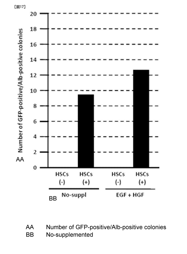
regeneration occurs at damaged tissue or transplanted tissue.
(3) The composition according to (2), wherein the composition
is administered within four days from the day of receiving damage
or transplantation.
(4) The composition according to (2), wherein the composition
is administered within one day from the day of receiving damage
or transplantation.
(5) The composition according to any one of (2) to (4), wherein
the damage is selected from tissue destruction, inflammation,
necrosis, fibrosis, surgical invasion and organ failure.
[0007]
(6) The composition according to any one of (1) to (5), wherein
the tissue regeneration involves differentiation and/or
3
CA 02895483 2015-06-17
proliferation of tissue stem cells.
(7) The composition according to any one of (1) to (6), wherein
the tissue regeneration involves proliferation of tissue
parenchymal cells.
(8) The composition according to any one of (1) to (7), wherein
the activated stellate cells are those subjected to treatment for
increasing expression of a protein selected from the group
consisting of HGF, EGF, and MMP14.
(9) The composition according to any one of (1) to (8), wherein
the composition is used to a subject having a condition in which
tissue regeneration is suppressed.
(10) The composition according to (9), wherein the condition of
suppressed tissue regeneration is selected from the group
consisting of inflammation, necrosis, fibrosis, organ failure,
decreased platelet count, genetic abnormalities, and reduction
of noradrenaline.
[0008]
(11) A method for promoting tissue regeneration in a subject,
comprising a step of administering the composition according to
any one of (1) to (10) to the subject in need thereof.
(12) A composition for differentiation and/or proliferation of
stem cells, comprising a component selected from the group
consisting of activated stellate cells, a decomposition product
of activated stellate cells, MMP14, collagen that has been treated
with MMP14, and a secretion product of activated stellate cells.
(13) A method for differentiation and/or proliferation of tissue
stem cells, comprising a step of contacting a component selected
from the group consisting of activated stellate cells, a
decomposition product of activated stellate cells, MMP14,
collagen that has been treated with MMP14, and a secretion product
4
CA 02895483 2015-06-17
of activated stellate cells, with the stem cells.
(14) A composition for promoting cell proliferation, comprising
a component selected from the group consisting of activated
stellate cells, a decomposition product of activated stellate
cells, MMP14, collagen that has been treated with MMP14, and a
secretion product of activated stellate cells.
[0009]
(15) A method for promoting cell proliferation, comprising a
step of contacting a component selected from the group consisting
of activated stellate cells, a decomposition product of activated
stellate cells, MMP14, collagen that has been treated with MMP14,
and a secretion product of activated stellate cells, with the
cells.
(16) A composition for suppressing cell proliferation,
comprising a substance that inhibits MMP14.
(17) The composition according to (16), wherein the cell to which
the substance inhibiting MMP14 acts is CAF and/or tumor cells.
(18) A cell culture substrate comprising collagen that has been
treated with MMP14.
(19) A cell culture vessel coated with collagen that has been
treated with MMP14.
[Advantageous Effects of the Invention]
[0010]
According to the present invention, not only in vivo tissue
regeneration, but also in vitro tissue formation can be promoted;
therefore, significant contribution in the biological and medical
fields can be expected.
Moreover, the promotion of tissue regeneration according
to the invention is particularly useful in situations wherein
tissue regeneration is suppressed, for example, a situation
CA 02895483 2015-06-17
involving a disease such as fibrosis.
[Brief Description of Drawings]
[0011]
[Fig. 1] Figure 1 is a diagram showing the flow of treatment of
partially hepatectomized rats.
[Fig. 2] Figure 2 is a photograph showing appearance of livers
of each group collected on day 5 and day 10 after partial
hepatectomy, in comparison to the liver before and immediately
after the partial hepatectomy.
[Fig. 3] Figure 3 is a graph showing change in the liver weight
of livers collected in each group.
[Fig. 4] Figure 4 is a diagram showing fluorescence microscope
images of the liver tissue collected on day 5 after partial
hepatectomy and co-stained with FITC-TUNEL, a-SMA and DAPI.
[Fig. 5] Figure 5 is a graph showing the number of a-SMA-positive
TUNEL-positive cells in the fluorescence microscope images of the
liver tissue collected on day 5 after partial hepatectomy and
co-stained with FITC-TUNEL, a-SMA and DAPI.
[0012]
[Fig. 6] Figure 6 is a diagram showing fluorescence microscope
images of the liver tissue collected on day 5 after partial
hepatectomy and co-stained with Ki67 and DAPI.
[Fig. 7] Figure 7 is a graph showing the number of Ki67-positive
cells in the fluorescence microscope images of the liver tissue
collected on day 5 after partial hepatectomy and co-stained with
Ki67 and DAPI.
[Fig. 8] Figure 8 is a diagram showing fluorescence microscope
images of the liver tissue collected on day 5 after partial
hepatectomy and co-stained with CD133 and DAPI.
[Fig. 9] Figure 9 is a diagram showing outline of the treatment
6
CA 02895483 2015-06-17
applied to each group of Example 3.
[Fig. 10] Figure 10 is a diagram showing the appearance of livers
collected in each group, in comparison to the liver immediately
after partial hepatectomy (day 0).
[0013]
[Fig. 11] Figure 11 is a graph showing the weight of livers
collected in each group, in comparison to that of the liver
immediately after partial hepatectomy (day 0).
[Fig. 12] Figure 12 is a diagram showing time course change in
a-SMA-positive cells of the liver tissue after partial
hepatectomy.
[Fig. 13] Figure 13 is a graph showing time course change in the
number of a-SMA-positive cells of the liver tissue after partial
hepatectomy.
[Fig. 14] Figure 14 is a diagram showing microscope images of
quiescent hepatic stellate cells (top view) and activated hepatic
stellate cells (bottom view).
[Fig. 15] Figure 15 is a diagram showing microscope images of
GFP-positive/albumin-positive colonies in the contact
co-culture of stem cells and stellate cells.
[0014]
[Fig. 16] Figure 16 is a diagram showing microscope images of
GFP-positive/albumin-positive colonies in contact co-culture of
stem cells and stellate cells, when EGF and HGF are added.
[Fig. 17] Figure 17 is a graph showing the number of
GFP-positive/albumin-positive colonies in contact co-culture of
stem cells and stellate cells.
[Fig. 18] Figure 18 is a graph showing the area of
GFP-positive/albumin-positive colonies in contact co-culture of
stem cells and stellate cells.
7
CA 02895483 2015-06-17
[Fig. 19] Figure 19 is a graph showing the area of
GFP-positive/albumin-positive colonies in non-contact
co-culture of stem cells and stellate cells.
[Fig. 20] Figure 20 is a graph showing proliferation of
GFP-positive/albumin-positive cells in non-contact co-culture
of stem cells and stellate cells, represented in terms of
absorbance.
[0015]
[Fig. 21] Figure 21 shows microscope images showing time course
change in the DNA synthesis in liver tissue after partial
hepatectomy.
[Fig. 22] Figure 22 is a graph showing time course change in the
percentage of BrdU-positive cells in hepatic cell after partial
hepatectomy.
[Fig. 23] Figure 23 shows microscope images showing time course
change in the DNA synthesis in liver tissue after partial
hepatectomy.
[Fig. 24] Figure 24 is a graph showing time course change in the
percentage of BrdU-positive cells in liver tissue after partial
hepatectomy.
[Fig. 25] Figure 25 shows microscope images showing BrdU-positive
liver cells when stem cells and stellate cells are co-cultured.
[0016]
[Fig. 26] Figure 26 is a graph showing the ratio of BrdU-positive
liver cells, when stem cells and stellate cells are co-cultured.
[Fig. 27] Figure 27 shows microscope images showing BrdU-positive
cells, when liver cells are cultured on collagen that has been
subjected to various treatments.
[Fig. 28] Figure 28 is a graph showing the ratio of BrdU-positive
cells, when liver cells are cultured on collagen that has been
8
CA 02895483 2015-06-17
subjected to various treatments.
[Fig. 29] Figure 29 shows microscope images showing BrdU-positive
cells, when hepatocytes and MMP14- and/or HGF-knocked down
stellate cells are co-cultured.
[0017]
[Fig. 30] Figure 30 is a graph showing the ratio of BrdU-positive
cells, when liver cells and MMP14- and/or HGF-knocked down
stellate cells are co-cultured.
[Fig. 31] Figure 31 shows microscope images showing BrdU-positive
cells, when liver cells are cultured on collagen that has been
treated with MMP14 under the presence of RGD peptide.
[Fig. 32] Figure 32 is a graph showing the ratio of BrdU-positive
cells, when hepatocytes are cultured on collagen that has been
treated with MMP14 under the presence of RGD peptide.
[Description of Embodiments]
[0018]
One aspect of the present invention relates to a composition
for promoting tissue regeneration, comprising a component
selected from the group consisting of activated stellate cells,
a decomposition product of activated stellate cells, MMP14,
collagen that has been treated with MMP14, and a secretion product
of activated stellate cells.
[0019]
Activated stellate cells can be obtained by subculturing
stellate cells isolated from a living body. Stellate cells are
known to be present in various tissues such as liver, pancreas,
kidney, intestine and lung (Zhao and Burt, JMol Histol. 2007 Mar;
38(1): 53-64), and any of these may be used. Stellate cells can
be isolated using any known method . Specific examples of a method
for isolating hepatic stellate cells are illustrated in Example
9
CA 02895483 2015-06-17
(1) below. Activated stellate cells are characterized by
expression of aSMA, and they can be selected using aSMA as a marker .
[0020]
Activated stellate cells may be those which have been
subjected to a treatment for increasing the expression of a
protein, selected from the group consisting of HGF, EGF, and MMP14.
Examples of such treatment include, but are not limited to,
introduction of a gene encoding said protein in the activated
stellate cells. Genes encoding HGF, EGF and MMP14 are known, and
a method for introducing genes are well-known in the art.
[0021]
A decomposition product of activated stellate cells can be
obtained by decomposing activated stellate cells using a variety
of techniques including physical and/or chemical methods.
Decomposition can be carried out by any method known to decompose
cells, for example, osmotic shock method, freezing and thawing
method, the use of surfactants, enzymatic digestion, sonication,
French press, and crushing with a mortar, crushing by a
homogenizer, and crushing by glass beads. Decomposition
technique without denaturing proteins or with a slight degree of
denaturation is preferred. By using such a technique, a MMP14
which is expressed on the cell membrane can be obtained without
impairing its function. Furthermore, decomposition products of
activated stellate cells preferably comprise a cell membrane
component.
[0022]
MMP14 (also referred to as MT1-MMP) is a MMP expressed on
the cell membrane. MMP14 can act on collagen I. The present
inventors have revealed that the effect of MMP14 on collagen I
is deeply involved in cell proliferation.
CA 02895483 2015-06-17
MMP14 in the present invention includes not only those
expressed on the cell membrane, but also those released from the
cell membrane. MMP14 may also be those naturally occurring, or
may be those that are artificially produced. Therefore, MMP14
includes recombinant MMP14. MMP14 of free form is known (for
example, Jo et al., Biochem J. 2000 Feb 1; 345 Pt 3: 511-9), and
MMP14 is also commercially available (for example, R&D Systems,
Cat. No. 918-MP-010, 918-MPN-010, etc.). Aminoacids andthe base
sequence encoding the same for MMP14 are known (for example, the
base sequence of human MMP14 is registered as GenBank Accession
No. NM 004995, and the amino acid sequence is registered as
GenBank Accession No. NP 004986).
[0023]
MMP14 in the present invention also includes a functional
variant thereof. Functional variants of MMP14 include, but are
not limited to, the following: (i) a variant having one or more,
typically one or several mutations in the amino acid sequence of
said protein, and still having equivalent functions of said
protein, (ii) a variant encoded by a nucleic acid having one or
more, typically one or several mutations in the base sequence of
the nucleic acid that has the base sequence of the gene encoding
said protein, or that encodes the same polypeptide as this nucleic
acid, and having equivalent functions, (iii) a variant encoded
by a nucleic acid that hybridizes the complementary strand, or
a fragment thereof, of the nucleic acid that has the base sequence
of the gene encoding said protein, or that encodes the same
polypeptide as this nucleic acid, or that encodes the variant of
(ii), under stringent conditions, and having equivalent functions
of said protein, (iv) a variant having an amino acid sequence that
has 60% or more, preferably 70% or more, more preferably 80% or
11
CA 02895483 2015-06-17
more, still more preferably 90% or more, and particularly
preferably 95% or more homology to the amino acid sequence of said
protein, and having equivalent functions of said protein, (v) a
variant encoded by a nucleic acid that has 60% or more, preferably
70% or more, more preferably 80% or more, still more preferably
90% or more, and particularly preferably 95% or more homology to
the base sequence of the gene encoding said protein, and having
equivalent functions of said protein, and the like.
[0024]
Those skilled in the art can appropriately produce the above
functional variants based on the sequence information of MMP14,
using any known techniques, such as chemical synthesis, cleavage
or insertion of nucleic acid by restriction enzyme, site-specific
mutagenesis, irradiation or ultraviolet irradiation.
Whether or not a certain variant has equivalent functions
as those of MMP14 can be evaluated by analyzing said variant in
terms of known functions of MMP14, for example, without limitation,
collagen degradation ability, using any known method, and by
comparing it with an appropriate negative control and MMP14 as
a positive control. For example, in one variant, if the above
function is better than the negative control, for example, 10%
or more, 25% or more, 50% or more, 75% or more, and even 100% or
more better than that of the negative control, and/or, if this
function is 1/100 or more, 1/50 or more, 1/25 or more, 1/10 or
more, 1/5 or more, and even 1/2 or more of that of CSABP, then
this variant is included in the functional variant of MMP14.
[0025]
The term "stringent conditions" as used herein refers to
well-known parameters in the art, which are described in standard
protocols such as Sambrook et al., Molecular Cloning: A Laboratory
12
CA 02895483 2015-06-17
Manual, 3d ed., Cold Spring Harbor Press (2001); Ausubel et al.,
Current Protocols in Molecular Biology, Greene Publishing
Associates (1992); and others.
[0026]
Stringent conditions in the present invention refer to, for
example, hybridization at 65 C using a hybridization buffer
consisting of 3.5 x SSC (0.15 M sodium chloride/0.15 M sodium
citrate, pH 7), 0.02% Ficoll, 0.02% polyvinyl pyrrolidone, 0.02%
bovine serum albumin, 25 mM NaH2PO4 (pH 7), 0.05% SDS, and 2 mM
EDTA. After hybridization, the membrane to which DNA has been
transferred is washed with 2 x SSC at room temperature, then with
0.1-0.5 x 550/0.1 x SDS up to the temperature of 68 C.
Alternatively, stringent hybridization may be performed using a
commercially available hybridization buffer, such as
ExpressHyb(R) Hybridization Solution (Clontech Laboratories,
Inc.) under hybridization and washing conditions described by the
manufacturer.
[0027]
Other available conditions and reagents which result in
comparable stringency exist; because those skilled in the art are
considered to be familiar with such conditions, these are not
specifically described herein. However, it is possible to
manipulate the conditions in order to enable clear identification
of nucleic acids encoding protein variants.
MMP14 and/or a functional variant thereof of the present
invention include, in addition to said protein itself or a
functional variant thereof, a nucleic acid encoding said protein
or a functional variant thereof.
[0028]
Collagen treated with MMP14 can be obtained by treating
13
CA 02895483 2015-06-17
collagen with MMP14. As the collagen, for example, collagen I
may be used. MMP14 used for the treatment may be, in addition
to the above MMP14 (including functional variants of MMP14), the
cells themselves which express MMP14, or a decomposition product
of the cells which comprises MMP14. Treatment time by MMP14 may
be, for example, at a temperature at which MMP14 can act, 1-120
h, 3-60 h, 6-48 h, and 12-36 h, etc. Collagen treated with MMP14
is preferably in a condition wherein RGD sequence is ready to
interact with cells.
[0029]
A secretion product of activated stellate cells can be
obtained from a culture supernatant of activated stellate cells.
A culture supernatant may be used as it is, or may be used after
concentration by dialysis or freeze drying, etc. Since secretion
products of activated stellate cells contain a variety of proteins,
preferably, they are handled such that the proteins are not
denatured.
[0030]
Tissue for which regeneration is promoted by the
composition of the present invention is not particularly limited,
and includes various tissues throughout the body. Examples of
such tissue include, without limitation, tissue wherein stellate
cells are present, tissue wherein fibrosis occurs, tissue wherein
stem cells are present, and the like. Specifically, examples
includes, without limitation, liver, pancreas, kidney, intestine,
lung, spleen, heart, bone marrow, vocal cord, skin, peritoneum,
eye, vessel, etc.
[0031]
The composition of the present invention is useful for
tissue regeneration that occurs in damaged tissues or
14
CA 02895483 2015-06-17
transplanted tissues. Damaged tissues include those subjected
to tissue destruction, inflammation, necrosis, fibrosis,
surgical invasion, and organ failure, etc. Administration time
of the composition of the present invention is not particularly
limited, and preferably the composition is administered within
four days from the day of receiving damage or transplantation,
or within one day from the day of receiving damage or
transplantation.
[0032]
Tissue regeneration by the composition of the present
invention may involve differentiation and/or proliferation of
stem cells. Also, tissue regeneration by the composition of the
present invention may involve proliferation of the tissue
parenchymal cells. Differentiation of stem cells can be
evaluated by, for example, detection of cellular markers specific
for differentiated cells, and detection of functions exhibited
by differentiated cells. Cell proliferation can be evaluated by
various known techniques, for example, measurement of living-cell
count overtime, size, volume or weight of the tissue, measurement
of DNA synthesis level, WST-1 method, BrdU (bromodeoxyuridine)
method, 3H-thymidine incorporation method and others.
[0033]
The composition of the present invention may be used for
a subject having a condition in which tissue regeneration is
suppressed. The condition in which tissue regeneration is
suppressed includes, without limitation, for example,
inflammation, necrosis, fibrosis, organ failure, decreased
platelet count, genetic abnormalities, and reduction of
noradrenaline.
[0034]
CA 02895483 2015-06-17
The amount of an active ingredient in the composition of
the present invention may be an amount with which tissue
regeneration is promoted upon administration of the composition.
In addition, the amount that does not cause an adverse effect
exceeding the benefits of administration is preferred. Such an
amount is either publicly known, or may be appropriately
determined by an in vitro test using cultured cells, and by a test
in a model animal such as mouse, rat, dog or pig, and such test
methods are well-known to those skilled in the art. Promotion
of tissue regeneration can be evaluated by the recovery of
functions, weight and size of the tissue, by means of biochemical
tests and image diagnosis using X-ray, ultrasound, MRI, CT, and
endos copy. The amount of an active ingredient may vary according
to dosage form of the composition. For example, when multiple
units of a composition are used for single administration, the
amount of an active ingredient to be blended into one unit of the
composition may be the amount of the active ingredient required
for the single administration divided by the multiple times. Such
amount can be appropriately adjusted by those skilled in the art.
[0035]
The present invention also relates to a method for producing
a composition to promote tissue regeneration, comprising blending
a component selected from the group consisting of activated
stellate cells, a decomposition product of activated stellate
cells, MMP14, collagen that has been treated with MMP14, and a
secretion product of activated stellate cells; and to the use of
said component in the production of a composition for promoting
tissue regeneration, and to said component used for promoting
tissue regeneration.
Each component and the amount thereof in the above
16
CA 02895483 2015-06-17
production method or use are as already described above. Blending
of each component can be carried out according to any known
technique.
[0036]
The present invention also relates to a method for promoting
tissue regeneration in a subject, comprising a step of
administering the above composition to the subject in need thereof.
The subject in the present method may be either those having
damaged tissue or tissue transplantation. Damages include,
without limitation, for example, tissue destruction,
inflammation, necrosis, fibrosis, surgical invasion, organ
failure and the like. Administration of the composition may be,
for example, within four days from the day of receiving damage
or transplantation, or within one day from the day of receiving
damage or transplantation.
[0037]
The present invention also relates to a composition for
differentiation and/or proliferation of stem cells, comprising
a component selected from the group consisting of activated
stellate cells, a decomposition product of activated stellate
cells, MMP14, collagen that has been treated with MMP14, and a
secretion product of activated stellate cells; a method for
producing a composition for differentiation and/or proliferation
of stem cells, comprising blending said component; a use of said
component in the production of a composition for differentiation
and/or proliferation of stem cells; said component used for
differentiation and/or proliferation of stem cells; and a method
for differentiation and/or proliferation of stem cells,
comprising a step of contacting said component with the stem
cells.
17
CA 02895483 2015-06-17
[0038]
Stem cells are not particularly limited, and include, for
example, tissue stem cells (somatic stem cells , adult stem cells ) ,
embryonic stem cells, iPS cells and the like. Stem cells may be
totipotent, pluripotent, multipotent or unipotent. Examples of
tissue stem cells include, but are not limited to, neural stem
cells, hematopoietic stem cells, mesenchymal stem cells, liver
stem cells, pancreatic stem cells, skin stem cells, muscle stem
cells, germ stem cells, etc. Stem cells may be of autologous,
or of other individuals of the same species or individuals of
different species. Stem cells that have been differentiated
and/or proliferated can be transplanted into a subject in need
thereof.
[0039]
The above method may be performed in vitro, in vivo or ex
vivo. Each component and an amount thereof in the above
composition, method or use are as already described above.
Blending of the components can be carried out according to any
known technique. When MMP14 is used as an active ingredient in
the above composition, method or use, it is preferred that
collagen (especially collagen I) is present around the cells of
interest in promoting proliferation.
[0040]
The present invention also relates to a composition for
promoting cell proliferation, comprising a component selected
from the group consisting of activated stellate cells, a
decomposition product of activated stellate cells, MMP14,
collagen that has been treated with MMP14, and a secretion product
of activated stellate cells; a method for producing a composition
for promoting cell proliferation, comprising blending said
18
CA 02895483 2015-06-17
component; a use of said component in the production of a
composition for promoting cell proliferation; said component used
for promoting cell proliferation; and a method for promoting cell
proliferation, comprising a step of contacting said component
with the cells.
Cells the proliferation of which is promoted include, but
are not limited to, for example, cells of the liver, pancreas,
kidney, intestine, lung, spleen, heart, bone marrow, vocal cord,
skin, peritoneum, eye, and vessel, etc. Cells may be of
autologous, or of other individuals of the same species or
individuals of different species. Cells that have been
differentiated and/or proliferated can be transplanted into a
subject in need thereof.
[0041]
The above method may be performed in vitro, in vivo or ex
vivo. Each component and an amount thereof in the above
composition, method or use are as already described above.
Blending of the components can be carried out according to any
known technique. When MMP14 is used as an active ingredient in
the above composition, method or use, it is preferred that
collagen (especially collagen I) is present around the cells of
interest in promoting proliferation.
[0042]
The present invention also relates to a composition for
suppressing cell proliferation, comprising a substance that
suppresses MMP14; a method for producing a composition for
suppressing cell proliferation, comprising blending said
substance; a use of said substance in the production of a
composition for suppressing cell proliferation; said substance
used for suppressing cell proliferation; and a method for
19
CA 02895483 2015-06-17
suppressing cell proliferation, comprising a step of contacting
said substance with the cells.
The present invention also relates to a composition for
treating cellular proliferative disease, comprising a substance
that suppresses MMP14; a method for producing a composition for
treating cellular proliferative disease, comprising blending
said substance; a use of said substance in the production of a
composition for treating cellular proliferative disease; said
substance used for treating cellular proliferative disease; and
a method for treating cellular proliferative disease, comprising
administrating a therapeutically effective amount of said
substance to a subject in need thereof.
[0043]
Examples of a substance that inhibits MMP14 include,
without limitation, a drug that inhibits production and/or
activity of MMP14, and a drug that promotes decomposition and/or
inactivation of MMP14. Examples of a drug that inhibits
production of MMP14 include, without limitation, RNAi molecule
for DNA encoding MMP14, ribozyme, antisense nucleic acid, DNA/RNA
chimera polynucleotide, and vectors expressing thereof.
[0044]
Inhibition of MMP14 can be determined by a degree of
inhibition of expression or activity of MMP14 in cells, compared
to the case wherein an MMP14 inhibitor is not reacted. Expression
of MMP14 can be evaluated by any known techniques including,
without limitation, for example, immunoprecipitation method
utilizing an anti-MMP14 antibody, EIA, ELISA, IRA, IRMA, Western
blotting, immunohistochemistry, immunocytochemistry, flow
cytometry, various hybridization methods, Northern blotting ,
Southern blotting, and a variety of PCR methods, utilizing a
CA 02895483 2015-06-17
nucleic acid that specifically hybridizes to the nucleic acid
encoding MMP14 or an unique fragment thereof, or to a
transcription product (e.g., mRNA) or a splicing product of said
nucleic acid.
[0045]
As used herein, RNAi molecules refer to any molecules that
provide RNA interference, and examples include, without
limitation, double-stranded RNA such as small interfering RNA
(siRNA), micro RNA (miRNA), short hairpin RNA (shRNA),
DNA-directed RNA (ddRNA), Piwi-interacting RNA (piRNA), repeat
associated siRNA (rasiRNA), and a variant thereof. These RNAi
molecules are commercially available, or they can be designed and
produced based on known sequence information, etc.
Furthermore, an antisense nucleic acid as used herein
includes RNA, DNA, PNA, or a composite thereof.
As used herein, DNA/RNA chimera polynucleotide is not
limited, and includes, for example, a double-stranded
polynucleotide composed of DNA and RNA that inhibits expression
of a target gene, as described in JP 2003-219893.
[0046]
Examples of cells the proliferation of which is suppressed
include, without limitation, cells the proliferation of which has
adverse effects, and cells the proliferation of which is involved
in diseases. Specifically, they include tumor cells, cancer
cells, activated stellate cells and the like. A substance that
suppresses MMP14 may be administered to these cells, and it may
also be administered to MMP14-expressing cells that support the
proliferation of these cells, for example, cancer-associated
fibroblast (CAF).
Examples of cellular proliferative disease include,
21
CA 02895483 2015-06-17
without limitation, benign or malignant tumors, hyperplasia,
keloid, Cushing syndrome, primary aldosteronism, erythroplakia,
polycythemiavera, leukoplakia, hyperplastic scar, lichen planus
and lentiginosis.
[0047]
When the active ingredient of various compositions and
methods of the present invention described herein is a nucleic
acid, such as RNAi molecule, ribozyme, antisense nucleic acid,
and DNA/RNA chimera polynucleotide, these may be used as a bare
nucleic acid as it is, or these may be supported by various vectors.
As the vector, any publicly known vectors such as plasmid vectors,
phage vectors, phagemid vectors, cosmid vectors, and viral
vectors can be used. Preferably, the vector comprises at least
a promoter that enhances the expression of a supporting nucleic
acid; in this case, the nucleic acid is preferably operably linked
to such promoters. The phrase "nucleic acid is operably linked
to promoter" means that the nucleic acid and the promoter are
located such that the protein encoded by the nucleic acid can be
suitably produced by the action of the promoter. The vector may
or may not be capable of replication in a host cell, and
transcription of genes may take place outside the nucleus of the
host cell, or may take place in the nucleus. In the latter case,
the nucleic acid may be incorporated into the genome of the host
cell.
[0048]
Furthermore, the active ingredient can be supported on a
variety of non-viral lipids or protein carriers. Examples of such
carriers include, without limitation, cholesterol, liposomes,
antibody protomers, cyclodextrinnanoparticles, fusion peptides,
aptamers, biodegradable polylactic acid copolymers, polymers and
22
CA 02895483 2015-06-17
the like, and they can enhance the incorporation efficiency into
cells (for example, see Pirollo and Chang, Cancer Res. 2008; 68
(5): 1247-50, etc.). In particular, cationic liposomes and
polymers (such as polyethyleneimine) are useful. Further
examples of useful polymers as such carriers include those
described in US 2008/0207553, US 2008/0312174 and the like.
[0049]
Various compositions of the invention as described herein
can be used in medical applications such as in vivo tissue
regeneration and treatment of diseases. Thus, various
compositions of the present invention can be pharmaceutical
compositions. In a pharmaceutical composition, as long as
effectiveness of the active ingredient is not interfered, the
active ingredient maybe combined with other optional ingredients.
Such optional ingredients include, for example, other
chemotherapeutic agents, pharmaceutically acceptable carriers,
excipients, and diluents, etc. Also, depending on the
administration route and the drug release manner, the composition
may be coated with a suitable material, for example, enteric
coating and timed-disintegrating material, and the composition
may be incorporated into appropriate drug release systems.
[0050]
Various compositions of the invention as described herein
(including various pharmaceutical compositions) may be
administered via various routes including both oral and
parenteral routes, for example, without limitation, oral,
intravenous, intramuscular, subcutaneous, topical, intratumoral,
rectal, intraarterial, intraportal, intraventricular,
transmucosal, transdermal, intranasal, intraperitoneal,
intrapulmonary, and intrauterine routes, and they may be
23
CA 02895483 2015-06-17
formulated in a dosage form suitable for each administration route.
Such dosage forms and formulation methods may be selected as
appropriate from any known ones (see, for example, "Hyojun
Yakuzaigaku" (Standard Pharmaceutical Science) , Ed. by Yoshiteru
Watanabe, et al., Nankodo, 2003) .
[0051]
Examples of dosage forms suitable for oral administration
include, but are not limited to, powders, granules, tablets,
capsules, solutions, suspensions, emulsions, gels, and syrups,
and examples of dosage forms suitable for parenteral
administration include injections such as an injectable solution,
an injectable suspension, an injectable emulsion, and an
injection in a form that is prepared at the time of use.
Formulations for parenteral administration may be in the form of
transplantation, or aqueous or nonaqueous isotonic sterile
solutions or suspensions.
[0052]
Various compositions of the invention as described herein
(including various pharmaceutical compositions) may be targeted
to specific tissues and cells. Targeting can be accomplished by
any known technique. When delivery to cancer is contemplated,
the following techniques can be used without limitation: passive
targeting, in which a formulation is made to have a size suitable
for exhibiting enhanced permeability and retention (EPR) effect,
such as a diameter of 50-200 pm, in particular 75-150 pm; and active
targeting, in which a ligand such as CD19, HER2, transferrin
receptor, folate receptor, VIP receptor, EGFR (Torchilin, AAPS
J. 2007; 9 (2) : E128-47) , RAAG10 (JP A 2005-532050) , PIPA (JP A
2006-506071) , KID3 (JP A 2007-529197) , etc., and a peptide having
RGD motif and NGR motif, and F3, LyP-1 (Ruoslahti et al., J Cell
24
CA 02895483 2015-06-17
Biol. 2010; 188(6):759-68) are used as a targeting agent.
Moreover, since retinoid or a derivative thereof is also known
to be useful as a targeting agent for cancer cells and CAF (WO
2008/120815), it is also possible to utilize a carrier comprising
retinoid as a targeting agent. Such carriers are described, in
addition to the above literatures, in WO 2009/036368, WO
2010/014117, and WO 2012/170952.
[0053]
Various compositions of the invention as described herein
(including various pharmaceutical compositions) may be supplied
in any form; however, from the viewpoint of storage stability,
they may be supplied in a form that can be prepared at the time
of use, for example, in a form that can be prepared at the site
of clinical practice or in the vicinity thereof by a physician
and/or pharmacist, nurse or other paramedical personnel, etc.
Such a form is particularly useful when the composition of the
present invention comprises a component that is difficult to store
in a stable manner, such as lipids, proteins, and nucleic acids.
In this case, the composition of the present invention is provided
in one or more containers which further comprise at least one of
essential constituents, and the composition is prepared, for
example, within 24 h, preferably 3 h prior to use, and more
preferably immediately before use. Upon preparation, reagents,
solvents and dispensing equipment usually available in a place
of preparation can be used as appropriate.
[0054]
Accordingly, the present invention also relates to a kit
for preparation of a composition, comprising one or more
containers containing singly or in combination active ingredients
which may be included in various compositions of the present
CA 02895483 2015-06-17
invention; and also relates to a constituent element necessary
for various compositions provided in a form of such a kit. The
kit of the present invention may comprise, in addition to the above,
instructions describing preparation method and administration
method of the various compositions of the present invention, for
example, an instruction, or an electronic recording medium such
as CD, DVD, etc. Furthermore, the kit of the invention may include
all of the constituent elements for completing the various
compositions of the invention, but need not always include all
of the constituent elements. Therefore, the kit of the present
invention may not include a reagent or a solvent usually available
at the site of clinical practice and experimental facility, such
as sterile water, physiological saline, and glucose solution,
etc.
[0055]
An effective amount in the various methods of the invention
described herein may be, for example, with respect to regeneration
of tissues, an amount that promotes tissue regeneration, or
eliminates delay in tissue regeneration; with respect to
treatment of a disease, an amount that reduces symptoms of the
disease, or delays or stops progression of the disease, and
preferably, an amount that suppresses or cures the disease. In
addition, an amount that does not cause an adverse effect
exceeding the benefits from administration is preferred. Such
an amount can be appropriately determined by an in vitro test using
cultured cells, or a test in a model animal such as mouse, rat,
dog or pig, and such test methods are well known to those skilled
in the art. Furthermore, dosage of a drug used in the treatment
methods of the invention is either known to those skilled in the
art or can be appropriately determined by the above-mentioned test,
26
CA 02895483 2015-06-17
etc.
[0056]
Specific dosage of an active ingredient to be administered
in the treatment methods of the invention described herein can
be determined in consideration of various conditions related to
a subject requiring treatment, for example, presence or absence
of conditions which inhibit regeneration, severity of symptoms,
general health, age, body weight of the subject, gender of the
subject, diet, time and frequency of administration,
pharmaceutical agents that are used in combination,
responsiveness to therapy, dosage form, and compliance to
therapy.
[0057]
The administration route includes various routes including
both oral and parenteral routes, for example, oral, intravenous,
intramuscular, subcutaneous, topical, intratumoral, rectal,
intraarterial, intraportal, intraventricular, transmucosal,
transdermal, intranasal, intraperitoneal, intrapulmonary, and
intrauterine routes.
The frequency of administration differs depending on the
properties of an agent or composition used and conditions of a
subject including those described above; and the frequency may
be, for example, multiple number of times per day (i.e., 2, 3,
4, or 5 times or more per day), once a day, every several days
(i.e., every 2, 3, 4, 5, 6, or 7 days, etc.), every week, or every
few weeks (i.e., every 2, 3, or 4 weeks).
[0058]
As used herein, the term "subject" means any living
individual, preferably an animal, more preferably a mammal, more
preferably a human individual. In the present invention, the
27
CA 02895483 2015-06-17
subject may be healthy, or may be suffering from some disease;
in the case where treatment of a particular disease is
contemplated, typically the subject means a subject suffering
from such disease, or a subject having a risk of suffering from
such disease.
In addition, the term "treatment" includes, as used herein,
medically acceptable all kinds of preventive and/or therapeutic
intervention for the purpose of cure, temporary remission or
prevention of disease. For example, the term "treatment"
includes medically acceptable intervention with variety of
purposes, including delay or stop of progression of disease,
regression or disappearance of lesions, prevention of onset or
prevention of recurrence.
[0059]
The present invention also relates to a cell culture
substrate comprising collagen that has been treated with MMP14.
The collagen treated with MMP14 is as described above with respect
to various compositions.
The cell culture substrate of the present invention is to
be used as a scaffold for cell growth, and it may be of various
forms such as membranous and gel forms. The cell culture
substrate is preferably sterile, but may be provided in a
non-sterile state, which will be sterilized at the time of use.
The cell culture substrate of the present invention may be used
to coat a cell culture vessel. Coating concentration may be,
without limitation, in terms of the concentration of collagen,
0.01-1000 pg/cm2, 0.1-100 pg/cm2, 1-50 pg/cm2, or 5-25 pg/cm2.
[0060]
The present invention also relates to a kit for producing
a cell culture substrate, comprising collagen that has been
28
CA 02895483 2015-06-17
treated with MMP14, or comprising MMP14 and collagen. The kit
may include instructions comprising information necessary to use
the collagen that has been treated with MMP14 as a cell culture
substrate, or information used to treat the collagen with MMP14,
for example, an instruction, or an electronic recording medium
such as CD, DVD, etc.
[0061]
The present invention also relates to a cell culture vessel
coated with collagen that has been treated with MMP14. As the
cell culture vessel, any of known vessels can be used. The cell
culture vessel of the present invention may be produced by coating
a cell culture vessel with collagen that has been treated with
MMP14, or it may be produced by treating a collagen-pre-coated
cell culture vessel with MMP14. MMP14 used for the treatment may
be attached to the cell membrane, or may be free from the cell
membrane. Thus, treatment with collagen may comprise culturing
cells that express MMP14, such as activated stellate cells, in
a collagen-coated cell culture vessel. Cells expressing MMP14
used in the treatment with collagen may be removed after the
treatment, or may be left in the cell culture vessel to which cells
to be proliferated may be added. This cell culture vessel is
excellent in cell proliferation, and can be used to promote cell
culture. In addition, because stellate cells can induce
differentiation of stem cells, a cell culture vessel which further
comprises stellate cells can be used for induction of
differentiation of stem cells. The coating concentration of
collagenmay be, without limitation, for example, 0 . 01-1000 pg/cm2,
0.1-100 pg/cm2, 1-50 pg/cm2, or 5-25 pg/cm2.
[0062]
Furthermore, the present invention also relates to a kit
29
CA 02895483 2015-06-17
for producing a cell culture vessel coated with collagen that has
been treated with MMP14, comprising collagen that has been treated
with MMP14, or comprising MMP14 and collagen. The kit may include
instructions comprising information necessary to coat a cell
culture vessel with collagen that has been treated with MMP14,
or information used to treat the collagen coated on the cell
culture vessel with MMP14, for example, an instruction, or an
electronic recording medium such as CD, DVD, etc. The kit of the
present invention may comprise a cell culture vessel; however,
a commercially available cell culture vessel may be separately
prepared and used.
[0063]
The present invention further relates to a method for
producing a cell culture vessel coated with collagen that has been
treated with MMP14, comprising a step of coating a cell culture
vessel coated with collagen that has been treated with MMP14, or
a step of treating the collagen coated on a cell culture vessel
with MMP14. MMP14 used for the treatment may be attached to the
cell membrane, or may be free from the cell membrane. Thus, a
step of treating the collagen coated on the cell culture vessel
with MMP14 comprises culturing cells that express MMP14, such as
activated stellate cells, in a collagen-coated cell culture
vessel, or, contacting the collagen coated on the cell culture
vessel with a decomposition product of cells expressing MMP14.
[Examples]
[0064]
The present invention is explained in further detail in the
Examples below; however, they are only illustrations and do not
limit the invention in any way.
[0065]
CA 02895483 2015-06-17
Example 1. Preparation of VA-lip siRNA
(1) Preparation of siRNA
As the sense and antisense strands of siRNA (Hokkaido System
Science Co., Ltd., Sapporo, Japan) that targets the base sequence
of gp46 (GenBank Accession No. M69246, SEQ ID NO: 1) , i.e., a rat
homolog of human HSP47 that is a common molecular chaperone of
collagen (I-IV type) , the following was used:
A: GUUCCACCAUAAGAUGGUAGACAACAG (sense strand siRNA starting from
the 757th base of the base sequence of gp46, SEQ ID NO: 2)
B: GUUGUCUACCAUCUUAUGGUGGAACAU (antisense strand siRNA, SEQ ID
NO: 3)
[0066]
As the siRNArandom (sometimes also referred to as
siRNAscramble) , the following was used:
C: CGAUUCGCUAGACCGGCUUCAUUGCAG (sense strand siRNA, SEQ ID NO:
4)
D: GCAAUGAAGCCGGUCUAGCGAAUCGAU (antisense strand siRNA, SEQ ID
NO: 5)
[0067]
In some experiments, a sense strand in which
6' -carboxyfluorescein (6-FAM) or fluorescein isothiocyanate
(FITC) is bound to the 5' end was used. These sequences were
confirmed to have no homology to other known rat mRNA in the BLAST
search.
[0068]
(2) Preparation of VA-lip siRNA
As the cationic lipid, cationic liposomes (Lipotrust) were
purchased from Hokkaido System Science Co., Ltd. (Sapporo, Japan) ,
which comprise
0,0 ' -ditetradecanoyl-N- (a-trimethylammonioacetyl ) diethanolami
31
CA 02895483 2015-06-17
nechloride(DC-6-14), cholesterol, and
dioleylphosphatidylethanolamine (DOPE) in a molar ratio of 4 : 3 : 3 .
Before use, the liposomes were prepared at a concentration of 1
mM (DC-6-14) by adding redistilled water (DDW) to the freeze-dried
lipid mixture under stirring condition. To prepare VA-bound
liposomes, 200 nmol of vitamin A (retinol, Sigma, USA) dissolved
in DMSO was mixed with a liposome suspension (100 nmol as DC-6-14)
in a 1.5-ml tube while stirring at 25 C. To prepare VA-bound
liposomes carrying siRNAgp46 (VA-lip-siRNAgp46), a siRNAgp46
solution (580 pmol/p1 in DDW) was added to a retinol-bound
liposome solution while stirring at room temperature. The molar
ratio of siRNA and DC-6-14 was 1:11.5, and the molar ratio of
vitamin A, DC-6-14 and siRNA was 11.5:11.5:1. To obtain desired
dosage for in vitro use, VA-lip siRNA was reconstituted in
phosphate-buffered saline (PBS).
[0069]
Example 2. Effect of administration of VA-lip siRNAgp46 to
partial hepatectomy rat on liver regeneration
(1) Preparation and treatment of partial hepatectomy rat
Partial hepatectomy rats were prepared by removing hepatic
left and median lobes, which represents about 70% of the total
liver, of male SD rats (150-200 g) (Slc Japan, Shizuoka, Japan).
These partial hepatectomy rats were administered with VA-lip
siRNAgp46 prepared in Example 1 (group I), VA-lip siRNAscramble
(mock control, group II) , or 5% glucose (siRNA-free control, group
III) in a volume of 300 p1/time every other day for a total of
five times (i.e., on days 1, 3, 5, 7 and 9 after partial
hepatectomy) via tail vein (Fig. 1, n=6). Here, each siRNA was
used at 0.75 mg/kg of rat body weight.
[0070]
32
CA 02895483 2015-06-17
(2) Evaluation of collected tissue
At 24 h after the 3' and 5th administration (i.e., on day
and day 10 after partial hepatectomy), the liver of the partial
hepatectomy rats was collected. After appearance of the
collected liver was observed and the weight was measured, the
liver was embedded using an OCT compound to prepare frozen
sections. The resulting sections were fixed with 4%
paraformaldehyde (PFA) and then subjected to blocking with 5% goat
serum-containing PBS, washed with PBS, then allowed to react
overnight using a Cy3 labeled anti-a-smooth muscle actin (a-SMA)
antibody (Sigma), an anti-Ki-67 antibody (Dako), or an anti-CD133
antibody at 4 C. After washing with PBS, the sections were
allowed to react using an A1exa488-labeled goat anti-mouse
antibody (Invitrogen) at room temperature for 60 min. After
washing with PBS, the sections were sealed with ProLong(R) Gold
with DAPI (Invitrogen), and observed by fluorescence microscope.
In addition, Tunel staining of the collected liver frozen sections
was carried out using in situ Apoptosis Detection Kit (TakaraBio,
Japan) according to the manufacturer's instructions. A
FITC-TUNEL antibody and a Cy3-labeled anti-a-SMA antibody were
used for co-staining of a-SMA and Tunel. The number of positive
cells was calculated as the average value of five visual fields
(200x magnification).
[0071]
(3) Results
Figure 2 shows appearance of livers of each group collected
on day 5 and day 10 after partial hepatectomy, in comparison to
the liver before and immediately after the partial hepatectomy.
In addition, Fig. 3 shows change in the weight of livers of each
group collected. As can be seen from the both figures, the livers
33
CA 02895483 2015-06-17
in group II and III have already recovered their previous size
and weight on day 5, whereas in group I in which VA-lip siRNAgp46
was administered, the size and weight of the livers did not recover
even 10 days after hepatectomy.
[0072]
Next, liver tissues collected on day 5 after hepatectomy
were co-stained with FITC-TUNEL, a-SMA and DAPI; and it was
clarified that in group I, the number of TUNEL-positive cells in
a-SMA positive cells was significantly larger than that in the
other groups (Figs. 4 and 5). This finding indicates that large
quantities of a-SMA positive cells such as activated hepatic
stellate cells underwent apoptosis due to the inhibition of gp46
by VA-lip siRNAgp46.
[0073]
Furthermore, when the liver tissues collected on day 5 after
hepatectomy were co-stained with Ki67 and DAPI, it was clarified
that the number of Ki67-positive cells in group I was
significantly smaller than that in the other groups subjected to
the same partial hepatectomy (groups II and III) (Figs. 6 and 7).
Meanwhile, the numbers of Ki67-positive cells in groups II and
III were significantly larger than that in normal liver tissue
without partial hepatectomy; because Ki67 is a marker of cell
proliferation, this indicates that cell proliferation was
enhanced in the liver tissue after partial hepatectomy.
[0074]
In addition, when the liver tissues collected on day 5 after
hepatectomy were co-stained with CD133 and DAPI, it was clarified
that the number of CD133-positive cells in group I was
significantly smaller than that in the other groups subjected to
the same partial hepatectomy (groups II and III) (Fig. 8). Since
34
CA 02895483 2015-06-17
CD133 is a marker of stem cells, this result indicates that
proliferation of stem cells was significantly suppressed by
VA-lip siRNAgp46.
Considering the above staining results in combination, the
following is indicated: due to apoptosis of a-SMA positive cells,
proliferation of cells including stem cells in the liver tissue
is inhibited, and, to put it the other way around, a-SMA positive
cells are essential for the cell proliferation in the liver tissue
toward tissue regeneration after partial hepatectomy, in
particular the proliferation of stem cells.
[0075]
Example 3. Effect of administration time of VA-lip siRNAgp46 on
liver regeneration in partial hepatectomy rat
(1) Preparation and treatment of partial hepatectomy rat
Partial hepatectomy rats were prepared similarly to Example
2. The partial hepatectomy rats were subjected to the following
treatments (Fig. 9).
Group A: No treatment (non-treated control group)
Group B: 5% glucose (solvent control group)
Group C: VA-lip siRNAscramble (mock control group)
Group D: VA-lip siRNAgp46 (test group)
Group E: VA-lip siRNAgp46 (delayed administration group)
(n=4 in each group)
[0076]
VA-lip siRNAgp46 and VA-lip siRNAscramble prepared in
Example 1 were used. In groups B-D, each of the administration
substance was administered in a volume of 300 pl/time every other
day for a total of six times (i.e., on days 0, 2, 4, 6, 8 and 10
after partial hepatectomy), and in group E for a total of four
times (i.e., on days 4, 6, 8 and 10 after partial hepatectomy),
CA 02895483 2015-06-17
via tail vein. In addition, each siRNA was used at 0.75 mg/kg
of rat body weight.
[0077]
(2) Evaluation of collected tissue
At 24 h after completion of the 6th (4th for group E)
administration (i.e., on day 11 after partial hepatectomy), the
liver of the partial hepatectomy rats was collected. After
appearance of the collected liver was observed and the weight was
measured, the liver was embedded using an OCT compound to prepare
frozen sections. The resulting sections were fixed with 4%
paraformaldehyde and then subjected to blocking with 5% goat
serum-containing PBS, washed with PBS, then allowed to react
overnight using a Cy3 labeled anti-a-smooth muscle actin (a-SMA)
antibody (Sigma) at 4 C. After washing with PBS, the sections
were sealed with ProLong(R) Gold with DAPI (Invitrogen), and
observed by fluorescence microscope.
[0078]
(3) Results
Figures 10 and 11 show appearance and weight of the livers
of each group collected in the above (2), in comparison to the
liver immediately after the partial hepatectomy (day 0). As can
be seen from the both figures, the livers in groups A-C and E
recovered their previous size and weight of before hepatectomy,
whereas in group D in which VA-lip siRNAgp46 was administered
starting from day 0, the size and weight of the liver did not
recover even 11 days after hepatectomy. In addition, in group
E in which VA-lip siRNAgp46 of the same dose was administered
starting from day 4, recovery of the size and weight of the liver
was observed; therefore, it was revealed that VA-lip siRNAgp46
should be administered in an early stage of tissue regeneration.
36
CA 02895483 2015-06-17
[0079]
Example 4. Time course change in the number of a-SMA-positive
cells in liver tissue after partial hepatectomy
(1) Preparation of partial hepatectomy rat and evaluation of
collected tissue
Partial hepatectomy rats were prepared similarly to Example
2. From the partial hepatectomy rats, the liver was collected
on day 0, 1, 2, 3, 4, 5 or 6 after partial hepatectomy. The
collected liver tissue was embedded using an OCT compound to
prepare frozen sections. The resulting sections were fixed with
4% paraformaldehyde and then subjected to blocking with 5% goat
serum-containing PBS, washed with PBS, then allowed to react
overnight using a Cy3 labeled anti-a-smooth muscle actin (a-SMA)
antibody (Sigma) at 4 C. After washing with PBS, the sections
were sealed with ProLong (R) Gold with DAPI (Invitrogen) , and
observed by fluorescence microscope.
[0080]
(2) Results
From the results shown in Figs. 12 and 13, we can see that
the number of a-SMA positive cells rapidly increases on day 3 after
partial hepatectomy, then gradually decreases. When considering
the results of Examples 2 and 3 as well as the finding that stellate
cells express a-SMA upon activation, this result suggests that
stellate cells present in the tissue at an early stage of the tissue
regeneration are activated, causing cell proliferation towards
tissue regeneration.
[0081]
Example 5. Involvement of stellate cells in differentiation of
stem cells
(1) Preparation of cells
37
CA 02895483 2015-06-17
As the stellate cells, hepatic stellate cells collected
from the liver of SD rats were used. That is, first, after
perfusing a EGTA solution and a collagenase solution in SD rats,
the liver was collected, and the collected liver was finely cut
into pieces and filtered through a cell strainer (pore diameter:
100 p.m) . To the resulting cell suspension, a solution of HBSS
+ 0.25% bovine serum albumin (BSA) was added, and centrifuged at
4 C and 500 rpm for 2 min. Supernatant was collected, and
centrifuged at 4 C and 1300 rpm for 5 min. After removing the
supernatant, a solution of HBSS + 0.25% BSA was added, then a 28.7%
Nycodenz solution (Axis Shield, Oslo, Norway) was added and mixed
to achieve the Nycodenz concentration of 13.2%. After overlaying
a solution of HBSS + 0.25% BSA, is was centrifuged at 4 C and 1400
x g for 20 min. After centrifugation was completed, the
intermediate layer was collected, and cultured using Dulbecco's
Modified Eagle's medium (DMEM) + 10% fetal bovine serum (FBS)
medium. The cells on day 1 of culturing were designated to be
quiescent hepatic stellate cells (qHSCs) ; and passage culture was
performed on day 5, then the cells after another 2 days of culturing
were designated to be activated hepatic stellate cells (aHSCs)
(Fig. 14) .
[0082]
As the stem cells, liver stem cells collected from the liver
of 4-week-old GFP transgenic rats (Sic Japan) were used. First,
after perfusing a EGTA solution and a collagenase solution in GFP
transgenic rats, the liver was collected, and the collected liver
was finely cut into pieces and filtered through a cell strainer
(pore diameter: 100 pm) . To the resulting cell suspension, Hank's
balanced salt solution (HBSS) + 0.25% bovine serum albumin (BSA)
solution were added, and centrifuged at 4 C and 500 rpm for 2 min.
38
CA 02895483 2015-06-17
Supernatant was collected, and centrifuged at 4 C and 1300 rpm
for 5 min. After removing the supernatant, Magnetic Activating
Cell Sorting (MACS)0 buffer (Miltenyi Biotec, Auburn, CA, USA)
was added to the precipitant and mixed. The cell number was
counted, then MACS was performed using a FITC-conjugated mouse
anti-CD45 antibody (BD Pharmingen), a rabbit polyclonal
anti-CD133 antibody (Abcam) and a mouse monoclonal anti-EpCAM
antibody (Santa Cruz); CD133-positive, EpCAM-positive and
CD45-negative cells were collected and used in this experiment
as the rat liver stem cells.
[0083]
(2) Contact co-culture of stem cells and stellate cells
To a 6-well plate to which type I collagen-coated coverslips
(IWAKI, Tokyo, Japan) were placed, the aHSCs obtained in the above
(1) were seeded at a density of 5 x 10⁴ cells/well, and cultured
in an incubator at 37 C and 5% CO2 for 48 h. Two days after aHSC
seeding, the liver stem cells obtained in the above (1) were seeded
on the aHSCs in the well at a density of 3 x 10⁴ cells/well, and
co-cultured in the incubator at 37 C and 5% CO2 for 9 days (as
the medium, Dulbecco ' s Modified Eagle 's Medium/Nutrient F-12 Ham
(DME/F12) + 10% FBS + ITS (10 mg/1 insulin, 5.5 mg/1 transferrin,
0.67 pg/1 selenium) + 0.1 pM dexamethasone + 10 mM nicotinamide
+ 50 pg/ml B-mercaptoethanol + 2 mM L-glutamine + 5 mM Hepes was
used). Depending on the conditions, 20 ng/ml of EGF and/or 50
ng/ml of HGF were added at the start of co-culture. Asa control,
liver stem cells alone were similarly cultured without aHSC.
On day 9 of co-culturing, immunostaining with an antibody
(rabbit polyclonal, MP Biomedicals) against albumin, i.e., a
liver cell marker, was performed, and
GFP-positive/albumin-positive colonies were photographed using
39
CA 02895483 2015-06-17
an inverted microscope (Nikon) at 100x magnification, then, from
the images obtained, the number of GFP-positive/albumin-positive
colonies was counted, and the area was calculated using
NIS-Elements software (Nikon) (Figs. 15-18) .
[0084]
(3) Non-contact co-culture of stem cells and stellate cells
The aHSCs obtained in the above (1) were seeded at a density
of 5 x 10⁴ cells/well on a cell culture insert (pore size: 0.4
pm, BD Falcon, Franklin Lakes, NJ, USA) , and cultured in an
incubator at 37 C and 5% CO2 for 48 h using DMEM + 10% FBS. Two
days after aHSC seeding, the liver stem cells obtained in the above
(1) were seeded on a 24-well plate to which type I collagen-coated
coverslips (IWAKI, Tokyo, Japan) were placed, at a density of 1
x 104 cells/well. Then, the above cell culture insert containing
aHSCs was inserted in the wells of the 24-well plate, and subjected
to co-culturing in an incubator at 37 C and 5% CO2 for 10 days
(as the medium, Dulbecco' s Modified Eagle's Medium/Nutrient F-12
Ham (DME/F12) + 10% FBS + ITS (10 mg/1 insulin, 5.5 mg/1 transferrin,
0.67 pg/1 selenium) + 0.1 p.M dexamethasone + 10 mM nicotinamide
+ 50 pg/ml p-mercaptoethanol + 2 mM L-glutamine + 5 mM Hepes was
used) . As a control, liver stem cells alone were similarly
cultured without aHSC.
[0085]
On day 10 of co-culturing, immunostaining with an
anti-albumin antibody (rabbit polyclonal, MP Biomedicals) was
performed and albumin positive colonies were photographed using
an inverted microscope (Nikon) at 100x magnification, and from
the images obtained, the area of the albumin positive colonies
was calculated using NIS-Elements software (Nikon) (Fig. 19) .
In another experiment, on day 10 of co-culturing, cell
CA 02895483 2015-06-17
proliferation was measured using Premix WST-1 Cell Proliferation
Assay System (Takara, Tokyo, Japan) by a microplate reader
(Bio-Rad Laboratories, Hercules, CA, USA) (Fig. 20) .
[0086]
(4) Results
Results of the contact co-culture experiments shown in Figs.
15-18 revealed that differentiation and proliferation of liver
stem cells into liver cells occur due to co-culturing of liver
stem cells with activated hepatic stellate cells. In addition,
it was clarified that the differentiation and proliferation of
liver stem cells into liver cells were significantly promoted by
the addition of EGF and HGF.
Results of the non-contact co-culture experiments shown in
Figs. 19 and 20 revealed that activated hepatic stellate cells
are capable of inducing differentiation and proliferation of
liver stem cells into liver cells, even when they are not in contact
with the liver stem cells.
These results indicate that the humoral factors secreted
from activated hepatic stellate cells induce differentiation and
proliferation of liver stem cells into liver cells.
[0087]
Example 6. Time course change in DNA synthesis in liver tissue
after partial hepatectomy
(1) Time course change in BrdU-positive cells
Partial hepatectomy rats were prepared similarly to Example
2. To investigate time course change in DNA synthesis in the liver
after partial hepatectomy, the rats were intraperitoneally
injected with BrdU at a concentration of 100 pg/g of body weight.
The liver tissue was collected at 3 h after the BrdU administration.
The collected liver tissue was fixed in 10% formalin, and embedded
41
CA 02895483 2015-06-17
in paraffin. The paraffin-embedded liver tissue was sectioned
into a thickness of 5 pm, activated using a 10 mM citrate buffer
at 120 C for 20 min, then blocked with 5% goat serum. After
blocking, it was allowed to react with a mouse monoclonal
anti-BrdU antibody (MBL) at 37 C for 60 min. The sections were
washed with PBS, then allowed to react with A1exa488-labeled goat
anti-mouse IgG (Invitrogen) at 37 C for 60 min. After washing
with PBS, the sections were sealed with a mounting agent
containing DAPI, and observed for the incorporation of BrdU in
the liver tissue. Results are shown in Fig. 21.
[0088]
To determine the percentage of cells in which DNA synthesis
occurs in the liver tissue of rats after partial hepatectomy, the
rats subjected to partial hepatectomy were intraperitoneally
injected with BrdU at a concentration of 100 pg/g of body weight.
At 3 h after the BrdU administration, the rats were perfused with
a 0.03% collagenase solution from the portal vein, then the liver
tissue was collected and finely cut into pieces to obtain a cell
suspension. The cell concentration of this cell suspension was
adjusted with Hank's solution containing 0.25% BSA (Invitrogen)
at 1 x 10⁷ cells/100 pl, and an APC-labeled mouse monoclonal
anti-BrdU antibody (BioLegends) was added and allowed to react
at 37 C for 60 mm. After completion of the reaction, the number
of BrdU-positive cells was measured using flow cytometry. Figure
22 shows the results. In the figure, the percentage of
BrdU-positive cells in the liver of rats after partial hepatectomy
was expressed by a number of BrdU-positive cells divided by the
total number of cells analyzed by the flow cytometry.
From the results shown in Figs. 21 and 22, we can see that
after partial hepatectomy, DNA synthesis in the liver tissue
42
CA 02895483 2015-06-17
increases in a bimodal manner. In other words, DNA synthesis shows
a first peak on day 1 after hepatectomy, then it decreases slightly
on day 2, increases again on day 3, and gradually decreases on
day 4 and after.
[0089]
(2) Time course change in the number of BrdU-positive liver cells
Partial hepatectomy rats were prepared similarly to Example
2. To investigate time course change in DNA synthesis in liver
cells of the liver after partial hepatectomy, the rats were
intraperitoneally injected with BrdU at a concentration of 100
pg/g of body weight. The liver tissue was collected at 3 h after
the BrdU administration. The collected liver tissue was fixed
in 10% formalin, and embedded in paraffin. The paraffin-embedded
liver tissue was sectioned into a thickness of 5 pm, activated
using a 10 'TIM citrate buffer at 120 C for 20 min, and blocked with
5% goat serum. After blocking, it was allowed to react with an
APC-labeled mouse monoclonal anti-BrdU antibody (BioLegends) and
a goat anti-HNF4a antibody (Santa Cruz) at 37 C for 60 min. After
washing with PBS, the sections were allowed to react with
A1exa488-labeled sheep anti-goat IgG (Invitrogen) at 37 C for 60
min. After washing with PBS, the sections were sealed with a
mounting agent containing DAPI, and observed for the
incorporation of BrdU by liver cells in the liver tissue. Results
are shown in Fig. 23.
[0090]
To determine the percentage of liver cells in which DNA
synthesis occurs in the liver tissue of rats after partial
hepatectomy, the rats subjected to partial hepatectomy were
intraperitoneally injected with BrdU at a concentration of 100
pg/g of body weight. At 3 h after the BrdU administration, the
43
CA 02895483 2015-06-17
rats were perfused with a 0.03% collagenase solution from the
portal vein, then the liver tissue was collected and finely cut
into pieces to obtain a cell suspension. The cell concentration
of this cell suspension was adjusted with Hank's solution
containing 0.25% BSA (Invitrogen) at 1 x 10⁷ cells/100 pl, and
an APC-labeled mouse monoclonal anti-BrdU antibody (BioLegends)
and a goat anti-HNF4a antibody (Santa Cruz) were added and allowed
to react at 37 C for 60 min. After washing with PBS, it was allowed
to react with A1exa488-labeled sheep anti-goat IgG (Invitrogen)
at 37 C for 60 min. After completion of the reaction, flow
cytometry was used to measure the number of BrdU-positive
HNF4a-positive cells. Results are shown in Fig. 24. In the
figure, the percentage of BrdU-positive liver cells in the liver
of rats after partial hepatectomy was expressed by a number of
BrdU-positive liver cells divided by the total number of cells
analyzed by the flow cytometry.
[0091]
From the results shown in Figs. 23 and 24, we can see that
the proliferation of liver cells peaks on 1 day after the partial
hepatectomy, then decreases gradually. Proliferation of all the
cells present in the liver including hepatic cells has two peaks
on day 1 and day 3 after partial hepatectomy, as shown in Figs.
21 and 22, and it is considered that the first peak on day 1 is
due mainly to the proliferation of liver cells, whereas the second
peak on day 3 is due to the proliferation of cells other than liver
cells, for example, stem cells, etc.
[ 0092 ]
Example 7. Involvement of stellate cells in proliferation of liver
cells
(1) Co-culture of liver cells and stellate cells
44
CA 02895483 2015-06-17
To evaluate effects of stellate cells on proliferation of
liver cells, co-culturing of liver cells and stellate cells was
performed. The aHSCs obtained in Example 5 were seeded on 60-mm
dishes at a concentration of 1 x 10⁵ cells, and cultured using
10% FBS-added DMEM at 37 C and 5% CO2 for 24 h. At 24 h of culturing,
the medium was removed and Opti-MEM I reducing medium (Invitrogen)
was added, and the cells were pre-cultured until ready for
transfection of siRNA. siRNA was mixed with RNAiMAX (Invitrogen)
to achieve the final siRNA concentration of 10 nM, and they were
allowed to stand at room temperature for 15 min to obtain siRNA
complexes. The following types of SiRNA were used.
GFP siRNA (Ambion, Silencer GFP siRNA, Cat. No.AM4626)
Scramble siRNA: those described in Example 1
gp46 siRNA: those described in Example 1
[0093]
The siRNA complexes were added to the pre-cultured aHSCs,
and cultured at 37 C and 5% 002 for 5 h. At 5 h of culturing,
the medium was replaced with 10% FBS-added DMEM, and cultured at
37 C and 5% CO2 for 48 h. At 48 h of culturing, the medium was
removed and Opti-MEM I reducing medium was added, and the cells
were pre-cultured until ready for transfection of siRNA. siRNA
(GFP siRNA, Scramble siRNA, gp46 siRNA) was mixed with RNAiMAX
to achieve the final siRNA concentration of 10 nM, and they were
allowed to stand at room temperature for 15 min. The siRNA
complexes were added to the pre-cultured aHSCs, and cultured at
37 C and 5% CO2 for 5 h. The medium was removed at 5 h of culturing,
and the cells were washed with PBS, and HSCs were recovered using
a trypsin-EDTA solution.
[0094]
The recovered HSCs were added at a concentration of 5 x 10⁴
CA 02895483 2015-06-17
cells/dish to the liver cells which were collected in advance from
GFP transgenic rats and seeded at a concentration of 5 x 10⁴
cells/dish on a 35-mm dish coated with collagen I; and cultured
at 37 C and 5% CO2 for 48 h. At 48 h of culturing, the medium
was replaced with DME/F12 medium containing 10 mM BrdU and 10%
FBS, and cultured at 37 C and 5% CO2 for another 24 h. At 24 h
of culturing, the medium was removed, the cells were washed with
PBS, to which 4% PFA was added and fixed at room temperature for
30 min. After fixing, the cells were washed with PBS, and
permeabilized by the addition of PBS containing 0 .2 N HC1 and 0 . 1%
Triton X-100.
[0095]
(2) Observation by fluorescence microscope
The cells permeabilized in the above (1) were washed with
PBS, and subjected to blocking with 5% goat serum, and a primary
antibody reaction was performed at 37 C for 60 min by adding a
mouse monoclonal anti-BrdU antibody (MBL) and a rabbit polyclonal
anti-GFP antibody (Invitrogen). After the primary antibody
reaction, the cells were washed with PBS, and a secondary antibody
reaction was performed at 37 C for 60 min by adding
Alexa488-labeled goat anti-rabbit IgG (Invitrogen) and
Alexa555-labeled goat anti-mouse IgG (Invitrogen). After the
secondary antibody reaction, the cells were washed with PBS and
sealed with a mounting agent containing DAPI. Fluorescence
microscope images are shown in Fig. 25.
[0096]
(3) FACS analysis
To determine the ratio of BrdU-positive GFP-positive liver
cells, the cells permeabilized in the above (1) were washed with
PBS, and an antibody reaction was performed at 37 C for 60 min
46
CA 02895483 2015-06-17
by adding an APC-labeled mouse monoclonal anti-BrdU antibody and
a FITC-labeled rabbit polyclonal anti-GFP antibody. After
completion of the antibody reaction, the cells were washed with
PBS, and the ratio of BrdU-positive GFP-positive liver cells was
determined by FACS analysis. Results are shown in Fig. 26. The
ratio of BrdU-positive cells was expressed by the number of
BrdU-positive cells divided by the total number of cells analyzed
by the flow cytometry.
[0097]
From the results shown in Figs. 25 and 26, we can see that,
when liver cells are co-cultured with aHSCs, proliferation of the
liver cells is enhanced compared to the case in which liver cells
are cultured alone, and this effect is suppressed when gp46 siRNA
is reacted to the aHSCs. These results indicate that aHSCs are
involved in the proliferation of liver cells.
[0098]
Example 8. Effects of collagen which has been subjected to various
treatments on proliferation of liver cells
(1) Culturing liver cells on collagen which has been subjected
to various treatments
Rat tail-derived collagen I (Sigma) was coated on a 60-mm
dish at a concentration of 10 pg/cm2. Denatured collagen I was
prepared by treating the rat tail-derived collagen I at 60 C for
30 min, and was coated on a 60-mm dish at a concentration of 10
pg/cm2. MMP14-treated collagen I was prepared by reacting the
rat tail-derived collagen I using a reaction solution (0.1 pg/ml
trypsin 3 (Recombinant Human Active Trypsin3, R &D systems, Cat.
No. 3714-SE), 50 mM Tris, 0.15 M NaCl, 10 mM CaC12, 5 pM ZnC12,
0.05% (v/v) Brij35, pH 7.5) containing active-type MMP14 (R & D
System), at room temperature for 20 h. After completion of the
47
CA 02895483 2015-06-17
reaction, MMP14-treated collagen I was coated on a 60-mm dish at
a concentration of 10 pg/cm2. Liver cells were collected from
the liver of a rat, seeded at a concentration of 2 x 10⁵ cells/dish,
and cultured at 37 C and 5% CO2 for 48 h. At 48 h of culturing,
the medium was replaced with DME/F12 medium containing 10 pM BrdU
and 10% FBS, and cultured at 37 C and 5% CO2 for another 24 h.
At 24 h of culturing, the medium was removed, the cells were washed
with PBS, and fixed by the addition of 4% PFA at room temperature
for 30 min. After fixing, the cells were washed with PBS, and
permeabilized by the addition of PBS containing 0.2 N HC1 and 0.1%
Triton X-100.
[0099]
(2) Observation by fluorescence microscope
The cells permeabilized in the above (1) were washed with
PBS, and subjected to blocking with 5% goat serum, and a primary
antibody reaction was performed at 37 C for 60 min by adding a
mouse monoclonal anti-BrdU antibody (MBL) and a rabbit polyclonal
anti-GFP antibody (Invitrogen) . After the primary antibody
reaction, the cells were washed with PBS, and a secondary antibody
reaction was performed at 37 C for 60 min by adding
A1exa488-labeled goat anti-rabbit IgG (Invitrogen) and
A1exa555-labeled goat anti-mouse IgG (Invitrogen) . After the
secondary antibody reaction, the cells were washed with PBS and
sealed with a mounting agent containing DAPI. Fluorescence
microscope images are shown in Fig. 27.
[0100]
(3) FACS analysis
To determine the ratio of BrdU-positive liver cells, the
cells permeabilized in the above (1) were washed with PBS, and
an antibody reaction was performed at 4 C for 30 min by adding
48
CA 02895483 2015-06-17
an APC-labeled mouse monoclonal anti-BrdU antibody. After
completion of the antibody reaction, the cells were washed with
PBS, and the ratio of BrdU-positive liver cells was determined
by FACS analysis. Results are shown in Fig. 28. The ratio of
BrdU-positive cells was expressed by the number of BrdU-positive
cells divided by the total number of cells (3 x 10⁴ cells) analyzed
by the flow cytometry.
[0101]
From the results shown in Figs. 27 and 28, we can see that,
proliferation of the liver cells was promoted in the dish coated
with denatured collagen I and MMP14-treated collagen I, compared
to the dish coated with untreated collagen. Since MMP14 having
a collagenase activity is expressed in HSCs, the above results
suggest that the action of MMP14 expressed by HSCs on the collagen
is involved as a cause of promoting proliferation of liver cells
by HSCs.
Example 9. Effects of MMP14 expression in aHSCs on proliferation
of liver cells
(1) Co-culture of liver cells and stellate cells
To evaluate effects of stellate cells on the proliferation
of liver cells, co-culturing of liver cells and stellate cells
was performed. The aHSCs obtained in Example 5 were seeded on
60-mm dishes at a concentration of 1 x 10⁵ cells, and cultured
using 10% FBS-added DMEM at 37 C and 5% CO2 for 24 h. At 24 h
of culturing, the medium was removed and Opti-MEM I reducing
medium (Invitrogen) was added, and the cells were pre-cultured
until ready for transfection of siRNA. siRNA was mixed with
RNAiMAX (Invitrogen) to achieve the final siRNA concentration of
nM, and the cells were allowed to stand at room temperature
for 15 min to obtain siRNA complexes. The following types of siRNA
49
CA 02895483 2015-06-17
were used.
GFP siRNA (Ambion, Silencer GFP siRNA, Cat. No.AM4626)
MMP14 siRNA:
Sense strand: 5 '-GCUCAUUCAUGGGUAGCGATT-3' (SEQ ID NO: 6)
Antisense strand: 5'-UCGCUACCCAUGAAUGAGCCT-3'(SEQ ID NO: 7)
HGF siRNA:
Sense strand: 5'-AUAUCUUUCCGGCAAGAAUUUGUGC-3'(SEQ ID NO: 8)
Antisense strand: 5 ' -GCACAAAUUCUUGCCGGAAAGAUAU-3 ' (SEQ ID NO: 9)
[0102]
The siRNA complexes were added to the pre-cultured aHSCs
and cultured at 37 C and 5% CO2 for 5 h. At 5 h of culturing,
the medium was replaced with 10% FBS-added DMEM, and cultured at
37 C and 5% CO2 for 48 h. At 48 h of culturing, the medium was
removed and Opti-MEM I reducing medium was added, and the cells
were pre-cultured until ready for transfection of siRNA. siRNA
(GFP siRNA, MMP14 siRNA, HGF siRNA) was mixed with RNAiMAX to
achieve the final siRNA concentration of 10 nM, and the cells were
allowed to stand at room temperature for 15 min. The siRNA
complexes were added to the pre-cultured aHSCs, and cultured at
37 C and 5% CO2 for 5 h. The medium was removed at 5 h of culturing,
and the cells were washed with PBS, and HSCs were recovered using
a trypsin-EDTA solution.
[0103]
The recovered HSCs were added at a concentration of 5 x 10⁴
cells/dish to the liver cells which were collected in advance from
GFP transgenic rats and seeded at a concentration of 5 x 10⁴
cells/dish on 35-mm dishes coated with collagen I, and cultured
at 37 C and 5% CO2 for 48 h. At 48 h of culturing, the medium
was replaced with DME/F12 medium containing 10 mM BrdU and 10%
FBS, and cultured at 37 C and 5% CO2 for another 24 h. At 24 h
CA 02895483 2015-06-17
of culturing, the medium was removed, the cells were washed with
PBS, to which 4% PFA was added and fixed at room temperature for
30 min. After fixing, the cells were washed with PBS, and
permeabilized by the addition of PBS containing 0.2 NHC1 and 0.1%
Triton X-100.
[0104]
(2) Observation by fluorescence microscope
The cells permeabilized in the above (1) were washed with
PBS, and subjected to blocking with 5% goat serum, and a primary
antibody reaction was performed at 37 C for 60 min by adding a
mouse monoclonal anti-BrdU antibody (MBL) and a rabbit polyclonal
anti-GFP antibody (Invitrogen). After the primary antibody
reaction, the cells were washed with PBS, and a secondary antibody
reaction was performed at 37 C for 60 min by adding
A1exa488-labeled goat anti-rabbit IgG (Invitrogen) and
Alexa555-labeled goat anti-mouse IgG (Invitrogen). After the
secondary antibody reaction, the cells were washed with PBS and
sealed with a mounting agent containing DAPI. Fluorescence
microscope images are shown in Fig. 29.
[0105]
(3) FACS analysis
To determine the ratio of BrdU-positive GFP-positive liver
cells, the cells permeabilized in the above (1) were washed with
PBS, and an antibody reaction was performed at 4 C for 30 min by
adding an APC-labeled mouse monoclonal anti-BrdU antibody and a
FITC-labeled rabbit polyclonal anti-GFP antibody,. After
completion of the antibody reaction, the cells were washed with
PBS, and the ratio of BrdU-positive GFP-positive liver cells was
determined by FACS analysis. Results are shown in Fig. 30. The
ratio of BrdU-positive cells was expressed by the number of
51
CA 02895483 2015-06-17
BrdU-positive cells divided by the total number of cells analyzed
by the flow cytometry.
[0106]
From the results shown in Figs. 29 and 30, we can see that
MMP14 expressed by aHSCs is involved in promoting proliferation
of hepatocytes by aHSCs, and that a degree of contribution of MMP14
is greater than that of HGF, which is known as a key factor in
promoting proliferation of hepatocytes.
[0107]
Example 10. Effects of RGD sequence on MMP14-treated collagen on
proliferation of liver cells
Rat liver cells collected from the livers of GFP transgenic
rats were seeded on DME/F12 medium at a concentration of 2 x 10⁵
cells/ml, and different concentrations of peptides (control
peptide (H-Gly-Arg-Gly-Glu-Glu-Ser-OH, Peptides International,
Cat. No. PFA-3907-PI): 500 pg/ml, GRGDS peptide (Peptide
Institute, Cat. No. 4189-v): 100 pg/ml, 200 pg/ml, 500 ug/m1) were
added, and allowed to react at 37 C for 30 min. The rat liver
cells after completion of the reaction were seeded at a
concentration of 5 x 10⁴ cells/dish on 35-mm dishes coated with
MMP14-treated collagen I, cultured on DME/F12 medium containing
mM BrdU and 10% fetal bovine serum at 37 C and 5% CO2 for 72
h. After completion of the culture, the medium was removed, the
cells were washed with PBS, and fixed by the addition of 4% PEA
at room temperature for 30 min. After fixing, the cells were
washed with PBS and permeabilized by the addition of PBS
containing 0.2 N HC1 and 0.1% Triton X-100.
[0108]
After washing with PBS and subjected to blocking with 5%
goat serum, a primary antibody reaction was performed at 37 C for
52
CA 02895483 2015-06-17
60 mm by adding a mouse monoclonal anti-BrdU antibody (MBL) and
a rabbit polyclonal anti-GFP antibody (Invitrogen). After the
primary antibody reaction, the cells were washed with PBS, and
a secondary antibody reaction was performed at 37 C for 60 min
by adding Alexa488-labeled goat anti-rabbit IgG (Invitrogen) and
A1exa555-labeled goat anti-mouse IgG (Invitrogen). After the
secondary antibody reaction, the cells were washed with PBS and
sealed with a mounting agent containing DAPI. Fluorescence
microscope images are shown in Fig. 31. The ratio of cell
proliferation was expressed by the number of BrdU-positive cells
divided by the total number of cells counted (Fig. 32).
[0109]
From the results shown in Figs. 31 and 32, we can see that
RGD sequence is involved in promoting proliferation of
hepatocytes by aHSCs. This suggests that the RGD sequence of
collagen I exposed by the action of MMP14 expressed by aHSCs is
involved in promoting proliferation of hepatocytes.
53