Note: Descriptions are shown in the official language in which they were submitted.
APPARATUS FOR IN-VITRO IMAGING AND ANALYSIS OF DENTAL
SAMPLES
BACKGROUND
The present disclosure relates to dentistry and oral health care.
With the widespread use of fluoride, the prevalence of dental caries
has been considerably reduced. Nonetheless, the development of a non-
invasive, non-contact technique that can detect and monitor early
demineralization and or carious lesions on or beneath the enamel, dentin, root
surface or dental restorations, is essential for the clinical management of
this
problem. A number of different diagnostic devices and methods have been
developed to meet this need, including laser-induced fluorescence of enamel
or to the fluorescence caused by porphyrins present in carious tissue [R.
Hibst, K. Konig, "Device for Detecting Dental Caries", U.S. Pat. No. 5,306,144
(1994)] and photothermal radiometry [A. Mandelis, L. Nicolaides, C. Feng,
and S. H. Abrams, "Novel Dental Depth Profilometric Imaging Using
Simultaneous Frequency-Domain Infrared Photothermal Radiometry and
Laser Luminescence", Biomedical Optoacoustics . Proc SPIE, A. Oraevsky
(ed), 3916, 130-137 (2000), L. Nicolaides, A. Mandelis, and S.H. Abrams,
1
CA 2895519 2019-06-20
CA 02895519 2015-06-18
WO 2014/094142
PCT/CA2013/050200
"Novel Dental Dynamic Depth Profilometric Imaging Using Simultaneous
Frequency-Domain Infrared Photothermal Radiometry and Laser
Luminescence", J Biomed Opt, 5, 31-39 (2000), and R. J. Jeon C. Han A.
Mandelis V. Sanchez S. H. Abrams "Diagnosis of Pit and Fissure Caries using
Frequency Domain Infrared Photothermal Radiometry and Modulated Laser
Luminescence" Caries Research 38,497-513 (2004)].
While the aforementioned methods and devices are general adapted
for clinical use, other systems have been developed for in-vitro analysis of
dental samples. Unfortunately, such systems generally are destructive in
nature, and require the dental sample to be histologically cut. Such systems
also lack sensitivity for determining the onset of dental defects and
pathologies.
SUMMARY
A detection system is provided for the measurement of in-vitro dental
samples. The detection system includes an optical detection module that is
configured for the detection of optical signals that are emitted in response
to
the absorption of an incident optical beam, and a control and processing unit
that is configured for processing the detected optical signals and generating
an image. The system also includes a sample holder that may be removed
and subsequently replaced without requiring recalibration of the system. In
some embodiments, the optical detection module is configured for combined
measurement of photothermal radiation and luminescence in response to the
absorption of the incident optical beam.
Accordingly, in one aspect, there is provided a system for performing
2
CA 02895519 2015-06-18
WO 2014/094142
PCT/CA2013/050200
in-vitro measurements on a dental sample, the system comprising:
a housing;
an optical detection module provided within said housing,
wherein said optical detection module is configured to direct an incident
optical beam over a measurement region and to detect optical radiation
responsively emitted by the dental sample when at least a portion of the
dental sample is positioned at or near the measurement region;
a control and processing unit provided within said housing,
wherein said control and processing unit is configured to control said optical
detection module and to generate an image by processing signals provided by
said optical detection module in response to the detection of the optical
radiation;
a sample holder for supporting the dental sample; and
an attachment mechanism provided within said housing for
removably securing said sample holder in a pre-selected position and
orientation relative to the measurement region;
wherein said attachment mechanism and said sample holder are
configured such that said sample holder can be removed from the system and
subsequently secured by said attachment mechanism without requiring
recalibration of a relative position and orientation between the dental sample
and the measurement region.
A further understanding of the functional and advantageous aspects of
the disclosure can be realized by reference to the following detailed
description and drawings.
3
CA 02895519 2015-06-18
WO 2014/094142
PCT/CA2013/050200
BRIEF DESCRIPTION OF THE DRAWINGS
Embodiments will now be described, by way of example only, with
reference to the drawings, in which:
Figure 1(a) is a block diagram of an example system for performing in-
vitro analysis of dental samples.
Figure 1(b) illustrates the process of photothermal radiation and
luminescence generation in a dental sample.
Figure 1(c) illustrates a non-limiting example implementation of an in-
vitro system in which the optical detection system is a photothermal radiation
and luminescence detection system.
Figure 1(d) illustrates an example implementation of a control and
processing unit.
Figure 1(e) illustrates the scanning process and the formation of an
image.
Figures 1(1)-1(h) are screenshots of a user interface for image
acquisition and image processing.
Figures 2(a)-(c) are images of an example implementation of an in-
vitro detection system for optically scanning and imaging dental samples.
Figure 3 is an assembly diagram showing several components of an
in-vitro detection system for optically scanning dental samples.
Figure 4 illustrates an example embodiment of a position and
orientation control assembly for achieving three-dimensional position and
orientation control of a sample holder.
Figures 5(a) and 5(b) show illustrations of an example implementation
of an optical block for housing several components of the optical detection
4
CA 02895519 2015-06-18
WO 2014/094142
PCT/CA2013/050200
system.
Figure 6 is a photograph of an example optical block for housing
several components of the optical detection system.
Figures 7(a)-(d) are images showing the removable insertion of an
example dental sample holder within the sample chamber of the system.
Figure 8(a)-(d) are photographs of the components of an example
sample holder.
Figures 9(a)-(h) are photographs showing the securing of a dental
sample on the example dental sample holder at a suitable height.
Figures 10(a), 10(b) and 10(c) show Canary Lab images of a dental
sample having a sound enamel surface, where (a) shows the Canary Image
and (b) shows the Canary Lab Image with contrast enhancement, and (c)
shows the amplitude and phase components of the Canary Lab images with
(with and without contrast enhancement).
Figures 11(a) and 11(b) show Canary and Canary Lab images of a
dental sample exhibiting an incipient white spot. Figure 11(b) shows the
amplitude and phase components of the Canary Lab images with (with and
without contrast enhancement).
Figure 12 shows Canary and Canary Lab images of another dental
sample exhibiting an incipient white spot.
Figures 13(a) and (b) show Canary Lab images of dental sample
exhibiting an advanced white spot lesion. Figure 11(b) shows the amplitude
and phase components of the Canary Lab images with (with and without
contrast enhancement).
Figures 14(a) and (b) show Canary Lab images of dental sample
5
CA 02895519 2015-06-18
WO 2014/094142
PCT/CA2013/050200
exhibiting a brown spot. Figure 14(b) shows the amplitude and phase
components of the Canary Lab images with (with and without contrast
enhancement).
Figures 15(a) and (b) show Canary Lab images of another dental
sample exhibiting a brown spot. Figure 15(b) shows the amplitude and phase
components of the Canary Lab images with (with and without contrast
enhancement).
Figures 16(a) and (b) show Canary Lab images of dental sample
having an amalgam restoration. Figure 16(b) shows the amplitude and phase
components of the Canary Lab images with (with and without contrast
enhancement).
Figures 17(a)-(e) show Canary Lab images of a sequential etching
experiment at various time points.
DETAILED DESCRIPTION
Various embodiments and aspects of the disclosure will be described
with reference to details discussed below. The following description and
drawings are illustrative of the disclosure and are not to be construed as
limiting the disclosure. Numerous specific details are described to provide a
thorough understanding of various embodiments of the present disclosure.
However, in certain instances, well-known or conventional details are not
described in order to provide a concise discussion of embodiments of the
present disclosure. It should be understood that the order of the steps of the
methods disclosed herein is immaterial so long as the methods remain
operable. Moreover, two or more steps may be conducted simultaneously or
6
CA 02895519 2015-06-18
WO 2014/094142
PCT/CA2013/050200
in a different order than recited herein unless otherwise specified.
As used herein, the terms, "comprises" and "comprising" are to be
construed as being inclusive and open ended, and not exclusive. Specifically,
when used in the specification and claims, the terms, "comprises" and
"comprising" and variations thereof mean the specified features, steps or
components are included. These terms are not to be interpreted to exclude
the presence of other features, steps or components.
As used herein, the term "exemplary" means "serving as an example,
instance, or illustration," and should not be construed as preferred or
advantageous over other configurations disclosed herein.
As used herein, the terms "about" and "approximately", when used in
conjunction with ranges of dimensions of particles, compositions of mixtures
or other physical properties or characteristics, are meant to cover slight
variations that may exist in the upper and lower limits of the ranges of
dimensions so as to not exclude embodiments where on average most of the
dimensions are satisfied but where statistically dimensions may exist outside
this region. It is not the intention to exclude embodiments such as these from
the present disclosure.
As used herein, the term "diagnostic" refers to the measurement of a
property of a sample. It is to be understood that this term is not intended to
be
limited to measurements for use in clinical diagnosis, and can instead refer
to
any type of measurement.
Referring now to Figure 1(a), an example system 100 for performing in-
vitro analysis of dental samples is illustrated. Apparatus 100 includes
optical
detection system 110, control and processing unit 120, sample positioning
7
CA 02895519 2015-06-18
WO 2014/094142
PCT/CA2013/050200
mechanism 130, removable dental sample holder 140, and power supply 170.
Optical detection system 110 directs an incident optical beam from
optical source 112 onto a dental sample supported by sample holder 140, and
detects, with one or more optical detectors 114, radiation responsively
emitted
from the dental sample.
Control and processing unit 120 is interfaced, through bus 160, with
optical detection system 110 for controlling optical source 112 and for
receiving signals detected by optical detectors 114. Control and processing
unit 120 is also interfaced with sample positioning mechanism 130, for
controlling sample position of an incident optical beam relative to the sample
and aligned photon detectors.
In one example implementation, optical detection system 110 may be a
fluorescence and/or luminescence detection system. In another example
implementation, described in detail below, optical detection system 110 may
direct an incident optical beam onto the dental sample and the one or more
detectors 114 may be adapted to detect photothermal radiation and/or
luminescence that is emitted from the sample upon absorption of the incident
optical radiation. In such an embodiment, a combination of laser photothermal
radiometry and modulated luminescence may be employed to detect, assess,
and monitor dental caries.
Photothermal radiation and luminescence technology is suitable for
detecting and/or monitoring changes in smooth surface caries, pit and fissure
caries, interproximal caries, root surface caries and erosive lesions. Using
pulses of laser light focused on a tooth, the tooth emits fluorescence (or
luminescence), and glows due to heat production, as shown in Figure 1(b).
8
The emitted radiation (mid-infrared) may be detected and processed to obtain
information about the tooth's condition. For example, early mineral loss from
a
tooth causes small changes in the ultrastructure creating a more porous, less
dense, environment. This affects the location, rate and transport of the
generated heat and fluorescence throughout the sample.
Figure 1(c) illustrates a non-limiting example in which optical detection
system 110 is a photothermal radiation and luminescence detection system,
where the Figure shows the main components of such a device. Further
details are disclosed in United States Patent Publication No.
US20070021670, published on January 25, 2007. United States Patent No.
6,584,341, issued to Mandelis et al. entitled "Method and apparatus for
detection of defects in teeth" discloses a similar system. Such a photothermal
and luminescence detection system, as disclosed in these two US patent
documents, may be used for scanning and data capture of dental tissue.
As shown in Figure 1(c), example photothermal radiation and
luminescence collinear detection system 300 includes laser light source 310
for irradiating a portion of a dental sample 320 with an incident optical beam
330 having a wavelength (or plurality of wavelengths) that are absorbed by
dental sample 320. Incident optical beam 330 is collimated by a collimating
lens(es) 305, reflected by dichroic or high-pass filter 364 (such as a
properly
coated germanium window), and focused onto dental sample 320 via focusing
and collection lens 360. Incident optical beam 330 is modulated via reference
waveform 315, which is provided by control and processing unit 120.
9
CA 2895519 2019-06-20
CA 02895519 2015-06-18
WO 2014/094142
PCT/CA2013/050200
Reference waveform modulates the laser beam directly, for example, via an
external optical chopper, or directly modulated, for example, via modulating
the laser driving current in the case of a semiconductor laser. Other
modulation methods and mechanisms may alternatively be employed.
Modulated photothermal radiation and modulated luminescence are
responsively emitted from dental sample 320 upon absorption of incident
optical beam 330. Modulated photothermal radiation and luminescence are
collected by focusing and collection lens 360. A portion of the collected
modulated luminescence is split off using beam pick-off mirror or prism 362
and optically filtered with filter 363 to form modulated luminescence beam
345, which is detected via photodetector 370. Collected modulated
photothermal radiation is transmitted through dichroic or high-pass filter 364
and focused onto infrared detector 375 by focusing lens 366. Camera 376
may be included to provide an image of the dental sample. Modulated
photothermal radiometric signals 382, modulated luminescence signals 384,
and camera output 386 are sent to control and processing unit 120 for
processing.
In one example implementation, laser light source 310 may a laser
diode having a wavelength of approximately 660 nm, an output laser power of
.. approximately 130 mW at maximum DC current, and the laser power may be
controlled such that the incident power on the sample is less than
approximately 50 mW, focused to an effective spot size of approximately 50
10 m, with a modulation frequency of approximately 2 Hz. The slower laser
modulation frequency of approximately 2Hz may assist an investigator in
monitoring changes in the dental sample from the tooth surface down to 5 mm
CA 02895519 2015-06-18
WO 2014/094142
PCT/CA2013/050200
below the tooth surface.
In another example implementation, the incident optical beam 330 and
the collected modulated photothermal radiation and modulated luminescence
may be delivered in a common fiber optic bundle, which is bifurcated such
that individual fibers, or collections of fiber, are appropriately routed to
the
laser source 310, infrared detector 375, and photodetector 370. For example,
a first optical fiber may have a proximal end in optical communication with
the
laser and a distal end in optical communication with the focusing and
collection lens 360, probe head for transmitting light from the light source a
first pre-selected number of multi-mode optical fibers are near-infrared-
transmitting optical fibers for transmitting the modulated luminescence
signals
to the photodiode detector 370, and a second pre-selected number of the
multi-mode optical fibers are mid-infrared-transmitting optical fibers for
transmitting the modulated photothermal radiometry signals to the infrared
detector 375. Such a fiber bundle implementation is described in further
detail
in US Patent Application Publication No. US20070021670.
In the present example implementation involving the detection of
modulated photothermal radiation and modulated luminescence, processing
and control unit 120 includes a phase-sensitive detection system for
demodulating the emitted modulated photothermal signals into photothermal
phase and amplitude components and the modulated luminescence signals
into luminescence phase and amplitude signals, and may also include a
waveform generator for providing a reference waveform for modulating
incident optical beam 330. In such an embodiment, the laser intensity is
modulated at a desired frequency and both the detector signal and a
11
CA 02895519 2015-06-18
WO 2014/094142
PCT/CA2013/050200
reference signal related to the phase of the modulated laser current is
provided to the lock-in amplifier. It will be apparent to those skilled in the
art
that other modulation methods may be used. The lock-in amplifier may be
provided on a data acquisition board housed within control and processing
unit 120. An example of suitable data acquisition board for providing lock-in
functionality is the National Instruments NI USB-6221-0EM board.
Alternatively, the lock-in amplified may be provided separately in an
additional
system that is interfaced to the control and processing unit.
Processing and control unit 120 may also be programmed to compare
the detected photothermal phase and amplitude signals to reference
photothermal phase and amplitude signals (such as signals pertaining to a
reference sample) and to compare the detected luminescence phase and
amplitude signals to reference luminescence phase and amplitude signals
(again, such as signals pertaining to a reference sample) to determine
differences, if any, between the dental sample and the reference values and
optionally correlating any differences with the presence of defects and/or
pathologies in the dental sample.
Figure 1(d) illustrates an example implementation of control and
processing unit 120, which may include one or more processors 230 (for
example, a CPU/microprocessor), bus 202, memory 235, which may include
random access memory (RAM) and/or read only memory (ROM), one or more
internal storage devices 240 (e.g. a hard disk drive, compact disk drive or
internal flash memory), a power supply 245, one more communications
interfaces 250, external storage 255, a display 260 and various input/output
devices and/or interfaces 265 (e.g., a receiver, a transmitter, a speaker, a
12
CA 02895519 2015-06-18
WO 2014/094142
PCT/CA2013/050200
display, an imaging sensor, such as those used in a digital still camera or
digital video camera, a clock, an output port, a user input device, such as a
keyboard, a keypad, a mouse, a position tracked stylus, a position tracked
probe, a foot switch, and/or a microphone for capturing speech commands).
Control and processing unit 120 may be programmed with a set of
instructions which when executed in the processor causes the system to
perform one or more methods described in the disclosure. Control and
processing unit 120 may include many more or less components than those
shown. For example, as noted above, processing and control unit 120 may
include a phase-sensitive detection system, such as a software-based lock-in
amplifier, and a waveform generator for producing the reference waveform.
Although only one of each component is illustrated in Figure 1(d), any
number of each component can be included control and processing unit 120.
For example, a computer typically contains a number of different data storage
media. Furthermore, although bus 202 is depicted as a single connection
between all of the components, it will be appreciated that the bus 202 may
represent one or more circuits, devices or communication channels which link
two or more of the components. For example, in personal computers, bus 202
often includes or is a motherboard.
In one embodiment, control and processing unit 120 may be, or
include, a general purpose computer or any other hardware equivalents.
Control and processing unit 120 may also be implemented as one or more
physical devices that are coupled to processor 120 through one of more
communications channels or interfaces. For example, control and processing
unit 120, or a portion thereof, can be implemented using application specific
13
CA 02895519 2015-06-18
WO 2014/094142
PCT/CA2013/050200
integrated circuits (ASIC). Alternatively, control and processing unit 120 can
be implemented as a combination of hardware and software, where the
software is loaded into the processor from the memory or over a network
connection.
While some embodiments have been described in the context of fully
functioning computers and computer systems, those skilled in the art will
appreciate that various embodiments are capable of being distributed as a
program product in a variety of forms and are capable of being applied
regardless of the particular type of machine or computer readable media used
to actually effect the distribution.
A computer readable medium can be used to store software and data
which when executed by a data processing system causes the system to
perform various methods. The executable software and data can be stored in
various places including for example ROM, volatile RAM, non-volatile memory
and/or cache. Portions of this software and/or data can be stored in any one
of these storage devices. In general, a machine readable medium includes
any mechanism that provides (i.e., stores and/or transmits) information in a
form accessible by a machine (e.g., a computer, network device, personal
digital assistant, manufacturing tool, any device with a set of one or more
processors, etc.).
Examples of computer-readable media include but are not limited to
recordable and non-recordable type media such as volatile and non-volatile
memory devices, read only memory (ROM), random access memory (RAM),
flash memory devices, floppy and other removable disks, magnetic disk
storage media, optical storage media (e.g., compact discs (CDs), digital
14
CA 02895519 2015-06-18
WO 2014/094142
PCT/CA2013/050200
versatile disks (DVDs), etc.), among others. The instructions can be embodied
in digital and analog communication links for electrical, optical, acoustical
or
other forms of propagated signals, such as carrier waves, infrared signals,
digital signals, and the like.
Referring again to Figures 1(a) and Figure 1(c), system 100 may also
include a scanning/positioning mechanism 130 for varying the position and/or
orientation of incident optical beam 330 relative to dental sample 320. In one
example embodiment, the position and orientation of dental sample is fixed
and incident optical beam 330 is scanned over a selected region on the dental
sample. For example, a scanning mechanism may employ a combination of
one or more scanning mirrors (controlled via a galvanometer) and a suitable
scanning lens, such as a flat-field, f-theta, or telecentric lens.
In another embodiment, the dental sample may be translated and/or
reoriented relative to a stationary incident optical beam, as shown in Figure
1(c) at 390. An example implementation of such an embodiment is described
in further detail below.
In another embodiment, one or more of the detectors may be an
imaging detector (for example, an array of pixels) and the incident beam may
be focused onto on area suitable for imaging with the imaging detector.
Examples of suitable imaging detectors are described in United States Patent
Publication No. US20070021670. The dental sample may also be translated
and/or reoriented relative to the imaging beam in order to image different
areas of a given sample.
Figure 1(e) illustrates an example process of scanning a dental sample
to obtain an image. In step (i), the incident optical beam is scanned relative
to
CA 02895519 2015-06-18
WO 2014/094142
PCT/CA2013/050200
the dental sample across a grid having a pre-selected pixel resolution. In
step
(ii), the measured signal for each pixel is recorded and optionally processed
to
obtain a numerical quantity (for example, amplitude and phase information
may be combined to form a single measure, and/or data from two or more
modalities may be combined to form a single measure). In step (iii), the
numerical values associated with each pixel are combined to form an image
that may be displayed to the operator via a user interface.
Figures 1(f)-1(h) show screen shots of an example user interface for
measuring and processing images obtained from system 300. Figure 1(f)
shows a user interface screen for defining the scanning area. A camera image
270 of the dental sample, obtained by camera 376, is displayed, and the
operator is instructed to select a desired scanning area. In the present
example, the scanning area is defined by the relative positioning of scanning
area box 272 relative to camera image 270. The operator may also select the
spatial scanning resolution to be used during the scanning process.
The scanning area is definable relative to the camera image due to
knowledge of the relative positioning between camera 376 and incident optical
beam 330. Due to the spatial registration (e.g. by a fixed mechanical
relationship) of these two modalities, is scanned relative to the dental
sample.
This known spatial relationship between the imaging camera and the incident
optical beam also allows for image registration between the camera image
and the measured image that is obtained from the radiation detected in
response to the incident optical beam.
Figure 1(g) shows an example user interface screen that may be
presented to the operator during the scanning of the sample, after a suitable
16
CA 02895519 2015-06-18
WO 2014/094142
PCT/CA2013/050200
scanning area has been selected. Scanning parameters, such as the current
scanning position, total scanning area, and remaining scanning time, may be
presented to the operator during the scanning process.
Figure 1(h) is an example user interface screen for displaying the
results of a scan. The example user interface screen shows four image
renderings, each providing the operator with different information. Image 280
shows the camera image with rectangle 272 denoting the area scanned
during image acquisition. Image 284 shows the camera image co-registered
with the measured image 284. In the present non-limiting example, the
measured image is an image associated with the photothermal and
luminescence signals produced in response to the incident optical beam. The
numerical scale plots the "Canary Number", a parameter described in detail in
Example 1 below.
Image 286 includes the camera image, and a co-registered enhanced
image 288, where the latter is obtained by processing the measured image to
improve its image quality. In one embodiment enhanced image 288 is
obtained by processing the data to improving image contrast. For example,
the contrast enhancement of the image may be applied to reduce the range of
values displayable in the image, in order to allow an investigator to examine
the region of interest with greater accuracy and/or precision. For example, in
one example implementation, the contrast enhanced image may be obtained
by applying an autoscaling algorithm to the image 284, such that the contrast
among the pixels with the greatest and least intensities are maximized.
In another example embodiment, the range of values displayed in the
image may be selected by the operator. This embodiment enables the
17
CA 02895519 2015-06-18
WO 2014/094142
PCT/CA2013/050200
operator to select and investigate a specific feature in higher resolution. It
is to
be understood that these image processing steps may be employed by a
processor or computing device interfaced with the system, such as processor
230 of control and processing unit 120, as shown in Figure 1(d).
Image 290 of Figure 1(h) shows another example image rendering
embodiment, in which contrast enhanced image 288 is combined with the
camera image to produce composite image 292. In composite image 292,
checkered pixels are provided to allow the operator to view the camera image
adjacent to every other pixel from the contrast enhanced image.
It is to be understood that the user interface screens shown herein, and
the form of the rendered and registered images shown in the screens, are
provided as example implementations only, and that the form and content of
the user interface may vary without departing from the scope of the present
disclosure.
Referring again to Figure 1(a), system 100 is configured to support a
dental sample holder 140, which may be removed from system 100. System
100 is includes a positioning and retention mechanism to allow for the
removal and subsequent replacement of dental sample holder 140 in a
predetermined position and orientation without requiring the recalibration of
the incident optical beam relative to the dental sample. This allows for a
wide
range of in-vitro analyses and experimental protocols, such as, but not
limited
to, the ability to remove a sample, process the dental sample to modify the
dental sample, and subsequently re-measure the dental sample, without
having to re-calibrate the relative position and/or orientation of the dental
sample relative to the incident optical beam.
18
CA 02895519 2015-06-18
WO 2014/094142
PCT/CA2013/050200
There are a wide range of different mechanisms for positioning and
removably retaining a dental sample holder within system 100, such that the
dental sample holder is removable and replaceable without requiring
recalibration. For example, system 100 may include, on a base or platform,
one or more mechanically keyed features for receiving a dental sample holder
in a pre-selected position and orientation. In some example implementations,
a locking or retention mechanism may be employed to mechanically fix the
position and orientation of the dental sample holder within system 100.
Example locking or retention mechanisms include a spring-biased locking
member, a magnet or electromagnet for removably attaching a magnetic
dental sample holder, a ball detent mechanism, one or more fasteners such
as a set screw, a friction fit mechanism, a vacuum fitting mechanism, or
another suitable locking mechanism.
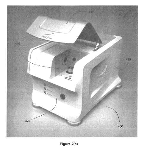
Referring now to Figures 2(a)-(c), an example implementation of an in-
vitro detection system 400 for optically scanning dental samples is shown,
where the example system has been configured for performing photothermal
radiation and luminescence measurements on a dental sample. In the present
example embodiment, the dental sample is mounted on a sample holder that
can be magnetically retained in a fixed position, removed from system 400,
.. and subsequently replaced without requiring calibration of the relative
position
between the dental sample holder and the incident optical beam, as described
further below.
In some embodiments, the dental sample may be, for example, whole
tooth samples, enamel sections, teeth containing composites, amalgam or
other filling materials, teeth covered with a dental sealant, and teeth from
19
CA 02895519 2015-06-18
WO 2014/094142
PCT/CA2013/050200
different non-human species.
System 400 includes main instrument body 410, front indicator and/or
control panel 420, door 430, and sample chamber 440. Door 430, when
closed, encloses sample chamber 440 in order to prevent external
background light from interfering with the measurement process. Door 440
also acts as a safety measure by preventing the incident optical beam from
propagating outside of the system. In some embodiments, an interlock
mechanism may be included, which turns off or blocks the output of the
internal laser whenever door 440 is opened.
As shown in Figure 2(c), sample chamber 440 includes a sample
holder receiving base 810 for magnetically and removably securing a dental
sample holder (described below) in a fixed position, focusing and collection
lens assembly 450 for delivering the incident optical beam and collecting
emitted photothermal radiation and luminescence, and optional imaging
camera 455 for obtaining images of the dental sample during analysis. In one
example implementation, imaging camera 455 obtains photographs depicting
the location of the scan, and may be a standard VGA camera (having a
resolution of 640 x 480 pixels), and may output the image in a format such as
JPEG.
Figure 3 provides an assembly diagram showing several components
of system 400. Chassis 500 mechanically supports the components of the
system and provides an external frame, to which side panels 502 and 504,
front panel 506, top panel 508, bottom panel 510, and rear panel 512 with fan
514, are assembled. Several internal optical components, including an optical
block (described below), detectors, a camera, and a laser, are shown
CA 02895519 2015-06-18
WO 2014/094142
PCT/CA2013/050200
supported by chassis 500 at 520. System 400 also includes infrared detector
power supply 550, USB hub 555, stepper motor driver 560, and data
acquisition board 565. The sample holder receiving base (not shown) is
supported by platform 530, which is translated and reoriented by position and
orientation control assembly 535. The sample chamber is defined by lower
and side wall portion 540, and back wall portion 545.
Figure 4 illustrates an example embodiment of position and orientation
control assembly 535 for achieving three-dimensional position and orientation
control of a sample holder. 605, 615, 610 and 625 are stepper motors for Z,
.. X, Focus, and rotational stages, respectively. 620 is a 2 axis
translational
stage.
In one example implementation, position and control assembly 535
may include a 360 rotational motorized stage (for example, with a 1.8 max.
resolution), with a spatial scan capability suitable for measurements across a
region of approximately 6 mm x 6 mm, optionally with a motorized sample
stage resolution of up to approximately 2 p.m, with an optional default
resolution of approximately 250 pm. In other embodiments, the resolution may
be larger, such as 250, 500, or 1000 pm.
Figures 5(a) and (b) and Figure 6 show an example implementation of
an optical block 750 for housing several components of the optical detection
system, for implementing the optical configuration shown in Figure 1(c).
Optical block 750, which may be formed of a resilient and thermally
conductive material such as aluminum, includes a plurality of channels and
recesses for housing one or more of the optical components of the optical
detection system. In the example embodiment shown in Figures 5(a) to (b)
21
CA 02895519 2015-06-18
WO 2014/094142
PCT/CA2013/050200
and Figure 6, particularly in Figure 5(b), optical block 750 includes a
primary
channel 755 for delivering the incident optical beam and the collected
photothermal radiation and luminescence.
Recess 775 is formed to house focusing and collection lens assembly
450, as shown in Figure 6. Recess 760 is provided to support a photodiode
assembly (not shown) that includes a photodiode and appropriate beam
sampling optics, such as a prism or mirror that intercepts a portion of the
collected luminescence beam. Recess 765 is provided to accommodate a
laser source, such as a semiconductor laser. Laser energy is deflected off of
a
dichroic or high-pass filter that is itself supported within slot 770. Recess
780
accommodates an infrared detector (or at least a portion thereof) for
detecting
the photothermal radiation that is transmitted through the dichroic or high-
pass filter. Accordingly, it is apparent that optical block 750 is an
implementation optical detection system 110 in which a plurality of the
optical
components are aligned and supported within a common optical bench, which
provides mechanical stability among the plurality of optical components.
Referring now to Figures 7(a)-(d), a series of images are show in which
dental sample holder 800 is placed onto base 810. In Figure 7(a), sample
chamber 440 is shown including base 810, which is connected (from below) to
position and orientation control assembly 535 (located beneath sample
chamber 440), such that the position and/or orientation of the dental sample
holder may be varied when dental the sample holder is installed on base 810.
Base 810 includes base magnet 815 for removably receiving and securing the
dental sample holder.
In Figure 7(b), a user is shown holding dental sample holder 800 in an
22
CA 02895519 2015-06-18
WO 2014/094142
PCT/CA2013/050200
inverted orientation, revealing sample holder magnet 805. Sample holder
magnet 805 and base magnet 815 are oriented such that when dental sample
holder 800 is contacted with base 810, sample holder is removably secured
and in a fixed and predetermined orientation. Although not shown in the
figure, base 810 and dental sample holder 800 may include one or more
keyed features that are defined and arranged so that dental sample holder
800 may only be received onto base 810 in a specific orientation. The non-
magnetic portions of dental sample holder 800 and base 810 may be formed
from aluminum, or another suitable resilient material, which is non-magnetic.
In other embodiments, base 810 and sample holder 800 may be formed from
materials that are substantially or entirely magnetic in nature (including,
but
not limited to, ferromagnetic and electromagnetic materials).
Figure 7(c) shows the user contacting dental sample holder 800 (which
includes dental sample 820) with the base, such that base magnet and
sample holder magnet apply a retaining force to secure dental sample holder
800 in place. Dental sample holder 800 is shown secured in place in Figure
7(d).
Figures 8(a)-8(d) and 9(a)-9(h) illustrate an example embodiment for
positioning a dental sample 820 on the dental sample holder, and for
accommodating various different sample heights. In this example
embodiment, the dental sample holder is composed of one or more platforms
(860, 870 and 880) that are magnetically connectable, and mountable to be
magnetically secured in base 810, in a manner as described above.
Figures 8(a)-8(d) also show mounting platform 850 for positioning a
dental sample at an appropriate height for scanning within the system.
23
CA 02895519 2015-06-18
WO 2014/094142
PCT/CA2013/050200
Sample platform 880, and one or more optional secondary platforms 860 and
870 (for example, having heights of 10 mm and 6.5 mm, respectively) may be
stacked and magnetically retained onto optional mounting platform 850 while
securing a dental sample to sample platform 880. Sample platform 880
includes a top surface for securing a dental sample (for example, using an
adhesive such as epoxy, or a retention mechanical mechanism). In the
embodiment shown, sample platform 880 also includes screw tap holes (e.g.
9mm apart), which may be employed to fix a resin molded sample. Example
lateral dimensions for sample platform 880 are 15 x 15 mm.
Magnetic retention between any two platforms is achieved by magnets
embedded within the platforms, such as magnets 855, 865, and 875 in
mounting platform and the two secondary platforms 860 and 870,
respectively, and magnets in the underside of the platforms (not shown in
Figures 8(a)-(d); alternatively, one or more magnets may extend through full
thickness of the platform, so that only one magnet is needed). Keyed features,
such as channels 866 and corresponding slots 867, may be included to
further secure the platforms during assembly, and to ensure that the platforms
can be attached in a selected configuration. It is to be understood that
although two secondary platforms (860 and 870) are shown in Figure 8, three
or more secondary platforms may be optionally employed.
As shown in Figures 9(a)-9(h), mounting platform 850 may include a
calibration marker or feature 895 that defined a reference location indicating
the location of the measurement region (the area or region that is to be
scanned and/or imaged by the system) relative to the bottom surface of the
sample holder when the sample holder is installed on the base. In the
24
CA 02895519 2015-06-18
WO 2014/094142
PCT/CA2013/050200
example implementation shown, marker 895 may be a block or other physical
marker having a front surface that approximately coincides with the focal
plane (i.e. the object plane) of the scanned area. For example, marker 895
may have dimensions of approximately 6mm x 6mm. In other embodiments,
marker 895 may be a portion of a surface, where the portion of the surface is
marked, for example, coloured, scribed, or otherwise modified, to show the
scanning area. In one embodiment, marker 895 includes an indication, such
as a cross hair or grid, that defines the center and the scanning area of the
measurement region. One or more additional markers (for example, lateral
markers on the sides of sample holder 800), may be included to indicate the
depth of field or Rayleigh range of the optical scanning beam.
Figures 9(a)-(d) show four different stacking combinations for selecting
a different height of the top surface of sample mounting platform 880 relative
to mounting platform 850, in order to position dental sample 820 at an
appropriate height relative to marker 895. In configuration (a), sample
mounting platform 880 is directly, and magnetically, attached to mounting
platform 850, while in configuration (d), both secondary platforms 860 and 870
are included in order to raise the height of sample platform 880 (as such, in
the present embodiment, sample platform 880 and the one or more optional
secondary platforms together constitute the sample holder).
Accordingly, when mounting a dental sample, sample mounting
platform 880, and, optionally, one or more secondary platforms (e.g. 860 and
870), are placed on mounting platform 850 in order to position dental sample
at an appropriate height. Dental sample 820 is then affixed to sample holder
800, where a selected portion of dental sample 820 is placed near to or in
CA 02895519 2015-06-18
WO 2014/094142
PCT/CA2013/050200
contact with marker 895 (as shown in Figures 9(e)-(h)), such that when
sample platform 880 (and the optional secondary platforms) is installed on
base 810, the selected portion of dental sample 820 is at substantially co-
incident, co-planar, or proximal with the scanning area. Sample platform 880,
and any optionally included secondary platforms, are then removed from
mounting platform 850 and installed onto the base 810 in sample chamber
440 (shown in Figure 7), in order to allow the incident optical beam to scan
the selected region of dental sample 820.
In one embodiment, as shown in Figures 9(a)-9(h), marker 895 and/or
a supporting structure 890 for marker 895 may be at least partially
transparent, in order to allow for the visualization of the selected portion
of
dental sample 820 when marker 895 is installed on dental sample holder 800.
In one example implementation, marker 895 and/or supporting structure 890
may be formed from transparent polycarbonate.
In some embodiments, the system may be employed for the non-
invasive and non-destructive longitudinal monitoring of a sample. In some
embodiments, the system may be employed for performing in-vitro diagnostic
measurements of dental samples. Advantageously, the system need not be
re-calibrated when the sample holder is removed and replaced. For example,
.. in some embodiments, the sample holder may be removed between
subsequent measurements in order to apply a treatment, therapy, and/or
induced degradation or pathology to the dental sample, and the system may
be employed to monitor the long term status of the dental sample. The lack of
a need to recalibrate the system (i.e. the relative position between the
dental
sample and the measurement region) between measurements avoids the
26
CA 02895519 2015-06-18
WO 2014/094142
PCT/CA2013/050200
need to calibrate the sample position for each measurement, a process that
can be both time consuming and error prone.
Example uses of the system include the measurement and/or
monitoring of ongoing demineralization and/or remineralization, white spot
lesions, brown spot lesions, longitudinal studies to assess the efficacy of
remineralization and/or demineralization agents, erosion studies, caries
around dental restorations, and the non-destructive imaging of in-situ treated
samples.
In one example implementation, the system may be employed to
monitor the effect of a treatment protocol on one or more carious lesions in a
dental sample, where successive measurements are made over time, in
between treatment steps. Due to the fixed calibration of the sample relative
to
the system, demineralization and remineralization studies may be performed
using the same sample, reducing the influence of inter-sample biological
variability.
In some embodiments, the system may be employed for the detection
and monitoring of small areas of decay, for example, as small as 50 microns
in size. The decay may be present, for example, on smooth enamel, or, as
erosive lesions caused by exposure to acidic liquids.
Embodiments of the present in-vitro dental detection system may be
useful in a wide range of applications due to the non-destructive nature of
the
analysis, the ability to obtain images with ability to specify measurement
resolution, the ability to monitor treatment efficacy in a sample as a
function of
time, and the ability to assess uniformity of investigated sample. In
embodiments that employ photothermal radiometry and luminescence
27
CA 02895519 2015-06-18
WO 2014/094142
PCT/CA2013/050200
detection, the system may be employed to compare to a sample to normal
healthy enamel or other mineralized tissue, and thereby assess the health of
the in-vitro sample and monitor ongoing changes.
The following examples are presented to enable those skilled in the art
to understand and to practice embodiments of the present disclosure. They
should not be considered as a limitation on the scope of the present
embodiments, but merely as being illustrative and representative thereof. In
the Examples below, the example implementation shown in Figures 2 to 9 is
henceforth referred to as the "Canary Lab System TM".
EXAMPLES
Example 1: Photothermal and Luminescence Detection of Dental Caries
In a photothermal radiation or photothermal radiation-luminescence
system, such as The Canary System'TM, a beam of energy (typically a laser)
intensity-modulated at a certain frequency is focused onto the sample surface.
The resulting periodic heat flow due to the absorbed optical energy in the
material is a diffusive process, producing a periodic temperature rise
(distribution) which is called a "thermal wave". This temperature distribution
in
turn causes a modulated thermal infrared (black-body or Planck radiation)
emission which is used to monitor the material under examination.
Photothermal radiation has the ability to penetrate, and yield information
about, an opaque medium well beyond the range of optical imaging.
Specifically, the frequency dependence of the penetration depth of thermal
waves makes it possible to perform depth profiling of materials.
In photothermal radiation applications involving turbid media, such as
hard dental tissue, depth information is obtained following optical-to-thermal
28
CA 02895519 2015-06-18
WO 2014/094142
PCT/CA2013/050200
energy conversion and transport of the incident laser power in two distinct
modes: conductively, from a near-surface distance controlled by the thermal
diffusivity of enamel (50-500 pm) [Brown WS, Dewey WA, Jacobs HR:
Thermal properties of teeth. J Dent Res 1970; 49: 752-754] and radiatively,
through blackbody emissions from considerably deeper regions
commensurate with the optical penetration of the diffusely scattered laser-
induced optical field (several mm). For example, deeper subsurface lesions
are possible by using a longer wavelength (830-nm) laser source than a 659-
nm probe [Jeon, Ft. J., Han, C., Mandelis, A., Sanchez, V., Abrams, S. H.,
.. "Non-intrusive, Non-contacting Frequency-Domain Photothermal Radiometry
and Luminescence Depth Profilometry of Carious and Artificial Sub-surface
Lesions in Human Teeth," Journal of Biomedical Optics 2004, July ¨ August
,9, # 4, 809 ¨ 819].
Photothermal radiation measurements of artificially induced caries on
human teeth have shown that the photothermal radiation amplitude increases
gradually with increasing demineralization time and decreases after
rem ineralization. The photothermal radiation phase also shows gradual and
consistent changes with demineralization and demineralization treatment.
This behaviour has been attributed to the higher scatter of the diffuse photon
field and to thermal-wave confinement in the form of standing waves in the
treated region, accompanied by decreased thermophysical properties
(thermal diffusivity and thermal conductivity).
Good correlation of photothermal radiation-luminescence results with
the mineral loss or the lesion depth measured with TMR results has indicated
that photothermal radiation-luminescence is capable of monitoring artificially
29
CA 02895519 2015-06-18
WO 2014/094142
PCT/CA2013/050200
created carious lesions, their evolution during demineralization, and the
reversal of the lesions under the growth of a remineralized surface layer
[Jeon
R. J., Hellen A., Matvienko A., Mandelis A., Abrams S. H., Amaechi B. T., In
vitro Detection and Quantification of Enamel and Root Caries Using Infrared
Photothermal Radiometry and Modulated Luminescence. Journal of
Biomedical Optics 13(3), 048803, 2008]. The photothermal radiation-
luminescence methodology for dental applications has been extensively
studied. Literature reports include applications in depth profiling, early
lesion
evaluation, caries detection in smooth, occlusal, root and interproximal
areas,
and theoretical modeling.
One of the main advantages of photothermal radiation-luminescence is
the ability to perform depth profiling through scanning of the excitation
source
modulation frequency. By selecting a fixed modulation frequency, radiometric
measurements at different depths in the enamel can be obtained. The first
attempt to apply the depth profilometric capability of photothermal radiation-
luminescence toward the inspection of dental defects was reported by
Mandelis et al.[ Jeon, R. J., Mandelis, A., Abrams, S. H., "Depth
profilometric
case studies in caries diagnostics of human teeth using modulated laser
radiometry and luminescence", Review of Scientific Instruments, 2003,
January, Volume 74 # 1, pages 380 ¨ 383]. In these studies a laser of 488 nm
was used as the excitation source. This work showed that the photothermal
radiometric signals were inversely correlated with the luminescence signals,
as a result of the nature of the two physical signal generation processes.
While the photothermal radiation amplitude increased for carious lesions the
luminescence amplitude decreased. The luminescence signal results were
CA 02895519 2015-06-18
WO 2014/094142
PCT/CA2013/050200
consistent with previous reports [R. Hibst et al.]. In addition, these studies
showed that the radiometric amplitude exhibited much superior dynamic (2
orders of magnitude signal resolution) range to luminescence (a factor of 2
only) in distinguishing between intact and cracked sub-surface structures in
.. the enamel. Furthermore, the radiometric signal (amplitude and phase)
produced dental images with much better defect localization, delineation, and
resolution than those obtained with modulated luminescence.
Further experimental studies [Jeon, R. J., Han, C., Mandelis, A.,
Sanchez, V., Abrams, S. H., "Non-intrusive, Non-contacting Frequency-
Domain Photothermal Radiometry and Luminescence Depth Profilometry of
Carious and Artificial Sub-surface Lesions in Human Teeth," Journal of
Biomedical Optics 2004, July ¨ August ,9, #4, 809 ¨ 819] used excitation
sources of 659 and 830 nm to assess the feasibility of photothermal radiation-
luminescence to detect deep lesions. Photothermal radiation frequency scans
over the surface of an occlusal fissure into demineralized enamel and dentin
showed higher amplitude than those for healthy teeth, as well as a
pronounced curvature in both the amplitude and phase signal channels.
These can be excellent markers for the diagnosis of subsurface carious
lesions. The results showed that photothermal radiation-luminescence is able
to detect artificial subsurface defects with sharp boundaries at depths
greater
than 5mm. In addition photothermal radiation exhibited superior sensitivity to
the presence of sharp boundaries, as well as to changes in natural
demineralized regions of the tooth. These results suggested the possibility to
detect carious lesions on both occlusal surfaces and the interproximal area of
.. the tooth [Jeon et al.].
31
CA 02895519 2015-06-18
WO 2014/094142
PCT/CA2013/050200
In experimental studies, it was found that photothermal radiation
Amplitude had a very strong correlation with lesion size and shape.
Luminescence phase provided limited information. Photothermal radiation
Phase provided an indication of operator movement if there was a strong shift
in the phase number from the norm. If this occurred, the operator was
instructed to re-measure the area.
In one embodiment, in which a single unified quantitative indication of
oral health is provided based on a measurement at a given location, the data
from each location is stored as four separate signals; photothermal radiation
amplitude and phase and luminescence amplitude and phase. A unified
measure is obtained according to the following weighting formula:
= photothermal radiation Amplitude weighted at 45% of the total value
= photothermal radiation Phase weighted at 15% of the total value
= luminescence Phase weighted at 10% of the total value
= luminescence Amplitude weighted at 30% of the total value
The four readings are compared to the readings one finds from the
healthy enamel surface and/or from a standardized piece of hydroxyapatite.
The measured signal number is compared to healthy enamel surface as well.
Preferably, results from the comparison step are provided on a fixed scale for
each reading, for example, on a scale of 1 to 100 (the scales need not be
equal for each reading type), indicating a severity of a condition. The four
fixed-scale results are then weighted as described above, providing the
operator a ranking or range (for example, on a scale from 1 ¨ 100) indicating
the health of the area examined. The utility of multiple readings in
diagnostic
assessment with a photothermal radiation and luminescence detection device
32
CA 02895519 2015-06-18
WO 2014/094142
PCT/CA2013/050200
was illustrated in Jeon [Jeon et al., "Diagnosis of Pit and Fissure Caries
Using
Frequency-Domain Infrared Photothermal Radiometry and Modulated Laser
Luminescence", Caries. Res. 38, 497-513, 2004].
In another embodiment, the reading from a single frequency is
combined in the following manner: (photothermal radiation amplitude x
photothermal radiation Phase) / (luminescence Amplitude x luminescence
Phase) to create one single reading. This metric is henceforth referred to as
the "Canary Number". Error checking may be performed by combining the
standard deviation from each reading into one number as follows:
Luminescence Amplitude x Luminescence Phase x Photothermal Radiation
Amplitude x Photothermal Radiation Phase.
The ratio of single reading / combined standard deviation is examined
and if the ratio increases dramatically this indicates an error in the reading
and this is conveyed to the operator. The single reading is then conveyed to
the operator along with its difference from the single reading derived from
examining health enamel and healthy teeth.
Example 2: Imaging of Various Dental Samples
Figure 10(a) depicts a 6 mm x 6 mm digital camera image (Left) and
corresponding Canary image (right) of the same tooth sample exhibiting
sound enamel. Both images were generated by The Canary Lab System.
A Canary image is taken within the square on the camera image, called
the region-of-interest. Each pixel represents a Canary Lab scan measurement
using PTR-LUM. Measurements were performed every 250 pm on the sample
to generate Canary Numbers for each measured spot. The result is a
33
CA 02895519 2015-06-18
WO 2014/094142
PCT/CA2013/050200
composite image, The Canary Image. Corresponding legend with a scale is
provided. The lower Canary Number indicates a sound tooth surface.
As shown in the Figures, the Canary Lab System image produces
excellent contrast between the measured white spot lesion and the
.. surrounding bulk sound enamel. The dark pixels in the Canary image within
the white spot lesion represent the deepest areas of the incipient lesion.
As a feature of The Canary Lab System, the user can improve the
spatial resolution of The Canary Image by performing measurements at
smaller increments (i.e. measurements every 200 pm).
Raw Canary Images that are produced following a measurement can
be edited using The Canary Software ¨ Image Enhancement Tool, in order to
enhance the contrast of the measured region of interest, as noted above. This
produces the 'Canary Lab Image' shown in Figure 10(b). The contrast
enhanced Canary Lab Image identifies the most advanced (darkest) areas of
.. the lesion from the more incipient (light) area.
Figure 10(c) shows the amplitude and phase components that were
employed to form the images shown in Figures 10(a) and 10(b). The first
column of images shows camera images of the sample. The second column
of images shows the Canary Image, and the third column of images shows
the contrast-enhanced Canary Image. The four images in the first column are
identical, while the images in the second and third columns are as follows:
the
first row shows the PTR amplitude, the second row shows the PTR phase, the
third row shows the LUM amplitude, while the fourth row shows the LUM
phase.
In another experiment, the Canary Lab System was employed for the
34
CA 02895519 2015-06-18
WO 2014/094142
PCT/CA2013/050200
imaging of a tooth exhibiting an incipient white spot. A natural incipient
caries
lesion was selected on an extracted tooth and mounted in the sample
chamber for scanning with The Canary Lab System. Images are shown in
Figures 11(a) and 11(b), where Figure 11(a) shows the Canary Image, while
Figure 11(b) shows the amplitude and phase components, in the same
manner as described in Figure 10(c). Measurements of two other dental
samples exhibiting white spots and advanced white spots are shown in Figure
12 and in Figures 13(a) and 13(b), respectively. Figure 13(a) shows the
Canary Image, while Figure 13(b) shows the amplitude and phase
components, in the same manner as described in Figure 10(c).
The Canary Lab System was also employed for the imaging of a tooth
exhibiting a brown spot. Images are shown in Figures 14(a) and 14(b). Figure
14(a) shows the Canary Image, while Figure 14(b) shows the amplitude and
phase components, in the same manner as described in Figure 10(c). A
second dental sample exhibiting a brown spot was also imaged, as shown in
Figures 15(a) and 15(b). Identification of the 'hot spots' or deepest, most
advanced areas of the lesion can clearly be identified in both the raw Canary
image and the Canary Lab Image. Figure 15(a) shows the Canary Image,
while Figure 15(b) shows the amplitude and phase components, in the same
manner as described in Figure 10(c).
Figures 16(a) and (b) show images generated from Canary Lab
System when imaging a tooth having an amalgam restoration. Figure 16(a)
shows the Canary Image, while Figure 16(b) shows the amplitude and phase
components, in the same manner as described in Figure 10(c).
Example 3: Sequential Etching Experiment
CA 02895519 2015-06-18
WO 2014/094142
PCT/CA2013/050200
A sound smooth surface of an extracted tooth was selected and
polished flat to remove outer enamel. The surface of interest was measured
and imaged in the Canary Lab System. Subsequently, 37% phosphoric acid
was used to etch a region of interest in the centre of the imaged area. The
etched circle can be seen in the outlined images below. The enamel surface
was etched for 5, 10 and 30 seconds with Canary Lab measurements
performed after each individual etch.
Referring now to Figures 17(a)-(e), the microporosities generated
following the 5 and 10 second etch is reflected in the Canary Lab image by
the fact that the lower Canary Numbers (light grey) are replaced with higher
Canary Numbers (dark grey). Following the 30 second etch the delimited
etched circle can clearly be visualized with the higher Canary Numbers
(darker grey). These trends are enhanced with the 60 second etch. This
expected behaviour occurs as the microporosities of the etched surface
confine the converted thermal energy to the defect region and as a result,
emits a greater photothermal response. This occurs with a concomitant
reduction in the luminescence response as the etched white surface is highly
scattering of both the incident and converted light.
The specific embodiments described above have been shown by way
of example, and it should be understood that these embodiments may be
susceptible to various modifications and alternative forms. It should be
further
understood that the claims are not intended to be limited to the particular
forms disclosed, but rather to cover all modifications, equivalents, and
alternatives falling within the spirit and scope of this disclosure.
36