Note: Descriptions are shown in the official language in which they were submitted.
CA 02895761 2015-06-18
WO 2014/099778
PCT/US2013/075384
PRESSURE-SENSING INTRAVASCULAR DEVICES,
SYSTEMS, AND METHODS
TECHNICAL FIELD
The present disclosure relates generally to intravascular devices, systems,
and
methods. In some embodiments, the intravascular devices are guide wires that
include one or
more electrical, electronic, optical, or electro-optical sensors positioned at
a distal end.
BACKGROUND
Heart disease is a critical healthcare issue for the individual patient and
for society as
a whole. Recent research has shown that treatment of heart disease, when
guided by
improved diagnostic methods such as functional assessment of the coronary
circulation using
intravascular pressure measurements, leads to both improved quality of life
for the patient
and reduced healthcare costs for society.
Intravascular catheters and guide wires are commonly utilized to measure the
pressure
within the blood vessel, to visualize the inner lumen of the blood vessel,
and/or to otherwise
obtain diagnostic information related to the blood vessel. To date, guide
wires containing
pressure sensors, imaging elements, and/or other electrical, electronic,
optical, or electro-
optical components have suffered from poor mechanical performance in
comparison to
standard guide wires that do not include such components. Existing pressure-
sensing guide
wires typically incorporate a single pressure sensor located approximately 3
cm from the
distal tip of the guide wire. Since the sensor is fixed in position on the
guide wire, the
pressure can only be measured at different locations within the vasculature by
advancing or
retracting the entire guide wire to position the sensor at the desired
location. Traditionally,
the pressure-sensing guide wire includes a sensor formed on a planar substrate
and having
terminals attached to the conductors of a cable which runs through the
intravascular device.
The sensor substrate is typically oriented such that the pressure sensitive
portion faces
radially outward into the blood stream. It is generally desired to separate
the substrate
slightly away from the walls of the intravascular device in order to
mechanically isolate the
pressure sensor substrate from the guide wire structure, so that bending and
torsional stresses
are not coupled to the sensor substrate where they could adversely affect the
pressure
measurement accuracy. This pressure sensing guide wire geometry provides
access for
-1-
CA 02895761 2015-06-18
WO 2014/099778
PCT/US2013/075384
intravascular pressure measurement, but results in compromises to the
mechanical structure
which lead to poor mechanical performance compared to that of a conventional
guide wire
without measurement capability. Furthermore, the fragile electrical
interconnects between
the sensor terminals and the electrical leads are vulnerable to failure. In
this conventional
configuration the small diameter of the intravascular device introduces places
constraints on
the sensor dimensions, exacerbating the limitations and associated problems.
Accordingly, there remains a need for improved intravascular devices, systems,
and
methods that preserve the desirable mechanical properties of the device while
providing a
more robust interconnect to one or more electrical, electronic, optical, or
electro-optical
components.
-2-
CA 02895761 2015-06-18
WO 2014/099778
PCT/US2013/075384
SUMMARY
According to embodiments disclosed herein an intravascular sensor assembly may
include a flexible elongate member having a longitudinal axis (LA); a core
member disposed
inside the flexible elongate member; and an elongated substrate disposed
distal to the core
member and inside the flexible elongate member, the elongated substrate
including at least
one electrode disposed within at least one recess in an outer surface of the
elongated
substrate, the at least one recess extending in a longitudinal direction; and
a sensor circuit
disposed on a distal surface of the elongated substrate, the sensor circuit
coupled to the at
least one electrode.
In some instances, a pressure-sensing guide wire is provided. The pressure-
sensing
guide wire includes a pressure sensor mounted such that a membrane of the
pressure sensor
extends across a width of the guide wire, instead of along the length of the
guide wire. As a
result of mounting the pressure sensor in this orientation, the thickness,
robustness, and
durability of the pressure sensor can be increased while staying within the
limited space
provided by the outer profile of the guide wire.
According to embodiments disclosed herein a sensor structure for use in an
intravascular device assembly may include a substrate having an elongated
shape with a
length defined along a longitudinal axis (LA) and a width extending
perpendicular to the
longitudinal axis, the shape further including a proximal surface and an
opposing distal
surface, each extending substantially perpendicular to the LA; and an outer
surface extending
substantially parallel to the LA between the proximal and distal surfaces; at
least one
electrode disposed longitudinally within at least one recess in the outer
surface of the
substrate, and a sensor circuit disposed on the distal surface, the sensor
circuit having at least
one lead or conductor coupled to the at least one electrode.
A system for performing measurements using a sensor exposed to an
intravascular
environment, the system including an intravascular device having: a flexible
elongate
member having a longitudinal axis (LA); a core member disposed inside the
flexible elongate
member; and an elongated substrate disposed distal to the core member and
inside the
flexible elongate member, the elongated substrate including at least one
electrode disposed
within at least one recess in an outer surface of the elongated substrate, the
at least one recess
extending in a longitudinal direction; and a sensor circuit disposed on a
distal surface of the
-3-
CA 02895761 2015-06-18
WO 2014/099778
PCT/US2013/075384
elongated substrate, the sensor circuit coupled to the at least one electrode;
and a control
console coupled to the intravascular device.
According to embodiments disclosed herein a method of forming a pressure-
sensing
guide wire may include forming an elongated substrate; forming a plurality of
recesses in an
outer surface of the elongated substrate; filling at least a portion of each
of the recesses with a
conductive material to form a plurality of electrodes; fabricating a sensor
circuit on a front
surface of the elongated substrate, the front surface extending perpendicular
to a longitudinal
axis of the elongated substrate; electrically coupling the plurality of
electrode to terminals of
the sensor circuit; electrically coupling a plurality of conductors of a
communication cable to
the plurality of electrodes; and securing the elongated substrate to a distal
portion of a
flexible elongate member.
These and other embodiments of the present invention will be described in
further
detail below with reference to the following drawings.
-4-
CA 02895761 2015-06-18
WO 2014/099778
PCT/US2013/075384
BRIEF DESCRIPTION OF THE DRAWINGS
FIG.1 is a diagrammatic, schematic side view of an intravascular device
according to
some embodiments.
FIG. 2 is a diagrammatic perspective view of a sensor structure according to
some
embodiments.
FIG. 3 is a diagrammatic partial cross-sectional front view of a sensor
structure
according to some embodiments.
FIG. 4 shows a partial perspective view of a distal portion in an
intravascular device
according to some embodiments.
FIG. 5 shows a partial perspective view of a coupling for an end sensor in a
sensor
structure according to some embodiments.
FIG. 6 shows a partial schematic view of a system for performing measurements
using an end sensor exposed to an intravascular environment according to some
embodiments.
FIG. 7 shows a flow chart for a method of manufacturing an intravascular
device
having an end sensor, according to some embodiments.
FIG. 8 shows a flow chart for a method of manufacturing an intravascular
device
having an end sensor, according to some embodiments.
FIG. 9 shows a flow chart for a method of obtaining a measurement of an
intravascular environment, according to some embodiments.
In the figures, elements having the same reference number have the same or
similar
functions.
-5-
CA 02895761 2015-06-18
WO 2014/099778
PCT/US2013/075384
DETAILED DESCRIPTION
For the purposes of promoting an understanding of the principles of the
present
disclosure, reference will now be made to the embodiments illustrated in the
drawings, and
specific language will be used to describe the same. It is nevertheless
understood that no
limitation to the scope of the disclosure is intended. Any alterations and
further
modifications to the described devices, systems, and methods, and any further
application of
the principles of the present disclosure are fully contemplated and included
within the present
disclosure as would normally occur to one skilled in the art to which the
disclosure relates. In
particular, it is fully contemplated that the features, components, and/or
steps described with
respect to one embodiment may be combined with the features, components,
and/or steps
described with respect to other embodiments of the present disclosure. For the
sake of
brevity, however, the numerous iterations of these combinations will not be
described
separately.
As used herein, "flexible elongate member" or "elongate flexible member"
includes at
least any thin, long, flexible structure that can be inserted into the
vasculature of a patient.
While each of the illustrated embodiments of the present disclosure includes a
flexible
elongate member having a cylindrical form with a circular cross-sectional
profile that defines
an outer diameter of the flexible elongate member, in other instances all or a
portion of the
flexible elongate member may have other geometric cross-sectional profiles
(e.g., oval,
rectangular, square, elliptical, etc.) or non-geometric cross-sectional
profiles. Flexible
elongate members include, for example, guide wires and catheters. In that
regard, a catheter
may or may not include a lumen extending along its length for receiving and/or
guiding other
instruments. If the catheter includes a lumen, the lumen may be centered or
offset with
respect to the cross-sectional profile of the device.
In most embodiments of the present disclosure, the flexible elongate member
includes
one or more electrical, electronic, optical, or electro-optical components.
For example,
without limitation, a flexible elongate member may include one or more of the
following
types of components: a pressure sensor, a temperature sensor, an imaging
element, an optical
fiber, an ultrasound transducer, a reflector, a mirror, a prism, an ablation
element, an RF
electrode, a conductor, and/or combinations thereof. Generally, these
components are
configured to obtain data from or deliver therapy to a vessel or other portion
of the anatomy
-6-
CA 02895761 2015-06-18
WO 2014/099778
PCT/US2013/075384
in which the flexible elongate member is disposed. Often the components are
also configured
to communicate with an external device for processing, display, activation,
and/or control. In
some aspects, embodiments of the present disclosure include imaging devices
for imaging
within the lumen of a vessel, including both medical and non-medical
applications.
However, some embodiments of the present disclosure are particularly suited
for use in the
context of human vasculature. Imaging of the intravascular space, particularly
the interior
walls of human vasculature can be accomplished by a number of different
techniques,
including ultrasound (often referred to as intravascular ultrasound ("IVUS")
and intracardiac
echocardiography ("ICE")) and optical coherence tomography ("OCT"). In other
instances,
infrared, thermal, or other imaging modalities are utilized.
The electrical, electronic, optical, and/or electro-optical components of the
present
disclosure are often disposed within a distal portion of the flexible elongate
member. As used
herein, "distal portion" of the flexible elongate member includes any portion
of the flexible
elongate member from the mid-point to the distal tip. As flexible elongate
members can be
solid, some embodiments of the present disclosure will include a housing
portion at the distal
portion for receiving the electrical or electronic components. Such housing
portions can be
tubular structures attached to the distal portion of the elongate member. Some
flexible
elongate members are tubular and have one or more lumens in which the
electrical or
electronic components can be positioned within the distal portion. In some
embodiments, the
distal portion does not include a separate housing for mounting the
electrical, electronic,
optical, and/or electro-optical component(s). In such instances, the distal
portion may have
an outer diameter equal to the outer diameter of the flexible elongate member.
In some
instances, the distal portion is coupled to proximal and distal flexible
elements (e.g., coils,
flexible tubing, etc.). Accordingly, in some implementations the distal
portion includes a
step-down outer diameter at each end such that the reduced outer diameter is
slightly smaller
than the inner diameter of the proximal and distal flexible elements. In
other
implementations, the distal portion has a uniform outer diameter that is
slightly smaller than
the inner diameter of the distal and proximal flexible elements.
The electrical, electronic, optical, and/or electro-optical components and the
associated communication lines are sized and shaped to allow for the diameter
of the flexible
elongate member to be very small. For example, the outside diameter of the
elongate
member, such as a guide wire or catheter, containing one or more electrical,
electronic,
-7-
CA 02895761 2015-06-18
WO 2014/099778
PCT/US2013/075384
optical, and/or electro-optical components as described herein are between
about 0.007"
(0.178 mm) and about 0.118" (3.0 mm), with some particular embodiments having
outer
diameters of approximately 0.014" (0.356 mm) and approximately 0.018" (0.457
mm). In
some embodiments, the outside diameter of the elongate member may have an OD
of 0.035"
(0.89 mm). As such, the flexible elongate members incorporating the
electrical, electronic,
optical, and/or electro-optical component(s) of the present application are
suitable for use in a
wide variety of lumens within a human patient besides those that are part or
immediately
surround the heart, including veins and arteries of the extremities, renal
arteries, blood vessels
in and around the brain, and other lumens.
"Connected" and variations thereof as used herein includes direct connections,
such
as being glued or otherwise fastened directly to, on, within, etc. another
element, as well as
indirect connections where one or more elements are disposed between the
connected
elements.
"Secured" and variations thereof as used herein includes methods by which an
member is directly secured to another element, such as being glued or
otherwise fastened
directly to, on, within, etc. another element, as well as indirect techniques
of securing two
elements together where one or more elements are disposed between the secured
elements.
Sensors used in embodiments consistent with the present disclosure may be
positioned within an intravascular device facing an axial direction. In that
regard, some
embodiments disclosed herein may generally resemble embodiments disclosed in
detail in US
Pat. Appl. No. 11/864,499 entitled "Intravascular Pressure Devices
Incorporating Sensors
Manufactured Using Deep Reactive Ion Etching," by Paul Douglas Corl, filed on
September
28, 2007, the contents of which are hereby incorporated by reference in their
entirety, for all
purposes. Furthermore, embodiments consistent with the present disclosure
provide a robust
mounting structure to a pressure sensing circuit facing an axial direction.
Thus relaxing the
need for a cantilevered sensor decoupled from external stresses induced by
guidewire
structures. Embodiments as disclosed herein may include sensor circuits formed
on a thick
wafer substrate that is then disposed on the robust mounting structure.
Referring now to Fig. 1, shown therein is a portion of an intravascular device
100
according to an embodiment of the present disclosure. In that regard, the
intravascular device
-8-
CA 02895761 2015-06-18
WO 2014/099778
PCT/US2013/075384
100 includes a flexible elongate member 102 having a distal portion 104
adjacent a distal end
105 and a proximal portion 106 adjacent a proximal end 107. A component 108 is
positioned
within the distal portion 104 of the flexible elongate member 102 proximal of
the distal tip
105. Generally, the component 108 is representative of one or more electrical,
electronic,
optical, or electro-optical components. In that regard, the component 108 is a
pressure
sensor, a temperature sensor, a flow or velocity sensor, an ASIC, a signal
conditioning
circuit, an RF communication module, a memory module, an imaging element, an
optical
fiber, an ultrasound transducer, a reflector, a mirror, a prism, an ablation
element, an RF
electrode, a conductor, and/or combinations thereof. The specific type of
component or
combination of components can be selected based on an intended use of the
intravascular
device. In some instances, the component 108 is positioned less than 10 cm,
less than 5, or
less than 3 cm from the distal tip 105. In some instances, the component 108
is positioned
immediately adjacent to the distal tip 105, and in such case, the distal tip
may consist of just a
thin coating or may be altogether absent. In some instances, the component 108
is positioned
within a housing of the flexible elongate member 102. In that regard, the
housing is a
separate component secured to the flexible elongate member 102 in some
instances. In other
instances, the component 108 is integrally formed as a part of the flexible
elongate member
102.
The intravascular device 100 also includes a connector 110 adjacent the
proximal
portion 106 of the device. In that regard, the connector 110 is spaced from
the proximal end
107 of the flexible elongate member 102 by a distance 112. Generally, the
distance 112 is
between 0% and 50% of the total length of the flexible elongate member 102.
While the total
length of the flexible elongate member can be any length, in some embodiments
the total
length is between about 90 cm and about 400cm, with some specific embodiments
having
lengths of 140cm, 190cm, or 300cm. Accordingly, in some instances the
connector 110 is
positioned at the proximal end 107. In other instances, the connector 110 is
spaced from the
proximal end 107. For example, in some instances the connector 110 is spaced
from the
proximal end 107 between about 0 cm and about 140cm. In some specific
embodiments, the
connector 110 is spaced from the proximal end by a distance of 0 cm, 30cm, or
140cm.
The connector 110 is configured to facilitate communication between the
intravascular device 100 and another device. More specifically, in some
embodiments the
connector 110 is configured to facilitate communication of data obtained by
the component
-9-
CA 02895761 2015-06-18
WO 2014/099778
PCT/US2013/075384
108 to another device, such as a computing device or processor. Accordingly,
in some
embodiments the connector 110 is an electrical connector. In such instances,
the connector
110 provides an electrical connection to one or more electrical conductors
that extend along
the length of the flexible elongate member 102 and are electrically coupled to
the component
108. Some specific embodiments of electrical conductors in accordance with the
present
disclosure are discussed below in the context of Figs. 5-11. In other
embodiments, the
connector 110 is an optical connector. In such instances, the connector 110
provides an
optical connection to one or more optical communication pathways (e.g., fiber
optic cable)
that extend along the length of the flexible elongate member 102 and are
optically coupled to
the component 108. Further, in some embodiments the connector 110 provides
both
electrical and optical connections to both electrical conductor(s) and optical
communication
pathway(s) coupled to the component 108. In that regard, it should again be
noted that
component 108 is comprised of a plurality of elements in some instances. In
some instances,
the connector 110 is configured to provide a physical connection to another
device, either
directly or indirectly. In other instances, the connector 110 is configured to
facilitate wireless
communication between the intravascular device 100 and another device.
Generally, any
current or future developed wireless protocol(s) may be utilized. In yet other
instances, the
connector 110 facilitates both physical and wireless connection to another
device.
As noted above, in some instances the connector 110 provides a connection
between
the component 108 of the intravascular device 100 and an external device.
Accordingly, in
some embodiments one or more electrical conductors, one or more optical
pathways, and/or
combinations thereof extend along the length of the flexible elongate member
102 between
the connector 110 and the component 108 to facilitate communication between
the connector
110 and the component 108. Generally, any number of electrical conductors,
optical
pathways, and/or combinations thereof can extend along the length of the
flexible elongate
member 102 between the connector 110 and the component 108. In some instances,
between
one and ten electrical conductors and/or optical pathways extend along the
length of the
flexible elongate member 102 between the connector 110 and the component 108.
For the
sake of clarity and simplicity, the embodiments of the present disclosure
described below
include three electrical conductors. However, it is understood that the total
number of
communication pathways and/or the number of electrical conductors and/or
optical pathways
is different in other embodiments. More specifically, the number of
communication
pathways and the number of electrical conductors and optical pathways
extending along the
40-
CA 02895761 2015-06-18
WO 2014/099778
PCT/US2013/075384
length of the flexible elongate member 102 is determined by the desired
functionality of the
component 108 and the corresponding elements that define component 108 to
provide such
functionality.
Embodiments consistent with the present disclosure may provide the ability to
extend
or retract a sensor to multiple locations along the length of the
intravascular device ¨ or to
expose a fixed sensor to pressures from axially disparate locations by
extending or retracting
a "snorkel". For example, in some implementations the sensor may be secured to
a central
core that is mechanically translatable relative to a surrounding elongate
member. In
embodiments where the sensor is a pressure sensor, blood pressure along the
vessel may be
mapped without moving the distal tip position of the intravascular device. The
distal tip
position may remain fixed with the exterior elongated member 130 while the
sensor is pulled
back with core member 135. Furthermore, an engagement feature of the sensor
structure 108
or other associated component may enable torque and rotation of the tip of the
wire, if
desired. Such embodiments having a pullback capability may be as disclosed in
U.S.
Provisional Patent Application No. 61/746,537 entitled "Pressure Guide Wire
with Sliding
Pressure Sensor," filed December 27, 2012, the contents of which are herein
incorporated by
reference in their entirety, for all purposes. Further, some embodiments
include features of
the devices disclosed in U.S. Provisional Patent Application No. 61/747,958
entitled
"Intravascular Devices Having Artificial Muscles and Associated Systems, and
Methods,"
filed December 31, 2012, the contents of which are herein incorporated by
reference in their
entirety, for all purposes.
FIG. 2 is a diagrammatic perspective view of sensor structure 108 for an end
sensor
220 according to some embodiments. Sensor structure 108 includes a substrate
210 having a
substantially cylindrical shape in the illustrated embodiment. Substrate 210
includes a distal
surface 211 and a proximal surface 212, a diameter D 215 and a length L 216.
Accordingly,
L 216 may be as thin as 0.001 mm, or 1, 2, 3, 5 mm, or even longer, with some
embodiments
being about 0.1 mm, and other embodiments being about 0.5 mm. In some
embodiments it is
desirable to have a shorter L 216 to reduce impact on a bending stiffness.
Embodiments
using longer L 216 may include a robust protection to avoid bending. Bending
is not
desirable as it may break the coupling to the sensor or the sensor itself,
with potential loss of
signal. Accordingly, it is desirable to have an aspect ratio defined as length
over diameter of
less than approximately 2. For example, in an embodiment of a 0.014" (0.356
mm) diameter
-11-
CA 02895761 2015-06-18
WO 2014/099778
PCT/US2013/075384
guide wire, where the component 108 may have a diameter of approximately
0.010" (0.25
mm), length L 216 may be as short as 0.020" (0.50 mm), or even less. Diameter
D 215 may
have a reduced dimension in order for sensor structure 108 to fit within
intravascular device
100. In some implementations, the sensor structure 108 has a diameter D 215
that is sized to
fit within a housing. For example, in some implementations the sensor
structure is disposed
within a housing having an opening through a sidewall to expose the sensor
structure to
ambient. In some particular embodiments, the housing containing the sensor
structure is
positioned between two flexible members (e.g., coils, polymer tubes, coil-
embedded polymer
tubes, and/or combinations thereof). The sensor structure 108 may be secured
to the housing
using any suitable techniques, including adhesive. For example, in some
instances the sensor
structure 108 is mounted lengthwise within a housing similar to that described
in U.S. Patent
No. 7,967,762 entitled "Ultra Miniature Pressure Sensor," the contents of
which are herein
incorporated by reference in their entirety, for all purposes. Accordingly, in
some
embodiments diameter D 215 may be 2 mm, 1 mm, 500 gm, or less. For example,
for guide
wires having an OD of about .0145" (0.37 mm), D 215 may be smaller than about
.0115"
(0.29 mm). For guide wires having an OD of about .018" (0.46 mm), D 215 may be
as large
as .0145" (0.37 mm). And for guide wires having an OD of about .035" (0.89
mm), D 215
may be as large as .030" (0.76 mm). This technology is particularly suitable
for the severely
space constrained geometries of smaller guide wires.
Substrate 210 may be made of silicon or any other material used in a
semiconductor
foundry, such as germanium, silica, quartz, glass, sapphire, or any ceramic
material.
Substrate 210 includes electrodes 230-1, 230-2, and 230-3 (collectively
referred to hereinafter
as electrodes 230). In some embodiments electrodes 230 include conductors
formed of gold,
silver, copper, aluminum, or any other conducting material. In some
embodiments, the end
sensor 220 includes a flexible membrane positioned over a cavity such that the
flexible
membrane seals the cavity. The applied pressure causes the membrane to deflect
into the
cavity in varying amounts. In some instances, the membrane is embedded with
conductive
materials that are patterned to form a piezoresistive, capacitive, nanowire,
nanofiber, and/or
other suitable pressure transducing circuit elements. Accordingly, the
pressure applied to the
membrane causes the membrane to flex, which causes the embedded circuit to
change
resistance, capacitance, and/or other measurable characteristic that can be
correlated to the
applied pressured. The membrane may have a square, rectangular, circular,
elliptical, other
geometrical, and/or non-geometrical shape.
-12-
CA 02895761 2015-06-18
WO 2014/099778
PCT/US2013/075384
End sensor 220 is coupled to electrodes 230 by conductors 235-1, 235-2, and
235-3
(collectively referred to hereinafter as conductors 235). Conductors 235 may
be electrically
conductive wires, conductive traces, or doped semiconductor materials.
Electrodes 230 may
be formed within vias etched through a silicon substrate (e.g., cylindrical
substrate 210) using
semiconductor manufacturing techniques. In other embodiments, the electrodes
230 are
formed in recesses formed in an outer surface of the silicon substrate.
Accordingly, electrodes
230 are disposed longitudinally, either through the substrate 210 or along a
surface of the
substrate 210, in a direction that is parallel to the LA.
In some instances, each electrode 230 has a proximal end adjacent to or at
proximal
surface 212 and an opposing distal end adjacent to or at distal surface 211.
In other instances,
the proximal end of each electrode is spaced distally from the proximal
surface 212. In that
regard, by keeping space within the through vias and/or the recesses in the
outer surface of
the substrate 210, conductors that are to be electrically coupled to the
electrodes 230 can be at
least partially positioned within the through vias and/or recesses where they
are electrically
coupled to the electrodes. For example, in some instances distal sections of
the conductors
are positioned within the through vias and/or recesses such that distal ends
of the conductors
are positioned adjacent to and/or in contact with proximal ends of the
electrodes. Then
solder, welding, and/or other suitable conductive coupling mechanism is
utilized to secure
and electrically couple the conductors to the electrodes.
End sensor 220 is disposed on distal surface 211, facing outwards, in the
distal
direction. Embodiments consistent with this configuration reduce the
constraint for having a
thin sensor layer in a cantilevered configuration. Further, the mechanical
robustness of
substrate 210 relieves sensor 220 from stress in the core wire and/or other
portions of the
guide wire. In addition, since distal surface 211 is aligned in a direction
substantially parallel
to a torque rotating core member 135 about the LA, sensor 220 is decoupled
from stresses
arising from torsional effects. Such configuration reduces design concerns
about the
fabrication process of sensor circuit 220, relaxing geometrical and mechanical
constraints.
In some embodiments, sensor 220 includes circuits and structures such as a
micro-
electromechanical system (MEMS), formed on a wafer. In some embodiments, end
sensor
220 may be formed using semiconductor manufacturing techniques such as
etching,
-13-
CA 02895761 2015-06-18
WO 2014/099778
PCT/US2013/075384
deposition, and implantation of conductive layers on a substrate. When sensor
220 faces the
distal direction, sensor 220 may have a thickness that reduces limitations to
the placement of
intravascular device 100 within a blood vessel. Thus, the wafer used for
making sensor 220
may be a thin wafer(approximately 50 to 100 jam), an ultra-thin wafer (less
than 50 iLtm and
as thin as 1 gm), or a wafer of regular thickness (typically 300 to 700 gm).
For example, in
some embodiments the entire component 108 including sensor 220 may be formed
on a 400
to 600 gm thick wafer which also provides substrate 210, and include
electrodes 230 formed
in vias through the wafer. In this context, the generally cylindrical cross-
sectional profile of
the component 108 is produced by an etching process such as deep reactive ion
etching. In
the case where the cross-sectional profile intersects one or more of the
through wafer vias,
those vias become recesses in the surface of the substrate 210. Accordingly,
sensor 220 may
include a circuit and/or structure such as a MEMS manufactured using a Deep
Reactive Ion
Etching (DRIE) technique, as disclosed in detail in U.S. Patent Application
No. 11/864,499,
entitled "Intravascular Pressure Devices Incorporating Sensors Manufactured
Using Deep
Reactive Ion Etching," filed September 28, 2007, the contents of which are
incorporated
herein by reference in their entirety, for all purposes.
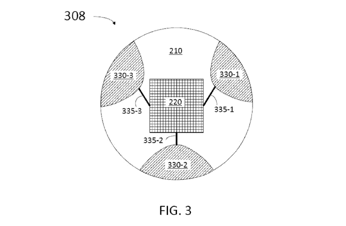
FIG. 3 is a diagrammatic end view of a sensor structure 308 for end sensor 220
according to some embodiments. Accordingly, sensor structure 308 includes
substrate 210
having a substantially cylindrical shape. Also, sensor structure 308 includes
electrodes 330-
1, 330-2, and 330-3 (collectively referred hereinafter as electrodes 330).
Electrodes 330 are
coupled to end sensor 220 through conductors 335-1, 335-2, and 335-3,
respectively
(hereinafter referred to as conductors 335). Conductors 335 may be as
conductors 235
described in detail above (cf. FIG. 2). Accordingly, electrodes 330 may be
formed as
indentions on substrate 210 with a conductive material deposited to fill in
the structure,
forming an approximately cylindrical shape. Furthermore, electrodes 330 may
extend from a
distal surface of sensor structure 308 (the surface including sensor 220) to a
proximal surface
of sensor structure 308, opposite the distal surface. In some implementations,
the sensor
structure 308 is coated with an insulating material after formation of the
electrodes 330
and/or electrically coupling of the electrodes 330 to conductors of a
communication cable.
FIG. 4 shows a partial perspective view of a distal portion 406 of an
intravascular
device 400 according to some embodiments. Distal portion 406 of intravascular
device 400
may include elements as distal portion 106 of intravascular device 100
described in detail
-14-
CA 02895761 2015-06-18
WO 2014/099778
PCT/US2013/075384
above (cf. FIG. 1). For example, distal portion 406 includes sensor structure
108 having
sensor 220 on a distal surface. In addition, distal portion 406 includes a
portion of elongate
flexible member 430 having holes 435-1 and 435-2 (collectively referred
hereinafter as holes
435). Holes 435 expose sensor 220 to ambient fluid such as blood and other
fluids present
inside the blood vessel. Generally, the holes or openings 435 may have any
shape, including
geometrical (e.g., oval, circular, ellipse, rectangle, triangle, square,
rhombus, etc.), non-
geometrical, and/or combinations thereof. Likewise, the intravascular device
400 may
include any number of openings to facilitate exposing the sensor 220 to the
surrounding
ambient fluid. In that regard, the number of openings may be dependent on the
size and/or
positioning of the openings.
FIG. 4 shows distal end 405 which may be as distal end 105 described in detail
above
(cf. FIG. 1). In addition, distal end 405 may have a tapered shape, as
illustrated in FIG. 4.
One of ordinary skill will recognize that the specific shape of distal end 405
is not limiting
and a straight shape may be used for distal end 405. In some embodiments, the
distal end 405
is closed. In other embodiments, the distal end 405 is open such that it
provides a further
passageway to expose the sensor 220 to the surrounding ambient fluid within
the vessel.
FIG. 5 shows a partial perspective view of a coupling for an end sensor in
sensor
structure 108 according to some embodiments. Accordingly, the coupling in FIG.
5 forms an
interface between core member 135 and sensor structure 108. FIG. 5 illustrates
a cable 501
including three wires, or conductors. Cable 501 may be a trifilar cable as
described in detail
in US Patent Application entitled "Intravascular Devices, Systems, and
Methods," Attorney
Docket No. 44755.824, the contents of which are incorporated herein by
reference in their
entirety, for all purposes.
Cable 501 is separated into leads adjacent a distal surface of core member 135
facing
a proximal surface 212 of sensor structure 108. In some embodiments, cable 501
includes
electrical conductors or wires forming leads 510-1, 510-2, and 510-3
(collectively referred
hereinafter as leads 510) that may be placed on a distal surface of core
member 135. Thus,
leads 510 end in a dot of solder material to make electrical contact with
electrodes 230 in
sensor structure 108 in some instances. In other instances, the leads 510 are
at least partially
positioned within the openings or recesses in which the electrodes 230 of the
sensor structure
108 are formed. In some instances, a distal surface of the core member 135 and
a proximal
surface of the sensor structure 108 are abutted against each other. In some
embodiments,
-15-
CA 02895761 2015-06-18
WO 2014/099778
PCT/US2013/075384
sensor structure 108 may be glued to core member 135 using an adhesive or
glue. In some
embodiments, the adhesive may be urethane acrylate, cyanoacrylate, silicone,
epoxy, and/or
combinations thereof; the adhesive is selected to secure sensor structure 108
to core member
135. In some instances, the sensor structure 108 is flexibly connected to the
core member
135. In yet other instances, the sensor structure 108 is not secured to the
core member 135,
but instead is held in place by attached conductive wires.
Embodiments consistent with the present disclosure provide a robust
interconnect
between cable 501 and electrodes 230. For example, as shown in FIG. 5 the
electrical contact
is sandwiched between a distal surface in core member 135 and proximal surface
212 in
sensor structure 108. Furthermore, the interconnect configuration for sensor
circuit 220 is
fully within the flexible elongate member 102 of intravascular device 100,
thus adding no
extra constraints for device geometry. The conductors may take any suitable
form, including
without limitation flex-foil, spiral wrapped, direct-write, wound wires,
and/or combinations
thereof.
FIG. 6 shows a partial schematic view of a system 600 for performing
measurements
using an end sensor exposed to an intravascular medium. System 600 includes an
intravascular device 100; an interface device 610 coupled to the intravascular
device; a
control console 620 including a processor circuit 621; and a display unit 630.
The
intravascular device 100 may be similar to those described above, including
having a sensor
structure similar to those described above.
Interface device 610 may include electronic circuits configured to provide
power and
signals to sensor circuit 220. Electronic circuits in interface device 610 may
also be
configured to receive and process signals from sensor circuit 220. For
example, interface
device 610 may include an analog-to-digital converter, enabling interface
device 610 to
perform analog-to-digital conversion of signals provided by sensor circuit
220. Console 620
may control the operation of interface device 610 by providing power and
receiving the
sensor circuit data processed by interface device 610. Once the data is
processed and further
analyzed in console 620, an image may be displayed on display unit 630. For
example, an
image may include a graphic display and charts representing pressure values
along a
longitudinal direction in a blood vessel.
-16-
CA 02895761 2015-06-18
WO 2014/099778
PCT/US2013/075384
FIG. 7 shows a flow chart for a method of manufacturing an intravascular
device
having an end sensor, according to some embodiments. Steps in method 700 may
be
performed manually by an operator, or automatically by a machine controlled by
a computer
having a processor circuit and a memory circuit. Further, according to some
embodiments,
steps in method 700 may be partially performed by an operator and some steps
may be
partially performed automatically by a machine controlled by a computer. The
intravascular
device in method 700 may be similar to one or more embodiments described in
the present
application. In some instances, the intravascular device includes a core
element, a sensor
structure for a sensor, and a flexible member. Furthermore, the intravascular
device of
method 700 may include a cable extending along the core member to provide
power and
collect data from the sensor (e.g., cable 501).
In some aspects, the end sensors of the present disclosure rely upon
manufacturing
techniques similar to those used for existing products, but with some
important differences.
One particular important difference is that the end sensor is not thinned in
the manner of
existing products. For example, in some implementations existing products
remove back-side
material of a wafer until the thickness of the resulting sensor device is
¨0.050-0.075 mm. A
thin sensor device is important in some existing products because the device
is placed in a
horizontal orientation (with the membrane facing parallel to the longitudinal
axis) and must
fit within the 0.356 mm diameter constraint of the guide wires in which they
are utilized. By
placing the sensor with the membrane facing perpendicular to the longitudinal
axis - toward
the distal (or proximal) end of the guide wire in accordance with the present
disclosure, the
length or thickness of the sensor can be optimized for strength, flexibility,
connectivity,
and/or combinations thereof.
In step 710 a substrate is formed in an elongated shape. Accordingly, the
elongated
shape is substantially cylindrical in some instances, with a longitudinal axis
parallel to the LA
of the intravascular device, and a front surface substantially perpendicular
to the LA.
However, the substrate may have other elongated shapes in other
implementations, including
elongated shapes having cross-sectional profiles that are geometrical, non-
geometrical, and/or
combinations thereof. The front surface is formed substantially planar with a
circular cross-
sectional profile in some instances. In some embodiments, step 710 includes
forming an
elongated substrate having a length of a few mm, such as 1, 2, 3, 5 mm, or
even longer. In
some embodiments it is desirable to have a shorter length to reduce impact on
a bending
-17-
CA 02895761 2015-06-18
WO 2014/099778
PCT/US2013/075384
stiffness. Embodiments using longer length may include a robust protection to
avoid
bending. Bending is not desirable as it may break the coupling to the sensor
or the sensor
itself, with potential loss of signal. Accordingly, it is desirable to have
length as short as
.020" (.50 mm), or even less. Step 710 may include forming an elongated
substrate having a
cross-sectional profile (e.g., a cylindrical shape with a circular cross-
sectional profile having
a diameter) with a width or diameter of about 2 mm, 1 mm, 500 gm, or less. For
example,
for wires having an OD of about .0145" (0.37 mm), the diameter may be smaller
than about
.0115" (0.29 mm). For wires having an OD of about .018" (0.46 mm), the
diameter may be
as large as .0145" (0.37 mm). And for wires having an OD of about .035" (0.89
mm), the
diameter may be as large as .030" (0.76 mm). In some embodiments step 710 may
include
forming a substrate from silicon, germanium, or an alloy of silicon and
germanium, using
semiconductor fabrication techniques. Materials used in step 710 may depend on
the specific
application and are not limiting of embodiments consistent with the present
disclosure. In
general, materials used in step 710 may be any material used in a
semiconductor foundry,
such as silica, quartz, glass, sapphire, any ceramic material, or even a
plastic such as vinyl.
In step 720 through holes and/or recesses are formed in the substrate. In some
embodiments, step 720 may include etching through holes parallel to the LA of
the elongated
substrate in step 710. Accordingly, step 720 may include forming holes as
through silicon
vias in an elongated silicon substrate provided in step 710. In some
embodiments, step 720
may include forming longitudinal notches or indentations on a side surface of
the elongated
substrate in step 710. In some embodiments, step 720 may be performed using
semiconductor fabrication techniques such as ion beam bombardment. In
some
embodiments, step 720 may include forming a micro-extrusion in the substrate
and
subsequently attaching the extruded portion to a functional cap.
In step 730 the through holes and/or recesses formed in step 720 are at least
partially
filled with a conductive material to form electrodes. Step 730 may include
techniques such
as flowing, sputtering, and/or vapor deposition of a conductive material
inside the through
holes and/or recesses formed in step 720. Step 730 may include using a
conductive material
such as gold, silver, copper, aluminum, an alloy of the above, or any
combination of the
above to at least partially fill the through holes and/or recesses.
In step 740 a sensor circuit is formed on a wafer substrate. For example, step
740
may include a DRIE process to form a MEMs circuit on a substrate. In some
instances, an
-18-
CA 02895761 2015-06-18
WO 2014/099778
PCT/US2013/075384
off-the-shelf pressure sensor is provided. In some instances, a pressure
sensor diaphragm and
resistor arrangement similar to that described in U.S. Patent No. 7,967,762,
entitled "Ultra
Miniature Pressure Sensor," is utilized.
In step 750 the sensor circuit is placed on the front surface of the elongated
substrate.
Accordingly, step 750 may include using an adhesive to securely place the
sensor circuit on
the elongated substrate. In some embodiments, step 750 may include bonding the
sensor
circuit on the front surface of the elongated substrate using semiconductor
manufacturing
techniques, such as flip-chip techniques. The front surface of the elongated
substrate in step
750 may be a surface substantially perpendicular to the LA of the elongated
substrate.
In step 760 conductors are formed joining the electrodes to the sensor circuit
terminals. In some embodiments, step 760 may include forming conductors using
semiconductor manufacturing techniques for depositing conducting elements
along a track.
In some embodiments, step 760 may include depositing semiconductor materials
and dopants
along trenches in the front surface of the elongated substrate. The tracks or
trenches used in
step 760 may join the electrodes formed in the elongated substrate to the
sensor circuit
terminals. Step 760 may include performing procedures used in the
semiconductor
manufacturing industry such as photolithography and DRIE. Step 760 includes
forming
tracks and trenches on the elongated substrate and depositing materials on the
tracks and in
the trenches. Ion beam deposition, sputtering, vapor deposition, and annealing
are procedures
that may be included in step 760, according to some embodiments.
In step 770 cable leads are electrically coupled to the electrodes formed in
step 730.
Step 770 may include forming bonds on a back surface of the elongated
substrate including
the electrodes. The back surface may be substantially perpendicular to the LA
of the
elongated substrate, and opposite to the front surface having the sensor
circuit according to
step 750. Cable leads in step 770 may include three wires, each connected to a
separate node
of the circuit. For example one wire may be connected to the ground node of
the
measurement circuit, while the other two wires may be connected to signal
nodes which carry
electrical signals representing the measurement of interest, such as pressure.
In step 780 the elongated substrate is bonded to the core member or other
structure of
the intravascular device. Accordingly, step 780 may include bonding a distal
surface in the
core member to a proximal surface in the elongated substrate using an
adhesive. The
-19-
CA 02895761 2015-06-18
WO 2014/099778
PCT/US2013/075384
proximal surface in the elongated substrate may be the back substrate having
bonds to the
electrodes as in step 770. In some instances, the elongated substrate is
bonded to a
component or components of the intravascular device other than the core
member, such as a
housing, a flexible element (e.g., coil, polymer tubing, coil-embedded polymer
tubing, etc.),
or otherwise.
FIG. 8 shows a flow chart for a method of manufacturing an intravascular
device
having an end sensor, according to some embodiments. Steps in method 800 may
be
performed manually by an operator, or automatically by a machine controlled by
a computer
having a processor circuit and a memory circuit. Further according to some
embodiments
some steps in method 800 may be partially performed by an operator and some
steps may be
partially performed automatically by a machine controlled by a computer. The
intravascular
device in method 800 may include features similar to the intravascular devices
described
above.
In step 810 a sensor circuit is formed on a substrate surface. Also in step
810, the
substrate is bonded to a core member, housing, flexible element (e.g., coil,
polymer tubing,
coil-embedded polymer tubing, etc.), and/or other element to form an
intravascular device,
such as a guide wire. Accordingly, in some embodiments step 810 may include
performing
one or more of steps 710 through 780 in method 700, as described in detail
above.
In step 820 the core member is disposed inside a flexible member of the
intravascular
device. In step 830 one or more through holes or openings are formed in a
distal portion of
the flexible member of step 820. For example, the through holes may be through
a side wall
of the flexible member, through a side wall of a housing, and/or other portion
of the flexible
member to expose the sensor circuit of step 810 to ambient (e.g., through
holes 435, cf. FIG.
4).
FIG. 9 shows a flow chart for a method 900 of obtaining a measurement of an
intravascular environment, according to some embodiments. Method 900 may be
partially
performed by an operator using a system for performing measurements with an
end sensor
exposed to an intravascular environment, such as system 600 described in
detail above (cf.
FIG. 6).
-20-
CA 02895761 2015-06-18
WO 2014/099778
PCT/US2013/075384
In step 910 the intravascular device is disposed at a position inside a blood
vessel. In
step 920 a power is provided to a sensor circuit in the intravascular device,
wherein the
sensor circuit is disposed substantially perpendicular to a longitudinal axis
of the
intravascular device (e.g., sensor circuit 220, cf. FIG. 2). In some
embodiments, step 920
may include providing a voltage to a cable running along the intravascular
device (e.g. cable
501, cf. FIG. 5). Further according to some embodiments, step 920 may include
providing an
optical signal to an optical fiber in a cable running along the intravascular
device. To that
end, in some instances the pressure sensor is an optical pressure sensor as
disclosed in one or
more of U.S. Patent No. 7,689,071, entitled "FIBER OPTIC PRESSURE SENSOR FOR
CATHETER USE," U.S. Patent No. 8,151,648, entitled "ULTRA-MINIATURE FIBER-
OPTIC PRESSURE SENSOR SYSTEM AND METHOD OF FABRICATION," U.S.
Application No. 13/415, 514, entitled "MINIATURE HIGH SENSITIVITY PRESSURE
SENSOR," each of which is incorporated by reference in its entirety, for all
purposes.
Accordingly, step 920 may be performed by the control console through the
interface device.
In step 930 a signal from the sensor circuit is received. For example, the
signal may
be received in the interface device. In step 940 the signal from the sensor
circuit is
processed. For example, in some embodiments an analog signal may be converted
to a
digital signal in the interface device. In step 950 a measurement from the
intravascular
environment is formed. Accordingly, step 950 may be partially performed using
the
processor circuit and the memory circuit in the control console. In some
embodiments, step
950 may include storing the processed signal from the sensor circuit and/or
storing the
position of the intravascular device inside the blood vessel. For example, the
processed
signal and the associated position of the intravascular device may be stored
in the memory
circuit in the control console in some instances. In some embodiments, step
950 may include
displaying the measurement in the display unit. In step 960 the intravascular
device is
displaced to a different position and another measurement is obtained by
repeating one or
more of steps 920, 930, 940, and 950.
Embodiments of the invention described above are exemplary only. One skilled
in
the art may recognize various alternative embodiments from those specifically
disclosed.
Those alternative embodiments are also intended to be within the scope of this
disclosure. As
such, the invention is limited only by the following claims.
-21-