Note: Descriptions are shown in the official language in which they were submitted.
CA 02896064 2015-06-19
WO 2014/100579
PCT/US2013/076909
FUNCTIONAL GAIN MEASUREMENT TECHNIQUE AND REPRESENTATION
Cross-Reference to Related Applications
This application claims the benefit of, and priority to, U.S. Provisional
Application Serial
No. 61/745,319, filed December 21, 2012, the contents of which are
incorporated by reference
herein in its entirety.
Field of the Invention
The present invention generally relates to methods and systems for measuring
the degree
of post-therapy improvement in cardiovascular procedures.
Background
Cardiovascular disease frequently arises from the accumulation of atheromatous
deposits
on inner walls of vascular lumen, particularly the arterial lumen of the
coronary and other
vasculature, resulting in a condition known as atherosclerosis. These deposits
can have widely
varying properties, with some deposits being relatively soft and others being
fibrous and/or
calcified. In the latter case, the deposits are frequently referred to as
plaque. These deposits can
restrict blood flow, leading to myocardial infarction in more severe cases.
Fractional flow reserve (FFR) is a physiological measurement typically used to
assess
blood flow. FFR is determined by measuring the maximum myocardial flow in the
presence of a
stenosis (i.e., a narrowing of the blood vessel) divided by the normal maximum
myocardial flow.
This ratio is approximately equal to the mean hyperemic (i.e., dilated vessel)
distal coronary
pressure divided by the mean aortic pressure. Distal coronary pressure is
usually measured with
a pressure sensor mounted on the distal portion of a guidewire after
administering a hyperemic
agent into the blood vessel. Mean aortic pressure is measured using a variety
of techniques in
areas proximal of the stenosis, for example, in the aorta.
FFR provides a convenient, cost-effective way to assess the severity of
coronary and
peripheral lesions. FFR also provides an index of stenosis severity that
allows rapid
determination of whether an arterial blockage is significant enough to limit
blood flow within the
artery, thereby requiring treatment. The normal value of FFR is about 1.00.
Values less than
0.80 are deemed significant and require treatment, which may include
angioplasty and stenting.
CA 02896064 2015-06-19
WO 2014/100579
PCT/US2013/076909
Although FFR is useful in determining whether or not treatment is needed,
current
methods have yet to address the effectiveness of the provided treatment. As it
is unknown
whether or not the provided treatment has adequately resolved the issue, the
patient may still face
a long road to full recovery.
Summary
The present invention provides a method for determining the effectiveness of a
cardiovascular procedure by comparing a physiological measurement, such as
blood flow, taken
after the cardiovascular procedure, with a physiological measurement taken
before the
procedure, and determining the difference between the two measurements. This
difference
provides a measure of the treatment's effectiveness. Accordingly, methods of
the invention not
only allow a physician to assess a blood vessel before and after therapy, but
also provide
contextual information that allows the physician to determine and document the
degree of post-
therapy improvement, also known as functional gain.
A physician, for example, may wish to determine the effectiveness of a stent
placed in a
vessel to improve blood flow. In accordance with the invention, a
physiological measurement
such as fractional flow reserve (FFR) is taken prior to delivering the stent.
This can be done
using ordinary means in the art, such as using a pressure-sensing guidewire
inserted into the
vessel to measure blood flow. After the stent is delivered, the FFR of the
vessel is again
measured. In accordance with the invention, the post-stent reading is compared
to the pre-stent
reading and the difference is ascertained. The difference in measurements is
the degree to which
the stent placement has improved blood flow.
Any physiological measurement is useful for practicing the invention,
including
fractional flow reserve (FFR), instant free-wave ratio (IFR), coronary flow
reserve (CFR),
hyperemic stenosis resistance (HSR), hyperemic microvascular resistance (HMR),
and index of
microvascular resistance (IMR). Even intravascular ultrasound (IVUS) and
optical coherence
tomography (OCT) can be used, particularly if assessing vessel dilation as the
physiological
measurement. In preferred embodiments of the invention, however, the
physiological
measurement is FFR.
Methods of the invention also encompass displays that facilitate the quick
assessment of
pre-therapy parameters, post-therapy parameters, and the difference between
the two. As
2
CA 02896064 2015-06-19
WO 2014/100579
PCT/US2013/076909
provided by the invention, the displays are fully customizable to provide a
selected physiological
measurement at various stages of a therapeutic procedure, in addition to
providing the functional
gain. Displays of the invention can be provided in a variety of ways,
including textual and
graphical.
The invention also encompasses systems for practicing the above methods.
Certain
aspects of the invention are particularly amenable for computer
implementation, such pre-
therapy assessment, post-therapy assessment, and the comparison between the
two steps.
Accordingly, systems of the invention may include computers and processors for
executing
methods of the invention.
Brief Description of the Drawings
FIG 1 illustrates a combination guidewire with pressure and flow measuring
capabilities
for use in practicing methods of the invention.
FIG. 2 illustrates an alternative view of the combination guidewire of FIG. 1.
FIG. 3 is a graphical display of a Fractional Flow Reserve (FFR)
determination.
FIG. 4 is an enlarged view of a sub-window provided in the graphical display
of FIG. 3.
FIG. 5 is another graphical display of information determined by exemplary
methods of
the invention.
FIG. 6A and 6B are graphical displays of information determined by exemplary
methods
of the invention.
FIG. 7 is a block diagram of an exemplary system for determining the
rotational
orientation of an imaging device.
FIG. 8 is a block diagram of an exemplary networked system for determining the
rotational orientation of an imaging device.
Detailed Description
The present invention generally relates to methods and systems for determining
the
degree of improvement (fractional gain) in vessel flow following a
cardiovascular procedure.
The invention generally involves determining fractional flow reserve prior to
administering a
therapy, administering the therapy, determining fractional flow reserve after
administering the
therapy, and comparing the two values to determine fractional gain.
3
CA 02896064 2015-06-19
WO 2014/100579
PCT/US2013/076909
Although methods of the invention encompass any physiological measurement, in
preferred aspects, the physiological measurement is FFR. Fractional flow
reserve (FFR) is a
criteria typically used to assess blood flow. Fractional flow reserve is
determined by measuring
maximum flow in the presence of a stenosis (i.e., a narrowing of the blood
vessel) divided by
normal maximum flow. This ratio is approximately equal to the mean hyperemic
(i.e., dilated
vessel) distal coronary pressure divided by the mean aortic pressure. Distal
coronary pressure is
usually measured with a pressure sensor mounted on the distal portion of a
guidewire after
administering a hyperemic agent into the blood vessel. Mean aortic pressure is
measured using a
variety of techniques in areas proximal of the stenosis, for example, in the
aorta.
FFR provides a convenient, cost-effective way to assess the severity of
coronary and
peripheral lesions. FFR also provides an index of stenosis severity that
allows rapid
determination of whether a blockage is significant enough to limit blood flow
within the artery,
thereby requiring treatment. The normal value of FFR is about 1.00. Values
less than 0.80 are
deemed significant and require treatment, which may include angioplasty and
stenting.
As encompassed by the invention, a baseline FFR measurement is taken prior to
conducting the therapeutic procedure. The procedure is then performed, and a
subsequent post-
therapy FFR measurement is taken. The post-therapy measurement is compared to
the baseline
measurement, and the degree in improvement is ascertained. As described
herein, the degree of
improvement resulting from the therapy is known as functional gain. For
example, the FFR of
an apparently occluded blood vessel is ascertained to be 0.75. As this is
below the threshold
value for therapeutic intervention, the patient will receive a stent to
restore flow in the vessel.
After the stent procedure, FFR is again assessed in the area of interest. This
time, the FFR is
determined to be 0.97. Comparing the second FFR reading to the first, the
patient has a
functional gain of 29%. While the second FFR determination does indicate that
the operation is
a success, (the blood flow is now essentially at normal levels), it does not
quantify the degree of
success. Methods of the invention provide just that, the ability to determine
and document the
degree of improvement after a therapeutic procedure has been performed.
Accordingly, methods
of the invention provide highly practical tools to monitor a patient's
progress after therapeutic
intervention.
Determination of FFR typically involves the insertion of a pressure sensing
guidewire
into a blood vessel and measuring pressure inside the vessel with the device.
The actual
4
CA 02896064 2015-06-19
WO 2014/100579
PCT/US2013/076909
parameters and calculations for determining FFR are well known in the art and
are described
above.
In practice, measuring pressure inside the vessel may also involve injecting a
local
anesthetic into the skin to numb the area of the patient prior to surgery. A
puncture is then made
with a needle in either the femoral artery of the groin or the radial artery
in the wrist before the
provided guidewire is inserted into the arterial puncture. Once positioned,
the guidewire may
then be used to measure pressure in the vessel, and subsequently FFR.
In a typical procedure, the guidewire may be advanced to a location on the
distal side of
the stenosis. The pressure may then be measured at a first flow state. Then,
the flow rate may be
significantly increased, for example by the use of drugs such as adenosine,
and the pressure
measured in this second, hyperemic, flow state. The pressure and flow
relationships at these two
flow states are then compared to assess the severity of the stenosis and
provide improved
guidance for any coronary interventions. As explained above, FFR is a
comparison of the
pressure within a vessel at positions prior to the stenosis and after the
stenosis. The level of FFR
determines the significance of the stenosis, which allows physicians to more
accurately identify
clinically relevant stenosis. For example, an FFR measurement above 0.80
indicates normal
coronary blood flow and a non-significant stenosis. A measurement below 0.80
indicates the
necessity of therapeutic intervention
Any medical device can be used in conjunction with the provided methods for
taking
physiological measurements (e.g., FFR), before and after a therapeutic
procedure. In certain
embodiments, the device is configured for insertion into a bodily lumen, such
as a guidewire or
catheter. In other embodiments, the medical device is a pressure-sensing
guidewire or catheter.
In additional embodiments, the medical device is flow-sensing guidewire or
catheter. In further
embodiments, the encompassed guidewire or catheter has both flow and pressure
measuring
capabilities.
An exemplary guidewire for practicing methods of the invention is depicted in
FIGS. 1
and 2. The exemplary guidewire shown has both pressure and flow measuring
capabilities. A
guidewire with both a pressure sensor and a flow sensor provides a desirable
environment in
which to calculate fractional flow reserve (FFR) using pressure readings, and
coronary flow
reserve (CFR) using flow readings. CFR, like FFR, is another exemplary
physiological
measurement that can be used in practicing methods of the invention. Moreover,
the invention is
5
CA 02896064 2015-06-19
WO 2014/100579
PCT/US2013/076909
equally applicable to any type of physiological measurement, including
fractional flow reserve
(FFR), instant free-wave ratio (IFR), coronary flow reserve (CFR), hyperemic
stenosis resistance
(HSR), hyperemic microvascular resistance (HMR), and index of microvascular
resistance
(IMR). Methods of the invention may also be applied to intravascular imaging
(IVUS) or optical
coherence tomography (OCT), particularly if these physiological measurements
were used to
determine compare the difference in luminal area. Any of these physiological
measurements can
be taken prior to therapeutic invention, subsequent to therapeutic invention,
and have the two
values compared to determine functional gain.
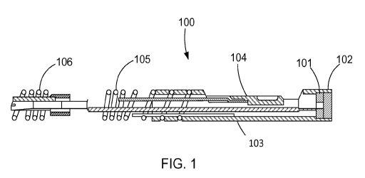
Turning to FIGS. 1 and 2, a combination sensor tip 100 for practicing the
invention is
illustrated. The combination sensor tip 100 includes a flow sensor 101, for
example an
ultrasound transducer, a Doppler flow sensor or any other suitable flow
sensor, disposed at or in
close proximity to the distal end 102 of the combination sensor tip 100. The
ultrasound
transducer 101 may be any suitable transducer, and may be mounted in the
distal end using any
conventional method, including the manner described in U.S. Pat. No.
5,125,137, which is fully
incorporated herein by reference. Conductors (not shown) may be secured to the
front and rear
sides of the ultrasound transducer 101, and the conductors may extend
interiorly to the proximal
extremity of a guide wire.
The combination sensor tip 100 also includes a pressure sensor 104 also
disposed at or in
close proximity to the distal end 102 of the combination sensor tip 100. The
pressure sensor 104
may be of the type described in U.S. Pat. No. 6,106,476, which is fully
incorporated herein by
reference. For example, the pressure sensor 104 may be comprised of a crystal
semiconductor
material having a recess therein and forming a diaphragm bordered by a rim. A
reinforcing
member may be bonded to the crystal to reinforce the rim of the crystal, and
may have a cavity
therein underlying the diaphragm and exposed to the diaphragm. A resistor
having opposite ends
may be carried by the crystal and may have a portion thereof overlying a
portion of the
diaphragm. Leads may be connected to opposite ends of the resistor and extend
proximally
within the guide wire. Additional details of suitable pressure sensors that
may be used as the
pressure sensor 104 are described in U.S. Pat. Nos. 6,106,476. U.S. Pat. No.
6,106,476 also
describes suitable methods for mounting the pressure sensor 104 within the
combination sensor
tip 100. In one embodiment, the pressure sensor 104 is oriented in a
cantilevered position within
a sensor housing 103. For example, the sensor housing 103 preferably includes
a lumen
6
CA 02896064 2015-06-19
WO 2014/100579
PCT/US2013/076909
surrounded by housing walls. When in a cantilevered position, the pressure
sensor 104 projects
into the lumen of the sensor housing 103 without contacting the walls of the
sensor housing 103.
As depicted in FIGS. 1 and 2, the combination sensor tip 100 incorporates a
sensor
housing 103 designed to enclose both the ultrasound transducer 101 and the
pressure sensor 104.
One advantage of the sensor housing 103 is that because the sensor housing 103
encloses both
the ultrasound transducer 101 and the pressure sensor 104, the need for two
separate housings,
i.e., one for an ultrasound transducer and one for a pressure sensor, is
eliminated. Accordingly,
the use of a common sensor housing 103 for the ultrasound transducer 101 and
the pressure
sensor 104 makes the combination sensor tip 100 easier to manufacture than
current designs.
Further detail on exemplary catheters for use in practicing the invention is
described in U.S.
Patent No. 8,277,386, incorporated herein by reference.
Methods of the invention also encompass displaying the obtained information,
including
the pre-therapy FFR, post-therapy-FFR, and functional gain in a format that is
convenient and
easily understandable to the physician. This may encompass displaying such
information
visually on a monitor or on a printed medium. The information may also be
presented textually
(using letters and/or numbers), graphically (e.g., bar graphs, pie charts,
etc.), or a combination of
the two. The display of such information is facilitated by systems of the
invention, described in
more detail below.
A visual display in accordance with the invention is provided in FIG. 3, which
depicts
FFR data in a visual format for display on a monitor. Sub-window 501 provides
data from FFR
measurements conducted before and after therapeutic intervention. An enlarged
view of data
presented in sub-window 501 is presented in FIG. 4. As presented, sub-window
501 provides
pre-therapy FFR data (Pre-RCA Mid 0.75) and post-therapy FFR data (Post-RCA
Mid 0.97).
Although the therapeutic procedure performed here involved a stent delivery,
the invention
encompasses any therapeutic procedure, particular cardiovascular procedures.
Additional
exemplary therapeutic procedures include, without limitation, angioplasties,
ablations, and
excisions. Any therapeutic procedure may be performed and its effectiveness
assessed using the
methods provided herein.
FIG. 5 shows another display based on the same data set. This time, however,
the display
provides the degree of improvement, i.e., the functional gain, after placing
the stent. As shown,
the post-therapy FFR (0.97) is compared to the pre-therapy FFR (0.75) to
arrive at an
7
CA 02896064 2015-06-19
WO 2014/100579
PCT/US2013/076909
improvement in FFR (and accordingly, vessel flow) of 29%. The pre- and post-
therapy
physiological measurements may be compared in any number of ways to arrive at
the functional
gain. In this example, functional gain is represented as a percentage
difference, however, the
pre-therapy measurement could have just as been easily been subtracted from
the post-therapy
measurement.
It is also encompassed that the display of information is highly customizable.
As shown
in FIG. 6A, physiological measurements can be taken at several points, any of
which can be used
to determine functional gain. In the example of FIG. 6A, a baseline FFR was
taken prior to any
therapeutic intervention (0.73). FFR was again measured after placing a stent
(0.82). Further
treatment involved post-stent dilatation with a high pressure balloon and FFR
was again assessed
(0.91). FFR was determined again as a final confirmation (0.91). The provided
customizable
displays allow for collapsing certain information, as shown in FIG. 6B. In
this case, functional
gain can be assessed using the baseline value and the final confirmation value
to determine the
degree of improvement after the two medical procedures.
As previously described herein, methods of the invention may also be applied
to
intravascular imaging (IVUS) or optical coherence tomography (OCT),
particularly if
physiological measurements, including FFR, IFR, CFR, HSR, HMR, and IMR, are
used to
determine or compare the difference in luminal area.
In some embodiments, the methods of the invention include use of an IVUS
imaging
assembly. The imaging assembly can be a phased-array IVUS imaging assembly, a
pull-back
type IVUS imaging assembly, including rotational IVUS imaging assemblies, or
an IVUS
imaging assembly that uses photoacoustic materials to produce diagnostic
ultrasound and/or
receive reflected ultrasound for diagnostics. IVUS imaging assemblies and
processing of IVUS
data are described for example in Yock, U.S. Pat. Nos. 4,794,931, 5,000,185,
and 5,313,949;
Sieben et al., U.S. Pat. Nos. 5,243,988, and 5,353,798; Crowley et al., U.S.
Pat. No. 4,951,677;
Pomeranz, U.S. Pat. No. 5,095,911, Griffith et al., U.S. Pat. No. 4,841,977,
Maroney et al., U.S.
Pat. No. 5,373,849, Born et al., U.S. Pat. No. 5,176,141, Lancee et al., U.S.
Pat. No. 5,240,003,
Lancee et al., U.S. Pat. No. 5,375,602, Gardineer et at., U.S. Pat. No.
5,373,845, Seward et al.,
Mayo Clinic Proceedings 71(7):629-635 (1996), Packer et al., Cardiostim
Conference 833
(1994), "Ultrasound Cardioscopy," Eur. J.C.P.E. 4(2):193 (June 1994), Eberle
et al., U.S. Pat.
No. 5,453,575, Eberle et al., U.S. Pat. No. 5,368,037, Eberle et at., U.S.
Pat. No. 5,183,048,
8
CA 02896064 2015-06-19
WO 2014/100579
PCT/US2013/076909
Eberle et al., U.S. Pat. No. 5,167,233, Eberle et at., U.S. Pat. No.
4,917,097, Eberle et at., U.S.
Pat. No. 5,135,486, and other references well known in the art relating to
intraluminal ultrasound
devices and modalities. All of these references are incorporated by reference
herein in their
entirety.
IVUS imaging is widely used in interventional cardiology as a diagnostic tool
for
assessing a diseased vessel, such as an artery, within the human body to
determine the need for
treatment, to guide an intervention, and/or to assess its effectiveness. An
IVUS device including
one or more ultrasound transducers is introduced into the vessel and guided to
the area to be
imaged. The transducers emit and then receive backscattered ultrasonic energy
in order to create
an image of the vessel of interest. Ultrasonic waves are partially reflected
by discontinuities
arising from tissue structures (such as the various layers of the vessel
wall), red blood cells, and
other features of interest. Echoes from the reflected waves are received by
the transducer and
passed along to an IVUS imaging system. The imaging system processes the
received
ultrasound echoes to produce a 360 degree cross-sectional image of the vessel
where the device
is placed.
There are two general types of IVUS devices in use today: rotational and solid-
state (also
known as synthetic aperture phased array). For a typical rotational IVUS
device, a single
ultrasound transducer element is located at the tip of a flexible driveshaft
that spins inside a
plastic sheath inserted into the vessel of interest. The transducer element is
oriented such that the
ultrasound beam propagates generally perpendicular to the axis of the device.
The fluid-filled
sheath protects the vessel tissue from the spinning transducer and driveshaft
while permitting
ultrasound signals to propagate from the transducer into the tissue and back.
As the driveshaft
rotates, the transducer is periodically excited with a high voltage pulse to
emit a short burst of
ultrasound. The same transducer then listens for the returning echoes
reflected from various
tissue structures. The IVUS imaging system assembles a two dimensional display
of the vessel
cross-section from a sequence of pulse/acquisition cycles occurring during a
single revolution of
the transducer. Suitable rotational IVUS catheters include, for example the
REVOLUTION 45
MHz catheter (offered by the Volcano Corporation).
In contrast, solid-state IVUS devices carry a transducer complex that includes
an array of
ultrasound transducers distributed around the circumference of the device
connected to a set of
transducer controllers. The transducer controllers select transducer sets for
transmitting an
9
CA 02896064 2015-06-19
WO 2014/100579
PCT/US2013/076909
ultrasound pulse and for receiving the echo signal. By stepping through a
sequence of transmit-
receive sets, the solid-state IVUS system can synthesize the effect of a
mechanically scanned
transducer element but without moving parts. The same transducer elements can
be used to
acquire different types of intravascular data. The different types of
intravascular data are
acquired based on different manners of operation of the transducer elements.
The solid-state
scanner can be wired directly to the imaging system with a simple electrical
cable and a standard
detachable electrical connector.
The transducer subassembly can include either a single transducer or an array.
The
transducer elements can be used to acquire different types of intravascular
data, such as flow
data, motion data and structural image data. For example, the different types
of intravascular
data are acquired based on different manners of operation of the transducer
elements. For
example, in a gray-scale imaging mode, the transducer elements transmit in a
certain sequence
one gray-scale IVUS image. Methods for constructing IVUS images are well-known
in the art,
and are described, for example in Hancock et al. (U.S. patent number
8,187,191), Nair et al.
(U.S. patent number 7,074,188), and Vince et al. (U.S. U.S. patent number
6,200,268), the
content of each of which is incorporated by reference herein in its entirety.
In flow imaging
mode, the transducer elements are operated in a different way to collect the
information on the
motion or flow. This process enables one image (or frame) of flow data to be
acquired. The
particular methods and processes for acquiring different types of
intravascular data, including
operation of the transducer elements in the different modes (e.g., gray-scale
imaging mode, flow
imaging mode, etc.) consistent with the present invention are further
described in U.S. Patent
Application No. 14/037,683, the content of which is incorporated by reference
herein in its
entirety.
The acquisition of each flow frame of data is interlaced with an IVUS gray
scale frame of
data. Operating an IVUS catheter to acquire flow data and constructing images
of that data is
further described in O'Donnell et al. (U.S. patent number 5,921,931), U.S.
Provisional Patent
Application No. 61/587,834, and U.S. Provisional Patent Application No.
61/646,080, the
content of each of which is incorporated by reference herein its entirety.
Commercially available
fluid flow display software for operating an IVUS catheter in flow mode and
displaying flow
data is CHROMAFLO (IVUS fluid flow display software offered by the Volcano
Corporation).
CA 02896064 2015-06-19
WO 2014/100579
PCT/US2013/076909
Suitable phased array imaging catheters include Volcano Corporation's EAGLE
EYE Platinum
Catheter, EAGLE EYE Platinum Short-Tip Catheter, and EAGLEEYE Gold Catheter.
Accordingly, as encompassed by the invention, baseline IVUS image data of the
vessel,
including flow data, may be captured by an IVUS catheter having flow data
capturing
capabilities, such as the phased-array catheters described above. The baseline
image data is
captured prior to conducting the therapeutic procedure. The procedure is then
performed, and
subsequent post-therapy IVUS image data, including flow data, of the vessel is
captured. The
post-therapy IVUS image data is then compared to the baseline IVUS image data,
upon which
the degree in improvement is ascertained according to methods previously
described herein.
In other embodiments, methods of the present invention include use of OCT
imaging.
OCT is a medical imaging methodology using a miniaturized near infrared light-
emitting probe.
As an optical signal acquisition and processing method, it captures micrometer-
resolution, three-
dimensional images from within optical scattering media (e.g., biological
tissue). Recently it has
also begun to be used in interventional cardiology to help diagnose coronary
artery disease. OCT
allows the application of interferometric technology to see from inside, for
example, blood
vessels, visualizing the endothelium (inner wall) of blood vessels in living
individuals.
OCT systems and methods are generally described in Castella et al., U.S.
Patent No.
8,108,030, Milner et al., U.S. Patent Application Publication No.
2011/0152771, Condit et al.,
U.S. Patent Application Publication No. 2010/0220334, Castella et al., U.S.
Patent Application
Publication No. 2009/0043191, Milner et al., U.S. Patent Application
Publication No.
2008/0291463, and Kemp, N., U.S. Patent Application Publication No.
2008/0180683, the
content of each of which is incorporated by reference in its entirety.
In OCT, a light source delivers a beam of light to an imaging device to image
target
tissue. Light sources can include pulsating light sources or lasers,
continuous wave light sources
or lasers, tunable lasers, broadband light source, or multiple tunable laser.
Within the light source
is an optical amplifier and a tunable filter that allows a user to select a
wavelength of light to be
amplified. Wavelengths commonly used in medical applications include near-
infrared light, for
example between about 800 nm and about 1700 nm.
Aspects of the invention may obtain imaging data from an OCT system, including
OCT
systems that operate in either the time domain or frequency (high definition)
domain. Basic
differences between time-domain OCT and frequency-domain OCT is that in time-
domain OCT,
11
CA 02896064 2015-06-19
WO 2014/100579
PCT/US2013/076909
the scanning mechanism is a movable minor, which is scanned as a function of
time during the
image acquisition. However, in the frequency-domain OCT, there are no moving
parts and the
image is scanned as a function of frequency or wavelength.
In time-domain OCT systems an interference spectrum is obtained by moving the
scanning mechanism, such as a reference minor, longitudinally to change the
reference path and
match multiple optical paths due to reflections within the sample. The signal
giving the
reflectivity is sampled over time, and light traveling at a specific distance
creates interference in
the detector. Moving the scanning mechanism laterally (or rotationally) across
the sample
produces two-dimensional and three-dimensional images.
In frequency domain OCT, a light source capable of emitting a range of optical
frequencies excites an interferometer, the interferometer combines the light
returned from a
sample with a reference beam of light from the same source, and the intensity
of the combined
light is recorded as a function of optical frequency to form an interference
spectrum. A Fourier
transform of the interference spectrum provides the reflectance distribution
along the depth
within the sample.
Several methods of frequency domain OCT are described in the literature. In
spectral-
domain OCT (SD-OCT), also sometimes called "Spectral Radar" (Optics letters,
Vol. 21, No. 14
(1996) 1087-1089), a grating or prism or other means is used to disperse the
output of the
interferometer into its optical frequency components. The intensities of these
separated
components are measured using an array of optical detectors, each detector
receiving an optical
frequency or a fractional range of optical frequencies. The set of
measurements from these
optical detectors forms an interference spectrum (Smith, L. M. and C. C.
Dobson, Applied Optics
28: 3339-3342), wherein the distance to a scatterer is determined by the
wavelength dependent
fringe spacing within the power spectrum. SD-OCT has enabled the determination
of distance
and scattering intensity of multiple scatters lying along the illumination
axis by analyzing a
single the exposure of an array of optical detectors so that no scanning in
depth is necessary.
Typically the light source emits a broad range of optical frequencies
simultaneously.
Alternatively, in swept-source OCT, the interference spectrum is recorded by
using a
source with adjustable optical frequency, with the optical frequency of the
source swept through
a range of optical frequencies, and recording the interfered light intensity
as a function of time
during the sweep. An example of swept-source OCT is described in U.S. Pat. No.
5,321,501.
12
CA 02896064 2015-06-19
WO 2014/100579
PCT/US2013/076909
Generally, time domain systems and frequency domain systems can further vary
in type
based upon the optical layout of the systems: common beam path systems and
differential beam
path systems. A common beam path system sends all produced light through a
single optical
fiber to generate a reference signal and a sample signal whereas a
differential beam path system
splits the produced light such that a portion of the light is directed to the
sample and the other
portion is directed to a reference surface. Common beam path systems are
described in U.S. Pat.
7,999,938; U.S. Pat. 7,995,210; and U.S. Pat. 7,787,127 and differential beam
path systems are
described in U.S. Pat. 7,783,337; U.S. Pat. 6,134,003; and U.S. Pat.
6,421,164, the contents of
each of which are incorporated by reference herein in its entirety.
In some embodiments, methods of the present invention may capture baseline OCT
image data of the vessel, wherein the baseline image data may be captured
prior to conducting
the therapeutic procedure. The procedure is then performed, and subsequent
post-therapy OCT
image data of the vessel is captured. The post-therapy image data is then
compared to the
baseline image data, upon which the degree in improvement is ascertained
according to methods
previously described herein.
In certain embodiments, angiogram image data may also be obtained
simultaneously with
the imaging data (IVUS or OCT). In such embodiments, the IVUS or OCT imaging
devices may
include one or more radiopaque labels that allow for co-locating image data
with certain
positions on a vasculature map generated by an angiogram. Co-registration
generally refers to
any method of re-aligning images, and in particular aligning or overlaying
images from different
modalities. Co-registration is often used to overlay structural and functional
images as well as
link functional scans to anatomical scans. Any number of modalities is useful
for co-registration.
Furthermore, modalities suitable for co-registration include functional
measurement parameters,
including, but not limited to, vessel flow, vessel pressure, FFR, iFR, CFR,
etc.
Details regarding image co-registration can be found in, for example, in U.S.
Patent No.
8,298,147; U.S. Patent Publication. Nos. 2012/0230565; 2011/0319752; and
2013/0030295; and
U.S. Patent Appin. Nos. 13/388,932; 61/776,863, 61/776,858; 61/777,155;
61/777,860;
61/779,610; and 61/792,230, each of which is incorporated herein by reference
in its entirety.
As noted above, it is contemplated that certain aspects of the invention are
particularly
amenable for implementation on computer-based systems. Accordingly, the
invention also
provides systems for practicing the above methods. The system may comprise a
processor and a
13
CA 02896064 2015-06-19
WO 2014/100579
PCT/US2013/076909
computer readable storage medium instructions that when executed, cause the
computer to
determine a baseline measurement prior to conducting a therapeutic procedure
and determine a
post-therapy measurement after conducting the therapeutic procedure. The
instructions may also
cause the computer to compare the post-therapy measurement to the baseline
measurement,
thereby determining the degree of post-therapy improvement after conducting
the therapeutic
procedure. In further aspects, the system displays the various measurements
and comparisons in
a form that is ready understandable to the operator, for example, in a textual
or graphical format.
A system of the invention may be implemented in a number of formats. An
embodiment
of a system 300 of the invention is shown in FIG. 7. The core of the system
300 is a computer
360 or other computational arrangement comprising a processor 365 and memory
367. The
memory has instructions which when executed cause the processor to determine a
baseline
measurement prior to conducting a therapeutic procedure and determine a post-
therapy
measurement after conducting the therapeutic procedure. The instructions may
also cause the
computer to compare the post-therapy measurement to the baseline measurement,
thereby
determining the degree of post-therapy improvement after conducting the
therapeutic procedure.
The physiological measurement data of vasculature will typically originate
from an intravascular
measurement device 320, which is in electronic and/or mechanical communication
with a
sensing catheter 325. Having collected the baseline measurement and post-
therapy
measurement, the processor then processes and outputs the results. The results
are typically
output to a display 380 to be viewed by a physician or technician. In some
embodiments the
display will include pre-therapy data, post-therapy data, and functional gain
data that correlate
the pre- and post-therapy data, as shown in FIG. 5. In certain embodiments,
the displayed
information is presented in a textual format as shown in FIG. 5. In other
embodiments, the
information may be presented in graphical format, like a pie chart or bar
graph.
Systems of the invention may rely on the operator instructing the computer
which
measurement is the baseline measurement and which is the post-procedure
measurement. Based
on those instructions, the computer would then determine the functional gain
achieved as a result
of the procedure. It is contemplated that computers may one day be able to
determine which
measurements are which without operator intervention. For example, the
software run by the
computer may use co-registration to know that certain measurements were made
at the same spot
and are thus related. It is also contemplated that systems of the invention
may integrate with the
14
CA 02896064 2015-06-19
WO 2014/100579
PCT/US2013/076909
case log and determine that a measurement has been made in the same location
immediately after
stent deployment, and therefore assign a post-therapy designation to the
measurement.
In advanced embodiments, system 300 may comprise an imaging engine 370 which
has
advanced image processing features, such as image tagging, that allow the
system 300 to more
efficiently process and display intravascular and angiographic images. The
imaging engine 370
may automatically highlight or otherwise denote areas of interest in the
vasculature. The
imaging engine 370 may also produce 3D renderings or other visual
representations of the
physiological measurements. In some embodiments, the imaging engine 370 may
additionally
include data acquisition functionalities (DAQ) 375, which allow the imaging
engine 370 to
receive the physiological measurement data directly from the catheter 325 or
collector 347 to be
processed into images for display.
Other advanced embodiments use the I/0 functionalities 362 of computer 360 to
control
the intravascular measurement 320. In these embodiments, computer 360 may
cause the imaging
assembly of catheter 325 to travel to a specific location, e.g., if the
catheter 325 is a pull-back
type. While not shown here, it is also possible that computer 360 may control
a manipulator,
e.g., a robotic manipulator, connected to catheter 325 to improve the
placement of the catheter
325.
A system 400 of the invention may also be implemented across a number of
independent
platforms which communicate via a network 409, as shown in FIG. 8. Methods of
the invention
can be performed using software, hardware, firmware, hardwiring, or
combinations of any of
these. Features implementing functions can also be physically located at
various positions,
including being distributed such that portions of functions are implemented at
different physical
locations (e.g., imaging apparatus in one room and host workstation in
another, or in separate
buildings, for example, with wireless or wired connections).
As shown in FIG. 8, the intravascular detecting system 320 facilitate
obtaining the data,
however the actual implementation of the steps can be performed by multiple
processors
working in communication via the network 409, for example a local area
network, a wireless
network, or the internet. The components of system 400 may also be physically
separated. For
example, terminal 467 and display 380 may not be geographically located with
the intravascular
detection system 320.
CA 02896064 2015-06-19
WO 2014/100579
PCT/US2013/076909
As shown in FIG. 8, imaging engine 859 communicates with host workstation 433
as
well as optionally server 413 over network 409. In some embodiments, an
operator uses host
workstation 433, computer 449, or terminal 467 to control system 400 or to
receive images. An
image may be displayed using an I/0 454, 437, or 471, which may include a
monitor. Any I/0
may include a monitor, keyboard, mouse, or touch screen to communicate with
any of processor
421, 459, 441, or 475, for example, to cause data to be stored in any
tangible, nontransitory
memory 463, 445, 479, or 429. Server 413 generally includes an interface
module 425 to
communicate over network 409 or write data to data file 417. Input from a user
is received by a
processor in an electronic device such as, for example, host workstation 433,
server 413, or
computer 449. In certain embodiments, host workstation 433 and imaging engine
855 are
included in a bedside console unit to operate system 400.
In some embodiments, the system may render three dimensional imaging of the
vasculature or the intravascular images. An electronic apparatus within the
system (e.g., PC,
dedicated hardware, or firmware) such as the host workstation 433 stores the
three dimensional
image in a tangible, non-transitory memory and renders an image of the 3D
tissues on the display
380. In some embodiments, the 3D images will be coded for faster viewing. In
certain
embodiments, systems of the invention render a GUI with elements or controls
to allow an
operator to interact with three dimensional data set as a three dimensional
view. For example, an
operator may cause a video affect to be viewed in, for example, a tomographic
view, creating a
visual effect of travelling through a lumen of vessel (i.e., a dynamic
progress view). In other
embodiments an operator may select points from within one of the images or the
three
dimensional data set by choosing start and stop points while a dynamic
progress view is
displayed in display. In other embodiments, a user may cause an imaging
catheter to be
relocated to a new position in the body by interacting with the image.
In some embodiments, a user interacts with a visual interface and puts in
parameters or
makes a selection. Input from a user (e.g., parameters or a selection) are
received by a processor
in an electronic device such as, for example, host workstation 433, server
413, or computer 449.
The selection can be rendered into a visible display. In some embodiments, an
operator uses host
workstation 433, computer 449, or terminal 467 to control system 400 or to
receive images. An
image may be displayed using an 1/0 454, 437, or 471, which may include a
monitor. Any I/0
may include a keyboard, mouse or touch screen to communicate with any of
processor 421, 459,
16
CA 02896064 2015-06-19
WO 2014/100579
PCT/US2013/076909
441, or 475, for example, to cause data to be stored in any tangible,
nontransitory memory 463,
445, 479, or 429. Server 413 generally includes an interface module 425 to
effectuate
communication over network 409 or write data to data file 417. Methods of the
invention can be
performed using software, hardware, firmware, hardwiring, or combinations of
any of these.
Features implementing functions can also be physically located at various
positions, including
being distributed such that portions of functions are implemented at different
physical locations
(e.g., imaging apparatus in one room and host workstation in another, or in
separate buildings,
for example, with wireless or wired connections). In certain embodiments, host
workstation 433
and imaging engine 855 are included in a bedside console unit to operate
system 400.
Processors suitable for the execution of computer program include, by way of
example,
both general and special purpose microprocessors, and any one or more
processor of any kind of
digital computer. Generally, a processor will receive instructions and data
from a read-only
memory or a random access memory or both. The essential elements of computer
are a
processor for executing instructions and one or more memory devices for
storing instructions and
data. Generally, a computer will also include, or be operatively coupled to
receive data from or
transfer data to, or both, one or more mass storage devices for storing data,
e.g., magnetic,
magneto-optical disks, or optical disks. Information carriers suitable for
embodying computer
program instructions and data include all forms of non-volatile memory,
including by way of
example semiconductor memory devices, (e.g., EPROM, EEPROM, NAND-based flash
memory, solid state drive (SSD), and other flash memory devices); magnetic
disks, (e.g., internal
hard disks or removable disks); magneto-optical disks; and optical disks
(e.g., CD and DVD
disks). The processor and the memory can be supplemented by, or incorporated
in, special
purpose logic circuitry.
To provide for interaction with a user, the subject matter described herein
can be
implemented on a computer having an I/0 device, e.g., a CRT, LCD, LED, or
projection device
for displaying information to the user and an input or output device such as a
keyboard and a
pointing device, (e.g., a mouse or a trackball), by which the user can provide
input to the
computer. Other kinds of devices can be used to provide for interaction with a
user as well. For
example, feedback provided to the user can be any form of sensory feedback,
(e.g., visual
feedback, auditory feedback, or tactile feedback), and input from the user can
be received in any
form, including acoustic, speech, or tactile input.
17
CA 02896064 2015-06-19
WO 2014/100579
PCT/US2013/076909
The subject matter described herein can be implemented in a computing system
that
includes a back-end component (e.g., a data server 413), a middleware
component (e.g., an
application server), or a front-end component (e.g., a client computer 449
having a graphical user
interface 454 or a web browser through which a user can interact with an
implementation of the
subject matter described herein), or any combination of such back-end,
middleware, and front-
end components. The components of the system can be interconnected through
network 409 by
any form or medium of digital data communication, e.g., a communication
network. Examples
of communication networks include cell networks (3G, 4G), a local area network
(LAN), and a
wide area network (WAN), e.g., the Internet.
The subject matter described herein can be implemented as one or more computer
program products, such as one or more computer programs tangibly embodied in
an information
carrier (e.g., in a non-transitory computer-readable medium) for execution by,
or to control the
operation of, data processing apparatus (e.g., a programmable processor, a
computer, or multiple
computers). A computer program (also known as a program, software, software
application, app,
macro, or code) can be written in any form of programming language, including
compiled or
interpreted languages (e.g., C, C++, Per1), and it can be deployed in any
form, including as a
stand-alone program or as a module, component, subroutine, or other unit
suitable for use in a
computing environment. Systems and methods of the invention can include
programming
language known in the art, including, without limitation, C, C++, Perl, Java,
ActiveX, HTML5,
Visual Basic, or JavaScript.
A computer program does not necessarily correspond to a file. A program can be
stored
in a portion of file 417 that holds other programs or data, in a single file
dedicated to the program
in question, or in multiple coordinated files (e.g., files that store one or
more modules, sub-
programs, or portions of code). A computer program can be deployed to be
executed on one
computer or on multiple computers at one site or distributed across multiple
sites and
interconnected by a communication network.
A file can be a digital file, for example, stored on a hard drive, SSD, CD, or
other
tangible, non-transitory medium. A file can be sent from one device to another
over network 409
(e.g., as packets being sent from a server to a client, for example, through a
Network Interface
Card, modem, wireless card, or similar).
18
CA 02896064 2015-06-19
WO 2014/100579
PCT/US2013/076909
Writing a file according to the invention involves transforming a tangible,
non-transitory
computer-readable medium, for example, by adding, removing, or rearranging
particles (e.g.,
with a net charge or dipole moment) into patterns of magnetization by
read/write heads, the
patterns then representing new collocations of information desired by, and
useful to, the user. In
some embodiments, writing involves a physical transformation of material in
tangible, non-
transitory computer readable media with certain properties so that optical
read/write devices can
then read the new and useful collocation of information (e.g., burning a CD-
ROM). In some
embodiments, writing a file includes using flash memory such as NAND flash
memory and
storing information in an array of memory cells include floating-gate
transistors. Methods of
writing a file are well-known in the art and, for example, can be invoked
automatically by a
program or by a save command from software or a write command from a
programming
language.
In certain embodiments, display 380 is rendered within a computer operating
system
environment, such as Windows, Mac OS, or Linux or within a display or GUI of a
specialized
system. Display 380 can include any standard controls associated with a
display (e.g., within a
windowing environment) including minimize and close buttons, scroll bars,
menus, and window
resizing controls. Elements of display 380 can be provided by an operating
system, windows
environment, application programming interface (API), web browser, program, or
combination
thereof (for example, in some embodiments a computer includes an operating
system in which an
independent program such as a web browser runs and the independent program
supplies one or
more of an API to render elements of a GUI). Display 380 can further include
any controls or
information related to viewing images (e.g., zoom, color controls,
brightness/contrast) or
handling files comprising three-dimensional image data (e.g., open, save,
close, select, cut,
delete, etc.). Further, display 380 can include controls (e.g., buttons,
sliders, tabs, switches)
related to operating a three dimensional image capture system (e.g., go, stop,
pause, power up,
power down).
In certain embodiments, display 380 includes controls related to three
dimensional
imaging systems that are operable with different imaging modalities. For
example, display 380
may include start, stop, zoom, save, etc., buttons, and be rendered by a
computer program that
interoperates with IVUS, OCT, or angiogram modalities. Thus display 380 can
display an image
19
CA 02896064 2015-06-19
WO 2014/100579
PCT/US2013/076909
derived from a three-dimensional data set with or without regard to the
imaging mode of the
system.
Incorporation by Reference
References and citations to other documents, such as patents, patent
applications, patent
publications, journals, books, papers, web contents, have been made throughout
this disclosure.
All such documents are hereby incorporated herein by reference in their
entirety for all purposes.
Equivalents
The invention may be embodied in other specific forms without departing from
the spirit
or essential characteristics thereof. The foregoing embodiments are therefore
to be considered in
all respects illustrative rather than limiting on the invention described
herein. Scope of the
invention is thus indicated by the appended claims rather than by the
foregoing description, and
all changes which come within the meaning and range of equivalency of the
claims are therefore
intended to be embraced therein.