Note: Descriptions are shown in the official language in which they were submitted.
NON-REPLICATING VIRUS-DERIVED PARTICLES AND USES THEREOF
[0001]
=
FIELD
[0002] The present disclosure relates generally to a non-replicating virus-
derived
particle and its use as an anti-cancer agent.
BACKGROUND
[0003] The following background discussion does not imply or admit
that anything
described below is prior art or part of the knowledge of people skilled in the
art in any
country.
[0004] Oncolytic viruses (0Vs) have been engineered through
attenuating mutations
or deletions which allow the virus to replicate exclusively in cells
associated with an impaired
immune response or enhanced metabolic activity, two key characteristics of
tumorigenesis.
Examples of current advanced oncolytic therapeutics include the Herpes Simplex
Virus
OncoVEXGM¨CSF and the vaccinia virus (JX594). To date, the main focus of the
OV field
has been the development of platforms where the live virus has preferential
replication/spreading capacity within the local tumor environment.
[0005] Rhabdoviruses viruses (RVs), such as vesicular stomatitis virus
(VSV) and
Maraba, are currently being explored as anti-cancer therapeutics. In tumors,
viral
propagation is enabled by unrestrained metabolic activities and impaired anti-
viral programs.
Tumor susceptibility to RV treatment is further enhanced due to pre-
disposition towards
virus-mediated apoptosis.
[0006] In the Rhabdovirus field, oncolytic platforms developed to date
utilize a
replication competent virus where the virus spreads between tumor cells. In
fact, reports
describing the use of live replication/expression competent rhabdovirus as a
direct
virotherapy for cancer typically compare efficacy to non-replicating/non-
expressing virus
- 1 -
CA 2896162 2020-03-09
CA 02896162 2015-06-22
WO 2014/094182
PCT/CA2013/051009
controls where no measurable efficacy is observed. In these reports, it is
concluded that
Rhabdovirus genome replication and/or expression is a critical and essential
component of
tumor cytotoxicity and therapeutic efficacy.
[0007] The lack of oncolytic effects in these previous studies is
reflected in the
methods used to disrupt virus genome replication and/or expression as well as
in the number
of virus particles used. Indeed, when these previous methods are used to
disrupt virus
genome replication and/or expression, no bioactivity of the virus is observed.
Furthermore, in
these studies, non-replicating virus controls are applied at the same dose as
their live virus
counterparts, and not at higher doses to ensure that each cell encounters a
non-replicating
particle.
[0008] Alternative, and preferably more effective, approaches are
desired to treat and
cure most forms of cancers. For example, the outcome for the majority of adult
patients
suffering from acute lymphoblastic or acute myeloid leukemia remains dismal.
This is in part
due to the significant immunocompromised nature of the disease. For a minority
of patients,
anti-tumor immune responses are partially restored through allogeneic stem
cell
transplantation after myeloablative conditioning. This therapy is potentially
curative, however
is associated with frequent adverse events and significant treatment-related
mortality. For
many patients with chronic-phase chronic myeloid leukemia (CM L), targeted
tyrosine kinase
inhibitor (TKI) therapy offers excellent disease control. However when
progression into acute
leukemic blast crisis occurs, very limited therapeutic options exist due to
development of
multi-drug resistance and the rapid kinetics of this form of recalcitrant
leukemia.
[0009] Hence there is need for alternative anti-cancer agents,
particularly for
immunocompromised patients. The anti-cancer agent, by virtue of its design and
components, would preferably be able to address current unmet clinical needs
and/or
overcome at least some of the above-discussed problems.
SUMMARY
[0010] The following summary is intended to introduce the reader to
one or more
inventions described herein, but not to define any of them. Inventions may
reside in any
combination of features described anywhere in this document.
[0011] While live OV strategies are being pursued to treat a variety
of tumor types,
their application in hematopoietic malignancies in particular is complicated
by several factors.
- 2 -
CA 02896162 2015-06-22
WO 2014/094182
PCT/CA2013/051009
Limited virion production and reduced spread between leukemia cells requires
high-dose
viral therapy to overcome these inefficiencies. However, uncontrolled live
virus spread and
off-target effects in normal tissue compromise the safety of this approach,
particularly in
immunosuppressed patients.
[0012] Issues associated with using live virus include: 1) safety, which
relies on the
ability of the live Rhabdovirus to spread only in diseased tumor tissue,
leaving healthy tissue
alone; 2)10w doses for administration, since the introduction of live
spreadable virus to a
patient requires the administration of relatively low doses of these live
viral agents to ensure
safety; 3) immune diversion from the tumor towards the live virus which
effectively decreases
the efficiency of anti-tumor immune responses; and 4) engineered live viruses
designed with
proclivity for tumor often have impaired production capacity compared to wild
type virus, and
consequently, formulation efficiencies and production costs are sub-optimal
from a
manufacturing perspective.
[0013] It has been previously shown that intra-tumoral injection with
VSV engineered
to have a deletion of the glycoprotein gene (VSVLG), which prevents final
virion assembly
and spread, elicits anti-tumor immune responses. However, treatment with VSVAG
cannot
provide a significant reduction of disseminated tumor bulk, partly due to the
inability to
manufacture and deliver therapeutically effective doses.
[0014] The authors are aware of no reports that detail the use of a
non-replicating
and non-expressing Rhabdovirus-derived platform as an anti-cancer therapeutic.
Non-
replicating virus-derived particles (NVRP) of the present disclosure, and non-
replicating
rhabdovirus-derived particles (NRRP) in particular, are wild type virus
particles modified so
as to lack the ability to spread between cells. Once modified, the non-
replicating virus-
derived particle (NVRP) cannot sustain virion replication.
[0015] NVRPs are unique in that they retain tropism, such as cytolytic
tropism,
against immortalized cells. This means that NVRPs will induce cell death
preferentially in
immortalized cells such as tumor or cancer cells and transformed immortalized
cells. Specific
examples of NVRPs have innate and/or adaptive immune-stimulating properties
against
immortalized cells.
[0016] In one aspect, the present disclosure describes a non-replicating
rhabdovirus-
derived particle that lacks the ability to spread between cells while having
tropism against
immortalized cells. The tropism may be a cytolytic tropism. The non-
replicating rhabdovirus-
- 3 -
CA 02896162 2015-06-22
WO 2014/094182
PCT/CA2013/051009
derived particle may have innate or adaptive immune-stimulating properties
against
immortalized cells.
[0017] In yet another aspect, the present disclosure provides a use of
a non-
replicating rhabdovirus-derived particle to treat a population of
hyperproliferative cells or
cancer cells. The population of hyperproliferative cells is preferably of
hematopoietic nature,
and preferably leukemic cells. The population of hyperproliferative cells may
be solid tumor
cells.
[0018] In still another aspect, the present disclosure describes a
method of treating a
patient having a population of hyperproliferative cells or cancer cells. The
method includes
administering to the patient non-replicating rhabdovirus-derived particles.
The population of
hyperproliferative cells may preferably be of hematopoietic nature, preferably
leukemic cells.
The population of hyperproliferative cells may be solid tumor cells.
[0019] Other aspects and features of the present disclosure will
become apparent to
those ordinarily skilled in the art upon review of the following description
of specific
embodiments in conjunction with the accompanying figures.
BRIEF DESCRIPTION OF DRAWINGS
[0020] Embodiments of the present disclosure will now be described, by
way of
example only, with reference to the attached Figures.
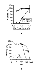
[0021] Fig. 1A is a graph showing the impact of UV dosage on NRRP-mediated
cytotoxicity on Vero and HDFN cells. No GFP signal was detected following UV-
induced
NRRP generation. Viability was quantified using the resazurin assay 72h post
infection. The
MOI of this experiment was set at 100 particles per cell. Error bars represent
the standard
deviation between triplicate experiments.
[0022] Fig. 1B is a graph showing the impact of MOI on the cytotoxicity
induced by
NRRPs in Vero and HDFN cells as illustrated by the viability as a function
MOI. Viability was
quantified using the resazurin assay 72h post infection. Error bars represent
the standard
deviation between triplicate experiments.
[0023] Fig. 2A is a set of images show the cytotoxicity of NRRPs in
Vero
immortalized cells through fluorescent and brightfield microscopy images of
Vero cells
treated with PBS, Live VSV-GFP or NRRPs taken at 24 and 72 hours post-
infection or post-
- 4 -
CA 02896162 2015-06-22
WO 2014/094182
PCT/CA2013/051009
treatment. The multiplicity of infection (M01) used in these experiments was
set at 100
particles per cell.
[0024] Fig. 2B is a graph showing the cytotoxicity of NRRPs through
resazurin
quantification of cellular viability 72h post treatment. Error bars represent
the standard
deviation between triplicate experiments.
[0025] Fig. 2C is a graph showing viral titers produced. NAN means
"not as a
number" as no virions were detected.
[0026] Fig. 3A is a set of fluorescent microscopy images (4X) of
leukemic (L1210)
and Vero cells treated with PBS, Live Maraba virus, and Maraba virus-derived
NRRPs.
Images were taken at 24h post treatment.
[0027] Fig. 3B is a graph showing viral titers obtained from tumor
cells.
[0028] Fig. 3C is a graph showing resazurin quantification of cellular
viability of L1210
leukemia cells and HDF normal cells, 72h post infection.
[0029] Fig. 4A is a set of images showing fluorescent images of L1210
and Vero cells
treated with PBS, Live VSV-GFP, or NRRPs.
[0030] Fig. 4B is a graph showing the viral titers generated from
L1210 acute
leukemia and Vero immortalized cells
[0031] Fig. 5 is an image of a Western blot of NRRP genome expression
compared
to the genome expression of a virus exposed to a UV dose of 20,000 mJ/cm2,
where loss of
cytotoxicity was observed, and a live virus as a control. Reference to lx or
2x refers to the
amount of protein loaded onto the gel. Proteins were extracted 15h post
infection.
[0032] Fig 6A is a set of fluorescent and brightfield images of Vero
cells treated with
chemically- generated, or busulfan-generated, NRRPs.
[0033] Fig. 68 is a brightfield microscopy image of Vero cells treated
with busulfan
alone, at the same dose used to generate NRRPs in Fig. 6A, for 15 hours.
[0034] Fig. 6C is a set of fluorescent and brightfield images of Vero
cells treated with
Live VSV-GFP.
[0035] Fig, 7A is a set of brightfield and fluorescent images of Vero
cells treated with
NRRPs, generated by taking 1E10 frozen wild type VSV and irradiating this
preparation with
15 kGy Cobalt-60.
[0036] Fig. 78 is a set of brightfield and fluorescent images of Vero
cells treated with
live wild type VSV-GFP.
- 5 -
CA 02896162 2015-06-22
WO 2014/094182
PCT/CA2013/051009
[0037] Fig. 7C is a set of brightfield and fluorescent images of Vero
cells in PBS.
[0038] Fig. 8A is a set of brightfield images of L1210 and HDF cells
treated with PBS
or NRRPs at an MOI of 100.
[0039] Fig. 8B is a graph showing resazurin quantification of
viability in leukemia and
normal cell lines. Murine cell lines are denoted by *.
[0040] Fig. 8C is a set of fluorescent microscopy images of PBS, live
VSV-GFP, or
NRRP treatment in murine human Jurkat 1-cell acute leukemia, murine A20 B-cell
lymphoblastic leukemia, A301 1-cell lymphoblastic leukemia, and HL60 acute
promyelocytic
leukemia and GM38 and HDF normal cell lines.
[0041] Fig. 9 is a set of graphs showing the flow cytometry analysis of
Annexin V-
APC and 7-AAD staining in L1210 cells treated with PBS or NRRPs.
[0042] Fig. 10 is a graph illustrating cell viability following a
resazurin quantification
assay for L1210 acute leukemia cell line taken 72 hours post treatment with UV-
generated
NRRPs and the combinatorial effect of UV-generated NRRPs with bendamustine(300
,M).
[0043] Fig. 11 is a graph illustrating cell viability following a resazurin
quantification
assay for L1210 acute leukemia cell line taken 72 hours post treatment with UV-
generated
NRRPs and the combinatorial effect of UV-generated NRRPs with dexamethasone
(451.IM).
[0044] Fig. 12 is a graph illustrating cell viability following a
resazurin quantification
assay for L1210 acute leukemia cell line taken 72 hours post treatment with UV-
generated
NRRPs and the combinatorial effect of UV-generated NRRPs with doxorubicin
(0.025 M).
[0045] Fig. 13 is a graph illustrating cell viability following a
resazurin quantification
assay for L1210 acute leukemia cell line taken 72 hours post treatment with UV-
generated
NRRPs and the combinatorial effect of UV-generated NRRPs with vincristine
(0.0125 piM).
[0046] Fig. 14 is a graph illustrating cell viability following a
resazurin quantification
assay for K562 Ph-positive myeloid leukemic cell line taken 15 hours post
treatment with UV-
generated NRRPs and the combinatorial effect of UV-generated NRRPs with
idarubicin (0.05
[0047] Fig. 15A is an illustration of a phenomenological model
developed by Le
Boeuf et al. to simulate NRRPs cytotoxicity in normal cells and tumors with
defects in
antiviral signaling pathways. To describe NRRP kinetics, the original model
was modified by
removing virus replication (X). Hashed lines describe the IFN-defects
associated with tumor
cells.
- 6 -
CA 02896162 2015-06-22
WO 2014/094182
PCT/CA2013/051009
[0048] Fig. 15B is a graph showing the simulated relationship between
defects in the
antiviral signaling pathway and viability post-treatment with NRRPs at 72hrs.
[0049] Fig. 150 is a graph showing the in vitro relationship between
MOI and viability
72h post-infection with NRRPs in normal HDF cells in the presence or absence
of IFN.
[0050] Fig. 15D is a graph showing the in vitro relationship between MOI
and viability
72h post-treatment with NRRPs in leukemic L1210 cells in the presence or
absence of IFN.
[0051] Fig. 16A is a set of brightfield microscopy images of two
Chronic Myeloid
Leukemia-blast crisis patient samples treated with PBS or NRRPs.
[0052] Fig. 16B is a set of fluorescent microscopy images (4X) of
acute leukemia
.. (CML blast-crisis) from human patient peripheral blood samples. Leukemia
enriched samples
collected from peripheral blood treated with PBS, Live VSV-GFP, or NRRPs
encoded for
GFP. Images are 24h post infection at M01=100.
[0053] Fig. 16C is a flow cytometry diagram complementing the data
presented in
Fig. 16A and 160 of Annexin-V and 0D33 staining in two CML-blast crisis
patient samples
treated with PBS or NRRPs (M01=100) 48h post-treatment. The CD33+ blast
population was
enriched by long term culture of the cells.
[0054] Fig. 16D are graphs showing flow cytometry analysis of 0D33
staining in the
two CML-blast crisis patient samples treated with PBS or NRRPs.
[0055] Fig. 17A is a set of brightfield microscopy images of a healthy
bone marrow
sample treated with PBS or NRRPs for 18 hours.
[0056] Fig. 17B is a graph showing the quantification of Annexin-V
staining in the
healthy bone marrow sample treated with PBS or NRRPs for 65 hours.
[0057] Fig. 18A is a graph showing the survival curve in a murine
blast crisis
treatment model. Following L1210 challenge in mice on day 1, mice received
three daily
doses NRRPs or PBS.
[0058] Fig. 18B is a set of graphs showing Luminex-based
quantification of cytokines
induced by NRRPs in L1210 bearing mice during acute blast crisis. All
identified cytokines
are induced over 2 fold by NRRP-treated mice and are statistically significant
(non-paired t-
test pV<0.05). pV has been corrected to account for multiple hypothesis
testing (Benjamini &
Hochberg Method).
- 7 -
CA 02896162 2015-06-22
WO 2014/094182
PCT/CA2013/051009
[0059] Fig. 19 is a graph showing the survival curve in a murine
immunocompetent
model of immunogenic apoptosis. Prior to L1210 challenge on day 1, mice
received three
weekly doses of y-irradiated L1210 cells incubated or not incubated with
NRRPs.
[0060] Fig. 20 is a set of brightfield microscopy images of myeloma
cell lines MPC-11
and RPMI-8226 taken 15 hours post treatment with PBS or NRRPs. NRRPs were
administered at an M01=250, a dose previously determined to have no impact on
normal cell
viability.
[0061] Fig. 21 is a graph showing cell viability following an
resazurin quantification
assay for myeloma cell lines MPC-11 and RPMI-8226 taken 15 hours post
treatment with
NRRPs administered at an MOI = 250. 5R4987 is a normal marrow stromal cell
line.
[0062] Fig. 22 is a graph illustrating cell viability following a
resazurin quantification
assay for MPC-11 multiple myeloma cell line taken 72 hours post treatment with
UV-
generated NRRPs and the combinatorial effect of UV-generated NRRPs with
melphalan
(20 M).
[0063] Fig. 23 is a graph illustrating cell viability following a resazurin
quantification
assay for MPC-11 multiple myeloma cell line taken 72 hours post treatment with
UV-
generated NRRPs and the combinatorial effect of UV-generated NRRPs with the
second
mitochondria-derived activator of caspase (SMAC) mimetic, LCL161(15 M).
[0064] Fig. 24 is a graph illustrating cell viability following a
resazurin quantification
assay for RPM 1-8226 multiple myeloma cell line taken 72 hours post treatment
with UV-
generated NRRPs and the combinatorial effect of UV-generated NRRPs with
carfilzomib
(5nM).
[0065] Fig. 25A is a set of brightfield microscopy images of a mouse
delayed brain
tumor glioblastoma cell line (DBT) taken 24 hrs post treatment with PBS or
NRRPs.
[0066] Fig. 25B is a set of brightfield microscopy images of an astrocytoma
cell line
(K1491) taken 24 hrs post treatment with PBS or NRRPs.
[0067] Fig. 250 is a set of brightfield microscopy images of a mouse
glioma cell line
(GL261) taken 24 hrs post treatment with PBS or NRRPs.
[0068] Fig. 26 is a graph showing cell viability following a resazurin
quantification
assay for brain cancer cell lines DBT, K1491, K1492, CT2A, and GL261 relative
to normal
HDFN control.
- 8 -
CA 02896162 2015-06-22
WO 2014/094182
PCT/CA2013/051009
[0069] Fig. 27 is a graph illustrating cell viability following a
resazurin quantification
assay for CT2A glioblastoma cell line taken 72 hours post treatment with UV-
generated
NRRPs and the combinatorial effect of UV-generated NRRPs with the HDAC
inhibitor SAHA
(1011M).
[0070] Fig. 28A is a set of fluorescent microscopy images (4X) of NRRP-
mediated
tumor cell cytotoxicity in resistant solid tumor cell lines. The set of images
show mouse
mammary or breast (4T1) and human kidney (786-0) cancer cells treated with
PBS, Live
VSV, and NRRPs. Images were taken at 24h post infection.
[0071] Fig. 28B is a set of brightfield microscopy images taken at 72h
post infection
of NRRP-mediated tumor cell cytotoxicity in resistant solid tumor cell lines,
in breast (4T1)
and kidney (786-0) cancer cells treated with PBS, Live VSV, and NRRPs.
[0072] Fig. 28C is a graph showing resazurin quantification of
cellular viability in
resistant solid tumor cell lines, in breast (4T1) and kidney (786-0) cancer
cells treated with
PBS, Live VSV, and NRRPs, 72h post infection.
[0073] Fig. 29 is a graph illustrating survival advantage in sub-cutaneous
CT-26
colon cancer treated with 2E9 UV-generated NRRPs on days 16, 18 and 21 post
tumor
embedment.
DETAILED DESCRIPTION
[0074] Generally, the present disclosure provides a non-replicating virus-
derived
particle and its use as an anti-cancer agent. A non-replicating virus-derived
particle (NRVP)
is a virus-derived particle that is able to bind and be internalized by a
cell, but has been
modified to prevent formation, or substantially reduce formation, of new virus
particles when
the NRVP is in the cell. One example of a NRVP is a non-replicating
rhabdovirus-derived
particle (NRRP).
[0075] The NRVP includes: an envelope having a sufficient number of
functional G
proteins on the surface of the envelope to allow the virus-derived particle to
bind a surface of
a cell and be internalized. It also includes an RNA polynucleotide with a
sequence that
encodes all the proteins required for new virus particle assembly, and a
mixture of proteins
that form a structure around the RNA polynucleotide. However, the RNA
structure of the
NRVP is sufficiently cross-linked, or has been cleaved to form discontinuous
segments of
RNA, such that the NRVP genome is unable be used to produce the proteins
required for
- 9 -
CA 02896162 2015-06-22
WO 2014/094182
PCT/CA2013/051009
new virus formation. For example, the RNA sequence may not be transcribed into
mRNA,
translated into protein, or both when the particle is in a cell. The
impairment or lack of
transcription and/or translation means that insufficient proteins are produced
in the cell and
new virus particles cannot be assembled.
[0076] The functional G protein may have a sequence that includes SEQ ID
NO: 1,
shown below, which is the sequence of the glycoprotein mature peptide of
vesicular
stomatitis Indiana virus. This functional G protein has NCB! accession number
NP 955548.1.
kftivfphnq kgnwknvpsn yhycpsssdl nwhndligta iqvkmpkshk aiqadgwmch
askwvttcdf rwygpkyitq sirsftpsve qckesieqtk qgtwlnpgfp pqscgyatvt
daeavivqvt phhvlvdeyt gewvdsgfin gkcsnyicpt vhnsttwhsd ykvkglcdsn
lismditffs edgelsslgk egtgfrsnyf ayetggkack mqyckhwgvr 1psgvwfema
dkdlfaaarf pecpegssis apscitsvdvs liqdverild yslogetwsk iraglpispv
dlsylapknp gtgpaftiin gtlkyfetry irvdiaapil srmvgmisgt tterelwddw
apyedveigp ngv1rtssgy kfplymighg mldsdlhlss kaqvfehphi qdaasqlpdd
eslffgdtgl sknpielveg wfsswkssia sfffiiglii glflvlrvgi hlciklkhtk
krqiytdiem nrlgk (SEQ ID NO: 1)
[0077] Alternatively, the functional G protein may have a sequence
that is at least
75% identical to SEQ ID NO: 1 so long as it is capable of binding to a surface
of a cell and
effecting internalization of the particle. For example, conservative
substitutions of amino
acids may be made without abrogating the ability of the protein to bind to the
surface of a cell
and effect internalization of the particle. Examples of conservative
substitutions are shown
below in Table 1.
- 10-
CA 02896162 2015-06-22
WO 2014/094182
PCT/CA2013/051009
Original Exemplary Substitutions Preferred
Residues . Substitutions
Ala Val, Leu, Ile Val
Arg Lys, Gin, Asn Lys
Asn Gin Gin
Asp Glu Glu
Cys Ser, Ala Ser
Gin Asn Asn
Glu Asp Asp
Gly Pro, Ala Ala
His Asn, Gin, Lys, Arg Arg
Ile Leu, Val, Met, Ala, Phe, Norleucine Leu
Leu Norleucine, Ile, Val, Met, Ala, Phe Ile
Lys Arg, 1,4 Diamino-butyric Acid, Gin, Asn Arg
Met Leu, Phe, Ile Leu
Phe Leu, Val, Ile, Ala, Tyr Leu
Pro Ala Gly
Ser Thr, Ala, Cys Thr
Thr Ser Ser
Trp Tyr, Phe Tyr
Tyr Trp, Phe, Thr, Ser Phe
Val Ile, Met, Len, Phe, Ala, Norleucine Leu
Table 1 ¨ Conservative Amino Acid Substitutions
[0078] Less conservative substitutions may be made in portions of the
G protein that
do not take part in the cell surface binding (such as in a trans-membrane
domain), while
more conservative substitutions might be required in portions of the protein
that interact with
a G protein receptor. G proteins are known in the art and a skilled person
would be able to
determine what amino acid substitutions would be possible without abrogating
the ability of
the protein to bind to the surface of a cell and effect internalization of the
particle.
[0079] The mixture of proteins that form a structure around the RNA
may include at
least N, P, M, and L proteins. A NRVP having N, P, M, G and L proteins may
include
rhabdovirus-derived NRVP. Rhadbovirus-derived NRVPs may be referred to as non-
replicating rhabdovirus -derived particles (NRRPs). For the purposes of the
present
disclosure, the term "Rhabdovirus" (NCB! Taxonomy ID: 11270) may include any
one of the
following genus of viruses and variants thereof: Cytorhabdovirus (NCBI
Taxonomy ID:
11305), Ephemerovirus (NCB! Taxonomy ID: 32613), Vesiculovirus (NCB! Taxonomy
ID:
-11-
CA 02896162 2015-06-22
WO 2014/094182
PCT/CA2013/051009
11271), unclassified Dimarhabdovirussupergroup (NCB! Taxonomy ID: 349166),
Lyssavirus
(NCBI Taxonomy ID: 11286), Novirhabdovirus (NCB! Taxonomy ID: 186778),
Nudeorhabdovirus (NCB! Taxonomy ID: 11306), unassigned rhabdovirus (NCB!
Taxonomy
ID: 686606) and unclassified rhabdovirus (NCB! Taxonomy ID: 35303). Species
within the
Rhabdovirus family include, but are not limited to, Maraba virus, Vesicular
stomatitis virus
(VSV) and Farmington virus.
[0080] The N protein may have a sequence that includes SEQ ID NO: 2,
shown
below, which is the sequence of the nucleocapsid protein of vesicular
stomatitis Indiana
virus. This N protein has NCB! accession number NC 041712.1.
msvtvkriid ntvivpklpa nedpveypad yfrkskeipl yinttkslsd lrgyvyulk
sgnvsiihvn sylygalkdi rgkldkdwss fginigkagd tigifdlvsl kaldgvlpdg
vsdasrtsad dkwlplyllg lyrvgrtqmp eyrkklmdgl tnqckmineq feplvpegrd
ifdvwgndsn ytkivaavdm ffhmfkkhec asfrygtivs rfkdcaalat fghlckitgm
stedvttwil nrevademvq mmlpgqeidk adsympylid fglsskspys svknpafhfw
gqltalllrs trarnarqpd dieytsltta gllyayavgs sadlaqqfcv gdnkytpdds
tgglttnapp qgrdvvewlg wfedqnrkpt pdmmqyakra vmslqglrek tigkyaksef
dk (SEQ ID NO: 2)
[0081] Alternatively, the N protein may have a sequence that is at
least 80% identical
to SEQ ID NO: 2 so long as it is capable of participating in the formation of
the protein
structure. For example, conservative substitutions of amino acids may be made
without
abrogating the ability of the protein to participate in the formation of the
protein structure.
Examples of conservative substitutions are shown in Table 1.
[0082] The P protein may have a sequence that includes SEQ ID NO: 3,
shown
below, which is the sequence of the NS protein of vesicular stomatitis Indiana
virus. This P
protein has NCB! accession number NC 041713.1.
mdnitkvrey lksysrldqa vgeideieaq raeksnyelf qedgveehtk psyfqaadds
dtesepeied nqglyagdpe aegvegfiqg plddyadeev dvvftsdwkp pelesdehgk
tlrltspegl sgeqksqwls tikavvqsak ywnlaectfe asgegvimke rqitpdvykv
tpvmnthpsq seaysdvwsl sktsmtfqpk kaslqpltis ldelfssrge fisvggdgrm
shkeail1g1 rykklyngar vkysl (SEQ ID NO: 3)
[0083] Alternatively, the P protein may have a sequence that is at
least 80% identical
to SEQ ID NO: 3 so long as it is capable of participating in the formation of
the protein
- 12 -
CA 02896162 2015-06-22
WO 2014/094182
PCT/CA2013/051009
structure. For example, conservative substitutions of amino acids may be made
without
abrogating the ability of the protein to participate in the formation of the
protein structure.
Examples of conservative substitutions are shown in Table 1.
[0084] The M protein may have a sequence that includes SEQ ID NO: 4,
shown
below, which is the sequence of the matrix protein of vesicular stomatitis
Indiana virus. This
M protein has NCI31 accession number NC 041714.1.
msslkkilgl kgkgkkskkl giapppyeed tsmeyapsap idksyfgvde mdtydpnqlr
yekffftvkm tvrsnrpfrt ysdvaaaysh wdhmyigmag krpfykilaf lgssnlkatp
avladqgqpe yhthcegray 1phrmgktpp mlnvpehfrr pfniglykgt ieltmtiydd
esleaapmiw dhfnsskfsd frekalmfgl ivekkasgaw vldsishfk (SEQ ID NO: 4)
[0085] Alternatively, the M protein may have a sequence that is at
least 80% identical
to SEQ ID NO: 4 so long as it is capable of participating in the formation of
the protein
structure. For example, conservative substitutions of amino acids may be made
without
abrogating the ability of the protein to participate in the formation of the
protein structure.
.. Examples of conservative substitutions are shown in Table 1.
[0086] The L protein may have a sequence that includes SEQ ID NO: 5,
shown
below, which is the sequence of the polymerase protein of vesicular stomatitis
Indiana virus.
This L protein has NCB! accession number NC 041716.1.
mevhdfetde fndfneddya treflnpder mtylnhadyn lnsplisddi dnlirkfnsl
pipsmwdskn wdgvlemits cganpistsq mhkwmgswlm sdnhdasqgy sflhevdkea
eitfdvvetf irgwgnkpie yikkerwtds fkilaylcqk fldlhkltli lnaysevell
nlartfkgkv rrsshgtnic rirvpslgpt fisegwayfk kldilmdrnf llmvkdviig
rmqtvlsmvc ridnlfseqd ifsllniyri gdkiverqgn fsydlikmve pionlklmk1
aresrplvpq fphfenhikt svdegakidr girflhdqim svktvdltiv iygsfrhwgh
pfidyytgle klhsqvtmkk didvsyakal asdlarivlf qqfndhkkwf vngdllphdh
pfkshvkent wptaaqvgdf gdkwhelpli kcfeipdlld psiiysdksh smnrsevlkh
vrmnpntpip skkvlqtmld tkatnwkefl keidekgldd ddliiglkgk erelklagrf
fslmswklre yfviteylik thfvpmfkgl tmaddltavi kkmldsssgq glksyeaici
anhidyekwn nhqrklsngp vfrvmgqflg ypslierthe ffeksliyyn grpdlmrvhn
ntlinstsqr vcwqggeggl eglrqkgwti lnllvigrea kirntavkvl aqgdnqvict
gyktkksrnv velqgalnqm vsnnekimta ikigtgklgl linddetmqs adylnygkip
ifrgvirgle tkrwsrvtcv tndqiptcan imssystnal tvahfaenpi namiqynyfg
- 13-
CA 02896162 2015-06-22
WO 2014/094182
PCT/CA2013/051009
tfarlllmmh dpalrqslye vqdkipglhs stfkyamlyl dpsiggvsgm slsrfliraf
pdpvteslsf wrfihvhars ehlkemsavf gnpeiakfri thidklvedp tslniamgms
panllktevk kcliesrqti rnqvikdati ylyheedrlr sflwsinplf prflsefksg
tflgvadgli slfqnsrtir nsfkkkyhre lddlivrsev sslthlgklh lrrgsckmwt
csathadtlr ykswgrtvig ttvphpleml gpqhrketpc apcntsgfny vsvhcpdgih
dvfssrgplp aylgsktses tsilqpwere skvplikrat rirdaiswfv epdsklamti
lsnihsltge ewtkrqhgfk rtgsalhrfs tsrmshggfa sqstaaltrl mattdtmrdl
gdqnfdflfq atllyaqitt tvardgwits ctdhyhiack sclrpieeit ldssmdytpp
dvshvlktwr ngegswgqei kqiyplegnw knlapaeqsy qvgrcigfly gdlayrksth
aedsslfpls iqgrirgrgf lkglldglmr asccqvihrr slahlkrpan avyggliyli
dklsysppf1 sltrsgpird eletiphkip tsyptsnrdm gvivrnyfky qcrliekgky
rshysqlwlf sdvlsidfig pfsisttllq ilykpflsgk dknelrelan lssllrsgeg
wedihvkfft kdillopeei rhackfgiak dnnkdmsypp wgresrgtit tipvyytttp
ypkmlemppr iqnpllsgir lgqlptgahy kirsilhgmg ihyrdflscg dgsggmtaal
lrenvhsrgi fnsllelsgs vmrgaspepp saletlggdk srcvngetcw eypsdlcdpr
twdyflrlka glglqidliv mdmevrdsst slkietnvrn yvhrildeqg vliyktygty
iceseknavt ilgpmfktvd lvqtefsssq tsevymvckg lkklidepnp dwssineswk
nlyafqsseq efarakkvst yftltgipsq fipdpfvnie tmlqifgvpt gvshaaalks
sdrpadllti slfymaiisy yninhirvgp ippnppsdgi aqnvgiaitg isfwlsimek
diplyggcla viqqsfpirw eaysvkggyk qkwstrgdg1 pkdtrtsdsl apignwirsl
elvrnqvrin pfneilfnql crtvdnhlkw snlrrntgmi ewinrriske drsilmlksd
lheenswrd (SEQ ID NO: 5)
[0087] Alternatively, the L protein may have a sequence that is at
least 70% identical
to SEQ ID NO: 5 so long as it is capable of participating in the formation of
the protein
structure. For example, conservative substitutions of amino acids may be made
without
abrogating the ability of the protein to participate in the formation of the
protein structure.
Examples of conservative substitutions are shown in Table 1.
[0088] In some examples, the NRVP may produce functional N, P, M and G
proteins
after the NRVP binds and is internalized by the cell. However, the NRVP lacks
the ability, or
has a reduced ability, to produce functional L protein. Without functional L
protein, or without
the correct amount of functional L protein, new virus particles cannot be
assembled.
- 14 -
CA 02896162 2015-06-22
WO 2014/094182
PCT/CA2013/051009
[0089] In other examples, the NRVP may produce functional N, P, and M
proteins
after the NRVP binds and is internalized by the cell. However, the NRVP lacks
the ability, or
has a reduced ability, to produce functional G and L proteins. Without
functional G and L
proteins, or without the correct amounts or ratios of functional G and L
proteins, new virus
particles cannot be assembled.
[0090] In still other examples, the NRVP may produce functional N and
P proteins
after the NRVP binds and is internalized by the cell. However, the NRVP lacks
the ability, or
has a reduced ability, to produce functional M, G and L proteins. Without
functional M, G and
L proteins, or without the correct amounts or ratios of functional M, G and L
proteins, new
virus particles cannot be assembled.
[0091] In still other examples, the NRVP may produce functional N
protein after the
NRVP binds and is internalized by the cell. However, the NRVP lacks the
ability, or has a
reduced ability, to produce functional P, M, G and L proteins. Without
functional P, M, G and
L proteins, or without the correct amounts or ratios of functional P, M, G and
L proteins, new
virus particles cannot be assembled.
[0092] In yet other examples, the NRVP lacks the ability, or has a
reduced ability, to
produce functional N, P, M, G and L proteins. VVithout functional N, P, M, G
and L proteins,
or without the correct amounts or ratios of functional N, P, M, G and L
proteins, new virus
particles cannot be assembled.
[0093] In order for the non-replicating virus-derived particle to be able
to bind the
surface of a cell and be internalized, the NRVP must have sufficient number of
functional G
proteins on the envelope of the virus particle. It is expected that a NRVP
having at least 5%
of the number of G proteins found on the wild-type virus particle would still
be able to bind a
cell and be internalized. Preferably, the NRVP would have at least 50% of the
number of G
proteins found on the wild-type virus particle, and more preferably the NRVP
would have at
least 100% of the number of G proteins found on the wild-type virus particle.
In specific
examples, the NRVP has at least 60 functional G proteins per particle, at
least 600 functional
G proteins per particle, or at least 1200 functional G proteins per particle.
[0094] As noted above, the NRVP includes RNA having a sequence that
encodes all
the proteins required for new virus particle assembly. One reason that the RNA
sequence
may be unable to produce those proteins when the NRVP is in a cell is if the
RNA is cross-
linked to such an extent that protein production is reduced or stopped. In
some examples, at
- 15-
CA 02896162 2015-06-22
WO 2014/094182
PCT/CA2013/051009
least 0.05% cross-linked nucleotides may be sufficient to reduce or stop
protein production
from the RNA sequence. In other examples, the cross-linked RNA may include at
least 0.5%
cross-linked nucleotides. It may be preferable to have at least 1% of the
nucleotides cross-
linked, and more preferable to have at least 10% or at least 20% of the
nucleotides cross-
linked.
[0095] Cross-linking the nucleotides may increase the likelihood of
rendering G-
proteins unable to bind a cell surface. Accordingly, it may be preferable that
less than 80% of
the nucleotides be cross-linked.
[0096] The nucleotides in the RNA structure may be cross-linked to
other RNA
nucleotides, to amino acids in a protein in the protein structure around the
RNA, or both.
[0097] In addition to the cross-linked RNA structure, the protein
structure around the
RNA may include a protein that has an amino acid that is: cross-linked to
another protein of
the protein structure; cross-linked to another amino acid of the same protein;
cross-linked to
the RNA structure; or any combination thereof.
[0098] Furthermore, the NRVP RNA structure may be unable to replicate by
ablating
the function of the NRVP RNA polymerase activity encoded by the P and L
proteins. This can
be effected by sufficient cross-linking of the P and L proteins to the RNA
structure, by cross-
linking the P and L proteins to other proteins, or by damaging NRVP protein
structure such
that the function of the P and L proteins are negatively affected.
[0099] Another reason that the RNA sequence may be unable to produce those
proteins when the NRVP is in a cell is if the RNA structure has been cleaved
to form
discontinuous segments of RNA. RNA viruses, such as rhabdoviruses, have a
single
continuous RNA polynucleotide that includes the sequences of all of the genes
that encode
the proteins required for viral replication. Cleaving the single continuous
polynucleotide into
two or more discontinuous RNA polynucleotides results defective genome
transcription,
translation, or both. Proteins that are encoded on a polynucleotide without a
transcription
initiation site cannot be transcribed. Furthermore, the genome cannot undergo
full-length
replication and cannot be properly incorporated into a nascent virus particle,
thereby
preventing virus particle production.
[00100] NRVPs may include at least two discontinuous RNA polynucleotides,
only one
of which comprises a transcription initiation site. However, it may be
preferable to cleave the
RNA into more than two segments. Accordingly, NRVPs preferably include at
least five, more
- 16 -
CA 02896162 2015-06-22
WO 2014/094182
PCT/CA2013/051009
preferably at least 10, and even more preferably at least 100 discontinuous
RNA
polynucleotides.
[00101] RNA viruses may have an RNA sequence with on the order of
11,000
nucleotides. In RNA viruses having RNA sequences with 11,000 nucleotides or
more, it may
be desirable to cleave the RNA into segments of no more than 10,000
nucleotides. A NRVP
resulting from the cleavage of an RNA virus with 11,000 nucleotides could then
have at least
one RNA segment of less than 10,000 nucleotides and another RNA segment of
less than
1,000 nucleotides. Since only one of the segments includes the transcription
initiation site, or
since the protein encoding sequence is discontinuous, the other of the
segments cannot be
transcribed or translated, and any proteins encoded on that segment would not
be produced.
[00102] It may be preferable to cleave the RNA into smaller portions.
For example, the
discontinuous RNA polynucleotides may be no more than 7000, no more than 5000,
no more
than 3000, or no more than 1000 nucleotides.
[00103] A non-replicating virus-derived particle is produced from a
live virus that
includes RNA having a sequence that encodes N, P, M, G and L proteins by:
optionally
separating the virus-derived particle from a UV absorbing compound; and then
subjecting the
live virus to an RNA damaging agent to either cross-link the RNA structure, or
cleave the
RNA structure, thus preventing the RNA from producing sufficient proteins
required for new
virus particle assembly.
[00104] The RNA structure of the live virus is sufficiently cross-linked so
that, when the
virus-derived particle is in a cell: RNA transcription into mRNA is reduced;
mRNA translation
into protein is reduced; or both. Similarly, the RNA structure of the live
virus is cleaved into
sufficiently discontinuous RNA segments so that, when the virus-derived
particle is in a cell:
RNA transcription into mRNA is reduced; mRNA translation into protein is
reduced; or both.
[00105] Cross-linking the RNA may be achieved by subjecting the live virus
to
electromagnetic radiation. The electromagnetic radiation may have a wavelength
of less than
about 1 mm. The energy associated with electromagnetic radiation increases as
the
wavelength decreases. Increased energy is associated with damage to DNA,
evidenced by
increased cancer rates on exposure to UV light, X-rays, and gamma radiation.
Accordingly, it
is preferable if the electromagnetic radiation has a wavelength of less than
about 500 mm,
and more preferable if the wavelength is less than about 280 nm. In particular
examples, the
wavelength is between about 0.1 picometers and 280 nm.
- 17-
CA 02896162 2015-06-22
WO 2014/094182
PCT/CA2013/051009
[00106] It may be especially desirable to use electromagnetic radiation
having a
wavelength between about 100 and about 280 nm as such a wavelength preferably
induces
cross-linking in nucleotides over cross-linking in proteins. When the
electromagnetic radiation
is in the UV spectrum, i.e. between about 100 nm and about 400 nm, the
solution containing
the live virus may be subjected to a dose of electromagnetic radiation between
about 100
mJ/cm2 and about 8,000 mJ/cm2. Preferably, the dose is between about 150
mJ/cm2 and
about 5,000 mJ/cm2. Even more preferably, the dose is between about 150 mJ/cm2
and
about 1,000 mJ/cm2. Still even more preferably, the dose is between about 150
mJ/cm2 and
about 500 mJ/cm2. Most preferably, the dose is between about 150 mJ/cm2 and
about 300
mJ/cm2.
[00107] The actual dose may be dependent on the characteristics of the
solution. For
example, if the solution includes dyes that absorb UV light, then a greater
dose is required.
Similarly, if the solution is irradiated from a single point and the container
is large, there may
be live virus that is not exposed to the full intensity of the UV light. In
such a situation, a
greater dose or stirring the solution may be beneficial. A skilled person
would be able to
determine the parameters necessary for providing an appropriate dose.
[00108] In situations where the media holding the live virus is turbid,
includes dye, or
otherwise absorbs UV light, it may be desirable to irradiate the live virus
with x-rays (i.e.
electromagnetic radiation having a wavelength between 0.01 and 10 nm) or gamma
rays (i.e.
electromagnetic radiation having a wavelength less than 10 picometers). When
the
electromagnetic radiation is gamma irradiation, the live virus may be
subjected to a dose
between about 1 kGy and about 50 kGy. More preferably, the dose is between
about 5 kGy
and about 20 kGy. The gamma radiation may be generated from cobalt-60.
[00109] The live virus may be subjected to the electromagnetic
radiation at a
temperature of 4 C or lower. For example, the live virus may be subjected to
UV radiation at
a temperature of about 4 C. In another example, the live virus may be
subjected to gamma
radiation at a temperature of about -80 C. In yet another example, the live
virus may be
subjected to gamma radiation at a temperature of about -130 C.
[00110] As noted above, the RNA structure may be cross-linked, or
cleaved into
sufficiently discontinuous RNA segments, to reduce or prevent RNA
transcription into mRNA;
mRNA translation into protein; or both. In addition to the electromagnetic
radiation discussed
above, this may be achieved by exposing the live virus to a chemical agent,
such as an
- 18-
CA 02896162 2015-06-22
WO 2014/094182
PCT/CA2013/051009
alkylating agent capable of crosslinking RNA, or a free radical forming agent
capable of
cleaving RNA. Examples of such cross-linking agents include busulfan,
cyclophosphamide,
melphalan, formaldehyde, carbodiimide and bissulfosuccinimidyl suberate .
Examples of free
radical forming agents include peroxides, hydrogen bromine, ammonium
persulfate and
hydroxyl radical.
[00111] The live virus may be separated from a UV-absorbing compound by
fractionating the growth medium used to generate the viral particles. The
growth medium
maybe fractionated, for example, in a sucrose gradient. Once the NRVP has been
prepared,
the NRVP may be separated by fractionating or filtering the diluent containing
the virus-
derived particles. The diluent may be fractionated, for example, in a sucrose
gradient or
filtered by tangential flow filtration.
[00112] The present disclosure also includes a method of stimulating an
immune
response by administering non-replicating virus-derived particles as described
above to a
patient. The administration of the NRVPs induces expression and release of
cytokines in the
patient. Exemplary cytokines which may be released in the patient include:
interleukins,
interferons, inflammatory cytokines, members of the CXC chemokine family,
members of the
tumor necrosis factor family, or any combination thereof. These factors can
result in the
presentation or recognition of tumor antigens.
[00113] The disclosure also includes a method of inducing cell death of
cancerous
cells in a patient. The method includes administering non-replicating virus-
derived particles
as described above to the patient.
[00114] The disclosure further includes a method of preferentially
inducing cell death
in cancerous cells or non-cancerous cells. The method includes administering
non-replicating
virus-derived particles as described above to the patient.
[00115] The cell death may be through apoptosis, for example caused by the
presence of the NRVPs, or constituents of the NRVPs, in the cell.
Alternatively, the cell death
may be due to recruitment of innate immune effector cells, adaptive immune
effector cells, or
any combination thereof, for example caused by cytokines released by the cell.
The adaptive
immune effector cells may be T-cells, B-cells, or both. The innate immune
effector cells may
include mast cells, phagocytes (such as macrophages, neutrophils, or dendritic
cells),
basophils, eosinophils, natural killer cells, y5 T cells, or any combination
thereof.
- 19-
CA 02896162 2015-06-22
WO 2014/094182
PCT/CA2013/051009
[00116] The patient is treated with sufficient numbers of NRVPs to
stimulate the
immune response or induce cell death of cancerous cells. Since the NRVPs do
not form live
virus particles, it is desirable to administer the NRVPs in an amount that is
greater than
treatments with replication competent viruses. The patient may be administered
with 1E10 to
1E15 non-replicating virus-derived particles, though in preferred examples the
patient is
administered with 1E11 to 1E13 non-replicating virus-derived particles.
[00117] There may be a synergistic benefit when combining treatment of
a patient with
NRVPs and treatment with a chemotherapeutic agent. The chemotherapeutic may
be, for
example: bendamustine, dexamethasone, doxorubicin, vincristine, imatinib,
disatinib or
idarubicin. These agents may improve sensitivity to NRVP-mediated apoptosis,
enhance
cytokine secretion, improve anti-tumor immune responses, promote vascular
shutdown, or
any combination thereof.
[00118] NRVPs may be used to treat solid tumors or non-solid tumors,
such as
leukemia. However, since NRVPs do not form live virus particles in a cell, it
is especially
desirable to expose all cancer cells to the injected NRVPs. This is in
contrast to
administration of replication competent viruses, where exposure of a portion
of the cancer
cells to the injected virus results in production of additional virus and
subsequent exposure of
the remaining cancer cells to the generated virus particles.
[00119] In view of the lack of production of virus particles, it is
preferable to use
NRVPs to treat leukemia since intravenous administration of the NRVPs results
in a
substantial fraction of the leukemic cells being exposed to the particles. In
contrast, with solid
tumors, a portion of the cells in the solid tumor may not be exposed to the
injected NRVPs.
The mode of administration of the non-replicating virus-derived particles may
be determined
by the cancer to be treated. The NRVPs may be administered to the patient
intratumorally,
intranasally, intramuscularly, intradermally, intraperitoneally, intra-
arterially, intravenously,
subcutaneously or intracranially.
[00120] Non-replicating virus-derived particles (NRVPs) of the present
disclosure may
be formed from wild type Rhabdovirus particles modified so as to lack the
ability to spread
between cells. The non-replicating Rhabdovirus-derived particle may be derived
from a
replication competent wild type Rhabdovirus particle. Once modified, the NRRP
cannot
sustain virion replication. NRRPs may retain cytolytic tropism against
immortalized cells.
- 20 -
CA 02896162 2015-06-22
WO 2014/094182
PCT/CA2013/051009
Specific examples of NRRPs have innate and/or adaptive immune-stimulating
properties
against immortalized cells.
[00121] For the purposes of the present disclosure, the expression
"immortalized cells"
means cells with unchecked cell division, and includes, without limitation,
hyperproliferative
cells, tumor or cancer cells and transformed immortalized cells.
Hyperproliferative cell(s)
refer to any neoplasm or any chronically infected cell or tissue. The neoplasm
can be, for
instance, any benign neoplasm, cystic neoplasm, carcinoma in situ, malignant
neoplasm,
metastatic neoplasm, or secondary neoplasm. The hyperproliferative cell may be
a
hematopoietic cancer cell or a cell from a solid tumor.
[00122] NRRPs according to the present disclosure may retain cytolytic
tropism
against immortalized cells. This means that NRRPs will induce cell death
preferentially in
immortalized cells such as tumor or cancer cells and transformed immortalized
cells.
[00123] The wild type Rhabdovirus may be modified to generate the NRRP
by a
means that disrupts its genome replication and/or expression. This means that
genome
replication and/or expression is decreased over parental baseline expression.
Genome
expression could also be ablated.
[00124] To disrupt genome expression of the wild type Rhabdovirus,
electromagnetic
(EM) irradiation can be used. Electromagnetic irradiation may include UV
irradiation, infrared,
X-ray, gamma and other types of irradiation in the EM spectrum such as UVC
(200-280
nanometer). Chemical-induced disruption can also be used to disrupt genome
expression of
the wild type Rhabdovirus. For example, a genome-damaging agent such as
busulfan can be
used.
[00125] The EM dose required to sufficiently disrupt genome expression
of the wild
type Rhabdovirus will be method dependent, and will vary according to
parameters such as
virus concentration, turbidity of the virus stock preparation, volume used,
the presence of
contaminants or purity of the virus stock preparation, the diluent used, and
the receptacle in
which the virus preparation is stored for the procedure (plastic, glass,
etc.). Chemical dosing
may also be affected by various parameters.
[00126] In one example, 50p1 of a 1E10 PFU/ml stock of the wild type
Rhabdoviruses
purified using the sucrose cushion method was irradiated at 250 mJ/cm2 (for
about 40
seconds).
- 21 -
CA 02896162 2015-06-22
WO 2014/094182
PCT/CA2013/051009
[00127] The present disclosure further provides a non-replicating
Rhabdovirus-derived
particle that has been made from a wild type Rhabdovirus-derived particle. The
wild type
virus has been modified to lack the ability to spread between cells but to
retain innate and/or
adaptive immune-stimulating properties.
[00128] The present disclosure also provides for a use of a NRVP, and
specifically a
NRRP, to treat a population of immortalized cells.
[00129] For the purposes of the present disclosure, "treat" would be
understood to
mean applications where the NRVP or NRRP is used alone or in combination with
radiation
therapies, chemotherapies, immuno-therapies, surgery, oncolytic virus-based
therapies or
other virus-based therapies.
[00130] A person skilled in the art will understand that
"chemotherapies" includes, but
is not limited to, therapies involving the use of mitotic inhibitors, IMiDS
such as lenalidomide
or pomalidomide, chromatin modifying agents, HDAC inhibitors such as SAHA,
hypomethylating agents, alkylating agents, mTOR inhibitors, tyrosine kinase
inhibitors,
proteasome inhibitors, antimetabolites, DNA damaging or DNA regulating agents,
phosphodiesterase inhibitors, SMAC mimetics such as LCL161, corticosteroids
and
cytokine/chemokines.
[00131] For example, chemotherapy would include therapies that use:
alkylating
agents, DNA damaging agents or DNA regulating agents, mitotic inhibitors,
tyrosine kinase
inhibitors, proteasome inhibitors, IMiDS, antimetabolites, mTOR inhibitors,
chromatin
modifying agents, HDAC inhibitors, hypomethylating agents, phosphodiesterase
inhibitors,
corticosteroids and cytokines/chemokines. Specific chemotherapies include, but
are not
limited to; bendamustine, busulfan, carboplatin, carmustine, chlorambucil,
cisplatin,
cyclophosphamide, dacarbazine, lomustine, melphalan, temozolomide, thiotepa,
oxaliplatin,
procarbazine, pentostatin, cladribine, clofarabine, cytarabine, fludarabine,
gemcitabine,
hydroxyurea, mercaptopurine, nelarabine, fluorouracil, bleomycin,
dactinomycin,
daunorubicin, doxorubicin, doxorubicin liposomal, idarubicin, mitoxantrone,
capecitabine,
topotecan, irinotecan, etoposide, paclitaxel, teniposide, thioguanine,
omacetaxin, altretamine,
asparaginase, asparaginase, pegaspargase, Isotretinoin, retinoic acid,
arsenic, vinblastine,
vincristine, vincristine liposomal, bosutinib, dasatinib, imatinib, nilotinib,
sunitinib,
vemurafenib, regorafenib, bortezomib, carfilzomib, thalidomide, lenalidomide,
pomalidomide,
methotrexate, pralatrexate, everolimus, Temsirolimus, vorinostat, romidepsin,
valproic acid,
- 22 -
CA 02896162 2015-06-22
WO 2014/094182
PCT/CA2013/051009
decitabine, azacitidine, anagrelide, cortisone, dexamethasone, prednisone and
triamcinolone, interferon alfa 2a, interferon alfa 2b, peginterferon alfa 2b,
interferon beta lb,
aldesleukin/IL-2, denileukin diftitox, granulocyte colony stimulating factor
and granulocyte
macrophage colony stimulating factor.
[00132] For the purposes of the present disclosure, the term
"immunotherapies" shall
mean immunotherapies targeting CD20 (such as rituximab, Ibritumomabtiuxetan
and
tositumomab), CD47, 0D33, CD38, CD138, CS1, 0D52 (such as alemtuzimab), VEGF
(such
as bevacizumab), Her2/Neu (such as Trastuzumab), EGFR (such as cetuximab and
nimotuzumab), CTLA4 (such as ipilimumab) or IGF-1 (such as ganitumab). Other
immunotherapies known to a person skilled in the art may also be included
within the scope
of the term "immuno-therapies".
[00133] The reference to "oncolytic virus-based therapies" includes
those known in the
art, including Pox virus-based therapies (Vaccinia-based viruses),Herpes
Simplex Virus-
based therapies (OncoVEXGM¨CSF), Rhabdovirus- based therapies (MG1, VSV-IFNb,
VSVd51), Reovirus (Reolysin), Adenovirus-based therapies (ONYX 015), Measles
virus-
based therapies, New Castle Disease virus-based therapies, Alpha virus-based
therapies,
and Parvovirus species-based therapies.
[00134] NRVPs and NRRPs can be administered intratumorally,
intranasally,
intramuscularly, intradermally, intraperitoneally, intra-arterially,
intravenously, subcutaneously
or intracranially.
[00135] The oncolytic properties of NRRPs in several different in-vitro
and in-vivo
models using two different Rhabdovirus-derived strains and several different
cell types
including patient samples were demonstrated, as discussed in greater detail
below.
[00136] Tumor specific cytotoxicity was characterized in a number of
assays including
microscopy characterization of cellular phenotype, resazurin cytotoxicity
quantification, and
flow cytometry of tumor cell killing.
[00137] Using an immune-protection model against L1210 indicates that
NRRP
activation of programmed cell death pathways leads to the generation of innate
and adaptive
immune response against the tumor. As such, treatment with NRRPs does not
require each
cell to become infected to maintain efficacy, and therefore may be used as a
treatment alone
or as an adjuvant in an anticancer therapeutic regimens.
- 23 -
CA 02896162 2015-06-22
WO 2014/094182
PCT/CA2013/051009
[00138] Luminex-based quantification of cytokines induced by NRRPs in
L1210
bearing mice during acute blast crisis was also performed. All identified
cytokines were
induced over 2 fold by NRRP-treated mice and are statistically significant
(non-paired t-test
pV<0.05). pV has been corrected to account for multiple hypothesis testing
(Benjamini &
Hochberg Method).
[00139] This experiment also shows that NRRPs may be optimally
effective when
applied at a high NRRP to cell ratio (i.e., > 1). This higher dosing ensures
that the majority of
cells within a cell population encounter a cytotoxic NRRP. This contrasts live
OV therapies,
which rely on viral spread to hopefully achieve therapeutic efficacy, and
inherently utilize a
low OV to cell ratio to promote safe delivery to the recipient.
Examples
[00140] For all figures except Fig. 1A, NRRPs were generated by UVC-
irradiation at a
dose of 250 mJ/cm2 of a 50p1 sample of 1E10 PFU/ml of live VSV-GFP, purified
using a
sucrose cushion method where the virus preparation was centrifuged through a
20% (w/v)
sucrose cushion in water (5 ml) at 148,000 x g for 120 minutes.
[00141] Example 1: VSV-based NRRPs generated by irradiation with
electromagnetic radiation.
[00142] UV photonic damage of rhabdoviruses may be used to generate a non-
replicating virus-derived particle that retained bioactivity. Using high-dose
UV irradiation
ablates the rhabdoviruses's genome, rendering the virus biologically inert.
However, it has
now been discovered that UV irradiation may be applied at a dose that still
allows the virus to
bind and be internalized by a cell, but stops, or substantially reduces, the
ability of the
particle to form new virus particles when the virus particle is in the cell.
Accordingly, virus
replication is lost, yet biological activities are maintained.
[00143] It was determined that irradiation of purified VSV (a
Rhabdovirus) expressing
green fluorescent protein with a dose between about 100 and about 1000 mJ/cm2
dose of UV
fluence generates a NRRP that retains cytolytic tropism against immortalized
cells (Figs. 1A
and 1B), but that lacks the ability to spread between cells (Fig. 2A).
[00144] A 250 mJ/cm2 dose of UV irradiation was applied to the wild
type strain of
VSV to generate VSV-based NRRPs according to the present disclosure. In Fig.
1A, the UV
- 24 -
CA 02896162 2015-06-22
WO 2014/094182
PCT/CA2013/051009
dose 1E2 corresponds to 100 mJ/cm2. As such, when irradiated at a dose of 250
mJ/cm2),
VSV-eGFP lost its expression capabilities, yet maintained potent cytotoxicity
against the
immortalized production cell line (Vero) (Fig. 2B). Tittering of the virus
following infection
confirmed that the resulting particle was unable to replicate in these cells
in sharp contrast
with live virus infection (Fig. 2C). This effect was equally observed when
using other
members of the Rhabdovirus family, including Maraba (Fig. 3A, 3B and 30).
[00145] Dose response curves, shown in Fig. 1A, indicate that
cytotoxicity is reduced
at UV doses above 1000 mJ/cm2 and completely abrogated at a UV dose of 10,000
mJ/cm2.
It is believed that cytotoxicity is abrogated at this dose because the G
proteins are cross-
linked to such an extent that they are unable to allow the treated virus to
bind the cell surface
and/or be internalized by the cell. By comparing and contrasting with normal
neonatal human
dermal fibroblats (HDF) (Figs. 1A and 1B), it appears that cytotoxicity is
preferential to
cancerous cells over non-cancerous cells. Indeed, non-cancerous cells appear
to require
around 10 times more virus to become sensitive to NRRP-mediated cytotoxicity
(Fig. 1B).
[00146] To confirm the absence of NRRP replication and spread in acute
leukemia
cells, GFP synthesis and viral titers were quantified following in-vitro
treatment of an
aggressive murine acute lymphoblastic leukemia cell line (L1210), alongside
the Vero control
cell line (normal kidney epithelial cells). In both treated cell lines, no
detectable live virus was
observed (Figs. 4A and 4B).
[00147] Western blot analysis of the viral genome indicates that the NRRPs
have a
reduced global genome expression (Fig. 5). UV-doses which block virion
production and
decrease genome expression are associated with distinct oncolytic activity. In
these
experiments, a high (greater than or equal to 1) multiplicity of infection
(M01), or particle to
cell ratio, may be used to ensure that each tumor cell encounters a NRRP and
induces
extensive cell death across the population (Fig. 1B).
[00148] Example 2: VSV-based NRRPs generated by exposure to an RNA
alkylating agent
[00149] In another example, NRRPs were chemically generated by treating
VSV with
6 mg/mL of busulfan at 4 C for 24 hours and added to Vero cells for 24 hours.
Less than 4%
of the Vero cells remained viable after treatment (Fig. 6A). This effect was
attributable to the
NRRPs since treatment with busulfan alone for 24 hours showed that Vero cells
remained
- 25 -
CA 02896162 2015-06-22
WO 2014/094182
PCT/CA2013/051009
around 82% viable (Fig. 6B). Fig. 6C shows cytopathic effect of live VSV-GFP
infected Vero
cells at 24 hours and that this live virus stock (VSV-GFP), from which the
NRRPs were
derived, was indeed replication competent ¨ by evidence of GFP expression.
[00150] Example 3: VSV-based NRRPs generated by exposure to gamma
radiation
[00151] In yet another example, NRRPs were generated by irradiating
1E10 frozen
VSV with 15kGy Cobalt-60 at -80 C and 1000 particles per cell were added to
Vero cells for
48 hours. Again, the cytopathic effect of NRRPs was clearly evident on these
immortalized
cells (Fig. 7A). The NRRP-induced morphological effects of cellular apoptosis
and death
compare to the cytopathic effects of treating the same cells with live VSV-
GFP, over the
same time period of 48 hours (Fig. 7B). Vero cells treated with PBS alone
remained fully
viable, without cytopathic effects and showed no fluorescence (Fig. 7C).
[00152] Example 4: NRRPS are an efficient treatment against leukemia cells
in
vitro
[00153] Whether acute leukemia cells are susceptible to NRRP-mediated
cell death
was examined with VSV-based NRRPs generated by the UV method. First, the
cytotoxicity
induced in the L1210 cell line and that observed in normal Human Dermal
Fibroblasts (HDF)
was determined. While both cell lines were susceptible to live virus
infection, NRRPs
exclusively induced death in leukemic L1210 cells (Fig. 8A). The classic
apoptotic
phenotype, characterized by a reduced cell diameter, a "shriveled" appearance
with
numerous apoptotic bodies and fragmented nuclear content, was observed in
acute leukemia
L1210 cells. Cytotoxicity was quantified using a standard resazurin assay in
several human
and murine cell lines. In these experiments, acute leukemias were highly
susceptible to
NRRP-mediated cell death while preserving the viability of normal cells (Fig.
8B). Similar
results were determined using Maraba-based NRRPs, an alternative Rhabdovirus
strain (Fig.
3A and 3B). The absence of genome expression was confirmed by fluorescence
microscopy
(Fig. 8C).
[00154] The level of apoptosis in L1210 cell lines was quantified by flow
cytometry.
Thirty hours post treatment, NRRPs induced extensive (84% of population)
early/late
apoptosis (Fig. 9). VSV-induced apoptosis has been shown to directly correlate
with the level
- 26 -
CA 02896162 2015-06-22
WO 2014/094182
PCT/CA2013/051009
of endoplasmic reticulum (ER) stress present (10). Interestingly, when the
cell's capacity to
mitigate ER stress is breached, immunogenic apoptosis can be induced (16).
NRRPs induce
this unique form of cellular death as described later.
[00155] In other examples, L1210 leukemia cells were treated with NRRPs
in
.. combination with either 300 M bendamustine (Fig. 10); 45 M dexamethasone
(Fig. 11);
0.025 M doxorubicin (Fig. 12) or 0.0125 M vincristine (Fig.13) for 72 hours.
NRRPs are
shown to induce cytotoxic effect on their own in the usual manner however in
combination
with the above drugs additional and/or synergistic cytotoxic effect is
observed. This
demonstrates that a unique therapeutic potentiation-effect occurs when NRRP-
therapy is
cornbined with other chemotherapeutics/pharmacologics.
[00156] In yet another example, K562 Ph-positive myeloid leukemic cells
were treated
with UV-generated NRRPs in combination with 0.05 M irarubicin (Fig. 14) for
72 hours. In
this example as well, the myeloid leukemic cell line was highly susceptible to
NRRP-
mediated cell death and a potentiation-effect was again observed using this
class of
chemotherapeutic in combination with NRRPs. These observations indicate that
NRRP-
therapy may indeed be augmented by the use of additional therapeutics. This
represents an
alternative strategy to treat cancer, particularly recalcitrant forms of
cancer that may require
this unique combinatorial approach for increased efficacy.
[00157] Example 5: Modelling depicting NRRPs anti-tumor specificity.
[00158] The model used to describe NRRPs specificity against cells with
defects in
anti-viral signalling pathways was adapted from our previous work described in
LeBoeuf et al
2013 (Fig. 15A). Briefly, this model is represented by a subset of six
ordinary differential
equations describing the transition between the cell populations (UP, IP, AP
and PP)
depending on the concentration of NRRPs (N) and interferon (I FN) in the
environment.
These equations are:
- 27 -
CA 02896162 2015-06-22
WO 2014/094182 PCT/CA2013/051009
dIJP
_________ = X [N] X [UP] ¨X1FN on, + KiFiv õ X [UP] + KiFiv f X [PP],
Lit 1+(BCOO)
diP ¨ICIFN on
_____________ = Kv1 X [N] x [UP] [1FN] + KIFN on X [IP] ¨y X [IP],
dt 1+ B COO
dAP on K1FN
[IP] ¨ Kvc X [AP] ¨ y X [AP],
dt [IPArla
1+ BC5o
dPP __________________ ,n Krõ, [UP] + Kvc [AP] ¨ KIFA, off >c [PP].
ECSo
dt ¨ 7-1
.L.+¨
[00159] The parameters used in the above equations represent the NRRP
internalization rate (KNI), the rate of IFN-signaling activation (KIFN on),
the rate of IFN-signaling
inactivation (KIFN off), the EC50 of IFN (EC50), the rate of cell death (yc)
and the rate NRRP
clearance (KNc).
[00160] The next subset of equation describes the dynamics of NVRPs (N)
and
interferon (IFN) whereby:
¨Kyr x [V] x [UP] Yv X PI,
dIFN
dt _______ ¨ KiTiv I_ X [IP] K1F.N2.1 X [AP] K122.2 X [PP] yfFN X IFN.
[00161] The parameters described in the above equations represent the rate
of NRRP
internalization (KNI), NRRP degradation (yN), IFN production from IP, AP and
PP (KIFNi, KIFN2.1
and KIFN2.2, respectively) and IFN degradation (y1FN).
[00162] The Monte Carlo simulation was performed by randomly varying
the above
parameters within a 1 log window (Table 2) surrounding physiological parameter
derived
from literature and experimental evidence (18). Simulations were performed in
Matlab using
ODE15s imposing a none-negativity constraint. Trends described in Fig. 15B
represent the
median value over 1000 simulations. The number of cells used in these
simulations was
- 28 -
CA 02896162 2015-06-22
WO 2014/094182 PCT/CA2013/051009
2.5E5, the media volume was set at 1m1, and the PFU to cell ratio was set at
100 particles
per cell. In these simulation, defects in IFN-signalling pathways were
simulated by
decreasing KIFN1, KIFN2.1, KIFN2.2, Kw and KFNon from 100% to 1% of their
original value.
[00163] To investigate the mechanism by which specificity against the
tumor cells is
achieved, the authors of the present disclosure simulated the cytotoxicity
induced by NRRPs
in normal and tumor cells. Recently, the authors of the present disclosure
have developed a
population-based model describing the relationship between cytotoxicity and
live oncolytic
virus replication dynamics in normal and tumor cells. According to this model,
an infection
cycle begins as the uninfected population of cells (UP) encounters virions.
This allows the
UP population to become infected, and, in the context of live virus, virions
and the cytokine
known as interferon (IFN) are released into the environment.
[00164] As IFN gradually increases, the population of cells activates
antiviral signalling
(AP) which over time allows this population to clear the viral infection and
become protected
against further insult (PP). To adapt this model to NRRPs, the authors of the
present
disclosure removed virus replication dynamics from the model, and simulated
the relationship
between NRRP-mediated cytotoxicity and the extent of defects in IFN signaling
pathways, a
process known to occur in ¨80% of cancers. These defects were simulated by
decreasing
the rate of IFN production, the rate of activation of IFN signaling and the
rate of NRRP
clearance between tumor and normal cells. To ensure that this observation is
systematic, a
Monte-Carlo simulation platform was utilized. Here, all kinetic parameters
were varied within
a 1 log window surrounding estimates derived from literature or experimental
evidence
(Table 2).
[00165] Following simulation across 1000 random parameter pairings
(Fig. 15B), the
authors of the present disclosure determined that as the cancer cells lose
their ability to
signal or respond to IFN, these cells becomes more sensitive to NRRP-mediated
cytotoxicity.
To validate this observation, the authors of the present disclosure
investigated the impact of
IFN on NRRP-mediated cytotoxicity in normal (HDF) and leukemic (L1210) cells.
Interestingly, while the IntronA (recombinant IFN) could further increase
normal cell
protection against NRRP insult (Fig. 15C), IntronA had no detectable impact on
leukemic
cells (Fig. 150).
- 29 -
CA 02896162 2015-06-22
WO 2014/094182 PCT/CA2013/051009
[00166] Table 2: List of parameters estimates surrounding the
experimental and
literature evidence described by Le Boeuf et al (2013)
Table 2
Parameter Range Utilized
7.5E-5 to7.5E-4 (V-1h-1)
0.25e-12 to 2.5e-12 (M)
In(2)/(0.2 to 2.0) (h-1)
In(2)/(5 to 50) (h-1)
In(2)/(2.5 to 25) (h-1)
In(2)/(0.25 to 2.5) (h-1)
to 100% (M/h)
8.3e-18 to 8.3e-17 (M/cell/h)
(ie 5000-50000 molecules/cell/h)
In(2)/(5 to 50) (h-1)
In(2)/(2.5 to 25) (h-1)
5 [00167] Example 6: NRRP Activity in AML blast crisis
[00168] The translational potential of the NRRP platform was
investigated in clinical
samples. Peripheral blood mononuclear cells were obtained from two human
patients with
high-burden acute blast crisis, and susceptibility towards NRRP-mediated cell
death was
tested. The patients had circulating blasts with a CD33 positive phenotype.
Both had
10 previously received extensive treatment for chronic myeloid leukemia
(CML) and developed
resistance to tyrosine kinase inhibitor (TKI) treatment. Similar to the
observation in L1210
blast cells, patient samples developed obvious NRRP-induced apoptosis with the
classic
morphology (Fig. 16A). Fluorescence microscopy confirmed the absence of NRRP
genome
expression (Fig.16B). Indeed post NRRP-treatment these CD33+ leukemia cells
stained
strongly for the apoptotic marker Annexin V (Fig. 160). Use of the non-
cultured patient
samples was used to evaluate specificity of this response. Indeed in both
patients, the
- 30 -
CA 02896162 2015-06-22
WO 2014/094182
PCT/CA2013/051009
preponderant leukemic 0D33+ population was ablated following NRRP treatment,
leaving
normal cells to dominate the sample (Fig. 16D).
[00169] To ensure that NRRPs do not affect normal white blood cells,
bone marrow
mononuclear cells isolated from a healthy donor were treated with PBS or
NRRPs. At both
early (18 hour) and late (65 hour) time points, NRRPs did not induce apoptosis
within these
samples (Fig. 17A and 17B).
[00170] Example 7: NRRP anti-leukemic activity in-vivo
[00171] A murine model of leukemic blast crisis was used to evaluate
the potential of
NRRPs as a therapeutic agent. Briefly, on day one, DBA/2 mice were challenged
with 1x10⁶
dose of L1210 blast cells. The following day, mice began a regimen of 3x10⁶
NRRPs
administered intravenously for three consecutive days, and survival was
monitored. In
parallel, separate cohorts of mice were treated with live VSV at the MTD of
2x10⁶ virus per
injection (19), or PBS under the same treatment schedule. NRRP treated mice
achieved 80%
.. survival up to day 40, representing a significant advantage versus those
treated with PBS
(P).0045) or live virus (P).044) (Fig. 18A). NRRPs were well tolerated and
administered at
the maximal feasible dose for this particular experiment, which represented a
1500x higher
dose than the MTD of live virus. Given that acute leukemia frequently
disseminates to the
central nervous system, and that wild type VSV is highly neurotoxic,
intracranial injections of
NRRPs and live virus were performed. While mice could tolerate the maximum
production
dose for intracranial injections of 1x10⁸ particles, all mice rapidly
succumbed to a 1x10⁴ dose
of live virus.
[00172] Prompted by the efficacy and differential MTD afforded by NRRP
therapy, it is
interesting to know whether the immune system is activated following
treatment. Murine
blood serum was collected from L1210 tumor bearing mice 20 hours after PBS or
NRRPs
treatment (Fig. 18B). In this analysis, it is clear that cytokines typically
known to recruit and
differentiate T-cells are induced following NRRP treatment. Examples of such
immune-
modulatory cytokines significantly induced by NRRP treatment include the
leukemia
inhibitory factor LI F, IL-2, IL-4, CCL-2, RANTES and MIP-la (Fig. 18B).
[00173] To confirm immune system stimulation, in particular T-cell
activation, the
authors of the present disclosure adopted a vaccine strategy described in
previous
publications. Experimentally, this platform consists of injecting apoptotic
cells into immuno-
- 31 -
CA 02896162 2015-06-22
WO 2014/094182
PCT/CA2013/051009
competent animals and measuring protective adaptive immunity against
subsequent tumor
challenge. Indeed, L1210 cells treated with NRRPs develop marked apoptosis as
can be
seen in Fig. 16C by the increase in Annexin-V staining. Therefore, this
classical experimental
approach was adopted to explore whether NRRPs trigger immunogenic apoptosis.
[00174] Two cohorts of DBA/2 mice (syngeneic to L1210) received three
weekly
intravenous doses of 1x10⁶ y-irradiated L1210 cells pre-treated with NRRPs.
Another cohort
received the same number of y-irradiated L1210 cells. One week following this
regimen, a
L1210 leukemic challenge (1x10⁶ cells) was administered via tail vein, and
survival recorded.
The cohort receiving NRRP-treated L1210 cells had 80% protection after
leukemic challenge,
which was otherwise uniformly lethal in the untreated L1210-administered
cohorts (Fig. 19).
Surviving mice were kept for >150 days to ensure long-lasting protection. This
is consistent
with the notion that NRRP-treated acute leukemia cells undergo immunogenic
apoptosis.
[00175] Using acute lymphoblastic and myeloid leukemia cell lines, as
well as primary
leukemia cells from heavily pre-treated CML patients in acute blast crisis, it
is demonstrated
that NRRPs are at least leukemia-specific cytolytic agents. Through the in-
vitro and in-vivo
experiments detailed above, it is confirmed that NRRPs offer a multimodal
therapeutic
platform.
[00176] Example 8: NRRP activity in multiple myeloma, brain cancer and
colon
cancer cell lines
[00177] In addition to the experiments detailed above, NRRPs were also
shown to be
cytopathic in multiple myeloma cell lines MCP-11 and RPMI-8226 (Fig. 20) when
the cells
lines were treated with PBS or VSV-derived NRRPs for 15 hours. Specifically,
Fig. 21 shows
cell viability following an Alamar blue cytotoxicity or resazurin assay for
myeloma cell lines
taken 72 hours post treatment with NRRPs administered at an MOI = 250. In this
experiment,
SR4987 is a normal marrow stromal cell line. As seen in Fig. 21, SR4987
demonstrates
resistance to NRRPs as it is a non-malignant cell. No NRRP or VSV genome
replication was
found when the NRRPs were generated, since no viral-encoded GFP was produced
(data
not shown).
[00178] In another example, MCP-11 multiple myeloma cell line was treated
with 20
tM melphalan (Fig. 22) or 1511M SMAC mimetic LCL161 (Fig. 23) in combination
with
NRRPs. Combination therapy augmented the cytopathic effect of NRRPs in both
cases.
- 32 -
CA 02896162 2015-06-22
WO 2014/094182
PCT/CA2013/051009
Synergistic activity between SMAC mimetics and NRRPs represents a promising
approach.
It is observed that SMAC mimetic anti-tumor activity is significantly
augmented or in some
cases essentially dependent-upon NRRP co-administration.
[00179] In yet another example, RPMI-8226 multiple myeloma cell line
was treated
with 5 nM carfilzomib with potentiating cytotoxic effect (Fig. 24). It is
demonstrated that co-
administration of NRRPs with an alkylating agent (such as melphalan), a
proteasome
inhibitor (such as carfilzomib) or a SMAC mimetic (such as LCL161) represents
an
alternative treatment strategy for various cancers, particularly promising in
hematopoietic-
based cancers, such as multiple myeloma.
[00180] The usefulness of NRRPs as an anti-cancer therapeutic is further
demonstrated by its effect on brain tumor cell lines. NRRPs-mediated
cytotoxicity was
determined in glioblastoma cell line CT2A, delayed brain tumor glioblastoma
cell line (DBT)
(Fig. 25A), astrocytoma cell lines K1491 (Fig. 25B) and K1492, and mouse
glioma cell line
(GL261) (Fig. 250), compared to HDNF normal cells, when these cells were
treated for 24
hrs with PBS or NRRPs (Fig. 26).
[00181] Also, in yet another example, glioblastoma cell line CT2A was
treated with 10
M of the HDAC inhibitor SAHA in combination with NRRPs and a potentiation
cytopathic
effect was observed compared to NRRPs with PBS (Fig. 27). HDAC inhibition has
shown a
modicum of promise as an anti-cancer agent. However, in combination with
NRRPs,
.. significant activity is noted, representing a very promising approach to
treat glioblastoma-
based malignancies, an unmet clinical need.
[00182] Renal (786-0) and breast cancer (4T1) cell lines are equally
sensitive to the
cytopathic effects of NRRPs (Figs. 28A, 28B, 280). In this series of
experiments, cell lines
were treated with NRRPs at an M01=250 and viability was quantified by
resazurin assay over
a 72h period. Fluorescence microscopy performed throughout the experiment
confirmed the
absence of genome expression.
[00183] In another example, subcutaneous 0T26 colon cancer cells were
implanted
into mice. The mice were then treated with 2E9 NRRPs on days 16, 18 and 21
post tumor
embedment (Fig. 29). Despite large tumor burden prior to NRRP-treatment,
prolonged
survival and cures were obtained when NRRPs were administered via the
intratumoral or
intravenous routes. PBS control-treated mice all rapidly reached endpoint.
This model
- 33 -
CA 02896162 2015-06-22
WO 2014/094182
PCT/CA2013/051009
represents additional evidence that solid tumors may also be amenable to NRRP-
based
regimens.
[00184] The Examples above show through in-silico and in-vitro testing
that NRRPs,
analogous to live virus, are tumor-selective given that they exploit defects
in innate immune
pathways common to most tumors. However, the safety margin afforded by the
NRRP
platform was exemplified by the observation that high titer intracranial NRRP
administration
was well tolerated by murine recipients.
[00185] The outcome for the majority of adult patients suffering from
acute
lymphoblastic or acute myeloid leukemia remains dismal. For a minority of
patients,
allogeneic stem cell transplantation after myeloablative conditioning is
potentially curative,
however this procedure is associated with frequent adverse events and
significant treatment-
related mortality. For many patients with chronic-phase CML, targeted tyrosine
kinase
inhibitor therapy offers excellent disease control. When progression into
acute blast crisis
occurs, very limited therapeutic options exist due to development of multi
drug resistance
and the rapid kinetics of this form of recalcitrant leukemia.
[00186] NRRPs exhibit both direct cytolytic and immunogenic properties
in multiple
acute leukemia murine models. A peculiar form of programmed cell death
involves the
induction of adaptive immune responses against the dying cell. This process,
commonly
referred to as immunogenic apoptosis, is essential to the efficacy of several
current
chemotherapeutics and is required for host defense against viral infection
including live RVs.
The in-vivo results above indicate that a similar process is induced by NRRPs
and is a
driving factor to treatment efficacy.
[00187] More relevant are the observations that multi-drug resistant
primary
myeloblasts from patients in CML blast-crisis are forced into apoptosis and
finally eradicated
by NRRP treatment. In addition, non-leukemic white cells procured from healthy
bone
marrow were not adversely affected. This observation suggests that despite the
potent
tumoricidal activity of NRRPs, the leukopenia commonly observed after standard
induction
and consolidation chemotherapy could be avoided. This may significantly
decrease treatment
related adverse events. Further, given the preservation of normal white blood
cells during
leukemic cytoreduction by NRRPs, the simultaneous induction of an effective
anti-leukemic
immune response may be attainable for the majority of patients who are not
candidates for
- 34 -
CA 02896162 2015-06-22
WO 2014/094182
PCT/CA2013/051009
high-dose radio-chemotherapy followed by allogeneic stem cell transplantation.
Following the
induction of immunogenic apoptosis by NRRPs, a broad array of immunomodulatory
cytokine
are released and likely assist in the development of effective adaptive immune
activity - a
critical component to achieving durable curative responses.
[00188] The Examples demonstrate the production of high-titer NRRPs.
Through the
induction cell lysis mainly via programmed cell death pathways, systemic and
intratumoral
immune responses, including natural killer cell activation as well as
dendritic cell activation,
or vasculature shutdown within the tumor - NRRPs harbor several anti-cancer
properties.
These features may be exploited by using NRRPs alone or as an adjuvant in
combination
with radiation therapies, chemotherapies, immuno-therapies, surgery, oncolytic-
virus derived
or other virus-derived therapeutic platforms.
[00189] In the preceding description, for purposes of explanation,
numerous details
are set forth in order to provide a thorough understanding of the examples.
However, it will
be apparent to one skilled in the art that these specific details are not
required.
[00190] The above-described examples are intended to be exemplary only.
Alterations, modifications and variations can be effected to the particular
examples by those
of skill in the art without departing from the scope, which is defined solely
by the claims
appended hereto.
- 35 -
CA 02896162 2015-06-22
WO 2014/094182
PCT/CA2013/051009
References
1. Hanahan D, Weinberg RA. The hallmarks of cancer. Cell 2000; 100: 57-70.
2. Hanahan D, Weinberg RA. Hallmarks of cancer: The next generation. Cell
2011; 144:
646-674.
3. Liu BL, Robinson M, Han Z-, Branston RH, English C, Reay P, et al.
ICP34.5 deleted
herpes simplex virus with enhanced oncolytic, immune stimulating, and anti-
tumour
properties. Gene Ther 2003; 10: 292-303.
4. Senzer N, Bedell C, Nemunaitis J. OncoVEXGM-CSF. Armed oncolytic
virus. Drugs
of the Future 2010; 35: 449-455.
5. Park B-, Hwang T, Liu T-, Sze DY, Kim J-, Kwon H-, et al. Use of a
targeted oncolytic
poxvirus, JX-594, in patients with refractory primary or metastatic liver
cancer: a phase I trial.
The Lancet Oncology 2008; 9: 533-542.
6. Breitbach CJ, Thorne SH, Bell JC, Kim DH. Targeted and armed
oncolytic poxviruses
for cancer: The lead example of JX-594. Curr Pharm Biotechnol 2012; 13: 1768-
1772.
7. Breitbach CJ, Burke J, Jonker D, Stephenson J, Haas AR, Chow LQM, et al.
Intravenous delivery of a multi-mechanistic cancer-targeted oncolytic poxvirus
in humans.
Nature 2011; 477: 99-104.
8. Barber GN. VSV-tumor selective replication and protein translation.
Oncogene 2005;
24: 7710-7719.
9. Stojdl DF, Lichty BD, TenOever BR, Paterson JM, Power AT, Knowles S, et
al. VSV
strains with defects in their ability to shutdown innate immunity are potent
systemic anti-
cancer agents. Cancer Cell 2003; 4: 263-275.
10. Mahoney DJ, Lefebvre C, Allan K, Brun J, Sanaei C, Baird S, et al.
Virus-Tumor
Interactome Screen Reveals ER Stress Response Can Reprogram Resistant Cancers
for
Oncolytic Virus-Triggered Caspase-2 Cell Death. Cancer Cell 2011; 20: 443-456.
- 36 -
CA 02896162 2015-06-22
WO 2014/094182
PCT/CA2013/051009
11. Saloura V, Wang L-S, Fridlender ZG, Sun J, Cheng G, Kapoor V, et al.
Evaluation of
an attenuated vesicular stomatitis virus vector expressing interferon-13 for
use in malignant
pleural mesothelioma: Heterogeneity in interferon responsiveness defines
potential efficacy.
Hum Gene Ther 2010; 21: 51-64.
12. Brun J, McManus D, Lefebvre C, Hu K, Falls T, Atkins H, et al.
Identification of
genetically modified maraba virus as an oncolytic rhabdovirus. Molecular
Therapy 2010; 18:
1440-1449.
13. VVillmon CL, Saloura V, Fridlender ZG, Wongthida P, Diaz RM, Thompson
J, et at.
Expression of IFN-13 enhances both efficacy and safety of oncolytic vesicular
stomatitis virus
for therapy of mesothelioma. Cancer Res 2009; 69: 7713-7720.
14. Galivo F, Diaz RM, Wongthida P, Thompson J, Kottke T, Barber G, et at.
Single-cycle
viral gene expression, rather than progressive replication and oncolysis, is
required for VSV
therapy of B16 melanoma. Gene Ther 2010; 17: 158-170.
15. Swarna Bais, Eric Bartee, Masmudur M. Rahman, Grant McFadden,
Christopher R.
Cogle. Oncolytic Virotherapy for Hematological Malignancies. Advances in
Virology 2012; 8.
16. Zitvogel L, Kepp 0, Senovilla L, Menger L, Chaput N, Kroemer G.
Immunogenic
tumor cell death for optimal anticancer therapy: The calreticulin exposure
pathway. Clinical
Cancer Research 2010; 16: 3100-3104.
17. Ebert 0, Harbaran S, Shinozaki K, Woo SLC. Systemic therapy of
experimental
breast cancer metastases by mutant vesicular stomatitis virus in immune-
competent mice.
Cancer Gene Ther 2005; 12: 350-358.
18. Finke J, Nagler A. Viewpoint: What is the role of allogeneic
haematopoietic cell
transplantation in the era of reduced-intensity conditioning - Is there still
an upper age limit?
A focus on myeloid neoplasia. Leukemia 2007; 21: 1357-1362.
19. Daenen S, Van Der Holt B, Dekker AW, VVillemze R, Rijneveld AW, Biemond
BJ, et
al. Intensive chemotherapy to improve outcome in patients with acute
lymphoblastic
- 37 -
CA 02896162 2015-06-22
WO 2014/094182
PCT/CA2013/051009
leukemia over the age of 40: A phase II study for efficacy and feasibility by
HOVON.
Leukemia 2012; 26: 1726-1729.
20. Giles FJ, O'Dwyer M, Swords R. Class effects of tyrosine kinase
inhibitors in the
treatment of chronic myeloid leukemia. Leukemia 2009; 23: 1698-1707.
21. Hehlmann R. Howl treat CML blast crisis. Blood 2012; 120: 737-747.
- 38 -