Note: Descriptions are shown in the official language in which they were submitted.
CA 02896446 2015-10-02
OSTEOPONTIN PRODUCTION INHIBITOR CONTAINING DICTYOPYRONE
DERIVATIVE OR DIHYDRODICTYOPYRONE DERIVATIVE AS ACTIVE
INGREDIENT
[Technical Field]
[0001]
The present invention relates to an osteopontin (OPN)
production inhibitor containing a dictyopyrone derivative or a
dihydrodictyopyrone derivative as an active ingredient and
capable of preventing or improving a disease (e.g., cancer
metastasis) caused by increased production of OPN.
[Background Art]
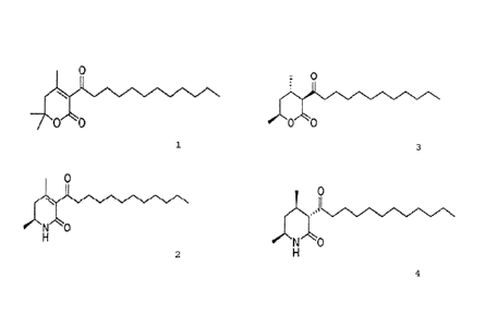
[0002]
OPN is a secreted acidic phosphorylated glycoprotein with
a molecular weight of about 41 kDa identified as a major
non-collagenous protein constituting the matrix of bone tissue
where calcium is deposited. OPN is widely expressed in milk,
urine, renal tubular, osteoclasts, osteoblasts, macrophages,
activated T cells, and various tumor tissues. OPN has been
considered to play a role in anchoring osteoclasts to
hydroxyapatite in bone matrix (Nonpatent Literature 1), but
other various functions of OPN have been reported such as
involvement in cell adhesion, cell migration, control of nitric
monoxide production, tumors, and the immune system.
[0003]
The expression of OPN correlates with tumor progression
and has an association with cancer metastasis. OPN has been
detected in plasma of patients with lung cancer, liver cancer,
breast cancer, or prostate cancer (Nonpatent Literature 2). It
has been reported that the expression of OPN mRNA in a cancer
site is higher than that in a normal site (Nonpatent Literature
3), and it has also been reported that the expression of OPN in
glioma tends to correlate with the degree of malignancy
(Nonpatent Literature 4). The correlation between OPN
expression and tumor has been confirmed also in animal models
(Nonpatent Literature 5). Based on these recent findings about
OPN, suppression of production of OPN that promotes metastasis
and invasion of tumor cells has come to be considered as one of
new approaches of an anti-cancer drug that prevents cancer
metastasis (Nonpatent Literature 6).
1
CA 02896446 2016-02-18
[Citation List]
[Nonpatent Literature]
[0004]
NPL 1: Miyauchi A, Alvarez J, Greenfield EM, Teti A, Grano M,
Colucci S, Zambonin-Zallone A, Ross FP, Teitelbaum SL, Cheresh
D, Hruska KA. (1991) Recognition of osteopontin and related
peptides by an alpha v beta 3 integrin stimulates immediate cell
signals in osteoclasts. J Biol Chem 266,20369-20374
NPL 2: Senger DR, Perruzzi CA, Gracey CF, Papadopoulos A, Tenen
DG. (1988) Secreted phosphoproteins associated with neoplastic
transformation: close homology with plasma proteins cleaved
during blood coagulation. Cancer Res 48, 5770-5774
NPL 3: Brown LF, PapadopopLos-Seigiou A, Brygida B, Manseau EJ,
TognazziK, Perruzzi CA, Dvorak HF, Senger DR. (1994) Osteopontin
expression in human carcinoma. Am J Pathol. 145, 610-623
NPL 4: Saitoh Y, Kuratsu JI, Takeshima H, Yamamoto S, Ushio Y
(1995) Expression of osteopontin in human glioma. Lab Invest 72,
55-63.
NPL 5: Suzuki M, Mose E, GalloyC, and Tarin T (2007) Osteopontin
Gene Expression Determines Spontaneous Metastatic Performance
of Orthotopic Human Breast Cancer Xenografts. Am J Pathol 171,
682-692
NPL 6: Weber GF (2001) Review: The metastasis gene osteopontin:
a candidate target for cancer therapy. Biochim Biophys Acta 1552,
61-85
NPL 7: Kikuchi H, Sasaki K, Sekiya J, Maeda M, Amagai A, Kubohara
Y and Oshima Y (2004) Dihydrodictyopyrone A and C: new members
of dictyopyrone family isolated from Dictyostelium cellular
slime molds Bioorg Med Chem 12, 3203-3214
NPL 8: Matsuura M, Suzuki T, Suzuki M, Tanaka R, Ito E and Saito
T (2011) Statin-mediated reduction of osteopontin expression
induces apoptosis and cell growth arrest in ovarian clear cell
carcinoma. Oncol Rep 25, 41-47
NPL 9: Haruhisa Kikuchi, Koji Nakamura, Yuzuru Kubohara, Naomi
Gokan, Kohei Hosaka, Yasuo Maedad and Yoshiteru Oshima,
Tetrahedron Letters 48 (2007) 5905-5909
2
CA 02896446 2016-02-18
[Summary]
Certain exemplary embodiments provide a composition for the
inhibition of osteopontin production comprising a dictyopyrone
derivative represented by the following chemical formula 1
or 2 as the active ingredient:
0
Chemical Formula 1,
0
Chemical Formula 2;
and at least one pharmaceutically acceptable excipient,
carrier or diluent.
Other exemplary embodiments provide a composition for the
inhibition of osteopontin production comprising a
dihydrodictyopyrone derivative represented by the following
formula 3 or 4 as the active ingredient:
: 0
Chemical Formula 3,
2a
CA 02896446 2016-02-18
0
Chemical Formula 4;
and at least one pharmaceutically acceptable excipient,
carrier or diluent.
Other exemplary embodiments provide a composition for the
inhibition of osteopontin production comprising a dictyopyrone
derivative represented by the following formula 5, 8 or 10 as
the active ingredient:
0
Chemical Formula 5,
0
v v
Chemical Formula 8,
0
N 0
Chemical Formula 10;
and at least one pharmaceutically acceptable excipient,
carrier or diluent.
2b
CA 02896446 2016-02-18
Other exemplary embodiments provide a composition for the
inhibition of osteopontin production comprising a
dihydrodictyopyrone derivative represented by the following
chemical formula 14, 15, or 16 as the active ingredient:
: 0
g
r0"--"t
Chemical Formula 14,
*
se'Ne`'o
Chemical Formula 15,
Ok
Chemical Formula 16;
and at least one pharmaceutically acceptable excipient,
carrier or diluent.
Other exemplary embodiments provide a use of a dictyopyrone
derivative represented by the following chemical formula 1 or
2:
0
***
0 0
Chemical Formula 1,
2c
CA 02896446 2016-02-18
0
Ni/10
H
Chemical Formula 2;
for inhibition of osteopontin production in a patient.
Other exemplary embodiments provide a use of a dictyopyrone
derivative represented by the following chemical formula 3 or
4:
: 0
i 1
oreN
0 0
Chemical Formula 3,
e'N'N
Chemical Formula 4;
for inhibition of osteopontin production in a patient.
Other exemplary embodiments provide a use of a dictyopyrone
derivative represented by the following chemical formula 5, 8
or 10:
2d
CA 02896446 2016-02-18
to
Chemical Formula 5,
0
Chemical Formula 8,
0
N 0
Chemical Formula 10;
for inhibition of osteopontin production in a patient.
Other exemplary embodiments provide a use of a dictyopyrone
derivative represented by the following chemical formula 14, 15
or 16:
: 0
-,N,N.7N,VN
¨N/kb
Chemical Formula 14,
2e
CA 02896446 2016-02-18
OH
Chemical Formula 15,
zOAc
00,N0/.0
Chemical Formula 16;
for inhibition of osteopontin production in a patient.
2f
CA 02896446 2016-02-18
[Technical Problem]
[0005]
As described above, suppression of OPN production has the
potential to prevent cancer metastasis. As drugs having OPN
production inhibitory effect, insulin resistance improvers as
PPAR-y (Peroxysome Proliferator-Activated Receptor-y) agonists
(troglitazone, pioglitazone, rosiglitazone), non-steroid
anti-inflammatory drugs (e.g., indomethacin, ibuprofen),
statin-based drugs for treatment of hypercholesteremia as
HMG-CoA reductase inhibitors (e.g., rosuvastatin, rovastatin,
simvastatin, pravastatin, fluvastatin, atorvastatin,
cerivastatin, pitavastatin, mevastatin) are known.
[0006]
Meanwhile, cellular slime molds are protists widely
distributed in the soil surface layer. Cellular slime molds
have both animal-like and plant-like properties very different
from each other and show both unicellular and multicellular forms,
and their life cycle includes major processes of developmental
systems of multicellular organisms, such as cell movement,
cytokinesis, and differentiation. Such cellular slime molds
are greatly different from organism species conventionally and
commonly used in natural product chemistry, and are therefore
expected to produce various novel compounds.
[0007]
It is therefore an object of the present invention to
provide an OPN production inhibitor capable of preventing a
disease (e.g., cancer metastasis) resulting from increased
production of OPN.
[Solution to Problem]
[0008]
In order to find a compound that suppresses OPN gene
expression, the present inventors have performed screening for
secondary metabolites of cellular slime molds such as D.
discoideum with the use of cell strains that express luciferase
as a reporter gene under control of OPN promoter.
[0009]
As a result, the present inventors have found compounds
that suppress luciferase expression from among dictyopyrone
derivatives and dihydrodictyopyrone derivatives.
3
CA 02896446 2015-10-02
[0010]
Further, the present inventors have also found that such
compounds that suppress luciferase activity under control of OPN
promoter reduce the amount of OPN produced by a human
non-small-cell lung cancer-derived cell line A549 or a human liver
cancer-derived cell line HepG2. Further, the present inventors
have also found that in Wound-Healing assay, the dictyopyrone
derivatives and dihydrodictyopyrone derivatives suppress the
ability of OPN to migrate cells, which is the physiological
function of OPN, and that in matrix gel invasion assay, the
dictyopyrone derivatives and dihydrodictyopyrone derivatives
suppress the metastatic and invasive capacity of cells. These
findings have led to the completion of the present invention.
[0011]
Specifically, the present invention relates to an
osteopontin production inhibitor containing a dictyopyrone
derivative or a dihydrodictyopyrone derivative as an active
ingredient.
[0012]
It has been reported that some dictyopyrone derivatives
and dihydrodictyopyrone derivatives have the activity of
suppressing the growth of human leukemia cell-derived K562 cells
(Nonpatent Literature 7), but their OPN production inhibitory
effect has not heretofore been reported.
[0013]
The dictyopyrone derivative is preferably a compound
represented by the following chemical formula 1 or 2.
[0014]
[Chemical Formula 1]
0
'1/4N
//iN00
CA 02896446 2015-10-02
[0015]
[Chemical Formula 2]
0
N,
H
[0016]
The dihydrodictyopyrone derivative is preferably a
compound represented by the following chemical formula 3 or 4.
[0017]
[Chemical Formula 3]
: 0
[0018]
[Chemical Formula 4]
0
- N. 0
[Advantageous Effects of Invention]
[0019]
The OPN production inhibitor according to the present
invention containing a dictyopyrone derivative or a
dihydrodictyopyrone derivative as an active ingredient is an OPN
production inhibitor whose mechanism of action is different from
that of an insulin resistance improver or a statin-based drug
for treatment of hypercholesteremia.
[Brief Description of Drawings]
[0020]
[Fig. 11 Fig. 1 is a graph showing the effect of addition
of a compound 3 (compound represented by the chemical formula 3)
on suppressing OPN production by A549 cells.
CA 02896446 2015-10-02
[Fig. 2] Fig 2 is a graph showing the effect of addition
of the compound 3 (compound represented by the chemical
formula 3) on suppressing OPN production by HepG2 cells.
[Fig. 3] Fig. 3 is a graph showing the effect of addition
of the compound 3 (compound represented by the chemical
formula 3) on suppressing healing of wounds in A549 cells.
[Fig 4] Fig. 4 is a graph showing the effect of addition
of the compound 3 (compound represented by the chemical
formula 3) on suppressing matrix invasion by A549 cells.
[Fig. 5] Fig. 5 is a graph showing the effect of addition
of MVA (mevalonic acid) on recovering OPN production suppressed
by SVS (simvastatin).
[Fig. 6] Fig. 6 is a graph showing the influence of MVA
addition on the OPN production-suppressing effect of the
compound 3.
[Description of Embodiments]
[0021]
An embodiment of the present invention will be described
with reference to the accompanying drawings as appropriate. The
present invention is not limited to the following description.
[0022]
<A. Method for confirming luciferase expression inhibitory
effect under control of OPN promoter>
A reporter vector pOPN1-luc obtained by inserting a human
OPN promoter sequence (-765 to 23) into the multiple cloning site
of pGL-3 basic vector (Promega) expresses luciferase when
transfected into animal cells. This pOPN1-luc was transfected
into a human non-small cell lung cancer-derived cell line A549
together with pPURTI" (Ciontech) that expresses a puromycin
resistance gene (puromycin-N-acetyl-transferase gene), and
cells that could grow in a puromycin-supplemented medium and
expressed luciferase were selected. The selected cells were
named A549/0PNluc cells and used for observation of luciferase
expression inhibitory effect under control of OPN promoter, as
described later.
[0023]
A test compound was added to a culture liquid containing
A549/0PNluc cells to observe its influence on the amount of
luciferase expressed in the cells. Here, it can be considered
that when the test compound has cytotoxity or cell growth-
CA 02896446 2015-10-02
suppressing effect, the total expression level of luciferase is
reduced due to a reduction in the number of living cells that
depends on the concentration of the test compound, and therefore
the luciferase expression inhibitory effect of the test compound
under control of OPN promoter cannot be properly evaluated. For
this reason, WST assay for quantification of cell proliferation
ability or cell viability by colorimetric measurement was first
performed to determine IC50 (concentration for 50% inhibition
of cell growth) of the test compound for cell growth. Then,
luciferase activity measurement was performed to determine EC50
(concentration for 50% inhibition of luciferase expression) of
the test compound for luciferase expression under control of OPN
promoter.
[0024]
Al) WST assay
A5 4 9 /OPNluc cells were suspended in DMEM medium containing
10% fetal calf serum (FCS) and 1% penicillin/streptomycin (P/S)
at 3 x 10⁴ cells/mL to obtain a cell suspension, and 100 L of
the cell suspension was dispensed into each well of a 96-well
plate. In order to perform the assay in triplicate, 3 wells were
prepared for a control group and 3 wells were prepared for each
test compound-treated group at each concentration. After the
dispensing, the 96-well plate was incubated in a CO2 incubator
(at 37 C and 5% CO2) for 24 4 hours.
[0025]
The test compound was dissolved in dimethylsulfoxide
(DMS0) to obtain a 50 mmol/L solution, and the test compound
solution was stored at -80 C. The test compound solution was
diluted with DMSO in 2-fold dilution series (usually, in the
range of 0.31 mmol/L to 20 mmol/L) to prepare test compound
solutions whose concentration varied by two fold for WST assay.
[0026]
Only DMSO (control) or the diluted test compound (sample)
solution was dispensed in an amount of 0.5 vL into each well
containing the cell suspension (200-fold dilution). The
solution in each well was mixed with a vortex mixer, and then
the 96-well plate was incubated in a CO2 incubator (at 37 C and
5% CO2) for 48 4 hours. Then, 10 vL of PremixTmWST-1 Reagent
(TAKARA BIO INC.) was added to each well. The solution in each
well was mixed with a vortex mixer, and then the 96-well plate
7
CA 02896446 2015-10-02
was incubated at 37 C and 5% CO2. After 60 minutes or 120 minutes,
absorbance values (450 nm) were measured using a microplate
reader (Bio-RadTM; Benchmark or Thermo Scientific; VarioskanTM
Flash) .
[0027]
The absorbance values of the control wells and the
absorbance values of the sample wells at each concentration were
input into an Excel" file to determine the percentages of
absorbance values of the sample wells at each concentration with
respect to the average of absorbance values of the control wells.
From the determined values, a fitted curve was determined by the
method of least squares to calculate 1050.
[0028]
A2) Luciferase assay
The same steps as in the above-described WST assay were
performed in which only DMSO (control) or the diluted test
compound (sample) solution was added in an amount of 0.5 1.1.1, to
each well containing the cell suspension to achieve 200-fold
dilution, and the solution in each well was mixed with a vortex
mixer, and the 96-well plate was incubated in a CO2 incubator
(at 37 C and 5% CO2) for 48 4 hours.
[0029]
A luciferase reagent was prepared by dissolving Luciferase
Assay Substrate (hereinafter, referred to as "LAS") supplied in
Luciferase Assay Systems (Promega: Cat# E1500) with Luciferase
Assay Buffer (LAB) . 5xCell Culture Lysis Reagent (hereinafter,
referred to as "CCLR") was diluted with water 5-fold to prepare
1xCCLR.
[0030]
After the incubation for 48 4 hours, the medium in each
well was completely removed, and 50 [.LL of 1xCCLR was dispensed
into each well. The 96-well plate was allowed to stand at room
temperature for 30 minutes, and then 1xCCLR in each well was used
as an assay sample. The luciferase reagent of 100 ILL was placed
in a tube for chemiluminescence measurement, and 20 idL of the
assay sample was added to the tube and mixed with the luciferase
reagent to measure chemiluminescence (Relative Luminescence
Intensity: Riit1) using Tuner Design Luminometer 20/20 (Promega) .
8
CA 02896446 2015-10-02
[0031]
The RLU values of the control wells and the RLU values of
the sample wells at each concentration were input into an Excel
file, and the percentages of RLU values of the sample wells at
each concentration with respect to the average of RIG values of
the control wells were determined. From these values, a fitted
curve was determined by the method of least squares to calculate
EC50.
[0032]
Table 1 shows 1050 values calculated by WST assay and EC50
values calculated by luciferase assay of dictyopyrone
derivatives and dihydrodictyopyrone derivatives as test
compounds represented by the following chemical formulas 1
to 16.
[0033]
[Chemical Formula 5]
0
ne/N-00
[0034]
[Chemical Formula 6]
[0035]
[Chemical Formula 7]
0
I
---------
oe.C1`
O0
9
CA 02896446 2015-10-02
[0036]
[Chemical Formula 8]
0
[0037]
[Chemical Formula 9]
0
. (We
.
00 0
[0038]
[Chemical Formula 10]
0
,s
[0039]
[Chemical Formula 11]
0
11 0
[0040]
[Chemical Formula 12]
0
40.A
0 0
CA 02896446 2015-10-02
[0041]
[Chemical Formula 13]
: 0
0
[0042]
[Chemical Formula 14]
0
0 0
[0043]
[Chemical Formula 15]
-
. .
0 0
[0044]
[Chemical Formula 16]
OAc
.410
[0045]
Here, the compounds represented by the chemical formulas 1,
2, 5 to 8 were produced according to a production method disclosed
in Nonpatent Literature 7, and the compounds represented by the
chemical formulas 3, 12, 13, 15, and 16 were produced according
to a production method disclosed in Nonpatent Literature 9.
CA 02896446 2015-10-02
[0046]
<Method for producing compound represented by chemical
formula 4>
(S)-3-dodecanoy1-5,6-dihydro-4,6-dimethy1-1H-pyridin-2
-one of 5 mg synthesized as described in Nonpatent Literature
7 was dissolved in 1 mL of methanol. Palladium-carbon (Pd 5%)
of 1 mg was added, and the mixture was stirred at room temperature
for 2 hours in an atmosphere of hydrogen to obtain a reaction
liquid. The reaction liquid was filtered to remove
palladium-carbon, and the filtrate was subjected to distillation
under a reduced pressure. The residue was subjected to silica
gel column chromatography, and 4 mg of the compound represented
by the chemical formula 4 was obtained from fractions eluted with
hexane-ethyl acetate (4:1).
[0047]
The obtained compound was analyzed by electron impact mass
spectrometry (EIMS) and NMR. The results of EIMS and NMR are
as follows.
'H-NMR (400 MHz, CDC13) d 5.76 (1H, br.$),
3.52-3.64 (1H,
m), 3.05 (1H, d, J - 11.2 Hz), 2.81 (1H, dt, J = 17.8, 7.5 Hz),
2.52 (1H, dt, J = 17.8, 7.4 Hz), 2.28-2.41 (1H, m), 1.87 (1H,
J= 13.0, 2.3 Hz), 1.52-1.68 (3H, m), 1.23-1.36 (16H, s),
1.17 (3H, d, J = 6.4 Hz), 0.94 (3H, d, J = 6.5 Hz), 0.88 (3H,
t, J = 6.4 Hz).
EIMS m/z (rel. int) 309 [MY' (11), 294 (5), 182 (10), 169
(13), 127 (100), 112 (55).
[0048]
<Method for producing compound represented by chemical
formula 9>
Suberic acid monomethyl ester of 297 mg was dissolved in
8 mL of methylene chloride, and 250 mg of Meldrum's acid, 453 mg
of 1-ethyl-3-(3-dimethylaminopropyl)carbodiimide
hydrochloride, and 19 mg of 4- ( dimethyl amino ) pyridine were added,
and the mixture was stirred at room temperature for 4 hours to
obtain a reaction liquid. Thirty milliliters of 0.5 M
hydrochloric acid was added to the reaction liquid, and the
mixture was subjected to extraction with 30 mL of ethyl acetate
three times. After the extraction, all the ethyl acetate layers
were combined, washed with 60 mL of water and then with 60 mL
cf saturated salt water, and dried with anhydrous sodium sulfate,
12
CA 02896446 2015-10-02
and then the solvent was removed by distillation under a reduced
pressure. The residue was dissolved in 5 mL of toluene, and
246 mg of (2S,4S)-2,4-pentanediol was added, and the mixture was
stirred at 120 C for 2 hours to obtain a reaction liquid. After
returned to room temperature, the reaction liquid was subjected
tc distillation under a reduced pressure. The residue was
subjected to silica gel column chromatography, and 200 mg of
1-(1S,3S)-3-hydroxy-l-methylbutyl 10-methyl 3-oxodecanedioate
was obtained from fractions eluted with hexane-ethyl acetate
(3:1).
[0049]
1-(1S,3S)-3-hydroxy-1-methylbutyl 10-methyl
3-oxodecanedioate of 196 mg was dissolved in 8 mL of methylene
chloride, and 109 mg of N-methylmorpholine N-oxide, 311 mg of
powdered molecular sieves 4A, and 11 mg of tetrapropylammonium
perruthenate were added in order, and the mixture was stirred
at room temperature for 5 hours to obtain a reaction liquid. The
reaction liquid was filtered, and the filtrate was subjected to
distillation under a reduced pressure. The residue was
subjected to silica gel column chromatography, and 133 mg of
1-(S)-1-methyl-3-oxobutyl 10-methyl 3-oxodecanedioate was
obtained from fractions eluted with hexane-ethyl acetate (4:1).
[0050]
1-(S)-1-methyl-3-oxobutyl 10-methyl 3-oxodecanedioate of
126 mg was dissolved in 2 mL of ethanol, and 41 mg of sodium
ethoxide was added, and the mixture was stirred at room
temperature for 10 hours to obtain a reaction liquid. The
reaction liquid was poured into 20 mL of 0.5 Mr hydrochloric acid,
and the mixture was subjected to extraction with 20 mL of ethyl
acetate three times. After the extraction, all the ethyl
acetate layers were combined, washed with 40 mL of water and then
with 40 mL of saturated salt water, and dried with anhydrous
sodium sulfate, and then the solvent was removed by distillation
under a reduced pressure. The residue was subjected to silica
gel column chromatography, and 88 mg of methyl
(S)-8-(4,6-dimethy1-2-oxo-5,6-Oihydro-2H-pyran-3-y1)-8-0xooc
tanoate (compound represented by chemical formula 9) was
obtained from fractions eluted with hexane-ethyl acetate (4:1).
13
CA 02896446 2015-10-02
[0051]
The obtained compound was analyzed by electron impact mass
spectrometry (EIMS) and NMR. The results of EIMS and NMR are
as follows.
'H-NMR (400 MHz, CDC13) d 4.50-4.60 (1H, m), 3.67 (3H, s),
2.74 (1H, dt, J= 17.3, 7.4 Hz), 2.73 (1H, dt, J= 17.3, 7.4 Hz),
2.45(1H, ddq, J - 17.9, 11.6, 0.9 Hz), 2.31 (1H, dd, J - 17.9,
3.8 Hz), 2.30 (2H, t, J - 7.4 Hz), 2.01 (3H, d, J - 0.9 Hz),
1.58-1.68 (4H, m), 1.44 (3H, d, J= 6.4 Hz), 1.30-1.38 (4H, m).
EIMS m/z (rel. int) 296 [M]
(6), 281 (10), 265 (11), 246
(14), 181 (20), 168 (83), 153 (100), 109 (30).
[0052]
<Method for producing compound represented by chemical
formula 10>
(S)-2-[1-Methyl-2-(2-methyl-1,3-dioxan-2-y1)ethyl]-iso
indol e- 1 , 3 -dione of 300 mg synthesized as described in Nonpatent
Literature 7 was dissolved in 6 mL of methanol, and 1038 mg of
hydrazine monohydrate was added, and the mixture was heated to
reflux for 6 hours to obtain a reaction liquid. After returned
to room temperature, the reaction liquid was mixed with 30 mL
of a 1 M aqueous sodium hydroxide solution, and the mixture was
subjected to extraction with 30 mL of ethyl acetate three times.
After the extraction, all the ethyl acetate layers were combined,
washed with 60 mL of water and then with 60 mL of saturated salt
water, and dried with anhydrous sodium sulfate, and then the
solvent was removed by distillation under a reduced pressure.
The residue was dissolved in 6 mL of toluene, and 565 mg of
5-butanoy1-2,2-dimethy1-1,3-dioxane-4,6-dione was added, and
the mixture was heated to reflux for 15 hours to obtain a reaction
liquid. After returned to room temperature, the reaction liquid
was subjected to distillation under a reduced pressure. The
residue was subjected to silica gel column chromatography, and
140 mg of
(S)-N-[1-methy1-2-(2-methyl-1,3-dioxan-2-yl)ethyl]-3-oxohexa
namide was obtained from fractions eluted with hexane-ethyl
acetate (2:1).
0053]
(S)-N-El-methy1-2-(2-methyl-1,3-dioxan-2-yl)ethyll-3-
oxohexanamide of 115 mg was dissolved in 4 mL of an 80% aqueous
acetic acid solution, and the mixture was stirred at room
14
CA 02896446 2015-10-02
temperature for 7 hours to obtain a reaction liquid. The
reaction liquid was subjected to distillation under a reduced
pressure, and the residue was subjected to silica gel column
chromatography, and 59 mg of
(S)-N-(1-methyl-3-oxobuty1)-3-oxodecanamide was obtained from
fractions eluted with hexane-ethyl acetate (2:1).
[0054]
(S)-N-(1-methyl-3-oxobuty1)-3-oxodecanamide of 11 mg was
dissolved in 1 mL of ethanol, and 5 mg of sodium ethoxide was
added, and the mixture was stirred at room temperature for 8 hours
to obtain a reaction liquid. The reaction liquid was poured into
mL of 0.5 M hydrochloric acid, and the mixture was subjected
tc extraction with 10 mL of ethyl acetate three times. After
the extraction, all the ethyl acetate layers were combined,
washed with 20 mL of water and then with 20 mL of saturated salt
water, and dried with anhydrous sodium sulfate, and then the
solvent was removed by distillation under a reduced pressure.
The residue was subjected to silica gel column chromatography,
and 4 mg of
(S)-3-butanoy1-5,6-dihydro-4,6-dimethy1-1H-pyridin-2-one
(compound represented by chemical formula 10) was obtained from
fractions eluted with hexane-ethyl acetate (2:1).
[0055]
The product was analyzed by electron impact mass
spectrometry (EIMS) and NMR. The results of EIMS and NMR are
as follows.
H-NMR (400 MHz, CDC13) d 5.45 (1H, br.$), 3.66-3.78 (1H,
m), 2.71 (2H, t, J = 7.4 Hz), 2.31 (1H, dd, J = 17.6, 7.6 Hz),
2.23 (1H, dd, J = 17.6, 7.2 Hz), 1.93 (3H, s), 1.64 (2H, quint,
J= 7.4 Hz), 1.24, (3H, d, J= 6.4 Hz), 0.94 (3H, t, J= 7.4 Hz).
EIMS m/z (rel. int) 195 [Mr- (19), 180 (100), 152 (54), 109
(22).
[0056]
<Method for producing compound represented by chemical
formula 11>
( S) [l-Methyl-2- (2-methyl-1, 3-dioxan-2-y1) ethyl]
isoindole-1,3-dione of 300 mg synthesized as described in
Nonpatent Literature 7 was dissolved in 6 mL of methanol, and
1038 mg of hydrazine monohydrate was added, and the mixture was
heated to reflux for 6 hours to obtain a reaction liquid. After
CA 02896446 2015-10-02
returned to room temperature, the reaction liquid was mixed with
30 mL of a 1 M aqueous sodium hydroxide solution, and the mixture
was subjected to extraction with 30 mL of ethyl acetate three
times. After the extraction, all the ethyl acetate layers were
combined, washed with 60 mL of water and then with 60 mL of
saturated salt water, and dried with anhydrous sodium sulfate,
and then the solvent was removed by distillation under a reduced
pressure. The residue was dissolved in 6 mL of toluene, and
564 mg of 2,2-dimethy1-5-octanoy1-1,3-dioxane-4,6-dione was
added, and the mixture was heated to reflux for 15 hours to obtain
a reaction liquid. After returned to room temperature, the
reaction liquid was subjected to distillation under a reduced
pressure. The residue was subjected to silica gel column
chromatography, and 51 mg of
(S)-N-[1-methyl-2-(2-methyl-1,3-dioxan-2-y1)ethyl]-3-oxodeca
namide was obtained from fractions eluted with hexane-ethyl
acetate (2:1).
[C057]
(S)-N-[1-methy1-2-(2-methy1-1,3-dioxan-2-y1)ethyl]-3-
oxodecanamide of 47 mg was dissolved in 3 mL of an 80% aqueous
acetic acid solution, and the mixture was stirred at room
temperature for 7 hours to obtain a reaction liquid. The
reaction liquid was subjected to distillation under a reduced
pressure, and the residue was subjected to silica gel column
chromatography, and 38 mg of
(S)-N-(1-methyl-3-oxobuty1)-3-oxodecanamide was obtained from
fractions eluted with hexane-ethyl acetate (2:1).
[0058]
(S)-N-(1-methyl-3-oxobuty1)-3-oxodecanamide of 23 mg was
dissolved in 2 mL of N,N-dimethylformamide, and 3 mg of sodium
hydride (60%, dispersed in mineral oil) was added, and the
mixture was stirred at room temperature for 10 hours to obtain
a reaction liquid. The reaction liquid was poured into 10 mL
of 0.5 M hydrochloric acid, and the mixture was subjected to
extraction with 10 mL of ethyl acetate three times. After the
extraction, all the ethyl acetate layers were combined, washed
with 20 mL of water and then with 20 mL of saturated salt water,
and dried with anhydrous sodium sulfate, and then the solvent
was removed by distillation under a reduced pressure. The
residue was subjected to silica gel column chromatography, and
16
CA 02896446 2015-10-02
9 mg of (S)-5,6-dihydro-4,6-dimethy1-3-octanoy1-1H-pyridin-
2-one (compound represented by the chemical formula 11) was
obtained from fractions eluted with hexane-ethyl acetate (2:1).
[0059]
The product was analyzed by electron impact mass
spectrometry (EIMS) and NMR. The results of EIMS and NMR are
as follows.
'H-NMR (400 MHz, CDC13) d 5.52 (1H, br.$), 3.65-3.78 (1H,
m), 2.72 (2H, t, J = 7.6 Hz), 2.31 (1H, dd, J = 17.2, 5.6 Hz),
2.23 (1H, dd, J = 17.2, 7.2 Hz), 1.92 (3H, s), 1.60 (2H, quint,
J = 7.6 Hz), 1.23-1.35 (8H, m), 1.24 (3H, d, J = 6.8 Hz), 0.87
(3H, t, J = 7.0 Hz).
EIMSm/z (rel. int) 251 [M]+ (33), 236 (57), 180 (100), 167
(69), 152 (76), 109 (23).
[C060]
<Method for producing compound represented by chemical
formula 14>
3-dodecanoy1-5,6-dihydro-4,6,6-trimethy1-2H-pyran-2-one
of 10 mg synthesized as described in Nonpatent Literature 7 was
dissolved in 1 mL of ethyl acetate. One milligram of 20%
palladium hydroxide-carbon was added, and the mixture was
stirred at room temperature for 20 hours in an atmosphere of
hydrogen to obtain a reaction liquid. The reaction liquid was
filtered to remove palladium-carbon, and the filtrate was
subjected to distillation under a reduced pressure. The residue
was subjected to silica gel column chromatography, and 9 mg of
the compound represented by the chemical formula 14 was obtained
from fractions eluted with hexane-ethyl acetate (19:1).
[0061]
The product was analyzed by electron impact mass
spectrometry (EIMS) and NMR. The results of EIMS and NMR are
as follows.
1H-NMR (400 MHz, CDC13) d 3.13 (1H, d, J - 10.8 Hz), 2.87
(1H, dt, J = 14.8, 7.2 Hz), 2.59-2.67 (1H, m), 2.55 (1H, dt,
J = 14.8, 6.8 Hz), 1.84 (1H, dd, J = 13.4, 4.0 Hz), 1.57-1.66
(2H, m), 1.51 (1H, t, J = 13.4 Hz), 1.43 (3H, s), 1.42 (3H, s),
1.25-1.31 (16H, m), 0.95 (3H, d, J - 6.4 Hz), 0.88 (3H, t, J
- 6.6 Hz).
EIMS m/z (rel. int) 324 [M] (3), 309 (6), 84 (100).
11
CA 02896446 2015-10-02
[0062]
[Table 1]
Dictyopyrone derivatives 1050 (pM) EC50 (pM)
Chemical formula 1 128.9 24.4
Chemical formula 2 59.1 29.5
Chemical formula 5 129.1 63
Chemical formula 6 >100 >100
Chemical formula 7 >100 >100
Chemical formula 8 111.4 55
Chemical formula 9 >100 >100
Chemical formula 10 109.5 63
Chemical formula 11 >100 >100
Dihydrodictyopyrone
1050 (pM) EC50 (pM)
derivatives
Chemical formula 3 56.2 15.9
Chemical formula 4 42.5 19.7
Chemical formula 12 >50 >50
Chemical formula 13 >50 >50
Chemical formula 14 54.1 29.5
Chemical formula 15 88 43.4
Chemical formula 16 >100 31.1
[0063]
The dictyopyrone derivatives represented by the chemical
formulas 1, 2, and 8 and the dihydrodictyopyrone derivatives
represented by the chemical formulas 3, 4, 15, and 16 had EC50
values equal to or less than 1/2 of their respective 1050 values.
In particular, the compounds represented by the chemical
formulas 1 to 4 (compounds 1 to 4) had EC50 values of less than
30 idM, that is, suppressed luciferase (OPN Luc) expression under
control of OPN promoter by 50% at low concentration.
[0064]
<B: Effect of compound 3 on suppressing OPN production by
human non-small cell lung cancer-derived cell line A549 and human
liver cancer-derived cell line HepG2>
As described above, some of the dictyopyrone derivatives
and dihydrodictyopyrone derivatives were confirmed to inhibit
luciferase expression under control of OPN promoter by 50% at
18
CA 02896446 2015-10-02
a concentration equal to or less than 1/2 of their respective
50% cell growth inhibition concentrations (1050) . Therefore, the
compound represented by the chemical formula 3 (compound 3) whose
EC50 value is lowest and IC50 value is two times or more its EC50
value was subjected to a confirmation test to determine whether
or not it could actually suppress OPN production by a cancer
cell-derived cell line.
[0065]
The confirmation test was performed in the following manner.
The compound 3 wad added to a culture liquid containing cells
of a human non-small cell lung cancer-derived cell line A549 or
a human liver cancer-derived cell line HepG2, and the amount of
OPN in culture supernatant after 2-day culture was measured using
Human Osteopontin Immunoassay kit (R&D systems) and compared
with the OPN amount of a compound 3-free group (control group).
[0066]
The compound 3 has also cell growth inhibitory effect.
Therefore, in order to properly evaluate the OPN production-
suppressing effect of the compound 3, 1xCCLR used in luciferase
assay was added to cells remaining in each well after removal
of culture supernatant to prepare a cell lysate, and the amount
of protein in the cell lysate was measured using BCA Protein Assay
Kit (Thermo Scientific). The influence of addition of the
compound 3 on the amount of OPN production was evaluated by
comparison of the amount of OPN per milligram of protein.
[0067]
B1) Sample preparation method
First, A549 cells or HepG2 cells were suspended in DMEM
medium. containing 105 FCS and 1% F/S at 4x 10⁴cells/mL to obtain
a cell suspension, and 500 tL of the cell suspension was dispensed
into each well of a 24-well plate. In order to perform the test
in triplicate, 3 wells were prepared for a control group and 3
wells were prepared for each test compound-treated group at each
concentration. The 24-well plate was incubated in a CO2 incubator
(at 37 C and 5% CO2) for 24 4 hours.
[0068]
The compound 3 as a test compound was dissolved in DMSO
to prepare a 50 mmol/L solution, and the solution was stored at
-80 C. The compound 3 solution was diluted with DMSO to prepare
a 3.75 mmol/L solution, a 7.5 mmol/L solution, and a 15 mmol/L
19
CA 02896446 2015-10-02
solution. The 7.5 mmol/L compound 3 solution or the 15 mmol/L
compound 3 solution was added in an amount of 2 L to each well
in which A549 cells were cultured. Only DMSO was added to each
control well in an amount of 2 L.
[0069]
The 3.75 mmol/L compound 3 solution or the 7.5 mmol/L
compound 3 solution was added in an amount of 2 L to each well
in which HepG2 cells were cultured. Only DMSO was added to each
control well in an amount of 2 L.
[0070]
The 24-well plate was rocked back and forth and side to
side to mix the solution in each well, and was then incubated
in a CO2 incubator (at 37 C and 5% CO2) for 48 4 hours. Then,
the total amount of culture supernatant was transferred into a
1.5 mL tube, and 500 L of D-PBS(-) was added to each well in
which cells are remained.
[0071]
The 24-well plate was gently rocked, and D-PBS(-) was
removed, and 1xCCLR prepared by 5-fold diluting 5xCCLR used in
luciferase assay with water was dispensed into each well in an
amount of 200 L. Then, the 24-well plate was rocked on a rocking
shaker for 15 minutes.
[0072]
After rocking the 24-well plate for 15 minutes, the cell
lysate in each well of the 24-well plate was transferred into
a 1.5 mL tube and centrifuged in a high-speed refrigerated micro
centrifuge at 4 C and 15000 rpm for 1 minute to remove impurities
such as cell debris. The thus obtained supernatant was used as
a sample for protein assay.
[0073]
On the other hand, the culture supernatant transferred into
a 1.5 mL tube was centrifuged in a high-speed refrigerated micro
centrifuge at 4 C and 15000 rpm for 1 minute to remove impurities
such as cell debris. The thus obtained supernatant was used as
a sample for ELISA.
[0074]
B2) Measurement of amount of OPN protein
The sample for ELISA was diluted in the following manner.
A549 cells (20-fold dilution): sample 5 p.1_, +culture medium
95 !.11_,
CA 02896446 2015-10-02
HepG2 cells (80-fold dilution) : sample 2 L + culture
medium 158 L
[0075]
Water of 1 mL was added to a vial of OPN standard supplied
in an ELISA kit, and the solution in the vial was gently mixed
and allowed to stand at room temperature for 15minutes to prepare
200 ng/mL OPN Standard. Then, as shown in Table 2, the OPN
standard was serially diluted with a diluent RD5-24 supplied in
the kit.
[0076]
[Table 2]
Vial Volume of R95-24 Type and amount of OPN
OPN concentration
prepared ( L) standard solution added (ng/mL)
200
60 L of 200 ng/mL OPN
A 540 pL 20
Standard
300 pL 300 L of vial A 10
300 pL 300 L of vial B 5
300 pL 300 L of vial C 2.5
300 pL 300 ta._, of vial D 1.25
300 pL 300 L of vial E 0.625
300 pL 300 L of vial F 0.312
300 pL 0 = Blank
[0077]
The necessary number of OPN Microplates and RD1 - 6 supplied
in the kit were prepared, and 100 L of RD1-6 was dispensed into
each well. The diluted OPN Standard (vial A to H) or the sample
was further added in an amount of 50 L to each well containing
RD1-6. Then, the upper end of each well was covered with a seal,
and the OPN microplates were allowed to stand at room temperature
for 2 hours. In this period, Wash Buffer Concentrate supplied
in the kit was diluted with water to prepare Wash Buffer.
[0078]
After allowing the OPN microplates to stand for 2 hours,
the liquid in each well was removed. The Wash Buffer of 250 L
was dispensed into each well and then removed to wash each well.
This washing operation was performed 4 times.
[0079]
OPN conjugate of 200 uL was dispensed into each well. Then,
the upper end of each well was covered with a seal, and the OPN
microplates were allowed to stand at room temperature for 2 hours.
CA 02896446 2015-10-02
In this period, Color Reagent A and Color Reagent B supplied in
the kit were mixed in equal amount to prepare Substrate Solution.
[0080]
After allowing the OPN microplates to stand for 2 hours,
the liquid in each well was removed. The Wash Buffer of 250 L
was dispensed into each well and then removed to wash each well.
This washing operation was performed 4 times.
[0081]
The Substrate Solution of 200 L was dispensed into each
well, and then the OPN microplates were allowed to stand at room
temperature for 30 minutes while being shielded from light. Then,
Stop Solution supplied in the kit was added to each well in an
amount of 50 L, and the liquid in each well was gently mixed
with a vortex mixer until the color of the liquid in each well
was entirely changed from blue to yellow. After the mixing,
absorbance values (at 450 nm and 570 nm) were measured using a
microplate reader (Bio-Rad; Benchmark or Thermo Scientific;
Varioskan Flash). The subtraction of the value at OD 570 nm from
the value at OD 450 nm was performed on all the measured data,
and the thus determined values were used for calculation performed
later.
[0082]
A calibration curve was prepared from the absorbance values
of the calibration curve samples. The amount of OPN protein was
calculated using the formula of the calibration curve from the
absorbance value of each sample.
[0083]
B3) Measurement of total amount of protein in cells
The sample for protein assay of 10 L and water of 90 L
were mixed to dilute the sample 10-fold. Calibration curve
samples were prepared by diluting a BSA solution with water as
shown in Table 3.
22
CA 02896446 2015-10-02
[0084]
[Table 3]
BSA
Water Type and volume of
BSA
Vial
concentration
( L) solution added
( g/mL)
2000
A 140 pL 20 L of 2000 ng/mL
BSA 250
80 pL 80 L of vial A 125
90 pL 60 L of vial B 50
80 pL 80 L of vial C 25
80 pL 20 L of vial D 5
80 pL 0
[0085]
BCA Reagent A and BCA Reagent B supplied in a protein assay
kit were mixed (50:1) to prepare Working Reagent. The calibration
curve sample (vial A to F) or the 10-fold diluted sample for
protein assay was dispensed into each well of a 96-well plate
in an amount of 25 L. In order to perform the assay in duplicate,
2 wells were prepared for a control group, and 2 wells were
prepared for each test compound-treated group at each
concentration.
[0086]
The Working Reagent of 200 L was added to each well, and
the solution in each well was mixed for 30 seconds with a vortex
mixer. The 96-well plate was heated at 60 C for 30 minutes and
then allowed to stand at room temperature for 15 minutes. Then,
absorbance values (550 nm) were measured using a mi cropl at e reader
(Bio-Rad; Benchmark or Thermo Scientific; Varioskan Flash).
[0087]
A calibration curve was prepared from the absorbance values
of the calibration curve samples, and the total protein
concentration of the diluted sample in each well was calculated
using the formula of the calibration curve. Further, the total
protein concentration was multiplied by the dilution factor (10)
to calculate the total protein concentration of the sample.
23
CA 02896446 2015-10-02
[0088]
(Expression of OPN (protein) amount)
The OPN amount (amount per milliliter of culture
supernatant) was converted to the amount of OPN per total amount
(0.5 mL) of the sample for ELISA. This converted value was
divided by the amount of protein in the total amount (0.2 mL)
of the sample for protein assay derived from the same well as
the sample for measuring OPN amount for conversion to the amount
of OPN per milligram of total protein in cells. Based on the
amount of OPN per milligram of protein, the influence of addition
of the compound 3 was evaluated.
[0089]
(Statistical processing)
The statistical processing of the OPN amounts was performed
using Excel statistics, Statcel 3. The OPN amounts of the
samples of the compound 3 (test compound)-treated groups were
subjected to Dunnett test for comparison with those of the
control group on an Excel file.
[0090]
Fig. 1 is a graph showing the effect of addition of the
compound 3 on suppressing OPN production by A549 cells. It was
confirmed that when the concentration of the compound 3 was
30 imol/L or 60 flmol/L, the compound 3 significantly suppressed
OPN production by A549 cells at a significance level of less than
5% or 1%, respectively as compared to the control group.
[0091]
Fig. 2 is a graph showing the effect of addition of the
compound 3 on suppressing OPN production by HepG2 cells. It was
confirmed that when the concentration of the compound 3 was
15 vtol/L or 30 lamol/L, the compound 3 significantly suppressed
OPN production by HepG2 cells at a significance level of less
than 1% as compared to the control group. In particular, when
the concentration of the compound 3 was 30 wrIol/L, the amount
of OPN produced by HepG2 cells was reduced to half or less of
that of the control group.
[0092]
It was confirmed from Figs. 1 and 2 that the compound 3
significantly reduced the amount of OPN produced by a human
non-small-cell lung cancer-derived cell line A549 or by a human
liver cancer-derived cell line HepG2.
24
CA 02896446 2015-10-02
[0093]
<C: Effect of compound 3 on suppressing wound-healing
capacity of human non-small cell lung cancer-derived cell line
A549>
The experimental results shown in Figs. 1 and 2 revealed
that the compound 3 that suppresses luciferase expression under
control of OPN promoter actually inhibited the production of OPN
as protein. In the next stage, a determination was made as to
whether or not the physiological function of cells induced by
OPN production was suppressed by the compound 3 as a test compound.
The determination was made by performing cell Wound-Healing
assay. This assay is commonly known as a method for evaluating
cell migration capacity. When a wound is made in a cell monolayer
by scratching some cells with a pipette tip or the like, the wound
is closed by migration of cells around the wound. The influence
of a test compound on such function was observed by adding the
test compound to a cell culture liquid.
[0094]
Cl) Method for Wound-Healing assay
Cells of a human non-small cell lung cancer-derived cell
line A549 were suspended in DMEM medium containing 10% FCS and
1% P/S at 3.5 x 10³ cells/mL to obtain a cell suspension, and
500 pL (1.75 x 10⁵ cells) of the cell suspension was dispensed
into each well of a 24-well plate. In order to perform the assay
in triplicate, 3 wells were prepared for a control group and 3
wells were prepared for each test compound-treated group at each
concentration. After the dispensing, the 24-well plate was
incubated in a CO2 incubator (at 37 C and 5% CO2) for 24 4 hours.
[0094]
After the incubation, the medium was removed from all the
wells, and a medium (serum-free medium) prepared by adding 0.1%
bovine serum albumin (BSA) and 1% P/S to DMEM was dispensed into
all the wells in an amount of 500 tL per well. The 24-well plate
was rocked back and forth and side to side, and then the medium
was again removed from all the wells. After removing the medium,
the serum-free medium was dispensed into all the wells in an
amount of 500 kLL per well.
[0095]
The compound 3 as a test compound was dissolved in DMSO
to prepare a 50 mmol/L solution, and the solution was stored at
CA 02896446 2015-10-02
-80 C. The compound 3 solution was diluted with DMSO to prepare
a 3.125 mmol/L solution and a 6.25 mmol/L solution. The
3.125 mmol/L compound 3 solution or the 6.25 mmol/L compound 3
solution was added in an amount of 2 L to each well in which
A549 cells were cultured. Only DMS0 was added to each control
well in an amount of 2 L.
[0096]
The 24-well plate was rocked back and forth and side to
side to mix the solution in each well, and was then incubated
in a CO2 incubator (at 37 C and 5% 002) for 24 4 hours.
[0097]
The cells in the 24-well plate were observed with a
microscope to confirm that the cells were 100% confluent. Then,
cells on the bottom surface of each well of the plate were
scratched at three positions with a 20 L-micropipette tip. As
the microscope, 1X71 inverted research microscope (OLYMPUSTm
CORPORATION) equipped with Retiga-2000 Fast 1394 Color
(QlmagingTM) as a CCD camera system was used.
[0098]
The wounds were observed with the microscope, and their
images were taken in a single shot by adjusting the microscope.
The magnification of the microscope was set to 40x (eyepiece:
10x, objective: 4x). After taking images, the 24-well plate was
incubated in a CO2 incubator (at 37 C and 5% CO2) for 48 4 hours.
[0099]
After the incubation, the wounds were observed with the
microscope, and their images were taken in a single shot by
adjusting the microscope. The magnification of the microscope
was set to 40x (eyepiece: 10x, objective: 4x).
[0100]
Based on the taken images, the areas of the wounds were
determined using Image-ProTM Plus 7.0J (MediaCybernetics).
Then, a wound healing rate (%) was calculated based on the
following calculation formula: Wound healing rate (%) - 100 -
[area of wound after 2 days from injury/area of wound just after
injury] x 100. The average wound-healing rates (%) of the
control group and the two compound 3-treated groups at different
concentrations were calculated. Further, the percentage of the
wound-healing rate of the compound-treated group with respect
26
CA 02896446 2015-10-02
to the wound-healing rate of the control group was also
calculated.
[0101]
C2) Statistical processing
The statistical processing of calculated values of the
wound-healing rate was performed using Excel statistics, Statce 1
3. Specifically, all the wound-healing rates of the compound
3-treated groups were subjected to Dunnett test for comparison
with the control group on an Excel file.
[0102]
Fig. 3 is a graph showing the effect of addition of the
compound 3 on suppressing wound healing in A549 cells. When the
concentration of the compound 3 was 12.5 mol/L, there was no
difference in wound-healing rate between the compound 3-treated
group and the control group, but when the concentration of the
compound 3 was 25 mol/L, there was a significant difference in
wound-healing rate between the compound 3-treated group and the
control group at a significance level of less than 1%. That is,
it was confirmed that the compound 3 significantly suppressed
wound healing in A549 cells at a concentration of 25 mol/L.
[0103]
<D: Effect of compound 3 on suppressing metastatic and
invasive capacity of human non-small cell lung cancer-derived
cell line A549>
As a second test for determining whether or not the
physiological function of cells induced by OPN production is
suppressed by a test compound, Matrix-Invasion assay was
performed. In this assay, cups (inserts) whose bottom was
covered with a membrane coated with collagen or laminin
( Mat r ige frm, BD Bioscience) as an ext race 1 lula r matrix component
and having (I) 8 m pores and a 24-well plate for inserting the
inserts were prepared. Cells suspended in a serum-free medium
were placed in the inside insert, and a medium containing a
substance inducing metastasis and invasion of cells (here, fetal
calf serum) was injected into each well of the outside 24-well
plate to determine the number of cells passing through the
Matrigel-coated membrane of the insert and coming into the
outside well.
27
CA 02896446 2015-10-02
[0104]
Matrigel is an artificial basement membrane matrix for cell
culture, more specifically a solubilized basement membrane
preparation extracted from Engelbreth-Holm-Swarm (EHS) mouse
sarcoma rich in extracellular matrix protein. Matrigel mainly
contains laminin, collagen IV, heparan sulfate proteoglycan, and
entactin/nidogen. Matrigel also contains TGF-P, epidermal
growth factor, insulin-like growth factor, fibroblast growth
factor, tissue plasminogen activator, and other growth factors
naturally produced by EHS tumors.
[0105]
Specifically, a fluorescent dye was introduced into A549
cells invading outside the membrane, and then the cells were
separated from the membrane to measure their fluorescence
intensity. At the same time, a fluorescent dye was introduced
also into a known number of cells for preparing a calibration
curve, and a calibration curve was prepared from their
fluorescence intensity. Then, the number of invading cells of
a test sample containing an unknown number of cells was
determined from the fitted curve.
[0106]
Cancer cells produce OPN by stimulation of fetal calf serum
added to the outside medium. However, when OPN is present in
the culture liquid, the cells produce Matrix Metalloproteinase
(MMP) that is a collagen-degrading enzyme so that Matrigel is
dissolved, and in addition, the cells transfer to the outside
well due to their improved migration capacity. As a
representative for the dictyopyrone derivatives and
dihydrodictyopyrone derivatives that suppress luciferase
expression under control of OPN promoter, the effect of the
compound 3 on suppressing matrix-invasive function of A549 cells
was observed.
[0107]
D1) Method for Matrix-Invasion assay
Cells of a human non-small cell lung cancer-derived cell
line A549 were suspended in DMEM medium containing 10% FCS and
1% P/S at 1.8 x 10⁵ cells/mL to obtain a cell suspension. The
cell suspension of 10 mL was dispensed into a 75-cm2 flask and
incubated in a CO2 incubator (at 37 C and 5% COT,) for 24 4 hours.
In the same manner, a A549 cell suspension for preparing a
28
CA 02896446 2015-10-02
calibration curve was prepared at 6 x 10⁴ cells/mL. The cell
suspension of 5 mL was dispensed into a 2 5 - cm2 flask and incubated
in a CO2 incubator (at 37 C and 5% CO2) for 24 4 hours. This
day was counted as Day 0 of operation.
[0108]
<Next day: Day 1 of operation>
The medium was removed from the 75-cm2 flask into which the
cells had been seeded. DMEM medium (serum-free medium)
containing 0.1% BSA and 1% P/S of 10 mL was dispensed into the
flask from which the medium had been removed, and the flask was
rocked back and forth and side to side so that the entire cell
surface was evenly covered with the medium. Then, the medium
was removed from the flask.
[0109]
Into the flask from which the medium had been removed, 10 mL
of the serum-free medium was dispensed. Then, the flask was
incubated in a CO2 incubator (at 37 C and 5% CO2) for 24 4 hours.
[0110]
<Day 2 of operation>
Matrigel ( 1 Omg/mL : BD Biosciences) was thawed on ice. Cell
culture inserts (for 24-well plates, with 8.0 m pores: BD
Biosciences) and the serum-free medium were cooled in ice before
use. The Matrigel was 25-fold diluted with the serum-free
medium cooled in ice to prepare a Matrigel solution.
[0111]
The necessary number of cell culture inserts were set in
the 24-well plate, and the Matrigel solution was dispensed into
the cell culture inserts in an amount of 50 L per insert and
spread over the entire membrane with a pipette chip tip. Then,
the 24-well plate was incubated in a CO2 incubator (at 37 C and
5% CO2) for 1 to 2 hours.
[0112]
Cell culture supernatant was removed from the 75-cm2 flask
in which medium replacement with the serum-free medium was
performed on the previous day. Calcium-magnesium-free
phosphate buffered saline (D-PBS(-)) of 10 ml was dispensed into
the flask, and the flask was rocked back and forth and side to
side so that the entire cell surface was evenly covered with
D-PBS(-), and then D-PBS(-) was removed (this operation is
referred to as "washing with D-PBS(-)"). Then, 3 to 5 mL of Cell
29
CA 02896446 2015-10-02
Dissociation Solution (CDS: Sigma) was dispensed into the flask.
The flask containing CDS was incubated in a CO2 incubator (at
37 C and 5% CO2) for about 20 minutes (with rocking every
minutes) to dissociate the cells from the bottom of the flask
(this operation is referred to as "cell dissociation with CDS") .
[0113]
The flask was centrifuged at 300 xg, and then the
supernatant was removed and the cells were suspended in 5 mL of
the serum-free medium to obtain a cell suspension. Part of the
cell suspension (10 L) was taken and mixed with 10 I.LL of a 0.4%
trypan blue solution with gentle pipetting. Then, the number
of cells was counted using Burker-Turk counting chamber to
determine a cell concentration (this operation is referred to
as "cell counting") .
[0114]
A cell suspension with a cell concentration of 2 x 10⁵
cells/mL was prepared in an amount of 8 mL using the serum-free
medium. The prepared cell suspension was gently dispensed into
the culture inserts, incubated in a CO2 incubator for 1 to 2 hours
after coating their respective surfaces with the Matrigel
solution, in an amount of 0.5 mL (lx 105 cells) per culture insert.
The assay was performed in triplicate.
[0115]
DMEM medium containing 10% SOS and 1% P/S was dispensed
into 15-mL tubes in an amount of 5 mL per tube. The 50 mmol/L
solution of the compound 3 in DMSO was dispensed into the tubes
in an amount of 2.5 iI (final concentration: 25 mol/L) or 5 I,
(final concentration: 50 i_imol/L) per tube to prepare a compound
3-supplemented medium. Doxycycline having the effect of
suppressing metastasis and invasion (DOXY; a 100 mmol/L solution
was previously prepared using DMSO and stored at -30 C) was used
as a positive control. The 100 mmol/L doxycycline solution of
3.75 i_LL (final concentration: 75 [.tmol/L) was added to 5 mL of
DMEM medium containing 10% PBS and 1% P/S to prepare a positive
control-supplemented medium. A negative control-supplemented
medium was prepared by adding 5 tiL of DMSO to 5 mL of DMEM medium
containing 10% FCS and 1% P/S.
[0116]
The compound 3-supplemented medium, the positive
control-supplemented medium, or the negative
:30
CA 02896446 2015-10-02
control-supplemented medium was dispensed into each well of the
24-well plate in an amount of 0.75 mL through a gap between the
culture insert and the plate. Then, the 24-well plate was
incubated in a CO2 incubator (at 37 C and 5% CO2) for 48 4 hours.
[0117]
<Day 3 of operation>
The medium in the 25-cm2 flask in which A549 cells were
cultured was removed and replaced with 5 mL of the serum-free
medium. Then, the 25-cm2 flask was incubated in a 002 incubator
(at 37 C and 5% CO2) for 24 4 hours.
[0118]
<Day 4 of operation>
A calcein-AM vial (50 g/vial: BD Biosciences) was returned
to room temperature, and 30 L of DMSO was added to and mixed
with calcein-AM to prepare a calcein-AM solution. CDS of 5 mL
was dispensed into a 15-mL tube, and 6 L of the calcein-AM
solution was added to and mixed with CDS to prepare a 1 x
ca lce in-AM solution ( ca 1 ce in-AM solution for inserts under test) .
On the other hand, CDS of 1.5 mL was dispensed into a 1.5-mL tube,
and 3.6 L of the calcein-AM solution was added to and mixed with
CDS to prepare a 2 x calcein-AM solution (calcein-AM solution
for calibration curve).
[0119]
D2) Preparation of cells for calibration curve
Cell culture supernatant was removed from the 25-cm2 flask
in which medium replacement was performed on the previous day,
and the above-described washing was performed using 5 mL of
D-PBS(-). Further, the above-described cell dissociation was
performed using 3 mL of CDS. Then, the above-described cell
counting was performed, and a cell suspension was prepared at
x 105 cells/mL using CDS. Then, as shown in Table 4, the cell
suspension was diluted with CDS. The cell suspension in each
of vials A to H was dispensed into each well of a 96-well black
plate (Corning) in an amount of 50 uL. The assay was performed
in triplicate.
CA 02896446 2015-10-02
[0120]
[Table 4]
Mixing ratio
Vial Cells/mL Cells/well
Suspension (mL) CDS (mL)
A 5 x 10 Cell suspension 25000
2 x 10 Vial A 0.4 0.6 10000
1 x 10' Vial B 0.5 0.5 5000
x 104 Vial C 0.5 0.5 2500
2 x 10⁴ Vial D 0.4 0.6 1000
1 x 10⁴ Vial E 0.5 0.5 SOO
5 x 10³ Vial F 0.5 0.5 250
0 0 0.5 0
[0121]
D3) Calcein-AM staining
D-PBS (-) of 0.75 mL was dispensed into each well of a fresh
24 well-plate. The medium in each insert was removed with a
pipette while the insert was picked up with tweezers, and the
insert was inserted into each well containing 0.75 mL of D-PBS(-).
D-PBS(-) of 0.5 mL was dispensed into each insert to wash the
insert with D-PBS(-).
[0122]
A new 24-well plate was prepared, and the 1 x calcein-AM
solution of 0.35 mL was dispensed into each well of the 24-well
plate. D-PBS (-) in each insert washed with D-PBS(-) was removed
with a pipette while the insert was picked up with tweezers, and
the insert was transferred into each well containing the 1 x
cal ce in-AM solution (fluorescence staining of cells in inserts
under test). The 24-well plate with the transferred inserts was
incubated in a CO2 incubator (at 37 C and 5% CO2) for 1 hour while
the side of the plate was gently tapped every 30 minutes.
[0123]
During the incubation, the 2 x calcein-AM solution of 50 [IL
was dispensed into each well of the 96-well black plate
containing the cell suspension, and the 96-well black plate was
wrapped with aluminum foil and allowed to stand at room
temperature (staining of cells for calibration curve).
32
CA 02896446 2015-10-02
[0124]
D4) Fluorescence measurement
After the 1-hour incubation, the 24-well plate with inserts
was rocked carefully so that the liquid in each well was mixed
without spilling. Then, each insert was removed with tweezers
from each well of the 24-well plate. The liquid in each well
from which the insert had been removed was transferred into 3
wells of a fresh 96-well black plate in an amount of 100 'AL per
well (plate for cells in inserts under test).
[0125]
The fluorescence of each well of each of the 96-well black
plate for cells for calibration curve and the 96-well black plate
for cells in inserts under test was measured (excitation: 485nm,
emission: 520nm) using a microplate reader (Molecular Device;
SpectraMaxTm M5 or Thermo Scientific; Varioskan Flash).
[0126]
05) Calculation of total number of invading cells
The fluorescence intensity (RLU) values of the cells for
calibration curve were input into Excel to determine the formula
of a fitted curve (calibration curve) by the method of least
squares. The number of invading cells was calculated using the
formula of the calibration curve from the fluorescence intensity
value of each well of the 96-well plate. The thus calculated
number of invading cells was multiplied by 3.5 to determine the
total number of invading cells per well of the 24-well plate.
Further, since the assay was performed in triplicate, the average
of total numbers of invading cells of 3 wells was calculated.
[0127]
D6) Statistical processing
Statistical processing was performed using Excel
statistics, Statcel 3. The total numbers of invading cells of
the positive control group and the compound 3-treated group were
subjected to Dunnett test for comparison with those of the
negative control group (control group) on an Excel file.
[0128]
Fig. 4 is a graph showing the effect of addition of the
compound 3 on suppressing matrix invasion by A549 cells. The
total number of invading cells of the positive control group
(doxycycline-treated group) was equal to or less than 1/3 of that
of the control group. On the other hand, the total number of
33
CA 02896446 2015-10-02
invading cells of the compound 3-treated group was smaller than
that of the control group at a concentration of 25 mol/L, but
there was no significant difference between these groups.
However, the total number of invading cells of the compound
3-treated group was about half of that of the control group at
a concentration of 50 mol/L, and there was a significant
difference between these groups at a significance level of less
than 1%. That is, it was confirmed that the compound 3
significantly suppressed matrix invasion by A549 cells at a
concentration of 50 mol/L.
[0129]
<E. Influence of mevalonic acid on OPN production
inhibitory effect>
Statin-based drugs for treatment of hypercholesteremia
(specifically, rosuvastatin, rovastatin, simvastatin,
pravastat in, fluvastatin, atorvastatin, cerivastat in,
pitava stat in , and meva stat in ) are HMG-CoA reducta se inhibitors.
It is considered that such HMG-CoA reductase inhibitors have OPN
production inhibitory effect because they inhibit HMG-CoA
reducta se and therefore reduce mevalonic acid so that a G-protein
such as Ras is influenced by a reduction in the amount of
isoprenoid derived from mevaloni c acid (Nonpatent Literature 8) .
[0130]
In order to determine whether the OPN production inhibitory
effect of a dictyopyrone derivative or a dihydrodictyopyrone
derivative contained as an active ingredient in the OPN
production inhibitor according to the present invention is based
on the same mechanism of action as a well-known stat in-based drug
for treatment of hypercholesteremia, simvastatin or the
compound 3 was added to a culture liquid containing A549/0PNluc
cells to determine its effect on gene expression under control
of OPN promoter and to observe whether or not the effect was
maintained even in the presence of mevalonic acid.
[0131]
El) WST assay
A549/0PNluc cells were suspended in DMEM medium containing
10% FCS and 1% P/S at 3x 10⁴ cells/mL to obtain a cell suspension.
The cell suspension of 100 L was dispensed into each well of
a 96-well plate, and the 96-well plate was incubated in a CO2
incubator (at 37 C and 5% CO2) for 24 4 hours.
34
CA 02896446 2015-10-02
[0132]
The 50 mmol/L solution of the compound 3 in DMSO stored
at -80 C was thawed by allowing it to stand at room temperature,
and was then diluted with DNS to prepare a 5.0 mmol/L solution.
On the other hand, simvastatin (SVS: Sigma) was dissolved in DMSO
to prepare a 50 mmol/L SVS solution, and the SVS solution was
stored at -80 C. This SVS solution was thawed by allowing it
to stand at room temperature before use and diluted with DMSO
to prepare a 1 mmol/L SVS solution.
[0133]
Mevalonic acid (MVA: Sigma) was dissolved in ethanol to
prepare a 0.5 mol/L MVA solution, and the MVA solution was stored
at -80 C. This MVA solution was thawed by allowing it to stand
at room temperature before use, and was then allowed to stand
at 37 C for 30 minutes. Then, the MVA solution was diluted with
D-PBS (-) to prepare a 20 mmol/L MVA solution and a 200 mmol/L
MVA solution. The prepared MVA solutions were allowed to stand
in a CO2 incubator (at 37 C and 5% CO2) for 20 minutes (during
incubation, these solutions were sometimes mixed) .
[0134]
The 20 mmol/L MVA solution or the 200 mmol/L MVA solution
was added in an amount of 0.5 L to each well containing the cell
suspension (MVA 100 lAmol/L: 4 wells, MVA 1 mmol/L: 4 wells) . The
solution in each well was mixed using a vortex mixer, and then
the 96-well plate was incubated in a CO2 incubator (at 37 C and
5% CO2) for 4 hours.
[0135]
The 5.0 mmol/L solution of the compound 3 was added to the
4 wells containing MVA previously added at 100 l_imol/L or 1 mmol/L
in an amount of 0.5 1AL per well. At the same time, the 5.0 mmol/L
solution of the compound 3 was added also to 2 wells containing
no MVA in an amount of 0.5 1AL per well. Further, the 1 mmol/L
SVS solution was added to the 4 wells containing MVA at 100 pmol/L
or 1 mmol/L in an amount of 0.5 1AL per well. At the same time,
the 1 mmol/L SVS solution was added also to 2 wells containing
no MVA in an amount of 0.5 iL per well. DMSO was added to 2 wells
containing none of MVA as a control drug, the compound 3, and
SVS in an amount of 0.5 iL per well (Control) . Table 5 shows
the type and amount of drug added to each test group.
CA 02896446 2015-10-02
[0136]
[Table 5]
Number of
Test group Type and amount of drug added
wells
Control DMS0 0.5 nL 2
Compound 3: 25 AM Compound 3: 5.0 mmol/L 0.5 AL 2
Compound 3: 5.0 mmol/L 0.5 AL +
Compound 3: 25 AM + MVA: 100 AM 2
MVA: 20 mmol/L 0.5 AL
Compound 3: 5.0 mmol/L 0.5 nL +
Compound 3: 25 AM + MVA: 1 mM 2
MVA: 200 mmol/L 0.5 AL
SVS: 5 pM SVS: 1 mmol/L 0.5 pL 2
SVS: 5 pM + MVA: 100 pM SVS: 1 mmol/L + MVA: 20 mmol/L 0.5 pL 2
SVS: 5 pM + MVA: 1 pM SVS: 1 mmol/L + MVA: 200 mmol/L 0.5 nL 2
[0137]
The solution in each well was mixed with a vortex mixer,
and then the 96-well plate was incubated in a CO2 incubator (at
37 C and 5% 002) for 48 4 hours. Then, Premix WST-1 Reagent
(TAKARA BIG INC.) of 10 'AL was added to each well. The solution
in each well was mixed with a vortex mixer, and then the 96-well
plate was incubated at 37 C and 5% 002. After 30 min, 90 min,
and 120 min, absorbance values (450 nm) were measured using a
microplate reader (Bio-Rad; Benchmark or Thermo Scientific;
Varioskan Flash).
[0138]
The absorbance values were input into an Excel file, and
the absorbance values of all the samples of each test group were
divided by the average absorbance value of Control to determine
the ratio of the absorbance value of each sample to the absorbance
value of Control (Ratio to Control).
Absorbance value of each sample absorbance value of
Control - Ratio to Control
[0139]
E2) Luciferase assay
After the WST assay, the supernatant in each well was
removed, and D-PBS(-) of 100 iL was dispensed into each well.
Luciferase Assay Substrate (LAS) supplied in Luciferase Assay
Systems (Promega) was dissolved in Luciferase Assay Buffer (LAB)
to prepare a luciferase reagent. Further, 5xCCLR was 5-fold
diluted with water to prepare 1xCCLR.
36
CA 02896446 2015-10-02
[01_40]
D-PBS(-) in each well was completely removed, and 1xCCLR
of 50 L was dispensed into each well. Then, the 96-well plate
was allowed to stand at room temperature for 30 minutes. After
allowing the 96-well plate to stand, 1xCCLR in each well was used
as a measurement sample. The luciferase reagent of 100 L was
dispensed into a tube for chemiluminescence measurement, and
20 L of the measurement sample was added to the tube and mixed
with the luciferase reagent. The chemiluminescence (Relative
Light Unit: RLU) of the measurement sample was measured using
GloMaxTm 20/20 Luminometer (Promega).
[0141]
The RLU values of Control and the RLU values of each
measurement sample were input into an Excel file. The RLU value
of each measurement sample was divided by the value of Ratio to
Control determined by WST assay to correct a difference in RLU
value caused by a difference in cell growth rate influenced by
addition of the compound 3 or SVS.
[0142]
Fig. 5 is a graph showing the effect of addition of MVA
on recovering luciferase activity suppressed by SVS. The
addition of SVS reduced luciferase activity of A549 cells under
control of OPN promoter. However, it was confirmed that
addition of MVA at 100 mol/L or 1 mmol/L erased the luciferase
activity-suppressing effect of SVS so that the luciferase
activity of A549 cells was recovered to the same level as Control.
[0143]
Fig. 6 is a graph showing the effect of addition of MVA
on the luciferase activity-suppressing effect of the compound 3.
As shown in Fig. 6, suppression of luciferase activity caused
by addition of the compound 3 was not recovered by addition of
MVA.
[0144]
The results shown in Figs. 5 and 6 suggested that SVS and
the compound 3 had different mechanisms of action in suppressing
luciferase activity.
[Industrial Applicability]
[0145]
The OPN production inhibitor according to the present
invention containing a dictyopyrone derivative or a
37
CA 02896446 2015-10-02
dihydrodictyopyrone derivative as an active ingredient is
expected to prevent a disease, such as cancer metastatis,
resulting from increased OPN production. The OPN production
inhibitor according to the present invention containing a
dictyopyrone derivative or a dihydrodictyopyrone derivative as
an active ingredient is useful in the fields of pharmaceuticals,
biochemistry, and biotechnology as an OPN production inhibitor
whose mechanism of action is different from that of a
conventional OPN production inhibitor.
38