Note: Descriptions are shown in the official language in which they were submitted.
CA 02896510 2015-06-25
WO 2014/105713
PCT/US2013/076993
INTRAVASCULAR ULTRASOUND CATHETER
FOR MINIMIZING IMAGE DISTORTION
TECHNICAL FIELD
The present disclosure relates generally to intravascular ultrasound (IVUS)
imaging
devices, systems, and methods, and in particular, to IVUS catheters used for
IVUS imaging in
IVUS devices, systems, and methods.
BACKGROUND
Intravascular ultrasound (IVUS) imaging is widely used in interventional
cardiology
as a diagnostic tool for assessing a vessel, such as an artery, within the
human body to
determine the need for treatment, to guide intervention, and/or to assess its
effectiveness. An
IVUS imaging system uses ultrasound echoes to form a cross-sectional image of
the vessel of
interest. Typically, IVUS imaging uses a transducer on an IVUS catheter that
both emits
ultrasound signals (waves) and receives the reflected ultrasound signals. The
emitted
ultrasound signals (often referred to as ultrasound pulses) pass easily
through most tissues
and blood, but they are partially reflected by discontinuities arising from
tissue structures
(such as the various layers of the vessel wall), red blood cells, and other
features of interest.
The IVUS imaging system, which is connected to the IVUS catheter by way of a
patient
interface module, processes the received ultrasound signals (often referred to
as ultrasound
echoes) to produce a cross-sectional image of the vessel where the IVUS
catheter is located.
There are primarily two types of IVUS catheters in common use today: solid-
state and
rotational. An exemplary solid-state IVUS catheter uses an array of
transducers (typically
64) distributed around a circumference of the catheter and connected to an
electronic
multiplexer circuit. The multiplexer circuit selects transducers from the
array for transmitting
ultrasound signals and receiving reflected ultrasound signals. By stepping
through a
sequence of transmit-receive transducer pairs, the solid-state IVUS catheter
can synthesize
the effect of a mechanically scanned transducer element, but without moving
parts. Since
there is no rotating mechanical element, the transducer array can be placed in
direct contact
with blood and vessel tissue with minimal risk of vessel trauma, and the solid-
state scanner
can be wired directly to the IVUS imaging system with a simple electrical
cable and a
standard detachable electrical connector.
1
CA 02896510 2015-06-25
WO 2014/105713
PCT/US2013/076993
An exemplary rotational IVUS catheter includes a single transducer located at
a tip of
a flexible driveshaft that spins inside a sheath inserted into the vessel of
interest. The
transducer is typically oriented such that the ultrasound signals propagate
generally
perpendicular to an axis of the IVUS catheter. In the typical rotational IVUS
catheter, a
fluid-filled (e.g., saline-filled) sheath protects the vessel tissue from the
spinning transducer
and driveshaft while permitting ultrasound signals to freely propagate from
the transducer
into the tissue and back. As the driveshaft rotates (e.g., at 30 revolutions
per second), the
transducer is periodically excited with a high voltage electrical pulse to
emit a short burst of
ultrasound. The ultrasound signals are emitted from the transducer and
propagate through the
fluid-filled sheath and sheath wall, in a direction generally perpendicular to
an axis of
rotation of the driveshaft. The same transducer then listens for returning
ultrasound echo
signals reflected from various tissue structures, and the IVUS imaging system
assembles a
two dimensional image of the vessel cross-section from a sequence of several
hundred of
these ultrasound pulse/echo acquisition sequences occurring during a single
revolution of the
transducer.
While the sheaths of conventional rotational IVUS catheter designs have been
sufficient for traditional PZT ultrasound transducers, it has been found the
conventional
sheaths fail to adequately minimize ultrasound signal distortion, while also
providing
sufficient strength and flexibility, for more advanced ultrasound transducer
technologies,
such as piezoelectric micromachined ultrasonic transducers (PMUT) that allow
for focusing
of the ultrasound beam and/or single crystal composite ultrasound transducers.
Accordingly,
there remains a need for improved ultrasound catheters for use in IVUS imaging
and
associated devices, systems, and methods of manufacturing.
2
CA 02896510 2015-06-25
WO 2014/105713
PCT/US2013/076993
SUMMARY
The present disclosure provides various embodiments of an ultrasound catheter
for
use in rotational intravascular ultrasound (IVUS) imaging.
An exemplary IVUS device includes a flexible elongate member having a lumen
extending therethrough, and an imaging core disposed within the lumen and
configured to
rotate within the lumen. The flexible elongate member has a proximal portion
coupled with a
distal portion, and the imaging core is further configured to transmit and
receive ultrasound
signals through the distal portion of the flexible elongate member. The distal
portion of the
flexible elongate member includes a first set of material layers, and the
proximal portion
includes a second set of material layers different from the first set of
material layers. The
first set of material layers and the second set of material layers are
designed to minimize
friction between the driveshaft and sheath to ensure smooth rotation of the
imaging core. The
first set of material layers is further designed to provide an average speed
of sound through
the first set of material layers that is substantially equivalent to a speed
of sound through
blood, in addition to satisfying other requirements. In some implementations,
the sheath of a
rotational IVUS catheter using an advanced technology focused ultrasound
transducer, such
as PMUT, according to the present disclosure satisfies a number of desired
operational
parameters, such as providing a low friction inner surface to ensure smooth
rotation of
spinning driveshaft, providing sufficient column strength to allow the sheath
to be advanced
through the vasculature without collapsing, minimizing the attenuation,
reflection, and mode
conversion of the ultrasound signals to permit the ultrasound signals to
freely propagate from
the transducer out into the tissue and back to the transducer. Further, the
sheath materials
also match, as closely as feasible, the speed of sound in saline and blood to
minimize the
refraction of the ultrasound beam as it emerges from or returns to the
transducer in order to
avoid distortion/degradation of the ultrasound beam that could result in a
blurrier image
compared to the potential for the advanced technology focused rotational IVUS
imaging
catheter. An interface module may be coupled with the proximal end of the
flexible elongate
member, and an image processing component may be in communication with the
interface
module.
Both the foregoing general description and the following detailed description
are
exemplary and explanatory in nature and are intended to provide an
understanding of the
present disclosure without limiting the scope of the present disclosure. In
that regard,
3
CA 02896510 2015-06-25
WO 2014/105713
PCT/US2013/076993
additional aspects, features, and advantages of the present disclosure will
become apparent to
one skilled in the art from the following detailed description.
4
CA 02896510 2015-06-25
WO 2014/105713
PCT/US2013/076993
BRIEF DESCRIPTIONS OF THE DRAWINGS
Aspects of the present disclosure are best understood from the following
detailed
description when read with the accompanying figures. It is emphasized that, in
accordance
with the standard practice in the industry, various features are not drawn to
scale. In fact, the
dimensions of the various features may be arbitrarily increased or reduced for
clarity of
discussion. In addition, the present disclosure may repeat reference numerals
and/or letters in
the various examples. This repetition is for the purpose of simplicity and
clarity and does not
in itself dictate a relationship between the various embodiments and/or
configurations
discussed.
FIG. 1 is a schematic illustration of an intravascular ultrasound (IVUS)
imaging
system according to various aspects of the present disclosure.
FIG. 2A is a diagrammatic cross-sectional view of a proximal portion of an
IVUS
catheter of the IVUS imaging system taken along line 2A-2A in FIG. 1 according
to various
aspects of the present disclosure.
FIG. 2B is a diagrammatic cross-sectional view of a distal portion of the IVUS
catheter of the IVUS imaging system taken along line 2B-2B in FIG. 1 according
to various
aspects of the present disclosure.
5
CA 02896510 2015-06-25
WO 2014/105713
PCT/US2013/076993
DETAILED DESCRIPTION
For the purposes of promoting an understanding of the principles of the
present
disclosure, reference will now be made to the embodiments illustrated in the
drawings, and
specific language will be used to describe the same. It is nevertheless
understood that no
limitation to the scope of the disclosure is intended. Any alterations and
further
modifications to the described devices, systems, and methods, and any further
application of
the principles of the present disclosure are fully contemplated and included
within the present
disclosure as would normally occur to one skilled in the art to which the
disclosure relates. In
particular, it is fully contemplated that the features, components, and/or
steps described with
respect to one embodiment may be combined with the features, components,
and/or steps
described with respect to other embodiments of the present disclosure. For the
sake of
brevity, however, the numerous iterations of these combinations will not be
described
separately.
FIG. 1 is a schematic illustration of an intravascular ultrasound (IVUS)
imaging
system 100 according to various aspects of the present disclosure. The IVUS
imaging system
100 uses ultrasound signals to generate cross-sectional images of vasculature,
for example, a
blood vessel, such as an artery. FIG. 1 has been simplified for the sake of
clarity to better
understand the inventive concepts of the present disclosure. Additional
features can be added
in the IVUS system 100, and some of the features described below can be
replaced or
eliminated for additional embodiments of the IVUS imaging system 100.
The IVUS imaging system 100 includes an IVUS catheter 102 coupled by a patient
interface module (PIM) 104 to an IVUS control system 106. The control system
106 is
coupled to a monitor 108 that displays an IVUS image (an image generated by
the IVUS
system 100), such as an image of a blood vessel in the human body. In some
embodiments,
wires associated with the IVUS imaging system 100 extend from the control
system 106 to
the interface module 104 such that signals from the control system 106 can be
communicated
to the interface module 104 and/or vice versa. In some embodiments, the
control system 106
communicates wirelessly with the interface module 104. Similarly, in some
embodiments,
wires associated with the IVUS imaging system 100 extend from the control
system 106 to
the monitor 108 such that signals from the control system 106 can be
communicated to the
monitor 108 and/or vice versa. In some embodiments, the control system 106
communicates
wirelessly with the monitor 108.
6
CA 02896510 2015-06-25
WO 2014/105713
PCT/US2013/076993
The IVUS catheter 102 is a rotational IVUS catheter, which may be similar to a
Revolution Rotational IVUS Imaging Catheter available from Volcano
Corporation and/or
rotational IVUS catheters disclosed in U.S. Patent No. 5,243,988 and U.S.
Patent No.
5,546,948, both of which are incorporated herein by reference in their
entirety. The catheter
102 is flexible such that it can adapt to the curvature of a blood vessel
during use. In that
regard, the curved configuration illustrated in FIG. 1 is for exemplary
purposes and in no way
limits the manner in which the catheter 102 may curve in other embodiments.
Generally, the
catheter 102 may be configured to take on any straight, arcuate, or other
desired profile when
in use. The catheter 102 includes an elongated, flexible catheter sheath
(member) 110
(having a proximal portion 112 with a proximal end portion 114 and a distal
portion 116 with
a distal end portion 118) shaped and configured for insertion into a lumen of
a blood vessel
(not shown).
A rotating imaging core 120 extends within a lumen 122 of the catheter sheath
110.
The imaging core 120 has a proximal end portion 124 disposed within the
proximal end
portion 114 of the catheter sheath 110 and a distal end portion 126 disposed
within the distal
end portion 118 of the catheter sheath 110. The distal end portion 118 of the
catheter sheath
110 and the distal end portion 126 of the imaging core 120 are inserted
together into a blood
vessel of interest during operation of the IVUS imaging system 100. The usable
length of the
catheter 102 (for example, the portion that can be inserted into a patient,
including the vessel
of interest) can be any suitable length and can vary depending upon the
application. The
proximal end portion 114 of the catheter sheath 110 and the proximal end
portion 124 of the
imaging core 120 are connected to the interface module 104. The proximal end
portions 114
and 124 are fitted with a catheter hub 130 that is removably connected to the
interface
module 104. The catheter hub 130 facilitates and supports a rotational
interface that provides
electrical and mechanical coupling between the catheter 102 and the interface
module 104.
The distal end portion 126 of the imaging core 120 includes a transducer
assembly
140. The transducer assembly 140 is configured to rotate (either by use of a
motor or other
rotary device or manually by hand) to obtain images of the vessel by
transmitting and
receiving ultrasound signals through the distal portion 116 of the catheter
sheath 110. The
transducer assembly 140 can be of any suitable type for visualizing the vessel
and, in
particular, a stenosis in the vessel. In the depicted embodiment, the
transducer assembly 140
includes a piezoelectric micromachined ultrasonic transducer ("PMUT")
transducer and
associated application-specific integrated circuit (ASIC), including those
transducer
7
CA 02896510 2015-06-25
WO 2014/105713
PCT/US2013/076993
assemblies disclosed in U.S. Provisional Patent Application No.61/646,062,
filed May 11,
2012, titled "CIRCUIT ARCHITECTURES AND ELECTRICAL INTERFACES FOR
ROTATIONAL INTRAVASCULAR ULTRASOUND (IVUS) DEVICES," which is hereby
incorporated by reference in its entirety. In other embodiments, the
ultrasound transducer
assembly 140 includes a focused ultrasound transducer assembly as disclosed in
U.S.
Provisional Patent Application No. 61/745,425, filed December 21, 2012, titled
"FOCUSED
ROTATIONAL IVUS TRANSDUCER USING SINGLE CRYSTAL COMPOSITE
MATERIAL," which is hereby incorporated by reference in its entirety. The
transducer
assembly 140 may include a housing having the PMUT transducer and associated
circuitry
disposed therein, where the housing has an opening that ultrasound signals
generated by the
PMUT transducer travel through. Alternatively, the transducer assembly 140
includes a
capacitive micromachined ultrasonic transducer ("CMUT"). Generally speaking,
the
concepts of the present disclosure can be applied to a wide array of imaging
energy sources
or emission protocols, including sound and/or light-based energy sources. In
some
implementations, at least the distal portions of the sheaths of the present
disclosure are
configured to facilitate use of focused energy beams such that distortion of
the beam is
minimized. This allows the benefits of improved imaging techniques, including
the
associated imaging processing, available with focused beam energy sources to
be fully
realized.
Rotation of the imaging core 120 (and thus rotation of the transducer assembly
140)
within the catheter sheath 110 is controlled by the interface module 104,
which provides user
interface controls that can be manipulated by a user. The interface module 104
can receive,
analyze, and/or display information received from the transducer assembly 140
through the
imaging core 120. It will be appreciated that any suitable functionality,
controls, information
processing and analysis, and display can be incorporated into the interface
module 104. In an
example, the interface module 104 receives data corresponding to the
ultrasound signals
(echoes) detected by the imaging core 120 and forwards the received echo data
to the control
system 106. In an example, the interface module 104 performs preliminary
processing of the
echo data prior to transmitting the echo data to the control system 106. The
interface module
104 may perform amplification, filtering, and/or aggregating of the echo data.
The interface
module 104 can also supply high- and low-voltage DC power to support operation
of the
catheter 102 including the circuitry within the transducer assembly 140.
8
CA 02896510 2015-06-25
WO 2014/105713
PCT/US2013/076993
As the imaging core 120, particularly the transducer assembly 140, is rotated
through
each revolution, the transducer assembly 140 emits ultrasound signals (pulses)
at different
angles and receives ultrasound signals (echoes) reflected from various
structures of the vessel
of interest. The received ultrasound signals provide radial image vectors
(lines) that the
interface module 104 and/or the control system 106 assemble into a cross-
sectional image of
the vessel. When generating the cross-sectional image, the interface module
104 and/or the
control system 106 assume that the transducer assembly 140 received the radial
image
vectors at evenly spaced angles within the vessel of interest, which means the
transducer
assembly 140 is rotated at a uniform angular velocity within the catheter
sheath 110.
However, ultrasound signal distortion results from the speed of sound of
through the sheath
being different than the speed of sound through blood and/or the fluid filling
the sheath. In
particular, beam distortions and mode conversions are caused by diffraction,
while the
strength of the ultrasound signals is degraded due to reflections resulting
from the
mismatches in the acoustic properties of the sheath material(s) and the
surrounding fluids. In
this context, mode conversion is understood to be the transition between
longitudinal and
sheer waves. Only longitudinal waves are present in liquids, but sheer and
longitudinal
waves are present in solids (such as the sheath). Because longitudinal waves
and sheer waves
travel at different velocities through the material (sheer waves travel slower
and have greater
attenuation), the mode conversions can result in a single ultrasound ray being
split into two or
more rays, which necessarily leads to beam distortion, especially where a
focused ultrasound
beam is desired. The distorted ultrasound signals resulting from the sheath
lead to
undesirable image distortion.
The present disclosure provides the catheter sheath 110 with a design that
optimizes
mechanical, chemical, and acoustic properties of the catheter 102, such that
the catheter
sheath 110 provides necessary structural support while minimizing beam
distortion resulting
from the acoustic properties of the material through which the ultrasound
signals travel to
facilitate use of more advanced ultrasound imaging techniques. More
specifically, the
proximal portion 112 of the catheter sheath 110 includes a different set of
material layers than
the distal portion 116 of the catheter sheath 110, such that the catheter
sheath 110 minimizes
image distortion, including ultrasound signal distortion. FIG. 2A is a
diagrammatic cross-
sectional view of the proximal portion 112 of the catheter 102, particularly
the catheter sheath
110, taken along line 2A-2A in FIG. 1 according to various aspects of the
present disclosure;
and FIG. 2B is a diagrammatic cross-sectional view of the distal portion 116
of the catheter
9
CA 02896510 2015-06-25
WO 2014/105713
PCT/US2013/076993
102, particularly the catheter sheath 110, taken along line 2B-2B in FIG. 1
according to
various aspects of the present disclosure. FIG. 2A and FIG. 2B have been
simplified for the
sake of clarity to better understand the inventive concepts of the present
disclosure. In that
regard, it is understood that additional material layers can be added to the
catheter sheath 110
and/or one or more of the material layers described below can be replaced or
eliminated for
additional embodiments of the catheter sheath 110 without departing from the
scope of the
present disclosure.
In some implementations, the catheter sheaths of the present disclosure are
particular
configured for use with PMUT or solid crystal composite rotational IVUS
devices having a
focused beam. In that regard, PMUT and/or other focused ultrasound transducers
provide
better image resolution than traditional rotational IVUS. In order to preserve
the benefits of a
highly focused ultrasound beam, the sheath of the catheter surrounding the
rotating
ultrasound transducer must not significantly distort the beam. However, the
sheath must also
meet the other requirements for a device suitable for introduction into a
patient's body. As a
result, in selecting the material properties for a sheath many, often
competing, requirements
must be taken into consideration. For example, it is often desirable for the
material properties
of the sheath to (1) minimize refraction that causes defocusing/distortion of
the ultrasound
beam; (2) minimize reflection that wastes energy (signal-to-noise ratio) when
the ultrasound
signals and/or reflections are partially reflected from a sheath interface;
(3) minimize
attenuation that wastes energy (signal-to-noise ratio) when the ultrasound
signals and/or
reflections are absorbed/dissipated by the sheath material(s); (4) minimize
mode conversion
that wastes energy (signal-to-noise ratio) when longitudinal waves are
converted to shear
waves and/or other modes at a sheath interface; (5) minimize friction with the
driveshaft that
spins inside the lumen of the sheath; (6) be compatible with low friction
hydrophilic
coating(s) to facilitate easier catheter motion inside a guiding catheter
and/or the vessel of
interest; and/or (7) have desired mechanical properties (e.g., flexibility,
longitudinal stiffness,
radial stiffness, durability, etc.) to provide the pushability, kink
resistance, and/or other
mechanical properties necessary for the intended use(s) of the device. As a
result, the
improved sheath designs of the present disclosure are particularly beneficial
for use with an
advanced technology focused rotational IVUS transducer to minimize distortion
of the
ultrasound beam while satisfying the other requirements of the IVUS catheter
sheath.
As indicated above, the proximal portion 112 of the catheter sheath 110
includes a
different set of material layers than the distal portion 116 of the catheter
sheath 110. It is
CA 02896510 2015-06-25
WO 2014/105713
PCT/US2013/076993
understood, however, that the catheter sheath 110 may have many different
sections along its
length having various combinations of material layers. In that regard, the
particular
combination of material layers utilized in each section is tailored to meet a
desired function
of that section of the intravascular device. To that end, some of the factors
that dictate the
type of material layers, including combinations thereof, selected for the
various sections will
now be discussed.
For some sections, a high lubricity, low friction material layer is desirable.
In that
regard, fluoropolymers (including without limitation PTFE, FEP, PFA, EFEP, and
ETFE) and
HDPE are generally favored for defining an inner lumen of the catheter such
that the surface
allows a smooth, low friction rotation of the driveshaft. An EFEP copolymer
may comprise a
functionalized EFEP copolymer and/or a terminally-functionalized EFEP
copolymer. The
chemical formula for a terminally-functionalized EFEP copolymer is:
X¨(CH2CH2)11,¨(CF2CF2).((CF2CF2)CF3)p¨Y,
where the letters m, n, and p represent integers. According to various
embodiments,
the end functional groups, ¨X and/or ¨Y, may include, but are not limited to
carboxyl
groups, carbonate groups, carboxyl halide groups, and/or carbonyl halide
groups. An EFEP
copolymer generally results from the copolymerization of tetrafluoroethylene
("TFE"),
hexafluoropropylene ("HFP"), and ethylene monomers at different mole
percentages via
different polymerization techniques. For example, an EFEP copolymer may
contain 20 to 90
mole percentage of TFE; 10 to 80 mole percentage of ethylene; and 1 to 70 mole
percentage
of HFP. In various embodiments, a functionalized EFEP copolymer as described
above may
contain, in addition to the monomer units contributed by TFE, HFP, and
ethylene, one or
more types of other monomers. These additional monomer(s) may be chosen such
that the
resulting EFEP copolymer maintains its inherent hydrophobicity. In
at least one
embodiment, for the convenience of melt processing during the making of a
catheter shaft,
for example, such EFEP copolymers may have relatively low melting points,
which may be
between approximately 160 C and 240 C, as measured by a differential
scanning
calorimeter ("DSC"), for instance. In various embodiments, the functionalized
FCP, such as
terminally-functionalized EFEP copolymer, may be semi-crystalline and have a
melting point
lower than about 250 C, and in at least one embodiment, may have a melting
point lower
than about 220 C. Functionalized EFEP copolymers are available from
commercial sources
as NEOFLONTM RP series resins (Daikin America, Inc., Orangeburg, N.Y., USA),
for
example. Additional details regarding EFEP copolymers, including terminally-
functionalized
11
CA 02896510 2015-06-25
WO 2014/105713
PCT/US2013/076993
EFEP copolymers, and their manufacture may be found in U.S. Pat. Nos.
6,911,509 and
7,220,807, incorporated herein by reference in their entireties.
For some sections, especially the distal section where the focused ultrasound
beams
and reflections will propagate through, material layer(s) that provide
acoustic velocities
similar to the fluid in which the device is intended to be used (e.g., blood,
saline, and/or other
biological fluid) are desirable. Differences between sheath acoustic velocity
and blood
acoustic velocity cause refraction that leads to beam distortion, which can
degrade the
resulting image. It is difficult, if not impossible, to find a single material
having the desired
acoustic velocity that also satisfies all of the other necessary requirements
of the sheath.
Hence, a multilayered material structure is utilized in most implementations
of the present
disclosure. Generally, refraction is minimized if the average velocity of a
multilayer sheath
matches the velocity of the fluid in which the device will be used. In some
instances, the
layer thicknesses of the multilayer sheath are optimized beyond this first
order approximation
using computer simulation (e.g., using finite element analysis to calculate
the optimum layer
thicknesses for minimum refraction and beam distortion relative to the fluid
in which the
device will be used). To this end, fluoropolymers (including without
limitation PTFE, FEP,
PFA, EFEP, and ETFE) tend to have lower acoustic velocity than blood and
saline, whereas
HDPE has a higher acoustic velocity than blood and saline. SOS. Low durometer
Pebax
materials (e.g., 35 D, 55 D) have desirable acoustic properties as they are
almost a perfect
match for intravascular environments containing blood and/or saline, but such
materials are
"sticky" (i.e., not lubricious) and too limp for pushability. Higher durometer
Pebax materials
(e.g., 60 D, 70 D) are more favorable in terms of stiffness for pushability,
but have less
desirable acoustic properties (e.g., in the range of HDPE, in some instances).
PEBA copolymers are amine-terminated, polar polymers that comprise poly(ether)
block amides. They are typically formed via polycondensation of carboxylic
acid polyamides
with alcohol termination polyethers. An exemplary modified or amine-terminated
PEBA
may include PEBAX sold by Arkema (Colobes Cedex, France). Other suitable
polymers
may include VESTAMID BS-1144 and/or BS-1145 sold by Evonik Degussa GmbH
(Essen,
Germany). An
exemplary modified or amine-terminated polyamide may include
VERSAMID 728 sold by Cognis Corporation (Cincinnati, Ohio, USA).
Similarly, ultrasound waves are reflected by discontinuities in acoustic
impedance.
Sheath reflections due to discontinuities in acoustic impedance can give rise
to reverberations
and other undesirable image artifacts, as well as wasting energy that results
in a reduced
12
CA 02896510 2015-06-25
WO 2014/105713
PCT/US2013/076993
signal-to-noise ratio. An important consideration in reducing sheath
reflections is minimizing
the discontinuity between layers. In this regard, the goal is to keep acoustic
impedance
differences between the different layers as small as practical. Relative to
the acoustic
impedances of saline (1.5 km/s) and blood (1.6 km/s), it is desirable to keep
the impedance
changes to within a few tenths of a km/s (e.g., 0.4 km/s) or less if possible.
Further, many
polymer materials have relatively high acoustic attenuation (e.g., 3-5 dB each
direction
through the sheath, which is equivalent to 6-10 dB of roundtrip attenuation
for the ultrasound
signal), particularly at the frequencies of interest for rotational IVUS
imaging (e.g., 20-
80MHz and, more particularly, 30-60MHz). Acoustic attenuation wastes energy
and,
therefore, degrades the signal-to-noise ratio. Accordingly, to the extent
possible, the sheath
materials are designed to impart an acoustic attenuation in the desired
frequency range of the
ultrasound imaging to be between about 0.5 dB and about 4.0 dB for each pass
through the
sheath, with an obvious preference for lower attenuations. These overall
acoustic attenuation
values can be correlated to the thickness of the sheath in order to determine
a desired
attenuation per thickness value for the sheath as follows. If it is presumed
that the sheath is
to have a thickness of 1/8 mm and a roundtrip attenuation of 5.0 dB (2.5 dB
one way) or less,
then the sheath materials must collectively define an effective attenuation of
20 dB per
millimeter. Similar approaches can be utilized for other sheath thicknesses
and/or attenuation
values.
Mode conversion from longitudinal waves to shear waves occurs whenever the
ultrasound waves encounter an interface between two solids or an interface
between a liquid
and a solid at an angle away from perpendicular. The shear waves follow a
different path
from that of the longitudinal waves, creating beam distortion. Further, the
shear waves tend
to be more highly attenuated than the longitudinal waves in the types of
polymer materials
likely to be used for the acoustic window portion of the sheath. As a result,
this mode
conversion contributes to both beam distortion and energy waste (i.e., reduced
signal-to-noise
ratio). In some instances, stiffer materials with higher acoustic velocities
are utilized in an
effort to minimize mode conversion.
In general, the distal portion of the sheath (i.e., the portion including the
acoustic
window) has a thin wall to minimize distortion and attenuation. Therefore, a
relatively stiff
polymer is needed to provide adequate column and torsional strength for the
catheter to be
suitable for safe use within vessels of a patient. But stiff materials tend to
have high acoustic
13
CA 02896510 2015-06-25
WO 2014/105713
PCT/US2013/076993
velocity relative to blood and saline and also tend to have high acoustic
impedance, which are
contrary to the other desired aspects of the acoustic window.
Finally, all portions of the sheath must be made of materials of sufficient
toughness
and durability to prevent the sheath materials from cracking under the severe
bending stress
that can sometimes occur if the catheter is inadvertently prolapsed or folded
within the
patient's vasculature. Brittle materials that could crack under such stress
may release embolic
debris into the vessel that can be a moderate to severe hazard to the patient,
potentially even
deadly.
In FIG. 2A, the proximal portion 112 of the catheter sheath 110 includes a
proximal
set of material layers that provide lubricity for smooth rotation of the
driveshaft, sufficient
stiffness for pushability, bondability for fusing to the distal portion 114
(including the
acoustic window through which ultrasound signals and reflections will
propagate), kink
resistance and toughness, and compatibility with hydrophilic coating(s) and/or
surface
treatment(s). In FIG. 2B, the distal portion 114 of the catheter sheath 110
includes a distal set
of material layers that facilitates an average speed of sound through the
distal set of material
layers that is substantially equivalent to a speed of sound through blood,
thereby minimizing
image distortion resulting from beam distortion of the ultrasound signals
traveling through
the distal portion 114 of the catheter sheath 110.
The particular combinations of material layers used for the proximal and
distal
portions of the catheter sheath are selected based on the various parameters
discussed above.
Accordingly, in some instances, the proximal portion 112 and/or the distal
portion 114
includes an inner layer of HDPE, FEP, PTFE, PFA, ETFE, and/or other
fluoropolymer that
defines an inner lumen of the sheath that provides low friction.
Unfortunately, many of these
materials are difficult to bond to. Further, fluoropolymers tend to have low
acoustic
velocities relative to blood and saline. Therefore, in the distal portion 114
of the sheath
where acoustic properties are important, the low friction, low velocity,
difficult to bond with
fluoropolymer inner layer is preferably paired with an outer layer having a
higher acoustic
velocity in order to produce an average sheath velocity close to that of
blood/saline that
results in a low refraction/beam distortion. Furthermore, in some instances
the outer layer is
readily bondable to other components of the sheath including the proximal
segment, distal
tip, and/or inner layer. To facilitate bonding the outer layer to the inner
layer, an intermediate
tie layer is included in some instances. In addition to or in lieu of the
intermediate tie layer,
the inner layer may be etched, plasma-treated, or otherwise modified to more
readily bond to
14
CA 02896510 2015-06-25
WO 2014/105713
PCT/US2013/076993
the outer layer. Choosing the materials to provide an average speed of sound
close to that of
saline/blood is a first order approximation to minimize refraction/distortion
of the ultrasound
beam. However, as noted above, a more optimal acoustic design can be found
through finite
element analysis of acoustic wave propagation through the sheath taking into
account the
material properties and structural design. Besides minimizing beam distortion
due to
refraction, consideration is also given to total internal reflection (cutoff)
at the interface from
a slower material to a faster material, as well as mode conversion from
longitudinal waves
into shear waves or other modes that mostly represent lost signal or increased
attenuation
with corresponding reduction in signal-to-noise ratio.
While some specific embodiments are discussed herein, various embodiments of
the
proximal set of material layers and the distal set of material layers that
achieve the
characteristics described herein include polytetrafluoroethylene (PTFE),
ethylene
tetrafluoroethylene (ETFE), fluorinated ethylene propylene (FEP), expanded
fluorinated
ethylene propylene (EFEP), polyether block amide (PEBA, for example available
under the
trade name PEBAX ), biocompatible polymers, and/or other materials that
achieve the
characteristics of the proximal portion 112 and the distal portion 116
described herein, or
combinations thereof. It is noted that, in the depicted embodiment, the
proximal portion 112
has a diameter or thickness, T1, that is greater than a diameter or thickness,
T2, of the distal
portion 116. In some embodiments, the proximal portion 112 and the distal
portion 116 have
a same thickness, or the proximal portion 112 has a thickness less than the
distal portion 116.
In FIGS. 2A and 2B, the proximal portion 112 of the catheter sheath 110 has an
inner
surface (diameter) 150 and an outer surface (diameter) 152, and the distal
portion 116 of the
catheter sheath 110 has an inner surface (diameter) 154 and an outer surface
(diameter) 156.
The inner surfaces 150 and 154 have a coefficient of friction with respect to
the imaging core
120 that minimizes friction between the imaging core 120 and the catheter
sheath 110.
Minimizing friction between the imaging core 120 and the inner surfaces 150,
154 of the
catheter sheath 110 allows the core to rotate more freely. In some
embodiments, the inner
surfaces 150 and 154 have a low coefficient of friction ( ). The coefficient
of friction is a
dimensionless scalar value that describes the ratio of the force of friction
between two bodies
and the force pressing them together. Because it is a two body measurement,
coefficient of
friction is typically indexed with respect to a common test material, such as
steel or glass.
Polyethylene, commonly used in catheters, has a coefficient of friction
against steel of 0.2.
Fluoropolymers suitable for use as the inner surface 150, typically have a
coefficient of
CA 02896510 2015-06-25
WO 2014/105713
PCT/US2013/076993
friction against steel of 0.1 or less. PTEE, which may be used for the inner
surface 150 in
some instances, has a coefficient of friction against steel of 0.04. EFEP,
used as the inner
surface 154 in some instances, has a coefficient of friction against steel of
0.06. Blends of
fluoropolymers and stiffer polymers such as polyimides and polyamides may also
be used to
provide better push characteristics in the catheter. In one embodiment, an
inner polymer
layer of the proximal portion of the catheter includes a polyimide/PTFE blend
that has a
coefficient of friction of 0.07 versus steel.
The outer surface 152 and/or outer surface 156 have a surface energy that
facilitates
application of a hydrophilic coating 158 on the outer surface 152 and/or the
outer surface
156. In some embodiments, the outer surfaces 152 and 156 have a surface energy
of between
about 20 dynes/cm2 and about 60 dynes/cm2. In the depicted embodiment, the
outer surfaces
152 and 156 have a surface energy greater than a surface energy of a catheter
sheath having
an outer surface formed by a polyethylene material, such as about 45
dynes/cm2. The
hydrophilic coating 158 on the outer surface 152 and/or the outer surface 156
reduces friction
between the catheter sheath 110 and the inner surface of the guiding catheter
and between the
catheter sheath and the vessel lumen that the catheter sheath 110 contacts
while in use. The
hydrophilic coating 158 also draws blood to the outer surface 156, thereby
providing wetting
between blood in the vasculature and the distal portion 116 of the catheter
sheath 110. It is
noted that, because polyethylene has a low surface energy and poor wetting
with respect to
blood, hydrophilic coatings cannot be readily applied to conventional
polyethylene catheters.
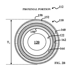
In the present example, referring to FIGS. 2A and 2B, the proximal set of
material
layers includes a proximal outer layer 160 (defining outer surface 152), a
proximal
intermediate layer 162, and a proximal inner layer 164 (defining inner surface
150); and the
distal set of material layers include a distal outer layer 170 (defining outer
surface 156) and a
distal inner layer 172 (defining inner surface 154). Other embodiments may
include more or
fewer layers in the proximal set of material layers and/or the distal set of
material layers. The
proximal outer layer 160 and the distal outer layer 170 include a material,
such as PEBAX ,
that exhibits a surface energy that facilitates application of the hydrophilic
coating 158 on the
outer surface 152 and/or the outer surface 156 of the catheter sheath 110, as
described above.
In the depicted embodiment, the proximal outer layer 160 and the distal outer
layer 170
include a same material, which facilitates coupling of the proximal portion
112 and the distal
portion 116 of the catheter sheath 110. For example, the proximal outer layer
160 and the
distal outer layer 170 are thermally fused together so that the proximal
portion 112 is
16
CA 02896510 2015-06-25
WO 2014/105713
PCT/US2013/076993
physically coupled with the distal portion 116. Alternatively, the proximal
outer layer 160
and the distal outer layer 170 include different materials that are assembled
together (e.g., by
thermally fusing the same material or compatible materials together and/or
gluing or
mechanically coupling) to couple the proximal portion 112 to the distal
portion 116.
The proximal intermediate layer 162 includes a material that provides strength
to the
proximal portion 112 of the catheter sheath 110. In some embodiments, the
proximal
intermediate layer 162 includes a metal, such as stainless steel, superelastic
material such as
Nitinol, high-strength polymer fibers (e.g., carbon-fiber, Spectra
(polyethylene fiber), Kevlar,
Dacron), and/or combinations thereof. In the present example, the proximal
intermediate
layer 162 includes a metal braid layer that provides strength to the proximal
portion 112 of
the catheter sheath 110, such as a stainless steel braid formed in polyimide
(SS Wire
Braid/PI). The metal braid layer includes divots (not shown) in which a
material of the
proximal inner layer 160 is formed, such that a proximal inner layer 170
formed of a
substantially constant thickness includes corresponding divots, thereby
minimizing contact
area between the proximal inner layer 170 and the imaging core 120 during use.
The proximal inner layer 160 defines the lumen 122 in the proximal portion 112
of
the catheter sheath 110. The proximal inner layer 160 includes a material
having a
coefficient of friction that minimizes friction between the imaging core 120
and the catheter
sheath 110, as described above. Minimizing friction between the imaging core
120 and the
catheter sheath 110 allows the imaging core to more freely rotate within the
catheter sheath.
In some embodiments, the material of the proximal inner layer 160 has a
coefficient of
friction ( ) between of 0.1 or less. In the depicted embodiment, the proximal
inner layer 160
includes a material having a static coefficient of friction of about 0.07 and
a kinetic
coefficient of friction of about 0.13. For example, the proximal inner layer
160 includes a
polymer blend, such as a PI/PTFE polymer blend.
The distal inner layer 172 defines the lumen 122 in the distal portion 116 of
the
catheter sheath 110. The distal inner layer 172 includes a material having a
coefficient of
friction that minimizes friction between the imaging core 120 and the catheter
sheath 110, as
described above, where the material also facilitates an average speed of sound
through the
distal portion 116 of the catheter sheath 110 that is substantially equivalent
to a speed of
sound through blood. Minimizing friction between the imaging core 120 and the
catheter
sheath 110 allows the imaging core to more freely rotate within the catheter
sheath, and
ensuring that the speed of sound through the distal portion 116 is
substantially equivalent to
17
CA 02896510 2015-06-25
WO 2014/105713
PCT/US2013/076993
the speed of sound through blood minimizes ultrasound signal distortion. In
some
embodiments, an average speed of sound through the distal inner layer 172 is
about 1.40 km/s
to about 1.70 km/s. In the depicted embodiment, for example, the distal inner
layer 172
includes EFEP, through which a speed of sound is about 1.40 km/s. In some
embodiments,
the distal inner layer 172 includes other materials that facilitate the speed
of sound through
the distal portion 116 being substantially equivalent to the speed of sound
through blood,
such as PEBAX 4033, EVA/Ve-634 (28% acetate) (about 1.67 km/s and about 1.68
km/s,
respectively), or a combination thereof.
As noted above, the distal set of material layers of the distal portion 116
facilitate an
average speed of sound through the distal set of material layers that is
substantially equivalent
to a speed of sound through blood, thereby minimizing image distortion
resulting from the
ultrasound signals traveling through the distal portion 116 of the catheter
sheath 110. In
some embodiments, an average speed of sound through the distal set of material
layers is
about 1.50 km/s to about 1.60 km/s. In some embodiments, an average speed of
sound
through the distal set of material layers is about 1.52 km/s. In the present
example, the distal
outer layer 170 includes the polyether block amide material, such as PEBAX ,
and the distal
inner layer 172 includes the EFEP material. A speed of sound through polyether
block amide
material, such as PEBAX , varies with its hardness. Accordingly, in
furtherance of the
present example, polyether block amide material, such as PEBAX , has a
hardness of about
72D (durometer), where a speed of sound through such material is about 1.99
km/s, and the
speed of sound through the EFEP material is about 1.40 km/s. By providing the
distal set of
material layers with about 75% EFEP material and about 25% polyether block
amide
material, an average speed of sound through the distal portion 116 of the
catheter sheath 110
is about 1.55 km/s. As a more general example, for a two layer sheath having a
thickness T,
if the first material layer comprises 75% of the thickness of the sheath
(i.e., 0.75T) and has an
acoustic velocity V1 and the second material layer comprises 25% of the
thickness of the
sheath (i. e., 0.25T) and has an acoustic velocity V2, then average acoustic
velocity of the
sheath can be calculated as Vavg = 0.75*Vi 0.25*V2 or other suitable
formulaic
representation. This approach can be extended to any number of material
layers. It is noted
that, in some embodiments, the lumen 122 is filled with a saline-type
material, where an
average speed of sound through the saline-type material is substantially
equivalent to the
speed of sound through blood.
18
CA 02896510 2015-06-25
WO 2014/105713
PCT/US2013/076993
From the foregoing, the disclosed catheter sheath 110 includes combinations of
materials in the proximal portion 112 and the distal portion 116 that minimize
ultrasound
signal distortion, while providing sufficient strength and flexibility for use
within human
vasculature. For example, the materials of the catheter sheath 110 minimize
friction between
the catheter sheath 110 and the imaging core 120, minimize friction between
the catheter
sheath 110 and the vasculature along its path to the vessel of interest,
enable application of a
hydrophilic coating on the outer surface of the catheter sheath 110, provide
sufficient strength
and flexibility, and/or facilitate travel of sound through the catheter sheath
110 similar to
travel of sound through blood. It is noted that thicknesses of the material
layers in the distal
portion 116 and the proximal portion 120 may be varied to achieve the desired
characteristics
and optimize the catheter's minimal contribution to image distortion.
Different embodiments
may have different advantages, and no advantage is necessarily required of any
embodiment.
Various methods may be employed to produce a catheter having the properties
discussed above. In various embodiments, a melt process, such as mono-
extrusion, sequential
extrusion, co-extrusion, and/or heat lamination (reflow), and the like may be
utilized to
produce the proximal and/or distal portions of the catheter. In more detail,
two such catheter
manufacturing processes are sequential mono-extrusion and heat lamination, or
reflow. In
the sequential mono-extrusion shaft manufacturing process, the inner polymeric
layer is first
extruded over a continuous, supportive core rod having a melting temperature
higher than
that of the extrusion temperature of the layer. Next, the outer polymeric
layer is over-
extruded onto the inner polymer layer. Various manufacturers can provide co-
extrusion
tubing to be used in the shafts of the invention, such as Teleflex Medical OEM
(Limerick,
Ireland). A sequential extrusion or a sequential mono-extrusion technique may
also be
utilized. In such an embodiment, a first polymer, such as EFEP, is extruded to
form the inner
layer and a second polymer, such as PEBA is then extruded onto the inner layer
to form the
second polymer layer. The mono-extrusion process may be carried out with a
regular single-
screw extruder. A hydrophilic barrier layer may then be formed on the outer
layer after the
extrusion is completed. The heat energy of the extrusion process and/or the
reflow/heat
lamination process, if such is applied, may assist in forming such a direct
and/or covalent
bond between the outer layer and inner layer.
Further, as noted above, in some implementations the proximal portion of the
catheter
includes reinforcing elements, such as braids, wires, cages, coils, hoops, or
helixes formed of
a suitable material. In such instances, the inner polymeric layer may be first
extruded (as
19
CA 02896510 2015-06-25
WO 2014/105713
PCT/US2013/076993
discussed above) and then the reinforcing element(s) may be formed by braiding
strands of
material, for example, onto the inner polymeric layer. In such an embodiment,
the outer
polymeric layer may be formed by extruding a polymer, such as an amine-
terminated PEBA,
for example, over the reinforcing element(s) and the inner polymeric layer.
Once completed,
the outer polymer layer may be coated with another polymer or coating to
provide a
hydrophilic outer surface.
Alternatively, in a reflow manufacturing process where the inner and outer
polymeric layers may be are prepared via polymer extrusion processes, a pre-
made
reinforcing element (e.g., in a given weaving pattern made via braiding) is
provided
separately. The inner layer, the reinforcing element, and the outer layer are
then layer-by-
layer introduced onto a supportive, metallic core rod and incorporated into a
single,
cylindrical, shaft body via a heat lamination, or reflow, processes by
applying an external
heat source over a proper shrink tube that completely and circumferentially
embraces the
shaft body to be formed. In some instances, this process results in a more
continuous axial
transition from the inner polymer layer to the outer polymer layer due to the
pressure and
heat. The inner and outer polymer layers may largely contain the reinforcing
element there
between and, ideally, be bonded onto the reinforcement element and/or to the
other layer
through the reinforcement element. As such, the contained reinforcement
element of the
bonded polymeric layers may provide some reinforcing effects for the shaft
body in terms of
column strength, fracture energy, and/or kink resistance, and the like.
Additionally, using
this process the interior ridges or projections associated with the shape of
the reinforcement
element are more pronounced, which results in even less contact surface area
on the interior
of the proximal portion of the catheter. Proximal shaft tubing fabricated with
either process
is available from commercial suppliers, such as Teleflex Medical OEM.
Persons skilled in the art will recognize that the apparatus, systems, and
methods
described above can be modified in various ways. Accordingly, persons of
ordinary skill in
the art will appreciate that the embodiments encompassed by the present
disclosure are not
limited to the particular exemplary embodiments described above. In that
regard, although
illustrative embodiments have been shown and described, a wide range of
modification,
change, and substitution is contemplated in the foregoing disclosure. It is
understood that
such variations may be made to the foregoing without departing from the scope
of the present
disclosure. Accordingly, it is appropriate that the appended claims be
construed broadly and
in a manner consistent with the present disclosure.