Note: Descriptions are shown in the official language in which they were submitted.
CA 02896555 2015-06-25
WO 2014/106158
PCT/US2013/078229
INTRAVASCULAR DEVICES, SYSTEMS, AND METHODS
TECHNICAL FIELD
The present disclosure relates to intravascular devices, systems, and methods.
In
some embodiments, the intravascular devices are guidewires that include one or
more
electronic, optical, or electro-optical components.
BACKGROUND
Heart disease is very serious and often requires emergency operations to save
lives. A
main cause of heart disease is the accumulation of plaque inside the blood
vessels, which
eventually occludes the blood vessels. Common treatment options available to
open up the
occluded vessel include balloon angioplasty, rotational atherectomy, and
intravascular stents.
Traditionally, surgeons have relied on X-ray fluoroscopic images that are
planar images
showing the external shape of the silhouette of the lumen of blood vessels to
guide treatment.
Unfortunately, with X-ray fluoroscopic images, there is a great deal of
uncertainty about the
exact extent and orientation of the stenosis responsible for the occlusion,
making it difficult
to find the exact location of the stenosis. In addition, though it is known
that restenosis can
occur at the same place, it is difficult to check the condition inside the
vessels after surgery
with X-ray.
A currently accepted technique for assessing the severity of a stenosis in a
blood
vessel, including ischemia causing lesions, is fractional flow reserve (FFR).
FFR is a
calculation of the ratio of a distal pressure measurement (taken on the distal
side of the
stenosis) relative to a proximal pressure measurement (taken on the proximal
side of the
stenosis). FFR provides an index of stenosis severity that allows
determination as to whether
the blockage limits blood flow within the vessel to an extent that treatment
is required. The
normal value of FFR in a healthy vessel is 1.00, while values less than about
0.80 are
generally deemed significant and require treatment.
Often intravascular catheters and guidewires are utilized to measure the
pressure
within the blood vessel, visualize the inner lumen of the blood vessel, and/or
otherwise obtain
data related to the blood vessel. To date, guidewires containing pressure
sensors, imaging
elements, and/or other electronic, optical, or electro-optical components have
suffered from
reduced performance characteristics compared to standard guidewires that do
not contain
such components. For example, the handling performance of previous guidewires
containing
-1-
CA 02896555 2015-06-25
WO 2014/106158
PCT/US2013/078229
electronic components have been hampered, in some instances, by the limited
space available
for the core wire after accounting for the space needed for the conductors or
communication
lines of the electronic component(s), the stiffness of the rigid housing
containing the
electronic component(s), and/or other limitations associated with providing
the functionality
of the electronic components in the limited space available within a
guidewire. Further, due
to its small diameter, in many instances the proximal connector portion of the
guidewire (i. e.,
the connector(s) that facilitate communication between the electronic
component(s) of the
guidewire and an associated controller or processor) is fragile and prone to
kinking, which
can destroy the functionality of the guidewire. For this reason, surgeons are
reluctant to
remove the proximal connector from the guidewire during a procedure for fear
of breaking
the guidewire when reattaching the proximal connector. Having the guidewire
coupled to the
proximal connector further limits the maneuverability and handling of the
guidewire.
Further, a problem with existing pressure and flow guidewires is that they
require a
complex assembly of many discrete components. That complex assembly process
has
limitations on design performance of the guidewire. The use of separate
conductive wires
running down the length of the wire reduces the space available for more
frontline supportive
cores and can result in numerous issues during use due to poor solder joints
with conductive
bands, electrical shorts due to insulation issues, and breakage of the
delicate conductive
wires.
Accordingly, there remains a need for improved intravascular devices, systems,
and
methods that include one or more electronic, optical, or electro-optical
components.
-2-
CA 02896555 2015-06-25
WO 2014/106158
PCT/US2013/078229
SUMMARY
The present disclosure is directed to intravascular devices, systems, and
methods that
include a guide wire having a solid core wire with electrical conductors
formed or wrapped
thereon.
The invention provides a more robust sensing guidewire that avoids the
assembly and
performance issues of prior art sensing guidewires. Guidewires of the
invention have a core
wire that is coated with an outer layer. Conductive wires are embedded in the
outer layer and
run the length of the body. The conductive wires act as the electrical pathway
for sensor
signals. The outer layer is removed (e.g., by ablation) at specific locations
on each
conductive wire where electrical connections are required. A conductive
material is then
applied to the exposed sections of wire. The sensor may then be coupled to the
guidewire via
the conductive material at one or more of the exposed sections. In this
manner, guidewires of
the invention eliminate the need to assemble a multitude of components to
create the
conductive band connections, the need for a hypotube, and the use of adhesives
and solder in
the guidewire. Reducing the number of components to assemble guidewires of the
invention
improves robustness of the assembled wire by eliminating a multitude of
processes that can
create failure conditions. Additionally, the ability to print the conductive
bands eliminates
the complexity associated with having to run and connect multiple wires.
Any type of sensor can be connected to guidewires of the invention and the
type of
measurement will determine the type of sensor used. In certain embodiments,
only a single
sensor is connected to the guidewire. In other embodiments, multiple sensors
are connected
to the guidewire. All of the sensors may be the same. Alternatively, the
sensors may differ
from each other and measure different characteristics inside a vessel.
Exemplary sensors are
pressure, flow, and temperature sensors. Any type of pressure sensor may be
used with
guidewires of the invention. In certain embodiments, the pressure sensor
includes a
crystalline semi-conductor material. Any type of flow sensor may be used with
guidewires of
the invention. In certain embodiments, the flow sensor includes an ultrasound
transducer.
Preferably, the guidewire of the invention includes both a pressure sensor and
a flow
sensor on the distal portion. Pressure sensors are able to obtain pressure
measurements and
flow sensors are able to obtain blood velocity measurements within a blood
vessel. The
ability to measure and compare both the pressure and velocity flow
significantly improves the
diagnostic accuracy of ischemic testing.
Numerous different methods exist to apply the conductive material to the
exposed sections on the body. In certain embodiments, printing is used and the
conductive
-3-
CA 02896555 2015-06-25
WO 2014/106158
PCT/US2013/078229
material is a conductive ink. Typically, the conductive ink includes a
conductive metal, such
as gold. The remainder of the outer layer in which the conductive wires are
embedded is
typically a polymeric material, such as polyimide.
Another aspect of the invention provides a method for measuring a
characteristic
inside a vessel. Methods of the invention involve providing a sensing
guidewire that includes
a body having an inner core and an outer layer. One or more conductive wires
are embedded
in the outer layer. The conductive wires are exposed at one or more locations
along the body.
A conductive material is layered over a plurality of the exposed locations,
and a sensor is
coupled to the body via the conductive material at one of the exposed
locations. The
guidewire is inserted into a vessel, and one or more sensors on the guidewire
measure one or
more characteristics inside the vessel.
In some embodiments, a guide wire having a solid core wire with electrical
conductors printed thereon is provided. In some instances, the electrical
conductors are
formed by defining a helically wrapped pattern around the solid core wire. The
pattern may
be defined with wire, by printing conductive ink, by isolating a conductive
skin or surface via
laser ablation into multiple conductive surfaces, by the LDS-MID process, etc.
The number
of electrical conductors is dependent upon the functionality of the device,
but in some
implementations includes between two and six conductors. In some
implementations, the
solid core wire operates as an electrical conductor of the guide wire. In some
instances, one
or more conductive bands are coupled to the electrical conductors adjacent a
proximal portion
of the guide wire. In some instances, the conductive bands are soldered,
welded, or glued
(with a conductive adhesive) to the electrical conductors. In some
embodiments, the
conductive bands are printed over an exposed portion of a corresponding
conductor ¨ another
is swaged. In some instances printed pattern is an antenna(s), heating
element(s), tactile
surface(s), alpha-numeric characters, etc.
In some instances, methods of assembling and/or manufacturing the guide wires
disclosed herein are provided. In some embodiments, the traditional need to
manually solder
loose 48 AWG insulated wires to 0.35 nm cylindrical conductive bands is
eliminated, which
increases manufacturing yields and reduces the necessary training and skill
required for
operators. Further, instead of relying upon a single solder connection, the
conductive bands
of the present disclosure are electrically coupled to an associated conductor
along a majority
of the length of the conductive band. Also, in some instances the number of
parts needed to
manufacture at least the proximal connector portion of the device is reduced.
-4-
CA 02896555 2015-06-25
WO 2014/106158
PCT/US2013/078229
The present disclosure enables the proximal connector region of a guide wire
that is
stronger and more durable than existing designs, while also easier to
manufacture.
Embodiments of the present disclosure utilize precision material deposition
(e.g., to coat
and/or trace precision patterns) and/or wire winding(s) with a solid core
member facilitating
the use of a larger core that provides better handling, strength, and
durability than existing
designs, which reduces the likelihood of unwanted bending, kinking, and/or
other damage to
the proximal connector portion of the intravascular device that can be
detrimental to the
function of the device.
Additional aspects, features, and advantages of the present disclosure will
become
apparent from the following detailed description.
-5-
CA 02896555 2015-06-25
WO 2014/106158
PCT/US2013/078229
BRIEF DESCRIPTION OF THE DRAWINGS
Illustrative embodiments of the present disclosure will be described with
reference to
the accompanying drawings, of which:
FIG. 1 is a diagrammatic, schematic side view of an intravascular device
according to
an embodiment of the present disclosure.
Collectively, Figs. 2-13 illustrate aspects of manufacturing an intravascular
device
according to an embodiment of the present disclosure.
FIG. 2 is a diagrammatic perspective view of a core member according to an
embodiment of the present disclosure.
FIG. 3 is a close-up diagrammatic perspective view of a proximal end of the
core
member of Fig. 2.
FIG. 4 is a diagrammatic perspective view of the core member of Figs. 2 and 3
after
application of an insulating layer to a section of the core member according
to an
embodiment of the present disclosure.
FIG. 5 is a diagrammatic perspective view of the core member of Figs. 2-4 with
a four
conductors helically wrapped around the core member according to an embodiment
of the
present disclosure.
FIG. 6 is a close-up diagrammatic perspective view of a proximal section of
the core
member, showing portions of two of the helically-wrapped conductors.
FIG. 7 is a close-up diagrammatic perspective view of a proximal section of
the core
member showing an exposed portion of the first conductor and portions of the
second, third,
and fourth conductors covered in an insulating material.
FIG. 8 is a close-up diagrammatic perspective view of a proximal section of
the core
member similar to that of Fig. 7, but showing an exposed portion of the second
conductor and
portions of the third and fourth conductors covered in an insulating material.
FIG. 9 is a close-up diagrammatic perspective view of a proximal section of
the core
member similar to those of Figs. 7 and 8, but showing an exposed portion of
the third
conductor and a portion of the fourth conductor covered in an insulating
material.
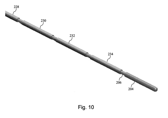
FIG. 10 is a diagrammatic perspective view of the core member of Figs. 2-9
with four
conductive bands positioned around the core member according to an embodiment
of the
present disclosure.
FIG. 11 is a close-up diagrammatic perspective view of a proximal section of
the core
member showing a spacing between portions of two adjacent conductive bands.
-6-
CA 02896555 2015-06-25
WO 2014/106158
PCT/US2013/078229
FIG. 12 is a close-up diagrammatic perspective view of the proximal section of
the
core member of Fig. 11 after the spacing between the two adjacent conductive
bands is filled
with an insulating material.
FIG. 13 is a diagrammatic perspective view of the core member of Figs. 2-12
after the
spacings between each of the conductive bands has been filled with an
insulating material.
FIG. 14 shows show an exemplary embodiment of a body with conductive wires
impregnated therein. The image on the left is a side view and the image of the
right is a
cross-sectional view.
FIG. 15 shows an area of the conductive wire that has been exposed from the
outer
layer.
FIG. 16 shows a conductive material applied to an exposed area, covering the
exposed
section of conductive wire to create a conductive band that is already in
contact with the
conductive wires.
FIG. 17 shows two exemplary conductive band configurations.
FIG. 18 shows a cross section of a guidewire having six conductive wires.
FIG. 19 is a system diagram according to certain embodiments.
-7-
CA 02896555 2015-06-25
WO 2014/106158
PCT/US2013/078229
DETAILED DESCRIPTION
For the purposes of promoting an understanding of the principles of the
present
disclosure, reference will now be made to the embodiments illustrated in the
drawings, and
specific language will be used to describe the same. It is nevertheless
understood that no
limitation to the scope of the disclosure is intended. Any alterations and
further
modifications to the described devices, systems, and methods, and any further
application of
the principles of the present disclosure are fully contemplated and included
within the present
disclosure as would normally occur to one skilled in the art to which the
disclosure relates. In
particular, it is fully contemplated that the features, components, and/or
steps described with
respect to one embodiment may be combined with the features, components,
and/or steps
described with respect to other embodiments of the present disclosure. For the
sake of
brevity, however, the numerous iterations of these combinations will not be
described
separately.
As used herein, "flexible elongate member" or "elongate flexible member"
includes at
least any thin, long, flexible structure that can be inserted into the
vasculature of a patient.
While the illustrated embodiments of the "flexible elongate members" of the
present
disclosure have a cylindrical profile with a circular cross-sectional profile
that defines an
outer diameter of the flexible elongate member, in other instances all or a
portion of the
flexible elongate members may have other geometric cross-sectional profiles
(e.g., oval,
rectangular, square, elliptical, etc.) or non-geometric cross-sectional
profiles. Flexible
elongate members include, for example, guidewires and catheters. In that
regard, catheters
may or may not include a lumen extending along its length for receiving and/or
guiding other
instruments. If the catheter includes a lumen, the lumen may be centered or
offset with
respect to the cross-sectional profile of the device.
In most embodiments, the flexible elongate members of the present disclosure
include
one or more electronic, optical, or electro-optical components. For example,
without
limitation, a flexible elongate member may include one or more of the
following types of
components: a pressure sensor, a temperature sensor, an imaging element, an
optical fiber, an
ultrasound transducer, a reflector, a mirror, a prism, an ablation element, an
RF electrode, a
conductor, and/or combinations thereof. Generally, these components are
configured to
obtain data related to a vessel or other portion of the anatomy in which the
flexible elongate
member is disposed. Often the components are also configured to communicate
the data to
an external device for processing and/or display. In some aspects, embodiments
of the
present disclosure include imaging devices for imaging within the lumen of a
vessel,
-8-
CA 02896555 2015-06-25
WO 2014/106158
PCT/US2013/078229
including both medical and non-medical applications. However, some embodiments
of the
present disclosure are particularly suited for use in the context of human
vasculature.
Imaging of the intravascular space, particularly the interior walls of human
vasculature can be
accomplished by a number of different techniques, including ultrasound (often
referred to as
intravascular ultrasound ("IVUS") and intracardiac echocardiography ("ICE"))
and optical
coherence tomography ("OCT"). In other instances, infrared, thermal, or other
imaging
modalities are utilized.
The electronic, optical, and/or electro-optical components of the present
disclosure are
often disposed within a distal portion of the flexible elongate member. As
used herein,
"distal portion" of the flexible elongate member includes any portion of the
flexible elongate
member from the mid-point to the distal tip. As flexible elongate members can
be solid,
some embodiments of the present disclosure will include a housing portion at
the distal
portion for receiving the electronic components. Such housing portions can be
tubular
structures attached to the distal portion of the elongate member. Some
flexible elongate
members are tubular and have one or more lumens in which the electronic
components can be
positioned within the distal portion.
The electronic, optical, and/or electro-optical components and the associated
communication lines are sized and shaped to allow for the diameter of the
flexible elongate
member to be very small. For example, the outside diameter of the elongate
member, such as
a guidewire or catheter, containing one or more electronic, optical, and/or
electro-optical
components as described herein are between about 0.0007" (0.0178 mm) and about
0.118"
(3.0 mm), with some particular embodiments having outer diameters of
approximately 0.014"
(0.3556 mm) and approximately 0.018" (0.4572 mm)). As such, the flexible
elongate
members incorporating the electronic, optical, and/or electro-optical
component(s) of the
present application are suitable for use in a wide variety of lumens within a
human patient
besides those that are part or immediately surround the heart, including veins
and arteries of
the extremities, renal arteries, blood vessels in and around the brain, and
other lumens.
"Connected" and variations thereof as used herein includes direct connections,
such
as being glued or otherwise fastened directly to, on, within, etc. another
element, as well as
indirect connections where one or more elements are disposed between the
connected
elements.
"Secured" and variations thereof as used herein includes methods by which an
element is directly secured to another element, such as being glued or
otherwise fastened
-9-
CA 02896555 2015-06-25
WO 2014/106158
PCT/US2013/078229
directly to, on, within, etc. another element, as well as indirect techniques
of securing two
elements together where one or more elements are disposed between the secured
elements.
Referring now to Fig. 1, shown therein is a portion of an intravascular device
100
according to an embodiment of the present disclosure. In that regard, the
intravascular device
100 includes a flexible elongate member 102 having a distal portion 104
adjacent a distal end
105 and a proximal portion 106 adjacent a proximal end 107. A component 108 is
positioned
within the distal portion 104 of the flexible elongate member 102 proximal of
the distal tip
105. Generally, the component 108 is representative of one or more electronic,
optical, or
electro-optical components. In that regard, the component 108 is a pressure
sensor, a
temperature sensor, an imaging element, an optical fiber, an ultrasound
transducer, a
reflector, a mirror, a prism, an ablation element, an RF electrode, a
conductor, and/or
combinations thereof. The specific type of component or combination of
components can be
selected based on an intended use of the intravascular device. In some
instances, the
component 108 is positioned less than 10 cm, less than 5, or less than 3 cm
from the distal tip
105. In some instances, the component 108 is positioned within a housing of
the flexible
elongate member 102. In that regard, the housing is a separate component
secured to the
flexible elongate member 102 in some instances. In other instances, the
housing is integrally
formed as a part of the flexible elongate member 102.
The intravascular device 100 also includes a connector 110 adjacent the
proximal
portion 106 of the device. In that regard, the connector 110 is spaced from
the proximal end
107 of the flexible elongate member 102 by a distance 112. Generally, the
distance 112 is
between 0% and 50% of the total length of the flexible elongate member 102.
While the total
length of the flexible elongate member can be any length, in some embodiments
the total
length is between about 1300 mm and about 4000 mm, with some specific
embodiments have
a length of 1400 mm, 1900 mm, and 3000 mm. Accordingly, in some instances the
connector
110 is positioned at the proximal end 107. In other instances, the connector
110 is spaced
from the proximal end 107. For example, in some instances the connector 110 is
spaced from
the proximal end 107 between about 0 mm and about 1400 mm. In some specific
embodiments, the connector 110 is spaced from the proximal end by a distance
of 0 mm, 300
mm, and 1400 mm.
The connector 110 is configured to facilitate communication between the
intravascular device 100 and another device. More specifically, in some
embodiments the
connector 110 is configured to facilitate communication of data obtained by
the component
108 to another device, such as a computing device or processor. Accordingly,
in some
-to-
CA 02896555 2015-06-25
WO 2014/106158
PCT/US2013/078229
embodiments the connector 110 is an electrical connector. In such instances,
the connector
110 provides an electrical connection to one or more electrical conductors
that extend along
the length of the flexible elongate member 102 and are electrically coupled to
the component
108. Some specific embodiments of electrical connectors in accordance with the
present
disclosure are discussed below in the context of Figs. 5-11. In other
embodiments, the
connector 110 is an optical connector. In such instances, the connector 110
provides an
optical connection to one or more optical communication pathways (e.g., fiber
optic cable)
that extend along the length of the flexible elongate member 102 and are
optically coupled to
the component 108. Further, in some embodiments the connector 110 provides
both
electrical and optical connections to both electrical conductor(s) and optical
communication
pathway(s) coupled to the component 108. In that regard, it should again be
noted that
component 108 is comprised of a plurality of elements in some instances. In
some instances,
the connector 110 is configured to provide a physical connection to another
device, either
directly or indirectly. In other instances, the connector 110 is configured to
facilitate wireless
communication between the intravascular device 100 and another device.
Generally, any
current or future developed wireless protocol(s) may be utilized. In yet other
instances, the
connector 110 facilitates both physical and wireless connection to another
device.
As noted above, in some instances the connector 110 provides a connection
between
the component 108 of the intravascular device 100 and an external device.
Accordingly, in
some embodiments one or more electrical conductors, one or more optical
pathways, and/or
combinations thereof extend along the length of the flexible elongate member
102 between
the connector 110 and the component 108 to facilitate communication between
the connector
110 and the component 108. Generally, any number of electrical conductors,
optical
pathways, and/or combinations thereof can extend along the length of the
flexible elongate
member 102 between the connector 110 and the component 108. In some instances,
between
one and ten electrical conductors and/or optical pathways extend along the
length of the
flexible elongate member 102 between the connector 110 and the component 108.
The
number of communication pathways and the number of electrical conductors and
optical
pathways extending along the length of the flexible elongate member 102 is
determined by
the desired functionality of the component 108 and the corresponding elements
that define
component 108 to provide such functionality.
Referring now to Figs. 2-13, shown therein are aspects of assembling and/or
manufacturing intravascular devices of the present disclosure that include
communication
pathways (e.g., electrical conductors and/or optical fibers) extending along
the length of the
-11-
CA 02896555 2015-06-25
WO 2014/106158
PCT/US2013/078229
device. In that regard, one of the major issues associated with existing
functional guidewires
is poor mechanical performance as compared to frontline guidewires. This
performance loss
is due in a large part to the typical design of the guidewires that severely
limits the space
available for the core or core wire due to the need to run the communication
lines along the
length of the device between the core wire and a surrounding hypotube. For the
sake of
clarity and simplicity, the embodiments of Figs. 2-13 include four electrical
conductors in
addition to an electrically conductive core. Those skilled in the art will
recognize that the
concepts are applicable to intravascular devices that include virtually any
number of electrical
conductors and/or optical fibers extending along the length of the core wire.
However, in
most implementations the intravascular device will include between 1 and 10
communication
pathways extending along the length of the core wire between a proximal
portion and a distal
portion of the intravascular device.
Referring more specifically to Fig. 2, shown therein is a diagrammatic
perspective
view of a core member 200 according to an embodiment of the present
disclosure. As shown,
the core member 200 includes an elongated shaft 202 and connector 204. In the
illustrated
embodiment, the connector 204 has an increased diameter with respect to the
shaft 202. In
some instances, the outer diameter of the connector 204 is the same as the
desired outer
diameter of the intravascular device that the core member 200 is intended to
form.
Accordingly, in some particular embodiments the outer diameter of the
connector 204 is
approximately 0.014". The difference in diameters between the shaft 202 and
the connector
204 may result from removing material away from a constant diameter rod to
define the shaft
and/or adding material to a constant diameter rod to define the connector. In
some instances,
the connector 204 is defined by a conductive band (such as those described
below for the
other conductors of the intravascular device) that is electrically coupled to
the core member.
In that regard, the core member 200 is formed of a conductive material (or at
least plated with
a conductive material) in some instances. In some instances, the core member
200 carries the
common or ground signal for the components of the intravascular device. As
shown in Fig.
3, the connector 204 defines the proximal most connector of the intravascular
device and, in
the illustrated embodiment, is positioned at the proximal tip of the
intravascular device. In
that regard, the proximal tip of the connector 204 is rounded. In some
implementations, the
proximal most connector is spaced distally from the proximal tip of the
intravascular device.
Referring now to Fig. 4, shown therein is a diagrammatic perspective view of
the core
member 200 after application of an insulating layer 206 to the shaft 202 of
the core member.
In that regard, the insulating layer 206 serves to electrically isolated the
conductive core
-12-
CA 02896555 2015-06-25
WO 2014/106158
PCT/US2013/078229
member 200 from the conductors that will be subsequently applied over the
shaft 202. The
insulating layer 206 may be formed of any suitable material. In some
implementations, the
insulating layer 206 is a parylene layer. Other elements may also be formed
over, placed
onto, and/or connected to shaft 202 in some instances, including flex-foil
wrap conductors,
conductive bands, pads, circuits, dielectrics, and/or other components of the
intravascular
device.
Referring now to FIG. 5, shown therein is a diagrammatic perspective view of
the
core member 200 with four conductors 208, 210, 212, and 214 helically wrapped
around the
shaft 202 of the core member (may also be conductive). As shown the proximal
ends of the
conductors 208, 210, 212, and 214 are spaced apart along the length of the
core member. In
some instances, the spacing between the ends of the conductors corresponds to
a desired
spacing between conductive bands that will be coupled to the conductors 208,
210, 212, and
214. The conductors 208, 210, 212, and 214 may be formed by electrically
printing (micro-
dispense, aero-jet, ink-jet, transfer, gravure, etc.) or plating of a
conductive material over the
insulating layer in a desired pattern. In some instances, a conductive ink is
utilized. In other
instances, 48 AWG or smaller conductors are helically wrapped around the shaft
202. In
such instances, the conductors may be insulated or not. In that regard, the
conductors may be
wire (Cu, etc.), carbon nanotube fiber conductors, conductive ink, conductive
polymer,
conductive film, and/or combinations thereof. If the conductors are not
insulated, then they
are kept isolated (i.e., spaced) from one another as shown in Fig. 5. Fig. 6
provides a close-
up diagrammatic perspective view of a proximal section of the core member 200,
showing
proximal end portions of helically-wrapped conductors 212 and 214.
In other embodiments, the conductors and/or other elements of the
intravascular
device are secured and/or wrapped around the core member using other
techniques, including
without limitation flex-foil wrapping, roll-to-roll printing, singulation,
wrapping tape with
conductors, utilizing conductive bands, utilizing contact pads, and/or
utilizing other features.
For example, in some instances a flex-foil wrap is utilized to define at least
a portion of the
conductors and/or circuitry. In that regard, insulated flexible foil
conductors are helically
wound onto the core member in some instances. The flexible foil conductors may
define one
or more conductors and/or circuitry such that a single foil conductor (having
a multiple
conductive leads/traces/circuits) and/or multiple foil conductors (each having
single or
multiple conductive leads/traces/circuits) may be utilized. Flexible foil
conductors allow for
a precise and consistent outer diameter, length, and pitch of the conductors
around the core
member, including facilitating automatic processing techniques. As a result,
the resulting
-13-
CA 02896555 2015-06-25
WO 2014/106158
PCT/US2013/078229
device can have improved consistency with respect to straightness and
flexibility. As another
example, in some instances a mill and fill approach is utilized to define the
conductors around
the core member.
Referring now to Fig. 7, shown therein is a close-up diagrammatic perspective
view
of a proximal section of the core member showing an exposed portion of
conductor 208 and
portions of conductors 210, 212, and 214 that have been covered in an
insulating material and
designated as 220, 222, and 224, respectively. In that regard, after the
conductors 208, 210,
212, and 214 have been formed/wrapped around the shaft 202, an insulating
layer is formed
over all or a majority of the length of the conductors. By either masking a
section of each
conductor and/or subsequently removing the applied (or existing) insulating
layer, a section
of each conductor 208, 210, 212, and 214 is exposed. Conductive bands are
coupled to the
exposed sections the conductors 208, 210, 212, and 214 as discussed below in
order to define
the proximal connector portion of the intravascular device. In this regard,
Fig. 8 is a close-up
diagrammatic perspective view of a proximal section of the core member 200
showing an
exposed portion of conductor 210 and the insulated portions 222 and 224 of
conductors 212
and 214. Similarly, Fig. 9 is a close-up diagrammatic perspective view of a
proximal section
of the core member 200 showing an exposed portion of conductor 212 and the
insulated
portion 224 of conductor 214.
Referring now to Fig. 10, shown therein is a diagrammatic perspective view of
the
core member 200 with five conductive bands 228, 230, 232, 234, and 204
positioned around
and/or defined by the core member in alignment with the exposed portions of
the conductors
208, 210, 212, 214, and 202 according to an embodiment of the present
disclosure. In some
instances, the conductive bands 228, 230, 232, and 234 are printed onto the
shaft 202 of the
core member 200 by electrically printing or plating of a conductive material
over the exposed
portions of the conductors 208, 210, 212, and 214. In that regard, the
conductive bands 228,
230, 232, and 234 are formed such that they have a uniform outer diameter
matching the
desired outer diameter of the intravascular device and/or the outer diameter
of connector 204
in some implementations. In some instances, the conductive bands are preformed
cylindrical
members that are positioned over the corresponding exposed sections of the
conductors 208,
210, 212, and 214 and electrically coupled to the conductors using solder or
other suitable
techniques. Fig. 11 provides a close-up diagrammatic perspective view of a
proximal section
of the core member showing a spacing between adjacent conductive bands 228 and
230.
Also shown in Fig. 11 is how the conductive band 228 is formed around, and
electrically
connected to conductor 208, while forming around the insulated portions 206,
220, 222, 224
-14-
CA 02896555 2015-06-25
WO 2014/106158
PCT/US2013/078229
of core member 202, conductors 210, 212, and 214 respectively. In the
illustrated
embodiment, the insulated portion 220 and the end of conductive band 228 are
substantially
aligned along the length of the core member 200, but in other embodiments the
insulated
portion 220 extends proximally towards conductive band 230 (including into a
portion of the
interior of conductive band 230, in some instances) to ensure the conductor
210 is isolated
from the conductor 208 and conductive band 228.
In some embodiments, the conductive bands are swaged and/or laser welded in
place.
In that regard, as a general manufacturing process swaging may be broken up
into two
categories. The first category of swaging involves the work piece being forced
through a
confining die to reduce its diameter, similar to the process of drawing wire.
This may also be
referred to as "tube swaging." The second category involves two or more dies
used to
hammer a round workpiece into a smaller diameter. This process is usually
called "rotary
swaging" or "radial forging." Tubes may be tagged (reduced in diameter to
enable the tube to
be initially fed through the die to then be pulled from the other side) using
a rotary swager,
which allows them to be drawn on a draw bench. Swaging is often the method of
choice for
precious metals since there is no loss of material in the process. In that
regard, in some
instances the conductive band is swaged around the core member and a portion
of the
conductive band is laser-welded to the exposed conductor underneath the
conductive band.
Referring now to Fig. 12, shown therein is a close-up diagrammatic perspective
view
of the proximal section of the core member of Fig. 11 after the spacing
between the adjacent
conductive bands 228 and 230 is filled with an insulating material 236.
Similarly, Fig. 13
provides a diagrammatic perspective view of the core member 200 after the
spacings between
each of the conductive bands has been filled with an insulating material,
defining insulating
spacers 236, 238, 240, and 242. The insulating spacers 236, 238, 240, and 242
are formed
such that they have a uniform outer diameter matching the desired outer
diameter of the
intravascular device and/or the outer diameters of conductive bands 204, 228,
230, 232, and
234 in some implementations. The insulating material utilized to form
insulating spacers
236, 238, 240, and 242 may be any suitable insulating material.
Fig. 14 shows an exemplary embodiment of a portion of an intravascular device
300
comprising a flexible elongate member 302 that includes a core member 304
surrounded an
outer layer 306 with conductive wires 308 impregnated therein. Both a side
view and a
cross-sectional end view of the flexible elongate member 302 are provided. The
core
member 304 can be formed of a suitable material such as stainless steel,
nickel and titanium
alloy (Nitinol), polyetheretherketone, heat straightened 304 stainless steel,
or other metallic
-15-
CA 02896555 2015-06-25
WO 2014/106158
PCT/US2013/078229
or polymeric materials, using techniques well known in the art. The outer
layer 306 can be
formed of a suitable polymeric material. In that regard, the outer layer 306
is coated onto the
wire using standard wire coating techniques. As the thickness of the coating
is built up,
conductive wires 308 are introduced into the coating process such that they
become
completely coated in the outer layer 306. The outer layer 406 may be any
polymeric
material, and a preferred material is polyimide. In certain embodiments, the
conductive wires
308 are space substantially equally around a diameter of the body. In certain
embodiments,
after reaching a desired diameter, a final coating that can provide lubricity
to the body is
applied. Any material that can provide lubricity may be used. An exemplary
material is
PTFE impregnated polyimide, silicone-based coatings, and hydrophilic based
coatings.
Fig. 15 shows an area of an embedded conductive wire 308 that has been
exposed from the outer layer 306. As shown in Fig. 15, one or more sections of
the outer
layer 306 are modified to expose corresponding individual sections of the
conductive wire
308. Any technique known in the art may be used to expose the sections of
conductive wire
308. Exemplary techniques include chemical etching, mechanical cutting and
shearing or
laser ablation. In certain embodiments, as shown in Fig. 15, laser ablation is
used to cut away
the desired sections of outer layer 306 to expose the embedded conductive wire
308.
Circumferential ablation may be utilized in some instances. Laser ablation of
polymeric
material is known in the art and accomplished by known techniques, such as
those described
in Kumagai (Applied Physics Letters, 65(14):1850 ¨ 1852, 2004); Sutcliffe
(Journal of
Applied Physics, 60(9):3315 ¨ 3322, 1986), and Blanchet et al. (Science,
262(5134):719-721,
1993), the content of each of which is incorporated by reference herein in its
entirety. A
reference ring at a proximal or distal end of the flexible elongate member 302
may be ablated
to identify where the conductive wires 308 reside in the outer layer 306. In
that regard, the
distal end of the conductive wires may be ground to the specified grind
profile for coupling
directly or indirectly to the component 108. In that regard, in some instances
the distal end of
flexible elongate member 302 is coupled to a distal working end similar to
those used in
current sensing guidewires. In some particular instances, the flexible
elongate member 302 is
coupled to a distal section, intermediate section, and/or proximal section
similar to those
described in one or more of U.S. Patent No. 5,125,137, U.S. Patent No.
5,873,835, U.S.
Patent No. 6,106,476, U.S. Patent No. 6,551,250, and U.S. Patent Application
No.
13/931,052, filed June 28, 2013, each of which is hereby incorporated by
reference in its
entirety.
-16-
CA 02896555 2015-06-25
WO 2014/106158
PCT/US2013/078229
As shown in Fig. 16, a conductive material can then be applied to the flexible
elongate member 302 over the exposed sections of the conductive wire 308. The
conductive
material covers the exposed sections of conductive wire 308 to define a
conductive band 310
that is in contact with the exposed conductive wire 308. The conductive
material will
generally be a metal, such as gold. Numerous techniques are known in the art
for apply the
conductive material to the conductive wire. In certain embodiments, the
conductive material
is printed and sintered onto the exposed sections of conductive wires.
Printing and sintering
of metal is well known in the art. See for example, Kydd (U.S. patent numbers
5,882,722
and 6,036,889), Karapatis et al. (Rapid Prototyping Journal, 4(2):77-89,
1998), and Kruth et
al., (Assembly Automation, 23(4):357-371, 2003), the content of each of which
is
incorporated by reference herein in its entirety.
Any desired pattern of conductive material may be placed onto the flexible
elongate
member 302. For example, the conductive bands can be solid, multiple rings, a
spiral, or any
other pattern that provides the optimum functionality. To that end, Fig. 17
shows two
exemplary conductive band configurations. The configuration on the left shows
a plurality of
conductive bands 310 each connected to a common conductive wire 308 to define
a
connector 312, while the configuration on the right shows a solid conductive
band 310 that
defines a connector for another conductive wire 308 of the flexible elongate
member.
Guidewires of the invention are complete by communicatively coupling the
component 108 to the conductive wires 308. In some particular instances,
portions of the
conductive wires 308 adjacent a distal end of the flexible elongate member 302
are
electrically coupled to the component 108 either directly or indirectly, using
soldering
welding, one or more additional conductive members, leads, and/or other known
techniques.
In some instances, sections of the outer layer 306 are removed to expose the
the distal
portions of the conductive wires 308 that will be coupled to the component
108. The
component 108 can be mounted within a distal section of the flexible elongate
member 302
using any suitable technique, including without limitation those disclosed in
one or more of
U.S. Patent No. 5,125,137, U.S. Patent No. 5,873,835, U.S. Patent No.
6,106,476, U.S. Patent
No. 6,551,250, U.S. Patent Application No. 13/931,052, filed June 28, 2013,
U.S. Patent
Application No. 14/135,117, filed December 19, 2013, U.S. Patent Application
No.
14/137,364, filed December 20, 2013, and U.S. Patent Application No.
14/139,543, filed
December 23, 2013, each of which is hereby incorporated by reference in its
entirety.
As discussed above with respect to component 108, the sensor(s) of the
intravascular
device 300 provide a means to obtain intraluminal measurements within a body
lumen and
-17-
CA 02896555 2015-06-25
WO 2014/106158
PCT/US2013/078229
are connected to the one or more conductive bands on the intravascular device,
which
transmit and receive signals from the sensor(s). For example, the guidewire of
the invention
can include a pressure sensor, a flow sensor, a temperature sensor or
combinations thereof.
Preferably, the guidewire is a combination guidewire that includes both a
pressure sensor and
a flow sensor. Pressure sensors can be used to measure pressure within the
lumen and flow
sensors can be used to measure the velocity of blood flow. Temperature sensors
can measure
the temperature of a lumen. A guidewire with both a pressure sensor and a flow
sensor
provides a desirable environment in which to calculate fractional flow reserve
(FFR) using
pressure readings, and coronary flow reserve (CFR) using flow readings.
Guidewires with
two or more sensors can be made by increasing the number of conductive wires.
For
example, Fig. 18 shows a cross section of the flexible elongate member 302
having six
conductive wires 308 embedded in the outer layer 306. In addition, the core
304 may also be
utilized as a conductor in some embodiments. Such embodiments provide enough
conductive
pathways to facilitate the use of at least two sensors with the flexible
elongate member 302.
The ability to measure and compare both the pressure and velocity flow and
create an
index of hyperemic stenosis resistance significantly improves the diagnostic
accuracy of this
ischemic testing. It has been shown that distal pressure and velocity
measurements,
particularly regarding the pressure drop-velocity relationship such as
Fractional Flow reserve
(FFR), Coronary flow reserve (CFR) and combined P-V curves, reveal information
about the
stenosis severity. For example, in use, the guidewire may be advanced to a
location on the
distal side of the stenosis. The pressure and flow velocity may then be
measured at a first
flow state. Then, the flow rate may be significantly increased, for example by
the use of
drugs such as adenosine, and the pressure and flow measured in this second,
hyperemic, flow
state. The pressure and flow relationships at these two flow states are then
compared to assess
the severity of the stenosis and provide improved guidance for any coronary
interventions.
The ability to take the pressure and flow measurements at the same location
and same time
with the combination tip sensor, improves the accuracy of these pressure-
velocity loops and
therefore improves the accuracy of the diagnostic information.
A pressure sensor allows one to obtain pressure measurements within a body
lumen.
A particular benefit of pressure sensors is that pressure sensors allow one to
measure of
fractional flow reserve (FFR) in vessel, which is a comparison of the pressure
within a vessel
at positions prior to the stenosis and after the stenosis. The level of FFR
determines the
significance of the stenosis, which allows physicians to more accurately
identify
hemodynamically relevant stenosis. For example, an FFR measurement above 0.80
indicates
-18-
CA 02896555 2015-06-25
WO 2014/106158
PCT/US2013/078229
normal coronary blood flow and a non-significant stenosis. Another benefit is
that a
physician can measure the pressure before and after an intraluminal
intervention procedure to
determine the impact of the procedure.
A pressure sensor can be mounted, for example, on a distal portion of the
guidewire.
The pressure sensor can be formed of a crystal semiconductor material having a
recess
therein and forming a diaphragm bordered by a rim. A reinforcing member is
bonded to the
crystal and reinforces the rim of the crystal and has a cavity therein
underlying the diaphragm
and exposed to the diaphragm. A resistor having opposite ends is carried by
the crystal and
has a portion thereof overlying a portion of the diaphragm. Electrical
conductor wires of the
senor are connected to a conductive band in the guidewire. Additional details
of suitable
pressure sensors that may be used with devices of the invention are described
in U.S. Pat. No.
6,106,476. U.S. Pat. No. 6,106,476 also describes suitable methods for
coupling the pressure
sensor to a guidewire. Those methods are applicable to coupling the sensor to
the conductive
bands in guidewires of the invention.
In certain aspects, the guidewire of the invention includes a flow sensor. The
flow
sensor can be used to measure blood flow velocity within the vessel, which can
be used to
assess coronary flow reserve (CFR). The flow sensor can be, for example, an
ultrasound
transducer, a Doppler flow sensor or any other suitable flow sensor, disposed
at or in close
proximity to the distal tip of the guidewire. The ultrasound transducer may be
any suitable
transducer, and may be mounted in the distal end using any conventional
method, including
the manner described in U.S. Pat. No. 5,125,137, 6,551,250 and 5,873,835.
Additional features of the invention include proximal and distal tip coils or
coverings.
Guidewires of the invention can be connected to an instrument, such as a
computing
device (e.g. a laptop, desktop, or tablet computer) or a physiology monitor,
that converts the
signals received by the sensors into pressure and velocity readings. The
instrument can
further calculate Coronary Flow Reserve (CFR) and Fractional Flow Reserve
(FFR) and
provide the readings and calculations to a user via a user interface.
In some embodiments, a user interacts with a visual interface to view images
associated with the data obtained by the intravascular devices of the present
disclosure. Input
from a user (e.g., parameters or a selection) are received by a processor in
an electronic
device. The selection can be rendered into a visible display. An exemplary
system including
an electronic device is illustrated in Fig. 19. As shown in Fig. 19, a sensor
engine 859
communicates with host workstation 433 as well as optionally server 413 over
network 409.
The data acquisition element 855 (DAQ) of the sensor engine receives sensor
data from one
-19-
CA 02896555 2015-06-25
WO 2014/106158
PCT/US2013/078229
or more sensors. In some embodiments, an operator uses computer 449 or
terminal 467 to
control system 400 or to receive images. An image may be displayed using an
I/0 454, 437,
or 471, which may include a monitor. Any I/0 may include a keyboard, mouse or
touchscreen to communicate with any of processor 421, 459, 441, or 475, for
example, to
cause data to be stored in any tangible, nontransitory memory 463, 445, 479,
or 429. Server
413 generally includes an interface module 425 to effectuate communication
over network
409 or write data to data file 417.
Processors suitable for the execution of computer program include, by way of
example, both general and special purpose microprocessors, and any one or more
processor
of any kind of digital computer. Generally, a processor will receive
instructions and data from
a read-only memory or a random access memory or both. The essential elements
of computer
are a processor for executing instructions and one or more memory devices for
storing
instructions and data. Generally, a computer will also include, or be
operatively coupled to
receive data from or transfer data to, or both, one or more mass storage
devices for storing
data, e.g., magnetic, magneto-optical disks, or optical disks. Information
carriers suitable for
embodying computer program instructions and data include all forms of non-
volatile
memory, including by way of example semiconductor memory devices, (e.g.,
EPROM,
EEPROM, solid state drive (SSD), and flash memory devices); magnetic disks,
(e.g., internal
hard disks or removable disks); magneto-optical disks; and optical disks
(e.g., CD and DVD
disks). The processor and the memory can be supplemented by, or incorporated
in, special
purpose logic circuitry.
To provide for interaction with a user, the subject matter described herein
can be
implemented on a computer having an I/0 device, e.g., a CRT, LCD, LED, or
projection
device for displaying information to the user and an input or output device
such as a keyboard
and a pointing device, (e.g., a mouse or a trackball), by which the user can
provide input to
the computer. Other kinds of devices can be used to provide for interaction
with a user as
well. For example, feedback provided to the user can be any form of sensory
feedback, (e.g.,
visual feedback, auditory feedback, or tactile feedback), and input from the
user can be
received in any form, including acoustic, speech, or tactile input.
The subject matter described herein can be implemented in a computing system
that
includes a back-end component (e.g., a data server 413), a middleware
component (e.g., an
application server), or a front-end component (e.g., a client computer 449
having a graphical
user interface 454 or a web browser through which a user can interact with an
implementation of the subject matter described herein), or any combination of
such back-end,
-20-
CA 02896555 2015-06-25
WO 2014/106158
PCT/US2013/078229
middleware, and front-end components. The components of the system can be
interconnected
through network 409 by any form or medium of digital data communication, e.g.,
a
communication network. Examples of communication networks include cell network
(e.g.,
3G or 4G), a local area network (LAN), and a wide area network (WAN), e.g.,
the Internet.
The subject matter described herein can be implemented as one or more computer
program products, such as one or more computer programs tangibly embodied in
an
information carrier (e.g., in a non-transitory computer-readable medium) for
execution by, or
to control the operation of, data processing apparatus (e.g., a programmable
processor, a
computer, or multiple computers). A computer program (also known as a program,
software,
software application, app, macro, or code) can be written in any form of
programming
language, including compiled or interpreted languages (e.g., C, C++, Pert),
and it can be
deployed in any form, including as a stand-alone program or as a module,
component,
subroutine, or other unit suitable for use in a computing environment. Systems
and methods
of the invention can include instructions written in any suitable programming
language
known in the art, including, without limitation, C, C++, Pert, Java, ActiveX,
HTML5, Visual
Basic, or JavaScript.
A computer program does not necessarily correspond to a file. A program can be
stored in a portion of file 417 that holds other programs or data, in a single
file dedicated to
the program in question, or in multiple coordinated files (e.g., files that
store one or more
modules, sub-programs, or portions of code). A computer program can be
deployed to be
executed on one computer or on multiple computers at one site or distributed
across multiple
sites and interconnected by a communication network.
A file can be a digital file, for example, stored on a hard drive, SSD, CD, or
other
tangible, non-transitory medium. A file can be sent from one device to another
over network
409 (e.g., as packets being sent from a server to a client, for example,
through a Network
Interface Card, modem, wireless card, or similar).
Writing a file according to the invention involves transforming a tangible,
non-
transitory computer-readable medium, for example, by adding, removing, or
rearranging
particles (e.g., with a net charge or dipole moment into patterns of
magnetization by
read/write heads), the patterns then representing new collocations of
information about
objective physical phenomena desired by, and useful to, the user. In some
embodiments,
writing involves a physical transformation of material in tangible, non-
transitory computer
readable media (e.g., with certain optical properties so that optical
read/write devices can then
read the new and useful collocation of information, e.g., burning a CD-ROM).
In some
-21-
CA 02896555 2015-06-25
WO 2014/106158
PCT/US2013/078229
embodiments, writing a file includes transforming a physical flash memory
apparatus such as
NAND flash memory device and storing information by transforming physical
elements in an
array of memory cells made from floating-gate transistors. Methods of writing
a file are well-
known in the art and, for example, can be invoked manually or automatically by
a program or
by a save command from software or a write command from a programming
language.
Persons skilled in the art will also recognize that the apparatus, systems,
and methods
described above can be modified in various ways. Accordingly, persons of
ordinary skill in
the art will appreciate that the embodiments encompassed by the present
disclosure are not
limited to the particular exemplary embodiments described above. In that
regard, although
illustrative embodiments have been shown and described, a wide range of
modification,
change, and substitution is contemplated in the foregoing disclosure. It is
understood that
such variations may be made to the foregoing without departing from the scope
of the present
disclosure. Accordingly, it is appropriate that the appended claims be
construed broadly and
in a manner consistent with the present disclosure.
-22-