Note: Descriptions are shown in the official language in which they were submitted.
CULTURING OF HUMAN EMBRYONIC STEM CELLS AT THE
AIR-LIQUID INTERFACE FOR DIFFERENTIATION INTO
PANCREATIC ENDOCRINE CELLS
CROSS-REFERENCE TO RELATED APPLICATIONS
[0001] This application claims priority to U.S. Provisional Application
61/747,662 (filed on
December 31, 2012).
FIELD OF THE INVENTION
[0002] The present invention is in the field of cell differentiation. More
specifically, the
present invention provides methods, cell cultures and media for generating
pancreatic endoderm,
pancreatic endocrine precursor cells, and single-hormone pancreatic endocrine
cells from human
pluripotent stem cells by culturing cells at the air-liquid interface.
BACKGROUND
[0003] Advances in cell-replacement therapy for Type I diabetes mellitus and a
shortage of
transplantable islets of Langerhans have focused interest on developing
sources of insulin-
producing cells, or beta (0) cells, appropriate for engraftment. One approach
is the generation of
functional 13 cells from pluripotent stem cells, such as, embryonic stem
cells.
[0004] In vertebrate embryonic development, a pluripotent cell gives rise to a
group of cells
comprising three germ layers (ectoderm, mesoderm, and endoderm) in a process
known as
gastrulation. Tissues such as, thyroid, thymus, pancreas, gut, and liver, will
develop from the
endoderm, via an intermediate stage. The intermediate stage in this process is
the formation of
definitive endoderm.
[0005] By the end of gastrulation, the endoderm is partitioned into anterior-
posterior domains
that can be recognized by the expression of a panel of factors that uniquely
mark anterior, mid,
and posterior regions of the endoderm. For example, HHEX, and SOX2 identify
the anterior
region while CDX1, 2, and 4 identify the posterior region of the endoderm.
[0006] Migration of endoderm tissue brings the endoderm into close proximity
with different
mesodermal tissues that help in regionalization of the gut tube. This is
accomplished by a
plethora of secreted factors, such as FGFs, WNTS, TGF-Ps, retinoic acid (RA),
and BMP ligands
1
CA 2896655 2018-06-01
CA 02896655 2015-06-26
WO 2014/105543 PCT/US2013/075939
and their antagonists. For example, FGF4 and BMP promote CDX2 expression in
the
presumptive hindgut endoderm and repress expression of the anterior genes Hhex
and SOX2
(2000 Development, 127:1563-1567). WNT signaling has also been shown to work
in parallel to
FGF signaling to promote hindgut development and inhibit foregut fate (2007
Development,
134:2207-2217). Lastly, secreted retinoic acid by mesenchyme regulates the
foregut-hindgut
boundary (2002 Curr Bio1,12:1215-1220).
[00071 The level of expression of specific transcription factors may be used
to designate the
identity of a tissue. During transformation of the definitive endoderm into a
primitive gut tube,
the gut tube becomes regionalized into broad domains that can be observed at
the molecular level
by restricted gene expression patterns. The regional.ized pancreas domain in
the gut tube shows a
very high expression of PDX I and very low expression of CDX2 and SOX2. PDX1,
NKX6.1,
PTF1A, and NKX2.2 are highly expressed in pancreatic tissue; and expression of
CDX2 is high
in intestinal tissue.
00081 Formation of the pancreas arises from. the differentiation of definitive
endoderm into
pancreatic endoderm. Dorsal and ventral pancreatic domains arise from the
foregut epithelium.
Foregut also gives rise to the esophagus, trachea, lungs, thyroid, stomach,
liver, and bile duct
system.
[00091 Cells of the pancreatic endoderm express the pancreatic-duodenal
homeobox gene
PDX1. In the absence of PDX1, the pancreas fails to develop beyond the
formation of ventral
and dorsal buds. Thus, PDX1 expression marks a critical step in pancreatic
organogenesis. The
mature pancreas contains, both exocrine and endocrine tissues arising from the
differentiation of
pancreatic endoderm.
100101 D'Amour et al. describe the production of enriched cultures of human
embryonic stem
cell-derived definitive endoderm in the presence of a high concentration of
activin and low
serum (Nature Biotechnology 2005, 23:1534-1541; U.S. Patent No. 7,704,738).
Transplanting
these cells under the kidney capsule of mice reportedly resulted in
differentiation into more
mature cells with characteristics of endodermal tissue (U.S. Patent No.
7,704,738). Human
embryonic stem cell-derived definitive endoderm cells can be further
differentiated into PDX1
positive cells after addition of FGF10 and retinoic acid (U.S. Patent App.
Pub. No.
2005/0266554). Subsequent transplantation of these pancreatic precursor cells
in the fat pad of
immune deficient mice resulted in the formation of functional pancreatic
endocrine cells
2
CA 02896655 2015-06-26
WO 2014/105543 PCT/US2013/075939
following a 3-4 months maturation phase (U.S. Patent No. 7,993,920 and U.S.
Patent No.
7,534,608).
[00111 Fisk etal. report a system for producing pancreatic islet cells from
human embryonic
stem cells (U.S. Patent No. 7,033,831). In this case, the differentiation
pathway was divided into
three stages. Human embryonic stein cells were first differentiated to
endoderm using a
combination of sodium butyrate and activin A (U.S. Patent No. 7,326,572). The
cells were then
cultured with BMP antagonists, such as Noggin, in combination with EGF or
betacellulin to
generate PDX1 positive cells. The terminal differentiation was induced by
nicotinamide.
[00121 Small molecule inhibitors have also been used for induction of
pancreatic endocrine
precursor cells. For example, small molecule inhibitors of TGF-13 receptor and
BMP receptors
(Development 2011, 138:861-871; Diabetes 2011, 60:239-247) have been used to
significantly
enhance the number of pancreatic endocrine cells. In addition, small molecule
activators have
also been used to generate definitive endoderm. cells or pancreatic precursor
cells (Curr Opin
Cell Rio! 2009, 21:727-732; Nature Chem Rio! 2009, 5:258-265).
[00131 HB9 (also known as H1XB9 and MNX1) is a BHILEI transcriptional
activator protein
expressed early in pancreas development starting at approximately embryonic
day 8. HB9 is
also expressed in notochord and spinal cord. Expression of HB9 is transient
and peaks at about
day 10.5 in pancreatic epithelium being expressed in PDX1. and NKX6.1
expressing cells. At
about day 12.5, HB9 expression declines and at later stages it becomes
restricted only to 13 cells.
In mice homozygous for a null mutation of HB9, the dorsal lobe of the pancreas
fails to develop
(Nat Genet 23:67-70, 1999; Nat Genet 23:71-75, 1999). HB9-/- 13-cells express
low levels of the
glucose transporter, GLUT2, and .NKX6.1. Furthermore, HB9 -/- pancreas shows a
significant
reduction in the number of insulin positive cells while not significantly
affecting expression of
other pancreatic hormones. Thus, temporal control of HB9 is essential to
normal 11 cell
development and function. While not much is known about factors regulating HB9
expression in
13 cells, a recent study in zebrafish suggests that retinoic acid can
positively regulate expression
of HB9 (Development, 138, 4597-4608, 2011).
[00141 The thyroid hormones, thyroxine ("14") and triiodothyronine ("13"), are
tyrosine-based
hormones produced by the thyroid gland and are primarily responsible for
regulation of
metabolism. The major form of thyroid hormone in the blood is T4, which has a
longer half-life
3
CA 02896655 2015-06-26
WO 2014/105543 PCT/US2013/075939
than 13. The ratio of T4 to 13 released into the blood is roughly 20 to 1. T4
is converted to the
more active T3 (three to four times more potent than 14) within cells by
deiodinase.
[00151 T3 binds to thyroid hormone receptors, TRctl and TRI31 (TR). TR is a
nuclear hormone
receptor, which heterodimerizes with retinoid X receptor. The dimers bind to
the thyroid
response elements (TREs) in the absence of ligand and act as transcriptional
repressors. Binding
of T3 to TR reduces the repression of TRE dependent genes and induces the
expression of
various target genes. While numerous studies have suggested a role for T3 in
increasing 11 cell
proliferation, reducing apoptosis, and improving insulin secretion, its role
in cell differentiation
is undefined.
[00161 Transforming growth factor (3 ("TGF-(3") is a member of a large family
of plciotropic
cytokines that are involved in many biological processes, including growth
control,
differentiation, migration, cell survival, fibrosis and specification of
developmental fate. TGF-13
superfamily members signal through a receptor complex comprising a type II and
type I receptor.
IGF-13 ligands (such as activins, and growth. differentiation factors
("GDF"s)) bring together a
type II receptor with a type I receptor. The type II receptor phosphorylates
and activates the type
I receptor in the complex. There are five mammalian type 11 receptors: Ti3R-
II, ActR-II, ActR-
IIB, BMPR-II, and AMHR-1I and seven type I receptors (ALKs 1-7). Activin and
related
ligands signal via combinations of ActR-11 or ActR-IIB and ALK4 or ALK5, and
BMPs signal
through combinations of ALK2, ALIC3, and ALK6 with ActR-II, ActR-IIB, or BMPR-
II. AMH
signals through a complex of AMHR-H with ALK.6, and nodal has been shown to
signal through
a complex of ActR-11B and A LK7 (cell. 2003,113(6):685-700). Following binding
of the TOF-
B ligand to the appropriate receptor, the ensuing signals are transduced to
the nucleus primarily
through activation of complexes of Smads. Upon activation, the type 1
receptors phosphorylate
members of the receptor-regulated subfamily of Smads. This activates them and
enables them to
form complexes with a common mediator Smad, Smad4. Smads 1, 5, and 8 are
substrates for
ALKs 1, 2, 3, and 6, whereas Smads 2 and 3 are substrates for ALKs 4, 5, and 7
(FASEB
j13:2105-2124). The activated Smad complexes accumulate in the nucleus, where
they are
directly involved in the transcription of target genes, usually in association
with other specific
DNA-binding transcription factors. Compounds that selectively inhibit the
receptors for TGF-13,
have been developed for therapeutic applications and for modulating cell fate
in the context of
reprogramming and differentiation from various stem cell populations. In
particular, ALK5
4
inhibitors have been previously used to direct differentiation of embryonic
stem cells to an
endocrine fate (Diabetes, 2011, 60(1):239-47).
100171 In general, the process of differentiating progenitor cells to
functional 1 cells goes
through various stages; and great strides have been made in improving
protocols to generate
pancreatic cells from progenitor cells such as human pluripotent stem cells.
Despite these
advances in research, each step in the process of differentiating progenitor
cells presents a unique
challenge. As such, there is still a need for a protocol resulting in
functional endocrine cells and,
in particular, functional 13 cells.
SUMMARY
[0017a] In one embodiment, there is provided a method for producing human
pancreatic
endocrine cells from human pluripotent stem cells, comprising the steps of:
a. differentiating the human pluripotent stem cells into human pancreatic
foregut precursor
cells; and
b. differentiating the human pancreatic foregut precursor cells into human
pancreatic
endocrine cells by culturing at the air-liquid interface with at least one
medium supplemented
with: (i) an ALK5 inhibitor; (ii) both the ALK5 inhibitor and a thyroid
hormone, wherein the
thyroid hormone is triiodothyronine, thyroxine, an analogue of
triiodothyronine, an analogue of
thyroxine, or a mixture thereof.
[0017b] In another embodiment, there is provided a method of producing human
pancreatic
endocrine cells comprising differentiating human pancreatic foregut precursor
cells into human
pancreatic endocrine cells by culturing at the air-liquid interface with at
least one medium
supplemented with i) an ALK5 inhibitor, or ii) both the ALK5 inhibitor and a
thyroid hormone,
wherein the thyroid hormone is triiodothyronine, thyroxine, an analogue of
triiodothyronine, an
analogue of thyroxine, or a mixture thereof.
10017c1 In another embodiment, there is provided a method for producing human
pancreatic
endocrine cells, comprising the steps of: differentiating human pancreatic
foregut precursor cells
derived from human pluripotent stem cells into human pancreatic
endoderm/endocrine precursor
cells by treating with a medium supplemented with an ALK5 inhibitor and
culturing at the air-
liquid interface; and differentiating the human pancreatic endoderm/endocrine
precursor cells into
human pancreatic endocrine cells by culturing at the air-liquid interface with
at least one medium
supplemented with an i) ALK5 inhibitor and ii) triiodothyronine or an analog
thereof.
Date Recue/Date Received 2020-04-24
[0017d] In another embodiment, there is provided an in vitro cell culture for
differentiating
human cells at an air-liquid interface comprising:
a. a culture vessel;
b. a volume of differentiation medium within the vessel sufficient to fill
only a portion of
the volume of the vessel;
c. air within the vessel that fills a portion of the vessel adjoining the
differentiation
medium;
d. a porous substrate located at the interface between the differentiation
medium and the
air; and
e. human cells derived from human pluripotent stem cells disposed upon the
surface of the
substrate such that the differentiation medium contacts only a portion of the
surface of the human
cells, wherein the differentiation medium comprises a growth medium
supplemented with i) an
ALK5 inhibitor, or ii) both the ALK5 inhibitor and a thyroid hormone, wherein
the thyroid
hormone is triiodothyronine, thyroxine, an analogue of triiodothyronine, an
analogue of thyroxine
or a mixture thereof.
BRIEF DESCRIPTION OF THE DRAWINGS
[0018] FIGS. lA to 1H show phase contrast images of cells cultured at the air-
liquid interface
using the methods described in Example 1 at the following time points: Day 1
(FIG. IA); Day 5
(FIG. 1B); Day 6 (FIG. IC); Day 7(FIG. ID); Day 9 (FIG. 1E); Day 13 (FIG. IF);
Day 16 (FIG.
IG); and Day 21 (FIG. 1H).
[0019] FIGS. 2A to 2K show images of cells differentiated for one week at the
air-liquid
interface using the methods described in Example 1 and immunostained for the
following: DAPI
(FIG. 2A); insulin (FIG. 2B); HB9 (FIG. 2C); DAPI (FIG. 2D); glucagon (FIG.
2E); insulin (FIG.
2F); DAPI (FIG. 2G); insulin (FIG. 2H); somatostatin (FIG. 21); NKX6.1 (FIG.
2J); and insulin
(FIG. 2K). Panels A-C, D-F, G-I and J-K were taken from the same field.
5a
CA 2896655 2019-07-23
100201 FIGS. 3A to 3H show images of cells differentiated for two weeks at the
air-liquid
interface using the methods described in Example 1 and immunostained for the
following: insulin
(FIG. 3A); glucagon (FIG. 3B); insulin (FIG. 3C); somatostatin (FIG. 3D);
insulin (FIG. 3E);
NKX6.1 (FIG. 3F); HB9 (FIG. 3G); and NKX6.1 (FIG. 2H). Panels A-B, C-D, E-F.
and G-H
were taken from the same field.
[0021] FIGS. 4A to 4D show images of cells differentiated for three weeks at
the air-liquid
interface using the methods described in Example 1 and immune stained for
insulin (FIG. 4A),
glucagon (FIG. 4B), insulin (FIG. 4C), and somatostatin (FIG. 4D). Panels A-B
and C-D were
taken from the same field.
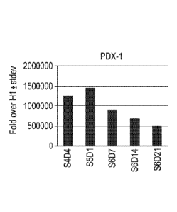
[0022] FIGS. 5A to 5R depict data from real-time PCR analyses of the
expression of the
following genes in cells of the human embryonic stem cell line HI
differentiated as outlined in
Example 1: PDX1 (FIG. 5A); NKX6.1 (FIG. 5B); PAX4 (FIG. 5C); PAX6 (FIG. 5D);
NGN3
=
5b
CA 2896655 2019-07-23
CA 02896655 2015-06-26
WO 2014/105543 PCT/US2013/075939
(FIG. 5E); NKX2.2 (FIG. 5F); ABCC8 (FIG. 5G); chromogranin-A (FIG. 5H); PCSK1
(FIG.
51); IAPP (FIG. 5.1); insulin (FIG. 5K); glucagon (FIG. 5L); somatostatin
(FIG. 5M); ghrelin
(FIG. 5N); PTF1A (FIG. 50); ZIC1 (FIG. 5P); CDX2 (FIG. 5Q); and SOX9 (FIG.
5R). Cells
were cultured at the air-liquid interface after Stage 5.
[00231 FIGS. 6A to 6L depict data from real-time PCR analyses of the
expression of the
following genes in cells of the human embryonic stem cell line HI
differentiated as outlined in
Example 2: PDX I (FIG.6A); NKX6.1 (FIG. 6B); FAX4 (FIG. 6C); PAX6 (FIG. 6D);
NGN3
(FIG. 6E); NKX2.2 (FIG. 6F); ABCC8 (FIG. 60); chromogranin-A (FIG. 61); PCSK1
61); IAPP (FIG. 6J); insulin (FIG. 6K); and glucagon (FIG. 6L).
[00241 FIGS. 7A to 7L depict data from real-time PCR analyses of the
expression of the
following genes in cells of the human embryonic stem cell line Hi
differentiated as outlined in
Example 3: PDX I (FIG.7A); NKX6.I (FIG. 7B); PAX4 (FIG. 7C); PAX6 (FIG. 7D);
NGN3
(FIG. 7E); NKX2.2 (FIG. 7F); ABCC8 (FIG. 7G); chromogranin-A (FIG. 7H); PCSK1
(FIG.
71); IAPP (FIG. 7J); insulin (FIG. 7K); and glucagon (FIG. 7L).
[00251 FIGS. 8A to 811 depict data from real-time PCR analyses of the
expression of the
following genes in cells of the human embryonic stem cell line HI
differentiated as outlined in
Example 4: PDXI (FIG.8A); NKX6.1 (FIG. 8B); NGN3 (FIG. 8C); ABCC8 (FIG. 8D);
PCSK1(FIG. 8E); Ghrelin (FIG. 8F); glucagon (FIG. 80); and insulin (FIG. 8H).
[00261 FIGS. 9A to 9F depict data from real-time PCR analyses of the
expression of the
following genes in cells of the human embryonic stem cell line HI
differentiated as outlined in
Example 4: PDX1 (FIG.9A); NKX6.1 (FIG. 9B); NGN3 (FIG. 9C); ABCC8 (FIG. 9D);
glucagon (FIG. 9E); and insulin (FIG. 9F).
100271 FIGS. 10A to 10B depict the results of immunostaining Stage 6 cells
cultured at the air-
liquid interface according to Example 4 and treated either with 1 micro molar
SD208 inhibitor
(FIG. 10A) or 1 micro molar ALK5 inhibitor II (FIG. 10B) and stained for
chromogranin-A
(pan-endocrine marker) and NKX6.I (Pancreatic precursor marker and (3 cell
specific marker).
100281 FIGS. 11A to I 1H show data from real-time PCR analyses of the
expression of the
following genes in cells of the human embryonic stem cell line HI
differentiated as described in
Example 6: ABCC8 11A); glucagon (FIG.
11B); amylin (FIG. 11C); insulin (FIG. 11D);
NGN3 (FIG. 11E); NKX2.2 (Fig. 11F); NKX6.1 (FIG. 110); and PDX1 (FIG. 11H).
The data is
shown as fold increase versus undifferentiated HI line.
6
CA 02896655 2015-06-26
WO 2014/105543 PCT/US2013/075939
100291 FIGS. 12A to 12H depict data from real-time PCR analyses of the
expression of the
following genes in cells of the human embryonic stem cell line HI
differentiated as outlined in
Example 7 and cultured at the ALI: ABCC8 (FIG.12A); glucagon (FIG. 12B);
amylin (FIG.
12C); insulin (FIG. 120); NGN3 (FIG. 12E); NKX2.2 (Fig. 12F); NKX6.1 (FIG.
12G); and
PDX1 (FIG. 12H).
[00301 FIGS. 13A to 13H depicts data from real-time PCR analyses of the
expression of the
following genes in cells of the human embryonic stern cell line HI
differentiated as outlined in
Example 8 and cultured at the ALI: ABCC8 (FIG. 13A); gl.ucagon (FIG. I 3B);
amylin (FIG.
13C); insulin (FIG. 13D); NGN3 (FIG. 13E); NKX2.2 (Fig. 13F); NKX6.1 (FIG.
13G); and
PDX1 (FIG. 13H).
[00311 FIGS. 14A to 1.4H depict data from real-time PCR. analyses of the
expression of the
following genes in cells of the human embryonic stem cell line HI
differentiated as outlined in
Example 9 and cultured at the ALI: ABCC8 (FIG. 14A); glucagon (FIG. 14B);
amylin (FIG.
14C); insulin (FIG. 14D); ISL-1 (FIG. 14E); MNX1 (FIG. 14F); NKX6.1 (FIG.
14G); and
SLC30A8 (FIG. 14H).
[00321 FIGS. 15A to 15J show FACS profile of Stage 5 day 3 cells,
differentiated according to
Example 10, and stained for: Isotype control (FIG. 15A); NKX.6.1 (FIG. 15B);
NKX2.2 (FIG.
15C); NKX6.1 (Y-axis) co-stained with insulin (X-axis) (FIG. 15D); PDX1 (X-
axis) co-stained
with KI-67 (Y-axis) (FIG. 15E); PAX6 (FIG. 15F); ISL-1 (FIG. 15G); FOXA2 (FIG.
15H);
NeuroD (FIG. 151); and glucagon (Y-axis) co-stained with insulin (X-axis)
(FIG. 15J).
(00331 FIGS. 16A to 161 show FACS profile of Stage 6 day 5 cells,
differentiated according to
Example 10, and stained for: Isotype control (FIG. 16A); NK)(6.1 (Y-axis) co-
stained with
chromogranin-A (X-axis) (FIG. 16B); NKX2.2 (Y-axis) co-stained with
chromogranin-A (X-
axis.) (FIG. 16C); NKX6.1 (Y-axis) co-stained with insulin (X-axis) (FIG.
160); PDX! (X-axis)
co-stained with 1(1-67 (Y-axis) (FIG. 16E); PAX6 (FIG. 16F);ISL-1 (F1G. 16G);
FOXA2 (FIG.
16H); and .NeuroD (FIG. 161).
[00341 FIGS. 17A to 171 show the FACS (Fluorescence-activated cell sorting)
profile of Stage
6 day 15 cells, differentiated according to Example 10, and stained for:
Isotype control (FIG.
17A); NIOC6.1 (Y-axis) co-stained with chromogranin-A (X-axis) (FIG. 17B);
NKX2.2 (Y-axis)
co-stained with chromogranin-A (X-axis) (FIG. 17C); glucagon (Y-axis) co-
stained with insulin
0C-axis) (FIG. 17D); NKX6.1 (Y-axis) co-stained with insulin (X-axis) (FIG.
17E); PDX1 (X-
7
CA 02896655 2015-06-26
WO 2014/105543 PCT/US2013/075939
axis) co-stained with KI-67 (Y-axis) (FIG. 17F); ISL-1(FIG. 17G); FOXA2 (FIG.
17H); and
NeuroD (FIG. 171).
[00351 FIG 18A to 18C show the FACS (Fluorescence-activated cell sorting)
profile of Stage 4
day 4 cells, differentiated according to Example I, and stained for: NKX6.1 (Y-
axis) co-stained
with chromogranin-A (X-axis) (FIG. 18A); PDX1 (X-axis) co-stained with KI-67
(Y-axis) (FIG.
18B); and NKX6.1 (Y-axis) co-stained with insulin (X-axis) (FIG. 18C).
[00361 FIG 19A to 1.9C show the FACS (Fluorescence-activated cell sorting)
profile of Stage 6
day 6 cells, differentiated according to Example 11, and stained for: NKX6.1
(Y-axis) co-stained
with chromogranin-A (X-axis) (FIG. 19A); PDX1 (X-axis) co-stained with KI-67
(Y-axis) (FIG.
19B); and NKX6.1 (Y-axis) co-stained with insulin (X-axis) (FIG. 19C).
[00371 FIG. 20 shows the in vivo kinetics of human C-peptide production in NOD-
SCID mice
transplanted with various populations of cells as described in Example 11.
[00381 FIGS. 21A to 21F depict data from real-time PCR analyses of the
expression. of the
following genes in cells of the human embryonic stem cell line HI
differentiated as outlined in
Example 12: Amylin (FIG. 21A); insulin (FIG. 21B); MAFA (FIG. 21C); NKX6.1
(FIG. 2W);
PTFla (FIG. 21E); and SOX9 (FIG. 21F).
[00391 FIGS. 22A to 22D show real-time PCR. data of the following genes in
cells of the
human embryonic stem cell line HI differentiated as outlined in Example 13:
MAFA. (F1G.22A);
insulin (FIG. 22B); Amylin (FIG. 22C); and NKX6.1 (FIG 22D).
[00401 FIGS. 23A to 23F depict data from. real-time PCR analyses of the
expression of the
following genes in cells of the human embryonic stem cell line H1
differentiated as outlined in
Example 5: PDX1 (FIG.23A); NKX6.1 (FIG. 23B); NGN3 (FIG. 23C); ABCC8 (FIG.
23D);
glucagon (FIG. 23E); and insulin (FIG. 23F).
8
CA 02896655 2015-06-26
WO 2014/105543 PCT/US2013/075939
DETAILED DESCRIPTION
100411 The following detailed description of the invention will be better
understood when read
in conjunction with the appended figures. Figures are provided for the ptupose
of illustrating
certain embodiments of the present invention. However, the invention is not
limited to the
precise arrangements, examples, and instrumentalities shown. For clarity of
disclosure, and not
by way of limitation, the detailed description of the invention is divided
into subsections that
describe or illustrate certain features, embodiments, or applications of the
present invention.
100421 The present invention is directed to differentiating endoderm
progenitor cells, such as
pluripotent stem cells, into cells exhibiting characteristics of pancreatic
endocrine cells by
culturing said progenitor cells, at least in part, at the air-liquid interface
that exists in an open
culture vessel or a culture vessel partially filled with medium. Although
referred to herein as
"air" for convenience, the invention is not limited to the mixture of gasses
and compositions
found in the ambient environment. The invention specifically contemplates and
includes
gaseous mixtures having compositions different from. the ambient environment
including, for
example, mixtures enriched for a particular component or in which a particular
component has
been depleted or eliminated.
[00431 Additionally, the present invention provides cell cultures for
differentiating pluripotent
stem cells into cells exhibiting characteristics of pancreatic endocrine
cells, as well as
differentiation media that initiates and facilitates such differentiation.
Advantageously, these cell
cultures and differentiation media may be used in conjunction with
differentiation at the air-
liquid interface to provide previously unattained yields of cells expressing
markers characteristic
of pancreatic endocrine cells.
[00441 The culturing may occur at the air-liquid interface for all stages
involved in the
differentiation pathway from pluripotent stem cell to pancreatic endocrine
cell, or it may involve
culturing on a planar culture submersed in medium for the early stages of
differentiation, and
culturing at the air-liquid interface during the later stags of
differentiation. Preferably, the
process of the invention involves the combination of culturing pluripotent
stem cells on a support
surface submerged in medium through the early stages, and then culturing at
the air-liquid
interface for the later stages of differentiation. In such embodiments, the
cells may initially be
seeded on a solid surface for submerged culturing and then removed from the
solid support and
re-seeded on a porous support for culturing at the air-liquid interface.
Alternatively, the cells
9
CA 02896655 2015-06-26
WO 2014/105543 PCT/US2013/075939
may be seeded initially on a porous support that is then submerged in media
for the early stages
of differentiation and subsequently positioned at the air-liquid interface for
the later stages of
differentiation. Culturing at the air-liquid interface for the later stages of
differentiation
significantly enhances the expression of endocrine markers in comparison to
culturing the cells
in a submerged state for the entire process, indicating that a greater
percentage of the cells have
differentiated into pancreatic endocrine cells.
[00451 in one embodiment, the present invention is directed to differentiating
endoderm
progenitor cells at the air-liquid interface of a culture vessel partially
filled with media into
pancreatic endoderm progenitor cells that are positive for NKX6.1, PDX1, and
HB9. This
invention is based, in part, on the discovery that culturing at the air-liquid
interface significantly
enhances expression of endocrine markers. Furthermore, it was discovered that
pancreatic
endocrine precursor cells can be readily generated at the air-liquid interface
resulting in
generation of predominantly single hormone insulin positive cells. Single-cell
seeding at the air-
liquid interface was found to improve consistency of insulin production.
Definitions
[00461 Stein cells are undifferentiated cells defined by their ability, at the
single cell level, to
both self-renew and differentiate. Stem cells may produce progeny cells,
including self-
renewing progenitors, non-renewing progenitors, and terminally differentiated
cells. Stem cells
are also characterized by their ability to differentiate in vitro into
functional cells of various cell
lineages from multiple germ layers (endoderm., mesoderm, and ectoderm). Stem
cells also give
rise to tissues of multiple germ layers following transplantation and
contribute substantially to
most. if not all, tissues following injection into blastocysts.
[00471 Stern cells are classified by their developmental potential.
Pluripotent stern cells are
able to give rise to all embryonic cell types.
100481 Differentiation is the process by which an unspecialized
("uncommitted") or less
specialized cell acquires the features of a specialized cell such as, for
example, a nerve cell or a
muscle cell. A differentiated cell is one that has taken on a more specialized
("committed")
position within the lineage of a cell. The term "committed", when applied to
the process of
differentiation, refers to a cell that has proceeded in the differentiation
pathway to a point where,
under normal circumstances, it will continue to differentiate into a specific
cell type or subset of
CA 02896655 2015-06-26
WO 2014/105543 PCT/US2013/075939
cell types, and cannot, under normal circumstances, differentiate into a
different cell type or
revert to a less differentiated cell type. "De-differentiation" refers to the
process by which a cell
reverts to a less specialized (or committed) position within the lineage of a
cell. As used herein,
the lineage of a cell defines the heredity of the cell, i.e., which cells it
came from and to what
cells it can give rise. The lineage of a cell places the cell within a
hereditary scheme of
development and differentiation. A lineage-specific marker refers to a
characteristic specifically
associated with the phenotype of cells of a lineage of interest and can be
used to assess the
differentiation of an uncommitted cell to the lineage of interest.
[00491 "Markers", as used herein, are nucleic acid or polypeptide molecules
that are
differentially expressed in a cell of interest. In this context, differential
expression means an
increased level for a positive marker and a decreased level for a negative
marker as compared to
an undifferentiated cell. The detectable level of the marker nucleic acid or
polypeptide is
sufficiently higher or lower in the cells of interest compared to other cells,
such that the cell of
interest can be identified and distinguished from other cells using any of a
variety of methods
known in the art.
[00501 A.s used herein, a cell is "positive for" a specific marker or
"positive" when. the specific
marker is sufficiently detected in the cell. Similarly, the cell is "negative
for" a specific marker,
or "negative" when the specific marker is not sufficiently detected in the
cell. In particular,
positive by PACS is usually greater than 2%, whereas the negative threshold by
PACS is usually
less than 1%. Positive by PCR is usually less than 34 cycles (Cts); whereas
negative by PCR is
usually more than 34.5 cycles.
[0051] In attempts to replicate the differentiation of pluripotent stem cells
into functional
pancreatic endocrine cells in static in vitro cell cultures, the
differentiation process is often
viewed as progressing through a number of consecutive stages. In particular,
the differentiation
process is commonly viewed as progressing through six stages. In this step-
wise progression,
"Stage 1" refers to the first step in the differentiation process, the
differentiation of pluripotent
stem cells into cells expressing markers characteristic of definitive endoderm
cells (hereinafter
referred to alternatively as "Stage I cells"). "Stage 2" refers to the second
step, the
differentiation of cells expressing markers characteristic of definitive
endoderm cells into cells
expressing markers characteristic of gut tube cells (hereinafter referred to
alternatively as "Stage
2 cells"). "Stage 3" refers to the third step, the differentiation of cells
expressing markers
11
CA 02896655 2015-06-26
WO 2014/105543 PCT/US2013/075939
characteristic of gut tube cells into cells expressing markers characteristic
of foregut endoderm
cells (hereinafter referred to alternatively as "Stage 3 cells"). "Stage 4"
refers to the fourth step,
the differentiation of cells expressing markers characteristic of foregut
endoderm cells into cells
expressing markers characteristic of pancreatic foregut precursor cells
(hereinafter referred to
alternatively as "Stage 4 cells"). "Stage 5" refers to the fifth step, the
differentiation of cells
expressing markers characteristic of pancreatic foregut precursor cells into
cells expressing
markers characteristic of pancreatic endoderm. cells and/or pancreatic
endocrine precursor cells
(hereinafter referred to collectively as "pancreatic endoderm/endocrine
precursor cells" or
alternatively as "Stage 5 cells"). "Stage 6" refers to the differentiation of
cells expressing
markers characteristic of pancreatic endoderm/endocrine precursor cells into
cells expressing
markers characteristic of pancreatic endocrine cells (hereinafter referred to
alternatively as
"Stage 6 cells").
[00521 However, it should be noted that not all cells in. a particular
population progress through
these stages at the same rate. Consequently, it is not uncommon in in vitro
cell cultures to detect
the presence of cells that have progressed less, or more, down the
differentiation pathway than
the majority of cells present in the population, particularly at the later
differentiation stages. For
example, it is not uncommon to see the appearance of markers characteristic of
pancreatic
endocrine cells during the culture of cells at Stage 5. For purposes of
illustrating the present
invention, characteristics of the various cell types associated with the above-
identified stages are
described herein.
[00531 "Definitive endoderm cells," as used herein, refers to cells which bear
the characteristics
of cells arising from the epiblast during gastrulation and which form the
gastrointestinal tract and
its derivatives. Definitive endoderm cells express at least one of the
following markers: FOXA2
(also known as hepatocytc nuclear factor 313("HNF3(3.")), GATA4, SOX17, CXCR4,
Brachyury,
Cerberus, OTX2, goosecoid, C-Kit, CD99, and MiX1,1. Markers characteristic of
the definitive
endoderm cells include CXCR4, FOXA2 and SOX17. Thus, definitive endoderm cells
may be
characterized by their expression of CXCR4. FOX-k2 and SOX17. In addition,
depending on the
length of time cells are allowed to remain in Stage 1, an increase in HNF4a
may be observed.
100541 "Gut tube cells," as used herein, refers to cells derived from
definitive endoderm that
can give rise to all endodermal organs, such as lungs, liver, pancreas,
stomach, and intestine.
Gut tube cells may be characterized by their substantially increased
expression of HNF4a over
12
CA 02896655 2015-06-26
WO 2014/105543 PCT/US2013/075939
that expressed by definitive endoderm cells. For example, ten to forty fold
increase in mRNA
expression of HNF4a may be observed during Stage 2.
[00551 "Foregut endoderm cells," as used herein, refers to endoderm cells that
give rise to the
esophagus, lungs, stomach, liver, pancreas, gall bladder, and a portion of the
duodenum. Foregut
endoderm cells express at least one of the following markers: PDX1, FOXA2,
CDX2, SOX2,
and HNF4a. Foregut endoderm cells may be characterized by an increase in
expression of
PDXI, compared to gut tube cells. For example, greater than fifty percent of
the cells in Stage 3
cultures typically express PDX1.
[00561 "Pancreatic foregut precursor cells," as used herein, refers to cells
that express at least
one of the following markers: PDX1, NKX6.1, EINF6, NGN3, SOX9, PAX4, PAX6,
ISLA,
gastrin, FOXA2, PTFla, PROXI and IINF4a. Pancreatic foregut precursor cells
may be
characterized by being positive for the expression of PDXI, NKX6.1, and SOX9.
[00571 "Pancreatic endoderm cells," as used herein, refers to cells that
express at least one of the
following markers: PDXI, NKX6.1, HNFI 13, PTF1 a, HNF6, IINF4 a, SOX9, NGN3;
gastrin;
1-1B9, or PROX I . Pancreatic endoderm cells may be characterized by their
lack of substantial
expression of CDX2 or SOX2.
[00581 "Pancreatic endocrine precursor cells," as used herein, refers to
pancreatic endoderm
cells capable of becoming a pancreatic hormone expressing cell. Pancreatic
endocrine precursor
cells express at least one of the following markers: NGN3; NKX2.2; NeuroDI;
ISLI ; PAX4;
PAX6; or ARX.. Pancreatic endocrine precursor cells may be characterized by
their expression
of NK.X2 .2 and NeuroD I .
[0059] "Pancreatic endocrine cells," as used herein, refer to cells capable of
expressing at least
one of the following hormones: insulin, glucagon, somatostatin, ghrelin, and
pancreatic
polypeptide. In addition to these hormones, markers characteristic of
pancreatic endocrine cells
include one or more of NGN3, NeuroD1, ISLI, PDX1, NKX6.I, PAX4, ARX, NKX2.2,
and
PAX6. Pancreatic endocrine cells expressing markers characteristic of B cells
can be
characterized by their expression of insulin and at least one of the following
transcription factors:
PDX1, NKX2.2, NKX6.1, NeuroD1, ISL1, HNF313, MAF.A and PAX6.
100601 Used interchangeably herein are "di", "1 d", and "day I"; "d2", "2d",
and "day 2", and so
on. These number letter combinations refer to a specific day of incubation in
the different stages
during the stepwise differentiation protocol of the instant application.
13
CA 02896655 2015-06-26
WO 2014/105543 PCT/US2013/075939
100611 "Glucose" is used herein to refer to dextrose, a sugar commonly found
in nature.
100621 "NeuroDI" is used herein to identify a protein expressed in pancreatic
endocrine
progenitor cells and the gene encoding it.
[00631 "LDN-193189" refers to 06-(4-(2-(piperidin-1-y1)ethoxy)pheny1)-3-
(pyridin-4-
y1)pyrazolo[1,5-a]pyrimidine, hydrochloride; DM-3189)) a BMP receptor
inhibitor available
under the trademark STEMOLECULETm from Stemgent, Inc., Cambridge, MA, USA.
C'haracterization, Source, Expansion and Culture of Pluripotent Stem Cells
A. Characterization of Pluripotent Stem Cells
100641 Pluripotent stem cells may express one or more of the designated TRA-1-
60 and TRA-
1-81 antibodies (Thomson et al. 1998, Science 282:1145-1147). Differentiation
of pluripotent
stem cells in vitro results in the loss of TRA-1-60 and TRA-1-81 expression.
Undifferentiated
pluripotent stem cells typically have alkaline phosphatase activity, which can
be detected by
fixing the cells with 4% paraformaldehyde, and then developing with an
alkaline phosphatase
substrate kit sold under the trademark VECTOR Red as a substrate, as
described by the
manufacturer (Vector Laboratories, CA, USA). Undifferentiated pluripotent stem
cells also
typically express OCT4 and TERT, as detected by RT-PCR.
100651 Another desirable phenotype of propagated pluripotent stem cells is a
potential to
differentiate into cells of all three germinal layers: endoderm, mesoderm, and
ectoderm.
Pluripotency of stem cells may be confirmed, for example, by injecting cells
into severe
combined immunodeficiency (SCID) mice, fixing the teratomas that form using 4%
paraform.aldehyde, and then examining histologically for evidence of cell
types from these three
germ layers. Alternatively, pluripotency may be determined by the creation of
emblyoid bodies
and assessing the embryoid bodies for the presence of markers associated with
the three germinal
layers.
[00661 Propagated pluripotent stem cell lines may be karyotyped using a
standard G-banding
technique and compared to published karyotypes of the corresponding primate
species. It is
desirable to obtain cells that have a "normal k.aryotype," which means that
the cells are eupl.oid,
wherein all human chromosomes are present and not noticeably altered.
14
B. Sources of Pluripotent Stem Cells
[0067] Exemplary types of pluripotent stem cells that may be used include
established lines of
pluripotent cells, including pre-embryonic tissue (such as, a blastocyst),
embryonic tissue, or
fetal tissue taken any time during gestation, typically but not necessarily,
before approximately
to 12 weeks gestation. Non-limiting examples are established lines of human
embryonic stem
cells or human embryonic germ cells, such as, the human embryonic stem cell
lines H1, H7, and
H9 (WiCell Research Institute, Madison, WI, USA). Cells taken from a
pluripotent stem cell
population already cultured in the absence of feeder cells are also suitable.
Induced pluripotent
cells (IPS), or reprogrammed pluripotent cells, derived from adult somatic
cells using forced
expression of a number of pluripotent related transcription factors, such as
OCT4, NANOG,
SOX2, KLF4, and ZFP42 (Annu Rev Genomics Hum Genet 2011, 12:165-185; see also
IPS,
Cell, 126(4): 663-676) may also be used. The human embryonic stem cells used
in the methods
of the invention may also be prepared as described by Thomson et al. (U.S.
Patent No.
5,843,780; Science, 1998, 282:1145-1147; Curr Top Dev Biol 1998, 38:133-165;
Proc Natl Acad
Sci USA. 1995, 92:7844-7848). Mutant human embryonic stem cell lines, such as,
BGOlv
(BresaGen, Athens, Ga.), or cells derived from adult human somatic cells, such
as, cells
disclosed in Takahashi et al., Cell 131: 1-12 (2007) may also be used. In
certain embodiments,
pluripotent stem cells suitable for use in the present invention may be
derived according to the
methods described in: Li et al. (Cell Stem Cell 4: 16-19, 2009); Maherali et
al. (Cell Stem Cell 1:
55-70, 2007); Stadtfeld et al. (Cell Stem Cell 2: 230-240); Nakagawa et al.
(Nature Biotechnol
26: 101-106, 2008); Takahashi et al. (Cell 131: 861-872, 2007); and U.S.
Patent App. Pub. No.
2011/0104805. In certain embodiments, the pluripotent stem cells may be of non-
embryonic
origins.
C. Expansion and Culture of Pluripotent Stem Cells
[0068] Pluripotent stem cells are typically cultured on a layer of feeder
cells that support the
pluripotent stem cells in various ways. Alternatively, pluripotent stem cells
may be cultured in a
culture system that is essentially free of feeder cells, but nonetheless
supports proliferation of
CA 2896655 2018-06-01
CA 02896655 2015-06-26
WO 2014/105543 PCT/US2013/075939
pluripotent stem cells without undergoing substantial differentiation. The
growth of pluripotent
stem cells in feeder-free culture without differentiation is often supported
using a medium
conditioned by culturing previously with another cell type. Alternatively, the
growth of
pluripotent stem cells in feeder-free culture without differentiation can be
supported using a
chemically defined medium.
[00691 Pluripotent cells may be readily expanded in culture using various
feeder layers or by
using matrix protein coated vessels. Alternatively, chemically defined
surfaces in combination
with defined media such as media sold under the trademark mTESRail (Ste. Cell
Technologies,
Vancouver, Canada) may be used for routine expansion of the cells. Pluripotent
cells may be
readily removed from culture plates using enzymatic digestion, mechanical
separation, or various
calcium chelators such as ethylenediaminetetraacetic acid (EDTA).
Alternatively, pluripotent
cells may be expanded in suspension in the absence of any matrix proteins or
feeder layer.
[00701 Many different methods of expanding and culturing pluripotent stem
cells may be used
in the claimed invention. For example, the methods of the invention may use
the methods of
Reubinoff ei al., Thompson etal., Richard et aL and U.S. Patent App. Pub. No.
2002/0072117.
Reubinoff et al. (Nature Biotechnology 18: 399-404 (2000)) and Thompson et al.
(Science 282:
1145-1147 (1998)) disclose the culture of pluripotent stem cell lines from
human blastocysts
using a mouse embryonic fibroblast feeder cell layer. Richards et al. (Stem
Cells 21: 546-556,
2003) evaluated a panel of eleven different human adult, fetal, and neonatal
feeder cell layers for
their ability to support human pluripotent stem cell culture, noting that
human embryonic stem
cell lines cultured on adult skin fibroblast feeders retain human embryonic
stem cell morphology
and remain pluripotent. U.S. Patent App. Pub. No. 2002/0072117 discloses cell
lines that
produce media that support the growth of primate pluripotent stem cells in
feeder-free culture.
The cell lines employed are mesenchymal and fibroblast-like cell lines
obtained from embryonic
tissue or differentiated from embryonic stem cells. U.S. Patent App. Pub. No.
2002/072117 also
discloses the use of the cell lines as a primary feeder cell layer.
[00711 Other suitable methods of expanding and culturing pluripotent stem
cells are disclosed,
for example, in Wang et al., Stojkovic ci al., Miyamoo et al. and Amit et al.
Wang et al. (Stem
Cells 23: 1221-1227, 2005) disclose methods for the long-term growth of human
pluripotent
stem cells on feeder cell layers derived from human embryonic stem cells.
Stojkovic et al. (Stem
Cells 2005 23: 306-314, 2005) disclose a feeder cell system derived from the
spontaneous
16
CA 02896655 2015-06-26
WO 2014/105543 PCT/US2013/075939
differentiation of human embryonic stem cells. Miyamoto etal. (Stem Cells 22:
433-440, 2004)
disclose a source of feeder cells obtained from human placenta. Amit et al.
(Biol. Reprod 68:
2150-2156, 2003) disclose a feeder cell layer derived from human foreskin.
[0072] Other suitable methods of expanding and culturing pluripotent stem
cells are disclosed,
for example, in Inzunza et al., U.S. Patent No. 6,642,048, WO 2005/014799, Xu
ci al. and U.S.
Patent App. Pub. No. 2007/0010011. Inzunza ci al. (Stem Cells 23: 544-549,
2005) disclose a.
feeder celi layer from human postnatal foreskin fibroblasts. U.S. Patent No.
6,642,048 discloses
media that support the growth of primate pluripotent stem cells in feeder-free
culture, and cell
lines useful for production of such media. U.S. Patent No. 6,642,048 reports
mesenchymal and
fibroblast-like cell lines obtained from embryonic tissue or differentiated
from embryonic stem
cells; as well as methods for deriving such cell lines, processing media, and
growing stem. cells
using such media. WO 2005/014799 discloses a conditioned medium. for the
maintenance,
proliferation, and differentiation of mammalian cells. WO 2005/014799 reports
that the culture
medium. produced via the disclosure is conditioned by the cell secretion
activity of murine cells;
in particular, those differentiated and immortalized transgenic hepatocytes,
named MMH (Met
Murine Hepatocyte). Xu et al. (Stem Cells 22: 972-980, 2004) discloses a
conditioned medium
obtained from human embryonic stem cell derivatives that have been genetically
modified to over
express human telom.erase reverse transcriptase. U.S. Patent App. Pub. No.
2007/0010011
discloses a chemically defined culture medium for the maintenance of
pluripotent stem cells.
10073i An alternative culture system employs serum-free medium supplemented
with growth
factors capable of promoting the proliferation of embryonic stem cells.
Examples of such culture
systems include, but are not limited, to Cheon et al., Levenstein et al. and
U.S. Patent App. Pub.
No. 2005/0148070. Cheon et al. (BioReprod D01:10.1095/biolreprod.105.046870,
October 19,
2005) disclose a feeder-free, serum-free culture system in which embryonic
stem cells are
maintained in unconditioned serum replacement (SR) medium supplemented with
different growth
factors capable of triggering embryonic stem cell self-renewal. Levenstein et
al. (Stem Cells 24:
568-574, 2006) disclose methods for the long-term culture of human embryonic
stem cells in the
absence of fibroblasts or conditioned medium, using media supplemented with
bFGF. U.S.
Patent App. Pub. No. 2005/0148070 discloses a method of culturing human
embryonic stem cells
in defined media without serum and without fibroblast feeder cells, the method
comprising:
culturing the stem cells in a culture medium containing albumin, amino acids,
vitamins, minerals,
17
CA 02896655 2015-06-26
WO 2014/105543 PCT/US2013/075939
at least one transferrin or transferrin substitute, at least one insulin or
insulin substitute, the culture
medium essentially free of mammalian fetal serum and containing at least about
100 ng/ml of a
fibroblast growth factor capable of activating a fibroblast growth factor
signaling receptor, wherein
the growth factor is supplied from a source other than just a fibroblast
feeder layer, the medium
supported the proliferation of stem cells in an undifferentiated state without
feeder cells or
conditioned medium.
[00741 Other suitable methods of culturing and expanding pl.uripotent stern
cells are disclosed
in U.S. Patent App. Pub. No. 2005/0233446, U.S. Patent No. 6,800,480, U.S.
Patent App. Pub.
No. 2005/0244962 and WO 2005/065354. U.S. Patent App. Pub. No. 2005/0233446
discloses a
defined media useful in culturing stern. cells, including undifferentiated
primate primordial stem
cells. In. solution, the media is substantially isotonic as compared to the
stem cells being cultured.
In a given culture. the particular medium comprises a base medium and an
amount of each of
bFGF, insulin, and ascorbic acid necessary to support substantially
undifferentiated growth of the
primordial stem cells. U.S. Patent No. 6,800,480 reports that a cell culture
medium for growing
primate-derived primordial stem cells in a substantially undifferentiated
state is provided which
includes a low osmotic pressure, low endotoxin basic medium that is effective
to support the
growth of primate-derived primordial stem cells. The disclosure of the
6,800,480 patent further
reports that the basic medium is combined with a nutrient serum effective to
support the growth of
primate-derived primordial stem cells and a substrate selected from the group
consisting of feeder
cells and an extracellular matrix component derived from feeder cells. This
medium is further
noted to include non-essential amino acids, an anti-oxidant, and a first
growth factor selected from
the group consisting of nucleosides and a pyruvate salt. U.S. Patent App. Pub.
No. 2005/0244962
reports that one aspect of the disclosure provides a method of culturing
primate embryonic stem
cells and that the stem cells in culture are essentially free of mammalian
fetal serum (preferably
also essentially free of any animal serum) and in the presence of fibroblast
growth factor that is
supplied from a source other than just a fibroblast feeder layer.
100751 WO 2005/065354 discloses a defined., isotonic culture medium that is
essentially feeder-
free and serum-free, comprising: a basal medium, .bFGF, insulin and ascorbic
acid in amounts
sufficient to support growth of substantially undifferentiated mammalian stem
cells.
Furthermore. WO 2005/086845 discloses a method for maintenance of an
undifferentiated stem
cell, said method comprising exposing a stem cell to a member of the
transforming growth
18
CA 02896655 2015-06-26
WO 2014/105543 PCT/US2013/075939
factor-13 (TGF-(3) family of proteins, a member of the fibroblast growth
factor (FGF) family of
proteins, or nicotinamide (NIC) in an amount sufficient to maintain the cell
in an undifferentiated
state for a sufficient amount of time to achieve a desired result.
[00761 The pluripotent stem cells may be plated onto a suitable culture
substrate. In one
embodiment, the suitable culture substrate is an extracellular matrix
component, such as those
derived from basement membrane or that may form part of adhesion molecule
receptor-ligand
couplings. A. suitable culture substrate is a reconstituted basement membrane
sold under the
trademark MATRIGELTm (BD Biosciences, Franklin Lakes, NJ). MA.TRIGELTm is a
soluble
preparation from Engelbreth-Holm Swarm tumor cells that gels at room
temperature to form a
reconstituted basement membrane.
[00771 Other extracellular matrix components and component mixtures known in
the art are
suitable as an alternative. Depending on the cell type being proliferated,
this may include
laminin, fibronectin, proteoglycan, entactin, heparan sulfate, and the like,
alone or in various
combinations.
100781 The pluripotent stem. cells may be plated onto the substrate in a
suitable distribution and
in the presence of a medium, which promotes cell survival, propagation, and
retention of the
desirable characteristics. All these characteristics benefit from careful
attention to the seeding
distribution and can readily be determined by one of skill in the art.
Suitable culture media may
be made from the following components, Dulbecco's modified Eagle's medium
(DMEM) sold
under the trademark GIBCOTm (Part #I1965-092) by Life Technologies
Corporation, Grand
Island, NY; Knockout Dulbecco's modified Eagle's medium (KO DMEM) sold under
the
trademark GIBCOTM (Part #10829-018) by Life Technologies Corporation, Grand
Island, NY;
Ham's F12/50% DMEM basal medium; 200 mM L-glutamine sold under the trademark
GIBCOrm (Part #15039-027) by Life Technologies Corporation, Grand Island, NY;
non-
essential amino acid solution sold under the trademark GiBCOTm (Part #11140-
050) by Life
Technologies Corporation, Grand Island, NY; 13-mercaptoethanol, Sigma-Aldrich
Company,
LLC Saint Louis, MO, (Part #M7522); human recombinant basic fibroblast growth
factor
(bFGF) sold under the trademark GIBC6114 (Part #13256- 029) by Life
Technologies
Corporation, Grand Island, NY.
19
CA 02896655 2015-06-26
WO 2014/105543 PCT/US2013/075939
Differentiation of Pluripotent Stem cells
100791 As pluripotent cells differentiate towards 13 cells, they differentiate
through various
stages each of which may be characterized by the presence or absence of
particular markers.
Differentiation of the cells into these stages is achieved by the specific
culturing conditions
including the presence and lack of certain factors added to the culture media.
In general, this
differentiation may involve differentiation of pluripotent stem cells into
definitive endoderm
cells. These definitive endoderm cells may then be further differentiated into
gut tube cells,
which may, in turn, be differentiated into foregut endoderm cells. Foregut
endoderm cells may
be differentiated into pancreatic foregut precursor cells which can, in turn,
be further
differentiated into pancreatic endoderm cells, pancreatic endocrine precursor
cells or both.
These cells may then be differentiated into pancreatic hormone producing cells
(such as 13 cells).
This invention provides for staged differentiation of pluripotent stem cells
toward pancreatic
endocrine cells by culturing the cells at the air-liquid interface that exists
within a culture vessel
partially filled with medium, specifically by culturing Stage 4 to Stage 6
cells at the air-liquid
interface.
Differentiation of Pluripotent Stem Cells into Cells Expressing Markers
Characteristic of Pancreatic Endocrine Cells
100801 Characteristics of pluripotent stem cells are well known to those
skilled in the art, and
additional characteristics of pluripotent stem cells continue to be
identified. Pluripotent stem cell
markers include, for example, the expression of one or more of the following:
ABCG2, cripto,
FOXD3, CONNEXIN43, CONNEXIN45, OCT4, SOX2, NANOG, hTER.T, UTFI, ZFP42,
SSEA.-3, SSEA.-4, TRA-1-60, TRA-1-81.
100811 Exemplary pluripotent stem. cells include the human embryonic stern
cell line 119 (1=1111
code: WA.09), the human embryonic stem. cell line HI (NIH code: WA01), the
human embryonic
stem cell line EP (NIH code: WA07), and the human embryonic stem cell line
SA002 (Cellartis,
Sweden). Also suitable are cells that express at least one of the following
markers characteristic
of pluripotent cells: A.BCG2, cripto, CD9, FOXD3, CONNEXIN43, CONNEXIN45,
OCT4,
SOX2, NANOG, hTERT, UTFI, ZFP42, SSEA-3, SSEA-4, TRA-1 -60, and TRA-1-81.
100821 Also, suitable for use in the present invention is a cell that
expresses at least one of the
CA 02896655 2015-06-26
WO 2014/105543 PCT/US2013/075939
markers characteristic of the definitive endoderm lineage. In one embodiment
of the present
invention, a cell expressing markers characteristic of the definitive endoderm
lineage is a
primitive streak precursor cell. In an alternate embodiment, a cell expressing
markers
characteristic of the definitive endodeiiii lineage is a mesendoderm cell. In
an alternate
embodiment, a cell expressing markers characteristic of the definitive
endoderm lineage is a
definitive endoderm cell.
[00831 Also suitable for use in the present invention is a cell that expresses
at least one of the
markers characteristic of the pancreatic endoderm lineage. In one embodiment
of the present
invention, a cell expressing markers characteristic of the pancreatic endoderm
lineage is a
pancreatic endoderm. cell wherein the expression of PDX.1 and NKX.6.1 arc
substantially higher
than the expression of CDX2 and SOX2. In certain embodiments, more than thirty
percent of the
cells express PDX1 and NKX6.1 and less than thirty percent of the cells
express CDX2 or SOX2
as measured by FACS. Particularly useful are cells in which the expression of
PDX1 and
NKX6.1 is at least two-fold higher than the expression of CDX2 or SOX2.
[00841 Also suitable for use in the present invention is a cell that expresses
at least one of the
markers characteristic of the pancreatic endocrine lineage. In one embodiment
of the invention,
a cell expressing markers characteristic of the pancreatic endocrine lineage
is a pancreatic
endocrine cell. In one embodiment, the pancreatic endocrine cell is capable of
expressing at
least one of the following hormones: insulin, glucagon, somatostatin, or
pancreatic polypeptide.
In a preferred embodiment, the pancreatic endocrine cell is an insulin-
producing 1 cell.
[00851 In certain embodiments of the invention, to arrive at cells expressing
markers
characteristic of pancreatic endocrine cells, a protocol starting with
pluripotent stem cells or
inducible pluripotent cells, preferably pluripotent stem cells, is employed.
This protocol includes
the following:
Stage 1: Pluripotent stem cells, such as embryonic stem cells obtained from
cell culture
lines, are treated with appropriate factors to induce differentiation into
cells
expressing markers characteristic of definitive endoderm cells.
Stage 2: Cells resulting from Stage I are treated with appropriate factors to
induce further
differentiation into cells expressing markers characteristic of gut tube
cells.
21
CA 02896655 2015-06-26
WO 2014/105543 PCT/US2013/075939
Stage 3: Cells resulting from Stage 2 are treated with appropriate factors to
induce further
differentiation into cells expressing markers characteristic of foregut
endoderm
cells.
Stage 4: Cells resulting from Stage 3 are treated with appropriate factors to
induce further
differentiation into cells expressing markers characteristic of pancreatic
foregut
precursor cells. The cells are optionally cultured at the air-liquid interface
at late
Stage 4.
Stage 5: Cells resulting from Stage 4 are treated with appropriate factors and
cultured at
the air-liquid interface to induce further differentiation into cells
expressing
markers characteristic of pancreatic endoderm/endocrine precursor cells.
Stage 6: Cells resulting from Stage 5 are treated with appropriate factors and
cultured at
the air-liquid interface to induce further differentiation into cells
expressing
markers characteristic of pancreatic endocrine cells.
100861 While the invention, in certain embodiments, encompasses
differentiating pluripotent
stem cells (e.g. pre-Stage 1 cells) to Stage 6 cells, the invention also
encompasses differentiating
cells at other intermediate stags towards Stage 6. in particular, the
invention encompasses
differentiation of Stage 4 to Stage 6 cells. Moreover, although the process is
described in
discrete stages, the treatment, as well as the progress of the cells through
the differentiation
process, may be sequential or continuous.
Stage 1: Differentiation of pluripotent stem cells into cells
expressing markers
characteristic of definitive endoderm cells
[00871 Pluripotent stem cells may be differentiated into cells expressing
markers characteristic
of definitive endoderm cells by any method known in the art or by any method
proposed herein.
Methods useful for differentiating pluripotent stem cells into cells
expressing markers
characteristic of definitive endoderm cells are disclosed in: U.S. Patent App.
Pub. No.
2007/0254359; U.S. Patent App. Pub. No. 2009/0170198; U.S. Patent App. Pub.
No.
2009/0170198; U.S. Patent App. Pub. No. 2011/0091971; U.S. Patent App. Pub.
No.
2010/0015711; U.S. Patent App. Pub. No. 2010/0015711; U.S. Patent App. Pub.
No.
2012/0190111; U.S. Patent App. Pub. No. 2012/0190112; U.S. Patent App. Pub.
No.
2012/0196365; U.S. Patent App. Pub. No. 20100015711; U.S. Patent App. Pub. No.
2012/0190111; U.S. Patent App. Pub. No. 2012/0190112; U.S. Patent App. Pub.
No.
2012/0196365; U.S. Patent App. Pub. No. 20100015711; U.S. Patent App. Pub. No.
2012/0190111; U.S. Patent App. Pub. No. 2012/0190112; U.S. Patent App. Pub.
No.
2012/0196365; U.S. Provisional Patent Application No. 61/076,900; U.S.
Provisional Patent
Application No. 61/076,908; and U.S. Provisional Patent Application No.
61/076,915, which are
relevant as they relate to pluripotent stem cells and to the differentiation
of pluripotent stem cells
into cells expressing markers characteristic of the definitive endoderm
lineage.
100881 In one embodiment of the invention, pluripotent stem cells are treated
with a medium
supplemented with activin A and WNT3A to result in the generation of cells
expressing markers
characteristic of definitive endoderm cells. Treatment may involve contacting
pluripotent stem
cells with a medium containing about 50 ng/ml to about 150 ng/ml,
alternatively about 75 ng/ml
to about 125 ng/ml, alternatively about 100 ng/ml of activin A. The treatment
may also involve
contacting the cells with about 10 ng/ml to about 50 ng/ml, alternatively
about 15 ng/ml to about
30 ng/ml, alternatively about 20 ng/ml of WNT3A. The pluripotent cells may be
cultured for
approximately two to five days, preferably about three days, to facilitate
their differentiation into
cells expressing markers characteristic of definitive endoderm cells. In one
embodiment, the
cells are cultured in the presence of activin A and WNT3A for one day,
followed by culturing in
the presence of activin A (without WNT3A being present) for the remainder.
[0089] In another embodiment of the invention, pluripotent stem cells are
treated with a
medium supplemented with growth differentiation factor 8 ("GDF8") and a
glycogen synthase
kinase-3 P ("GSK3p") inhibitor (such as the cyclic aniline-pyridinotriazine
compounds disclosed
in U.S. Patent App. Pub. No. 2010/0015711;) to induce differentiation into
cells expressing
markers characteristic of definitive endodemi cells. A preferred GSK3(3
inhibitor is 14-Prop-2-
en-1-y1-3,5,7,14,17,23,27-heptaazatetracyclo [19.3.1.1-2,6¨.1-8,12¨]heptacosa-
1(25),2(27),3,5,8(26),9,11,21,23-nonaen-16-one, referred to herein as ("MCX
Compound").
Treatment may involve contacting pluripotent stem cells with a medium
supplemented with
about 50 ng/ml to about 150 ng/ml, alternatively about 75 ng/ml to
23
CA 2896655 2018-06-01
CA 02896655 2015-06-26
WO 2014/105543 PCT/US2013/075939
about 125 ng/ml, alternatively about 100 ng/ml of GDF8. The treatment may also
involve
contacting the cells with about 0.1 to 5 j.tM, alternatively about 0.5 to
about 2.5 gM, preferable
about 1 AM of MCX compound. The pluripotent cells may be cultured for
approximately two to
five days, preferably two to three days, to facilitate their differentiation
into cells expressing
markers characteristic of definitive endoderm cells.
[00901 In one embodiment, the cells are cultured in the presence of GDF8 and
MCX compound
for one day, followed by culturing in the presence of GDF8 and a lower
concentration of MCX
compound for one day, followed by culturing in the presence of GDR for one day
in the absence
of the MCX compound. In particular, the cells are cultured in the presence of
GDF8 and about 1
ittM of MCX compound for one day, followed by culturing in the presence of
GDF8 and about
0.1 plY1 of MCX compound for one day, followed by culturing in the presence of
GDF8 for one
day in the absence of the MCX compound. In an alternate embodiment, the cells
are cultured in
the presence of GDF8 and about I AM of MCX compound for one day, followed by
culturing in
the presence of GDR and about 0.1 fitM MCX compound for one day.
[00911 Generation of cells expressing markers characteristic of definitive
endoderm cells may
be determined by testing for the presence of the markers before and after
following a particular
protocol. Pluripotent stem cells typically do not express such markers. Thus,
differentiation of
pluripotent cells can be detected when the cells begin to express markers
characteristic of
definitive endoderm cells. Methods for assessing expression of protein and
nucleic acid markers
in cultured or isolated cells are standard in the art. These methods include
RT-PCR, Northern
blots, in situ hybridization (see, e.g., Current Protocols in Molecular
Biology (Ausubel ei al., eds.
2001 supplement)), and immunoassays (such as immunohistochemical analysis of
sectioned
material, Western blotting, and for markers that arc accessible in intact
cells, flow cytometry
analysis (FACS) (see, e.g., Harlow and Lane, Using Antibodies: A Laboratory
Manual, New
York: Cold Spring Harbor Laboratory Press (1998)).
100921 Additionally, the efficiency of differentiation may be determined by
exposing a treated
cell population to an agent (such as an antibody) that specifically recognizes
a protein marker
expressed by the differentiated cells of interest.
100931 The differentiated cells may also be further purified. For example,
after treating
pluripotent stem cells with the methods of the present invention, the
differentiated cells may be
purified by exposing a treated cell population to an agent (such as an
antibody) that specifically
24
CA 02896655 2015-06-26
WO 2014/105543 PCT/US2013/075939
recognizes a protein marker characteristically expressed by the differentiated
cells being purified.
Stage 2: Differentiation of cells expressing markers characteristic of
definitive
endoderm cells into cells expressing markers characteristic of
gut tube cells
[0094j The cells expressing markers characteristic of definitive endoderm
cells may be further
differentiated into cells expressing markers characteristic of gut tube cells.
In one embodiment,
the formation of cells expressing markers characteristic of gut tube cells
includes culturing the
cells expressing markers characteristic of definitive endoderm cells with a
medium containing
fibroblast growth factor ("FG17")7 or FGF10 to differentiate these cells. For
example, the culture
medium may include from about 25 ng/ml to about 75 ng/ml, alternatively from
about 30 nginaL
to about 60 nglml, alternatively about 50 ng/ml of FGF7 or FGFI 0, preferably
FGF7. The cells
may bc cultured under these conditions for about two to three days, preferably
about two days.
10095j In another embodiment, differentiation into cells expressing markers
characteristic of
gut tube cells includes culturing cells expressing markers characteristic of
definitive endoderm
cells with FGF7 or FGF 10 and ascorbic acid (Vitamin C). The culture medium
may include
from about 0.1 mM to about 0.5 mM ascorbic acid, alternatively from about 0.2
mM to about 0.4
mM, alternatively about 0.25 rriM of ascorbic acid. The culture medium may
also include from
about 10 ng/ml to about 35 ng/ml, alternatively from about 15 ng/ml to about
30 ng/ml,
alternatively about 25 ng/ml of FGF7 or FGF10, preferably FGF7. For example,
the culture
medium may include about 0.25 mM of ascorbic acid and about 25 ng/ml of FGF7.
In one
embodiment, the Stage 1 cells are treated for 2 days with FGF7 and ascorbic
acid.
Stage 3: Differentiation of cells expressing markers characteristic of
gut tube cells
into cells expressing markers characteristic of foregut endoderm cells
[0096j Cells expressing markers characteristic of gut tube cells may be
further differentiated
into cells expressing markers characteristic of foregut endoderm cells. In one
embodiment,
Stage 2 cells are further differentiated into Stage 3 cells by culturing these
cells in a culture
medium supplemented with a Smoothened ("SMO") receptor inhibitor (such as "MRT
10" (N-
R[3-benzoylarnino)phenyllamino]thioxomethyl]-3,4,5-trimethoxybenzamide)) or
Cyclopamine)
CA 02896655 2015-06-26
WO 2014/105543 PCT/US2013/075939
or a Sonic Hedgehog ("SHH") signaling pathway antagonist (such as Smoothened
Antagonist 1
("SANT-1") ((E)-4-benzyl-N-((3,5-dimethy1-1-phenyl-1H-pyrazol-4-y1) methylene-
piperazin-l-
amine)), or Hedgehog Pathway Inhibitor 1 ("HPI-1") (2-methoxyethyl 1,4,5,6,7,8-
hexahydro-4-
(3-hydroxypheny1)-7-(2-methoxypheny1)-2-methyl-5-oxo-3-quinolinecarboxylate)),
retinoic
acid, and Noggin. Alternatively, Stage 2 cells may be differentiated into
Stage 3 cells by
culturing these cells in a culture medium supplemented with a SMO receptor
inhibitor, SHH
signaling pathway antagonist, retinoic acid, and Noggin. The cells may be
cultured for
approximately two to four days, preferably about two days. In one embodiment,
the medium is
supplemented with from about 0.1 p,M to about 0.3 p.M of SANT-1, from about
0.5 p.M to about
3 p.M of retinoic acid and from about 75 ng/ml to about 125 ngtml of Noggin.
In another
embodiment, the medium is supplemented with about 0.25 p.M of SANT-1, about 2
p.M of
retinoic acid and about 100 ng/ml of Noggin.
[00971 In an alternate embodiment, Stage 2 cells are further differentiated
into Stage 3 cells by
treating the Stage 2 cells with a medium supplemented with FGF7 or FGF10,
retinoic acid, a
SMO receptor inhibitor (such as MRT10 or Cyclopamine) or SHH signaling pathway
antagonist
(such as SANT-1 or HPI-1), a protein kinase C ("PKC") activator (such as
((2S,5S)-(E,E)-8-(5-
(4-(Trifluoromethyl)pheny1)-2,4-pentadienoylamino)benzolactam ("TPB")) EMD
Chemicals,
Inc., Gibbstown, NJ), phorbol-12,13-dibutyrute ("PDBu"), phorbol-12-myristate-
13-acetate
("PMA") or indolactam V ("ILV")), a bone morphogenic protein ("BMP") inhibitor
(such as
LDN-193189, Noggin or Chordin), and ascorbic acid. In another embodiment, the
medium may
be supplemented with FGF7 or RIFIO, retinoic acid, an SMO receptor inhibitor,
an SHH
signaling pathway antagonist (such as SANT-1), a PKC activator (such as TPB),
a BMP inhibitor
(such as LDN-193189), and ascorbic acid. The cells may be cultured in the
presence of these
growth factors, small molecule agonists, and antagonists for about two to four
days, preferably
about two to three days.
[00981 In a further embodiment, the medium is supplemented with from about 15
ng/ml to
about 35 ng/ml of FGF7, from about 0.5 p.M to about 2 p.M of retinoic acid,
from about 0.1 p.M
to about 0.4 p.M of SANT-1, from about 100 to about 300 nM of TPB, from about
50 nM to
about 200 nM of LDN-193189, and from about 0.15 mlY1 to about 0.35 in.M of
ascorbic acid. In
another embodiment, the medium is supplemented with about 25 ng/ml of FGF7,
about 1 AM of
26
CA 02896655 2015-06-26
WO 2014/105543 PCT/US2013/075939
retinoic acid, about 0.25 uM of SANT-1, about 200 tiM of TPB, about 100 nM of
LDN-193189,
and from about 0.25 mM of ascorbic acid.
Generation of Stage 4 to Stage 6 cells by culturing at the air-liquid
interface
100991 Although the present invention contemplates culturing at the air-liquid
interface for all
stages in the path from pluripotent cell to pancreatic endocrine cell, the
invention preferably
provides for the formation of Stage 1 to Stage 3 cells in submerged culture,
and Stage 4 to Stage
6 cells by culturing cells at the air-liquid interface. Accordingly, in
certain embodiments, the
present invention provides a stepwise method of differentiating pluripotent
cells comprising
culturing during Stages 4 to 6 at the air-liquid interface. In certain
embodiments, cells may be
cultured at the air-liquid interface during the entirely of Stages 4 through
6. In other
embodiments, only late Stage 4 to Stage 6, or only Stages 5 and 6, or only
Stages 4 and 5, or
only Stages 4 and 6 include culturing at the air-liquid interface.
1.0100i When cells arc cultured at the air-liquid interface (air-liquid
interface), the cells may be
cultured on a porous substrate such that the cells are in contact with air on
the top side and with
cell culture media at the bottom side. For example, a sufficient volume of
media may be added
to the bottom of a culture vessel containing the porous substrate (e.g. a
filter insert) such that the
media contacts the bottom surface of cells residing on the substrate but does
not encapsulate or
submerge them. Suitable porous substrates can be formed of any material that
will not adversely
affect the growth and differentiation of the cells. Exemplary porous
substrates are made of
polymers such as polyethylene terephthalate (PET), polyester, or
polycarbonate. Suitable porous
substrates may be coated or uncoated. In one embodiment, the porous substrate
may be coated
with MATRIGEI,Tm. In one embodiment of the invention, the porous substrate is
a porous filter
insert, which may be coated with MATRIGELTm. Preferably, however, the porous
substrate is
an uncoated filter insert. The porosity of the substrate should be sufficient
to maintain cell
viability and promote differentiation of the cells. Suitable substrates
include filter inserts having
a pore size of from about 0.3 to about 3.0 p.m, about 0.3 to about 2.0 p.m,
about 0.3 to about 1.0
p.m., about 0.3 to about 0.8 p.m. about 0.3 to about 0.6 p.m, about 0.3 to
about 0.5 um, about 0.5
to about 3.0 p.m. about 0.6 to about 3.0 p.m, about 0.8 to about 3.0 p.m,
about 1.0 to about 3.0
p.m., about 2.0 um to about 3.0 p.m, preferably about 0.4 p.m, and a pore
density of from about
50 to about 120 million pores/cm2, about 60 to about 110 million pores/cm2,
about 70 to about
27
CA 02896655 2015-06-26
WO 2014/105543 PCT/US2013/075939
100 million pores/cm2, preferably about 80 to about 100 million pores/cm2,
about 90 to about
100 million pores/cm2, more preferably about 100 million pores/cm2
[0101] The media may advantageously be exchanged or refreshed daily or every
other day.
The cells grown on top of the porous substrate are generally not single cells,
rather they are in
the form of a sheet or exist as an aggregate cluster of cells. Cells cultured
at the air-liquid
interface may experience much higher oxygen tension as compared to cells
submerged in media.
[0102] The present invention thus encompasses the generation of Stage 4 to
Stage 6 cells,
preferably Stage 5 and Stage 6 cells, at the air-liquid interface. Stage 4
cells may be cultured
entirely in planar cultures, entirely at the air-liquid interface, or the
cells may be cultured in
submerged planar culture during the early portion of Stage 4 and then cultured
at the air-liquid
interface for the latter portion of Stage 4. These cells may be produced by
differentiating
pluripotent stem cells or by further differentiating Stage 3, 4 or 5 cells
derived from other means.
[0103] In one embodiment, the present invention provides a method for
producing cells
expressing markers characteristic of pancreatic endocrine cells, perferably
cells, from
pluripotent stern cells, comprising culturing pluripotent stem. cells,
differentiating the pluripotent
stem cells into cells expressing markers characteristic of foregut endoderm
cells; and
differentiating the cells expressing markers characteristic of foregut
endoderm cells into cells
expressing markers characteristic of pancreatic endocrine/13 cells by
culturing at the air-liquid
interface.
[0104] hi another embodiment, the present invention provides a method for
producing cells
expressing markers characteristic of pancreatic endocrine cells, preferably 1
cells, from
pluripotent stem cells, comprising culturing pluripotent stem cells,
differentiating the pluripotent
stem cells into cells expressing markers characteristic of pancreatic foregut
precursor cells, and
differentiating the cells expressing markers characteristic of pancreatic
foregut precursor cells
into cells expressing markers characteristic of pancreatic endocrine cells by
culturing at the air-
liquid interface.
101051 The method may include treatment with a medium supplemented with
triiodothyronine
(I3), thyroxine (T4), analogues of 13 or 14, or mixtures thereof (collectively
referred to
hereafter as "T3/T4"), or an activin receptor-like kinase ("ALK") 5 inhibitor,
or both T3/T4 and
an ALK5 inhibitor. Suitable thyroid hormone analogues may include: GC-1
(Sobertirome)
available from R & D Systems, Inc. Catalogue # 4554; DITPA (3,5-
diiodothyropropionic acid);
28
KB-141, discussed in]. Steroid Biochem. MoL Biol. 2008, 111: 262-267 and Proc.
Natl. Acad.
Sci. US 2003, 100: 10067-10072; MB07344, discussed in Proc. Natl. Acad. Sci.
US 2007, 104:
15490-15495; T0681, discussed in PLoS One, 2010, 5e8722 andi Lipid Res. 2009,
50: 938-944;
and GC-24, discussed in PLoS One, 2010 e8722 and Endocr. Pract 2012, 18(6):
954-964.
Useful ALK5 inhibitors include: ALK5 inhibitor II (Enzo, Farmingdale, NY);
ALK5i (Axxora,
San Diego, CA); SD208 (R & D systems (MN)); TGF-B inhibitor SB431542 (Xcess
Biosciences
(San Diego, CA)); ITD-1 (Xcess Biosciences (San Diego, CA)); LY2109761 (Xcess
Biosciences
(San Diego, CA)); A83-01 (Xcess Biosciences (San Diego, CA)); LY2157299 (Xcess
Biosciences (San Diego, CA)); TGF-I3 receptor inh V (EMD Chemicals, Gibstown,
NJ); TGF-13
receptor inh I (EMD Chemicals, Gibstown, NJ); TGF-I3 receptor inh IV (EMD
Chemicals,
Gibstown, NJ); TGF-13 receptor inh VII (EMD Chemicals, Gibstown, NJ); TGF-13
receptor inh
VIII (EMD Chemicals, Gibstown, NJ); TGF-I3 receptor inh II (EMD Chemicals,
Gibstown, NJ);
TGF-P receptor inh VI (EMD Chemicals, Gibstown, NJ); TGF-I3 receptor inh III
(EMD
Chemicals, Gibstown, NJ). The method may include differentiating the cells
expressing markers
characteristic of foregut endoderm cells into cells expressing markers
characteristic of pancreatic
foregut precursor cells by treatment with a medium supplemented with T3/T4 or
ALK5 inhibitor
and culturing in a planar culture. The method may also include differentiating
cells expressing
markers characteristic of pancreatic foregut precursor cells into cells
expressing markers
characteristic of f3 cells by treatment with media supplemented with T3/T4, or
an ALK5
inhibitor, or both, and culturing at the air-liquid interface.
[0106] In one embodiment, the method includes treatment with a medium
supplemented with
T3/T4 and an ALK5 inhibitor. In other embodiments, the method includes
treating Stage 3 cells
with a medium supplemented with T3/T4 or an ALK5 inhibitor. The method may
also include
treating cells expressing markers characteristic of pancreatic
endoderm/endocrine precursor cells
with a medium supplemented with T3/T4 and an ALK5 inhibitor.
[0107] One embodiment of the invention is a method of forming cells expressing
markers
characteristic of p cells comprising differentiating cells expressing markers
characteristic of
foregut endoderm cells into cells expressing markers characteristic of cells
by culturing at the
air-liquid interface. A cell expressing markers characteristic of f3 cells
expresses insulin and at
least one of the following transcription factors: PDX1, NKX2.2, NKX6.1,
NeuroD1, 1SL1,
29
CA 2896655 2018-06-01
CA 02896655 2015-06-26
WO 2014/105543 PCT/US2013/075939
HNF313, MAFA, PAX4, and PAX6. In one embodiment, the methods of the invention
result in
the formation of cells, which are positive for NKX6.1, PDX1, and HB9.
Accordingly, the
invention provides a method of inducing the expression of PDX1, NKX6.1 and HB9
in human
cells by culturing pancreatic endoderm cells at the air-liquid interface under
conditions sufficient
to induce such expression. The invention also provides a method for inducing
the expression of
PDX1, NKX6.1 and NGN3 in human cells by culturing pancreatic endoderm cells at
the air-
liquid interface. The method may include treatment with a medium supplemented
with T3, an
ALK5 inhibitor, or both. Thus, in one embodiment, the medium may be
supplemented with T3,
while in another embodiment, the medium may be supplemented with an ALK5
inhibitor. In
another embodiment, the medium. may be supplemented with both T3 and an ALK5
inhibitor.
The Stage 6 cells may be cells that are positive for NKX6.1, PDX I., and HB9.
In other
embodiments, the Stage 6 cells are single hormone positive cells. For example,
the Stage 6 cells
may be cells that (a) co-express NKX6.1 and chrom.ogranin-A. or (b) co-express
NKX6.1 and
[01081 Culturing of the cells at the air-liquid interface includes seeding the
cells on a porous
substrate such as a porous filter insert. In certain embodiments, the
substrate pore size may
range from about 0.4 to about 3 microns, or any of the pore sizes mentioned
herein. Seeding
may be accomplished by releasing cells as single cells or clusters of cells
from monolayer
cultures into a suspension and subsequently aliquoting the cell suspension
onto a porous
substrate positioned at the air-liquid interface. The cells may be seeded onto
the porous substrate
from a suspension comprising about 1000 cells/p1 to about 100,000 cells/pl,
about 1000 cells/u1
to about 90,000 cells/p1 , about 1000 cells/pi to about 80,000 cells/pl, about
1000 cells/g1 to
about 70,000 cells/pi , about 1000 cells/g1 to about 60,000 cells/pl, about
1000 cells4t1 to
about 50,000 cells/ p1 , about 1000 cells/pi to about 40,000 cells/pl , about
1000 cells/p1 to
about 30,000 cells/W, about 1000 cells/pi to about 20,000 cells/pl, about 1000
cells/Al to
about 10,000 cells/p1 , about 1000 cells/p1 to about 5000 cells/W , about 5000
cells/pl to about
100,000 cells/pl, about 10,000 cells/p1 to about 100,000 cells/pi, about
20,000 cells/ill to
about 100,000 cells/pl, about 30,000 cells/Id to about 100,000 cells/ 1, about
40,000 cells/ill to
about 100,000 cells/pl, about 50,000 cells/p1 to about 100,000 cells/ pi ,
about 60,000 cells/p1 to
about 100,000 cells/pl, about 20,000 cells! p.1 to about 80,000 cells! p.1,
about 30,000 cells/pi to
about 70,000 cells/Al, about 40,000 cells/p1 to about 60,000 cells! p.!,
preferably about 50,000
CA 02896655 2015-06-26
WO 2014/105543 PCT/US2013/075939
cells/111. The cells may be seeded as droplets of the cell suspension
containing individual cells or
clumps of cells. The resulting cell deposit may contain from about 5 x 10⁶ to
about 5 x 10⁷
cells/cm2, about 6 x 10⁶ to about 5 x 10⁷ cells/cm2, about 7 x 10⁶ to about 5
x 107 cells/cm2, about
8 x 10⁶ to about 5 x 10⁷ cells/cm2, about 9 x 10⁶ to about 5 x 10⁷ cells/cm2,
about 1 x 10⁷ to
about 5 x 10⁷ cells/cm2, about 2 x 10⁷ to about 5 x 10⁷ cells/cm2, about 2 x
107 to about 5 x 10⁷
cells/cm2, about 3 x 10⁷ to about 5 x 10⁷ cells/cm2, about 3 x 10⁷ to about 5
x 107 cells/cm2, about
4 x 10⁷ to about 5 x 10⁷ cells/cm2, about 5 x 10⁶ to about 4 x 10⁷ cells/cm2,
about 5 x 10⁶ to about
3 x 10⁷ cells/cm2, about 5 x 10⁶ to about 2 x 10⁷ cells/cm2, about 5 x 10⁶ to
about 1 x 10⁷
cells/cm2, about 5 x 10⁶ to about 9 x 10⁶
cells/cm2, about 5 x 10⁶ to about 8 x 10⁶ cells/cm2,
about 5 x 10⁶
to about 7 x 10⁶
cells/cm2, about 5 x 10⁶ to about 6 x 10⁶ cells/cm2, about 7 x 10⁶ to
about 4 x 10⁷ cells/cm2, about 8 x 10⁶ to about 3 x 10⁷cells/cm2, about 9 x
106 to about 2 x 10⁷
cells/cm2, preferably on the order of or about] x 107 cells/cm2.
[01091 In one embodiment, the invention relates to a method of enhancing
expression of HB9
protein by culturing and differentiating a population of PDX I and NKX6.1 co-
positive
pancreatic endoderm precursor cells into PDXI and NKX6.1 co-positive
pancreatic endocrine
cells at the air-liquid interface on a porous substrate. Alternatively, HB9
protein expression can
be induced by culturing and differentiating a population of foregut endoderm
cells, consisting
primarily of PDX1 positive cells, at the air-liquid interface. In some
embodiments, the
population of pancreatic endoderm cells is obtained by a stepwise
differentiation of pluripotent
cells at least, in part, at the air-liquid interface.
[01101 In another embodiment, the invention provides a method of enhancing the
number of
single hormone positive cells (e.g. cells that co-express NKX6.1 and insulin
or cells that produce
NKX6.1 and chromogranin-A) by culturing and differentiating a population of
PDX1 and
NKX6.1 co-expressing cells at an air-liquid interface. In another embodiment,
pancreatic
endoderm cells cultured at the air-liquid interface are further differentiated
to pancreatic
endocrine cells by treatment with a compound selected from the following: ALK5
inhibitor,
BMP inhibitor, gamma-secretase inhibitor, Ephrin ligands, EphB inhibitor, PKC
inhibitor, EGFr
inhibitor, retinoic acid, vitamin C, T3,14, glucose, cell cycle regulators,
WNT regulators, SHH
inhibitor, or combinations thereof.
[01111 In some embodiments, pancreatic endoderm cells cultured at the air-
liquid interface are
further differentiated into pancreatic endocrine precursor cells and to
pancreatic hormone
31
CA 02896655 2015-06-26
WO 2014/105543 PCT/US2013/075939
expressing cells. In an alternate embodiment, the invention encompasses cells
prepared by the
methods of the invention that express insulin but not NKX6.I I. In some
embodiments, a
pancreatic endoderm population generated at the air-liquid interface is
transplanted into diabetic
animals for further in vivo maturation to functional pancreatic endocrine
cells.
Stage 4: Differentiation of cells expressing markers characteristic of
foregut endoderm cells into cells expressing markers characteristic
of pancreatic forcgut precursor cells
101121 In one embodiment, the methods of the invention include treating Stage
3 cells with a
differentiation medium comprising a growth medium supplemented with one or
more of the
following: (a) an ALK5 inhibitor selected from the group consisting of: TGF-I3
receptor inh V,
IGF-0 receptor inh I, TGF-I3 receptor inh IV, TGF-I3 receptor inh VII, TGF-11
receptor inh VIII,
TGF-13 receptor inh II. TGF-11 receptor inh VI, TGF-13 receptor inh III, TGF-B
inhibitor
SB431542, 5D208, ITD-1, LY2109761, A83-01, LY2157299, ALK5i and ALK5 inhibitor
II; (b)
a thyroid hormone selected from the group consisting of 13, T4, analogues of
T3, analogues of
14 and mixtures thereof; (c) a smoothened receptor inhibitor selected from
MRTIO or
cyclopamine; (d) a SHH signaling pathway antagonist selected from SANT-1 or
HPI-1; (e) a
BMP Receptor Inhibitor selected from. LDN-193189, Noggin or Chord.in; (0 a PKC
activator
selected from. TPB, PDBu, PMA, and ILV; (g) a fibroblast growth factor
selected from FGF7 or
FGF10; (h) retinoic acid; (i) ascorbic acid; (j) heparin; and (k) zinc
sulfate. For example, a
growth medium such as MCDB131 or BLAR. may be supplemented with a SMO
inhibitor (such
as MRT10 or Cyclopamine) or signaling pathway antagonist (such as SANT-1 or
HPI-1), a
BMP inhibitor (such as LDN-193189, Noggin or Chordin), ascorbic acid, and a
PKC activator
(such as TPB, PDBu, PMA. or ILV), to provide a useful differentiation media.
Culturing Stage 3
cells in such medium for about two to four days, preferably about three days,
usually is sufficient
to differentiate the Stage 3 cells into Stage 4 cells. In another embodiment,
the medium may be
supplemented with a SMO inhibitor and SHH signaling pathway antagonist. In a
preferred
embodiment, the Stage 3 cells may be treated with a medium supplemented with
about 0.25 ttM
SANT-1; about 100 nM retinoic acid; about 2 riglml. FGF7; about 100 nM LDN-
193189; about
0.25 mM ascorbic acid; and about 100 nM TPB for three days. In another
embodiment, the
32
CA 02896655 2015-06-26
WO 2014/105543 PCT/US2013/075939
medium is further supplemented with T3, such as from about 5 nM to about 25
nM, alternatively
about 10 nM of T3.
[01131 In Stage 4, the cells may be cultured on a planar culture or at the air-
liquid interface.
Specifically, the present invention provides an in vitro cell culture for
differentiating cells
derived from pluripotent stem cells at the air-liquid interface comprising:
(a) a culture vessel; (b)
a volume of growth medium within said vessel sufficient to fill only a portion
of the volume of
said vessel; (c) air within said vessel that fills a portion of said vessel
adjoining said medium; (d)
a porous substrate located at the interface between said medium and said air;
and (e) cells
derived from pluripotent stem cells disposed upon the surface of said
substrate such that said
medium. contacts only a portion of the surface of said cells. Alternatively,
cells expressing
markers characteristic of foregut endoderm cells can be differentiated into
cells expressing
markers characteristic of pancreatic foregut precursor cells by treatment with
a medium.
supplemented as described above in a planar culture.
[01141 In a further embodiment, the cells at the end of Stage 4 may be treated
with a Rho-
associated kinase ("ROCK.") inhibitor such as Y27632 ((lR,40-4-((R)-1-
aminoethyl.)-N-
(pyridin-4-Acyclohexanecarboxamide), G5K269962 (N434[2-(4-Amino-1,2,5-
oxadiazol-3?-
y1)-1-ethyl-1H-imidazo[4,5-c]pyridin-6-yl]oxy}phenyli-442-(4-
morpholinyl)ethoxylbenzamide),
Ell 152 ((S)-(9-2-Methyl-1-[(4-methyl-5-isoquinolinAsulfonyl]homopiperazine,
2HC1,) and,
5R3677 (N4242-(Dim.ethylamino)ethoxy]-4-(1.1-1-pyrazol-4-yl)phenyl-2,3-dihydro-
1,4-
benzodioxin-2-carboxamide dihydrochloride). In certain embodiments about 10 uM
of the
ROCK inhibitor may be used.
[01151 In certain embodiments, only late in Stage 4 are cells cultured at the
air-liquid interface.
In one embodiment, only late Stage 4 cells that were treated with a ROCK
inhibitor are cultured
at the air-liquid interface. In certain embodiments, the cells may be treated
with a cell
detachment solution, such as a solution containing proteolytic and
collagenolytic enzymes prior
to culturing at the air-liquid interface.
101161 In an alternate embodiment, Stage 3 cells may be treated with a
differentiation medium
comprising a growth medium supplemented with an ALK5 inhibitor, Noggin, and a
PKC
activator (such as TPB). In certain embodiments, the medium may be
supplemented with about
0.1 AM ALK5 inhibitor, about 100 ng/mL of Noggin, and about 500 nM TPB. The
cell culture
may be in a monolayer format. The treatment may last for a total of about
three days. In certain
33
CA 02896655 2015-06-26
WO 2014/105543 PCT/US2013/075939
embodiments, the cells may be treated for two days and then on the last day
the cells may be
treated with proteolytic enzymes, collagenolytic enzymes or both, such as
dispase, and broken
into cell clusters having a diameter of less than about 100 microns followed
by culturing in the
presence of an ALK5 inhibitor and LDN-193189. In certain embodiments, the cell
clusters
having a diameter of less than about 100 microns may be cultured in a medium
supplemented
with about 200 nM ALK5 inhibitor and about 100 nM LDN-193189.
Stage 5: Differentiation of cells expressing markers characteristic of
pancreatic
foregut precursor cells into cells expressing markers characteristic of
pancreatic endoderm/endocrine precursor cells
[0117] In one embodiment, the methods of the invention include treating Stage
4 cells with a
differentiation medium comprising a growth medium supplemented with one or
more of the
following: (a) an ALK5 inhibitor selected from the group consisting of: TGF-I3
receptor inh V,
TGF-13 receptor inh I, TGF-0 receptor inh IV, TGF-13 receptor inh VII, TGF-II
receptor inh VIII,
IGF-1I receptor inh II, TGF-II receptor inh VI, TGF-13 receptor inh III, TGF-B
inhibitor
SB431542, 5D208, ITD-1, LY2109761, A83-01, LY21.57299, ALK5i and ALK5
inhibitor II;
(b) a thyroid hormone selected from. the group consisting of T3, T4, analogues
of T3, analogues
of T4 and mixtures thereof; (c) a smoothened receptor inhibitor selected from
MRTI 0 or
cyclopamine; (d) a SHH signaling pathway antagonist selected from SANT-1 or
HPI-1; (e) a
BMP Receptor Inhibitor selected from LDN-193189, Noggin or Chordin; (f) a PKC
activator
selected from TPB, PDBu, PMA, and ILV; (g) a fibroblast growth factor selected
from FGF7 or
FGF10; (h) retinoic acid; (i) ascorbic acid; (j) ht.--parin; and (k) zinc
sulfate, and culturing the
cells at the air-liquid interface for about two to four days, preferably about
three days, to
differentiate the cells into Stage 5 cells. In another embodiment, the growth
medium is
supplemented with a SMO inhibitor (such as MRT I 0 or cyclopamine) or SHH
signaling pathway
antagonist (such as SANT-1 or HPI-1), retinoic acid, T3, ascorbic acid, a BMP
Receptor
Inhibitor (such as LDN-193189, Noggin, or Chordin) and an ALK5 inhibitor. In
another
embodiment, the methods of the invention include treating Stage 4 cells with a
medium
supplemented with a SMO inhibitor, SHH signaling pathway antagonist, retinoic
acid; T3,
ascorbic acid, a BMP Receptor Inhibitor and an ALK5 inhibitor and culturing
the cells at the air-
liquid interface for about two to four days, preferably about three days, to
differentiate the cells
into Stage 5 cells. In one embodiment, the Stage 4 cells are differentiated
into Stage 5 cells by
34
CA 02896655 2015-06-26
WO 2014/105543 PCT/US2013/075939
treating the cells with a medium supplemented with about 0.25 1.t.M SANT-1,
about 50 nM
retinoic acid, about 0.25 mM ascorbic acid, about 50 nM LDN-193189, about 10
nM of T3 and
about 1000 nIVI ALK5 inhibitor. In certain embodiments, the ALK5 inhibitor is
SD208 0245-
Chloro-2-fluorophenyl)pteridin-4-yl]pyridin-4-yl-amine). In one embodiment,
the medium is
supplemented with about 1000 nM of SD208.
101181 In yet another embodiment, the methods of the invention include
treating Stage 4 cells
with a medium supplemented with heparin, a SMO inhibitor or SHH signaling
pathway
antagonist, retinoic acid, a BMP Receptor Inhibitor and an ALK5 inhibitor and
culturing the cells
at the air-liquid interface for about two to four days, preferably about three
days, to differentiate
the cells into Stage 5 cells. In an alternate embodiment, the medium may be
supplemented with
both a SMO inhibitor and SHIT signaling pathway antagonist, along with.
retinoic acid, a BMP
Receptor Inhibitor and an ALK5 inhibitor.
[01191 The medium. may further be supplemented with ZnSO4. For example, about
10 i.tM
ZnSO4may be added. Thus, in one embodiment, the Stage 4 cells may be
differentiated into
Stage 5 cells by treating the Stage 4 cells with a medium supplemented with
heparin, ZnSO4, a
SMO inhibitor or SHH signaling pathway antagonist, retinoic acid, LDN-193189
and ALK5
inhibitor II. In an alternate embodiment, the medium may be supplemented with
both a SMO
inhibitor and SHH signaling pathway antagonist. In one embodiment, the Stage 4
cells are
differentiated into Stage 5 cells by treating the cells with a medium.
supplemented with about 10
Agitni of heparin, about 0.25 iiM SANT-1., about 50 nM retinoic acid, about 50
nM LIDN-
193189, about 10 nM of T3 and about 1000 nM ALK5 inhibitor. Suitable ALK5
inhibitors
include but are not limited to SD208, ALK5 inhibitor II, TGF-13 receptor inh
V. IGF-1:1 receptor
inh 1, TGF-13 receptor inh IV. IGF-13 receptor inh VII, TGF-11 receptor inh
VIII, IGF-11 receptor
inh 11, TGF-13 receptor inh VI, TGF-13 receptor inh III and combinations
thereof.
101201 In one embodiment, the ALK5 inhibitor is ALK5 inhibitor 11. In another
embodiment,
about 1000 nM of .ALK5 inhibitor 11 is used. In an alternate embodiment, the
Stage 4 cells are
treated with a medium supplemented with about 10 jig/ml of heparin, about 0.25
1.1.M SANT-1,
about 50 nM retinoic acid, about 100 nM LDN-193189, and about 10000 nIVI of
ALK5 inhibitor
[01211 In yet another alternate embodiment, the methods of the invention
include treating Stage
4 cells with a medium supplemented with a SMO inhibitor or SHH signaling
pathway antagonist,
CA 02896655 2015-06-26
WO 2014/105543 PCT/US2013/075939
retinoic acid, and an ALK5 inhibitor and culturing the cells at the air-liquid
interface for about 2
days to differentiate the cells into Stage 5 cells. In an alternate
embodiment, the medium may be
supplemented with both a SMO inhibitor and SHH signaling pathway antagonist.
In one
embodiment, the Stage 4 cells are differentiated into Stage 5 cells by
treating the cells with a
medium supplemented with about 0.25 AM SANT-1, about 50 nM retinoic acid,
about 50 nM
LDN-193189, and about 1000 nM of an ALK5 inhibitor (such as SD208 or ALK5
inhibitor II).
In certain embodiments, the medium may be MC:DB-131 (Life Technologies
Corporation, Grand
Island, 1=1Y).
[01221 The amount of cells seeded for culturing at the air-liquid interface
may vary. For
example, to culture the cells at the air-liquid interface, droplets of a cell
suspension containing
from about 2 x 10⁵ cells/pi to about 6 x 10⁵ cells/pl, 3 x 10⁵ cells/p1 to
about 6 x 10⁵ cells/pi, 4 x
105 cells/p1 to about 6 x 10⁵ cells/pl, 5 x 10⁵ cells/p1 to about 6 x 10⁵
cells/pi, 2 x 10⁵ cells/ Al to
about 5 x 10⁵ cells/p.1, 2 x 10⁵ cells/pi to about 4 x 10⁵ cells/ p.1, or
about 3 x 10⁵ cells/pi may be
seeded onto a porous substrate such as a filter located at the air-liquid
interface. In some
embodiments, droplets of a cell suspension containing from about 0.5 x 10⁵
cells/p1 to about 0.75
x 105cells/pi, about 0.6 x 10⁵ cells/p.I to about 0.75 x 10⁵cells/ 1, or about
0.5 x 10⁵ cells! p1 to
about 0.6 x 10⁵cells/g1 are seeded onto a porous support to be cultured at the
air-liquid interface.
[01231 In another embodiment, the methods of the invention include treating
Stage 4 cells with
a medium supplemented with a BMP Receptor Inhibitor (e.g., LDN-193189, Noggin
or Chordin)
and an ALK5 inhibitor for about 1 day to differentiate Stage 4 cells into
Stage 5 cells. For
example, the medium may be supplemented with about 100 nM of LDN-193189 and
with about
200 nM of ALK5 inhibitor. Preferably, this embodiment also includes pre-
treating the cells with
dispasc. The cells may be in the form of clusters. In certain embodiments, the
cells may be
treated with a cell detachment solution, such as a solution containing
protcolytic and
collagenolytic enzymes prior to culturing at the air-liquid interface. In one
embodiment, Stage 4
cells cultured according to embodiments of the invention are utilized and
differentiated into
Stage 5 cells, while in other embodiments Stage 4 cells cultured according to
other protocols
may be utilized.
101241 In accordance with the foregoing method, the invention further provides
a cell culture
for differentiating cells expressing markers characteristic of pancreatic
foregut precursor cells
into cells expressing markers characteristic of pancreatic endoderm/pancreatic
endocrine
36
CA 02896655 2015-06-26
WO 2014/105543 PCT/US2013/075939
precursor cells comprising: (a) a culture vessel; (b) a volume of growth
medium within said
vessel sufficient to fill only a portion of the volume of said vessel; (c) air
within said vessel that
fills a portion of said vessel adjoining said medium; (d) a porous substrate
located at the interface
between said medium and said air; and (e) cells expressing markers
characteristic of pancreatic
foregut precursor cells derived from pluripotent stem cells disposed upon the
surface of said
substrate such that said medium contacts only a portion of the surface of said
cells.
[01251 In certain embodiments, culturing cells in Stage 5 at the air-liquid
interface may
enhance expression of pancreatic hormones. Accordingly, the invention also
provides for
methods of enhancing expression of pancreatic hormones by culturing cells at
the air-liquid
interface. In some embodiments, the cells in. Stage 5 may be treated as
described herein, and in
the Tables VIII to XIII below. In certain embodiments, the method may also
reduce expression
of PTFla, SOX9, CDX2 (intestine marker), ZIC I (ectoderm. marker), and SOX2
(anterior
endoderm marker).
[01261 In one embodiment, the method includes differentiating cells expressing
markers
characteristic of pancreatic foregut precursor cells into cells expressing
markers characteristic of
pancreatic endocrine cells by treatment with a medium supplemented with T3/T4,
or an ALK5
inhibitor or both T3/T4 and an ALK5 inhibitor and culturing at the air-liquid
interface
Stage 6: Differentiation, of cells expressing markers characteristic of
pancreatic endoderm/pancreatic endocrine precursor cells into
cells expressing markers characteristic of pancreatic endocrine cells
101271 In one embodiment of the invention, the methods include treating Stage
5 cells with a
differentiation medium comprising a growth medium supplemented with one or
more of the
following: (a) an ALK5 inhibitor selected from the group consisting of: TGF-13
receptor inh V.
'17GF-13 receptor inh I, TGF-13 receptor inh IV, TGF-0 receptor inh VII, TGF-
13 receptor inh VIII,
TGF-13 receptor inh II, TGF-13 receptor inh VI, TGF-13 receptor inh III, TGF-B
inhibitor
SB431.542, SD208, ITD-1, LY2109761, A83-01, LY2157299, ALK5i and ALK5
inhibitor II;
(b) a thyroid hormone selected from the group consisting of T3, T4, analogues
of T3, analogues
of T4 and mixtures thereof; (c) a smoothened receptor inhibitor selected from
MRTIO or
cyclopamine; (d) a SHH signaling pathway antagonist selected from SANT-1 or
HP1-1; (e) a
BMP Receptor Inhibitor selected from LDN-193189, Noggin or Chordin; (f) a PKC
activator
selected from TPB, PDBu, PMA, and ILV; (g) a fibroblast growth factor selected
from FGF7 or
37
CA 02896655 2015-06-26
WO 2014/105543 PCT/US2013/075939
FGF10; (h) retinoic acid; (i) ascorbic acid; 0) heparin; and (k) zinc sulfate
and culturing at the
air-liquid interface for about two to four days, preferably about three days,
to differentiate the
Stage 5 cells into Stage 6 cells. In one embodiment, the growth medium is
supplemented with a
SMO inhibitor (such as MRTIO or Cyclopatnine) or SHH signaling pathway
antagonist (such as
SANT-I or HPI-1), retinoic acid, ascorbic acid, 13/T4, and an ALK5 inhibitor.
In an alternate
embodiment, the medium may be supplemented with both a SMO inhibitor and SHH
signling
pathway antagonist. The Stage 5 cells may be differentiated into Stage 6 cells
by treatment with
a medium supplemented with about 0.25 uM SANT-1, about 50 nM RA, about 0.25 mM
ascorbic acid, about 500 mM of ALK5 inhibitor, and about 0.1 nM of 13 for
about three days.
Alternatively, Stage 5 cells may be differentiated into Stage 6 cells by
treatment with a medium
supplemented with about 0.25 1.04 SANT-1, about 50 nM retinoic acid, about
0.25 mM ascorbic
acid, about 500 nM ALK5 inhibitor and 10 nM T3 for about three days. The cells
may be
cultured in such media for an additional two days, or more, if desired.
[01281 Alternatively, Stage 5 cells may be differentiated into Stage 6 cells
by treatment with a
medium supplemented with heparin, a SMO inhibitor or SIM signaling pathway
antagonist, a
BMP inhibitor, T3/14, and an ALK5 inhibitor and culturing at the air-liquid
interface for about
six to fourteen days, alternatively about 6 days, alternatively about 7 days,
alternatively about 8
days, alternatively about 9 days, alternatively about 10 days, alternatively
about 11 days,
alternatively about 12 days, alternatively about 13 days, and alternatively
about 14 days. In an
alternate embodiment, the medium may be supplemented with both a SMO inhibitor
and SHH
signaling pathway antagonist. For example, the cells may be cultured in the
medium
supplemented with about 10 ttg/inl of heparin, about 0.25 uM SANT-1, about 100
nM LDN-
193189, about 1000 nM of T3 and about 500 to about 10,000 nM, about 1000 to
about 10,000
nM, about 5000 to about 10,000 n1\4, about 600 to about 5000 nM, about 700 to
about 5000 nM,
about 800 to about 5000 nM, about 900 to about 5000 nM, about 1000 n1\4 to
about 5000 nM,
about 600 to about 1000 nM, about 700 to about 1000 nM, about 800 to about
1000 nM, about
600 to about 1200 nM, about 700 to about 1200 nM, about 800 to about 1200 nM,
about 900 to
about 1200 nM, alternatively about 500 nM, alternatively about 1000 mM, and
alternatively
about 10,000 nM of an ALK5 inhibitor.
[01291 Suitable ALK5 inhibitors include but are not limited to 5D208, ALK5
inhibitor II, TGF-
i3 receptor inh V, TGF-13 receptor inh I, TGF-I3 receptor inh IV. TGF-11
receptor inh VII, TGF-I3
38
CA 02896655 2015-06-26
WO 2014/105543 PCT/US2013/075939
receptor inh VIII, IGF-13 receptor inh IIõ TGF-I3 receptor inh VI, TGF-f3
receptor inh III and
combinations thereof.
[01301 In one embodiment, the ALK5 inhibitor is ALK5 inhibitor II. In another
embodiment,
about 1000 nM of ALK5 inhibitor II is used. Accordingly, in one embodiment,
Stage 5 cells
may be differentiated into Stage 6 cells by treatment with a medium
supplemented with heparin,
SMO inhibitor or SHH signaling pathway antagonist, a BMP inhibitor, T3/T4, and
ALK5
inhibitor and culturing at the air-liquid interface for about six days. In an
alternate embodiment,
the medium may be supplemented with both a SMO inhibitor and SHH signaling
pathway
antagonist. In certain embodiments, the cells may be treated with a cell
detachment solution,
such as a solution containing protcolytic and collagenolytic enzymes prior to
culturing at the air-
liquid interface.
[01311 In another embodiment, Stage 5 cells may be differentiated into Stage 6
cells by
treatment with a medium supplemented with heparin, a SMO inhibitor or SHH
signaling
pathway antagonist, a BMP inhibitor, T3, and ALK5 inhibitor II and culturing
at the air-liquid
interface for about 5 days to about 15 days, about 6 days to about 14 days,
about 7 days to about
13 days, about 8 days to about 12 days, about 9 days to about 11 days, about 5
days to about 10
days, about 10 days to about 15 days, alternatively about 5 days,
alternatively about 6 days,
alternatively about 7 days, alternatively about 8 days, alternatively about 9
days, alternatively
about 10 days, alternatively about 11 days, alternatively about 12 days,
alternatively about 13
days, alternatively about 14 days, alternatively about 15 days. In one
embodiment, the cells are
cultured at the air-liquid interface for 5 days or more, 6 days or more, 7
days or more, 8 days or
more, 9 days or more, 10 days or more, 11 days or more, 12 days or more, 13
days or more, 14
days or more, 15 days or more. In one embodiment, the cells arc cultured at
the air-liquid
interface for 15 days or less, 14 days or less, 13 days or less, 12 days or
less, 11 days or less, 10
days or less, 9 days or less, 8 days or less,7 days or less, 6 days or less, 5
days or less. In one
embodiment, the cells are cultured at the air-liquid interface for about 10
days. In another
embodiment, the cells are cultured at the air-liquid interface for about 11
days. In an alternate
embodiment, the cells are cultured at the air-liquid interface for about 12
days. In yet another
embodiment, the cells are cultured at the air-liquid interface for about 15
days. In these
embodiments, the medium may be supplemented with about 10 pg/m1 of heparin,
about 0.25 i.tM
SANT-1, about 100 nM LDN-193189, about 1000 nM of 13 and about 10,000 nM of
ALK5
39
CA 02896655 2015-06-26
WO 2014/105543 PCT/US2013/075939
inhibitor II. In certain embodiments, the medium may be further supplemented
with Zinc sulfate
(ZnSO4). For example, the medium may be further supplemented with about I() uM
ZnSO4. In
an alternate embodiment, the medium may be supplemented with both a SMO
inhibitor and SHH
signaling pathway antagonist
[01321 In accordance with the foregoing method, the invention further provides
a cell culture
for differentiating cells expressing markers characteristic of pancreatic
endoderm/pancreatic
endocrine precursor cells into cells expressing markers characteristic of
pancreatic endocrine
cells, comprising: (a) a culture vessel; (b) a volume of growth medium within
said vessel
sufficient to fill only a portion of the volume of said vessel; (c) air within
said vessel that fills a
portion of said vessel adjoining said medium; (d) a porous substrate located
at the interface
between said medium and said air; and (d) cells expressing markers
characteristic of pancreatic
endoderm/pancreatic endocrine precursor cells derived from pluripotent stem.
cells disposed upon.
the surface of said substrate such that said medium contacts only a portion of
the surface of said
cells.
[01331 In one embodiment, Stage 5 cells cultured according to embodiments of
the invention
are utilized and differentiated into Stage 6 cells, while in other embodiments
Stage 5 cells
cultured according to other protocols may be utilized.
[01341 In another embodiment, the methods of the invention result in the
generation of Stage 6
cells, which are single-hormone positive. Thus, in one embodiment, the methods
of the
invention result in Stage 6 cells which co-express NKX6.I. and chromogranin-A.
In another
embodiment, the methods of the invention result in Stage 6 cells which co-
express NK.X6.I and
101351 In certain embodiments of the invention, the method employs BLAR a
custom medium
(see Table II) at Stages 4 to 6. The medium may preferably be exchanged every
day or
alternatively every other day. In certain embodiments of the invention, the
methods include
treating the Stage 4 to Stage 6 cells with the specified components in the
amounts recited in
Tables VIII to XIII, herein.
[01361 In another embodiment, the invention relates to a method of producing
Stage 6 cells co-
expressing NKX6.1 and chromogranin-A comprising culturing at the air-liquid
interface in
Stages 4 to 6, preferably Stages 5 and 6. In yet another embodiment, the
invention relates to a
CA 02896655 2015-06-26
WO 2014/105543 PCT/US2013/075939
method of producing single hormone insulin positive cells expressing NKX6.I
cells by culturing
at the air-liquid interface in Stages 4 to 6, preferably Stages 5 and 6.
[01371 Culturing cells at the air-liquid interface during, or after, Stage 4
may significantly
enhance expression of pancreatic endoderm markers along with endocrine-related
markers.
Accordingly, the invention provides for methods of enhancing expression of
pancreatic
endoderm and endocrine-related markers by culturing cells during, or after
Stage 4 at the air-
liquid interface.
[01381 In another embodiment, the invention also provides for methods of
increasing the yield
of NKX6.1 positive cells co-expressing insulin, chromogranin-A or chromagranin-
A and insulin
by culturing Stage 4 and subsequent cells at the air-liquid interface in the
presence of an ALK5
inhibitor. In one embodiment, the ALK5 inhibitor is ALK.5 inhibitor II. Other
suitable ALK 5
inhibitors include but are not limited to, TGF-13 receptor inh V. TOF-(3
receptor inh I, TGF43
receptor inh IV, TGF-fl receptor inh VII, TGF-II receptor inh VIII, TGF-13
receptor inh H, TGF-13
receptor inh VI, TGF-0 receptor inh III and combinations thereof. In some
embodiments, in
addition to the ALK5 inhibitor, the cells may be treated as described in the
Tables VIII to XIII
below.
[01391 In one embodiment, the invention provides for methods of increasing the
NKX6.1
positive cells co-expressing insulin, chromogranin-A or chromagranin-A and
insulin by culturing
cells during Stage 5 at the air-liquid interface in the presence of ALK5
inhibitor II. In one
embodiment, the method further comprises culturing cells during Stage 5 in the
presence of
A.LK5 inhibitor II and T3.
In vivo maturation of Stage 6 cells
[01401 In certain embodiments of the invention, Stage 6 cells prepared in
accordance with the
methods of the invention may be further matured in vivo. in one embodiment,
these cells may be
matured further by in vivo transplantation into a mammal. For example, the
cells may be
transplanted under the kidney capsule of a mouse. In one embodiment, the Stage
6 cells that are
further matured in vivo are cells that co-express NXK6.1 and insulin. In
another embodiment,
the Stage 6 cells that are further matured in vivo are cells that co-express
NXK6.I and
chrotnagranin. In an alternate embodiment, in vivo maturation of (a) cells
that co-express
NXK6.1 and insulin or (b) cells that co-express NXK6.1 and chromagranin
results in early C-
41
CA 02896655 2015-06-26
WO 2014/105543 PCT/US2013/075939
peptide production. In certain embodiments, the level of C-peptide production
from
transplanting approximately 3 million Stage 6 cells is similar to the amount
of C-peptide
produced by transplanting approximately 3,000 human islets.
[0141] Culturing at the air-liquid interface according to the methods
described herein is also
well-suited for use in screening compounds for their effect on the secretion
of pancreatic
hormones and endocrine markers. In particular, Stage 4 to Stage 6 cells
cultured at the air-liquid
interface can be used in various culture formats, including from 384 to 6-well
formats, to
evaluate the effect that the inclusion of a variety of small molecules or
biologics, at various doses
and time intervals, have on subsequent expression of pancreatic endoderm,
pancreatic endocrine
precursor, pancreatic endocrine, and pancreatic ft cell markers. Such an
evaluation may be
accomplished by measuring gene expression by PCR, protein expression by FA.CS,
immune
staining, or by ELISA for secretion of factors by cells affected by the
addition of the small
molecules or biologics.
Cells obtainable by the methods of the invention
[0142] The invention also provides a cell or population of cells obtainable by
a method of the
invention. The invention also provides a cell or population of cells obtained
by a method of the
invention.
10143j The invention also provides a cell or population of cells, preferably
expressing markers
characteristic of pancreatic endocrine cells, characterized by significant co-
expression of
NKX6.1 and chromogranin-A. The invention also provides an insulin positive
cell or a
population of insulin positive cells, preferably expressing markers
characteristic of pancreatic
endocrine cells, characterized by NKX6.1 expression (optionally >30%). These
are previously
undescribed cell populations as explained in Example 10.
Methods for treatment
10144i The invention provides methods of treatment. In particular, the
invention provides
methods for treating a patient suffering from, or at risk of developing,
diabetes.
42
CA 02896655 2015-06-26
WO 2014/105543 PCT/US2013/075939
[0145] The invention also provides a cell or population of cells obtainable or
obtained by a
method of the invention for use in a method of treatment. In particular, the
invention provides a
cell or population of cells obtainable or obtained by a method of the
invention for use in a
method of treating a patient suffering from, or at risk of developing,
diabetes.
[0146] The diabetes may be Type 1 or Type 2 diabetes.
[0147] In one embodiment, the method of treatment comprises implanting cells
obtained or
obtainable by a method of the invention into a patient.
[0148] In one embodiment, the method of treatment comprises
differentiating pluripotent stem cells in vitro into Stage 1, Stage 2, Stage
3, Stage 4, Stage
or Stage 6 cells, for example as described herein,
and implanting the differentiated cells into a patient.
[0149] in one embodiment, the method further comprises the step of culturing
pluripotent stem
cells, for example as described herein, prior to the step of differentiating
the pluripotent stem.
cells.
[0150] In one embodiment, the method further comprises the step of
differentiating the cells in
vivo, after the step of implantation.
[0151.] In one embodiment, the patient is a mammal, preferably a human.
[0152] In one embodiment, the cells may be implanted as dispersed cells or
formed into
clusters that may be infused into the hepatic portal vein. Alternatively,
cells may be provided in
biocompatible degradable polymeric supports, porous non-degradable devices or
encapsulated to
protect from host immune response. Cells may be implanted into an appropriate
site in a
recipient. The implantation sites include, for example, the liver, natural
pancreas, renal
subcapsular space, omentum., peritoneum, subserosal space, intestine, stomach,
or a
subcutaneous pocket.
43
CA 02896655 2015-06-26
WO 2014/105543 PCT/US2013/075939
101531 To enhance further differentiation, survival or activity of the
implanted cells in vivo,
additional factors, such as growth factors, antioxidants or anti-inflammatory
agents, can be
administered before, simultaneously with, or after the administration of the
cells. These factors
can be secreted by endogenous cells and exposed to the administered cells in
situ. Implanted
cells can be induced to differentiate by any combination of endogenous and
exogenously
administered growth factors known in the art.
101541 The amount of cells used in implantation depends on a number of various
factors
including the patient's condition and response to the therapy, and can be
determined by one
skilled in the art.
101551 In one embodiment, the method of treatment further comprises
incorporating the cells
into a three-dimensional support prior to implantation. The cells can be
maintained in vitro on
this support prior to implantation into the patient. Alternatively, the
support containing the cells
can be directly implanted in the patient without additional in vitro
culturing. The support can
optionally be incorporated with at least one pharmaceutical agent that
facilitates the survival and
function of the transplanted cells.
EXAMPLES
Example 1
Culturing pancreatic endocrine precursor cells at the air-liquid interface
101561 This example examines and demonstrates that pancreatic endocrine
precursor cells
(Stage 5 cells) can be further matured upon culturing at the air-liquid
interface. To culture
pancreatic endocrine precursor cells at the air-liquid interface, embryonic
stem cells were
differentiated into pancreatic endocrine precursor cells based on the protocol
discussed below.
101571 Cells of the human embryonic stem cell line HI were seeded as single
cells at 1 x 10⁵
cells/cm2on MATRIGELTm (1:30 dilution; BD Biosciences, Franklin Lakes, N.1)-
coated dishes
in triTESR*1 media (StemCell Technologies, Vancouver, Canada) supplemented
with 10 AM of
Y27632 (Rock inhibitor, Catalog No. Y0503, Sigma-Aldrich, St. Louis, MO).
Forty-eight hours
44
CA 02896655 2015-06-26
WO 2014/105543 PCT/US2013/075939
post-seeding, the cultures were washed in incomplete PBS (phosphate buffered
saline without
Mg or Ca). The cells were then differentiated according to the following
protocol:
a) Stage 1: (3 days): 60-70% confluent adherent cultures of undifferentiated
HI cells
plated on 1:30 M.ATRIGELTm coated surfaces were exposed to GIBCO RPMI 1640
medium (Life Technologies Corporation, Grand Island, NY) supplemented with
0.2%
fetal bovine serum (FBS) (Hyclone, Utah), 100 ng/ml activin-A (AA; Pepro-tech;
Rocky Hill, NJ), and 20 ng/ml of Wnt3A (R&D Systems, Inc., Minneapolis, MN)
for
day one only. For the next two days, the cells were cultured in GIBCO RPMI
with
0.5 /0 FBS and 100 ng/ml AA.
b) Stage 2: (3 days): The Stage 1 cells were then exposed to Dulbecco's
modified eagle's
medium (DMEM-F12) (Life Technologies Corporation, NY) supplemented with 2%
FBS and 50 ng/ml of FGF7 (Pepro-tech) for three days.
c) Stage 3: (4 days): The Stage 2 cells were then cultured for four days in
DMEM-HG
medium (Life Technologies Corporation, Grand island, NY) supplemented with
0.25
1.IM SANT-1 (Sigma-Aldrich; St. Louis, MO), 2 tM retinoic acid (Sigma-
Aldrich), 100
ng/ml of Noggin (R&D Systems), and 1% (v/v) of a supplement sold under the
trademark B27 by Life Technologies Corporation, Grand Island, NY (Catalogue#:
17504044).
d) Stage 4: (3 days): The Stage 3 cells were then cultured for three days in
DMEM-HG
medium supplemented with 0.1 M ALK5 inhibitor (ALK5i; Axxora, San Diego, CA),
100 ng/ml of Noggin, 500 nM TPB ((25,5S)-(E,E)-8-(5-(4-
(Trifluoromethyl)pheny1)-
2,4-pentadienoylamino)benzolactam; EMD Chemicals Inc, Gibbstown NJ) and I% B27
in monolayer format. For the last day of culture, the cells were treated with
5 mg/ml
Dispase (Becton Dickinson, Bedford, MA, #354235) for 5 minutes, followed by
gentle
pipetting to mix and break into cell clusters (< 100 micron). The cell
clusters were
transferred into a disposable polystyrene 125 ml Spinner Flask (Corning), and
spun at
80 to 100 rpm overnight in suspension with DMEM-HG supplemented with 200 nM
ALK5 inhibitor, 100 nM LDN-193189 (Stemgent, CA), and 1% B27.
CA 02896655 2015-06-26
WO 2014/105543 PCT/US2013/075939
e) Stage 5: (1 day): The Stage 4 cells were then treated with 5 mg/mIDispase
for 5
minutes, followed by gentle Opening to mix and break into cell clusters (< 100
micron). The cell clusters were transferred into a disposable polystyrene 125
ml
Spinner Flask (Corning, NY), and spun at 80 to 100 rpm overnight in suspension
with
DMEM-HG supplemented with 200 nM ALK5 inhibitor, 100 nM LDN-193189
(Stemgent, CA), and 1% B27.
[01581 Stage 5 day 1 clusters were seeded on 0.4 micron porous cell culture
filter inserts (BD
Biosciences, PET membranes, #353493) in 6-well plates (in 10 microliter
aliquots containing ¨1
million cells) and cultured for 3 weeks at the air-liquid interface by adding
1.5 ml of DMEM-HG
+ 1% B27 at the bottom of the insert and no media above the insert. FIGS. I A
to H depict phase
contrast images of the clusters at various time points post-seeding at the air-
liquid interface.
FIGS. 2 A to K show immunostaining results for the following proteins at 1
week post-seeding
of the cell clusters on the filters: DAPI (FIG. 2A); insulin (FIG. 2B); HB9
(Fig. 2C); DAPI (FIG.
2D); glucagon(FIG. 2E); insulin (FIG. 2F); DAPI (FIG. 2G); insulin (FIG. 2H);
somatostatin
(FIG. 21); NKX6.1 (FIG. 2,1); and insulin (FIG. 2K). While FIGS. 3 A to H
depict
immunostaining results for the following proteins at 2 weeks post-seeding on
the filters: insulin
(FIG. 3A); glucagon (FIG. 313); insulin (FIG. 3C); somatostatin (FIG. 3D);
insulin (FIG. 3E);
NKX6.1 (FIG. 3F); HB9 (FIG. 3G); and NKX6.1 (FIG. 3H). In FIG. 2, panels A-C,
D-F, G-I
and were taken from the same fields. In FIG. 3, panels A-B, C-D, E-F, and G-
H,
respectively, were taken from the same fields. FIGS. 4 A. to D depict the
results of
immunostaining for the following proteins at 3 weeks post-seeding on the
filters: insulin (FIG.
4A); glugacon (FIG. 4B); insulin (FIG. 4C); and somatostatin (FIG. 4D). In
FIG. 4, panels A-B
and C-D, respectively, were taken from the sam.e fields.
10159! At Stage 4 and subsequent cultures, mRNA was collected for PCR analysis
of relevant
pancreatic endoderm/endocrine genes. Total RNA was extracted with the RNeasy
Mini Kit
(Qiagen; Valencia, CA) and reverse-transcribed using a High Capacity cDNA
Reverse
Transcription Kit (Applied Biosystems, Foster City, CA) according to
manufacturers'
instructions. cDNA was amplified using Taqman Universal Master Mix and Taqman
Gene
Expression Assays which were pre-loaded onto custom Taqman Arrays (Applied
Biosystems).
The data were analyzed using Sequence Detection Software (Applied Biosystems)
and normalied
to undifferentiated human embryonic stem (hES) cells using the AACt method
((i.e. qPCR results
46
CA 02896655 2015-06-26
WO 2014/105543 PCT/US2013/075939
corrected with internal controls (AACt = ACtsample ACtreferenc0). All primers
were purchased
from Applied Biosystems. FACS and immunofluorescence analysis was done as
previously
described (Diabetes, 61, 20126, 2012).
[01601 FIGS. SA to R depict data from real-time PCR analyses of the expression
of the
following genes in cells of the human embryonic stem cell line H1
differentiated as outlined in
Example 1: PDX1 (FIG.5A); NKX6.1 (FIG. 5B); Pax4 (FIG. 5C); Pax6 (FIG. SD);
NGN3 (FIG.
SE); NKX2.2 (FIG. SF); ABCC8 (FIG. SG); chromogranin-A (FIG. 5H); PCSK1 (FIG.
51); IAPP
SA insulin (FIG. 5K); glucagon (FIG. 5L); somatostatin (FIG. 5M); ghrelin
(FIG. 5N);
Ptfl a (FIG. 50); Zicl (FIG. SP); CDX2 (FIG. 5Q); and SOX9 (FIG. SR).
Following a 3-week
culture at the air-liquid interface, there was a significant time-dependent
increase in the
expression of markers associated with maturation of endocrine cells, such as
ABCC8, IAPP
(Amylin), and PCSK1. There was a significant drop in PTF la and SOX9
expression and very
low expression of CDX2 (intestine marker), ZIC1 (ectoderm marker), and SOX2
(anterior
endoderm marker), while expression of NKX6.1 and PDX1 were maintained at a
very high level.
Expression of all of the pancreatic hormones was significantly enhanced
through the 3-week
culture period at the air-liquid interface.
Example 2
Culturing pancreatic endocrine precursor cells at the air-liquid interface
using various filter inserts
[0161.1 This example examines the type and porosity of filter inserts in
differentiation of
pancreatic endoderm cells at the air-liquid interface. To examine the effects
of type and porosity
of the filter inserts, embryonic stem. cells were differentiated using the
protocol discussed below.
[01621 Cells of the human embryonic stem cell line Hi (passage 40) were seeded
as single cells
at 1 x 10⁵ cells/cm2on MATRIGELTm (1:30 dilution; BD Biosciences, N.1)-coated
dishes or
MATRIGELTm-coated filter inserts (Millipore PIHT 30R 48) in a media comprising
DMEM-F12
(Invitrogen, (a), GlutaMaxi'm (1:100 dilution, Invitrogen), 0.25 mM ascorbic
acid (Sigma, MO),
100 ng/ml of RiF2 (R & D systems, MN), 1 ng/ml of IGF-13 (R & D systems.), rrs-
x (1:100
dilution), 2% fatty-acid free BSA (Lampire, PA), and 20 ng/ml of IGF-1 (R & D
systems),
supplemented with 10 JAM of Y27632 (Rock inhibitor, Catalog No. Y0503, Sigma-
Aldrich, St.
Louis, MO). Forty-eight hours post-seeding, the cultures were washed in
incomplete PBS
47
CA 02896655 2015-06-26
WO 2014/105543 PCT/US2013/075939
(phosphate buffered saline without Mg or Ca). The cells were then
differentiated according to
the following protocol:
a. Stage 1(3 days): Cells were cultured for one day in MCDB-131 medium
(Invitrogen Catalog No.10372-019) supplemented with 2% fatty acid-free BSA
(Proliant, Catalog No. 68700), 0.0012 glinl sodium bicarbonate (Sigma-Aldrich,
Catalog No. S3187), 1X GlutaMatrm (Invitrogen, Catalog No. 35050-079), 4.5
mM D-Glucose (Sigma-Aldrich, Catalog No. G8769), 100 nemi GDF8 (R&D
Systems) and 1 gM MCX compound. The cells were then cultured for an
additional day in MCDB-131 medium supplemented with 2% fatty acid-free
BSA, 0.0012 g/ml sodium bicarbonate, IX GlutaMairm, 4.5 mM D-glucose, 100
ng/ml GDF8, and 0.1 gM MCX compound. Subsequently, the cells were then
cultured for an additional day in MCDB-131 medium supplemented with 2% fatty
acid-free BSA, 0.0012 g/ml sodium bicarbonate, 1X GlutaMaxTm, 4.5 mM
D-Glucose, and 100 ng/ml GDF8.
b. Stage 2 (2 days): The Stage 1 cells were then treated for two days with
MCDB-
131 medium supplemented with 2% fatty acid-free BSA; 0.0012 g/ml sodium
bicarbonate; IX GlutaMaxTm; 4.5 mM D-glucose; 0.25 mM ascorbic acid (Sigma,
MO) and 25 nglml FGF7 (R & D Systems, MN).
c. Stage 3 (2 days): The Stage 2 cells were then treated with MCDB-13I medium
supplemented with a 1:200 dilution of ITS-X (Invitrogen, CA); 4.5 mM glucose;
IX GlutaMairm; 0.0017 g/ml sodium bicarbonate; 2% fatty acid-free BSA; 0.25
givl SANT-1 (Sigma, MO); I gM RA (Sigma, MO); 25 ng/mIFGF7; 0.25 mM
ascorbic acid; 200 riM TPB (PKC activator; Catalog No. 565740; EMD
Chemicals, Gibbstown, NJ); and 100 nM LDN-193189 (BMP receptor inhibitor;
Catalog No. 04-0019; Stem.gent) for two days.
d. Stage 4 (3 days): The Stage 3 cells were then treated with MCDB-131 medium.
supplemented with a 1:200 dilution of ITS-X; 4.5 mM glucose; 1X GlutaMaxTm;
0.0017 g/mi sodium bicarbonate; 2% fatty acid-free BSA; 0.25 gM SANT-1; 100
48
CA 02896655 2015-06-26
WO 2014/105543 PCT/US2013/075939
nM RA; 2 neml FGF7; 100 nM LDN-193189; 0.25 mM ascorbic acid; 10 nM T3
(T6397, Sigma) and 100 nM TPB for three days.
e. Stage 5 (3 days): The Stage 4 cells were then treated with MCDB-131 medium
supplemented with a 1:200 dilution of ITS-X; 4.5 mIVI glucose; lx GlutaMaxTm;
0.0015 g/ml sodium bicarbonate; 2% fatty acid-free BSA; 0.25 ftM SANT-1; 50
nM RA; 0.25 mM ascorbic acid; 10 nM T3, 50 nM LDN-193189; 1000 nM ALK5
inhibitor SD208, for three days. SD208 (2-(5-Chloro-2-fluorophenyl)pteridin-4-
yl]pyridin-4-yl-amine) is a 2,4-disubstituted pteridinc, ATP-competitive
inhibitor
of the TGF-13R I kinase, disclosed in Molecular Pharmacology 2007, 72:152-161.
and having the structure shown in Formula I.
49
CA 02896655 2015-06-26
WO 2014/105543 PCT/US2013/075939
Formula 1:
N'fsPs*
'NH
N
er \1=1 F
lj 41
Nt4,
f. Stage 6 (5 days): The Stage 5 cells were then treated with MCDB-131 medium.
supplemented with a 1:200 dilution of ITS-X; 4.5 mM glucose; IX GlutaMagm;
0.0015 g,/m1 sodium bicarbonate; 2% fatty acid-free BSA; 0.25 p.M SANT-1; 50
nM
RA; 0.025 mM ascorbic acid; 500 nM ALK5 inhibitor; 0.1 nM T3 for three days.
10163j In some cultures, at Stage 3 to Stage 6, cells cultured on planar
dishes were treated with
1X ACCUTASETm (StemCell Tech, Vancouver) for 1-3 minutes at room temperature
followed
by removal of the enzym.e and scraping of the cells by a cell scraper. The
resulting suspension of
cells was seeded at a density of about 2-6 x 10⁶ cells onto 0.4 micron porous
cell culture filter
inserts having a surface area of approximately 4.2 cm2 in 6-well plates. The
various filter inserts
used are identified in Table I. 1.5 ml of media was added to the bottom of
each insert and no
further media was added to the apical side of the filter. In some cultures,
the filters were coated
for 1 hour at room temperature with MATRIGELrm (1:30 dilution) and the cells
were seeded on
top of the coated inserts at the air-liquid interface or with the media on top
of the insert. The
media was replaced every other day for the duration of the study.
10164] FIG. 6 depicts data from real-time PCR analyses of the expression of
the following
genes in cells of the human embryonic stem cell line H1 differentiated as
outlined in Example 2:
PDX1 (FIG.6A); NKX6.1 (FIG. 6B); PAX4 (FIG. 6C); PAX6 (FIG. 6D); NGN3 (FIG.
6E);
NKX2.2 (FIG. 6F); ABCC8 (FIG. 6G); chromogranin-A (FIG. 6H); PCSK1 (FIG. 61);
IAPP
(FIG. 6J); insulin (FIG. 6K); and glucagon (FIG. 5L). Culturing Stage 4 cells
on filter inserts
(Corning, 3412, BD 353493, and Millipore PIHT 30R 48) significantly enhanced
pancreatic
endoderm markers along with endocrine-related markers. Coating of the filters
with
MATRIGELTm significantly diminished the benefits of culturing on the filter at
the air-liquid
CA 02896655 2015-06-26
WO 2014/105543 PCT/US2013/075939
interface. Furthermore, cells cultured on low pore density filter inserts (BD
353090, Corning
3452) showed less survival and differentiation.
Table 1. List of filter inserts evaluated in Example 2
Filter insert Polymer Pore size Pore density Coating
compositio (microns) (#/cm2)
BD, #353493 PET 0.4 100 +/- 10 x 106 No
BD, #353090 PET 0.4 2 +/- 0.2 x 10⁶ No
+/- MATRIGELTm coating
Corning, #3452 Polyester 0.4 4 x 10⁶
(1:30 dilution)
Corning, #3412 Polycarbonate 0.4 100 x 106 No
Millipore, #PIHT +/- MATRIGELTm coating
PET 0.4 100 x 106
30R48 (1:30 dilution)
Example 3
Pancreatic endoderm cells cultured at the air-liquid interface showed enhanced
expression of endocrine markers as compared to planar cultures or cultures
maintained
on filters at liquid-liquid interface
101651 This example is directed to differences in the propensity of
differentiation of pancreatic
endoderm cells cultured on planar substrates as compared to those cultured at
the air-liquid
interface. In addition, the effect of the air-liquid interface was further
highlighted by
differentiating cells on inserts but with media added to both the top and
bottom of the inserts.
101661 Cells of the human embryonic stem cell line HI (passage 40) were seeded
as single cells
at 1 x 10⁵ cells/cm2on MATRIGEI,Tm (1:30 dilution; BD Biosciences, NI)-coated
dishes or
MATRIGELTm-coated filter inserts (Millipore P11-IT 30R 48) in a media
comprising of DMEM-
F12 (Invitrogen, Ca), GlutaMaxTm (1:100 dilution, Invitrogen), 0.25 tuM
ascorbic acid (Sigma,
MO), 100 neml of RiF2 (R & D systems, MN), 1 nenil of TGF-I3 (R & D systems),
ITS-X
(1:1(X) dilution), 2% fatty-acid free BSA (Lampire, PA), and 20 ng/ml of IGF-1
(R & D
systems), supplemented with 10 p.M of Y27632 (Rock inhibitor, Catalog No.
Y0503, Sigma-
Aldrich, St. Louis, MO). Forty-eight hours post-seeding, the cultures were
washed in incomplete
51
CA 02896655 2015-06-26
WO 2014/105543 PCT/US2013/075939
PBS (phosphate buffered saline without Mg or Ca). For cells cultured on filter
inserts, at the
beginning of Stage 1, in some cultures media was added only to the bottom of
the insert and the
top of the insert was kept at the air-liquid interface, while in other
cultures media was also added
to the top of the filter insert as well as to the bottom of the insert. The
cells were then
differentiated according to the following protocol:
a) Stage 1 (3 days): The cells were cultured for one day in MCDB-131 medium
(irwitrogen,
Catalog No.10372-019) supplemented with 2% fatty acid-free BSA (Proliant,
Catalog
No. 68700), 0.0012 g/ml sodium bicarbonate (Sigma-Aldrich, Catalog No. S3187),
1X
GlutaMaxml (Invitrogen, Catalog No. 35050-079), 4.5 mM D-glucose (Sigma-
Aldrich,
Catalog No. 08769), 100 ng/m1 GDF8 (R&D Systems) and 1 jiM MCX compound. The
cells were then cultured for an additional day in MCDB-131 medium supplemented
with
2% fatty acid-free BSA, 0.0012 glml sodium bicarbonate, lx GlutaMaxTm, 4.5 mM
D-glucose, 100 ng/m1GDF8, and 0.1 1AM MCX compound. Subsequently the cells
were
cultured for an additional day in MCDB-131 medium supplemented with 2% fatty
acid-
free BSA, 0.0012 g/mi sodium bicarbonate, lx GlutaMax.Tm, 4.5 mM D-glucose,
and 100
ng/ml GDF8.
b) Stage 2 (2 days): The Stage 1 cells were then treated for two days with
MCDB-131
medium supplemented with 2% fatty acid-free BSA; 0.0012 g/mi sodium
bicarbonate; 1X
GlutaMaxTm; 4.5 mM D-glucose; 0.25 mM ascorbic acid (Sigma, MO) and 25 ng/ml
FGF7 (R & D Systems, MN).
c) Stage 3 (2 days): The Stage 2 cells were then treated with BLAR. custom
medium
(manufactured by Invitrogen, see Table II for the components of MAR media)
supplemented with a 1:200 dilution of ITS-X (Invitrogen, CA.); 4.5 mM glucose;
1X
GlutaMaxTm; 0.0017 g/m.1 sodium bicarbonate; 2% fatty acid-free BSA; 0.25i.tM
SANT-
1 (Sigma, MO); 1 i.tM RA (Sigma, MO); 25 nglml FGF7; 0.25 mM ascorbic acid;
200
nM TPB (PK.0 activator; Catalog No. 565740; EMD Chemicals, Gibbstown, N.1);
and
100 nM LDN-193189 (BMP receptor inhibitor; Catalog No. 04-0019; Stemgent) for
two
days.
52
CA 02896655 2015-06-26
WO 2014/105543 PCT/US2013/075939
d) Stage 4(3 days): The Stage 3 cells were then treated with BLAR medium
supplemented
with a 1:200 dilution of ITS-X; 4.5 mM glucose; lx GlutaMaxTm; 0.0017 g/m1
sodium
bicarbonate; 2% fatty acid-free BSA; 0.25 uM SANT-1; 100 nM RA; 2 ng/ml FGF7;
100
nM LDN-193189; 0.25 mM ascorbic acid; 10 nM 13 (T6397, Sigma) and 100 nM TPB
for three days.
e) Stage 5 (3 days): The Stage 4 cells were treated with BLAR medium
supplemented with
a 1:200 dilution of ITS-X; 4.5 mM glucose; lx GlutaMaxTm; 0.0015 glint sodium
bicarbonate; 2% fatty acid-free BSA; 0.25 p.M SANT-1; 50 nM RA; 0.25 mM
ascorbic
acid; 10 nM T3; 50 nM LDN-193189; 1000 nM ALK5 inhibitor (SD208) for three
days.
f) Stage 6 (5 days): The Stage 5 cells were then treated with BLAR medium
supplemented
with a 1:200 dilution of ITS-X; 4.5 mINA glucose; lx GlutaMaxTm; 0.0015 g/ml
sodium
bicarbonate; 2% fatty acid-free BSA; 0.25 uM SANT-1; 50 nM RA; 0.025 mM
ascorbic
acid; 500 nM ALK5 inhibitor; 0.1 nM 13 for three days.
101671 In some cultures, at the end of Stage 3, cells cultured on planar
dishes were treated with
IX ACCUTASErm (StemCell Tech, Vancouver) for 1-3 minutes at room temperature
followed
by removal of the enzyme and scraping of the cells by a cell scraper. The
resulting suspension of
cells were seeded at a density of 2-6 x 10⁶ cells (in 50-100 IA aliquots) on
0.4 micron porous cell
culture filter inserts (BD 353493) in 6-well plates. 1.5 ml of media was added
to the bottom of
each insert and no further media was added to the apical side of the filter.
The media was
replaced every other day for the duration of the study.
101681 FIG. 7 depicts data from real-time PCR analyses of the expression of
the following
genes in cells of the human embryonic stem cell line HI differentiated as
outlined in Example 3:
PDX1 (FIG.7A); NKX6.1 (FIG. 7B); PAX4 (FIG. 7C); PAX6 (FIG. 70); NGN3 (FIG.
7E);
NKX2.2 (FIG. 7F); ABCC8 (FIG. 7G); chromogranin-A (FIG. 7H); PCSK1 (FIG. 71);
IAPP
(FIG. 7.1); insulin (FIG. 7K); and glucagon (FIG. 7L).
[01691 Culturing cells on filter inserts at the air-liquid interface in Stages
4 through 6,
significantly enhanced pancreatic endocrine-related markers. Furthermore,
cells cultured and
differentiated on MATRIGELTm-coated filter inserts from the start of Stage 1
with media on top
and bottom of the filter inserts did show lower levels of pancreatic endoderm
and endocrine
53
CA 02896655 2015-06-26
WO 2014/105543
PCT/US2013/075939
expression as compared to cells cultured on planar cultures or at the air-
liquid interface.
Furthermore, pancreatic endoderm cells cultured on filter inserts at the air-
liquid interface
showed the highest expression of pancreatic endocrine cells as compared to all
the tested
configurations.
Table H. List of components of BLA R media
Component Concentration (iaM)
Amino Acids
Olycine 3.0E-02
A lanine 3.0E-02
Arginine 3.0E-01
Aspargine 1.0E-01
Aspartic Acid 1.0E-01
Cy stei 2.0E-01
Ghtlamic acid 3.0E-02
Hi stidine 1.1E-01
Isoleucine 1.0E-02
Leucine 9.0E-02
Lysine hydrochloride 1.5E-01
Methiane 3.0E-02
Pheny la 1 an Me 3.01.-A2
Proline 1.0E-01
Seri ne 1.0E-01
Theron me 3.0E-02
Tryptophan 2.0E-03
Tyrosincdisodium 1.0E-02
Vair-liquid interfacene 3.0E-02
Vitamins
Biotin 3.0E-05
Choliue chloride 5.0E,-03
1)-Calcium pantothenate 1.5E-03
Foliate Acid Calcium salt 2.3E-03
N iacinarnide 4.9E-03
Pyridoxine hydrochloride 9.7E-04
Riboflavin 1.0E-05
_________________________________________________________ 1
54
CA 02896655 2015-06-26
WO 2014/105543
PCT/US2013/075939
Table H. List of components of BEAR media
Component Concentration (mM)
Thiamine hydrochloride 3.0E-03
Vitamin B12 3.7E-06
2.8E-03
Minerals/other
Calcium Chloride (CaCl2-2H20) 3.0E-0 I
Cupric sulfate (cuso4-51-12o) 4.8E-06
Ferric sulfate (FeSO4-7H20) 1.0E-03
Magnesium Sulfate (MgSO4-71120) 4.1E-01
Potassium Chloride (KCl) 3.8E-F00
Selenious Acid 1-12Se03
Sodium Bicarbonate (NatiCO3) 1.4E+01
Sodium Chloride (NaC1) 1.1E+02
Sodium Phosphate dibasic (Na2HPO4-7H20) 5.0E-01
Zinc Sulfate (ZnSO4-H20) 1.0E-04
Adenine 1.01-03
D-Glucose (Dextrose) 5.0E+00
Lipoic Acid 1.2E+05
Phenol Red 1.0E-02
Sodium Pyruvate 1.01=:+00
Thyrnidirte 9.8E-05
REMAINDER OF PAGE INTENTIONALLY LEFT BLANK
CA 02896655 2015-06-26
WO 2014/105543 PCT/US2013/075939
Example 4
Pancreatic endoderm cells cultured at the air-liquid interface and treated
with ALK5
inhibitor 11 showed a significantly larger number of NKX6.1 positive cells co-
expressing
chromogranin-A and insulin
101701 This example was carried out to show that ALK5 inhibitor II was unique
in generating
a significant population of cells at the air-liquid interface that expressed
NKX6.1 and insulin or
chromogranin-A. Furthermore, this observation was unique to cultures at the
air-liquid interface.
Submerged cultures in monolayer planar cultures failed to show a significant
number of NKX6.I
cells expressing insulin or chromogranin-A.
[1:1171] Cells of the human embryonic stem cell line HI (passage 40) were
seeded as single cells
at 1 X 10⁵ cells/cm2 on MATRIGELTm (1:30 dilution; BD Biosciences, NI-coated
dishes or
Matrigel-coated filter inserts (Millipore PIHT 30R 48) in a media comprising
of DMEM-F12
(Invitrogen, Ca), Glutamax (1:100 dilution, Invitrogen), 0.25 mM ascorbic acid
(Sigma, Mo),
100 ng/m1 of FGF2 (R & D systems, MN), I. ng/m1 of IGF-B (R & D systems), ITS-
X (1:100
dilution), 2% fatty-acid free BSA (Lampire, PA), and 20 ng/ml of IGF-I (R & D
systems),
supplemented with 101.IM of Y27632 (Rock inhibitor, Catalog No. Y0503,
SigmaAldrich, St.
Louis, MO). Forty-eight hours post seeding, cultures were washed in incomplete
PBS
(phosphate buffered saline without Mg or Ca). The cells were then
differentiated according to
the following protocol:
a) Stage 1(3 days): Cells were cultured for one day in MCDB-I31 medium
(Invitrogen
Catalog No.10372-019) supplemented with 2% fatty acid-free BSA (Proliant
Catalog
No. 68700), 0.0012 g/m1 sodium bicarbonate (SigmaAldrich Catalog No. S3 I 87),
IX
GlutaMaxTm (Invitrogen Catalog No. 35050-079), 4.5 mM D-Glucose (SigmaAldrich
Catalog No. G8769), 100 ngtml GDF8 (R&D Systems) and 1 jiM MCX compound.
Cells were then cultured for an additional day in MCDB-131 medium supplemented
with 2% fatty acid-free BSA, 0.0012 g/m1 sodium bicarbonate, IX GlutaMaxTm,
4.5
mM D-Glucose, 100 nglml GDF8, and 0.1 AM MCX compound. Cells were then
cultured for an additional day in MCDB-131 medium supplemented with 2% fatty
acid-free BSA, 0.0012 g/ml sodium bicarbonate, IX GlutaMaxTm, 4.5 niM
D-Glucose, and 100 ng/ml GDF8.
56
CA 02896655 2015-06-26
WO 2014/105543 PCT/US2013/075939
b) Stage 2 (2 days): The Stage 1 cells were then treated for two days with
MCDB-131
medium supplemented with 2% fatty acid-free BSA; 0.0012 g/m1 sodium
bicarbonate; IX GlutaMaxTm; 4.5 mM D-Glucose; 0.25 mM ascorbic acid (Sigma,
MO) and 25 ng/mIFGF7 (R & D Systems, MN).
c) Stage 3 (2 days): The Stage 2 cells were then treated with BLAR custom
medium
(Invitrogen) supplemented with a 1:200 dilution of rrs-x (Invitrogen, Ca); 4.5
mM
Glucose; 1X GlutaMaxTm; 0.0017 g/ml sodium bicarbonate; 2% fatty acid-free
BSA;
0.25 pM SANT-1 (Sigma, MO); 1 pM RA (Sigma, MO); 25 ng/ml FGF7; 0.25 mM
ascorbic acid; 200 nM TPB (PKC activator; Catalog No. 565740; EMD Chemicals,
Gibstown, NJ); and 100 nM LDN- 193189 (BMP receptor inhibitor; Catalog No. 04-
0019; Stemgent) for two days.
d) Stage 4 (3 days): The Stage 3 cells were then treated with BLAR medium
supplemented with a 1:200 dilution of ITS-X; 4.5 mM Glucose; IX GlutaMaxTm;
0.0017 glml sodium bicarbonate; 2% fatty acid-free BSA; 0.25 pM SANT-1; 100 nM
RA; 2 ng/rnIFGF7; 100 nM LDN-193189; 0.25 mM ascorbic acid; and 100 nM TPB
for three days.
c) Stage 5 (- 3 days): The Stage 4 cells were then treated with BLAR
medium
supplemented with a 1:200 dilution of ITS-X; 20 mM Glucose; IX GlutaMaxml;
0.0015 g/ml sodium bicarbonate; 2% fatty acid-free BSA; 0.25 pM SANT-1; 50 nM
RA; 50 nM LDN; 500-1000 nM of various ALK5 inhibitors (see Table III for the
list
of inhibitors used) for three days.
f) Stage 6 (7 days): The Stage 5 cells were then treated with BLAR medium
supplemented with a 1:200 dilution of ITS-X; 20 mM Glucose; IX GlutaMaxTm;
0.0015 g/m1 sodium bicarbonate; 2% fatty acid-free BSA; 0.25 AM SANT-1; 50
nIVI
RA; 500-1000 nM ALK5 inhibitor (see Table III for list of inhibitors tested).
[0172] In some cultures, at the end of day one of Stage 4, cells cultured on
planar dishes were
treated with IX Accutasc (StemCell Tech, Vancouver) for 1-3 min at room
temperature followed
by removal of the enzyme and scraping of cells by a cell scraper. The
resulting suspension of
57
CA 02896655 2015-06-26
WO 2014/105543 PCT/US2013/075939
cells were seeded at a density of 2-4 X 10⁶ cells (in 25-50 I aliquots) on
0.4 micron porous cell
culture filter inserts (BD 353493) in 6-well plates. 1.5 ml of media was added
to the bottom of
each insert and no further media was added to the apical side of the filter.
Media was replaced
every other day for the duration of the study.
101731 Figure 8 depicts data from real-time PCR analyses of the expression of
the following
genes in cells of the human embryonic stem cell line HI differentiated and
cultured at the air-
liquid interface as outlined in Example 4 after day 1 of Stage 4, day 3 of
Stage 5 and day 6 of
Stage 6 at the air-liquid interface: PDX1 (FIG 8A), NKX6.1 (FIG. 8B), NGN3
(FIG. 8C),
ABCC8 (FIG. 8D), PCSK1 (FIG. 8E), Ghrelin (Fig. 8F), glucagon (FIG. 8G), and
insulin (FIG.
8H).
101741 Figure 9 depicts data from real-time PCR analyses of the expression of
the following
genes in cells of the human embryonic stem cell line H1 differentiated as
outlined in Example 4
and cultured in planar monolayer cultures at day 3 of Stage 5 and day 4 of
Stage 6 for PDXI
(FIG 9A), NKX6.1 (FIG. 9B), NGN3 (FIG. 9C), AIICC8 (FIG. 9D), PCSKI (FIG. 9E),
Ghrelin
(Fig. 9F), glucagon (FIG. 9G), and insulin (FIG. 9H). Comparison of Figs 8 and
9 reveal that
treatment of planar cultures at Stage 5 and Stage 6 with ALK5 inhibitor II
resulted in a drop in
insulin expression at Stage 6 as compared to Stage 5. However, treatment of
cultures at the air-
liquid interface with ALK5 inhibitor resulted in enhancement of insulin
expression at Stage 6 as
compared to Stage 5. The same pattern also applied to NGN3 and NKX6.1
expression in the air-
liquid interface cultures as compared to monolayer cultures.
[01751 Figure 10 shows immunostaining results for Stage 6 cells cultured at
the air-liquid
interface in media treated either with 1 micro molar SD208 inhibitor (Panel
9A.) or 1 micro molar
ALK5 inhibitor II (Panel 10B) and stained for chromogranin-A (pan-endocrine
marker) and
NKX6.1 (Pancreatic precursor marker and 13 cell specific marker). Cultures
treated with ALK.5
inhibitor II resulted in co-expression of NKX6.1 and chromogranin-A.. However,
cultures
treated with SD208 showed very low co-expression of NK..X6.1 and chromogranin-
A.
[01761 Culturing pancreatic foregut precursor cells on filter inserts at the
air-liquid interface in
combination with ALK5 inhibitor 11 significantly enhanced the number of NKX6.1
positive cells
co-expressing insulin or chromogranin-A. Furthermore, the same protocol
applied to cells
cultured on traditional monolayer cultures failed to show significant numbers
of NKX6.1
positive cells co-expressing insulin or chromogranin-A.. Lastly, treatment of
cells cultured at the
58
CA 02896655 2015-06-26
WO 2014/105543 PCT/US2013/075939
air-liquid interface with ALK5 inhibitors other than ALK5 inhibitor 11 failed
to show a
significant number of NKX6.1 positive cells co-expressing insulin or
chromogranin-A. These
results indicate that a unique combination of culturing at the air-liquid
interface with a medium
supplemented with ALK5 inhibitor II resulted in co-expression of
insulinichromogranin-A and
NKX6.1.
Table ALK5 inhibitors tested in Example 4
Compound Vendor Catalogue Number
TGF-B inhibitor SB431542 Xcess Biosciences (San Diego, Ca) M6001.5-25s
SD208 R. & D systems (MN) 3269
IT D-1 Xcess Biosciences (San Diego, Ca) M600060-2S
LY2109761 Xcess Biosciences (San Diego, Ca) M60035-2S
A83-01 Xcess Biosciences (San Diego, Ca) M60021-2S
LY2157299 Xcess Biosciences (San Diego, Ca) M60064-2S
--------- -
ALK5 inhibitor II Enzo (Farmingdale, NY) ALX-270-445
Example 5
Comparison of various ALK5 inhibitors in Stages 5 and 6 for
cells cultured at the air-liquid interface
[01771 This example shows that ALK5 inhibitor 11 was unique in generating a
significant
population of cells at the air-liquid interface that expressed NKX6.1 and
insulin or
chromogranin-A. Additional TGF43 inhibitors tested are listed in Table
101781 Cells of the human embryonic stem cell line HI (passage 40) were seeded
as single
cells at 1 x 10⁵ cellslem2on MATRIGELTm (1:30 dilution; BD Biosciences, NJ)-
coated dishes in
a media comprising DMEM-F12 (Invitrogen, Ca), GlutaMaxTm (1:100 dilution,
Invitrogen), 0.25
59
CA 02896655 2015-06-26
WO 2014/105543 PCT/US2013/075939
mM ascorbic acid (Sigma, MO), 100 ng/ml of FGF2 (R & D systems, MN), 1 ng/ml
of TGF-I3
(R & D systems), ITS-X (1:100 dilution), 2% fatty-acid free BSA (Lampire, PA),
and 20 ng/ml
of IF-1 (R & D systems), supplemented with 10 AM of Y27632 (Rock inhibitor,
Catalog No.
Y0503, Sigma-Aldrich). Forty-eight hours post-seeding, cultures were washed in
incomplete
PBS (phosphate buffered saline without Mg or Ca). The cells were then
differentiated according
to the following protocol:
a. Stage 1 (3 days): Cells were cultured for one day in MCDB-131 medium
(Invitrogen
Catalog No.10372-019) supplemented with 2% fatty acid-free BSA (Proliant,
Catalog
No. 68700); 0.0012 g/m1 sodium bicarbonate (Sigma-Aldrich, Catalog No. S3187);
lx
GlutaI4axTM (Invitrogen, Catalog No. 35050-079); 4.5 mM D-glucose (Sigma-
Aldrich,
Catalog No. G8769); 100 ng/ml GDF8 (R&D Systems); and 1 AM MCX compound.
Cells were then cultured for an additional day in MCDB-131 medium supplemented
with: 2% fatty acid-free BSA; 0.0012 g/m1 sodium bicarbonate; 1X GlutaMaxTm;
4.5
mM D-Glucose; 100 ng/ml GDR; and 0.1 pM MCX compound. Cells were then
cultured for an additional day in MCDB-131 medium supplemented with 2% fatty
acid-
free BSA., 0.0012 g/ml sodium bicarbonate, 1X GlutaMaxTm, 4.5 mM D-gl.ucose,
and
100 ng/m1GDF8.
b. Stage 2 (2 days): The Stage I cells were then treated for two days with
MCDB-131
medium supplemented with 2% fatty acid-free BSA; 0.0012 glml sodium
bicarbonate;
1.X GlutaMaxTm; 4.5 rnM D-glucose; 0.25 mM ascorbic acid (Sigma,k10) and 25
rig/m1FGF7 (R & D Systems, MN).
c. Stage 3 (2 days): The Stage 2 cells were then treated with BLAR. custom
medium
(Invitrogen) supplemented with a 1:200 dilution of ITS-X (Invitrogen, C.A);
4.5 mM
glucose; IX GlutaMaxTm; 0.0017 g/m1. sodium bicarbonate; 2% fatty acid-free
BSA;
0.25 AM SA.NT-I (Sigma, MO); 1 AM RA (Sigma, MO); 25 ng/ml FGF7; 0.25 mM
ascorbic acid; 200 nM TPB (PKC activator; Catalog No. 565740; EMD Chemicals,
Gibbstown, NJ); and 100 nM LDN-1.93189 (BMP receptor inhibitor; Catalog No. 04-
0019; Stem.gent) for two days.
CA 02896655 2015-06-26
WO 2014/105543 PCT/US2013/075939
d. Stage 4 (2 days): The Stage 3 cells were then treated with BLAR medium
supplemented with a 1:200 dilution of ITS-X; 4.5 mM glucose; IX GlutaMaxTm;
0.0017 g/m1 sodium bicarbonate; 2% fatty acid-free BSA; 0.25 AM SANT-1; 100 nM
RA; 2 ng/ml FGF7; 100 nM LDN-193189; 0.25 rnM ascorbic acid; and 100 nM TPB
for two days, then at the end of stage 4, cells cultured on planar dishes were
treated for
4 hours with 10 1.M of Y27632, rinsed with PBS and treated for 5 minutes at
room
temperature with lx TrypLErm (Invitrogen) followed by removal of the enzyme,
rinsing with basal media and scraping of cells by a cell scraper. The
resulting
suspension of cells was seeded at a density of 0.5-0.75 x 10⁵ cells (in 10 gl
aliquots)
onto 0.4 micron porous celi culture filter inserts (BD 353493) in 6-well
plates, or onto
cm filter inserts (Coming, #34I9) in 10 cm. dishes. 1.5 ml of media was added
to
the bottom of each insert in the 6-well plates and 7.5 ml of media was added
to the
bottom of each 10 cm insert.). No further media was added to the apical side
of the
filter. Media was replaced every day for the duration of the study.
e. Stage 5 (3 days): The Stage 4 cells were then cultured at the air-liquid
interface in
BLAR medium supplemented with a 1:200 dilution of ITS-X; 20 mM glucose; IX
GlutaMaxTm; 0.0015 g/m1 sodium bicarbonate; 2% fatty acid-free BS.A; 10 pg/mi
of
heparin (Sigma, #H3 149), 0.25 pM SANT-I; 50 nM RA; 100 nM LDN-193189; 1000
nM of various ALK5 inhibitor (see Table W for the list of inhibitors used) for
three
days.
f. Stage 6 (6 days): The Stage 5 cells were then treated with BLAR medium
supplemented with a 1:200 dilution of ITS-X; 20 mM glucose; IX GlutaMaxTm;
0.0015
ern.1 sodium bicarbonate: 2% fatty acid-free BSA; 10 gg/m1 of heparin (Sigma,
413149), 0.25 uM SANT-1; 100 nM LDN-193189, 1000 n1\1 T3, 1000 nM ALK5
inhibitor (see Table IV for list of inhibitors tested) for six days.
[01791 Figure 23 depicts data from. real-time PCR analyses of the expression
of the following
genes in cells of the human embryonic stem cell line HI differentiated and
cultured at the air-
liquid interface as outlined in Example 5 after day 4 of Stage 5, and day 6 of
Stage 6: PDX1
(FIG 23A), NKX6.I (FIG. 23B), NON3 (FIG. 23C), ABCC8 (FIG. 23D), glucagon
(FIG. 23E),
61
CA 02896655 2015-06-26
WO 2014/105543 PCT/US2013/075939
and insulin (F1G. 23F). Similar to the results of Example 4, culturing
pancreatic foregut
precursor cells on filter inserts at the air-liquid interface and treatment
with ALK5 inhibitor II
significantly enhanced the expression of insulin and glucagon. Treatment of
pancreatic foregut
precursor cells with other ALK5 inhibitors did not result in significant
expression of either
insulin or glucagon.
Table IV- ALK5 (also referred to as TGF4 receptor) inhibitors tested in
Example 5
Compound Vendor Catalogue Number
TGF-13 receptor inh V EMD 616456
TGF-I3 receptor inh :1 EMD 616451
TGF-13 receptor inh 1V EMD 616454
TGF-I3 receptor inh VII EMD 616458
TGF-il receptor inh VIII EMD 616459
TGF-I3 receptor inh H EMD 616452
TGF-11 receptor inh VI EMD 616464
TGF-13 receptor inh III EMD 616453
Example 6
Effect of seeding cell density at the air-liquid interface on subsequent
differentiation into endocrine cells
[01801 This example identifies a range of seeding densities at the air-liquid
interface and the
resulting expression of endocrine markers. To conduct the studies in this
example, embryonic
stem cells were differentiated using the protocol discussed below.
[01811 Cells of the human embryonic stem cell line HI (passage 40) were seeded
as single
cells at 1 x 10⁵ cells/cm2on MATRIGELTm (1:30 dilution; BD Biosciences, NJ)-
coated dishes in
a media comprising DMEM-F12 (Invitrogen, CA), GlutaMaxTm (1:100 dilution,
Invitrogen),
0.25 mM ascorbic acid (Sigma, MO), 100 ng/ml of FGF2 (R & D systems, MN), 1
ng/ml of
IGF-0 (R & D systems), ITS-X (1:100 dilution), 2% fatty-acid free BSA
(Lampire, PA), and 20
ng/ml of 1GF-1 (R & D systems), supplemented with 10 ItM of Y27632 (Rock
inhibitor, Catalog
CA 02896655 2015-06-26
WO 2014/105543 PCT/US2013/075939
=No. Y0503, Sigma). Forty-eight hours post-seeding, cultures were washed in
incomplete PBS
(phosphate buffered saline without Mg or Ca). The cells were then
differentiated according to
the following protocol:
a) Stage 1 (3 days): Cells were cultured for one day in MCDB-131 medium
(Invitrogen,
Catalog No.! 0372-019) supplemented with 2% fatty acid-free BSA (Proliant,
Catalog
No. 68700); 0.0012 g/m1 sodium bicarbonate (Sigma-Aldrich, Catalog No. S3187);
IX
GlutaMairm (Invitrogen, Catalog No. 35050-079); 4.5 mM D-glucosc (Sigma-
Aldrich,
Catalog No. G8769); 100 ng/ml GDF8 (R&D Systems); and 1 p.M MCX compound.
Cells were then cultured for an additional day in MCDB-131 medium supplemented
with
2% fatty acid-free BSA, 0.0012 glml sodium bicarbonate, IX GlutaMaxrm, 4.5 mM
D-glucose, 100 ng/ml GDF8, and 0.1 uM MCX compound. Subsequently, the cells
were
cultured for an additional day in MCDB-131 medium supplemented with 2% fatty
acid-
free BSA, 0.0012 glml sodium bicarbonate, 1X GlutaMaxTm, 4.5 mM D-Glucose, and
100 ng/ml GDF8.
b) Stage 2 (2 days): The Stage 1 cells were then treated for two days with
MCDB-131
medium supplemented with 2% fatty acid-free BSA; 0.0012 g/m1 sodium
bicarbonate; 1X
GlutaMaxTm; 4.5 mM D-glucose; 0.25 mM ascorbic acid (Sigma, MO); and 25 ng/ml
FGF7 (R & D Systems, MN).
c) Stage 3 (2 days): The Stage 2 cells were then treated with BLAR custom
medium
(Invitrogen) supplemented with a 1:200 dilution of ITS-X (Invitrogen, CA); 4.5
mM
glucose; lx GlutaMaxTm; 0.0017 g/m1 sodium bicarbonate; 2% fatty acid-free
BSA; 0.25
uM SANT-I (Sigma, MO); 1 tIM RA (Sigma, MO); 25 ng/ml FG177; 0.25 mM ascorbic
acid; 200 nM TPB (PKC activator; Catalog No. 565740; EMD Chemicals, Gibbstown,
NJ); and 100 nM LDN-193189 (BMP receptor inhibitor; Catalog No. 04-0019;
Stemgent)
for two days. Then, at the end of Stage 3, cells cultured on planar dishes
were treated for
4 hours with 10 AM of Y27632, rinsed with PBS and treated for 5 minutes at
room
temperature with IX TrypLETm (Invitrogen) followed by removal of the enzyme,
rinsing
with basal media and scraping of cells by a cell scraper. The resulting
suspension of cells
were seeded at a density of 0.1, 0.5, 1 and 5 x 10⁶ cells (in 10 IA aliquots)
onto 0.4 micron
63
CA 02896655 2015-06-26
WO 2014/105543 PCT/US2013/075939
porous cell culture filter inserts (BD 353493) in 6-well plates. 1.5 ml of
media was added
to the bottom of each insert and no further media was added to the apical side
of the
filter. Media was replaced every day for the duration of the study.
d) Stage 4 (2 days): The Stage 3 cells were then cultured at the air-liquid
interface in BLAR
medium supplemented with a 1:200 dilution of iTS-X; 4.5 mM glucose; IX
GlutaMaxTm;
0.0017 g/ml sodium bicarbonate; 2% fatty acid-free BSA; 10 jig/m1 of heparin
(Sigma,
#H3149), 0.25 M SANT-1; 100 nM RA; 2 ng/ml FGF7; 100 nM LDN-193189; 0.25
mM ascorbic acid; and 100 nM TH3 for two days.
e) Stage 5 (3 days): The Stage 4 cells were then treated with BLAR medium
supplemented
with a 1:200 dilution of ITS-X; 20 mM glucose; 1X GlutaMairm; 0.0015 g/ml
sodium
bicarbonate; 2% fatty acid-free BSA; 10 jig/m1 of heparin (Sigma, 413149),
0.25 gM
SANT-1; 50 nM RA; 100 nM LDN-193189; 10000 nM of ALK5 inhibitor II for three
days.
f) Stage 6 (14 days): The Stage 5 cells were then treated with BLAR medium
supplemented with a 1:200 dilution of ITS-X; 20 mM glucose; lx GlutaMagm;
0.0015
g/m1 sodium bicarbonate; 2% fatty acid-free BSA; 10 jig/m1 of heparin (Sigma,
#H3149),
0.25 p.M SANT-1; 10000 nM ALES inhibitor, 100 nM LDN-193189, and 1000 nM T3
for fourteen days.
[01821 RNA samples were collected at Stages 4, 5, and 6, and analyzed by real-
time PCR.
Figure 11 depicts data from real-time PCR analyses of the expression of the
following genes in
cells of the human embryonic stem cell line H1 differentiated as outlined in
Example 6 and
cultured at the air-liquid interface: ABCC8 (FIG. 11A); glucagon (FIG. 11B);
amylin (FIG.
11C); insulin (FIG. 11D); NGN3 (FIG. 11E); NKX2.2 (Fig. 11F); NKX6.1 (FIG.
110); and
PDX1 (FIG. 11H). Seeding densities in the range of 0.1-1 x 10⁶ cells/10 I
resulted in similar
expression of pancreatic endoderm and endocrine markers at Stages 5 and 6. At
the highest
tested seeding density (5 x 10⁶ cells/10 I) at the air-liquid interface,
there was a drop in
expression of endocrine markers.
64
CA 02896655 2015-06-26
WO 2014/105543 PCT/US2013/075939
Example 7
Comparison of 0.4, I, and 3 micron pore size filter inserts
101831 This example compares the effect of filter pore size on subsequent
differentiation at the
air-liquid interface. To conduct the studies in this example, embryonic stem
cells were
differentiated using the protocol discussed below.
101841 Cells of the human embryonic stem cell line HI (passage 40) were seeded
as single
cells at 1 x 10⁵ cells/cm2on IVIATRIGELT" (1:30 dilution; BD Biosciences, MD-
coated dishes in
a media comprising of DMEM-F12 (Invitrogen, CA), GlutaMaxT" (1:100 dilution,
Invitrogen),
0.25 mM ascorbic acid (Sigma, MO), 100 ng/ml of FGF2 (R & D systems, MN), 1
neml of
TGF-f3 (R & D systems), ITS-X (1:100 dilution), 2% fatty-acid free BSA
(Larnpire, PA), and 20
ng/m1 of IGF-1 (R & D systems), supplemented with 101AM of Y27632 (Rock
inhibitor, Catalog
No. Y0503, Sigma). Forty-eight hours post-seeding, cultures were washed in
incomplete PBS
(phosphate buffered saline without Mg or Ca). The cells were then
differentiated according to
the following protocol:
a) Stage [(3 days): Cells were cultured for one day in MCDB-13I medium
(Invitrogen,
Catalog No.! 0372-019) supplemented with 2% fatty acid-free BSA (Proliant,
Catalog
No. 68700); 0.0012 g/m1 sodium bicarbonate (Sigma-Aldrich, Catalog No. S3187),
'IX
GlutaMaxT" (Invitrogen Catalog No. 35050-079); 4.5 mM D-glucose (Sigma-
Aldrich,
Catalog No. G8769), 100 ng/m1G.DF8 (R&D Systems); and 1 tM MCX. Cells were
then cultured for an additional day in MCDB-13I medium supplemented with 2%
fatty
acid-free BSA, 0.0012 g/m1 sodium bicarbonate, IX GlutaMaxT", 4.5 mM D-
gl.ucose,
100 ng/ml GDF8, and 0.1 ttM. MCX compound. Cells were then cultured for an
additional day in MCDB-I31 medium supplemented with 2% fatty acid-free BSA.,
0.0012 g/m1 sodium bicarbonate, IX (ilutaMaxT", 4.5 mM D-Glucose, and 100
ng/ml
GDF8.
b) Stage 2 (2 days): The Stage 1 cells were then treated for two days with
MCDB- 131
medium supplemented with 2% fatty acid-free BSA.; 0.0012 girril sodium.
bicarbonate; IX
GlutaMaxT"; 4.5 mM D-glucose; 0.25 mM ascorbic acid (Sigma, MO) and 25 ng/ml
FGF7 (R & D Systems, MN).
CA 02896655 2015-06-26
WO 2014/105543 PCT/US2013/075939
c) Stage 3 (2 days): The Stage 2 cells were then treated with BLAR custom
medium
(invitrogen) supplemented with a 1:200 dilution of ITS-X (Invitrogen, CA); 4.5
mM
glucose; 1X GlutaMaxTm; 0.0017 g/m1 sodium bicarbonate; 2% fatty acid-free
BSA; 0.25
uM SANT-1 (Sigma, MO); I AM RA (Sigma, MO); 25 ng/ml FGF7; 0.25 mM ascorbic
acid; 200 nM TPB (PKC activator; Catalog No. 565740; EMD Chemicals, Gibbstown,
NJ); and 100 nM LDN-193189 (BMP receptor inhibitor; Catalog No. 04-0019;
Stemgent)
for two days.
d) Stage 4(2 days): The Stage 3 cells were then treated with BLAR medium
supplemented
with a 1:200 dilution of ITS-X; 4.5 mM glucose; IX GlutaMaxTm; 0.0017 g/m1
sodium
bicarbonate; 2% fatty acid-free BSA; 0.25 AM SANT-1; 100 nM RA; 2 FGF7; 100
nM LDN-193189; 0.25 rrtM ascorbic acid; and 100 nM TPB for two days. Then, at
the
end of stage 4, cells cultured on planar dishes were treated for 4 hours with
10 I.tM of
Y27632, rinsed with PBS and treated for 5 minutes at room temperature with IX
TrypLETm (invitrogen) followed by removal of the enzyme, rinsing with basal
media and
scraping of cells by a cell scraper. The resulting suspension of cells were
seeded at a
density of 0.5-0.75 x 10⁶ cells (in 10 ptl aliquots) onto 0.4, 1, or 3 micron
porous cell
culture filter inserts in 6-well plates. 1.5 ml of media was added to the
bottom of each
insert and no further media was added to the apical side of the filter. Media
was replaced
every day for the duration of the study.
e) Stage 5 (3 days): The Stage 4 cells were then cultured at the air-liquid
interface in BLAR
medium supplemented with a 1:200 dilution of ITS-X; 20 mM glucose; IX
GlutaMaxTm;
0.0015 g/m1 sodium bicarbonate; 2% fatty acid-free BSA; 10 }Wmi of heparin
(Sigma,
#H3149),0.25 ptM SANT-I ; 50 nM RA; 100 nM1..DN-193189; 10000 nM of ALK.5
inhibitor ii for three days.
f) Stage 6 (15 days): The Stage 5 cells were then treated with BLAR medium
supplemented with a 1:200 dilution of ITS-X; 20 mM glucose; IX GlutaMaxTm;
0.0015
g/ml sodium bicarbonate; 2% fatty acid-free BSA; 10 jig/m1 of heparin (Sigma,
#H3149),
0.25 faM SANT-1; 100 nM LDN-193189, 1000 nM T3, 10000 nM ALK5 inhibitor II for
fifteen days.
66
CA 02896655 2015-06-26
WO 2014/105543 PCT/US2013/075939
101851 RNA samples were collected at Stage 6 and analyzed by real-time PCR.
Figure 12
depicts data from real-time PCR analyses of the expression of the following
genes in cells of the
human embryonic stem cell line Hi differentiated as outlined in this Example
and cultured at the
air-liquid interface: ABCC8 (FIG. 12A); glucagon (FIG. 12B); amylin (FIG.
12C); insulin (FIG.
12D); NGN3 (FIG. 12E); NKX2.2 (Fig. 12F); NKX6.1 (FIG. 12G); and PDX1 (FIG.
12H).
Filter inserts pore sizes ranging from 0.4 to 3 micron did not significantly
impact expression of
pancreatic endoderm or endocrine markers at the air-liquid interface.
Example 8
Comparison of differentiating pancreatic foregut precursor cells
at the air-liquid interface to liquid-liquid (1_11,) interface on filter
inserts
101861 This example compares the impact of culturing at the air-liquid
interface to culturing at
the liquid-liquid interface on differentiation of pancreatic foregut precursor
cells on filter inserts.
To conduct the studies in this example, embryonic stem cells were
differentiated using the
protocol discussed below.
[01871 Cells of the human embryonic stem cell line HI (passage 40) were seeded
as single
cells at 1 x 10⁵ cells/cm2on MATRIGELTm (1:30 dilution; BD Biosciences, N.1)-
coated dishes in
a media comprising of DMEM-F12 (Invitrogen, CA), GlutaMaxTm (1:100 dilution,
Invitrogen),
0.25 mM ascorbic acid (Sigma, MO), 100 ng/ml of FGF2 (R & D systems, MN), I
nem]. of
TGF-0 (R. & D systems), ITS-X (1:100 dilution), 2% fatty-acid free BSA
(Lampire, PA), and 20
ng/ml of1GF-1 (R. & D systems), suppl.emented with 10 ItM of Y27632 (Rock
inhibitor, Catalog
No. Y0503, Sigma-Aldrich). Forty-eight hours post-seeding, cultures were
washed in
incomplete PBS (phosphate buffered saline without Mg or Ca). The cells were
then
differentiated according to the following protocol:
a) Stage 1 (3 days): Cells were cultured for one day in MCDB-131 medium
(Invitrogen
Catalog No.10372-019) supplemented with 2% fatty acid-free BSA (Proliant,
Catalog
No. 68700); 0.0012 g/m1 sodium. bicarbonate (Sigma-Aldrich Catalog No. S3187);
1X
GlutaMaxTm (Invitrogen, Catalog No. 35050-079), 4.5 mM D-glucose (Sigma-
Aldrich,
Catalog No. G8769); 100 ng/ml GDF8 (R&D Systems); and 11.tM MCX compound.
Cells were then cultured for an additional day in MCDB-131 medium supplemented
with
67
CA 02896655 2015-06-26
WO 2014/105543 PCT/US2013/075939
2% fatty acid-free BSA, 0.0012 g/ml sodium bicarbonate, lx GlutaMaxim, 4.5
m114 D-
glucose, 100 ng/m1GDF8, and 0.1 p.M MCX compound. Cells were then cultured for
an
additional day in MCDB-I31 medium supplemented with 2% fatty acid-free BSA,
0.0012 g/m1 sodium bicarbonate, IX GlutaMaxTm, 4.5 mM D-glucose, and 100 ng/ml
GDF8.
b) Stage 2 (2 days): The Stage 1 cells were then treated for two days with
MCDB-131
medium supplemented with 2% fatty acid-free BSA; 0.0012 g/m1 sodium
bicarbonate; IX
GlutaMaxTm; 4.5 mM D-glucose; 0.25 mM ascorbic acid (Sigma, MO) and 25 ng/ml
FGF7 (R & D Systems, MN).
c) Stage 3 (2 days): The Stage 2 cells were then treated with BLAR custom
medium
(Invitrogen) supplemented with a 1:200 dilution of rrs-x (Invitrogen, CA); 4.5
mM
glucose; IX GlutaMaxTm; 0.0017 g/ml sodium bicarbonate; 2% fatty acid-free
BSA; 0.25
JIM SANT-I (Sigma, MO); I ILM RA (Sigma, MO); 25 ng/ml FGF7; 0.25 mM ascorbic
acid; 200 nM TPB (PKC activator; Catalog No. 565740; EMD Chemicals, Gibbstown,
N.1); and 100 nM LDN-193189 (BMP receptor inhibitor; Catalog No. 04-0019;
Stemgent)
for two days.
d) Stage 4(2 days): The Stage 3 cells were then treated with BLAR medium
supplemented
with a 1:200 dilution of ITS-X; 4.5 mM glucose; lx GlutaMaxTm; 0.0017 g/m1
sodium
bicarbonate; 2% fatty acid-free BSA; 0.25 p.M SANT-1; 100 nM RA; 2 ng/ml FGF7;
100
nM LDN-193189; 0.25 mM ascorbic acid; and 100 nM TPB for two days, then at the
end
of Stage 4, cells cultured on planar dishes were treated for 4 hours with 10
p.M of
Y27632, rinsed with PBS and treated for 5 minutes at room temperature with lx
TrypLETm (Invitrogen) followed by removal of the enzyme, rinsing with basal
media and
scraping of cells by a cell scraper. The resulting suspension of cells were
seeded at a
density of 0.5-0.75 x 10⁶ cells (in 10 pl aliquots) on MATRIGELTm-coated 0.4
micron
porous cell culture filter inserts in 6-well plates. 1.5 ml of media was added
to the bottom
of each insert and no further media was added to the apical side of the
filter. For L/L
condition, media was also added on top of the filter inserts resulting in
liquid-liquid
interface. Media was replaced every day for the duration of the study.
68
CA 02896655 2015-06-26
WO 2014/105543 PCT/US2013/075939
e) Stage 5 (3 days): The Stage 4 cells were then cultured at the air-liquid
interface in BLAR
medium supplemented with a 1:200 dilution of ITS-X; 20 mM glucose; IX
GlutaMaxTm;
0.0015 g/m1 sodium bicarbonate; 2% fatty acid-free BSA; 10 nglinl of heparin
(Sigma,
#H3149), 0.25 LIM SANT-1; 50 nIVI RA; 100 nM LDN-193189; 10000 nM of various
ALK5 inhibitor II for three days.
f) Stage 6 (10 days): The Stage 5 cells were then treated with BLAR medium
supplemented with a 1:200 dilution of ITS-X; 20 mM glucose; IX GlutaMaxTm;
0.0015
g/ml sodium bicarbonate; 2% fatty acid-free BSA; 10 ug/m1 of heparin (Sigma,
#H3149),
0.25 p.M SANT-1; 100 nM LDN-193189, 1000 nM T3, 10000 nM ALK5 inhibitor II for
ten days.
101881 RNA samples were collected at Stages 5 and 6 and analyzed by real-time
PCR. Figure
13 depicts data from real-time PCR analyses of the expression of the following
genes in cells of
the human embryonic stem cell line HI differentiated as outlined in Example 8
and cultured at
the air-liquid interface: ABCC8 (FIG. 13A); glucagon (FIG. 13B); amylin (FIG.
13C); insulin
(FIG. 13D); NGN3 (FIG. 13E); NKX2.2 (Fig. 13F); NKX6.1 (FIG. 13G); and PDX1
(FIG. 13H).
The most dramatic difference was seen with significant up-regulation (7X) of
glucagon in L/L
condition as compared to the air-liquid interface.
Example 9
Pancreatic endoderm/endocrine precursor cells cultured at the air-liquid
interface can be
used to screen a library of compounds
101891 This example examines the use of air-liquid interface cultures to
screen libraries of
compounds. To do so embryonic stern cells were differentiated using the
protocol discussed
below.
101901 Cells of the human embryonic stem cell line H1 (passage 40) were seeded
as single
cells at I x 105 cells/cm2on MATRIGELTm (1:30 dilution; BD Biosciences, NJ)-
coated dishes in
a media comprising of DMEM-F12 (Invitrogen, Ca), GlutaMax TM (1 :100 dilution,
Invitrogen),
0.25 mM ascorbic acid (Sigma, MO), 100 rig/m1 of FGF2 (R & D systems, MN), I
ng/m1 of
IGF-0 (R & D systems), ITS-X (1:100 dilution), 2% fatty-acid free BSA
(Lampire, PA), and 20
ng/m1 of IGF-1 (R & D systems), supplemented with 10 nM of Y27632 (Rock
inhibitor, Catalog
69
CA 02896655 2015-06-26
WO 2014/105543 PCT/US2013/075939
=No. Y0503, Sigma). Forty-eight hours post-seeding, cultures were washed in
incomplete PBS
(phosphate buffered saline without Mg or Ca). The cells were then
differentiated according to
the following protocol:
a) Stage I (3 days): Cells were cultured for one day in MCDB-131 medium
(Invitrogen
Catalog No.! 0372-019) supplemented with 2% fatty acid-free BSA (Proliant,
Catalog
No. 68700), 0.0012 g/ml sodium bicarbonate (Sigma-Aldrich, Catalog No. S3I87),
IX
GlutaMairm (lnvitrogen, Catalog No. 35050-079), 4.5 mM D-glucose (Sigma-
Aldrich,
Catalog No. G8769), 100 ng/m1GDF8 (R&D Systems) and 1 p.M MCX compound.
Cells were then cultured for an additional day in MCDB-131 medium supplemented
with
2% fatty acid-free BSA, 0.0012 glml sodium bicarbonate, IX GlutaMaxrm, 4.5
triM D-
glucose, 100 ngirrd GDF8, and 0.1 i.tM MCX compound. Cells were then cultured
for an
additional day in MCDB-131 medium supplemented with 2% fatty acid-free BSA,
0.0012 g/mIsodium bicarbonate, lx GlutaMaxml, 4.5 mM D-glucose, and 100 ng/ml
GDF8.
b) Stage 2 (2 days): The Stage 1 cells were then treated for two days with
MCDB-131
medium supplemented with 2% fatty acid-free BSA; 0.0012 g/m1 sodium
bicarbonate; 1X
GlutaMaxTm; 4.5 mM D-glucose; 0.25 mM ascorbic acid (Sigma, MO) and 25 ng/ml
FGF7 (R & D Systems, MN).
c) Stage 3 (2 days): The Stage 2 cells were then treated with BLAR custom
medium
(Invitrogen) supplemented with a 1:200 dilution of ITS-X (Invitrogen, CA); 4.5
mM
glucose; 1X. GlutaMaxTm; 0.0017 g/m1 sodium bicarbonate; 2% fatty acid-free
BSA; 0.25
1.1M SANT-1 (Sigma, MO); 11.1M RA (Sigma, MO); 25 ng/ml FGF7; 0.25 mM ascorbic
acid; 200 nM TPB (PKC activator; Catalog No. 565740; EMD Chemicals, Gibbstown,
NJ); and 100 nM LDN-193189 (BMP receptor inhibitor; Catalog No. 04-0019;
Stemgent)
for two days.
d) Stage 4 (2 days): The Stage 3 cells were then treated with BLAR medium
supplemented
with a 1:200 dilution of ITS-X; 4.5 mM glucose; IX GlutaMaxTm; 0.0017 g/m1
sodium
bicarbonate; 2% fatty acid-free BSA; 0.25 jiM SANT-I; 100 nM RA.; 2 ng/ml
FGF7; 100
nM LDN-193189; 0.25 mM ascorbic acid; and 200 rtM TPB for two days, then at
the end
CA 02896655 2015-06-26
WO 2014/105543 PCT/US2013/075939
of Stage 4, cells cultured on planar dishes were treated for 4 hours with 10
uNI of
Y27632, rinsed with PBS and treated for 5 minutes at room temperature with IX
Try, pLETm (Invitrogen) followed by removal of the enzyme, rinsing with basal
media and
scraping of cells by a cell scraper. The resulting suspension of cells were
seeded at a
density of 0.5-0.75 x 10⁶ cells (in 101.11 aliquots) on MATRIGELTm-coated 0.4
micron
porous cell culture filter inserts in 6-well plates. 1.5 ml of media was added
to the
bottom of each insert and no further media was added to the apical side of the
filter.
e) Stage 5 (3 days): The Stage 4 cells were then cultured at the air-liquid
interface in BLAR
medium supplemented with a 1:200 dilution of ITS-X.; 20 mM glucose; IX
GlutaMaxTm;
0.0015 g/m1 sodium bicarbonate; 2% fatty acid-free BSA; 10 jig/m1 of heparin
(Sigma,
#H3149), 10 M ZnSO4 (Sigma, Z0251), 0.25 uM SANT-1; 50 riNI RA; 100 nM LDN-
193189; 10000 riM of ALK.5 inhibitor II for three days.
f) Stage 6 (12 days): The Stage 5 cells were then treated with BLAR. medium
supplemented with a 1:200 dilution. of ITS-X; 20 mM glucose; IX GlutaMaxTm;
0.0015
g/ml sodium. bicarbonate; 2% fatty acid-free BSA; 10 jig/nil of heparin
(Sigma, #H3149),
tiM ZnSO4 (Sigma, Z0251), 0.25 p.M SANT-1; 100 nM LDN-193189; 1000 nM13;
10000 nM ALK5 inhibitor II for twelve days. At this stage, compounds listed in
Table V
were screened to identify potential compounds that affect endoderm and
endocrine
markers.
Table V. List of compounds tested at Stage 6
Catalogue#Nendor Inhibitor Target Kinase Tested
concentration
328007-EMD ERK inhibitor 11 1 ERK1, ERK2 2 fiM
420119-EMD .INK Inhibitor II j INK 2 JIM
420136-EMD .INK Inhibitor IX 1 JNK2, INK3 2 uM
444939-EMD MEK1/2 Inhibitor I MEK1/2 2 uM
454861-EMD MNK1. Inhibitor MNK1 2 AM
475863-ENID MK2a Inhibitor MK2a 2 JIM
71
CA 02896655 2015-06-26
WO 2014/105543 PCT1US2013/075939
Table V. List of compounds tested at Stage 6
Catalogue#Nendor Inhibitor Target Kinase Tested
concentration
506156-EMD p38 MAP Kinase I p38, CK1 2 uM
Inhibitor V
513000-EMD PD 98059 1 MEK 2 uM
553014-EMD R.af Kinase Inhibitor IV I B-Raf 2 uM
559389-EMD SB 203580 p38 MAPK 2 p.M
616373-EMD Tp12 Kinase Inhibitor Tp12 Kinase 2
p.M
692000-EMD ZM 336372 c-Raf 1 2 uM
M60043- IDH1 Isocitrate 2 uM
25/XcessBio dehydrogenase
M60668- AGI5198 Isocitrate 2 1.1M
25/XcessBio dehydrogenase
[0191.1 RNA samples were collected at Stage 6 and analyzed by real-tim.e PCR.
Figure 14
depicts data from. real-time PCR analyses of the expression of the following
genes in cells of the
human embryonic stem cell line HI differentiated as outlined in Example 9 and
cultured at the
air-liquid interface: ABCC8 (FIG. 14A); glucagon (FIG. 14B); amylin (FIG.
14C); insulin (FIG.
14D); ISL-1 (FIG. 14E); MNX1 (FIG. 14F); NKX6.1 (FIG. 14G); and SLC30A8 (FIG.
14H).
This example shows that the potential of cells cultured at the air-liquid
interface as a screening
tool.
Example 1.0
FACS profile of Stage 5 and Stage 6 cells cultured at the air-liquid interface
101921 This example studies the composition of Stage 5 and Stage 6 cultures at
the air-liquid
interface. To conduct the studies in this example, embryonic stem cells were
differentiated into
Stage 5 and Stage 6 cultures using the protocol described below.
[01931 Cells of the human embryonic stem cell line HI (passage 40) were seeded
as single
cells at 1 x 10⁵ cells/cm2on MATRIGELTm (1:30 dilution; BD Biosciences, NJ)-
coated dishes in
a media comprising of DMEM.-F12 (Invitrogen, Ca), GlutaMaxTm (1:100 dilution,
Invitrogen),
72
CA 02896655 2015-06-26
WO 2014/105543 PCT/US2013/075939
0.25 mM ascorbic acid (Sigma, MO), 100 ng/ml of FGF2 (R & D systems, MN), 1
ng/ml of
TGF43 (R & D systems), ITS-X (1:100 dilution), 2% fatty-acid free BSA (Umpire,
PA), and 20
ng/ml of IGF-1 (R & D systems), supplemented with 10 uM of Y27632 (Rock
inhibitor, Catalog
No. Y0503, Sigma). Forty-eight hours post-seeding, cultures were washed in
incomplete PBS
(phosphate buffered saline without Mg or Ca). The cells were then
differentiated according to
the following protocol:
a) Stage I (3 days): Cells were cultured for one day in MCDB-131 medium
(Invitrogen,
Catalog No.10372-019) supplemented with 2% fatty acid-free BSA (Proliant,
Catalog
No. 68700), 0.0012 glml sodium bicarbonate (Sigma-Aldrich Catalog No. S3187);
IX
GlutaMaxTm (Invitrogen, Catalog No. 35050-079); 4.5 mM D-glucose (Sigma-
Aldrich,
Catalog No. G8769); 100 ng/m1GDF8 (R&D Systems); and 1 uM MCX compound.
Cells were then cultured for an additional day in MCDB-131 medium supplemented
with
2% fatty acid-free BSA, 0.0012 g/m1 sodium bicarbonate, IX Gluta.MaxTm, 4.5 mM
D-
glucose, 100 ng/mIGDF8, and 0.1 1t1s4 MCX compound. Cells were then cultured
for an.
additional day in MCDB-I31 medium supplemented with 2% fatty acid-free BSA.,
0.0012 g/m.I sodium bicarbonate, IX GlutaMaxTm, 4.5 mM D-glucose, and 100
ng/ml
GDF8.
b) Stage 2 (2 days): the Stage I cells were then treated for two days with
MCDB-131
medium supplemented with 2% fatty acid-free BSA.; 0.0012 g/m1 sodium.
bicarbonate; IX
GlutaMaxTm; 4.5 mM D-glucosc; 0.25 mM ascorbic acid (Sigma, MO) and 25 ng/ml
FGF7 (R & D Systems, MN).
c) Stage 3 (2 days): The Stage 2 cells were then treated with BIAR custom
medium.
(Invitrogen) supplemented with a 1:200 dilution of ITS-X (Invitrogen, Ca); 4.5
mM
glucose; IX GlutaMaxTm; 0.0017 g/m1 sodium bicarbonate; 2% fatty acid-free
BSA.; 0.25
ItM SANT-I (Sigma, MO); 1 p.M RA (Sigma, MO); 25 ng/ml FGF7; 0.25 mM ascorbic
acid; 200 nM TPB (PKC activator; Catalog No. 565740; EMD Chemicals, Gibbstown,
NJ); and 100 nM LDN-193189 (BMP receptor inhibitor; Catalog No. 04-0019;
Stemgent)
for two days.
73
CA 02896655 2015-06-26
WO 2014/105543 PCT/US2013/075939
d) Stage 4(3 days): The Stage 3 cells were then treated with BLAR medium
supplemented
with a 1:200 dilution of ITS-X; 4.5 mM glucose; lx GlutaMaxTm; 0.0017 g/m1
sodium
bicarbonate; 2% fatty acid-free BSA; 0.25 1.1.M SANT-1; 100 nM RA; 2 ng/ml
FGF7; 100
nM LDN-193189; 0.25 mM ascorbic acid; and 200 nM TPB for three days, then at
the
end of Stage 4, cells cultured on planar dishes were treated for 4 hours with
1011M of
Y27632, rinsed with PBS and treated for 5 minutes at room temperature with 1X
Try, pLETm (Invitrogen) followed by removal of the enzyme, rinsing with basal
media and
scraping of cells by a cell scraper. The resulting suspension of cells were
seeded at a
density of 0.5-0.75 x 10⁶ cells (in 10 pl aliquots) on MATRIGELTm-coated 0.4
micron
porous cell culture filter inserts in 6-well plates. 1.5 ml of media was added
to the
bottom of each insert and no further media was added to the apical side of the
filter.
e) Stage 5 (3 days): The Stage 4 cells were then cultured at the air-liquid
interface in BLAR
medium supplemented with a 1:200 dilution of ITS-X; 20 mM glucose; IX
GlutaMaxTm;
0.0015 glml sodium bicarbonate; 2% fatty acid-free BSA; 10 gg/m1 of heparin
(Sigma,
#H3149), 1011M ZnSO4 (Sigma, Z0251), 0.25 p.M SANT-1; 50 nM RA; 100 nM LDN-
193189; 10000 nM of various ALK5 inhibitor II for three days.
f) Stage 6 (15 days): The Stage 5 cells were then treated with BL.AR medium.
supplemented with a 1:200 dilution of ITS-X; 20 mM Glucose; IX Glutal\4axTM;
0.0015
g/m1 sodium. bicarbonate; 2% fatty acid-free BSA; 10 lig/m1 of heparin (Sigma,
#H3149),
101.1M ZnSO4 (Sigma, Z0251), 0.25 t.tM SANT-1; 1.00 nM LDN-193189, 1000 nM T3,
10000 riM. ALK5 inhibitor II for 5-15 days.
101941 Cells were harvested at Stage 5 and various time points at Stage 6 and
analyzed by
FACS. FACS staining was conducted as previously described (Diabetes, 61,
2016,2012) and
using antibodies listed in Table VI. Figure 15 depicts FACS profile of cells
collected at Stage 5.
Figure 16 shows FA.CS profile of Stage 6 day 5 cells cultured at the air-
liquid interface. Lastly,
Figure 17 shows profile of Stage 6 day 15 of cells cultured at the air-liquid
interface. As shown
in Figure 15, at Stage 5, there were few cells co-expressing insulin and
NKX6.1 (-1%) and a
significant portion of PDX1 positive cells were in active cell cycle as
measured by co-expression
of PDX1 and KI-67 (-23%; KI-67 is indicative of cells that are in active cell
cycle). However,
74
CA 02896655 2015-06-26
WO 2014/105543 PCT/US2013/075939
by Stage 6 day 5 (Figure 16), there was a significant drop in proliferating
PDX1+ cells (8%)
while there was a significant increase in the number of NKX6.1+ cells co-
expressing
chromogranin-A (51%; chromogranin-A is a pan endocrine marker) or insulin
(14%). Moreover,
there was a significant rise in cells expressing endocrine precursor markers
ISL-1, NeuroD, and
NKX2.2. This indicates that unique cultures of Stage 6 allowed for rapid
maturation of cells
away from a proliferating progenitor fate to early maturing endocrine cells.
In addition, an
increase in the percentage of cells co-expressing insulin and NXK6.1 (33%) was
observed by
prolonging Stage 6 to 15 days (Figure 17). Moreover, there was further
decrease in the
percentage of PDX1 positive cells which were in cell cycle (1%) and a further
increase in the
percentage of ISLA. and NeuroD. Lastly, the majority of hormone positive cells
were single
hormone insulin positive cells (34% single hormone insulin positive cells, 7%
single hormone
glucagon positive cells, and 8% poly hormone cells). Significant co-expression
of NKX6.1 and
chromogranin-A. and single hormone insulin positive cells expressing NKX6.1
(>30%)
highlights a previously undescribed cell population.
CA 02896655 2015-06-26
WO 2014/105543 PCT/US2013/075939
Table VI. List of Antibodies used for FACS analysis of cells generated in
Example
Antigen Species Souree/Catalogue# Dilution
Glucagon Mouse Sigma-Aldrich, 02654 1:250
insulin Rabbit Cell Signaling, 3014B 1:10
Developmental Studies Hybridoma Bank;
NKX6.1 Mouse 1:50
F55Al2
NKX2.2 Mouse Developmental Studies Hybridoma Bank; 1:100
PDX1 Mouse BD BioSciences, 562161 1:50
Ki67 Mouse BD Biosciences, 558595 1:20
Pax6 Mouse BD Biosciences, 561552 1:20
Chromogranin-A Rabbit Dako, A0430 1:40
ISL-1 Mouse BD Biosciences, 562547 1:20
NeuroD Mouse BD Bioscience, 563001 1:40
FOXA2 Mouse BD Bioscience, 561589 1:80
Example 11
in vivo maturation of NKX6.1+ Chromogranin-A+ insulin+ cells,
NKX6.1. + Chromogranin-A- insulin- and pancreatic progenitors co-expressing
PDX1 and
NKX6.i versus human islets in SCID mice
101951 This example highlights the in vitro composition of differentiated
cells and the affect on
in vivo cell performance. In particular, 5 million Stage 4 day 4 (PDX1+
NKX6.14-) pancreatic
foregut precursor cells prepared according to Example 1 on planar cultures, 5
million NKX6.1-1-
chromogranin-A negative cells cultured at the air-liquid interface, and 3
million NKX6.14-
chromogranin-A positive cells prepared according to Example 10 at the air-
liquid interface were
transplanted into the kidney capsule of non-diabetic SCID mice as described in
(Diabetes 2012,
61(8):2016-29) . The mice were tracked for circulating human C-peptide as a
measure of the
76
CA 02896655 2015-06-26
WO 2014/105543 PCT/US2013/075939
maturation state of cells as a function of time. In addition, in separate
cohorts of mice, 1500-
4000 cadaveric human islet (PRODO labs, Irvine, CA) equivalents were
transplanted as a
positive control.
[01961 The NKX6.1+ chromogranin-A negative population was prepared as follows:
[01971 Cells of the human embryonic stem cell line HI (passage 40) were seeded
as single
cells at I x 105 cells/cm2 on IVIATRIGELTm (1:30 dilution; BD Biosciences, NJ)-
coated dishes in
a media comprising of DMEM-F12 (Invitrogen, Ca), GlutaMaxml (1:100 dilution,
Invitrogen),
0.25 mM ascorbic acid (Sigma, MO), 100 ng/ml of FGF2 (R & D systems, MN), I
ng/ml of
TGF-0 (R & D systems), ITS-X (1:100 dilution), 2% fatty-acid free BSA
(Lampire, PA), and 20
ng/ml of IGF-1 (R. & D systems), supplemented with 10 pM of Y27632 (Rock
inhibitor, Catalog
No. Y0503, Sigma). Forty-eight hours post-seeding, cultures were washed in
incomplete PBS
(phosphate buffered saline without Mg or Ca). The cells were then
differentiated according to
the following protocol:
a. Stage 1(3 days): Cells were cultured for one day in. MCDB-131 medium.
(Invitrogen, Catalog No.10372-019) supplemented with 2% fatty acid-free BSA.
(Proliant, Catalog No. 68700); 0.0012 g/ml sodium bicarbonate (Sigma-Aldrich,
Catalog No. S3187); 1X GlutaMaxml (Invitrogen, Catalog No. 35050-079); 4.5
mM D-glucose (Sigma-Aldrich Catalog No. G8769); 100 ng/ml GDF8 (R&D
Systems); and 1 p.M MCX compound. Cells were then cultured for an
additional day in MCDB-131 medium supplemented with 2% fatty acid-free
BSA, 0.0012 g/ml sodium bicarbonate, IX GlutaM.aXlm, 4.5 mM D-glucose,
1(X) ng/ml GDF8, and 0.1 uM MCX compound. Cells were then cultured for an
additional day in MCDB-131 medium supplemented with 2% fatty acid-free
BSA., 0.0012 g/m1 sodium bicarbonate, IX GlutaMaxTm, 4.5 mM D-glucose,
and 100 ng/mIGDF8.
b. Stage 2 (2 days): The Stage 1 cells were then treated for two days with
MCDB-
131 medium supplemented with 2% fatty acid-free BSA; 0.0012 glmi sodium
bicarbonate; IX GlutaMaxTm; 4.5 mM D-glucose; 0.25 mM ascorbic acid
(Sigma, MO) and 25 ng/ml FGF7 (R. & D Systems, MN).
77
CA 02896655 2015-06-26
WO 2014/105543 PCT/US2013/075939
c. Stage 3 (2 days): The Stage 2 cells were then treated with MCDB-131
medium
supplemented with a 1:200 dilution of ITS-X (invitrogen, CA); 4.5 mM
glucose; IX GlutaMaxTm; 0.0017 g/ml sodium bicarbonate; 2% fatty acid-free
BSA; 0.25 AM SANT-1 (Sigma, MO); 1 AM RA (Sigma, MO); 25 ng/mIFGF7;
0.25 mM ascorbic acid; 200 nM TPB (PKC activator; Catalog No. 565740;
EMD Chemicals, Gibbstown, NJ); and 100 nM LDN-193189 (BMP receptor
inhibitor; Catalog No. 04-0019; Stemgent) for two days. Then the Stage 3 cells
were treated with IX ACCUTASErm for 1-3 minutes at room temperature
followed by removal of the enzyme and scraping of cells by a cell scraper. The
resulting suspension of cells were seeded at a density of ¨2 x 10⁶ cells/10
g.1 on
0.4 micron porous cell culture filter inserts. 1.5 ml of media was added to
the
bottom of each insert and no further media was added to the apical side of the
filter.
d. Stage 4 (2 days): The Stage 3 cells were then cultured at the air-liquid
interface
in MCDB-131 medium supplemented with a 1:200 dilution of ITS-X; 4.5 mM
glucose; IX GlutaMaxTm; 0.0017 g/ml sodium bicarbonate; 2% fatty acid-free
BSA; 0.25 AM SANT-1; 100 nM RA; 2 nglinl FGF7; 100 nM LDN-193189;
0.25 mM ascorbic acid; 100 nM T3 (T6397, Sigma) and 100 nM TPB for two
days.
c. Stage 5 (2 days): The Stage 4 cells were then treated with MCDB-
131 medium
supplemented with a 1:200 dilution of ITS-X; 20 mM glucose; IX GlutaMaxTm;
0.0015 g/m1 sodium bicarbonate; 2% fatty acid-free BSA; 0.25 AM SANT-I; 50
al RA.; 50 nM LDN-193189; 500 nM ALK5 inhibitor (SD208) for two days.
f. Stage 6 (6 days): The Stage 5 cells were then treated with MCDB-
I31 medium
supplemented with a 1:200 dilution of ITS-X; 20 mM glucose; IX GlutaMax.Tm;
0.0015 g/m.1 sodium. bicarbonate; 2% fatty acid-free BSA; 0.25 AM SANT-I; 50
nM RA.; 500 nM ALK5 inhibitor; for six days
[01981 Table WI below highlights the expression level of various pancreatic
endoderm. and
endocrine markers for the three human embryonic stem cell derived populations.
Specifically,
78
Table VII compares the results of the following: (1) Stage 4 day 4 population
generated
according to Example 1 (Stage 4 day 4); (2) Pancreatic endoderm/endocrine
precursor population
generated according to Example 11 (Pancreatic endoderm/endocrine); and (3)
NKX6.1+
chromogranin-A+ insulin+ population generated according to Example 10 (NKX6.1+
chromogranin-A+ insulin+). FACS profile information for each is depitcted in
FIGS. 17-19.
Specifically, FIG. 17 depicts the FACS profile of the NKX6.1 + chromogranin-A
+ population;
FIG. 18 depicts the FACS profile of the Stage 4 day 4 cells; and FIG. 19
depicts the FACS
profile of Stage 6 day 6 pancreatic endocrine cells generated according to
Example 11.
Table VII- Comparison of expression profile of three transplanted population
as
measured by FACS
Population
Marker S4 day 4a Pancreatic endoderm /
endocrineb -- NKX6.1+ chromogranin-A+
insulin+c
% PDX1+ Ki-67+ 25 17 1
% NKX6.1+ 10 10 57
Chromogranin-A+
% NKX6.1- 19 53 31
Chromograin+
% Insulin+ NKX6.1+ 1 1 33
a S4 day 4 population was generated according to Example 1.(See Figure 18)
b Pancreatic endoderm/endocrine population was generated according to Example
11. (See Figure 19)
NKX6.1+ chromogranin-A+ insulin+ population was generated according to Example
10. (See Figure 17)
[0199] After implementation, the mice were periodically tested for the
concentration of
circulating human c-peptide. Circulating human C-peptide was tested by
collecting blood via
saphenous vein. Plasma was stored at -20 C and later assayed using a human C-
peptide by
ELISA kit (Alpco Diagnostics, Salem, NH). The results are shown graphically in
FIG 20.
[0200] Figure 20 shows the kinetics of C-peptide production from the three ES-
derived
populations as compared to various doses of human islets. The population of
cells expressing
substantial co-expression of NKX6.1 and chromogranin-A and NKX6.1 and insulin
resulted in
significant early production of C-peptide. The level of C-peptide production
was similar to
transplanting approximately 4000 human islets at 12 weeks. However, by 16
weeks post-
79
CA 2896655 2018-06-01
transplant, the levels of human C-peptide had almost doubled the magnitude of
C-peptide seen
with transplanting 4000 human islets.
[0201] However, transplanting progenitor cells expressing PDX1 and NKX6.1, or
a mixed
population of pancreatic precursor cells and polyhormonal cells (chromogrartin-
A+ NKX6.1-)
required significantly longer periods of time to secrete equivalent levels of
C-peptide as 4000
human islets.
Example 12
Addition of gamma secretase inhibitor XX further augments maturation markers
of Stage
6 cells cultured at the air-liquid interface
[0202] This example highlights that NOTCH inhibitors, such as gamma secretase
inhibitors,
further enhance maturation markers of J3 cells while retaining expression of
NKX6.1. Cells of the
human embryonic stem cell line 111 (passage 42) were seeded as single cells at
I x 105 cells/cm2
on MATRIGELTm (1:30 dilution; BD Biosciences, NJ)-coated dishes in a media
comprising of
DMEM-F12 (Invitrogen, Ca), GlutaMaxTm (1:100 dilution, Invitrogen), 0.25 mM
ascorbic acid
(Sigma, MO), 100 ng/ml of FGF2 (R & D systems, MN), 1 ng/ml of TGF-f3 (R & D
systems),
ITS-X (1:100 dilution), 2% fatty-acid free BSA (Lampire, PA), and 20 ng/ml of
IGF-1 (R & D
systems), supplemented with 10 M of Y27632 (Rock inhibitor, Catalog No. Y0503,
Sigma).
Forty-eight hours post-seeding, cultures were washed in incomplete PBS
(phosphate buffered
saline without Mg or Ca). The cells were then differentiated according to the
following protocol:
a) Stage 1 (3 days): Cells were cultured for one day in MCDB-131 medium
(Invitrogen
Catalog No.10372-019) supplemented with 2% fatty acid-free BSA (Proliant
Catalog No.
68700), 0.0012 g/ml sodium bicarbonate (Sigma-Aldrich Catalog No. S3187); 1X
GlutaMaxTm (Invitrogen Catalog No. 35050-079); 4.5 mM D-glucose (Sigma-Aldrich
Catalog No. G8769); 100 ng/ml GDF8 (R&D Systems); and 1 tiM MCX compound.
Cells were then cultured for an additional day in MCDB-131 medium supplemented
with
2% fatty acid-free BSA, 0.0012 g/ml sodium bicarbonate, 1X GlutaMaxTm, 4.5 mM
glucose, 100 100 ng/ml GDF8, and 0.1 LAM MCX compound. Cells were then
cultured for an
additional day in MCDB-131 medium supplemented with 2% fatty acid-free BSA,
CA 2896655 2018-06-01
CA 02896655 2015-06-26
WO 2014/105543 PCT/US2013/075939
0.0012 g/ml sodium bicarbonate, lx GlutaMaxml, 4.5 mM D-glucose, and 100 nglml
GDF8.
b) Stage 2 (2 days): The Stage I cells were then treated for two days with
MCDB-131
medium supplemented with 2% fatty acid-free BSA; 0.0012 gimi sodium
bicarbonate; IX
GlutaMaxTm; 4.5 mM D-glucose; 0.25 InM ascorbic acid (Sigma, MO) and 25 ng/ml
FGF7 (R & D Systems, MN).
c) Stage 3 (2 days): The Stage 2 cells were then treated with BLAR custom
medium
(invitrogen) supplemented with a 1:200 dilution of ITS-X (invitrogen, Ca); 4.5
mM
glucose; IX GlutaMaxTm; 0.0017 g/ml sodium bicarbonate; 2% fatty acid-free
BSA; 0.25
AM SANT-I (Sigma, MO); 1 AM RA (Sigma, MO); 25 ng/ml FGF7; 0.25 mM ascorbic
acid; 200 nM TPB (PKC activator; Catalog No. 565740; EMD Chemicals, Gibbstown,
NJ); and 100 nM LDN-193189 (BMP receptor inhibitor; Catalog No. 04-0019;
Stemgent)
for two days.
d) Stage 4 (3 days): The Stage 3 cells were then treated with BLAR medium
supplemented
with a 1:200 dilution of ITS-X; 4.5 mIVI glucose; lx GlutaMairm; 0.0017 g/m1
sodium
bicarbonate; 2% fatty acid-free BSA; 0.25 AM SANT-1; 100 nM RA; 2 nem] F6F7;
100
nM LDN-193189; 0.25 mM ascorbic acid; and 200 nM 'TPB for three days, then at
end of
Stage 4, cells cultured on planar dishes were treated for 4 hours with 10 AM
of Y27632,
rinsed with PBS and treated for 5 minutes at room temperature with 1X TrypLETm
(Invitrogen) followed by removal of the enzyme, rinsing with basal media and
scraping
of cells by a cell scraper. The resulting suspension of cells were seeded at a
density of
0.5-0.75 x 10⁶ cells (in 10 Al aliquots) on MATRIGELTm-coated 0.4 micron
porous cell
culture filter inserts in 6-well plates. 1.5 ml of media was added to the
bottom of each
insert and no further media was added to the apical side of the filter.
e) Stage 5 (3 days): The Stage 4 cells were then cultured at the air-liquid
interface in BLAR
medium supplemented with a 1:200 dilution of iTS-X; 20 mM glucose; IX
GlutaMaxTm;
0.0015 g/m1 sodium bicarbonate; 2% fatty acid-free BSA; 10 jig/ml of heparin
(Sigma,
#113149), 1011M ZnSO4 (Sigma, Z0251), 0.25 AM SANT-1; 50 nM RA; 100 nM LDN-
193189; 10000 nM of various ALK5 inhibitor II for three days.
81
f) Stage 6 (14 days): The Stage 5 cells were then treated with BLAR medium
supplemented with a 1:200 dilution of ITS-X; 20 mM Glucose; 1X GlutaMaxTm;
0.0015
g/ml sodium bicarbonate; 2% fatty acid-free BSA; 10 jig/ml of heparin (Sigma,
#H3149),
IAM ZnSO4 (Sigma, Z0251), 0.25 IA SANT-1; 100 nM LDN-193189, 1000 nM T3,
10000 nM ALK5 inhibitor II for 14 days.
[0203] At Stage 6, various doses (100 nM to 5000 nM) of gamma Secretase
inhibitor XX
(EMD, #565789) were tested. mRNA was collected at Stage 6 day 4 and Stage 6
day 8. FIG. 21
depicts the PCR data for key f3 cell maturation markers along with pancreatic
progenitor markers.
As shown in FIG. 21, maturation markers, such as Arnylin (Panel 21A), insulin
(Panel 21B), and
MAFA (Panel 21C) were significantly upregulated while NKX6.1 (Panel 21D)
expression was
not significantly affected. However, pancreatic precursor markers, such as
PTFla (Panel 21 E)
and SOX9 (Panel 21 F) were significantly down regulated.
Example 13
Presence of ALK5 inhibitor is essential for upregulation of MAFA and further
addition
of T3 further enhances MAFA expression
[0204] This example highlights the ability of ALK5 inhibitor II addition to
upregulate MAFA
expression, and that addition of T3 with ALK5 inhibitor and LDN-193189 further
enhances
expression of MAFA.
[0205] Cells of the human embryonic stem cell line H1 (passage 42) were seeded
as single
cells at 1 x 10⁵ cells/cm2 on MATRIGELTm (1:30 dilution; BD Biosciences, NJ)-
coated dishes in
a media comprising of DMEM-F12 (Invitrogen, Ca), GlutaMaxTm (1:100 dilution,
Invitrogen),
0.25 mM ascorbic acid (Sigma, MO), 100 ng/ml of FGF2 (R & D systems, MN), 1
ng/ml of
TGF-13 (R & D systems), ITS-X (1:100 dilution), 2% fatty-acid free BSA
(Lampire, PA), and 20
ng/ml of IGF-1 (R & D systems), supplemented with 10 [IM of Y27632 (Rock
inhibitor, Catalog
No. Y0503, Sigma). Forty-eight hours post-seeding, cultures were washed in
incomplete PBS
(phosphate buffered saline without Mg or Ca). The cells were then
differentiated according to
the following protocol:
82
CA 2896655 2018-06-01
CA 02896655 2015-06-26
WO 2014/105543 PCT/US2013/075939
a) Stage 1 (3 days): Cells were cultured for one day in MCDB-131 medium
(Invitrogen Catalog No.10372-019) supplemented with 2% fatty acid-free BSA
(Proliant Catalog No. 68700), 0.0012 glinl sodium bicarbonate (Sigma-Aldrich
Catalog No. S3187); 1X GlutaMaxim (Invitrogen Catalog No. 35050-079); 4.5
rnIVI D-glucose (Sigma-Aldrich Catalog No. 08769); 100 ng/rnl GDF8 (R&D
Systems); and I AM MCX compound. Cells were then cultured for an additional
day in MCDB-131 medium supplemented with 2% fatty acid-free BSA, 0.0012
g/m.1 sodium bicarbonate, IX GlutaMaxism, 4.5 mM D-glucose, 100 ng/ml GDF8,
and 0.1 jxM MCX compound. Cells were then cultured for an additional day in
MCDB-131 medium. supplemented with 2% fatty acid-free BSA, 0.0012 g/m1
sodium. bicarbonate, IX GlutaMaxTm, 4.5 mM D-glucose, and 100 ng/m1 GDF8.
b) Stage 2 (2 days): The Stage 1 cells were then treated for two days with
MCDB-
131 medium supplemented with 2% fatty acid-free BSA; 0.0012 g/m1 sodium
bicarbonate; IX GlutaMaxrm; 4.5 mM D-glucose; 0.25 mM ascorbic acid (Sigma,
MO) and 25 ng/ml FGF7 (R & D Systems, MN).
c) Stage 3 (2 days): The Stage 2 cells were then treated with BLAR custom
medium
(I.nvitrogen) supplemented with a 1:200 dilution of ITS-X (Invitrogen, Ca);
4.5
mM glucose; IX GlutaMaxTm; 0.0017 g/mi sodium bicarbonate; 2% fatty acid-
free BSA.; 0.251.1M. SANT-1 (Sigma, MO); I AM RA. (Sigma, MO); 25 ng/ml.
FGF7; 0.25 mM ascorbic acid; 200 nM TPB (PKC activator; Catalog No. 565740;
EMD Chemicals, Gibbstown, NJ); and 100 nM LDN-193189 (BMP receptor
inhibitor; Catalog No. 04-0019; Stemgent) for two days.
d) Stage 4 (3 days): The Stage 3 cells were then treated with BLAR medium
supplemented with a 1:200 dilution of ITS-X; 4.5 mM glucose; IX GlutaMaxTm;
0.0017 g/ml sodium. bicarbonate; 2% fatty acid-free BSA; 0.25 i.tM SANT-1; 100
nM RA.; 2 ng/ml FGF7; 100 nM LDN-I93189; 0.25 mM ascorbic acid; and 200
nM TPB for three days, then at the end of Stage 4, cells cultured on planar
dishes
were treated for 4 hours with 10 ILM of Y27632, rinsed with PBS and treated
for 5
minutes at room temperature with IX TrypLErm (Invitrogen) followed by
83
removal of the enzyme, rinsing with basal media and scraping of cells by a
cell
scraper. The resulting suspension of cells were seeded at a density of 0.5-
0.75 x
106 cells (in 10 jii aliquots) on MATRIGELTm-coated 0.4 micron porous cell
culture filter inserts in 6-well plates. 1.5 ml of media was added to the
bottom of
each insert and no further media was added to the apical side of the filter.
e) Stage 5 (3 days): The Stage 4 cells were then cultured at the air-liquid
interface in
BLAR medium supplemented with a 1:200 dilution of ITS-X; 20 mM glucose; 1X
GlutaMaxTm; 0.0015 g/ml sodium bicarbonate; 2% fatty acid-free BSA; 10 [tg/m1
of heparin (Sigma, #H3149), 10 1,1M ZnSO4 (Sigma, Z0251), 0.25 p.M SANT-1;
50 nM RA; 100 nM LDN-193189; 10000 nM of ALK5 inhibitor II for three days.
f) Stage 6 (8 days): The Stage 5 cells were then treated with BLAR medium
supplemented with a 1:200 dilution of ITS-X; 20 mM Glucose; 1X GlutaMaxTm;
0.0015 g/m1 sodium bicarbonate; 2% fatty acid-free BSA; 10 jig/ml of heparin
(Sigma, #H3149), 10 p.M ZnSO4 (Sigma, Z0251), 0.25 p,M SANT-1; 100 nM
LDN-193189, 1000 nM T3, 10000 nM ALK5 inhibitor II for 8 days.
[0206] At Stage 6, ALK5 inhibitor, T3, or LDN were removed in various
combinations to test
for the impact of each factor on expression of NKX6.1, insulin, and MAFA. mRNA
was
collected at Stage 6 day 5 and Stage 6 day 8. FIG. 22 depicts the PCR data for
key 13 cell
maturation markers along with pancreatic progenitor markers. As shown in FIG.
22, removal of
ALK5 inhibitor at Stage 6 resulted in a dramatic drop in expression of MAFA.
Whereas the
combination of ALK5 inhibitor, LDN-183189 (BMP receptor inhibitor) and T3
significantly
enhanced expression of MAFA (FIG. 22A), insulin (FIG. 22B), Amylin (FIG. 22C),
and
moderately improved expression of NKX6.1 (FIG. 22D).
84
CA 2896655 2018-06-01
Example 14
Additional protocol for culturing Stage 6 cells at the Air-Liquid Interface
[0207] This example discloses additional materials and methods for culturing
Stage 6 cells at
the air-liquid interface.
[0208] Materials used include the following: 10 cm filter inserts from Coming
(catalog
number 3419, 0.4 micron polycarbonate membrane); MCDB-131 medium (Invitrogen,
Catalog
No.10372-019) or BLAR custom medium (manufactured by Invitrogen); ITS-X
(Invitrogen, Ca);
thyroid hormone (T3): Sigma ALK5 inhibitor II- ENZO (Catalog number- ALX-27-
445); LDN-
193189- StemGent (#04-0074); heparin (Sigma, H3149); and BSA- Fatty acid-free
(Proliant/Lampire, 7500804).
[0209] Preparation of Stage 6 basal media: Add 1.5 grams/liter of sodium
bicarbonate to
MCDB 131 media, plus 2% BSA, plus 1:200X ITS-X, plus additional 15 mM glucose.
[0210] Preparation of Stage 6 differentiation media: To the Stage 6 basal
media, add 10
microMolar ALK5 inhibitor II, 100 nM LDN-193189, and 1 microMolar T3.
Methods
[0211] Add 7.5 ml of Stage 6 differentiation media to the bottom of a 10 cm
filter insert. Add
clusters of cells in small volumes (20-30 1) to the top of the filter
inserts. Typically,
approximately 50 cell clusters are placed per 10 cm of insert. At seeding,
each cell cluster
contains approximately 0.5 M cells.
[0212] The media is preferably changed every day. Cells can be removed from
the filter insert
by removing cell aggregates individually, such as by using a wide mouth
pipette tip, or all of the
aggregates can be removed at once by rinsing the top of the filter with basal
media. The cell
aggregates are loosely attached to the inserts.
[0213] In addition to the specific culture conditions described in the
foregoing Examples, other
suitable culture conditions for differentiating pluripotent cells, or their
progeny, into pancreatic
endocring cells are set forth in Tables VIII to XIII. As used in these tables,
"ALK5 inh." is
ALK5 inhibitor, "RA" is retinoic acid, "Vit. C" is ascorbic acid, "inh." is
inhibitor, "act." is
activator, ZnSO4 is Zinc Sulfate, "MCX" is MCX compound, and "AA" is activin
A. In certain
embodiments, any one of the treatments at one stage (e.g. Stage 4) may be
combined with any
one of the treatments at another stage (e.g., Stage 5).
CA 2896655 2018-06-01
CA 02896655 2015-06-26
WO 2014/105543 PCT/US2013/075939
1 Table VIII: Protocol for differentiating cells including culturing at the
air-liquid interface
Stage 1 Stage 2 Stage 3 Stage 4 Stage 5-Filter ' Stage 6-
Filter
Basal media MCDB131 MCDB131 BLAR BLAR BLAR BLAB
httennediate Intermediate High High Intemtediale
Intermediate
Bicarbonate Bicarbonate Bicarbonate Bicarbonate .
Bicarbonate Bicarbonate
(Total: 2.35 WI) (fatal: 2.35 WI) (Total: 2.93 WI) (Total: 2.93
WI) (Total: 2.35 WI) (Total: 2.35 gill
Supplement 0.5% PAP-BSA, 0.5% MP-BSA, 2% FAP-BSA, 2% PAP-BSA, 2% PAP-
BSA, 2% PAP-BSA,
mM glucose 10 mM glucose 1:200 frs-x., 1:200 ITS-X, 1:200 rrs-
x, 1:200 ITS-X,
10 mM glucose 10 mM glucose 20 mM glucose 20 mM
glucose
Growth 100 nglml 25 ngiml FGF7 25 agrml FGF7 2 ng/m1 R3F7
10 pgiml 10 ggitn1
factors ' GDR; heparin heparin
Small 1.0 p1%/1MCX 0.25 mM Vit C 1 uM RA 100 nM LDN
0.25 irM SANT 10000 nM
molecule Day 1 0.25 p.M. SANT 0.25 gM SANT 50 riM RA
ALK5 inh
agonist/ 100 Al MCX at 200 nM TPB 100 nM TPB 10000 nM I
gMT3
Day 2 100 nM LDN- 100 nM RA ALK5 inh
100 nM LDN-
antagonist
193189 0.25 mM Vie C 100 nM LDN-
193189
0.25 mM Vit C 193189 10 mm ZnSO4
10 mM ZuSO4
Duration Approximately ----A-Pir-0-;-11-r-----A----pp¨ro¨x-i-n-ra ==til¨ y--
---Aiir;;TitniTt-eIT AiiirTtiliii-a-t-elir ---------
(days) 2 to 5 days, 2 to 3 days, 2 to 4 days, 2 to 4
days, 2 to 4 days, Approximately
preferably preferably preferably preferably
preferably 3-15 days
about 3 days about 2 days about 2 days about 3 days
about 3 days
Type of Air-liquid ' Air-
liquid
Planar Planar Planar Planar
Culture interface
interface
86
CA 02896655 2015-06-26
WO 2014/105543
PCT1US2013/075939
Table IX: Reagents used in differentiation protocol described in Table VII
Reagent Concentration Vendor Catalogue #
MCDB131 (Add 1:100 X Glutamax) Invitrogen 10372019
Intermediate bicarbonate: Add
BLAR (Custom 1.5 g of sodium
media) bicarbonate/1000 ml media.
High-Bicarbonate: Add 2.5 g
of bicarbonate/1000 ml of
media
FAF-BSA 0.5% at SI -S2 Proliant 68700
2% at S3-S6
ITS-X 1:200 dilution Invitrogen 51500056
Glucose 10 mM for Si -S4 Sigma G8769
20 mM at S5-S6
GDF8 100 tig/m1 Peprotech 120-00
MCX 1.0 04 for day land 100 nIVI
for day 2
FGF7 25 ng/m1 at S2, 25 ng/m1 at S3, R & D Systems 251-KG
2 ng/ml. at S4
RA 1 AM at S3 Sigma R2625
100 nM at S4
50 nM at S5
SANT-1 (Shh 0.25 JAM Sigma S4572
inhibitor)
LDN-193189 100 nM at S3-S6 Stemgent 04-0019
(BMP r antagonist)
'rPB (PKC activator) 200 nM At S3, 100 nM at S4 ChemPartner Custom
Ascorbic acid (Vit C) 0.25 mM at S2-S4 Sigma A4544
ALK5 inh II 10000 nM at S5-S6 ENZO ALX-270-
87
CA 02896655 2015-06-26
WO 2014/105543
PCT/US2013/075939
445
13 1 AM at S6 Sigma 16397
Heparin 10 Ag/ml at S5-S6 Sigma H3149
ZnSulfate 10 AM at S5-S6 Sigma Z0251
Filter inserts for 6- 0.4 micron filters from 131) or
BD 353493
well plates Millipore Millipore PIHT15R48
REMAINDER OF PAGE INTENTIONALLY LEFT BLANK
88
CA 02896655 2015-06-26
WO 2014/105543 PCT/US2013/075939
Table X: Exemplary culture conditions suitable for use in the methods of the
invention
Stage 4 Stage 5 Stage 6
Treatment of Stage 3 cells Stage 4 cells Stage 5
with at least ALK5 inh.; Noe(tiii
RA; FOF7; Vit. C. _____
13
______________________________ Alk 5 ink _______
T3 & ALK5 inh. (e.g. ALK5
iuh. 1.1)
13 & ALIO inh. (e.g. ALK5
inh. H); RA; LDN-193189
ALK5 inh. (e.g. ALK5 ink
H); RA; LDN-193189
13 & ALK5 inh. (e.g. ALK5
inh. 11)
T3; ALK5 inh. (e.g. ALK5
inh. 11) & LDN-193189
13; ALK5 inh. (e.g. ALK5
inh. 11); LDN-193189 &
heparin
Other optional PKC act. (e.g. TPB); SANT-
RA; Vit. C.; SANT-1; RA; Vit. C.; SANT-1;
components' 1; ROCK inh. (e.g. Y27632) ZnSO4;BMP inh. (e.g. LDN- ZnSO4; BMP
inh. (e.g.
(at least one 01) 193189); heparin LDN-193189); heparin
Culture at Planar (the air-liquid the air-liquid interface
the air-liquid interface
interface optional Cor late
Stage 4)
Duration of Approximately 2 to 4 days; Approximately 2 to 4 days;
Approximately 3 to 15 days
Treatment preferably 2 to 3 days preferably 2 to 3 days
a excluded from list if mentioned in "with at least" category.
REMAINDER OF PAGE INTENTIONALLY LEFT BLANK
89
[0214] Exemplary ranges of the components recited in Table X as used in the
methods of the
invention are shown below:
Table XI: Exemplary amounts of culture components suitable for use in the
methods of
the invention
Component Exemplary Suitable Amount Alternatively
T3 about 0-1500 nM about 10 nM
about 1000 nM
ALK5 inhibitor about to 75 nM to about 15000 nM about 100 nM
about 200 nM
about 1000 nM
about 2000 nM
about 10000nM
SANT-1 from about 0.1 i_tM to about 0.3 uM about 0.25 uM
Retinoic Acid from about 25 nM to about 150 nM about 50 nM
about 100 nM
Ascorbic Acid from about 0.1 to about 0.4 mM about 0.25 mM
FGF7 from about 2 to about 35 ng/ml about 2 ng/ml
about 25 ng/ml
BMP Receptor Inhibitor from about 50 to about 150 mM about 100 mM
(e.g. LDN-193189)
PKC activator (e.g. From about 50 to about 150 mM about 200 mM
TPB)
Noggin from about 50 ng/ml to about 150 about 100 ng/ml
ng/ml
Heparin from about 5 ug/m1 to about 15 about 10 g/m1
ug/m1
ROCK inhibitor (e.g. from about 5 uM to about 15 M about 10 uM
Y27632)
Zn Sulfate from about 5 uM to about 15 uM about 10 i.tM
[0215] Table XI shown below, illustrates alternate exemplary culture
conditions suitable for use
in embodiment of methods of the invention.
REMAINDER OF PAGE INTENTIONALLY LEFT BLANK
CA 2896655 2018-06-01
CA 02896655 2015-06-26
WO 2014/105543
PCT/US2013/075939
Table XII: Exemplary culture conditions suitable for use in embodiments of the
methods of the invention
Stage 1 Stage 2 Stage 3 Stage 4 Stage 5
Stage 6
Treatment of Plurirsotent Stage 1 Cells Stage 2 Cells
Stage 3 cells Stage 4 cells Stage 5
stein cells
With at least (.01:8 & MCX
AA & Wnt3A
FGF7 &
Vit C
FGF7; RA;
SANT; TPB;
LDN &
VitC ,
ALK5
Noggin
RA, FGF7, Vit.
C.
T3
FGF7; LDN-
193189;
SANT;
TPB; RA
&VitC
Alk 5 inti.
T3 & ALK5
intl.
T3 & ALI(5
lob., RA
A LK5 inh.,
RA. LDN
heparin;
SANT.
RA;
ALIO
inh; LDN;
7..rtSO4
T3 & ALK5 inh.
T3. ALK5 inh. &
LDN
T3, ALK5
LDN 8t heparin
heparin; ALK5
bib.; T3;
LDN;
PKC act. (e.g. RA; Vit. C.; RA: Vit. C.;
Other optional
TPB); SANT- SANT-1; SANT-1; ZnS041
components'
ZnSO4; BMP BMP init.
(e.g.
fat least one ROCK inh. inh.
(e.g. LDN); heparin
of; (e.g. Y27632) LDN); heparin
Duration Approximately Approximately Approximately Approximately
Approximately Approximately 3
(days) 2 to 5 days, 2 to 3 days, 2 to 4 days, 2 to 4
days, -- 2 to 4 days, -- to 15 days,
preferably preferably preferably preferably preferably
preferably about 7
about 3 days about 2 days about 2 days about 3
days about 3 days to 15 days
Type of Planar Planar Planar Planar Air
Liquid Air Liquid
Culture (optional air- Interface
Interface (Filter)
91
Table XII: Exemplary culture conditions suitable for use in embodiments of the
methods of the invention
Stage 1 Stage 2 Stage 3 Stage 4 Stage 5 Stage 6
liquid interface (Filter)
late Stage 4)
a excluded from list if mentioned in "with at least category"
[0216] Table XII shown below, illustrates alternate exemplary culture
conditions suitable for use
in embodiment of methods of the invention.
Table XIII: Exemplary culture conditions suitable for use in embodiments of
the methods of the invention
Stage 1 Stage 2 Stage 3 Stage 4 Stage 5 Stage 6
Treatment of Pluripotent Stage 1 Cells Stage 2
Cells Stage 3 cells Stage 4 cells Stage 5
stem cells
With at least GDF8 and FGF7 and FGF7; RA; FGF7; heparin;
heparin;
MCX Vit C SANT; TPB; LDN; SANT; SANT; RA; ALK5
inh.;
LDN and Vit TPB; RA and ALK5 inh; T3; LDN;
Vit C LDN; ZnSO4 ZnSO4
Exemplary ¨100 ng/ml ¨25 ng/ml ¨25 ng/ml ¨2 ng/ml
¨0.25 M ¨10 g/m1
amounts GDF8 and FGF7; ¨0.25 FGF7; ¨1 FGF7; ¨100 SANT
heparin;
¨1.0 M mM Vit C M RA nM LDN ¨50 nM RA ¨10000 nM
MCX Day 1 ¨0.25 M ¨0.25 M ¨10000 nM ALK5 inh
¨100 nM SANT SANT ALK5 inh ¨1 p.M T3
MCX at Day ¨200 nM ¨100 nM TPB ¨100 nM ¨100 nM
2 TPB ¨100 nM RA LDN LDN
¨100 nM ¨0.25 mM Vit ¨10 mM ¨10 mM
LDN C ZnSO4 ZnSO4
¨0.25 mM
Vit C
Duration Approximately
(days) Approximately Approximately Approximatel Approximately 2
Approximately
3 to 15 days,
2 to 5 days, 2 to 3 days, y 2 to 4 days, to 4 days, 2
to 4 days,
preferably
preferably preferably preferably preferably about
preferably
about 7 to 15
about 3 days about 2 days about 2 days 3 days about 3
days
days
Type of Air Liquid Air Liquid
Culture Planar Planar Planar Planar Interface
Interface
(Filter) (Filter)
[0217] As detailed above, the present invention provides, inter alia, a method
of forming cells
expressing markers characteristic of 13 cells comprising differentiating cells
expressing markers
characteristic of foregut endoderm cells into cells expressing markers
characteristic of I cells by
treatment with a medium supplemented with T3/T4, or an ALK5 inhibitor, or both
T3/T4 and an
ALK5 inhibitor and culturing at the air-liquid interface. In one embodiment,
only Stage 4 to
Stage 6 cells are cultured at the air-liquid interface. The Stage 6 cells may
be positive for
92
CA 2896655 2018-06-01
NKX6.1, PDX1, and HB9. Accordingly, the invention also provides a method of
inducing
PDX1, NKX6.1, and HB9 expression in cells derived from pluripotent stem cells
comprising: (a)
culturing pluripotent stem cells; (b) differentiating the pluripotent stem
cells into cells expressing
markers characteristic of the foregut endoderm cells; and (c) differentiating
the cells expressing
markers characteristic of the foregut endoderm cells into cells expressing
PDX1, NKX6.1, and
HB9 by treatment with a medium supplemented with T3/T4, or an ALK5 inhibitor,
or both
T3/T4 and an ALK5 inhibitor, and culturing at the air-liquid interface.
Further, the resulting
Stage 6 cells may be single hormone positive cells. In one embodiment, the
Stage 6 cells co-
express NKX6.1 and chromogranin-A. In another embodiment, the stage 6 cells co-
express
NK.X6.1 and insulin.
[0218] In certain embodiments, the methods include treating Stage 5 cells with
a medium
supplemented with T3/T4 and an ALK5 inhibitor, such as ALK5 inhibitor II. In
these
embodiments, the medium may advantageously be supplemented further with one or
more of
retinoic acid, ascorbic acid, SANT-1 or LDN-139189.
[0219] While the invention has been described and illustrated herein by
references to various
specific materials, procedures and examples, it is understood that the
invention is not restricted to
the particular combinations of material and procedures selected for that
purpose. Numerous
variations of such details can be implied as will be appreciated by those
skilled in the art. It is
intended that the specification and examples be considered as exemplary, only,
with the true
scope and spirit of the invention being indicated by the following claims.
93
CA 2896655 2018-06-01