Note: Descriptions are shown in the official language in which they were submitted.
METHODS FOR ISOLATING MICRO VESICLES
RELATED APPLICATIONS
[0001] This application claims priority to and benefit of U.S.
Provisional
Application No. 61/748,575 filed on January 3, 2013.
FIELD OF THE INVENTION
[0002] The invention provides novel methods and kits for isolating
microvesicles
from a biological sample and for extracting nucleic acids from the
microvesicles.
BACKGROUND
100031 Membrane vesicles that are shed by cells and are < 0.8ptm in
diameter are
referred collectively as microvesicles. Microvesicles from various cell
sources have been
extensively studied with respect to protein and lipid content. Recently,
microvesicles have
been found to also contain both DNA and RNA, including genomic DNA, cDNA,
mitochondrial DNA, microRNA (miRNA), and messenger RNA (tnRNA).
[0004] Due to the genetic and proteomic information contained in
microvesicles
shed by cells, current research is directed at utilizing microvesicles to gain
further insight
into the status of these cells, for example, disease state or predisposition
for a disease.
Accordingly, there is a need for methods of isolating microvesicles from
biological samples
and methods of extracting high quality nucleic acids for accurate diagnosis of
medical
conditions and diseases.
SUMMARY OF THE INVENTION
[0005] The present invention provides methods and kits for isolating
microvesicles
by capturing the microvesicles to a surface and subsequently lysing the
microvesicles to
release the nucleic acids contained therein. In some embodiments, the methods
and kits
isolate and extract the RNA from the microvesicle fraction. These methods and
kits are
referred to herein as EX050 or EX050-based methods and/or kits. In some
embodiments,
the methods and kits isolate and extract the DNA from the microvesicle
fraction. The RNA
can then be processed for further analysis. These methods and kits are
referred to herein as
1
CA 2897207 2020-03-26
CA 02897207 2015-07-03
WO 2014/107571 PCMJS2014/010173
EX052 or EX052-based methods and/or kits, and can include derivatives of the
EX052
methods and/or kits, referred to herein as EX052.2. The DNA can then be
processed for
further analysis.
[0006] The present invention also provides methods and kits for isolating
microvesicles by capturing the microvesicles to a surface and subsequently
eluting the
microvesicles from the capture surface. These methods and kits are referred to
herein as
EX051. The microvesicles can then be processed for further analysis.
[0007] Previous procedures used to isolate microvesicle fractions and
extract nucleic
acids from the microvesicle fraction of a biological sample relied on the use
of
ultracentrifugation, e.g., spinning at less than 10,000 xg for 1-3 hrs,
followed by removal of
the supernatant, washing the pellet, lysing the pellet and purifying the
nucleic acids, e.g.,
RNA on a column. These previous methods demonstrated several disadvantages
such as
being slow, tedious, subject to variability between batches, and not suited
for scalability.
The methods and kits for isolation and extraction overcome these disadvantages
and provide
a spin-based column for isolation and extraction that is fast, robust and
easily scalable to
large volumes.
[0008] The methods and kits isolate and extract RNA from a biological
sample
using the following the general procedure, which is referred to herein as
"EX050." First,
the microvesicle fraction is bound to a membrane filter, and the filter is
washed. Then, a
reagent is used to perform on-membrane lysis and release of the RNA.
Chloroform
extraction is then performed using PLG tubes, followed by ethanol
conditioning. The RNA
is then bound to a silica column, washed and then eluted. The RNA can then be
processed
for further analysis.
[0009] The membranes used in the EX050 methods and kits have large pores
and,
overall, are positively charged. In some embodiments, more than one membrane
is used in
the EX050 methods and kits, for example, two or more membranes are used. In
some
embodiments, three membranes are used. In some embodiments, more than three
membranes are used. In some embodiments, each layer of membrane is a different
type of
membrane. In some embodiments, each layer of membrane is the same type of
membrane.
In some embodiments, the layers of membranes are a combination of at least two
different
types of membranes. In some embodiments, each layer of membrane is composed of
a
different material. In some embodiments, each layer of membrane is composed of
the same
2
CA 02897207 2015-07-03
WO 2014/107571
PCT/US2014/010173
material. In some embodiments, each layer of membrane is charged. In some
embodiments,
at least one layer is not charged. In some embodiments, each layer of membrane
has the
same charge. In some embodiments, each layer of membrane has the same charge.
In some
embodiments, each layer of membrane has a different charge.
100101 In some embodiments, the membrane or at least one layer of membrane
is a
positively charged membrane. In some embodiments, the capture surface or at
least one
layer is a regenerated cellulose, strong basic anion exchanger ("RC/SBAE")
membrane,
which is a positively charged membrane and is an anion exchanger with
quaternary amines.
For example, the RC/SBAE membrane is functionalized with quaternary ammonium,
R-
CH2-N-(CH3)3. In some embodiments, the membrane has a pore size that is at
least 3 um.
100111 The number of membranes used in the EX050 methods and kits
correlates
with the total volume of sample that can be analyzed at one time. The number
of layers and
the capacity, e.g., binding capacity, flowthrough rate or other measurement,
of each layer
used in the methods and kits affects the total volume of sample size that can
be used. Where
a layer or each layer has a higher binding capacity, more layers can be used
in the methods
and/or kits, and where a layer or each layer has a lower binding capacity, few
layers can be
used in the methods and/or kits. Furthermore, the viscosity and composition of
the sample
also affects the total volume of sample size that can be used. For example, in
some
embodiments where the sample is plasma, about 1 ml of samples is processed for
each layer
of membrane used in the EX050 methods and kits.
[0012] In some embodiments, the agent used for on-membrane lysis is QIAzol.
In
some embodiments, the QIAzol is used at a volume of about 700 ul.
[0013] The methods and kits isolate and extract DNA from a biological
sample
using the following the general procedure, which is referred to herein as
"EX052." First,
the microvesicle fraction is bound to a membrane filter, and the filter is
washed. Then, a
reagent is used to perform on-membrane lysis and release of the nucleic acids,
e.g., RNA.
Ethanol precipitation is then performed, followed by lysis and protease
digestion. The DNA
is then bound to a silica column, washed and then eluted. The DNA can then be
processed
for further analysis.
100141 The membranes used in the EX052 methods and kits have large pores
and,
overall, are positively charged. In some embodiments, more than one membrane
is used in
the EX052 methods and kits, for example, two or more membranes are used. In
some
3
CA 02897207 2015-07-03
WO 2014/107571
PCT/US2014/010173
embodiments, three membranes are used. In some embodiments, more than three
membranes are used. In some embodiments, each layer of membrane is a different
type of
membrane. In some embodiments, each layer of membrane is the same type of
membrane.
In some embodiments, the layers of membranes are a combination of at least two
different
types of membranes. In some embodiments, each layer of membrane is composed of
a
different material. In some embodiments, each layer of membrane is composed of
the same
material. In some embodiments, each layer of membrane is charged. In some
embodiments,
at least one layer is not charged. In some embodiments, each layer of membrane
has the
same charge. In some embodiments, each layer of membrane has the same charge.
In some
embodiments, each layer of membrane has a different charge.
[0015] In some
embodiments, the membrane or at least one layer of membrane is a
positively charged membrane. In some embodiments, the capture surface or at
least one
layer is a regenerated cellulose, strong basic anion exchanger ("RC/SBAE")
membrane,
which is a positively charged membrane and is an anion exchanger with
quaternary amines.
For example, the RC/SBAE membrane is functionalized with quaternary ammonium,
R-
CH2-N-(CH3)3. In some embodiments, the membrane has a pore size that is at
least 3 urn.
[0016] The number
of membranes used in the EX052 methods and kits correlates
with the total volume of sample that can be analyzed at one time. The number
of layers and
the capacity, e.g., binding capacity, flowthrough rate or other measurement,
of each layer
used in the methods and kits affects the total volume of sample size that can
be used. Where
a layer or each layer has a higher binding capacity, more layers can be used
in the methods
and/or kits, and where a layer or each layer has a lower binding capacity, few
layers can be
used in the methods and/or kits. Furthermore, the viscosity and composition of
the sample
also affects the total volume of sample size that can be used. For example, in
some
embodiments where the sample is plasma, about 1 ml of samples is processed for
each layer
of membrane used in the EX052 methods and kits.
[0017] In some
embodiments, the agent used for on-membrane lysis is QIAzol. In
some embodiments, the QIAzol is used at a volume of about 700 ul.
[0018] The
membranes used in the EX051 methods and kits have large pores and,
overall, are positively charged. In some embodiments, more than one membrane
is used in
the EX051 methods and kits, for example, two or more membranes are used. In
some
embodiments, three membranes are used. In some embodiments, more than three
4
CA 02897207 2015-07-03
WO 2014/107571
PCT/US2014/010173
membranes are used. In some embodiments, each layer of membrane is a different
type of
membrane. In some embodiments, each layer of membrane is the same type of
membrane.
In some embodiments, the layers of membranes are a combination of at least two
different
types of membranes. In some embodiments, each layer of membrane is composed of
a
different material. In some embodiments, each layer of membrane is composed of
the same
material. In some embodiments, each layer of membrane is charged. In some
embodiments,
at least one layer is not charged. In some embodiments, each layer of membrane
has the
same charge. In some embodiments, each layer of membrane has the same charge.
In some
embodiments, each layer of membrane has a different charge.
[0019] In some
embodiments, the membrane or at least one layer of membrane is a
positively charged membrane. In some embodiments, the capture surface or at
least one
layer is a regenerated cellulose, strong basic anion exchanger ("RC/SBAE")
membrane,
which is a positively charged membrane and is an anion exchanger with
quaternary amines.
For example, the RC/SBAE membrane is functionalized with quaternary ammonium,
R-
CH2-N-(CH3)3. In some embodiments, the membrane has a pore size that is at
least 3 urn.
[0020] The number
of membranes used in the EX051 methods and kits correlates
with the total volume of sample that can be analyzed at one time. The number
of layers and
the capacity, e.g., binding capacity, flowthrough rate or other measurement,
of each layer
used in the methods and kits affects the total volume of sample size that can
be used. Where
a layer or each layer has a higher binding capacity, more layers can be used
in the methods
and/or kits, and where a layer or each layer has a lower binding capacity, few
layers can be
used in the methods and/or kits. Furthermore, the viscosity and composition of
the sample
also affects the total volume of sample size that can be used. For example, in
some
embodiments where the sample is plasma, about 1 ml of samples is processed for
each layer
of membrane used in the EX051 methods and kits.
[0021] In some
embodiments, the agent used for on-membrane lysis is QIAzol. In
some embodiments, the QIAzol is used at a volume of about 700 ul.
[0022]
Purification of the microvesicle fraction in any of these methods and/or kits
is performed using ion exchange techniques. In some embodiments, the ion
exchange
technique is a technique selected from those shown in the working examples
provided
herein.
CA 02897207 2015-07-03
WO 2014/107571 PCT/US2014/010173
[0023] In one aspect, the method for extracting nucleic acids from a
biological
sample comprises (a) providing a biological sample; (b) contacting the
biological sample
with a capture surface under conditions sufficient to retain the microvesicle
fraction on or in
the capture surface; (c) lysing the microvesicle fraction while the
microvesicles arc on or in
the capture surface; and (d) extracting the nucleic acids from the
microvesicle fraction.
Alternatively, the method for extracting nucleic acids from the biological
sample further
comprises eluting the microvesicle fraction from the capture surface after
step (b),
collecting the eluted microvesicle fraction, and extracting the nucleic acids
from the eluted
microvesicle fraction. Optionally, the eluted microvesicle fraction can be
concentrated by a
spin concentrator to obtain a concentrated microvesicle fraction, and the
nucleic acids are
subsequently extracted from the concentrated microvesicle fraction.
[0024] In one embodiment, the capture surface is positively charged. In
another
embodiment, the capture surface is negatively charged. In yet another
embodiment, the
capture surface is neutral. In some embodiments, the capture surface includes
more than one
layer. In some embodiments, more than one membrane is used in the EX051
methods and
kits, for example, two or more membranes are used. In some embodiments, three
membranes are used. In some embodiments, more than three membranes are used.
In some
embodiments, each layer of membrane is a different type of membrane. In some
embodiments, each layer of membrane is the same type of membrane. In some
embodiments, the layers of membranes are a combination of at least two
different types of
membranes. In some embodiments, each layer of membrane is composed of a
different
material. In some embodiments, each layer of membrane is composed of the same
material.
In some embodiments, each layer of membrane is charged. In some embodiments,
at least
one layer is not charged. In some embodiments, each layer of membrane has the
same
charge. In some embodiments, each layer of membrane has the same charge. In
some
embodiments, each layer of membrane has a different charge.
[0025] In one embodiment, the capture surface is a bead. For example, the
bead is
magnetic. Alternatively, the bead is non-magnetic. In yet another embodiment,
the bead is
functionalized with an affinity ligand.
[0026] Preferably, the capture surface is a membrane. In one aspect, the
membrane
comprises regenerated cellulose. For example, the membrane has a pore size in
the range of
3-5 um. In another aspect, the membrane comprises polyethersulfone (PES). For
example,
6
the membrane has a pore size in the range of 20 nm to 0.8 urn. In another
aspect, the
membrane is positively charged.
[0027] In some aspects, the membrane is functionalized. For example,
the
membrane is functionalized with quaternary ammonium R-CH2-N+(CH3)3.
[0028] In one embodiment, the capture surface comprises three
membranes, wherein
said three membranes are directly adjacent to one another.
[0029] Preferably, the biological sample is plasma, serum, urine,
cerebrospinal fluid
or cell culture supernatant.
[0030] In some aspects, the method and/or kit described herein
further comprises
contacting the biological sample with a loading buffer. The loading buffer is
in the range of
pH 4-8. In one aspect, the loading buffer has a neutral pH.
[0031] The methods and/or kits described herein provide for the
extraction of
nucleic acids from microvesicles. Preferably, the extracted nucleic acids are
RNA. The
extracted RNA may comprise messenger RNA, ribosomal RNA, transfer RNA, small
RNAs
such as microRNAs, noncoding RNA, and any other short RNAs and/or RNA
fragments, or
any combination thereof.
[0032] In some embodiments, the methods and/or kits are used to
remove species of
nucleic acids from a biological sample. For example, the EX050 and/or kits are
used to
remove species of RNAs from a biological sample, including, by way of non-
limiting
example, vesicle-bound RNA from a biological sample to isolate reciprocal
RNA(s) from
the flow-through.
[0033] Various aspects and embodiments of the invention will now be
described in
detail. It will be appreciated that modification of the details may be made
without departing
from the scope of the invention. Further, unless otherwise required by
context, singular terms
shall include pluralities and plural terms shall include the singular.
[0034]
Nothing in this regard should be construed as an
admission that the inventors are not entitled to antedate such disclosure by
virtue of prior
invention or for any other reason. All statements as to the date or
representations as to the
7
CA 2897207 2020-03-26
CA 02897207 2015-07-03
WO 2014/107571 PCT/US2014/010173
contents of these documents are based on the information available to the
applicants and do
not constitute any admission as to the correctness of the dates or contents of
these documents.
BRIEF DESCRIPTION OF THE FIGURES
[0035] Figure 1 is a series of graphs depicting how plasma microvesicular
RNA can
be isolated using a 0.65um positively charged Q PES filter in a vacuum format
(Millipore).
Figure IA depicts a bioanalyzer plot comparing the quality, concentration, and
size
distribution of microvesicle total RNA extracted from 4mL normal control
plasma by
ultracentrifugation and 0.65um positively charged Q polyethersulfone
(PES)vacuum
filtration (filter and filtrate). Relative fluorescence units (FU) are plotted
against time (s).
The 25 s peak represents an internal standard. The most prominent peak
represents small
RNA. The peaks at ¨41 s and ¨47 s represent 18S and 28S, respectively. Figure
1B depicts
levels of mRNA and mature miRNA that were analyzed using quantitative RT-PCR
from
the same samples. The relative amount value is presented as the mean SD.
100361 Figure 2 is a series of graphs depicting how BRAF V600E mutations
can be
detected in 2mL. plasma and 12mL plasma using a 0.65um positively charged Q
PES filter
in a vacuum format (Millipore). BRAF V600E copy numbers were assessed using
quantitative RT-PCR from 2mL and 12mL plasma extracted by ultracentrifugation
and
0.65um positively charged Q PES vacuum filtration (filter, filtrate, and
wash).
[0037] Figure 3 is a series of graphs depicting how plasma microvesicular
RNA can
be isolated using a 3-Sum positively-charged Q regenerated cellulose filter in
a spin column
format (Sartorius). Figure 3A depicts a bioanalyzer plot comparing the
quality,
concentration, and size distribution of microvesicle total RNA extracted from
4mL normal
control plasma by ultracentrifugation and 3-Sum positively charged Q
regenerated cellulose
spin column filtration (filter, filtrate, and wash). Relative fluorescence
units (FU) are plotted
against time (s). The 25 s peak represents an internal standard. The most
prominent peak
represents small RNA. The peaks at ¨41 s and ¨47 s represent 18S and 28S,
respectively.
Figure 3B depicts levels of mRNA and mature miRNA that were analyzed using
quantitative RT-PCR from the same samples. The relative amount value is
presented as the
mean SD.
100381 Figure 4 is a series of graphs depicting how plasma microvesicular
RNA can
be isolated with a 0.8um negatively charged S PES filter (Pall) in a homemade
spin column
format. Figure 4A is a bioanalyzer plot comparing the quality, concentration,
and size
8
CA 02897207 2015-07-03
WO 2014/107571
PCT/US2014/010173
distribution of microvesicle total RNA extracted from 4mL normal control
plasma by
ultracentrifugation and a 0.8um negatively charged S PES spin column
filtration. Relative
fluorescence units (FU) are plotted against time (s). The 25 s peak represents
an internal
standard. The most prominent peak represents small RNA. The peaks at ¨41 s and
¨47 s
represent 18S and 28S, respectively. Figure 4B depicts levels of mRNA and
mature miRNA
that were analyzed using quantitative RT-PCR from the same samples. The
relative amount
value is presented as the mean SD.
[0039] Figure 5 is a series of graphs depicting how plasma microvesicular
RNA can
be isolated with a 0.8um positively charged Q PES filter (Pall) in a homemade
spin column
format. Figure 5A is a bioanalyzer plot comparing the quality, concentration,
and size
distribution of microvesicle total RNA extracted from 4mL normal control
plasma by
ultracentrifugation and a 0.8um positively charged Q PES spin column
filtration. Relative
fluorescence units (FU) are plotted against time (s). The 25 s peak represents
an internal
standard. The most prominent peak represents small RNA. The peak at ¨41 s
represents
18S. Figure 5B depicts levels of mRNA and mature miRNA that were analyzed
using
quantitative RT-PCR from the same samples. The relative amount value is
presented as the
mean SD.
[0040] Figure 6 is a graph depicting how plasma microvesicular RNA can be
isolated with a 0.8um positively charged Q PES syringe filter (Pall). Plasma
microvesicular
RNA can be isolated with a 0.8um negatively charged S PES syringe filter
(Pall). Levels of
mRNA and mature miRNA were analyzed using quantitative RT-PCR from 4mL plasma
extracted by ultracentrifugation, 0.8um positively charged Q PES syringe
filtration (filter
and filtrate) and 0.8um negatively charged S PES syringe filtration (filter
and filtrate). The
relative amount value is presented as the mean SD.
[0041] Figure 7 is a series of graphs depicting how plasma microvesicular
RNA can
be isolated using a charged nylon syringe filter. Figure 7A is a bioanalyzer
plot comparing
the quality, concentration, and size distribution of microvesicle total RNA
extracted from
4mL normal control plasma by ultracentrifugation and negatively charged nylon
syringe
filtration (filter and filtrate). Relative fluorescence units (FU) are plotted
against time (s).
The 25 s peak represents an internal standard. The most prominent peak
represents small
RNA. The peaks at ¨41 s and ¨47 s represent 18S and 28S, respectively. Figure
7B depicts
9
CA 02897207 2015-07-03
WO 2014/107571
PCT/US2014/010173
levels of mRNA and mature miRNA that were analyzed using quantitative RT-PCR
on the
same samples. The cycle threshold (Ct) value is presented as the mean SD.
100421 Figure 8 is a series of graphs depicting how urine microvesicular
RNA can
be isolated using a 0.65um positively charged Q PES filter in a vacuum format
(Millipore).
Figure 8A is a bioanalyzer plot comparing the quality, concentration, and size
distribution
of microvesicle total RNA extracted from 10mL normal control urine by
ultracentrifugation
and a 0.65um positively charged Q PES vacuum filtration (filter and filtrate).
Relative
fluorescence units (FU) are plotted against size (nt). The 25 s peak
represents an internal
standard. The most prominent peak represents small RNA. The peaks at ¨1900 nt
and 3900
nt represent 18S and 28S, respectively. Figure 8B depicts levels of mRNA and
mature
miRNA that were analyzed using quantitative RT-PCR from the same samples. The
relative
amount value is presented as the mean SD.
100431 Figure 9 is a graph depicting how qRT-PCR is inhibited in samples
plasma
samples extracted using a negatively charged S regenerated cellulose filter in
a spin column
format (Thermo Scientific). Levels of GAPDH were analyzed using quantitative
RT-PCR
on 4mL plasma samples extracted using ultracentrifugation, 3-Sum positively
charged Q
regenerated cellulose spin column filtration and 3-5um negatively charged S
regenerated
cellulose spin column filtration. All RNA samples were diluted 1:10 and 1:100
prior to
cDNA synthesis. The cycle threshold (Ct) value is presented as the mean SD.
100441 Figure 10 is a series of graphs depicting how microvesicles are
stable in
acidic pH. Figure 10A is a bioanalyzer plot comparing the quality,
concentration, and size
distribution of microvesicle total RNA extracted from 1.9mL normal control
plasma by
centrifugation Relative fluorescence units (FU) are plotted against size (nt).
The 25 nt peak
represents an internal standard. The most prominent peak represents small RNA.
The peaks
at ¨1900 nt and ¨4000 nt represent 18S and 28S, respectively. Figure 10B
depicts levels of
mRNA and mature miRNA that were analyzed using quantitative RT-PCR on the same
samples. The cycle threshold (Ct) value is presented as the mean SD.
100451 Figure 11 is a series of graphs depicting how microvesicles are not
stable in
basic pH. Figure 11A is a bioanalyzer plot comparing the quality,
concentration, and size
distribution of microvesicle total RNA extracted from 1.9mL normal control
plasma by
centrifugation. Relative fluorescence units (FU) are plotted against size
(nt). The 25 nt peak
represents an internal standard. The most prominent peak represents small RNA.
It should
CA 02897207 2015-07-03
WO 2014/107571 PCT/US2014/010173
be shown at ¨150 nt. However, due to a technical error the peak is shown ¨0
nt. In addition,
due to a technical error the peaks at ¨1500 nt and ¨3700 nt represent 18S and
28S,
respectively. Instead, the 18S and 28S peaks should be shown at ¨1900 nt and
at ¨4700 nt,
respectively. Figure 11B depicts levels of mRNA and mature miRNA that were
analyzed
using quantitative RT-PCR on the same samples. The cycle threshold (Ct) value
is
presented as the mean SD.
[0046] Figure 12 is a series of graphs depicting how microvesicle capture
and/or
microvesicle stability on a charged filter are affected by buffer pH and/or
buffer
concentration and/or buffer type (comparing buffers that are the same
concentration, but
NOT the same functional group concentration). Figure 12A is a bioanalyzer plot
comparing
the quality, concentration, and size distribution of microvesicle total RNA
extracted from
4.8mL normal control plasma by ultracentrifugation and positively charged Q
regenerated
cellulose centrifugal filtration (filter and filtrate). Filtration samples
were isolated with the
following buffer sets:
= 100mM Bis Tris Propane, 150m1M NaC1, pH6.8 (2X Loading Buffer) and 50m1M
Bis Tris Propane, 150mM NaCl, pH7 (Equilibration and Wash Buffer)
= 100mM Tris, 150mM NaCl, pH8 (2X Loading Buffer) and 50m1M Tris, 150mM
NaC1, pH8 (Equilibration and Wash Buffer)
= 100mM Diethanolamine, 150m1M NaC1, pH9 (2X Loading Buffer) and 50mM
Diethanolamine, 150mM NaC1, pH9 (Equilibration and Wash Buffer)
[0047] In Figure 12A, the legend identifies the sample by the
equilibration and wash
buffer only. Relative fluorescence units (FU) are plotted against time (s).
The 25 s peak
represents an internal standard. The most prominent peak represents small RNA.
The peaks
at ¨41 s and ¨47 s represent 18S and 28S, respectively. Figure 12B depicts
levels of mRNA
and mature miRNA that were analyzed using quantitative RT-PCR from the same
samples.
The cycle threshold (Ct) value is presented as the mean SD.
[0048] Figure 13 is a series of graphs depicting how microvesicle capture
and/or
microvesicle stability on a charged filter are affected by buffer pH and/or
buffer
concentration and/or buffer type (comparing buffers that are the same
concentration, but
NOT the same functional group concentration). Figure 13A is a bioanalyzer plot
comparing
the quality, concentration, and size distribution of microvesicle total RNA
extracted from
3.8mL normal control plasma by ultracentrifugation and positively charged Q
regenerated
11
CA 02897207 2015-07-03
WO 2014/107571 PCT/US2014/010173
cellulose centrifugal filtration. Filtration samples were isolated with the
following buffer
sets:
= 100mM Bis Tris Propane, 150mM NaC1, pH6 (2X Loading Buffer) and 50mM Bis
Tris Propane, 150m1M NaCl, pH6.5 (Equilibration and Wash Buffer)
= 100mM Bis Tris Propane, 150mM NaCl, pH6.8 (2X Loading Buffer) and 50mM
Bis
Tris Propane, 150m1M NaCl, pH7 (Equilibration and Wash Buffer)
= 100mM Triethanolamine (TEA), 150mM NaC1, pH6.5 (2X Loading Buffer) and
50m1M Triethanolamine, 150mM NaC1, pH7.0 (Equilibration and Wash Buffer)
= 100mM Bis Tris Propane, 150mM NaC1, pH7.4 (2X Loading Buffer) and 50mM
Bis
Tris Propane, 150mM NaCl, pH7.5 (Equilibration and Wash Buffer)
= 100mM Tris, 150m1V1 NaC1, pH7.4 (2X Loading Buffer) and 50mM Tris, 150mM
NaCl, pH7.5 (Equilibration and Wash Buffer)
= 100mM Bis Tris Propane, 150mM NaCl, pH8 (2X Loading Buffer) and 50mM Bis
Tris Propane, 150m1M NaCl, pH8 (Equilibration and Wash Buffer)
= 100mM Tris, 150mM NaC1, pH8 (2X Loading Buffer) and 50mM Tris, 150mM
NaCl, pH8 (Equilibration and Wash Buffer)
= 100mM Tris, 150m1V1 NaC1, pH8.5 (2X Loading Buffer) and 50m1V1 Tris,
150mM
NaCl, pH8.5 (Equilibration and Wash Buffer)
100491 In Figure 13A, the legend identifies the sample by the equilibration
and wash
buffer only. Relative fluorescence units (FU) are plotted against time (s).
The 25 s peak
represents an internal standard. The most prominent peak represents small RNA.
Figure
14B depicts levels of mRNA and mature miRNA that were analyzed using
quantitative RT-
PCR from the same samples. The cycle threshold (Ct) value is presented as the
mean SD.
100501 Figure 14 is a series of graphs depicting how microvesicle capture
and/or
microvesicle stability on a charged filter are affected by buffer pH and
buffer concentration
(comparing buffers that are the same functional group concentration, but not
overall
concentration). Figure 14A is a bioanalyzer plot comparing the quality,
concentration, and
size distribution of microvesicle total RNA extracted from 3.8m1L normal
control plasma by
ultracentrifugation and positively charged Q regenerated cellulose centrifugal
filtration.
Filtration samples were isolated with the following buffer sets:
= 117mM Bis Tris, 150mM NaCl, pH1.9 (2X Loading Buffer) and 58.5mM Bis
Tris,
150m1VI NaCl, pH6 (Equilibration and Wash Buffer)
12
CA 02897207 2015-07-03
WO 2014/107571 PCT/US2014/010173
= 117mM Bis Tris, 150mM NaC1, pH6.1 (2X Loading Buffer) and 58.5mM Bis
Tris,
150mM NaCl, pH6.5 (Equilibration and Wash Buffer)
= 100mM Bis Tris Propane, 150mM NaCl, pH6 (2X Loading Buffer) and 50mM Bis
Tris Propane, 150mM NaC1, pH6.5 (Equilibration and Wash Buffer)
= 100mM Bis Tris Propane, 150mM NaCl, pH6.8 (2X Loading Buffer) and 50mM
Bis
Tris Propane, 150mM NaC1, pH7 (Equilibration and Wash Buffer)
= 100mM Bis Tris Propane, 150nriM NaC1, pH7.4 (2X Loading Buffer) and 50mM
Bis
Tris Propane, 150mM NaC1, pH7.5 (Equilibration and Wash Buffer)
= 200mM Tris, 150mM NaCl, pH7.5 (2X Loading Buffer) and 100mM Tris, 150mM
NaCl, pH7.5 (Equilibration and Wash Buffer)
[0051] In Figure 14A, the legend identifies the sample by the equilibration
and wash
buffer only. Relative fluorescence units (FU) are plotted against time (s).
The 25 s peak
represents an internal standard. The most prominent peak represents small RNA.
The peaks
at ¨41 s and ¨47 s represent 18S and 28S, respectively. Figure 14B depicts
levels of mRNA
and mature miRNA that were analyzed using quantitative RT-PCR from the same
samples.
The cycle threshold (Ct) value is presented as the mean + SD.
[0052] Figure 15 is a series of graphs depicting how microvesicle capture
and
microvesicle stability on a charged filter is affected by the concentration of
buffer. Figure
15A is a bio analyzer plot comparing the quality, concentration, and size
distribution of
microvesicle total RNA extracted from 3.8mL normal control plasma by
ultracentrifugation
and positively charged Q regenerated cellulose centrifugal filtration.
Filtration samples were
isolated with the following buffer sets:
= 100mM Bis Tris Propane, 0.15mM NaCl, pH6 (2X Loading Buffer) and 50mM Bis
Tris Propane, 0.15mM NaC1, pH6.5 (Equilibration and Wash Buffer)
= 500m1M Bis Tris Propane, 900mM NaCl, pH6.4 (2X Loading Buffer) and 250m1M
Bis Tris Propane, 450mM NaCl, pH6.5 (Equilibration and Wash Buffer)
[0053] In Figure 15A, the legend identifies the sample by the equilibration
and wash
buffer only. Relative fluorescence units (FU) are plotted against time (s).
The 25 s peak
represents an internal standard. The most prominent peak represents small RNA.
The peaks
at ¨41 s and ¨47 s represent 18S and 28S, respectively. Figure 15B depicts
levels of mRNA
and mature miRNA that were analyzed using quantitative RT-PCR from the same
samples.
The cycle threshold (Ct) value is presented as the mean SD.
13
CA 02897207 2015-07-03
WO 2014/107571
PCT/US2014/010173
[0054] Figure 16 is a series of graphs depicting how microvesicles are
stable and
overall RNA yield is not affected by low to high concentrations of salt.
Figure 16A is a
bioanalyzer plot comparing the quality, concentration, and size distribution
of microvesicle
total RNA extracted from 1.9mL normal control plasma by centrifugation.
Microvesicle
pellets were resuspended in 50mM Bis Tris Propane, pH6.5 buffer with the
following NaC1
concentrations and incubated for 20 min prior to lysis: 0.15M NaC1, 0.3M NaC1,
0.6M
NaCl, 1.2M NaCl and 2.4M NaCl.
[0055] Relative fluorescence units (FU) are plotted against size (nt). The
25 nt peak
represents an internal standard. The most prominent peak represents small RNA.
The peaks
at ¨1900 nt and ¨3900 nt represent 18S and 28S, respectively. Figure 16B
depicts levels of
mRNA and mature miRNA that were analyzed using quantitative RT-PCR on the same
samples. The cycle threshold (Ct) value is presented as the mean SD.
100561 Figure 17 is a series of graphs depicting how microvesicle capture
and
microvesicle stability on a charged filter is not affected by salt
concentration of the loading
buffer. Figure 17A is a bio analyzer plot comparing the quality,
concentration, and size
distribution of microvesicle total RNA extracted from 3.8mL normal control
plasma by
ultracentrifugation and positively charged Q regenerated cellulose centrifugal
filtration
(filter and filtrate). Filtration samples were isolated with the following
buffer sets:
= 100mM Bis Tris Propane, 0.15M NaC1, pH6.0 (2X Loading buffer) and 50mM
Bis
Tris Propane, 0.15M NaCl, pH6.5 (Equilibration buffer)
= 100mM Bis Tris Propane, 1.05M NaC1, pH6.0 (2X Loading buffer) and 50m1V1
Bis
Tris Propane, 0.6M NaC1, pH6.5 (Equilibration buffer)
= 100mM Bis Tris Propane, 2.25M NaC1, pH6.0 (2X Loading buffer) and 50mM
Bis
Tris Propane, 1.2M NaC1, pH6.5 (Equilibration buffer)
= 100mM Bis Tris Propane, 4.65M NaC1, pH6.0 (2X Loading buffer) and 50m1V1
Bis
Tris Propane, 2.4M NaC1, pH6.5 (Equilibration buffer)
[0057] The filtration samples were not washed before elution. In Figure
17A, the
legend identifies the samples by the equilibration buffer only. Relative
fluorescence units
(FU) are plotted against time (s). The 25 s peak represents an internal
standard. The most
prominent peak represents small RNA. Figure 17B depicts levels of mRNA and
mature
miRNA that were analyzed using quantitative RT-PCR from the same samples. The
cycle
threshold (Ct) value is presented as the mean SD.
14
CA 02897207 2015-07-03
WO 2014/107571 PCT/US2014/010173
[0058] Figure 18 is a series of graphs depicting how microvesicle capture
and
microvesicle stability on a charged filter is not affected by salt
concentration of the loading
or wash buffer. Figure 18A is a bioanalyzer plot comparing the quality,
concentration, and
size distribution of microvesicle total RNA extracted from 3.8mL normal
control plasma by
ultracentrifugation and positively charged Q regenerated cellulose centrifugal
filtration.
Filtration samples were isolated with the following buffer sets:
= 100mM Bis Tris Propane, 0.15mM NaCl, pH6 (2X Loading Buffer) and 50mM Bis
Tris Propane, 0.15mM NaCl, pH6.5 (Equilibration and Wash Buffer)
= 100mM Bis Tris Propane, 1.05M NaCl, pH6 (2X Loading Buffer) and 50m1V1
Bis
Tris Propane, 0.6M NaC1, pH6.5 (Equilibration and Wash Buffer)
= 100mM Bis Tris Propane, 2.25M NaCl, pH6 (2X Loading Buffer) and 50m1VI
Bis
Tris Propane, 1.2M NaC1, pH6.5 (Equilibration and Wash Buffer)
= 100mM Bis Tris Propane, 4.65M NaCl, pH6 (2X Loading Buffer) and 50mM Bis
Tris Propane, 2.4M NaC1, pH6.5 (Equilibration and Wash Buffer).
100591 In Figure 18A, the legend identifies the samples by the NaCl
concentration in
the equilibration and wash buffer only. Relative fluorescence units (FU) are
plotted against
time (s). The 25 s peak represents an internal standard. The most prominent
peak represents
small RNA. The peaks at ¨42 s and ¨50 s represent 18S and 28S, respectively.
Figure 18B
depicts levels of mRNA and mature miRNA that were analyzed using quantitative
RT-PCR
from the same samples. The cycle threshold (Ct) value is presented as the mean
SD.
[0060] Figure 19 is a graph depicting how the RNA lysis buffer affects the
RNA
yield from microvesicles isolated with a charged filter. Levels of mRNA and
mature
miRNA that were analyzed using quantitative RT-PCR from 4mL plasma extracted
by
ultracentrifugation and positively charged Q PES vacuum filtration. Filtration
samples were
isolated with Qiazol or Promega lysis buffer. The relative amount value is
presented as the
mean SD.
[0061] Figure 20 is a series of graphs depicting how a second volume of
Qiazol does
not significantly improve the RNA yields when isolating microvesicles on a
charged filter.
Figure 20A is a bioanalyzer plot comparing the quality, concentration, and
size distribution
of microvesicle total RNA extracted from 2mL normal control plasma by
ultracentrifugation
and positively charged Q PES vacuum filtration. Filter samples were isolated
with two
volumes of Qiazol lysis buffer. Relative fluorescence units (FU) are plotted
against time (s).
CA 02897207 2015-07-03
WO 2014/107571 PCT/US2014/010173
The 25 s peak represents an internal standard. The most prominent peak
represents small
RNA. The peaks at ¨41 s and ¨47 s represent 18S and 28S, respectively. There
were
technical difficulties with the second Qiazol elution sample. The internal
standard is instead
at ¨18 s and the small RNA peak is at 25 s. Figure 20B depicts levels of mRNA
and mature
miRNA that were analyzed using quantitative RT-PCR from the same samples. The
cycle
threshold (Ct) value is presented as the mean SD.
100621 Figure 21 is a series of graphs depicting how a second volume of
Qiazol does
not significantly improve the RNA yields when isolating microvesicles on a
charged filter.
Figure 21A is a bioanalyzer plot comparing the quality, concentration, and
size distribution
of microvesicle total RNA extracted from 4mL normal control plasma by
ultracentrifugation
and positively charged Q regenerated cellulose centrifugal filtration.
Filtration samples were
isolated with two volumes of Qiazol lysis buffer. Relative fluorescence units
(FU) are
plotted against size (nt). The 25 nt peak represents an internal standard. The
most prominent
peak represents small RNA. The peaks at ¨1900 nt and ¨3900 nt represent 18S
and 28S,
respectively. Figure 21B depicts levels of mRNA and mature miRNA that were
analyzed
using quantitative RT-PCR from the same samples. The cycle threshold (Ct)
value is
presented as the mean + SD.
100631 Figure 22 is a series of graphs depicting how microvesicular RNA can
be
isolated using a 20nm PES neutral syringe filter. Figure 22A is a bioanalyzer
plot
comparing the quality, concentration, and size distribution of microvesicle
total RNA
extracted from 4mL normal control plasma by ultracentrifugation and 20nm
neutral PES
syringe filtration. Relative fluorescence units (FU) are plotted against size
(nt). The 25 nt
peak represents an internal standard. The most prominent peak represents small
RNA. The
peaks at ¨1900 nt and ¨3900 nt represent 18S and 28S, respectively. Figure 22B
depicts
levels of mRNA were analyzed using quantitative RT-PCR from the same samples.
The
cycle threshold (Ct) value is presented as the mean SD.
100641 Figure 23 is a series of graphs depicting how microvesicular RNA can
be
isolated using a 20nm PES neutral syringe filter. Figure 23A is a bioanalyzer
plot
comparing the quality, concentration, and size distribution of microvesicle
total RNA
extracted from 4mL normal control plasma by ultracentrifugation and 20nm
neutral PES
syringe filtration. Relative fluorescence units (FU) are plotted against time
(s). The 25 s
peak represents an internal standard. The most prominent peak represents small
RNA. The
16
CA 02897207 2015-07-03
WO 2014/107571
PCT/US2014/010173
peaks at ¨41 s and ¨47 s represent 18S and 28S, respectively. Figure 23B
depicts levels of
mRNA were analyzed using quantitative RT-PCR from the same samples. The cycle
threshold (Ct) value is presented as the mean + SD.
100651 Figure 24 is a series of graphs depicting how microvesicular miRNA
can be
isolated using a 20nm PES neutral syringe filter (Tisch). Figure 24A is a
bioanalyzer plot
comparing the quality, concentration, and size distribution of microvesicle
total RNA
extracted from 2mL normal control plasma by ultracentrifugation and 20nm
neutral PES
syringe filtration. Relative fluorescence units (FU) are plotted against size
(nt). The 25 nt
peak represents an internal standard. The most prominent peak represents small
RNA. The
peaks at ¨1900 nt and ¨3900 nt represent 18S and 28S, respectively. Figure 24B
depicts
levels of mature miRNA that were analyzed using quantitative RT-PCR from the
same
samples. The cycle threshold (Ct) value is presented as the mean SD.
100661 Figure 25 is a series of graphs depicting how microvesicle capture
is RNA
dependent. Some RNAs are more efficiently captured on the filter compared to
others
(GAPDH vs. miR-451). This may depend on whether the RNA is protected by
proteins
and/or microvesicles and on microvesicle size. Figure 25A is a bioanalyzer
plot comparing
the quality, concentration, and size distribution of microvesicle total RNA
extracted from
2mL normal control plasma by neutral PES syringe filtration (filter and
filtrate). Relative
fluorescence units (FU) are plotted against size (nt). The 25 nt peak
represents an internal
standard. The most prominent peak represents small RNA. The peaks at ¨1900 nt
and
¨3900 nt represent 18S and 28S, respectively. Figure 25B depicts levels of
mRNA and
mature miRNA that were analyzed using quantitative RT-PCR from the same
samples. The
cycle threshold (Ct) value is presented as the mean SD.
[0067] Figure 26 is a series of graphs depicting how microvesicular RNA can
be
isolated using a 30nm and a 50nm PES neutral syringe filter. Figure 26A is a
bioanalyzer
plot comparing the quality, concentration, and size distribution of
microvesicle total RNA
extracted from 2mL normal control plasma by ultracentrifugation and 30nm (5um
or
0.05um glass fiber (GF) prefilter) and 50nm (5um GF prefilter) neutral PES
syringe
filtration (filter and filtrate). Relative fluorescence units (FU) are plotted
against time (s).
The 25 s peak represents an internal standard. The most prominent peak
represents small
RNA. The peaks at ¨41 s and ¨47 s represent 18S and 28S, respectively. Figure
26B depicts
17
CA 02897207 2015-07-03
WO 2014/107571
PCT/US2014/010173
levels of mRNA and mature miRNA that were analyzed using quantitative RT-PCR
from
the same samples. The cycle threshold (Ct) value is presented as the mean +
SD.
[0068] Figure 27 is a series of graphs depicting how microvesicular RNA can
be
isolated using a 0.2um PES neutral filter in a spin column format (Pall).
Figure 27A is a
bioanalyzer plot comparing the quality, concentration, and size distribution
of microvesicle
total RNA extracted from 4mL normal control plasma by ultracentrifugation and
neutral
0.2um PES centrifugal filtration (filter and filtrate). Relative fluorescence
units (FU) are
plotted against size (nt). The 25 nt peak represents an internal standard. The
most prominent
peak represents small RNA. The peaks at ¨1900 nt and ¨4200 nt represent 18S
and 28S,
respectively. Figure 27B depicts levels of mRNA and mature miRNA that were
analyzed
using quantitative RT-PCR from the same samples. The cycle threshold (Ct)
value is
presented as the mean SD.
100691 Figure 28 is a series of graphs depicting how microvesicular RNA can
be
isolated using a 0.8um PES neutral syringe filter. Figure 28A is a bioanalyzer
plot
comparing the quality, concentration, and size distribution of microvesicle
total RNA
extracted from 4mL normal control plasma by ultracentrifugation and neutral
0.8um PES
syringe filtration (filter and filtrate). Relative fluorescence units (FU) are
plotted against
time (s). The 25 s peak represents an internal standard. The most prominent
peak represents
small RNA. The peaks at ¨41 s and ¨47 s represent 18S and 28S, respectively.
Figure 28B
depicts levels of mRNA and mature miRNA that were analyzed using quantitative
RT-PCR
from the same samples. The cycle threshold (Ct) value is presented as the mean
SD.
[0070] Figure 29 is a series of graphs depicting how microvesicular RNA can
be
isolated using a 0.8um PES neutral filter in a spin column format (Pall).
Figure 29A is a
bioanalyzer plot comparing the quality, concentration, and size distribution
of microvesicle
total RNA extracted from 4mL normal control plasma by ultracentrifugation and
neutral
0.8um PES centrifugal filtration (filter and filtrate). The 0.8um filtrate
sample was only
isolated from half of the total sample volume. Relative fluorescence units
(FU) are plotted
against size (nt). The 25 nt peak represents an internal standard. The most
prominent peak
represents small RNA. The peaks at ¨1900 nt and ¨4200 nt represent 18S and
28S,
respectively. Figure 29B depicts levels of mRNA and mature miRNA that were
analyzed
using quantitative RT-PCR from the same samples. The cycle threshold (Ct)
value is
18
CA 02897207 2015-07-03
WO 2014/107571 PCT/US2014/010173
presented as the mean SD. The "0.8um Filter" and "Ultracentrifugation"
samples Ct
values have been adjusted to account for only a partial isolation.
100711 Figure 30 is a graph depicting how microvesicular RNA yield is
affected by
a lysis buffer type when isolating microvesicles on a neutral PES filter.
Figure 30 is a
bioanalyzer plot comparing the quality, concentration, and size distribution
of microvesicle
total RNA extracted from 6mL normal control plasma by neutral PES syringe
filtration.
Filtration samples were lysed with Qiazol (Qiagen), RLT (Qiagen), or miCURY
(Exiqon).
Relative fluorescence units (FU) are plotted against time (s). The 25 s peak
represents an
internal standard. The most prominent peak represents small RNA. The peaks at
¨41 s and
¨47 s represent 18S and 28S, respectively.
[0072] Figure 31 is a series of graphs depicting how an additional elution
with
Qiazol does not significantly improve the RNA yields in the isolation of
microvesicular
RNA on a 20nm PES neutral syringe filter. Figure 31A is a bioanalyzer plot
comparing the
quality, concentration, and size distribution of microvesicle total RNA
extracted from 6mL
normal control plasma by neutral PES syringe filtration. Filtration samples
were lysed with
two volumes of Qiazol. Relative fluorescence units (FU) are plotted against
size (nt). The
25 nt peak represents an internal standard. The most prominent peak represents
small RNA.
The peaks at ¨1900 nt and ¨4200 nt represent 18S and 28S, respectively. Figure
31B
depicts levels of mRNA were analyzed using quantitative RT-PCR from the same
samples.
The cycle threshold (Ct) value is presented as the mean + SD.
[0073] Figure 32 is a series of graphs depicting how microvesicle stability
and/or
microvesicular RNA yield is affected by a wash step when isolating
microvesicles on a
neutral filter. Figure 32A is a bioanalyzer plot comparing the quality,
concentration, and
size distribution of microvesicle total RNA extracted from 6m1L normal control
plasma by
neutral PES syringe filtration. Filtration samples were washed with OmL, 20mL,
or 50mL of
10mM Sodium phosphate, 2mM Potassium phosphate, 2.7mM KC1, 137mM NaC1, pH 7.4
buffer. Relative fluorescence units (FU) are plotted against size (nt). The 25
nt peak
represents an internal standard. The most prominent peak represents small RNA.
The peaks
at ¨1900 nt and ¨4200 nt represent 18S and 28S, respectively. Figure 32B
depicts levels of
mRNA and mature miRNA that were analyzed using quantitative RT-PCR from the
same
samples. The cycle threshold (Ct) value is presented as the mean SD.
19
CA 02897207 2015-07-03
WO 2014/107571
PCT/US2014/010173
[0074] Figure 33 is a series of graphs depicting how RNA gets stuck on the
20nm
PES filter in a syringe format and cannot be easily eluted off. Larger RNA
(ex. GAPDH) is
harder to elute off than smaller RNA (let-7a). Figure 33A is a bioanalyzer
plot comparing
the quality, concentration, and size distribution of lOng control total RNA
isolated by
resuspension in Qiazol and subsequent RNA isolation or by 20nm neutral PES
syringe
filtration followed elution in Qiazol and subsequent RNA isolation. Relative
fluorescence
units (FU) are plotted against size (nt). The 25 nt peak represents an
internal standard. The
most prominent peak represents small RNA. The peaks at ¨1900 nt and ¨3900 nt
represent
18S and 28S, respectively. Figure 33B depicts levels of mRNA and mature miRNA
that
were analyzed using quantitative RT-PCR from the same samples. The cycle
threshold (Ct)
value is presented as the mean SD.
[0075] Figure 34 is a schematic representation of the general flow chart
for
microvesicle isolation with beads and RNA extraction.
[0076] Figure 35 is a graph depicting the isolation of microvesicles using
different
types of magnetic beads.
[0077] Figure 36 is a graph depicting the recovery of microvesicles using
different
types of magnetic beads.
[0078] Figure 37 is a schematic representation of the flow chart for
microvesicle
isolation with magnetic beads and RNA extraction.
[0079] Figure 38 is a graph depicting microvesicle isolation with TEA vs.
imidazole
treated epoxy beads tested on selected mRNA targets with RT-qPCR (threshold =
0.1).
[0080] Figure 39 is a graph depicting recovery of selected mRNA targets
from
microvesicles isolated with TEA vs. imidazole treated epoxy beads.
[0081] Figure 40 is a graph depicting microvesicle isolation with TEA vs.
imidazole
treated epoxy beads tested on selected micro RNA targets with RT-qPCR
(threshold = 0.1).
[0082] Figure 41 is a graph depicting recovery of selected micro RNA
targets from
microvesicles isolated with TEA vs. imidazole treated epoxy beads.
[0083] Figure 42 is a graph depicting microvesicle isolation with non-
magnetic
beads tested on selected mRNA with RT-qPCR, threshold = 0.1, X: cationic, R:
anionic.
[0084] Figure 43 is a graph depicting recovery of selected mRNA targets
from
microvesicles isolated with non-magnetic cationic/anionic exchange resins.
CA 02897207 2015-07-03
WO 2014/107571
PCT/US2014/010173
[0085] Figure 44 is a graph depicting microvesicle isolation with non-
magnetic
beads tested on micro RNA with RT-qPCR, threshold = 0.1, X: cationic, R:
anionic.
[0086] Figure 45 is a graph depicting recovery analysis on micro RNA
cationic/anionic exchange resin.
[0087] Figure 46 is a graph depicting microvesicle isolation with control
beads
tested on selected mRNA with RT-qPCR, threshold = 0.1).
[0088] Figure 47 is a graph depicting recovery of selected mRNA targets
from
microvesicles isolated with control beads.
[0089] Figure 48 is a graph depicting microvesicle isolation with control
beads
tested on micro RNA with RT-qPCR, threshold = 0.1.
[0090] Figure 49 is a graph depicting recovery of micro RNA targets from
microvesicles isolated with control beads.
100911 Figure 50 is a schematic representation depicting beads titration on
microvesicle isolation and RNA extraction.
[0092] Figures 51A-51G are a series of graphs depicting evaluation of
microvesicle
capture using RT-qPCR for RN7SL (Fig. 51A), GAPDH (Fig. 51B), RNaseP (Fig.
51C),
B2M (Fig. 51D), GUSB (Fig. 51E), HPRT1 (Fig. 51F), and Let-7a (Fig. 51G).
[0093] Figure 52 is a graph depicting Ct comparisons between individual and
mixed
beads (bead/microvesicle (B/E) ratio at 2:1 for all targets).
[0094] Figure 53 is a graph depicting the recovery comparisons between
individual
vs. mixed beads at B/E ratio 2:1.
[0095] Figure 54 is a schematic representation depicting the microvesicle
isolation
with magnetic beads (MBs) and RNA extraction.
[0096] Figures 55A-55G are a series of graphs depicting the evaluation of
microvesicle recovery at a fixed bead to microvesicle ratio (B/E) with plasma
titration for
RN7SL (Fig. 55A), GAPDH (Fig. 55B), RNaseP (Fig. 55C), B2M (Fig. 55D), GUSB
(Fig.
55E), HPRT1 (Fig. 55F), and Let-7a (Fig. 55G).
[0097] Figure 56 is a graph depicting recovery comparisons for various
targets at
B/E ratio 5:1 plasma titrations from 0.4 mL, 1 mL, to 4 mt.
[0098] Figure 57 is a graph depicting time course study of MB binding on
microvesicle isolation.
21
CA 02897207 2015-07-03
WO 2014/107571
PCT/US2014/010173
[0099] Figure 58 is a graph depicting a time course study on epoxy beads-
microvesicle binding (1 min, 5 min, 15 min, 30 min, and 60 min).
[00100] Figure 59 is a graph depicting a time course study of beads-
microvesicle
binding (B/E = 5:1, 0.4 mL Plasma).
[00101] Figure 60 is a graph depicting a time course study of beads-
microvesicle
binding (B/E = 5:1, 0.4 mL Plasma).
[00102] Figure 61 is a graph depicting a sense check study on microvesicle-
epoxy
MB binding.
[00103] Figure 62 is a graph depicting an average Ct of selected RNA
targets in
collected fractions (epoxy beads only).
[00104] Figure 63 is a graph depicting a % recovery of selected RNA targets
(epoxy
beads only).
[00105] Figure 64 is a series of schematics showing the EX050 column. The
panel
on the left shows the outside of the filter holder. The middle panel shows a
cross section,
which shows the placement of the inner tube within the filter holder. The
upper right panel
shows the membrane filter that is held in place at the bottom of the filter
holder, between
the inner tube (above the membrane) and the outer fit (bottom of the column).
The lower
right panel shows the configuration of the outer fit that allows liquids to
flow-through.
[00106] Figure 65 is a series of photographs showing the complete assembly
of the
EX050 column with collection tube. The collection tube shown in the left and
middle
pictures is a 50 mL conical tube. The right picture shows the top view of the
EX050 filter
holder.
[00107] Figure 66 is a graph depicting different functionalized membranes
while
using the same buffer conditions. The y-axis shows the Ct values of select
mRNA targets
extracted from microvesicles isolated using the different functionalized
membranes listed in
the x-axis. The functionalized membranes are: Q, S, D, IDA, aldehyde, and
DE81.
[00108] Figure 67 is a graph depicting the binding capacity of the EX050
column.
Increasing volumes (0.5m1, 1.0 ml, 2.0m1, 4.0m1, 8.0m1, and 16m1) of plasma
were added to
the EX050 column. Extracted RNA was assessed for Ct values of select mRNA
targets.
The graph demonstrates that volumes 0.5m1 to 4.0 ml resulted in linear
increase in
expression signal.
22
CA 02897207 2015-07-03
WO 2014/107571 PCT[US2014/010173
[00109] Figure 68 is a graph depicting the binding capacity of the EX050
column, as
demonstrated by detection of copy number relative to increasing volume of
biological
sample (plasma).
[00110] Figure 69 is a graph depicting the loading capacity of a column
with 3 layers
of membrane. The first set of bar values for each volume represents the
expression detected
from the plasma sample, while the second set of bar values for each volume
represents the
expression detected from the flowthrough after the first microvesicle fraction
capture. The
percentage of the copy number from the flow-through in relation to the normal
sample
loading step was calculated using 2AcT.
[00111] Figure 70 is a graph demonstrating the number of layers of membrane
required to capture all of the microvesicles from 4m1 of plasma, measured by
RNA
detection of specific mRNA targets from the microvesicles captured on the
membrane.
[00112] Figure 71 is a scanning electron microscopy picture showing
exosomes
captured on and in the membrane of a loaded EX050 column.
[00113] Figure 72 shows is a graph showing that a large range of types of
loading
buffers for the biological samples is compatible with the EX050 procedure. The
y-axis
represents the Ct values for the tested mRNA targets. Replicate experiments
are shown.
[00114] Figure 73 is a graph showing that the EX050 procedure tolerates low
pH
buffer conditions. The y-axis represents the Ct values for the tested mRNA
targets.
Replicate experiments are shown.
[00115] Figure 74 is a graph showing that the EX050 procedure also
tolerates
varying concentrations of detergent, such as SDS, Tween20, and TritanX-100, in
the buffer
system. The y-axis represents the Ct values for the tested mRNA targets.
[00116] Figure 75 is a graph showing a series of bioanalyzer plots
demonstrating that
the total RNA isolated from EX050 can be separated into a large and small
fraction by
using different ethanol concentration in the silica column binding buffer
during extraction.
[00117] Figure 76 is a graph showing that the RNA purified by EX050 is PCR-
amplifiable RNA, i.e., suitable for amplification and PCR processing.
Expression of the
tested mRNA targets was detected by amplification-based qPCR.
[00118] Figure 77 is a graph showing that ethanol titration can be
optimized to isolate
mRNA and miRNA.
23
CA 02897207 2015-07-03
WO 2014/107571 PCT[US2014/010173
[00119] Figure 78 is a graph showing that EX050 purifies 100% of mRNA from
commercially available cancer exosomes (at 3000, 1500, and 150 ng dry weight).
Total
RNA extracted (Direct lysis) is compared to EX050 isolation (EX050), and
extraction from
flow through (EXO Flow Through).
[00120] Figure 79 is a graph showing that the EX050 procedure without any
additional process steps isolates very little DNA. Incubation with turbo DNase
or RNase A
is compared to EX050. Negative controls are represented by RT- (without
reverse
transcriptase). Replicate isolations are shown.
[00121] Figure 80 is a graph demonstrating that EX050 is robust to parallel
processing of many samples. 8 EX050 replicates was performed with adding 3
minutes of
delay for pipetting for each single step in the isolation. The standard
deviation for individual
assays between the isolation replicates is <0.5 Cts.
[00122] Figure 81 is a series of graphs that show the EX050 analysis for
two input
volumes (0.2m1 and 2m1) that was performed in different labs on different
continents with
different operators and PCR chemistry reagents. (A) MUC; (B) MSP; (C) CMH; (D)
MEM;
(E) IND; (F) LAX; (G) SAN; (H) AUS. Experiments were performed in triplicate.
[00123] Figure 82 is a series of graphs showing that EX050 analysis
performed in
different labs by first-time users with identical plasma and PCR chemistry
reagents.
[00124] Figure 83 is a graph demonstrating that EX050 can be adapted to a
mini-spin
column format. Ct values were compared between EX050 mini-spin columns and
ultracentrifugation for target mRNAs.
[00125] Figure 84 is a graph demonstrating different functionalized
membranes in
miniature regenerated cellulose columns compared to ultracentrifugation for
two different
input sample volumes.
[00126] Figure 85 is two scanning electron microscopy pictures showing the
microvesicles ultracentrifugation (left) and EX050 (right) isolation methods.
[00127] Figure 86 is two bioanalyzer plots showing that the profiles from
RNA
extracted from plasma by EX050 and ultracentrifugation have similar RNA size-
distributions. (A) Cancer patient #1; (B) Cancer patient #2.
[00128] Figure 87 is one graph showing the comparison between RNA
extractions
from plasma by self-assembled column, EX050, and ultracentrifugation.
Replicate
experiments were performed.
24
CA 02897207 2015-07-03
WO 2014/107571
PCT[US2014/010173
[00129] Figure 88 is one graph showing the comparison between RNA
extractions
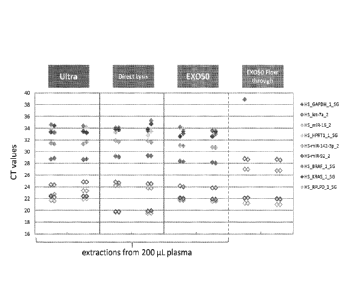
from 200u1 of plasma by ultracentrifugation, direct lysis, and EX050. RNA
extraction from
EX050 flow-through was also analyzed.
100130] Figure 89 is one graph showing the comparison between RNA
extractions
from 200u1 of plasma by the EX050 kit and miRNeasy, and 4 ml of plasma by the
EX050
kit and ultracentrifugation.
[00131] Figure 90 is one graph showing that EX050 isolates RNA containing
mutations from melanoma, BRAF V600E, compared to ultracentrifugation.
[00132] Figure 91 is one graph showing RNA isolation after elution from the
EX050
column yields as much RNA as the EX050 process.
[00133] Figure 92 is a schematic demonstrating the EX052 protocol for
isolating a
microvesicle fraction, releasing the microvesicle nucleic acids, and
extracting RNA and
DNA using two separate protocols.
[00134] Figure 93 is a schematic demonstrating the EX052.2 protocol for
isolating a
microvesicle fraction, releasing the microvesicle nucleic acids, and
extracting RNA and
DNA using a single protocol.
[00135] Figure 94 is a graph showing the effect of chloroform concentration
in phase
separation for isolating microvesicle RNA and DNA in a single extraction, as
demonstrated
by detection of wild-type BRAF RNA and DNA.
100136] Figure 95 is a graph showing the effect of chloroform concentration
in phase
separation for isolating microvesicle RNA and DNA in a single extraction, as
demonstrated
by detection of GAPDH RNA and DNA.
[00137] Figure 96 is a graph showing that the adjustment of pH in phase
separation
influences the DNA extraction and detection.
[00138] Figure 97 is a graph showing the effect of titration of sample
volume of
cerebrospinal fluid (CSF) on microvesicle RNA extraction and detection.
[00139] Figure 98 is a graph showing the comparison of detection of
microvesicle
RNA targets from ultracentrifugation and EX060 isolation methods.
[00140] Figure 99 is a graph showing the comparison of detection of
microvesicle
RNA targets from ultracentrifugation and EX060 isolation methods for different
patient
CSF samples. Patient samples are designated by patient ID. Varying sample
volumes were
utilized. (*) indicates post-mortem sample.
CA 02897207 2015-07-03
WO 2014/107571 PCT[US2014/010173
[00141] Figure 100 is a graph showing the effect of CSF sample volume
(0.25m1,
0.5m1, 1.0m1 and 2.0m1) on different microvesicle RNA isolation and extraction
methods.
UC (ultracentrifugation), uCSC (urine filtration method), and EX060.
100142] Figure 101 is a series of bioanalyzer plots depicting the RNA
profiles from
extraction from 2 different urine samples using the EX070 protocol compared to
the urine
circulating stem cell (uCSC) method.
[00143] Figure 102 is a graph showing the correlation between RNA detection
after
isolation and extraction by EX070 compared to the urineCSC method.
[00144] Figure 103 is two graphs showing the detection of different RNA
targets
after isolation and extraction by EX070 or uCSC method. RNA was extracted and
analyzed
from the isolated microvesicle fraction (EX070 or uCSC) and the flow-through
or
supernatant fraction after isolation (EX070 flow or uCSC flow). (A) mRNA
targets; (B)
miRNA targets.
[00145] Figure 104 is a graph depicting CTs (y-axis) for 4 mRNAs across 4
sample
types. All points are the average of experimental duplicates on each of 2
microvesicle
isolations. "SJCRH" is plasma from a pediatric patient, "DAOY" is a
medulloblastoma cell
line and "DAOY MED" is microvesicles from the medium of those cells.
Commercially
available plasma gave no results.
[00146] Figure 105 is a graph depicting the ability of EX050 to isolate all
mRNA
from 100 p.L cell culture supernatant.
DETAILED DESCRIPTION OF THE INVENTION
[00147] The present invention provides methods of isolating microvesicles
by
capturing the microvesicles to a surface and subsequently lysing the
microvesicles to release
the nucleic acids, particularly RNA, contained therein. Microvesicles are shed
by eukaryotic
cells, or budded off of the plasma membrane, to the exterior of the cell.
These membrane
vesicles are heterogeneous in size with diameters ranging from about 10 nm to
about 5000
nm. All membrane vesicles shed by cells < 0.8Rm in diameter are referred to
herein
collectively as "microvesicles." These microvesicles include microvesicles,
microvesicle-
like particles, prostasomes, dexosomes, texosomes, ectosomes, oncosomes,
apoptotic
bodies, retrovirus-like particles, and human endogenous retrovirus (HERV)
particles. Small
microvesicles (approximately 10 to 1000nm, and more often 30 to 200 nm in
diameter) that
26
CA 02897207 2015-07-03
WO 2014/107571 PCT[US2014/010173
are released by exocytosis of intracellular multivesicular bodies are referred
to in the art as
"microvesicles."
100148] Current methods of isolating microvesicles include
ultracentrifugation,
ultrafiltration, e.g., using 100 kD filters, polymer precipitation techniques,
and/or filtration
based on size. However, there exists a need for alternative methods that are
efficient and
effective for isolating microvesicles and, optionally, extracting the nucleic
acids contained
therein, preferably microvesicle RNA, for use in a variety of applications,
including
diagnostic purposes.
[00149] In some embodiments, the present invention provides methods of
isolating
microvesicles by capturing the microvesicles to a surface and subsequently
lysing the
microvesicles to release the nucleic acids, particularly RNA, contained
therein. In some
embodiments, the present invention provides methods of isolating microvesicles
by
capturing the microvesicles to a surface and subsequently eluting the intact
microvesicles
from the capture surface.
[00150] Microvesicles are a rich source of high quality nucleic acids,
excreted by all
cells and present in all human biofluids. The RNA in microvesicles provides a
snapshot of
the transcriptome of primary tumors, metastases and the surrounding
microenvironment in
real-time. Thus, accurate assessment of the RNA profile of microvesicles by
assays
provides companion diagnostics and real-time monitoring of disease. This
development has
been stalled by the current standard of isolating exosomes which is slow,
tedious, variable
and not suited for a diagnostic environment.
[00151] The isolation and extraction methods and/or kits provided herein
referred to
as the EX050 plasma exosome RNA isolation methods and/or kits use a spin-
column based
purification process using an affinity membrane that binds microvesicles. The
methods and
kits of the disclosure allow for the capability to run large numbers of
clinical samples in
parallel, using volumes from 0.2 up to 4 mL on a single column. The isolated
RNA is highly
pure, protected by a vesicle membrane until lysis, and intact vesicles can be
eluted from the
EX050 membrane. The EX050 procedure is able to deplete all mRNA from plasma
input,
and is equal or better in mRNA/miRNA yield when compared to
ultracentrifugation or
direct lysis. In contrast, the EX050 methods and/or kits enrich for the
microvesicle bound
fraction of miRNAs, and they are easily scalable to large amounts of input
material. This
ability to scale up enables research on interesting, low abundant transcripts.
In comparison
27
CA 02897207 2015-07-03
WO 2014/107571 PCT/US2014/010173
with other commercially available products on the market, the methods and kits
of the
disclosure provide unique capabilities that are demonstrated by the examples
provided
herein.
[00152] The EX050 methods and kits isolate and extract nucleic acids, e.g.,
RNA
from a biological sample using the following the general procedure. First, the
microvesicle
fraction is bound to a membrane filter, and the filter is washed. Then, a
reagent is used to
perform on-membrane lysis and release of the nucleic acids, e.g., RNA.
Chloroform
extraction is then performed using PLG tubes, followed by ethanol
conditioning. The
nucleic acids, e.g., RNA, is then bound to a silica column, washed and then
eluted.
[00153] A schematic representation of the general flow through of using a
capture
surface method to isolate microvesicle RNA from a biological sample is shown
below:
Wash
Biological Sample (optional) ________
...................................... Capture Direct Lyse
" .................................... Surface = = on Capture Surface
Loading Buffer (optional)
Standard
Downstream Assay wn.mb Pure RNA) =Aum:!'''
RNA isolation
[00154] A schematic representation of the general flow through of using
membrane
method to isolate microvesicle RNA from plasma sample is shown below:
Plasma Wash(optional))
Direct Lyse
Loading Buffer j
Membrane on Membrane
Standard
Downstream Assay ' [ Pure RNA 4I.RNA isolation
1001551 A schematic representation of the general flow through of using a
bead
method to isolate microvesicle RNA from plasma sample is shown below:
28
CA 02897207 2015-07-03
WO 2014/107571 PCT/US2014/010173
Wash
Plasma (optional)
Direct Lyse
............................ i Bead
L on Bead
Loading Buffer
.................... = Standard I
Downstream Assay =Avon [ Pure RNA I laRm
RNA isolation
........
[00156] A schematic representation of the general flow through of using a
capture
surface method to isolate microvesicle RNA from a urine sample is shown below:
Wash
(optional)
Capture Direct Lyse
Urine Sample mom,
Surface ..................................... on Capture Surface
.................................... = = Standard ¨1
Downstream Assay] 1,4ftr [Pure RNA
................................................. = RNA isolation
[00157] As demonstrated by the working examples provided herein, the format
of the
capturing surface, e.g., beads or a filter (also referred to herein as a
membrane), does not
affect the ability of the methods provided herein to efficiently capture
microvesicles from a
biological sample. Moreover, the surface charge of the capturing surface does
not affect the
ability of the methods provided herein to efficiently capture microvesicles.
In addition, the
working examples demonstrate that the ionic strength of the binding and wash
buffer is of
limited importance in the methods provided herein.
[00158] A wide range of surfaces are capable of capturing microvesicles
according to
the methods provided herein, but not all surfaces will capture microvesicles
(some surfaces
do not capture anything).
[00159] The present disclosure also describes a device for isolating and
concentrating
microvesicles from biological or clinical samples using disposable plastic
parts and
centrifuge equipment. For example, the device comprises a column comprising a
capture
surface (i.e., a membrane filter), a holder that secures the capture surface
between the outer
fit and an inner tube, and a collection tube. The outer fit comprises a large
net structure to
29
CA 02897207 2015-07-03
WO 2014/107571 PCT[US2014/010173
allow passing of liquid, and is preferably at one end of the column. The inner
tube holds the
capture surface in place, and preferably is slightly conus-shaped. The
collection tube may be
commercially available, i.e., 50m1 Falcon tube. The column is preferably
suitable for
spinning, i.e., the size is compatible with standard centrifuge and micro-
centrifuge
machines.
[00160] In embodiments where the capture surface is a membrane, the device
for
isolating the microvesicle fraction from a biological sample contains at least
one membrane.
In some embodiments, the device comprises one, two, three, four, five or six
membranes.
Preferably, the device comprises three membranes. In embodiments where the
device
comprises more than one membrane, the membranes are all directly adjacent to
one another
at one end of the column. In embodiments where the device comprises more than
one
membrane, the membranes are all identical to each other, i.e., are of the same
charge and/or
have the same functional group.
[00161] As demonstrated by working Examples 1-32 provided herein, the
choice of
buffer component(s), the pH of the buffer and the choice of lysis buffer
affect the ability of
the methods provided herein to efficiently capture microvesicles and release
the nucleic
acids, particularly RNA, contained therein. In some embodiments, the lysis
buffer is a
phenol-based lysis buffer. For example, the lysis buffer is Q1Azolg lysis
reagent (Qiagen).
[00162] Working Examples 1-32 provided herein also demonstrate that capture
by
filtering through a pore size smaller than the microvesicles is not the
primary mechanism of
capture by the methods provided herein. However, filter pore size is
nevertheless very
important, e.g. because mRNA gets stuck on a 20nrn filter and cannot be
recovered,
whereas microRNAs can easily be eluted off, and e.g. because the filter pore
size is an
important parameter in available surface capture area.
[00163] Working Examples 1-32 provided herein have also demonstrated the
feasibility of microvesicle isolation with beads (magnetic or non-magnetic)
that would be a
more suitable format in clinical utility. Results showed that positively
charged, negatively
charged, and neutral beads all showed good microvesicle capturing
efficiencies. This
observation suggests that they may act on different ligands on the surface of
microvesicle,
thus one microvesicle can be captured with different (functionalized)
particles via different
(multiple) forces.
CA 02897207 2015-07-03
WO 2014/107571 PCT[US2014/010173
[00164] The methods provided herein use any of a variety of capture
surfaces. In
some embodiments, the capture surface is a membrane, also referred to herein
as a filter or a
membrane filter. In some embodiments, the capture surface is a commercially
available
membrane. In some embodiments, the capture surface is a charged commercially
available
membrane. In some embodiments, the capture surface is neutral. In some
embodiments, the
capture surface is selected from Mustang Ion Exchange Membrane from PALL
Corporation; Vivapure Q membrane from Sartorius AG; Sartobind Q, or Vivapure
Q
Maxi H; Sartobind CD D from Sartorius AG, Sartobind (S) from Sartorius AG,
Sartobind
Q from Sartorius AG, Sartobind 0 IDA from Sartorius AG, Sartobind Aldehyde
from
Sartorius AG, Whatman DE81 from Sigma, Fast Trap Virus Purification column
from
EMD Millipore; Thermo Scientific* Pierce Strong Cation and Anion Exchange Spin
Columns.
[00165] In embodiments where the capture surface is charged, the capture
surface can
be a charged filter selected from the group consisting of 0.65um positively
charged Q PES
vacuum filtration, 3-5um positively charged Q RC spin column filtration, 0.8um
positively
charged Q PES homemade spin column filtration, 0.8um positively charged Q PES
syringe
filtration, 0.8um negatively charged S PES homemade spin column filtration,
0.8um
negatively charged S PES syringe filtration, and 50nm negatively charged nylon
syringe
filtration. Preferably, the charged filter is not housed in a syringe
filtration apparatus, as
Qiazol/RNA is harder to get out of the filter in these embodiments.
Preferably, the charged
filter is housed at one end of a column.
[00166] In embodiments where the capture surface is a membrane, the
membrane can
be made from a variety of suitable materials. In some embodiments, the
membrane is
polyethersulfone (PES) (e.g., from Millipore or PALL Corp.). In some
embodiments, the
membrane is regenerated cellulose (RC) (e.g., from Sartorius or Pierce).
[00167] In some embodiments, the capture surface is a positively charged
membrane.
In some embodiments, the capture surface is a Q membrane, which is a
positively charged
membrane and is an anion exchanger with quaternary amines. For example, the Q
membrane is functionalized with quaternary ammonium, R-CH2-N'(C1-13)3. In some
embodiments, the capture surface is a negatively charged membrane. In some
embodiments,
the capture surface is an S membrane, which is a negatively charged membrane
and is a
cation exchanger with sulfonic acid groups. For example, the S membrane is
functionalized
31
CA 02897207 2015-07-03
WO 2014/107571 PCT/US2014/010173
with sulfonic acid, R-CH2-S03-. In some embodiments, the capture surface is a
D
membrane, which is a weak basic anion exchanger with diethylamine groups, R-
CH2-
NH (C,H5)2. In some embodiments, the capture surface is a metal chelate
membrane. For
example, the membrane is an IDA membrane, functionalized with minodiacetic
acid ¨
N(CH2COOFF)2. In some embodiments, the capture surface is a microporous
membrane,
functionalized with aldehyde groups, -CHO. In other embodiments, the membrane
is a weak
basic anion exchanger, with diethylaminoethyl (DEAE) cellulose. Not all
charged
membranes are suitable for use in the methods provided herein, e.g., RNA
isolated using a
regenerated cellulose, strong acidic cation exchanger ("RC/SACE") membrane
spin column
showed RT-qPCR inhibition and, thus, unsuitable for PCR related downstream
assay.
[00168] In embodiments where the capture surface is charged, microvesicles
can be
isolated with a positively charged filter. In embodiments where the capture
surface is
charged, the capture surface is preferably not a negatively charged
regenerated cellulose S
filter, as this embodiment causes PCR inhibition. In contrast, negatively
charged PES filters
do not exhibit this phenomenon.
[00169] In embodiments where the capture surface is charged, the pH during
microvesicle capture is a pH <7. In some embodiments, the pH is greater than 4
and less
than or equal to 8.
[00170] In embodiments where the capture surface is a positively charged Q
filter,
the buffer system includes a wash buffer comprising 250mM Bis Tris Propane,
pH6.5-7Ø
In embodiments where the capture surface is a positively charged Q filter, the
lysis buffer is
Qiazol. In embodiments where the capture surface is a positively charged Q
filter, the lysis
buffer is present at one volume. In embodiments where the capture surface is a
positively
charged Q filter, the lysis buffer is present at more than one volume.
[00171] In embodiments where the capture surface is neutral, the capture
surface is a
filter selected from the group consisting of 20nm neutral PES syringe
filtration (Tisch and
Exomir), 30nm neutral PES syringe filtration (Sterlitech), 50nm neutral PES
syringe
filtration (Sterlitech), 0.2um neutral PES homemade spin column filtration
(Pall), 0.8um
neutral PES homemade spin column filtration (Pall) and 0.8um neutral PES
syringe
filtration (Pall). In embodiments where the capture surface is neutral,
preferably the neutral
capture surface is not housed in a syringe filter.
32
CA 02897207 2015-07-03
WO 2014/107571 PCT[US2014/010173
[00172] In embodiments where the capture surface is neutral, the lysis
buffer is
Qiazol. In embodiments where the capture surface is neutral, the lysis buffer
is present at
one volume. In embodiments where the capture surface is neutral, the lysis
buffer is present
at more than one volume. In embodiments where the capture surface is neutral,
the methods
include a wash step.
[00173] Depending on the membrane material, the pore sizes of the membrane
range
from 3 lam to 20 nm. For example, in embodiments where the capture surface is
a
commercially available PES membrane, the membrane has a pore size of 20nm
(Exomir),
0.65 m (Millipore) or 0.8um (Pall). In embodiments where the capture surface
is a
commercially available RC membrane, the membrane has a pore size in the range
of 3-51um
(Sartorius, Pierce).
[00174] The surface charge of the capture surface can be positive, negative
or neutral.
In some embodiments, the capture surface is a positively charged bead or
beads. In some
embodiments, the capture surface is a negatively charged bead or beads. In
some
embodiments, the capture surface is a neutral bead or beads. In some
embodiments, the
capture surface is a neutral filter, e.g., a PES membrane that is not surface
modified with a
charged group.
[00175] In some embodiments, the methods provided herein include a variety
of
buffers including loading and wash buffers. In some embodiments, loading and
wash
buffers can be of high or low ionic strength. In some embodiments, the salt
concentration,
e.g., NaC1 concentration, can be from 0 to 2.4M. The buffers can include a
variety of
components. In some embodiments, the buffers include one or more of the
following
components: Tris, Bis-Tris, Bis-Tris-Propane, Imidazole, Citrate, Methyl
Malonic Acid,
Acetic Acid, Ethanolamine, Diethanolamine, Triethanolamine (TEA) and Sodium
phosphate. In the methods provided herein, the pH of loading and wash buffers
is important.
Filters tend to clog when plasma samples at set to pH < 5.5 before loading
(the plasma will
not spin through the column at all), and at higher pH microvesicle RNA
recovery is lower
due to instability of the microvesicles. At neutral pH, the RNA recovery from
microvesicles
is optimal. In some embodiments, the buffer used is at DC concentration, 2X
concentration,
3X concentration, or 4X concentration. For example, the loading or binding
buffer is at 2X
concentration while the wash buffer is at DC concentration.
33
CA 02897207 2015-07-03
WO 2014/107571 PCT[US2014/010173
[00176] In some embodiments, the methods include one or more wash steps,
for
example, after contacting the biological sample with the capture surface. In
some
embodiments, detergents are added to the wash buffer to facilitate removing
the non-
specific binding (i.e., contaminants, cell debris, and circulating protein
complexes or nucleic
acids), to obtain a more pure microvesicle fraction. Detergents suitable for
use include, but
are not limited to, sodium dodecyl sulfate (SDS), Tween-20, Tween-80, Triton X-
100,
Nonidet P-40 (NP-40)õ Brij-35, Brij-58, octyl glucoside, octyl thioglucoside,
CHAPS or
CHAPSO.
[00177] In some embodiments, the capture surface, e.g., membrane, is housed
within
a device used for centrifugation; e.g. spin columns, or for vacuum system e.g.
vacuum filter
holders, or for filtration with pressure e.g. syringe filters. In a preferred
embodiment, the
capture surface is housed in a spin column or vacuum system.
[00178] The isolation of microvesicles from a biological sample prior to
extraction of
nucleic acids is advantageous for the following reasons: 1) extracting nucleic
acids from
microvesicles provides the opportunity to selectively analyze disease or tumor-
specific
nucleic acids obtained by isolating disease or tumor-specific microvesicles
apart from other
microvesicles within the fluid sample; 2) nucleic acid-containing
microvesicles produce
significantly higher yields of nucleic acid species with higher integrity as
compared to the
yield/integrity obtained by extracting nucleic acids directly from the fluid
sample without
first isolating microvesicles; 3) scalability, e.g., to detect nucleic acids
expressed at low
levels, the sensitivity can be increased by concentrating microvesicles from a
larger volume
of sample using the methods and/or kits described herein; 4) more pure or
higher
quality/integrity of extracted nucleic acids in that proteins, lipids, cell
debris, cells and other
potential contaminants and PCR inhibitors that are naturally found within
biological
samples are excluded before the nucleic acid extraction step; and 5) more
choices in nucleic
acid extraction methods can be utilized as isolated microvesicle fractions can
be of a smaller
volume than that of the starting sample volume, making it possible to extract
nucleic acids
from these fractions or pellets using small volume column filters.
[00179] Several methods of isolating microvesicles from a biological sample
have
been described in the art. For example, a method of differential
centrifugation is described
in a paper by Raposo et al. (Raposo et al., 1996), a paper by Skog et.
al.(Skog et al., 2008)
and a paper by Nilsson et. al.(Nilsson et al., 2009). Methods of ion exchange
and/or gel
34
CA 02897207 2015-07-03
WO 2014/107571 PCT/US2014/010173
permeation chromatography are described in US Patent Nos. 6,899,863 and
6,812,023.
Methods of sucrose density gradients or organelle electrophoresis are
described in U.S.
Patent No. 7,198,923. A method of magnetic activated cell sorting (MACS) is
described in a
paper by Taylor and Gercel Taylor (Taylor and Gercel-Taylor, 2008). A method
of
nanomembrane ultrafiltration concentration is described in a paper by
Cheruvanky et al.
(Cheruvanky et al., 2007). A method of Percoll gradient isolation is described
in a
publication by Miranda et al. (Miranda et al., 2010). Further, microvesicles
may be
identified and isolated from bodily fluid of a subject by a microfluidic
device (Chen et al.,
2010). In research and development, as well as commercial applications of
nucleic acid
biomarkers, it is desirable to extract high quality nucleic acids from
biological samples in a
consistent, reliable, and practical manner.
[00180] An object of the present invention is therefore to provide a method
for quick
and easy isolation of nucleic acid-containing particles from biological
samples such as body
fluids and extraction of high quality nucleic acids from the isolated
particles. The method of
the invention may be suitable for adaptation and incorporation into a compact
device or
instrument for use in a laboratory or clinical setting, or in the field.
[00181] In the working examples provided herein, human plasma from normal
healthy controls was obtained from Bioreclamation LLC with a blood collection
SOP
developed by Exosome Diagnostic Inc. Briefly, blood was collected into K2 EDTA
tubes
and mixed well with complete inversions. Then the tubes were centrifuged at
1300xg for
10min to separate the blood cells and plasma. Then plasma was removed and
filtered
through a 0.8ium filter to remove cell debris and platelets. All plasma
samples were then
aliquoted into lml cryovials and store at -80 C until use.
[00182] Before RNA isolation, the membrane was conditioned by passing
through
equilibrium buffer. The thawed plasma sample was diluted with loading buffer.
The diluted
plasma sample was slowly passed through the membrane that adsorbs the
microvesicles.
The membrane was then washed with a wash buffer to remove any weakly bound
plasma
components. Then a lysis reagent was passed through the membrane to lyse the
microvesicles. RNA was isolated using the miRNeasy kit (Qiagen).
[00183] RNA was assessed for quality and concentration with the 2100
Bioanalyzer
(Agilent) using a RNA 6000 Pico Chip. The relative quantity of the extracted
RNA was
CA 02897207 2015-07-03
WO 2014/107571 PCT/US2014/010173
measured by RT-qPCR using selected human gene expression assays from Applied
Biosystems (Taqman Assay).
[00184] While the examples provided herein used plasma samples, the skilled
artisan
will appreciate that these methods are applicable to a variety of biological
samples. Other
suitable biological samples include urine, cerebrospinal fluid, blood
including blood
components, e.g., plasma and serum, sputum, pleural fluid, nipple aspirates,
lymph fluid,
fluid of the respiratory, intestinal, and genitourinary tracts, tear fluid,
saliva, breast milk,
fluid from the lymphatic system, semen, intraorgan system fluid, ascitic
fluid, tumor cyst
fluid, amniotic fluid, cell culture supernatant and combinations thereof.
[00185] The methods and kits of the disclosure are suitable for use with
samples
derived from a human subject. The methods and kits of the disclosure are
suitable for use
with samples derived from a non-human subject such as, for example, a rodent,
a non-
human primate, a companion animal (e.g., cat, dog, horse), and/or a farm
animal (e.g.,
chicken).
[00186] In some embodiments, a pre-processing step prior to isolation,
purification or
enrichment of the microvesicles is performed to remove large unwanted
particles, cells
and/or cell debris and other contaminants present in the biological sample.
The pre-
processing steps may be achieved through one or more centrifugation steps
(e.g., differential
centrifugation) or one or more filtration steps (e.g., ultrafiltration), or a
combination thereof
Where more than one centrifugation pre-processing steps are performed, the
biological
sample may be centrifuged first at the lower speed and then at the higher
speed. If desired,
further suitable centrifugation pre-processing steps may be carried out.
Alternatively or in
addition to the one or more centrifugation pre-processing steps, the
biological sample may
be filtered. For example, a biological sample may be first centrifuged at
20,000g for 1 hour
to remove large unwanted particles; the sample can then be filtered, for
example, through a
0.8 ttm filter.
[00187] In some embodiments, one or more centrifugation steps are performed
before
or after contacting the biological sample with the capture surface to separate
microvesicles
and concentrate the microvesicles isolated from the biological fraction. For
example, the
sample is centrifuged at 20,000 g for 1 hour at 4 C. To remove large unwanted
particles,
cells, and/or cell debris, the samples may be centrifuged at a low speed of
about 100-500g,
preferably about 250-300g. Alternatively or in addition, the samples may be
centrifuged at a
36
CA 02897207 2015-07-03
WO 2014/107571 PCT[US2014/010173
higher speed. Suitable centrifugation speeds are up to about 200,000g; for
example from
about 2,000g to less than about 200,000g. Speeds of above about 15,000g and
less than
about 200,000g or above about 15,000g and less than about 100,000g or above
about
15,000g and less than about 50,000g are preferred. Speeds of from about
18,000g to about
40,000g or about 30,000g; and from about 18,000g to about 25,000g are more
preferred.
Particularly preferred is a centrifugation speed of about 20,000g. Generally,
suitable times
for centrifugation are from about 5 minutes to about 2 hours, for example,
from about 10
minutes to about 1.5 hours, or more preferably from about 15 minutes to about
1 hour. A
time of about 0.5 hours may be preferred. It is sometimes preferred to subject
the biological
sample to centrifugation at about 20,000g for about 0.5 hours. However the
above speeds
and times can suitably be used in any combination (e.g., from about 18,000g to
about
25,000g, or from about 30,000g to about 40,000g for about 10 minutes to about
1.5 hours,
or for about 15 minutes to about 1 hour, or for about 0.5 hours, and so on).
The
centrifugation step or steps may be carried out at below-ambient temperatures,
for example
at about 0-10 C, preferably about 1-5 C, e.g., about 3 C or about 4 C.
[00188] In some embodiments, one or more filtration steps are performed
before or
after contacting the biological sample with the capture surface. A filter
having a size in the
range about 0.1 to about 1.0 !,im may be employed, preferably about 0.8 gm or
0.22 mm.
The filtration may also be performed with successive filtrations using filters
with decreasing
porosity.
[00189] In some embodiments, one or more concentration steps are performed,
in
order to reduce the volumes of sample to be treated during the chromatography
stages,
before or after contacting the biological sample with the capture surface.
Concentration may
be through centrifugation of the sample at high speeds, e.g. between 10,000
and 100,000 g,
to cause the sedimentation of the microvesicles. This may consist of a series
of differential
centrifugations. The microvesicles in the pellet obtained may be reconstituted
with a smaller
volume and in a suitable buffer for the subsequent steps of the process. The
concentration
step may also be performed by ultrafiltration. In fact, this ultrafiltration
both concentrates
the biological sample and performs an additional purification of the
microvesicle fraction.
In another embodiment, the filtration is an ultrafiltration, preferably a
tangential
ultrafiltration. Tangential ultrafiltration consists of concentrating and
fractionating a
solution between two compartments (filtrate and retentate), separated by
membranes of
37
CA 02897207 2015-07-03
WO 2014/107571 PCT/US2014/010173
determined cut-off thresholds. The separation is carried out by applying a
flow in the
retentate compartment and a transmembrane pressure between this compartment
and the
filtrate compartment. Different systems may be used to perform the
ultrafiltration, such as
spiral membranes (Millipore, Amicon), flat membranes or hollow fibers (Amicon,
Millipore, Sartorius, Pall, GF, Sepracor). Within the scope of the invention,
the use of
membranes with a cut-off threshold below 1000 kDa, preferably between 100 kDa
and 1000
kDa, or even more preferably between 100 kDa and 600 kDa, is advantageous.
[00190] In some embodiments, one or more size-exclusion chromatography step
or
gel permeation chromatography steps are performed before or after contacting
the biological
sample with the capture surface. To perform the gel permeation chromatography
step, a
support selected from silica, acrylamide, agarose, dextran, ethylene glycol-
methacrylate co-
polymer or mixtures thereof, e.g., agarose-dextran mixtures, are preferably
used. For
example, such supports include, but are not limited to: SUPERDEXO 200HR
(Pharmacia),
TSK G6000 (TosoHaas) or SEPHACRYLO S (Pharmacia).
[00191] In some embodiments, one or more affinity chromatography steps are
performed before or after contacting the biological sample with the capture
surface. Some
microvesicles can also be characterized by certain surface molecules. Because
microvesicles
form from budding of the cell plasma membrane, these microvesicles often share
many of
the same surface molecules found on the cells they originated from. As used
herein,
"surface molecules" refers collectively to antigens, proteins, lipids,
carbohydrates, and
markers found on the surface or in or on the membrane of the microvesicle.
These surface
molecules can include, for example, receptors, tumor-associated antigens,
membrane
protein modifications (e.g., glycosylated structures). For example,
microvesicles that bud
from tumor cells often display tumor-associated antigens on their cell
surface. As such,
affinity chromatography or affinity exclusion chromatography can also be
utilized in
combination with the methods provided herein to isolate, identify, and or
enrich for specific
populations of microvesicles from a specific donor cell type (Al-Nedawi et
al., 2008; Taylor
and Gercel-Taylor, 2008). For example, tumor (malignant or non-malignant)
microvesicles
carry tumor-associated surface antigens and may be detected, isolated and/or
enriched via
these specific tumor-associated surface antigens. In one example, the surface
antigen is
epithelial cell adhesion molecule (EpCAM), which is specific to microvesicles
from
carcinomas of long, colorectal, breast, prostate, head and neck, and hepatic
origin, but not of
38
CA 02897207 2015-07-03
WO 2014/107571
PCT[US2014/010173
hematological cell origin (Balzar et al., 1999; Went et al., 2004).
Additionally, tumor-
specific microvesicles can also be characterized by the lack of certain
surface markers, such
as CD80 and CD86. In these cases, microvesicles with these markers may be
excluded for
further analysis of tumor specific markers, e.g., by affinity exclusion
chromatography.
Affinity chromatography can be accomplished, for example, by using different
supports,
resins, beads, antibodies, aptamers, aptamer analogs, molecularly imprinted
polymers, or
other molecules known in the art that specifically target desired surface
molecules on
microvesicles.
[00192] Optionally,
control particles may be added to the sample prior to microvesicle
isolation or nucleic acid extraction to serve as an internal control to
evaluate the efficiency
or quality of microvesicle purification and/or nucleic acid extraction. The
methods and/or
kits described herein provide for the efficient isolation and the control
particles along with
the microvesicle fraction. These control particles include Q-beta
bacteriophage, virus
particles, or any other particle that contains control nucleic acids (e.g., at
least one control
target gene) that may be naturally occurring or engineered by recombinant DNA
techniques.
In some embodiments, the quantity of control particles is known before the
addition to the
sample. The control target gene can be quantified using real-time PCR
analysis.
Quantification of a control target gene can be used to determine the
efficiency or quality of
the microvesicle purification or nucleic acid extraction processes.
[00193] Preferably,
the control particle is a Q-beta bacteriophage, referred to herein as
"Q-beta particle." The Q-beta particle used in the methods and/or kits
described herein may
be a naturally-occurring virus particle or may be a recombinant or engineered
virus, in
which at least one component of the virus particle (e.g., a portion of the
genome or coat
protein) is synthesized by recombinant DNA or molecular biology techniques
known in the
art. Q-beta is a member of the leviviridae family, characterized by a linear,
single-stranded
RNA genome that consists of 3 genes encoding four viral proteins: a coat
protein, a
maturation protein, a lysis protein, and RNA replicase. Due to its similar
size to average
microvesicles, Q-beta can be easily purified from a biological sample using
the same
purification methods used to isolate microvesicles, as described herein. In
addition, the low
complexity of the Q-beta viral single-stranded gene structure is advantageous
for its use as a
control in amplification-based nucleic acid assays. The Q-beta particle
contains a control
target gene or control target sequence to be detected or measured for the
quantification of
39
CA 02897207 2015-07-03
WO 2014/107571
PCT[US2014/010173
the amount of Q-beta particle in a sample. For example, the control target
gene is the Q-beta
coat protein gene. After addition of the Q-beta particles to the biological
sample, the nucleic
acids from the Q-beta particle are extracted along with the nucleic acids from
the biological
sample using the extraction methods and/or kits described herein. Detection of
the Q-beta
control target gene can be determined by RT-PCR analysis, for example,
simultaneously
with the biomarkers of interest (i.e., BRAF). A standard curve of at least 2,
3, or 4 known
concentrations in 10-fold dilution of a control target gene can be used to
determine copy
number. The copy number detected and the quantity of Q-beta particle added can
be
compared to determine the quality of the isolation and/or extraction process.
[00194] In a
preferred embodiment, the Q-beta particles are added to the urine sample
prior to nucleic extraction. For example, the Q-beta particles are added to
the urine sample
prior to ultrafiltration and/or after the pre-filtration step.
[00195] In some embodiments, 50, 100, 150, 200, 250, 300, 350, 400, 450,
500, 1,000 or
5,000 copies of Q-beta particles added to a bodily fluid sample. In a
preferred embodiment,
100 copies of Q-beta particles are added to a bodily fluid sample. The copy
number of Q-
beta particles can be calculated based on the ability of the Q-beta
bacteriophage to infect
target cells. Thus, the copy number of Q-beta particles is correlated to the
colony forming
units of the Q-beta bacteriophage.
Nucleic Acid Extraction
[00196] The present invention is directed towards the use of a capture
surface for the
improved isolation, purification, or enrichment of microvesicles. The methods
disclosed
herein provide a highly enriched microvesicle fraction for extraction of high
quality nucleic
acids from said microvesicles. The nucleic acid extractions obtained by the
methods and/or
kits described herein may be useful for various applications in which high
quality nucleic
acid extractions are required or preferred, such as for use in the diagnosis,
prognosis, or
monitoring of diseases or medical conditions.
100197] Recent
studies reveal that nucleic acids within microvesicles have a role as
biomarkers. For example, WO 2009/100029 describes, among other things, the use
of
nucleic acids extracted from microvesicles in GBM patient serum for medical
diagnosis,
prognosis and therapy evaluation. WO 2009/100029 also describes the use of
nucleic acids
extracted from microvesicles in human urine for the same purposes. The use of
nucleic
acids extracted from microvesicles is considered to potentially circumvent the
need for
CA 02897207 2015-07-03
WO 2014/107571 PCT/US2014/010173
biopsies, highlighting the enormous diagnostic potential of microvesicle
biology (Skog et
al., 2008).
[00198] The quality or purity of the isolated microvesicles can directly
affect the
quality of the extracted microvesicle nucleic acids, which then directly
affects the efficiency
and sensitivity of biomarker assays for disease diagnosis, prognosis, and/or
monitoring.
Given the importance of accurate and sensitive diagnostic tests in the
clinical field, methods
for isolating highly enriched microvesicle fractions from biological samples
are needed. To
address this need, the present invention provides methods for isolating
microvesicles from
biological sample for the extraction of high quality nucleic acids from a
biological sample.
As shown herein, highly enriched microvesicle fractions are isolated from
biological
samples by methods and/or kits described herein, and wherein high quality
nucleic acids
subsequently extracted from the highly enriched microvesicle fractions. These
high quality
extracted nucleic acids are useful for measuring or assessing the presence or
absence of
biomarkers for aiding in the diagnosis, prognosis, and/or monitoring of
diseases or other
medical conditions.
[00199] As used herein, the term "biological sample" refers to a sample
that contains
biological materials such as DNA, RNA and protein. In some embodiments, the
biological
sample may suitably comprise a bodily fluid from a subject. The bodily fluids
can be fluids
isolated from anywhere in the body of the subject, preferably a peripheral
location,
including but not limited to, for example, blood, plasma, serum, urine,
sputum, spinal fluid,
cerebrospinal fluid, pleural fluid, nipple aspirates, lymph fluid, fluid of
the respiratory,
intestinal, and genitourinary tracts, tear fluid, saliva, breast milk, fluid
from the lymphatic
system, semen, intra-organ system fluid, ascitic fluid, tumor cyst fluid,
amniotic fluid and
cell culture supernatant, and combinations thereof In some embodiments, the
preferred
body fluid for use as the biological sample is urine. In other embodiments,
the preferred
body fluid is serum. In yet other embodiments, the preferred body fluid is
plasma. In other
embodiments, the preferred body fluid is cerebrospinal fluid. Suitably a
sample volume of
about 0.1m1 to about 30m1 fluid may be used. The volume of fluid may depend on
a few
factors, e.g., the type of fluid used. For example, the volume of serum
samples may be
about 0.1m1 to about 4m1, preferably about 0.2m1 to 4m1. The volume of plasma
samples
may be about 0.1m1 to about 4m1, preferably 0.5m1 to 4m1. The volume of urine
samples
41
CA 02897207 2015-07-03
WO 2014/107571 PCT/US2014/010173
may be about 10 ml to about 30m1, preferably about 20 ml. Biological samples
can also
include fecal or cecal samples, or supernatants isolated therefrom.
[00200] The term "subject" is intended to include all animals shown to or
expected to
have nucleic acid-containing particles. In particular embodiments, the subject
is a mammal,
a human or nonhuman primate, a dog, a cat, a horse, a cow, other farm animals,
or a rodent
(e.g. mice, rats, guinea pig. etc.). A human subject may be a normal human
being without
observable abnormalities, e.g., a disease. A human subject may be a human
being with
observable abnormalities, e.g., a disease. The observable abnotinalities may
be observed by
the human being himself, or by a medical professional. The term "subject,"
"patient," and
"individual" are used interchangeably herein.
[00201] As used herein, the term "nucleic acids" refer to DNA and RNA. The
nucleic
acids can be single stranded or double stranded. In some instances, the
nucleic acid is DNA.
In some instances, the nucleic acid is RNA. RNA includes, but is not limited
to, messenger
RNA, transfer RNA, ribosomal RNA, non-coding RNAs, microRNAs, and HERV
elements.
[00202] As used herein, the term "high quality" in reference to nucleic
acid extraction
means an extraction in which one is able to detect 18S and 28S rRNA,
preferably in a ratio
of approximately 1:1 to approximately 1:2; and more preferably, approximately
1:2. Ideally,
high quality nucleic acid extractions obtained by the methods and/or kits
described herein
will also have an RNA integrity number of greater than or equal to 5 for a low
protein
biological sample (e.g., urine), or greater than or equal to 3 for a high
protein biological
sample (e.g., serum), and a nucleic acid yield of greater than or equal to 50
pg/ml from a 20
ml low protein biological sample or a 1 ml high protein biological sample.
[00203] High quality RNA extractions are desirable because RNA degradation
can
adversely affect downstream assessment of the extracted RNA, such as in gene
expression
and mRNA analysis, as well as in analysis of non-coding RNA such as small RNA
and
microRNA. The new methods and/or kits described herein enable one to extract
high quality
nucleic acids from microvesicles isolated from a biological sample so that an
accurate
analysis of nucleic acids within the microvesicles can be performed.
[00204] Following the isolation of microvesicles from a biological sample,
nucleic
acid may be extracted from the isolated or enriched microvesicle fraction. To
achieve this,
in some embodiments, the microvesicles may first be lysed. The lysis of
microvesicles and
extraction of nucleic acids may be achieved with various methods known in the
art. In one
42
CA 02897207 2015-07-03
WO 2014/107571 PCT[US2014/010173
embodiment, the lysis and extraction steps may be achieved using a
commercially available
Qiagen RNeasy Plus kit. In another embodiment, the lysis and extraction steps
may be
achieved using a commercially available Qiagen miRNeasy kit. In yet another
embodiment,
the nucleic acid extraction may be achieved using phenol:chloroform according
to standard
procedures and techniques known in the art. Such methods may also utilize a
nucleic acid-
binding column to capture the nucleic acids contained within the
microvesicles. Once
bound, the nucleic acids can then be eluted using a buffer or solution
suitable to disrupt the
interaction between the nucleic acids and the binding column, thereby
successfully eluting
the nucleic acids.
[00205] In some embodiments, the nucleic acid extraction methods also
include the
step of removing or mitigating adverse factors that prevent high quality
nucleic acid
extraction from a biological sample. Such adverse factors are heterogeneous in
that different
biological samples may contain various species of adverse factors. In some
biological
samples, factors such as excessive DNA may affect the quality of nucleic acid
extractions
from such samples. In other samples, factors such as excessive endogenous
RNase may
affect the quality of nucleic acid extractions from such samples. Many agents
and methods
may be used to remove these adverse factors. These methods and agents are
referred to
collectively herein as an "extraction enhancement operations." In some
instances, the
extraction enhancement operation may involve the addition of nucleic acid
extraction
enhancement agents to the biological sample. To remove adverse factors such as
endogenous RNases, such extraction enhancement agents as defined herein may
include, but
are not limited to, an RNase inhibitor such as Superase-In (commercially
available from
Ambion Inc.) or RNaseINplus (commercially available from Promega Corp.), or
other
agents that function in a similar fashion; a protease (which may function as
an RNase
inhibitor); DNase; a reducing agent; a decoy substrate such as a synthetic RNA
and/or
carrier RNA; a soluble receptor that can bind RNase; a small interfering RNA
(siRNA); an
RNA binding molecule, such as an anti-RNA antibody, a basic protein or a
chaperone
protein; an RNase denaturing substance, such as a high osmolarity solution, a
detergent, or a
combination thereof
[00206] For example, the extraction enhancement operation may include the
addition
of an RNase inhibitor to the biological sample, and/or to the isolated
microvesicle fraction,
prior to extracting nucleic acid; preferably the RNase inhibitor has a
concentration of
43
CA 02897207 2015-07-03
WO 2014/107571
PCT/US2014/010173
greater than 0.027 AU (I X) for a sample equal to or more than 1 pi in volume;
alternatively, greater than or equal to 0. 1 35 AU (5X) for a sample equal to
or more than 1
pl; alternatively, greater than or equal to 0.27 AU (10X) for a sample equal
to or more than
I [il; alternatively, greater than or equal to 0.675 AU (25X) for a sample
equal to or more
than 1 pl; and alternatively, greater than or equal to 1 .35 AU (50X) for a
sample equal to or
more than 1 pi; wherein the I X concentration refers to an enzymatic condition
wherein
0.027 AU or more RNase inhibitor is used to treat microvesicles isolated from
1 pl or more
bodily fluid, the 5X concentration refers to an enzymatic condition wherein
0.135 AU or
more RNase inhibitor is used to treat microvesicles isolated from 1 pl or more
bodily fluid,
the 10X protease concentration refers lo an enzymatic condition wherein 0.27
AU or more
RNase inhibitor is used to treat particles isolated from 1 pl or more bodily
fluid, the 25X
concentration refers to an enzymatic condition wherein 0.675 AU or more RNase
inhibitor
is used to treat microvesicles isolated from 1 pl or more bodily fluid, and
the 50X protease
concentration refers to an enzymatic condition wherein 1 .35 AU or more RNase
inhibitor is
used to treat particles isolated from 1 pl or more bodily fluid. Preferably,
the RNase
inhibitor is a protease, in which case, 1 AU is the protease activity that
releases folin-
positive amino acids and peptides corresponding to 1 pmol tyrosine per minute.
[00207] These enhancement agents may exert their functions in various ways,
e.g.,
through inhibiting RNasc activity (e.g., RNase inhibitors), through a
ubiquitous degradation
of proteins (e.g., proteases), or through a chaperone protein (e.g., a RNA-
binding protein)
that binds and protects RNAs. In all instances, such extraction enhancement
agents remove
or at least mitigate some or all of the adverse factors in the biological
sample or associated
with the isolated particles that would otherwise prevent or interfere with the
high quality
extraction of nucleic acids from the isolated particles.
[00208] In some embodiments, the quantification of 18S and 28S rRNAs
extracted
can be used determine the quality of the nucleic acid extraction.
Detection of nucleic acid biomarkers
[00209] In some embodiments, the extracted nucleic acid comprises RNA. In
this
instance, the RNA is preferably reverse-transcribed into complementary DNA
(cDNA)
before further amplification. Such reverse transcription may be performed
alone or in
combination with an amplification step. One example of a method combining
reverse
transcription and amplification steps is reverse transcription polymerase
chain reaction (RT-
44
PCR), which may be further modified to be quantitative, e.g., quantitative RT-
PCR as
described in US Patent No. 5,639,606.
Another example of the method comprises two separate steps: a first of reverse
transcription to convert RNA into cDNA and a second step of quantifying the
amount of
cDNA using quantitative PCR. As demonstrated in the examples that follow, the
RNAs
extracted from nucleic acid-containing particles using the methods disclosed
herein include
many species of transcripts including, but not limited to, ribosomal 18S and
28S rRNA,
microRNAs, transfer RNAs, transcripts that are associated with diseases or
medical
conditions, and biomarkers that are important for diagnosis, prognosis and
monitoring of
medical conditions.
[00210] For example, RT-PCR analysis determines a Ct (cycle threshold)
value for
each reaction. In RT-PCR, a positive reaction is detected by accumulation of a
fluorescence
signal. The Ct value is defmed as the number of cycles required for the
fluorescent signal to
cross the threshold (i.e., exceeds background level). Ct levels are inversely
proportional to
the amount of target nucleic acid, or control nucleic acid, in the sample
(i.e., the lower the
Ct level, the greater the amount of control nucleic acid in the sample).
[00211] In another embodiment, the copy number of the control nucleic
acid can be
measured using any of a variety of art-recognized techniques, including, but
not limited to,
RT-PCR. Copy number of the control nucleic acid can be determined using
methods known
in the art, such as by generating and utilizing a calibration, or standard
curve.
[00212] In some embodiments, one or more biomarkers can be one or a
collection of
genetic aberrations, which is used herein to refer to the nucleic acid amounts
as well as
nucleic acid variants within the nucleic acid-containing particles.
Specifically, genetic
aberrations include, without limitation, over-expression of a gene (e.g., an
oncogene) or a
panel of genes, under-expression of a gene (e.g., a tumor suppressor gene such
as p53 or
RB) or a panel of genes, alternative production of splice variants of a gene
or a panel of
genes, gene copy number variants (CNV) (e.g., DNA double minutes) (Hahn,
1993), nucleic
acid modifications (e.g., methylation, acetylation and phosphorylations),
single nucleotide
polymorphisms (SNPs), chromosomal rearrangements (e.g., inversions, deletions
and
duplications), and mutations (insertions, deletions, duplications, missense,
nonsense,
synonymous or any other nucleotide changes) of a gene or a panel of genes,
which
mutations, in many cases, ultimately affect the activity and function of the
gene products,
CA 2897207 2020-03-26
CA 02897207 2015-07-03
WO 2014/107571 PCT/US2014/010173
lead to alternative transcriptional splice variants and/or changes of gene
expression level, or
combinations of any of the foregoing.
1002131 The analysis of nucleic acids present in the isolated particles is
quantitative
and/or qualitative. For quantitative analysis, the amounts (expression
levels), either relative
or absolute, of specific nucleic acids of interest within the isolated
particles are measured
with methods known in the art (described below). For qualitative analysis, the
species of
specific nucleic acids of interest within the isolated microvesicles, whether
wild type or
variants, are identified with methods known in the art.
[00214] The present invention also includes various uses of the new methods
of
isolating microvesicles from a biological sample for high quality nucleic acid
extraction
from a for (i) aiding in the diagnosis of a subject, (ii) monitoring the
progress or
reoccurrence of a disease or other medical condition in a subject, or (iii)
aiding in the
evaluation of treatment efficacy for a subject undergoing or contemplating
treatment for a
disease or other medical condition; wherein the presence or absence of one or
more
biomarkers in the nucleic acid extraction obtained from the method is
determined, and the
one or more biomarkers are associated with the diagnosis, progress or
reoccurrence, or
treatment efficacy, respectively, of a disease or other medical condition.
Kits for isolating microvesicles from a biological sample
[00215] One aspect of the present invention is further directed to kits for
use in the
methods disclosed herein. The kit comprises a capture surface apparatus
sufficient to
separate microvesicles from a biological sample from unwanted particles,
debris, and small
molecules that are also present in the biological sample. The present
invention also
optionally includes instructions for using the foregoing reagents in the
isolation and optional
subsequent nucleic acid extraction process.
EXAMPLES
[00216] While the examples provided herein use a variety of membranes and
devices
used for centrifugation and/or filtration purposes, it is to be understood
that these methods
can be used with any capture surface and/or housing device that allows for the
efficient
capture of microvesicles and release of the nucleic acids, particularly, RNA,
contained
therein.
46
CA 02897207 2015-07-03
WO 2014/107571
PCT[US2014/010173
Example 1: Plasma Microvesicular RNA can be isolated using a 0.65um positively
charged Q PES filter in a vacuum format
[00217] Normal control plasma was obtained from Bioreclamation LLC. Blood
was
collected into K2 EDTA tubes and mixed well with complete inversions. The
tubes were
centrifuged at 1300xg for 10min to separate the blood cells and plasma. Then,
the plasma
was filtered through a 0.8 m mixed cellulose ester filter (Millipore,
Billerica, Ma., USA) to
remove cell debris and platelets, and divided into 1 mL aliquots. Aliquots
were frozen at -
80 C until needed.
[00218] Isolation of 4mL plasma microvesicular RNA was conducted using
ultracentrifugation or Fast Trap Adenovirus purification and concentration kit
(Millipore,
Billerica, Ma., USA) 0.65um positively charged Q polyethersulfone vacuum
filtration.
[00219] In the ultracentrifugation method, one 1 mi. aliquots of plasma
from four
subjects were transferred to a 5mL polyallomer tube (Beckman-Coulter, Miami,
Fl., USA)
containing 8 pL RNasin Plus (40 U/ul, Promega, Madison, Wi., USA) RNase
inhibitor, and
incubated for 5 minutes at room temp. Following incubation, the plasma was
diluted in 1
ml. PBS (10mM Sodium phosphate, 2mM Potassium phosphate, 2.7mM KC1, 137mM
NaC1, pH 7.4). Microvesicles were pelleted by ultracentrifugation at 120,000g
for 80
minutes at 8 C. The microvesicle pellet was washed in 42 ut PBS and 8 uL
RNasin Plus,
and incubated for 20 minutes at room temp. The microvesicle pellet was lysed
in 700u1
Qiazol Reagent (Qiagen, Valencia, Ca., USA).
[00220] In the 0.65um positively charged Q polyethersulfone vacuum
filtration
method, one lmL aliquots of plasma from the same four subjects used in the
ultracentrifugation method were pooled, diluted with 5mL PBS, and mixed with 1
ml. 10X
Binding Buffer (Millipore). Before the plasma sample was applied, the filter
was
conditioned by passing through 25m1L Equilibration Buffer (Millipore) by
vacuum. Then,
the plasma sample was passed through the filter by vacuum. The filtrate was
saved for
further analysis. The filter was washed with 20mL Wash Buffer (Millipore) by
vacuum.
Then the microvesicles were lysed with Qiazol. 2.25mL Qiazol was applied to
the
membrane and distributed by drawing approximately 5 drops of Qiazol through
the filter by
vacuum. The filter was then incubated for 5 min at room temp and eluted by
vacuum.
[00221] The filtrate samples were transferred to two 5m1L polyallomer tubes
(Beckman-Coulter, Miami, Fl., USA) containing 8 L RNasin Plus (40 U/ 1,
Promega,
47
CA 02897207 2015-07-03
WO 2014/107571 PCT/US2014/010173
Madison, Wi., USA) RNase inhibitor, and incubated for 5 minutes at room temp.
Microvesicles were pelleted by ultracentrifugation at 120,000g for 80 minutes
at 8 C. The
microvesicle pellets were washed in 42 uL PBS and 8 AL RNasin Plus, and
incubated for 20
minutes at room temp. The microvesicle pellets were lysed in 700u1 Qiazol
Reagent
(Qiagen, Valencia, Ca., USA).
[00222] For the ultracentrifugation and filtration (filter and filtrate)
samples,
microvesicular RNA was isolated using the miRNeasy kit (Qiagen) according to
the
manufacturer's recommendation. The RNA quality and concentration was assessed
with the
2100 Bioanalyzer (Agilent, Palo Alto, Ca., USA) using a RNA 6000 Pico Chip
(FIG 1A).
Two-thirds and two-ninths of the total volume of microvesicular RNA was
converted to
cDNA using the SuperScript VILO cDNA Synthesis Kit (Invitrogen, Carlsbad, Ca.,
USA)
and the Taqman MicroRNA Reverse Transcription Kit (Applied Biosystems, Foster
City,
Ca., USA), respectively, according to the manufacturer's protocol.
[00223] To compare microvesicular RNA isolated by filtration (filter and
filtrate) and
ultracentrifugation, RT-PCRs were performed on mRNA and non-coding RNA
(including
microRNA) using primers and probes specific to GAPDH, RN7SL, RNaseP, miR-16,
miR-
150, and let-7a. For GAPDH and RNaseP, the amplification reactions were
performed in a
20u1 volume containing 2X Taqman Gene Expression Master Mix (Applied
Biosystems,
Foster City, Ca., USA), 20X Taqman Gene Expression Assay (Applied Biosystems,
Foster
City, Ca., USA), and a 1:15 fraction of the cDNA reverse transcribed with the
SuperScript
VILO cDNA Synthesis Kit. For RN7SL, the amplification reactions were performed
in a
20u1 volume containing 2X Taqman Gene Expression Master Mix, 900nM RN7SL
Forward
CAAAACTCCCGTGCTGATCA (SEQ ID NO 1), 900nM RN7SL Reverse
GGCTGGAGTGCAGTGGCTAT (SEQ ID NO 2), 250nM RN7SL Probe
TGGGATCGCGCCTGT (SEQ ID NO 3), and a 1:15 fraction of the cDNA reverse
transcribed with the SuperScript VILO cDNA Synthesis Kit. For miR-16, miR-150,
and let-
7a, the amplification reactions were performed in a 20u1 volume containing 2X
Taqman
Universal PCR Master Mix, 20X Taqman MicroRNA Assay (Applied Biosystems,
Foster
City, Ca., USA), and a 1:20 fraction of the cDNA reverse transcribed with the
Taqman
MicroRNA Reverse Transcription Kit. Amplification conditions consisted of: 1
cycle of
50 C, 2 min; 1 cycle of 95 C, 10 min; 40 cycles of 95 C, 10 min and 60 C, 1
min. See FIG
1B.
48
CA 02897207 2015-07-03
WO 2014/107571 PCT[US2014/010173
Example 2: Detecting BR4F_V600E mutation in plasma sample from melanoma
patient using Millipore Fast Trap Virus Purification Column
[00224] Melanoma plasma was obtained from Ludwig Maximilians University in
accordance with protocols approved by the IRB and as described in EXAMPLE 1.
[00225] Isolation of 2mL and 12mL plasma microvesicular RNA was conducted
using ultracentrifugation or Fast Trap Adenovirus purification and
concentration kit
(Millipore, Billerica, Ma., USA) 0.65um positively charged Q polyethersulfone
vacuum
filtration.
[00226] In the ultracentrifugation method, 2mL and 3X4mL plasma were
transferred
to a 5mL polyallomer tube (Beckman-Coulter, Miami, Fl., USA) containing 8 iaL
RNasin
Plus (40 U/111, Promcga, Madison, Wi., USA) RNasc inhibitor, and incubated for
5 minutes
at room temp. Following incubation, the 2mL and each 4mL plasma was diluted in
3mL and
lmL PBS, respectively. Microvesicles were pelleted and lysed as described in
EXAMPLE 1.
[00227] In the 0.65um positively charged Q polyethersulfone vacuum
filtration
method, 2m1. and 12mL plasma was diluted with 2.5 mL and 15mL PBS,
respectively, and
mixed with 500u1 and 3mL 10X Binding Buffer (Millipore), respectively. Before
the plasma
samples were applied, the filters were conditioned by passing through 25mL
Equilibration
Buffer (Millipore) by vacuum. Then, the plasma samples were passed through the
filter by
vacuum. The filtrates were saved for further analysis. The filters were washed
with 20mL
Wash Buffer (Millipore) by vacuum. The washes were saved for further analysis.
The filters
were lysed as described in EXAMPLE 1.
[00228] The 2mL and 12mL. plasma filtrate samples were transferred to one
and six
5mL polyallomer tubes (Beckman-Coulter, Miami, Fl., USA), respectively. The
wash
samples were each transferred to four 5mL polyallomer tubes. The polyallomer
tubes were
incubated for 5 min at room temp with 8 iaL RNasin Plus (40 U/1.11, Promcga,
Madison, Wi.,
USA) RNase inhibitor. Microvesicles were pelleted and lysed as described in
EXAMPLE 1.
[00229] For the ultracentrifugation and filtration (filter, filtrate, and
wash) samples,
microvesicular RNA was isolated, and the quality and concentration were
assessed, as
described in EXAMPLE 1.
[00230] For the ultracentrifugation and filtration (filter, filtrate, and
wash) samples,
microvesicular RNA was isolated, and the quality and concentration were
assessed, as
49
CA 02897207 2015-07-03
WO 2014/107571 PCT/US2014/010173
described in EXAMPLE 1. See FIG. 12A. Six-sevenths of the total volume of
microvesicular RNA was converted to cDNA using the SuperScript VILO cDNA
Synthesis
Kit (Invitrogen, Carlsbad, Ca., USA) according to the manufacturer's protocol.
[00231] To compare the ability of microvesicular RNA isolated by filtration
(filter,
filtrate, and wash) and ultracentrifugation to detect BRAF mutation, RT-PCRs
were
performed on wild-type and mutant BRAF mRNA. The BRAF mutation assay uses ARMS
(Amplificatory refractive and Mutation System) allele specific PCR. The assay
was adapted
from AstraZeneca (Cheshire, UK). The mutation assay contains primers
specifically for
amplifying BRAF T1799A (detecting V600E, V600K or V600D, depending on
additional
mutations present at 1798 or 1800 positions). The amplification reactions were
performed in
a 20u1 volume containing 2X Taqman Universal PCR Master Mix, 900nM BRAF WT
Forward AAAAATAGGTGATTTTGGTCTAGCTACAGT (SEQ ID NO 4), 900nM BRAF
MT ARMS Forward AAAAATAGGTGATTTTGGTCTAGCTACATA (SEQ ID NO 5),
900nM BRAF JS EIS Reverse TGGATCCAGACAACTGTTCAA (SEQ ID NO 6), 250nM
BRAF AZ E15 Probe GATGGAGTGGGTCCCATCAG (SEQ ID NO 7), and a 1:20
fraction of the cDNA. Amplification conditions consisted of: 1 cycle of 50 C,
2 min; 1
cycle of 95 C, 10 min; 50 cycles of 95 C, 10 min and 60 C, 1 min. See FIG 2.
Example 3: Regenerated Cellulose Spin Column
[00232] Normal control plasma was obtained from Bioreclamation LLC, as
described
in EXAMPLE 1.
[00233] Isolation of 4mL plasma microvesicular RNA was conducted using
ultracentrifugation or 3um positively charged Q regenerated cellulose
centrifugal filtration.
[00234] The ultracentrifugation sample was prepared, pelleted, and lysed as
described
in EXAMPLE 1.
[00235] In the 3um positively charged Q regenerated cellulose centrifugal
filtration
method, ImL aliquots of plasma from the same four subjects used in the
ultracentrifugation
method were pooled and diluted with 444u1 10X Loading Buffer (Sartorius).
Before the
plasma sample was applied, the filter was conditioned by passing through 5mL
1X Washing
Buffer (Sartorius) at 500xg for 5 min. Then, the plasma sample was passed
through the filter
at 500xg for 5 min. The filtrate was saved for further analysis. The filter
was washed twice
with 18mL Washing Buffer (Sartorius) at 500xg for 5 min. The first wash was
saved for
CA 02897207 2015-07-03
WO 2014/107571 PCT/US2014/010173
further analysis. Then the microvesicles were lysed with Qiazol. lmL Qiazol
was applied to
the membrane and distributed at 100xg for 1 sec. The filter was then incubated
for 10 min at
room temp and eluted at 500xg for 5 min.
[00236] The filtrate and wash samples were transferred to one and four 5mL
polyallomer tubes (Beckman-Coulter, Miami, Fl., USA), respectively. The
polyallomer
tubes were incubated for 5 min at room temp with 8 jit RNasin Plus (40 U/jil,
Promega,
Madison, Wi., USA) RNase inhibitor. The wash polyallomer tubes were diluted
with 500u1
PBS. Microvesicles were pelleted and lysed as described in EXAMPLE 1.
[00237] For the ultracentrifugation and filtration (filter, filtrate, and
wash) samples,
microvesicular RNA was isolated, and the quality and concentration were
assessed, as
described in EXAMPLE 1. See FIG. 3A. cDNA was synthesized as described in
EXAMPLE 1.
[00238] To compare microvesicular RNA isolated by filtration (filter,
filtrate, and
wash) and ultracentrifugation, RT-PCRs were performed on mRNA and non-coding
RNA
(including microRNA) using primers and probes specific to GAPDH, RN7SL,
RNaseP,
miR-16, miR-150, and let-7a, as described in EXAMPLE 1. See Fig. 3B.
Example 4: Pall Mustang S membrane in Spin Column (Homemade Column)
[00239] Normal control plasma was obtained from Bioreclamation LLC, as
described
in EXAMPLE 1.
[00240] Isolation of 4mL plasma microvesicular RNA was conducted using
ultracentrifugation or Mustang S (Pall, Port Washington, Ny., USA) 0.8um
negatively
charged S polyethersulfone centrifugal filtration using a homemade spin column
device.
[00241] The ultracentrifugation sample was prepared, pelleted, and lysed as
described
in EXAMPLE 1.
[00242] In the 0.8um negatively charged S polyethersulfone centrifugal
filtration
method, the spin column was made by placing three cut filter layers in a spin
column case
and sealing with a Viton 0-ring. One lniL aliquots of plasma from the same
four subjects
used in the ultracentrifugation method were pooled and diluted 2X Loading
Buffer (100m1VI
sodium phosphate, pH6.8). Before the plasma sample was applied, the filter was
conditioned by passing through 18mL Equilibration Buffer (50mM Sodium
Phosphate,
150mM NaC1, pH6.8) at 500xg for 5 min. Then, the plasma sample was passed
through the
51
CA 02897207 2015-07-03
WO 2014/107571 PCT[US2014/010173
filter at 200xg for 2 min. The filter was washed with 18mL Wash Buffer (50m1VI
Sodium
Phosphate, 150mM NaCl, pH6.8) at 200xg for 2 min. Then the microvesicles were
lysed
with Qiazol. ImL Qiazol was applied to the membrane and distributed at 100xg
for 1 min.
The filter was then incubated for 10 min at room temp and eluted at 100xg for
2 min
followed by 2,000xg for 2 min.
[00243] For the ultracentrifugation and filtration samples, microvesicular
RNA was
isolated, and the quality and concentration were assessed, as described in
EXAMPLE 1. See
FIG. 4A. cDNA was synthesized as described in EXAMPLE 1.
[00244] To compare microvesicular RNA isolated by filtration and
ultracentrifugation, RT-PCRs were performed on mRNA and non-coding RNA
(including
microRNA) using primers and probes specific to GAPDH, RN7SL, RNaseP, miR-16,
miR-
150, and let-7a, as described in EXAMPLE 1. See FIG. 4B.
Example 5: Pall Mustang Membrane Q in Spin Column
[00245] Normal control plasma was obtained from Bioreclamation LLC, as
described
in EXAMPLE 1.
[00246] Isolation of 4mL plasma microvesicular RNA was conducted using
ultracentrifugation or 0.8um Mustang Q (Pall) positively charged Q
polyethersulfone
centrifugal filtration using a homemade spin column device.
[00247] The ultracentrifugation sample was prepared, pelleted, and lysed as
described
in EXAMPLE 1.
[00248] In the 0.8um positively charged Q polyethersulfone centrifugal
filtration
method, the spin column was made as described in EXAMPLE 4. One lmL aliquots
of
plasma from the same four subjects used in the ultracentrifugation method were
pooled and
diluted with 5mL PBS and mixed with lmL 10X Loading Buffer (1M Tris, 1M NaCl,
0.1M
MaC12, pH8). Before the plasma sample was applied, the filter was conditioned
by passing
through 18mL Equilibration Buffer (0.1M Tris, 0.1M NaC1, 0.01M MaC12, pH8) at
500xg
for 5 min. Then, the plasma sample was passed through the filter at 200xg for
2 min. The
filter was washed with 18mL Wash Buffer (0.1M Tris, 0.2M NaCl, 0.01M MgCl2,
pH8) at
200xg for 2 min. Then the microvesicles were lysed with Qiazol. lmL Qiazol was
applied
to the membrane and distributed at 100xg for 1 sec. The filter was then
incubated for 10 min
at room temp and eluted at 100xg for 2 min followed by 2,000xg for 2 min.
52
CA 02897207 2015-07-03
WO 2014/107571 PCT[US2014/010173
[00249] For the ultracentrifugation and filtration samples, microvesicular
RNA was
isolated, and the quality and concentration were assessed, as described in
EXAMPLE 1. See
FIG. 5A. cDNA was synthesized as described in EXAMPLE 1.
[00250] To compare microvesicular RNA isolated by filtration and
ultracentrifugation, RT-PCRs were performed on mRNA and non-coding RNA
(including
microRNA) using primers and probes specific to GAPDH, RN7SL, RNaseP, miR-16,
miR-
150, and let-7a, as described in EXAMPLE 1. See FIG. 5B.
Example 6: Pall Mustang membrane Q or S in syringe filter
[00251] Normal control plasma was obtained from Bioreclamation LLC, as
described
in EXAMPLE 1.
[00252] Isolation of 4mL plasma microvesicular RNA was conducted using
ultracentrifugation or Mustang Q (Pall) 0.8um positively charged Q
polyethersulfone
syringe filtration or Mustang S (Pall) 0.8um negatively charged S
polyethersulfone syringe
filtration.
[00253] The ultracentrifugation sample was prepared, pelleted, and lysed as
described
EXAMPLE 1.
[00254] In the 0.8um positively charged Q polyethersulfone syringe
filtration method
and the 0.8um negatively charged S polyethersulfone syringe filtration method,
two lmL
aliquots of plasma from the same four subjects used in the ultracentrifugation
method were
pooled and diluted with 5m1L PBS and mixed with lniL 10X Loading Buffer (Q:
200mM
Tris, 100mM NaC1, pH7.4; S: 200mM Phosphate, 100mM NaC1, pH7.4). Before the
plasma
samples were applied, the filters were conditioned by passing through 20mL of
the
appropriate Equilibration Buffer (Q: 20m1v1 Tris, 100mM NaCl, pH7.4; S: 20mM
Phosphate, 100mM NaCl, pH7.4) by syringe. Then, the plasma samples were passed
through the filters by syringe. The filtrates were saved for further analysis.
The filters were
washed with 20mL of the appropriate Wash Buffer (Q: 20m1M Tris, 100mM NaCl,
pH7.4;
S: 20m1VI Phosphate, 100mM NaC1, pH7.4) by syringe. Then, residual fluid was
removed
from the filters and the microvesicles were lysed with Qiazol. 700u1 Qiazol
was applied to
the membranes and distributed by syringe. The filters were then incubated for
5 min at room
temp and eluted by syringe.
53
CA 02897207 2015-07-03
WO 2014/107571 PCT[US2014/010173
[00255] The filtrate samples were prepared, pelleted, and lysed as
described in
EXAMPLE 1.
[00256] For the ultracentrifugation and filtration (filter and filtrate)
samples,
microvesicular RNA was isolated, and the quality and concentration were
assessed, as
described in EXAMPLE 1. cDNA was synthesized as described in EXAMPLE 1.
[00257] To compare microvesicular RNA isolated by filtration (filter and
filtrate) and
ultracentrifugation, RT-PCRs were performed on mRNA and non-coding RNA
(including
microRNA) using primers and probes specific to GAPDH, RN7SL, RNaseP, miR-16,
miR-
150, and let-7a, as described in EXAMPLE 1. See FIG. 6.
Example 7: Negatively-Charged Nylon (Syringe Filter)
[00258] Normal control plasma was obtained from Bioreclamation LLC, as
described
in EXAMPLE 1.
[00259] Isolation of 4mL plasma microvesicular RNA was conducted using
ultracentrifugation or 50nm negatively charged nylon syringe filtration
(Sterlitech, Kent,
Wa., USA).
[00260] The ultracentrifugation sample was prepared, pelleted, and lysed as
described
in EXAMPLE 1.
[00261] In the 50nm negatively charged nylon syringe filtration method, one
lmL
aliquots of plasma from the same four subjects used in the ultracentrifugation
method were
pooled. The plasma sample was passed through the filter by syringe. The
filtrate was saved
for further analysis. The filter was washed with 20mL PBS by syringe. Then,
residual fluid
was removed from the filter by syringe. For lysis, 700u1 Qiazol was applied to
the
membranes and distributed by syringe. The filter was then incubated for 20 sec
at room
temp and eluted by syringe.
[00262] The filtrate was transferred to a 5m1L polyallomer tube (Beckman-
Coulter,
Miami, Fl., USA) containing 8 IA RNasin Plus (40 U/iitl, Promega, Madison,
Wi., USA)
RNasc inhibitor and incubated for 5 min at room temp. Following incubation,
the filtrate
was diluted with lmL PBS. Microvesicles were pelleted and lysed as described
in
EXAMPLE 1.
[00263] For the ultracentrifugation and filtration (filter and filtrate)
samples,
microvesicular RNA was isolated, and the quality and concentration were
assessed, as
54
CA 02897207 2015-07-03
WO 2014/107571
PCT/US2014/010173
described in EXAMPLE 1. See FIG. 7A. cDNA was synthesized as described in
EXAMPLE 1.
[00264] To compare microvesicular RNA isolated by filtration (filter and
filtrate) and
ultracentrifugation, RT-PCRs were performed on mRNA and non-coding RNA
(including
microRNA) using primers and probes specific to GAPDH, RN7SL, RNaseP, miR-16,
miR-
150, and let-7a, as described in EXAMPLE 1. See FIG. 7B.
Example 8: Isolation of microvesicle RNA from Urine Sample
[00265] Normal control urine was obtained in house and was filtered through
a 0.8
lam mixed cellulose ester filter (Millipore) to remove cell debris. The
filtered urine was
stored at 4 C until needed.
[00266] Isolation of 10mL urine microvesicular RNA was conducted using 100K
centrifugal filter unit concentration (Millipore) or Fast Trap Adenovirus
purification and
concentration kit (Millipore) 0.65um positively charged Q polyethersulfone
vacuum
filtration.
[00267] In the 100K centrifugal filter unit concentration, 10mL urine
transferred to a
100K centrifugal filter unit and concentrated at 4,500xg for 5 min. The
concentrated
microvesicle sample was then lysed in 700u1 Qiazol.
[00268] In the 0.65um positively charged Q polyethersulfone vacuum
filtration
method, 10mL urine from the same subject used in the 100K centrifugal filter
unit
concentration was diluted with 8mL PBS and mixed with 2 naL 10X Binding Buffer
(Millipore). The filter was equilibrated, loaded with the plasma sample,
washed, and lysed
as described in EXAMPLE 1.
[00269] The filtrate sample was transferred to four 5mL polyallomer tubes
(Beckman-Coulter, Miami, Fl., USA) containing 8 AL RNasin Plus (40 U/gl,
Promega,
Madison, Wi., USA) RNase inhibitor and incubated for 5 min at room temp.
Microvesicles
were pelleted and lysed as described in EXAMPLE 1.
[00270] For the ultracentrifugation and filtration (filter and filtrate)
samples,
microvesicular RNA was isolated, and the quality and concentration were
assessed, as
described in EXAMPLE 1. See FIG. 8A. cDNA was synthesized as described in
EXAMPLE 1.
CA 02897207 2015-07-03
WO 2014/107571
PCT[US2014/010173
[00271] To compare urine microvesicular RNA isolated by filtration (filter
and
filtrate) and 100K centrifugal filter unit concentration, RT-PCRs were
performed on mRNA
and non-coding RNA (including microRNA) using primers and probes specific to
GAPDH,
RN7SL, RNaseF', miR-16, miR-150, and let-7a, as described in EXAMPLE 1. See
FIG. 8B.
Example 9: Surface Charge (S Inhibition)
[00272] Normal control plasma was obtained from Bioreclamation LLC, as
described
in EXAMPLE 1.
[00273] Isolation of 4mL plasma microvesicular RNA was conducted using
ultracentrifugation or modified 3um positively charged Q regenerated cellulose
centrifugal
filtration or modified 3um negatively charged S regenerated cellulose
centrifugal filtration.
[00274] The ultracentrifugation sample was prepared, pelleted and lysed as
described
in EXAMPLE 1.
[00275] In the 3um positively charged Q regenerated centrifugal filtration
method
and the 3um negatively charged S regenerated cellulose centrifugal filtration
method, the
spin columns were each modified by removing 21 out of 24 filter layers. One
lmL aliquots
of plasma from the same four subjects used in the ultracentrifugation method
were pooled.
[00276] For the ultracentrifugation and filtration samples, microvesicular
RNA was
isolated, and the quality and concentration were assessed, as described in
EXAMPLE 1.
Prior to cDNA synthesis, the RNA was diluted 1:10 and 1:100. Three-fifths of
the total
volume of microvesicular RNA, the 1:10 RNA dilution, and the 1:100 RNA
dilution were
converted to cDNA using the SuperScript VILO cDNA Synthesis Kit (Invitrogen,
Carlsbad,
Ca., USA), according to the manufacturer's protocol.
[00277] To determine if there was any inhibition of RT-PCR at the RNA
level,
dilutions of microvesicular RNA isolated by filtration and ultracentrifugation
were
compared. RT-PCRs were performed on GAPDH. The amplification reactions were
performed in a 20u1 volume containing 2X Taqman Gene Expression Master Mix
(Applied
Biosystems, Foster City, Ca., USA), 20X Taqman Gene Expression Assay (Applied
Biosystems, Foster City, Ca., USA), and a 1:15 fraction of the cDNA reverse
transcribed
with the SuperScript VILO cDNA Synthesis Kit. Amplification conditions were as
described in EXAMPLE 1. See FIG. 9.
56
CA 02897207 2015-07-03
WO 2014/107571 PCT[US2014/010173
[00278] If there was not any inhibition of RT-PCR at the RNA level, each
1:10
dilution would result in a 3.3Ct increase, as shown in ultracentrifugation and
3um positively
charged Q regenerated cellulose filtration. However, the average Ct value
decreased with
dilution for the 3um negatively charged S regenerated cellulose filtration,
indication RT-
PCR inhibition at the RNA level.
[00279] It was observed that the filters tended to clog when plasma samples
were at a
pH set to less than or equal to 5.5 before loading, and the plasma would not
spin through the
column.
Example 10: Microvesicles are stable in acidic pH
[00280] Normal control plasma was obtained from Bioreclamation LLC, as
described
in EXAMPLE 1.
[00281] Isolation of 1.9mL plasma microvesicular RNA was conducted using
centrifugation. The pellets were resuspended, in duplicate, in the following
buffers:
= 50mM Methyl MaIonic Acid, 150mM NaCl, pH2.5
= 50mM Methyl MaIonic Acid, 150mM NaCl, pH3.5
= 50mM Acetic Acid, 150mM NaC1, pH4.5
= 10mM Sodium phosphate, 2mM Potassium phosphate, 2.7mM KCl, 137mM
NaC1, pH 7.4
[00282] Specifically, 16 lmL aliquots of plasma from one subject was
pooled. 1.9mL
of the pooled plasma was transferred to eight 2.0mL Eppendorf tubes and
pelleted at
21,130xg for 30 min at 8 C. The microvesicle pellet was resuspended in 150u1
of the
appropriate buffer and incubated for 20 min at room temp. The microvesicle
pellet was then
lysed in 700u1 Qiazol Reagent (Qiagen, Valencia, Ca., USA).
[00283] The microvesicular RNA was isolated, and the quality and
concentration
were assessed, as described in EXAMPLE 1. See FIG. 10A. cDNA was synthesized
as
described in EXAMPLE 1.
[00284] To determine the stability of plasma microvesicles in acidic pH, RT-
PCRs
were performed on mRNA and non-coding RNA (including microRNA) using primers
and
probes specific to GAPDH, RN7SL, RNaseP, miR-16, miR-150, and let-7a, as
described in
EXAMPLE 1. See FIG. 10B.
57
CA 02897207 2015-07-03
WO 2014/107571 PCT[US2014/010173
Example 11: Microvesicles are not stable in basic pH.
[00285] Normal control plasma was obtained from Bioreclamation LLC, as
described
in EXAMPLE 1.
[00286] To determine the stability of plasma microvesicles in basic pH,
microvesicular pellets were resuspended in the following buffers:
= 50mM Bis Tris Propane, 150mM NaCl, pH7
= 50mM Tris, 150mM NaCI, pH8
= 50mM Diethanolamine, 150mM NaCl, pH9
= 50mM Ethanolamine, 150m1v1 NaCl, pH10
[00287] Plasma microvesicular RNA isolation and analysis was performed as
described in EXAMPLE 10. See FIG. 11A and 11B.
Example 12: Microvesicle capture and/or microvesicle stability on a charged
membrane are affected by buffer pH and/or buffer concentration and/or buffer
type.
[00288] Normal control plasma was obtained from Bioreclamation LLC, as
described
in EXAMPLE 1.
[00289] Isolation of 4.8mL plasma microvesicle RNA was conducted using
ultracentrifugation or 3um positively charged Q regenerated cellulose
centrifugal filtration.
[00290] In the ultracentrifugation method, 1 ml. aliquots of plasma from
five subjects
were pooled. 4.8mL of the pooled plasma was transferred to a 5mL polyallomer
tube
(Beckman-Coulter, Miami, Fl., USA) containing 8 iaL RNasin Plus (40 U/jil,
Promega,
Madison, Wi., USA) RNase inhibitor, and incubated for 5 minutes at room temp.
Microvesicles were pelleted and lysed as described in EXAMPLE 1.
[00291] In the 3um positively charged Q regenerated cellulose centrifugal
filtration
method, equilibration, loading, and wash buffers were tested with different
buffer types and
pHs. The following buffer sets were used:
= 100mM Bis Tris Propane, 150mM NaCl, pH6.8 (2X Loading Buffer) and
50mM Bis Tris Propane, 150mM Nan, pH7 (Equilibration and Wash
Buffer),
= 100mM Tris, 150mM NaC1, pH8 (2X Loading Buffer) and 50mM Tris,
150m1JV1 NaCl, pH8 (Equilibration and Wash Buffer), or
58
CA 02897207 2015-07-03
WO 2014/107571 PCT/US2014/010173
= 100mM Diethanolamine, 150mM NaCl, pH9 (2X Loading Buffer) and
50mM Diethanolamine, 150mM NaC1, pH9 (Equilibration and Wash
Buffer).
[00292] First, three lmL aliquots of plasma from the same five subjects
used in the
ultracentrifugation method were pooled. 4.8mL of the pooled plasma was
aliquoted into
separate 50mL conical tubes for each buffer set and mixed with 4.8m1L of the
appropriate
2X loading buffer. The pH of each diluted plasma sample was assessed for
accuracy. Before
the plasma sample was applied, the filter was conditioned by passing through
5mL of the
appropriate equilibration buffer at 500xg for 5 min. Then, the plasma sample
was passed
through the filter at 500xg for 5min. The filtrate was saved for further
analysis. The filter
was washed twice with 18mL of the appropriate wash buffer at 500xg for 5min.
Then the
microvesicles were lysed with Qiazol. lmL Qiazol was applied to the membrane
and
distributed by centrifugation at 100xg for 1 sec. The filter was then
incubated for 10 min at
room temp and eluted at 500xg for 5min.
[00293] The filtrate samples were each prepared, pelleted and lysed as
described in
EXAMPLE 1.
[00294] For the ultracentrifugation and filtration (filter and filtrate)
samples,
microvesicular RNA was isolated, and the quality and concentration were
assessed, as
described in EXAMPLE 1. See FIG. 12A. cDNA was synthesized as described in
EXAMPLE 1.
[00295] To determine the preferred pH for microvesicular RNA filtration and
to
compare microvesicular RNA isolated by filtration (filter and filtrate) and
ultracentrifugation, RT-PCRs were performed on mRNA and non-coding RNA
(including
microRNA) using primers and probes specific to GAPDH, RN7SL, RNaseP, miR-16,
miR-
150, and let-7a, as described in EXAMPLE 1.
[00296] As shown in FIG. 12B, pH and/or buffer type has a significant
effect on the
filtration samples for GAPDH, RN7SL, RNaseP, let-7a, and miR-150. The 100mM
Bis Tris
Propane, 150mM NaC1, pH6.8 (2X Loading Buffer) and 50mM Bis Tris Propane,
150m1VI
NaC1, pH7 (Equilibration and Wash Buffer) buffer set is preferred for the
filter samples. pH
and/or buffer type does not have a significant effect on the filtration
samples for miR-16.
Additionally, the levels of RNA for GAPDH, RN7SL, and RNascP are similar for
filter and
ultracentrifuge samples. The average Ct-value was 0.9Ct lower for let-7a and
0.7Ct higher
59
CA 02897207 2015-07-03
WO 2014/107571
PCT/US2014/010173
for miR-150 in the ultracentrifuge sample compared to the preferred filter
sample. The
average Ct-value was 2.3Ct lower for miR-16 in the ultracentrifuge sample
compared to the
preferred filter sample. This is most likely due to differences in packaging
of microRNA in
microvesicics. Some microRNAs are found in ribonucleoprotein complexes in
plasma and
would not necessarily be captured by positively charged filtration.
Example 13: Microvesicle capture and/or microvesicle stability on a charged
filter are
affected by buffer pH and/or buffer concentration and/or buffer type.
[00297] Normal control plasma was obtained from Bioreclamation LLC, as
described
in EXAMPLE 1.
[00298] Isolation of 3.8mL plasma microvesicle RNA was conducted using
ultracentrifugation or 3um positively charged Q regenerated cellulose
centrifugal filtration.
[00299] In the ultracentrifugation method, 1 mi. aliquots of plasma from
four
subjects were pooled. 3.8raL of the pooled plasma was transferred to a 5mL
polyallomer
tube (Beckman-Coulter, Miami, Fl., USA) containing 8 jiL RNasin Plus (40
U/ial, Promega,
Madison, Wi., USA) RNase inhibitor, and incubated for 5 minutes at room temp.
Following
incubation, the plasma was diluted in 1.2mL PBS. Microvesicles were pelleted
and lysed as
described in EXAMPLE 1.
[00300] In the 3um positively charged Q regenerated cellulose centrifugal
filtration
method, equilibration, loading, and wash buffers were tested with different
buffer types and
pHs. The following buffer sets were used:
= 100mM Bis Tris Propane, 150m1M NaC1, pH6 (2X Loading Buffer) and 50m1M
Bis Tris Propane, 150m1M Nan, pH6.5 (Equilibration and Wash Buffer)
= 100mM Bis Tris Propane, 150mM Nan, pH6.8 (2X Loading Buffer) and 50m1VI
Bis Tris Propane, 150mM NaC1, pH7 (Equilibration and Wash Buffer)
= 100mM Triethanolamine, 150mM NaC1, pH6.5 (2X Loading Buffer) and 50mM
Triethanolamine, 150m1V1 NaC1, pH7.0 (Equilibration and Wash Buffer)
= 100mM Bis Tris Propane, 150m1M Nan, pH7.4 (2X Loading Buffer) and 50m1M
Bis Tris Propane, 150m1M NaC1, pH7.5 (Equilibration and Wash Buffer)
= 100mM Tris, 150mM NaC1, pH7.4 (2X Loading Buffer) and 50mM Tris,
150mM NaC1, pH7.5 (Equilibration and Wash Buffer)
CA 02897207 2015-07-03
WO 2014/107571 PCT/US2014/010173
= 100mM Bis Tris Propane, 150mM NaCI, pH8 (2X Loading Buffer) and 50m1M
Bis Tris Propane, 150mM Nan, pH8 (Equilibration and Wash Buffer)
= 100mM Tris, 150m1M NaC1, pH8 (2X Loading Buffer) and 50mM Tris, 150mM
Nan, pH8 (Equilibration and Wash Buffer)
= 100mM Tris, 150mM NaC1, pH8.5 (2X Loading Buffer) and 50m1VI Tris,
150mM NaC1, pH8.5 (Equilibration and Wash Buffer)
[00301] First, eight ImL aliquots of plasma from the same four subjects
used in the
ultracentrifugation method were pooled. 3.8mL of the pooled plasma was
aliquoted into
separate 50mL conical tubes for each buffer set and mixed with 3.8m1L of the
appropriate
2X loading buffer. The pH of each diluted plasma sample was assessed for
accuracy. Before
the plasma sample was applied, the filter was conditioned by passing through
5mL of the
appropriate equilibration buffer at 500xg for 5 min. Then, the plasma sample
was passed
through the filter at 500xg for 5min. The filter was washed twice with 18mL of
the
appropriate wash buffer at 500xg for 5min. Then the microvesicles were lysed
with Qiazol.
lmL Qiazol was applied to the membrane and distributed by centrifugation at
100xg for 1
sec. The filter was then incubated for 10 min at room temp and eluted at 500xg
for 5min.
[00302] For the ultracentrifugation and filtration samples, microvesicular
RNA was
isolated, and the quality and concentration were assessed, as described in
EXAMPLE 1. See
FIG. 13A. cDNA was synthesized as described in EXAMPLE 1.
[00303] To determine the preferred pH and buffer type for microvesicular
RNA
filtration and to compare microvesicular RNA isolated by filtration and
ultracentrifugation,
RT-PCRs were performed on mRNA and non-coding RNA (including microRNA) using
primers and probes specific to GAPDH, RN7SL, RNaseP, miR-16, miR-150, and let-
7a, as
described in EXAMPLE 1. See FIG. 13B.
100304] The following buffer sets are preferred: 100mM Bis Tris Propane,
150mM
NaCl, pH6 (2X Loading Buffer) and 50mM Bis Tris Propane, 150mM NaC1, pH6.5
(Equilibration and Wash Buffer); and 100mM Bis Tris Propane, 150mM NaC1, pH6.8
(2X
Loading Buffer) and 50mM Bis Tris Propane, 150mM NaCl, pH7 (Equilibration and
Wash
Buffer).
61
CA 02897207 2015-07-03
WO 2014/107571 PCT[US2014/010173
Example 14: Microvesicle capture and/or microvesicle stability on a charged
filter are
affected by buffer pH and buffer concentration.
[00305] Normal control plasma was obtained from Bioreclamation LLC, as
described
in EXAMPLE 1.
[00306] Isolation of 3.8mL plasma microvesicle RNA was conducted using
ultracentrifugation or 3um positively charged Q regenerated cellulose
centrifugal filtration.
[00307] The ultracentrifugation sample was prepared, pelleted, and lysed as
described
in EXAMPLE 13.
[00308] In the 3um positively charged Q regenerated cellulose centrifugal
filtration
method, equilibration, loading, and wash buffers were tested with different
buffer types and
pHs. The following buffer sets were used:
= 117mM Bis Tris, 150mM NaCl, pH1.9 (2X Loading Buffer) and 58.5mM Bis
Tris, 150mM NaCl, pH6 (Equilibration and Wash Buffer)
= 117mM Bis Tris, 150m1V1 NaC1, pH6.1 (2X Loading Buffer) and 58.5mM Bis
Tris, 150mM NaCl, pH6 .5 (Equilibration and Wash Buffer)
= 100mM Bis Tris Propane, 150m1M Nan, pH6 (2X Loading Buffer) and 50m1M
Bis Tris Propane, 150rnM NaC1, pH6.5 (Equilibration and Wash Buffer)
= 100mM Bis Tris Propane, 150mM NaC1, pH6.8 (2X Loading Buffer) and 50mM
Bis Tris Propane, 150mM Nan, pH7 (Equilibration and Wash Buffer)
= 100mM Bis Tris Propane, 150mM NaC1, pH7.4 (2X Loading Buffer) and 50mM
Bis Tris Propane, 150mM Nan, pH7.5 (Equilibration and Wash Buffer)
= 200mM Tris, 150mM NaCl, pH7.5 (2X Loading Buffer) and 100mM Tris,
150mM NaC1, pH7.5 (Equilibration and Wash Buffer)
[00309] First, six lmL aliquots of plasma from the same four subjects used
in the
ultracentrifugation method were pooled. 3.8mL of the pooled plasma was
aliquoted into
separate 50mL conical tubes for each buffer set and mixed with 3.8m1L of the
appropriate
2X loading buffer. The pH of each diluted plasma sample was assessed for
accuracy. The
filters were equilibrated, loaded with the plasma sample, washed, and lysed as
described in
EXAMPLE 13.
[00310] For the ultracentrifugation and filtration samples, microvesicular
RNA was
isolated, and the quality and concentration were assessed, as described in
EXAMPLE 1. See
FIG. 14A. cDNA was synthesized as described in EXAMPLE 1.
62
CA 02897207 2015-07-03
WO 2014/107571 PCT[US2014/010173
[00311] To determine the preferred pH and buffer type for microvesicular
RNA
filtration and to compare microvesicular RNA isolated by filtration and
ultracentrifugation,
RT-PCRs were performed on mRNA and non-coding RNA (including microRNA) using
primers and probes specific to GAPDH, RN7SL, RNaseP, miR-16, miR-150, and let-
7a, as
described in EXAMPLE 1. See FIG. 14B.
[00312] The following buffer sets are preferred: 100mM Bis Tris Propane,
150mM
NaC1, pH6 (2X Loading Buffer) and 50mM Bis Tris Propane, 150mM NaC1, pH6.5
(Equilibration and Wash Buffer); and 100mM Bis Tris Propane, 150mM NaCl, pH6.8
(2X
Loading Buffer) and 50mM Bis Tris Propane, 150mM NaCl, pH7 (Equilibration and
Wash
Buffer).
Example 15: Microvesicle capture and microvesicle stability on a charged
filter is
affected by the concentration of buffer.
[00313] Normal control plasma was obtained from Bioreclamation LLC, as
described
in EXAMPLE 1.
[00314] Isolation of 3.8mL plasma microvesiele RNA was conducted using
ultracentrifugation or 3um positively charged Q regenerated cellulose
centrifugal filtration.
[00315] The ultracentrifugation sample was prepared, pelleted, and lysed as
described
in EXAMPLE 13.
[00316] In the 3um positively charged Q regenerated cellulose centrifugal
filtration
method, equilibration, loading, and wash buffers were tested with varying
buffer
concentration. The following buffer sets were used:
= 100mM Bis Tris Propane, 0.15mM NaC1, pH6 (2X Loading Buffer) and 50mM
Bis Tris Propane, 0.15mM NaC1, pH6.5 (Equilibration and Wash Buffer)
= 500mM Bis Tris Propane, 900mM NaC1, pH6.4 (2X Loading Buffer) and
250m1M Bis Tris Propane, 450m1M Nan, pH6.5 (Equilibration and Wash Buffer)
[00317] First, two lmL aliquots of plasma from the same four subjects used
in the
ultracentrifugation method were pooled. 3.8mL of the pooled plasma was
aliquoted into
separate 50naL conical tubes for each buffer set and mixed with 3.8m1L of the
appropriate
2X loading buffer. The pH of each diluted plasma sample was assessed for
accuracy. The
filters were equilibrated, loaded with the plasma sample, washed, and lysed as
described in
EXAMPLE 13.
63
CA 02897207 2015-07-03
WO 2014/107571
PCT[US2014/010173
[00318] For the ultracentrifugation and filtration samples, microvesicular
RNA was
isolated, and the quality and concentration were assessed, as described in
EXAMPLE 1. See
FIG. 15A. cDNA was synthesized as described in EXAMPLE 1.
[00319] To determine the preferred buffer concentration for microvesicular
RNA
filtration and to compare microvesicular RNA isolated by filtration and
ultracentrifugation,
RT-PCRs were performed on mRNA and non-coding RNA (including microRNA) using
primers and probes specific to GAPDH, RN7SL, RNaseP, miR-16, miR-150, and let-
7a, as
described in EXAMPLE 1. See FIG. 15B.
Example 16: Microvesicles are stable and overall RNA yield is not affected by
low to
high concentrations of salt.
[00320] Normal control plasma was obtained from Bioreclamation LLC, as
described
in EXAMPLE 1.
[00321] To determine the stability of plasma microvesicles in NaC1,
microvesicular
pellets were resuspended, in duplicate, in 50mM Bis Tris Propane, pH6.5 buffer
with the
following NaC1 concentrations:
= 0.15M NaC1
= 0.3M NaCl
= 0.6M NaCl
= 1.2M NaCl
= 2.4M NaCl
[00322] Specifically, 20 lmL aliquots of plasma from one subject were
pooled and
1.9mL of the pooled plasma was transferred to ten 2.0mL Eppendorf tubes.
Plasma
microvesicles were pelleted, resuspended, and lysed as described in EXAMPLE
10.
[00323] The microvesicular RNA was isolated, and the quality and
concentration
were assessed, as described in EXAMPLE 1. See FIG. 16A. cDNA was synthesized
as
described in EXAMPLE 1.
[00324] To determine the stability of plasma microvesicles in varying salt
concentrations, RT-PCRs were performed on mRNA and non-coding RNA (including
microRNA) using primers and probes specific to GAPDH, HPRT1, RN7SL, RNaseP,
miR-
16, and let-7a. For GAPDH, RNaseP, and HPRT1, the amplification reactions were
performed in a 20u1 volume containing 2X Taqman Gene Expression Master Mix
(Applied
64
CA 02897207 2015-07-03
WO 2014/107571 PCT/US2014/010173
Biosystems, Foster City, Ca., USA), 20X Taqman Gene Expression Assay (Applied
Biosystems, Foster City, Ca., USA), and a 1:20 fraction of the cDNA reverse
transcribed
with the SuperScript VILO cDNA Synthesis Kit. For RN7SL, the amplification
reactions
were performed in a 20u1 volume containing 2X Taqman Gene Expression Master
Mix,
900nM RN7SL Forward CAAAACTCCCGTGCTGATCA (SEQ ID NO I), 900nM RN7SL
Reverse GGCTGGAGTGCAGTGGCTAT (SEQ ID NO 2), 250nM RN7SL Probe
TGGGATCGCGCCTGT (SEQ ID NO 3), and a 1:20 fraction of the cDNA reverse
transcribed with the SuperScript VILO cDNA Synthesis Kit. For miR-16 and let-
7a, the
amplification reactions were performed in a 20u1 volume containing 2X Taqman
Universal
PCR Master Mix, 20X Taqman MicroRNA Assay (Applied Biosystems, Foster City,
Ca.,
USA), and a 1:20 fraction of the cDNA reverse transcribed with the Taqman
MicroRNA
Reverse Transcription Kit. Amplification conditions were performed as
described in
EXAMPLE 1. See FIG. 16B.
Example 17: Microvesicle capture and microvesicle stability on a charged
filter is not
affected by salt concentration of the loading buffer.
[00325] Normal control plasma was obtained from Bioreclamation LLC, as
described
in EXAMPLE 1.
[00326] Isolation of 3.8mL plasma microvesicle RNA was conducted using
ultracentrifugation or 3um positively charged Q regenerated cellulose
centrifugal filtration.
[00327] The ultracentrifugation sample was prepared, pelleted, and lysed as
described
in EXAMPLE 13.
[00328] In the 3um positively charged Q regenerated cellulose centrifugal
filtration
method, equilibration and loading buffers were tested with varying salt
concentrations. The
following buffer sets were used:
= 100mM Bis Tris Propane, 0.15M NaC1, pH6.0 (2X Loading buffer) and 50mM
Bis Tris Propane, 0.15M NaC1, pH6.5 (Equilibration buffer)
= 100mM Bis Tris Propane, 1.05M NaC1, pH6.0 (2X Loading buffer) and 50m1VI
Bis Tris Propane, 0.6M NaC1, pH6.5 (Equilibration buffer)
= 100mM Bis Tris Propane, 2.25M NaC1, pH6.0 (2X Loading buffer) and 50mM
Bis Tris Propane, 1.2M NaC1, pH6.5 (Equilibration buffer)
CA 02897207 2015-07-03
WO 2014/107571
PCT/US2014/010173
= 100mM Bis Tris Propane, 4.65M NaC1, pH6.0 (2X Loading buffer) and 50mM
Bis Tris Propane, 2.4M NaC1, pH6.5 (Equilibration buffer)
[00329] First, four lmL aliquots of plasma from the same four subjects used
in the
ultracentrifugation method were pooled. 3.8m1L of the pooled plasma was
aliquoted into
separate 50mL conical tubes for each buffer set and mixed with 3.8m1L of the
appropriate
2X loading buffer. Before the plasma sample was applied, the filter was
conditioned by
passing through 5m1, of the appropriate equilibration buffer at 100xg for 5
min. Then, the
plasma sample was passed through the filter at 100xg for 5min. The filtrate
was saved for
further analysis. Without a wash, the microvesicles were lysed with Qiazol.
lmL Qiazol
was applied to the membrane and distributed by centrifugation at 100xg for 1
sec. The filter
was then incubated for 10 min at room temp and eluted at 200xg for 2min
followed by
5000xg for 2min.
[00330] 3.8mL of the plasma filtrate samples were each transferred to two
5mL
polyallomer tubes (Beckman-Coulter, Miami, Fl., USA) containing 81aL RNasin
Plus (40
U/ 1, Promega, Madison, Wi., USA) RNase inhibitor and were incubated for 5 min
at room
temp. Following incubation, each filtrate aliquot was diluted in 1.2mL PBS.
Microvesicles
were pelleted and lysed as described in EXAMPLE 1.
[00331] For the ultracentrifugation and filtration samples, microvesicular
RNA was
isolated, and the quality and concentration were assessed, as described in
EXAMPLE 1. See
FIG. 17A. cDNA was synthesized as described in EXAMPLE 1.
[00332] To determine the preferred salt concentration for loading the
plasma sample
in the filtration method, and to compare microvesicular RNA isolated by
filtration (filter
and filtrate) and ultracentrifugation, RT-PCRs were performed on mRNA and non-
coding
RNA (including microRNA) using primers and probes specific to GAPDH, HPRT1,
RN7SL, BRAF, miR-16, and let-7a. For GAPDH and HPRT1, the amplification
reactions
were performed in a 20u1 volume containing 2X Taqman Gene Expression Master
Mix
(Applied Biosystems, Foster City, Ca., USA), 20X Taqman Gene Expression Assay
(Applied Biosystems, Foster City, Ca., USA), and a 1:20 fraction of the cDNA
reverse
transcribed with the SuperScript VILO cDNA Synthesis Kit. For RN7SL, the
amplification
reactions were performed in a 20u1 volume containing 2X Taqman Gene Expression
Master
Mix, 900nM RN7SL Forward CAAAACTCCCGTGCTGATCA (SEQ ID NO 1), 900nM
RN7SL Reverse GGCTGGAGTGCAGTGGCTAT (SEQ ID NO 2), 250nM RN7SL Probe
66
CA 02897207 2015-07-03
WO 2014/107571 PCT/US2014/010173
TGGGATCGCGCCTGT (SEQ ID NO 3), and a 1:20 fraction of the cDNA reverse
transcribed with the SuperScript VILO cDNA Synthesis Kit. For BRAF, the
amplification
reactions were performed in a 20u1 volume containing 2X Taqman Gene Expression
Master
Mix, 900nM BRAF WT Forward AAAAATAGGTGATTTTGGTCTAGCTACAGT (SEQ
ID NO 4), 900nM BRAF JS EIS Reverse TGGATCCAGACAACTGTTCAA (SEQ ID
NO 6), 250nM BRAF AZ El 5 Probe GATGGAGTGGGTCCCATCAG (SEQ ID NO 7),
and a 1:20 fraction of cDNA reverse transcribed with the SuperScript VILO cDNA
Synthesis Kit. For miR-16 and let-7a, the amplification reactions were
performed in a 20u1
volume containing 2X Taqman Universal PCR Master Mix, 20X Taqman MicroRNA
Assay
(Applied Biosystems, Foster City, Ca., USA), and a 1:20 fraction of the cDNA
reverse
transcribed with the Taqman MicroRNA Reverse Transcription Kit. Amplification
conditions were performed as described in EXAMPLE 1. See FIG. 17B.
Example 18: Microvesicle capture and microvesicle stability on a charged
filter is not
affected by salt concentration of the loading or wash buffer.
[00333] Normal control plasma was obtained from Bioreclamation LLC, as
described
in EXAMPLE 1.
[00334] Isolation of 3.8mL plasma microvesicle RNA was conducted using
ultracentrifugation or 3um positively charged Q regenerated cellulose
centrifugal filtration.
[00335] The ultracentrifugation sample was prepared, pelleted, and lysed as
described
in EXAMPLE 13.
[00336] In the 3um positively charged Q regenerated cellulose centrifugal
filtration
method, equilibration and loading buffers were tested with varying salt
concentrations. The
following buffer sets were used:
= 100mM Bis Tris Propane, 0.15m1V1 NaC1, pH6 (2X Loading Buffer) and 50mM
Bis Tris Propane, 0.15mM NaC1, pH6.5 (Equilibration and Wash Buffer)
= 100mM Bis Tris Propane, 1.05M NaCl, pH6 (2X Loading Buffer) and 50mM
Bis Tris Propane, 0.6M NaC1, pH6.5 (Equilibration and Wash Buffer)
= 100mM Bis Tris Propane, 2.25M NaCl, pH6 (2X Loading Buffer) and 50mM
Bis Tris Propane, 1.2M NaC1, pH6.5 (Equilibration and Wash Buffer)
= 100mM Bis Tris Propane, 4.65M NaCl, pH6 (2X Loading Buffer) and 50mM
Bis Tris Propane, 2.4M NaCl, pH6.5 (Equilibration and Wash Buffer)
67
CA 02897207 2015-07-03
WO 2014/107571 PCT[US2014/010173
[00337] First, four lmL aliquots of plasma from the same four subjects used
in the
ultracentrifugation method were pooled. 3.8mL of the pooled plasma was
aliquoted into
separate 50mL conical tubes for each buffer set and mixed with 3.8m1L of the
appropriate
2X loading buffer. Before the plasma sample was applied, the filter was
conditioned by
passing through 5mL of the appropriate equilibration buffer at 100xg for 5
min. Then, the
plasma sample was passed through the filter at 100xg for 5min. The filtrate
was saved for
further analysis. The filter was washed with 18mL of the appropriate wash
buffer at 100xg
for 5min. Then the microvesicles were lysed with Qiazol. lmL Qiazol was
applied to the
membrane and distributed by centrifugation at 100xg for 1 sec. The filter was
then
incubated for 10 min at room temp and eluted at 200xg for 2min followed by
5000xg for
2min.
[00338] For the ultracentrifugation and filtration samples, microvesicular
RNA was
isolated, and the quality and concentration were assessed, as described in
EXAMPLE 1. See
FIG. 18A. cDNA was synthesized as described in EXAMPLE 1.
[00339] To determine the preferred salt concentration for washing the
plasma sample
in the filtration method and to compare microvesicular RNA isolated by
filtration and
ultracentrifugation, RT-PCRs were performed on mRNA and non-coding RNA
(including
microRNA) using primers and probes specific to GAPDH, RN7SL, HPRT1, BRAF, miR-
16, and let-7a, as described in EXAMPLE 17. Sec FIG. 18B.
Example 19: The RNA lysis buffer affects the RNA yield from microvesicles
isolated
with a charged filter
[00340] Normal control plasma was obtained from Bioreclamation LLC, as
described
in EXAMPLE 1.
100341] Isolation of 4mL plasma microvesicular RNA was conducted using
ultracentrifugation or Fast Trap Adenovirus purification and concentration kit
(Millipore,
Billerica, Ma., USA) 0.65um positively charged Q polyethersulfone vacuum
filtration. In
the 0.65um positively charged Q polyethersulfone vacuum filtration method, the
microvesicular RNA was lysed with either Qiazol (Qiagen) or Promega Lysis
Reagent
(Promega).
[00342] The ultracentrifugation sample was prepared, pelleted, and lysed as
described
in EXAMPLE 1.
68
CA 02897207 2015-07-03
WO 2014/107571 PCT[US2014/010173
[00343] The 4mL plasma for 0.65um positively charged Q polyethersulfone
vacuum
filtration lysed with Qiazol (Qiagen) was prepared, and the filter was
equilibrated, loaded
with the plasma sample, washed, and lysed as described in EXAMPLE 1.
[00344] For the ultracentrifugation and 0.65um positively charged Q
polyethersulfone vacuum filtration lysed with Qiazol samples, microvesicular
RNA was
isolated, as described in EXAMPLE 1.
[00345] In the 0.65um positively charged Q polyethersulfone vacuum
filtration lysed
with Promega Lysis Reagent (Promega) method, one lmL aliquots of plasma from
the same
four subjects used in the ultracentrifugation method were pooled and mixed
with 4mL 2X
Loading Buffer (20mM Tris, pH7). Before the plasma sample was applied, the
filter was
conditioned by passing through 25mL Equilibration Buffer (Millipore) by
vacuum. Then,
the plasma sample was passed through the filter by vacuum. The filter was
washed with
20m1L Wash Buffer (10mM Tris, pH7) by vacuum. Then the microvesicles were
lysed with
Promega Lysis Reagent. 2.25mL Promega Lysis Reagent was applied to the
membrane and
distributed by drawing approximately 5 drops of Promega Lysis Reagent through
the filter
by vacuum. The filter was then incubated for 5 min at room temp and eluted by
vacuum. To
isolate the microvesicular RNA, 0.27 volume 100% isopropanol was added to the
lysate,
mixed by pipet, transferred to a spin column, and centrifuged at 13,000xg for
30 sec until all
of the lysate was applied. The column was then washed twice with 500u1RNA Wash
Solution (Promega) at 13,000xg for 30 sec. A final 300u1RNA Wash Solution was
applied
to the column at 13,000xg for 2 min. To dry the membrane, the column was
centrifuged at
13,000xg for 2 min. RNA was eluted with 20u1H20 into a 1.5m1L Eppendorf tube,
incubated at room temp for 1 min, and centrifuged at 10,000xg for 1 min.
[00346] For all microvesicular RNA samples, the quality and concentration
were
assessed, as described in EXAMPLE 1. cDNA was synthesized as described in
EXAMPLE 1.
[00347] To determine the preferred lysis buffer for microvesicular RNA
filtration and
to compare microvesicular RNA isolated by filtration and ultracentrifugation,
RT-PCRs
were performed on mRNA and non-coding RNA (including microRNA) using primers
and
probes specific to GAPDH, RN7SL, RNaseP, miR-16, miR-150, and let-7a, as
described in
EXAMPLE 1. See FIG. 19.
69
CA 02897207 2015-07-03
WO 2014/107571 PCT[US2014/010173
Example 20: A second volume of Qiazol does not significantly improve the RNA
yields
when isolating microvesicles on a charged filter.
[00348] Normal control plasma was obtained from Bioreclamation LLC, as
described
in EXAMPLE 1.
[00349] Isolation of 4mL plasma microvesicular RNA was conducted using
ultracentrifugation or Fast Trap Adenovirus purification and concentration kit
(Millipore,
Billerica, Ma., USA) 0.65um positively charged Q polyethersulfone vacuum
filtration.
[00350] The ultracentrifugation sample was prepared, pelleted, and lysed as
described
in EXAMPLE 1.
[00351] The 4mL plasma for 0.65um positively charged Q polyethersulfone
vacuum
filtration was prepared, and the filter was equilibrated, loaded with the
plasma sample, and
washed as described in EXAMPLE 1. The filter microvesicles were lysed with
Qiazol.
2.25mL Qiazol was applied to the membrane and distributed by drawing
approximately 5
drops of Qiazol through the filter by vacuum. The filter was then incubated
for 5 min at
room temp and eluted by vacuum. The lysis was repeated with an additional
2.25mL Qiazol.
The two lysates were isolated for microvesicular RNA separately.
[00352] For the ultracentrifugation and filtration samples, microvesicular
RNA was
isolated, and the quality and concentration were assessed, as described in
EXAMPLE 1. See
FIG. 20A. cDNA was synthesized as described in EXAMPLE 1.
[00353] To determine the preferred and needed number of volumes of lysis
buffer for
microvesicular RNA filtration, and to compare microvesicular RNA isolated by
filtration
and ultracentrifugation, RT-PCRs were performed on mRNA and non-coding RNA
(including microRNA) using primers and probes specific to GAPDH, RN7SL,
RNaseP,
miR-16, miR-150, and let-7a, as described in EXAMPLE 1. See FIG. 20B.
Example 21: A second volume of Qiazol does not significantly improve the RNA
yields
when isolating microvesicles on a charged filter.
[00354] Normal control plasma was obtained from Bioreclamation LLC, as
described
in EXAMPLE 1.
[00355] Isolation of 4mL plasma microvesicular RNA was conducted using
ultracentrifugation or 3um positively charged Q regenerated cellulose
centrifugal filtration.
CA 02897207 2015-07-03
WO 2014/107571 PCT[US2014/010173
[00356] The ultracentrifugation sample was prepared, pelleted, and lysed as
described
in EXAMPLE 1.
[00357] The 4mL plasma for 3um positively charged Q regenerated cellulose
centrifugal filtration was prepared, and the filter was equilibrated, loaded
with the plasma
sample, and washed as described in EXAMPLE 1. The filter microvesicles were
lysed with
Qiazol. lmL Qiazol was applied to the membrane and distributed at 100xg for l
sec. The
filter was then incubated for 10 min at room temp and eluted at 500xg for 5
min. The lysis
was repeated with an additional lmL Qiazol. The two lysates were isolated for
microvesicular RNA separately.
[00358] For the ultracentrifugation and filtration samples, microvesicular
RNA was
isolated, and the quality and concentration were assessed, as described in
EXAMPLE 1. See
FIG. 21A. cDNA was synthesized as described in EXAMPLE 1.
[00359] To determine the preferred and needed number of volumes of lysis
buffer for
microvesicular RNA filtration, and to compare microvesicular RNA isolated by
filtration
and ultracentrifugation, RT-PCRs were performed on mRNA and non-coding RNA
(including microRNA) using primers and probes specific to GAPDH, RN7SL,
RNaseP,
miR-16, miR-150, and let-7a, as described in EXAMPLE 1. See FIG. 21B.
Example 22: Microvesicular RNA can be isolated using a 20nm PES neutral
syringe
filter
[00360] Normal control plasma was obtained in house, as described in
EXAMPLE 1.
[00361] Isolation of 4mL plasma microvesicular RNA was conducted using
ultracentrifugation or ExoMir kit (Bioo Scientific, Austin, Tx., USA) 20nm
neutral
polyethersulfone syringe filtration.
[00362] In the ultracentrifugation method, 4mL plasma from the same subject
was
transferred to a 5m1L polyallomer tube (Beckman-Coulter, Miami, Fl., USA)
containing 8
iuL RNasin Plus (40 U/iul, Promega, Madison, Wi., USA) RNase inhibitor, and
incubated
for 5 minutes at room temp. Following incubation, the plasma was diluted with
lmL PBS.
Microvesicles were pelleted and lysed as described in EXAMPLE 1.
[00363] In the 20nm neutral polyethersulfone syringe filtration method, 4mL
from
the same subject used in the ultracentrifugation method was treated with
200u1Proteinase K
(>600 mAU/ml, Qiagen) for 15 min at room temp. Following incubation, the
plasma was
71
CA 02897207 2015-07-03
WO 2014/107571 PCT/US2014/010173
passed through a filter stack (0.8um neutral mixed cellulose ester filter
(Millipore) + 20nm
neutral polyethersulfone filter (Bioo Scientific) by syringe. The filter stack
was washed with
20mL PBS by syringe. Then, the 0.8um neutral mixed cellulose ester filter was
discarded,
and the residual fluid was removed from the 20nm neutral polyethersulfone
filter only by
syringe. For lysis, 700u1Qiazol was applied to the filter and distributed by
syringe. The
filter was then incubated for 20 sec at room temp and eluted by syringe.
[00364] For the ultracentrifugation and filtration samples, microvesicular
RNA was
isolated, and the quality and concentration were assessed, as described in
EXAMPLE 1. See
FIG. 22A. Six-sevenths of the total volume of microvesicular RNA was converted
to cDNA
using the SuperScript VILO cDNA Synthesis Kit (Invitrogen, Carlsbad, Ca.,
USA),
according to the manufacturer's protocol.
[00365] To compare microvesicular RNA isolated by filtration and
ultracentrifugation, RT-PCRs were performed on mRNA and non-coding RNA using
primers and probes specific to GAPDH, RN7SL, 18S, BRAF, HERV K, and PO. For
GAPDH, 18S, and PO, the amplification reactions were performed in a 20u1
volume
containing 2X Taqman Gene Expression Master Mix (Applied Biosystems, Foster
City, Ca.,
USA), 20X Taqman Gene Expression Assay (Applied Biosystems, Foster City, Ca.,
USA),
and a 1:20 fraction of cDNA. For RN7SL, the amplification reactions were
performed in a
20u1 volume containing 2X Taqman Gene Expression Master Mix, 900nM RN7SL
Forward
CAAAACTCCCGTGCTGATCA (SEQ ID NO 1), 900nM RN7SL Reverse
GGCTGGAGTGCAGTGGCTAT (SEQ ID NO 2), 250nM RN7SL Probe
TGGGATCGCGCCTGT (SEQ ID NO 3), and a 1:20 fraction of cDNA. For BRAF, the
amplification reactions were performed in a 20u1 volume containing 2X Taqman
Gene
Expression Master Mix, 900nM BRAF WT Forward
AAAAATAGGTGATTTTGGTCTAGCTACAGT (SEQ ID NO 4), 900n1V1 BRAF JS EIS
Reverse TGGATCCAGACAACTGTTCAA (SEQ ID NO 6), 250nM BRAF AZ EIS Probe
GATGGAGTGGGTCCCATCAG (SEQ ID NO 7), and a 1:20 fraction of cDNA. For
HERV K, the amplification reactions were performed in a 20u1 volume containing
2X
Taqman Gene Expression Master Mix, 900nM HERV K Forward
ACCCAACAGCTCCGAAGAGA (SEQ ID NO 8), 900nM HERV K Reverse
CCCCACATTTCCCCCTTT (SEQ ID NO 9), HERV K Probe CGACCATCGAGAACAG
72
CA 02897207 2015-07-03
WO 2014/107571 PCT[US2014/010173
(SEQ ID NO 10), and a 1:20 fraction of cDNA. Amplification conditions were as
described
in EXAMPLE 1. See FIG. 22B.
Example 23: Microvesicular RNA can be isolated using a 20nm PES neutral
syringe
filter
[00366] Normal control plasma was obtained from Bioreclamation LLC, as
described
in EXAMPLE 1.
[00367] Isolation of 4mL plasma microvesicular RNA was conducted using
ultracentrifugation or 20nm neutral polyethersulfone syringe filtration
(Tisch, North Bend,
Oh., USA).
[00368] The ultracentrifugation sample was prepared, pelleted, and lysed as
described
in EXAMPLE 1.
[00369] In the 20nm neutral polyethersulfone syringe filtration method, one
ImL
aliquots of plasma from the same four subjects used in the ultracentrifugation
method were
pooled and passed through the filter by syringe. The filter was washed with
20mL PBS by
syringe. Then, the residual fluid was removed by syringe. For lysis, 700u1
Qiazol was
applied to the filter and distributed by syringe. The filter was then
incubated for 20 sec at
room temp and eluted by syringe.
[00370] For the ultracentrifugation and filtration samples, microvesicular
RNA was
isolated, and the quality and concentration were assessed, as described in
EXAMPLE 1. See
FIG. 23A. cDNA was synthesized as described in EXAMPLE 22.
[00371] To compare microvesicular RNA isolated by filtration and
ultracentrifugation, RT-PCRs were performed on mRNA and non-coding RNA using
primers and probes specific to GAPDH, RN7SL, and RNaseP. For GAPDH and RNaseP,
the amplification reactions were performed in a 20u1 volume containing 2X
Taqman Gene
Expression Master Mix (Applied Biosystems, Foster City, Ca., USA), 20X Taqman
Gene
Expression Assay (Applied Biosystems, Foster City, Ca., USA), and a 1:10
fraction of
cDNA. For RN7SL, the amplification reactions were performed in a 20u1 volume
containing 2X Taqman Gene Expression Master Mix, 900nM RN7SL Forward (SEQ ID
NO 1), 900nM RN7SL Reverse (SEQ ID NO 2), 250nM RN7SL Probe (SEQ ID NO 3),
and a 1:10 fraction of cDNA. Amplification conditions were as described in
EXAMPLE 1.
See FIG. 23B.
73
CA 02897207 2015-07-03
WO 2014/107571 PCT[US2014/010173
Example 24: Isolation of Microvesicular miRNA using a 20nm PES neutral syringe
filter (Tisch)
[00372] Microvesicular miRNA can be isolated using a 20nm PES neutral
syringe
filter (Tisch). See FIG. 24.
Example 25: Effect of RNA on Microvesicle Capture
[00373] The microvesicle capture is RNA dependent. Some RNAs are more
efficiently captured on the filter compared to others (GAPDH vs. miR-451).
This may
depend on whether the RNA is protected by proteins and/or microvesicles and on
microvesicle size. See FIG. 25.
Example 26: Isolation of Microvesicular RNA Using Neutral Filters
[00374] Microvesicular RNA can be isolated using a 30nm and a 50nm PES
neutral
syringe filter. See FIG. 26.
[00375] Microvesicular RNA can be isolated using a 0.2um PES neutral filter
in a
spin column format (Pall). See FIG. 27.
[00376] Microvesicular RNA can be isolated using a 0.8um PES neutral
syringe
filter. See FIG. 28.
[00377] Microvesicular RNA can be isolated using a 0.8um PES neutral filter
in a
spin column format (Pall). See FIG. 29.
[00378] Microvesicular RNA yield is affected by a lysis buffer type when
isolating
microvesicles on a neutral PES filter. See FIG. 30.
[00379] An additional elution with Qiazol does not significantly improve
the RNA
yields in the isolation of microvesicular RNA on a 20nm PES neutral syringe
filter. See
FIG. 31.
[00380] Microvesicle stability and/or microvesicular RNA yield is affected
by a wash
step when isolating microvesicles on a neutral filter. See FIG. 32.
[00381] RNA gets stuck on the 20nm PES filter in a syringe format and
cannot be
easily eluted off. Larger RNA (ex. GAPDH) is harder to elute off than smaller
RNA (let-
7a). See FIG. 33.
74
CA 02897207 2015-07-03
WO 2014/107571 PCT[US2014/010173
Example 27: Isolation of Microvesicular RNA Using Beads
[00382] The studies provided herein demonstrate the feasibility of using
charged or
neutral (magnetic) beads for isolation of microvesicle RNA. These studies
demonstrate
affinity capture with beads under various different conditions of charges or
functional
groups, binding and wash buffers, bead loads/concentrations and/or incubation
times. The
general flow chart for microvesicle isolation with beads and RNA extraction is
shown in
Figure 34.
[00383] Screening of different types of magnetic beads: The following four
types of
magnetic beads were evaluated in the studies described herein:
Name of Sitihttatechaeadsollim erismituoinbcat
M&M!!M!MMM!!!M!!!!!!!!!!!!M!E!!!E!!! EEHEEMEES!!mEemE!!
R!!!!!!!M!!',g,Af.*estutiVE!E
mg
Dimethylamino magnetic particles 3.6 urn DM-30-10 2.5%
(1X 10018 beads)
5 mg
Jeffamines Magnetic Particles 3.39 urn JAM-30-10 2.5%
(1 X 101'8 beads)
1.5 mg
Amino Cross-linked Magnetic Particles 1-2 urn AMX-10-10 2.5%
(1 X 10^9 beads)
1.5 mg
Carboxyl Magnetic Particles, Cross-linked 1.22 urn CMX-10-10 2.5%
(1 X 10'19 beads
[00384] The microvesicle isolation and recovery obtained using each of
these types
of beads are shown in Figures 35 and 36. Results showed both amine (JAM)
functionalized
and carboxyl (CMX) functionalized beads captured microvesicles similarly in
most targets
as evaluated with RT-qPCR.
[00385] Microvesicle RNA Isolation with Quaternary Amine Beads: Epoxy beads
were treated with imidazole to generate immidazolium cations or with TEA to
produce
positively charged quaternary amine with the anticipation that these
positively charged
amines may capture microvesicles more effectively than the 10 or 2 amines. An
overview
of the methods used in this study is presented in Figure 37. The following
types of amine
magnetic beads were evaluated:
Estimated = =i:Total:=N
Nam covBeadsipprie toteght !i!ipgatngi@ Dimensionm
OSt110.:i ead5Aa
Epoxy beads
EPM-05 0.5 urn 8.5X101 3.8 X 10⁹
(treated with immidazole in house)
Epoxy beads
EPM-05 0.5 urn 8.5X101 3.8 X 10⁹
(treated with TEA in house)
CA 02897207 2015-07-03
WO 2014/107571
PCT[US2014/010173
[00386]
Microvesicle capture was evaluated using RT-qPCR. Briefly, the following
steps were performed: lysis of captured microvesicles and extraction of total
RNA by
Qiazole method; purification of total RNA with Qiagen miRNeasy kit/MinElute
column (20
uL); checked RNA profile on Bioanalyzer (1 uL); RT reaction with VILO RT kit
(12uL)
and Micro RT kit (4 uL); Real time PCR with 2 uL of cDNA on the following
targets:
VILO products (GAPDH (new assay kits), RN7SL, RNaseP, B2M, GUSB, HPRT1) and
Micro RT products (miR16, miR150, Let-7a). The microvesicle isolation results
tested
using selected mRNA targets are shown in Figure 38, and the recovery of the
selected
mRNA targets from the isolated microvesicles is shown in Figure 39. The
microvesicle
isolation results tested using selected micro RNA targets are shown in Figure
40, and the
recovery of the selected micro RNA targets from the isolated microvesicles is
shown in
Figure 41.
[00387]
Reproducibility Studies: The reproducibility of microvesicle isolation using
TEA treated beads was evaluated in the studies described herein. Briefly,
triplicates of
microvesicle isolation were performed with TEA Beads, 0.4 mL plasma pool BH11,
qiazole
lysis and total RNA extraction (miRNeasy kit). A summary of the results is
shown in the
table below:
Data Summary: Triplicates of Microvesicle Isolation with TEA Beads
a Avg Ct %CV 1
it UC Control TEA Beads Supernatant UC Control TEA
Beads Supernatant
1 RN7S1_ 18.39 19.08 18.90 0.21 0.41 1.62
..,
K..
. GAPDH 29.20 30.23 29.76 0.73 0.21 1.78
, RNaseP 27.51 27.56 28.52 0.98 1.30 2.80
, 2i': B M
23.80 24.65 27.70 2.79 9.05 17.57
...
GUSB 36.11 38.37 36.00 2.62 0.26 1.41 ,
,..
..
:K HPRT 34.26 36.95 34.28 0.81 0.35 3.09 :
miR16 23.27 21.63 24.00 2.80 2.82 3.72
:
miR150 26.75 29.62 27.22 1.40 0.86 1.78
30.23 29.89 32.10 3.83 1.84 3.40
[00388]
Preliminary results showed that TEA beads appeared to be 1 to 2 fold better
than imidazole beads in microvesicle isolation. RT-qPCR results showed a gene
dependent
pattern in the distribution of microvesicle RNA between supernatants and beads
extraction:
76
CA 02897207 2015-07-03
WO 2014/107571
PCT/US2014/010173
= GAPDH and RN7SL: close to 1:1 distribution
= RNaseP: 2.1 to 2.8 X in beads > in supernatant
= B2M: 6.6 to 7.3X in beads > in supernatant
= miR16: 3.3 to 6.5X in beads > in supernatant
= Let-7a: 4.6 to 7.1X in beads > in supernatant
= GUSB: 2 to 3 X in supernatant > in beads
= HPRT1: 3X in supernatant > in beads
= miR150: 3 to 4X in supernatant > in beads
[00389] Without intending to be bound by theory, the data suggest that
there might be
multiple classes of microvesicles bearing different surface (membrane)
characteristics such
as charge, polarity, etc.. In addition to its membrane nature, each type of
microvesicle may
carry different RNA expression patterns as well.
[00390] Confirmation of Microvesicle Isolation with Other Beads: The
studies
described herein were designed to confirm microvesicle isolation with other
quaternary
amine or sulfonate functionalized non-magnetic ionic exchange beads. The
following beads
were evaluated: Commercially manufactured (Hamilton); Quaternary Amine
(Cationic)
Resin: PRP-X400 and Sulfonate (Anionic exchange) Resin: RCX30. The
characteristics of
each bead type are summarized in the tables below:
Cation Exchange Resin Tested
::tiamiitortgumStocki:a Amouritused:
gaReroductnarne4Hamiltort)om mOimen$16rm
;g;];mm;N;];m;m;mgganzgloggimon em=g1g;ggEM In the
PRP-X400 (sulfonate) 12-20 urn 79591 0.2g/2mL 20 mg
PRP-X400 (Sulfonate) 12-20 um 79591 0.2g/2mL 50 mg
Anion Exchange Resin Tested
BENNE MME Mgigiffi OROMPEMHamflton Amount
used
produanammtHemiitatiPilii;i1;DImension
Mniitheiidadyig
RCX-30 (Quarternary amine) 7 urn 79706 0.2g/2mL 8 mg
RCX-30 (Quarternary amine) 7 urn 79706 0.2g/2mL 20 mg
[00391] The microvesicle isolation results with non-magnetic beads using
selected
mRNA targets are shown in Figure 42, where X is cationic and R is anionic. The
recovery
of these selected niRNA targets is shown in Figure 43. The microvesicle
isolation results
77
CA 02897207 2015-07-03
WO 2014/107571 PCT/US2014/010173
with non-magnetic beads using selected micro RNA targets are shown in Figure
44, where
X is cationic and R is anionic. The recovery of these selected micro RNA
targets is shown
in Figure 45.
[00392] In summary, results showed that microvesicles were not detected
from
cationic exchange resin as analyzed from RT-qPCR on selected mRNA and micro
RNA
targets. Anionic exchange resin captures microvesicles similarly to TEA
treated magnetic
beads on the tested RNA targets. Gene dependent pattern in the distribution of
microvesicle
RNA between supernatants and beads extraction was observed:
= GAPDH, GUSB, RN7SL, HPRT: close to 1:1 distribution
= RNaseP: 2.5X in beads > in supernatant
= B2M: 10-15X in beads > in supernatant
= miR16: ¨2X in beads > in supernatant
= Let-7a: ¨3.5X in beads > in supernatant
= miR150: 3 to 4X in supernatant > in beads
[00393] Evaluation of microvesicle biding to selected control beads: In the
studies
described herein, the background level of binding to various control beads was
evaluated. In
these studies, untreated epoxy beads and unfunctionalized polysterene beads
were used. In
particular, the following beads were tested:
Control Beads Tested:
Name tir Obi
MinttildOdg:
Epoxy beads (B-Epoxy) EPM-05 0.5 um 8.5X10¹⁹ 3.8 X 10⁹
Polystyrene beads (B-PM) PM-20 2.49 um 8.5X101. 3.8 X 10⁹
[00394] The microvesicle isolation results with control beads using
selected mRNA
targets are shown in Figure 46, and the recovery of these selected mRNA
targets is shown in
Figure 47. The microvesicle isolation results with control using selected
micro RNA targets
are shown in Figure 48, and the recovery of these selected micro RNA targets
is shown in
Figure 49.
1003951 In summary, similar microvesicle isolation pattern was observed
with both
anion exchangers (Hamilton TEA resin and in-house TEA modified beads).
Sulfonate
exchanger (cation exchange resin) showed very poor microvesicle recovery as
evaluated via
78
CA 02897207 2015-07-03
WO 2014/107571 PCT/US2014/010173
RT-qPCR. Without intending to be bound by theory, this could be due to poor
microvesicle
capturing and/or RT-qPCR inhibitors leached out from cation resin. However, a
preliminary
study with carboxyl beads (cationic) did not show such effects as sulfonatc
resin; thus, it
may not be a general issue of cation exchangers Results showed a gene
dependent pattern in
the distribution of microvesicle RNA between supernatants and beads
extraction. Again,
without intending to be bound by theory, the data suggest that there might be
multiple
classes of microvesicles bearing different surface (membrane) characteristics
such as
charge, polarity, proteins (antigens), carbohydrates, etc.. The observed
differential RNA
expression may indirectly associate to their microvesicle origins.
Example 28: Effect of Bead Titration of Capturing Efficiency and RNA Yield
[00396] In the studies described herein, four types of beads were evaluated
using a
fixed plasma volume (0.4 mL) and an increasing amount of beads to expand the
beads to
microvesicle ratio (also referred to herein as B:E ratio) from 0.05:1 to 20:1.
The following
table presents the characteristics of the magnetic beads used for microvesicle
RNA isolation
in these studies:
igow Nofflomogop Sia,,QõMia gtitlftlONCiallialWOC MOKOKM
V.T.M.Tni4gagyige MagiairM tigagirlre?
TEA treated Hydrophobic/ 3.8 X
A Spherotech EPM-05 0.5 urn 8.5
X 1010 45 uL (1 mg) 2.5:1
beads 4 amine 109
4.0 X
B Epoxy beads
Spherotech Hydrophilic/ EPM-05 0.5 urn 2.0 X 10¹¹ 20 uL (1 mg) 2.5:1
Reactive 109
Polystyrene 3.2 X
Spherotech Hydrophobic PM-20 2.49 urn 1.6X 10 9 200 uL (5 mg) 5:1
beads 108
MyOne Life Tech Hydrophobic/
8.0 X
655.01D 1 um 1.0 X 10¹¹ 40 uL
(4 mg) 10:1
Tosylactivated (Dynabeads) Reactive 109
[00397] Titrations of Beads in this study give a B/E (= ratio of bead SA to
cut SA of
microvesicle) from 0.05:1, 0.25:1, 1:1, 2.5:1 to 5:1 for beads A & B; from
0.1:1, 0.5:1, 2:1,
5:1, to 10:1 for beads C; and from 0.2:1, 1:1, 4:1, 10:1, to 20:1 for beads D.
The standard
control was ultracentrifugation with 0.4 mL plasma. An overview of the methods
used in
this study is shown in Figure 50.
[00398] Evaluation of microvesicle capturing was performed via RT-qPCR.
Real
time PCR was performed on the following 7 targets and recovery calculated
using
ultracentrifugation results as 100%: VILO products (GAPDH, RN7SL, RNaseP, B2M,
79
CA 02897207 2015-07-03
WO 2014/107571 PCT/US2014/010173
GUSB and HPRT1) and Micro RT products (Let-7a). The percent recovery as a
function of
B:E ratio for each target is shown in Figures 51A-51G.
1003991 A Ct comparison between individual and mixed beads is shown in
Figure 52,
where the beads/microvesicle ratio was 2:1 for all targets. Ct values from
mixed beads was
similar to bead C (PM), i.e., significantly higher than individual beads,
except for let-7a
target. Without intending to be bound by theory, this is most likely an
inhibitory effect of
bead C.
[00400] A recovery comparison between individual and mixed beads is shown
in
Figure 53, where the beads/microvesicle ratio was 2:1 for all targets. The %
recovery from
mixed beads was similar to bead C, i.e., significantly lower than individual
beads for all
targets, except let-7a. Without intending to be bound by theory, the poor
recovery with
mixed beads is most likely an inhibitory effect from bead C. A summary of the
% recovery
for each target is shown in the table below:
..:iTaitilitii efit.4
HiNiM:MaaaM:HaaaiEiHaaiMM:HaiEiHaMM8IENtitinagEMEMEEMEMUMEME:HaiME7
A 100% 4% 18% 46% 67% 63%
RN7SL B 100% 8% 28% 58% 73% 67%
C 100% 39% 38% 11% 5% 3% . .:
D 100% 32% 47% 55% , 81% . 72%
; ::::::::::::H::4Q:N ::A.:;P. ....................................
:::.:P;;,kZ:: ::::: .................................... :::::::X.N:
:::::=.r.::: :::NZ:BN:M:Pt:::::i.;::::::::: :E=19::::::::,:M:q;P:.:2
A 100% 3% 14% 37% 62% 70%
GA PDH 13 100% 6% 25% 53% 48% 54%
C 100% 27% 8% 2% 1% 0%
D 100% 25% 29% 33% 48% 45%
;;i=i.;itai.;:.;i.;=.;i i.;i:i.;itki.;:.;i.;.;ii.:aatigg NUM
.;:.;.;.;4.;.;:.;.;.;.;=
A 100% 5% 18% 52% 77% 92%
BNaseP B 100% 8% 35% 67% 52% 72%
C 100% 38% 58% 33% 15% 8%
D 100% , 33% 38% 43% , 57% 61%
=.p.A:M:p:;=10mogw: ,.:%gmm:!',Ii.4:::m:n!im mgm .m.g4niimAggmgn:. mum:iggqg
A 100% 2% 13% 31% 49% 52%
B2M 13 100% 5% 21% 43% 54% 54%
C 100% 26% 9% 1% 0% 0%
D 100% 23% 34% 40% 59% 53%
A 100% 0% 18% 22% 69% 68%
GUSB B 100% 5% 0% 77% 78% 52%
C 100% 24% 4% 0% 0% 0%
D 100% 17% 31% 23% 27% 61%
A 100% 3% 6% 17% 38% 38%
HPRT 13 100% 3% 10% 30% 37% 32%
C 100% 11% 4% 0% 0% 0%
D 100% 7% 14% 15% 46% 17%
01..;,;:;:;..;:;::,;,0:Zi:ii:i 02 15 H. ::..:::l'ii.'isi:ifi :::..gr.H::
.:::Z:V.: H..4'i.'ifif.:si.:',..::fifi.'is5g:..:
A 100% 5% 30% 79% 116% 114% ..,.t
let-7a B 100% 10% 34% 73% 83% 76%
C 100% 47% 98% 115% 93% 98% .
..:,..
D 100% 35% 40% .:.:.:.:.:.:.: .:.:.:.;53%
76% 64%
CA 02897207 2015-07-03
WO 2014/107571 PCT[US2014/010173
[00401] In summary,
bead titration studies on selected RNA targets showed that a
2.5:1 ratio of bead/microvesicles as chosen in previous studies (tested with
beads A & B),
achieves a saturation level for most genes (except RNAseP, GAPDH). Results
showed
microvesicles are captured by beads with different surface characteristics
(charged, neutral,
hydrophobic, etc.). Without intending to be bound by theory, this agrees with
the previous
hypothesis: there might be multiple classes of microvesicles bearing different
surface
(membrane) characteristics such as charge, polarity, etc. In addition to its
membrane nature,
each type of microvesicle may carry different RNA expression patterns as well.
[00402] Bead C (PM) appeared inhibitory to all targets tested except let-
7a. The
inhibition could be at RT level (microRT kit for let-7a) or at microvesicle
capturing. Mix of
A, B, C, and D beads performed similarly to bead C, suggesting that inhibition
was
inherited from bead C. This also reduces the possibility of inhibition acting
at microvesicle
capture. The source of inhibition can be confirmed by simply eliminating C
from the mix of
4 beads.
Example 29: Titrations of Plasma Volume using a Fixed Beads to Microvesicle
Ratio
[00403] In the
studies described herein, plasma titration was evaluated using four
types of beads to evaluate microvesicle recovery at a fixed bead:microvesicle
ratio. The
characteristics of the beads used are shown in the table below:
iniNn&diM
TEA treated beads
A TEA-EPM-05 0.5 urn 8.5 X 10¹⁰ 45 uL (1 rng) 3.8
X 109 2.5:1
(B-TEA)
Epoxy beads
EPM-05 0.5 urn 2.0 X 10¹¹ 20 uL (1 mg) 4.0 X
109 25:1
(B-Epoxy)
Polystyrene beads
PIVIS-30 3.69 urn 4.9 X 10'' 288 uL (7.5 mg) 1.4 X 10⁹
5:1
Tasylactivated
Range to cover in the Range to cover in the Range to cover
in the Range to cover in the
titration for beads A: titration for beads B: titration for
beads C: titration for beads D:
5:1 (45 ul) 5:1 (20 ul.) 5:1 (288 ul.) 5:1 (40 W.)
[00404]
Ultracentrifugation with 0.4, 1, 4 mL plasma in 1X PBS were used as the
controls. The bead aliquots were prepared as shown in the table below:
81
CA 02897207 2015-07-03
WO 2014/107571
PCTIUS2014/010173
ML of plasma pool 0.4 1
of Beads A 45 (2.5:1) 112.5* 450
uL of Beads B 20 (2.5:1) 50* 200
At 5:1 ratio of SA
ul of Beads C 288 (5:1) 720
Beads/Exosome ________________________________________________________
ul of Beads D 40 (10:1) 100 400
Mix of A, B, C, D 393
*Note: Tubes for beads A-1 mL and beads B-1 mL fell from the magnet separator,
which resulted in some beads loss in both samples
1004051 An overview of the methods used in these studies is shown in Figure
54. The
average Ct value for each target is shown in Figures 55A-55G. Recovery
comparisons for
various targets at a bead:microvesicle ratio of 5:1 and plasma titrations from
0.4 mL, 1 mL
to 4 mL are shown in Figure 56. A summary of the % recovery results is shown
in the table
below:
.. .. . . . .. . .. . . HPRT
i.
4-04 631.
58.49 (67%) 43.16 (62%) 48.97 77%) 52.15 (45%) 24.16 (69?µ) 46.74 (36%) 67.58
(116%)
50.81 51.25 59.76 61.47 51.64 62.45
51.19
A.4 324.9 50,15 48.77 71.13 71.41 39.34 36.93
102.66
4.30 47.42 ;73%) 43.73 0.6%) 47.44 (52%) . 55.70 (54.%) 24.74 (79%)
48.24 (37%) 63.78 (8.3%)
B-1 898 45.83 40.86 50.18 48.78 47.52 42.43
49.59
B-43062 65.51 50.97 74.39 71.93 46.07 37.31
96.06
C-0.4 593 50.69 (5%) 34.71 (1%1 56.58 (15%) . 36.36 (0%) 17.22 (0%)
27.77 W%1) 72.06 (93%)
C-1 973 55.56 36.42 56.70 41.47 20.39 34.28
72.61
D-0,4 202 56.20 1,81.50
39.98 (48%) 55.19 48.44 (59'.'.0 17.71 ( 77%) 54.84 (46'.%) 52.65
0-1 8.1 g 52.61 44.66 47.60 . 51.65 37.20 39.60
36.86
=-========:========, 0-4 3484 93.34 52.89 78.24 75.66
42.05 33.14 81.05
(l) 616
t/C-0.4 (2) 269
- = =
C-1 961
4. µ31
1004061 In summary, plasma titrations at fixed beads/microvesicle ratio
(5:1) showed
moderate reproducibility in the range of 0.4, 1, and 4 mL plasma volume.
82
CA 02897207 2015-07-03
WO 2014/107571 PCT[US2014/010173
Example 30: Time Course Study of Beads-Microvesicle Binding in Plasma
[00407] In the studies described herein, epoxy magnetic beads (EPM-30) were
evaluated for binding to microvesicles (at a bead:microvesicle ratio of 5:1)
from 0.4 mL
plasma. The table below presents the magnetic beads used for microvesicle
isolation in
these studies:
Ago
mftolOka
.Marsufacturer Cat # LjInTertS1011:Beads added
XIalaeV* KaNNIRMitealr:U.NEEME:MH:MMUMPFAMPfM:H:MERWM:.MM::]:MM:
OPCMCIVAM
A Epoxy beads Spherotech Hydrophilic/ EPIVI-
20 2.85 urn 218 uL (5.5 mg) 2.4 X 10⁸
Reactive
[00408] An overview of the methods used in these studies is shown in Figure
57.
Five aliquots of EPM-30 beads mixed with 0.4 mL Plasma (B/E = 5:1) were
incubated at
4 C (on a rotor) and stopped at 1 min, 5 min, 15 min, 30min, and 60 min. Beads
were
separated and washed with lx BB followed by Qiazol lysis and total RNA
isolation with
miRNeasy kit. RT-qPCR was performed on selected mRNA targets and let-7a
microRNA.
The standard control was ultracentrifugation with 0.4 mL plasma.
[00409] The average Ct values for various targets in the time course study
on epoxy
bead-microvesicle binding at 1 min, 5 min, 15 min, 30 min and 60 min are shown
in Figure
58, and the % recovery values for each target at the same time points are
shown in Figure
59. The % recovery as a function of bead-microvesicle binding time is shown in
Figure 60.
[00410] Results showed that 30 min (15 min for HPRT and let-7a) of
incubation
gives the highest microvesicle RNA detection for all targets except EGFR.
Signals started
declining at 60 min. EGFR was detected with very poor signals (-4.4% recovery
at 5 min).
Each single type of beads (Epoxy) seems performing similarly (in % recovery)
(as observed
previously).
[00411] Future studies will be designed to evaluate whether microvesicles
captured to
one beads type are the same as those bound to other types of beads, but
through different
affinities/interactions.
Example 31: Additional Studies of Microvesicle Binding to Epoxy Beads
[00412] The studies described herein were designed to evaluate, if not
captured
completely, whether the remaining microvesicles in the supernatant can be
further captured
by additional fresh beads, and to evaluate the background level of
microvesicle adsorption
83
CA 02897207 2015-07-03
WO 2014/107571
PCT/US2014/010173
to Eppendorf tube. An overview of these studies is shown in Figure 61.
Briefly, these
studies were performed using the following steps:
= Performed one plasma (0.4 mL, pre-mixed in 1 mL ofl X BB) binding
sequentially to two epoxy beads aliquots (B/E ratio = 5). Monitored beads
bound
microvesicles (to both aliquots) as well as free un-bound microvesicles
present
in the supernatant.
= Performed mock beads binding by adding pre-mixed plasma (0.4 mL) to an
empty Eppendorf tube. Monitored tube bound microvesicles as well as free un-
bound microvesicles present in the supernatant.
= The amounts of microvesicles bound to beads/tube or free in the
supernatant
were measured and expressed as the amounts of selected RNA targets via RT-
qPCR (Note: qPCR was performed as single target assay).
[00413] The average Ct values for a variety of RNA targets in collected
fractions
(epoxy beads only) are shown in Figure 62, and the percent recovery for these
RNA targets
is shown in Figure 63.
[00414] A high linearity (R2>0.99) curve was determined for either EGFR or
BRAF
standards, indicating a reliable RT-qPCR assay for either target. Results from
sequential
beads binding study showed that microvesicles are primarily (90-124%) captured
to the
first beads aliquot and <10% to the 2nd beads aliquot. However, there are
still 7-59%
microvesicles present in the supernatant, suggesting that those microvesicles
are either
inactive or not in the optimal conditions for beads binding. Mock beads
binding results
showed that microvesicles are considered exclusively in the supernatants, only
0.05-0.51%
of them are adsorbed to the tube. While the capturing of EGFR microvesicles
remains
problematic, future studies for screening and testing a variety of beads will
be performed.
[00415] The studies provided herein have demonstrated the feasibility of
microvesicle isolation with beads (magnetic or non-magnetic) that would be a
more suitable
format in clinical utility. Results showed that positively charged, negatively
charged, and
neutral beads all showed good microvesicle capturing efficiencies. This
observation
suggests that they may act on different ligands on the surface of
microvesicle, thus one
microvesicle can be captured with different (functionalized) particles via
different
(multiple) forces.
84
CA 02897207 2015-07-03
WO 2014/107571 PCT[US2014/010173
Example 32: EX050 Microvesicle Isolation and RNA Extraction
[00416] This example describes EX050 spin column and the protocol for
microvesicle isolation, subsequent RNA extraction, and target expression
analysis of the
RNA preparation. The method described in this example was used for the
following
experiments, optionally with some variation as described in the specific
working example.
[00417] The EX050 spin column, herein referred to as EX050, comprises a
membrane filter (preferably, Q fimctionalized). The column can also comprise a
column
holder that holds the membrane filter between the outer fit and an inner tube
(FIG. 64). The
column is placed in a collection tube (FIG. 65). After adding the biological
sample to the
membrane filter, the column is spun, and the flow-through is discarded. Wash
buffer is
added to the membrane and the column is spun, and the flow-through is
discarded. The
wash step may be repeated if desired. A lysis buffer, i.e., Qiazol, is then
added to the
membrane for lysis of the microvesicles and subsequent release of microvesicle
RNA. The
column is spun again to collect the microvesicle RNA. RNA extraction can be
performed by
chloroform extraction and ethanol conditioning, and/or using silica columns
and eluting the
isolated RNAs from the silica columns.
[00418] Subsequent detection of RNA targets by quantitative PCR is utilized
to
measure the quality of microvesicle isolation and/or RNA extraction. Once the
RNA is
extracted from the microvesicle fraction, the RNA is reverse transcribed into
cDNA, for
example by using Superscripth ER) VILO cDNA Synthesis kit (Life Technologies)
for
mRNA or miScriptII RT kit (Qiagen) for short RNAs. Quantitative PCR or Taqman
analysis is performed using specific RNA target primers, and optionally
probes, to
determine Ct values. Expression analysis is performed in duplicate or
triplicate.
[00419] A variety of RNA targets were selected for expression analysis.
Both large
(i.e., ribosomal RNA and messenger RNA) and small RNAs (i.e., miRNAs) were
detected.
Examples of ribosomal RNAs detected include 18S and 28S. Target messenger RNAs
included housekeeping genes, such as HPRT1, GAPDH. Other messenger RNAs
included
cancer specific genes, such as wildtype (wt) BRAF, mutated BRAF V600E.
Examples of
target miRNAs include let7a, miR16. In some experiments, target genes from
control
particles, such as Qbeta were assessed (i.e., Qbeta coat protein).
CA 02897207 2015-07-03
WO 2014/107571
PCT[US2014/010173
Example 33: Analysis of Different Membrane Filters
[00420] Membrane filters suitable for isolating microvesicles are
preferably charged.
The table below presents the various types of membrane filters that arc
suitable of isolating
microvesicles:
Sample No. Membrane Separation Method Functional group
(1) + (7) Regenerated Weak basic anion exchanger
Diethylamine R-CH2-
Cellulose, >3 gm NEI+(C2H5)2
pore size
("RC/WBAE")
(2) + (8) Regenerated Strong acidic cation
Sulfonic acid R-CH2-503-
Cellulose, >3 gm exchanger
pore size
("RC/SACE")
(3) + (9) Regenerated Strong basic anion
Quaternary ammonium R-
Cellulose, >3 gm exchanger CH2-INt(CH3)3
pore size
("RC/SBAE")
(4) + (10) Metal Chelate Metal chelate membrane
Iminodiacetic acid ¨
Membrane, 3-5 gm, N(CH2COOH )2
pore size
("MCM")
(5) + (11) Regenerated Microporous Membrane
Aldehyde -CHO
Cellulose Coupling
Membrane, 0.45 gm
pore size
("RCCM")
(6) + (12) DEAE Cellulose Weak basic anion exchanger DEAE
cellulose paper
Paper
("DEAE")
[00421] The ability of each of the membranes listed in the table above for
isolating
microvesicles was assessed. Specifically, assessment of the RNA target signal
(Ct values)
was determined as a means to quantify the quality of microvesicle isolation.
Microvesicles
86
CA 02897207 2015-07-03
WO 2014/107571 PCT/US2014/010173
were isolated using the following membranes: regenerated cellulose membranes,
e.g.,
reinforced stabilize cellulose membranes that are reinforced with regenerated
cellulose, a
metal chelate membrane, a regenerated cellulose coupling membrane, and DEAE
cellulose
paper. The separation methods for each membrane tested are shown in the table
above.
Biological samples were added to columns containing the different membranes,
similar to
the column described in EXAMPLE 32. The flow-through was discarded and the
microvesicles were lysed, for example, on the membrane using lysis buffer.
Microvesicle
RNA was subsequently extracted using a silica column. Detection of mRNA target
genes
GAPDH, HPRT, and wildtype (wt) BRAF, and miRNA target genes hsa-let-7a and hsa-
miR16 was determined by qPCR analysis (FIG. 66). Comparison of the Ct values
obtained
for each membrane filter for each specific target genes demonstrated the
capability of each
of the membranes to successfully isolate microvesicles from a biological
sample. All buffer
conditions for each membrane were the same.
[00422] As shown in FIG. 66, all of the membranes were able to isolate
microvesicles
sufficient for RNA extraction and detection of at least one of the RNA
targets. However,
membranes RC/SBAE; RC/WBAE; and RCCM performed better than the other membranes
tested. The RC/SBAE functionalized membrane performed the best in comparison
to all
other membranes. Specifically, for each of the mRNA and miRNA targets assayed,
lower Ct
values were obtained from the RNA preparation from microvesicles isolated by
RC/SBAE
membrane compared to any other membrane from a microvesicle fraction. The
capture
surface of the EX050 columns described in the following examples is comprised
of the
RC/SBAE membrane.
[00423] These results demonstrate that positively charged (anionic) ion
exchange
membranes purified vesicles at physiological pH. However, negatively charged
ion
exchange inefficiently purified vesicles at physiological pH.
[00424] FIG. 71 shows the microvesicles bound to the EX050 membrane, as
detected
by scanning electron microscopy. The arrows indicate the captured
microvesicles.
Example 34: EX050 Membrane Input Capacity
[00425] Next, the input capacity of the RC/SBAE membrane was assessed by
titrating the amount of input plasma volume added to the membrane. Increasing
volumes of
sample input was added to the RC/SBAE membrane; for example, 0.5m1, 1.0m1,
2.0m1,
87
CA 02897207 2015-07-03
WO 2014/107571
PCT/US2014/010173
4.0m1, 8.0m1, and 16m1 of plasma. Microvesicle fractions were isolated and
microvesicle
RNA was extracted as described in Example 32. QPCR analysis was performed to
detect
mRNA genes wt BRAF and HPRT1 and miRNAs let7a, miR16, and 18S. As shown in
FIG.
67, the qF'CR signal for both mRNA and miRNA genes increased linearly (i.e., a
linear
decrease in Ct value) from samples between 0.5m1 and 4.0m1. However, samples
greater
than 4.0m1 (i.e., 8.0m1 and 16m1) failed to demonstrate additional increase in
detected
signal. These results indicate that sample volumes greater than 4m1 would not
exhibit any
increased In FIG 68, the copy number from the RNA extracted from the
microvesicle
fraction isolated by EX050 was compared to the copy number from the flow-
through. This
experiment identifies the maximal volume of sample input that would saturate
the
membrane, such that microvesicles would be present in the flow-through and RNA
signal
from the flow-through would be detected. For example, input volumes greater
than 4.0m1
did not demonstrate linearly increased signal when compared to the signal
detected from the
sample of 4.0m1. Consistent with these results, the signal detected from the
flow through
increased with volumes greater than 4.0m1.
[00426] Relative
expression levels of various target genes were compared between
initial loading and isolation of the biological sample in a first EX050 column
to the flow-
through in a second EX050 column. Biological sample was added to EX050, with
microvesicle isolation, RNA extraction and target gene analysis performed as
described in
EXAMPLE 32. The flow-through from the first EX050 column was then loaded onto
a
second EX050 column for microvesicle isolation, RNA extraction, and target
gene analysis
with the same methods as used for the initial biological sample with the first
EX050
column. Varying volumes of initial biological sample (0.5m1 ¨ 16m1 of plasma)
was tested.
The target genes include mRNA targets: wt BRAF, GAPDH, and HPRT1; and miRNAs:
1et7a, miR16; and ribosomal RNA 18S. The expression levels of each target gene
were
normalized to the detected expression level of the corresponding target gene
from the first
loading step. As shown in FIG. 69, the flow-through from samples between 0.5m1
and
4.0m1 had very little expression of any target genes, indicating that the
EX050 had
efficiently captured all of the microvesicles from the biological sample In
contrast, the flow-
through from samples of 8.0m1 and 16m1 showed at least 10% increase in
expression
detected for some target genes, and up to 200% detected for other target
genes. These
results show that the flow-through from the larger sample input volumes still
contained
88
CA 02897207 2015-07-03
WO 2014/107571 PCT[US2014/010173
microvesicles that were isolated from the second EX050 column, thereby
demonstrating
that sample volumes of 8m1 and 16m1 are not completely depleted of
microvesicles.
Furthermore, these results demonstrate that two isolation and lysis steps are
not needed to
isolate all of the microvesicles from a biological sample between 0.5m1 and
4m1.
Example 35: Determining the Number of Membrane Layers to Deplete Sample
[00427] The number of membrane layers was assessed for complete depletion
of
microvesicle RNA from a 4.0m1plasma sample. One to six layers of RC/SBAE were
placed
adjacent to one another in an EX050 column. The microvesicle RNA was extracted
and
analyzed as described in EXAMPLE 32 from the sample and from the flow-through.
The
target genes assessed were wt BRAF and GAPDH.
[00428] The results shown in FIG. 70 show that one and two layers were
insufficient
to capture all of the microvesicles from the sample, as demonstrated by the
signal detected
from the flow-through (filtrate) data points. However, the columns with 4
layers or more did
not show any increase in signal from the input data points, while the flow-
through (filtrate)
was completely depleted of any signal. Thus, these results show that 3 layers
is optimal to
ensure sufficient microvesicle capture, while increasing the number of layers
to 4 or greater
does not yield any added benefit for microvesicle capture.
Example 36: EX050 Buffer Conditions for Microvesicle Isolation
[00429] Varying buffer conditions were assessed for optimal microvesicle
isolation
using the EX050 columns and methods as described in EXAMPLE 32. Different
loading
buffers that were added to each sample for loading onto the column were
assessed. The
buffers tested included:
= 160mM Tris, 142m1VT cr, pH 7.0
= 192m1IVI imidazole; 159mM Cl, pH 6.5-6.8
= 300mM Bis Tris, 211mM Cl-, pH 6.3-6.4
= 40mM Citrate, 124mM Nat, pH 6.7-6.9
= 50mM Phosphate, 265mM Nat, 185mM cr, pH 7.0-7.2
= 80mM Bis Tris Propane, 14m1M Cr, pH 6.7.
[00430] As shown in FIG. 72, all buffers resulted in successful
microvesicle
isolation, such that RNA targets BRAF, GAPDH, and HPRT were detected.
89
CA 02897207 2015-07-03
WO 2014/107571
PCT[US2014/010173
[00431] Varying pH conditions were also assessed. Buffers were prepared at
2X PBS
pH 2.0, pH 4.75, pH 10.25, and pH 13.0 for loading the sample. Wash buffers
were at IX.
1000 copies of Qbeta was spiked with the sample upon loading into EX050
column.
Buffers with increasing pH demonstrated that optimal buffer conditions are
between pH 2-
10.25, while very basic buffers resulted in less efficient microvesicle
isolation (FIG. 73).
The optimal range for isolation is between pH 4-8.
[00432] Buffers were tested with varying amounts of detergent to show that
the
process is tolerant to buffers with detergents of varying strength. FIG. 74
demonstrates that
SDS up to 0.001%, Triton X-100 up to 0.01%, and Tween up to 0.1% detergent in
both the
binding and wash buffer resulted in successful microvesicle isolation and
subsequent
microvesicle RNA extraction analysis.
Example 37: EX050 RNA preparation
[00433] The results shown in FIG. 76 demonstrate that the RNA purified by
EX050
is PCR-amplifiable RNA, i.e., suitable for amplification and PCR processing,
and that
EX050 isolates the same amount and quality of RNA as ultracentrifugation from
a large
volume of plasma. 4m1 of plasma sample were processed either by EX050 or by
ultracentrifugation. Comparison of isolation and detection of RNA targets (wt
BRAF,
GAPDH, Qbeta, 1et7a, miR16) showed that EX050 purifies RNA of similar quality
as
obtained by ultracentrifugation. With respect to some target genes, EX050 was
shown to
have improved RNA quality, as indicated by increased Ct signal.
[00434] Furthermore, EX050 purifies a high percentage of mRNA from
microvesicles. For example, EX050 purifies 100% of mRNA from commercially
available
cancer exosomes or cancer cell culture supernatant. Increasing input of
commercial
available cancer exosomes from a colon cancer cell line (150, 1500, and 3000
ng) was
processed by EX050 or by direct lysis (using Qiagen miRNeasy RNA isolation
kit). The
results shown in FIG. 78 demonstrate that the flow-through fro EX050
processing showed
no detection of mRNA (no Ct signal for RPLO and very high Ct signal for
GAPDH),
therefore indicating that 100% of the mRNA was successfully extracted from the
microvesicle fraction of the sample.
[00435] The EX050 RNA preparation also contains very little contaminating
DNA.
The background level of detection of certain targets from the RNA preparation
that is not
CA 02897207 2015-07-03
WO 2014/107571 PCT/US2014/010173
reverse transcribed (-RT) shows that there is some background level of
contaminating DNA
(FIG. 79). EX050 RNA preparations were digested with RNase A, which showed
loss of Ct
signal for the target genes, to show that the preparation mostly contains RNA
and very little
contaminating DNA. QPCR analysis of the RNase-digested RNA prep showed very
low
background level of BRAF similar to that detected when the sample is not
reverse-
transcribed. Addition of DNase was shown to remove the background DNA signal,
as
shown by the loss of signal in the DNase-digested samples that were not
reverse-
transcribed.
Example 38: EXO 50 Operability Experiments
[00436] EX050 is robust for parallel processing of many samples. Eight 4m1
of
plasma samples were processed in parallel using EX050, with each sample having
added 3
minutes of delay in pipetting for each single step in isolation. Thus, one
sample was
processed as normal (0 minutes), a second sample was processed with 3 minutes
between
each step (3 minutes), a third sample was processed with 6 minutes between
each step (6
minutes), etc. with the eight sample processed with 21 minutes between each
step. There
was no significant difference in the detected Ct values for targets GAPDH, wt
BRAF, hsa-
miR-142-3p, and hsa-miR-92a, even with 21 minutes between each step (FIG. 80).
The
standard deviation for the individual assays between the isolation replicates
were less than
0.5 Cts. This experiment demonstrates that the EX050 process allows for
processing of a
long row of replicates without change in isolation efficiency - as long as 21
minutes
pipetting time allows per step.
[00437] The operability of the EX050 process by different users at
different research
sites, using different qPCR analysis reagents was compared in an on-site
experiment in
order to assess the ease of usability of the EX050 process and the
reproducibility of the
results. The sample used in this experiment comprised a pool of plasma from
900 single
blood draws. The sample was shipped on dry-ice to 8 different labs, some on
different
continents. Each lab performed the EX050 process with 2m1 or 0.2m1 plasma in
triplicate.
RT-qPCR analysis on 2 mRNAs (i.e., wt BRAF, GAPDH) and 2 miRNAs (i.e., miR-16
and
let-7a) was performed, and the results were compared in FIG. 81. In all labs
the EX050
procedure is reproducible and the PCR output scales linear with plasma input
from 0.2 mL
to 2 mL. First-time users using the same PCR primers showed very similar
output PCR
91
CA 02897207 2015-07-03
WO 2014/107571 PCT/US2014/010173
signal (FIG. 81). These results demonstrate the robustness of the EX050 for
reproducibility
independent of user and research site.
Example 39: EX050 Mini Spin-Column Format
[00438] The EX050 format was also adapted to a mini-spin column format to
assess
its reproducibility and scalability. The EX050 mini format comprises a
miniature
RC/SBAE membrane. Miniature RC/WBAE and RC/SACE membranes were also tested for
comparison. 2m1 and 0.2m1 of plasma sample was tested. The standard buffers
were used:
binding buffer (2X) 100mM Phosphate Buffer, 370mM NaCl; wash buffer (1X) 50mM
phosphate buffer, 185mM NaCl. The results from the mini-format was also
compared to
ultracentrifugation. Detection of target mRNAs and miRNAs showed that the
miniature
RC/SBAE membrane had the best Ct signal compared to the miniature RC/SACE and
miniature RC/WBAE membranes (FIG. 84). The comparison between EX050 mini
format
(RC/SBAE membrane) and ultracentrifugation is shown in FIG. 83.
Example 40: Comparison of EX050 to Other Methods
[00439] The standard ultracentrifugation process comprises pre-filtering
the
biological sample, i.e., plasma through a 0.8i,tm filter. The filtrate is then
loaded into an
ultracentrifuge and spun at >100,000xg for 1-3 hours. The supernatant is
removed and the
pellet containing the microvesicles is lysed. The microvesicle RNA is then
purified on a
silica column, i.e., a commercially available RNA isolation column (Qiagen),
using
manufacturer's protocol.
[00440] Comparison of bioanalyzer profiles from the RNA extracted by
ultracentrifugation or by EX050 were similar with respect to RNA-size
distributions (FIG.
86). EX050 was shown to isolate both small and large RNAs. Separation of the
microvesicle RNA was achieved by ethanol fractionation. rRNA (i.e., large RNA)
peaks
were clearly visible at 2Kb and 4Kb. The majority of the isolated small RNAs
were between
50-200 base pairs.
1004411 Comparison between ultracentrifugation, EX050, and self-assembled
membrane capture system is shown in FIG. 87. The self-assembled membrane
capture
system also comprises Q-functionalized membranes. Two separate technical
replicate
extractions are shown for each column. The non-filled circles (miR-16 and miR-
92a) are
92
CA 02897207 2015-07-03
WO 2014/107571 PCT/US2014/010173
known to exist as free circulating protein bound miRNAs. The results shown
herein
demonstrate that EX050 and the self-assembled Q membranes result in increased
Ct signal
for microvesicle RNA targets.
[00442] A similar comparison experiment was also performed using 0.2m1 of
plasma,
comparing ultracentrifugation, direct lysis, and EX050. As shown in FIG. 88,
EX050 also
performed better than direct lysis for most all genes. EX050 flow-through
demonstrates that
some miRNA signal was still detected in the flow-through.
[00443] However, increasing sample volumes to 4m1plasma showed that EX050
had
increased detected Ct signal in comparison to ultracentrifugation (FIG. 89).
Example 41: EX050 Detection of Cancer Mutations
[00444] Importantly, EX050 was shown to successfully detect cancer
mutations from
2m1plasma samples from a melanoma patient. Samples were processed by
ultracentrifugation (UC) or EX050. The copy number was determined by ciPCR
analysis of
wild-type BRAF (wt BRAF) and compared to the copy number of mutated BRAF (BRAF
V600E). As shown in FIG. 90, ultracentrifugation and EX050 were both able to
detect wt
BRAF copies. However, only EX050 was able to detect the presence of mutated
BRAF
V600E. These results demonstrate that EX050 was able to detect cancer
mutations from
microvesicle RNA, while ultracentrifugation was unable to resolve the presence
of the
cancer mutation.
Example 42: EX051 Elution Step
[00445] This example shows the optional elution step for isolating the
microvesicles
from the filter membrane prior to RNA lysis and extraction, also referred to
herein as
EX051. Specifically, the microvesicles were isolated from the EX050 column by
a
wash/elution step to isolate intact isolated microvesicles from the membrane.
10m1 of
wash/elution buffer was added to the column twice. In some cases, a different
NaCl
concentration was used the first time compared to the second time, while in
some cases, the
same NaCl concentrations were used. For example, 185mM, 1000mM, and 2000m1M
NaC1
buffer was used for the first wash. For the second was, 185mM, 1000mM, and
2000mM
NaC1 buffer was used. The columns were spun at 5,000xg for 1 minute.
Comparison of Ct
values for various genes (i.e., GAPDH, BRAF, Kras, etc.) using the various
elution
93
CA 02897207 2015-07-03
WO 2014/107571
PCT/US2014/010173
protocols compared to EX050 demonstrated that RNA isolation of EX051 was
comparable
to that of EX050 (FIG. 91).
[00446] Scanning electron microscopy of the eluted microvesicles from EX051
showed that microvesicles from 50-200 nm in size (FIG. 85). In comparison,
microvesicles
isolated by ultracentrifugation revealed more fibrous protein structure-like
materials in the
isolated microvesicles, which can potentially affect RNA lysis and extraction,
RNA purity,
and ability to detect/amply RNA sequences from the RNA preparation. The EX051
isolation contained particles that are similar to purified material obtained
using UC
procedures, but not similar to source material. The flow-through from the
EX051 method
still contained a large amount of particles that were different from purified
vesicle material.
[00447] An additional concentrating step can be utilized with the EX051
protocol.
Various spin concentrators (for example, VivaSpin 20 columns) with different
size cutoff
values were used, for example, 10,000 MW, 50,000 MW, and 100,000 MW cutoff
concentrators were used. The eluates were spun for 5 minutes. If the retentate
volume was
too high (> 200 iuL), the retentate was spin in 5 min increments to the
desired volume, i.e.,
less than 200 IA. The concentrated eluate was transferred from the upper
chamber of the
concentrator (filter side) with gentle resuspending. The concentrated eluate
was filled up to
200 IA total volume by using lx wash buffer. Then the samples were ready for
lysis and
RNA extraction. Concentration spin speeds and times ranged from 800xg and
4500xg for
concentrating the microvesicle eluate without significant changes to RNA yield
or
subsequent Ct values obtained from RNA expression analysis.
Example 43: EX052 Isolation of DNA
[00448] The EX050 column can also be used to isolate all of the DNA from
the
plasma. Two methods for utilizing the EX050 column for DNA isolation in
addition to
RNA is depicted in FIG. 92 (EX052) and FIG. 93 (EX052.2). Specifically, the
difference
between the two processes EX052 and EX052.2, is that the RNA and DNA
extraction is
combined in one tube in EX052.2, for ease in usability, streamlining of the
protocol, and
increased reproducibility. FIG. 94 shows a gain of 1.5 Cts in EX050 RNA + DNA
(EX052.2). FIG. 95 shows that increasing the amount of chloroform during phase
separation adds the DNA back to the aqueous phase, such that the DNA is co-
isolated with
94
CA 02897207 2015-07-03
WO 2014/107571
PCT/US2014/010173
the normal EX050 procedure. Further optimization of pH levels during phase
separation
also adds the DNA to the prep, as shown in FIG. 96.
[00449] Thus, the EX050 methods can be used, as shown in EX052 and EX052.2,
to isolate all DNA from plasma samples. The DNA is recovered from the lower,
hydrophobic phase of the QIAzol lysis after phase separation. The EX050
methods, in
combination with the EX052 methods (e.g., EX052 or EX052.2), separate RNA and
DNA
at similar levels for the same sample volume, and the RNA and DNA can be
separated from
each other. These methods of the disclosure capture the same or more mRNA and
much
more miRNA than a commercially available isolation kit, e.g., Qiagen.
Example 44: EX060 Microvesicle Isolation and RNA Extraction from CSF
[00450] This example demonstrates that microvesicle RNA can be isolated and
extracted from cerebrospinal fluid (CSF) using the EX050 RC/SBAE membrane ¨
containing column, in the process referred to herein as EX060.
[00451] A titration experiment was performed, with increasing volumes of
CSF
sample (0.5m1, lml, 2m1, and 4m1) from a patient. The CSF sample was pre-
filtered by
0.8um filter before freezing. The CSF was processed through the column as
described in
EXAMPLE 32. The results from qF'CR analysis are shown in FIG. 97 and show that
Ct
signal for target genes (both mRNA and miRNAs) increased linearly with sample
volume.
This experiment demonstrates that EX060 successfully isolated microvesicles
from CSF
samples.
[00452] An additional experiment was performed, comparing EX060 and
ultracentrifugation from different patient samples and sources. Flow-through
from the
EX060 isolation and supernatant from the ultracentrifugation were also
assessed to
determine the quality of the microvesicle extraction. Genes assessed were mRNA
targets
GAPDH and HIF1A, and miRNA targets hsa-miR-16 and hsa-miR-124-3p. As shown in
Figure 99, the microvesicle RNA was successfully isolated, similar to results
obtained from
ultracentrifugation.
[00453] As shown in Figure 100, EX060 isolates RNA from CSF better than
methods based on size filtration or ultracentrifugation. For EX060, the RNA
was extracted
using 70% Et0H, while the uCSC extraction used 30% isopropanol, and the UC
extraction
used 60% Et0H. EX060 demonstrated a linear PCR output in a range from at least
0.2 to 2
CA 02897207 2015-07-03
WO 2014/107571 PCT[US2014/010173
mL of CSF input. In the flow-through of EX060, only some miR-16 miRNA was
found,
and no GAPDH and HIF1A mRNA was found. In contrast for the uCSC extractions,
miR-
16 was found only in the columns fraction, and GAPDH and HIF1A and some miR-16
were
found in the flow-through fraction. The UC extraction showed that most of the
mRNA and
miRNA remained in the supernatant. No HIFla and only some GAPDH were found in
the
pellet fraction.
Example 45: EX070 Microvesicle Isolation and RNA Extraction from Urine
[00454] The studies presented herein were designed to compare EX050-based
isolation of microvesicle RNA, a method referred to herein as EX070, with
filtration based
isolation (uCSC). Each isolation method was run using a 20 ml urine sample.
For the
EX070 isolation, the RNA was extracted isolated according to EXAMPLE 32, but
with
30% Et0H for mRNA-only capture. For the uCSC isolation, the RNA was extracted
and
isolated according to EXAMPLE 32.
[00455] As shown in Figure 101, RNA isolated by EX070 and filtration
demonstrated similar RNA size-distributions when the ethanol conditioning was
adjusted to
only isolate large RNAs. The studies also demonstrated that both methods
yielded PCR
signal of the same strength.
[00456] As shown in Figures 102, 103A and 103B, the load of the flow-
throughs
from EX070 to a fresh uCSC column yielded much higher Ct values (about 8 Ct
values) for
the mRNA signal, signifying that in the flow-through are only less than 1%
signal is left.
Thus, EX070 captured mRNA and miRNA with > 99% efficiency since there was less
than
1% of mRNA and miRNAs left in the flow through.
[00457] The flow-through from the uCSC loaded on an EX070 column yielded
little
to no signal for the mRNAs in 2 of 4 patients, and if there was a signal
detectable then it
was about 15 Ct values higher than the on-column data point. There is less
than 1% mRNA
signal left in the urine flow-through both EX070 and uCSC.
Example 46: EX050 Microvesicle Isolation in Cell Supernatant Samples
[00458] The studies presented herein were designed to test the use the
EX050
methods and kits with cell supernatant samples. The samples were processed as
set forth
above in EXAMPLE 32.
96
CA 02897207 2015-07-03
WO 2014/107571
PCT[US2014/010173
[00459] As seen in Figures 104 and 105, EX050 isolated vesicle RNA from
human
cell culture supernatant. Different concentrations of 2x Binding Buffer (BB)
with the G1i36
cell line supernatant were used, as shown in Figure 105.
Other Embodiments
[00460] While the invention has been described in conjunction with the
detailed
description thereof, the foregoing description is intended to illustrate and
not limit the scope
of the invention, which is defined by the scope of the appended claims. Other
aspects,
advantages, and modifications are within the scope of the following.
97