Note: Descriptions are shown in the official language in which they were submitted.
CA 02897662 2017-01-16
=
DECELLULARIZED BIOMATERIAL FROM
NON-MAMMALIAN TISSUE
[00011
BACKGROUND OF THY INVENTION
[0002] Tissue engineering efforts are ongoing to produce methods and materials
for
replacing biological functions, typically repairing or replacing whole tissues
or portions
thereof. In this regard, wound treatment and sldn repair are areas of
predominant focus. as
the loss of skin integrity due to illness or injury can lead to chronic, life
threatening
complications.
[0003] Wound healing involves complex interactions between cells, growth
factors, and
extracellular matrix (ECM) components to reconstitute tissue following injury.
The wound
healing process in adult mammalian tissue has been well characterized and can
be broken
down into three stages - inflammation, proliferation, and remodeling.
[0004] Typically, in response to an incision or trauma the body conveys blood,
blood
products, and proteins into the void (also referred to as the cavity or
negative space) formed
at the wound. During early inflammation, a wound exudate begins to form under
the
influence of inflammatory mediators and as a result of -vasodilation. Fibrin
and fibronectin
present in clotting blood provide a scaffold over which cells such as
keratinocytes, platelets
and leukocytes migrate to the wound site. Bacteria and debris are phaabcytosed
and
removed, and growth factors are released that stimulate the migration and
division of
fibroblasts.
[00051 The subsequent stage of wound healing involves new tissue formation as
fibrous
connective tissue, termed granulation tissue (composed of fibroblasts,
macrophages and
neovasculature) replaces the fibrin clot. New blood vessels are formed during
this stage, and
fibroblasts proliferate and produce a provisional ECM by excreting collagen
and fibronectin.
Nearly all mammalian cells require adhesion to a surface in order to
proliferate and function
properly. The ECM fulfills this function. Initially, the provisional ECM
contains of a
network of Type 111 collagen, a weaker form of collagen that is rapidly
produced. This is
CA 02897662 2015-07-08
WO 2014/110269
PCT/US2014/010890
later replaced by the stronger Type I collagen (which contributes to scar
formation). At the
same time, re-epithelialization of the epidermis occurs. During this process,
epithelial cells
proliferate and migrate over the newly forming tissue as proteases such as
metalloportineaes
(MMPs) and collagenases at the leading edge of the migrating cells help to
invade the clot.
These enzymes in addition to growth factor signaling (cell-cell interactions)
and cell-ECM
interactions (signal transduction from interactions between cells, integrins
(cell surface
receptors), laminin, collagen, fibronectin, and other ECM proteins) stimulate
cell migration
into the wound and ECM degradation.
[0006] Finally, in the remodeling phase, collagen is remodeled and realigned
along tension
lines and cells that are no longer needed are removed by apoptosis. Wound
contraction
occurs as fibroblasts transform into myofibroblasts through their interactions
with ECM
proteins and growth factors. Myofibroblasts then interact with collagen,
vitronectin, and
other ECM proteins to contract the wound. As the remodeling phase proceeds,
fibronectin
and hyaluronic acid are replaced by collagen bundles that lend strength to the
tissue.
[0007] By applying biological, chemical and engineering principles to tissue
repair and
regeneration, tissue engineers have developed transplantable products for use
in promoting
the tissue repair and regeneration processes. The ability to restore
biomechanical function of
damaged tissue presents a true challenge. In response, both synthetic and
biological scaffold
products have been developed that mimic(to some extent) tissue structure and
mechanical
behavior to promote tissue repair. Such products serve as a temporary
replacement, both
mechanically and functionally, for damaged, diseased or absent tissue.
[0008] Ideally, transplantable scaffold products should support cell adhesion,
proliferation
and differentiation and act as an interim synthetic extracellular matrix (ECM)
for cells prior
to the formation of new tissue. Scaffold materials should be biocompatible,
biodegradable
and exhibit low antigenicity. The implant should degrade at a rate roughly
equal to that of
the new tissue formation. Once implanted, the scaffold must have the
mechanical properties
necessary to temporarily offer structural support until the new tissue has
formed.
Additionally, scaffold products must be porous, providing an appropriate path
for nutrient
transmission and tissue ingrowth. Tissue scaffolds also should promote fast
healing and
facilitate the development or regeneration of new tissue that resembles normal
host tissue in
both appearance and function. To this end, implanted scaffold products should
offer (i)
2
CA 02897662 2015-07-08
WO 2014/110269
PCT/US2014/010890
bioactive stimulation, e.g., protein and molecular signaling, to encourage
cell migration,
proliferation and differentiation, and (ii) mechanical or structural support
for these processes.
[0009] Today, the development of synthetic scaffolds is an area of active
research.
Synthetic scaffolds have been manufactured from chemical compounds such as
fibrous
polymers, gelatin, apatite, and polymer/ceramic composites, polylactic acid,
collagen. These
scaffolds are designed to mimic the structure of the naturally occurring ECM
and have shown
some success in bone tissue engineering.
[0010] In addition to synthetic scaffold products, biological scaffolds
obtained from
mammalian tissues are commercially available for use as allografts
(transplanted cell or tissue
from a donor of the same species) or xenografts (transplanted cells or tissue
from a donor of a
different species). Biological scaffolds are composed of mammalian ECM
harvested from,
for example, dermis, urinary bladder, small intestine, mesothelium,
pericardium, bone or
aminiotic membrane of various mammals including human (either live donor or
cadaver),
porcine, bovine and equine. These commercially available products are commonly
used for
the repair and reconstruction of injured or missing tissues and organs such as
soft tissue,
tendons, cardiac tissue, neural tissue, chronic wounds, dura mater, bone and
cornea.
[0011] Biological scaffold products may comprise skin cells in addition to
extracellular
matrices produced by tissue and subjected to a decellularization process. They
are contacted
with a wound site to give mechanical support for cell migration and
proliferation as part of
the wound healing process. In addition, factors such as growth factors or
other proteins also
may be provided that promote the wound healing process. The mechanical and
material
properties of biological scaffolds and the host tissue response to these
biomaterials are
believed to be influenced by the three dimensional configuration of the
material and
production processing methods. It further is believed that growth factors,
surface topology
and the distribution of surface ligands and modulation of the host innate
immune response all
contribute to the eventual functional performance of biological scaffolds for
tissue repair or
reconstruction. Tottey et al., Biomaterials 32: 128-36 (2011).
[0012] In transplantation the use of human amniotic membrane (AM) has
particular
advantages, due to the structure of the relatively thick basement membrane,
associated
devitalized amniotic epithelial cells and stroma, and corresponding growth
factor profile and
structural protein composition. Meller et al., Dtsch Arztebl Int'1108: 243-8
(2011). For
example, AM contains epidermal growth factor (EGF) and keratinocyte growth
factor (KGF),
3
CA 02897662 2017-01-16
which are important growth factors for promoting wound healing. In addition,
larninin and
type VII collagen present in the AM elicit an epitheliotropic effect. AM also
is thought to
reduce the expression of various growth factors and pro-inflammatory cytokines
while
releasing anti-inflammatory cytokines such as IL-10, IL-1 receptor
antagonists, thus
modulating the inflammatory response favorably for wound healing. AM is
immunoprivileged, moreover, likely by virtue of low IVIEIC I expression, and
so rejection of
AM tissue is uncommon. These characteristics make AM an ideal substrate, for
instance,
with respect to ocular surface reconstruction, in pelvic reconstruction, and
in the treatment of
ulcers, among other wound-healing applications.
[0013] The use of conventional tissue scaffold products is not without
drawbacks, however.
Tissue harvesting from human donors can produce undesirable consequences such
as donor
site morbidity or infection associated with remo.val of skin for donation.
Disease
transmission risk and intersample variation are additional drawbacks
associated with
biological scaffold products. In addition, it may be difficult to obtain
sufficient tissue
components necessary to cover large areas of damaged tissue. Furthermore,
conventional
biological and synthetic materials can be costly, not effective in many
instances, and limited
in availability.
[00141 Accordingly, an abiding exists need for suitable tissue substrate
bioniaterial for use
in transplantation to promote tissue regeneration while restoring
functionality. Both the
research industry and the medical transplant community would benefit from such
a product
that is readily available, does not impose additional complications to a donor
or recipient
(such as requiring an additional surgery to harvest the substrate), and
exhibits all or some of
the inherent material functionality reflective of the physiochemical,
electrochemical, and
biochemical properties of natural tissue.
SUMMARY OF THE INVENTION
[0015} The biomaterial of the present invention is obtained from tissue of a
urodele.
"Urodele" here denotes a salamander of the order Urodela, also known as the
order Ca.udata,
in the class Amphibia. In terms of phylogeny the relevant families include
Ambystomatidae,
Clyptobranchidae, Amphiumidae, Proteidae, and Sirenidae. See Wiens et al.
õ5:yst. Biol. 54:
91-110 (2005)..
Accordingly, the urodele category includes, for example, the Pacific Giant
Salamander
4
CA 02897662 2015-07-08
WO 2014/110269
PCT/US2014/010890
(Andrias davidianus), the Tiger Salamander (Ambystoma tigrinum), and the
Mexican Axolotl
(Ambystoma mexicanum). The present inventor has recognized that the skin and
ECM of
urodeles possess desirable characteristics analogous to those of AM, making
them an ideal
source of biomaterial for xenotransplantation.
Urodele ECM and tissue regeneration
[0016] As amphibians, most urodeles begin life as aquatic animals in a larval
state and
undergo metamorphosis from a juvenile form with gills to an adult,
terrestrial, air-breathing
form with lungs. During metamorphosis, a urodele's physical features are
altered in
preparation for life on land. These alterations include caudal fin resorbtion,
thickening of the
skin, the development of dermal glands and resorption of gills. Sexual
maturity also occurs
during this time in most urodeles. Some families of urodeles are "neotenic,"
which means
that individuals with such families can exhibit juvenile features, such as
gills and fins, even
after reaching sexually maturity. Indeed, neotenic urodeles often retain their
aquatic
(juvenile) form for the duration of their lives. Thus, the Mexican Axolotl
normally remains
in the neotenic state throughout its adult life although, under certain
circumstances, it can
undergo metamorphosis and transform into a terrestrial form.
[0017] Axolotls also are known for their ability to regenerate amputated body
parts, which
typically results in the complete restoration of both the structure and
function of the damaged
limb or organ. Aquatic axolotls undergo rapid re-epithelialization during
wound healing and
limb regeneration, both of which are scar-less processes. Similarly,
metamorphic terrestrial
axolotls retain several larval skin features and also exhibit scar-free wound
healing, albeit at a
slower rate than their aquatic, pre-metamorphic counterpart.
[0018] The healing process in axolotls varies from that observed in adult
mammals. The
axolotl process more closely resembles the scar-free healing process of fetal
and embryonic
wounds. Thus, such wounds likewise exhibit re-epithelialization and basement
membrane
reformation that occur at a faster rate (is "enhanced") than do the
corresponding events in
postnatal mammals.
[0019] Moreover, the cutaneous and subcutaneous structures of an axolotl
resemble that of
the amniotic/chorionic interface, in the sense that axolotl skin is composed
of fused ectoderm
and mesoderm. Axolotl skin also is rich in growth factors and antimicrobial
peptides, similar
CA 02897662 2015-07-08
WO 2014/110269
PCT/US2014/010890
to the AM. Furthermore, axolotl ECM is immunoprivileged and contains collagen
III and
tissue inhibitors of metalloproteinases (TIMPs), inter alia, also in
resemblance to AM.
[0020] Of particular importance for transplantation purposes, more generally,
is the
immunologically privileged state of the human neonate (e.g., fetal dermis) and
the AM, a
state mirrored by axolotl ECM, whereby immogenicity is rarely manifested. By
virtue of the
reduced immune response and the generally decreased inflammatory response as
compared to
adult humans, neonatal and axolotl skin healing alike are not characterized by
accelerated
tissue resorption, as is observed in adult human wound healing. Rather, the
growth factor
profile, enzymatic activity, structural composition and immunomodulating
effect of urodele
and neonate tissues alike favor an appropriately staged removal of structural
scaffolding and
tissue growth into the resulting negative void space, in addition to enhanced
re-
epithelialization, during wound healing. This results in an optimal wound
healing
environment and process. Also, the high concentration of antimicrobial
peptides present in
AM and urodele tissue further contributes to the favorable environment and
enhanced re-
epithelialization observed during wound healing.
[0021] A key aspect of the present invention is the inventor's recognition
that the growth
factor profile, connective tissue matrix constituents, and immunoprivileged
status of urodele
ECM and accompanying cutaneous tissue, plus the presence of antimicrobial
peptides
therein, render urodele-derived tissue an ideal source for biological
scaffolds for
xenotransplantation.
[0022] In accordance with the invention, therefore, a biological scaffold
biomaterial is
provided that is the product of a process comprising (A) obtaining a tissue
sample from a
urodele, where the tissue comprises ECM, inclusive of the basement membrane,
and
(B) subjecting the tissue sample to a decellularization process that maintains
the structural
and functional integrity of the extracellular matrix, by virtue of retaining
its fibrous and non-
fibrous proteins, glycoaminoglycans (GAGs) and proteoglycans, while removing
sufficient
cellular components of the sample to reduce or eliminate antigenicity and
immunogenicity for
xenograft purposes. Also provided is methodology for using the urodele-derived
biomaterial
to enhance restoration of skin homeostasis, to reduce the severity, duration
and associated
damage caused by post-surgical inflammation, and to promote progression of
natural healing
and regeneration processes. In addition, biomaterial of the invention promotes
the formation
6
CA 02897662 2015-07-08
WO 2014/110269
PCT/US2014/010890
of remodeled tissue that is comparable in quality, function and compliance to
undamaged
human tissue.
Decellularization
[0023] The biomaterial of the invention is produced by decellularizing a
tissue sample
obtained from a urodele. The primary constituent of the resulting urodele
biomaterial is
ECM, possibly with devitalized epithelial cells, which can retain moisture and
otherwise
protect the wound-healing environment.
[0024] Urodele skin is one example of an appropriate starting material for the
present
invention. Thus, the starting material that is subjected to decellularization
can comprise
urodele dermis and basement membrane, with or without epidermis. Even upon
decellularization, moreover, the biomaterial of the invention can comprise,
with the ECM,
adjacent epithelial cells that may be rendered non-viable by the process.
Alternatively, non-
cutaneous urodele tissues can serve as the starting material of the invention,
particularly those
comprising a basement membrane or epithelial tissues that form the lining of
various body
cavities, i.e., parietal mesothelial tissues found, for example in the
thoracic cavity, the
abdominal cavity, and pericardium. Tissues that contain substantial amounts of
fibrous
connective tissue, such as cartilage, tendon, bone, dura mater and fascia,
also are illustrative
of appropriate starting materials of the present invention.
[0025] Effected via any conventional decellularization methodology, urodele
tissue
decellularization is performed to remove immunogenic cellular antigens that
can induce an
inflammatory response or immune-mediated tissue rejection, while preserving
the structural
integrity and composition of the associated ECM. Generally, ECM structural
components,
many if not all of which remain intact following decellularization, are well-
tolerated by
xenogeneic recipients. ECM components that may be present in the final
biomaterial of the
invention include proteins such as collagen (e.g., fibrous collagen I and
collagen III, as well
non-fibrous collagen IV, collagen V and collagen VII), elastin, fibronectin,
laminin,
yitronectin, thrombosponsdins, osteopontin and tenascins, plus GAGs (e.g.,the
proteoglycans,
decoran and yersican and sulfated GAGs, e.g., heparin sulfate, keratan
sulfate, dermatan
sulfate and chondroitin sulfate) and growth factors such VEGF, BMP, TGF and
FGF. For
some indications the post-decellularization material comprises at least
collagen IV, laminin,
7
CA 02897662 2015-07-08
WO 2014/110269
PCT/US2014/010890
sulfated GAGs and one or more growth factors in amounts that approximate pre-
decellularization levels when viewed via histological and immunohistological
staining.
[0026] Suitable techniques for decellularizing tissues, pursuant to the
invention, include
physical methods such as freezing, direct pressure application, sonication,
and agitation. In
addition or in the alternative, chemical methods can be employed, such as
alkaline and acid
treatments, application of detergents (including amphoteric, cationic, anionic
and non-ionic
detergents), organic solvents, hypotonic or hypertonic solutions and chelating
agents.
Enzymatic approaches including protease digestion and treatment with one or
more nucleases
also may be used to decellularize urodele tissue. In addition or
alternatively, the urodele
tissue is subjected to cleaning, sterilization, disinfection, antibiotic
treatment and/or viral
inactivation.
[0027] According to one aspect of the invention, a biomaterial is provided.
The material is
produced by the process that includes (A) obtaining a tissue sample from a
urodele, which
tissue sample comprises extracellular matrix, and (B) decellularizing the
sample to retain
structural and functional integrity while removing sufficient cellular
components of the
sample to reduce or eliminate antigenicity of the biomaterial as a xenograft.
In some
embodiments, decellularizing comprises subjecting said tissue sample to an
alkaline
treatment. In embodiments, the process can further comprise subjecting said
sample to
sterilization. In embodiments, the process can further comprise devitalizing
cells.
[0028] According to one aspect of the invention, a tissue graft is provided.
The graft
includes extracellular matrix components derived from a urodele. In
embodiments, the
extracellular matrix components are substantially free of components that
induce an immune
response when implanted as a xenograft. In embodiments, the extracellular
matrix
components are non-toxic.
[0029] According to one aspect of the invention, a decellularized Urodele ECM
is
provided. In any of the embodiments, the decellularized ECM can be derived
from Axolotl
tissue. In any of the embodiments, the decellularized ECM can include basement
membrane.
In any of the embodiments, the decellularized ECM can be infused with, coated
with,
combined with or attached, covalently or non-coalently, to an agent xenogenic
to a Urodele.
In any of the embodiments, the agent can be any one or any combination of a
growth factor, a
cytokine, a chemokine, a protein, a carbohydrate, a sugar, a steroid, an
antimicrobial agent, a
synthetic polymer, an adhesive, a drug and/or a human agent (i.e., an agent
found in a human,
8
CA 02897662 2015-07-08
WO 2014/110269
PCT/US2014/010890
isolated, synthetically or recombinantly produced). Further such agents
forming part of the
invention are described in more detail below. In some embodiments, the agent
is a cell,
optionally a human cell. In some embodiments, the agent is a progenitor cell,
optionally a
human progenitor cell. Further such cells forming part of the invention are
described in more
detail below. In any of the embodiments, the decellularized ECM can take on
any variety of
shapes, as the material can be formed, laminated, homogenized, gelled, etc. In
some
embodiments, the ECM is a sheet. The sheet optionally can include
perforations. The sheet
optionally can include a backing and/or an adhesive. The backing may be
biodegradable or
may be non-biodegradable. In some embodiments, the decelularized ECM is a dry
powder. In
some embodiments the decelularized ECM is a reconstituted gel. In any of the
embodiments,
the ECM can be sterile.
[0030] According to one aspect of the invention, a package is provided. The
package
contains sterile, decellularized Urodele ECM or a Urodele fraction derived
from the sterile,
decellularized Urodele ECM. The package can contain any of the ECMs described
above. For
example, the package can include a sheet, a dry powder or a reconstituted gel
of
decellularized Urodele ECM. The package can contain any product, for example
any implant,
that comprises sterile, decellularized Urodele ECM or a Urodele fraction
derived from the
sterile, decellularized Urodele ECM. Such an implant may be made in whole or
only in minor
part of the ECM of the invention.
[0031] The invention also provides a sterile medical implant comprising
decellularized
Urodele ECM or a Urodele fraction derived from the decellularized Urodele ECM.
Examples
of such medical implants include a biocompatible sheet, mesh, gel, graft,
plug, tissue or
device. Devices include, for example, coated stents, bone replacements, joint
replacements,
implantable hardware and the like. The implant can be fabricated entirely or
in part from the
ECM. The implant also can encapsulate, can be infused with, coated with,
impregnated with,
laminated with, or covalently or non-covalently attached to the ECM of the
invention.
[0032] The invention also provides a material, the material being coated with,
impregnated
with, encapsulating, or having attached thereto isolated, decellularized
Urodele ECM or a
Urodele fraction derived from the isolated, decellularized Urodele ECM. The
material can be
natural or synthetic. Examples are metals, plastics, ceramics and fibers.
[0033] According to one aspect of the invention, a tissue culture system is
provided. The
system comprises (a) an isolated Urodele decellularized ECM, (b) tissue
culture medium, and
9
CA 02897662 2015-07-08
WO 2014/110269
PCT/US2014/010890
(c) cells xenogenic to the Urodele. The cells may be from animal, and in some
embodiments,
the cell is a mammalian cell. The cell can be any type of cell capable of
culture. In
embodiments, the cell is a human cell, optionally a progenitor cell.
[0034] According to one aspect of the invention, a conditioned tissue culture
medium is
provided. The medium, which can be any commonly used in liquid tissue culture,
is
conditioned with isolated Urodele decellularized ECM or a Urodele fraction
derived from
isolated, decellularized Urodele ECM. Numerous liquid tissue culture media are
commercially available and well known to those of ordinary skill in the art.
[0035] According to one aspect of the invention, a device is provided. The
device is at least
two sheets of isolated Urodele decellularized ECM laminated to one another.
The sheets can
be from the same or different tissue. The sheets can an orientation and can be
oriented in the
same direction or oriented at angles to one another. The sheets can further
comprise any agent
xenogenic to the Urodele, which agent may be coated on, infused or impregnated
within, or
otherwise attached to one or more of the laminated sheets.
[0036] In any embodiment described above involving a sheet, the sheet may
further
comprise a backing and/or adhesive.
[0037] According to one aspect of the invention, a product is provided. The
product is
prepared by isolating Urodele ECM from a Urodele, decellularizing the ECM, and
sterilizing
the decellularized ECM. The preparation of the device can further involve any
one or more of
the following steps (presented in no particular order): forming the ECM into a
shape,
homogenizing the ECM, laminating the ECM to a material, combining agents with
the ECM
such as by coating, impregnating, or otherwise attaching the agent to the ECM,
and so on.
[0038] According to one aspect of the invention, a method of preparing a
biologic material
is provided. The method involves (A) obtaining a tissue sample from a urodele,
which tissue
sample comprises extracellular matrix, and (B) decellularizing the sample to
remove
sufficient cellular components of the sample to reduce or eliminate
antigenicity of the
biomaterial as a xenograft. In embodiments, the method can further involve
performing the
decellularization in a manner to retain structural and functional integrity of
the ECM
sufficient to permit the ECM to be useful as a matrix upon and within which
cells can grow.
In any of the embodiments, the method can further involve homogenizing the ECM
to form a
particulate or powder. In some embodiments, the method can further involve
reconstituting
the powder as a gel. In any of the embodiments, the method can further involve
sterilizing the
CA 02897662 2017-01-16
ECM. In any of the embodiments, the method can further involve attaching the
ECM to an
agent xenogenie to a Urodele.
[0039] According to another aspect of the invention, the materials such as
implants,
devices, sheets, gels and powders can be used in methods for treating
subjects, where the
materials are applied to wounds, surgical beds, and to internal and external
tissues, generally,
to prevent adhesion, provide tissue support, for example for suturing tissue,
for treating a
hernia or as a tissue plug, for treating bums and derm-abrasion, as well as
other conditions
described below.
[0040] In any of the embodiments above, the ECM is or can be isolated.
BRIEF DESCRIPTION OF THE FIGURES
10041] FIGURE 1 shows native axolotl tissue samples prepared for histological
examination, to identify EC elements.
[0042] FIGURE 2 hematoxylin and eosin (H&E) and Alcian Blue staining of native
axolotl
dermal tissue and human amniotic membrane (40x magnification).
[0043] FIGURE 3 shows immunohistocemical staining via species-specific
collagen IN' and
laminin antibodies of native axolotl dermal tissue and human amniotic membrane
tissue, a:
40x maglification.
[0044] FIGURE 4 depicts axolotl skin and human amniotic membrane samples
prepared
for histological examination, to identify EC elements.
[0045] FIGURE. 5 illustrates a histological evaluation of H&E-stained, paired
native and
post-processed sections of axolotl dermal tissue.
[0046] FIGURE 6 presents ELISA data for DNA content in pre- and post-processed
axotol
tissue, with comparison data from human amniotic membrane tissue.
DETAILED DESCRIPTION OF THE PREFERRED EMBODIMENTS
Definitions
[0047] "Antimicrobial polypeptides" ( or "AMPs" ) means small peptides of
variable
length, sequence and structure with broad spectrum activity against a wide
range of
microorganisms including bacteria, viruses, fungi, protozoa, parasites,
prions, and
tumor/cancer cells. (See, e.g., Zaiou, I Mol Med, 2007; 85:317),
AMPs have broad-spectrum of rapid onset of killing activities, with
potentially low levels of induced resistance and concomitant broad anti-
inflammatory effects.
II
CA 02897662 2015-07-08
WO 2014/110269
PCT/US2014/010890
Anti-microbial polypeptides include defensins, such as a-defensins (e.g.,
neutrophil defensin
1, defensin alpha 1, neutrophil defensin 3, neutrophil defensin 4, defensin 5,
defensin 6), p-
defensins (e.g., beta-defensin 1, beta-defensin 2, beta-defensin 103, beta-
defensin 107, beta-
defensin 110, beta-defensin 136), and 0-defensins. Anti-microbial polypeptides
include
cathelicidins such as hCAP18.
[0048] "Biocompatible" means that a composition and its normal degradation
products in
vivo are substantially non-toxic and non-carcinogenic in a subject within
useful, practical
and/or acceptable tolerances.
[0049] "Cytocompatible" means that a composition can sustain the viability and
growth of
a population of cells.
[0050] "Decellularized ECM" means extra cellular matrix sufficiently free of
cellular
components to eliminate or reduce antigenicity of the extra cellular matrix to
an extent where
the matrix would be considered non-toxic as a xenograft.
[0051] "Isolated" when used in connection with the ECM of the invention means
separated
from other Urodele tissue.
[0052] "Non-toxic" means that a composition, when implanted in a subject,
causes little or
no adverse reaction or substantial harm to cells and tissues in the body, and
does not cause a
substantial adverse reaction or substantial harm to cells and tissues in the
body, for instance,
the composition does not cause necrosis, an infection, or a substantial immune
response
resulting in harm to tissues from the implanted or applied composition.
[0053] "Progenitor cell" means a cell that can differentiate under certain
conditions into a
more-differentiated cell type. Non-limiting examples of progenitor cells
include stem cells
that may be totipotent, pluripotent, multipotent stem cells, or referred to as
progenitor cells.
Additional non-limiting examples of progenitor cells include perivascular stem
cells,
blastema cells, and multilineage progenitor cells.
[0054] "Retain structural and functional integrity" used in connection with
the ECM of the
invention means retaining sufficient structure and function to permit and
support the use of
the matrix as a substrate for the growth of cells in vivo or in vitro.
[0055] "Subject" means an animal. In some embodiments the animal is a mammal.
The
mammal can be a dog, cat, a horse, a cow, a goat, a sheep, a pig or a non-
human primate. In
any embodiment the mammal can be a human.
12
CA 02897662 2015-07-08
WO 2014/110269
PCT/US2014/010890
[0056] "Treatment" or "treating" means administration or application to a
subject by any
suitable dosage means, regimen and route of a composition with the object of
achieving a
desirable clinical/medical end-point, such as assisting in wound healing,
tissue closure,
bulking tissue, preventing tissue adhesion, providing structural support to
tissue, providing a
protective barrier, correcting a defect, etc.
[0057] "Urodele fraction derived from decellularized Urodele ECM" means an
extract or
isolate of decellularized Urodele ECM maintaining sufficient characteristics
of a Urodele in
terms of chemical structure and/or relative chemical concentrations of two (
or three, or four,
or five or more) chemical entities in the extract or isolate to distinguish
the extract as
obtained from a Urodele by any one or more of electron microscopy, HPLC,
immunohistochemistry, and the like.
General Preparative Methodology
[0058] According to the invention, urodele tissue samples obtained for
decellularization
can be treated in the manner detailed in US 2008/0046095 or US 2010/0104539.
Thus, tissue
samples may be subjected to cleaning and chemical decontamination. In this
manner, a tissue
sample is washed for approximately 10 to 30 minutes in a sterile basin
containing 18% NaCl
(hyperisotonic saline) solution that is at or near room temperature. Visible
cellular debris,
such as epithelial cells adjacent to the tissue basement membrane, is gently
scrubbed away
using a sterile sponge to expose the basement membrane. Using a blunt
instrument, a cell
scraper or sterile gauze, any residual debris or contamination also is
removed. Other
techniques including, but not limited to, freezing the membrane, physical
removal using a cell
scraper, or exposing the cells to nonionic detergents, anionic detergents, and
nucleases also
may be used to remove cells.
[0059] In one embodiment, urodele tissue is decellularized using alkaline
treatment.
[0060] The tissue is placed into a sterile container, such as a Nalgene jar,
for the next step
of chemical decontamination. Thus, each container is aseptically filled with
18% saline
solution and sealed (or closed with a top). The containers then are placed on
a rocker
platform and agitated for between 30 and 90 minutes, which further cleans the
tissue of
contaminants.
[0061] In a sterile environment using sterile forceps, the tissue is gently
removed from the
container containing the 18% hyperisotonic saline solution and placed into an
empty
container. This empty container with the tissue is then aseptically filled
with a pre-mixed
13
CA 02897662 2015-07-08
WO 2014/110269
PCT/US2014/010890
antibiotic solution. Preferably, the premixed antibiotic solution is comprised
of a cocktail of
antibiotics, such as Streptomycin Sulfate and Gentamicin Sulfate. Other
antibiotics, such as
Polymixin B Sulfate and Bacitracin, or similar antibiotics available now or in
the future, are
suitable as well. It is preferred that the antibiotic solution be at room
temperature when
added so that it does not change the temperature of or otherwise damage the
tissue. This
container containing the tissue and antibiotics is then sealed or closed and
placed on a rocker
platform and agitated for, preferably, between 60 and 90 minutes. Such rocking
or agitation
of the tissue within the antibiotic solution further cleans the tissue of
contaminants and
bacteria.
[0062] In a sterile environment, the container is opened and, using sterile
forceps, the tissue
is gently removed and placed in a sterile basin containing sterile water or
normal saline (0.9%
saline solution). The tissue is allowed to soak in place in the sterile
water/normal saline
solution for at least 10 to 15 minutes. The tissue may be slightly agitated to
facilitate removal
of the antibiotic solution and any other contaminants from the tissue.
[0063] In some cases, the present invention involves treating urodele tissue
using a
chemical sterilization methodology, as illustrated the TutaplastO and
Allowash0 procedures,
optionally in combination with mechanical processes that gently agitate
chemical agents, as
in the BioCleanse0 system. Thus, urodele tissue is subjected to oxidative
and/or alkaline
treatments as well as osmotic treatment to break down cell walls, to
inactivate pathogens, and
to remove bacteria. In addition, tissue may be subjected to delipidization,
solvent
dehydration (to permit room temperature storage of tissue without damaging the
collagen
structure) and/or low-dose gamma irradiation to ensure sterility of the final
product.
[0064] Efficient cell removal upon decellularization can be verified by
various known
means, including histological analyses to detect nuclear and cytoplasmic
structures,
immunohistochemical or immunofluorescent assaying for indicative intracellular
proteins,
and DNA detection. The nature of desirable components in the final urodele-
derived scaffold
biomaterial varies depending on the clinical indication being treated. Once a
particular
indication is identified, the knowledgeable clinician can determine which
components in the
urodele tissue sample should be retained in the final scaffold product, and
standard
methodology can be employed to ensure that the desired components are present
following
decellularization.
14
CA 02897662 2015-07-08
WO 2014/110269
PCT/US2014/010890
[0065] Samples may be viewed histologically before, during, and/or after
decellularization
to monitor the process and to confirm that the desired degree of cellular
component removal
is reached. For instance, tissues can be analyzed for cytoskeletal content to
gauge sufficient
decellularization. Intracellular protein content also may be assayed to
determine if
decellularization is sufficient. In addition, the tissue sample thickness and
chemical makeup
may be monitored to determine when sufficient decellularization has been
achieved. Periodic
monitoring during processing allows for a real time response to the observed
tissue
properties.
[0066] In some cases, a sufficiently decellularized tissue comprises no more
than 50 ng
dsDNA per mg ECM dry weight. Alternatively, for some indications, a
sufficiently
decellularized tissue lacks visible nuclear material in a tissue section
stained with 4',6-
diamindino-2-phenylindole (DAPI) or haematoxyilin and eosin (H&E).
[0067] In scenarios where removal of an adjacent epithelial cell layer is
required, the
presence or absence of epithelial cells remaining in the sample can be
evaluated using
techniques known in the art. For example, after removal of the epithelial cell
layer, a
representative tissue sample from the processing lot is placed onto a standard
microscope
examination slide. The tissue sample is then stained using Eosin Y Stain and
evaluated as
described below. The sample is then covered and allowed to stand. Once an
adequate amount
of time has passed to allow for staining, visual observation is done under
magnification. The
presence of cells and cellular material will appear darker than the areas
which have been de-
epithelialized.
[0068] Once cellular removal has progressed sufficiently, conventional methods
are
employed to confirm the retention of desired structural and functional
properties of the
remaining ECM scaffold. The specific structural testing that should be
conducted depends on
the intended clinical application of the final scaffold product. In some
cases, the urodele
tissue starting material may be monitored before, during, and after
decellularization to ensure
that the desired structural components and configuration are maintained in the
final product.
[0069] One method for determining whether the desired ECM components are
present
involves staining parallel tissue sections and examining them histologically
to determine
whether the desired constituents and structural orientation of the urodele
tissue have been
preserved. For instance, urodele tissue can be stained with H&E and
immunoperoxidase
stain for laminin to assess preservation of ECM and laminin. In general, the
three-
CA 02897662 2015-07-08
WO 2014/110269
PCT/US2014/010890
dimensional configuration of ECM components remaining in the final biomaterial
scaffold
product should approximate that of pre-decellularized material when viewed via
histological
staining. Another component one can assay for is AMPs, as the ECM of the
invention is rich
in AMPs.
[0070] Accordingly, the urodele-derived biomaterial of the invention comprises
ECM
components useful for directing enhanced re-epithelialization and promoting
efficient tissue
regeneration or wound healing. The inventive biomaterial also serves as a
matrix and
reservoir for bioactive peptides such as growth factors, adhesion proteins and
AMPs.
Accordingly, the biomaterial functions effectively as a biological scaffold
for tissue
regeneration, providing both the necessary bioactive stimulation and
structural support. The
product can be used as is, cut into smaller pieces or shapes, laminated to
itself or other
materials, pre-punctured to provide openings for securing attachments, formed
into desired
three dimensional shapes, as well as other formats, discussed in more detail
below.
Powders and Gels
[0071] In embodiments, the scaffold can be further processed into small grains
or a powder.
The fine particles can be hydrated in water, saline or a suitable buffer or
medium to produce a
paste or gel. This fine material, paste or gel produced from it may be used
for a multitude of
purposes, described in greater detail below.
[0072] Although numerous methods exist, two exemplary methods may be used to
produce
a particulate form of the scaffold. The first method involved lyophilizing the
disinfected
material and then immersing the sample in liquid nitrogen. The snap frozen
material is then
reduced to small pieces with a blender so that the particles are small enough
to be placed in a
rotary knife mill, such as a Wiley mill. A #60 screen can be used to restrict
the collected
powder size to a desired size, for example less than 250 mm. A Sonic sifter or
other
classification device can be used to remove larger particles and/or to obtain
a particle size
distribution within a desired range. A second method is similar to the
previous method except
the sample is first soaked in a 30% (w/v) NaCl solution for 5 min. The
material is then snap
frozen in liquid nitrogen to precipitate salt crystals, and lyophilized to
remove residual water.
This material is then comminuted as described in above. By precipitating NaCl
within the
sample, it is expected that the embedded salt crystals would cause the
material to fracture into
more uniformly sized particles. The particles are then suspended in deionized
water and
centrifuged for 5 min at 1000 rpm three times to remove the NaCl. The
suspension is snap
16
CA 02897662 2015-07-08
WO 2014/110269
PCT/US2014/010890
frozen and lyophilized again. Finally, the powder is placed in a rotary knife
mill to
disaggregate the individual particles.
[0073] The powder can be hydrated to create a gel, with or without other
gelling materials
to supplement gelling.
[0074] The powder, paste or gel can be applied without further processing to
treat a subject.
It can be sprayed, painted, injected or otherwise applied to a wound or
surgical site. The gel
can be shaped. The powder, paste or gel also can be placed inside a "bag",
such as a
polymeric synthetic material or a ECM sheet as described herein to produce a
larger three-
dimensional structure, such as an orthopedic implant for cartilage repair
(e.g., knee or TMJ
cartilage repair) or an implant for breast reconstruction or augmentation. In
such a case, a
bag of a desirable size and shape is formed from sheets of ECM material or
other
biocompatible polymeric material, and then the bag or cover can be filled with
the tissue-
derived powder or gel described herein. The shape of the device or implant can
vary with its
intended use. The bag may be molded into a useful shape by any useful molding
technique,
such as the shape of cartilage for the ear, nose, knee, TMJ, rib, etc., prior
to filling the molded
bag with the scaffold material described herein. In one example, a
biodegradeable polymeric
matrix (e.g., PEUU or PEEUU) is sprayed or electrodeposited onto a mold. The
resultant
molded cover can then be filled with the material. Heat, for example, may be
used to seal the
cover.
Additives
[0075] In another embodiment, at least one agent xenogenic to a Uroldele is
added to the
ECM or Urodele fraction thereof before it is implanted in the subject,
otherwise administered
to the subject or used in cell culture. Generally, the agents include any
agent useful in cell
culture or as a therapeutic or therapeutic adjuvant. The agents can be coated
on, infused into
or otherwise covalently or non-covalently attached to or incorporated onto or
into the ECM of
the invention. The agents also can be otherwise combined with a product that
contains the
ECM, for example, as by mixing powders of the agent and ECM together. Each
agent may
be used alone with the ECM of the invention or in combination with other
agents. Non-
limiting examples of such agents include antimicrobial agents, growth factors,
cytokines,
chemokines , emollients, retinoids, steroids, and cells, including but not
limited to the
subject's own cells.
17
CA 02897662 2015-07-08
WO 2014/110269
PCT/US2014/010890
[0076] In certain non-limiting embodiments, the agent is a growth factor. Non-
limiting
examples of growth factors include basic fibroblast growth factor (bFGF),
acidic fibroblast
growth factor (aFGF), vascular endothelial growth factor (VEGF), hepatocyte
growth factor
(HGF), insulin-like growth factors 1 and 2 (IGF-1 and IGF-2), platelet derived
growth factor
(PDGF), stromal derived factor 1 alpha (SDF-1 alpha), nerve growth factor
(NGF), ciliary
neurotrophic factor (CNTF), neurotrophin-3, neurotrophin-4, neurotrophin-5,
pleiotrophin
protein (neurite growth-promoting factor 1), midkine protein (neurite growth-
promoting
factor 2), brain-derived neurotrophic factor (BDNF), tumor angiogenesis factor
(TAF),corticotrophin releasing factor (CRF), transforming growth factors
.alpha. and .beta.
(TGF-.alpha. and TGF-.beta.), interleukin-8 (IL-8), granulocyte-macrophage
colony
stimulating factor (GM-CSF), interleukins, and interferons. Commercial
preparations of
various growth factors, including neurotrophic and angiogenic factors, are
available from R
& D Systems, Minneapolis, Minn.; Biovision, Inc, Mountain View, Calif.;
ProSpec-Tany
TechnoGene Ltd., Rehovot, Israel; and Cell Sciences®, Canton, Mass.
[0077] In certain non-limiting embodiments, the therapeutic agent is an
antimicrobial
agent, such as, without limitation, an anti-microbial peptide, isoniazid,
ethambutol,
pyrazinamide, streptomycin, clofazimine, rifabutin, fluoroquinolones,
ofloxacin,
sparfloxacin, rifampin, azithromycin, clarithromycin, dapsone, tetracycline,
erythromycin,
ciprofloxacin, doxycycline, ampicillin, amphotericin B, ketoconazole,
fluconazole,
pyrimethamine, sulfadiazine, clindamycin, lincomycin, pentamidine, atovaquone,
paromomycin, diclazaril, acyclovir, trifluorouridine, foscamet, penicillin,
gentamicin,
ganciclovir, iatroconazole, miconazole, Zn-pyrithione, and silver salts such
as chloride,
bromide, iodide and periodate.
[0078] In certain non-limiting embodiments, the therapeutic agent is an anti-
inflammatory
agent, such as, without limitation, an NSAID, such as salicylic acid,
indomethacin, sodium
indomethacin trihydrate, salicylamide, naproxen, colchicine, fenoprofen,
sulindac, diflunisal,
diclofenac, indoprofen, sodium salicylamide; an anti-inflammatory cytokine; an
anti-
inflammatory protein; a steroidal anti-inflammatory agent; or an anti-clotting
agents, such as
heparin.
[0079] Other drugs that may promote wound healing and/or tissue regeneration
may also be
included.
18
CA 02897662 2015-07-08
WO 2014/110269
PCT/US2014/010890
[0080] The agent may be dispersed within the scaffold by any useful method,
e.g., by
adsorption and/or absorption. For example, the therapeutic agent may be
dissolved in a
solvent (e.g., DMSO) and added to the scaffolding. In another embodiment, the
agent is
mixed with a carrier polymer (e.g., polylactic-glycolic acid microparticles,
agarose, a
poly(ester urethane) urea elastomer (PEUU) or a poly(ether ester urethane)
urea elastomer
(PEEUU)), which is subsequently dispersed within or applied to the scaffold.
By blending the
agent with a carrier polymer or elastomeric polymer, the rate of release of
the therapeutic
agent may be controlled by the rate of polymer degradation and/or by release
from the
polymer by diffusion or otherwise. Likewise, a therapeutic agent may be
provided in any
dissolvable matrix for extended release, as are known in the pharmaceutical
arts, including
sugar or polysaccharide matrices. The agent also may be included with the
powdered ECM
and gelled with the powdered ECM. The agent may be covalently attached to the
ECM of the
invention. The foregoing are meant to be non-limiting examples.
Extracts
[0081] In addition to the decellularized ECM in its native state or ground as
a particulate or
powder, the invention also provides extracts and isolates of the same. As
mentioned above,
the Urodele ECM is loaded with antimicrobial peptides, growth promoting
factors, collagen
and laminins, and Urodele fractions of the ECM are useful according to the
invention.
[0082] Extraction buffers are well known in the art. One such buffer is 4 M
guanidine and 2
M urea each prepared in 50 mM Tris-HC1, pH 7.4. The powder form of the ECM can
be
suspended in the relevant extraction buffer (e.g., 25% w/v) containing
phenylmethyl
sulphonyl fluoride, N-ethylmaleimide, and benzamidine (protease inhibitors)
each at 1 mM
and vigorously stirred for 24 hours at 4 C. The extraction mixture can then
be centrifuged
and the supernatant collected. The insoluble material can be washed in the
extraction buffer,
centrifuged, and the wash combined with the original supernatant. The
supernatant can be
dialyzed against deionized water. The dialysate can then be centrifuged to
remove any
insoluble material and the supernatant used immediately or lyophilized for
long term storage.
Such an isolate will contain growth factors in concentrations specific to
Urodeles.
[0083] In another aspect, the extraction is done by conditioning medium. A
method of
making Urodele tissue-specific extract by taking the powdered ECM, forming a
solution
thereby generating a supernatant and a particulate, wherein the supernatant is
an extract and
19
CA 02897662 2017-01-16
isolating the extract from the particulate. One also could grow cells on the
ECM, and isolate
the supernatant after a period of time of cell growth.
Synthetic Materials
[00841 Synthetic biocompatible and cyto-compatable material can be combined
with the
ECM, such as, for example, (a) a structural support for a sheet or a gel of
the ECM, (b) a
structural support for shaping the ECM, (c) a coating for the ECM (or a
coating containing
the particulate ECM), a supplemental gelling agent, or (d) a sustained release
material for the
particulate ECM or an isolate thereof. Such polymers have been known to be
applied to other
ECM materials as a backing sheet, including materials that are themselves
biodegradable.
Suitable synthetic material for a matrix can be biocompatible to preclude
migration and
immunological complications, and can be able to support cell growth and
differentiated cell
function. Some are resorbable, allowing for a completely natural tissue
replacement. Some
can be configurable into a variety of shapes and have sufficient strength to
prevent collapse
upon implantation. Studies indicate that the biodegradable polyester polymers
made of
polyglyc.-,olic acid fulfill all of these criteria (Vacanti, et al. J. Fed.
Surg. 233-9 (1988); Cima,
et al. Biotechnol. Bioeng. 38:145 (1991); Vacanti, et al. Plast. Reconstr.
Sum. 88:753-9
(1991)). Other synthetic biodegradable support matrices include synthetic
polymers such as
polyanhydrides, polyorthoesters, and polylactic acid. Further examples of
synthetic polymers
and methods of incorporating or embedding cells into these matrices are also
lcnown in the
art. See e.g., U.S. Pat. Nos. 4,298,002 and 5,308,7
100851 As a non-limiting example, the powder may be formulated with tri-block
co-
polymers. See international pubished application W02012131104 and W02012131106
.
Other examples include
poloxamers, which are nonionic triblock copolymers composed of a central
hydrophobic
chain of polyoxypropylene (poly(propylene oxide)) flanked by two hydrophilic
chains of
polyoxyethylene.(poly(ethylene oxide)). Poloxamers are also known by the trade
name
Pluronics (BASF). Certain poloxamers are useful as sustained release materials
for
pharmaceuticals.
[00861 Particles of the invention also may be encapsulated into a polymer,
hydrogel and/or
surgical sealant. As a non-limiting example, the polymer, hydrogel or surgical
sealant may be
PLGA, ethylene vinyl acetate (EVAc), poloxamer, GELS1TEg, (Nanotherapeutics,
Inc.
Alachua, Fla.), HYLENEXq.) (Halozyme Therapeutics, San Diego Calif.), surgical
sealants
CA 02897662 2015-07-08
WO 2014/110269
PCT/US2014/010890
such as fibrinogen polymers (Ethicon Inc. Cornelia, Ga.), TISSELLO (Baxter
International,
Inc Deerfield, Ill.), PEG-based sealants, and COSEALO (Baxter International,
Inc Deerfield,
Ill.). In another embodiment, the particle may be encapsulated into any
polymer known in the
art which may form a gel when injected into a subject. As another non-limiting
example, the
particle may be encapsulated into a polymer matrix which may be biodegradable.
Additional
examples of polymers for controlled release and/or targeted delivery may also
include at
least one controlled release coating. Controlled release coatings include, but
are not limited
to, OPADRYO, polyvinylpyrrolidone/vinyl acetate copolymer,
polyvinylpyrrolidone,
hydroxypropyl methylcellulose, hydroxypropyl cellulose, hydroxyethyl
cellulose,
EUDRAGIT RLO, EUDRAGIT RS and cellulose derivatives such as ethylcellulose
aqueous dispersions (AQUACOATO and SURELEASEO.
Uses
[0087] The decellularized ECMs described herein are useful for growing cells,
tissues,
organs in virtually any in vivo, ex vivo, or in vitro use. The ECMs can be
used as a substrate
to facilitate the growth and/or differentiation of cells. In vitro, the ECMs
are useful as a cell
growth substrate to support the growth in culture of cells, including
virtually any type of cells
or cell-lines, including stem cells, progenitor cells or differentiated cells.
In one embodiment,
the cells are cancer cells. In one embodiment, the cancer cells form nodules
when grown on
the ECMs. Cells on the substrate also may be grown into tissue, organ or body
part
precursors, or even mature tissues or structures. Cells grown on ECMs may be
used for
implantation, for wound dressings, for in vitro drug testing, for modeling
differentiation, etc.
The cells may be matched in tissue cell type to the ECM or unmatched. The
cells are
xenogenic.
[0088] The ECM of the invention is useful in vivo as a cell growth scaffold
for tissue
growth for any useful purpose, including repair, replacement or augmentation
of tissue in a
subject in either humans or animals. For example, the materials are useful in
repair and/or
replacement of tissue lost or damaged during trauma or surgery, for example in
loss of tissue
after tumor removal. The materials are useful for structural repair, such as
inguinal hernia
repair, parastomal reinforcement, soft tissue reinforcement, surgical staple-
line reinforcement
during, for example, bariatric surgery or lung resection, umbilical hernia
grafts, Peyronie's
repair grafts, incision grafts and fistula plugs. The materials are useful for
wound dressings,
such as for burns, graft and split-thickness graft coverings, ulcers including
decubitis ulcers
21
CA 02897662 2015-07-08
WO 2014/110269
PCT/US2014/010890
and dermal abrasion procedures. The materials are useful for cosmetic
purposes, for example
in breast, lip or buttock augmentation. An aspect of the invention
particularly appealing for
anti-adhesion surgical uses is the properties of the basement membrane, which
inhibit or
prevent adhesion. The presence of the AMPs make the ECM of the invention
particularly
well suited for the foregoing applications.
[0089] As mentioned above, the materials described herein can be molded or
contained
within a structure to form desired shapes, such as, for cartilage repair or
replacement by
seeding the material with, e.g., chondrocytes and/or chondroprogenitor cells.
The materials
can be ground into a powder and used to reconstitute and/or form gels, as cell
culture
additives, as a powder, spray, liquid, suspension or coating for application
to (a) a wound, (b)
an implant, (c) a wound dressing, etc.
[0090] In one embodiment, for example, adipose stem cells are propagated in
the cell
growth scaffolds described herein. Adipose stem cells are of mesodermal
origin. They
typically are pluripotent, and have the capacity to develop into mesodermal
tissues, such as:
mature adipose tissue; bone; heart, including, without limitation,
pericardium, epicardium,
epimyocardium, myocardium, pericardium, and valve tissue; dermal connective
tissue;
hemangial tissues; muscle tissues; urogenital tissues; pleural and peritoneal
tissues; viscera;
mesodermal glandular tissues; and stromal tissues. The cells not only can
differentiate into
mature (fully differentiated) cells, they also can differentiate into an
appropriate precursor
cell (for example and without limitation, preadipocytes, premyocytes,
preosteocytes). Also,
depending on the culture conditions, the cells can also exhibit developmental
phenotypes
such as embryonic, fetal, hematopoetic, neurogenic, or neuralgiagenic
developmental
phenotypes.
[0091] In one embodiment, a subject's own cells are dispersed within the
matrix. For
example, in the production of cartilaginous tissue, chondrocytes and/or
chondroprogenitor
cells can be dispersed within the matrix and optionally grown ex vivo prior to
implantation.
Likewise, skin cells of a subject can be dispersed within the scaffolding
prior to implantation
on a damaged skin surface of a subject, such as a burn or abrasion.
[0092] When used as a gel, a non-limiting example is injecting the gel into a
subject at a
desirable site, such as in a wound. In one instance, the gel can be injected
in a bone breakage
or in a hole drilled in bone to facilitate repair and/or adhesion of
structures, such as
replacement ligaments, to the bone. In another use, finely comminuted
particles can be
22
CA 02897662 2015-07-08
WO 2014/110269
PCT/US2014/010890
sprayed onto a surface of a subject, such as in the case of large surface
abrasions or burns.
The scaffold can also be sprayed onto skin sutures to inhibit scarring. The
ECM of the
invention can be place or sutured in place inside the body at a surgical site
such as mentioned
above. All of these treatments are embraced by the present invention.
[0093] Urodele decellularized ECM can be used also for sustained delivery of
therapeutic
molecules, proteins or metabolites, to a site in a host. See, for example,
U.S. 2004/0181240,
which describes an amniotic membrane covering for a tissue surface which may
prevent
adhesions, exclude bacteria or inhibit bacterial activity, or to promote
healing or growth of
tissue, and U.S. patent No. 4,361,552, which pertains to the preparation of
cross-linked
amnion membranes and their use in methods for treating burns and wounds. The
ECMs of the
invention can be used in the same manner.
Pharmaceutical Formulations
[0094] Although the descriptions of pharmaceutical compositions provided
herein are
principally directed to pharmaceutical compositions which are suitable for
administration to
humans, it will be understood by the skilled artisan that such compositions
are generally
suitable for administration to any other animal, e.g., to non-human animals,
e.g. non-human
mammals. Modification of pharmaceutical compositions suitable for
administration to
humans in order to render the compositions suitable for administration to
various animals is
well understood, and the ordinarily skilled veterinary pharmacologist can
design and/or
perform such modification with merely ordinary, if any, experimentation.
[0095] The pharmaceutical compositions described herein may be prepared by any
method
known in the art of pharmacology. In general, such preparatory methods include
the step of
bringing the active ingredient into association with an excipient and/or one
or more other
accessory ingredients, and then, if necessary and/or desirable, dividing,
shaping and/or
packaging the product into a desired single- or multi-use configuration.
[0096] The ECM in accordance with the invention may be prepared, packaged,
and/or sold
in bulk, as a single unit dose, and/or as a plurality of single unit doses.
For example, the
composition may comprise between 0.1% and 100% (w/w) of the ECM. When other
active
agents are included, relative amounts of agents combined with the ECM of the
invention will
be known to those of ordinary skill in the art, similar to those amounts used
in combination
with ECM as formulated in the prior art. Relative amounts also may vary,
depending upon
23
CA 02897662 2017-01-16
the identity, size, and/or condition of the subject being treated and further
depending upon the
route by which the ECM is to be administered.
100971 Pharmaceutical formulations may additionally comprise a
pharmaceutically
acceptable excipient, which, as used herein, includes, but is not limited to,
any and all
solvents, dispersion media, diluents, or other liquid vehicles, dispersion or
suspension aids,
surface active agents, isotonic agents, thickening or emulsifying agents,
preservatives, and
the like, as suited to the particular dosage form desired. Various excipients
for formulating
pharmaceutical compositions and techniques for preparing the composition are
known in the
art. See Remington: THE SCIENCE. AND PRACTICE OF PHARmAcY (21 St Ed.), A. R.
Gennaro,
Lippincott, Williams & Wilkins (Baltimore, Md., 2006)
24
CA 02897662 2015-07-08
WO 2014/110269
PCT/US2014/010890
EXAMPLES
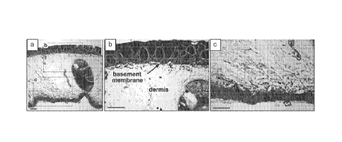
Example 1. Processing axolotl dermis
Axolotl dermis samples can be decellularized by preparing excised samples from
healthy or healing axolotl dermal tissue and then subjecting the samples to
hypo/hyperosmotic soaks for cell lysis, solvent dehydration, and oven drying.
Specific
processing of these grafts includes storage in 15- 26% NaCl, multiple
hypo/hyperosmotic
soaks (utilizing NaCl solutions and water) , and then solvent dehydration
using ethanol, and
then evaporation of the solvent either with air drying or oven drying at 37
C.
Histological examination of native axolotl dermal tissue was performed to
identify the
presence of the notable ECM elements, such as the basement membrane. See FIG 1
and FIG.
4. Comparative histological and immunohistochemical analysis of native axolotl
dermal
tissue and human amniotic membrane was performed to compare the ECM structure
and
constituents, and to assess relative concentration and distribution of
critical constituents. See
FIG. 2, FIG. 3, and FIG. 4. Figure 3 shows immunohistocemical staining via
species-specific
collagen IV and laminin antibodies of native axolotl dermal tissue and human
amniotic
membrane tissue, at 40x magnification. Figure 2 shows H&E and Alcian Blue
staining (40x)
of native axolotl dermal tissue and human amniotic membrane, and it
demonstrates the
comparable histoarchitecture and presence of sulfated glycosaminoglycans in
both tissues.
Histological evaluation with hematoxylin and eosin-stained, paired native and
post-processed
sections of axolotl dermal tissue (see FIG. 5)showed post-process preservation
of the
extracellular matrix histoarchitecture and the absence of cells or any
significant concentration
of cellular debris.
Example 2. Splitting and lamination of acellular dehydrated axolotl dermis.
Decellularized dehydrated axolotl dermis can be split, via a mechanical
splitter, to
isolate heterogeneous matrix into homogenous sections. Isolated sections of
desired
thickness then can be rehydrated and lyophilized to obtain multilayered
laminate structures of
desired orientation with facial surface features. More specifically, dual-
sided basement
membrane structure, with interior open porous matrix obtained from the
reticular dermis
region of the dermal matrix, can be constructed to obtain desired facial
surface properties.
Alternatively, isolated native section can be used in native form for desired
clinical outcome.
CA 02897662 2015-07-08
WO 2014/110269
PCT/US2014/010890
For example, open porous homogenous matrix of the reticular dermis can be used
to obtain
augmentation of soft tissue structures.
A laminated custom construct with sulfated gags on both facial surface and
collagen
IV and laminin could be obtained for desirable dual-surface, anti-adhesion and
antimicrobial
properties for clinical benefit. In addition, multilayer structures could be
constructed to
prolong in vivo durability of the graft.
Example 3 Preparation of solubilized acellular dehydrated axolotl dermis,
pericardium,
fascia lata, periosteum, peritoneum, or dura mater
Decellularized dehydrated axolotl native or isolated section of acellular
urodele
connectivue tissye matrix can be prepared by sectioning decellularized soft
tissue structures
into 1 cm2 sections and homogenizing the sections in a Warring blender (-100
grams of
tissue) in aqueous 1M glacier acetic acid for 30-60seconds. Preparation of
sponge can be
obtained by the addition of varying volumes of water followed then
neutralization and
lypoholization of the slurry in a mold of desired geometric shape. The
resultant porosity will
correlate to the volume of water added to the matrix. Additionally, a selected
range of
bioactive extracts can be added to the slurry prior to neutralization,
including particulated or
small protein constituents extracted from digested human or urodele
mineralized and
nonmineralized connective tissues, such as demineralized bone matrix, elastin,
or bone
morphogenic proteins, which can be covalently loaded into constructs. Extracts
will be
covalently bound with collagen fibers after neutralization and return to
physiological
condition where fibrillogenisis will occur. Subsequent release of bioactive
constituents will
occur during proteolyctic degradation in vivo and ensure molecules are not
consume or
exposed during acute inflammation in vivo. Alternatively, aqueous NaCl can be
added to the
slurry, prior to neutralization, to obtain a sustained, low viscosity solution
for injection,
which is stable at room temperature. Injection of slurry through ion-selective
membrane will
remove salt ions and permit for fibrillogenisis to occur post injection and
formation of three-
dimensional matrix.
26
CA 02897662 2017-01-16
Example 4 Preparation of sterilized particulated or powder form of mineralized
and non-
mineralized deceliularized and dehydrated urodele connective tissue matrix
Following deeellularization of sections of mineralized collagen urodele
connective
tissue, one can perform a demineralization process, similar to that employed
by Urist, and
solvent or 1.,Tophilization dehydration, cryornilline of sectioned acellular
demineralized,
mineralized, or non-mineralized urodele connective tissue extracellular
matrix, thereby to
obtain particulated or powder form of the ECM with preserved
histoarhictiecutre and
function. The final particle size distribution can be varied depending on
duration and
sieving, post-cyromilling, between 125 and 850 microns. Low-dose cold gamma
irradiation
or e-beam irradiation (<25 can be employed to sterilize acellular ECM
sheets,
particulate or powder and custom engineered constructs
Example 5. In vitro characterization of acellular mineralized and non-
mineralized urodele
healthy or healing connective tissue matrix
Through a series of in vitro analyses one can verify decellularization and
preservation
of native or custom engineered functional and structural properties of
deceIlularized
extracellular matrix constructs and.or particulate, including =Inlayer
laminated constructs
such as a dual-sided basement membrane sheet matrix or isolated native
homogenous open
porous matrix or solubilized, lyophilized and loaded ECM-derived collagen
sponge.
DNA content, as a marker for cell debris, can be employed to assess
decellularization
quantitatively, using a single, ethanol-based extraction technique with a
fluorometric dye,
Quant-iT PicoGreen, (Molecular Probes, USA), in a ratio of 170 ut, working
solution to
30 .L samples/standards in a 96-well plate. Paired native and post-processed
analysis and
comparison to commercially available tissue ECM's can be performed to verify
acceptability.
See Figure 6.
ELISA analysis for quantification of bioactive constituent and native and post
process protelyctic resorption profiles can be performed. Upon digestion with
collagenase
(232 ¨ 262 mg/unit activity), normalized-weight-to-surface-area sections of
decellularized
and dehydrated urodele ECM tissue or constructs (Sigma, USA), in a pH 7.6
buffer (50 mM
Tris-HC1, 200 mM CaCl2, 50 mM NaCl) for 24 hours at 37 C. can be analyzed at
various
time points to construct a relative resorption curve of pre- and post-process
tissue to verify
preservation of histoarchitecture. Solubilized collagen following digestion
can be assessed,
Trademark*
27
CA 02897662 2015-07-08
WO 2014/110269
PCT/US2014/010890
using a Sircol kit (Biocolor Ltd., UK), in 100 p.L aliquots of acid/salt-
washed digests.
Specifically, levels of BMP 2/4 and TGF- 1 growth factors or sulfated gags can
be assessed,
following digestion, by means of commercially available ELISA kits (R&D
Systems,
Minneapolis, MN). Protein content in 110 dilution digests can be measured via
a standard
Bradford absorbance assay.
Microscopic evaluation of samples can be performed using fixation in 4%
paraformaldehyde and paraffin embedding, sectioning at 5 p.m, and routine
histological
staining (Histoserv, Inc., USA). Longitudinal cross sections can be stained
with hematoxylin
and eosin. Images can be acquired and anyzed using standard brightfield
techniques on an
Olympus IM inverted microscope. Samples can analyzed using scanning electron
microscopy after dehydration in a graded ethanol series (15%, 30%, 50%, 70%,
9,0,/o,
J and
100%), critical-point drying in CO2, and sputter coating with gold. Samples
can be
visualized in an FEI Quanta 600 FEG scanning electron microscope, and
representative
images of scaffold ultrastructure can be acquired.
Direct cell contact methodology (IS010993, Part 5), for qualitative cell
viability
assessment at 24 hours, can be conducted for cytotoxicity testing and at
extended time points
(48 and 72 hours) to gauge cell proliferation and adhesion efficiency. A
manual count of
non-adherent cells in a hemocytometer, following transfer and trypsinization
of the culture
wells, can be conducted. A CellTiter 96 assay can performed to quantify viable
cells after
four days. A live/dead cell staining kit can used as well to visualize
scaffolds via
fluorescence microscopy at 24 hours and day four, thereby to verify
biocompatibility.
28