Note: Descriptions are shown in the official language in which they were submitted.
TREATMENT OF VASCULOPATHY WITH PROSTACYCLIN AND
MESENCHYMAL STEM CELLS
[0001] This application claims priority to US Application Serial No.
61/750,458,
filed January 9, 2013.
BACKGROUND
[0002] The present application relates to the use of mesenehymal stem cells in
treatment of
vasculopathy, including pulmonary arterial hypertension (PAH) and other types
of pulmonary
hypertension, peripheral vascular disease (PVD), critical limb ischemia (CLI),
coronary
artery disease, diabetic vasculopathy, etc.
[0003] Pulmonary arterial hypertension is a progressive lung disorder which,
untreated,
leads to death on average within 2.8 years after being diagnosed. An
increasing constriction
of the pulmonary circulation leads to increased stress on the right heart,
which may develop
into right heart failure. By definition, the mean pulmonary arterial pressure
(mPAP) in a case
of chronic pulmonary hypertension is >25 mmHg at rest or >30 mmHg during
exertion
(normal value <20 mmHg). The pathophysiology of pulmonary arterial
hypertension is
characterized by vasoconstriction and remodeling of the pulmonary vessels. In
chronic PAH
there is neomuscularization of initially unmuscularized pulmonary vessels, and
the vascular
muscles of the already muscularized vessels increase in circumference. This
increasing
obliteration of the pulmonary circulation results in progressive stress on the
right heart, which
leads to a reduced output from the right heart and eventually ends in right
heart failure (M.
Humbert et al., J. Am. Coll. Cardiol. 2004, 43, 13S-24S). PAH is an extremely
rare disorder,
with a prevalence of 1-2 per million. The average age of the patients has been
estimated to be
36 years, and only 10% of the patients were over 60 years of age. Distinctly
more women
than men are affected (G. E. D'Alonzo et al., Ann. Intern. Med. 1991, 115, 343-
349).
1
Date Recue/Date Received 2021-03-10
CA 02897805 2015-07-09
WO 2014/110094
PCT/US2014/010623
[0004] Standard therapies available on the market (e.g. prostacyclin
analogues, endothelin
receptor antagonists, phosphodiesterase inhibitors) are able to improve the
quality of life, the
exercise tolerance and the prognosis of the patients. The principles of these
therapies are
primarily hemodynamic, influencing vessel tone but having no direct influence
on the
pathogenic remodeling processes. In addition, the possibility of using these
medicaments is
restricted through the sometimes serious side effects and/or complicated types
of
administration. The period over which the clinical situation of the patients
can be improved
or stabilized by specific monotherapy is limited. Eventually the therapy
escalates and thus a
combination therapy is applied, where a plurality of medicaments must be given
concurrently. Despite all the advances in the therapy of pulmonary arterial
hypertension
there is as yet no prospect of cure of this serious disorder.
[0005] The term peripheral vascular disease (PVD) refers to damage,
dysfunction or
obstruction within peripheral arteries and veins. Peripheral artery disease is
the most common
form of PVT) Peripheral vascular disease is the most Common disease of the
arteries and is a
very common condition in the United States. It occurs mostly in people older
than 50 years.
Peripheral vascular disease is a leading cause of disability among people
older than 50 years,
as well as in those people with diabetes. About 10 million people in the
United States have
peripheral vascular disease, which translates to about 5% of people older than
50 years. The
number of people with the condition is expected to grow as the population
ages. Men are
slightly more likely than women to have peripheral vascular disease.
[0006] Critical limb ischemia (CLI), due to advanced peripheral arterial
occlusion, is
characterized by reduced blood flow and oxygen delivery at rest, resulting in
muscle pain at
rest and non-healing skin ulcers or gangrene (Rissanen et al., Eur. J. Clin.
Invest 31:651-666
(2001); Dormandy and Rutherford, J. Vase. Surg. 31:S1-S296 (2000)). Critical
limb
ischemia is estimated to develop in 500 to 1000 per million individuals in one
year ("Second
European Consensus Document on Chronic Critical Leg Ischemia", Circulation
84(4 Suppl.)
IV 1-26 (1991)). In patients with critical limb ischemia, amputation, despite
its associated
morbidity, mortality and functional implications, is often recommended as a
solution against
disabling symptoms (M. R. Tyrrell et al., Br. J. Surg. 80: 177-180 (1993); M.
Eneroth et al.,
2
CA 02897805 2015-07-09
WO 2014/110094
PCT/US2014/010623
Int. Orthop. 16: 383-387 (1992)). There exists no optimal medical therapy for
critical limb
ischemia (Circulation 84(4 Suppl.): IV 1-26 (1991))
[0007] Coronary artery disease (atherosclerosis) is a progressive disease in
humans wherein
one or more coronary arteries gradually become occluded through the buildup of
plaque. The
coronary arteries of patients having this disease are often treated by balloon
angioplasty or
the insertion of stents to prop open the partially occluded arteries.
Ultimately, these patients
are required to undergo coronary artery bypass surgery at great expense and
risk.
SUMMARY
[0008] In one embodiment, the current disclosure is directed to a method for
treating or
preventing vasculopathy in a subject in need thereof, comprising administering
to the subject
a prostacyclin and a composition comprising a mesenchymal stem cell (MSC) or a
part of a
culture medium that has been in contact with the MSC and contains one or more
component(s) of the MSC. The prostacyclin and the composition can be
administered
concurrently or separately.
[0009] In some embodiments, prior to the administration, the MSC has been in
contact with
prostacyclin. Likewise, the culture medium or the MSC from which the culture
medium is
obtained can be placed in contact with prostacyclin, prior to such
administration.
Accordingly, in some embodiments, the method further includes such a pre-
treatment step.
[0010] Non-limiting examples of components obtained from a part of the MSC
culture
include an exosome, a microvesicle, a microRNA, a messenger RNA, a non-coding
RNA, a
mitochondria, a growth factor, or combinations thereof.
[0011] Such methods, in one aspect, further entail administering to the
subject an
endothelial progenitor cell (EPC). In one aspect, the EPC is obtained from the
subject. In
some aspects, the EPC is transformed with a nucleic acid that increases the
expression of
biological activity of a protein selected from the group consisting of
endothelial nitric oxide
synthasc (eNOS), heme oxygenase (HMOX1) and prostacyclin synthase (PTG1S). In
one
aspect, the nucleic acid encodes the protein.
3
[0012] Examples of prostacyclin include, without limitation, epoprostenol
sodium,
treprostinil, beraprost, ilprost, and a PGI2 receptor agonist. In one aspect,
the prostacyclin is
treprostinil or a pharmaceutically acceptable salt or ester thereof.
[0013] Further provided, in embodiment, is a pharmaceutical composition
comprising a
therapeutically effective amount of a prostacyclin and a composition
comprising a
mesenchymal stem cell (MSC) or a culture medium that has been in contact with
the MSC
and contains compounds released from the MSC and a pharmaceutically acceptable
carrier.
In some aspects, the composition further comprises an endothelial progenitor
cell (EPC).
[0014] Yet another embodiment provides a method for preparing a composition
comprising
a mesenchymal stem cell (MSC) or a culture medium that has been in contact
with the MSC
and contains compounds released from the MSC for in vivo delivery, comprising
contacting
the MSC with a prostacyclin. Treated composition obtainable by such a method
is also
provided.
[0015] In other embodiments, the pharmaceutical composition further comprises
at least
one pharmaceutically-acceptable carrier or at least one therapeutic agent. In
another
embodiment, the subject is suffering from vasculopathy, such as pulmonary
arterial
hypertension (PAH), peripheral vascular disease (PVD), critical limb ischemia
(CLI),
coronary artery disease, or diabetic vasculopathy. In other embodiments the
current method
reduces thrombosis in pulmonary arteries, reduces inflammation in pulmonary
arteries,
reduces the proliferation of intimal smooth muscle in pulmonary arteries,
reduces the
formation of plexiform lesions in pulmonary arteries, increases the amount of
nitric oxide in
pulmonary arteries, increases the amount of PGI2 in pulmonary arteries,
reduces the level of
Endothelin-1 in pulmonary arteries, or reduces the amount of growth factors in
pulmonary
arteries. In other embodiments, the current method promotes proper endothelial
morphology
in pulmonary arteries.
4
Date Recue/Date Received 2021-03-10
[0015A] In one embodiment, there is provided use of (i) a part of a culture
medium that has been in
contact with a mesenchymal stem cell (MSC) and a prostacyclin or
pharmaceutically acceptable salts
or esters thereof, wherein the part of the culture medium comprises components
released from the
MSC and does not comprise the MSC; or (ii) an MSC-derived exosome and a
prostacyclin or
pharmaceutically acceptable salts thereof, for treating or preventing a
disease in a subject in need
thereof. The MSC or the MSC-derived exosome is obtained by pre-treating ex
vivo a culture of
MSCs with the prostacyclin during expansion of the MSCs. The MSC or the MSC-
derived exosome
has an increased VEGF production compared to an untreated MSC or MSC-derived
exosome. The
disease is selected from the group consisting of pulmonary arterial
hypertension (PAH), peripheral
vascular disease (PVD), critical limb ischemia (CLI), coronary artery disease
and diabetic
vasculopathy.
[0015B] In one embodiment, there is provided a pharmaceutical composition
comprising (i) a part
of a culture that has been in contact with a mesenchymal stem cell (MSC) and a
prostacyclin or
pharmaceutically acceptable salts or esters thereof, wherein the part of the
culture medium comprises
components released from the MSC and does not comprise the MSC; or (ii) a MSC-
derived exosome
and a prostacyclin or pharmaceutically acceptable salts or esters thereof,
wherein the part of a culture
that has been in contact with the MSC or the MSC-derived exosome is obtained
by ex vivo pre-
treating a culture of MSCs during expansion of the MSCs with a prostacyclin,
wherein the MSC or
the MSC-derived exosome has an increased VEGF production compared to an
untreated MSC or
MSC-derived exosome, and a pharmaceutically acceptable carrier.
[0015C] In one embodiment, there is provided a method for preparing (i) a part
of a culture
medium that has been in contact with a mesenchymal stem cell (MSC), wherein
the part of the
culture medium comprises components released from the MSC and does not
comprise the MSC; or
(ii) a MSC-derived exosome for in vivo delivery, comprising obtaining the part
of a culture medium
that has been in contact with the MSC or the MSC-derived exosome from a
culture of MSCs ex vivo
pretreated with a prostacyclin during the expansion of the MSCs, wherein the
MSC or the MSC-
derived exosome has an increased VEGF production compared to an untreated MSC
or MSC-
derived exosome.
BRIEF DESCRIPTION OF THE DRAWINGS
[0016] Provided as embodiments of this disclosure are drawings which
illustrate by exemplification
only, and not limitation.
4a
Date Recue/Date Received 2022-02-28
CA 02897805 2015-07-09
WO 2014/110094
PCT/US2014/010623
[0017] FIG. 1 shows the results of immunophenotype analysis of human bone
marrow-
derived MSC.
[0018] FIG. 2 is a chart showing VEGF secretion by human bone marrow MSC after
24
hours of exposure to treprostinil.
[0019] FIG. 3A-B present a MSC secretion chart (A) and a gene expression chart
(B) of
VEGF after 24 hours exposure to treprostinil.
[0020] FIG. 4 presents representative images of MSC exposed to increasing
concentrations
of treprostinil.
[0021] FIG. 5 is chart showing cellular viability of MSC exposed to
treprostinil.
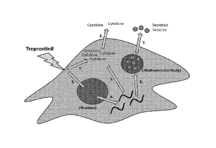
[0022] FIG. 6 illustrates a model for the effects of treprostinil on cell
signaling, gene
expression, and the release of paracrine factors.
[0023] FIG. 7A-B presents images and a chart showing MSC treated with or
without 250
pginaL treprostinil.
[0024] FIG. 8 presents two charts showing altered expression in selected genes
in MSC
treated with treprostinil.
[0025] FIG. 9A-B present two heatmaps that cluster MSC treated with
treprostinil from
controls with most significantly differentially expressed genes (FIG. 9A) or
other genomic
sequences or expression tags (FIG. 9B).
[0026] FIG. 10 presents charts showing that the RNA content in MSC-derived
exosomes is
altered with treprostinil treatment.
[0027] FIG. 11A-B show size distribution of exosomes derived from treprostinil-
treated
and -untreated MSC.
[0028] Some or all of the figures are schematic representations for
exemplification; hence,
they do not necessarily depict the actual relative sizes or locations of the
elements shown.
The figures are presented for the purpose of illustrating one or more
embodiments with the
explicit understanding that they will not be used to limit the scope or the
meaning of the
claims that follow below.
DETAILED DESCRIPTIONS
100291 Unless otherwise specified, "a" or "an" means "one or more."
100301 Unless specifically defined otherwise, all technical and scientific
terms used herein
shall be taken to have the same meaning as commonly understood by one of
ordinary skill in
the art (e.g., in stem cell biology, cell culture, molecular genetics,
immunology,
immunohistochemistry, protein chemistry, and biochemistry).
100311 Unless otherwise indicated, the recombinant protein, cell culture, and
immunological techniques utilized in the present disclosure are standard
procedures, well
known to those skilled in the art. Such techniques are described and explained
throughout the
literature in sources such as, J. Perbal, A Practical Guide to Molecular
Cloning, John Wiley
and Sons (1984), J. Sambrook et al., Molecular Cloning: A Laboratory Manual,
Cold Spring
Harbour Laboratory Press (1989), T. A. Brown (editor), Essential Molecular
Biology: A
Practical Approach, Volumes 1 and 2, IRL Press (1991), D. M. Glover and B. D.
Hames
(editors), DNA Cloning: A Practical Approach, Volumes 1-4, IRL Press (1995 and
1996),
and F. M. Ausubel ct al. (editors), Current Protocols in Molecular Biology,
Greene Pub.
Associates and Wiley-Interscience (1988, including all updates until present),
Ed Harlow and
David Lane (editors) Antibodies: A Laboratory Manual, Cold Spring Harbour
Laboratory,
(1988), and J. E. Coligan et al. (editors) Current Protocols in Immunology,
John Wiley &
Sons (including all updates until present).
100321 It is herein discovered that both prostacyclin and mesenchymal stem
cells (MSCs)
possess therapeutic activities for vasculopathy. The combination of
prostacyclin and MSCs,
furthermore, produces synergistic effects. Such combination can be either co-
administration,
which can be concurrent or separate, of prostacyclin and MSCs to a patient, or
administration
to the patient a MSC composition that has been pre-treated with a
prostacyclin.
100331 It is shown that MSCs can ameliorate vasculopathy in patients, and it
is
contemplated that such a therapeutic effect is achieved due to MSCs' ability
to improve the
6
CA 2897805 2020-03-16
CA 02897805 2015-07-09
WO 2014/110094
PCT/US2014/010623
local microenvironment by delivering anti-inflammatory and pro-angiogenic
factors to the
diseased area. MSCs, however, are short-lived in the body and not
regenerative.
[0034] Piostacyclin, such as treprostinil (TP), has been used for treating
pulmonary arterial
hypertension (PAH) patients. In this respect, prostacyclin has been shown to
possess
vasodilatory and anti-platelet aggregation activities.
[0035] An unexpected discovery is that prostacyclin can enhance the activity
of MSCs for
the treatment of vasculopathy, exhibiting synergism for such treatment. In
this respect, it is
observed that prostacyclin enhances MSCs' beneficial effect on blood vessel
growth. For
instance, prostacyclin increases the expression of VEGF at both protein and
gene levels.
Changes in secreted cytokines are also observed as a result of prostacyclin
exposure. For
instance, IL-6 is increased ¨50-fold while MCP-1 is decreased ¨6-7-fold.
[0036] Such synergism is also evident when the patient is further administered
an
endothelial progenitor cell (EPC). It is therefore contemplated that
prostacyclin may enhance
the activity of EPCs through MSCs. By virtue of such synergism, therefore, the
combinatory
use of prostacyclin and MSC, optionally together with EPC, can lead to
improved therapeutic
outcome and/or reduced need of each agent alone which, in turn, can result in
reduced
adverse effects potentially caused by each agent alone, at a higher dose.
[0037] It is further shown that such synergism is applicable to MSC-
conditioned culture
medium. To this end, it is observed that the exosomes of prostacyclin-treated
MSC have
higher levels of VEGF-A, which may promote increased VEGF production in target
cells
through a mechanism of horizontal gene transfer. Further, exposure to
prostacyclin yields a
more uniform population of exosomes.
[0038] As used herein, a "MSC-conditioned culture medium" refers to a culture
medium
that has been in contact with a MSC (e.g., for the purpose of culturing the
MSC) and thus
contains compounds released from the MSC. Non-limiting examples of such
released
compounds include exosomes or other microvesicles which can enclose messenger
RNA,
non-coding RNA, microRNAs, mitochondria, growth factors, or other types of
bioactive
agents.
7
CA 02897805 2015-07-09
WO 2014/110094
PCT/US2014/010623
[0039] A "culture medium" as used herein, encompasses (a) both a culture
medium that
contains the typical components used for culturing a MSC, such as amino acids,
glucose, and
various salts, with or without the MSC, and (b) a composition isolated from
the culture
medium that contains compounds released from the MSC during the culturing.
[0040] Accordingly, one embodiment of the present disclosure provides a method
for
treating or preventing vasculopathy in a subject in need thereof, comprising
administering to
the subject a prostacyclin and a composition comprising a mesenchymal stem
cell (MSC) or a
MSC-conditioned culture medium (collectively a "MSC composition").
[0041] In one aspect, the prostacyclin and the MSC composition are
administered
concurrently. In another aspect, the prostacyclin and the MSC composition are
administered
separately. When administered separately, the prostacyclin can be administered
prior to, or
following the administration of the MSC composition.
[0042] In another embodiment, provided is a method for treating or preventing
vasculopathy in a subject in need thereof, comprising contacting a composition
comprising an
isolated mesenchymal stem cell (MSC) or a MSC-conditioned culture medium with
a
prostacyclin, and then administering the MSC composition to the subject.
[0043] Non-limiting examples of vasculopathy include pulmonary arterial
hypertension
(PAH), peripheral vascular disease (PVD), critical limb ischemia (CLI),
coronary artery
disease and diabetic vasculopathy.
[0044] As used herein, the term "subject" (also referred to herein as a
"patient") includes
warm-blooded animals, preferably mammals, including humans. In a preferred
embodiment,
the subject is a primate. In an even more preferred embodiment, the subject is
a human.
[0045] As used herein the terms "treating", "treat" or "treatment" include
administering a
therapeutically effective amount of cells as defined herein sufficient to
reduce or eliminate at
least one symptom of vasculopathy.
8
CA 02897805 2015-07-09
WO 2014/110094
PCT/US2014/010623
[0046] As used herein the terms "preventing", "prevent" or "prevention"
include
administering a therapeutically effective amount of cells as defined herein
sufficient to stop
or hinder the development of at least one symptom of vasculopathy.
A. Prostacyclin
[0047] The term "prostacyclin" used herein explicitly comprises any
prostaglandin 17
(PGI2), any prostacyclin analogues, and any PGI7 receptor agonists. Non-
limiting examples
of prostacyclin suitable for the present technology include epoprostenol
sodium (e.g.
Flolang), treprostinil(e.g. TYVASO , Remoduling), ilprost (e.g. Ventavisg),
and PGI9
receptor agonist (e.g. Selexipag). In one aspect, the prostacyclin is
treprostinil or a
pharmaceutically acceptable salt or ester thereof.
B. Mesenchymal Stem Cells (MSCs)
[0048] Mesenchymal stem cells (MSCs) are cells found in bone marrow, blood,
dental pulp
cells, adipose tissue, skin, spleen, pancreas, brain, kidney, liver, heart,
retina, brain, hair
follicles, intestine, lung, lymph node, thymus, bone, ligament, tendon,
skeletal muscle,
dermis, and periosteum; and are capable of differentiating into different germ
lines such as
mesoderm, endoderm and ectoderm. Thus, MSCs are capable of differentiating
into a large
number of cell types including, but not limited to, adipose, osseous,
cartilaginous, elastic,
muscular, and fibrous connective tissues. The specific lineage-commitment and
differentiation pathway which these cells enter depends upon various
influences from
mechanical influences and/or endogenous bioactive factors, such as growth
factors,
cytokines, and/or local microenvironmental conditions established by host
tissues. MSCs are
thus non-hematopoietic progenitor cells which divide to yield daughter cells
that are either
stem cells or are precursor cells which in time will irreversibly
differentiate to yield a
phenotypic cell. Examples of MSCs include mesenchymal precursor cells (MPCs).
[0049] As used herein, the term "stem cell" refers to self-renewing cells that
are capable of
giving rise to phenotypically and genotypically identical daughters as well as
at least one
other final cell type (e.g., terminally differentiated cells). The term "stem
cells" includes
totipotential, pluripotential and multipotential cells, as well as progenitor
and/or precursor
cells derived from the differentiation thereof.
9
CA 02897805 2015-07-09
WO 2014/110094
PCT/US2014/010623
[0050] As used herein, the term "totipotent cell" or "totipotential cell"
refers to a cell that is
able to form a complete embryo (e.g., a blastocyst).
[0051] As used herein, the term "pluripotent cell" or "pluripotential cell"
refers to a cell
that has complete differentiation versatility, i.e., the capacity to grow into
any of the
mammalian body's approximately 260 cell types. A pluripotent cell can be self-
renewing, and
can remain dormant or quiescent within a tissue.
[0052] The term -multipotential cell- or -multipotent cell- refers to a cell
which is capable
of giving rise to any of several mature cell types. As used herein, this
phrase encompasses
adult or embryonic stem cells and progenitor cells, and multipotential progeny
of these cells.
Unlike a pluripotent cell, a multipotent cell does not have the capacity to
form all of the cell
types.
[0053] As used herein, the term "progenitor cell" or "precursor cell" refers
to a cell that is
committed to differentiate into a specific type of cell or to form a specific
type of tissue.
[0054] In a preferred embodiment, cells used in the methods of the disclosure
are enriched
from a sample obtained from a subject. The terms 'enriched', 'enrichment' or
variations
thereof are used herein to describe a population of cells in which the
proportion of one
particular cell type or the proportion of a number of particular cell types is
increased when
compared with the untreated population.
[0055] In a preferred embodiment, the cells used in the present disclosure are
TNAP
STRO-1+, VCAM-1+, THY-1+, STRO-2+, CD45 , CD146+, 3G5+ or any combination
thereof.
[0056] When we refer to a cell as being "positive" for a given marker it may
be either a low
(lo or dim) or a high (bright, bri) expresser of that marker depending on the
degree to which
the marker is present on the cell surface, where the terms relate to intensity
of fluorescence or
other color used in the color sorting process of the cells. The distinction of
lo (or dim or dull)
and bri will be understood in the context of the marker used on a particular
cell population
being sorted. When we refer herein to a cell as being "negative" for a given
marker, it does
not mean that the marker is not expressed at all by that cell. It means that
the marker is
CA 02897805 2015-07-09
WO 2014/110094
PCT/US2014/010623
expressed at a relatively very low level by that cell, and that it generates a
very low signal
when detectably labeled.
[0057] When used herein the term "TNAP" is intended to encompass all isoforms
of tissue
non-specific alkaline phosphatase. For example, thc term encompasses the liver
isoform
(LAP), the bone isoform (BAP) and the kidney isoform (KAP). In a preferred
embodiment,
the TNAP is BAP. In a particularly preferred embodiment, TNAP as used herein
refers to a
molecule which can bind the STRO-3 antibody produced by the hybridoma cell
line
deposited with ATCC on 19 Dec. 2005 under the provisions of the Budapest
Treaty under
deposit accession number PTA-7282.
[0058] Stem cells useful for the methods can be derived from adult tissue, an
embryo,
extraembryonic tissue, or a fetus. The term "adult" is used in its broadest
sense to include a
postnatal subject. In a preferred embodiment, the term "adult" refers to a
subject that is
postpubertal. The term, "adult" as used herein can also include cord blood
taken from a
female.
[0059] In some aspects, the stem cells can be progeny cells (which can also be
referred to
as expanded cells) which are produced from the in vitro culture of the stem
cells described
herein. Expanded cells of the disclosure may have a wide variety of phenotypes
depending on
the culture conditions (including the number and/or type of stimulatory
factors in the culture
medium), the number of passages and the like. In certain embodiments, the
progeny cells are
obtained after about 2, about 3, about 4, about 5, about 6, about 7, about 8,
about 9, or about
passages from the parental population. However, the progeny cells may be
obtained after
any number of passages from the parental population.
[0060] The progeny cells can be obtained by culturing in any suitable medium.
The term
"medium", as used in reference to a cell culture, includes the components of
the environment
surrounding the cells. Media may be solid, liquid, gaseous or a mixture of
phases and
materials. Media include liquid growth media as well as liquid media that do
not sustain cell
growth. Media also include gelatinous media such as agar, agarose, gelatin and
collagen
matrices. The term "medium" also refers to material that is intended for use
in a cell culture,
11
CA 02897805 2015-07-09
WO 2014/110094
PCT/US2014/010623
even if it has not yet been contacted with cells. In other words, a nutrient
rich liquid prepared
for bacterial culture is a medium.
[0061] In an embodiment, the progeny cells are obtained by isolating TNAP+
cells from
bone marrow using magnetic beads labelled with the STRO-3 antibody, and plated
in a-
MEM supplemented with 20% fetal calf serum, 2 mM L-glutamine and 100 gm L-
ascorbate-
2-phosphate.
[0062] In one embodiment, such expanded cells (at least after 5 passages) can
be TNAP-,
CC9+, HLA class I+, HLA class II-, CD14-, CD19-; CD3-, CD11a-c-, CD31-, CD86-
and/or
CD 80-. However, it is possible that under different culturing conditions to
those described
herein that the expression of different markers may vary. Also, whilst cells
of these
phenotypes may predominate in the expended cell population it does not mean
that there is
not a minor proportion of the cells that do not have this phenotype(s) (for
example, a small
percentage of the expanded cells may be CC9-). In one preferred embodiment,
expanded cells
of the disclosure still have the capacity to differentiate into different cell
types.
[0063] In one embodiment, an expended cell population used in the methods of
the
disclosure comprises cells wherein at least 25%, more preferably at least 50%,
of the cells are
CC9+.
[0064] In another embodiment, an expended cell population used in the methods
of the
disclosure comprises cells wherein at least 40%, more preferably at least 45%,
of the cells are
STRO-1+.
[0065] In a further embodiment, the progeny cells may express markers selected
from the
group consisting of LFA-3, THY-1, VCAM-1, PECAM-1, P-selectin, L-selectin,
3G5,
CD49a/CD49b/CD29, CD49c/CD29, CD49d/CD29, CD29, CD18, CD61, integrin beta, 6-
19,
thrombomodulin, CD10, CD13, SCF, PDGF-R, EGF-R, IGF1-R, NGF-R, FGF-R, Leptin-
R,
(STRO-2=Leptin-R), RANKL, STRO-lbright and CD146 or any combination of these
markers.
[0066] In one embodiment, the progeny cells are Multipotential Expanded MSC
Progeny
(MEMPs) as defined in WO 2006/032092. Methods for preparing enriched
populations of
12
CA 02897805 2015-07-09
WO 2014/110094
PCT/US2014/010623
MSC from which progeny may be derived are described in WO 01/04268 and WO
2004/085630. In an in vitro context MSCs will rarely be present as an
absolutely pure
preparation and will generally be present with other cells that are tissue
specific committed
cells (TSCCs). WO 01/04268 refers to harvesting such cells from bone marrow at
purity
levels of about 0.1% to 90%. The population comprising MSC from which progeny
are
derived may be directly harvested from a tissue source, or alternatively it
may be a population
that has already been expanded ex vivo.
[0067] For example, the progeny may be obtained from a harvested, unexpanded,
population of substantially purified MSC, comprising at least about 0.1, 1, 5,
10, 20, 30, 40,
50, 60, 70, 80 or 95% of total cells of the population in which they are
present. This level
may be achieved, for example, by selecting for cells that are positive for at
least one marker
selected from the group consisting of TNAP, STRO-1 bri, VCAM-1, THY-1,
CD146
and STRO-2.
[0068] The MSC starting population may be derived from any one or more tissue
types set
out in WO 01/04268 or WO 2004/085630, namely bone marrow, dental pulp cells,
adipose
tissue and skin, or perhaps more broadly from adipose tissue, teeth, dental
pulp, skin, liver,
kidney, heart, retina, brain, hair follicles, intestine, lung, spleen, lymph
node, thymus,
pancreas, bone, ligament, bone marrow, tendon and skeletal muscle.
[0069] MEMPS can be distinguished from freshly harvested MSCs in that they are
positive
for the marker STRO-lbri and negative for the marker Alkaline phosphatase
(ALP). In
contrast, freshly isolated MSCs are positive for both STRO-lbri and ALP. In a
preferred
embodiment of the present disclosure, at least 15%, 20%, 30%, 40%, 50%, 60%,
70%, 80%,
90% or 95% of the administered cells have the phenotype STRO- 1 bil, ALP-. In
a further
preferred embodiment the MEMPS are positive for one or more of the markers
Ki67, CD44
and/or CD49c/CD29, VLA-3, a3131. In yet a further preferred embodiment the
MEMPs do
not exhibit TERT activity and/or are negative for the marker CD18.
[0070] In one embodiment, the cells are taken from a patient with
vasculopathy, cultured in
vitro using standard techniques and administered to a patient as an autologous
or allogeneic
transplant. In an alternative embodiment, cells of one or more of the
established human cell
13
CA 02897805 2015-07-09
WO 2014/110094
PCT/US2014/010623
lines are used. In another useful embodiment of the disclosure, cells of a non-
human animal
(or if the patient is not a human, from another species) are used.
[0071] The present technology can be practiced using cells from any non-human
animal
species, including but not limited to non-human primate cells, ungulate,
canine, feline,
lagomorph, rodent, avian, and fish cells. Primate cells with which the
disclosure may be
performed include but are not limited to cells of chimpanzees, baboons,
cynomolgus
monkeys, and any other New or Old World monkeys. Ungulate cells with which the
disclosure may be performed include but are not limited to cells of bovines,
porcines, ovines,
caprines, equines, buffalo and bison. Rodent cells with which the disclosure
may be
performed include but are not limited to mouse, rat, guinea pig, hamster and
gerbil cells.
Examples of lagomorph species with which the disclosure may be performed
include
domesticated rabbits, jack rabbits, hares, cottontails, snowshoe rabbits, and
pikas. Chickens
(Gallus gallus) are an example of an avian species with which the disclosure
may be
performed
[0072] Cells can be stored before use. Methods and protocols for preserving
and storing of
eukaryotic cells, and in particular mammalian cells, are well known in the art
(cf , for
example, Pollard, J. W. and Walker, J. M. (1997) Basic Cell Culture Protocols,
Second
Edition, Humana Press, Totowa, N.J.; Freshney, R. I. (2000) Culture of Animal
Cells, Fourth
Edition, Wiley-Liss, Hoboken, N.J.). Any method maintaining the biological
activity of the
isolated stem cells such as mesenchymal stem/progenitor cells, or progeny
thereof, may be
utilized in connection with the present disclosure. In one preferred
embodiment, the cells are
maintained and stored by using cryo-preservation.
[0073] Cells can be obtained using a variety of techniques. For example, a
number of cell-
sorting techniques by which cells are physically separated by reference to a
property
associated with the cell-antibody complex, or a label attached to the antibody
can be used.
This label may be a magnetic particle or a fluorescent molecule. The
antibodies may be cross-
linked such that they form aggregates of multiple cells, which are separable
by their density.
Alternatively the antibodies may be attached to a stationary matrix, to which
the desired cells
adhere.
14
100741 In a preferred embodiment, an antibody (or other binding agent) that
binds TNAP+,
STRO-1+, VCAM-1+, THY-1+, STRO-2+, 3G5+, CD45+, CD146+ is used to isolate the
cells. More preferably, an antibody (or other binding agent) that binds TNAP+
or STRO-1 +
is used to isolate the cells.
100751 Various methods of separating antibody-bound cells from unbound cells
are known.
For example, the antibody bound to the cell (or an anti-isotype antibody) can
be labelled and
then the cells separated by a mechanical cell sorter that detects the presence
of the label.
Fluorescence-activated cell sorters are well known in the art. In one
embodiment, anti-TNAP
antibodies and/or an STRO-1 antibodies are attached to a solid support.
Various solid
supports are known to those of skill in the art, including, but not limited
to, agarose beads,
polystyrene beads, hollow fiber membranes, polymers, and plastic petri dishes.
Cells that are
bound by the antibody can be removed from the cell suspension by simply
physically
separating the solid support from the cell suspension.
100761 Super paramagnetic microparticles may be used for cell separations. For
example,
the microparticles may be coated with anti-TNAP antibodies and/or STRO-1
antibodies. The
antibody-tagged, super paramagnetic microparticles may then be incubated with
a solution
containing the cells of interest. The microparticles bind to the surfaces of
the desired stem
cells, and these cells can then be collected in a magnetic-field.
100771 In another example, the cell sample is allowed to physically contact,
for example, a
solid phase-linked anti-TNAP monoclonal antibodies and/or anti-S TRO-1
monoclonal
antibodies. The solid-phase linking can comprise, for instance, adsorbing the
antibodies to a
plastic, nitrocellulose, or other surface. The antibodies can also be adsorbed
on to the walls of
the large pores (sufficiently large to permit flow-through of cells) of a
hollow fiber
membrane. Alternatively, the antibodies can be covalently linked to a surface
or bead, such as
Pharmacia Sepharosem4 6 MB macrobeads. The exact conditions and duration of
incubation
for the solid phase-linked antibodies with the stem cell containing suspension
will depend
upon several factors specific to the system employed. The selection of
appropriate conditions,
however, is well within the skill of the art.
Date Recue/Date Received 2022-02-28
CA 02897805 2015-07-09
WO 2014/110094
PCT/US2014/010623
[0078] The unbound cells are then eluted or washed away with physiologic
buffer after
allowing sufficient time for the stem cells to be bound. The unbound cells can
be recovered
and used for other purposes or discarded after appropriate testing has been
done to ensure that
the desired separation had been achieved. The bound cells are then separated
from the solid
phase by any appropriate method, depending mainly upon the nature of the solid
phase and
the antibody. For example, bound cells can be eluted from a plastic petri dish
by vigorous
agitation. Alternatively, bound cells can be eluted by enzymatically "nicking"
or digesting an
enzyme-sensitive "spacer" sequence between the solid phase and the antibody.
Spacers bound
to agarose beads are commercially available from, for example, Pharmacia.
[0079] The eluted, enriched fraction of cells may then be washed with a buffer
by
centrifugation and said enriched fraction may be cryopreserved in a viable
state for later use
according to conventional technology, culture expanded and/or introduced into
the patient.
C. MSC-conditioned culture media
[0080] It is discovered that MSCs can carry out their activities through
compounds that can
be released into the extracellular environment during growth or
differentiation. In some
aspects, such compounds include a microvesicle, referred to as exosome, which
is between
about 30 nm and about 200 nm in diameter. Exosomes can be internalized by host
cells in
vivo.
[0081] Exosomes are vesicles derived from the multivesicular body sorting
pathway.
Recent studies show that exosomes are bioactive vesicles useful for
intercellular
communication and facilitation of the immunoregulatory process. MSC exosomes
contain
20S proteasomes and numerous RNAs (messenger RNA, non-coding RNA, microRNA).
[0082] In addition to exosomes, MSC also release other bioactive
molecules/vesicles useful
for the purpose of the present disclosure. Such molecules and vesicles
include, without
limitation, mitochondria and growth factors. Method of preparing culture media
that contain
such molecules and vesicles released from MSC and further isolating particular
molecules
and vesicles are known in the art. See, for instance, Hu et al., Frontiers in
Genetics, 2:56, 1-9
(2012).
16
CA 02897805 2015-07-09
WO 2014/110094
PCT/US2014/010623
D. Pre-treatment of MSC with prostacyclin
[0083] In some embodiments, prior to coadministering a MSC or a MSC-
conditioned
culture medium with prostacyclin to a patient, the MSC or MSC-conditioned
culture medium
can be optionally pre-treated with prostacyclin. Accordingly, also provided,
in one
embodiment, is a method for preparing a mesenchymal stem cell (MSC) or MSC-
conditioned
culture medium for in vivo delivery, comprising contacting the MSC or MSC-
conditioned
culture medium with a prostacyclin. Yet another embodiment provides a treated
MSC or
MSC-conditioned culture medium obtainable by such a method.
[0084] Pre-treatment of a cell or a medium with a chemical compound
encompasses known
techniques. In one aspect, the prostacyclin can be added to and co-incubated
with a culture
medium that contains a MSC. Optionally, however, such co-incubation can
further involve
the addition of a growth factor (e.g., VEGF and Angiopoietin-1 or -2, platelet-
derived growth
factor) and/or hypoxia.
[0085] MSCs or MSC-conditioned culture media can be treated with prostacyclin
in various
ways. For example, prostacyclin can be used to treat MSCs ex vivo during the
expansion of
MSCs; prostacyclin can also be used to treat MSCs after administration. In
some aspects, the
concentration of prostacyclin is at least about 100 mimL, or at least about
150 tig/mL, 200
p.g/mL, or 250 1..tg/mL. In some aspects, the concentration of prostacyclin is
not more than
about 400 ng/mL, or not more than about 350 p.g/mL, 300 i.tg/mL or 250
i.tg/mL.
[0086] According to one embodiment of the present disclosure, MSCs can be
prepared
from the recipient's own blood or bone marrow. In that case, prostacyclin can
also be used to
treat MSCs before they are isolated from the recipients.
E. Endothelial progenitor cell (EPC)
[0087] As provided, the synergism between prostacyclin and MSCs for the
treatment of
vasculopathy is also evident when a patient is further administered with an
endothelial
progenitor cell (EPC). Thus, for any embodiment of the presently disclosed
method, the
patient further is administered an endothelial progenitor cell (EPC).
17
CA 02897805 2015-07-09
WO 2014/110094
PCT/US2014/010623
[0088] In some embodiments, the EPC can also be pre-treated with prostacyclin.
The EPCs
treated with prostacyclin exhibit a hypetproliferative phenotype with enhanced
angiogenic
properties, which are advantageous in treating vasculopathy compared to
untreated EPCs.
[0089] EPCs can be treated with prostacyclin in various ways. For example,
prostacyclin
can be used to treat EPCs ex vivo during the expansion of EPCs; prostacyclin
can be co-
administered with EPCs to the recipient; prostacyclin can also be used to
treat EPCs after
transplantation. According to one embodiment of the present disclosure, EPCs
are prepared
from the recipient's own blood or bone marrow. In that case, prostacyclin can
also be used to
treat EPCs before they are isolated from the recipients.
[0090] An EPC is an undifferentiated cell that can be induced to proliferate.
EPCs are
capable of self-maintenance, such that with each cell division, at least one
daughter cell will
also be an EPC cell. EPCs are capable of being expanded 100, 250, 500, 1000,
2000, 3000,
4000, 5000 or more fold.
[0091] Phenotyping of EPCs reveals that these cells express the committed
hematopoietic
marker CD45. Additionally, an EPC may be immunoreactive for VEGFR-2 and/or Tie-
2.
Optionally, the EPC is immunoreactive for CD14. The EPC is a multipotent
progenitor cell.
[0092] Vascular endothelial growth factor (VEGF) acts through specific
tyrosine kinase
receptors that includes VEGFR-1 (fit-1) and VEGFR-2 (flk-1/KDR) and VEGFR-
3/Flt-4
which convey signals that are essential for embryonic angiogenesis and
hematopoiesis. While
VEGF binds to all three receptors, most biological functions are mediated via
VEGFR-2 and
the role of VEGFR-1 is currently unknown. VEGFR3/F1t4 signaling is known to be
important
for the development of lymphatic endothelial cells and VEGFR3 signaling may
confer
lymphatic endothelial-like phenotypes to endothelial cells. VEGFRs relay
signals for
processes essential in stimulation of vessel growth, vasorelaxation, induction
of vascular
permeability, endothelial cell migration, proliferation and survival.
Endothelial cells express
all different VEGF-Rs. During embryogenesis, it has been reported that a
single progenitor
cell, the hemangioblast can give rise to both the hematopoietic and vascular
systems.
[0093] Tie-2 is an endothelial-specific receptor tyrosine kinase and a
receptor for
angiopoietin 1. It is a type I membrane protein that is expressed
predominantly in the
18
CA 02897805 2015-07-09
WO 2014/110094
PCT/US2014/010623
endothelium of actively growing blood vessels and may represent the earliest
mammalian
endothelial cell lineage marker. Tie-2 is likely involved in the regulation of
endothelial cell
proliferation and differentiation and may direct the special orientation of
endothelial cells
during the formation of blood vessels.
[0094] The CD14 antigen is a high affinity receptor for the complex of
lipopolysaccharides
(LPS) and LPS-Binding protein (LBP). The CD14 antigen is part of the
functional
heteromeric LP S receptor complex comprised of CD14, TLR4 and MD-2. CD14 is
strongly
expressed on most human monocytes and macrophages in peripheral blood, other
body fluids
and various tissues, such as lymph nodes and spleen. CD14 is weakly expressed
on
subpopulations of human neutrophils and myeloid dendritic cells.
[0095] The CD45 antigen is a tyrosine phosphatase, also known as the leukocyte
common
antigen (LCA). CD45 is present on all human cells of hematopoietic origin,
except erythroid
cells, platelets and their precursor cells. The CD45 molecule is required for
T cell and B cell
activation and is expressed in at least 5 isoforms, depending on the
activation status of the
cell.
[0096] VEGFR-1+, VEGFR-2+ and Tie-2+ cells constituted approximately 3Ø+-
Ø2%,
0.8±0.5%, 2.0±0.3% of the total population of mononuclear cells in blood
respectively.
CD14+NEGFR-2+ cells constituted approximately 2.0±0.5% of the total
population of
monocytes and 0.08±0.04% of mononuclear cells in blood.
[0097] EPCs can be maintained in vitro in long-term cultures. The EPCs are
capable of
being passed in culture 2, 3, 4, 5, 6, 7, 8, 9, 10, 11, 12 or more times.
[0098] EPCs comprise endothelial colony-forming cells, typically developed
after 1-3
weeks of cell culture. Endothelial colony-forming cells have the
characteristics of precursor
cells committed to the endothelial lineage and are capable of merging into
neovessels,
according to Smardj a et al., Angiogenesis 14(1):17-27 (2011).
[0099] The isolation, purification, ex vivo culturing and characterizing of
EPCs are
described in Hill et al, N. Engl. J. Med. 348:593-600 (2003), Assmus et al.,
Circulation
106:3009-16 (2002), Wang et al., J. Am. Coll. Cardiol. 49:1566-71 (2007), and
Kalka et al.,
19
P.N.A.S. 97:3422-7 (2000).
Further, the isolation, purification, ex vivo culturing and characterizing of
endothelial colony-forming cells are described in Yoder et al., Blood 109:1801-
1809 (2007),
Ingram et al., Blood 104:2752-2760 (2004), and Smardja et al., Angiogenesis
14(1): 17-27
(2011).
101001 For example, the population of cells arc isolated by means of positive
selection, or
by a mixture of both positive and negative selection in either order. The
population of
progenitor cells is purified. A purified population of EPCs contains a
significantly higher
proportion of EPCs than the crude population of cells from which the cells are
isolated.
101011 For example, the purification procedure should lead at least to a five-
fold increase,
preferably at least a ten-fold increase, more preferably at least a fifteen
fold increase, most
preferably at least a twenty fold increase, and optimally at least a twenty-
five fold increase in
EPCs with respect to the total population. The purified population of EPC
should include at
least 15%, preferably at least 20%, more preferably at least 25%, most
preferably at least
35%, and optimally at least 50% of EPCs.
101021 The methods described herein can lead to mixtures comprising up to 75%,
preferably up to 80%, more preferably up to 85%, most preferably up to 90% and
optimally
up to 95% of stem cells. Such methods arc capable of producing mixtures
comprising 99%,
99.90% and even 100% of EPCs. Accordingly, the purified populations of the
disclosure
contain significantly higher levels of EPCs than those that exist in nature,
as described above.
[01031 The purified population of EPCs can be isolated by contacting a crude
mixture of
cells containing a population of stem cells that express an antigen
characteristic of the EPCs
with a molecule that binds specifically to the extracellular portion of the
antigen. Such a
technique is known as positive selection. The binding of the EPCs to the
molecule permit the
EPCs to be sufficiently distinguished from contaminating cells that do not
express the antigen
to permit isolating the stem cells from the contaminating cells. The antigen
is preferably
VEGFR, and more preferably VEGFR-2.
101041 The molecule used to separate progenitor cells from the contaminating
cells can be
any molecule that binds specifically to the antigen that characterizes the
EPCs. The molecule
CA 2897805 2020-03-16
can be, for example, a monoclonal antibody, a fragment of a monoclonal
antibody, or, in the
case of an antigen that is a receptor, the ligand of that receptor. For
example, in the case of a
VEGF receptor, such as FLK-1, the ligand is VEGF.
101051 The unique isolated cells of the present disclosure can be separated
from other cells
by virtue of their CD45+ state and possession of vascular endothelial growth
factor receptors
(VEGFR), e.g. VEGFR-2. The cells can be isolated by conventional techniques
for separating
cells, such as those described in Civin, U.S. Pat. Nos. 4,714,680, 4,965,204,
5,035,994, and
5,130,144, Tsukamoto et al U.S. Pat. No. 5,750,397, and Loken et al, U.S. Pat.
No.
5,137,809. Thus, for
example, a CD45 specific monoclonal antibody or a VEGFR-specific antibody can
be
immobilized on a solid support such as nitrocellulose, agarose beads,
polystyrene beads,
hollow fiber membranes, magnetic beads, and plastic petri dishes. The entire
cell population
is then be passed through the solid support or added to the beads.
101061 Cells that are bound to the binding molecule can be removed from the
cell
suspension by physically separating the solid support from the remaining cell
suspension. For
example, the unbound cells may be eluted or washed away with physiologic
buffer after
allowing sufficient time for the solid support to bind the stem cells.
[0107] The bound cells can be separated from the solid phase by any
appropriate method,
depending mainly upon the nature of the solid phase and the binding molecule.
For example,
bound cells can be eluted from a plastic petri dish by vigorous agitation.
Alternatively, bound
cells can be eluted by enzymatically "nicking" or digesting an enzyme-
sensitive "spacer"
sequence between the solid phase and an antibody. Suitable spacer sequences
bound to
agarose beads are commercially available from, for example, Pharmacia.
[0108] The eluted, enriched fraction of cells may then be washed with a buffer
by
centrifugation and preserved in a viable state at low temperatures for later
use according to
conventional technology. The cells may also be used immediately, for example
by being
infused intravenously into a recipient.
101091 Those which remain attached to the solid support are those cells which
contain a
marker which is recognized by the antibody used. Thus, if the anti-CD45
antibody is used,
21
CA 2897805 2020-03-16
CA 02897805 2015-07-09
WO 2014/110094
PCT/US2014/010623
then the resulting population will be greatly enriched in CD45+ cells. If the
antibody used is
VFGFR, then the resulting population will be greatly enriched in VEGFR+ cells.
That
population may then be enriched in the other marker by repeating the steps
using a solid
phase having attached thereto an antibody to the other marker.
[0110] Another way to sort CD45+ VEGFR+ cells is by means of flow cytometry,
most
preferably by means of a fluorescence-activated cell sorter (FACS), such as
those
manufactured by Becton-Dickinson under the names FACScan or FACSCalibur. By
means of
this technique, the cells having a CD45 marker thereon are tagged with a
particular
fluorescent dye by means of an anti-CD45 antibody which has been conjugated to
such a dye.
Similarly, the VEGFR marker of the cells are tagged with a different
fluorescent dye by
means of an anti-VEGFR antibody which is conjugated to the other dye. When the
stained
cells are placed on the instrument, a stream of cells is directed through an
argon laser beam
that excites the fluorochrome to emit light. This emitted light is detected by
a photo-
multiplier tube (PMT) specific for the emission wavelength of the
fitiorochrome by virtue of
a set of optical filters. The signal detected by the PMT is amplified in its
own channel and
displayed by a computer in a variety of different forms--e.g., a histogram,
dot display, or
contour display. Thus, fluorescent cells which emit at one wavelength, express
a molecule
that is reactive with the specific fluorochrome-labeled reagent, whereas non-
fluorescent cells
or fluorescent cells which emit at a different wavelength do not express this
molecule but
may express the molecule which is reactive with the fluorochrome-labeled
reagent which
fluoresces at the other wavelength. The flow cytometer is also semi-
quantitative in that it
displays the amount of fluorescence (fluorescence intensity) expressed by the
cell. This
correlates, in a relative sense, to the number of the molecules expressed by
the cell.
[0111] Flow cytometers can also be equipped to measure non-fluorescent
parameters, such
as cell volume or light scattered by the cell as it passes through the laser
beam. Cell volume is
usually a direct measurement. The light scatter PMTs detect light scattered by
the cell either
in a forward angle (forward scatter; FSC) or at a right angle (side scatter;
SSC). FSC is
usually an index of size, whereas SSC is an index of cellular complexity,
although both
parameters can be influenced by other factors.
22
CA 02897805 2015-07-09
WO 2014/110094
PCT/US2014/010623
[0112] Preferably, the flow cytometer is equipped with more than one PMT
emission
detector. The additional PMTs may detect other emission wavelengths, allowing
simultaneous detection of more than one fluorochrome, each in individual
separate channels.
Computers allow the analysis of each channel or the correlation of each
parameter with
another. Fluorochromes which are typically used with FACS machines include
fluorescein
isothiocyanate (FITC), which has an emission peak at 525 nm (green), R-
phycoerythrin (PE),
which has an emission peak at 575 nm (orange-red), propidium iodide (PI),
which has an
emission peak at 620 nm (red), 7-aminoactinomycin D (7-AAD), which has an
emission peak
at 660 nm (red), R-phycoerythrin Cy5 (RPE-Cy5), which has an emission peak at
670 nm
(red), and allophycocyanin (APC), which has an emission peak at 655-750 nm
(deep red).
[0113] These and other types of FACS machines may have the additional
capability to
physically separate the various fractions by deflecting the cells of different
properties into
different containers.
[0114] Any other method for isolating the CD45+ VEGFR+ population of a
starting
material, such as bone marrow, peripheral blood or cord blood, may also be
used in
accordance with the present disclosure. The various subpopulations (e.g.,
CD14+, Tie2+,
CD144-) of the present disclosure may be isolated in similar manners.
[0115] Either before or after the crude cell populations are purified as
described above, the
population of progenitor cells may be further concentrated by methods known in
the art. For
example, the progenitor cells can be enriched by positive selection for one or
more antigens
characteristic of EPCs. Such antigens include, for example, CD14 or Tie-2.
[0116] In one embodiment, blood is withdrawn directly from the circulating
peripheral
blood of a donor. The blood is percolated continuously through a column
containing the solid
phase-linked binding molecule, such as an antibody VEGFR-2, to capture EPCs.
The
progenitor cell-depleted blood is returned immediately to the donor's
circulatory system by
methods known in the art, such as hemapheresis. The blood is processed in this
way until a
sufficient number of progenitor cells binds to the column. The stem cells are
then isolated
from the column by methods known in the art. This method allows rare
peripheral blood
progenitor cells to be harvested from a very large volume of blood, sparing
the donor the
23
CA 02897805 2015-07-09
WO 2014/110094
PCT/US2014/010623
expense and pain of harvesting bone marrow and the associated risks of
anesthesia, analgesia,
blood transfusion, and infection.
[0117] EPCs are cultivated and proliferated using the methods described
herein. Cells are
obtained peripheral blood by isolating peripheral blood mononuclear cells
(PBMC) by
density gradient centrifugation.
[0118] Cell suspensions are seeded in any receptacle capable of sustaining
cells,
particularly culture flasks, culture plates or roller bottles, and more
particularly in small
culture flasks such as 25 cm2 culture flasks. Cells cultured in suspension are
resuspended at
approximately 5x10⁴ to 2x10⁵ cells/ml (for example, lx i05 cells/ml). Cells
plated on a fixed
substrate are plated at approximately 2-3x10³ cells/cm2. Optionally, the
culture plates are
coated with a matrix protein such as collagen. The cells can be placed into
any known culture
medium capable of supporting cell growth, including HEM, DMEM, RPMI, F-12, and
the
like, containing supplements which are required for cellular metabolism such
as glutamine
and other amino acids, vitamins, minerals and proteins such as transferrin and
the like. The
culture medium may also contain antibiotics to prevent contamination with
yeast, bacteria
and fungi such as penicillin, streptomycin, gentamicin and the like. The
culture medium may
contain serum derived from bovine, equine, chicken and the like.
[0119] Conditions for culturing should be close to physiological conditions.
The pH of the
culture medium should be close to physiological pH. (for example, between pH 6-
8, between
about pH 7 to 7.8, or at pH 7.4). Physiological temperatures range between
about 30 C. to
40 C. EPCs are cultured at temperatures between about 32 C. to about 38 C.
(for example,
between about 35 C. to about 37 C.).
[0120] Optionally, the culture medium is supplemented with at least one
proliferation-
inducing ("mitogenic") growth factor. A "growth factor" is protein, peptide or
other molecule
having a growth, proliferation-inducing, differentiation-inducing, or trophic
effect on EPCs.
"Proliferation-inducing growth factors" are trophic factor that allows EPCs to
proliferate,
including any molecule that binds to a receptor on the surface of the cell to
exert atrophic, or
growth-inducing effect on the cell. Proliferation-inducing growth factors
include EGF,
amphiregulin, acidic fibroblast growth factor (aFGF or FGF-1), basic
fibroblast growth factor
24
CA 02897805 2015-07-09
WO 2014/110094
PCT/US2014/010623
(bFGF or FGF-2), transforming growth factor alpha (TGFa), VEGF and
combinations
thereof. Growth factors are usually added to the culture medium at
concentrations ranging
between about 1 fg/ml to 1 mg/ml. Concentrations between about 1 to 100 ng/ml
are usually
sufficient. Simple titration assays can easily be performed to determine the
optimal
concentration of a particular growth factor.
[0121] The biological effects of growth and trophic factors are generally
mediated through
binding to cell surface receptors. The receptors for a number of these factors
have been
identified and antibodies and molecular probes for specific receptors are
available. EPCs can
be analyzed for the presence of growth factor receptors at all stages of
differentiation. In
many cases, the identification of a particular receptor provides guidance for
the strategy to
use in further differentiating the cells along specific developmental pathways
with the
addition of exogenous growth or trophic factors.
[0122] Generally, after about 3-10 days in vitro, the culture medium of EPCs
is replenished
by aspirating the medium, and adding fresh medium to the culture flask.
Optionally, the
aspirated medium is collected, filtered and used as a condition medium to
subsequently
passage EPCs. For example the 10%, 20%, 3-oz/0,
u 40% or more condition medium is used.
[0123] The EPC cell culture can be easily passaged to reinitiate
proliferation. For example,
after 3-7 days in vitro, the culture flasks are shaken well and EPCs are then
transferred to a
50 ml centrifuge tube and centrifuged at low speed. The medium is aspirated,
the EPCs are
resuspended in a small amount of culture medium. The cells are then counted
and replated at
the desired density to reinitiate proliferation. This procedure can be
repeated weekly to result
in a logarithmic increase in the number of viable cells at each passage. The
procedure is
continued until the desired number of EPCs is obtained.
[0124] EPCs and EPC progeny can be cryopreserved by any method known in the
art until
they are needed. (See, e.g., U.S. Pat. No. 5,071,741, PCT International patent
applications
W093/14191, W095/07611, W096/27287, W096/29862, and W098/14058, Karlsson et
al.,
65 Biophysical J. 2524-2536 (1993)). The EPCs can be suspended in an isotonic
solution,
preferably a cell culture medium, containing a particular cryopreservant. Such
cryopreservants include dimethyl sulfoxide (DMSO), glycerol and the like.
These
CA 02897805 2015-07-09
WO 2014/110094
PCT/US2014/010623
cryopreservants are used at a concentration of 5-15% (for example, 8-10%).
Cells are frozen
gradually to a temperature of -10 C. to -150 C. (for example, -20 C. to -100
C., or -70 C. to -
80 C.).
F. Genetic modification of the cells
[0125] In one embodiment, the cells of the present disclosure, MSCs and/or
EF'Cs, are
genetically modified. In one aspect, such genetic modification enhances the
therapeutic
activity of the cells. Non-limiting examples of such modification include
enhanced
expression or activation of an endothelial nitric oxide synthase (eNOS), heme
oxygenase
(HMOX1) and prostacyclin synthase (PTGIS).
[0126] In one aspect, the cell is transformed with a nucleic acid that
increases the
expression of biological activity of a protein selected from the group
consisting of endothelial
nitric oxide synthase (eNOS), heme oxygenase (HMOX1) and prostacyclin synthase
(PTGIS). In one aspect, the nucleic acid encodes the protein.
[0127] In some aspects, the cells are genetically modified to produce a
heterologous
protein. Sometimes, the cells will be genetically modified such that the
heterologous protein
is secreted from the cells. However, in an embodiment the cells can be
modified to express a
functional non-protein encoding polynucleotide such as dsRNA (typically for
RNA
silencing), an antisense oligonucleotide or a catalytic nucleic acid (such as
a ribozyme or
DNAzyme).
[0128] Genetically modified cells may be cultured in the presence of at least
one cytokine
in an amount sufficient to support growth of the modified cells. The
genetically modified
cells thus obtained may be used immediately (e.g., in transplant), cultured
and expanded in
vitro, or stored for later uses. The modified cells may be stored by methods
well known in the
art, e.g., frozen in liquid nitrogen.
[0129] Genetic modification as used herein encompasses any genetic
modification method
which involves introduction of an exogenous or foreign polynucleotide into a
cell described
herein or modification of an endogenous gene within the cell. Genetic
modification includes
but is not limited to transduction (viral mediated transfer of host DNA from a
host or donor to
26
CA 02897805 2015-07-09
WO 2014/110094
PCT/US2014/010623
a recipient, either in vitro or in vivo), transfection (transformation of
cells with isolated viral
DNA genomes), liposome mediated transfer, electroporation, calcium phosphate
transfection
or coprecipitation and others. Methods of transduction include direct co-
culture of cells with
producer cells (Bregni et al., 1992) or culturing with viral supernatant alone
with or without
appropriate growth factors and polycations.
[0130] An exogenous polynucleotide is preferably introduced to the cell in a
vector. The
vector preferably includes the necessary elements for the transcription and
translation of the
inserted coding sequence. Methods used to construct such vectors are well
known in the art.
For example, techniques for constructing suitable expression vectors are
described in detail in
Sambrook et al., Molecular Cloning: A Laboratory Manual, Cold Spring Harbor
Press, N.Y.
(3rd Ed., 2000); and Ausubel et al., Current Protocols in Molecular Biology,
John Wiley &
Sons, Inc., New York (1999).
[0131] Vectors may include, but are not limited to, viral vectors, such as
retroviruses,
adenoviruses, adeno-associated viruses, and herpes simplex viruses; cosmids;
plasmid
vectors; synthetic vectors; and other recombination vehicles typically used in
the art. Vectors
containing both a promoter and a cloning site into which a polynucleotide can
be operatively
linked are well known in the art. Such vectors are capable of transcribing RNA
in vitro or in
vivo, and are commercially available from sources such as Stratagene (La
Jolla, Calif.) and
Promega Biotech (Madison, Wis.). Specific examples include, pSG, pSV2CAT, pXt1
from
Stratagene; and pMSG, pSVL, pBPV and pSVK3 from Pharmacia.
[0132] Vectors can include retroviral vectors (see, Coffin et al.,
"Retroviruses", Chapter 9
pp; 437-473, Cold Springs Harbor Laboratory Press, 1997). Vectors useful in
the disclosure
can be produced recombinantly by procedures well known in the art. For
example,
W094/29438, W097/21824 and W097/21825 describe the construction of retroviral
packaging plasmids and packing cell lines. Exemplary vectors include the pCMV
mammalian
expression vectors, such as pCMV6b and pCMV6c (Chiron Corp.), pSFFV-Neo, and
pBluescript-Sk+. Non-limiting examples of useful retroviral vectors are those
derived from
murine, avian or primate retroviruses. Common retroviral vectors include those
based on the
Moloney murine leukemia virus (MoMLV-vector). Other MoMLV derived vectors
include,
Lmily, L1NGFER, M1NGFR and MINT. Additional vectors include those based on
Gibbon
27
CA 02897805 2015-07-09
WO 2014/110094
PCT/US2014/010623
ape leukemia virus (GAIN) and Moloney murine sarcoma virus (MOMSV) and spleen
focus
forming virus (SFFV). Vectors derived from the murine stem cell virus (MESV)
include
MESV-MiLy. Retroviral vectors also include vectors based on lentiviruses, and
non-limiting
examples include vectors based on human immunodeficiency virus (HIV-1 and HIV-
2).
[0133] In producing retroviral vector constructs, the viral gag, pol and env
sequences can
be removed from the virus, creating room for insertion of foreign DNA
sequences. Genes
encoded by foreign DNA are usually expressed under the control a strong viral
promoter in
the long terminal repeat (LTR). Selection of appropriate control regulatory
sequences is
dependent on the host cell used and selection is within the skill of one in
the art. Numerous
promoters are known in addition to the promoter of the LTR. Non-limiting
examples include
the phage lambda PL promoter, the human cytomegalovirus (CMV) immediate early
promoter; the U3 region promoter of the Moloney Murine Sarcoma Virus (MMSV),
Rous
Sacroma Virus (RSV), or Spleen Focus Forming Virus (SFFV); Granzyme A
promoter; and
the Grawyme B promoter Additionally inducible or multiple control elements may
he used
The selection of a suitable promoter will be apparent to those skilled in the
art.
[0134] Such a construct can be packed into viral particles efficiently if the
gag, pol and env
functions are provided in trans by a packing cell line. Therefore, when the
vector construct is
introduced into the packaging cell, the gag-pol and env proteins produced by
the cell,
assemble with the vector RNA to produce infectious virons that are secreted
into the culture
medium. The virus thus produced can infect and integrate into the DNA of the
target cell, but
does not produce infectious viral particles since it is lacking essential
packaging sequences.
Most of the packing cell lines currently in use have been transfected with
separate plasmids,
each containing one of the necessary coding sequences, so that multiple
recombination events
are necessary before a replication competent virus can be produced.
Alternatively the
packaging cell line harbours a provirus. The provirus has been crippled so
that although it
may produce all the proteins required to assemble infectious viruses, its own
RNA cannot be
packaged into virus. RNA produced from the recombinant virus is packaged
instead.
Therefore, the virus stock released from the packaging cells contains only
recombinant virus.
Non-limiting examples of retroviral packaging lines include PA12, PA317,
PE501, PG13,
PSI.CRIP, RDI 14, GP7C-tTA-G10, ProPak-A (PPA-6), and PT67.
28
CA 02897805 2015-07-09
WO 2014/110094
PCT/US2014/010623
[0135] Other suitable vectors include adenoviral vectors (see, WO 95/27071)
and adeno-
associated viral vectors. These vectors are all well known in the art, e.g.,
as described in Stem
Cell Biology and Gene Therapy, eds. Quesenberry et al., John Wiley & Sons,
1998; and U.S.
Pat. Nos. 5,693,531 and 5,691,176. The use of adenovirus-derived vectors may
be
advantageous under certain situation because they arc not capable of infecting
non-dividing
cells. Unlike retroviral DNA, the adenoviral DNA is not integrated into the
genome of the
target cell. Further, the capacity to carry foreign DNA is much larger in
adenoviral vectors
than retroviral vectors. The adeno-associated viral vectors are another useful
delivery system.
The DNA of this virus may be integrated into non-dividing cells, and a number
of
polynucleotides have been successful introduced into different cell types
using adeno-
associated viral vectors.
[0136] In some embodiments, the construct or vector will include two or more
heterologous
polynucleotide sequences. Preferably the additional nucleic acid sequence is a
polynucleotide
which encodes a selective marker, a structural gene, a therapeutic gene, or a
cytokine/chemokine gene.
[0137] A selective marker may be included in the construct or vector for the
purposes of
monitoring successful genetic modification and for selection of cells into
which DNA has
been integrated. Non-limiting examples include drug resistance markers, such
as G148 or
hygromycin. Additionally negative selection may be used, for example wherein
the marker is
the HSV-tk gene. This gene will make the cells sensitive to agents such as
acyclovir and
gancyclovir. The NeoR (neomycin/G148 resistance) gene is commonly used but any
convenient marker gene may be used whose gene sequences are not already
present in the
target cell can be used. Further non-limiting examples include low-affinity
Nerve Growth
Factor (NGFR), enhanced fluorescent green protein (EFGP), dihydrofolate
reductase gene
(DHFR) the bacterial hisD gene, murine CD24 (HSA), murine CD8a(lyt), bacterial
genes
which confer resistance to puromycin or phleomycin, and .beta.-glactosidase.
[0138] The additional polynucleotide sequence(s) may be introduced into the
cell on the
same vector or may be introduced into the host cells on a second vector. In a
preferred
embodiment, a selective marker will be included on the same vector as the
polynucleotide.
29
CA 02897805 2015-07-09
WO 2014/110094
PCT/US2014/010623
[0139] The present disclosure also encompasses genetically modifying the
promoter region
of an endogenous gene such that expression of the endogenous gene is up-
regulated resulting
in the increased production of the encoded protein compared to a wild type
cell.
G. Pharmaceutical compositions and administration methods
[0140] One embodiment of the present disclosure provides a pharmaceutical
composition
comprising a therapeutically effective amount of a mesenchymal stem cell (MSC)
or a MSC-
conditioned culture medium and a prostacyclin and a pharmaceutically
acceptable carrier. In
one aspect, the composition further comprises an endothelial progenitor cell
(EPC).
[0141] In one aspect, the pharmaceutical composition further comprises at
least one
pharmaceutically-acceptable carrier. The phrase "pharmaceutically acceptable"
refers to
those compounds, materials, compositions, and/or dosage forms which are,
within the scope
of sound medical judgment, suitable for use in contact with the tissues of
human beings and
animals without excessive toxicity, irritation, allergic response, or other
problem or
complication, commensurate with a reasonable benefit/risk ratio. The phrase
"pharmaceutically-acceptable carrier" as used herein means a pharmaceutically-
acceptable
material, composition or vehicle, such as a liquid or solid filler, diluent,
excipient, or solvent
encapsulating material.
[0142] Pharmaceutically acceptable carriers include saline, aqueous buffer
solutions,
solvents and/or dispersion media. The use of such carriers are well known in
the art. The
solution is preferably sterile and fluid to the extent that easy syringability
exists. Preferably,
the solution is stable under the conditions of manufacture and storage and
preserved against
the contaminating action of microorganisms such as bacteria and fungi through
the use of, for
example, parabens, chlorobutanol, phenol, ascorbic acid, thimerosal, and the
like.
[0143] Some examples of materials and solutions which can serve as
pharmaceutically-
acceptable carriers include: (1) sugars, such as lactose, glucose and sucrose;
(2) starches, such
as corn starch and potato starch; (3) cellulose, and its derivatives, such as
sodium
carboxymethyl cellulose, ethyl cellulose and cellulose acetate; (4) powdered
tragacanth; (5)
malt; (6) gelatin; (7) talc; (8) excipients, such as cocoa butter and
suppository waxes; (9) oils,
such as peanut oil, cottonseed oil, safflower oil, sesame oil, olive oil, corn
oil and soybean
CA 02897805 2015-07-09
WO 2014/110094
PCT/US2014/010623
oil; (10) glycols, such as propylene glycol; (11) polyols, such as glycerin,
sorbitol, mannitol
and polyethylene glycol; (12) esters, such as ethyl oleate and ethyl laurate;
(13) agar; (14)
buffering agents, such as magnesium hydroxide and aluminum hydroxide; (15)
alginic acid;
(16) pyrogen-free water; (17) isotonic saline; (18) Ringer's solution; (19)
ethyl alcohol; (20)
pH buffered solutions; (21) polyesters, polycarbonates and/or polyanhydrides;
and (22) other
non-toxic compatible substances employed in pharmaceutical formulations.
[0144] The pharmaceutical compositions useful for the methods of the
disclosure may
comprise a polymeric carrier or extracellular matrix.
[0145] A variety of biological or synthetic solid matrix materials (i.e.,
solid support
matrices, biological adhesives or dressings, and biological/medical scaffolds)
are suitable for
use in this disclosure. The matrix material is preferably medically acceptable
for use in in
vivo applications. Non-limiting examples of such medically acceptable and/or
biologically or
physiologically acceptable or compatible materials include, but are not
limited to, solid
matrix materials that arc absorbable and/or non-absorbable, such as small
intestine
submucosa (SIS), e.g., porcine-derived (and other SIS sources); crosslinked or
non-
crosslinked alginate, hydrocolloid, foams, collagen gel, collagen sponge,
polyglycolic acid
(PGA) mesh, polyglactin (PGL) mesh, fleeces, foam dressing, bioadliesives
(e.g., fibrin glue
and fibrin gel) and dead de-epidermized skin equivalents in one or more
layers.
[0146] Fibrin glues are a class of surgical sealants which have been used in
various clinical
settings. As the skilled address would be aware, numerous sealants are useful
in compositions
for use in the methods of the disclosure. However, a preferred embodiment of
the disclosure
relates to the use of fibrin glues with the cells described herein.
[0147] When used herein the term "fibrin glue" refers to the insoluble matrix
formed by the
cross-linking of fibrin polymers in the presence of calcium ions. The fibrin
glue may be
formed from fibrinogen, or a derivative or metabolite thereof, fibrin (soluble
monomers or
polymers) and/or complexes thereof derived from biological tissue or fluid
which forms a
fibrin matrix. Alternatively, the fibrin glue may be formed from fibrinogen,
or a derivative or
metabolite thereof, or fibrin, produced by recombinant DNA technology.
31
CA 02897805 2015-07-09
WO 2014/110094
PCT/US2014/010623
[0148] The fibrin glue may also be formed by the interaction of fibrinogen and
a catalyst of
fibrin glue formation (such as thrombin and/or Factor XIII). As will be
appreciated by those
skilled in the art, fibrinogen is proteolytically cleaved in the presence of a
catalyst (such as
thrombin) and converted to a fibrin monomer. The fibrin monomers may then form
polymers
which may cross-link to form a fibrin glue matrix. The cross-linking of fibrin
polymers may
be enhanced by the presence of a catalyst such as Factor XIII. The catalyst of
fibrin glue
formation may be derived from blood plasma, cryoprecipitate or other plasma
fractions
containing fibrinogen or thrombin. Alternatively, the catalyst may be produced
by
recombinant DNA technology.
[0149] The rate at which the clot forms is dependent upon the concentration of
thrombin
mixed with fibrinogen. Being an enzyme dependent reaction, the higher the
temperature (up
to 37° C.) the faster the clot formation rate. The tensile strength of
the clot is
dependent upon the concentration of fibrinogen used.
[0150] Use of fibrin glue and methods for its preparation and use are
described in U.S. Pat.
No. 5,643,192. U.S. Pat. No. 5,643,192 discloses the extraction of fibrinogen
and thrombin
components from a single donor, and the combination of only these components
for use as a
fibrin glue. U.S. Pat. No. 5,651,982, describes another preparation and method
of use for
fibrin glue. U.S. Pat. No. 5,651,982, provides a fibrin glue with liposomes
for use as a topical
sealant in mammals.
[0151] Several publications describe the use of fibrin glue for the delivery
of therapeutic
agents. For example, U.S. Pat. No. 4,983,393 discloses a composition for use
as an intra-
vaginal insert comprising agarose, agar, saline solution glycosaminoglycans,
collagen, fibrin
and an enzyme. Further, U.S. Pat. No. 3,089,81.5 discloses an injectable
pharmaceutical
preparation composed of fibrinogen and thrombin and U.S. Pat. No. 6,468,527
discloses a
fibrin glue which facilitates the delivery of various biological and non-
biological agents to
specific sites within the body. Such procedures can be used in the methods of
the disclosure.
[0152] Suitable polymeric carriers include porous meshes or sponges formed of
synthetic or
natural polymers, as well as polymer solutions. One form of matrix is a
polymeric mesh or
sponge; the other is a polymeric hydrogel. Natural polymers that can be used
include proteins
32
CA 02897805 2015-07-09
WO 2014/110094
PCT/US2014/010623
such as collagen, albumin, and fibrin; and polysaccharides such as alginate
and polymers of
hyaluronic acid. Synthetic polymers include both biodegradable and non-
biodegradable
polymers. Examples of biodegradable polymers include polymers of hydroxy acids
such as
polylactic acid (PLA), polyglycolic acid (PGA), and polylactic acid-glycolic
acid (PLGA),
polyorthoesters, polyanhydrides, polyphosphazenes, and combinations thereof
Non-
biodegradable polymers include polyacrylates, polymethacrylates, ethylene
vinyl acetate, and
polyvinyl alcohols.
[0153] Polymers that can form ionic or covalently crosslinked hydrogels which
are
malleable are used to encapsulate cells. A hydrogel is a substance formed when
an organic
polymer (natural or synthetic) is cross-linked via covalent, ionic, or
hydrogen bonds to create
a three-dimensional open-lattice structure which entraps water molecules to
form a gel.
Examples of materials which can be used to form a hydrogel include
polysaccharides such as
alginate, polyphosphazines, and polyacrylates, which are crosslinked
ionically, or block
copolymers such as PluironicsTM or TetronicsTm, polyethylene oxide-
polypropylene glycol
block copolymers which are crosslinked by temperature or pH, respectively.
Other materials
include proteins such as fibrin, polymers such as polyvinylpyrrolidone,
hyaluronic acid and
collagen.
[0154] In general, these polymers are at least partially soluble in aqueous
solutions, such as
water, buffered salt solutions, or aqueous alcohol solutions, that have
charged side groups, or
a monovalent ionic salt thereof. Examples of polymers with acidic side groups
that can be
reacted with cations are poly(phosphazenes), poly(acrylic acids),
poly(methacrylic acids),
copolymers of acrylic acid and methacrylic acid, poly(vinyl acetate), and
sulfonated
polymers, such as sulfonated polystyrene. Copolymers having acidic side groups
formed by
reaction of acrylic or methacrylic acid and vinyl ether monomers or polymers
can also be
used. Examples of acidic groups are carboxylic acid groups, sulfonic acid
groups,
halogenated (preferably fluorinated) alcohol groups, phenolic OH groups, and
acidic OH
groups. Examples of polymers with basic side groups that can be reacted with
anions are
poly(vinyl amines), poly(vinyl pyridine), poly(vinyl imidazole), and some
imino substituted
polyphosphazenes. The ammonium or quaternary salt of the polymers can also be
formed
33
CA 02897805 2015-07-09
WO 2014/110094
PCT/US2014/010623
from the backbone nitrogens or pendant imino groups. Examples of basic side
groups are
amino and imino groups.
[0155] Further, a composition used for a method of the disclosure may comprise
at least
one therapeutic agent. For example, the composition may contain an analgesic
to aid in
treating inflammation or pain, or an anti-infective agent to prevent infection
of the site treated
with the composition. More specifically, non-limiting examples of useful
therapeutic agents
include the following therapeutic categories: analgesics, such as nonsteroidal
anti-
inflammatory drugs, opiate agonists and salicylates; anti-infective agents,
such as
antihelmintics, antianaerobics, antibiotics, aminoglycoside antibiotics,
antifungal antibiotics,
cephalosporin antibiotics, macrolide antibiotics, miscellaneous .beta.-lactam
antibiotics,
penicillin antibiotics, quinolone antibiotics, sulfonamide antibiotics,
tetracycline antibiotics,
antimycobacterials, antituberculosis antimycobacterials, antiprotozoals,
antimalarial
antiprotozoals, antiviral agents, anti-retroviral agents, scabicides, anti-
inflammatory agents,
corticosteroid anti-inflammatory agents, antipniriticsilocal anesthetics,
topical anti-infectives,
antifungal topical anti-infectives, antiviral topical anti-infectives;
electrolytic and renal
agents, such as acidifying agents, alkalinizing agents, diuretics, carbonic
anhydrase inhibitor
diuretics, loop diuretics, osmotic diuretics, potassium-sparing diuretics,
thiazide diuretics,
electrolyte replacements, and uricosuric agents; enzymes, such as pancreatic
enzymes and
thrombolytic enzymes; gastrointestinal agents, such as antidiarrheals,
gastrointestinal anti-
inflammatory agents, gastrointestinal anti-inflammatory agents, antacid anti-
ulcer agents,
gastric acid-pump inhibitor anti-ulcer agents, gastric mucosal anti-ulcer
agents, H2-blocker
anti-ulcer agents, cholelitholytic agent's, digestants, emetics, laxatives and
stool softeners,
and prokinetic agents; general anesthetics, such as inhalation anesthetics,
halogenated
inhalation anesthetics, intravenous anesthetics, barbiturate intravenous
anesthetics,
benzodiazepine intravenous anesthetics, and opiate agonist intravenous
anesthetics; hormones
and hormone modifiers, such as abortifacients, adrenal agents, corticosteroid
adrenal agents,
androgens, anti-androgens, immunobiologic agents, such as immunoglobulins,
immunosuppressives, toxoids, and vaccines; local anesthetics, such as amide
local anesthetics
and ester local anesthetics; musculoskeletal agents, such as anti-gout anti-
inflammatory
agents, corticosteroid anti-inflammatory agents, gold compound anti-
inflammatory agents,
immunosuppressive anti-inflammatory agents, nonsteroidal anti-inflammatory
drugs
34
CA 02897805 2015-07-09
WO 2014/110094
PCT/US2014/010623
(NSAIDs), salicylate anti-inflammatory agents, minerals; and vitamins, such as
vitamin A,
vitamin B, vitamin C, vitamin D, vitamin E, and vitamin K.
[0156] Compositions useful for the methods of the present disclosure may
include cell
culture components, e.g., culture media including amino acids, metals,
coenzyme factors, as
well as small populations of other cells, e.g., some of which may arise by
subsequent
differentiation of the stem cells.
[0157] Compositions useful for the methods of the present disclosure may be
prepared, for
example, by sedimenting out the subject cells from the culture medium and re-
suspending
them in the desired solution or material. The cells may be sedimented and/or
changed out of
the culture medium, for example, by centrifugation, filtration,
ultrafiltration, etc.
[0158] The skilled artisan can readily determine the amount of cells and
optional carrier(s)
in compositions and to be administered in methods of the disclosure. In an
embodiment, any
additives (in addition to the active cell(s)) arc present in an amount of
0.001 to 50% (weight)
solution in phosphate buffered saline, and the active ingredient is present in
the order of
micrograms to milligrams, such as about 0.0001 to about 5 wt %, preferably
about 0.0001 to
about 1 wt %, still more preferably about 0.0001 to about 0.05 wt % or about
0.001 to about
20 wt %, preferably about 0.01 to about 10 wt %, and still more preferably
about 0.05 to
about 5 wt %. Of course, for any composition to be administered to an animal
or human, and
for any particular method of administration, it is preferred to determine
therefore: toxicity,
such as by determining the lethal dose (LD) and LD50 in a suitable animal
model e.g., rodent
such as mouse; and, the dosage of the composition(s), concentration of
components therein
and timing of administering the composition(s), which elicit a suitable
response. Such
determinations do not require undue experimentation from the knowledge of the
skilled
artisan, this disclosure and the documents cited herein. And, the time for
sequential
administrations can be ascertained without undue experimentation.
[0159] Compositions useful for the methods of the present disclosure can be
administered
via, inter alia, localized injection, including catheter administration,
systemic injection,
localized injection, intravenous injection, intrauterine injection or
parenteral administration.
When administering a therapeutic composition described herein (e.g., a
pharmaceutical
composition), it will generally be formulated in a unit dosage injectable form
(solution,
suspension, emulsion).
101601 According to one embodiment of the present disclosure, the compositions
can be co-
administered with at least one other medicine for vasculopathy, which
comprises
prostaglandin I, (PGI2), prostacyclin analogues, phosphodiestcrase-5 (PDE-5)
inhibitor,
endothelin receptor antagonist (ETRA), tyrosine kinase inhibitors, and soluble
guanylatc
cyclase stimulator.
[0161] According to one embodiment of the present disclosure, the method for
treating
vasculopathy may further comprises reducing thrombosis in pulmonary arteries;
reducing
inflammation in pulmonary arteries; reducing the proliferation of intimal
smooth muscle in
pulmonary arteries; reducing the formation of plexiform lesions in pulmonary
arteries;
increasing the amount of nitric oxide in pulmonary arteries; increasing the
amount of PGI2 in
pulmonary arteries; reducing the level of Endothelin-1 in pulmonary arteries;
reducing the
amount of growth factors in pulmonary arteries; or promoting proper
endothelial morphology
in pulmonary arteries.
101621 Treating vasculopathy by administering/transplanting progenitor cells
are described
in Wang et al., J. Am. Coll. Cardiol. 49:1566-71 (2007), Zhao et al. Circ.
Res. 96:442-450
(2005), and Nagaya et al., Circulation 108:889-895(2003).
[0163] Administration/transplantation of cells into the damaged blood vessels
has the
potential to repair damaged vascular tissue, e.g., veins, arteries,
capillaries, thereby restoring
vascular function. However, the absence of suitable cells for transplantation
purposes has
prevented the full potential of this procedure from being met. "Suitable"
cells are cells that
meet one or more of the following criteria: (1) can be obtained in large
numbers; (2) can be
proliferated in vitro to allow insertion of genetic material, if necessary;
(3) capable of
surviving indefinitely and facilitate vascular repair on transplantation r;
and (4) are non-
immunogenic, preferably obtained from a patient's own tissue or from a
compatible donor.
Suitable cells may be autologous, allogeneic or xenogeneic.
36
CA 2897805 2020-03-16
10164] The cells can be administered to a subject with abnormal vasculature or
coronary
failure symptoms. The cells can be prepared from the recipient's own blood or
bone marrow.
In such instances the EPCs can be generated from dissociated tissue and
proliferated in vitro
using the methods described above. Upon suitable expansion of cell numbers,
the EPCs may
be harvested, genetically modified if necessary, and readied for direct
injection into the
recipient's vasculature
[0165] The cells can be prepared from donor tissue that is xenogeneic to the
host. For
xenografts to be successful, some method of reducing or eliminating the immune
response to
the implanted tissue is usually employed. Thus the recipients can be
immunosuppressed,
either through the use of immunosuppressive drugs such as cyclosporin, or
through local
immunosuppression strategies employing locally applied immunosuppressants.
Local
immunosuppression is disclosed by Gruber, 54 Transplantation 1-11 (1992). U.S.
Pat. No.
5,026,365 discloses encapsulation methods suitable for local
immunosuppression.
10166] As an alternative to employing immunosuppression techniques, methods of
gene
replacement or knockout using homologous recombination in embryonic stem
cells, taught by
Smithies et al., 317 Nature 230-234 (1985), and extended to gene replacement
or knockout in
cell lines (Zheng et al., 88 Proc. Natl. Acad. Sci. 8067-8071 (1991)), can be
applied to EPCs
for the ablation of major histocompatibility complex (MHC) genes. EPCs lacking
MHC
expression allows for the grafting of enriched endothelial cell populations
across allogeneic,
and perhaps even xenogencic, histocompatibility barriers without the need to
immunosuppress the recipient. General reviews and citations for the use of
recombinant
methods to reduce antigenicity of donor cells are also disclosed by Gruber, 54
Transplantation 1-11 (1992). Exemplary approaches to the reduction of
immunogenicity of
transplants by surface modification are disclosed by PCT International patent
application WO
92/04033 and PCT/US99/24630. Alternatively the immunogenicity of the graft may
be
reduced by preparing EPCs from a transgenic animal that has altered or deleted
MHC
antigens.
[0167] The cells can be encapsulated and used to deliver factors to the host,
according to
known encapsulation technologies, including microcncapsulation (see, e.g.,
U.S. Pat. Nos.
4,352,883; 4,353,888; and 5,084,350) and
37
CA 2897805 2020-03-16
macroencapsulation (see, e.g. U.S. Pat. Nos. 5,284,761, 5,158,881, 4,976,859
and 4,968,733
and PCT International patent applications WO 92/19195 and WO 95105452).
Macroencapsulation is described in U.S. Pat. Nos.
5,284,761; 5,158,881; 4,976,859; 4,968,733; 5,800,828 and PCT International
patent
application WO 95/05452. Multiple macroencapsulation devices can be implanted
in the host.
101681 Cells prepared from tissue that is allogeneic to that of the recipient
can be tested for
use by the well-known methods of tissue typing, to closely match the
histocompatibility type
of the recipient.
101691 Cells administered to the vasculature can form a vascular graft, so
that the cells form
normal connections with neighboring vascular cells, maintaining contact with
transplanted or
existing endothelial cells. Thus the transplanted cells can re-establish the
vascular tissue
which have been damaged due to disease and aging.
101701 Functional integration of the graft into the host's vascular tissue can
be assessed by
examining the effectiveness of grafts on restoring various functions.
101711 According to one embodiment of the present disclosure, cells can be co-
administered to the recipient with at least one growth factor, such as FGF,
VEGF-A, VEGF-
B, BMP-4, TGF-Beta, etc.
EXAMPLES
[0172] The present technology is further defined by reference to the following
non-limiting
examples. It will be apparent to those skilled in the art that many
modifications, both to
compositions and methods, may be practiced without departing from the scope of
the current
invention.
Example 1. Optimization of treprostinil concentration for cellular response in
BM-MSC
101731 This example identifies minimum treprostinil concentrations required to
enhance the
angiogcnic potential of human bone marrow mesenchymal stem cells (BM-MSC).
38
CA 2897805 2020-03-16
CA 02897805 2015-07-09
WO 2014/110094
PCT/US2014/010623
[0174] A single vial of human bone marrow-derived MSC was expanded and seeded
into
twenty (20) wells of 6-well plates using standard growth medium. At 95-99%
confluency,
cells were thoroughly washed with phosphate-buffered saline (PBS). Cells were
then
exposed to media containing 0, 0.1, 1.0, 10, or 100 ttglnaL of treprostinil
(n=4 wells for each
concentration).
[0175] After 24 hours of culture, the conditioned media was collected from
each replicate
and analyzed for Vascular Endothelial Growth Factor (VEGF) protein by enzyme-
linked
immunosorbent assay (ELISA). The goal of this experiment was to determine the
optimal
concentration of treprostinil needed to elicit a cellular response in MSC
(using VEGF as a
read out).
[0176] Flow cytometry analysis (FIG. 1) demonstrated that the bone marrow MSC
used in
this study were positive for MSC markers CD73, CD105, CD90, and HLA-ABC. Cells
were
negative or low for CD34, CD45, CD14, CD19 and HLA-DR. Definition of MSC was
established by the International Society for Cellular Therapy (Dominici et
al., Cytotherapy
8(4):315-7, 2006).
[0177] FIG. 2 is a chart showing VEGF secretion by human bone marrow MSC after
24
hours of exposure to treprostinil. Cell culture supernatant was assayed for
VEGF by ELISA
(n=4 per group). As shown in FIG. 2, no statistically significant differences
were observed
among the dosage groups (error bars represent the standard deviation of the
test group).
[0178] This experiment suggests that treprostinil concentrations of 100
p..g/mL or less may
not significantly enhance the angiogenic potential of human bone marrow MSC.
However,
there is a slight trend of increased VEGF secretion as treprostinil increased.
Subsequent
examples investigated higher concentrations of treprostinil.
Example 2. Optimization of treprostinil concentration for cellular response in
BM-MSC
[0179] This example identifies 250 tigimL as a good concentration of
treprostinil for
enhancing the angiogcnic potential of human BM-MSC.
39
CA 02897805 2015-07-09
WO 2014/110094
PCT/US2014/010623
[0180] A follow-up experiment to Example 1 was conducted to determine if
treprostinil
concentrations above 100 ittg/mL affected MSC secretion of VEGF. As before, a
single vial
(same lot/batch) of bone marrow-derived MSC was expanded to thirty (30) wells
of 6-well
plates using standard growth medium. At 95-99% confluency, cells were
thoroughly washed
with PBS. Cells were then exposed to media containing 0, 100, 200, 300, or 400
1..tg/mL of
treprostinil (n=6 wells for each concentration).
[0181] Conditioned media was assayed for VEGF by ELISA after 24 hours of
treprostinil
exposure (n=4 replicates). Cells from those replicates were lysed, and RNA was
extracted to
determine VEGF-A gene expression by qRT-PCR. Cells from the remaining two (2)
replicates were trypsinized, and assayed for cell viability by trypan blue
exclusion.
[0182] Cell culture supernatant was assayed for VEGF protein by ELISA (FIG.
3A, n=4).
Cell lysates from those cultures were assayed for VEGF-A gene expression by
qRT-PCR, and
normalized to the control value (FIG. 3B, n=4). In both figures, error bars
represent the
standard deviation in each test group.
[0183] FIG. 4 includes representative images of MSC exposed to increasing
concentrations
of treprostinil. At the highest dose (400 lug/mL), the increased numbers of
rounded up,
detaching cells suggested a cytotoxic effect of treprostinil on MSC.
[0184] MSC were stained with trypan blue to determine the total number of live
and dead
cells in each well (FIG. 5, n=2 wells per group). Percent viability was
calculated as the ratio
between trypan blue negative cells and the total population (100 x
Live/Total). While there
were too few replicates to perform statistical analysis, there was a trend of
decreased viability
as treprostinil concentration increased above 100 lag/mL. However, cell
viability did not
decrease below 85% at any dose level tested in this experiment.
[0185] This example demonstrates that high levels of treprostinil negatively
impacted
cellular viability of MSC. At 100 ittg/mL, VEGF secretion increased ¨2-fold,
but VEGF-A
gene expression was not significantly different from untreated controls after
24 hours of
exposure. VEGF-A gene expression did increase over 5-fold at the 200 ug/mL
level of
treprostinil. and VEGF secretion was observed at ¨3-fold of control values.
VEGF secretion
did not increase above this value, even with higher concentrations of
treprostinil, suggesting
CA 02897805 2015-07-09
WO 2014/110094
PCT/US2014/010623
that the effect was saturated. Therefore, 250 ng/mL was selected as the
optimal treprostinil
concentration to use in subsequent studies.
Example 3. Comprehensive analysis of MSC exposed to 250 lag/mL treprostinil
[0186] Based on the previous examples, this example selected 250 ng/mL as the
treprostinil
dose to elicit a cellular response in MSC. This concentration was based on
increased VEGF
production compared to untreated control cells, and minimal cytotoxic effects.
[0187] Human bone marrow MSC were expanded and seeded into six (6) 1225 flasks
using
standard growth medium (Table 1). At 95-99% confluency, cells were thoroughly
washed
with PBS. Three (3) flasks were replenished with basal media containing 250
ngintL
treprostinil. (+)Tre, and the remaining three (3) flasks were replenished with
unsupplemented
basal media, (-)Tre.
Table 1. Study design to evaluate the effects of treprostinil on MSC activity.
Sample # Media Cell analysis Media analysis
n = 3 (+)Tre RNA isolation for = Secreted proteins
gene Inicroan-ay = Exosome RNA content
n = 3 (-)Tre RNA isolated for gene = Secreted proteins,
micro array = Exo some RNA content
[0188] After 24 hours of culture, representative images were captured from (+)
Tre and (-)
Tre cultures. Conditioned media was collected from each replicate, divided
into two samples
of appropriate volumes, and analyzed separately for: 1) secreted proteins
(Myriad RBM
InflammationMAP 1.0) and 2) exosome RNA content. Cells were lysed directly
from
culture flasks, processed for total RNA isolation, and analyzed for gene
expression by
microarray (Illumina Human HT12 Expression BeadChip).
[0189] FIG. 6 illustrates a model for the effects of treprostinil on cell
signaling, gene
expression, and the release of paracrine factors, and with the table below
showing assays
useful to test the effects.
Cellular function Assay
1. Intracellular signaling
2. Cytokine release Immunoassay
41
CA 02897805 2015-07-09
WO 2014/110094
PCT/US2014/010623
3. Nuclear signaling
4. RNA expression RT-PCR, Microarray
5. Protein translation
6. RNA packaging
7. Vesicle release RT-PCR, Particle analysis
[0190] To characterize the effect of treprostinil on MSC, cells were analyzed
by qRT-PCR
and microarray to identify changes in gene expression (4). Cell culture
supernatant media
was assayed for selected inflammatory cytokines by bead-based immunoassays
(2). Secreted
vesicles were isolated from cell culture supernatant, and assayed for RNA
content by qRT-
PCR (7). Vesicles were also assayed for size and concentration by tunable
resistive pulse
sensing, or TRPS (7) (refer to Example 4).
[0191] Cells that were exposed to 250 ug/mL of treprostinil for 24 hours (FIG.
7A, right
panel) showed no obvious changes in morphology compared to untreated cells
(FIG. 7A, left
panel). Cell viability was assessed in trypsinized cells in both treprostinil-
treated and -
untreated cultures. No significant cell death was observed as a result of
treprostinil exposure,
see FIG. 7B (>95% viability in all replicates and conditions).
[0192] Gene expression of VEGF-A was confirmed in MSC by qRT-PCR. Treprostinil
increased VEGF-A expression ¨3.5-fold over untreated controls (FIG. 8, upper
panel).
Additionally, miR-21 was more abundant in exosomes derived from treprostinil-
exposed
MSC, while let-7b was less prevalent compared to controls (FIG. 8, lower
panel; asterisks
indicate statistical significance (p<0.05)).
[0193] Microarray gene expression analysis was also performed on MSC from
cultures
without (-) or with (+) 250 ug/mL treprostinil. Three (3) biological
replicates from each
condition were analyzed. Of the 77,612 sequences identified among all
replicates, only
24,273 were detected above the arbitrary background of 50 counts. 2,984 RNA
sequences
were unique to Tre(-) cultures, while 1,781 RNA sequences were unique to
Tre(+) cultures
(panel A). Genes detected in both conditions were further analyzed for
differential expression
(panel). Of the 19,508 genes commonly expressed, only 1,690 differed
significantly (p<0.01).
268 genes were found to be at least 4-fold higher in untreated MSC, and 171
were found to
be at least 4-fold higher in Tre(+) MSC cultures.
42
CA 02897805 2015-07-09
WO 2014/110094
PCT/US2014/010623
[0194] As shown in FIG. 9A and 9B, differentially expressed genes clearly
separated (in
terms of clustering) Tre(+) MSC cultures from controls, suggesting that
treprostinil exhibited
significant impact on the function or activity of the MSC cells.
[0195] Tables 2-3 list genes that are upregulated in response to treprostinil.
Genes that are
expressed only in Tre(+) cultures with at least an average value of 500 counts
are shown in
Table 2. Genes that are expressed at least 10-fold higher in Tre(+) compared
to untreated
cells are shown in Table 3.
[0196] Tables 4-5 list genes that are downregulated in response to
treprostinil. Genes that
are downregulated at least 10-fold in Tre(+) cultures are shown in Table 4.
Genes that are
expressed only in Tre(-) cultures (that is to say, completely turned off in
the Tre(+) cultures)
are shown in Table 5.
43
Table 2. Gene expressed in Tre(+) only with >500 counts
0
ls.)
Treprostinil (-)
Treprostinil (+) Fold o
....
Gene Refseq Description
4=.=
Rep 1 Rep 2 Rep 3 AVG Rep 1 Rep 2
Rep 3 AVG Change 1--.
PTGS2 NM_000963 Prostaglandin-endoperoxide synthase 2 A
81 56 46 10216 14673 7776 10888 N/A 1--
.
o
(prostaglandin G/H synthase and lill lii
ll il ili.:,. õ o
4=.=
cyclooxygenase) :i:ii n :i,i *
ni :i:il :l li:::: ilnlill
ANGPTL4 NM_139314 Angiopoietin-like 4, transcript variant 1
li:if) 8 5 4 * 1570 2337 1371 1759 N/A
HAS1 NM_001523 Hyaluronan synthase 1 0 0 69 23
1149 2571 1465 1728 N/A
PDE4D NM_001197222 Phosphodiesterase 4D, cAMP-specific,
ilil49 0 0 16 1417 2044 1570 1677 N/A
transcript variant 8
...ili
STC1 NM_003155 Stanniocalcin 1 :: 29 54 26
36 = 1313 2344 1095 1584 N/A
_.:
PDK4 NM_002612 Pyruvate dehydrogenase kinase, isozyme 4,
l:2.7 47 26 33 1296 1726 1173 1399 N/A
nuclear gene encoding mitochondrial ::::::::
:::.:::: M
P
Protein
l:l:l:lll
--zri
NGFR NM_002507 Nerve growth factor receptor ' 18 10
19 16 1217 1818 1074 1370 N/A 3
.,
0
.6. BMP6 NM 001718 Bone morphogenetic protein 6 37 43
37 39 1210 1581 1181 1324 N/A 0 _
4.
PLOD2 NM_000935 Procollagen-lysine,2-oxoglutarate 5- 68
29 27 42 :' 1169 1679 925 1258 N/A .
u,
dioxygenase 2, transcript variant 2 L.
1
-
::::::::ill .
..]
ATF3 NM_001030287 Activating transcription factor 3, transcript
;;;;f1 46 31 26 "".; 1001 1.579 1041 1207 N/A
ilill
,,,
variant 3
::::::õ
PDE4B NM_001037339 Phosphodiesterase 4B, cAMP-specific,
:i:lfr 36 7 14 1160 1371 1056 1196 N/A
transcript variant b
...:
::t--
PDE4D NM_001197221 Phosphodiesterase 4D, cAMP-specific,
lilir¨ 74 33 36 l= 1130 1170 1071 1124 N/A
transcript variant 7
:::--r,rm
SLC16A6 NM_004694 Solute carrier family 16, member 6
i::::7:i::i 11 11 10 l'illq 975 1342 784 1034 N/A
:
(monocarboxylic acid transporter 7), in: i-.:.i
transcript variant 2
l-d
n
HAS1 NM_001523 Hyaluronan synthase 1 i:::0 0 0
0 = l:: 1355 730 789 958 N/A 1-3
SMOX NM_175840 Spermine oxidase, transcript variant 2 65
0 3 22 835 1372 658 955 N/A
IL11 NM 000641 I nterleukin 11 18 66 65
50 851 1237 700 929 N/A o
1--.
4-
KYNU NM 003937 Kynureninase, transcript variant 1 33
33 16 27 867 922 797 862 N/A C:13
GDNF NM_000514 Glial cell derived neurotrophic factor,
llE..4:: 689 845 824 786 N/A =
::::.2i ii*:::::::,:,::2i*::::;:x:::i:iL:::::::::::::* o
r..)
c...)
transcript variant 1 .M.:, . .:
.
,...m 0
GDNF NM_199231 Glial cell derived neurotrophic factor,
::0:l 0 0 ......
0 775 1013 464 751 N/A
N
I-,
transcript variant 2
4=.=
SEC31A NM_001077207 SEC31 homolog A (S. cerevisiae), transcript
E]01.1 (!r-. ,:A',:' 4.0 g 737 887 573 732 N/A 1-
-,
1--.
0
variant 5 o
....
-.iH--
o
PAQR5 NM 017705 _ Progestin and adipoQ receptor family ;35
60 48 48 703 745 664 704 N/A 4=.=
member V, transcript variant 2
--tt,
ATF3 NM_001674 Activating transcription factor 3, transcript
::.:48
.. 24 40
37 - 668 920 442 676 N/A
variant 1
..31
ATP6V0D2 NM_152565 f . . .. ... . ..
.......:...:.....
ATPase, H+ transporting, lysosomal 38kDa Th:."" " 15 "n:6
:'i*" - 10¨ 547 893 499 646 N/A
VO subunit d2
KTN1 NM_001079522 Kinectin 1 (kinesin receptor), transcript
E:47' 0 73 40 ...........
531
612 596 579 N/A
!].
variant 3
SLC4A2 NM_001199693 Solute carrier family 4, anion exchanger,
EV' 0 0 ..." 167tg, 821 176 574 524 N/A
P
member 2, transcript variant 3 ,;:;:;....:.....
2
TRH NM_007117 Thyrotropin-releasing hormone (TRH), i!I.V
13 .:' 7 .........'.. 11.1 419 697 428 515 N/A
00
..,
mRNA.
0
.6. :.:.:.:.:.:,..x.
ii--
.
uli ST6GALNAC6 NM_013443 --tml-:,-.:.
ST6 galactosyl-N-acetylgalactosaminide- '7.::..' . 0
0 2 --
424 651 445 506 N/A
sialyltransferase 6
.6......m.:::4]....m...............: ],.......x.:..]:::......A, .
u,
,
,
.
...]
Table 3. Genes with 10-fold increase in Tre(+) compared to Tre(-)
Treprostinil (-)
Treprostinil (+) Fold
Gene Refseq Description
Rep 1 Rep 2 Rep 3 AVG Rep 1 Rep 2
Rep 3 AVG Change
IGFBP1 NM_000596 Insulin-like growth factor binding protein 1
40 78 42 53 5468 8123 5321 6304 118.2
IL6 NM_000600 Interleukin 6 (interferon, beta 2) 49
86 53 63 6490 8445 5640 6859 109.5
l-d
PRKAG2 NM_024429 Protein kinase, AMP-activated, gamma 2 75
112 85 91 7233 8152 6080 7155 78.6 n
1-q
non-catalytic subunit, transcript variant b
PLIN2 NM_001122 Perilipin 2, transcript variant 1 1608
2409 1910 1976 82301 124225 72254 92927 47.0
o
GDF15 NM_004864 Growth differentiation factor 15 321
583 358 421 11450 18978 10088 13505 32.1 1--.
.r.-
SLC6A15 NM_182767 Salute carrier family 6 (neutral amino acid
36 89 36 54 1201 1568 1062 1277 23.7
1-.
o
transporter), member 15, transcript variant
c,
r.)
r.,.)
1
0
IER3 NM_003897 Immediate early response 3 108 152
136 132 2769 3595 2558 2974 22.5 N
0
1-,
CD55 NM_000574 CD55 molecule, decay accelerating factor for
258 452 367 359 7472 9108 6555 7712 21.5 4=.=
1--L
complement, transcript variant 1
1--
o
SCG2 NM_003469 Secretogranin II 49 56 49
51 1014 1187 1069 1090 21.3 o
o
4=.=
C13orf33 NM_032849 Chromosome 13 open reading frame 33 737
1129 889 918 17304 24329 16255 19296 21.0
SIK1 NM_173354 Salt-inducible kinase 1 34 79 62
58 1144 1320 1086 1183 20.3
PITPNC1 NM_181671 Phosphatidylinositol transfer protein, 33
65 57 52 702 1007 764 824 16.0
cytoplasmic 1, transcript variant 2
HSD11B1 NM_005525 Hydroxysteroid (11-beta) dehydrogenase 1,
54 87 24 55 822 966 758 849 15.4
transcript variant 1
SAT1 Spermidine/spermine N1-acetyltransferase 68
138 62 89 1141 1529 954 1208 13.5
1, transcript variant 2, non-coding RNA
VEGFA NM_001025370 Vascular endothelial growth factor A, 389
510 260 386 4761 6614 4293 5223 13.5 P
2
transcript variant 6
00
0
PALLD NM_001166110 Palladin, cytoskeletal associated protein,
0 126 108 78 1158 885 1055 1033 13.3 ..,
0
4=.
0
a
0
transcript variant 4
0
GPRC5A NM_003979 G protein-coupled receptor, family C, group
279 415 370 355 4151 5981 3920 4684 13.2 .
u,
,
5, member A
0
...]
CD55 NM_001114752 CD55 molecule, decay accelerating factor for
161 214 230 202 2497 3119 2372 2663 13.2 0
o
complement, transcript variant 2
LIPG NM_006033 Lipase, endothelial 95 171 121
129 1552 1946 1607 1702 13.2
IDH1 NM_005896 Isocitrate dehydrogenase 1 (NADP-F), soluble
129 223 114 155 1920 2406 1792 2039 13.1
RND3 NM_005168 Rho family GTPase 3, transcript variant 2
3542 4708 3790 4013 48372 65016 41881 51756 12.9
DUSP1 NM_004417 Dual specificity phosphatase 1 66 98
91 85 825 1489 963 1092 12.9
NR4A2 NM_006186 Nuclear receptor subfamily 4, group A, 39
66 59 55 635 810 592 679 12.5
member 2
IV
n
C11orf96 NM_001145033 Chromosome 11 open reading frame 96 148
203 177 176 2050 2381 1885 2105 12.0 1-3
PTP4A1 NM_003463 Protein tyrosine phosphatase type IVA, 0
174 0 58 849 696 510 685 11.8
member 1
1--L
SREBF1 NM_004176 Sterol regulatory element binding 0 1
444 148 1709 1842 1577 1709 11.5 .r.-
transcription factor 1, transcript variant 2
o
VEGFA NM 001171626 Vascular endothelial growth factor A, 470
756 765 664 6586 9874 6349 7603 11.5 c,
r.)
r.,4
transcript variant 4
0
RCAN1 NM_203418 Regulator of calcineurin 1, transcript
variant 511 863 1068 814 7766 11997 6962 8908
10.9 NO
0
I¨,
3
.6.
'
SLC3A2 NM_001013251 Solute carrier family 3, member 2, transcript
2478 4147 2946 3190 31831 39703 29042 33526 10.5
1--L
1--.
o
variant 6
o
o
.6.
Table 4. Genes with 10-fold decrease in Tre(+) compared to Tre(-)
Treprostinil (-)
Treprostinil (+) Fold
Gene Refseq Description
Rep 1 Rep 2 Rep 3 AVG
Rep 1 Rep 2 Rep 3 AVG Change
ISLR NM_005545 Immunoglobulin superfamily containing
4070 7084 5446 5533 612 543 490 548 10.1
leucine-rich repeat, transcript variant 1
S100A4 NM 002961 5100 calcium binding protein A4, transcript
1057 1585 1294 1312 149 114 113 125 10.5 P
variant 1
2
0
0
CELF1 NM 001172640 CUGBP, Elav-like family member 1, 648
628 658 644 80 66 31 59 10.9 ..,
0
4=.
o
-4 transcript variant 5
0
EPB411.2 NM_001199389 Erythrocyte membrane protein band 4.1-
935 1631 1251 1272 146 63 131 113 11.2 0
,-.
u,
,
like 2 (EPB41L2), transcript variant 5
0
4
RCAN2 NM_001251974 Regulator of calcineurin 2, transcript
989 1346 1318 1218 129 109 76 105 11.6
variant 2
ANLN NM_018685 Anillin, actin binding protein 595
885 528 669 67 68 37 57 11.7
COL6A3 NM_004369 Collagen, type VI, alpha 3, transcript
variant 41492 68881 52605 54326 5033 4570 4303 4635 11.7
1
MEST NM_177525 Mesoderm specific transcript homolog
2666 4031 3958 3552 367 268 258 298 11.9
(mouse), transcript variant 3
METTL7A NM_014033 Methyltransferase like 7A 1075 1603
1220 1299 146 95 85 109 11.9
,-0
CPA4 NM_016352 Carboxypeptidase A4, transcript variant 1
461 740 693 631 51 62 45 53 12.0 n
1-q
SLC2Al2 NM_145176 Solute carrier family 2 (facilitated
glucose 1049 1525 1269 1281 110 114 92 105 12.2
transporter), member 12
o
OLFML1 NM_198474 Olfactomedin-like 1 1204 2191
1808 1734 177 128 114 140 12.4 1--L
4-
GDF5 NM_000557 Growth differentiation factor 5 721
1190 739 883 83 64 65 71 12.5 Ci3
1¨.
o
DDAH1 NM_001134445 Dimethylarginine dimethylaminohydrolase
1369 2058 1626 1685 136 155 107 133 12.7 cn
r.)
c.,4
1, transcript variant 2
0
ACTN1 NM_001130005 Actinin, alpha 1, transcript variant 3
1104 1891 1381 1459 165 115 64 ' ' 115 12.7 NO
0
1-,
PRELP NM_002725 Proline/arginine-rich end leucine-rich
1166 1673 1582 1474 159 104 80 114 12.9 4=.=
1--L
repeat protein, transcript variant 1
1--
o
PALLD NM_001166109 Palladin, cytoskeletal associated protein,
7556 11050 10821 9809 1116 638 503 753 13.0 o
o
4=.=
transcript variant 3
DKFZp547J0510 cDNA FLJ42650 fis, clone BRACE3027478 1345
2491 1958 1932 183 135 125 148 13.0
ANK3 NM_001204403 Ankyrin 3, node of Ranvier (ankyrin G),
573 1098 762 811 86 59 27 57 14.2
transcript variant 3
ANGPT1 NM_001146 Angiopoietin 1, transcript variant 1 518
1017 804 780 80 73 7 53 14.6
MEST NM_002402 Mesoderm specific transcript homolog 866
1719 1264 1283 98 138 20 85 15.1
(mouse), transcript variant 1
PDESA NM_033430 Phosphodiesterase 5A, cGMP-specific,
2547 3986 3551 3361 174 286 206 222 15.2
transcript variant 2
P
CXCL12 NM_199168 Chemokine (C-X-C motif) ligand 12,
8468 12501 10752 10574 853 627 534 671 15.8
transcript variant 1
...i
0
00 SLC14A1 NM_015865 Solute carrier family 14 (urea
transporter), 1328 2174 1360 1621 95 102 67 88 18.5
member 1, transcript variant 2
.
i,
' OLFML2B NM_015441 Olfactomedin-like
2B 1147 1802 1447 1465 115 60 36 70 20.8 .
...i
SYNP02 NM_001128933 Synaptopodin 2, transcript variant 2
1812 2822 3020 2551 142 104 65 104 24.6 .
LMOD1 NM_012134 Leiomodin 1 (smooth muscle) 1235 1960
1972 1722 107 51 38 65 26.4
COL21A1 NM_030820 Collagen, type XXI, alpha 1 1125 1991
1590 1569 78 57 37 57 27.4
CTGF NM_001901 Connective tissue growth factor
16535 26281 26219 23012 1125 603 494 741 31.1
MXRA5 NM_015419 Matrix-remodelling associated 5 1333
2083 1861 1759 97 28 37 54 32.6
Table 5. Genes expressed in Tre(-) only with >500 counts
Iv
n
1-q
Treprostinil (-)
Treprostinil (+) Fold o
Gene Refseq Description
i--L
Rep 1 Rep 2 Rep 3 AVG
Rep 1 Rep 2 Rep 3 AVG Change 4-
Ci3
TIAM2 NM_012454 T-cell lymphoma invasion and metastasis 2,
326 593 592 504 .;=;.p --i' 24 37 20
'..li N/A 1--,
o
transcript variant 1
cn
r.)
c.,4
KIAA0930 NM_015264 KIAA0930, transcript variant 1 504 577
453 511 liil :f¨lillilil il;¨:t."jr-21:::::4g."¨ili
N/A
0
CALD1 NM_033140 Caldesmon 1, transcript variant S 605
370 594 523 0 0 0 ; 0 N/A "
o
i....
MBNL1 NM_207297 Muscleblind-like (Drosophila), transcript
293 719 565 526 lil138 lt:IX 0 l 46 l:l N/A =P
variant 7
lilil I ll 1--
NAP1L3 NM_004538 Nucleosome assembly protein 1-like 3 449
655 510 538 li68 35 42 I 48 -ili N/A o
o
4=.=
CLDN11 NM_005602 Claudin 11, transcript variant 1 446 786
398 543 ..2 38 0 I 13 N/A
,
FAM198B NM_001128424 Family with sequence similarity 198, member 543 811
323 559 0 0 46 .. I 15 .. N/A
B, transcript variant 3
SLC7A8 NM_182728 Solute carrier family 7, member 8, transcript
427 815 598 613 i!Or i:i::]:]:tg 39 i.::i:
:]1:]::43:i:i: N/A
variant 2
..?..
ASPM NM_018136 Asp (abnormal spindle) homolog, 538 826
510 625 lil20 .......11i lil2M... 30 .. l 26 .....l:l
N/A
microcephaly associated (Drosophila),
transcript variant 1
TCF12 NM_207040 Transcription factor 12, transcript variant 5
593 803 549 648 l]lp - 119 0 i 40 .l:l N/A
P
SCN2A NM_021007 Sodium channel, voltage gated type II alpha
488 848 621 652 lil0 ..... 23 38 20 l:l N/A 00
..
.
..;
subunit, transcript variant 1
0
.6.
--!+.4:: ,r,r,5--- o
o ASPH NM_001164754
Aspartate beta-hydroxylase, transcript 591 1048 335 658 I':111
:Cfk 0 :.::: I 37 lll N/A
variant 10
u,
CIT NM_001206999 Citron (rho
interacting serine/threonine ____ 548 862 575 661 :4
27r: o ====== ''::::.1================= N/A ,
0
,
kinase 21), transcript variant 1
TPM1 NM_001018007 Tropomyosin 1 (alpha), transcript variant 2
816 267 1109 731 - 107 0 0 36 :: N/A
ST8SIA1 NM_003034 ' ST8 alpha-N-acetyl-neuraminide alpha-2,8-
590 912 691 731 l]:::81 :::1::::ti]l 54 i 45 li N/A
sialyltransferase 1
--1: ¨
FAM84A NM_145175 Family with sequence similarity 84, member
564 923 712 733 M3 46¨ 32 45 '.......11 N/A
.11
A
--=::
- .1;r:;rn
SPATA20 NM_022827 Spermatogenesis associated 20 707 925
568 733 '45 0 0 15 ¨TN/A
PRRT2 NM_145239 Proline-rich transmembrane protein 2, 587
955 691 744 l'll65 19 24i: I 36 '..ii N/A
IV
transcript variant 1
1-3
LRRC17 NM_001031692 Leucine rich repeat containing 17, transcript
608 896 733 746 li- i43 'elll' It 13 22 .".".":l
N/A
variant 1
..... ....:
SNX14 NM_153816 Sorting nexin 14, transcript variant 1 602
1135 702 813 '55 0 0 : 18 'll N/A o
1--.
4-
OLFML1 NM_198474 Olfactomedin-like 1 742 900 806
816 54 16 56 42 N/A
1¨,
cz
RASA4 NM_006989 RAS p21 protein activator 4, transcript variant
459 1209 902 857
3.4..........,:1::::1).,.............fi.i.0i...;:::;:i..,,,.i.:.1... N/A
ez,
r.1
c...)
1
0
MAP1B NM_005909 Microtubule-associated protein 1B 556
915 1188 886 :'47 0 80 42 N/A
MEOX2 NM_005924 Mesenchyme homeobox 2 727 1124 1093
982 57 10 25 31 N/A 4=.=
MYLK NM_053025 Myosin light chain kinase, transcript variant 1
602 1519 852 991 0 o 0 N/A
SLC14A1 NM_015865 Solute carrier family 14 (urea transporter),
732 1312 1065 1036 3S 13 7 19 N/A
4=.=
member 1, transcript variant 2
FLG NM_002016 Filaggrin 751 1239 1165
1052 45 17 22 i 28 N/A
ARPC4 NM_001024960 Actin related protein 2/3 complex, subunit 4,
1236 924 1289 1150 35 '6' 0 [ 12 If N/A
20kDa, transcript variant 3
RBFOX2 NM_001031695 RNA binding protein, fox-1 homolog (C. 1529
1132 1289 1316 26 0 fff.i 10 N/A
elegans) 2, transcript variant 1
.
ASPH NM_004318 Aspartate beta-hydroxylase, transcript 3340
3680 1137 2719 3 0 I N/A
variant 1
SREBF1 NM_001005291 Sterol regulatory element binding 2304
4238 2265 2936 N/A." .."
transcription factor 1, transcript variant 1
......
JI
CID
r.)
CA 02897805 2015-07-09
WO 2014/110094
PCT/US2014/010623
[0197] Bead-based immunoassays (Luminex) was performed to assess the
concentration of
46 cytokines in cell culture supernatants (Myriad RBM Human InflammationMAP
1.0). Of
the 46 evaluated, 6 were differentially secreted in treprostinil-treated and -
untreated MSC
cultures (see Table 6, n=3 per group). Asterisks indicate statistical
significance between the
groups based on a Student's T Test (*p<0.001, or **p<0.0001).
Table 6. Inflammatory cytokine secretion is altered in MSC treated with
treprostinil
Protein .\ ame Abbreviation (+) Treprostinil (-)
Treprostinil
Fenitin FRTN 0.96 +/- 0.01 ng/mL 0.70 +/- 0.11 ng/mL
(INCREASED)
Inter1eukin-6 IL-6 3580 +/- 384 pg/mL** 62 +7- 6 pg/mL
(INCREASED)
Inter1eukin-8 IL-8 Below detection 2.4 +/- 1.0 pg/mL
(DECREASED)
Monocyte Chemotactic MCP-1 47 +/- 9 pg/mL' 379 +/- 35 pg/mL
Protein 1 (DECREASED)
Tissue Inhibitor of TIMP-1 12 +/- 1 ngimL* 29 +7- 3 ng/mL
Metalloproteinases 1 (DECREASED)
Vascular Endothelial VEGF 612 +/- 37 pg/mL** 235 +7- 16 pg/mL
Growth Factor (INCREASED)
[0198] Total RNA was extracted from exosomal preparations, and ciRT-PCR was
performed with the same primer/probe sets used in experiments with the parent
cells (refer to
FIG. 8). VEGF-A gene transcripts present in MSC-derived exosomes were
increased ¨4-fold
as a result of treprostinil exposure (FIG. 10). Additionally, iniR-21 and miR-
199-3p were
significantly more abundant in exosomes derived from treprostinil-treated
cells (p<0.05).
[0199] This example shows that the gone expression and secretory profiles of
MSC were
altered upon 24 hours of exposure to treprostinil in vitro. Treprostinil
increased the
angiogenic potential of MSC based on the observation that VEGF protein and
gene were both
increased. Furthermore, the exosomes of treprostinil-treated MSC had higher
levels of
VEGF-A, which could promote increased VEGF production in target cells through
a
mechanism of horizontal gene transfer.
[0200] Furthermore, miR-21 and miR-199a-3p were observed, which could also
influence
the activity in target cells (Lee etal., Circulation 126(22):2601-11, 2012).
Changes in
secreted cytokines were also observed as a result of treprostinil exposure. In
particular, IL-6
51
CA 02897805 2015-07-09
WO 2014/110094
PCT/US2014/010623
was produced ¨50-fold more compared to control MSC, while MCP-1 was secreted
¨6-7-fold
less.
Example 4. Physical analysis of exosomes derived from MSC exposed to 250 pg/mL
treprostinil.
[0201] The experimental procedure described in Example 3 was repeated to
generate
enough exosomes for additional analysis. Conditioned media from treprostinil-
treated and -
untreated MSC cultures were analyzed by tunable pulse resistive sensing
(TRPS). This
method quantifies the number of particles suspended in a sample, as well as
the size of each
particle, based on changes in electrical current through the sample.
[0202] Size distribution of exosomes derived from treprostinil-treated and -
untreated MSC
is presented in FIG. 11A-B. Exosome preparations from treprostinil-treated MSC
(FIG. 11A)
and -untreated MSC (FIG. 11B) were analyzed for 50-600 nm sized particles by
tunable
resistive pulse sensing (TRPS). These representative histograms for each
exosome
population demonstrated that a majority of the particles were 150-200 nm in
size in both
groups. Treprostinil-treat MSC yielded a more uniform population of exosomes,
with nearly
60% of the population falling into the ¨200 nm size category. Total particle
count for each
condition was >500 counts.
[0203] Particle concentration of exosomal preparations was determined by TRPS.
Fewer
particles were observed in the (-0 Treprostinil preparation compared to
control (n=1). Mean
size, mode size and size range were comparable between the two groups, and
included in
Table 7.
[0204] Table 7. Exosome size and concentration in (+) Treprostinil and (-)
Treprostinil
preparations
Parameter (+) Treprostinil (-) Treprostinil
Concentration 8.9 E6 per mL 1.5 E7 per mL
Mean Diameter 213.3 nm 210.0 nm
Mode Diameter 164.4 nm 147.1 nm
Max Diameter 503.0 nm 482.8 nm
MM Diameter 139.2 nm 128.3 nm
52
102051 This example suggests that treprostinil could yield a more uniform
population of
exosomes.
102061 Although the foregoing refers to particular preferred embodiments, it
will be
understood that the present disclosure is not so limited. It will occur to
those of ordinary skill
in the art that various modifications may be made to the disclosed embodiments
and that such
modifications are intended to be within the scope of the present disclosure.
53
CA 2897805 2020-03-16