Note: Descriptions are shown in the official language in which they were submitted.
CA 02898177 2015-07-14
WO 2014/118773
PCT/1L2014/050097
1
HUMAN IMPLANTABLE TISSUE EXPANDERS
FIELD OF THE INVENTION
The present invention relates to human implantable tissue expanders, suitable,
inter
alia, for augmentation or reconstruction of breast, pectorals, calf muscles
and other soft
tissue defects.
BACKGROUND OF THE INVENTION
Soft-tissue implants are used in various locations in the human body. The most
common use is for reconstructing or improving the normal body contour or
augmenting
the female breast. The most common breast prostheses generally include a
flexible
elastomeric shell or envelope, typically made of silicone, which is filled
with a soft gel,
mainly silicone gel, a saline solution or a combination of both.
US 3,683,424 discloses a compound prosthesis that has an elastic sack or
envelope
which contains an open-cells foam core and a quantity of a liquid in the cells
of the core.
The envelope has a flexible tube for adding the liquid at time of implantation
so the size of
the implant can be adjusted as desired.
US 4,298,998 discloses a breast prosthesis claiming to overcome the tightness
and
contracture of the fibrous capsule which forms around an existing prosthesis.
The
construction of the prosthesis causes the capsule to form at a predetermined,
controlled
distance from the surface thereof. This prosthesis is constructed with a first
phase or outer
temporary component and a second phase or inner permanent component. The inner
component is a container or sac of a flexible, non-absorbable material filled
with a fluid or
gel filler material. The temporary outer component is an outer container or
cover of a
material which is absorbable under the conditions of use, and an inert filler
material,
preferably an absorbable, biologically acceptable liquid, e.g. saline
solution, filling the
space between the inner and outer components.
US 4,650,487 discloses a surgically implantable, multi-lumen, high profile
mammary implant which includes a first, flexible, elastic lumen at least
partly filled with a
soft gel material and having a front wall approximating the shape of a human
breast and a
second, firmer, flexible lumen within the first lumen and connected thereto
solely at the
rear wall of the first lumen. A third lumen preferably inflatable surrounds
the first lumen
and is inflated with saline solution.
CA 02898177 2015-07-14
WO 2014/118773
PCT/1L2014/050097
2
US 5,236,454 discloses an implantable stacked breast prosthesis comprising two
or
more separate chambers stacked on each other, and fastened together
eccentrically, so as
to give a normal contour to the reconstructed or augmented breast and to
prevent slippage
of the chambers. At least one of the chambers is collapsed and may be variably
filled with
liquid.
US 5,358,521 discloses a multi-layer prosthesis that simulates tissue
tactility by
structuring the plurality of layers of material making up the prosthesis to
include lubricant
coating between the layers. It is the plurality of layers and the lubricity of
their movement
which contributes greatly to the tactile simulation of human tissue. Present
in the
prosthesis is a ballast lumen which moves freely and contributes mass and
motility to the
prosthesis.
US 5,376,117 discloses breast prostheses for subcutaneous implantation for
breast
augmentation. The prostheses include an outer shell having a smooth non-porous
outer
envelope and a non-woven porous outer layer affixed to the envelope.
US 5,437,824 discloses a breast prosthesis for implantation beneath the skin.
In
one preferred embodiment the prosthesis has an outer elastic shell which
encloses a
biocompatible fluid and a silicone foam insert of unitary construction having
the shape and
approximate consistency and tactility of breast tissue. The foam insert
occupies
substantially the entire volume enclosed by the shell of the implantable
prosthesis and
consists of a foam body that is molded to the shape of the breast. In another
preferred
embodiment only a portion of the volume enclosed by the cell is occupied by
the foam
insert. In yet another embodiment a foam insert comprising an open-cell and
closed-cell
foam body may directly implanted beneath the skin for breast augmentation or
reconstruction without a shell.
US 5,824,081 discloses a tissue implant having visco-elastic characteristics
which
simulate the natural tissue that is intended to be augmented or replaced. The
implant is
comprised of a shell or envelope enclosing a compound foam body and a fluid
filler
material.
US 6,187,043 discloses an implant and coverings for an implant for use in the
human body. Coverings for implants are constructed to present a biocompatible
surface to
the body and to provide a textured surface which serves to disorganize scar
tissue which
forms around the implant.
CA 02898177 2015-07-14
WO 2014/118773
PCT/1L2014/050097
3
US 6,875,233 discloses a hinging breast implant capable a being variably sized
and
that includes an exterior shell and an inner bladder. The exterior shell is
typically a
bellows having a plurality of pleats so that the outer size of the implant is
variable so that
different sizes and shapes can be obtained. The inner bladder can be filled
with a suitable
filling material, liquid, gas or solid. As the bladder is filled, the exterior
shell expands in a
manner that creates a lifting effect and a ballooning effect.
US 8,236,054 discloses an implantable soft tissue prosthesis comprising a
hollow
shell formed of a flexible elastomeric envelope, the shell having an inner
volume and an
exterior surface, when the inner volume is filled with an elastomeric silicone
tubing that is
preshaped conforming to the inner volume of the shell, the prosthesis being
adapted to be
surgically implanted in a human breast.
US 2002/0038147 discloses an improved permanently implantable breast tissue
prosthesis comprising angularly and immutably attached base and dome envelopes
wherein the base envelope is of a substantially triangular shape and the dome
envelope is
of a substantially discoid shape, each envelope having a shell defining an
inner fluid
containable chamber and an outer textured surface to be in direct contact with
breast tissue
and a valve formed as a part of a wall in base and dome envelopes, the valve
facilitating
the introduction, containment or removal of fluid within the containable
chamber of each
envelope.
US 2004/0162613 discloses a cosmetic and reconstructive prosthesis containing
a
rupture indicator, which includes an external envelope of medical grade
elastomer
containing a fluid material and a biologically compatible chemical indicator
for indicating
rupture of the prosthesis, and an internal envelope of medical grade elastomer
disposed
within the external envelope, the internal envelope containing an implant
filling material.
WO 2007/000756, to the inventor of the present invention, discloses, inter
alia, a
human implantable tissue expander comprising a flexible enclosure for at least
one
material having at least one fluid flow characteristic; and a flexible and
resilient skeleton
associated with said flexible enclosure and being operative to maintain said
flexible
enclosure in a predetermined three-dimensional configuration generally
independently of
its orientation relative to gravitational acceleration.
WO 2008/081439, to the inventor of the present invention and others,
discloses,
inter alia, an implantable tissue expander including an internal skeletal
element extending
between a base surface and an outer surface and including at least one
plurality of elongate
CA 02898177 2015-07-14
WO 2014/118773
PCT/1L2014/050097
4
cells extending along mutually generally parallel axes from the base surface
to the outer
surface and being defined by elongate cell walls formed of a resilient
material; and a
sealed enclosure, sealing the internal skeletal element and adapted for
preventing body
fluids from filling the plurality of elongate cells.
WO 2010/049926, to the inventor of the present invention, discloses, inter
alia, a
reconstructive breast prosthesis suitable for implantation into a void in a
breast following a
lumpectomy procedure in which a body of tissue is excised from the breast, the
reconstructive breast prosthesis including an implant body at least generally
configured to
assume an implant shape corresponding to the shape of the body of tissue
excised from the
breast and an implant shape retaining structure adapted to maintain the
implant body in the
implant shape, the reconstructive breast prosthesis having an overall density
which is less
than the density of the body of tissue excised from the breast.
There still remains a need for improved implantable tissue expanders.
SUMMARY OF THE INVENTION
The present invention provides, according to some embodiments, human
implantable tissue expanders comprising an inner foam filling enclosed within
an
expansion restricting layer that can be made of substantially non-stretchable
mesh or a
sheet of material, and further within a shell composed of one or more layers.
The foam
filling is typically closed-cell foam. Upon changes of surrounding pressure,
for example at
low ambient pressure, a foam filling may expand and its shape and volume may
be altered.
Tissue expanders according to embodiments of the present invention comprise a
substantially non-stretchable layer that is configured to prevent such
undesired expansion.
In addition, tissue expanders according to embodiments of the present
invention comprise
several features intended to confer natural tactility to the implant. The
tissue expanders
disclosed herein may find use in the augmentation and/or reconstruction of
various soft
tissues, including breast, pectorals, calf muscles etc.
According to one aspect, the present invention provides a human implantable
tissue expander comprising: an inner foam filling; a substantially non-
stretchable, resilient,
expansion restricting layer configured to retain a volume of said foam filling
upon changes
of ambient pressure, temperature or both; and a sealing shell comprising one
or more
layers formed of a resilient material.
CA 02898177 2015-07-14
WO 2014/118773
PCT/1L2014/050097
In some embodiments, the inner foam filling is enclosed within the
substantially
non-stretchable, resilient, expansion restricting layer, and the sealing shell
is an outer shell
surrounding said substantially non-stretchable, resilient, expansion
restricting layer.
In other embodiments, the inner foam filling is enclosed within the sealing
shell,
5 and the substantially non-stretchable, resilient, expansion restricting
layer is an outer layer
surrounding said sealing shell.
In some embodiments, the tissue expander is substantially devoid of a
lubricating
material. In particular, in some embodiments, the one or more layers of the
sealing shell
are substantially devoid of a lubricant coating.
In some preferred embodiments, the inner foam filling comprises closed-cell
foam.
In some embodiments, the inner foam filling comprises a single foam element.
In some embodiments, the inner foam filling comprises a plurality of foam
elements. In some embodiments, the tissue expander comprises a plurality of
foam
elements, each enclosed within a substantially non-stretchable resilient
expansion
restricting layer.
In some embodiments, the tissue expander comprises a plurality of foam
elements,
wherein at least some of said plurality of foam elements are collectively
enclosed within a
single substantially non-stretchable resilient expansion restricting layer. In
some
embodiments, the tissue expander comprises a plurality of foam elements, all
collectively
enclosed within a single substantially non-stretchable resilient expansion
restricting layer.
In some embodiments, the substantially non-stretchable resilient expansion
restricting layer constitutes a distinct layer. In other embodiments, the
substantially non-
stretchable resilient expansion restricting layer is at least partially
embedded in said
sealing shell.
In some embodiments, the tissue expander further comprises a flexible sealed
enclosure, enclosing said foam filling. In some embodiments, the flexible
sealed enclosure
is the immediate layer enclosing said foam filling, and the substantially non-
stretchable
resilient expansion restricting layer is a distinct layer overlaying said
flexible sealed
enclosure. In other embodiments, the substantially non-stretchable resilient
expansion
restricting layer is at least partially embedded in said flexible sealed
enclosure.
In some embodiments, the substantially non-stretchable resilient expansion
restricting layer comprises a plurality of substantially non-stretchable
resilient expansion
restricting layers.
CA 02898177 2015-07-14
WO 2014/118773
PCT/1L2014/050097
6
In some embodiments, the sealing shell comprises a first layer configured to
define
the consistency and tactility of the sealing shell, and a second layer
overlaying said first
layer and configured to define the mechanical properties of said sealing
shell.
In some embodiments, the one or more layers of said sealing shell are of
uniform
thickness.
In some embodiments, the one or more layers of said sealing shell are of
varying
thickness.
In some embodiments, the tissue expander further comprises an internal
skeleton
element.
In some embodiments, the internal skeleton element comprises an array of
elongated cells extending longitudinally between a base surface and an outer
surface along
mutually parallel axes and being defined by elongate cell walls formed of a
resilient
material.
In some embodiments, the elongated cells fully extend between opposing
surfaces
of the tissue expander. In other embodiments, the elongated cells partially
extend between
opposing surfaces of the tissue expander.
In some embodiments, the inner foam filling substantially fills said elongated
cells.
In some embodiments, the inner foam filling extends outside the base surface,
the
outer surface or both of said array of elongated cells.
In some embodiments, the internal skeleton element comprises one or more
flexible tubes.
In some embodiments, the inner foam filling substantially fills said one or
more
flexible tubes.
In some embodiments, the inner foam filling further fills voids among folds of
the
flexible tubes, and between an outer wall of a tube and an inner wall of the
tissue
expander.
In some embodiments, the substantially non-stretchable resilient expansion
restricting layer further comprises one or more joining means, such as
sutures, glue or
both, configured to retain a shape and volume of said inner foam filling upon
changes of
ambient pressure, temperature or both.
In some embodiments, the tissue expander further comprises an outer mesh
partially covering an outermost layer of the tissue expander.
In some embodiments, the outer mesh comprises a single mesh patch.
CA 02898177 2015-07-14
WO 2014/118773
PCT/1L2014/050097
7
In some embodiments, the outer mesh comprises a plurality of mesh patches.
In some embodiments, the tissue expander further comprises a balloon
configured
to inflate upon introduction of liquid, gas or a combination thereof into an
interior thereof,
and deflate upon removal of liquid, gas or a combination thereof from said
interior thereof.
In some embodiments, the balloon is external to an outermost layer of the
tissue
expander.
In some embodiments, the external balloon is a distinct compartment attached
to an
outermost layer of the tissue expander.
In some embodiments, the external balloon shares a common wall with an
outermost layer of the tissue expander.
In some embodiments, the balloon is internal to an innermost layer enclosing
said
foam filling.
In some embodiments, the internal balloon is a distinct compartment embedded
within the inner foam filling, unattached to an innermost layer enclosing said
foam filling.
In some embodiments, the internal balloon is a distinct compartment embedded
within the inner foam filling and attached to an innermost layer enclosing
said foam
filling.
In some embodiments, the internal balloon shares a common wall with an
innermost layer enclosing said foam filling.
In some embodiments, the internal balloon is between said substantially non-
stretchable resilient expansion-restriction layer and an innermost layer of
said outer
sealing shell.
In some embodiments, a tissue expander comprising a balloon further comprises
a
tube communicating with the interior of the balloon.
In some embodiments, a tissue expander comprising a balloon further comprises
a
valve communicating with the interior of the balloon. The valve according to
embodiments of the present invention is configured to permit fluids to flow
therethrough
when in an open position, and substantially block fluid flow therethrough when
in a closed
position. When in a closed position, the valve is configured to maintain the
balloon sealed.
In some embodiments, the valve is an integrated valve in the sealing shell,
communicating with the interior of the balloon.
In some embodiments, the tissue expander comprises a balloon, a tube
communicating with the interior of the balloon, and a valve connecting between
the
CA 02898177 2015-07-14
WO 2014/118773
PCT/1L2014/050097
8
balloon and tube, wherein the valve is configured to allow passage of fluids
between the
tube and balloon when in an open position, and substantially block passage of
fluids
between the tube and balloon when in a closed position.
In some typical embodiments, the valve is a self-sealing valve.
In some embodiment, the tissue expander comprises a device (e.g., a plate)
with an
identifying code configured for non-invasive identification of said tissue
expander when
implanted in a subject.
In some embodiments, a human implantable tissue expander is provided, the
tissue
expander comprising: an inner foam filling enclosed within a flexible sealed
enclosure; a
substantially non-stretchable resilient expansion restricting layer at least
partially
embedded in said flexible sealed enclosure; and an outer sealed shell
comprising one or
more layers formed of a resilient material.
These and further aspects and features of the present invention will become
apparent from the figures, detailed description and claims which follow.
BRIEF DESCRIPTION OF THE FIGURES
Figure 1. A perspective view illustration of a tissue expander according to
some
embodiments of the present invention.
Figure 2A-2D. Cross-sectional illustrations of tissue expanders according to
some
embodiments of the present invention. Figure 2E. A perspective view
illustration of a
tissue expander according to some embodiments of the present invention. Figure
2F. A
cross-sectional illustration of a tissue expander according to some
embodiments of the
present invention. Figures 2G-2H. Top view and cross-sectional illustrations
of a tissue
expander according to some embodiments of the present invention.
Figures 3A-3B. Cross-sectional and cutaway top view illustrations of a tissue
expander according to some embodiments of the present invention.
Figures 4A-4B. Perspective view illustrations of skeleton elements according
to
some embodiments of the present invention. Figure 4C. Cross-sectional
illustration of a
tissue expander according to some embodiments of the present invention.
Figures 5A-5B. Perspective and top view illustrations of a tissue expander to
some
embodiments of the present invention.
Figures 6A-6F. Cross-sectional illustrations of tissue expanders according to
some
embodiments of the present invention.
CA 02898177 2015-07-14
WO 2014/118773
PCT/1L2014/050097
9
DETAILED DESCRIPTION OF THE INVENTION
The present invention is directed to human implantable tissue expanders.
Figure 1 illustrates a perspective view of a tissue expander (100) according
to
some embodiments of the present invention, suitable, for example, for breast
augmentation
and/or reconstruction. The tissue expanders according to embodiments of the
present
invention are sized and shaped in accordance with their intended location in
the human
body. As illustrated in Figure 1, in some embodiments, the implant comprises a
generally
flat surface (102) at one side thereof, and a generally convex surface (104)
at another,
opposing, side thereof.
A tissue implant according to embodiments of the present invention is
preferably
resiliently deformable and compressible, and can be deformed or compressed to
a
deformed, compressed shape in which it has a substantially reduced minimum
dimension,
thereby permitting insertion of the implant through an aperture in a cutaneous
layer when
the implant is in the deformed, compressed shape, and allowing the implant, by
virtue of
its resiliency and ability to decompress, to regain a desired original three
dimensional
shape when placed at a desired location within the body for augmentation or
reconstruction of the desired three dimensional shape of a body portion.
Figures 2A-2D illustrate cross-sectional side views of tissue expanders (200)
according to some embodiments of the present invention, suitable, for example,
for breast
augmentation and/or reconstruction. Figure 2A shows a tissue expander (200)
comprising
a flat base surface (202) and a convex outer surface (204). The tissue
expander (200) has
an inner volume filled with a foam filling (206) and defined by a
substantially non-
stretchable, resilient expansion restricting layer (208), such as mesh,
enclosing the foam
filling.
As used herein, the phrases "substantially non-stretchable expansion
restricting
layer", "substantially non-expandable expansion restricting layer" or simply
"expansion
restricting layer", refer to a layer, such as a mesh, that does not stretch or
expand, namely
elongate in any direction or allow an increase in volume, to more than about
10% relative
to its initial length or volume, preferably the expansion restricting layer
does not stretch or
expand to more than about 1-5% relative to its initial surface area and
preferably the
expansion restricting layer does not stretch or expand at all under pressure
changes of
about -0.9 atmosphere. The expansion restricting layer defines a fixed surface
area of the
foam body of the implant, preventing the expansion of the gas in the foam-body
under
CA 02898177 2015-07-14
WO 2014/118773
PCT/1L2014/050097
negative pressure changes. According to some embodiments, the term "fixed"
surface area
may refer to a constant or substantially constant surface area.
The expansion restricting layer according to embodiments of the present
invention
is formed of a biocompatible material, such as polyester, polyethylene,
polyamide,
5 Gortex , cellophane, aluminum foil or others known to be used for
implantation in the
human body.
The expansion restricting layer may be a woven fabric, a non-woven fabric, a
knitted fabric or a sheet of material or a combination of such. The expansion
restricting
layer may be formed of two substantially non-expandable sheets joined
together. The
10 expansion restricting sheet may be meshed. A knitted or woven layer may
be characterized
by the thickness of the layer being uniform or varied, and also by varied or
uniform pore
size, thread thickness and type of threads. The expansion restricting layer
may be formed
of a single piece, or multiple pieces or strands of material in any suitable
manner,
including for example, weaving, injection molding, extruding, winding or
wrapping. The
expansion restricting layer may be closed to create a sealed enclosure by
sewing,
ultrasonic welding, gluing or other techniques known in the art.
In some embodiments, the expansion restricting layer is pre-formed, the foam
filling is inserted inside the preformed expansion restricting layer, and the
edges of the
expansion restricting layer are then sealed to form a sealed expansion
restricting layer
enclosing the foam filling. In other embodiments, the expansion restricting
layer is formed
as an outer layer of the filling.
The expansion restricting layer has typically lower elongation capability and
higher tensile strength capability compared to the other layers/enclosures
that constitute
the tissue expander according to embodiments of the present invention. In some
embodiments, the expansion restricting layer is composed of a plurality of
layers. For
example, a mesh may compose a plurality of mesh layers.
The foam filling according to some embodiments of the present invention is a
matrix characterized by a closed-cell structure filled with gas, for example,
air-filled foam.
The foam filling can be produced by methods known in the art, for example, by
mixing at
room temperature two different biocompatible polymers, e.g. silicones, that
release gas
(e.g., hydrogen, oxygen or ammonia) in an exothermic reaction upon mixing
thereof. The
generated gas is trapped within the silicone and generates closed-cell foam
upon curing,
meaning that each pocket of gas is completely surrounded by solid material.
The gas is
CA 02898177 2015-07-14
WO 2014/118773
PCT/1L2014/050097
11
replaced spontaneously by air until partial gas pressure equilibrium is
reached. Part of the
outer layer of the foam may include open cells. Additionally, in order to
change the
consistency of the foam body filling the implant, the cured foam body or
several elements
of the foam body may undergo pressure modification, e.g. weight milling that
causes the
transformation of some of the closed cells into open cells, thus softening the
consistency
of the foam body. The density of the foam filling when filled with gas is
generally less
than about 0.5 gram per cubic centimeter and preferably less than about 0.3
gram per cubic
centimeter. Pore size and number of cells per unit volume are typically
defined by
manufacturing parameters, such as the curing temperature and ambient pressure,
and can
vary according to the desired weight and consistency of the foam filling.
The foam filling has a defined shape that corresponds to its intended location
within the body. The foam filling can be manufactured by molding, cutting
partial
volumes from a larger foam lump and joining them together, or extrusion. For
example, a
foam filling can be prepared by mixing two parts of uncured silicone
generating gas by a
gas forming reaction, filling or injecting the dispersion into a mold and
allowing it to cure
at room temperature. The size of the cells or pores can be controlled by
changing pressure
within the mold at various pressure differences and various time frames, where
higher
pressure results in the formation of smaller cells. The size of the cells can
also be
controlled by changing the temperature of the mold, where higher temperatures
result in
the formation of larger cells.
The illustrated foam filling (206) is shaped to include a flat base surface
and a
convex outer surface. The expansion restricting layer (208) is configured to
minimize
configurational changes of the foam filling, or retain the volume of the foam
filling, due to
changes of the internal pressure of the gas inside the foam cells, upon
changes in the
ambient pressure, temperature or both. For example, the expansion restricting
layer is
configured to prevent an undesired expansion of the foam filling upon a
decrease of
ambient pressure. The foam filling (206) illustrated in Figure 2A comprises a
single foam
element that substantially fills the inner volume of the tissue expander. In
alternative
embodiments, exemplified in Figure 2D, the foam filling is composed of a
plurality of
separate foam elements (206a-d), that collectively fill the inner volume of
the tissue
expander. In the embodiment illustrated in Figure 2D, the plurality of foam
elements are
enclosed within a single expansion restriction layer (208). In other
embodiments, the
tissue expander comprises a plurality of expansion restricting layers, each
enclosing a
CA 02898177 2015-07-14
WO 2014/118773
PCT/1L2014/050097
12
single foam element out of the plurality of foam elements. In additional
embodiments,
some or all of the foam elements are glued or joined together.
The tissue expander (200) further comprises an elastomeric sealing shell
(210). In
the illustrated embodiment, the shell is an outer layer overlaying the
expansion restricting
layer (and foam filling). In other embodiments, the foam filling is enclosed
within the
sealing shell, and the expansion restricting layer is the outer layer,
surrounding the sealing
shell. Thus, the expansion restricting layer according to embodiments of the
present
invention may constitute an outer layer or an intermediate layer.
The illustrated shell is sealed and completely encloses the foam filling (206)
and
expansion restricting layer (208). The illustrated shell (210) is composed of
first (212) and
second (214) layers, wherein the first layer defines the consistency and
tactility of the shell
and the second layer defines the mechanical properties of the shell. Each
layer may have a
uniform or varied thickness. In some embodiments, exemplified in Figure 2E
that shows a
perspective view of a tissue expander (200), the external surface of the first
(outer) layer
has a plurality of grooves (220) constructed therein in the manufacturing
process, which
may have different dimensions. Such grooves are advantageous, for example,
when the
shell is constructed by over molding the layers in an inverse order, where the
outer layer is
molded first, and the subsequent inner layers are molded over the outer layer.
The grooves
allow the outer layer to form the exact desired shape upon inversion of the
resulting shell.
The layers may have the same or different thickness. The number of layers and
their
characteristics (such as the polymers the layers are made of), typically
define the
consistency and tactility of the shell, and consequently the consistency and
tactility of the
tissue expander.
The illustrated tissue expander (200) further comprises a flexible sealed
enclosure
(216) located between the expansion restricting layer (208) and foam filling
(206),
enclosing the foam filling.
The expansion restricting layer (208) illustrated in Figure 2A constitutes a
distinct
layer overlaying the flexible sealed enclosure (216) and foam filling (206),
and underlying
the shell (210). In alternative embodiments, the expansion restricting layer
is wholly or
partially embedded in the flexible sealed enclosure. Embedding the expansion
restricting
layer in the flexible sealed enclosure may improve the consistency and
tactility of the
tissue implant by softening its touch. In additional embodiments, the
expansion restricting
layer is wholly or partially embedded in the shell, typically in the innermost
layer of the
CA 02898177 2015-07-14
WO 2014/118773
PCT/1L2014/050097
13
shell. In some embodiments, an expansion restricting layer that constitutes a
separate layer
is embedded in a biocompatible polymer such as silicone.
When the expansion restriction layer constitutes a distinct layer (rather than
embedded in the flexible sealed enclosure or in one of the layers of the outer
shell), it may
be affixed (for example, glued) to its immediate underlying and/or overlying
layer. For
gluing the expansion restricting layer to an underlying layer, the expansion
restricting
layer can be manufactured to enable the passage of glue through the mesh
pores.
An alternative configuration is illustrated in Figure 2B, which shows a tissue
expander (200) comprising a foam filling (206), an expansion restricting layer
(208)
enclosing the foam filling, and a shell (210) composed of a single layer. The
foam filling
(206) is confined within an expansion restricting layer envelope, without an
intervening
layer (or enclosure) between them.
Another alternative configuration is illustrated in Figure 2C, which shows a
tissue
expander (200) comprising a foam filling (206), an expansion restricting layer
(208)
enclosing the foam filling, and a shell (210) composed of two layers (212,
214). The foam
filling (206) is confined within an expansion restricting envelope, without an
intervening
layer (or enclosure) between them.
A foam filling, an expansion restricting layer surrounding the foam filling
and an
optional flexible sealed enclosure can be collectively referred to as the core
of the tissue
expander, according to some embodiments of the present invention. In some
embodiments, the core is covered by an outer shell comprising one or more
layers, and
fills substantially the entire volume of the outer shell. The core may include
a skeleton
element, as will be further described below.
The layers of the outer shell, as well as the flexible sealed enclosure, are
typically
formed of biocompatible, resilient materials, such as silicone, and
manufactured by
molding. Manufacturing of the outer shell may be performed by a single-layer
molding of
each layer independently, followed by joining (for example gluing) the layers
together.
Alternatively, over-molding may be performed, where successive layers are
molded one
on top of the other. Dip molding using pre-formed mandrels can be used for
manufacturing the outer shell, by serial dipping steps to form the layers that
constitute the
shell. In addition, a combination of the above methods may be used. In some
embodiments, the outermost layer of the shell is molded first, and the inner
layer(s) are
molded over the external layer. The resulting shell is then turned inside out
and laid over
CA 02898177 2015-07-14
WO 2014/118773
PCT/1L2014/050097
14
the core containing the foam filling, expansion restriction layer (e.g., a
mesh) and
optionally one or more flexible sealed enclosures. The different components of
the tissue
expander may be formed of the same or different materials.
Varying thicknesses of the layers that constitute the outer shell and every
other
layer or structure of the implant according to embodiments of the present
invention can be
facilitated by transfer/ compression/ injection molding or any other technique
using molds
for manufacturing.
Figures 2F-2H illustrates alternative configurations of an implantable tissue
expander according to embodiments of the present invention.
Figure 2F illustrates a cross-sectional view of a tissue expander (200)
characterized by an egg-shaped three-dimensional configuration, suitable, for
example, for
lumpectomy procedures. In the illustrated embodiment, the tissue expander
(200)
comprises an inner foam filling (206) enclosed within a flexible sealed
enclosure (216)
having an expansion restricting layer (208) embedded therein. The illustrated
tissue
expander (200) further comprises an outer shell (210) composed of first (212)
and second
(214) layers. In some embodiments, the external surface of second layer (214)
is textured.
In some embodiments, first layer (212) is characterized by a softer
consistency compared
to second layer (214).
Figures 2G-2H illustrate a tissue expander characterized by a wedge-shaped
three-
dimensional configuration, suitable, for example, for
segmentectomy/quadrantectomy
procedures.
Figure 2G is a top view of the wedge-shaped tissue expander (200). When viewed
from the top, the illustrated tissue expander (200) comprises a first (220)
and second (230)
arcs at opposing ends thereof, wherein first arc (220) has greater width than
second arc
(230).
Figure 2H is a cross-sectional view of the wedge-shaped tissue expander (200)
across line IIH-IIH of Figure 2G. When viewed from the side, the illustrated
tissue
expander (200) comprises a generally flat posterior surface (202), intended to
face the
chest wall and a contoured anterior surface (204), intended to face an
overlaying breast
tissue. The side view of the illustrated tissue expander follows the natural
silhouette of the
female breast, which slopes downwards to form a fuller projection at its lower
part.
Contoured anterior surface (204) forms a slope such that one end of the tissue
expander
has greater thickness than the opposing end thereof. Tissue expander (200)
comprises an
CA 02898177 2015-07-14
WO 2014/118773
PCT/1L2014/050097
inner foam filling (206) enclosed within a flexible sealed enclosure (216)
having an
expansion restricting layer (208) embedded therein. The illustrated tissue
expander (200)
further comprises an outer shell (210) composed of first (212) and second
(214) layers.
The tissue expanders according to embodiments of the present invention may
5 comprise one or more internal skeleton elements.
The term "skeleton element" is used throughout to refer to an element which
provides structural support and optionally defines a predetermined three-
dimensional
shape of the tissue implant.
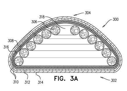
Figures 3A-3B respectively illustrate a cross-sectional side view and a
cutaway
10 top view of a tissue expander (300) according to some embodiments of the
present
invention, suitable, for example, for breast augmentation and/or
reconstruction. Figures
3A-3B show a tissue expander (300) as illustrated in Figure 2A, that further
comprises an
internal skeleton element in the form of a folded tube (318) formed of a
resilient material,
such as soft silicone or polyurethane. Typically, the tube is hollow and
contains therein the
15 foam filling. The foam filling substantially fills the tube, meaning
that it occupies
substantially the entire inner volume of the tube. In some embodiments, the
tube has a
polygonal cross-section. In particular embodiments, the tube has a hexagonal
cross-
section. Figures 3A-3B illustrate partial sections of the hollow tube showing
the foam
filling (306) in the tube. Preferably, the foam filling is confined within the
tube and
substantially fills the tube. In some embodiments, the foam material also
surrounds the
tube such that it occupies voids formed between the folds of the tube or
between the
external wall of the tube and an outer enclosure or mesh. The diameter of the
tube may be
constant or varied along its length. The thickness of the tube wall may be
constant or
varied along its length.
The illustrated tissue expander (300) comprises a flat base surface (302) and
a
convex outer surface (304). The tissue expander (300) comprises an inner foam
filling
(306) within a helical tube (318). The helical tube is folded so as to create
an overall
conical structure that conforms to the shape and design of the tissue expander
(300). The
foam filling and tube are enclosed within a flexible sealed enclosure (316),
and further
within a non-stretchable, resilient expansion restricting layer (308). The
tissue expander
(300) further comprises an outer shell (310) comprising two layers (312, 314).
CA 02898177 2015-07-14
WO 2014/118773
PCT/1L2014/050097
16
Figures 4A-4B illustrate alternative forms of skeleton elements that may be
contained within a tissue expander according to some embodiments of the
present
invention.
Figure 4A is a perspective view of a resiliently deformable skeleton element
(400)
that includes an array of elongate cells (402) extending along mutually
generally parallel
axes (404) from an imaginary flat base surface (406) to an imaginary convex
outer surface
(408) that is tucked in adjacent the imaginary base surface (406). Elongate
cells (402) are
mutually defined by elongate cell walls (410) formed of a resilient material.
In the
illustrated embodiment, the array of elongate cells (402) is characterized in
that it includes
a central cylindrical cell (412) and that elongate cell walls (410) are of
generally uniform
thickness. It is also characterized in that a regular pattern of partial cells
(414) are located
along the periphery of the array. In the illustrated embodiment, all of the
partial cells (414)
are identical. In alternative embodiments, this is not necessarily the case.
In yet additional
alternative embodiments, the elongate cell walls (410) need not be of
generally uniform
thickness and may be of different thicknesses and/or varying thickness.
Figure 4B is a perspective view of a resiliently deformable skeleton element
(400)
that includes an array of identical elongated cells (402), each having an
hexagonal cross
section, extending along mutually generally parallel axes (404) from an
imaginary flat
base surface (406) to an imaginary convex outer surface (408), which is tucked
in adjacent
the imaginary base surface (406). Elongate cells (402) are mutually defined by
elongate
cell walls (410) formed of a resilient material. In the illustrated
embodiment, the array of
elongate cells (402) is preferably characterized in that elongate cell walls
(410) are of
generally uniform thickness. It is also characterized in that a regular
pattern of partial cells
(414) are located along the periphery of the array. In the illustrated
embodiment, the
partial cells (414) are not identical.
Figure 4C is a cross-sectional side view of a tissue expander (400) according
to
some embodiments of the present invention, comprising an internal skeleton
element in
the form of an array of elongated cells, for example a skeleton element as
shown in
Figures 4A-B. As seen in Figure 4C, the illustrated tissue expander (400)
comprises a flat
base surface (422) and a convex outer surface (424). The tissue expander
comprises an
array of elongate cells (402) extending along mutually generally parallel axes
(404),
defined by elongated cell walls (410) formed of a resilient material.
CA 02898177 2015-07-14
WO 2014/118773
PCT/1L2014/050097
17
In the illustrated embodiment, the elongated cells fully extend between the
base
(422) and outer (424) surfaces of the tissue expander, such that substantially
all the edges
(412) of the cell walls are in contact with the innermost layer (414)
enclosing the foam
filling (406). For simplicity, Figure 4C presents only the innermost layer
enclosing the
foam, which is, for example, a flexible sealed enclosure or an expansion
restricting layer.
It is appreciated that the tissue expander includes additional layers, such as
the layers that
constitute the sealing shell. In the illustrated embodiment, the foam filling
(406)
substantially fills all the cells of the skeleton element. In alternative
embodiments, the
foam filling fills only some of the cells. The volume and amount of foam
within each cell
can vary among the cells.
In alternative embodiments, the elongated cells partially extend between the
base
and outer surfaces of the implantable tissue expander, such that only some (or
none) of the
edges of the cell walls are in contact with the innermost layer enclosing the
foam filling.
According to these embodiments, the foam filling may fill the cells and
further extend
outside the cells, to fill voids between the skeleton element and an inner
wall of the tissue
expander.
Thus, in some embodiments, the foam filling fills the cells defined by the
cell walls
and further extends outside the base surface and/or outer surface of the array
of elongated
cells, thus softening the touch of an implant containing a skeleton element.
The internal skeletal element may be formed of the same or different material
as
the other components of the tissue expander.
Additional types of skeleton elements, as well as methods for their
production, are
described, for example, in WO 2007/000756, WO 2008/081439 and WO 2010/049926.
Figures 5A-5B respectively illustrate a perspective view and a top view of a
tissue
expander (500) according to some embodiments of the present invention,
suitable, for
example, for breast augmentation and/or reconstruction. Figures 5A-5B show a
tissue
expander (500) of the present invention that comprises an outer mesh (510)
partially
covering the external surface of the tissue expander. The external surface of
a tissue
expander according to embodiments of the present invention is typically the
outermost
layer of the outer shell of the tissue expander.
The illustrated tissue expander (500) comprises a flat base surface (502)
intended
to face the chest wall, and a convex outer surface (504) intended to face
breast tissue. In
the illustrated embodiment, the tissue expander comprises a single mesh patch
(510) at the
CA 02898177 2015-07-14
WO 2014/118773
PCT/1L2014/050097
18
apex of the convex surface of the tissue expander. In alternative embodiments,
the outer
mesh comprises a plurality of mesh patches, at different positions on the
external surface
of the tissue expander and of different patch size, patch thickness and pore
size. The outer
mesh may be knitted or woven or made of a complete or meshed sheet, and made
from a
biocompatible material like polyester or polyamide for instance.
Following implantation of a tissue expander, connective tissue slowly grows
and
surrounds the tissue expander. The presence of patches of mesh on the external
surface of
the tissue expander facilitates tissue ingrowth into the pores of the mesh,
eventually
forming a tissue-implant complex that anchors the implant to the surrounding
tissues.
In a breast implant for example, an anterior mesh patch, on the convex surface
thereof, will anchor the implant to the overlaying breast tissue, thus
creating a new
implant-breast tissue complex that acts as a single unit against external
forces applied
thereto, and mimics a natural breast to a great extent. A posterior mesh
patch, on the flat
surface thereof, will anchor the implant to the chest wall, and is likely to
mimic the natural
breast to a lesser extent.
The tissue expanders according to embodiments of the present invention may
comprise one or more balloons.
The term "balloon" is used throughout to refer to a flexible, sealed enclosure
configured for controlled inflation and deflation, particularly after
implantation of the
tissue expander. The balloon is being inflatable upon introduction of liquid
or gas into an
interior thereof, and deflatbale upon removal of liquid or gas from said
interior thereof. In
some embodiments, the balloon is an external balloon attached to the outermost
layer of
the implant. In other embodiments, the balloon is internal. In some
embodiments, an
internal balloon is embedded within the foam filling. In other embodiments, an
internal
balloon is outside the foam filling, for example between an expansion-
restriction layer and
an outer shell, attached to the inner surface of the shell.
The balloon is typically associated with a port enabling communication to the
interior of the balloon for inflation and or deflation. There are many ports
known and
described in the literature and one example for a port is an integrated valve
mechanism
comprising a port integrated, for example, in the shell of the implantable
tissue expander
and accessed by a needle through the skin. Another example is a remote valve
mechanism
comprising a tube communicating with the interior of the balloon and
protruding from the
tissue expander such that it is accessible to a surgeon after the tissue
expander is inserted
CA 02898177 2015-07-14
WO 2014/118773
PCT/1L2014/050097
19
into a subject. The tube is preferably connected to the balloon via a self-
sealing valve that
is incorporated into the wall of the balloon and configured to maintain the
balloon sealed
after the removal of the tube. The tube and valve may facilitate the
introduction, or
injection, of a filling, such as liquid, gas or a combination thereof, into
the balloon, or
removal of the filling of the balloon from its interior.
Before implantation in a subject, the balloon is preferably in a collapsed,
deflated
form. The implantable tissue expander comprising the deflated balloon can be
temporarily
and resiliently deformed and compressed as described above in order for a
surgeon to
insert it through an aperture in a cutaneous layer of the subject.
After insertion and placement of the implantable tissue expander in a
designated
location in the subject, the balloon can be inflated, namely filled with
liquid and/or gas
until a desired volume is achieved, resulting in an implantable tissue
expander with an
improved tissue expansion capability. In some embodiments, when a self-sealing
valve
and tube are used, the tube is then preferably removed, for example, pulled
out of the self-
sealing valve. The balloon remains sealed by virtue of the self-sealing valve.
Following a certain time interval, typically when the surgeon appreciates that
sufficient tissue expansion and tissue relaxation have been achieved, the
balloon can be
deflated. In some embodiments, the balloon is made from a needle-penetrable
material that
permits the insertion of a needle and withdrawal of the internal filling.
According to these
embodiments, deflation of the balloon can be performed by inserting a needle
through the
skin into the balloon through the balloon wall, and withdrawing the balloon
filling. Upon
withdrawal of the filling, the balloon remains in a deflated, collapsed form,
and the rest of
the implantable tissue expander, namely the foam filling enclosed within the
layers
described herein, serves as a filler of the expanded tissue. In some
embodiments, where
the balloon is inflated with gas, the natural permeability of the silicone to
may allow the
gas to escape from the balloon into the surrounding tissues where it is
dissolved in the
interstitial fluids and absorbed into the lymph and blood to be released from
the body
naturally. The loss of gas from the balloon gradually decreases the pressure
inside the
balloon and leads to its deflation, thus obviating the need described above to
evacuate the
liquid or gas from the balloon after the required tissue expansion has been
achieved. Using
this method also allows the surgeon to remove the tube during surgery.
The inclusion of a balloon in a tissue expander according to embodiments of
the
present invention may be particularly beneficial in primary tissue
augmentation
CA 02898177 2015-07-14
WO 2014/118773
PCT/1L2014/050097
procedures, such as primary augmentation of a non-ptotic breast, with no ample
skin and
tissue redundancy. In such cases, in the absence of a balloon, the pressure
applied on the
foam-filled implant by the surrounding tissue may variably result in the
deformation of the
implant rather than the desired augmentation of the tissue. If an inflated
balloon is present,
5 sufficient counter-pressure is applied, thereby facilitating augmentation
of the tissue
overlaying the implantable tissue expander. For implantable tissue expanders
intended for
primary tissue augmentation, for example in primary breast augmentation, the
balloon is
preferably located at the posterior surface of the implant, facing the chest
wall. The
balloon may not be needed in procedures such as immediate reconstruction after
10 mastectomy, replacement of an implant in a previously augmented breast,
or in an
augmentation-reduction procedure (mastopexy with an implant), where excess
skin is
available.
Thus, in some embodiments, an implantable tissue expander of the present
invention includes a first compartment filled with foam and characterized by a
defined,
15 pre-determined three-dimensional configuration, and a second, flexible
compartment
comprising liquid filling, gas filling or a combination thereof, that is
configured for
controlled inflation and deflation. The first compartment according to these
embodiments
is configured for permanent support of an augmented tissue, and the second
compartment
is configured for temporary tissue expansion. It is to be understood that the
term
20 "permanent" does not indicate that the implant cannot be removed or
replaced.
The size of the balloon, namely its volume at manufacturing and at inflation
can
vary, and is typically determined according to the type and size of the
implant.
Figures 6A-6B illustrate cross-sectional side views of an implantable tissue
expander (600) according to some embodiments of the present invention,
suitable, for
example, for breast augmentation and/or reconstruction, which includes an
external
balloon (620). The balloon is illustrated in inflated (Figure 6A) and deflated
(Figure 6B)
states.
The illustrated implantable tissue expander (600) comprises a flat base
surface
(602) and a convex outer surface (604) or a curved outer layer. The
implantable tissue
expander (600) comprises an implant inner core comprised of inner foam filling
(606)
enclosed within a non-stretchable resilient expansion restricting layer (608).
The
implantable tissue expander (600) further comprises an outer shell (610)
comprising
generally at least two layers (612, 614).
CA 02898177 2015-07-14
WO 2014/118773
PCT/1L2014/050097
21
In Figures 6A-B, the illustrated tissue expander comprises an external balloon
(620) affixed to the external surface of the outermost layer of the shell
(614) of the
implantable tissue expander, facing the flat base surface (602) of the tissue
expander.
In Figure 6A, the balloon (620) is shown in an inflated state. The balloon can
be
filled with a liquid, preferably a biocompatible liquid such as saline, or gas
such as air. In
the illustrated embodiments, a tube (622) is connected to the balloon via a
self-sealing
valve (624), such as a duck beak-type valve. The balloon (620) is preferably
constructed
from a non-porous, flexible, biocompatible material, such as silicone
elastomer. The
balloon (620) can be over-molded on the external surface of the shell (614) or
attached to
it with an adhesive or other suitable attachment means. In alternative
embodiments, the
balloon and the outer shell of the implant may share a common wall. For
example, layer
(612) enclosing the implant inner core may also constitute a wall of the
balloon.
The balloon is typically shaped as an ellipsoid or elongated sphere.
The direction of expansion is generally symmetrical with the overall shape of
the
tissue expander, or more particularly with the shape of the foam filling, but
can in
principle be different, as defined by design, medical use and manufacturing
processes.
In Figure 6B, the balloon (620) is shown in an inflated state.
Figure 6C illustrates a cross-sectional side view of an implantable tissue
expander
(600) similar to the one described in Figures 6A-6B, which includes an
internal balloon
(620) embedded within the foam filling. The balloon is illustrated in an
inflated state.
When inflated, the balloon applies force against the inner foam filling (606).
A tube (622)
is connected to the balloon via a self-sealing valve (624), such as a duck
beak-type valve,
and protrudes through the shell (610) of the tissue expander. In some
embodiments, one
end of the valve is incorporated in the wall of the balloon. Another end of
the valve,
configured to accommodate the tube, is in line with the outermost layer of the
implant's
shell. Upon filling of the balloon and removal of the tube, the valve self-
seals and
maintains the balloon sealed.
The illustrated balloon compartment (620) is a distinct compartment within the
implant inner core, comprised of the foam filling (606) and non-stretchable
resilient
expansion restricting layer (608).
Figure 6D illustrates another design of an implantable tissue expander (600)
that
contains an internal balloon as described Figure 6C. The illustrated design is
CA 02898177 2015-07-14
WO 2014/118773
PCT/1L2014/050097
22
characterized by an egg-shaped configuration, and may be suitable for,
instance, for
lumpectomy procedures.
In tissue expanders according to embodiments of the present invention
containing
an internal balloon, the balloon may constitute an internal pocket within the
implant inner
core, and thus restricted by the non-stretchable resilient expansion
restricting layer (608).
Alternatively, as illustrated in Figures 6E-6F, an internal balloon may be
anchored to an
internal layer of the shell (612) outside the non-stretchable resilient
expansion restricting
layer (608), thus not restricted by it.
A tissue expander according to embodiments of the present invention may
further
include a plate (not shown) with an embedded, chemically etched, or laser cut
for
example, code/indicator identifying the tissue expander. The plate is
preferably made of a
biocompatible non magnetic material, such as stainless steel, or other non
metallic
polymers, such as polyketones (PEEK) or ceramic materials for example, which
do not
interfere with CT or MRI scans.
In some embodiments, a device, such as a plate, with an identifying code
embedded therein is placed within the tissue expander during manufacturing.
The code
may be of any alphanumeric character with an optional additional symbol or
design or any
printable or designed sign. The plate can have a uniquely or non-uniquely
identifying
code. The code length can vary, thus allowing representation of a unique code
if chosen
once number of tissue expanders manufactured exceeds the maximal variations in
a
specific code length. In some embodiments, the plate can be visualized by
available
imaging techniques, including, inter alia, x-ray, ultrasound, C/T or MRI etc.
In some
embodiments, the code can be identified without the need to remove the implant
from the
patient's body, thus providing a registry tool and mechanism for noninvasive
implant
identification.
In some embodiments a passive or active electronic device, such as an RF
(Radio
Frequency) ID chip as a non-limiting example, can be installed in the tissue
expander and
used for identification of the tissue expander noninvasively by an external
device
communicating with the internally implanted device.
The foregoing description of the specific embodiments will so fully reveal the
general nature of the invention that others can, by applying current
knowledge, readily
modify and/or adapt for various applications such specific embodiments without
undue
CA 02898177 2015-07-14
WO 2014/118773
PCT/1L2014/050097
23
experimentation and without departing from the generic concept, and,
therefore, such
adaptations and modifications should and are intended to be comprehended
within the
meaning and range of equivalents of the disclosed embodiments. It is to be
understood that
the phraseology or terminology employed herein is for the purpose of
description and not
of limitation. The means, materials, and steps for carrying out various
disclosed functions
may take a variety of alternative forms without departing from the invention.