Note: Descriptions are shown in the official language in which they were submitted.
8 17 8 9 8 1 8
1
METHODS OF TREATING CHOLANGIOCARCINOMA
RELATED APPLICATIONS
This application claims the benefit of U.S. Provisional Application No.
61/754,509, filed January 18, 2013 and U.S. Provisional Application No.
61/756,372,
filed January 24, 2013.
SEQUENCE LISTING
to The instant application contains a Sequence Listing which has been
submitted
electronically in ASCII format.
BACKGROUND
Cancer represents the phenotypic end-point of multiple genetic lesions that
endow
cells with a full range of biological properties required for tumorigenesis.
Indeed, a
hallmark genomic feature of many cancers is the presence of numerous complex
chromosome structural aberrations, including txanslocations, intra-chromosomal
inversions, point mutations, deletions, gene copy number changes, gene
expression level
changes, and gennline mutations, among others.
Cholangiocarcinoma is a cancer that includes mutated epithelial cells that
originate in the bile ducts. Cholangiocarcinoma is a relatively rare neoplasm
that is
classified as an adenocarcinoma (a cancer that forms glands or secretes
significant
amounts of mucins). It has an annual incidence rate of about 1-2 cases per
100,000 in
the Western world, but rates of cholangiocarcinoma have been rising worldwide
over
the past several decades (Landis S. etal. (1998) CA Cancer J Clin 48(1): 6-29;
Patel
T (2002) BMC Cancer 2: 10. doi:10.1186/1471-2407-2-10).
Cancer of the bile ducts can arise within the liver as an intrahepatic
cholangiocarcinoma (ICC) or originate from extrahepatic bile ducts as a bile
duct
carcinoma, also referred to as an extra-hepatic cholangiocarcinoma. ICC is the
second most common primary hepatic malignancy after hepatocellular carcinoma
Date Recue/Date Received 2020-06-25
PCT/US14/12136 18-11-2014 PCT/US2014/012136 18.03.2015
CA 02898326 2015-07-15
(HCC), and accounts for 3% of the malignant tumors of the gastrointestinal
system
and 15% of primary hepatic malignancies. Because ICC has a routine histologic
appearance of an adenocarcinoma, the diagnosis of ICC on a liver biopsy
requires an
immunohistochemical (MC) study of the tumor and a thorough clinical workup
including imaging studies to rule out a metastatic adenocarcinoma to the
liver.
=
--IA- 1 / 1
AMENDED SHEET - TEA/US
CA 02898326 2015-07-15
WO 2014/113729 PCT/US2014/012136
2
Numerous studies have indicated that the incidence and mortality trout iiuL
are increasing worldwide. ICC is associated with primary sclerosing
cholangitis,
parasitic biliary infection, polycystic disease of the liver, congenital
intrahepatic bile
duct dilatation (Caroli's Disease), congenital hepatic fibrosis, and
choledochal cysts.
Chronic Hepatitis C infection is an established cause of ICC with some studies
describing a more than 300 fold increase in ICC incidence in patients with
long-
standing hepatitis C infections. ICC has also been associated with cigarette
smoking,
alcohol consumption and exposure to a variety of toxins and chemical
carcinogens.
The onset of symptoms of ICC are often vague, typically arise late in the
course of the
disease and include abdominal pain, anorexia and palpable abdominal mass
lesions.
Thus, the median survival for ICC is less than 6 months for inoperable tumors
and
only 20 to 40% for patients who undergo surgery and achieve clear margins.
Cholangiocarcinoma is considered to be an incurable and rapidly lethal
malignancy, unless both the primary tumor and any metastases can be fully
resected
(removed surgically). No potentially curative treatment exists at this time
except
surgery; however, most patients have advanced stage disease at presentation
and are
inoperable at the time of diagnosis. Cholangiocarcinoma has near-100% fatality
due
to attendant liver complications from the damage to the organ. Patients with
cholangiocarcinoma are generally managed with chemotherapy, radiation therapy,
and
other palliative care measures.
Thus, the need still exists for identifying novel genetic lesions associated
with
cancers such as cholangiocarcinomas. Such genetic lesions can be an effective
approach
to develop compositions, methods and assays for evaluating and treating cancer
patients.
SUMMARY
The invention is based, at least in part, on the discovery, in
cholagiocarcinomas, of novel rearrangement events that give rise to
alterations in a
fibroblast growth factor receptor 2 (FGFR2) gene or a neurotrophic tyrosine
receptor
kinase (NTRIC1) gene. In certain embodiments, the alteration is chosen from a
translocation, a deletion, an inversion, a rearrangement, or an amplification
of, an
FGFR2 gene or the NTRK gene. For example, the alteration can be chosen from an
alteration described in Table 1 and FIGs. 1A-1C. In one embodiment, the
alteration
CA 02898326 2015-07-15
WO 2014/113729 PCT/US2014/012136
3
includes a fragment of an FGFR2 gene or the NTRK1 gene, e.g., as exemplineu in
Table 1, FIGs. 1A-1C and FIGs. 2-17. Thus, the invention provides new insights
into
the treatment of these cancers, such as cholangiocarcinomas.
Therefore,described
herein are methods for treating a cholangiocarcinoma carcinoma, including
.. intrahepatic cholangiocarcinoma (ICC) and extrahepatic cholangiocarcinoma,
as well
as novel FGFR2 and NTRK1 molecules (e.g., fusion molecules); methods and
reagents for identifying, assessing or detecting an alteration in an FGFR2
and/or
NTRK1.
Accordingly, in one aspect, the invention features a method of treating a
subject having a cholangiocarcinoma. The method includes administering to the
subject an effective amount of an agent (e.g., a therapeutic agent) that
targets,
antagonizes or inhibits an FGFR2 or NTRK1 (e.g., an FGFR2 or NTRK1 gene
product, e.g., an FGFR2 or NTRK1 protein), thereby treating the subject.
In another aspect, the invention features, a method of treating a subject
having
a cholangiocarcinoma. The method includes administering to the subject an
effective
amount of a kinase inhibitor (e.g., a tyrosine kinase inhibitor), thereby
treating the
subject.
In one embodiment, the method further includes acquiring knowledge of one
or both of:
(i) the presence (or absence) of an alteration in FGFR2 gene product, e.g., an
FGFR2 protein; or
(ii) the presence (or absence) of an alteration in NTRK1 gene product, e.g.,
an
NTRK1 protein,
in the subject, or a cancer or tumor sample from the subject.
In another embodiment, the method further includes identifying the subject, or
a cancer or tumor sample from the subject, as having one or both of:
(i) the presence (or absence) of an alteration in FGFR2 gene product, e.g., an
FGFR2 protein; or
81789818
4
(ii) the presence (or absence) of an alteration in NTRK1 gene product, e.g.,
an
NTRK1 protein.
In certain embodiments, the presence of the FGFR2 or NTRK1 alteration, or
both, in
the subject is indicative that the subject is likely to respond to the agent.
In yet other embodiments, the agent is administered responsive to a
determination of
the presence of the FGFR2 or NTRK1 alteration, or both, in the subject, or the
cancer or
tumor sample from the subject.
In one embodiment, the invention features a use of a therapeutic agent that
antagonizes or inhibits an FGFR2 gene product for treating a subject having a
cholangiocarcinoma, wherein the subject or the cholangiocarcinoma comprises or
is identified
as having an FGFR2 fusion nucleic acid molecule or an FGFR2 fusion
polypeptide, wherein
the FGFR2 fusion nucleic acid molecule or the FGFR2 fusion polypeptide is
chosen from
FGFR2-TACC3, FGFR2-KIAA1598, and BICC1-FGFR2, and wherein said therapeutic
agent
is selected from the group consisting of: (i) an antibody molecule against the
FGFR2 gene
product; (ii) a kinase inhibitor that inhibits the FGFR2 gene product; and
(iii) an siRNA,
antisense RNA, or other nucleic acid based inhibitor of the FGFR2 gene
product.
Cholangiocarcinoma
In certain embodiments, the cholangiocarcinoma comprises one or more mutated
cells that originate in the bile duct. In certain embodiments, the
cholangiocarcinoma is chosen
.. from an intrahepatic cholangiocarcinoma or an extrahepatic
cholangiocarcinoma. In other
embodiments, the cholangiocarcinoma comprises, or is identified as having, an
alteration that
is chosen from a translocation, a deletion, an inversion, a rearrangement, or
an amplification
of, an FGFR2 gene or the NTRK gene. In one embodiment, the cholangiocarcinoma
comprises, or is identified as having, an alteration chosen from an alteration
described in
Table 1 or FIGs. 1A-1C. In one embodiment, the cholangiocarcinoma comprises,
or is
identified as having, an alteration includes a fragment of an FGFR2 gene or
the NTRK1 gene,
e.g., as exemplified in Table 1, FIGs. 1A-1C and FIGs. 2-17. In yet other
embodiments, the
cholangiocarcinoma comprises, or is identified as having, a fusion molecule of
FGFR2; e.g., a
fusion molecule chosen from FGFR2-TACC3, FGFR2-KIAA1598, BICC1-FGFR2, FGFR2-
Date Recue/Date Received 2020-06-25
81789818
4a
BICC1, PARK2-FGFR2, FGFR2-NOL4, or ZDHHC6-FGFR2 as described, e.g., in Table
1,
FIGs. 1A-1C and FIGs. 2-17. In other embodiments, the cholangiocarcinoma
comprises, or is
identified as having, a rearrangement or an amplification of FGFR2 as
described, e.g., in
Table 1, FIGs. 1A-1C and FIGs. 2-17.
In certain embodiments, the alteration in FGFR2 results in upregulation,
increased
activity (e.g., increased transformative or oncogenic activity, kinase
activity and/or
dimerization), and/or increased level of an FGFR2 gene product (e.g., an FGFR2
protein),
compared to a wildtype activity of FGFR2.
Date Recue/Date Received 2020-06-25
CA 02898326 2015-07-15
WO 2014/113729 PCT/US2014/012136
Subjects
In certain embodiments, the subject has an alteration in FGFR2 or NTRK1, or
both, e.g., the subject has a cholangiocarcinoma comprising an alteration in
FGFR2 or
5 .. NTRK1, or both, e.g., as described herein. In other embodiments, the
subject is
identified, or has been previously identified, as having a cholangiocarcinoma
(e.g., an
intrahepatic cholangiocarcinoma (ICC) or an extrahepatic cholangiocarcinoma)
comprising an alteration in FGFR2 or NTRK1, or both, e.g., as described
herein. In
other embodiments, the subject has, or is identified as having, an alteration
that is
to chosen from a translocation, a deletion, an inversion, a rearrangement,
or an
amplification of, an FGFR2 gene or the NTRK gene. In one embodiment, the
subject
has, or is identified as having, an alteration chosen from an alteration
described in
Table 1 or FIGs. 1A-1C. In one embodiment, the subject has, or is identified
as
having, an alteration includes a fragment of an FGFR2 gene or the NTRK1 gene,
e.g.,
.. as exemplified in Table 1, FIGs. 1A-1C and FIGs. 2-17. In yet other
embodiments,
the subject has, or is identified as having, a fusion molecule of FGFR2; e.g.,
a fusion
molecule chosen from FGFR2-TACC3, FGFR2-KIAA1598, BICC1-FGFR2, FGFR2-
BICC1, PARK2-FGFR2, FGFR2-NOL4, or ZDHHC6-FGFR2 as described, e.g., in
Table 1, FIGs. 1A-1C and FIGs. 2-17. In other embodiments, the subject has, or
is
identified as having, a rearrangement or an amplification of FGFR2 as
described, e.g.,
in Table 1, FIGs. 1A-1C and FIGs. 2-17.
In one embodiment, the subject is a human. In one embodiment, the subject
has, or is at risk of having a cholangiocarcinoma (e.g., a cholangiocarcinoma
as
described herein) at any stage of disease, e.g., Stage I, II, IIIA-IIIC or IV
of
intrahepatic cholangiocarcinoma; Stage 0, III or IV of extrahepatic
cholangiocarcinoma; or a metastatic cancer. In other embodiments, the subject
is a
cancer patient, e.g., a patient having a cholangiocarcinoma as described
herein.
In one embodiment, the subject is undergoing or has undergone treatment with a
different (e.g., non- FGFR2 or non-N'I'RK1) therapeutic agent or therapeutic
modality.
In one embodiment, the non-FGFR2 or non-NTRK1 therapeutic agent or therapeutic
modality is a chemotherapy, immunotherapy, or a surgical procedure. In one
embodiment, the non-FGFR2 or non-NTRK1 therapeutic agent or therapeutic
modality
CA 02898326 2015-07-15
WO 2014/113729 PCT/US2014/012136
6
comprises one or more (or all) of: a surgical procedure, Ilurouracil (e.g., 54-
u, Auructi,
Efudex), doxorubicin (Adriamycin, Rubex), gemcitabine (e.g., Gemzar) and/or
cisplatin
(Platinol).
In one embodiment, responsive to the determination of the presence of the
FGFR2
or NTRK1 alteration, the different therapeutic agent or therapeutic modality
is
discontinued. In yet other embodiments, the subject has been identified as
being likely or
unlikely to respond to the different therapeutic agent or therapeutic
modality.
In certain embodiments, the subject has participated previously in a clinical
trial, e.g., a clinical trial for a different (e.g., non-FGFR2 or non-NTRK1)
therapeutic
agent or therapeutic modality. In other embodiments, the subject is a cancer
patient
who has participated in a clinical trial, e.g., a clinical trial for a
different (e.g., non-
FGFR2 or non-NTRK1) therapeutic agent or therapeutic modality.
Agents
In certain embodiments, the agent (e.g., the therapeutic agent) used in the
methods targets and/or inhibits FGFR2 or NTRK1 (e.g., a FGFR2 or NTRK1 gene or
gene product as described herein). In one embodiment, the agent binds and
inhibits
FGFR2 or NTRK1. In one embodiment, the agent is a reversible or an
irreversible
FGFR2 inhibitor. In certain embodiments, the agent is a pan-FGFR2 inhibitor.
In one embodiment, the agent is an antibody molecule, e.g., an anti- FGFR2 or
NTRK1 antibody molecule (e.g., a monoclonal or a bispecific antibody), or a
conjugate thereof (e.g., an antibody to FGFR2 or NTRK1 conjugated to a
cytotoxic
agent (e.g., mertansine DM1)).
In one embodiment, the agent is a kinase inhibitor. In one embodiment, the
kinase inhibitor is chosen from: a multi-specific kinase inhibitor, an FGFR2
inhibitor
(e.g., a pan-FGFR2 inhibitor), an NTRK1 inhibitor, and/or a small molecule
inhibitor
that is selective for FGFR2 or NTRK1; and/or a FGFR2 or NTRK1 cellular
immunotherapy.
In an embodiment, the therapeutic agent is chosen from a kinase inhibitor; a
multi-specific kinase inhibitor; an FGF receptor inhibitor (e.g., a pan FGFR2
inhibitor); an antibody molecule (e.g., a monoclonal antibody) against FGFR2;
and/or
a small molecule (e.g., kinase) inhibitor that is selective for FGFR2 or
NTRK1.
CA 02898326 2015-07-15
WO 2014/113729 PCT/US2014/012136
7
In an embodiment the therapeutic agent is selected from antisense molecules,
ribozymes, RNAi, triple helix molecules that hybridize to a nucleic acid
encoding the
fusion, or a transcription regulatory region that blocks or reduces mRNA
expression
of FGFR2 or NTRK1.
In an embodiment the kinase inhibitor is chosen from: a kinase inhibitor; a
multi-specific kinase inhibitor; an FGF receptor inhibitor (e.g., a pan FGFR2
inhibitor); and/or a kinase inhibitor that is selective for FGFR2 or NTRK1.
In an embodiment, the therapeutic agent is chosen from: Regorafenib;
Ponatinib; AZD-2171 (Cediranib); AZD-4547; BGJ398; BB3F1120; Brivanib;
Dovitinib; ENMD-2076; JNJ42756493; Masitinib; Lenvatinib; LY2874455;
Pazopanib; PD-173955; R406; PD173074; Danusertib; Dovitinib Dilactic Acid;
TSIJ-
68; Tyrphostin AG 1296; MK-2461; Brivanib Alaninate; Lestaurtinib; PHA-848125;
K252a; AZ-23; and/or Oxindole-3.
In an embodiment, the therapeutic agent is chosen from Regorafenib or
Ponatinib.
Other features and embodiments of the invention include one or more of the
following.
In an embodiment, the method includes acquiring knowledge of the presence
of an alteration, e.g., fusion, from 'fable 1, FIGs. 1A-1C and FIGs. 2-17 in
said
subject.
In an embodiment the therapeutic agent is administered responsive to the
determination of presence of the alteration, e.g., fusion, in a tumor sample
from said
subject.
In an embodiment the determination of the presence of the alteration, e.g.,
fusion,
comprises sequencing.
In an embodiment the subject is undergoing or has undergone treatment with a
different therapeutic agent or therapeutic modality, e.g., a non-FGFR2 or non-
NTRK1
therapeutic agent or therapeutic modality.
In an embodiment responsive to a determination of the presence of the
alteration,
e.g., fusion, the different therapeutic agent or therapeutic modality is
discontinued.
CA 02898326 2015-07-15
WO 2014/113729 PCT/US2014/012136
8
In an embodiment the different therapeutic agent or therapeutic mouanty is a
chemotherapy or a surgical procedure. In one embodiment, the non-FGFR2 or non-
NTRK1 therapeutic agent or therapeutic modality comprises one or more (or all)
of: a
surgical procedure, flurouracil (e.g., 5-FU, Adrucil, Efudex), doxorubicin
(Adriamycin,
Rubex), gemcitabine (e.g., Gemzar) and/or cisplatin (Platinol).
In another aspect, the invention features, a method of determining the
presence of
an alteration, e.g., a fusion, disclosed herein in cholangiocarcinoma sample,
comprising:
directly acquiring knowledge that an alteration, e.g., a fusion nucleic acid
molecule, of Table 1, FIGs. 1A-1C and FIGs. 2-17 is present in a sample from a
subject.
In an embodiment the acquiring step comprises sequencing.
In an embodiment the method further comprises administering a kinase
inhibitor to the subject responsive to the determination of the presence of
the
alteration, e.g., the fusion, in the sample from the subject.
The invention also provides, methods of: identifying, assessing or detecting
an
alteration, e.g., fusion, of an FGFR2 or an NTRK1, e.g., that arises in a
cholangiocarcinoma. Exemplary alteration, e.g., fusions, include those
summarized in
Table 1, FIGs. 1A-1C and FIGs. 2-17, including a fusion of FGFR2 (e.g., an
FGFR2
fusion molecule (e.g., a gene product or fragment thereof)) to a partner from
Table 1,
FIGs. 1A-1C and FIGs. 2-17, or a fusion of NTRK1 (e.g., an NTRK1 fusion
molecule
(e.g., a gene product or fragment thereof)) to a partner of Table 1. In one
embodiment,
the FGFR2 or NTRK1 is fused to a second gene, or a fragment thereof, e.g., as
described
in Table 1, FIGs. 1A-1C and FIGs. 2-17. In other embodiments, the fusion
molecule is
chosen from FGFR2-TACC3, FGFR2-KIAA1598, BICC1-FGFR2, FGFR2-BICC1,
PARK2-FGFR2, FGFR2-NOI,4, ZDHHC6-FGFR2, or RABGAP1L-NTRK1, e.g., as
described, e.g., in 'fable 1, FIGs. 1A-1C and FIGs. 2-17. Included are fusion
molecules;
isolated fusions nucleic acid molecules, nucleic acid constructs, host cells
containing the
nucleic acid molecules; purified fusion polypeptides and binding agents;
detection
reagents (e.g., probes, primers, antibodies, kits, capable, e.g., of specific
detection of a
fusion nucleic acid or protein); screening assays for identifying molecules
that interact
with, e.g., inhibit, fusions, e.g., novel kinase inhibitors or binders of
FGFR2 or NTRK1.
CA 02898326 2015-07-15
WO 2014/113729 PCT/US2014/012136
9
Nucleic Acid Molecules
In one aspect, the invention features an isolated nucleic acid molecule, or an
isolated preparation of nucleic acid molecules, that includes a genetic
alteration disclosed
.. herein. Such nucleic acid molecules or preparations thereof can include a
genetic
alteration described herein or can be used to detect, e.g., sequence, a
genetic alteration
disclosed herein. In other embodiments, the alteration of FGFR2 or NRTKI is
chosen
from an alteration set forth in Table 1, FIGs. IA- I C and FIGs. 2-17. In
other
embodiments, the fusion nucleic acid molecule is chosen from FGFR2-TACC3,
FGFR2-
KIAA1598, BICC1-FGFR2, FGFR2-BICC1, PARK2-FGFR2, FGFR2-NOL4,
ZDHHC6-FGFR2, or RABGAP1L-NTRK1, e.g., as described, e.g., in Table 1, FIGs.
IA-
1C and FIGs. 2-17.
Nucleic Acid Detection and Capturing Reagents
The invention also features a nucleic acid molecule, e.g., nucleic acid
fragment, suitable as probe, primer, bait or library member that includes,
flanks,
hybridizes to, which are useful for identifying, or are otherwise based on, a
fusion
described herein. In certain embodiments, the probe, primer or bait molecule
is an
oligonucleotide that allows capture, detection or isolation of a fusion
nucleic acid
molecule described herein, e.g., a fusion of FGFR2 to a second gene, or
fragment
thereof, e.g., BICC1, KIAA1598, TACC3, PARK2, NOL4, or ZDHHC6-FGFR2: or a
fusion of NTRKI to a second gene, or a fragment thereof, e.g., RAB GAP IL
(e.g., as
described in Table I, FIGs. 1A-1C and FIGs. 2-17).
The oligonucleotide can comprise a nucleotide sequence substantially
complementary to a fragment of a fusion between partners described herein
nucleic acid
molecules described herein. The sequence identity between the nucleic acid
fragment,
e.g., the oligonucleotide, and the target sequence need not be exact, so long
as the
sequences are sufficiently complementary to allow the capture, detection or
isolation of
the target sequence. In one embodiment, the nucleic acid fragment is a probe
or primer
that includes an oligonucleotide between about 5 and 25, e.g., between 10 and
20, or 10
and 15 nucleotides in length. In other embodiments, the nucleic acid fragment
is a bait
CA 02898326 2015-07-15
WO 2014/113729 PCT/US2014/012136
that includes an oligonucleotide between about 100 to 300 nucleotides, 13u anu
Liu
nucleotides, or 150 and 200 nucleotides, in length.
In one embodiment, the nucleic acid fragment can be used to identify or
capture,
e.g., by hybridization, a fusion nucleic acid molecule described herein, e.g.,
a fusion of
5 FGFR2 to a second gene, or fragment thereof, e.g., BICC1, KIAA1598,
TACC3,
PARK2, NOL4, or ZDHHC6; or a fusion of NTRK1 to a second gene, or a fragment
thereof, e.g., RABGAP1L (e.g., as described in Table 1, FIGs. 1A-1C and FIGs.
2-17).
For example, the nucleic acid fragment can be a probe, a primer, or a bait,
for use in
identifying or capturing, e.g., by hybridization, a fusion described herein.
In one
10 embodiment, the nucleic acid fragment can be useful for identifying or
capturing a fusion
breakpoint. In one embodiment, the nucleic acid fragment hybridizes to a
nucleotide
sequence that includes a breakpoint, e.g., a breakpoint of the fusion.
The probes or primers described herein can be used, for example, for FISH
detection or PCR amplification. In one exemplary embodiment where detection is
based
on PCR, amplification of the fusion junction can be performed using a primer
or a primer
pair, e.g., for amplifying a sequence flanking the fusion junctions described
herein, e.g.,
the mutations or the junction of a chromosomal rearrangement described herein.
In one embodiment, a pair of isolated oligonucleotide primers can amplify a
region containing or adjacent to a position in the fusion. For example,
reverse primers
can be designed to hybridize to a nucleotide sequence within genomic or mRNA
sequence of one partner, and the forward primers can be designed to hybridize
to a
nucleotide sequence within the other fusion partner.
In other embodiments, the nucleic acid fragment includes a bait that comprises
a
nucleotide sequence that hybridizes to a fusion nucleic acid molecule
described herein,
.. and thereby allows the capture or isolation said nucleic acid molecule. In
one
embodiment, a bait is suitable for solution phase hybridization. In other
embodiments, a
bait includes a binding entity, e.g., an affinity tag, that allows capture and
separation, e.g.,
by binding to a binding entity, of a hybrid formed by a bait and a nucleic
acid hybridized
to the bait.
In other embodiments, the nucleic acid fragment includes a library member
comprising a nucleic acid molecule described herein. In one embodiment, the
library
member includes a rearrangement that results in a fusion described herein.
CA 02898326 2015-07-15
WO 2014/113729 PCT/US2014/012136
11
The nucleic acid fragment can be delectably labeled with, e.g., a ramoiaoei, a
fluorescent label, a bioluminescent label, a chemiluminescent label, an enzyme
label, a
binding pair label, or can include an affinity tag; a tag, or identifier
(e.g., an adaptor,
barcode or other sequence identifier).
Fusion Polypeptides
In another aspect, the invention features a fusion polypeptide (e.g., a
purified
fusion polypeptide), a biologically active or antigenic fragment thereof, as
well as
reagents (e.g., antibody molecules that bind to a fusion polypeptide), methods
for
modulating a fusion polypeptide activity and detection of a fusion
polypeptide.
In certain embodiments, the fusion polypeptide is chosen from FGFR2-
TACC3, FGFR2-K1AA1598, BICC1-FGFR2, FGFR2-B1CC1, PARK2-FGFR2,
FGFR2-NOL4, ZDHHC6-FGFR2, or RABGAP1L-NTRK1, e.g., as described, e.g., in
Table 1, FIGs. 1A-1C and FIGs. 2-17.
In one embodiment, the fusion polypeptide has at least one biological activity
of one or both of its partners.
In other embodiments, the nucleic acid molecule includes a nucleotide sequence
encoding a fusion polypeptide that includes a fragment of a each partner of a
fusion
described herein.
In a related aspect, the invention features fusion polypeptide or fragments
operatively linked to heterologous polypeptides to form fusion proteins.
In another embodiment, the fusion polypeptide or fragment is a peptide, e.g.,
an
immunogenic peptide or protein, that contains a fusion junction described
herein. Such
immunogenic peptides or proteins can be used to raise antibodies specific to
the fusion
protein. In other embodiments, such immunogenic peptides or proteins can be
used for
vaccine preparation. The vaccine preparation can include other components,
e.g., an
adjuvant.
In another aspect, the invention features antibody molecules that binds to a
fusion
polypeptide or fragment described herein. In embodiments the antibody can
distinguish
wild type from fusion.
CA 02898326 2015-07-15
WO 2014/113729 PCT/US2014/012136
12
Detection Reagents and Detection of Mutations
In another aspect, the invention features a detection reagent, e.g., a
purified or
an isolated preparation thereof. Detection reagents can distinguish a nucleic
acid, or
protein sequence, having an alteration, e.g., a breakpoint, of a
rearrangement, e.g., of a
fusion nucleic acid molecule described herein. Exemplary fusions include a
fusion of
FGFR2 to a second gene, or fragment thereof, e.g., BICCI, KIAA1598, TACC3,
PARK2, NOL4, or ZDIIIIC6; or a fusion of NTRKI to a second gene, or a fragment
thereof, e.g., RABGAPIL, e.g., as described, e.g., in Table 1, FIGs. 1A- IC
and FIGs.
2-17.
Detection reagents, e.g., nucleic acid-based detection reagents, can be used
to
identify mutations in a target nucleic acid, e.g., DNA, e.g., genomic DNA or
cDNA,
or RNA, e.g., in a sample, e.g., a sample of nucleic acid derived from a
cholanaiocarcinoma. Detection reagents, e.g., antibody-based detection
reagents, can
be used to identify mutations in a target protein, e.g., in a sample, e.g., a
sample of
protein derived from, or produced by, a cholangiocarcinoma cell.
Nucleic Acid-based Detection Reagents
In an embodiment, the detection reagent comprises a nucleic acid molecule,
e.g., a DNA. RNA or mixed DNA/RNA molecule, comprising sequence which is
.. complementary with a nucleic acid sequence on a target nucleic acid (the
sequence on
the target nucleic acid that is bound by the detection reagent is referred to
herein as
the "detection reagent binding site" and the portion of the detection reagent
that
corresponds to the detection reagent binding site is referred to as the
"target binding
site"). In an embodiment, the detection reagent binding site is disposed in
relationship to the interrogation position such that binding (or in
embodiments, lack of
binding) of the detection reagent to the detection reagent binding site allows
differentiation of mutant and reference sequences for an alteration described
herein
(e.g., an alteration or a fusion nucleic acid molecule described in Table 1,
FIGs. IA-
1C and FIGs. 2-17), e.g., a fusion of FGIR2 to a second gene, or fragment
thereof,
e.g., BICC I, KIAAI598, TACC3, PARK2, NOL4, or ZDHHC6; or a fusion of
NTRKI to a second gene, or a fragment thereof, e.g., RABGAPIL), from a
reference
CA 02898326 2015-07-15
WO 2014/113729 PCT/US2014/012136
13
sequence. The detection reagent can be modified, e.g., with a label or other
moiety,
e.g., a moiety that allows capture.
In an embodiment, the detection reagent comprises a nucleic acid molecule,
e.g., a DNA, RNA or mixed DNA/RNA molecule, which, e.g., in its target binding
site, includes the interrogation position and which can distinguish (e.g., by
affinity of
binding of the detection reagent to a target nucleic acid or the ability for a
reaction,
e.g., a ligation or extension reaction with the detection reagent) between a
mutation,
e.g., a translocation described herein, and a reference sequence. In
embodiments, the
interrogation position can correspond to a teiminal, e.g., to a 3' or 5'
terminal
nucleotide, a nucleotide immediately adjacent to a 3' or 5' terminal
nucleotide, or to
another internal nucleotide, of the detection reagent or target binding site.
In embodiments, the difference in the affinity of the detection reagent for a
target nucleic acid comprising the mutant and that for a target nucleic acid
comprising
the reference sequence allows determination of the presence or absence of the
mutation (or reference) sequence. Typically, such detection reagents, under
assay
conditions, will exhibit substantially higher levels of binding only to the
mutant or
only to the reference sequence, e.g., will exhibit substantial levels of
binding only to
the mutant or only to the reference sequence.
In embodiments, binding allows (or inhibits) a subsequent reaction, e.g., a
.. subsequent reaction involving the detection reagent or the target nucleic
acid. E.g.,
binding can allow ligation, or the addition of one or more nucleotides to a
nucleic
acid, e.g., the detection reagent, e.g., by DNA polymerase, which can be
detected and
used to distinguish mutant from reference. In embodiments, the interrogation
position
is located at the terminus, or sufficiently close to the tet minus, of the
detection reagent
or its target binding site, such that hybridization, or a chemical reaction,
e.g., the
addition of one or more nucleotides to the detection reagent, e.g., by DNA
polymerase, only occurs, or occurs at a substantially higher rate, when there
is a
perfect match between the detection reagent and the target nucleic acid at the
interrogation position or at a nucleotide position within 1, 2, or 3
nucleotides of the
interrogation position.
In an embodiment, the detection reagent comprises a nucleic acid, e.g., a
DNA, RNA or mixed DNA/RNA molecule wherein the molecule, or its target binding
CA 02898326 2015-07-15
WO 2014/113729 PCT/US2014/012136
14
site, is adjacent (or flanks), e.g., directly adjacent, to the interrogation
position, anu
which can distinguish between a mutation, e.g., a translocati on described
herein, and a
reference sequence, in a target nucleic acid.
In embodiments, the detection reagent binding site is adjacent to the
.. interrogation position, e.g.. the 5' or 3'teiminal nucleotide of the
detection reagent, or
its target binding site, is adjacent, e.g., between 0 (directly adjacent) and
1,000, 500,
400, 200, 100, 50, 10, 5, 4, 3, 2, or 1 nucleotides from the interrogation
position. In
embodiments, the outcome of a reaction will vary with the identity of the
nucleotide at
the interrogation position allowing one to distinguish between mutant and
reference
sequences. E.g., in the presence of a first nucleotide at the interrogation
position a
first reaction will be favored over a second reaction. E.g., in a ligation or
primer
extension reaction, the product will differ, e.g., in charge, sequence, size,
or
susceptibility to a further reaction (e.g., restriction cleavage) depending on
the identity
of the nucleotide at the interrogation position. In embodiments the detection
reagent
comprises paired molecules (e.g., forward and reverse primers), allowing for
amplification, e.g., by PCR amplification, of a duplex containing the
interrogation
position. In such embodiments, the presence of the mutation can be determined
by a
difference in the property of the amplification product, e.g., size, sequence,
charge, or
susceptibility to a reaction, resulting from a sequence comprising the
interrogation
position and a corresponding sequence having a reference nucleotide at the
interrogation positions. In embodiments, the presence or absence of a
characteristic
amplification product is indicative of the identity of the nucleotide at the
interrogation
site and thus allows detection of the mutation.
In embodiments, the detection reagent, or its target binding site, is directly
adjacent to the interrogation position, e.g., the 5' or 3'terminal nucleotide
of the
detection reagent is directly adjacent to the interrogation position. In
embodiments,
the identity of the nucleotide at the interrogation position will determine
the nature of
a reaction, e.g., a reaction involving the detection reagent, e.g., the
modification of
one end of the detection reagent. E.g., in the presence of a first nucleotide
at the
interrogation position a first reaction will be favored over a second
reaction. By way
of example, the presence of a first nucleotide at the interrogation position,
e.g., a
nucleotide associated with a mutation, can promote a first reaction, e.g., the
addition
CA 02898326 2015-07-15
WO 2014/113729 PCT/US2014/012136
of a complementary nucleotide to the detection reagent. By way of example, tne
presence of an A at the interrogation position will cause the incorporation of
a T,
having, e.g., a first colorimetric label, while the presence of a G and the
interrogation
position will cause the incorporation for a C, having, e.g., a second
colorimetric label.
5 In an embodiment, the presence of a first nucleotide at the nucleotide
will result in
ligation of the detection reagent to a second nucleic acid. E.g., a third
nucleic acid
can be hybridized to the target nucleic acid sufficiently close to the
interrogation site
that if the third nucleic acid has an exact match at the interrogation site it
will be
ligated to the detection reagent. Detection of the ligation product, or its
absence, is
10 indicative of the identity of the nucleotide at the interrogation site
and thus allows
detection of the mutation.
A variety of readouts can be employed. E.g., binding of the detection reagent
to the mutant or reference sequence can be followed by a moiety, e.g., a
label,
associated with the detection reagent, e.g., a radioactive or enzymatic label.
In
15 embodiments the label comprises a quenching agent and a signaling agent
and
hybridization results in altering the distance between those two elements,
e.g.,
increasing the distance and un-quenching the signaling agent. In embodiments,
the
detection reagent can include a moiety that allows separation from other
components
of a reaction mixture. In embodiments, binding allows cleavage of the bound
detection reagent, e.g., by an enzyme, e.g., by the nuclease activity of the
DNA
polymerase or by a restriction enzyme. The cleavage can be detected by the
appearance or disappearance of a nucleic acid or by the separation of a
quenching
agent and a signaling agent associated with the detection reagent. In
embodiments,
binding protects, or renders the target susceptible, to further chemical
reaction, e.g.,
labeling or degradation, e.g., by restriction enzymes. In embodiments binding
with
the detection reagent allows capture separation or physical manipulation of
the target
nucleic acid to thereby allow for identification. In embodiments binding can
result in
a detectable localization of the detection reagent or target, e.g., binding
could capture
the target nucleic acid or displace a third nucleic acid. Binding can allow
for
determination of the presence of mutant or reference sequences with FISH,
particularly in the case of rearrangements. Binding can allow for the
extension or
other size change in a component, e.g., the detection reagent, allowing
distinction
CA 02898326 2015-07-15
WO 2014/113729 PCT/US2014/012136
16
between mutant and reference sequences. Binding can allow for the production,
e.g.,
by PCR, of an amplicon that distinguishes mutant from reference sequence.
In an embodiment the detection reagent, or the target binding site, is between
5
and 500, 5 and 300, 5 and 250, 5 and 200, 5 and 150, 5 and 100, 5 and 50, 5
and 25, 5
and 20, 5 and 15, or 5 and 10 nucleotides in length. In an embodiment the
detection
reagent, or the target binding site, is between 10 and 500, 10 and 300, 10 and
250, 10
and 200, 10 and 150, 10 and 100, 10 and 50, 10 and 25,10 and 20, or 10 and 15,
nucleotides in length. In an embodiment the detection reagent, or the target
binding
site, is between 20 and 500, 20 and 300, 20 and 250, 20 and 200, 20 and 150,
20 and
100, 20 and 50, or 20 and 25 nucleotides in length. In an embodiment the
detection
reagent, or the target binding site, is sufficiently long to distinguish
between mutant
and reference sequences and is less than 100, 200, 300, 400, or 500
nucleotides in
length.
Preparations of Mutant Nucleic Acid and Uses Thereof
In another aspect, the invention features purified or isolated preparations of
a
neoplastic or tumor cell nucleic acid, e.g., DNA, e.g., genomic DNA or cDNA,
or
RNA, containing an interrogation position described herein, useful for
determining if
a mutation disclosed herein is present. The nucleic acid includes the
interrogation
position, and typically additional fusion sequence on one or both sides of the
interrogation position. In addition the nucleic acid can contain heterologous
sequences, e.g., adaptor or priming sequences, typically attached to one or
both
terminus of the nucleic acid. The nucleic acid also includes a label or other
moiety,
e.g., a moiety that allows separation or localization.
In embodiments, the nucleic acid is between 20 and 1,000, 30 and 900, 40 and
800, 50 and 700, 60 and 600, 70 and 500, 80 and 400, 90 and 300, or 100 and
200
nucleotides in length (with or without heterologous sequences). In one
embodiment,
the nucleic acid is between 40 and 1,000, 50 and 900, 60 and 800, 70 and 700,
80 and
600. 90 and 500, 100 and 400, 110 and 300, or 120 and 200 nucleotides in
length
(with or without heterologous sequences). In another embodiment, the nucleic
acid is
between 50 and 1,000, 50 and 900, 50 and 800, 50 and 700, 50 and 600, 50 and
500,
50 and 400, 50 and 300, or 50 and 200 nucleotides in length (with or without
CA 02898326 2015-07-15
WO 2014/113729 PCT/US2014/012136
17
heterologous sequences). In embodiments, the nucleic acid is of sufficient
'engin to
allow sequencing (e.g., by chemical sequencing or by determining a difference
in Tin
between mutant and reference preparations) but is optionally less than 100,
200, 300,
400, or 500 nucleotides in length (with or without heterologous sequences).
Such preparations can be used to sequence nucleic acid from a sample, e.g., a
neoplastic or tumor sample. In an embodiment the purified preparation is
provided by
in situ amplification of a nucleic acid provided on a substrate. In
embodiments the
purified preparation is spatially distinct from other nucleic acids, e.g.,
other amplified
nucleic acids, on a substrate.
In an embodiment, the purified or isolated preparation of nucleic acid is
derived from a cholangiocarcinoma.
Such preparations can be used to determine if a sample comprises mutant
sequence, e.g., a translocation as described herein. In one embodiment, the
translocation includes a breakpoint, e.g., a breakpoint in fusion nucleic acid
molecule
.. described herein, e.g., a fusion of FGFR2 to a second gene, or fragment
thereof, e.g.,
BICCI, KIAA1598, TACC3, PARK2, NOL4, or ZDHHC6; or a fusion of NTRKI to
a second gene, or a fragment thereof, e.g., RABGAP1L (e.g., an alteration or a
fusion
nucleic acid molecule described in Table 1, FIGs. 1A-1C and FIGs. 2-17).
In another aspect, the invention features, a method of determining the
sequence of an interrogation position for a mutation described herein,
comprising:
providing a purified or isolated preparations of nucleic acid or fusion
nucleic
acid, e.g., DNA, e.g., genomic DNA or cDNA, or RNA, containing an
interrogation
position described herein, sequencing, by a method that breaks or forms a
chemical
bond, e.g., a covalent or non-covalent chemical bond, e.g., in a detection
reagent or a
target sequence, the nucleic acid so as to determine the identity of the
nucleotide at an
interrogation position. The method allows determining if a mutation described
herein
is present.
In an embodiment, sequencing comprises contacting the fusion nucleic acid
with a detection reagent described herein.
In an embodiment, sequencing comprises determining a physical property,
e.g., stability of a duplex form of the fusion nucleic acid, e.g.. T111, that
can distinguish
mutant from reference sequence.
CA 02898326 2015-07-15
WO 2014/113729 PCT/US2014/012136
18
In an embodiment, the fusion nucleic acid is derived from a
cholangiocarcinoma.
Reaction Mixtures and Devices
In another aspect, the invention features, purified or isolated preparations
of a
fusion nucleic acid, e.g.. DNA, e.g., genomic DNA or cDNA, or RNA, containing
an
interrogation position described herein, useful for determining if a mutation
disclosed
herein is present, disposed in sequencing device, or a sample holder for use
in such a
device. In an embodiment, the fusion nucleic acid is derived from a
cholangiocarcinoma.
In another aspect, the invention features, purified or isolated preparations
of a
fusion nucleic acid, e.g., DNA, e.g., genomic DNA or cDNA, or RNA, containing
an
interrogation position described herein, useful for determining if a mutation
disclosed
herein is present, disposed in a device for deteimining a physical or chemical
property, e.g., stability of a duplex, e.g., Till or a sample holder for use
in such a
device. In an embodiment, the device is a calorimeter. In an embodiment the
fusion
nucleic acid is derived from a cholangiocarcinoma.
The detection reagents described herein can be used to determine if a mutation
described herein is present in a sample. In embodiments, the sample comprises
a
nucleic acid that is derived from a cholangiocarcinoma. The cell can be from a
neoplastic or a tumor sample, e.g., a biopsy taken from the neoplasm or the
tumor;
from circulating tumor cells, e.g., from peripheral blood; or from a blood or
plasma
sample. In an embodiment, the fusion nucleic acid is derived from a
cholangiocarcinoma.
Accordingly, in one aspect, the invention features a method of making a
reaction mixture, comprising:
combining a detection reagent, or purified or isolated preparation thereof,
described herein with a target nucleic acid derived from a cholangiocarcinoma
which
comprises a sequence having an interrogation position for a mutation described
herein.
In another aspect, the invention features a reaction mixture, comprising:
a detection reagent, or purified or isolated preparation thereof, described
CA 02898326 2015-07-15
WO 2014/113729 PCT/US2014/012136
19
herein; and
a target nucleic acid derived from a cholangiocarcinoma cell which comprises
a sequence having an interrogation position for a mutation described herein.
In an embodiment of the reaction mixture, or the method of making the
reaction mixture: the detection reagent comprises a nucleic acid, e.g., a DNA,
RNA or
mixed DNA/RNA, molecule which is complementary with a nucleic acid sequence on
a target nucleic acid (the detection reagent binding site) wherein the
detection reagent
binding site is disposed in relationship to the interrogation position such
that binding
of the detection reagent to the detection reagent binding site allows
differentiation of
mutant and reference sequences for a mutant described herein.
In an embodiment of the reaction mixture, or the method of making the
reaction mixture, the cholangiocarcinoma is as described herein.
In an embodiment of the reaction mixture, or the method of making the
reaction mixture: the alteration, e.g., the mutation is an alteration, e.g., a
mutation,
described herein, including: a translocation, a deletion, an invention, a
rearrangement,
an amplification as described herein (e.g., an alteration as described in
Table 1, FIGs.
IA-IC and FIGs. 2-17). In one embodiment, the alteration, e.g., mutation, is a
fusion
described herein, e.g., a fusion of FGFR2 to a second gene, or fragment
thereof, e.g.,
BICC1, KIAA1598, TACC3, PARK2, NOL4 or ZDHHC6; or a fusion of NTRK1 to a
second gene, or a fragment thereof, e.g., RABGAP1L).
An alteration, e.g., a mutation described herein, can be distinguished from a
reference, e.g., a non-mutant or wildtype sequence, by reaction with an enzyme
that
reacts differentially with the mutation and the reference. E.g., they can be
distinguished by cleavage with a restriction enzyme that has differing
activity for the
mutant and reference sequences. E.g., the invention includes a method of
contacting a
nucleic acid comprising an alteration, e.g., a mutation, described herein with
such an
enzyme and determining if a product of that cleavage which can distinguish
mutant
form reference sequence is present.
In one aspect the inventions provides, a purified preparation of a restriction
enzyme cleavage product which can distinguish between mutant and reference
sequence, wherein one end of the cleavage product is defined by an enzyme that
CA 02898326 2015-07-15
WO 2014/113729 PCT/US2014/012136
cleaves differentially between mutant and reference sequence. In an
embournient, inc
cleavage product includes the interrogation position.
Protein-based Detection Reagents, Methods, Reaction Mixtures and Devices
5 A mutant protein described herein can be distinguished from a reference,
e.g.,
a non-mutant or wild-type protein, by reaction with a reagent, e.g., a
substrate, e.g, a
substrate for catalytic activity or functional activity, or an antibody, that
reacts
differentially with the mutant and reference protein. In one aspect, the
invention
includes a method of contacting a sample comprising a mutant protein described
10 herein with such reagent and determining if the mutant protein is
present in the
sample.
In another embodiment, the invention features, an antibody that can
distinguish a mutant protein described herein, e.g., a mutant protein
corresponding to
fusion described herein, e.g., a fusion of FGFR2 to a second gene, or fragment
15 thereof, e.g., BICC1, KIAA1598, TACC3, PARK2, NOL4 or ZDHHC6, or a
fusion
of NTRK1 to a second gene, or a fragment thereof, e.g., RABGAPIL, or an
associated mutation from a reference, e.g., a non-mutant or wildtype protein
(e.g., a
fusion polypeptide described in Table 1, FIGs. 1A-1C and FIGs. 2-17).
Accordingly, in one aspect, the invention features a method of making a
20 reaction mixture comprising combining a detection reagent, or purified
or isolated
preparation thereof, e.g., a substrate, e.g., a substrate for phosphorylation
or other
activity, or an antibody, described herein with a target fusion protein
derived from a
cholangiocarcinoma cell which comprises a sequence having an interrogation
position
for a mutation described herein.
In another aspect, the invention features a reaction mixture, comprising:
a detection reagent, or purified or isolated preparation thereof, e.g., a
substrate,
e.g., a substrate for phosphorylation or other activity, or an antibody,
described herein;
and a target fusion protein derived from a cholangiocarcinoma cell which
comprises a
sequence having an interrogation position for a mutation described herein.
In an embodiment of the reaction mixture, or the method of making the
reaction mixture the detection reagent comprises an antibody specific for a
mutant
fusion protein described herein.
CA 02898326 2015-07-15
WO 2014/113729 PCT/US2014/012136
21
In an embodiment of the reaction mixture, or the method of making me
reaction mixture the cholangiocarcinoma cell.
In an embodiment of the reaction mixture, or the method of making the
reaction mixture the mutation is a mutation described herein, including: a
translocation event, e.g., a translocation as described herein. In one
embodiment, the
mutation is a breakpoint, found in a fusion described herein, e.g., a fusion
of FGFR2
to a second gene, or fragment thereof, e.g., BICC1, KIAA1598, TACC3, PARK2,
NOL4, or ZDHHC6; or a fusion of NTRK1 to a second gene, or a fragment thereof,
e.g., RABGAP1L) (e.g., a fusion described in Table 1, FIGs. 1A-1C and FIGs. 2-
17).
Kits
In another aspect, the invention features a kit comprising a detection reagent
as
described herein.
Screening Methods
In another aspect, the invention features a method, or assay, for screening
for
agents that modulate, e.g., inhibit, the expression or activity of fusion as
described herein.
The method includes contacting a fusion, or a cell expressing a fusion, with a
candidate
agent; and detecting a change in a parameter associated with a fusion, e.g., a
change in
the expression or an activity of the fusion. The method can, optionally,
include
comparing the treated parameter to a reference value, e.g., a control sample
(e.g.,
comparing a parameter obtained from a sample with the candidate agent to a
parameter
obtained from a sample without the candidate agent). In one embodiment, if a
decrease
in expression or activity of the fusion is detected, the candidate agent is
identified as an
inhibitor. In another embodiment, if an increase in expression or activity of
the fusion is
detected, the candidate agent is identified as an activator. In certain
embodiments, the
fusion is a nucleic acid molecule or a polypeptide as described herein.
In one embodiment, the contacting step is effected in a cell-free system,
e.g., a
cell lysate or in a reconstituted system. In other embodiments, the contacting
step is
effected in a cell in culture, e.g., a cell expressing fusion (e.g., a
mammalian cell, a tumor
cell or cell line, a recombinant cell). In yet other embodiments, the
contacting step is
CA 02898326 2015-07-15
WO 2014/113729 PCT/US2014/012136
22
effected in a cell in vivo (a -expressing cell present in a subject, e.g., an
animal subject
(e.g., an in vivo animal model).
In certain embodiments, a method for screening for an agent that modulates,
e.g.,
inhibits, the expression or activity of an FGFR2 or NTRK1 alteration, e.g., a
fusion, from
Table 1, FIGs. 1A-1C and FIGs. 2-17 is disclosed. The method includes:
optionally, determining if the alteration, e.g., fusion, is present;
contacting the alteration, e.g., fusion, (or a host cell expressing the
alteration, e.g.,
fusion) with a candidate agent; and
detecting a change in a parameter associated with the alteration, e.g.,
fusion.
In an embodiment, the parameter is the expression or an activity of the FGFR2
or
NTRK1 alteration, e.g., a fusion.
In other embodiments, the parameter is selected from one or more of:
(i) direct binding of the candidate agent to the FGFR2 or NTRK1 alteration,
e.g.,
a fusion molecule (e.g., fusion polypeptide);
(ii) a change in kinase activity;
(iii) a change in an activity of a cell containing the alteration (e.g., the
fusion),
e.g., a change in proliferation, morphology or tumorigenicity of the cell;
(iv) a change in tumor present in an animal subject, e.g., size, appearance,
proliferation, of the tumor; or
(v) a change in the level, e.g., expression level, of the alteration, e.g.,
fusion
polypeptide or nucleic acid molecule.
Exemplary parameters evaluated include one or more of:
(i) a change in binding activity, e.g., direct binding of the candidate agent
to a
fusion polypeptide; a binding competition between a known ligand and the
candidate
agent to a fusion polypeptide;
(ii) a change in kinase activity, e.g., phosphorylation levels of a fusion
polypeptide (e.g., an increased or decreased autophosphorylation); or a change
in a target
of an fusion, In certain embodiments, a change in kinase activity, e.g.,
phosphorylation,
is detected by any of Western blot (e.g., using an anti-fusion antibody, mass
spectrometry, immunoprecipitation, immunohistochemistry, immunomagnetic beads,
among others;
CA 02898326 2015-07-15
WO 2014/113729 PCT/US2014/012136
23
(iii) a change in an activity of a cell containing a fusion (e.g., a tumor cen
or a
recombinant cell), e.g., a change in proliferation, morphology or
tumorigenicity of the
cell;
(iv) a change in tumor present in an animal subject, e.g., size, appearance,
proliferation, of the tumor; or
(v) a change in the level, e.g., expression level, of a fusion polypeptide or
nucleic
acid molecule.
In one embodiment, a change in a cell free assay in the presence of a
candidate
agent is evaluated. For example, an activity of a fusion, or interaction of a
fusion with a
downstream ligand can be detected. In one embodiment, a fusion polypeptide is
contacted with a ligand, e.g., in solution, and a candidate agent is monitored
for an ability
to modulate, e.g., inhibit, an interaction, e.g., binding, between the fusion
polypeptide and
the ligand.
In other embodiments, a change in an activity of a cell is detected in a cell
in
culture, e.g., a cell expressing a fusion (e.g., a mammalian cell, a tumor
cell or cell line, a
recombinant cell). In one embodiment, the cell is a recombinant cell that is
modified to
express a fusion nucleic acid, e.g., is a recombinant cell transfected with a
fusion nucleic
acid. The transfected cell can show a change in response to the expressed
fusion, e.g.,
increased proliferation, changes in morphology, increased tumorigenicity,
and/or
acquired a transfoimed phenotype. A change in any of the activities of the
cell, e.g., the
recombinant cell, in the presence of the candidate agent can be detected. For
example, a
decrease in one or more of: proliferation, tumorigenicity, transformed
morphology, in
the presence of the candidate agent can be indicative of an inhibitor of a
fusion. In other
embodiments, a change in binding activity or phosphorylation as described
herein is
detected.
In yet other embodiment, a change in a tumor present in an animal subject
(e.g.,
an in vivo animal model) is detected. In one embodiment, the animal model is a
tumor
containing animal or a xenograft comprising cells expressing a fusion (e.g.,
tumorigenic
cells expressing a fusion). The candidate agent can be administered to the
animal subject
and a change in the tumor is detected. In one embodiment, the change in the
tumor
includes one or more of a tumor growth, tumor size, tumor burden, survival, is
evaluated.
CA 02898326 2015-07-15
WO 2014/113729 PCT/US2014/012136
24
A decrease in one or more of tumor growth, tumor size, tumor burden, or an
tncreaseu
survival is indicative that the candidate agent is an inhibitor.
In other embodiments, a change in expression of a fusion can be monitored by
detecting the nucleic acid or protein levels, e.g., using the methods
described herein.
In certain embodiments, the screening methods described herein can be repeated
and/or combined. In one embodiment, a candidate agent that is evaluated in a
cell-free or
cell-based described herein can be further tested in an animal subject.
In one embodiment, the candidate agent is a small molecule compound, e.g., a
kinase inhibitor, a nucleic acid (e.g., antisense, siRNA, aptamer, ribozymes,
microRNA),
1() an antibody molecule (e.g., a full antibody or antigen binding fragment
thereof that binds
to the fusion). The candidate agent can be obtained from a library (e.g., a
commercial
library of kinase inhibitors) or rationally designed.
Methods for Detecting Fusions
In another aspect, the invention features a method of determining the presence
of
a fusion as described herein. In one embodiment, the fusion is detected in a
nucleic acid
molecule or a polypeptide. The method includes detecting whether a fusion
nucleic acid
molecule or polypeptide is present in a cell (e.g., a circulating cell), a
tissue (e.g., a
tumor), or a sample, e.g., a tumor sample, from a subject. In one embodiment,
the sample
.. is a nucleic acid sample. In one embodiment, the nucleic acid sample
comprises DNA,
e.g., genomic DNA or cDNA, or RNA, e.g., mRNA. In other embodiments, the
sample
is a protein sample.
In one embodiment, the sample is, or has been, classified as non-malignant
using
other diagnostic techniques, e.g., immunohistochemistry.
In one embodiment, the sample is acquired from a subject (e.g., a subject
having
or at risk of having a cancer, e.g., a patient), or alternatively, the method
further includes
acquiring a sample from the subject. The sample can be chosen from one or more
of:
tissue, e.g., cancerous tissue (e.g., a tissue biopsy), whole blood, serum,
plasma, buccal
scrape, sputum, saliva, cerebrospinal fluid, urine, stool, circulating tumor
cells,
.. circulating nucleic acids, or bone marrow. In certain embodiments, the
sample is a tissue
(e.g., a tumor biopsy), a circulating tumor cell or nucleic acid.
CA 02898326 2015-07-15
WO 2014/113729 PCT/US2014/012136
In embodiments, the tumor is from a cancer described herein, e.g., is cnosen
irom
a cholangiocarcinoma, e.g., an intrahepatic or an extrahepatic
cholangiocarcinoma.
In one embodiment, the subject is at risk of having, or has a
cholangiocarcinoma.
In other embodiments, the fusion is detected in a nucleic acid molecule by a
5 method chosen from one or more of: nucleic acid hybridization assay,
amplification-
based assays (e.g., polymerase chain reaction (PCR)), PCR-RFLP assay, real-
time
PCR, sequencing, screening analysis (including metaphase cytogenetic analysis
by
standard karyotype methods, FISH (e.g., break away FISH), spectral karyotyping
or
MFISH, comparative genomic hybridization), in situ hybridization, SSP, HPLC or
10 mass-spectrometric genotyping.
In one embodiment, the method includes: contacting a nucleic acid sample,
e.g., a genomic DNA sample (e.g., a chromosomal sample or a fractionated,
enriched
or otherwise pre-treated sample) or a gene product (mRNA, cDNA), obtained from
the subject, with a nucleic acid fragment (e.g., a probe or primer as
described herein
15 (e.g., an exon-specific probe or primer) under conditions suitable for
hybridization,
and determining the presence or absence of the fusion nucleic acid molecule.
The
method can, optionally, include enriching a sample for the gene or gene
product.
In a related aspect, a method for determining the presence of a fusion nucleic
acid
molecule is provided. The method includes: acquiring a sequence for a position
in a
20 nucleic acid molecule, e.g., by sequencing at least one nucleotide of
the nucleic acid
molecule (e.g., sequencing at least one nucleotide in the nucleic acid
molecule that
comprises the fusion), thereby determining that the fusion is present in the
nucleic acid
molecule. Optionally, the sequence acquired is compared to a reference
sequence, or a
wild type reference sequence. In one embodiment, the nucleic acid molecule is
from a
25 cell (e.g., a circulating cell), a tissue (e.g., a cholangiocarcinoma),
or any sample from a
subject (e.g., blood or plasma sample). In other embodiments, the nucleic acid
molecule
from a tumor sample (e.g., a tumor or cancer sample) is sequenced. In one
embodiment,
the sequence is determined by a next generation sequencing method. The method
further
can further include acquiring, e.g., directly or indirectly acquiring, a
sample, e.g., a
cholangiocarcinoma.
In another aspect, the invention features a method of analyzing a tumor or a
circulating tumor cell. The method includes acquiring a nucleic acid sample
from the
CA 02898326 2015-07-15
WO 2014/113729 PCT/US2014/012136
26
tumor or the circulating cell; and sequencing, e.g., by a next generation
sequencing
method, a nucleic acid molecule, e.g., a nucleic acid molecule that includes a
fusion as
described herein.
In yet other embodiment, a fusion polypeptide is detected. The method
includes: contacting a protein sample with a reagent which specifically binds
to a
fusion polypeptide ; and detecting the formation of a complex of the fusion
polypeptide and the reagent. In one embodiment, the reagent is labeled with a
detectable group to facilitate detection of the bound and unbound reagent. In
one
embodiment, the reagent is an antibody molecule, e.g., is selected from the
group
it) consisting of an antibody, and antibody derivative, and an antibody
fragment.
In yet another embodiment, the level (e.g., expression level) or activity the
fusion is evaluated. For example, the level (e.g., expression level) or
activity of the
fusion (e.g., mRNA or polypeptide) is detected and (optionally) compared to a
pre-
determined value, e.g., a reference value (e.g., a control sample).
In yet another embodiment, the fusion is detected prior to initiating, during,
or
after, a treatment in a subject having a fusion.
In one embodiment, the fusion is detected at the time of diagnosis with a
cancer.
In other embodiment, the fusion is detected at a pre-determined interval,
e.g., a first point
in time and at least at a subsequent point in time.
In certain embodiments, responsive to a determination of the presence of the
fusion, the method further includes one or more of:
(1) stratifying a patient population (e.g., assigning a subject, e.g., a
patient, to
a group or class);
(2) identifying or selecting the subject as likely or unlikely to respond to a
treatment, e.g., a kinase inhibitor treatment as described herein;
(3) selecting a treatment option, e.g., administering or not administering a
preselected therapeutic agent, e.g., a kinase inhibitor as described herein;
or
(4) prognosticating the time course of the disease in the subject (e.g.,
evaluating the likelihood of increased or decreased patient survival).
In certain embodiments, responsive to the determination of the presence of a
fusion, the subject is classified as a candidate to receive treatment with a
therapy
disclosed herein, e.g., from Table 2. In one embodiment, responsive to the
determination
CA 02898326 2015-07-15
WO 2014/113729 PCT/US2014/012136
27
of the presence of a fusion, the subject, e.g., a patient, can further be
assigneu to a
particular class if a fusion is identified in a sample of the patient. For
example, a patient
identified as having a fusion can be classified as a candidate to receive
treatment with a
therapy disclosed herein, e.g.. from Table 2. In one embodiment, the subject,
e.g., a
patient, is assigned to a second class if the mutation is not present. For
example, a patient
who has a tumor that does not contain a fusion, may be determined as not being
a
candidate to receive a therapy disclosed herein, e.g., from Table 2.
In another embodiment, responsive to the determination of the presence of the
fusion, the subject is identified as likely to respond to a treatment that
comprises a
therapy disclosed herein, e.g.. from Table 2.
In yet another embodiment, responsive to the detei mination of the presence
of
the fusion, the method includes administering a kinase inhibitor, e.g., a
kinase
inhibitor as described herein, to the subject.
Method of Evaluating a Tumor or a Subject
In another aspect, the invention features a method of evaluating a subject
(e.g., a
patient), e.g., for risk of having or developing a cancer, e.g.,
cholangiocarcinoma, e.g., a
intrahepatic cholangiocarcinoma (ICC). The method includes: acquiring
information or
knowledge of the presence of a fusion as described herein in a subject (e.g.,
acquiring
genotype information of the subject that identifies a fusion as being present
in the
subject); acquiring a sequence for a nucleic acid molecule identified herein
(e.g., a
nucleic acid molecule that includes a fusion sequence); or detecting the
presence of a
fusion nucleic acid or polypeptide in the subject), wherein the presence of
the fusion is
positively correlated with increased risk for, or having, a cancer associated
with such a
fusion.
The method can further include acquiring, e.g., directly or indirectly, a
sample
from a patient and evaluating the sample for the present of a fusion as
described herein.
The method can further include the step(s) of identifying (e.g., evaluating,
diagnosing, screening, and/or selecting) the subject as being positively
correlated with
increased risk for, or having, a cancer associated with the fusion.
In another embodiment, a subject identified has having a fusion is identified
or
selected as likely or unlikely to respond to a treatment, e.g., a therapy
disclosed herein,
CA 02898326 2015-07-15
WO 2014/113729 PCT/US2014/012136
28
e.g., from Table 2. The method can further include treating the subject with a
tnerapy
disclosed herein, e.g., from Table 2.
In a related aspect, a method of evaluating a patient or a patient population
is
provided. The method includes: identifying, selecting, or obtaining
information or
knowledge that the patient or patient population has participated in a
clinical trial;
acquiring information or knowledge of the presence of a fusion in the patient
or patient
population (e.g., acquiring genotype information of the subject that
identifies a fusion as
being present in the subject); acquiring a sequence for a nucleic acid
molecule identified
herein (e.g., a nucleic acid molecule that includes a fusion sequence); or
detecting the
1() presence of a fusion nucleic acid or polypeptide in the subject),
wherein the presence of
the fusion identifies the patient or patient population as having an increased
risk for, or
having, a cholangiocarcinoma associated with the fusion.
In some embodiments, the method further includes treating the subject with an
inhibitor, e.g., a kinase inhibitor as described herein.
Reporting
Methods described herein can include providing a report, such as, in
electronic,
web-based, or paper form, to the patient or to another person or entity, e.g.,
a caregiver,
e.g., a physician, e.g., an oncologist, a hospital, clinic, third-party payor,
insurance
company or government office. The report can include output from the method,
e.g., the
identification of nucleotide values, the indication of presence or absence of
a fusion as
described herein, or wildtype sequence. In one embodiment, a report is
generated, such
as in paper or electronic form, which identifies the presence or absence of an
alteration
described herein, and optionally includes an identifier for the patient from
which the
sequence was obtained.
The report can also include infoimation on the role of a fusion as described
herein, or wild-type sequence, in disease. Such infonnation can include
information on
prognosis, resistance, or potential or suggested therapeutic options. The
report can
include information on the likely effectiveness of a therapeutic option, the
acceptability
.. of a therapeutic option, or the advisability of applying the therapeutic
option to a patient,
e.g., a patient having a sequence, alteration or mutation identified in the
test, and in
embodiments, identified in the report. For example, the report can include
information,
81789818
29
or a recommendation on, the administration of a drug, e.g., the administration
at a
preselected dosage or in a preselected treatment regimen, e.g., in combination
with other
drugs, to the patient. In an embodiment, not all mutations identified in the
method are
identified in the report. For example, the report can be limited to mutations
in genes
having a preselected level of correlation with the occurrence, prognosis,
stage, or
susceptibility of the cancer to treatment, e.g., with a preselected
therapeutic option. The
report can be delivered, e.g., to an entity described herein, within 7, 14, or
21 days from
receipt of the sample by the entity practicing the method.
In another aspect, the invention features a method for generating a report,
e.g., a
personalized cancer treatment report, by obtaining a sample, e.g., a tumor
sample, from a
subject, detecting a fusion as described herein in the sample, and selecting a
treatment
based on the mutation identified. In one embodiment, a report is generated
that annotates
the selected treatment, or that lists, e.g., in order of preference, two or
more treatment
options based on the mutation identified. In another embodiment, the subject,
e.g., a
patient, is further administered the selected method of treatment.
Unless otherwise defined, all technical and scientific terms used herein have
the
same meaning as commonly understood by one of ordinary skill in the art to
which this
invention belongs. Although methods and materials similar or equivalent to
those
described herein can be used in the practice or testing the invention,
suitable methods and
materials are described below. In case of conflict, the present specification,
including definitions, will control. In addition, the materials,
methods, and the example are illustrative only and not intended to be
limiting.
The details of one or more embodiments featured in the invention are set forth
in
the accompanying drawings and the description below. Other features, objects,
and
advantages featured in the invention will be apparent from the description and
drawings,
and from the claims.
BRIEF DESCRIPTION OF DRAWINGS
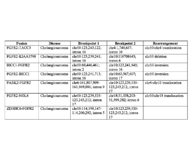
FIGs. 1A-1C are tables summarizing the fusion molecules and the
rearrangement events described herein.
Date Recue/Date Received 2020-06-25
CA 02898326 2015-07-15
WO 2014/113729 PCT/US2014/012136
FIG. 1A summarizes the following: the name of the fusion (referreu to as
"fusion"); the tissue source (referred to as "disease"); the approximate
locations of the
first and second breakpoints that give rise to the rearrangement events (+ 50
nucleotides) (referred to as "Breakpoint 1" and "Breakpoint 2," respectively);
and the
5 type of rearrangement (referred to as "rearrangement").
FIG. 1B summarizes the following: the name of the fusion (referred to as
"fusion"); the accession number of the full length sequences that contain the
5'- and
the 3'- exon sequences (referred to as "5' Transcript ID" and "3' Transcript
ID,"
respectively); and the identity of the exon(s) of the 5' transcript and the
exon(s) of the
10 3' transcript. The sequences corresponding to the accession numbers
provided in
FIG. 1B are set forth in the figures appended herein. Alternatively, the
sequences can
be found by searching the RefSeq Gene as databased at UCSC Genome Browser
(genome.ucsc.edu). For example, the following link can be used:
http://genome.ucsc.edu/cgi-bin/hgc?hgsid=359255927&c=chr10&o=123237843&t=
15 .. 123356159&g=refGene&i=NM_001144915 to search for Accession Number =
NM_001144915.
FIG. IC summarizes the following: the name of the fusion; the SEQ ID NOs.
of the 5' partner and the 3' partner; and the figure in which the sequence is
shown.
For example, the Nt and Aa sequences of FGFR2 have SEQ ID NOs: 1 and 2,
20 respectively, which are shown in FIGs. 2 and 3, respectively. The Nt and
Aa
sequences of TACC3 have SEQ ID NOs: 3 and 4, which are shown in FIGs. 4 and 5,
respectively.
FIGs. 2A-2B depict the nucleotide sequence of FGFR2 cDNA
(NM_001144915, SEQ ID NO: 1). The exon boundaries are shown in bold and
25 .. underlined. The start of the first exon and the end of the last exon are
shown by a
single underline (e.g., shown as A). Further exons (second, third, fourth and
so on)
are indicated consecutively from 5' to 3' orientation by the underline of two
consecutive nucleotides. For example, nucleotides GT at positions 169-170
correspond to the 3'-end of the first exon at position 0, and the 5'-start of
the second
30 exon is at position T. The start codon is shown in bold and italics. The
stop codon is
shown in italics and underlined.
FIG. 3 depicts the amino acid sequence of FGFR2 (SEQ ID NO: 2).
CA 02898326 2015-07-15
WO 2014/113729 PCT/US2014/012136
31
FIGs. 4A-4B depict the nucleotide sequence of TACC3 cDNA (Nimi_uuoi4z,
SEQ ID NO: 3). The exon boundaries are shown in bold and underlined. The start
of
the first exon is shown by a single underline. Further exons (second, third,
fourth) are
indicated consecutively from 5' to 3' orientation by the underline of two
consecutive
nucleotides (as exemplified in FIGs. 2A-2B above). The start codon is shown in
bold
and italics. The stop codon is shown in italics and underlined.
FIG. 5 depicts the amino acid sequence of TACC3 (SEQ ID NO: 4).
FIGs. 6A-6C depict the nucleotide sequence of KIAA1598 cDNA
(NM_001127211, SEQ ID NO: 5). The exon boundaries are shown in bold and
underlined. The start of the first exon is shown by a single underline.
Further exons
(second, third, fourth) are indicated consecutively from 5' to 3' orientation
by the
underline of two consecutive nucleotides (as exemplified in FIGs. 2A-2B
above). The
start codon is shown in bold and italics. The stop codon is shown in italics
and
underlined.
FIG. 7 depicts the amino acid sequence of KIAA1598 (SEQ ID NO: 6).
FIGs. 8A-8B depict the nucleotide sequence of BICC1 cDNA
(NM_001080512, SEQ ID NO: 7). The exon boundaries are shown in bold and
underlined. The start of the first exon is shown by a single underline.
Further exons
(second, third, fourth) are indicated consecutively from 5' to 3' orientation
by the
underline of two consecutive nucleotides (as exemplified in FIGs. 2A-2B
above). The
start codon is shown in bold and italics. The stop codon is shown in italics
and
underlined.
FIG. 9 depicts the amino acid sequence of BICC1 (SEQ ID NO: 8).
FIGs. 10A-10B depict the nucleotide sequence of PARK2 cDNA
(NM_004562, SEQ ID NO: 9). The exon boundaries are shown in bold and
underlined. The start of the first exon is shown by a single underline.
Further exons
(second, third, fourth) are indicated consecutively from 5' to 3' orientation
by the
underline of two consecutive nucleotides (as exemplified in FIGs. 2A-2B
above). The
start codon is shown in bold and italics. The stop codon is shown in italics
and
underlined.
FIG. 11 depicts the amino acid sequence of PARK2 (SEQ ID NO: 10).
CA 02898326 2015-07-15
WO 2014/113729 PCT/US2014/012136
32
FIGs. 12A-12B depict the nucleotide sequence of FGFR2 cDNA
(NM_000141, SEQ ID NO: II). The exon boundaries are shown in bold and
underlined. The start of the first exon is shown by a single underline.
Further exons
(second, third, fourth) are indicated consecutively from 5' to 3' orientation
by the
underline of two consecutive nucleotides (as exemplified in FIGs. 2A-2B
above). The
start codon is shown in bold and italics. The stop codon is shown in italics
and
underlined.
FIG. 13 depicts the amino acid sequence of FGFR2 (SEQ ID NO: 12).
FIGs. 14A-14B depict the nucleotide sequence of NOL4 cDNA (NM_003787,
SEQ ID NO: 13). The exon boundaries are shown in bold and underlined. The
start
of the first exon is shown by a single underline. Further exons (second,
third, fourth)
are indicated consecutively from 5' to 3' orientation by the underline of two
consecutive nucleotides (as exemplified in FIGs. 2A-2B above). The start codon
is
shown in bold and italics. The stop codon is shown in italics and underlined.
FIG. 15 depicts the amino acid sequence of NOL4 (SEQ ID NO: 14).
FIGs. 16 depicts the nucleotide sequence of ZDHHC6 cDNA (NM_022494,
SEQ ID NO: 15). The exon boundaries are shown in bold and underlined. The
start
of the first exon is shown by a single underline. Further exons (second,
third, fourth)
are indicated consecutively from 5' to 3' orientation by the underline of two
consecutive nucleotides (as exemplified in FIGs. 2A-2B above). The start codon
is
shown in bold and italics. The stop codon is shown in italics and underlined.
FIG. 17 depicts the amino acid sequence of ZDHHC6 (SEQ ID NO: 16).
DETAILED DESCRIPTION
Described herein are novel alterations, e.g., rearrangement events, found in
cholanaiocarcinomas. In certain embodiments, the rearrangement events are
found in
an FGFR2 gene or an NTRK gene, e.g., as exemplified in Table 1, FIGs. 1A-1C
and
FIGs. 2-17. In certain embodiments, the novel rearrangement events give rise
to
fusion molecules that includes a fragment of a first gene and a fragment of a
second
gene, e.g., a fusion that includes a 5'-exon and a 3' -exon summarized in
FIGs. 1A-1C
and FIGs. 2-17. The term "fusion" or "fusion molecule" is used generically
herein,
and includes any fusion molecule (e.g., gene, gene product (e.g., cDNA, tuRNA,
or
CA 02898326 2015-07-15
WO 2014/113729 PCT/US2014/012136
33
polypeptide), and variant thereof) that includes a fragment of first gene anu
a
fragment of second gene described herein, including, e.g., an FGFR2-TACC3,
EUFR2-KIAA1598, BICC1-FGFR2, FGFR2-BICC1, PARK2-FGFR2, FGER2-
NOL4, ZDHHC6-FGFR2, or RABGAP1L-N __ fRK1, e.g., as described in Table 1,
.. FIGs. 1A-1C and FIGs. 2-17. Expression of the fusion molecules was detected
in
cholangiocarcinomas, thus suggesting an association with neoplastic growth or
cancer
(including pre-malignant, or malignant and/ or metastatic growth).
Cholangiocarcinoma (also known as bile duct cancer) can arise from the
tissues in the bile duct. Cholangiocarcinoma can occur in any part of the bile
duct.
.. The part of the tube that is outside of the liver is called extrahepatic.
It is in this
portion of the bile duct where cancer usually arises. A perihilar cancer, also
called a
Klatskin tumor, begins where many small channels join into the bile duct at
the point
where it leaves the liver. About two-thirds of all cholangiocarcinomas occur
here.
Distal cholangiocarcinoma occurs at the opposite end of the duct from
perihilar
cancer, near where the bile duct empties into the small intestine. About one-
fourth of
all cholangiocarcinomas are distal cholangiocarcinomas. About 5% to 10% of
cholangiocarcinomas are intrahepatic, or inside the liver. Adenocarcinoma is
the most
common type of extrahepatic cholangiocarcinoma, and accounting for up to 95%
of
all cholangiocarcinomas. Adenocarcinoma is cancer arising from the mucus
glands
lining the inside of the bile duct. Cholangiocarcinoma is another term that
may be
used to describe this type of cancer.
Accordingly, the invention provides, at least in part, the following: methods
for
treating a cholangiocarcinoma using an inhibitor of one of the alterations
described
herein, e.g., an PGFR2 or an NTRK1 inhibitor; methods for identifying,
assessing or
detecting an alteration, e.g., fusion molecule as described herein; methods
for identifying,
assessing, evaluating, and/or treating a subject having a cancer, e.g., a
cholangiocarcinoma having a fusion molecule as described herein; isolated
fusion nucleic
acid molecules, nucleic acid constructs, host cells containing the nucleic
acid molecules;
purified fusion polypeptides and binding agents; detection reagents (e.g.,
probes, primers,
.. antibodies, kits, capable, e.g., of specific detection of a fusion nucleic
acid or protein);
screening assays for identifying molecules that interact with, e.g., inhibit,
the fusions,
e.g., novel kinase inhibitors; as well as assays and kits for evaluating,
identifying,
CA 02898326 2015-07-15
WO 2014/113729 PCT/US2014/012136
34
assessing and/or treating a subject having a cancer, e.g., a
cholangiocarcinonta naving a
fusion. The compositions and methods identified herein can be used, for
example, to
identify new inhibitors; to evaluate, identify or select a subject, e.g., a
patient, having a
cancer; and to treat or prevent a cancer, such as a cholangiocarcinoma.
Certain terms are defined. Additional terms are defined throughout the
specification.
As used herein, the articles "a" and "an" refer to one or to more than one
(e.g., to at
least one) of the grammatical object of the article.
The term "or" is used herein to mean, and is used interchangeably with, the
term
"and/or", unless context clearly indicates otherwise.
"About" and "approximately" shall generally mean an acceptable degree of error
for
the quantity measured given the nature or precision of the measurements.
Exemplary degrees
of error are within 20 percent (%), typically, within 10%, and more typically,
within 5% of a
given value or range of values.
"Acquire" or "acquiring" as the Willis are used herein, refer to obtaining
possession of a physical entity, or a value, e.g., a numerical value, by
"directly acquiring"
or "indirectly acquiring" the physical entity or value. "Directly acquiring"
means
performing a process (e.g., performing a synthetic or analytical method) to
obtain the
physical entity or value. "Indirectly acquiring" refers to receiving the
physical entity or
value from another party or source (e.g., a third party laboratory that
directly acquired the
physical entity or value). Directly acquiring a physical entity includes
perfoiming a
process that includes a physical change in a physical substance, e.g., a
starting material.
Exemplary changes include making a physical entity from two or more starting
materials,
shearing or fragmenting a substance, separating or purifying a substance,
combining two
or more separate entities into a mixture, perfoiming a chemical reaction that
includes
breaking or forming a covalent or non-covalent bond. Directly acquiring a
value includes
performing a process that includes a physical change in a sample or another
substance,
e.g., performing an analytical process which includes a physical change in a
substance,
e.g., a sample, analyte, or reagent (sometimes referred to herein as "physical
analysis"),
perfoiming an analytical method, e.g., a method which includes one or more of
the
following: separating or purifying a substance, e.g., an analyte, or a
fragment or other
CA 02898326 2015-07-15
WO 2014/113729 PCT/US2014/012136
derivative thereof, from another substance; combining an analyte, or fragment
or outer
derivative thereof, with another substance, e.g., a buffer, solvent, or
reactant; or changing
the structure of an analyte, or a fragment or other derivative thereof, e.g.,
by breaking or
forming a covalent or non-covalent bond, between a first and a second atom of
the
5 analyte; or by changing the structure of a reagent, or a fragment or
other derivative
thereof, e.g., by breaking or forming a covalent or non-covalent bond, between
a first and
a second atom of the reagent.
"Acquiring a sequence" as the teim is used herein, refers to obtaining
possession
of a nucleotide sequence or amino acid sequence, by "directly acquiring" or
"indirectly
10 acquiring" the sequence. "Directly acquiring a sequence" means
performing a process
(e.g., performing a synthetic or analytical method) to obtain the sequence,
such as
perfoiming a sequencing method (e.g., a Next Generation Sequencing (NGS)
method).
"Indirectly acquiring a sequence" refers to receiving infoimation or knowledge
of, or
receiving, the sequence from another party or source (e.g., a third party
laboratory that
15 directly acquired the sequence). The sequence acquired need not be a
full sequence, e.g.,
sequencing of at least one nucleotide, or obtaining information or knowledge,
that
identifies a fusion disclosed herein as being present in a subject constitutes
acquiring a
sequence.
Directly acquiring a sequence includes performing a process that includes a
20 physical change in a physical substance, e.g., a starting material, such
as a tissue sample,
e.g., a biopsy, or an isolated nucleic acid (e.g., DNA or RNA) sample.
Exemplary
changes include making a physical entity from two or more starting materials,
shearing or
fragmenting a substance, such as a genomic DNA fragment; separating or
purifying a
substance (e.g., isolating a nucleic acid sample from a tissue); combining two
or more
25 separate entities into a mixture, performing a chemical reaction that
includes breaking or
forming a covalent or non-covalent bond. Directly acquiring a value includes
performing
a process that includes a physical change in a sample or another substance as
described
above.
"Acquiring a sample" as the term is used herein, refers to obtaining
possession of
30 a sample, e.g., a tissue sample or nucleic acid sample, by "directly
acquiring" or
"indirectly acquiring" the sample. "Directly acquiring a sample" means
perfoiming a
process (e.g., performing a physical method such as a surgery or extraction)
to obtain the
CA 02898326 2015-07-15
WO 2014/113729 PCT/US2014/012136
36
sample. "Indirectly acquiring a sample" refers to receiving the sample from
anomer party
or source (e.g., a third party laboratory that directly acquired the sample).
Directly
acquiring a sample includes performing a process that includes a physical
change in a
physical substance, e.g., a starting material, such as a tissue, e.g., a
tissue in a human
patient or a tissue that has was previously isolated from a patient. Exemplary
changes
include making a physical entity from a starting material, dissecting or
scraping a tissue;
separating or purifying a substance (e.g., a sample tissue or a nucleic acid
sample);
combining two or more separate entities into a mixture; perfoiming a chemical
reaction
that includes breaking or fonning a covalent or non-covalent bond. Directly
acquiring a
to sample includes performing a process that includes a physical change in
a sample or
another substance, e.g., as described above.
"Binding entity" means any molecule to which molecular tags can be directly or
indirectly attached that is capable of specifically binding to an analyte. The
binding
entity can be an affinity tag on a nucleic acid sequence. In certain
embodiments, the
binding entity allows for separation of the nucleic acid from a mixture, such
as an avidin
molecule, or an antibody that binds to the hapten or an antigen-binding
fragment thereof.
Exemplary binding entities include, but are not limited to, a biotin molecule,
a hapten, an
antibody, an antibody binding fragment, a peptide, and a protein.
"Complementary" refers to sequence complementarity between regions of two
nucleic acid strands or between two regions of the same nucleic acid strand.
It is known
that an adenine residue of a first nucleic acid region is capable of forming
specific
hydrogen bonds ("base pairing") with a residue of a second nucleic acid region
which is
antiparallel to the first region if the residue is thymine or uracil.
Similarly, it is known
that a cytosine residue of a first nucleic acid strand is capable of base
pairing with a
residue of a second nucleic acid strand which is antiparallel to the first
strand if the
residue is guanine. A first region of a nucleic acid is complementary to a
second region
of the same or a different nucleic acid if, when the two regions are arranged
in an
antiparallel fashion, at least one nucleotide residue of the first region is
capable of base
pairing with a residue of the second region. In certain embodiments, the first
region
comprises a first portion and the second region comprises a second portion,
whereby,
when the first and second portions are arranged in an antiparallel fashion, at
least about
50%, at least about 75%, at least about 90%, or at least about 95% of the
nucleotide
CA 02898326 2015-07-15
WO 2014/113729 PCT/US2014/012136
37
residues of the first portion are capable of base pairing with nucleotide
resiuues in me
second portion. In other embodiments, all nucleotide residues of the first
portion are
capable of base pairing with nucleotide residues in the second portion.
The term "cancer" or "tumor" is used interchangeably herein. These teims refer
to the presence of cells possessing characteristics typical of cancer-causing
cells, such as
uncontrolled proliferation, immortality, metastatic potential, rapid growth
and
proliferation rate, and certain characteristic morphological features. In one
embodiment,
the cancer is a cholangiocarcinoma.
The term "neoplasm" or "neoplastic" cell refers to an abnormal proliferative
stage, e.g., a hyperproliferative stage, in a cell or tissue that can include
a benign, pre-
malignant, malignant (cancer) or metastatic stage.
Cancer is "inhibited" if at least one symptom of the cancer is alleviated,
terminated, slowed, or prevented. As used herein, cancer is also "inhibited"
if
recurrence or metastasis of the cancer is reduced, slowed, delayed, or
prevented.
"Chemotherapeutic agent" means a chemical substance, such as a cytotoxic or
cytostatic agent, that is used to treat a condition, particularly cancer.
As used herein, "cancer therapy" and "cancer treatment" are synonymous
terms.
As used herein, "chemotherapy" and "chemotherapeutic" and
"chemotherapeutic agent" are synonymous terms.
The terms "homology" or "identity," as used interchangeably herein, refer to
sequence similarity between two polynucleotide sequences or between two
polypeptide sequences, with identity being a more strict comparison. The
phrases
"percent identity or homology" and "% identity or homology" refer to the
percentage
of sequence similarity found in a comparison of two or more polynucleotide
sequences or two or more polypeptide sequences. "Sequence similarity" refers
to the
percent similarity in base pair sequence (as determined by any suitable
method)
between two or more polynucleotide sequences. Two or more sequences can be
anywhere from 0-100% similar, or any integer value there between. Identity or
similarity can be determined by comparing a position in each sequence that can
be
aligned for purposes of comparison. When a position in the compared sequence
is
occupied by the same nucleotide base or amino acid, then the molecules are
identical
CA 02898326 2015-07-15
WO 2014/113729 PCT/US2014/012136
38
at that position. A degree of similarity or identity between polynucleotide
sequences
is a function of the number of identical or matching nucleotides at positions
shared by
the polynucleotide sequences. A degree of identity of polypeptide sequences is
a
function of the number of identical amino acids at positions shared by the
polypeptide
sequences. A degree of homology or similarity of polypeptide sequences is a
function
of the number of amino acids at positions shared by the polypeptide sequences.
The
term "substantially identical," as used herein, refers to an identity or
homology of at
least 75%, at least 80%, at least 85%, at least 90%, 91%, 92%, 93%, 94%, 95%,
96%,
97%, 98%, 99%, or more.
"Likely to" or "increased likelihood," as used herein, refers to an increased
probability that an item, object, thing or person will occur. Thus, in one
example, a
subject that is likely to respond to treatment with a kinase inhibitor, alone
or in
combination, has an increased probability of responding to treatment with the
inhibitor alone or in combination, relative to a reference subject or group of
subjects.
"Unlikely to" refers to a decreased probability that an event, item, object,
thing
or person will occur with respect to a reference. Thus, a subject that is
unlikely to
respond to treatment with a kinase inhibitor, alone or in combination, has a
decreased
probability of responding to treatment with a kinase inhibitor, alone or in
combination, relative to a reference subject or group of subjects.
"Sequencing" a nucleic acid molecule requires determining the identity of at
least
1 nucleotide in the molecule. In embodiments, the identity of less than all of
the
nucleotides in a molecule are determined. In other embodiments, the identity
of a
majority or all of the nucleotides in the molecule is determined.
"Next-generation sequencing or NOS or NO sequencing" as used herein, refers to
any sequencing method that determines the nucleotide sequence of either
individual
nucleic acid molecules (e.g., in single molecule sequencing) or clonally
expanded proxies
for individual nucleic acid molecules in a highly parallel fashion (e.g.,
greater than 105
molecules are sequenced simultaneously). In one embodiment, the relative
abundance of
the nucleic acid species in the library can be estimated by counting the
relative number of
.. occurrences of their cognate sequences in the data generated by the
sequencing
experiment. Next generation sequencing methods are known in the art, and are
described, e.g., in Metzker, M. (2010) Nature Biotechnology Reviews 11:31-46,
81789818
39
Next generation sequencing can detect a variant present in less than 5%
of the nucleic acids in a sample.
"Sample," "tissue sample," "patient sample," "patient cell or tissue sample"
or
"specimen" each refers to a collection of similar cells obtained from a tissue
of a subject
or patient. The source of the tissue sample can be solid tissue as from a
fresh, frozen
and/or preserved organ, tissue sample, biopsy, or aspirate; blood or any blood
constituents; bodily fluids such as cerebral spinal fluid, amniotic fluid,
peritoneal fluid or
interstitial fluid; or cells from any time in gestation or development of the
subject. The
tissue sample can contain compounds that are not naturally intermixed with the
tissue in
nature such as preservatives, anticoagulants, buffers, fixatives, nutrients,
antibiotics or the
like. In one embodiment, the sample is preserved as a frozen sample or as
formaldehyde-
or parafonnaldehyde-fixed paraffin-embedded (FFPE) tissue preparation. For
example,
the sample can be embedded in a matrix, e.g., an 1411)E block or a frozen
sample.
A "tumor nucleic acid sample" as used herein, refers to nucleic acid molecules
from a tumor or cancer sample. Typically, it is DNA, e.g., genomic DNA, or
cDNA
derived from RNA, from a tumor or cancer sample. In certain embodiments, the
tumor
nucleic acid sample is purified or isolated (e.g., it is removed from its
natural state).
A "control" or "reference" "nucleic acid sample" as used herein, refers to
nucleic
acid molecules from a control or reference sample. Typically, it is DNA, e.g.,
genomic
.. DNA, or cDNA derived from RNA, not containing the alteration or variation
in the gene
or gene product, e.g., not containing a fusion. In certain embodiments, the
reference or
control nucleic acid sample is a wild type or a non-mutated sequence. In
certain
embodiments, the reference nucleic acid sample is purified or isolated (e.g.,
it is removed
from its natural state). In other embodiments, the reference nucleic acid
sample is from a
non-tumor sample, e.g., a blood control, a normal adjacent tumor (NAT), or any
other
non-cancerous sample from the same or a different subject.
"Adjacent to the interrogation position," as used herein, means that a site
sufficiently close such that a detection reagent complementary with the site
can be
used to distinguish between a mutation, e.g., a mutation described herein, and
a
reference sequence, e.g., a non-mutant or wild-type sequence, in a target
nucleic acid.
Directly adjacent, as used herein, is where 2 nucleotides have no intervening
nucleotides between them.
Date Recue/Date Received 2020-06-25
CA 02898326 2015-07-15
WO 2014/113729 PCT/US2014/012136
"Associated mutation," as used herein, refers to a mutation within a
preselected distance, in terms of nucleotide or primary amino acid sequence,
from a
definitional mutation, e.g., a mutant as described herein, e.g., a
translocation,
breakpoint or fusion molecule described herein. In embodiments, the associated
5 mutation is within n, wherein n is 2, 5, 10, 20, 30, 50, 100, or 200
nucleotides from
the definitional mutation (n does not include the nucleotides defining the
associated
and definitional mutations). In embodiments, the associated mutation is a
translocation mutation.
"Interrogation position," as used herein, comprises at least one nucleotide
(or,
10 in the case of polypeptides, an amino acid residue) which corresponds to
a nucleotide
(or amino acid residue) that is mutated in a mutation of interest, e.g., a
mutation being
identified, or in a nucleic acid (or protein) being analyzed, e.g., sequenced,
or
recovered.
A "reference sequence," as used herein, e.g., as a comparator for a mutant
15 sequence, is a sequence which has a different nucleotide or amino acid
at an
interrogation position than does the mutant(s) being analyzed. In an
embodiment, the
reference sequence is wild-type for at least the interrogation position.
Headings, e.g., (a), (b), (i) etc, are presented merely for ease of reading
the
specification and claims. The use of headings in the specification or claims
does not
20 require the steps or elements be performed in alphabetical or numerical
order or the order
in which they are presented.
Various aspects featured in the invention are described in further detail
below.
Additional definitions are set out throughout the specification.
25 .. FGFR2 and NTRK1 Alterations
Described herein are novel rearrangements of the FGFR2 and NTRK1 genes
in cholangiocarcinomas.
FGFR2 Alterations
30 The FGFR family plays an important role in cell differentiation, growth
and
angiogenesis (reviewed in Powers etal. (2000), Endocr. Relat. Cancer, 7(3):165-
197,
and gain of function mutations in FGFRs have been reported in several cancer
types
CA 02898326 2015-07-15
WO 2014/113729 PCT/US2014/012136
41
(reviewed in Eswarakumar etal. (2005), Cytokine Growth Factor Rev., lov):1.-
149).
FGFR2 (Fibroblast growth factor receptor 2) is a member of the fibroblast
growth factor receptor family, where amino acid sequence is highly conserved
between members and throughout evolution. FGFR family members differ from one
another in their ligand affinities and tissue distribution. A full-length
representative
protein consists of an extracellular region, composed of three immunoglobulin-
like
domains, a single hydrophobic membrane-spanning segment and a cytoplasmic
tyrosine kinase domain. FGFR2 is composed of three immunoglobulin c-2 type
domains, one transmembrane domain, and one tyrosine kinase catalytic domain.
The
extracellular portion of the protein interacts with fibroblast growth factors,
setting in
motion a cascade of downstream signals, ultimately influencing mitogenesis and
differentiation. This particular family member is a high-affinity receptor for
acidic,
basic and/or keratinocyte growth factor, depending on the isoform. Multiple
alternatively spliced transcript variants encoding different isoforms have
been noted
for the FGFR2 gene. "lhe FGFR2 amino and nucleotide sequences are known in the
art. Exemplary sequences for human FGFR2 are provided herein as SEQ ID NOs:1
and 11, and FIGs. 2 and 12 (nucleotide), and SEQ ID NOs:2 and 12, and FIGs. 3
and
13 (amino acid).
FGFR2 amplification has been reported in several cancer types, most
frequently in gastric cancer (3-4%) (Matsumoto etal., 2012, Br. J. Cancer,
106(4):727-732, Hara et al., 1998, Lab Invest., 78(9):1143-1153) and breast
cancer (1-
11%) (Heiskanen etal., 2001, Anal Cell Pathol. 22(4):229-234, Adnane etal.,
1991;
Oncogene 6(4):659-663, Turner etal., 2010, Oncogene 29(14):2013-2023). FG1,R2
has been shown to be expressed in cholangiocarcinoma, leading to activation of
the
MEK1/2 pathway (Narong etal., 2011, Oncol. Lett. 2(5):821-825). The FGFR2
alterations described herein are expected to result in activation and/or
upregulation of
the FGFR2 protein. Accordingly, treatment with an agent that reduces (e.g.,
inhibits)
FGFR2 is encompassed by the invention. In one embodiment, the agent is
Regorafenib. Regorafenib inhibits cellular kinases including FGFR2, and has
been
approved for treatment of some metastatic colorectal cancer (mCRC) patients
(PDA.gov, Nov 2012). The multi-kinase inhibitor ponatinib (AP24534), recently
CA 02898326 2015-07-15
WO 2014/113729 PCT/US2014/012136
42
approved by the FDA for use in chronic myelogenous leukemia based on me
results
of a Phase 2 trial, has also been shown in preclinical studies to have
substantial
activity against all four FUER kinases (Cortes et al., 2012, American Society
of
Hematology ASH, Abstract 163, Gozgit et al., 2012, Mol. Cancer Ther.,
11(3):690-
699). Clinical trials of multiple Fgfr inhibitors are currently underway
(Turner and
Grose, 2010, Nat. Rev. Cancer, 10(2):116-129).
Each of the FGFR2 alterations is described herein in more detail
FGFR2-TACC3
The FGFR2-TACC3 fusion has not been reported. However, similar FGFR3-
TACC3 fusions have been previously reported in glioblastoma and in a bladder
cancer
cell line; these fusions were found to be activating and to have
transformative
potential (Williams etal., Hum. Mot. Genet ePub, Dec. 2012, Singh et al.,
2012,
Science 337(6099):1231-1235). The FGFR2-TACC3 fusion is therefore expected to
be oncogenic. FGFR2 amplification has also been reported in several cancer
types,
most frequently in gastric cancer and breast cancer as described herein.
Inhibitors of
FGFR2, such as Regorafenib and ponatinib can be used to treat
cholangiosarcoma.
In one embodiment, the rearrangement, nucleotide and amino acid sequences
for FGFR2 (exons 1-16)-TACC3 (exons 11-16) are depicted in FIGs. 1A-5 and SEQ
ID NOs. 1-4.
FGFR2-KIAA1598
The FGFR2-KIA A1598 rearrangement results in truncation of the 31TTR of
the FGFR2 gene, which can result in upregulation of the FGFR2 protein. FGFR2
amplification has also been reported in several cancer types, most frequently
in gastric
cancer and breast cancer as described herein. Inhibitors of FGFR2, such as
Regorafenib and ponatinib can be used to treat cholangiosarcoma.
In one embodiment, the rearrangement, nucleotide and amino acid sequences
for FGFR2 (exons 1-16)-KIAA1598 (exons 7-17) are depicted in FIGs. 1A-1C, 2-3
and 6-7 and SEQ ID NOs. 1-2 and 5-6.
CA 02898326 2015-07-15
WO 2014/113729 PCT/US2014/012136
43
B1CC1-FGFR2
The BICC1-FGFR2 fusion has not been reported in cholangiocarcinoma, or
other cancers. FGFR2 amplification has also been reported in several cancer
types,
most frequently in gastric cancer and breast cancer as described herein.
Treatment
Inhibitors of FGFR2, such as Regorafenib and ponatinib can be used to treat
cholangiosarcoma.
In one embodiment, the rearrangement, nucleotide and amino acid sequences
for BICC I (exons I-2)-FGFR2 (exon 17) are depicted in FIGs. 1A-1C, 2-3 and 8-
9
and SEQ ID NOs. 1-2 and 7-8.
FGFR2-BICC1
The FGFR2-BICC1 result in an in-frame fusion including the N-terminal
portion of FGFR2 (containing the kinase domain) nearly the entire coding
sequence of
BICC1 (Garcia-Mayoral et al., 2007, Structure 15(4):485-498, Kim and Bowie,
2003,
Trends Biochem. Sci. 28(12):625-628). Other in-frame fusions containing the
kinase
domain of FGFR2 have been shown to result in kinase activation (Singh et al.,
2012,
Science 337(6099):1231-1235, Lorenzi et al., 1996, Proc. Natl. Acad. Sci. USA,
93(17):8956-8961). A recent report has described an FGFR2 fusion gene in
cholangiocarcinoma (Wu et al. Cancer Discov ePub, May 2013). FGFR2 mutations
have been reported in 2% of tumors analyzed in COSMIC, with the highest
prevalence in endometrial cancer (10%) and lower incidence in several other
cancers
(COSMIC, Feb 2013). F GER2 signaling has been described as tumorigenic in
lung,
pancreatic and gastric cancers (Yamayoshi et al., 2004, J. Pathol., 204(1):110-
118;
Cho et al., 2007, Am. J. Pathol., 170(6):1964-1974; Toyokawa et al., 2009,
Omni.
Rep., 21(4):875-880). However, FGFR2 has also been described as a tumor
suppressor in the context of other cancers, such as melanoma (Gartside et al.,
2009,
Mol. Cancer Res., 7(1):41-54). Clinical trials of multiple FGFR inhibitors are
currently underway (Turner and Grose, 2010, Oncogene, 29(14):2013-2023).
Inhibitors of FGFR2, such as Regorafenib and ponatinib can be used to treat
cholangiosarcoma.
CA 02898326 2015-07-15
WO 2014/113729 PCT/US2014/012136
44
In one embodiment, the rearrangement, nucleotide and amino acid sequences
for FGFR2 (exons 1-16)-BICC1 (exons 18-21) are depicted in FIGs. 1A-1C, 2-3
and
8-9 and SEQ ID NOs. 1-2 and 7-8.
PARK2-FGFR2
The PARK2-FGFR2 fusion results in a fusion that includes the N-terminal
portion of PARK2, which encodes the E3 ligase parkin, and the last exon (aa
768-
821) of FGFR2 (Uniprot). The portion of FGFR2 not included in this fusion is
predicted to encode a protein truncated after the functional kinase domain.
Similar
1() truncations of FGFR2 (764* and 776*) have been described as oncogenic,
efficiently
transfoi __ ming cultured cells (Lorenzi et al., 1997, Oncogene 15(7):817-26).
Therefore,
this fusion is expected to activate the FGFR2 signaling.
FGFR2 amplification has also been reported in several cancer types, most
frequently in gastric cancer and breast cancer as described herein. Treatment
Inhibitors of FGFR2, such as Regorafenib and ponatinib can be used to treat
cholangiosarcoma.
In one embodiment, the rearrangement, nucleotide and amino acid sequences
for PARK2 (exons 1-9)-FGFR2 (exon 18) are depicted in FIGs. 1A-1C and 10-13
and
SEQ ID NOs. 9-12.
In one embodiment, the rearrangement comprises a fusion of PARK2 (intron9)
to FGFR2 (intron17). The expected genomic coordinates are:
= FGFR2 breakpoint: chr10:123239535-123243212.
= PARK2 breakpoint: chr6:161807909-161969886.
The fusion is comprised of 10 complete exons, all coming from the reverse
strand. The fusion is in frame. The orientation of the fusion is expected to
be 5'
fusion partner exons: PARK2 (exons1-9) to 3' fusion partner exons: FGFR2
(exon18).
The fused domains include:
(i) PARK2, E3 ubiquitin-protein ligase parkin, has one ubiquitin homologue
domain and two zink finger domains. The fusion, which includes exons 1-9 of
PARK2 contains the entire ubiquitin homologue domain and part of the first
zink
finger domain, which are the core set of exons to give reasonable activity;
and
(ii) FGFR2, the fusion includes the last exon of FGFR2.
CA 02898326 2015-07-15
WO 2014/113729 PCT/US2014/012136
The refSeq IDs for the nucleotide and amino acid sequences are:
= PARK2: NM_004562 provided herein as SEQ ID NOs: 9-10 and FIGs. 10-
11, respectively.
= FGFR2: NM_000141 provided herein as SEQ ID NOs: 11-12 and FIGs.
5 12-13, respectively.
FGFR2-NOL4
The FGFR2-NOL4 fusion results in an in-frame fusion, containing transcribed
exons 1-17 of FGFR2 (coding for amino acids 1-768) fused to NOL4 transcribed
10 exons 7-11 (coding for amino acids 353-638). The resulting fusion
protein contains
the N-terminus of FGFR2, which includes the protein kinase domain, fused to
the C-
terminus of the NOL4 protein (UniProt.org). FGFR2-involving fusions containing
the
FGFR2 kinase domain have been reported to be activating and oncogenic,
including
FGFR-TACC and FGFR2-ERAG1 (Singh et al., 2012, Science 337(6099):1231-1235,
15 Lorenzi et al., 1996, Proc. Natl. Acad. Sci. USA, 93(17):8956-8961).
FGFR2 mRNA has been shown to be expressed in cholangiocarcinoma cell lines,
leading to activation of the MEK1/2 pathway (Narong and Leelawat, 2011,
supra).
Tumors with FGFR2 amplification or activating mutations can be sensitive to
FUER
inhibitors as described herein. FGFR2 has been associated with resistance to
20 chemotherapeutics; shRNA inhibition of FGFR2 increased the sensitivity
of ovarian
epithelial cancer cells to cisplatin (Cole et al., 2010, Cancer Biol Ther
10(5):495-
504). Inhibitors of FGFR2, such as Regorafenib and ponatinib can be used to
treat
cholangiosarcoma.
In one embodiment, the rearrangement, nucleotide and amino acid sequences
25 for FGFR2 (exons 1-17) and NOL4 (exons 7-11) are depicted in FIGs. 1A-1C
and 12-
15 and SEQ ID NOs. 11-14.
In one embodiment, the rearrangement comprises a fusion of FGFR2
(intron17) to NOL4 (intron 6). The expected genomic coordinates are:
= FGFR2 breakpoint: chr10:123239535-123243212.
30 = NOL4 breakpoint: chr18:31538203-31599282.
The fusion is comprised of 22 complete exons, all coining from the reverse
strand. The fusion is in frame.
CA 02898326 2015-07-15
WO 2014/113729 PCT/US2014/012136
46
The orientation of the fusion is expected to be 5' fusion partner exons:
yuricz
(exonsl-17) to 3' fusion partner exons: NOL4 (exons7-11).
The fused domains include:
FGFR2, the fusion includes the core set of exons for all active domains of
this
transmembrane protein.
The refSeq IDs for the nucleotide and amino acid sequences are:
= FGFR2: NM_000141 provided herein as SEQ ID NOs: 11-12 and FIGs.
12-13, respectively.
= NOL4: NM_003787 provided herein as SEQ ID NOs: 13-14 and FIGs. 14-
15, respectively.
ZDHHC6-FGFR2
The ZDHHC6-FGFR2 fusion results in a fusion including the N-temiinal
portion of ZDHHC6 (exons 1-5), which encodes the integral transmembrane domain
of a palmitoyltransferase ZDHHC6, and the last exon (aa 768-821) of FGFR2
(Uniprot). The portion of FGFR2 not included in this fusion is predicted to
encode a
protein truncated after the functional kinase domain. Similar truncations of
FGFR2
(764* and 776*) have been described as oncogenic, efficiently transforming
cultured
cells (Lorenzi et al., 1997, Oncogene 15(7):817-26). Therefore, the ZDHHC6 -
FGFR2 fusion is predicted to activate FGFR2 signaling by truncating the
remaining
FGFR2 allele. A recent report has described an FGFR2 fusion gene in
cholangiocarcinoma, as well as a truncated FGFR2 similar to the one observed
here in
a patient with prostate cancer (Wu et al. Cancer Discov ePub, May 2013).
Inhibitors
of FGFR2, such as Regorafenib and ponatinib can be used to treat
cholangiosarcoma.
In one embodiment, the rearrangement, nucleotide and amino acid sequences
for ZDHHC6 (exons 1-5) and FGFR2 (exon 18) are depicted in FIGs. 1A-1C and 12-
13 and 16-17 and SEQ ID NOs. 11-12 and 15-16.
In one embodiment, the rearrangement comprises a fusion of ZDHHC6
(intron5) to FGFR2 (intron17). The expected genomic coordinates are:
= FGFR2 breakpoint: chr10:123239535-123243212
= ZDHHC6 breakpoint: chrl 0:114198147-114200292
PCT/US14/12136 18-11-2014
PCT/US2014/012136 18.03.2015
CA 02898326 2015-07-15
F2036-7046W0
The fusion is comprised of 6 complete exons, all coming from the reverse
strand. The fusion is in frame.
The orientation of the fusion is expected to be 5' fusion partner exons:
ZDHHC6 (exons1-5) to 3' fusion partner exons: FGFR2 (exon18).
The fused domains include:
(i) ZDHHC6 is a zinc-finger involved in transferase activity, transferring
acyl
groups and zinc ion binding. It contains 4 potential transmembrane domains and
one
zinc finger domain. All of these domains are contained within the first 5
exons, and
therefore retained in the fusion product; and
(ii) FGFR2, the fusion includes the last exon of FGFR2.
The annotations above are based on the following refSeq IDs
= ZDHHC6: NM_022494 provided herein as SEQ ID NOs: 15-16 and FIGs.
16-17, respectively.
= FGFR2: NM_000141 provided herein as SEQ ID NOs: 11-12 and FIGs.
12-13, respectively.
RABGAP1L-NTRK1
NTRK1 (Neurotrophic Tyrosine Kinase, Receptor, Type 1) is a member of the
neurotrophic tyrosine kinase receptor (NTKR) family. This kinase is a membrane-
bound receptor that, upon neurotrophin binding, phosphorylates itself and
members of
the MAPK pathway. The presence of this kinase leads to cell differentiation
and may
play a role in specifying sensory neuron subtypes. Mutations in this gene have
been
associated with congenital insensitivity to pain, anhidrosis, self-mutilating
behavior,
mental retardation and cancer. Alternate transcriptional splice variants of
this gene
have been found. The NTRK1 amino and nucleotide sequences are known in the
art.
An exemplary amino acid and nucleotide sequence for human NTRK1 are provided
herein as SEQ ID NO:17 and SEQ ID NO:18, respectively.
NCBI Reference Sequence: NP_00101233 1
1 mlrggrrgql gwhswaagpg sllawlilas agaapcpdac cphgssglrc trdgaldslh
61 hlpgaenite lyienqqhlq hlelrdlrgl gelrnitivk sglrfvapda fhftprlsrl
121 nlsfnalesl swktvggls1 gelvlsgnpl hcscalrwlq rweeeglggv peqklqchgq
181 gplahmpnas cgvptikvqv pnasvdvgdd vllrcqvegr gleqagwilt eleqsatvmk
241 sgglpslglt lanvtsdlnr knvtcwaend vgraevsvqv nvsfpasvql htavemhhwc
47
AMENDED SHEET -1PEATUS
PCT/US14/12136 18-11-2014
PCT/US2014/012136 18.03.2015
CA 02898326 2015-07-15
F2036-7046W0
301 ipfsvdggpa pslrwlfngs vinetsfift eflepaanet vrhgclrinq pthvnngnyt
361 llaanpfgqa sasimaafmd npfefnpedp ipdtnstsgd pvekkdetpf gvsvavglav
421 faclflstll lvinkcgrrn kfginrpavl apedglamsl hfmtlggssl sptegkgsgl
481 qghiienpery fsdacvhhik rrdivlkwel gegafgkvfl aechnllpeq dkmlvavkal
541 keasesarqd fqreaelltm lqhqhivrff gvctegrpll mvfeymrhgd lnrflrshgp
601 dakllagged vapgplglgq llavasqvaa gmvylaglhf vhrdlatrnc lvgqglvvki
661 gdfgmsrdiy stdyyrvggr tmlpirwmpp esilyrkftt esdvwsfgvv lweiftygkq
721 pwyglsntea idcitqgrel erpracppev yaimrgcwqr epqqrhsikd vharlqalaq
781 appvyldvlg
(SEQ NO: 17)
NCBI Reference Sequence: NM_001012331
1 tgcagctggg agcgcacaga cggctgcccc gcctgagcga ggcgggcgcc gccgcgatgc
61 tgcgaggcgg acggcgeggg cagcttggct ggcacagctg ggctgcgggg ccgggcagcc
121 tgctggcttg gctgatactg gcatctgcgg gcgccgcacc ctgccccgat gcctgctgcc
181 cccacggctc ctcgggactg cgatgcaccc gggatggggc cctggatagc ctccaccacc
241 tgcccggcgc agagaacctg actgagctct acatcgagaa ccaggagcat ctgcagcatc
301 tggagctccg tgatctgagg.ggcctggggg agctgagaaa cctcaccatc gtgaagagtg
361 gtctccgttt cgtggcgcca gatgecttcc atttcactcc tcggctcagt cgcctgaatc
421 tctccttcaa cgctctggag tctctetcct ggaaaactgt gcagggcctc tccttacagg
481 aactggtcct gtcggggaac cctctgcact gttcttgtgc cctgcgctgg ctacagcgct
541 gggaggagga gggactgggc ggagtgcctg aacagaagct gcagtgtcat gggcaagggc
601 ccctggccca catgcccaat gccagctgtg gtgtgcccac gctgaaggtc caggtgccca
661 atgcctcggt ggatgtgggg gacgacgtgc tgctgcggtg ccaggtggag gggcggggcc
721 tggagcaggc cggctggatc ctcacagagc tggagcagtc agccacggtg atgaaatctg
781 ggggtctgcc atccctgggg ctgaccctgg ccaatgtcac cagtgacctc aacaggaaga
841 acgtgacgtg ctgggcagag aacgatgtgg gccgggcaga ggtctctgtt caggtcaacg
901 tctccttccc ggccagtgtg cagctgcaca cggcggtgga gatgcaccac tggtgcatcc
961 ccttctctgt ggatgggcag ccggcaccgt ctctgcgctg gctcttcaat ggctccgtgc
1021 tcaatgagac cagcttcatc ttcactgagt tcctggagcc ggcagccaat gagaccgtgc
1081 ggcacgggtg tctgcgcctc aaccagccca cccacgtcaa caacggcaac tacacgctgc
1141 tggctgccaa ccccttcggc caggcctccg cctccatcat ggctgccttc atggacaacc
1201 ctttcgagtt caaccccgag gaccccatcc ctgacactaa cagcacatct ggagacccgg
1261 tggagaagaa ggacgaaaca ccttttgggg tctcggtggc tgtgggcctg gccgtctttg
1321 cctgcctctt cctttctacg ctgctccttg tgctcaacaa atgtggacgg agaaacaagt
1381 ttgggatcaa ccgcccggct gtgctggctc cagaggatgg gctggccatg tccctgcatt
1441 tcatgacatt gggtggcagc tecctgtecc ccaccgaggg caaaggctct gggctecaag
1501 gccacatcat cgagaaccca caatacttca gtgatgcctg tgttcaccac atcaagcgcc
1561 gggacatcgt gctcaagtgg gagctggggg agggcgcctt tgggaaggtc ttccttgctg
1621 agtgccacaa cctcctgcct gagcaggaca agatgctggt ggctgtcaag gcactgaagg
1681 aggagtccga gagtgctcgg caggacttcc agcgtgaggc tgagctgctc accatgctgc
1741 agcaccagca catcgtgcgc ttcttcggeg tctgcaccga gggccgcccc ctgctcatgg
1801 tctttgagta tatgcggcac ggggacctca accgcttcct ccgatcccat ggacctgatg
1861 ccaagctgct ggctggtggg gaggatgtgg ctccaggccc cctgggtctg gggcagctgc
1921 tggccgtggc tagccaggtc gctgcgggga tggtgtacct ggcgggtctg cattttgtgc
1981 accgggacct ggccacacgc aactgtctag tgggccaggg actggtggtc aagattggtg
2041 attttggcat gagcagggat atctacagca ccgactatta ccgtgtggga ggccgcacca
2101 tgctgcccat tcgctggatg ccgcccgaga gcatcctgta ccgtaagttc accaccgaga
2161 gcgacgtgtg gagcttcggc gtggtgctct gggagatctt cacctacggc aaggaggcct
2221 ggtaccagct ctccaacacg gaggcaatcg actgcatcac gcagggacgt gagttggagc
2281 ggccacgtgc ctgcccacca gaggtctacg ccatcatgcg gggctgctgg cagcgggagc
2341 cccagcaacg ccacagcatc aaggatgtgc acgcccggct gcaagccetg gcccaggcac
2401 ctcctgtcta cctggatgtc ctgggctagg gggccggccc aggggctggg agtggttagc
2461 cggaatactg gggcctgccc tcagcatccc ccatagctcc cagcagcccc agggtgatct
2521 caaagtatct aattcaccct cagcatgtgg gaagggacag gtgggggctg ggagtagagg
2581 atgttcctgc ttctctaggc aaggtcccgt catagcaatt atatttatta tcccttgaaa
43
AMENDED SHEET - MENUS
PCT/US14/12136 18-11-2014
PCT/US2014/012136 18.03.2015
CA 02898326 2015-07-15
F2036-7046W0
2641 aaaaaaa
(SEQ ID NO: 18)
Therapeutic Methods and Agents
The invention features methods of treating a cholangiocarcinoma, e.g., a
cholangiocarcinoma harboring a fusion described herein. The methods include
administering a therapeutic agent, e.g., which antagonizes the function of
FGFR2 or
NTRK1. The therapeutic agent can be a small molecule, protein, polypeptide,
peptide, nucleic acid, e.g., a siRNA, antisense or micro RNA. Exemplary agents
and
to classes of agents are provded in Table 2.
Table 2
Kinase inhibitors
Multi- kinase inhibitors
Pan-kinase inhibitors
Kinase inhibitors having activity for or selectivity for FGFR2
Kinase inhibitors having activity for or selectivity for NTRK
siRNA, antisense RNA, or other nucleic acid based inhibitors of FGFR2 or NTRK
Antagonists of FGFR2, e.g., antibodies or small molecules that bind FGFR2
Antagonists of NTRK1,e.g., antibodies or small molecules that bind NTRK
AZD-2171
AZD-4547
BGJ398
BIBF1120
Brivanib
Cediranib
Dovitinib
ENMD-2076
JNJ42756493
Masitinib
Lenvatinib
LY2874455
=
Ponatinib
Pazopanib
R406
Regorafenib
Other therapeutic agents disclosed herein.
PD173074
PD173955
Danusertib
Dovitinib Dilactic Acid
49
AMENDED SHEET -1PEA/US
CA 02898326 2015-07-15
WO 2014/113729 PCT/US2014/012136
TSU-68
Tyrphostin AG 1296
MK-2461
Brivanib Alaninate
Lestaurtinib
PHA-848125
K252a
AZ-23
Oxindole-3
AV369b
ACTB 1003
Volasertib
R1530
Loxo-101
ARRY-470
ARRY-786
RXDX-101
RXDX-102
These treatments can be provided to a patient having had unsatisfactory
response to a cytotoxic chemotherapy or opportunistic resection.
An agent from Table 2 can be administered, alone or in combination, e.g., in
5 combination with other chemotherapeutic agents or procedures, in an
amount
sufficient to reduce or inhibit the tumor cell growth, and/or treat or prevent
the
cancer(s), in the subject.
Exemplary agents are discussed in more detail below.
1() Regorafenib
Regorafenib is a multi-kinase inhibitor that inhibits multiple membrane-bound
and intracellular kinases, including those in the RET, VEGFR1/2/3, KIT, PDGFR,
FGFR1/2, and RAF pathways. Regorafenib has been approved to treat patients
with
metastatic colorectal cancer who have been previously treated with
fluoropyrimidine-,
15 .. oxaliplatin-, and irinotecan-based chemotherapy, an anti-VEGF therapy,
and, if
KRAS wild type, an anti-EGFR therapy. Tumors with Fgfr2 activation may be
sensitive to regorafenib. Regorafenib is being studied in clinical trials for
multiple
solid tumor types.
CA 02898326 2015-07-15
WO 2014/113729 PCT/US2014/012136
51
In some embodiments, the kinase inhibitor is regorafenib. Regoralemn
(STIVARGA, Bayer) is a small molecule inhibitor of multiple membrane-bound and
intracellular kinases. In in vitro biochemical or cellular assays, regorafenib
or its
major human active metabolites M-2 and M-5 inhibited the activity of FGFR1 and
FGFR-2 as well as multiple other kinases. STIVARGA Product Label dated
September 27, 2012. Regorafenib has the chemical name: 1-(4-chloro-3-
(trifluoromethyl)pheny1)-3-(2-fluoro-4-(2-(methylcarbamoyl)pyridin-4-
yloxy)phenyl)urea; and has the following structure:
F
\,õLF
F
N )-0
HN
/ 0
Regorafenib Chemical Structure
Molecular Weight: 482.82.
Ponatinib
Ponatinib is a multi-kinase inhibitor targeting BCR-ABL, as well as VEGFRs
and FGFRs. Ponatinib has been approved by the FDA for use in chronic myeloid
leukemia (CML) and Philadelphia chromosome-positive acute lymphoblastic
leukemia (ALL). Activating mutations or amplification of FGER2 can result in
sensitivity to ponatinib (Gozgit et al., 2012, Mol. Cancer Ther., 11(3):690-
699).
In some embodiments, the kinase inhibitor is ponatinib (AP24534, ICLUSIG,
Ariad). Ponatinib is a small molecule kinase inhibitor. Ponatinib inhibited
the in vitro
tyrosine kinase activity of ABL and T315I mutant ABL with IC50 concentrations
of
0.4 and 2.0 nM, respectively. Ponatinib inhibited the in vitro activity of
additional
kinases with IC50 concentrations between 0.1 and 20 nM, including members of
the
VEGER, PDGFR, FGER, EPH receptors and SRC families of kinases, and KIT, RET,
CA 02898326 2015-07-15
WO 2014/113729 PCT/US2014/012136
52
TIE2, and FLT3. ICLUSIG Product Label dated December 14, 2012. Ponatimp nas
the chemical name: 3-(2-(imidazo11,2-b1pyridazin-3-yl)ethyny1)-4-methyl-N-(4-
((4-
methylpiperazin-1-y0methyl)-3-(trifluoromethyl)phenyebenzamide; and has the
following structure:
Cr)
N
ib S4_ _7\
0
CF3
Ponatinib Chemical Structure
Molecular Weight: 532.56.
AZD-4547
In certain embodiments the kinase inhibitor is AZD-4547. AZD-4547 is an
orally bioavailable small molecule inhibitor of the fibroblast growth factor
receptor
(FOUR). AZD-4547 binds to and inhibits FGFR1, 2 and 3 tyrosine kinases. FOUR,
up-regulated in many tumor cell types, is a receptor tyrosine kinase essential
to tumor
cellular proliferation, differentiation and survival. AZD4547 is under
clinical
investigation for the treatment of FGFR-dependent tumors. AZD-4547 has the
chemical name N-(5-(3,5-dimethoxyphenethyl)-1H-pyrazol-3-y1)-4-((3S,5R)-3,5-
dimethylpiperazin-1-yl)benzamide; and has the following structure:
FIN -'
0 '
r'-<"
2r-N; NH
AZD-4547 Chemical Structure
Molecular Weight: 463.57.
CA 02898326 2015-07-15
WO 2014/113729 PCT/US2014/012136
53
BGJ398
In some embodiments, the kinase inhibitor is B61398. BGJ398 (NVP-
B61398) is a potent, selective, and orally bioavailable small molecule
inhibitor of the
FGFR2 tyrosine kinases. BGJ398 inhibits the proliferation of various FGFR-
dependent cell lines including breast and lung cancers harboring FGPR1
amplification, FGFR2-amplified gastric cancer cell lines and FGFR3-mutated
bladder
cancers. BGJ398 has the chemical name: 3-(2,6-dichloro-3,5-dimethoxypheny1)-1-
(6-
(4-(4-ethylpiperazin-1-yl)phenylamino)pyrimidin-4-y1)-1-methylurea; and has
the
following structure:
fCI
= -4,-- 0
I it,
NNCN- Pr'
CI H
N
BGJ398 Chemical Structure
Molecular Weight: 560.48.
Masitinib
In some embodiments, the kinase inhibitor is masitinib. Masitinib (AB1010)
(commercial names: Masivet, Kinavet) is a small molecule tyrosine-kinase
inhibitor
that is used in the treatment of mast cell tumors in animals, particularly
dogs.
Masitinib inhibits the receptor tyrosine kinase c-Kit, as well as the platelet
derived
growth factor receptor (PDGFR) and fibroblast growth factor receptor (FGFR).
Masitinib has the chemical name: N-(4-methy1-3-(4-(pyridin-3-yl)thiazol-2-
ylamino)pheny1)-4-((4-methylpiperazin-1-y1)methyl)benzamide; and has the
following structure:
CA 02898326 2015-07-15
WO 2014/113729 PCT/US2014/012136
54
0
,/1
"1(71.
3
= N
Masitinib Chemical Structure
Molecular Weight: 498.64.
Lenvatinib
In some embodiments, the kinase inhibitor is Lenvatinib (E7080). Lenvatinib
is a small molecule multi-kinase inhibitor that is being investigated for the
treatment
of various types of cancer by Eisai Co. It inhibits multiple receptor tyrosine
kinases
including VEGF, FGF and SCF receptors. Lenvatinib (E7080) has the chemical
name:
1-(4-(6-carbamoy1-7-methoxyquinolin-4-yloxy)-2-chloropheny1)-3-
cyclopropylurea;
and has the following structure:
r,
- = HN
11
H
CI
Lenvatinib (E7080) Chemical Structure
Molecular Weight: 426.85.
Dovitinib
In some embodiments, the kinase inhibitor is dovitinib. Dovitinib (dovitinib
lactate, also known as receptor tyrosine kinase inhibitor TKI258; code names:
TKI258
or CHIR-258) is an orally bioavailable lactate salt of a benzimidazole-
quinolinone
CA 02898326 2015-07-15
WO 2014/113729 PCT/US2014/012136
compound. Dovitinib strongly binds to fibroblast growth factor receptor 3
troulci)
and inhibits its phosphorylation. In addition, dovitinib may inhibit other
members of
the RTK superfamily, including the vascular endothelial growth factor
receptor;
fibroblast growth factor receptor 1; platelet-derived growth factor receptor
type 3;
5 FMS-like tyrosine kinase 3; stem cell factor receptor (c-KIT); and colony-
stimulating
factor receptor 1. See National Cancer Institute Drug Dictionary at
cancer.gov/drugdictionary?cdrid=488976. Dovitinib has the chemical name: I-
amino-
5-fluoro-3 -(6-(4-methylpiperazin-l-y1)-1H-benzo[dlimidazol-2-yflquinolin-
2(1H)-
one; and has the following structure:
Lkr..
/1¨Th
F NH2 N
Dovitinib Chemical Structure
Molecular Weight: 392.43.
Dovitinib Dilactic Acid
In some embodiments, the kinase inhibitor is dovitinib dilactic acid (TKI258
dilactic acid). Dovitinib dilactic acid is a multitargeted RTK inhibitor,
mostly for class
III (FLT3/c-Kit) with IC50 of 1 nM/2 nM, also potent to class IV (FGFRI/3) and
class V (VEGFR1-4) RTKs with IC50from 8-13 nM, less potent to InsR, EGFR, c-
Met, EphA2, Tie2, IGFR1 and HER2. Dovitinib dilactic acid has the chemical
name:
Propanoic acid, 2-hydroxy-, compd. with 4-amino-5-fluoro-3-[6-(4-methyl-l-
piperaziny1)-1H-benzimidazol-2-y11-2(1H)-quinolinone; and has the following
structure:
CA 02898326 2015-07-15
WO 2014/113729 PCT/US2014/012136
56
4r--\
C N--4+\\ N-
U if
HNr
OH OH
0 6
Dovitinib Dilactic Acid Chemical Structure
Molecular Weight: 572.59.
Brivanib
In some embodiments, the kinase inhibitor is brivanib (BMS-540215).
Brivanib is the alaninate salt of the VEGFR-2 inhibitor BMS-540215 and is
hydrolyzed to the active moiety BMS-540215 in vivo. BMS-540215, a dual
tyrosine
kinase inhibitor, shows potent and selective inhibition of VEGFR and
fibroblast
growth factor receptor (FGER) tyrosine kinases. Brivanib has the chemical
name: (R)-
1-(4-(4-fluoro-2-methyl -1H-i ndo1-5 -yloxy)-5-methylpyrrolo[l ,2-fl [1
,2,4ltriazin-6-
yloxy)propan-2-ol: and has the following structure:
0
1
F
N
N / 17)\ :P11
µ74µ
Brivanib Chemical Structure
Molecular Weight: 370.38.
ENMD-2076
In certain embodiments the kinase inhibitor is ENMD-2076. ENMD-2076 is
orally bioavailable small molecule inhibitor of the Aurora kinase A, as well
as kinases
CA 02898326 2015-07-15
WO 2014/113729 PCT/US2014/012136
57
involved in angiogenesis (VEGFRs, FGFRs). The mechanism of action oi
LINIVIU-
2076 involves several pathways key to tumor growth and survival: angiogenesis,
proliferation, and the cell cycle. ENMD-2076 has received orphan drug
designation
from the United States Food and Drug Administration (the "FDA") for the
treatment
of ovarian cancer, multiple myeloma and acute myeloid leukemia ("AML"). ENMD-
2076 has the chemical name (E)-N-(5-methy1-1H-pyrazol-3-y1)-6-(4-
methylpiperazin-
1-y1)-2-styrylpyrimidin-4-amine; and has the following structure:
Hp -.N
NH
N
1 1
4 N
Cr'A.
ENMD-2076 Chemical Structure
Molecular Weight: 375.47.
Cediranib
In some embodiments, the kinase inhibitor is Cediranib. Cediranib (also
Recentin or AZD2171) is a small molecule inhibitor of vascular endothelial
growth
factor (VEGF) receptor tyrosine kinases. See, e.g., WO 2007/060402. Cediranib
also
inhibits platelet derived growth factor (PDGFR)-associated kinases c-Kit,
PDGFR-a,
and PDGFR-(3. Cediranib also inhibits FGFR-1 and PUFR-4. Brave, S.R. Molecular
Cancer Ther, 10(5): 861-873, published online March 25, 2011, doi:
10.1158/1535-
71634. Cediranib has the chemical name 4-(4-fluoro-2-methy1-1H-indo1-5-yloxy)-
6-
methoxy-7-(3-(pyn-olidin-1-yl)propoxy)quinazoline; and has the following
structure:
,)
Y
N N
Cediranib Chemical Structure
Molecular Weight: 450.51.
CA 02898326 2015-07-15
WO 2014/113729 PCT/US2014/012136
58
BIBF 1120
In some embodiments, the kinase inhibitor is BIM-1120 (Nintedanib). BIBF
1120 (Nintedanib) is an indolinone derivative that inhibits the process of
blood vessel
.. foimation (angiogenesis) in tumors. See, e.g., W02001/27081; W02004/13099;
W02010/081817. It potently blocks the VEGF receptor (VEGFR), PDGFR and
fibroblast growth factor receptor (FGFR) kinase activity in enzymatic assays
(IC(50),
20-100 nmol/L). BIBF 1120 inhibits mitogen-activated protein kinase and Akt
signaling pathways in three cell types contributing to angiogenesis,
endothelial cells,
pericytes, and smooth muscle cells, resulting in inhibition of cell
proliferation
(EC(50), 10-80 nmol/L) and apoptosis. BIBF1120 has the chemical name: (Z)-
methyl
34(4-(N-methy1-2-(4-methylpiperazin-1-
yl)acetamido)phenylamino)(phenyl)methylene)-2-oxoindoline-6-carboxylate ; and
has
the following structure:
I
0
Nintedanib Chemical Structure
Molecular Weight: 539.62.
LY2874455
In some embodiments, the kinase inhibitor is LY2874455. LY2874455 is a
small molecule that inhibits all four FGFRs with a similar potency in
biochemical
assays. It exhibits potent activity against FGF/FGFR-mediated signaling in
several
cancer cell lines and shows a broad spectrum of antitumor activity in several
tumor
xenograft models representing the major FGF/FGFR2 relevant tumor histologies
CA 02898326 2015-07-15
WO 2014/113729 PCT/US2014/012136
59
including lung, gastric, and bladder cancers and multiple myeloma. LY28 / 44-1-
JJ
exhibits a 6- to 9- fold in vitro and in vivo selectivity on inhibition of FGF-
over
VEGF-mediated target signaling in mice. Furthermore, LY2874455 did not show
VEGF receptor 2-mediated toxicities such as hypertension at efficacious doses.
See
Zhao, G. et al. Mol Cancer Ther. 2011 Nov;10(11):2200-10. doi: 10.1158/1535-
7163.
LY2874455has the chemical name: (R)-(E)-2-(4-(2-(5-(1-(3.5-Dichloropyridin-4-
yeethoxy)-1II-indazol-3y1)viny1)-1II-pyrazol-1-y1)ethanol; and has the
following
structure:
H
N.
N I
N
I
CI - N
LY2874455 Chemical Structure
Molecular Weight: 444.31.
JNJ42756493
In some embodiments, the kinase inhibitor is JNJ42756493. JNJ42756493 is
an orally bioavailable, pan fibroblast growth factor receptor (FGFR)
inhibitor. Upon
oral administration, JNJ-42756493 binds to and inhibits FGFR, which may result
in
the inhibition of FGFR-related signal transduction pathways and thus the
inhibition of
tumor cell proliferation and tumor cell death in FGFR-overexpressing tumor
cells.
Pazopanib
In some embodiments, the kinase inhibitor is pazopanib. Pazopanib
(Votrient ) is a potent and selective multi-targeted receptor tyrosine kinase
inhibitor.
The FDA has approved it for renal cell carcinoma and soft tissue sarcoma.
Pazopanib
has the chemical name: 5-[[4-[(2,3-dimethy1-2H-indazol-6y1)methylamino]-2-
pyrimidinyllaminol-2-methylbenzenesulfonamide monohydrochloride; and has the
following structure:
CA 02898326 2015-07-15
WO 2014/113729 PCT/US2014/012136
Ui..
t -
= ,.p-M=i
el*NN k
6.* elLa .s.e= ''%% = ..,,Ati
'b0 0 ' 4 N k .80.
Pazopanib Chemical Structure
Molecular Weight: 473.99.
5
PD-173955
In some embodiments, the kinase inhibitor is PD-173955. PD-173955 is a
potent tyrosine kinase inhibitor. PD-173955 is a src tyrosine kinase
inhibitor.
PD173955 inhibited Bcr-Abl-dependent cell growth. PD173955 showed cell cycle
10 arrest in G(1). PD173955 has an IC(50) of 1-2 nM in kinase inhibition
assays of Bcr-
Abl, and in cellular growth assays it inhibits Bcr-Abl-dependent substrate
tyrosine
phosphorylation. PD173955 inhibited kit liaand-dependent c-kit
autophosphorylation
(IC(50) = approximately 25 nM) and kit ligand-dependent proliferation of M07e
cells
(IC(50) = 40 nM) but had a lesser effect on interleukin 3-dependent (IC(50) =
250
15 nM) or granulocyte macrophage colony-stimulating factor (IC(50) = 1
microM)-
dependent cell growth. PD-173955 has the chemical name: 6-(2,6-dichloropheny1)-
8-
methy1-2-(3-methylsulfanylanilino)pyrido[2,3-d]pyrimidin-7-one; and has the
following structure:
1 H
ONNN S
20 CI
PD-173955 Chemical Structure
Molecular Weight: 443.35.
CA 02898326 2015-07-15
WO 2014/113729 PCT/US2014/012136
61
R406
In some embodiments, the kinase inhibitor is R406. R406 is a potent tyrosine
kinase inhibitor. R406 is a potent Syk inhibitor with IC50 of 41 nM, strongly
inhibits
Syk but not Lyn, 5-fold less potent to Flt3. R406 has the chemical name: 642,6-
dichloropheny1)-8-methy1-2-(3-methylsulfanylanilino)pyrido[2,3-d]pyrimidin-7-
one;
and has the following structure:
1
==., ..--- ..--4,, 1 r .--k.:-.T. t
.....õ...õ5õ....
S
R406 Chemical Structure
Molecular Weight: 628.63.
PD173074
In some embodiments, the kinase inhibitor is PD173074. PD173074 is a
potent FCiFR1 inhibitor with IC50 of -25 nM and also inhibits VEGFR2 with IC50
of
100-200 nM, -1000-fold selective for FGFR1 than PDGFR and c-Src. PD173074 has
the chemical name: 1-tert-buty1-3-(2-(4-(diethylamino)butylamino)-6-(3,5-
dimethoxyphenyl)pyrido[2,3-d]pyrimidin-7-yeurea; and has the following
structure:
..---
0
-ci-'-a
q,
N..----:,,:s_v,..,---,-kz...,-- -1".5¨.,,,
ii A. _, ...
r----- H. -.N. .N---
L..
' Cr--- "-NH
L.,--
----"'S,
I
PD173074 Chemical Structure
Molecular Weight: 523.67.
CA 02898326 2015-07-15
WO 2014/113729 PCT/US2014/012136
62
Danusertib
In some embodiments, the kinase inhibitor is danusertib (PHA-739358).
Danusertib is an Aurora kinase inhibitor for Aurora A/B/C with IC50 of 13
nM/79
nM/61 nM, modestly potent to Abl, TrkA, c-RET and FGFR1, and less potent to
Lck,
VEGFR2/3, c-Kit, and CDK2. Danusertib has the chemical name: (R)-N-(5-(2-
methoxy-2-phenylacety1)-1,4,5,6-tetrahydropyrrolo[3,4-c]pyrazol-3-y0-4-(4-
methylpiperazin-1-y1)benzamide; and has the following structure:
H
,,...N
,
IFu
.-.. 1
(
0 ....,-
----
)..._),
jf---N
/
Danusertib Chemical Structure
Molecular Weight: 474.55.
TSU-68
In some embodiments, the kinase inhibitor is TSU-68 (5U6668). 5U6668 has
greatest potency against PDGFR autophosphorylation with Ki of 8 nM, but also
strongly inhibits Flk-1 and FGFR I trans-phosphorylation, little activity
against IGF-
1R, Met, Src, Lek, Zap70, Abl and CDIC2; and does not inhibit EGFR. 5U6668 has
the chemical name: (Z)-3-(2,4-dimethy1-54(2-oxoindolin-3-ylidene)methyl)-1H-
pyrrol-3-y0propanoic acid; and has the following structure:
0
...-:-L.
\F---r---. 01-1
I `= ,
rN
H
C, ---
-.0
H
CA 02898326 2015-07-15
WO 2014/113729 PCT/US2014/012136
63
SIJ6668 Chemical Structure
Molecular Weight: 310.35.
Tyrphostin AG 1296 iIn some embodiments, the kinase inhibitor is tyrphostin AG
1296 (AG 1296). Tyrphostin AG 1296 (AG 1296) is an inhibitor of PDGFR with
IC50 of 0.3-0.5 M, no activity to EGFR. Tyrphostin AG 1296 has the chemical
name: Quinoxaline, 6,7-dimethoxy-2-phenyl-; and has the following structure:
0 " N
Tyrphostin AG 1296 Chemical Structure
Molecular Weight: 266.29.
MK-2461
In some embodiments, the kinase inhibitor is MK-2461. MK-2461 is a potent,
multi-targeted inhibitor for c-Met(WT/mutants) with IC50 of 0.4-2.5 nM, less
potent
to Ron, Fla ; 8- to 30-fold greater selectivity of c-Met targets versus FGER1,
FGER2,
FGFR3, PDGER[3, KDR, Flt3, Flt4, TrkA, and TrkB. MK-2461 has the chemical
name: N-((2R)-1,4-Dioxan-2-ylmethyl)-N-methyl-N'-[3-(1-methy1-1H-pyrazol-4-y1)-
5-oxo-5H-benzo[4,51cyclohepta[1,2-b]pyridin-7-yl]sulfamide; and has the
following
structure:
;
-- T
0
CA 02898326 2015-07-15
WO 2014/113729 PCT/US2014/012136
64
MK-2461 Chemical Structure
Molecular Weight: 495.55.
Brivanib Alaninate
In some embodiments, the kinase inhibitor is brivanib alaninate (BMS-
582664). Brivanib alaninate (BMS-582664) is the prodrug of BMS-540215, an ATP-
competitive inhibitor against VEGFR2 with IC50 of 25 nM. Brivanib alaninate
has
the chemical name: (S)-((R)-1-(4-(4-fluoro-2-methy1-1H-indo1-5-yloxy)-5-
methylpyrrolo[1,2-11[1,2,4]triazin-6-yloxy)propan-2-y1) 2-aminopropanoate; and
has
the following structure:
rThr
F
6- 0 - N
Xft,0
NH2
Brivanib Alaninate Chemical Structure
Molecular Weight: 441.46.
Lestaurtinib
In certain embodiments the kinase inhibitor is lestaurtinib. Lestaurtinib is a
potent JAK2, FLT3 and TrkA inhibitor (IC50 values are 0.9, 3 and < 25 nM
respectively) that prevents STAT5 phosphorylation (IC50 = 20 - 30 nM).
Exhibits
antiproliferative activity in vitro (IC50 = 30 - 100 nM in HEL92.1.7 cells)
and is
effective against myeloproliferative disorders in vivo. Lestaurtinib has the
chemical
name: (9S,10S,12R)-2,3,9,10,11,12-Hexahydro-10-hydroxy-10-(hydroxymethyl)-9-
methyl-9,12-epoxy-11J-dii ndolo[1,2,3-fg :3',2', I '-k/]pyrrolo[3,4-il
[1,6lbenzodiazocin-
1-one; and has the following structure:
CA 02898326 2015-07-15
WO 2014/113729 PCT/US2014/012136
OH
H
--)1
-.110 e
0 -
0 N
Lestaurtinib Chemical Structure
Molecular Weight: 439.46.
5
PHA-848125
In certain embodiments the kinase inhibitor is PHA-848125 (Milciclib).
Milciclib is an orally bioavailable inhibitor of cyclin-dependent kinases
(CDKs) and
thropomyosin receptor kinase A (TRKA), with potential antineoplastic activity.
I() CDK2/ FRKA inhibitor PHA-848125 AC potently inhibits cyclin-
dependent kinase
2 (CDK2) and exhibits activity against other CDKs including CDK1 and CDK4, in
addition to TRKA. PHA-848125 (Milciclib) has the chemical name: N,1,4,4-
tetramethy1-8-((4-(4-methylpiperazin-l-y1)phenyl)amino)-4,5-dihydro-1H-
pyrazolo[4,3-h]quinazoline-3-carboxamide; and has the following structure:
N
N
0
N
Milciclib Chemical Structure
Molecular Weight: 460.57.
7()
K252a
In certain embodiments the kinase inhibitor is K252a. K252a is an analog of
Staurosporine (Cat. No. 1048) that acts as a non-selective protein kinase
inhibitor.
CA 02898326 2015-07-15
WO 2014/113729 PCT/US2014/012136
66
Iinhibits PKA (Ki = 18 nM), PKC (Ki = 25 nM), and PKG (Ki = 20 nM). 'Amenity
inhibits CaMK (Ki = 1.8 nM), competitively with ATP and noncompetitively with
the
substrate. K252a has the following structure:
H
ri
1 (
" , \
i
114.6 -....,.-----
/ \\;==r,,,./(4
o
K252a Chemical Structure
Molecular Weight: 467.47.
AZ-23
In certain embodiments the kinase inhibitor is AZ-23. AZ-23 is a potent
and selective tyrosine kinase Trk inhibitor with IC50 to 2 and 8 nM for TrkA
and
TrkB respectively; AZ-23 showed in vivo TrkA kinase inhibition and efficacy in
mice
following oral administration; having potential for therapeutic utility in
neuroblastoma and multiple other cancer indications. AZ-23 has the chemical
name:
5-chloro-N- R1S)-1-(5-fluoropyridin-2-yBethyll-M-(5-propan-2-yloxy-1H-pyrazol-
3-
yepyrimidine-2,4-diamine; and has the following structure:
N ,NH
\I.---/--1-/¨cl
HN
C1,,,;:õ..1-1,,,
kq,11--1,1 ,L N.
H 1)'T
AZ-23 Chemical Structure
Molecular Weight: 391.83.
CA 02898326 2015-07-15
WO 2014/113729 PCT/US2014/012136
67
Oxindole 3
In certain embodiments the kinase inhibitor is oxindole 3. Oxindole 3 has the
chemical name: 1,2 Dihydro-3H-indo1-3-one ; and has the following structure:
N
0
Oxindole 3 Chemical Structure
Molecular Weight: 133.147.
In other embodiments, the inhibitor is a pan FGFR inhibitor. For example, the
inhibitor is ACTB-1003 as described in Burd, A. et al. (2010) EJC Supplements
Vol.
8(7): page 51; Patel, K. et al. (2010) Journal of Clinical Oncology, ASCO
Annual
Meeting Abstracts. Vol 28, No 15_suppl (May 20 Supplement), 2010: e13665.
In other embodiments, the inhibitor is an oral inhibitor.
In other embodiments, the inhibitor is Volasertib. Volasertib has the
following chemical structure:
mi
e N
N
0
Volasertib (BI 6727) Chemical Structure
Molecular Weight: 618.81
CA 02898326 2015-07-15
WO 2014/113729 PCT/US2014/012136
68
In another embodiment, the inhibitor is R1530. R1530 is a
pyrazolobenzodiazepine small molecule with potential antiangiogenesis and
antineoplastic activities. R1530 is also a mitosis-angiogenesis inhibitor
(MAI) that
inhibits multiple receptor tyrosine kinases involved in angiogenesis, such as
vascular
endothelial growth factor receptor (VEGFR)-1, -2, -3, platelet-derived growth
factor
receptor (PDGFR) beta, FMS-like tyrosine kinase (Flt)-3, and fibroblast growth
factor
receptor (FGER) -1, -2. In addition, this agents exhibits anti-proliferative
activity by
initiating mitotic arrest and inducing apoptosis. R1530 has a chemical name:
542-
chloropheny1)-7-fluoro-8-methoxy-3-methy1-2,10-dihydrobenzo[e]pyrazolo[4,3-
1() b][1,4]diazepine (described in, e.g., Kolinsky K, et al. Cancer
Chernother Phartnacol.
2011 Dec;68(6):1585-94. Epub 2011 May S. PubMed PMID: 21553286).
In another embodiment, the inhibitor is ARRY-470. ARRY-470 has the
following structure and chemical name:
L )
y/
=w
ARRY-4115
In another embodiment, the inhibitor is RXDX-101 or RXDX-102. RXDX-
101 is an orally available, selective tyrosine kinase inhibitor of the TrkA,
ROS1 and
ALK proteins. RXDX-101 is designed as a targeted therapeutic candidate to
treat
patients with cancers that harbor activating alterations to TrkA, ROS1 and
ALK.
RXDX-102 is an orally available, selective pan-TRK tyrosine kinase inhibitor,
or
inhibitor of the TrkA, TrkB and TrkC proteins. RXDX-102 is designed as an
oncogene-targeted therapeutic candidate to treat patients with cancers that
harbor
activating alterations to TrkA, TrkB or TrkC.
CA 02898326 2015-07-15
WO 2014/113729 PCT/US2014/012136
69
In one embodiment, the therapeutic agent is a kinase inhibitor. For example,
the
kinase inhibitor is a multi-kinase inhibitor or a specific inhibitor.
Exemplary kinase
inhibitors include, but are not limited to, axitinib (AG013736), bosutinib
(SKI-606),
cediranib (RECENTINTM, AZD2171), dasatinib (SPRYCELO, BMS-354825), erlotinib
(TARCEVAO), gefitinib (IRESSAO), imatinib (GleevecO, CGP57148B, STI-571),
lapatinib (TYKERBO, TYVERBC)), lestaurtinib (CEP-701), neratinib (HKI-272),
nilotinib (TASIGNAO), semaxanib (semaxinib, SU5416), sunitinib (SUTENTO,
SU11248), toceranib (PALLADIA ), vandetanib, vatalanib (PTK787, PTK/ZK),
sorafenib (NEXAVARO), ENMD-2076, PCI-32765, AC220, dovitinib lactate (TKI258,
CHIR-258), BIBW 2992 (TOVOKTM), S0X523, PF-04217903, PF-02341066, PF-
299804, BMS-777607, ABT-869, MP470, BII3F 1120 (VARCiATEF01)), AP24534, ENTJ-
26483327, MGCD265, DCC-2036, BMS-690154, CEP-11981, tivozanib (AV-951), OSI-
930, MM-121, XL-184, XL-647, and XL228.
In other embodiments, the anti-cancer agent inhibits the expression of nucleic
acid encoding fusions. Examples of such antagonists include nucleic acid
molecules,
for example, antisense molecules, ribozymes, RNAi, triple helix molecules that
hybridize to a nucleic acid encoding a fusion, and blocks or reduces mRNA
expression of a fusion.
In other embodiments, the kinase inhibitor is administered in combination
with a second therapeutic agent or a different therapeutic modality, e.g.,
anti-cancer
agents, and/or in combination with surgical and/or radiation procedures.
In yet another embodiment, the inhibitor is an antibody molecule (e.g., an
antibody or an antigen-binding fragment thereof). In one embodiment, the
antibody
molecule binds to FGFR2, e.g., binds to the extracellular ligand binding
domain of
FGFR2. In one embodiment, the antibody molecule binds to an isoform of FGFR2,
e.g., binds to a Illb-isoform of FGFR2. In one embodiment, the antibody
molecule is
AV369b described in Bai et al. (2010) 22na EORTC-NCI-AACR Symposium, Berlin,
Germany 16-19, 2010. In one embodiment, the antibody molecule: competes for
binding, binds to a similar epitope as AV369b and/or has one or more of the
properties as AV369b.
By "in combination with," it is not intended to imply that the therapy or the
therapeutic agents must be administered at the same time and/or formulated for
CA 02898326 2015-07-15
WO 2014/113729 PCT/US2014/012136
delivery together, although these methods of delivery are within the scope at
me
invention. The pharmaceutical compositions can be administered concurrently
with,
prior to, or subsequent to, one or more other additional therapies or
therapeutic agents.
In general, each agent will be administered at a dose and/or on a time
schedule
5 determined for that agent. In will further be appreciated that the
additional
therapeutic agent utilized in this combination can be administered together in
a single
composition or administered separately in different compositions. The
particular
combination to employ in a regimen will take into account compatibility of the
inventive pharmaceutical composition with the additional therapeutically
active agent
10 and/or the desired therapeutic effect to be achieved.
"Treat," "treatment," and other forms of this word refer to the administration
of a kinase inhibitor, alone or in combination with a second agent to impede
growth of
a cancer, to cause a cancer to shrink by weight or volume, to extend the
expected
survival time of the subject and or time to progression of the tumor or the
like. In
15 those subjects, treatment can include, but is not limited to, inhibiting
tumor growth,
reducing tumor mass, reducing size or number of metastatic lesions, inhibiting
the
development of new metastatic lesions, prolonged survival, prolonged
progression-
free survival, prolonged time to progression, and/or enhanced quality of life.
As used herein, unless otherwise specified, the terms "prevent," "preventing"
20 and "prevention" contemplate an action that occurs before a subject
begins to suffer
from the re-growth of the cancer and/or which inhibits or reduces the severity
of the
cancer.
As used herein, and unless otherwise specified, a "therapeutically effective
amount" of a compound is an amount sufficient to provide a therapeutic benefit
in the
25 treatment or management of the cancer, or to delay or minimize one or
more
symptoms associated with the cancer. A therapeutically effective amount of a
compound means an amount of therapeutic agent, alone or in combination with
other
therapeutic agents, which provides a therapeutic benefit in the treatment or
management of the cancer. The term "therapeutically effective amount" can
30 encompass an amount that improves overall therapy, reduces or avoids
symptoms or
causes of the cancer, or enhances the therapeutic efficacy of another
therapeutic agent.
CA 02898326 2015-07-15
WO 2014/113729 PCT/US2014/012136
71
As used herein, and unless otherwise specified, a "prophylactically eilecuive
amount" of a compound is an amount sufficient to prevent re-growth of the
cancer, or
one or more symptoms associated with the cancer, or prevent its recurrence. A
prophylactically effective amount of a compound means an amount of the
compound,
alone or in combination with other therapeutic agents, which provides a
prophylactic
benefit in the prevention of the cancer. The term "prophylactically effective
amount"
can encompass an amount that improves overall prophylaxis or enhances the
prophylactic efficacy of another prophylactic agent.
As used herein, the teim "patient" or "subject" refers to an animal, typically
a
human (i.e., a male or female of any age group, e.g., a pediatric patient
(e.g, infant,
child, adolescent) or adult patient (e.g., young adult, middle-aged adult or
senior
adult) or other mammal, such as a primate (e.g., cynomolgus monkey, rhesus
monkey). When the term is used in conjunction with administration of a
compound
or drug, then the patient has been the object of treatment, observation,
and/or
administration of the compound or drug.
Isolated Nucleic Acid Molecules
One aspect featured in the invention pertains to isolated nucleic acid
molecules
that include a fusion, including nucleic acids which encode a fusion
polypeptide or a
portion of such a polypeptide. The nucleic acid molecules include those
nucleic acid
molecules which reside in genomic regions identified herein. As used herein,
the teim
"nucleic acid molecule" includes DNA molecules (e.g., cDNA or genomic DNA) and
RNA molecules (e.g., mRNA) and analogs of the DNA or RNA generated using
nucleotide analogs. The nucleic acid molecule can be single-stranded or double-
stranded; in certain embodiments the nucleic acid molecule is double-stranded
DNA.
Isolated nucleic acid molecules also include nucleic acid molecules sufficient
for use as hybridization probes or primers to identify nucleic acid molecules
that
correspond to a fusion, e.g., those suitable for use as PCR primers for the
amplification or mutation of nucleic acid molecules.
An "isolated" nucleic acid molecule is one which is separated from other
nucleic acid molecules which are present in the natural source of the nucleic
acid
molecule. In certain embodiments, an "isolated" nucleic acid molecule is free
of
CA 02898326 2015-07-15
WO 2014/113729 PCT/US2014/012136
72
sequences (such as protein-encoding sequences) which naturally flank the
nucleic aciu
(i.e., sequences located at the 5' and 3' ends of the nucleic acid) in the
genomic DNA
of the organism from which the nucleic acid is derived. For example, in
various
embodiments, the isolated nucleic acid molecule can contain less than about 5
kB,
less than about 4 kB, less than about 3 kB, less than about 2 kB, less than
about 1 kB,
less than about 0.5 kB or less than about 0.1 kB of nucleotide sequences which
naturally flank the nucleic acid molecule in genomic DNA of the cell from
which the
nucleic acid is derived. Moreover, an "isolated" nucleic acid molecule, such
as a
cDNA molecule, can be substantially free of other cellular material or culture
medium
m) when produced by recombinant techniques, or substantially free of
chemical
precursors or other chemicals when chemically synthesized.
The language "substantially free of other cellular material or culture medium"
includes preparations of nucleic acid molecule in which the molecule is
separated
from cellular components of the cells from which it is isolated or
recombinantly
produced. Thus, nucleic acid molecule that is substantially free of cellular
material
includes preparations of nucleic acid molecule having less than about 30%,
less than
about 20%, less than about 10%, or less than about 5% (by dry weight) of other
cellular material or culture medium.
A fusion nucleic acid molecule can be isolated using standard 'molecular
biology techniques and the sequence information in the database records
described
herein. Using all or a portion of such nucleic acid sequences, fusion nucleic
acid
molecules as described herein can be isolated using standard hybridization and
cloning techniques (e.g., as described in Sambrook et al., ed., Molecular
Cloning: A
Laboratory Manual, 2nd ed., Cold Spring Harbor Laboratory Press, Cold Spring
Harbor, NY, 1989).
A fusion nucleic acid molecule can be amplified using cDNA, mRNA, or
genomic DNA as a template and appropriate oligonucleotide primers according to
standard PCR amplification techniques. The nucleic acid molecules so amplified
can
be cloned into an appropriate vector and characterized by DNA sequence
analysis.
Furthermore, oligonucleotides corresponding to all or a portion of a nucleic
acid
molecule featured in the invention can be prepared by standard synthetic
techniques,
e.g., using an automated DNA synthesizer.
CA 02898326 2015-07-15
WO 2014/113729 PCT/US2014/012136
73
In another embodiment, a fusion nucleic acid molecule comprises a nucleic
acid molecule which has a nucleotide sequence complementary to the nucleotide
sequence of the fusion nucleic acid molecule or to the nucleotide sequence of
a
nucleic acid encoding a fusion protein. A nucleic acid molecule which is
complementary to a given nucleotide sequence is one which is sufficiently
complementary to the given nucleotide sequence that it can hybridize to the
given
nucleotide sequence thereby forming a stable duplex.
Moreover, a fusion nucleic acid molecule can comprise only a portion of a
nucleic acid sequence, wherein the full length nucleic acid sequence or which
encodes
a fusion polypeptide. Such nucleic acid molecules can be used, for example, as
a
probe or primer. The probe/primer typically is used as one or more
substantially
purified oliaonucleotides. The oligonucleotide typically comprises a region of
nucleotide sequence that hybridizes under stringent conditions to at least
about 7, at
least about 15, at least about 25, at least about 50, at least about 75, at
least about 100,
at least about 125, at least about 150, at least about 175, at least about
200, at least
about 250, at least about 300, at least about 350, at least about 400, at
least about 500,
at least about 600, at least about 700, at least about 800, at least about
900, at least
about 1 kb, at least about 2 kb, at least about 3 kb, at least about 4 kb, at
least about 5
kb, at least about 6 kb, at least about 7 kb, at least about 8 kb, at least
about 9 kb, at
least about 10 kb, at least about 15 kb, at least about 20 kb, at least about
25 kb, at
least about 30 kb, at least about 35 kb, at least about 40 kb, at least about
45 kb, at
least about 50 kb, at least about 60 kb, at least about 70 kb, at least about
80 kb, at
least about 90 kb, at least about 100 kb, at least about 200 kb, at least
about 300 kb, at
least about 400 kb, at least about 500 kb, at least about 600 kb, at least
about 700 kb,
at least about 800 kb, at least about 900 kb, at least about 1 mb, at least
about 2 mb, at
least about 3 mb, at least about 4 mb, at least about 5 mb, at least about 6
mb, at least
about 7 mb, at least about 8 mb, at least about 9 mb, at least about 10 mb or
more
consecutive nucleotides of a fusion nucleic acid.
In another embodiment, an isolated fusion nucleic acid molecule is at least 7,
at least 15, at least 20, at least 25, at least 30, at least 35, at least 40,
at least 45, at
least 50, at least 55, at least 60, at least 65, at least 70, at least 75, at
least 80, at least
85, at least 90, at least 95, at least 100, at least 125, at least 150, at
least 175, at least
CA 02898326 2015-07-15
WO 2014/113729 PCT/US2014/012136
74
200, at least 250, at least 300, at least 350, at least 400, at least 450, at
least DJu, at
least 650, at least 700, at least 800, at least 900, at least 1000, at least
1200, at least
1400, at least 1600, at least 1800, at least 2000, at least 2200, at least
2400, at least
2600, at least 2800, at least 3000, or more nucleotides in length and
hybridizes under
stringent conditions to a fusion nucleic acid molecule or to a nucleic acid
molecule
encoding a protein corresponding to a marker featured in the invention.
As used herein, the tem' "hybridizes under stringent conditions" is intended
to
describe conditions for hybridization and washing under which nucleotide
sequences
at least 60%, at least 65%, at least 70%, at least 75%, at least 80%, or at
least 85%
to identical to each other typically remain hybridized to each other. Such
stringent
conditions are known to those skilled in the art and can be found in sections
6.3.1-
6.3.6 of Current Protocols in Molecular Biology, John Wiley & Sons, N.Y.
(1989).
Another, non-limiting example of stringent hybridization conditions are
hybridization
in 6X sodium chloride/sodium citrate (SSC) at about 45 C, followed by one or
more
washes in 0.2X SSC, 0.1% SDS at 50-65 C.
The invention also includes molecular beacon nucleic acid molecules having
at least one region which is complementary to a fusion nucleic acid molecule,
such
that the molecular beacon is useful for quantitating the presence of the
nucleic acid
molecule featured in the invention in a sample. A "molecular beacon" nucleic
acid is
a nucleic acid molecule comprising a pair of complementary regions and having
a
fluorophore and a fluorescent quencher associated therewith. The fluorophore
and
quencher are associated with different portions of the nucleic acid in such an
orientation that when the complementary regions are annealed with one another,
fluorescence of the fluorophore is quenched by the quencher. When the
complementary regions of the nucleic acid molecules are not annealed with one
another, fluorescence of the fluorophore is quenched to a lesser degree.
Molecular
beacon nucleic acid molecules are described, for example, in U.S. Patent
5.876,930.
In one embodiment, a fusion includes an in-frame fusion of an exon of
fibroblast growth factor receptor 2 (FGFR2), e.g., one more exons of FGFR2
(e.g.,
one or more of exons 1-16 of FGFR2) or a fragment thereof, and an exon of a
partner
as set forth in FIG. 1B (e.g., a transforming, acidic coiled-coil containing
protein 3
(TACC3), e.g., one or more exons of a TACC3 (e.g., one or more of exons 11-16
of
CA 02898326 2015-07-15
WO 2014/113729 PCT/US2014/012136
TACC3) or a fragment thereof. In other embodiments, one or more exons 01
KIAA1598, BICCI , PARK2, NOL4 or ZDHHC6 are fused as summarized in FIGs.
1A-1C. For example, the FGFR3-TACC3 fusion can include an in-frame fusion
within an intron of FGFR2 or a fragment thereof, with an intron of TACC3,
5 KIAA1598, BICC1 PARK2, NOL4 or ZDHHC6, or a fragment thereof, as depicted
in
FIG. 1A. In one embodiment, the fusion of the FGFR2- fusion comprises the
nucleotide sequence of: chromosome 10 at one or more of the nucleotides shown
in
FIG. IA (plus or minus 10, 20, 30, 50, 60, 70, 80, 100 or more nucleotides)
and a
partner in chromosome 4 or 10 at one or more of the nucleotides shown in FIG.
1A
10 (plus or minus 10, 20, 30, 50, 60, 70, 80, 100 or more nucleotides). In
one
embodiment, the FGFR3-TACC3 fusion is a translocation, e.g., a translocation
of a
portion of chromosome 10 and 4. In another embodiment, the FGFR2- KIAA1598
fusion is a deletion, e.g., a deletion of chromosome 10. In another
embodiment, the
FGFR2- BICC1 fusion is an inversion, e.g., a an inversion of chromosome 10
(e.g., as
15 summarized in FIG. 1A).
In certain embodiments, the FGFR2-TACC3, FGFR2- KIAA1598, FGFR2-
BICC1, BICC1-FGFR2, PARK2-FGFR2, FGFR2-NOL4, or ZDHHC6-FGFR2 fusion
is in a 5'- to 3'- configuration (also referred to herein as, for example, "5'-
FGFR2-
TACC-3')." The term "fusion" or "fusion molecule" can refer to a polypeptide
or a
20 nucleic acid fusion, depending on the context. It may include a full-
length sequence
or a fragment thereof, e.g., a fusion junction (e.g., a fragment including a
portion of
FGFR2 and a portion of TAC3, KIAA1598, BICC1 PARK2, NOL4 or ZDHHC6,
e.g., a portion of the FGFR3-TACC3, FGFR2- KIAA1598, FGFR2- BICC1, BICC1-
FGFR2 PARK2-FGFR2, FGFR2-NOL4, or ZDHHC6-FGFR2 fusion described
25 herein). In one embodiment, the FGFR2-TACC3 fusion polypeptide includes
the
amino acid sequence shown in FIG. 3 (SEQ ID NO:2) and/or FIG. 5 (SEQ ID NO:4),
or an amino acid sequence substantially identical thereto. In another
embodiment, the
FGFR2-TACC3 fusion nucleic acid includes the nucleotide sequence shown in
FIGs.
2A-2B (SEQ ID NO:1) and/or FIGs. 4A-4B (SEQ ID NO:3), or a nucleotide sequence
30 substantially identical thereto. In another embodiment, the FGFR2-
KIAA1598
fusion polypeptide includes the amino acid sequence shown in FIG. 3 (SEQ ID
NO:2)
and/or FIG. 7 (SEQ ID NO:6), or an amino acid sequence substantially identical
CA 02898326 2015-07-15
WO 2014/113729 PCT/US2014/012136
76
thereto. In another embodiment, the FGFR2- KIAA1598 fusion nucleic acto
inctuues
the nucleotide sequence shown in FIGs. 2A-2B (SEQ ID NO:1) and/or FIGs. 6A-6C
(SEQ ID NO:5), or a nucleotide sequence substantially identical thereto. In
another
embodiment, the FGFR2- BICC1 fusion polypeptide includes the amino acid
sequence shown in FIG. 3 (SEQ ID NO:2) and/or FIG. 9 (SEQ ID NO:8), or an
amino
acid sequence substantially identical thereto. In another embodiment, the
FGFR2-
BICC1 fusion nucleic acid includes the nucleotide sequence shown in FIG. 2
(SEQ ID
NO:1) and/or FIG. 8 (SEQ ID NO:7), or a nucleotide sequence substantially
identical
thereto. In another embodiment, the BICC1-FGFR2 fusion polypeptide includes
the
amino acid sequence shown in FIG. 9 (SEQ ID NO:8) and/or FIG. 3 (SEQ ID NO:2),
or an amino acid sequence substantially identical thereto. In another
embodiment, the
BICC1-FGFR2 fusion nucleic acid includes the nucleotide sequence shown in
FIGs.
8A-8B (SEQ ID NO:7) and/or FIGs. 2A-2B (SEQ ID NO:1), or a nucleotide sequence
substantially identical thereto. In another embodiment, the PARK2-FGFR2 fusion
polypeptide includes the amino acid sequence shown in FIG. 11 (SEQ ID NO:10)
and/or FIG. 13 (SEQ ID NO:12), or an amino acid sequence substantially
identical
thereto. In another embodiment, the FGFR2-NOL4 fusion polypeptide includes the
amino acid sequence shown in FIG. 15 (SEQ ID NO:14) and/or FIG. 13 (SEQ ID
NO:12), or an amino acid sequence substantially identical thereto. In another
embodiment, the ZDIIIIC6-FGFR2 fusion polypeptide includes the amino acid
sequence shown in FIG. 16 (SEQ ID NO:15) and/or FIG. 13 (SEQ ID NO:12), or an
amino acid sequence substantially identical thereto.
In one embodiment, the FGFR2 fusion polypeptide comprises sufficient FGFR2
and sufficient partner sequence such that the fusion has kinase activity,
e.g., has elevated
activity, e.g., FGFR2 tyrosine kinase activity, as compared with wild type
FGFR2, e.g., in
a cell of a cancer referred to herein (a cholanaiocarcinoma). In one
embodiment, the
partner, e.g., TACC3 sequence, has a coiled-coil domain, e.g., it may dimerize
with one
or more partners.
In certain embodiments. the FGFR2-TACC3 fusion comprises one or more (or all
of) exons 1-16 from FGFR2 and one or more (or all of) exons 11-16 from TACC3
(e.g.,
one or more of the exons shown in FIGs 2-5). In certain embodiments, the FGFR2-
TACC3 fusion comprises at least 1, 2, 3, 4, 5, 6, 7, 9, 10, 11, 12, 13, 14,
15, 16 or more
CA 02898326 2015-07-15
WO 2014/113729 PCT/US2014/012136
77
exons from FGFR2 and at least 1, 2, 3, 4, 5, 6, 7, 8, or more exons from 'I
from the FGFR3 and TACC3 sequences shown in FIGs. 2-5 (SEQ ID NOs:1-4).
In certain embodiments, the FGFR2- KIAA1598 fusion comprises one or more
(or all of) exons 1-16 from FGFR2 and one or more (or all of) exons 7-17 from
KIAA1598 (e.g., one or more of the exons shown in FIGs 2-3 and 6-7). In
certain
embodiments, the FGFR2- KIAA1598 fusion comprises at least 1, 2, 3, 4, 5, 6,
7, 9, 10,
11, 12, 13, 14, 15, 16 or more exons from FGFR2 and at least 1, 2, 3, 4, 5, 6,
7, 8, or
more exons from KIAA1598 (e.g., from the FGFR2 and KIAA1598 sequences shown in
FIGs 2-3 and 6-7 (SEQ ID NOs:1-2 and 5-6).
In certain embodiments, the FGFR2- BICC1 fusion comprises one or more (or all
of) exons 1-16 from FGFR2 and one or more (or all of) exons 18-21 from BICC1
(e.g.,
one or more of the exons shown in FIGs 2-3 and 8-9). In certain embodiments,
the
FGFR2- BICC1 fusion comprises at least 1, 2, 3, 4, 5, 6, 7, 9, 10, 11, 12, 13,
14, 15, 16 or
more exons from FGFR2 and at least 1, 2, 3, 4. or more exons from BICC1 (e.g.,
from
the FGFR2 and BICC1 sequences shown in FIGs 2-3 and 8-9 (SEQ ID NOs:1-2 and 7-
8).
In certain embodiments, the BICC1-FGFR2-fusion comprises one or more (or all
of) exons 1-2 FROM BICC1 and exon 17 from FGFR2 (e.g., one or more of the
exons
shown in FIGs 2-3 and 8-9) (e.g., from the FGFR2 and BICC1 sequences shown in
FIGs
2-3 and 8-9 (SEQ ID NOs:1-2 and 7-8).
In certain embodiments, the PARK2-FGFR2-fusion comprises one or more (or all
of) exons 1-9 of PARK2 and exon 18 from FGFR2 (e.g., one or more of the exons
shown
in FIGs. 10-11 and 12-13) (e.g., from the PARK2 and FGFR2 sequences shown in
FIGs
10-11 and 12-13 (SEQ ID NOs:9-10 and 11-12).
In certain embodiments, the FGFR2-NOL4-fusion comprises one or more (or all
of) exons 1-17 of FGFR2 and exon 7-11 from NOL4 (e.g., one or more of the
exons
shown in FIGs. 12-13 and 14-15 and) (e.g., from the FGFR2 and NOL4 sequences
shown
in FIGs 12-13 and 14-15 (SEQ ID NOs: 11-12 and 13-14).
In certain embodiments, the ZDHHC6-FGFR2-fusion comprises one or more (or
all of) exons 1-5 of ZDIIIIC6 and exon 18 from FGFR2 (e.g., one or more of the
exons
shown in FIGs. 16-17 and 12-13) (e.g., from the ZDHHC6 and FGFR2 sequences
shown
in FIGs 16-17 and 12-13 (SEQ ID NOs:15-16 and 11-12).
CA 02898326 2015-07-15
WO 2014/113729 PCT/US2014/012136
78
FGFR2 Fusion Nucleic Acid Molecules
In one aspect, the invention features a nucleic acid molecule (e.g., an
isolated
or purified) nucleic acid molecule that includes a fragment of an FGFR2 gene,
and a
fragment of a TACC3, KIAA1598, BICC1. PARK2, NOL4 or ZDHHC6 as
summarized in FIGs. 1A-1C gene. In one embodiment, the nucleotide sequence
encodes a FGFR2 fusion polypeptide that includes an FGFR2 tyrosine kinase
domain
or a functional fragment thereof. In another embodiment, the nucleotide
sequence
encodes a fragment of the FGFR2 polypeptide of SEQ ID NO:2 or 12, or a
fragment
thereof; or a sequence substantially identical thereto. In other embodiments,
the
nucleic acid molecule includes a fragment of the TACC3 gene encoding the amino
acid sequence of SEQ ID NO:4, or a fragment thereof; or a sequence
substantially
identical thereto. In other embodiments, the nucleic acid molecule includes a
fragment of the KIAA1598 gene encoding the amino acid sequence of SEQ ID NO:6,
or a fragment thereof; or a sequence substantially identical thereto. In other
embodiments, the nucleic acid molecule includes a fragment of the BICCI gene
encoding the amino acid sequence of SEQ II) NO:8, or a fragment thereof; or a
sequence substantially identical thereto. In other embodiments, the nucleic
acid
molecule includes a fragment of the PARK2 gene encoding the amino acid
sequence
of SEQ ID NO:10, or a fragment thereof; or a sequence substantially identical
thereto.
In other embodiments, the nucleic acid molecule includes a fragment of the
NOL4
gene encoding the amino acid sequence of SEQ ID NO:14, or a fragment thereof;
or a
sequence substantially identical thereto. In other embodiments, the nucleic
acid
molecule includes a fragment of the ZDHHC6 gene encoding the amino acid
sequence of SEQ ID NO:16, or a fragment thereof; or a sequence substantially
identical thereto.
In one embodiment, the nucleic acid molecule includes a fusion, e.g., an in-
frame fusion, between an intron of FGFR2, or a fragment thereof), and an
intron of
TACC3, KIAA1598, BICCI PARK2, NOL4 or ZDHHC6, or a fragment thereof, as
depicted in FIG. IA. The FGFR2 fusion can comprise a fusion of the nucleotide
sequence of: chromosome 10 at one or more of the nucleotides depicted in FIG.
IA
(plus or minus 10, 20, 30, 50, 60, 70, 80, 100 nucleotides) and chromosome 4
or 10 at
one or more of the nucleotides depicted in FIG. 1A (plus or minus 10, 20, 30,
50, 60,
CA 02898326 2015-07-15
WO 2014/113729 PCT/US2014/012136
79
70, 80, 100 nucleotides), or a fragment thereof. In another embodiment, tile
turtc2.,
fusion comprises a nucleotide sequence shown in FIGs. 2A-2B (SEQ ID NO: I) or
FIGs. 12A-12B (SEQ Ill NO:11) and a partner chosen from: FIGs. 4A-4B (SEQ ID
NO:3), or a fragment thereof; FIGs. 6A-6C (SEQ ID NO:5), or a fragment
thereof;
FIGs. 8A-8B (SEQ ID NO:3), or a fragment thereof; FIGs. 10A-10B (SEQ ID NO:9),
or a fragment thereof; FIGs. 14A-14B (SEQ ID NO:13), or a fragment thereof; or
FIG. 16 (SEQ ID NO:15), or a fragment thereof.
In one embodiment, the FGFR2 fusion comprises a nucleotide sequence
substantially identical to the nucleotide sequence shown in FIGs. 2A-2B (SEQ
ID NO:
1), or a fragment thereof or FIGs. 12A-12B (SEQ ID NO:11) and a partner chosen
from:
FIGs. 4A-4B (SEQ ID NO:3), or a fragment thereof; FIGs. 6A-6C (SEQ ID NO:5),
or a
fragment thereof; FIGs. 8A-8B (SEQ ID NO:3), or a fragment thereof; EEGs. 10A-
10B
(SEQ ID NO:9), or a fragment thereof; FIGs. 14A-14B (SEQ ID NO:13), or a
fragment
thereof; or FIG. 16 (SEQ ID NO:15), or a fragment thereof; or a nucleotide
sequence at
least 70%, at least 75%, at least 80%, at least 85%, at least 90%, at least
95%, at least
99%, at least 99.5 or greater, identical thereto. In one embodiment, the EGER2
fusion
comprises a nucleotide sequence containing at least 50, 100, 150, 200, 500,
1000, 1500,
2000, 2500, 3000, or more nucleotides of the nucleotide sequence shown in
FIGs. 2A-2B
(SEQ ID NO: 1) or FIGs. 12A-12B (SEQ ID NO:11) and a partner chosen from:
FIGs.
4A-4B (SEQ ID NO:3), or a fragment thereof; FIGs. 6A-6C (SEQ ID NO:5), or a
fragment thereof; FIGs. 8A-8B (SEQ ID NO:3), or a fragment thereof; FIGs. 10A-
10B
(SEQ ID NO:9), or a fragment thereof; FIGs. 14A-14B (SEQ ID NO:13), or a
fragment
thereof; or FIG. 16 (SEQ ID NO:15), or a fragment thereof, or a sequence
substantially
identical thereto.
In certain embodiments, the BICC1-EGER2-fusion comprises one or more (or all
of) exons 1-2 and exon 17 from FGER2 (e.g., one or more of the exons shown in
FIGs 2-
3 and 8-9, or a sequence substantially identical thereto) (e.g., from the
FGER2 and
BICC1 sequences shown in FIGs 2-3 and 8-9 (SEQ ID NOs:1-2 and 7-8, or a
sequence
substantially identical thereto).
In one embodiment, the nucleic acid molecule is complementary to at least a
portion of a nucleotide sequence disclosed herein, e.g., is capable of
hybridizing under a
stringency condition described herein to SEQ ID NO:1 or SEQ ID NO:11 and/or
SEQ ID
CA 02898326 2015-07-15
WO 2014/113729 PCT/US2014/012136
NO:3, SEQ ID NO:5, SEQ ID NO:7, SEQ ID NO:9, SEQ ID NO:13, or Sty IV INV: ID,
or a fragment thereof. In yet another embodiment, the nucleic acid molecule
hybridizes
to a nucleotide sequence that is complementary to at least a portion of a
nucleotide
sequence disclosed herein, e.g., is capable of hybridizing under a stringency
condition to
5 a nucleotide sequence complementary to SEQ ID NO:1 or SEQ ID NO:11 and/or
SEQ ID
NO:3, SEQ ID NO:5, SEQ ID NO:7, SEQ ID NO:9, SEQ ID NO:13, or SEQ ID NO:15,
or a fragment thereof.
In an embodiment, the FGFR2-TACC3 nucleic acid molecule comprises
sufficient FGFR2 and sufficient TACC3 sequence such that the encoded 5' FGFR3-
2'
10 TACC3 fusion has kinase activity, e.g., has elevated activity, e.g.,
FGFR2 kinase activity,
as compared with wild type FGFR2, e.g., in a cell of a cancer referred to
herein. In
certain embodiments, the 5' FGFR2-3' TACC3 fusion comprises exons 1-16 from
FGFR2 and exon 11-16 from TACC3. In certain embodiments, the FGFR3-TACC3
fusion comprises at least 1, 2, 3, 4, 5, 6, 7, 9, 10, 11, 12, 13, 14, 15, 16,
or more exons
15 from FGFR2 and at least 1, 2, 3, 4, 5, 6, 7, 9, 10, or more exons from
TACC3. In certain
embodiments, the FGFR2- KIAA1598 fusion comprises one or more (or all of)
exons 1-
16 from FGFR2 and one or more (or all of) exons 7-17 from KIAA1598 (e.g., one
or
more of the exons shown in FIGs 2-3 and 6-7 or a sequence substantially
identical
thereto). In certain embodiments, the FGFR2- KIAA1598 fusion comprises at
least 1, 2,
20 3, 4, 5, 6, 7, 9, 10, 11, 12, 13, 14, 15, 16 or more exons from FGFR2
and at least 1, 2, 3,
4, 5, 6, 7, 8, or more exons from KIAA1598 (e.g., from the FGFR2 and KIAA1598
sequences shown in FIGs 2-3 and 6-7 (SEQ ID NOs:1-2 and 5-6), or a sequence
substantially identical thereto. In certain embodiments, the FGFR2- BICC1
fusion
comprises one or more (or all of) exons 1-16 from FGFR2 and one or more (or
all of)
25 exons 18-21 from BICC1 (e.g., one or more of the exons shown in FIGs 2-3
and 8-9, or a
sequence substantially identical thereto). In certain embodiments, the FGFR2-
BICC1
fusion comprises at least 1, 2, 3, 4, 5, 6, 7, 9, 10, 11, 12, 13, 14, 15, 16
or more exons
from FGFR2 and at least 1, 2, 3, 4, or more exons from BICC1 (e.g., from the
FGFR2
and BICC1 sequences shown in FIGs 2-3 and 8-9 (SEQ ID NOs:1-2 and 7-8, or a
30 .. sequence substantially identical thereto). In certain embodiments, the
BICC1-FGFR2-
fusion comprises one or more (or all of) exons 1-2 and exon 17 from FGFR2
(e.g., one or
more of the exons shown in FIGs 2-3 and 8-9, or a sequence substantially
identical
CA 02898326 2015-07-15
WO 2014/113729 PCT/US2014/012136
81
thereto) (e.g., from the FGFR2 and BICC1 sequences shown in FIGs 2-3 anu
oty
ID NOs:1-2 and 7-8, or a sequence substantially identical thereto). Additional
fusions
and exon combinations are disclosed in FIG. 1B.
In another embodiment, the nucleic acid molecule includes a nucleotide
sequence
.. that includes a breakpoint, e.g., a breakpoint depicted in FIG. 1A, or a
sequence
substantially identical thereto. In one embodiment, the nucleic acid molecule
is
complementary to at least a portion of a nucleotide sequence disclosed herein,
e.g., is
capable of hybridizing under a stringency condition described herein to SEQ ID
NO:1 or
SEQ ID NO:11 and/or SEQ ID NO:3, SEQ ID NO:5, SEQ ID NO:7, SEQ ID NO:9, SEQ
ID NO:13, or SEQ ID NO:15 or a fragment thereof. In yet other embodiment, the
nucleic
acid molecule hybridizes to a nucleotide sequence that is complementary to at
least a
portion of a nucleotide sequence disclosed herein, e.g., is capable of
hybridizing under a
stringency condition described herein to a nucleotide sequence complementary
to SEQ
ID NO:1 or SEQ ID NO:11 and/or SEQ ID NO:3, SEQ ID NO:5, SEQ ID NO:7, SEQ ID
NO:9, SEQ ID NO:13, or SEQ ID NO:15, or a fragment thereof.
In a related aspect, the invention features nucleic acid constructs that
include the
FGFR2 nucleic acid molecules described herein. In certain embodiments, the
nucleic
acid molecules are operatively linked to a native or a heterologous regulatory
sequence.
Also included are vectors and host cells that include the FGFR2 nucleic acid
molecules
described herein, e.g., vectors and host cells suitable for producing the
nucleic acid
molecules and polypeptides described herein.
In a related aspect, methods of producing the nucleic acid molecules and
polypeptides described herein are also described.
In another aspect, the invention features nucleic acid molecules that reduce
or
inhibit the expression of a nucleic acid molecule that encodes a FGFR2 fusion
described
herein. Examples of such nucleic acid molecules include, for example,
antisense
molecules, ribozymes, RNAi, triple helix molecules that hybridize to a nucleic
acid
encoding FGFR2, or a transcription regulatory region of FGFR2, and blocks or
reduces
mRNA expression of FGFR2.
Nucleic Acid Detection and Capturing Reagents
CA 02898326 2015-07-15
WO 2014/113729 PCT/US2014/012136
82
The invention also features a nucleic acid molecule, e.g.. nucleic 'dem
tragment,
suitable as probe, primer, bait or library member that includes, flanks,
hybridizes to,
which are useful for identifying, or are otherwise based on, the FGFR2 fusions
described
herein. In certain embodiments, the probe, primer or bait molecule is an
oligonucleotide
that allows capture, detection or isolation of an FGFR2 fusion nucleic acid
molecule
described herein. The oligonucleotide can comprise a nucleotide sequence
substantially
complementary to a fragment of the FGFR2fusion nucleic acid molecules
described
herein. The sequence identity between the nucleic acid fragment, e.g., the
oligonucleotide, and the target FGFR2 sequence need not be exact, so long as
the
sequences are sufficiently complementary to allow the capture, detection or
isolation of
the target sequence. In one embodiment, the nucleic acid fragment is a probe
or primer
that includes an oligonucleotide between about 5 and 25, e.g., between 10 and
20, or 10
and 15 nucleotides in length. In other embodiments, the nucleic acid fragment
is a bait
that includes an oligonucleotide between about 100 to 300 nucleotides, 130 and
230
nucleotides, or 150 and 200 nucleotides, in length.
In one embodiment, the nucleic acid fragment can be used to identify or
capture, e.g., by hybridization, an FGFR2 fusion. For example, the nucleic
acid
fragment can be a probe, a primer, or a bait, for use in identifying or
capturing, e.g.,
by hybridization, an FGFR2fusion described herein. In one embodiment, the
nucleic
acid fragment can be useful for identifying or capturing an FGFR2 breakpoint,
e.g.,
the nucleotide sequence of: chromosome 10 at the nucleotides depicted in FIG.
IA
plus or minus 10, 20, 30, 40 50, 60, 80, 100, 150 nucleotides and chromosome 4
at the
nucleotides depicted in FIG. 1A plus or minus 10, 20, 30, 40 50, 60, 80, 100,
150
nucleotides.
In one embodiment, the nucleic acid fragment hybridizes to a nucleotide
sequence
within a chromosomal rearrangement that creates an in-frame fusion of intron
16 of
FGFR3 with an intron depicted in FIG. IA. In one embodiment, the nucleic acid
fragment hybridizes to a nucleotide sequence in the region
In another embodiment, the nucleic acid fragment hybridizes to a nucleotide
sequence that comprises at least 6, 12, 15, 20, 25, 50, 75, 100, 150 or more
nucleotides
from exon 16 of FGFR2 (e.g., from the nucleotide sequence of FGFR3 preceding
the
fusion junction with the partner, e.g., a partner depicted in FIGs. IA-1C.
CA 02898326 2015-07-15
WO 2014/113729 PCT/US2014/012136
83
In another embodiment, the nucleic acid fragment hybridizes to a nucteouue
sequence that comprises at least 6, 12, 15, 20, 25, 50, 75, 100, 150 or more
nucleotides from exon 16 of FGFR2 (e.g., from the nucleotide sequence of FGFR2
preceding the fusion junction with a partner TACC3, KIAA1598, BICC1, PARK2,
NOL4 or ZDHHC6.
The probes or primers described herein can be used, for example, for FISH
detection or PCR amplification. In one exemplary embodiment where detection is
based
on PCR, amplification of the FGFR2 fusion junction fusion junction can be
performed
using a primer or a primer pair, e.g., for amplifying a sequence flanking the
fusion
junctions described herein, e.g., the mutations or the junction of a
chromosomal
rearrangement described herein, e.g., FGFR2.
In one embodiment, a pair of isolated oligonucleotide primers can amplify a
region containing or adjacent to a position in the FGFR2 fusion. For example,
forward
primers can be designed to hybridize to a nucleotide sequence within FGFR2
genomic or
mRNA sequence, and the reverse primers can be designed to hybridize to a
nucleotide
sequence of TACC3, KIAA1598, BICC1, PARK2, NOL4 or ZDHHC6.
In another embodiment, the nucleic acid fragments can be used to identify,
e.g.,
by hybridization, an FGFR2 fusion. In one embodiment, the nucleic acid
fragment
hybridizes to a nucleotide sequence that includes a fusion junction between
the FGFR2
transcript and the partner transcript.
In other embodiments, the nucleic acid fragment includes a bait that comprises
a
nucleotide sequence that hybridizes to the the FGFR2 fusion nucleic acid
molecule
described herein, and thereby allows the capture or isolation said nucleic
acid molecule.
In one embodiment, a bait is suitable for solution phase hybridization. In
other
embodiments, a bait includes a binding entity, e.g., an affinity tag, that
allows capture and
separation, e.g., by binding to a binding entity, of a hybrid formed by a bait
and a nucleic
acid hybridized to the bait.
In other embodiments, the nucleic acid fragment includes a library member
comprising the FGFR2 nucleic acid molecule described herein. In one
embodiment, the
library member includes a rearrangement that results in the FGFR2 fusion
described
herein.
CA 02898326 2015-07-15
WO 2014/113729 PCT/US2014/012136
84
The nucleic acid fragment can be delectably labeled with, e.g., a ramoiaDei, a
fluorescent label, a bioluminescent label, a chemiluminescent label, an enzyme
label, a
binding pair label, or can include an affinity tag; a tag, or identifier
(e.g., an adaptor,
barcode or other sequence identifier).
Detection Reagents and Detection of Mutations
In another aspect, the invention features a detection reagent, e.g., a
purified or
an isolated preparation thereof. Detection reagents can distinguish a nucleic
acid, or
protein sequence, having a breakpoint, e.g., a FGFR2 breakpoint; from a
reference
sequence (e.g., a breakpoint disclosed herein, e.g., in FIGs. 1A-1C. In one
embodiment, the detection reagent detects (e.g., specifically detects) a FGFR2
fusion
nucleic acid or a polypeptide (e.g., distinguishes a wild type 1ACC3 or
another
TACC3 fusion (or FGFR2) from a FGFR2 nucleic acid (e.g., as described herein).
Detection reagents, e.g., nucleic acid-based detection reagents, can be used
to
identify mutations in a target nucleic acid, e.g., DNA, e.g., genomic DNA or
cDNA,
or RNA, e.g., in a sample, e.g., a sample of nucleic acid derived from a
neoplastic or
tumor cell, e.g., a melanocytic neoplasm, melanoma or metastatic cell.
Detection
reagents, e.g., antibody-based detection reagents, can be used to identify
mutations in
a target protein, e.g., in a sample, e.g., a sample of protein derived from,
or produced
by, a neoplastic or tumor cell, e.g., a cholangiocarcinoma or metastatic cell.
Probes
The invention also provides isolated nucleic acid molecules useful as probes.
Such nucleic acid probes can be designed based on the sequence of a fusion.
Probes based on the sequence of a fusion nucleic acid molecule as described
herein can be used to detect transcripts or genomic sequences corresponding to
one or
more markers featured in the invention. The probe comprises a label group
attached
thereto, e.g., a radioisotope, a fluorescent compound, an enzyme, or an enzyme
co-
factor. Such probes can be used as part of a test kit for identifying cells or
tissues
which express the fusion protein such as by measuring levels of a nucleic acid
molecule encoding the protein in a sample of cells from a subject, e.g.,
detecting
CA 02898326 2015-07-15
WO 2014/113729 PCT/US2014/012136
mRNA levels or determining whether a gene encoding the protein has been
nimateu
or deleted.
Probes featured in the invention include those that will specifically
hybridize to a
gene sequence described in herein. Typically these probes are 12 to 20, e.g.,
17 to 20
5 nucleotides in length (longer for large insertions) and have the
nucleotide sequence
corresponding to the region of the mutations at their respective nucleotide
locations on
the gene sequence. Such molecules can be labeled according to any technique
known in
the art, such as with radiolabels, fluorescent labels, enzymatic labels,
sequence tags,
biotin, other ligands, etc. As used herein, a probe that "specifically
hybridizes" to a fusion
10 gene sequence will hybridize under high stringency conditions.
A probe will typically contain one or more of the specific mutations described
herein. Typically, a nucleic acid probe will encompass only one mutation. Such
molecules may be labeled and can be used as allele-specific probes to detect
the mutation
of interest.
15 In one aspect, the invention features a probe or probe set that
specifically
hybridizes to a nucleic acid comprising an inversion resulting in a fusion. In
another
aspect, the invention features a probe or probe set that specifically
hybridizes to a
nucleic acid comprising a deletions resulting in a fusion.
Isolated pairs of allele specific oligonucleotide probes are also provided,
where
20 the first probe of the pair specifically hybridizes to the mutant
allele, and the second
probe of the pair specifically hybridizes to the wildtype allele. For example,
in one
exemplary probe pair, one probe will recognize the fusion junction in the
fusion, and the
other probe will recognize a sequence downstream or upstream of, neither of
which
includes the fusion junction. These allele-specific probes are useful in
detecting an
25 fusion partner somatic mutation in a tumor sample, e.g.,
cholangiocarcinoma sample.
Primers
The invention also provides isolated nucleic acid molecules useful as primers.
The term "primer" as used herein refers to a sequence comprising two or more
30 deoxyribonucleotides or ribonucleotides, e.g., more than three, and more
than eight, or at
least 20 nucleotides of a gene described in herein, where the sequence
corresponds to a
sequence flanking one of the mutations or a wild type sequence of a gene
identified
CA 02898326 2015-07-15
WO 2014/113729 PCT/US2014/012136
86
herein gene. Primers may be used to initiate DNA synthesis via the PCR
potynierase
chain reaction) or a sequencing method. Primers featured in the invention
include the
sequences recited and complementary sequences which would anneal to the
opposite
DNA strand of the sample target. Since both strands of DNA are complementary
and
mirror images of each other, the same segment of DNA will be amplified.
Primers can be used to sequence a nucleic acid, e.g.. an isolated nucleic acid
described herein, such as by an NGS method, or to amplify a gene described in
the
Example, such as by PCR. The primers can specifically hybridize, for example,
to the
ends of the exons or to the introns flanking the exons. The amplified segment
can then be
further analyzed for the presence of the mutation such as by a sequencing
method. The
primers are useful in directing amplification of a target polynucleotide prior
to
sequencing. In another aspect, the invention features a pair of
oligonucleotide primers
that amplify a region that contains or is adjacent to a fusion junction
identified in the
Example. Such primers are useful in directing amplification of a target region
that
includes a fusion junction identified herein, e.g., prior to sequencing. The
primer
typically contains 12 to 20, or 17 to 20, or more nucleotides, although a
primer may
contain fewer nucleotides.
A primer is typically single stranded, e.g., for use in sequencing or
amplification
methods, but may be double stranded. If double stranded, the primer may first
be treated
to separate its strands before being used to prepare extension products. A
primer must be
sufficiently long to prime the synthesis of extension products in the presence
of the
inducing agent for polymerization. The exact length of primer will depend on
many
factors, including applications (e.g., amplification method), temperature,
buffer, and
nucleotide composition. A primer typically contains 12-20 or more nucleotides,
although
a primer may contain fewer nucleotides.
Primers are typically designed to be "substantially" complementary to each
strand
of a genomic locus to be amplified. Thus, the primers must be sufficiently
complementary to specifically hybridize with their respective strands under
conditions
which allow the agent for polymerization to perfoim. In other words, the
primers should
have sufficient complementarity with the 5 and 3' sequences flanking the
mutation to
hybridize therewith and permit amplification of the genomic locus.
CA 02898326 2015-07-15
WO 2014/113729 PCT/US2014/012136
87
The term "substantially complementary to" or "substantially the sequence
reters
to sequences that hybridize to the sequences provided under stringent
conditions and/or
sequences having sufficient homology with a sequence comprising a fusion
junction
identified in the Example, or the wildtype counterpart sequence, such that the
allele
specific oligonucleotides hybridize to the sequence. In one embodiment, a
sequence is
substantially complementary to a fusion junction in an inversion event, e.g.,
to a fusion
junction. "Substantially the same" as it refers to oligonucleotide sequences
also refers to
the functional ability to hybridize or anneal with sufficient specificity to
distinguish
between the presence or absence of the mutation. This is measurable by the
temperature
of melting being sufficiently different to permit easy identification of
whether the
oligonucleotide is binding to the normal or mutant gene sequence identified in
the
Example.
In one aspect, the invention features a primer or primer set for amplifying a
nucleic acid comprising an inversion resulting in a fusion. In another aspect,
the
invention features a primer or primer set for amplifying a nucleic acid
comprising a
deletion resulting in an fusion.
Isolated pairs of allele specific oligonucleotide primer are also provided,
where
the first primer of the pair specifically hybridizes to the mutant allele, and
the second
primer of the pair specifically hybridizes to a sequence upstream or
downstream of a
mutation, or a fusion junction resulting from, e.g., an inversion,
duplication, deletion,
insertion or translocation. For example, in one exemplary primer pair, one
probe will
recognize a translocation, such as by hybridizing to a sequence at the fusion
junction
between the fusion partner transcripts, and the other primer will recognize a
sequence
upstream or downstream of the fusion junction. These allele-specific primers
are useful
for amplifying a fusion sequence from a tumor sample, e.g.,
cholangiocarcinoma.
Similarly, in one exemplary primer pair, one probe will recognize a fusion,
such as by
hybridizing to a sequence at the fusion junction between the transcripts, and
the other
primer will recognize a sequence upstream or downstream of the fusion
junction. These
allele-specific primers are useful for amplifying a fusion sequence from a
tumor sample.
In another exemplary primer pair, one primer can recognize an translocation
such
as by hybridizing to a sequence at the fusion junction between the
transcripts, and the
other primer will recognize a sequence upstream or downstream of the fusion
junction.
CA 02898326 2015-07-15
WO 2014/113729 PCT/US2014/012136
88
These allele-specific primers are useful for amplifying a fusion sequence irom
a
cholangi ocarcinom a sample.
In addition, an exemplary primer pair can be designed such that one primer
recognizes an fusion, such as by hybridizing to a sequence at the fusion
junction between
the transcripts, and the other primer will recognize a sequence upstream or
downstream
of the fusion junction. These allele-specific primers are useful for
amplifying a fusion
sequence from a tumor sample, e.g., a cholangiocarcinoma sample.
Primers can be prepared using any suitable method, such as conventional
phosphotriester and phosphodiester methods or automated embodiments thereof.
In one
1() such automated embodiment, diethylphosphoramidites are used as starting
materials and
may be synthesized as described by Beaucage, et al., Tetrahedron letters,
22:1859-1862,
(1981). One method for synthesizing oligonucleotides on a modified solid
support is
described in U.S. Pat. No. 4,458,066.
An oligonucleotide probe or primer that hybridizes to a mutant or wildtype
allele
is said to be the complement of the allele. As used herein, a probe exhibits
"complete
complementarity" when every nucleotide of the probe is complementary to the
corresponding nucleotide of the allele. Two polynucleotides are said to be
"minimally
complementary" if they can hybridize to one another with sufficient stability
to permit
them to remain annealed to one another under at least conventional "low-
stringency"
conditions. Similarly, the polynucleotides are said to be "complementary" if
they can
hybridize to one another with sufficient stability to permit them to remain
annealed to one
another under conventional "high-stringency" conditions. Conventional
stringency
conditions are known to those skilled in the art and can be found, for example
in
Molecular Cloning: A Laboratory Manual, 3rd edition Volumes 1, 2, and 3. J. F.
Sambrook, D. W. Russell, and N. Irwin, Cold Spring Harbor Laboratory Press,
2000.
Departures from complete complementarity are therefore permissible, as long as
such departures do not completely preclude the capacity of a probe to
hybridize to an
allele. Thus, in order for a polynucleotide to serve as a primer or probe it
need only be
sufficiently complementary in sequence to be able to form a stable double-
stranded
structure under the particular solvent and salt concentrations employed.
Appropriate
stringency conditions which promote DNA hybridization are, for example, 6.0 X
sodium
chloride/sodium citrate (SSC) at about 45 C, followed by a wash of 2.0 X SSC
at 50 C.
CA 02898326 2015-07-15
WO 2014/113729 PCT/US2014/012136
89
Such conditions are known to those skilled in the art and can be found, for
example in
Current Protocols in Molecular Biology, John Wiley & Sons, N.Y. (1989). Salt
concentration and temperature in the wash step can be adjusted to alter
hybridization
stringency. For example, conditions may vary from low stringency of about 2.0
X SSC at
40 C to moderately stringent conditions of about 2.0 X SSC at 50 C to high
stringency
conditions of about 0.2 X SSC at 50 C.
Fusion Proteins and Antibodies
One aspect featured in the invention pertains to purified fusion polypeptides,
and biologically active portions thereof. In one embodiment, the native fusion
polypeptide can be isolated from cells or tissue sources by an appropriate
purification
scheme using standard protein purification techniques. In another embodiment,
fusion polypeptide is produced by recombinant DNA techniques. Alternative to
recombinant expression, a fusion polypeptide can be synthesized chemically
using
standard peptide synthesis techniques.
FGFR2 Fusion Polypeptides
In another embodiment, the FGER2 fusion comprises an amino acid sequence
shown in FIG. 3 (SEQ ID NO:2) or FIG. 13 (SEQ ID NO:12) or a fragment thereof,
and a
partner chosen from an amino acid sequence of SEQ ID NO: 4, SEQ ID NO: 6, SEQ
ID
NO: 8, SEQ ID NO: 10, SEQ ID NO: 14, or SEQ ID NO: 16, or a fragment thereof.
In
one embodiment, the FGFR2 fusion comprises an amino acid sequence
substantially
identical to the amino acid sequence shown in FIG. 3 (SEQ ID NO:2) or FIG. 13
(SEQ
ID NO:12) and SEQ ID NO: 4, SEQ ID NO: 6, SEQ ID NO: 8, SEQ ID NO: 10, SEQ ID
NO: 14, or SEQ ID NO: 16, or a fragment thereof. In one embodiment, the FGFR2
fusion
comprises an amino acid sequence at least 70%, at least 75%, at least 80%, at
least 85%,
at least 90%, at least 95%, at least 99%, at least 99.5 or greater, identical
to the amino
acid sequence shown in FIG. 3 (SEQ ID NO:2) or FIG. 13 (SEQ ID NO:12) and SEQ
ID
NO: 4, SEQ ID NO: 6, SEQ ID NO: 8, SEQ ID NO: 10, SEQ ID NO: 14. or SEQ ID NO:
16. In one embodiment, the FGFR2 fusion comprises a sequence containing at
least 10,
20, 50, 100, 500, 600, 700, 800, 900, 1000, or more amino acids of the amino
acid
sequence shown in FIG. 3 (SEQ ID NO:2) or FIG. 13 (SEQ ID NO:12); and at least
5,
CA 02898326 2015-07-15
WO 2014/113729 PCT/US2014/012136
10, 20, 50, 100, 500, 600, 700, 800, 900, 1000, or more amino acids of the
amino aciu
sequence shown in SEQ ID NO: 4, SEQ ID NO: 6, SEQ ID NO: 8, SEQ ID NO: 10, SEQ
Ill NO: 14, or SEQ ID NO: 16. In one embodiment, the FGFR2 fusion comprises an
amino acid sequence containing at least 10, 20, 50, 100, 500, 600, 700, 800,
900, 1000, or
5 more contiguous amino acids of the amino acid sequence shown in FIG. 3
(SEQ ID
NO:2) or FIG. 13 (SEQ ID NO:12); and at least 5, 10, 20, 50, 100, 500, 600,
700, 800,
900, 1000, or more contiguous amino acids of the amino acid sequence shown in
SEQ ID
NO: 4, SEQ ID NO: 6, SEQ ID NO: 8, SEQ ID NO: 10, SEQ ID NO: 14, or SEQ ID NO:
16. In one embodiment, the FGFR2 fusion polypeptide includes a FGFR2 receptor
10 tyrosine kinase domain or a functional fragment thereof. In an
embodiment, the FGFR2
fusion polypeptide comprises sufficient partner sequence, e.g., TACC3, and
sufficient
FGFR2 sequence such that it has kinase activity, e.g., has elevated activity,
e.g., FGFR2
kinase activity, as compared with wild type FGFR2, e.g., in a cell of a cancer
referred to
herein.
15 In another aspect, the invention features a FGFR2 fusion polypeptide
(e.g., a
purified FGFR2 fusion polypeptide), a biologically active or antigenic
fragment
thereof, as well as reagents (e.g., antibody molecules that bind to a FGFR2
fusion
polypeptide), methods for modulating a FGFR2 polypeptide activity and
detection of
a FGFR2 polypeptide.
20 In one embodiment, the FGFR2 fusion polypeptide has at least one
biological
activity, e.g., an FGFR2 kinase activity. In one embodiment, at least one
biological
activity of the FGFR2 fusion polypeptide is reduced or inhibited by an anti-
cancer
drug, e.g., a kinase inhibitor (e.g., a multikinase inhibitor or an FGFR2-
specific
inhibitor). In one embodiment, at least one biological activity of the FGFR2
fusion
25 polypeptide is reduced or inhibited by an FGFR2 kinase inhibitor chosen
from an
inhibitor depicted in Table 2.
In yet other embodiments, the FGFR2 fusion polypeptide is encoded by a nucleic
acid molecule described herein.
In certain embodiments. the FGFR2 fusion polypeptide comprises one or more of
30 encoded exons 1-16 from FGFR2 and one or more of encoded exons of a
partner depicted
in FIGs. 1A-1C.
CA 02898326 2015-07-15
WO 2014/113729 PCT/US2014/012136
91
In one embodiment, the FGFR2 fusion polypeptide includes a FG1-1c2, tyrosine
kinase domain or a functional fragment thereof. In a related aspect, the
invention features
PUFR2 fusion polypeptide or fragments operatively linked to heterologous
polypeptides
to form fusion proteins.
In another embodiment, the FGFR2 fusion polypeptide or fragment is a peptide,
e.g., an immunogenic peptide or protein, that contains a fusion junction
described herein.
Such immunogenic peptides or proteins can be used to raise antibodies specific
to the
fusion protein. In other embodiments, such immunogenic peptides or proteins
can be
used for vaccine preparation. The vaccine preparation can include other
components,
to e.g., an adjuvant.
In another aspect, the invention features antibody molecules that bind to a
FGFR2 fusion polypeptide or fragment described herein. In embodiments the
antibody can distinguish wild type TACC3, KIAA1598, BICC1 or PARK2, NOL4 or
ZDHHC6 (or FGFR2) from FGFR2.
An "isolated" or "purified" protein or biologically active portion thereof is
substantially free of cellular material or other contaminating proteins from
the cell or
tissue source from which the protein is derived, or substantially free of
chemical
precursors or other chemicals when chemically synthesized. The language
"substantially free of cellular material" includes preparations of protein in
which the
protein is separated from cellular components of the cells from which it is
isolated or
recombinantly produced. Thus, protein that is substantially free of cellular
material
includes preparations of protein having less than about 30%, less than about
20%, less
than about 10%, or less than about 5% (by dry weight) of heterologous protein
(also
referred to herein as a "contaminating protein"). When the protein or
biologically
active portion thereof is recombinantly produced, it can be substantially free
of culture
medium, i.e., culture medium represents less than about 20%, less than about
10%, or
less than about 5% of the volume of the protein preparation. When the protein
is
produced by chemical synthesis, it can substantially be free of chemical
precursors or
other chemicals, i.e., it is separated from chemical precursors or other
chemicals
which are involved in the synthesis of the protein. Accordingly such
preparations of
the protein have less than about 30%, less than about 20%, less than about
10%, less
CA 02898326 2015-07-15
WO 2014/113729 PCT/US2014/012136
92
than about 5% (by dry weight) of chemical precursors or compounds other man me
polypeptide of interest.
Biologically active portions of a fusion polypeptide include polypeptides
comprising amino acid sequences sufficiently identical to or derived from the
amino
acid sequence of the fusion protein, which include fewer amino acids than the
full
length protein, and exhibit at least one activity of the corresponding full-
length
protein, e.g., a kinase activity. A biologically active portion of a protein
featured in
the invention can be a polypeptide which is, for example, 10, 25, 50, 100 or
more
amino acids in length. Moreover, other biologically active portions, in which
other
to regions of the protein are deleted, can be prepared by recombinant
techniques and
evaluated for one or more of the functional activities of the native form of a
polypeptide.
In certain embodiments, the fusion polypeptide has an amino acid sequence of
a protein encoded by a nucleic acid molecule disclosed herein. Other useful
proteins
are substantially identical (e.g., at least 60, at least 65, at least 70, at
least 75, at least
80, at least 85, at least 86, at least 87, at least 88, at least 89, at least
90, at least 91, at
least 92, at least 93, at least 94, at least 95, at least 96, at least 97, at
least 98, at least
99, at least 99.5% or greater) to one of these sequences and retain the
functional
activity of the protein of the corresponding full-length protein yet differ in
amino acid
sequence.
To determine the percent identity of two amino acid sequences or of two
nucleic acids, the sequences are aligned for optimal comparison purposes
(e.g., gaps
can be introduced in the sequence of a first amino acid or nucleic acid
sequence for
optimal alignment with a second amino or nucleic acid sequence). The amino
acid
residues or nucleotides at corresponding amino acid positions or nucleotide
positions
are then compared. When a position in the first sequence is occupied by the
same
amino acid residue or nucleotide as the corresponding position in the second
sequence, then the molecules are identical at that position. The percent
identity
between the two sequences is a function of the number of identical positions
shared
by the sequences (i.e., % identity = # of identical positions/total # of
positions (e.g.,
overlapping positions) x100). In one embodiment the two sequences are the same
length.
81789818
93
The determination of percent identity between two sequences can be
accomplished using a mathematical algorithm. Another, non-limiting example of
a
mathematical algorithm utilized for the comparison of two sequences is the
algorithm
of Karlin and Altschul (1990) Proc. Natl. Acad. Sci. USA 87:2264-2268,
modified as
in Karlin and Altschul (1993) Proc. Natl. Acad. Sci. USA 90:5873-5877. Such an
algorithm is incorporated into the NBLAST and XBLAST programs of Altschul, et
al.
(1990) J. Mol. Biol. 215:403-410. BLAST nucleotide searches can be performed
with
the NBLAST program, score = 100, wordlength = 12 to obtain nucleotide
sequences
homologous to a nucleic acid molecules featured in the invention. BLAST
protein
searches can be performed with the XBLAST program, score = 50, word length = 3
to
obtain amino acid sequences homologous to protein molecules featured in the
invention. To obtain gapped alignments for comparison purposes, Gapped BLAST
can be utilized as described in Altschul et al. (1997) Nucleic Acids Res.
25:3389-
3402. Alternatively, PSI-Blast can be used to perform an iterated search which
detects distant relationships between molecules. When utilizing BLAST, Gapped
BLAST, and PSI-Blast programs, the default parameters of the respective
programs
(e.g., XBLAST and NBLAST) can be used. Another non-limiting
example of a mathematical algorithm utilized for the comparison
of sequences is the algorithm of Myers and Miller, (1988) Comput Appl
Biosci, 4:11-7. Such an algorithm is incorporated into the ALIGN program
(version
2.0) which is part of the GCG sequence alignment software package. When
utilizing
the ALIGN program for comparing amino acid sequences, a PAM120 weight residue
table, a gap length penalty of 12, and a gap penalty of 4 can be used. Yet
another
useful algorithm for identifying regions of local sequence similarity and
alignment is
the FASTA algorithm as described in Pearson and Lipman (1988) Proc. Natl.
Acad.
Sci. USA 85:2444-2448. When using the FASTA algorithm for comparing nucleotide
or amino acid sequences, a PAM120 weight residue table can, for example, be
used
with a k-tuple value of 2.
The percent identity between two sequences can be determined using
techniques similar to those described above, with or without allowing gaps. In
calculating percent identity, only exact matches are counted.
Date Recue/Date Received 2020-06-25
CA 02898326 2015-07-15
WO 2014/113729 PCT/US2014/012136
94
An isolated fusion polypeptide, or a fragment thereof, can be used as an
immunogen to generate antibodies using standard techniques for polyclonal and
monoclonal antibody preparation. The full-length fusion polypeptide can be
used or,
alternatively, the invention provides antigenic peptide fragments for use as
immunogens. The antigenic peptide of a protein featured in the invention
comprises
at least 8 (or at least 10, at least 15, at least 20, or at least 30 or more)
amino acid
residues of the amino acid sequence of one of the polypeptides featured in the
invention, and encompasses an epitope of the protein such that an antibody
raised
against the peptide forms a specific immune complex with a marker featured in
the
invention to which the protein corresponds. Exemplary epitopes encompassed by
the
antigenic peptide are regions that are located on the surface of the protein,
e.g.,
hydrophilic regions. Hydrophobicity sequence analysis, hydrophilicity sequence
analysis, or similar analyses can be used to identify hydrophilic regions.
An immunogen typically is used to prepare antibodies by immunizing a
suitable (i.e., immunocompetent) subject such as a rabbit, goat, mouse, or
other
mammal or vertebrate. An appropriate immunogenic preparation can contain, for
example, recombinantly-expressed or chemically-synthesized polypeptide. The
preparation can further include an adjuvant, such as Freund's complete or
incomplete
adjuvant, or a similar immunostimulatory agent.
Accordingly, another aspect featured in the invention pertains to antibodies
directed against a fusion polypeptide. In one embodiment, the antibody
molecule
specifically binds to fusion, e.g., specifically binds to an epitope formed by
the fusion.
In embodiments the antibody can distinguish wild type from fusion.
Another aspect featured in the invention provides antibodies directed against
a
fusion polypeptide are contemplated. In one embodiment, the antibody molecule
specifically binds to La fusion, e.g., specifically binds to an epitope foimed
by the
fusion. In embodiments the antibody can distinguish wild typefrom the fusion..
The terms "antibody" and "antibody molecule" as used interchangeably herein
refer to immunoglobulin molecules and immunologically active portions of
immunoglobulin molecules, i.e., molecules that contain an antigen binding site
which
specifically binds an antigen, such as a polypeptide featured in the
invention. A
molecule which specifically binds to a given polypeptide featured in the
invention is a
CA 02898326 2015-07-15
WO 2014/113729 PCT/US2014/012136
molecule which binds the polypeptide, but does not substantially bind other
molecules
in a sample, e.g., a biological sample, which naturally contains the
polypeptide.
Examples of immunologically active portions of immunoglobulin molecules
include
F(ab) and F(a17)2 fragments which can be generated by treating the antibody
with an
5 enzyme such as pepsin. The invention provides polyclonal and monoclonal
antibodies. The term "monoclonal antibody" or "monoclonal antibody
composition,"
as used herein, refers to a population of antibody molecules that contain only
one
species of an antigen binding site capable of immunoreacting with a particular
epitope.
10 Polyclonal antibodies can be prepared as described above by immunizing a
suitable subject with a fusion polypeptide as an immunogen. Antibody-producing
cells can be obtained from the subject and used to prepare monoclonal
antibodies by
standard techniques, such as the hybridoma technique originally described by
Kohler
and Milstein (1975) Nature 256:495-497, the human B cell hybridoma technique
(see
15 Kozbor et al., 1983, Immunol. Today 4:72), the EBV-hybridoma technique
(see Cole
et al., pp. 77-96 In Monoclonal Antibodies and Cancer Therapy, Alan R. Liss,
Inc.,
1985) or trioma techniques. The technology for producing hybridomas is well
known
(see generally Current Protocols in Immunology, Coligan et al. ed., John Wiley
&
Sons, New York, 1994). Hybridoma cells producing a monoclonal antibody are
20 detected by screening the hybridoma culture supernatants for antibodies
that bind the
polypeptide of interest, e.g., using a standard ELISA assay.
Alternative to preparing monoclonal antibody-secreting hybridomas, a
monoclonal antibody can be identified and isolated by screening a recombinant
combinatorial immunoglobulin library (e.g., an antibody phage display library)
with
25 the polypeptide of interest. Kits for generating and screening phage
display libraries
are commercially available (e.g., the Pharmacia Recombinant Phage Antibody
System,
Catalog No. 27-9400-01; and the Stratagene SurfZAP Phage Display Kit, Catalog
No.
240612). Additionally, examples of methods and reagents particularly amenable
for
use in generating and screening antibody display library can be found in, for
example,
30 U.S. Patent No. 5,223,409; PCT Publication No. WO 92/18619; PCT
Publication No.
WO 91/17271; PCT Publication No. WO 92/20791; PCT Publication No. WO
92/15679; PCT Publication No. WO 93/01288; PCT Publication No. WO 92/01047;
CA 02898326 2015-07-15
WO 2014/113729 PCT/US2014/012136
96
PCT Publication No. WO 92/09690; PCT Publication No. WO 90/02809; rucns er at.
(1991) Bio/Technology 9:1370-1372; Hay et al. (1992) Hum. Antibod. Hybridotnas
3:81-85; Huse et al. (1989) Science 246:1275- 1281; Griffiths et al. (1993)
EMBO J.
12:725-734.
Additionally, recombinant antibodies, such as chimeric and humanized
monoclonal antibodies, comprising both human and non-human portions can be
made
using standard recombinant DNA techniques. Such chimeric and humanized
monoclonal antibodies can be produced by recombinant DNA techniques known in
the art, for example using methods described in PCT Publication No. WO
87/02671;
European Patent Application 184,187; European Patent Application 171,496;
European Patent Application 173,494; PCT Publication No. WO 86/01533; U.S.
Patent No. 4,816,567; European Patent Application 125,023; Better et al.
(1988)
Science 240:1041-1043; Liu et al. (1987) Proc. Natl. Acad. Sci. USA 84:3439-
3443;
Liu et al. (1987) J. Immunol. 139:3521- 3526; Sun et al. (1987) Proc. Natl.
Acad. Sci.
USA 84:214-218; Nishimura et al. (1987) Cancer Res. 47:999-1005; Wood et al.
(1985) Nature 314:446-449; and Shaw etal. (1988) J. Natl. Cancer Inst. 80:1553-
1559; Morrison (1985) Science 229:1202-1207; Oi et al. (1986) Bio/Techniques
4:214; U.S. Patent 5,225,539; Jones et al. (1986) Nature 321:552-525;
Verhoeyan et
al. (1988) Science 239:1534; and Beidler et al. (1988) J. Itnmunol. 141:4053-
4060.
Completely human antibodies can be produced using transgenic mice which
are incapable of expressing endogenous immunoglobulin heavy and light chains
genes, but which can express human heavy and light chain genes. For an
overview of
this technology for producing human antibodies, see Lonberg and Huszar (1995)
Int.
Rev. Immunol. 13:65-93). For a detailed discussion of this technology for
producing
human antibodies and human monoclonal antibodies and protocols for producing
such
antibodies, see, e.g., U.S. Patent 5,625,126; U.S. Patent 5,633,425; U.S.
Patent
5,569,825; U.S. Patent 5,661,016; and U.S. Patent 5,545,806. In addition,
companies
such as Abgenix, Inc. (Freemont, CA), can be engaged to provide human
antibodies
directed against a selected antigen using technology similar to that described
above.
An antibody directed against a fusion polypeptide or a fusion polypeptide
(e.g., a monoclonal antibody) can be used to isolate the polypeptide by
standard
techniques, such as affinity chromatography or immunoprecipitation. Moreover,
such
CA 02898326 2015-07-15
WO 2014/113729 PCT/US2014/012136
97
an antibody can be used to detect the marker (e.g., in a cellular lysate or
cen
supernatant) in order to evaluate the level and pattern of expression of the
marker.
Detection can be facilitated by coupling the antibody to a detectable
substance.
Examples of detectable substances include, but are not limited to, various
enzymes,
.. prosthetic groups, fluorescent materials, luminescent materials,
bioluminescent
materials, and radioactive materials. Examples of suitable enzymes include,
but are
not limited to, horseradish peroxidase, alkaline phosphatase, P-galactosidase,
or
acetylcholinesterase; examples of suitable prosthetic group complexes include,
but are
not limited to, streptavidin/biotin and avidin/biotin; examples of suitable
fluorescent
materials include, but are not limited to, umbelliferone, fluorescein,
fluorescein
isothiocyanate, rhodamine, dichlorothazinylamine fluorescein, dansyl chloride
or
phycoerythrin; an example of a luminescent material includes, but is not
limited to,
luminol; examples of bioluminescent materials include, but are not limited to,
luciferase, luciferin, and aequorin, and examples of suitable radioactive
materials
125 131 35 3
include, but are not limited to, I, I, S or H.
An antibody directed against a fusion polypeptide can also be used
diagnostically to monitor protein levels in tissues or body fluids (e.g., in a
tumor cell-
containing body fluid) as part of a clinical testing procedure, e.g., to, for
example,
determine the efficacy of a given treatment regimen.
Antigens and Vaccines
Embodiments featured in the invention include preparations, e.g., antigenic
preparations, of the entire fusion or a fragment thereof, e.g., a fragment
capable of
raising antibodies specific to the fusion protein, e.g., a fusion junction
containing
fragment (collectively referred to herein as a fusion specific polypeptides or
FSP).
The preparation can include an adjuvant or other component.
An FSP can be used as an antigen or vaccine. For example, an FSP can be
used as an antigen to immunize an animal, e.g., a rodent, e.g., a mouse or
rat, rabbit,
horse, goat, dog, or non-human primate, to obtain antibodies, e.g., fusion
protein
specific antibodies. In an embodiment a fusion specific antibody molecule is
an
antibody molecule described herein, e.g., a polyclonal. In other embodiments a
fusion
specific antibody molecule is monospecific, e.g., monoclonal, human,
humanized,
CA 02898326 2015-07-15
WO 2014/113729
PCT/US2014/012136
98
chimeric or other monospecific antibody molecule. Afusion protein specific
anabouy
molecule can be used to treat a subject having a cholangiocarcinoma.
Embodiments featured in the invention include vaccine preparations that
comprise an FSP capable of stimulating an immune response in a subject, e.g.,
by
raising, in the subject, antibodies specific to the fusion protein. The
vaccine
preparation can include other components, e.g., an adjuvant. The vaccine
preparations can be used to treat a subject having cholangiocarcinoma.
Expression Vectors, Host Cells and Recombinant Cells
to In another
aspect, the invention includes vectors (e.g., expression vectors), containing
a nucleic acid encoding a fusion polypeptide or encoding an fusion polypeptide
as described
herein. As used herein, the term "vector" refers to a nucleic acid molecule
capable of
transporting another nucleic acid to which it has been linked and can include
a plasmid,
cosmid or viral vector. The vector can be capable of autonomous replication or
it can
integrate into a host DNA. Viral vectors include, e.g., replication defective
retroviruses,
adenoviruses and adeno- associated viruses.
A vector can include a fusion nucleic acid in a form suitable for expression
of the
nucleic acid in a host cell. Preferably the recombinant expression vector
includes one or
more regulatory sequences operatively linked to the nucleic acid sequence to
be expressed.
The term "regulatory sequence" includes promoters, enhancers and other
expression control
elements (e.g., polyadenylation signals). Regulatory sequences include those
which direct
constitutive expression of a nucleotide sequence, as well as tissue-specific
regulatory and/or
inducible sequences. The design of the expression vector can depend on such
factors as the
choice of the host cell to be transformed, the level of expression of protein
desired, and the
like. The expression vectors can be introduced into host cells to thereby
produce a fusion
polypeptide, including fusion proteins or polypeptides encoded by nucleic
acids as described
herein, mutant forms thereof, and the like).
The term "recombinant host cell" (or simply "host cell" or "recombinant
cell"), as
used herein, is intended to refer to a cell into which a recombinant
expression vector has been
introduced. It should be understood that such tefins are intended to refer not
only to the
particular subject cell, but to the progeny of such a cell. Because certain
modifications may
occur in succeeding generations due to either mutation or environmental
influences, such
CA 02898326 2015-07-15
WO 2014/113729
PCT/US2014/012136
99
progeny may not, in fact, be identical to the parent cell, but are still
incluueu wlimn the scope
of the tetin "host cell" as used herein.
The recombinant expression vectors can be designed for expression of a fusion
polypeptide in prokaryotic or eukaryotic cells. For example, polypeptides
featured in the
invention can be expressed in E. coli, insect cells (e.g., using baculovirus
expression vectors),
yeast cells or mammalian cells. Suitable host cells are discussed further in
Goeddel, (1990)
Gene Expression Technology: Methods in Enzymology 185, Academic Press, San
Diego,
CA. Alternatively, the recombinant expression vector can be transcribed and
translated in
vitro, for example using T7 promoter regulatory sequences and T7 polymerase.
Expression of proteins in prokaryotes is most often carried out in E. coli
with vectors
containing constitutive or inducible promoters directing the expression of
either fusion or
non-fusion proteins. Fusion vectors add a number of amino acids to a protein
encoded
therein, usually to the amino terminus of the recombinant protein. Such fusion
vectors
typically serve three purposes: 1) to increase expression of recombinant
protein; 2) to
increase the solubility of the recombinant protein; and 3) to aid in the
purification of the
recombinant protein by acting as a ligand in affinity purification. Often, a
proteolytic
cleavage site is introduced at the junction of the fusion moiety and the
recombinant protein to
enable separation of the recombinant protein from the fusion moiety subsequent
to
purification of the fusion protein. Such enzymes, and their cognate
recognition sequences,
.. include Factor Xa, thrombin and enterokinase. Typical fusion expression
vectors include
pGEX (Pharmacia Biotech Inc; Smith, D.B. and Johnson, K.S. (1988) Gene 67:31-
40),
pMAL (New England Biolabs, Beverly, MA) and pRIT5 (Pharmacia, Piscataway, NJ)
which
fuse glutathione S-transferase (GST), maltose E binding protein, or protein A,
respectively, to
the target recombinant protein.
Purified fusion polypeptides can be used in activity assays (e.g., direct
assays or
competitive assays described in detail below), or to generate antibodies
specific for fusion
polypeptides.
To maximize recombinant protein expression in E. coil is to express the
protein in a
host bacteria with an impaired capacity to proteolytically cleave the
recombinant protein
(Gottesman, S., (1990) Gene Expression Technology: Methods in Enzymology 185,
Academic
Press, San Diego, California 119-128). Another strategy is to alter the
nucleic acid sequence
of the nucleic acid to be inserted into an expression vector so that the
individual codons for
CA 02898326 2015-07-15
WO 2014/113729 PCT/US2014/012136
100
each amino acid are those preferentially utilized in E. coli (Wada et al.,
(hvi..) Nucleic Picws
Res. 20:2111-2118). Such alteration of nucleic acid sequences can be carried
out by standard
DNA synthesis techniques.
The fusion polypeptide expression vector can be a yeast expression vector, a
vector
for expression in insect cells, e.g., a baculovirus expression vector or a
vector suitable for
expression in mammalian cells.
When used in mammalian cells, the expression vector's control functions can be
provided by viral regulatory elements. For example, commonly used promoters
are derived
from polyoma, Adenovirus 2, cytomegalovirus and Simian Virus 40.
In another embodiment, the promoter is an inducible promoter, e.g., a promoter
regulated by a steroid hormone, by a polypeptide hormone (e.g., by means of a
signal
transduction pathway), or by a heterologous polypeptide (e.g., the
tetracycline-inducible
systems, "Tet-On" and "Tet-Off'; see, e.g., Clontech Inc., CA, Gossen and
Bujard (1992)
Proc. Natl. Acad. Sci. USA 89:5547, and Paillard (1989) Human Gene Therapy
9:983).
In another embodiment, the recombinant mammalian expression vector is capable
of
directing expression of the nucleic acid preferentially in a particular cell
type (e.g., tissue-
specific regulatory elements are used to express the nucleic acid). Non-
limiting examples of
suitable tissue-specific promoters include the albumin promoter (liver-
specific; Pinkert et al.
(1987) Genes Dev. 1:268-277), lymphoid-specific promoters (Calame and Eaton
(1988) Adv.
Immunol. 43:235-275), in particular promoters of T cell receptors (Winoto and
Baltimore
(1989) EMBO J. 8:729-733) and immunoglobulins (Banerji et al. (1983) Cell
33:729-740;
Queen and Baltimore (1983) Cell 33:741-748), neuron-specific promoters (e.g.,
the
neurofilament promoter; Byrne and Ruddle (1989) Proc. Natl. Acad. Sci. USA
86:5473-
5477), pancreas-specific promoters (Edlund et al. (1985) Science 230:912-
1716), and
mammary gland-specific promoters (e.g., milk whey promoter; U.S. Patent No.
4,873,316
and European Application Publication No. 264,166). Developmentally-regulated
promoters
are also encompassed, for example, the murine box promoters (Kessel and Gruss
(1990)
Science 249:374-379) and the -fetoprotein promoter (Campes and Tilghman (1989)
Genes
Dev. 3:537-546).
The invention further provides a recombinant expression vector comprising a
DNA
molecule featured in the invention cloned into the expression vector in an
antisense
orientation. Regulatory sequences (e.g., viral promoters and/or enhancers)
operatively linked
CA 02898326 2015-07-15
WO 2014/113729 PCT/US2014/012136
101
to a nucleic acid cloned in the antisense orientation can be chosen which
uffect me
constitutive, tissue specific or cell type specific expression of antisense
RNA in a variety of
cell types. The antisense expression vector can be in the form of a
recombinant plasmid,
phagemid or attenuated virus.
Another aspect the invention provides a host cell which includes a nucleic
acid
molecule described herein, e.g., fusion nucleic acid molecule within a
recombinant
expression vector or a fusion nucleic acid molecule containing sequences which
allow it to
homologous recombination into a specific site of the host cell's genome.
A host cell can be any prokaryotic or eukaryotic cell. For example, a fusion
1() polypeptide can be expressed in bacterial cells (such as E. coli),
insect cells, yeast or
mammalian cells (such as Chinese hamster ovary cells (CHO) or COS cells, e.g.,
COS-7
cells, CV-1 origin S V40 cells; Gluzman (1981) Cell 23:175-182). Other
suitable host cells
are known to those skilled in the art.
Vector DNA can be introduced into host cells via conventional transfonnation
or
transfection techniques. As used herein, the terms "transformation" and
"transfection" are
intended to refer to a variety of art-recognized techniques for introducing
foreign nucleic acid
(e.g., DNA) into a host cell, including calcium phosphate or calcium chloride
co-
precipitation, DEAE-dextran-mediated transfection, lipofection, or
electroporation.
A host cell can be used to produce (e.g., express) a fusion polypeptide.
Accordingly,
the invention further provides methods for producing a fusion polypeptide
using the host
cells. In one embodiment, the method includes culturing the host cell (into
which a
recombinant expression vector encoding a polypeptide has been introduced) in a
suitable
medium such that the fusion polypeptide is produced. In another embodiment,
the method
further includes isolating a fusion polypeptide from the medium or the host
cell.
In another aspect, the invention features, a cell or purified preparation of
cells which
include a fusion transgene, or which otherwise misexpress fusion. In another
aspect, the
invention features, a cell or purified preparation of cells which include a
fusion transgene, or
which otherwise misexpress a fusion.
The cell preparation can consist of human or non-human cells, e.g., rodent
cells, e.g.,
mouse or rat cells, rabbit cells, or pig cells. In embodiments, the cell or
cells include a fusion
transgene, e.g., a heterologous form of a fusion, e.g., a gene derived from
humans (in the case
of a non-human cell) or a fusion transgene, e.g., a heterologous form of a
fusion. The fusion
CA 02898326 2015-07-15
WO 2014/113729
PCT/US2014/012136
102
transgene can be misexpressed, e.g., overexpressed or underexpressed. In omer
preierreu
embodiments, the cell or cells include a gene that mis-expresses an endogenous
fusion, e.g., a
gene the expression of which is disrupted, e.g., a knockout. Such cells can
serve as a model
for studying disorders that are related to mutated or mis-expressed fusion
alleles (e.g.,
cancers) or for use in drug screening, as described herein.
Screening Methods
In another aspect, the invention features a method, or assay, for screening
for
agents that modulate, e.g., inhibit, the expression or activity of a fusion,
described herein.
1() The method includes contacting a fusion, or a cell expressing a fusion,
with a candidate
agent; and detecting a change in a parameter associated with a fusion, e.g., a
change in
the expression or an activity of the fusion. The method can, optionally,
include
comparing the treated parameter to a reference value, e.g., a control sample
(e.g.,
comparing a parameter obtained from a sample with the candidate agent to a
parameter
obtained from a sample without the candidate agent). In one embodiment, if a
decrease
in expression or activity of the fusion is detected, the candidate agent is
identified as an
inhibitor. In another embodiment, if an increase in expression or activity of
the fusion is
detected, the candidate agent is identified as an activator. In certain
embodiments, the
fusion is a nucleic acid molecule or a polypeptide as described herein.
In one embodiment, the contacting step is effected in a cell-free system,
e.g., a
cell lysate or in a reconstituted system. In other embodiments, the contacting
step is
effected in a cell in culture, e.g., a cell expressing a fusion (e.g., a
mammalian cell, a
tumor cell or cell line, a recombinant cell). In yet other embodiments, the
contacting step
is effected in a cell in viva.
Exemplary parameters evaluated include one or more of:
(i) a change in binding activity, e.g., direct binding of the candidate agent
to a
fusion polypeptide; a binding competition between a known ligand and the
candidate
agent to a fusion polypeptide;
(ii) a change in kinase activity, e.g., phosphorylation levels of a fusion
polypeptide (e.g., an increased or decreased autophosphorylation); or a change
in
phosphorylation of a target of fusion. In certain embodiments, a change in
kinase
CA 02898326 2015-07-15
WO 2014/113729 PCT/US2014/012136
103
activity, e.g., phosphorylation, is detected by any of Western blot, mass
spectrometry,
immunoprecipitation, immunohistochemistry, immunomagnetic beads, among others;
(iii) a change in an activity of a cell containing a fusion (e.g., a tumor
cell or a
recombinant cell), e.g., a change in proliferation, morphology or
tumorigenicity of the
cell;
(iv) a change in tumor present in an animal subject, e.g., size, appearance,
proliferation, of the tumor; or
(v) a change in the level, e.g., expression level, of a fusion polypeptide or
nucleic
acid molecule.
In one embodiment, a change in a cell free assay in the presence of a
candidate
agent is evaluated. For example, an activity of a Fusion, or interaction of a
Fusion with a
downstream ligand can be detected. In one embodiment, a Fusion polypeptide is
contacted with a ligand, e.g., in solution, and a candidate agent is monitored
for an ability
to modulate, e.g., inhibit, an interaction, e.g., binding, between the Fusion
polypeptide
and the ligand. In one exemplary assay, purified Fusion protein is contacted
with a
ligand, e.g., in solution, and a candidate agent is monitored for an ability
to inhibit
interaction of the fusion protein with the ligand, or to inhibit
phosphorylation of the
ligand by the fusion protein. An effect on an interaction between the fusion
protein and a
ligand can be monitored by methods known in the art, such as by absorbance,
and an
effect on phosphorylation of the ligand can be assayed, e.g., by Western blot,
immunoprecipitation, or immunomagnetic beads.
In other embodiments, a change in an activity of a cell is detected in a cell
in
culture, e.g., a cell expressing a Fusion (e.g., a mammalian cell, a tumor
cell or cell line, a
recombinant cell). In one embodiment, the cell is a recombinant cell that is
modified to
express a Fusion nucleic acid, e.g., is a recombinant cell transfected with a
Fusion nucleic
acid. The transfected cell can show a change in response to the expressed K
Fusion, e.g.,
increased proliferation, changes in morphology, increased tumorigenicity,
and/or
acquired a transformed phenotype. A change in any of the activities of the
cell, e.g., the
recombinant cell, in the presence of the candidate agent can be detected. For
example, a
decrease in one or more of: proliferation, tumorigenicity, transformed
morphology, in
the presence of the candidate agent can be indicative of an inhibitor of a
Fusion. In other
CA 02898326 2015-07-15
WO 2014/113729 PCT/US2014/012136
104
embodiments, a change in binding activity or phosphorylation as describeu
nerem is
detected.
In an exemplary cell-based assay, a nucleic acid comprising a Fusion can be
expressed in a cell, such as a cell (e.g., a mammalian cell) in culture. The
cell containing
a nucleic acid expressing the Fusion can be contacted with a candidate agent,
and the cell
is monitored for an effect of the candidate agent. A candidate agent that
causes decreased
cell proliferation or cell death can be determined to be a candidate for
treating a tumor
(e.g., a cancer) that carries a Fusion.
In one embodiment, a cell containing a nucleic acid expressing a Fusion can be
to monitored for expression of the Fusion protein. Protein expression can
be monitored by
methods known in the art, such as by, e.g., mass spectrometry (e.g., tandem
mass
spectrometry), a reporter assay (e.g., a fluorescence-based assay), Western
blot, and
immunohistochemistry. By one method, decreased expression is detected. A
candidate
agent that causes decreased expression of the Fusion protein as compared to a
cell that
.. does not contain the nucleic acid fusion can be determined to be a
candidate for treating a
tumor (e.g., a cancer) that carries a Fusion.
In yet other embodiment, a change in a tumor present in an animal subject
(e.g.,
an in vivo animal model) is detected. In one embodiment, the animal model is a
tumor
containing animal or a xenograft comprising cells expressing a Fusion (e.g.,
tumorigenic
cells expressing a Fusion). The candidate agent can be administered to the
animal subject
and a change in the tumor is detected. In one embodiment, the change in the
tumor
includes one or more of a tumor growth, tumor size, tumor burden, survival, is
evaluated.
A decrease in one or more of tumor growth, tumor size, tumor burden, or an
increased
survival is indicative that the candidate agent is an inhibitor.
In one exemplary animal model, a xenograft is created by injecting cells into
mouse. A candidate agent is administered to the mouse, e.g., by injection
(such as
subcutaneous, intraperitoneal, or tail vein injection, or by injection
directly into the
tumor) or oral delivery, and the tumor is observed to determine an effect of
the candidate
anti-cancer agent. The health of the animal is also monitored, such as to
determine if an
animal treated with a candidate agent survives longer. A candidate agent that
causes
growth of the tumor to slow or stop, or causes the tumor to shrink in size, or
causes
CA 02898326 2015-07-15
WO 2014/113729 PCT/US2014/012136
105
decreased tumor burden, or increases survival time, can be considered to be a
canutuate
for treating a tumor (e.g., a cancer) that carries a Fusion.
In another exemplary animal assay, cells expressing a Fusion are injected into
the
tail vein, e.g., of a mouse, to induce metastasis. A candidate agent is
administered to the
mouse, e.g., by injection (such as subcutaneous, intraperitoneal, or tail vein
injection, or
by injection directly into the tumor) or oral delivery, and the tumor is
observed to
determine an effect of the candidate anti-cancer agent. A candidate agent that
inhibits or
prevents or reduces metastasis, or increases survival time, can be considered
to be a
candidate for treating a tumor (e.g., a cancer) that carries a Fusion.
to Cell proliferation can be measured by methods known in the art, such as
PCNA
(Proliferating cell nuclear antigen) assay, 5-bromodeoxyuridine (BrdUrd)
incorporation,
Ki-67 assay, mitochondrial respiration, or propidium iodide staining. Cells
can also be
measured for apoptosis, such as by use of a TUNEL (Teiminal Deoxynucleotide
Transferase dUTP Nick End Labeling) assay. Cells can also be assayed for
presence of
angiogenesis using methods known in the art, such as by measuring endothelial
tube
foimation or by measuring the growth of blood vessels from subcutaneous
tissue, such as
into a solid gel of basement membrane.
In other embodiments, a change in expression of a Fusion can be monitored by
detecting the nucleic acid or protein levels, e.g., using the methods
described herein.
In certain embodiments, the screening methods described herein can be repeated
and/or combined. In one embodiment, a candidate agent that is evaluated in a
cell-free or
cell-based described herein can be further tested in an animal subject.
In one embodiment, the candidate agent is identified and re-tested in the same
or a
different assay. For example, a test compound is identified in an in vitro or
cell-free system,
and re-tested in an animal model or a cell-based assay. Any order or
combination of assays
can be used. For example, a high throughput assay can be used in combination
with an
animal model or tissue culture.
Candidate agents suitable for use in the screening assays described herein
include,
e.g., small molecule compounds, nucleic acids (e.g., siRNA, aptamers, short
hairpin
RNAs, antisense oligonucleotides, ribozymes, antagomirs, microRNA mimics or
DNA,
e.g., for gene therapy) or polypeptides, e.g., antibodies (e.g., full length
antibodies or
antigen-binding fragments thereof, Fab fragments, or scFv fragments). The
candidate
CA 02898326 2015-07-15
WO 2014/113729
PCT/US2014/012136
106
anti-cancer agents can be obtained from a library (e.g., a commercial
library), or can De
rationally designed, such as to target an active site in a functional domain
of fusion
partner.
In other embodiments, the method, or assay, includes providing a step based on
proximity-dependent signal generation, e.g., a two-hybrid assay that includes
a first fusion
protein (e.g., a Fusion protein), and a second fusion protein (e.g., a
ligand), contacting the
two-hybrid assay with a test compound, under conditions wherein said two
hybrid assay
detects a change in the formation and/or stability of the complex, e.g., the
formation of the
complex initiates transcription activation of a reporter gene.
In one non-limiting example, the three-dimensional structure of the active
site of
Fusion is determined by crystallizing the complex formed by the Fusion and a
known
inhibitor. Rational drug design is then used to identify new test agents by
making alterations
in the structure of a known inhibitor or by designing small molecule compounds
that bind to
the active site of the Fusion.
The candidate agents can be obtained using any of the numerous approaches in
combinatorial library methods known in the art, including: biological
libraries; peptoid
libraries (libraries of molecules having the functionalities of peptides, but
with a novel, non-
peptide backbone which are resistant to enzymatic degradation but which
nevertheless remain
bioactive; see, e.g., Zuckermann, R.N. et al. (1994) J. Med. Chem. 37:2678-
85); spatially
addressable parallel solid phase or solution phase libraries; synthetic
library methods
requiring deconvolution; the 'one-bead one-compound' library method; and
synthetic library
methods using affinity chromatography selection. The biological library and
peptoid library
approaches are limited to peptide libraries, while the other four approaches
are applicable to
peptide, non-peptide oligomer or small molecule libraries of compounds (Lam
(1997)
Anticancer Drug Des. 12:145).
Examples of methods for the synthesis of molecular libraries can be found in
the art,
for example in: DeWitt etal. (1993) Proc. Natl. Acad. Sci. U.S.A. 90:6909; Erb
et al. (1994)
Proc. Natl. Acad. Sc!. USA 91:11422; Zuckermann et al. (1994). J. Med. Chetn.
37:2678;
Cho et al. (1993) Science 261:1303; Carrell etal. (1994) Angew. Chein. Int.
Ed. Engl.
33:2059; Carell etal. (1994) Angew. Chem. Int. Ed. Engl. 33:2061; and Gallop
etal. (1994)
J. Med. Cheri. 37:1233.
CA 02898326 2015-07-15
WO 2014/113729
PCT/US2014/012136
107
Libraries of compounds may be presented in solution (e.g., Houghten 2.)
Biotechniques 13:412-421), or on beads (Lam (1991) Nature 354:82-84), chips
(Fodor (1993)
Nature 364:555-556), bacteria (Ladner, U.S. Patent No. 5,223,409), spores
(Ladner U.S.
Patent No. 5,223,409), plasmids (Cull et al. (1992) Proc Nall Acad Sci USA
89:1865-1869)
or on phage (Scott and Smith (1990) Science 249:386-390; Devlin (1990) Science
249:404-
406; Cwirla et al. (1990) Proc. Natl. Acad. Sci. 87:6378-6382; Felici (1991)
J. Mol. Biol.
222:301-310; Ladner supra.).
The interaction between two molecules can also be detected, e.g., using
fluorescence
energy transfer (FET) (see, for example, Lakowicz et al., U.S. Patent No.
5,631,169;
1() Stavrianopoulos, etal., U.S. Patent No. 4,868,103). A fluorophore label
on the first, 'donor'
molecule is selected such that its emitted fluorescent energy will be absorbed
by a fluorescent
label on a second, 'acceptor' molecule, which in turn is able to fluoresce due
to the absorbed
energy. Alternately, the 'donor' protein molecule may simply utilize the
natural fluorescent
energy of tryptophan residues. Labels are chosen that emit different
wavelengths of light,
such that the 'acceptor' molecule label may be differentiated from that of the
'donor'. Since
the efficiency of energy transfer between the labels is related to the
distance separating the
molecules, the spatial relationship between the molecules can be assessed. In
a situation in
which binding occurs between the molecules, the fluorescent emission of the
'acceptor'
molecule label in the assay should be maximal. An FET binding event can be
conveniently
measured through standard fluorometric detection means known in the art (e.g.,
using a
fluorimeter).
In another embodiment, determining the ability of the Fusion protein to bind
to a
target molecule can be accomplished using real-time Biomolecular Interaction
Analysis
(BIA) (see, e.g., Sjolander, S. and Urbaniczky, C. (1991) Anal. Chem. 63:2338-
2345 and
Szabo et al. (1995) Cum Opin. S'truct. Biol. 5:699-705). "Surface plasmon
resonance" or
"B IA" detects biospecific interactions in real time, without labeling any of
the
interactants (e.g., BIAcore). Changes in the mass at the binding surface
(indicative of a
binding event) result in alterations of the refractive index of light near the
surface (the
optical phenomenon of surface plasmon resonance (SPR)), resulting in a
detectable signal
which can be used as an indication of real-time reactions between biological
molecules.
Nucleic Acid Inhibitors
CA 02898326 2015-07-15
WO 2014/113729 PCT/US2014/012136
108
In yet another embodiment, the Fusion inhibitor inhibits the expression oi
nucleic aciu
encoding the fusion. Examples of such fusion inhibitors include nucleic acid
molecules, for
example, antisense molecules, ribozymes, siRNA, triple helix molecules that
hybridize to a
nucleic acid encoding a Fusion, or a transcription regulatory region, and
blocks or reduces
mRNA expression of the fusion.
In one embodiment, the nucleic acid antagonist is a siRNA that targets mRNA
encoding a Fusion. Other types of antagonistic nucleic acids can also be used,
e.g., a dsRNA,
a ribozyme, a triple-helix former, or an antisense nucleic acid. Accordingly,
isolated nucleic
acid molecules that are nucleic acid inhibitors, e.g., antisense, RNAi, to a
Fusion-encoding
nucleic acid molecule are provided.
An "antisense" nucleic acid can include a nucleotide sequence which is
complementary to a "sense" nucleic acid encoding a protein, e.g.,
complementary to the
coding strand of a double-stranded cDNA molecule or complementary to an mRNA
sequence. The antisense nucleic acid can be complementary to an entire fusion
coding
.. strand, or to only a portion thereof. In another embodiment, the antisense
nucleic acid
molecule is antisense to a "noncodina region" of the coding strand of a
nucleotide sequence
encoding fusion (e.g., the 5' and 3' untranslated regions). Anti-sense agents
can include, for
example, from about 8 to about 80 nucleobases (i.e., from about 8 to about 80
nucleotides),
e.g., about 8 to about 50 nucleobases, or about 12 to about 30 nucleobases.
Anti-sense
compounds include ribozymes, external guide sequence (EGS) oligonucleotides
(oligozymes), and other short catalytic RNAs or catalytic oligonucleotides
which hybridize to
the target nucleic acid and modulate its expression. Anti-sense compounds can
include a
stretch of at least eight consecutive nucleobases that are complementary to a
sequence in the
target gene. An oligonucleotide need not be 100q complementary to its target
nucleic acid
.. sequence to be specifically hybridizable. An oligonucleotide is
specifically hybridizable
when binding of the oligonucleotide to the target interferes with the normal
function of the
target molecule to cause a loss of utility, and there is a sufficient degree
of complementarity
to avoid non-specific binding of the oligonucleotide to non-target sequences
under conditions
in which specific binding is desired, i.e., under physiological conditions in
the case of in vivo
assays or therapeutic treatment or, in the case of in vitro assays, under
conditions in which the
assays are conducted.
CA 02898326 2015-07-15
WO 2014/113729 PCT/US2014/012136
109
Hybridization of antisense oligonucleotides with mRNA can interfere wun one or
more of the normal functions of mRNA. The functions of mRNA to be interfered
with
include all key functions such as, for example, translocation of the RNA to
the site of protein
translation, translation of protein from the RNA, splicing of the RNA to yield
one or more
mRNA species, and catalytic activity which may be engaged in by the RNA.
Binding of
specific protein(s) to the RNA may also be interfered with by antisense
oligonucleotide
hybridization to the RNA.
Exemplary antisense compounds include DNA or RNA sequences that specifically
hybridize to the target nucleic acid, e.g., the mRNA encoding Fusion. The
complementary
region can extend for between about 8 to about 80 nucleobases. The compounds
can include
one or more modified nucleobases. Modified nucleobases are known in the art.
Descriptions
of modified nucleic acid agents are also available. See, e.g., U.S. Patent
Nos. 4,987,071;
5,116.742; and 5,093,246; Woolf et al. (1992) Proc Nati Acad Sci USA;
Antisense RNA and
DNA, D.A. Melton, Ed., Cold Spring Harbor Laboratory, Cold Spring Harbor, N.Y.
(1988);
89:7305-9; Haselhoff and Gerlach (1988) Nature 334:585-59; Helene, C. (1991)
Anticancer
Drug Des. 6:569-84; Helene (1992) Ann. N.Y. Acad. Sci. 660:27-36; and Maher
(1992)
Bioassays 14:807-15.
The antisense nucleic acid molecules are typically administered to a subject
(e.g., by
direct injection at a tissue site), or generated in situ such that they
hybridize with or bind to
cellular mRNA and/or genomic DNA encoding a fusion to thereby inhibit
expression of the
protein, e.g., by inhibiting transcription and/or translation. Alternatively,
antisense nucleic
acid molecules can be modified to target selected cells and then be
administered systemically.
For systemic administration, antisense molecules can be modified such that
they specifically
bind to receptors or antigens expressed on a selected cell surface, e.g., by
linking the
antisense nucleic acid molecules to peptides or antibodies which bind to cell
surface receptors
or antigens. The antisense nucleic acid molecules can also be delivered to
cells using the
vectors described herein. To achieve sufficient intracellular concentrations
of the antisense
molecules, vector constructs in which the antisense nucleic acid molecule is
placed under the
control of a strong pol II or pol III promoter are preferred.
In yet another embodiment, the antisense nucleic acid molecule is an a-
anomeric
nucleic acid molecule. An a-anomeric nucleic acid molecule foims specific
double-stranded
hybrids with complementary RNA in which, contrary to the usual 0-units, the
strands run
CA 02898326 2015-07-15
WO 2014/113729
PCT/US2014/012136
110
parallel to each other (Gaultier etal. (1987) Nucleic Acids. Res. 15:6625-0o4i
). Inc
anti sense nucleic acid molecule can also comprise a 2'-o-methylribonucleotide
(Inoue et al.
(1987) Nucleic Acids Res. 15:6131-6148) or a chimeric RNA-DNA analogue (Inoue
et al.
(1987) FEBS Lett. 215:327-330).
siRNAs are small double stranded RNAs (dsRNAs) that optionally include
overhangs. For example, the duplex region of an siRNA is about 18 to 25
nucleotides in
length, e.g., about 19, 20, 21, 22, 23, or 24 nucleotides in length.
Typically, the siRNA
sequences are exactly complementary to the target mRNA. dsRNAs and siRNAs in
particular
can be used to silence gene expression in mammalian cells (e.g., human cells).
siRNAs also
include short hairpin RNAs (shRNAs) with 29-base-pair stems and 2-nucleotide
3'
overhangs. See, e.g., Clemens etal. (2000) Proc. Natl. Acad. Sci. USA 97:6499-
6503; Billy
etal. (2001) Proc. Natl. Sci. USA 98:14428-14433; Elbashir etal. (2001)
Nature. 411:494-8;
Yang et al. (2002) Proc. Natl. Acad. Sci. USA 99:9942-17947; Siolas etal.
(2005), Nat.
Biotechnol. 23(2):227-31; 20040086884; U.S. 20030166282; 20030143204;
20040038278;
and 20030224432.
In still another embodiment, an antisense nucleic acid featured in the
invention is a
ribozyme. A ribozyme having specificity for a Fusion-encoding nucleic acid can
include one
or more sequences complementary to the nucleotide sequence of a fusion cDNA
disclosed
herein (i.e., SEQ ID NO:6), and a sequence having known catalytic sequence
responsible for
mRNA cleavage (see U.S. Pat. No. 5,093,246 or IIaselhoff and Gerlach (1988)
Nature
334:585-591). For example, a derivative of a Tetrahymena L-19 IVS RNA can be
constructed in which the nucleotide sequence of the active site is
complementary to the
nucleotide sequence to be cleaved in a Fusion -encoding naRNA. See, e.g., Cech
et al. U.S.
Patent No. 4,987,071; and Cech etal. U.S. Patent No. 5,116,742. Alternatively,
fusion
mRNA can be used to select a catalytic RNA having a specific ribonuclease
activity from a
pool of RNA molecules. See, e.g., Bartel, D. and Szostak, J.W. (1993) Science
261:1411-
1418.
Inhibition of a Fusion gene can be accomplished by targeting nucleotide
sequences
complementary to the regulatory region of the fusion to form triple helical
structures that
prevent transcription of the Fusion gene in target cells. See generally,
Helene, C. (1991)
Anticancer Drug Des. 6:569-84; Helene, C. i (1992) Ann. N.Y. Acad. Sci. 660:27-
36; and
Maher, L.J. (1992) Bioassays 14:807-15. The potential sequences that can be
targeted for
CA 02898326 2015-07-15
WO 2014/113729
PCT/US2014/012136
111
triple helix formation can be increased by creating a so-called "switchbacx
nucleic acm
molecule. Switchback molecules are synthesized in an alternating 5'-3', 3'-5'
manner, such
that they base pair with first one strand of a duplex and then the other,
eliminating the
necessity for a sizeable stretch of either purines or pyrimidines to be
present on one strand of
a duplex.
The invention also provides detectably labeled oligonucleotide primer and
probe
molecules. Typically, such labels are chemiluminescent, fluorescent,
radioactive, or
colorimetric.
A fusion nucleic acid molecule can be modified at the base moiety, sugar
moiety or
phosphate backbone to improve, e.g., the stability, hybridization, or
solubility of the
molecule. For non-limiting examples of synthetic oligonucleotides with
modifications see
Toulmo (2001) Nature Biotech. 19:17 and Faria et al. (2001) Nature Biotech.
19:40-44. Such
phosphoramidite oligonucleotides can be effective antisense agents.
For example, the deoxyribose phosphate backbone of the nucleic acid molecules
can
be modified to generate peptide nucleic acids (see Hyrup B. et al. (1996)
Bioorganic &
Medicinal Chemistry 4: 5-23). As used herein, the terms "peptide nucleic acid"
or "PNA"
refers to a nucleic acid mimic, e.g., a DNA mimic, in which the deoxyribose
phosphate
backbone is replaced by a pseudopeptide backbone and only the four natural
nucleobases are
retained. The neutral backbone of a PNA can allow for specific hybridization
to DNA and
RNA under conditions of low ionic strength. The synthesis of PNA oligomers can
be
perfoimed using standard solid phase peptide synthesis protocols as described
in Hyrup B. et
al. (1996) supra and Perry-O'Keefe etal. Proc. Natl. Acad. Sci. 93: 14670-675.
PNAs of Fusion nucleic acid molecules can be used in therapeutic and
diagnostic
applications. For example, PNAs can be used as antisense or antigene agents
for sequence-
specific modulation of gene expression by, for example, inducing transcription
or translation
arrest or inhibiting replication. PNAs of fusion nucleic acid molecules can
also be used in the
analysis of single base pair mutations in a gene, (e.g., by PNA-directed PCR
clamping); as
'artificial restriction enzymes' when used in combination with other enzymes,
(e.g., Si
nucleases (IIyrup B. etal. (1996) supra)); or as probes or primers for DNA
sequencing or
.. hybridization (Hyrup B. et al. (1996) supra; Perry-O'Keefe supra).
In other embodiments, the oligonucleotide may include other appended groups
such
as peptides (e.g., for targeting host cell receptors in vivo), or agents
facilitating transport
CA 02898326 2015-07-15
WO 2014/113729 PCT/US2014/012136
112
across the cell membrane (see, e.g., Letsinger et al. (1989) Proc. Natl. Acaa.
JCL U311
86:6553-6556; Lemaitre et al. (1987) Proc. Natl. Acad. Sci. USA 84:648-652;
W088/09810)
or the blood-brain barrier (see, e.g., WO 89/10134). In addition,
oligonucleotides can be
modified with hybridization-triggered cleavage agents (see, e.g., Krol et al.
(1988) Bio-
Techniques 6:958-976) or intercalating agents (See, e.g., Zon (1988) Pharm.
Res. 5:539-549).
To this end, the oligonucleotide may be conjugated to another molecule, (e.g.,
a peptide,
hybridization triggered cross-linking agent, transport agent, or hybridization-
triggered
cleavage agent).
In some embodiments, a nucleic acid inhibitor described herein is provided for
the
1() inhibition of expression of an fusion nucleic acid in vitro.
Evaluation of Subjects
Subjects, e.g., patients, can be evaluated for the presence of a fusion. A
patient
can be evaluated, for example, by determining the genomic sequence of the
patient, e.g.,
by an NGS method. Alternatively, or in addition, evaluation of a patient can
include
directly assaying for the presence of a fusion in the patient, such as by an
assay to detect
a fusion nucleic acid (e.g., DNA or RNA), such as by, Southern blot, Northern
blot, or
RT-PCR, e.g., qRT-PCR. Alternatively, or in addition, a patient can be
evaluated for the
presence of a protein fusion, such as by immunohistochemistry, Western blot,
immunoprecipitation, or immunomagnetic bead assay.
Evaluation of a patient can also include a cytogenetic assay, such as by
fluorescence in situ hybridization (FISH), to identify the chromosomal
rearrangement
resulting in the fusion. For example, to perform FISH, at least a first probe
tagged
with a first detectable label can be designed to target one fusion partner,
and at least a
second probe tagged with a second detectable label can be designed to target
the other
fusion partner. The at least one first probe and the at least one second probe
will be
closer together in patients who carry the fusion than in patients who do not
carry the
Fusion. Additional methods for fusion detection are provided below.
In one aspect, the results of a clinical trial, e.g., a successful or
unsuccessful
clinical trial, can be repurposed to identify agents that target a fusion. By
one exemplary
method, a candidate agent used in a clinical trial can be reevaluated to
determine if the
agent in the trial targets a fusion, or is effective to treat a tumor
containing a particular
CA 02898326 2015-07-15
WO 2014/113729 PCT/US2014/012136
113
fusion. For example, subjects who participated in a clinical trial for an
agent, sucn as a
kinase inhibitor, can be identified. Patients who experienced an improvement
in
symptoms, e.g., cancer (e.g., lung cancer) symptoms, such as decreased tumor
size, or
decreased rate of tumor growth, can be evaluated for the presence of a Fusion.
Patients
.. who did not experience an improvement in cancer symptoms can also be
evaluated for
the presence of a Fusion. Where patients carrying a Fusion are found to have
been more
likely to respond to the test agent than patients who did not carry such a
fusion, then the
agent is deteimined to be an appropriate treatment option for a patient
carrying the
fusion.
"Reevaluation" of patients can include, for example, determining the genomic
sequence of the patients, or a subset of the clinical trial patients, e.g., by
an NGS method.
Alternatively, or in addition, reevaluation of the patients can include
directly assaying for
the presence of a Fusion in the patient, such as by an assay to detect a
fusion nucleic acid
(e.g., RNA), such as by RT-PCR, e.g., qRT-PCR. Alternatively, or in addition,
a patient
can be evaluated for the presence of a protein fusion, such as by
immunohistochemistry,
Western blot, immunoprecipitation, or immunomagnetic bead assay.
Methods for Detection of Fusion Nucleic Acids and Polypeptides
Methods for evaluating a fusion gene, mutations and/or gene products are
known to those of skill in the art. In one embodiment, the fusion is detected
in a
nucleic acid molecule by a method chosen from one or more of: nucleic acid
hybridization assay, amplification-based assays (e.g., polymerase chain
reaction
(PCR)), PCR-RFLP assay, real-time PCR, sequencing, screening analysis
(including
metaphase cytogenetic analysis by standard karyotype methods, FISH (e.g.,
break
away FISH), spectral karyotyping or WISH, comparative genomic hybridization),
in
situ hybridization, SSP, HPLC or mass-spectrometric genotyping.
Additional exemplary methods include, traditional "direct probe" methods
such as Southern blots or in situ hybridization (e.g., fluorescence in situ
hybridization
(FISH) and FISII plus SKY), and "comparative probe" methods such as
comparative
genomic hybridization (CGH), e.g., cDNA-based or oligonucleotide-based CGH,
can
be used. The methods can be used in a wide variety of foimats including, but
not
limited to, substrate (e.g., membrane or glass) bound methods or array-based
CA 02898326 2015-07-15
WO 2014/113729 PCT/US2014/012136
114
approaches.
In certain embodiments, the evaluation methods include the probes/primers
described herein.
In one embodiment, probes/primers can be designed to detect a fusion or a
reciprocal thereof. These probes/primers are suitable, e.g., for FISH or PCR
amplification. In one embodiment, FISH analysis is used to identify the
chromosomal
rearrangement resulting in the fusions as described above. In one approach, a
variation of a FISH assay, e.g., "break-away FISH", is used to evaluate a
patient. .
Other variations of the FISH method known in the art are suitable for
evaluating a
patient.
Probes are used that contain DNA segments that are essentially
complementary to DNA base sequences existing in different portions of
chromosomes. Examples of probes useful according to the invention, and
labeling
and hybridization of probes to samples are described in two U.S. patents to
Vysis, Inc.
U.S. Patent Nos. 5,491,224 and 6,277,569 to Bittner, et al.
Additional protocols for FISH detection are described below.
Chromosomal probes are typically about 50 to about 105 nucleotides in length.
Longer probes typically comprise smaller fragments of about 100 to about 500
nucleotides in length. Probes that hybridize with centromeric DNA and locus-
specific
DNA are available commercially, for example, from Vysis, Inc. (Downers Grove,
Ill.), Molecular Probes, Inc. (Eugene, Oreg.) or from Cytocell (Oxfordshire,
UK).
Alternatively, probes can be made non-commercially from chromosomal or genomic
DNA through standard techniques. For example, sources of DNA that can be used
include genomic DNA, cloned DNA sequences, somatic cell hybrids that contain
one,
or a part of one, chromosome (e.g., human chromosome) along with the normal
chromosome complement of the host, and chromosomes purified by flow cytometry
or microdissection. The region of interest can be isolated through cloning, or
by site-
specific amplification via the polymerase chain reaction (PCR). See, for
example,
Nath and Johnson, Biotechnic IIistochem., 1998, 73(1):6-22, Wheeless et al.,
Cytometry 1994, 17:319-326, and U.S. Patent No. 5,491,224.
The probes to be used hybridize to a specific region of a chromosome to
determine whether a cytogenetic abnormality is present in this region. One
type of
CA 02898326 2015-07-15
WO 2014/113729 PCT/US2014/012136
115
cytogenetic abnormality is a deletion. Although deletions can be of one or
more
entire chromosomes, deletions normally involve loss of part of one or more
chromosomes. If the entire region of a chromosome that is contained in a probe
is
deleted from a cell, hybridization of that probe to the DNA from the cell will
normally
not occur and no signal will be present on that chromosome. If the region of a
chromosome that is partially contained within a probe is deleted from a cell,
hybridization of that probe to the DNA from the cell can still occur, but less
of a
signal can be present. For example, the loss of a signal is compared to probe
hybridization to DNA from control cells that do not contain the genetic
abnormalities
which the probes are intended to detect. In some embodiments, at least 1, 5,
10, 20,
30, 40, 50, 60, 70, 80, 90, 100, 110, 120, 130, 140, 150, 160, 170, 180, 190,
200, or
more cells are enumerated for presence of the cytogenetic abnoimality.
Cytogenetic abnormalities to be detected can include, but are not limited to,
non-reciprocal translocations, balanced translocations, intra-chromosomal
inversions,
point mutations, deletions, gene copy number changes, gene expression level
changes,
and germ line mutations. In particular, one type of cytogenetic abnormality is
a
duplication. Duplications can be of entire chromosomes, or of regions smaller
than an
entire chromosome. If the region of a chromosome that is contained in a probe
is
duplicated in a cell, hybridization of that probe to the DNA from the cell
will
normally produce at least one additional signal as compared to the number of
signals
present in control cells with no abnormality of the chromosomal region
contained in
the probe.
Chromosomal probes are labeled so that the chromosomal region to which
they hybridize can be detected. Probes typically are directly labeled with a
fluorophore, an organic molecule that fluoresces after absorbing light of
lower
wavelength/higher energy. The fluorophore allows the probe to be visualized
without
a secondary detection molecule. After covalently attaching a fluorophore to a
nucleotide, the nucleotide can be directly incorporated into the probe with
standard
techniques such as nick translation, random priming, and PCR labeling.
Alternatively, deoxycytidine nucleotides within the probe can be transaminated
with a
linker. The fluorophore then is covalently attached to the transaminated
deoxycytidine nucleotides. See, U.S. Patent No. 5,491,224.
CA 02898326 2015-07-15
WO 2014/113729 PCT/US2014/012136
116
U.S. Patent No. 5,491,224 describes probe labeling as a number at inc
cytosine residues having a fluorescent label covalently bonded thereto. The
number
of fluorescently labeled cytosine bases is sufficient to generate a detectable
fluorescent signal while the individual so labeled DNA segments essentially
retain
their specific complementary binding (hybridizing) properties with respect to
the
chromosome or chromosome region to be detected. Such probes are made by taking
the unlabeled DNA probe segment, transaminating with a linking group a number
of
deoxycytidine nucleotides in the segment, covalently bonding a fluorescent
label to at
least a portion of the transaminated deoxycytidine bases.
Probes can also be labeled by nick translation, random primer labeling or PCR
labeling. Labeling is done using either fluorescent (direct)-or haptene
(indirect)-
labeled nucleotides. Representative, non-limiting examples of labels include:
AMCA-6-dUTP, CascadeBlue-4-dUTP, Fluorescein-12-dUTP, Rhodamine-6-dUTP,
TexasRed-6-dUTP, Cy3-6-dUTP, Cy5-dUTP, Biotin(BIO)-11-dUTP,
Digoxygenin(DIG)-11-dUTP or Dinitrophenyl (DNP)-11-dUTP.
Probes also can be indirectly labeled with biotin or digoxygenin, or labeled
with radioactive isotopes such as 32p and .3H, although secondary detection
molecules
or further processing then is required to visualize the probes. For example, a
probe
labeled with biotin can be detected by avidin conjugated to a detectable
marker. For
example, avidin can be conjugated to an enzymatic marker such as alkaline
phosphatase or horseradish peroxidase. Enzymatic markers can be detected in
standard colorimetric reactions using a substrate and/or a catalyst for the
enzyme.
Catalysts for alkaline phosphatase include 5-bromo-4-chloro-3-indolylphosphate
and
nitro blue tetrazolium. Diaminobenzoate can be used as a catalyst for
horseradish
peroxidase.
Probes can also be prepared such that a fluorescent or other label is not part
of
the DNA before or during the hybridization, and is added after hybridization
to detect
the probe hybridized to a chromosome. For example, probes can be used that
have
antigenic molecules incorporated into the DNA. After hybridization, these
antigenic
molecules are detected using specific antibodies reactive with the antigenic
molecules.
Such antibodies can themselves incorporate a fluorochrome, or can be detected
using
a second antibody with a bound fluorochrome.
CA 02898326 2015-07-15
WO 2014/113729 PCT/US2014/012136
117
However treated or modified, the probe DNA is commonly purifieu in onter to
remove unreacted, residual products (e.g., fluorochrome molecules not
incorporated
into the DNA) before use in hybridization.
Prior to hybridization, chromosomal probes are denatured according to
methods well known in the art. Probes can be hybridized or annealed to the
chromosomal DNA under hybridizing conditions. "Hybridizing conditions" are
conditions that facilitate annealing between a probe and target chromosomal
DNA.
Since annealing of different probes will vary depending on probe length, base
concentration and the like, annealing is facilitated by varying probe
concentration,
hybridization temperature, salt concentration and other factors well known in
the art.
Hybridization conditions are facilitated by varying the concentrations, base
compositions, complexities, and lengths of the probes, as well as salt
concentrations,
temperatures, and length of incubation. For example, in situ hybridizations
are
typically performed in hybridization buffer containing 1-2x SSC, 50-65%
formamide
and blocking DNA to suppress non-specific hybridization. In general,
hybridization
conditions, as described above, include temperatures of about 25 C to about
550 C,
and incubation lengths of about 0.5 hours to about 96 hours.
Non-specific binding of chromosomal probes to DNA outside of the target
region can be removed by a series of washes. Temperature and concentration of
salt
in each wash are varied to control stringency of the washes. For example, for
high
stringency conditions, washes can be carried out at about 65 C to about 80
C, using
0.2x to about 2x SSC, and about 0.1% to about 1% of a non-ionic detergent such
as
Nonidet P-40 (NP40). Stringency can be lowered by decreasing the temperature
of
the washes or by increasing the concentration of salt in the washes. In some
applications it is necessary to block the hybridization capacity of repetitive
sequences.
Thus, in some embodiments, tRNA, human genomic DNA, or Cot-I DNA is used to
block non-specific hybridization. After washing, the slide is allowed to drain
and air
dry, then mounting medium, a counterstain such as DAPI, and a coverslip are
applied
to the slide. Slides can be viewed immediately or stored at -20 C. before
examination.
For fluorescent probes used in fluorescence in situ hybridization (FISH)
techniques, fluorescence can be viewed with a fluorescence microscope equipped
with
81789818
118
an appropriate filter for each fluorophore, or by using dual or triple band-
pass inter
sets to observe multiple fluorophores. See, for example, U.S. Patent No.
5,776,688.
Alternatively, techniques such as flow cytometry can be used to examine the
hybridization pattern of the chromosomal probes.
In CGH methods, a first collection of nucleic acids (e.g., from a sample,
e.g., a
possible tumor) is labeled with a first label, while a second collection of
nucleic acids
(e.g., a control, e.g., from a healthy cell/tissue) is labeled with a second
label. The
ratio of hybridization of the nucleic acids is determined by the ratio of the
two (first
and second) labels binding to each fiber in the array. Where there are
chromosomal
deletions or multiplications, differences in the ratio of the signals from the
two labels
will be detected and the ratio will provide a measure of the copy number.
Array-
based CGH can also be performed with single-color labeling (as opposed to
labeling
the control and the possible tumor sample with two different dyes and mixing
them
prior to hybridization, which will yield a ratio due to competitive
hybridization of
probes on the arrays). In single color CGH, the control is labeled and
hybridized to
one array and absolute signals are read, and the possible tumor sample is
labeled and
hybridized to a second array (with identical content) and absolute signals are
read.
Copy number difference is calculated based on absolute signals from the two
arrays.
Hybridization protocols suitable for use with the methods featured in the
invention are described, e.g., in Albertson (1984) EMBO J. 3: 1227-1234;
Pinkel
(1988) Proc. NatL Acad. Sci. USA 85: 9138-9142; EPO Pub. No. 430,402; Methods
in
Molecular Biology, Vol. 33: In situ Hybridization Protocols, Choo, ed., Humana
Press, Totowa, N.J. (1994), etc. In one embodiment, the hybridization protocol
of
Pinkel, et al. (1998) Nature Genetics 20: 207-211, or of Kallioniemi (1992)
Proc.
Nat! Acad Sci USA 89:5321-5325 (1992) is used. Array-based CGH is described in
U.S. Patent No. 6,455,258.
In still another embodiment, amplification-based assays can be used to
measure presence/absence and copy number. In such amplification-based assays,
the
nucleic acid sequences act as a template in an amplification reaction (e.g.,
Polymerase
Chain Reaction (PCR). In a quantitative amplification, the amount of
amplification
product will be proportional to the amount of template in the original sample.
Date Recue/Date Received 2020-06-25
CA 02898326 2015-07-15
WO 2014/113729 PCT/US2014/012136
119
Comparison to appropriate controls, e.g., healthy tissue, provides a measure
cm me
copy number.
Methods of "quantitative" amplification are well known to those of skill in
the
art. For example, quantitative PCR involves simultaneously co-amplifying a
known
quantity of a control sequence using the same primers. This provides an
internal
standard that can be used to calibrate the PCR reaction. Detailed protocols
for
quantitative PCR are provided in Innis, et al. (1990) PCR Protocols, A Guide
to
Methods and Applications, Academic Press, Inc. N.Y.). Measurement of DNA copy
number at microsatellite loci using quantitative PCR analysis is described in
Ginzonger, et al. (2000) Cancer Research 60:5405-5409. The known nucleic acid
sequence for the genes is sufficient to enable one of skill in the art to
routinely select
primers to amplify any portion of the gene. Fluorogenic quantitative PCR can
also be
used. In fluorogenic quantitative PCR, quantitation is based on amount of
fluorescence signals, e.g., TaqMan and sybr green.
Other suitable amplification methods include, but are not limited to, ligase
chain reaction (LCR) (see Wu and Wallace (1989) Genomics 4: 560, Landegren, et
al.
(1988) Science 241:1077, and Barringer et al. (1990) Gene 89: 117),
transcription
amplification (Kwoh, et al. (1989) Proc. Natl. Acad. Sci. USA 86: 1173), self-
sustained sequence replication (Guatelli, et al. (1990) Proc. Nat. Acad. Sci.
USA 87:
1874), dot PCR, and linker adapter PCR, etc.
Nucleic Acid Samples
A variety of tissue samples can be the source of the nucleic acid samples used
in
the present methods. Genomic or subgenomic DNA fragments can be isolated from
a
subject's sample (e.g., a tumor sample, a normal adjacent tissue (NAT), a
blood sample
or any notinal control)). In certain embodiments, the tissue sample is
preserved as a
frozen sample or as formaldehyde- or paraformaldehyde-fixed paraffin-embedded
(FFPE)
tissue preparation. For example, the sample can be embedded in a matrix, e.g.,
an FFPE
block or a frozen sample. The isolating step can include flow-sorting of
individual
chromosomes; and/or micro-dissecting a subject's sample (e.g., a tumor sample,
a NAT,
a blood sample).
CA 02898326 2015-07-15
WO 2014/113729 PCT/US2014/012136
120
Protocols for DNA isolation from a tissue sample are known in the art.
Additional methods to isolate nucleic acids (e.g., DNA) from formaldehyde- or
paraformaldehyde-fixed, paraffin-embedded (FFPE) tissues are disclosed, e.g.,
in Cronin
M. et al., (2004) Am J Pathol. 164(1):35-42; Masuda N. et al., (1999) Nucleic
Acids Res.
27(22):4436-4443; Specht K. et al., (2001)Am J Pathol. 158(2):419-429, Ambion
RecoverAllTM Total Nucleic Acid Isolation Protocol (Ambion. Cat. No. AM1975,
September 2008), and QIAamp DNA FFPE Tissue handbook (Qiagen, Cat. No. 37625,
October 2007). RecoverAllTM Total Nucleic Acid Isolation Kit uses xylene at
elevated
temperatures to solubilize paraffin-embedded samples and a glass-fiber filter
to capture
nucleic acids. QIAampO DNA FFPE Tissue Kit uses QIAamp DNA Micro technology
for purification of genomic and mitochondrial DNA.
The isolated nucleic acid samples (e.g., genomic DNA samples) can be
fragmented or sheared by practicing routine techniques. For example, genomic
DNA can
be fragmented by physical shearing methods, enzymatic cleavage methods.
chemical
cleavage methods, and other methods well known to those skilled in the art.
The nucleic
acid library can contain all or substantially all of the complexity of the
genome. The term
"substantially all" in this context refers to the possibility that there can
in practice be
some unwanted loss of genome complexity during the initial steps of the
procedure. The
methods described herein also are useful in cases where the nucleic acid
library is a
portion of the genome, i.e.. where the complexity of the genome is reduced by
design. In
some embodiments, any selected portion of the genome can be used with the
methods
described herein. In certain embodiments, the entire exome or a subset thereof
is
isolated.
Methods can further include isolating a nucleic acid sample to provide a
library
(e.g., a nucleic acid library). In certain embodiments, the nucleic acid
sample includes
whole genomic, subgenomic fragments, or both. The isolated nucleic acid
samples can
be used to prepare nucleic acid libraries. Thus, in one embodiment, the
methods featured
in the invention further include isolating a nucleic acid sample to provide a
library (e.g., a
nucleic acid library as described herein). Protocols for isolating and
preparing libraries
.. from whole genomic or subgenomic fragments are known in the art (e.g.,
Illumina's
genomic DNA sample preparation kit). In certain embodiments, the genomic or
subgenomic DNA fragment is isolated from a subject's sample (e.g., a tumor
sample, a
CA 02898326 2015-07-15
WO 2014/113729 PCT/US2014/012136
121
normal adjacent tissue (NAT), a blood sample or any normal control)). In one
embodiment, the sample (e.g., the tumor or NAT sample) is a preserved. For
example,
the sample is embedded in a matrix, e.g., an FIVE block or a frozen sample. In
certain
embodiments, the isolating step includes flow-sorting of individual
chromosomes; and/or
microdissecting a subject's sample (e.g., a tumor sample, a NAT, a blood
sample). In
certain embodiments, the nucleic acid sample used to generate the nucleic acid
library is
less than 5, less than 1 microgram, less than 500 ng, less than 200 ng, less
than 100 ng,
less than 50 ng or less than 20 ng (e.g., 10 ng or less).
In still other embodiments, the nucleic acid sample used to generate the
library
includes RNA or cDNA derived from RNA. In some embodiments, the RNA includes
total cellular RNA. In other embodiments, certain abundant RNA sequences
(e.g.,
ribosomal RNAs) have been depleted. In some embodiments, the poly(A)-tailed
mRNA
fraction in the total RNA preparation has been enriched. In some embodiments,
the
cDNA is produced by random-primed cDNA synthesis methods. In other
embodiments,
the cDNA synthesis is initiated at the poly(A) tail of mature mRNAs by priming
by
oligo(d'1)-containing oligonucleotides. Methods for depletion, poly(A)
enrichment, and
cDNA synthesis are well known to those skilled in the art.
The method can further include amplifying the nucleic acid sample (e.g., DNA
or
RNA sample) by specific or non-specific nucleic acid amplification methods
that are well
known to those skilled in the art. In some embodiments, certain embodiments,
the
nucleic acid sample is amplified, e.g., by whole-genome amplification methods
such as
random-primed strand-displacement amplification.
In other embodiments, the nucleic acid sample is fragmented or sheared by
physical or enzymatic methods and ligated to synthetic adapters, size-selected
(e.g., by
preparative gel electrophoresis) and amplified (e.g., by PCR). In other
embodiments, the
fragmented and adapter-ligated group of nucleic acids is used without explicit
size
selection or amplification prior to hybrid selection.
In other embodiments, the isolated DNA (e.g., the genomic DNA) is fragmented
or sheared. In some embodiments, the library includes less than 50% of genomic
DNA.
such as a subfraction of genomic DNA that is a reduced representation or a
defined
portion of a genome, e.g., that has been subfractionated by other means. In
other
embodiments, the library includes all or substantially all genomic DNA.
81789818
122
In some embodiments, the library includes less than 50% of genomic VIVA, sucn
as a subfraction of genomic DNA that is a reduced representation or a defined
portion of
a genome, e.g., that has been subfractionated by other means. In other
embodiments, the
library includes all or substantially all genomic DNA. Protocols for isolating
and
preparing libraries from whole genomic or subgenomic fragments are known in
the art
(e.g., Illumina's genomic DNA sample preparation kit). Alternative DNA
shearing
methods can be more automatable and/or more efficient (e.g., with degraded
FFPE
samples). Alternatives to DNA shearing methods can also be used to avoid a
ligation
step during library preparation.
The methods described herein can be performed using a small amount of nucleic
acids, e.g., when the amount of source DNA is limiting (e.g., even after whole-
genome
amplification). In one embodiment, the nucleic acid comprises less than about
5 p g, 4
pg, 3 jag, 2 p g, 1 pg, 0.8 p g, 0.7 it.tg, 0.6 pg, 0.5 jug, or 400 ng, 300
ng, 200 ng, 100 ng, 50
ng, or 20 ng or less of nucleic acid sample. For example, to prepare 500 ng of
hybridization-ready nucleic acids, one typically begins with 3 lag of genomic
DNA. One
can start with less, however, if one amplifies the genomic DNA (e.g., using
PCR) before
the step of solution hybridization. Thus it is possible, but not essential, to
amplify the
genomic DNA before solution hybridization.
In some embodiments, a library is generated using DNA (e.g., genomic DNA)
.. from a sample tissue, and a corresponding library is generated with RNA (or
cDNA)
isolated from the same sample tissue.
Design of Baits
A bait can be a nucleic acid molecule, e.g., a DNA or RNA molecule, which can
hybridize to (e.g., be complementary to), and thereby allow capture of a
target nucleic
acid. In one embodiment, a bait is an RNA molecule. In other embodiments, a
bait
includes a binding entity, e.g., an affinity tag, that allows capture and
separation, e.g., by
binding to a binding entity, of a hybrid formed by a bait and a nucleic acid
hybridized to
the bait. In one embodiment, a bait is suitable for solution phase
hybridization.
Baits can be produced and used by methods and hybridization conditions as
described in US 2010/0029498 and Gnirke, A. et al. (2009) Nat Biotechnol.
27(2):182-
189, and USSN 61/428,568, filed December 30, 2010.
Date Recue/Date Received 2020-06-25
CA 02898326 2015-07-15
WO 2014/113729 PCT/US2014/012136
123
For example, biotinylated RNA baits can be produced by obtaining a pool 01
syntnetic
long oligonucleotides, originally synthesized on a micromay, and amplifying
the
oligonucleotides to produce the bait sequences. In some embodiments, the baits
are
produced by adding an RNA polymerase promoter sequence at one end of the bait
sequences, and synthesizing RNA sequences using RNA polymerase. In one
embodiment, libraries of synthetic oligodeoxynucleotides can be obtained from
commercial suppliers, such as Agilent Technologies, Inc., and amplified using
known
nucleic acid amplification methods.
Each bait sequence can include a target-specific (e.g., a member-specific)
bait
sequence and universal tails on each end. As used herein, the term "bait
sequence" can
refer to the target-specific bait sequence or the entire oligonucleotide
including the
target-specific "bait sequence" and other nucleotides of the oligonucleotide.
In one embodiment, the bait is an oligonucleotide about 200 nucleotides in
length,
of which 170 nucleotides are target-specific "bait sequence-. The other 30
nucleotides
(e.g., 15 nucleotides on each end) are universal arbitrary tails used for PCR
amplification.
The tails can be any sequence selected by the user. The bait sequences
described herein
can be used for selection of exons and short target sequences. In one
embodiment, the
bait is between about 100 nucleotides and 300 nucleotides in length. In
another
embodiment, the bait is between about 130 nucleotides and 230 nucleotides in
length. In
yet another embodiment, the bait is between about 150 nucleotides and 200
nucleotides in
length. The target-specific sequences in the baits, e.g., for selection of
exons and short
target sequences, are between about 40 nucleotides and 1000 nucleotides in
length. In
one embodiment, the target-specific sequence is between about 70 nucleotides
and 300
nucleotides in length. In another embodiment, the target-specific sequence is
between
about 100 nucleotides and 200 nucleotides in length. In yet another
embodiment, the
target-specific sequence is between about 120 nucleotides and 170 nucleotides
in length.
Sequencing
The invention also includes methods of sequencing nucleic acids. In one
embodiment, any of a variety of sequencing reactions known in the art can be
used to
directly sequence at least a portion of a fusion. In one embodiment, the
fusion
sequence is compared to a corresponding reference (control) sequence.
CA 02898326 2015-07-15
WO 2014/113729 PCT/US2014/012136
124
In one embodiment, the sequence of the fusion nucleic acid molecule is
detel __ mined by a method that includes one or more of: hybridizing an
oligonucleotide,
e.g., an allele specific oligonucleotide for one alteration described herein
to said nucleic
acid; hybridizing a primer, or a primer set (e.g., a primer pair), that
amplifies a region
comprising the mutation or a fusion junction of the allele; amplifying, e.g.,
specifically
amplifying, a region comprising the mutation or a fusion junction of the
allele; attaching
an adapter oligonucleotide to one end of a nucleic acid that comprises the
mutation or a
fusion junction of the allele; generating an optical, e.g., a colorimetric
signal, specific to
the presence of the one of the mutation or fusion junction; hybridizing a
nucleic acid
comprising the mutation or fusion junction to a second nucleic acid, e.g., a
second nucleic
acid attached to a substrate; generating a signal, e.g., an electrical or
fluorescent signal,
specific to the presence of the mutation or fusion junction; and incorporating
a nucleotide
into an oligonucleotide that is hybridized to a nucleic acid that contains the
mutation or
fusion junction.
In another embodiment, the sequence is determined by a method that comprises
one or more of: determining the nucleotide sequence from an individual nucleic
acid
molecule, e.g., where a signal corresponding to the sequence is derived from a
single
molecule as opposed, e.g., from a sum of signals from a plurality of clonally
expanded
molecules; determining the nucleotide sequence of clonally expanded proxies
for
individual nucleic acid molecules; massively parallel short-read sequencing;
template-based sequencing; pyrosequencing; real-time sequencing comprising
imaging
the continuous incorporation of dye-labeling nucleotides during DNA synthesis;
nanopore sequencing; sequencing by hybridization; nano-transistor array based
sequencing; polony sequencing; scanning tunneling microscopy (STM) based
sequencing; or nanowire-molecule sensor based sequencing.
Any method of sequencing known in the art can be used. Exemplary
sequencing reactions include those based on techniques developed by Maxam and
Gilbert (Proc. Nail Acad Sci USA (1977) 74:560) or Sanger (Sanger et al.
(1977)
Proc. Nat. Acad. Sci 74:5463). Any of a variety of automated sequencing
procedures
can be utilized when performing the assays (Biotechniques (1995) 19:448),
including
sequencing by mass spectrometry (see, for example, U.S. Patent Number
5,547,835
and international patent application Publication Number WO 94/16101, entitled
DNA
81789818
125
Sequencing by Mass Spectrometry by H. Koster; U.S. Patent Number 5,54 /,8iD
anu
international patent application Publication Number WO 94/21822 entitled DNA
Sequencing by Mass Spectrometry Via Exonuclease Degradation by H. Koster), and
U.S. Patent Number 5,605,798 and International Patent Application No.
PCT/I JS96/03651 entitled DNA Diagnostics Based on Mass Spectrometry by H.
Koster; Cohen et al. (1996) Adv Chromatogr 36:127-162; and Griffin et al.
(1993)
App! Biochem Biotechnol 38:147-159).
Sequencing of nucleic acid molecules can also be carried out using next-
generation sequencing (NGS). Next-generation sequencing includes any
sequencing
method that determines the nucleotide sequence of either individual nucleic
acid
molecules or clonally expanded proxies for individual nucleic acid molecules
in a highly
parallel fashion (e.g., greater than 105 molecules are sequenced
simultaneously). In one
embodiment, the relative abundance of the nucleic acid species in the library
can be
estimated by counting the relative number of occurrences of their cognate
sequences in
the data generated by the sequencing experiment. Next generation sequencing
methods
are known in the art, and are described, e.g., in Metzker, M. (2010) Nature
Biotechnology
Reviews 11:31-46.
In one embodiment, the next-generation sequencing allows for the determination
of the nucleotide sequence of an individual nucleic acid molecule (e.g.,
Helicos
BioSciences' FIeliScope Gene Sequencing system, and Pacific Biosciences'
PacBio RS
system). In other embodiments, the sequencing method determines the nucleotide
sequence of clonally expanded proxies for individual nucleic acid molecules
(e.g., the
Solexa sequencer, Illumina Inc., San Diego, Calif; 454 Life Sciences
(Branford, Conn.),
and Ion Torrent). e.g., massively parallel short-read sequencing (e.g., the
Solexa
sequencer, Illumina Inc., San Diego, Calif.), which generates more bases of
sequence per
sequencing unit than other sequencing methods that generate fewer but longer
reads.
Other methods or machines for next-generation sequencing include, but are not
limited
to, the sequencers provided by 454 Life Sciences (Branford, Conn.), Applied
Biosystems
(Foster City, Calif.; SOLiD sequencer), and Helicos BioSciences Corporation
(Cambridge, Mass.).
Platforms for next-generation sequencing include, but are not limited to,
Roche/454's Genome Sequencer (GS) FLX System, Illumina/Solexa's Genome
Analyzer
Date Recue/Date Received 2020-06-25
CA 02898326 2015-07-15
WO 2014/113729 PCT/US2014/012136
126
(GA), Life/APG's Support Oligonucleotide Ligation Detection (SOLiD) system,
Polonator's C1.007 system, Helicos BioSciences' Heli Scope Gene Sequencing
system,
and Pacific Biosciences" PacBio RS system.
NGS technologies can include one or more of steps, e.g., template preparation,
sequencing and imaging, and data analysis.
Template preparation
Methods for template preparation can include steps such as randomly breaking
nucleic acids (e.g., genomic DNA or cDNA) into smaller sizes and generating
sequencing
templates (e.g., fragment templates or mate-pair templates). The spatially
separated
templates can be attached or immobilized to a solid surface or support,
allowing massive
amounts of sequencing reactions to be perfoimed simultaneously. Types of
templates
that can be used for NGS reactions include, e.g., clonally amplified templates
originating
from single DNA molecules, and single DNA molecule templates.
Methods for preparing clonally amplified templates include, e.g., emulsion PCR
(emPCR) and solid-phase amplification.
EmPCR can be used to prepare templates for NGS. Typically, a library of
nucleic
acid fragments is generated, and adapters containing universal priming sites
are ligated to
the ends of the fragment. The fragments are then denatured into single strands
and
captured by beads. Each bead captures a single nucleic acid molecule. After
amplification and enrichment of emPCR beads, a large amount of templates can
be
attached or immobilized in a polyacrylamide gel on a standard microscope slide
(e.g.,
Polonator), chemically crosslinked to an amino-coated glass surface (e.g.,
Life/APG;
Polonator), or deposited into individual PicoTiterPlate (PTP) wells (e.g.,
Roche/454), in
which the NGS reaction can be perfoimed.
Solid-phase amplification can also be used to produce templates for NGS.
Typically, forward and reverse primers are covalently attached to a solid
support. The
surface density of the amplified fragments is defined by the ratio of the
primers to the
templates on the support. Solid-phase amplification can produce hundreds of
millions
spatially separated template clusters (e.g., Illumina/Solexa). The ends of the
template
clusters can be hybridized to universal sequencing primers for NGS reactions.
CA 02898326 2015-07-15
WO 2014/113729 PCT/US2014/012136
127
Other methods for preparing clonally amplified templates also inciuue, e.g.,
Multiple Displacement Amplification (MDA) (Lasken R. S. Curr Opin Microbiol.
2007;
10(5):510-6). MDA is a non-PCR based DNA amplification technique. The reaction
involves annealing random hexamer primers to the template and DNA synthesis by
high
fidelity enzyme, typically 29 at a constant temperature. MDA can generate
large sized
products with lower error frequency.
Template amplification methods such as PCR can be coupled with NGS platfomis
to target or enrich specific regions of the genome (e.g., exons). Exemplary
template
enrichment methods include, e.g., microdroplet PCR technology (Tewhey R. et
al.,
Nature Biotech. 2009, 27:1025-1031), custom-designed oligonucleotide
microarrays
(e.g., Roche/NimbleGen oligonucleotide microarrays), and solution-based
hybridization
methods (e.g., molecular inversion probes (MIPs) (Porreca 0. J. et at., Nature
Methods,
2007, 4:931-936; Krishnakumar S. et at., Proc. Natl. Acad. Sci. USA, 2008,
105:9296-
9310; Turner E. H. et al., Nature Methods, 2009, 6:315-316), and biotinylated
RNA
capture sequences (Gnirke A. et al., Nat. Biotechnol. 2009; 27(2):182-17)
Single-molecule templates are another type of templates that can be used for
NUS
reaction. Spatially separated single molecule templates can be immobilized on
solid
supports by various methods. In one approach, individual primer molecules are
covalently attached to the solid support. Adapters are added to the templates
and
templates are then hybridized to the immobilized primers. In another approach,
single-
molecule templates are covalently attached to the solid support by priming and
extending
single-stranded, single-molecule templates from immobilized primers. Universal
primers
are then hybridized to the templates. In yet another approach, single
polymerase
molecules are attached to the solid support, to which primed templates are
bound.
Sequencing and imaging
Exemplary sequencing and imaging methods for NOS include, but are not limited
to, cyclic reversible termination (CRT), sequencing by ligation (SBL), single-
molecule
addition (pyrosequencing), and real-time sequencing.
CRT uses reversible terminators in a cyclic method that minimally includes the
steps of nucleotide incorporation, fluorescence imaging, and cleavage.
Typically, a DNA
polymerase incorporates a single fluorescently modified nucleotide
corresponding to the
CA 02898326 2015-07-15
WO 2014/113729 PCT/US2014/012136
128
complementary nucleotide of the template base to the primer. DNA synthesis is
terminated after the addition of a single nucleotide and the unincorporated
nucleotides are
washed away. Imaging is performed to detemiine the identity of the
incorporated labeled
nucleotide. Then in the cleavage step, the terminating/inhibiting group and
the
fluorescent dye are removed. Exemplary NGS platfoims using the CRT method
include,
but are not limited to, Illumina/Solexa Genome Analyzer (GA), which uses the
clonally
amplified template method coupled with the four-color CRT method detected by
total
internal reflection fluorescence (TIRF); and Helicos BioSciences/HeliScope,
which uses
the single-molecule template method coupled with the one-color CRT method
detected
by TIRF.
SBI, uses DNA ligase and either one-base-encoded probes or two-base-encoded
probes for sequencing. Typically, a fluorescently labeled probe is hybridized
to its
complementary sequence adjacent to the primed template. DNA ligase is used to
ligate
the dye-labeled probe to the primer. Fluorescence imaging is perfomied to
determine the
identity of the ligated probe after non-ligated probes are washed away. The
fluorescent
dye can be removed by using cleavable probes to regenerate a 5'-PO4 group for
subsequent ligation cycles. Alternatively, a new primer can be hybridized to
the template
after the old primer is removed. Exemplary SBL platforms include, but are not
limited
to, Life/APG/SOLID (support oligonucleotide ligation detection), which uses
two-base-
.. encoded probes.
Pyrosequencing method is based on detecting the activity of DNA polymerase
with another chemiluminescent enzyme. Typically, the method allows sequencing
of a
single strand of DNA by synthesizing the complementary strand along it, one
base pair at
a time, and detecting which base was actually added at each step. The template
DNA is
immobile, and solutions of A, C, G, and T nucleotides are sequentially added
and
removed from the reaction. Light is produced only when the nucleotide solution
complements the first unpaired base of the template. The sequence of solutions
which
produce chemiluminescent signals allows the determination of the sequence of
the
template. Exemplary pyrosequencing platfomis include, but are not limited to,
Roche/454, which uses DNA templates prepared by emPCR with 1-2 million beads
deposited into PTP wells.
CA 02898326 2015-07-15
WO 2014/113729 PCT/US2014/012136
129
Real-time sequencing involves imaging the continuous incorporation oi uye-
labeled nucleotides during DNA synthesis. Exemplary real-time sequencing
platforms
include, but are not limited to, Pacific Biosciences platform, which uses DNA
polymerase molecules attached to the surface of individual zero-mode waveguide
(ZMW)
detectors to obtain sequence information when phospholinked nucleotides are
being
incorporated into the growing primer strand; Life/VisiGen platform, which uses
an
engineered DNA polymerase with an attached fluorescent dye to generate an
enhanced
signal after nucleotide incorporation by fluorescence resonance energy
transfer (FRET);
and LI-COR Biosciences platform, which uses dye-quencher nucleotides in the
sequencing reaction.
Other sequencing methods for NGS include, but are not limited to, nanopore
sequencing, sequencing by hybridization, nano-transistor array based
sequencing, polony
sequencing, scanning tunneling microscopy (STM) based sequencing, and nanowire-
molecule sensor based sequencing.
Nanopore sequencing involves electrophoresis of nucleic acid molecules in
solution through a nano-scale pore which provides a highly confined space
within which
single-nucleic acid polymers can be analyzed. Exemplary methods of nanopore
sequencing are described, e.g., in Branton D. et al., Nat Biotechnol. 2008;
26(10):1146-
53.
Sequencing by hybridization is a non-enzymatic method that uses a DNA
microarray. Typically, a single pool of DNA is fluorescently labeled and
hybridized to
an array containing known sequences. Hybridization signals from a given spot
on the
array can identify the DNA sequence. The binding of one strand of DNA to its
complementary strand in the DNA double-helix is sensitive to even single-base
mismatches when the hybrid region is short or is specialized mismatch
detection proteins
are present. Exemplary methods of sequencing by hybridization are described,
e.g., in
Hanna G.J. et al., J. Clin. Microbiol. 2000; 38 (7): 2715-21; and Edwards J.R.
et al., Mut.
Res. 2005; 573 (1-2): 3-12.
Polony sequencing is based on polony amplification and sequencing-by-synthesis
.. via multiple single-base-extensions (FISSEQ). Polony amplification is a
method to
amplify DNA in situ on a polyacrylamide film. Exemplary polony sequencing
methods
are described, e.g., in US Patent Application Publication No. 2007/0087362.
CA 02898326 2015-07-15
WO 2014/113729 PCT/US2014/012136
130
Nano-transistor array based devices, such as Carbon NanoTube Hem tnect
Transistor (CNTFET), can also be used for NGS. For example, DNA molecules are
stretched and driven over nanotubes by micro-fabricated electrodes. DNA
molecules
sequentially come into contact with the carbon nanotube surface, and the
difference in
current flow from each base is produced due to charge transfer between the DNA
molecule and the nanotubes. DNA is sequenced by recording these differences.
Exemplary Nano-transistor array based sequencing methods are described, e.g.,
in U.S.
Patent Application Publication No. 2006/0246497.
Scanning tunneling microscopy (STM) can also be used for NGS. STM uses a
1() piezo-electric-controlled probe that performs a raster scan of a
specimen to form images
of its surface. STM can be used to image the physical properties of single DNA
molecules, e.g., generating coherent electron tunneling imaging and
spectroscopy by
integrating scanning tunneling microscope with an actuator-driven flexible
gap.
Exemplary sequencing methods using STM are described, e.g., in U.S. Patent
Application Publication No. 2007/0194225.
A molecular-analysis device which is comprised of a nanowire-molecule sensor
can also be used for NGS. Such device can detect the interactions of the
nitrogenous
material disposed on the nanowires and nucleic acid molecules such as DNA. A
molecule guide is configured for guiding a molecule near the molecule sensor,
allowing
an interaction and subsequent detection. Exemplary sequencing methods using
nanowire-molecule sensor are described, e.g., in U.S. Patent Application
Publication
No. 2006/0275779.
Double ended sequencing methods can be used for NGS. Double ended
sequencing uses blocked and unblocked primers to sequence both the sense and
antisense
strands of DNA. Typically, these methods include the steps of annealing an
unblocked
primer to a first strand of nucleic acid; annealing a second blocked primer to
a second
strand of nucleic acid; elongating the nucleic acid along the first strand
with a
polymerase; terminating the first sequencing primer; deblocking the second
primer; and
elongating the nucleic acid along the second strand. Exemplary double ended
sequencing
methods are described, e.g., in U.S. Patent Serial No. 7,244,567.
81789818
131
Data analysis
After NGS reads have been generated, they can be aligned to a known reference
sequence or assembled de nova.
For example, identifying genetic variations such as single-nucleotide
polymorphism and structural variants in a sample (e.g., a tumor sample) can be
accomplished by aligning NGS reads to a reference sequence (e.g., a wild-type
sequence). Methods of sequence alignment for NGS are described e.g., in
Trapnell C.
and Salzberg S.L. Nature Biotech., 2009, 27:455-457.
Examples of de nova assemblies are described, e.g., in Warren R. et al.,
Bioinformatics, 2007, 23:500-501; Butler J. et al., Genome Res., 2008, 18:810-
820; and
Zerbino D.R. and Birney E., Genome Res., 2008, 18:821-829.
Sequence alignment or assembly can be performed using read data from one or
more NGS platforms, e.g., mixing Roche/454 and Illumina/Solexa read data.
Algorithms and methods for data analysis are described in USSN 61/428,568,
filed December 30, 2010.
Fusion Expression Level
In certain embodiments, fusion expression level can also be assayed. Fusion
expression can be assessed by any of a wide variety of methods for detecting
expression of a transcribed molecule or protein. Non-limiting examples of such
methods include immunological methods for detection of secreted, cell-surface,
cytoplasmic, or nuclear proteins, protein purification methods, protein
function or
activity assays, nucleic acid hybridization methods, nucleic acid reverse
transcription
methods, and nucleic acid amplification methods.
In certain embodiments, activity of a particular gene is characterized by a
measure of gene transcript (e.g., mRNA), by a measure of the quantity of
translated
protein, or by a measure of gene product activity, fusion expression can be
monitored
in a variety of ways, including by detecting mRNA levels, protein levels, or
protein
activity, any of which can be measured using standard techniques. Detection
can
involve quantification of the level of gene expression (e.g., genomic DNA,
cDNA,
mRNA, protein, or enzyme activity), or, alternatively, can be a qualitative
assessment
of the level of gene expression, in particular in comparison with a control
level. The
Date Recue/Date Received 2020-06-25
CA 02898326 2015-07-15
WO 2014/113729 PCT/US2014/012136
132
type of level being detected will be clear from the context.
Methods of detecting and/or quantifying the fusion gene transcript (mRNA or
cDNA made therefrom) using nucleic acid hybridization techniques are known to
those of skill in the art (see Sambrook et al. supra). For example, one method
for
evaluating the presence, absence, or quantity of cDNA involves a Southern
transfer as
described above. Briefly, the mRNA is isolated (e.g., using an acid
guanidinium-
phenol-chlorofomi extraction method, Sambrook et al. supra.) and reverse
transcribed
to produce cDNA. The cDNA is then optionally digested and run on a gel in
buffer
and transferred to membranes. Hybridization is then carried out using the
nucleic acid
probes specific for the fusion cDNA, e.g., using the probes and primers
described
herein.
In other embodiments, expression is assessed by preparing genomic DNA or
mRNA/cDNA (i.e., a transcribed polynucleotide) from cells in a subject sample,
and
by hybridizing the genomic DNA or mRNA/cDNA with a reference polynucleotide
.. which is a complement of a polynucleotide comprising the fusion, and
fragments
thereof. cDNA can, optionally, be amplified using any of a variety of
polymerase
chain reaction methods prior to hybridization with the reference
polynucleotide.
Expression of a fusion as described herein can likewise be detected using
quantitative
PCR (QPCR) to assess the level of expression.
Detection of Fusion Polyp eptide
The activity or level of a fusion polypeptide can also be detected and/or
quantified by detecting or quantifying the expressed polypeptide. The fusion
polypeptide can be detected and quantified by any of a number of means known
to
those of skill in the art. These can include analytic biochemical methods such
as
electrophoresis, capillary electrophoresis, high perfoimance liquid
chromatography
(HPLC), thin layer chromatography (TLC), hyperdiffusion chromatography, and
the
like, or various immunological methods such as fluid or gel precipitin
reactions,
immunodiffusion (single or double), immunoelectrophoresis, radioimmunoassay
(RIA), enzyme-linked immunosorbent assays (ELISAs), immunofluorescent assays,
Western blotting, immunohistochemistry (IHC) and the like. A skilled artisan
can
adapt known protein/antibody detection methods.
CA 02898326 2015-07-15
WO 2014/113729 PCT/US2014/012136
133
Another agent for detecting a fusion polypeptide is an antibody molecule
capable of binding to a polypeptide corresponding to a marker, e.g., an
antibody with
a detectable label. Techniques for generating antibodies are described herein.
The
term "labeled", with regard to the probe or antibody, is intended to encompass
direct
labeling of the probe or antibody by coupling (i.e., physically linking) a
detectable
substance to the probe or antibody, as well as indirect labeling of the probe
or
antibody by reactivity with another reagent that is directly labeled. Examples
of
indirect labeling include detection of a primary antibody using a
fluorescently labeled
secondary antibody and end-labeling of a DNA probe with biotin such that it
can be
detected with fluorescently labeled streptaviclin.
In another embodiment, the antibody is labeled, e.g., a radio-labeled,
chromophore-labeled, fluorophore-labeled, or enzyme-labeled antibody. In
another
embodiment, an antibody derivative (e.g., an antibody conjugated with a
substrate or
with the protein or ligand of a protein-ligand pair { e.g., biotin-
streptavidin) ), or an
antibody fragment (e.g., a single-chain antibody, an isolated antibody
hypervariable
domain, etc.) which binds specifically with a fusion protein, is used.
Fusion polypeptides from cells can be isolated using techniques that are
known to those of skill in the art. The protein isolation methods employed
can, for
example, be such as those described in Harlow and Lane (Harlow and Lane, 1988,
.. Antibodies: A Laboratory Manual, Cold Spring Harbor Laboratory Press, Cold
Spring
Harbor, New York).
Means of detecting proteins using electrophoretic techniques are well known
to those of skill in the art (see generally, R. Scopes (1982) Protein
Purification,
Springer-Verlag, N.Y.; Deutscher, (1990) Methods in Enzymology Vol. 182: Guide
to
Protein Purification, Academic Press, Inc., N.Y.).
In another embodiment, Western blot (immunoblot) analysis is used to detect
and
quantify the presence of a polypeptide in the sample.
In another embodiment, the polypeptide is detected using an immunoassay. As
used herein, an immunoassay is an assay that utilizes an antibody to
specifically bind to
the analyte. The immunoassay is thus characterized by detection of specific
binding of a
polypeptide to an anti-antibody as opposed to the use of other physical or
chemical
properties to isolate, target, and quantify the analyte.
CA 02898326 2015-07-15
WO 2014/113729 PCT/US2014/012136
134
The fusion polypeptide is detected and/or quantified using any of a numper 01
immunological binding assays (see, e.g., U.S. Patent Nos. 4,366,241;
4,376,110;
4,517,288; and 4,837,168). For a review of the general immunoassays, see also
Asai
(1993) Methods in Cell Biology Volume 37: Antibodies in Cell Biology, Academic
Press,
Inc. New York; Stites & Ten- (1991) Basic and Clinical Immunology 7th Edition.
Kits
In one aspect, the invention features, a kit, e.g., containing an
oligonucleotide
having a mutation described herein, e.g., a fusion. Optionally, the kit can
also contain an
oligonucleotide that is the wildtype counterpart of the mutant
oligonucleotide.
A kit featured in the invention can include a carrier, e.g., a means being
compartmentalized to receive in close confinement one or more container means.
In one
embodiment the container contains an oligonucleotide, e.g., a primer or probe
as
described above. The components of the kit are useful, for example, to
diagnose or
identify a mutation in a tumor sample in a patient. The probe or primer of the
kit can be
used in any sequencing or nucleotide detection assay known in the art, e.g., a
sequencing
assay, e.g., an NGS method, RT-PCR, or in situ hybridization.
In some embodiments, the components of the kit are useful, for example, to
diagnose or identify a fusion in a tumor sample in a patient, and to
accordingly identify
an appropriate therapeutic agent to treat the cancer.
A kit featured in the invention can include, e.g., assay positive and negative
controls, nucleotides, enzymes (e.g., RNA or DNA polymerase or ligase),
solvents or
buffers, a stabilizer, a preservative, a secondary antibody, e.g., an anti-HRP
antibody
(IgG) and a detection reagent.
An oligonucleotide can be provided in any form, e.g., liquid, dried, semi-
dried, or
lyophilized, or in a form for storage in a frozen condition.
Typically, an oligonucleotide, and other components in a kit are provided in a
form that is sterile. An oligonucleotide, e.g., an oligonucleotide that
contains an
mutation, e.g., a fusion, described herein, or an oligonucleotide
complementary to a
fusion described herein, is provided in a liquid solution, the liquid solution
generally is an
aqueous solution, e.g., a sterile aqueous solution. When the oligonucleotide
is provided
CA 02898326 2015-07-15
WO 2014/113729 PCT/US2014/012136
135
as a dried form, reconstitution generally is accomplished by the addition at a
suname
solvent. The solvent, e.g., sterile buffer, can optionally be provided in the
kit.
The kit can include one or more containers for the composition containing an
oligonucleotide in a concentration suitable for use in the assay or with
instructions for
dilution for use in the assay. In some embodiments, the kit contains separate
containers,
dividers or compartments for the oligonucleotide and assay components, and the
informational material. For example, the oligonucleotides can be contained in
a bottle or
vial, and the informational material can be contained in a plastic sleeve or
packet. In
other embodiments, the separate elements of the kit are contained within a
single,
undivided container. For example, an oligonucleotide composition is contained
in a
bottle or vial that has attached thereto the info' [national material in
the fain) of a label. In
some embodiments, the kit includes a plurality (e.g., a pack) of individual
containers,
each containing one or more unit fotnis (e.g., for use with one assay) of an
oligonucleotide. For example, the kit includes a plurality of ampoules, foil
packets, or
blister packs, each containing a single unit of oligonucleotide for use in
sequencing or
detecting a mutation in a tumor sample. The containers of the kits can be air
tight and/or
waterproof. The container can be labeled for use.
For antibody-based kits, the kit can include: (1) a first antibody (e.g.,
attached to a
solid support) which binds to a fusion polypeptide; and, optionally, (2) a
second, different
antibody which binds to either the polypeptide or the first antibody and is
conjugated to a
detectable agent.
In one embodiment, the kit can include informational material for performing
and
interpreting the sequencing or diagnostic. In another embodiment, the kit can
provide
guidance as to where to report the results of the assay, e.g., to a treatment
center or
healthcare provider. The kit can include forms for reporting the results of a
sequencing
or diagnostic assay described herein, and address and contact infonnation
regarding
where to send such forms or other related information; or a URL (Uniform
Resource
Locator) address for reporting the results in an online database or an online
application
(e.g., an app). In another embodiment, the infoimational material can include
guidance
regarding whether a patient should receive treatment with a particular
chemotherapeutic
drug, depending on the results of the assay.
CA 02898326 2015-07-15
WO 2014/113729 PCT/US2014/012136
136
The informational material of the kits is not limited in its form. In many
cases,
the informational material, e.g., instructions, is provided in printed matter,
e.g., a printed
text, drawings, and/or photographs, e.g., a label or printed sheet. However,
the
informational material can also be provided in other formats, such as computer
readable
material, video recording, or audio recording. In another embodiment, the
informational
material of the kit is contact information, e.g., a physical address, email
address, website,
or telephone number, where a user of the kit can obtain substantive
information about the
sequencing or diagnostic assay and/or its use in the methods described herein.
The
informational material can also be provided in any combination of formats.
In some embodiments, a biological sample is provided to an assay provider,
e.g., a
service provider (such as a third party facility) or a healthcare provider,
who evaluates the
sample in an assay and provides a read out. For example, in one embodiment, an
assay
provider receives a biological sample from a subject, such as a blood or
tissue sample,
e.g., a biopsy sample, and evaluates the sample using an assay described
herein, e.g., a
.. sequencing assay or in situ hybridization assay, and determines that the
sample contains a
fusion. The assay provider, e.g., a service provider or healthcare provider,
can then
conclude that the subject is, or is not, a candidate for a particular drug or
a particular
cancer treatment regimen.
CA 02898326 2015-07-15
WO 2014/113729
PCT/US2014/012136
137
Exemplary Remangements
Table 1
¨
BICC1-FGFR2 This is an in-frame fusion (chr10 inversion). The breakpoint
in
FGFR2 is found approximately in the middle of the kinase
domain. This was selected since the breakpoint is close to a rare
exon that was baited. There are possibly additional breakpoints.
FGFR2- This is a chr10 deletion. The breakpoint is in the 3' utr of
FGFR2
KIAA1598 so the entire protein is intact. This is similar to the FGFR3-
1ACC3 structure.
FGFR2-TACC3 This is an in-frame fusion (chr4;10 translocation). The
breakpoints are in FGFR2 intron 17 and TACC3 intron10. The
FGFR2 brkpt is right after the kinase domain. FG1-R3-TACC3 has
been recently reported as a potential driver in GBM (e.g., PMID:
22837387).
RABGAPIL- This is an in-frame fusion (chrl tandem duplication). Again
the
NTRKI breakpoint found within the tyrosine kinase domain. This
rearrangement is complex. NTRKI is also amplified.
Additional description of the alterations disclosed herein in provided in
FIGs.
1A-1C and FIGs. 2-17, which are summarized below.
CA 02898326 2015-07-15
WO 2014/113729 PCT/US2014/012136
138
Fusion Description
FGFR2-TACC3 chr10:chr4 translocation
FGFR2-KIAA1598 chr10 deletion
BICC1-FGFR2 chr10 inversion
FGFR2-BICC1 chr10:inversion
Genomic Location
Fusion Breakpoint 1 Breakpoint 2
FGFR2- FGFR2(NM 001144915): TACC3(NM 006342):
TACC3 chr10:123,243,122; intron16 chr4:1,740,657;
intron10
FGFR2- FGFR2(NM 001144915): KIAA1598(NM 001127211):
K1AA1598 chr10:123,239,241; chr10:118708643;
intron16 intron6
BICCI- BICC1(NM 001080512): FGFR2(NM_001144915): chr10:123,241,845;
FGFR2 chr10:60446461; intron16
intron2
FGFR2- FGFR2(NM 001144915): BICCI (NM_001080512):chrl 0:60,567,607;
BICC 1 chr10:123,241,713;intron16 intron17
Exons
Fusion Exons
FGFR2-TACC3 FGFR2 (exon 1-16) - TACC3 (exon11-16)
FGFR2-KIAA1598 FGFR2 (exon 1-16) - KIAA1598 (exon7-17)
BICCI-FGFR2 BICCI (exon 1-2) - FGFR2 (exon17)
FGFR2-BICC1 FGFR2 (exon 1-16) - BICC1(exon18-21)
Exons in the 5'-partner and the 3'-partner
Fusion 5' Partner 3' Partner
FGFR2-TACC3 FGFR2:kinase domain exon 10-16, TACC3: coiled-coil
region exon
included in fusion product 11-16, included in fusion
product
FGFR2-KIAA1598 FGFR2:kinase domain exon 10-16, KIAA1598
included in fusion product
BICCI-FGFR2 BICCI: unknown function in FGFR2:kinase domain exon
10-
fusion product 16, not included in fusion
Product
FGFR2-BICC1 FGFR2:kinase domain exon 10-16, BICC1:
included in fusion product
The RefSeq Gene are databased at LICSC Genome Browser
(http://genome.ucsc.edu/cgi-
CA 02898326 2015-07-15
WO 2014/113729 PCT/US2014/012136
139
bin/hgc ?hgsid=309144129&c=chr4&o=1795038&t=1810599&g=refGenem=iN iviuu
0142
Fusion 5' Partner 3' Partner
FGFR2-TACC3 FGFR2:NM_001144915 'f ACC3:NM_006342
FGFR2-K1AA1598 FGFR2:NM 001144915 K1AA1598:NM 001127211
BICC1-FGFR2 BICC1:NM_001080512 FGFR2:NM_001144915
FGFR2-BICC1 FGFR2:NM_001144915 BICC1:NM_001080512
The invention is further illustrated by the following example, which should
not be
construed as further limiting.
EXAMPLE
Sequencing of approximately 30 cholangiocarcinomas has revealed 3 FGFR2
fusions, 1 FGFR2 amplification and 1 NTRAK 1 fusion. Hepatocellular carcinomas
are far more common worldwide. HCC is a tumor derived from hepatocytes and ICC
is derived from the intrahepatic bile duct epithelium (also known as the
cholangiole.
Both HCC and ICC are related to hepatitis C infection. As these rearrangements
were
not selected through hybridization capture reaction it is believed that
rearrangements
of this type are far more common in these cancers than the observed frequency.
Equivalents
Those skilled in the art will recognize, or be able to ascertain using no more
than routine experimentation, many equivalents to the specific embodiments
described herein. Such equivalents are intended to be encompassed by the
following
claims.