Note: Descriptions are shown in the official language in which they were submitted.
CA 02898496 2015-07-16
WO 2014/116937
PCT/US2014/012932
Kvl .3 Antagonists and Methods of Use
This application claim the benefit of U.S. Provisional
Application No. 61/757,389, filed 28 January 2013, and the
U.S. Provisional Application No. 61/756,777, filed 25 January
2013, the entire contents of which are incorporated herein by
reference.
Field of the Invention
The present invention relates to antagonists of Kv1.3,
polynucleotides encoding them, and methods of making and
using the foregoing. The antagonists are based on variants
of the OdK2 peptide.
Background of the Invention
Ion channels regulate a diversity of cellular functions
through generation of ionic currents, including cardiac, CNS,
and immune physiology. It is estimated that between 5-30% of
marketed drugs may regulate ion channel activity (Overington
et al., Nat Reviews Drug Discovery 5:993-6, 2006). Subfamily
selectivity is a desired feature of new therapeutics to
improve efficacy and safety of current non-selective drugs,
and poses a significant challenge for small molecules and
known naturally occurring peptide toxins (Wickenden et al.,
Future Med Chem 4:661-79, 2012). This is especially true
within large homologous families such as voltage-gated K+,
Ca+ and Na+ channels.
Kv1.3, the potassium voltage-gated channel subfamily A
member 3, is expressed on T cells and functions to regulate T
cell activation. Sustained calcium signaling is required for
T cell activation for upregulation of cell surface activation
markers and increase in cytokine production and proliferation
via calcineurin dependent dephosphorylation and nuclear
translocation of nuclear factor of activated T cells (NFAT).
Inositol triphosphate (IP3) dependent release of internal
calcium stores from the endoplasmic reticulum activates the
1
CA 02898496 2015-07-16
WO 2014/116937
PCT/US2014/012932
calcium release activated calcium channels (CRAC) on the cell
surface, providing an influx of extracellular calcium and
sustained calcium signaling (reviewed in Cahalan et al.,
Immunol Rev 231:59-87, 2009). An efflux of potassium is
required for the cells to remain in a hyperpolarized state
and for calcium influx to be maintained for full T cell
activation. This potassium efflux appears to be regulated
through the voltage-gated potassium channel Kv1.3 and the
calcium-activated potassium channel KCa3.1. Blockers
selective for Kv1.3 have demonstrated that Kv1.3 is the
potassium channel responsible for regulating calcium
signaling, even in the absence of any inhibition of KCa3.1.
(Beeton et al., Mol Pharmacol 67:1369-81, 2005). Blocking
Kv1.3 depolarizes T cells and inhibits calcium entry,
cytokine production, and proliferation of activated T cells
in vitro (reviewed in Cahalan et al., Immunol Rev 231:59-87,
2009).
Kv1.3 blockers have been shown to reduce T cell
dependent disease progression in autoimmune models, such as
experimental autoimmune encephalomyelitis (EAE), experimental
arthritis, delayed-type hypersensitivity (DTH), allergic
contact dermatitis and glomerulonephritis (Rangaraju et al.,
Expert Opin Ther Targets 13:909-24, 2009; Beeton et al., Proc
Natl Acad Sci U S A. 103:17414-9, 2006; Koo et al., J Immunol
158:5120-8, 1997; Hyodo et al., Am J Physiol Renal Physiol
299:F1258-69, 2010). The calcium calcineurin NEAT pathway
inhibitors cyclosporine A (Neoral, Sandimmune, Gengraf' and
Tacrolimus (FK-506 or fujimycin) are approved treatments for
severe immune disorders, including transplant rejection and
severe rheumatoid arthritis. The broad distribution of
calcineurin in tissues such as kidneys may result in a higher
degree of mechanism based toxicity, narrow safety margins,
and limited therapeutic application for these compounds. T
cell inhibition using selective Kv1.3 blockers may result in
increased safety profile and greater efficacy in the
2
CA 02898496 2015-07-16
WO 2014/116937
PCT/US2014/012932
treatment of T cell mediated inflammatory and autoimmune
diseases.
Kv1.3 may play a role in regulating weight gain and
improving insulin sensitivity. Kv1.3 deficient mice show
reduced weight gain, higher insulin sensitivity, and reduced
plasma glucose levels (Xu et al., Hum Mol Genet 12:551-9,
2003). Kv1.3 blockers have been shown to increase glucose
transporter 4 (GLUT4) cell surface expression in skeletal
muscle and adipose tissue, and result in increased insulin
sensitivity in normal and ob/ob obese mice, and to increase
glucose uptake in primary adipocytes in vitro (Xu et al.,
Proc Natl Acad Sci USA 101:3112-7, 2004). In humans, a
single nucleotide polymorphism (SNP) in the Kv1.3 gene has
been associated with decreased insulin sensitivity and
impaired glucose tolerance (Tschritter, Clin Endocrinol Metab
91:654-8, 2006).
Kv1.3 may have a critical function in smooth muscle
proliferative disorders like restenosis in patients following
vascular surgery, such as angioplasty. Kv1.3 expression is
increased in proliferating human and mouse smooth muscle
cells. Kv1.3 blockers inhibit calcium entry, reduce smooth
muscle cell migration, and inhibit neointimal hyperplasia in
ex vivo human vein samples (Cheong et al., Cardiovasc Res
89:282-9, 2011).
Increasing evidence indicates that Kv1.3 channels are
involved in the activation and/or proliferation of many types
of cells, including tumor cells (Bielanska et al., Curr
Cancer Drug Targets 9:904-14, 2009), microglia (Khanna et
al., Am J Physiol Cell Physiol 280:C796-806, 2001) and
differentiation of neuronal progenitor cells (Wang et al., J
Neurosci 30:5020-7, 2010) suggesting that Kv1.3 blockers may
be beneficial in the treatment of neuroinflammatory and
neurodegenerative diseases, and cancers.
Toxin peptides produced by a variety of organisms have
evolved to target ion channels. Snakes, scorpions, spiders,
bees, snails, sea anemone, insects, arachnids, cnidarians,
3
CA 02898496 2015-07-16
WO 2014/116937
PCT/US2014/012932
reptiles, and mollusks are a few examples of organisms that
produce venom that can serve as a rich source of small
bioactive toxin peptides or "toxins" that potently and
selectively target ion channels and receptors. In most
cases, these toxin peptides have evolved as potent
antagonists or inhibitors of ion channels, by binding to the
channel pore and physically blocking the ion conduction
pathway or by antagonizing channel function by binding to a
region outside the pore (e.g., the voltage sensor domain).
Toxin peptides are typically about 20-80 amino acids long
with distinct disulfide bond pairing, and can be divided into
a number of superfamilies based on their disulfide
connections and peptide folds. Many venom toxins are being
engineered to improve their properties such as selectivity
(King, Expert Opin Biol Ther 11:1469-84, 2011; Escoubas and
King, Expert Review Proteomics 6:221-4, 2009).
Venom peptides demonstrating Kv1.3 blocking include ShK,
OdK2, OsK1, margatoxin, kaliotoxin etc (see Chandy et al.,
Trends in Pharmacol Sci 25:280-9, 2004). Kv1.3 blockers OdK2
and OsK1 (alpha-KTx3.7) are homologous members of the a-KTx3
scorpion toxin family from the venom of Odontobuthus doriae
and Orthochirus scrobiculosus, respectively (Abdel-Mottaleb
et al., Toxicon 51:1424-30, 2008; Mouhat et al., Biochem J
385 (Pt 1):95-104, 2005; Int. Pat. Publ. No. W02006/002850).
OsK1 (alpha-KTx3.7) was reported to block Kv1.3, Kv1.1 and
Kv1.2 channels potently and KCa3.1 channel moderately (Mouhat
et al., Biochem J 385 (Pt 1):95-104, 2005). OdK2 (alpha-
KTx3.11) was reported to block Kv1.3 while having no activity
on Kv1.1, Kv1.2, Kv1.4, Kv1.5, and Kv1.6) (Abdel-Mottaleb et
al., Toxicon 51:1424-30, 2008; Epub 2008 Mar 29).
Engineered toxin peptides with improved potency,
selectivity and/or half life including OsK1 and ShK have been
reported (Int. Pat. Appl. Publ. W02006/002850; Int. Pat.
Appl. Publ. W02006/042151; Int. Pat. Appl. Publ.
W02008/088422, Int. Pat. Appl. Publ. W02006/116156).
4
CA 02898496 2015-07-16
WO 2014/116937
PCT/US2014/012932
There exists a need for more potent and selective Kv1.3
blockers for the therapeutic treatment of Kv1.3-mediated
diseases such as T-cell mediated inflammatory and autoimmune
diseases such as lupus and multiple sclerosis.
Brief Description of the Drawings
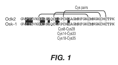
Figure 1 Amino acid sequence alignment of native OdK2
(SEQ ID NO: 1) and OsK1 (SEQ ID NO: 2) (shown as OsK-1 in the
figure). Cysteine residues are highlighted in grey.
Disulfide bridges and cysteine pairs are shown. The nine
divergent residues between OdK2 and OsK1 are highlighted in
black.
Figure 2 Binding of A) KV1C2 (Odk2-Fc fusion) and B)
KV1N2 (OsK1-Fc fusion) to Kv1.3 E3C cells (solid black),
Kv1.3 E3C cells in the presence of a 10-fold excess ShK
(dashed grey), and to Kv1.5 (negative control cells; dotted
grey). Binding was detected with anti-human Fc-Cy5 using flow
cytometry. Data are shown as histogram overlays of Geometric
mean fluorescence intensity (GMFI) (Geom. Mean, Red A: Cy5).
Figure 3. Inhibition of memory T cell proliferation by
KV1C2 (OdK2-Fc fusion) Cr). Each data point is the mean SD
of triplicate reactions. Negative control IgG4 Fc (0) did
not inhibit T-cell proliferation.
Figure 4. A) Amino acid sequences, B) Activity and
selectivity of peptide variant fusion proteins determined in
binding and thallium flux assays using cells expressing Kv1.3
and Kv1.1.
Figure 5. A) Inhibition of T cell activation by purified Odk2
chimera Fc fusion proteins at single 100 nM concentration.
KV1B03 (M) is identical to KV1C2 (OdK2 Fc fusion) (0). B)
Concentration dependent inhibition of T cell activation by
KV1D261. Negative control IgG4 Fc did not inhibit T-cell IL-
2 production. C) Correlation between binding to Kv1.3 E3C
cells and T-cell inhibition for select variants.
CA 02898496 2015-07-16
WO 2014/116937
PCT/US2014/012932
Figure 6. Activity of p261 C-terminal extension HSA fusion
protein library. Amino acid sequences of the C-terminal
extensions and the resulting extended p261 amino acid
sequences are shown, along with activity in binding and
thallium flux assays.
Figure 7. Characterization of A) p261 B) p579 C-terminal
extension HSA fusion proteins.
Figure 8. Characteristics of select OdK2 variant fusion
proteins.
Figure 9. Pharmacokinetics of KV1D261 34 in minipigs.
Figure 10. Ex-vivo inhibition of IL-17A secretion from
lymphocytes following in vivo administration of KV1D261 34 in
minipigs.
Figure 11. Cell numbers from draining lymph nodes at day 10
following antigen challenge in the delayed type
hypersensitivity (DTH) minipig model.
Summary of the Invention
The invention provides an isolated peptide antagonist of
Kv1.3 having an amino acid sequence comprising:
(i) the sequence shown in SEQ ID NO: 1 having a
substitution of glycine to isoleucine at position 10
(G10I), and optionally having 1, 2, 3, 4, 5, 6 or 7
additional substitutions; or
(ii) an amino acid sequence which is at least 80% identical
to SEQ ID NO: 1, further comprising a G10I substitution.
The invention also provides an isolated peptide
antagonist of Kv1.3 comprising the sequence of SEQ ID NO: 55
or 86.
The invention also provides fusion proteins comprising
the peptide antagonist of the invention.
The invention also provides an isolated polynucleotide
encoding the peptide antagonist or fusion protein of the
invention.
The invention also provides a vector comprising the
isolated polynucleotide of the invention.
6
CA 02898496 2015-07-16
WO 2014/116937
PCT/US2014/012932
The invention also provides a host cell comprising the
vector of the invention.
The invention also provides a method of producing the
antagonist or fusion protein of the invention, comprising
culturing the host cell of the invention and recovering the
antagonist or fusion protein expressed by the host cell.
The invention also provides a pharmaceutical composition
comprising the antagonist or fusion protein of the invention
and a pharmaceutically acceptable carrier.
The invention also provides a method of suppressing T cell
activation in a subject having a condition associated with
undesired T cell activation, comprising administering to the
subject an effective amount of the antagonist or fusion
protein of the invention to suppress T cell activation.
Detailed Description of the Invention
All publications, including but not limited to patents
and patent applications, cited in this specification are
herein incorporated by reference as though fully set forth.
As used herein and in the claims, the singular forms
"a," "and," and "the" include plural reference unless the
context clearly dictates otherwise.
Unless defined otherwise, all technical and scientific
terms used herein have the same meaning as commonly
understood by one of ordinary skill in the art to which an
invention belongs. Although any compositions and methods
similar or equivalent to those described herein can be used
in the practice or testing of the invention, exemplary
compositions and methods are described herein.
The term "polypeptide" means a molecule that comprises
at least two amino acid residues linked by a peptide bond to
form a polypeptide. Polypeptides of less than about 80 amino
acids may be referred to as "peptides". Polypeptides may
also be referred as "proteins".
The term "polynucleotide" means a molecule comprising a
chain of nucleotides covalently linked by a sugar-phosphate
7
CA 02898496 2015-07-16
WO 2014/116937
PCT/US2014/012932
backbone or other equivalent covalent chemistry. Double and
single-stranded DNAs and RNAs are typical examples of
polynucleotides.
The term "complementary sequence" means a second
isolated polynucleotide sequence that is antiparallel to a
first isolated polynucleotide sequence and that comprises
nucleotides complementary to the nucleotides in the first
polynucleotide sequence.
The term "vector" means a polynucleotide capable of
being duplicated within a biological system or that can be
moved between such systems. Vector polynucleotides typically
contain elements, such as origins of replication,
polyadenylation signal or selection markers that function to
facilitate the duplication or maintenance of these
polynucleotides in a biological system. Examples of such
biological systems may include a cell, virus, animal, plant,
and reconstituted biological systems utilizing biological
components capable of duplicating a vector. The
polynucleotides comprising a vector may be DNA or RNA
molecules or hybrids of these.
The term "expression vector" means a vector that can be
utilized in a biological system or a reconstituted biological
system to direct the translation of a polypeptide encoded by
a polynucleotide sequence present in the expression vector.
The term "wild type OdK2" or "OdK2" or "native OdK2" as
used herein refers to scorpion Odontobuthus doriae OdK2
polypeptide having a sequence shown in SEQ ID NO: 1
(GVPTDVKCRGSPQCIQPCKDAGMRFGKCMNGKCHCTPK).
The term "wild type OsK1" or "OsK1" or "native OsK1" as
used herein refers to scorpion Orthochirus scrobiculosus OsK1
polypeptide having a sequence shown in SEQ ID NO: 2
(GVIINVKCKISRQCLEPCKKAGMRFGKCMNGKCHCTPK).
The term "variant" or "OdK2 variant" as used herein
refers to a polypeptide that differs from the wild type OdK2
polypeptide of SEQ ID NO: 1 by one or more modifications for
8
CA 02898496 2015-07-16
WO 2014/116937
PCT/US2014/012932
example, substitutions, insertions or deletions of
nucleotides or amino acids.
Throughout the specification, residue numbering of OdK2
variants is according to SEQ ID NO: 1. For example, "G10" in
the specification refers to the glycine residue at position
of SEQ ID NO: 1. Accordingly, OdK2 G10I refers to an OdK2
variant having glycine at position 10 substituted for
isoleucine, and OdK2 G10I,P12R refers to an OdK2 variant
having glycine at position 10 substituted for isoleucine, and
proline at position 12 substituted for arginine.
Numbering of a given amino acid or polynucleotide
sequence "corresponds to" or is "relative to" the numbering
of a selected amino acid or polynucleotide sequence when the
position of any given amino acid residue or nucleotide
residues is designated by reference to the same or to an
equivalent position in the selected amino acid or
polynucleotide sequence rather than by the actual numerical
position of the component in the sequence. Thus, for
example, the numbering of a given amino acid position in a
given polypeptide sequence corresponds to the same or
equivalent amino acid position in a selected polypeptide
sequence used as a reference sequence.
An "equivalent position" (for example, an "equivalent
amino acid position" or "equivalent nucleic acid position" or
"equivalent residue position") is defined herein as a
position (such as, an amino acid position or nucleic acid
position or residue position) of a test polypeptide (or test
polynucleotide) sequence which aligns with a corresponding
position of a reference polypeptide (or reference
polynucleotide) sequence, when optimally aligned using an
alignment algorithm as described herein. The equivalent
amino acid position of the test polypeptide need not have the
same numerical position number as the corresponding position
of the reference polypeptide; likewise, the equivalent
nucleic acid position of the test polynucleotide need not
9
CA 02898496 2015-07-16
WO 2014/116937
PCT/US2014/012932
have the same numerical position number as the corresponding
position of the reference polynucleotide.
Two polypeptide sequences are "optimally aligned" when
they are aligned using defined parameters, i.e., a defined
amino acid substitution matrix, gap existence penalty (also
termed gap open penalty), and gap extension penalty, so as to
arrive at the highest similarity score possible for that pair
of sequences. The BLOSUM62 matrix (Henikoff and Henikoff
(1992) Proc. Natl. Acad. Sci. USA 89(22):10915-10919) is
often used as a default scoring substitution matrix in
polypeptide sequence alignment algorithms (such as BLASTP).
The gap existence penalty is imposed for the introduction of
a single amino acid gap in one of the aligned sequences, and
the gap extension penalty is imposed for each residue
position in the gap. Unless otherwise stated, alignment
parameters employed herein are: BLOSUM62 scoring matrix, gap
existence penalty = 11, and gap extension penalty = 1. The
alignment score is defined by the amino acid positions of
each sequence at which the alignment begins and ends (e.g.
the alignment window), and optionally by the insertion of a
gap or multiple gaps into one or both sequences, so as to
arrive at the highest possible similarity score.
"Kv1.3" (also known as KCNA3, HPCN3, HGK5, HuKIII, or
HLK3) as used herein refers to the well known human potassium
voltage-gated channel subfamily A member 3 having a sequence
shown in UniProt accession number P22001 and in SEQ ID NO:
418.
"Antagonist of Kv1.3" or "antagonist" as used herein
refers to an OdK2 variant or OdK2 variant fusion protein of
the invention that inhibits or blocks Kv1.3 function by at
least 10%, 20%, 30%, 40%, 50%, 60%, 70%, 75%, 80%, 85%, 90%,
95% or 100%. Amino acid sequence of the wild type OdK2 is
shown in SEQ ID NO: 1.
"Fusion protein" as used herein refers to a protein that
includes polypeptide or peptide components derived from more
than one parental polypeptide or peptide.
CA 02898496 2015-07-16
WO 2014/116937
PCT/US2014/012932
"Half-life extending moiety" as used herein refers to a
molecule or protein or domain that, when conjugated to the
OdK2 variant increases the in vivo half life of the resulting
OdK2 variant fusion protein when compared to the free
peptide.
"Percent binding" or "%Binding" as used herein refers to
a ratio of geometric mean fluorescence intensities (Geo. MFI
or GMFI) for an OdK2 variant fusion protein when compared to
the control, obtained from a FACS assay using cells
expressing Kv1.3 or Kv1.1 channels.
"Binding selectivity" as used herein refers to the ratio
of %Binding obtained for Kv1.3 to %Binding obtained for
Kv1.1.
"Selective" or "selectivity" as used herein refers to
the ratio of an ICH value for Kv1.1 to an ICH value for Kv1.3
for an OdK2 variant fusion protein or OdK2 variant.
Selectivity can be assessed using various methodologies, for
example electrophysiological patch clamp assays or thallium
flux assays as described herein. Selectivity may vary
slightly depending on the assay chosen for measurements.
The Kv1.3 blocking peptides Odk2 (SEQ ID NO: 1) and Osk1
(SEQ ID NO: 2) are members of the a-KTx3 scorpion toxin
family that differ in amino acid sequence at nine positions.
Both OdK2 and Osk1 are 38 amino acids in length, and are each
stabilized by three disulfide bonds with paring between Cys8-
Cys28, Cys14-Cys33, and Cys18-Cys35 (Abdel-Mottaleb et al.,
Toxicon 51:1424-30, 2008; Mouhat et al., Biochem J. 385(Pt
1):95-104, 2005; Int. Pat. Publ. No. W02006/002850). The
folded peptides form an a-helix held in close proximity to a
3 stranded anti parallel f3-sheet by the disulfide bonds.
OdK2 and OsK1 are pore blockers that inhibit channel function
through binding to the outer vestibule of the pore region,
inserting lysine 27 into the water filled pore, and occluding
ion flow. OsK1 (alpha-KTx3.7) is reported to block Kv1.3,
Kv1.1 and Kv1.2, channels potently and KCa3.1 channel
moderately, with an ICH of 0.014 nM, 0.6 nM, 5.4 nM, and 225
11
CA 02898496 2015-07-16
WO 2014/116937
PCT/US2014/012932
nM, respectively (Mouhat et al., Biochem J 385 (Pt 1):95-104,
2005). OdK2 (alpha-KTx3.11) is reported to block Kv1.3 in
Xenopus laevis oocytes, with an ICH of 7.2 nM, and is
reported to have no activity on other Kv1.x subtypes tested
(Kv1.1, Kv1.2, Kv1.4, Kv1.5, and Kv1.6) (Abdel-Mottaleb et
al., Toxicon 51:1424-30, 2008). These data indicate that
OsK1 is very potent but lacks sufficient subtype selectivity,
whereas OdK2 appears selective but not highly potent.
The present invention provides isolated OdK2 variants
and OdK2 variant fusion proteins that inhibit Kv1.3,
polynucleotides encoding them, vectors, host cells, and
methods of using the polynucleotides and polypeptides of the
invention. The OdK2 variants and OdK2 variant fusion
proteins of the invention are more potent towards Kv1.3 when
compared to the parent molecules with retained and/or
enhanced selectivity. The polypeptides of the invention
inhibit potassium currents, thallium flux and/or T cell
activation resulting from Kv1.3 activity and therefore may be
useful in the treatment of various conditions associated with
activated T cells, such as inflammatory and autoimmune
diseases.
Antagonist Peptides
The invention provides an isolated peptide antagonist of
Kv1.3 having an amino acid sequence comprising:
(i) the sequence shown in SEQ ID NO: 1 having a
substitution of glycine to isoleucine at position 10
(G10I), and optionally having 1, 2, 3, 4, 5, 6 or 7
additional substitutions; or
(ii) an amino acid sequence which is at least 80% identical
to SEQ ID NO: 1, further comprising a G10I substitution.
In some embodiments the isolated peptide antagonist
comprises a sequence with no more than 7, no more than 6, no
more than 5, no more than 4, no more than 3, or no more than
2 substitutions relative to SEQ ID NO: 1.
12
CA 02898496 2015-07-16
WO 2014/116937
PCT/US2014/012932
In some embodiments the isolated peptide antagonist
comprises a sequence with at least 81%, at least 82%, at
least 83%, at least 84%, at least 85%, at least 86%, at least
87%, at least 88%, at least 89%, at least 90%, at least 91%,
at least 92%, at least 93%, at least 94%, at least 95%, at
least 96%, at least 97%, at least 98%, or at least 99%
identity to SEQ ID NO: 1.
In some embodiments, therefore, the peptide antagonist
of Kv1.3 may comprise the sequence of any one of SEQ ID NOs:
3, 4, 5, 6, 7, 8, 9, 10, 11, 12, 13, 14, 15, 16, 17, 18, 19,
20, 21, 22, 23, 24, 25, 26, 27, 28, 29, 30, 31, 32, 33, 34,
35, 36, 37, 38, 39, 40, 41, 42, 43, 44, 45, 46, 47, 48, 49,
50, 51, 52, 53, 54, 56, 57, 58, 59, 60, 61, 62, 63, 64, 65,
66, 67, 68, 69, 70, 71, 72, 73, 74, 75, 76, 77, 78, 79, 80,
81, 82, 83, 84, 85, 87, 88, 89, 90, 91, 92, 93, 94, 95, 96,
97, 98, 99, 100, 101, 102, 103, 104, 105, 106, 107, 108, 109,
or 110. The substitution G10I is associated with improved
selectivity and/or improved affinity for Kv1.3.
In some embodiments described herein, the antagonist of
Kv1.3 comprises the sequence
GVPXaa1Xaa2VKCXaa3ISRQCXaa4Xaa5PCKDAGMRFGKCMNGKCHCTPK (SEQ ID
NO: 426); wherein
a) Xaa1 is I or T, Q or E;
b) Xaa2 is N or D;
c) Xaa3 is K or R, E, A or Q;
d) Xaa4 is I, E, L, D, Q, H, V, K or A;
e) Xaa5 is E K, L, Q, D, V or H; and
the peptide antagonist of Kv1.3 has an optional C-terminal
extension of four amino acids. For example, the peptide
antagonist of Kv1.3 may comprise the amino acid sequence of
SEQ ID NOs: 3, 13, 21, 22, 24, 26, 29, 30, 32, 34, 38, 39,
42-46, 49, 51, 59, 63, 65, 69, 71, 73, 76, 78, 81-83, 85, 87,
89, 92, 96, 101, 103, 104 and 108.
In some embodiments described herein, the antagonist of
Kv1.3 comprises the sequence
13
CA 02898496 2015-07-16
WO 2014/116937
PCT/US2014/012932
GVPXaa4Xaa2VKCXaa3ISRQCXaa4Xaa5PCKDAGMRFGKCMNGKCHCTPK (SEQ ID
NO: 427); wherein
Xaal is I or T;
Xaa2 is N or D;
Xaa3 is K or R;
Xaa4 is I or E; and
Xaa5 is E or K; and
the peptide antagonist of Kv1.3 has an optional C-terminal
extension of four amino acids. For example, the peptide
antagonist of Kv1.3 may comprise the amino acid sequence of
SEQ ID NOs: 3, 22, 34 or 42.
As will be seen by reference to figure 4A, the peptide
antagonists of the invention retain the native disulfide
bridges between C8-C28, C14-C33 and C18-C35. The exemplary
antagonists of the invention described in this section and
elsewhere can have substantially enhanced selectivity for
Kv1.3 against Kv1.1, for example 100, 200, 300, 400, 500,
1000, 2000, 3000, 4000, 5000, 6000 or at least 7000 fold
improved selectivity compared to SEQ ID NO: 1.
Fusion proteins
The antagonist of the invention can be an isolated
fusion protein that comprises an antagonist peptide of the
invention. In some embodiments described herein, the fusion
protein comprises a sequence with at least 80% identity to
SEQ ID NO: 1, and which comprises the substitution G10I
relative to SEQ ID NO: 1.
In some embodiments described herein, the fusion protein
comprises a peptide antagonist of Kv1.3 conjugated to a half-
life extending moiety, wherein the peptide antagonist of
Kv1.3 comprises the sequence shown in SEQ ID NO: 1, further
comprising a substitution of G10I, and optionally having 1,
2, 3, 4, 5, 6 or 7 additional substitutions.
In some embodiments described herein, the fusion protein
comprises a peptide antagonist of Kv1.3 conjugated to a half-
life extending moiety, wherein the peptide antagonist of
14
CA 02898496 2015-07-16
WO 2014/116937
PCT/US2014/012932
Kv1.3 comprises the sequence at least 80% identical to the
amino acid sequence shown in SEQ ID NO: 1, further comprising
a substitution of glycine to isoleucine at residue position
(G10I).
In some embodiments, the fusion protein comprises a
peptide antagonist of Kv1.3 conjugated to a half-life
extending moiety, wherein the peptide antagonist of Kv1.3
comprises the sequence
GVPXaa1Xaa2VKCXaa2ISRQCXaa4Xaa5PCKDAGMRFGKCMNGKCHCTPK (SEQ ID
NO: 426); wherein
Xaal is I or T, Q or E;
Xaa2 is N or D;
Xaa2 is K or R, E, A or Q;
Xaa4 is I, E, L, D, Q, H, V, K or A;
Xaa5 is E K, L, Q, D, V or H; and
the peptide antagonist of Kv1.3 has an optional C-
terminal extension of four amino acids.
In some embodiment described herein, the peptide
antagonist of Kv1.3 comprises the amino acid sequence of SEQ
ID NOs: 3, 13, 21, 22, 24, 26, 29, 30, 32, 34, 38, 39, 42-46,
49, 51, 59, 63, 65, 69, 71, 73, 76, 78, 81-83, 85, 87, 89,
92, 96, 101, 103, 104 and 108.
In some embodiment described herein, the antagonist is
an isolated fusion protein comprising a peptide antagonist of
Kv1.3 conjugated to a half-life extending moiety, wherein the
peptide antagonist of Kv1.3 comprises the sequence
GVPXaa1Xaa2VKCXaa2ISRQCXaa4Xaa5PCKDAGMRFGKCMNGKCHCTPK (SEQ ID
NO: 427); wherein
Xaal is I or T;
Xaa2 is N or D;
Xaa2 is K or R;
Xaa4 is I or E; and
Xaa5 is E or K; and
the peptide antagonist of Kv1.3 has an optional C-
terminal extension of four amino acids.
CA 02898496 2015-07-16
WO 2014/116937
PCT/US2014/012932
In some embodiment described herein, the peptide antagonist
of Kv1.3 comprises the amino acid sequence of SEQ ID NOs: 3,
22, 34 or 42.
In some embodiment described herein, the C-terminal
extension comprises the amino acid sequence of SEQ ID NOs:
123-268.
In some embodiment described herein, the C-terminal
extension comprises the amino acid sequence of SEQ ID NOs:
128, 143, 155, 188, 206-210, 212, 214, 216, 219, 223, 224,
227, 230, 232 -235, 237, 239, 240, 243, 252, 261-263, or 268.
The OdK2 variant fusion proteins of the invention are more
potent and selective when compared to the fusion protein of
native OdK2 sequence, such as KV1C2 (parent KV1C2 fusion
protein) of SEQ ID NO: 425. Exemplary fusion proteins of the
invention are those comprising OdK2 variant peptides of SEQ
ID NOs: 3, 22, 34 or 42 conjugated to human serum albumin
(HSA) via a linker AS(AP)20G5 (SEQ ID NO: 116).
The parent KV1C2 fusion protein has an ICH of about 13 nM
(1.3x10⁻⁸ M) for inhibiting potassium currents in whole cell
patch clamp studies in CHO cells transfected with human
Kv1.3, and an ICH value of about 21.4 nM (2.14x10⁻⁸ M) for
inhibiting thallium flux in cells expressing Kv1.3 using
FLIPRED Tetra instrument (Molecular Devices). The OdK2
variant fusion protein of the invention as described herein
is "equally potent or more potent" Kv1.3 inhibitor when the
ICH value in the patch clamp assay described in the materials
and methods is about 13 nM (1.3x10⁻⁸ M) or less, for example
1.0x10⁻⁸ M, 5.0x10⁻⁹ M, 1.0 x10⁻⁹ M, 5.0 x10⁻¹⁰ m, 1.0 x10⁻¹ M,
5.0 x10⁻¹¹ M, 1.0 x10⁻¹¹ M, 5.0 x10⁻¹² m, 1.0 x10⁻¹² M or less,
or the ICH value in the thallium flux assay described in the
materials and methods is about 21.4 nM (2.14x10⁻⁸ M) or less,
for example 1.0x10⁻⁸ M, 5.0x10⁻⁹ M, 1.0 x10⁻⁹ M, 5.0 x10⁻¹ M,
1.0 x10⁻¹⁰ m,
5.0 x10⁻¹¹ M, 1.0 x10⁻¹¹ M, 5.0 x10⁻¹² M, 1.0 x10⁻¹²
M or less. The IC50 values for patch clamp and thallium flux
for exemplary fusion proteins are shown in Figure 8.
16
CA 02898496 2015-07-16
WO 2014/116937
PCT/US2014/012932
The OdK2 variant and OdK2 variant fusion proteins of the
invention as described herein are selective for Kv1.3.
Selectivity can be assessed against Kv1.1 using the ratio of
an ICH value for Kv1.1 to an ICH value for Kv1.3 for an OdK2
variant fusion protein or OdK2 variant. Selectivity can be
further tested against other Kv channels, such as Kv1.2,
Kv1.4, Kv1.5, and against hERG, KCa3.1, or Nav1.5 using
standard methods. The exemplary OdK2 variant fusion proteins
of the invention as described herein can have substantially
selectivity for Kv1.3 against Kv1.1, for example 100, 200,
300, 400, 500, 1000, 2000, 3000, 4000, 5000, 6000 or at least
7000 fold selectivity. The parent KV1C2 fusion protein is
68-fold more selective towards human Kv1.3 when compared to
human Kv1.1, therefore, the exemplary OdK2 variant fusion
proteins of the invention as described herein can have
substantially enhanced selectivity, for example about 1.5, 3,
4.5, 6, 7.5, 10, 15, 20, 25, 30, 35, 40, 45, 50, 55, 60, 65,
70, 75, 80, 85, 90, 95, 100 or at least 105 fold improved
selectivity when compared to the KV1C2 fusion protein. The
presence of glutamic acid at position corresponding to Xaa5
in SEQ ID NO: 426 has been observed to improve selectivity.
Residue positions 41 1 5 9, 15 and 16 (residue numbering
according to native OdK2 peptide of SEQ ID NO: 1) can be
substituted in the native OdK2 to improve both potency and
selectivity of the resulting variants and their fusion
proteins. The residue positions can be substituted with any
amino acid residue as long as the resulting OdK2 variant or
its fusion protein, in the above whole cell patch clamp assay
or thallium flux assay retains an ICH of about 13 nM (1.3x10⁻⁸
M) or 21.4 nM (2.14x10⁻⁸), respectively, or less, and has
selectivity (expressed as a ratio of ICH values obtained
using patch clamp as described above) for Kv1.3 against Kv1.1
of at least 100. The amino acid sets that can be used for
diversification at each selected position include amino acid
residues TIQE at position 4, ND at position 5, REAKQ at
position 9, ELDIQHVKA at position 15, and KELQDVH at position
17
CA 02898496 2015-07-16
WO 2014/116937
PCT/US2014/012932
16. A glutamic acid (E) at position 15 is associated with
increased selectivity for Kv1.3. The substitution G10I is
associated with improved selectivity and/or improved affinity
for Kv1.3 (residue numbering according to SEQ ID NO: 1).
Diversification of OdK2 and its fusion proteins using the
amino acid sets described above has resulted in variants
displaying improved binding affinity and improved binding
selectivity for Kv1.3 when compared to the native peptide or
its fusion protein. In another diversification scheme, the
amino acid sets that can be used for diversification at each
selected position include amino acid residues IT at position
4, ND at position 5, KR at position 9, IE at position 15, and
EK at position 16. The resulting variants and/or their
fusion proteins can be assessed for selectivity, potency,
binding affinity and binding selectivity using well known
assays and the ones described within. Exemplary OdK2
variants and their fusion proteins with improved potency and
selectivity are variants of SEQ ID NOs: 3, 22, 34 and 42, and
their human serum albumin or Fc fusion proteins. Exemplary
OdK2 variants with improved binding affinity and %Binding
selectivity are variants of SEQ ID NOs: 3, 13, 21, 22, 24,
26, 29, 30, 32, 34, 38, 39, 42-46, 49, 51, 59, 63, 65, 69,
71, 73, 76, 78, 81-83, 85, 87, 89, 92, 96, 101, 103, 104 and
108.
Additional OdK2 variants and OdK2 variant fusion proteins
are within the scope of the invention. For example,
substitutions can be made in the native OdK2 peptide to
positions other than positions 41 1 5 9, 15 and 16 as long as
the resulting OdK2 variant and the OdK2 variant fusion
protein retains similar selectivity and potency towards Kv1.3
when compared to the parent molecule. Exemplary
modifications are for example conservative substitutions that
will result in OdK2 variant fusion proteins with similar
characteristics to those of the parent molecules.
Conservative replacements are those that take place within a
family of amino acids that are related in their side chains.
18
CA 02898496 2015-07-16
WO 2014/116937
PCT/US2014/012932
Genetically encoded amino acids can be divided into four
families: (1) acidic (aspartate, glutamate); (2) basic
(lysine, arginine, histidine); (3) nonpolar (alanine, valine,
leucine, isoleucine, proline, phenylalanine, methionine,
tryptophan); and (4) uncharged polar (glycine, asparagine,
glutamine, cysteine, serine, threonine, tyrosine).
Phenylalanine, tryptophan, and tyrosine are sometimes
classified jointly as aromatic amino acids. Alternatively,
the amino acid repertoire can be grouped as (1) acidic
(aspartate, glutamate); (2) basic (lysine, arginine
histidine), (3) aliphatic (glycine, alanine, valine, leucine,
isoleucine, serine, threonine), with serine and threonine
optionally be grouped separately as aliphatic-hydroxyl; (4)
aromatic (phenylalanine, tyrosine, tryptophan); (5) amide
(asparagine, glutamine); and (6) sulfur-containing (cysteine
and methionine) (Stryer (ed.), Biochemistry, 2nd ed, WH
Freeman and Co., 1981). Non-conservative substitutions can
be made to the native OdK2 peptide that involves
substitutions of amino acid residues between different
classes of amino acids to improve properties of the OdK2
variants and OdK2 variant fusion proteins. Whether a change in
the amino acid sequence of a polypeptide or fragment thereof
results in a functional homolog can be readily determined by
assessing the ability of the modified polypeptide or fragment
to produce a response in a fashion similar to the unmodified
polypeptide or fragment using the assays described herein.
Peptides, polypeptides or proteins in which more than one
replacement has taken place can readily be tested in the same
manner. Exemplary additional OdK2 variants and/or OdK2
variant fusion proteins having substitutions resulting in
enhanced binding or binding specificity are those having the
amino acid sequence of SEQ ID NOs: 4-12, 14-20, 23, 25, 27,
28, 31, 33, 35-37, 40, 41, 47, 48, 50, 52-58, 60-62, 64, 66-
68, 70, 72, 74, 75, 77, 79, 80, 84, 86, 88, 90, 91, 93-95,
97-100, 102, 105-107, 109 and 110.
19
CA 02898496 2015-07-16
WO 2014/116937
PCT/US2014/012932
The OdK2 variants (i.e. antagonists according to the
invention) as described herein can be fused to a half-life
extending moiety to form fusion proteins of the invention.
Exemplary half-life extending moieties that can be used
include well known human serum albumin, transthyretin (TTR),
a thyroxine-binding globulin TGB), albumin-binding domains,
or an Fc or fragments thereof. Biologically suitable
polymers or copolymers can also be used, for example
ethylene glycol, polyethylene glycol (PEG) molecules, such as
PEG5000 or PEG20000, dextran, polylysine, fatty acids and
fatty acid esters of different chain lengths, for example
laurate, myristate, stearate, arachidate, behenate, oleate,
arachidonate, octanedioic acid, tetradecanedioic acid,
octadecanedioic acid, docosanedioic acid, and the like,
polylysine, octane, or carbohydrates (dextran, cellulose,
oligo- or polysaccharides.
In another embodiment, the half-life extending moiety of
the fusion protein described herein is human serum albumin,
albumin binding domain (ADB), or polyethylene glycol (PEG).
In another embodiment, the half-life extending moiety of
the fusion protein described herein is human serum albumin.
In another embodiment, the half-life extending moiety of
the fusion protein described herein is conjugated to the
peptide antagonist of Kv1.3 via a linker.
In another embodiment, the linker of the fusion protein
described herein comprises the amino sequence of SEQ ID NOs:
112-122.
The half-life extending moiety can be conjugated directly
to the OdK2 variant peptide antagonist of the invention or
indirectly via a linker. Exemplary peptide linkers that can
be used in fusion proteins of the invention as described
herein are linkers having the amino acid sequence of SEQ ID
NOs: 112-122. Non-peptide half-life extending moieties can
be conjugated directly to the OdK2 variant using well known
chemical coupling methods. For example, OdK2 variants can be
pegylated using known methods and those described in U.S.
CA 02898496 2015-07-16
WO 2014/116937
PCT/US2014/012932
Pat. No. U58043829. Peptide or protein half-life extending
moieties can be linked to the peptide during translation of
the nucleic acid encoding the fusion protein, as explained in
more detail below.
OdK2 variants incorporating half-life extending moieties
may be compared for functionality by several well known
assays. For example, pharmacokinetic properties of OdK2
variants coupled to PEG or human serum albumin may be
evaluated in well known in vivo models.
The OdK2 variant fusion proteins of the invention as
described herein may be engineered to incorporate a C-
terminal extension of four amino acids to the C-terminus of
the Odk2 variant before conjugation of the extended peptide
to a half-life extending moiety. By not wishing to be bound
by any theory, it is believed that extending the C terminus
of the OdK2 variant peptide in the fusion proteins would
allow for increased binding interactions of the peptide with
the extracellular loops of the Kv1.3 channel and increased
potency. Exemplary OdK2 fusion proteins with C-terminally
extended peptide portion are shown in Figure 6, Figure 7A and
Figure 8. The fusion proteins with C-terminally extended
peptide portion are typically more potent Kv1.3 inhibitors
when compared to the corresponding fusion proteins without
the extension. IC50 values for exemplary C-terminally
extended variants described herein can be about 1x10⁻⁹ M or
less, for example about 1x10⁻⁹ M or less, about 1x10⁻¹ M or
less, about 1x10-11 M or less, or about 1x10-12 M or less as
measured in thallium flux assay described below. Exemplary
C-terminal extensions are those shown in SEQ ID NOs: 123-268.
In another embodiment, the isolated fusion protein of the
invention comprises:
the peptide antagonist of Kv1.3 of SEQ ID NOs: 3, 22, 34
or 42;
optionally the C-terminal extension of SEQ ID NOs: 128,
143, 155, 188, 206- 210, 212, 214, 216, 219, 223, 224,
21
CA 02898496 2015-07-16
WO 2014/116937
PCT/US2014/012932
227, 230, 232, 235, 237, 239, 240, 243, 252, 261-263, or
268;
the linker of SEQ ID NO: 116 or SEQ ID NO:119; and
human serum albumin as the half-life extending moiety.
In another embodiment, the isolated fusion protein of the
invention comprises
the peptide antagonist of Kv1.3 of SEQ ID NO: 42;
the linker of SEQ ID NO: 116; and
human serum albumin as the half-life extending moiety.
In another embodiment, the isolated fusion protein of
the invention comprises
the peptide antagonist of Kv1.3 of SEQ ID NO: 42;
the C-terminal extension SEQ ID NO: 209;
the linker of SEQ ID NO: 116; and
human serum albumin as the half-life extending moiety.
In another embodiment, the isolated fusion protein of the
invention comprises
the peptide antagonist of Kv1.3 of SEQ ID NO: 3;
the C-terminal extension of SEQ ID NO: 235;
the linker of SEQ ID NO: 116; and
human serum albumin as the half-life extending moiety.
In another embodiment, the isolated fusion protein of the
invention comprises
the peptide antagonist of Kv1.3 of SEQ ID NO: 42;
the C-terminal extension of SEQ ID NO: 235;
the linker of SEQ ID NO: 116; and
human serum albumin as the half-life extending moiety.
In another embodiment, the isolated fusion protein of
the invention as described herein is at least 100 fold more
selective towards human Kv1.3 than towards human Kv1.1, when
selectivity is measured as a ratio of an ICH value of the
isolated fusion protein for Kv1.1 to an ICH value of the
isolated fusion protein for Kv1.3 in a patch clamp assay in
cells transfected with Kv1.1 and Kv1.3, respectively.
In another embodiment, the isolated fusion protein of
the invention as described herein inhibits potassium currents
22
ak 02898496 2015-07-16
WO 2014/116937
PCT/US2014/012932
with an IC50 value at least about 10 fold less than an IC50
value for a parent KV1C2 fusion protein of SEQ ID NO: 425 in
a patch clamp assay in cells transfected with human Kv1.3.
In another embodiment, the isolated fusion protein of
the invention as described herein inhibits potassium currents
with an IC50 value of about 1.5x10⁻⁸ M or less in a patch clamp
assay in cells transfected with human Kv1.3.
In another embodiment, the isolated fusion protein of
the invention as described herein inhibits in vitro thallium
flux with and IC50 value of about 2.2x10⁻⁸ M or less in cells
transfected with human Kv1.3.
Another embodiment of the invention is an isolated
fusion protein comprising a peptide antagonist of Kv1.3
conjugated to a half-life extending moiety via a linker, the
peptide antagonist of Kv1.3 having an optional C-terminal
extension of four amino acids, wherein
the peptide antagonist of Kv1.3 comprises the amino acid
sequence of SEQ ID NOs: 3-110;
the C-terminal extension comprises the amino acid
sequence of SEQ ID NOs: 123-268;
the linker comprises the amino acid sequence of SEQ ID
NOs: 116 or 119; and
the half-life extending moiety is human serum albumin.
Another embodiment of the invention is an isolated
peptide antagonist of Kv1.3 comprising the sequence
GVPXaa1Xaa2VKCXaa2ISRQCXaa4Xaa5PCKDAGMRFGKCMNGKCHCTPK (SEQ ID
NO: 426); wherein
Xaal is I or T, Q or E;
Xaa2 is N or D;
Xaa2 is K or R, E, A or Q;
Xaa4 is I, E, L, D, Q, H, V, K or A;
Xaa5 is E K, L, Q, D, V or H; and
the peptide antagonist of Kv1.3 has an optional C-
terminal extension of four amino acids.
23
CA 02898496 2015-07-16
WO 2014/116937
PCT/US2014/012932
In another embodiment the isolated peptide antagonist of
Kv1.3 comprises the amino acid sequence of SEQ ID NOs: 3, 13,
21, 22, 24, 26, 29, 30, 32, 34, 38, 39, 42-46, 49, 51, 59,
63, 65, 69, 71, 73, 76, 78, 81-83, 85, 87, 89, 92, 96, 101,
103, 104 and 108.
Another embodiment of the invention is an isolated
peptide antagonist of Kv1.3 comprising the sequence
GVPXaa4Xaa2VKCXaa3ISRQCXaa4Xaa5PCKDAGMRFGKCMNGKCHCTPK (SEQ ID
NO: 427); wherein
Xaal is I or T;
Xaa2 is N or D;
Xaa3 is K or R;
Xaa4 is I or E; and
Xaa5 is E or K; and
the peptide antagonist of Kv1.3 has an optional C-
terminal extension of four amino acids.
In another embodiment the isolated peptide antagonist of
Kv1.3 comprises the amino acid sequence of SEQ ID NOs: 3, 22,
34 or 42.
Another embodiment of the invention is an isolated
peptide antagonist of Kv1.3 comprising the sequence of SEQ ID
NOs: 3-110.
The OdK2 variant polypeptides and their fusion proteins
of the invention may be produced by chemical synthesis, such
as solid phase peptide synthesis, on an automated peptide
synthesizer. Alternatively, the polypeptides of the
invention can be obtained from polynucleotides encoding the
polypeptides by the use of cell-free expression systems such
as reticulocyte lysate based expression systems, or by
standard recombinant expression systems. Those skilled in
the art will recognize other techniques for obtaining the
polypeptides of the invention.
Generation of the OdK2 variants is typically achieved at
the nucleic acid level. The polynucleotides can be
synthesized using chemical gene synthesis according to
methods described in U.S. Pat. No. U56521427 and U56670127,
24
CA 02898496 2015-07-16
WO 2014/116937
PCT/US2014/012932
utilizing degenerate oligonucleotides to generate the desired
variants, or by standard PCR cloning and mutagenesis.
Libraries of variants can be generated by standard cloning
techniques to clone the polynucleotides encoding the OdK2
variants into the vector for expression.
The OdK2 variant fusion proteins are typically made by
standard molecular biology approaches.
The OdK2 variants and their fusion proteins are tested
for their ability to inhibit Kv1.3 using methods described
herein. An exemplary assay is an assay measuring inhibition
of thallium influx into the cells in cells overexpressing
Kv1.3 using FLIPRC) Tetra instrument (Molecular Devices).
Another exemplary assay employs electrophysiological
recordings to measure ionic flux across the cell membrane
using well known patch clamp techniques and described herein.
Another embodiment of the invention is an isolated
polynucleotide comprising a polynucleotide encoding the OdK2
variant and OdK2 variant fusion protein of the invention.
The polynucleotides of the invention may also comprise
at least one non-coding sequence, such as transcribed but not
translated sequences, termination signals, ribosome binding
sites, mRNA stabilizing sequences, introns and
polyadenylation signals. The polynucleotide sequences may
also comprise additional sequences encoding additional amino
acids. These additional polynucleotide sequences may, for
example, encode a marker or well known tag sequences such as
a hexa-histidine or a HA tag which facilitate the
purification of fused polypeptides. Certain exemplary
polynucleotides are disclosed herein, however, other
polynucleotides which, given the degeneracy of the genetic
code or codon preferences in a given expression system,
encode the antagonists of the invention are also within the
scope of the invention. Exemplary polynucleotides are
polynucleotides comprising a sequence shown in SEQ ID NOs:
429-430.
CA 02898496 2015-07-16
WO 2014/116937
PCT/US2014/012932
Another embodiment of the invention is a vector
comprising an isolated polynucleotide encoding the OdK2
variants and their fusion proteins of the invention. The
vectors of the invention are useful for maintaining
polynucleotides, duplicating polynucleotides, or driving
expression of a polypeptide encoded by a vector of the
invention in biological systems, including reconstituted
biological systems. Vectors may be chromosomal-, episomal-
and virus-derived such as vectors derived from bacterial
plasmids, bacteriophages, transposons, yeast episomes,
insertion elements, yeast chromosomal elements,
baculoviruses, papova viruses such as SV40, vaccinia viruses,
adenoviruses, fowl pox viruses, pseudorabies viruses,
picornaviruses and retroviruses and vectors derived from
combinations thereof, such as cosmids and phagemids.
In one embodiment of the invention the vector is an
expression vector. Expression vectors typically comprise
nucleic acid sequence elements that can control, regulate,
cause or permit expression of a polypeptide encoded by such a
vector. Such elements may comprise transcriptional enhancer
binding sites, RNA polymerase initiation sites, ribosome
binding sites, and other sites that facilitate the expression
of encoded polypeptides in a given expression system. Such
expression systems may be cell-based, or cell-free systems
well known in the art. Nucleic acid sequence elements and
parent vector sequences suitable for use in the expression of
encoded polypeptides are also well known. An exemplary
plasmid-derived expression vector useful for expression of
the polypeptides of the invention comprises an E. coli origin
of replication, an ampicillin resistance (Amp) gene, a CMV
promoter, a signal sequence, and a SV40 polyadenlyation site.
Another embodiment of the invention is an isolated host
cell comprising a vector of the invention. Exemplary host
cells include Archaea cells; bacterial cells such as
Streptococci, Staphylococci, Enterococci, E. coli,
Streptomyces, cyanobacteria, B. subtilis and S. aureus;
26
CA 02898496 2015-07-16
WO 2014/116937
PCT/US2014/012932
fungal cells such as Kluveromyces, Saccharomyces,
Basidomycete, Candida albicans or Aspergillus; insect cells
such as Drosophila S2 and Spodoplera Sf9; animal cells such
as CHO, COS, HeLa, C127, 3T3, BHK, HEK293, CV-1, Bowes
melanoma and myeloma; and plant cells, such as gymnosperm or
angiosperm cells. The host cells in the methods of the
invention may be provided as individual cells, or populations
of cells. Populations of cells may comprise an isolated or
cultured population of cells or cells present in a matrix
such as a tissue.
Introduction of a polynucleotide, such as a vector, into
a host cell can be effected by methods well known to those
skilled in the art. These methods include calcium phosphate
transfection, DEAE-Dextran mediated transfection,
microinjection, cationic lipid-mediated transfection and
electroporation.
Another embodiment of the invention is a method of
producing the isolated fusion protein of the invention
comprising the steps of culturing the host cell under
conditions sufficient for the expression of at least one odK2
variant fusion protein, and recovering the fusion protein
expressed by the host cell.
Host cells can be cultured under any conditions suitable
for maintaining or propagating a given type of host cell and
sufficient for expressing a polypeptide. Culture conditions,
media, and related methods sufficient for the expression of
polypeptides are well known in the art. For example, many
mammalian cell types can be aerobically cultured at 37 C
using appropriately buffered DMEM media while bacterial,
yeast and other cell types may be cultured at 37 C under
appropriate atmospheric conditions in LB media.
In the methods of the invention the expression of the
OdK2 variant can be confirmed using a variety of well known
methods. For example, expression of a polypeptide can be
confirmed using detection reagents, such as antibodies using
27
CA 02898496 2015-07-16
WO 2014/116937
PCT/US2014/012932
for example FACS or immunofluorescent techniques, or using
SDS-PAGE or HPLC.
Methods of Treatment
Another aspect of the invention is a method of
modulating the activity of Kv1.3 in a biological tissue, the
method comprising contacting a biological tissue expressing
Kv1.3 with a Kv1.3 modulating amount of an OdK2 variant or
its fusion protein of the invention, or a pharmaceutically
acceptable salt thereof.
OdK2 variants and OdK2 variant fusion proteins of the
invention may be utilized in any therapy where it is desired
to treat, reduce or alleviate symptoms of Kv1.3-mediated
diseases such as inflammatory and autoimmune diseases,
diabetes, obesity or cancers.
The methods of the invention may be used to treat an
animal patient belonging to any classification. Examples of
such animals include mammals such as humans, rodents, dogs,
cats zoo animals and farm animals.
The OdK2 variants and/or the OdK2 variant fusion
proteins of the invention may be useful for the prophylaxis
and treatment of Kv1.3 mediated conditions, such as
inflammatory conditions, allergies and allergic conditions,
hypersensitivity reactions, autoimmune diseases, severe
infections, and organ or tissue transplant rejection. The
OdK2 variants and/or the OdK2 variant fusion proteins of the
invention are also useful in the preparation of a medicament
for such treatment, wherein the medicament is prepared for
administration in dosages defined herein.
One embodiment of the invention is method of suppressing
T cell activation in a subject having a condition associated
with undesired T cell activation, comprising administering to
the subject an effective amount of the isolated fusion
protein of the invention to suppress T cell activation.
T cell activation can be measured by well known methods,
such as measuring reduction of IL-2 production by T cells.
28
CA 02898496 2015-07-16
WO 2014/116937
PCT/US2014/012932
"Suppressing T cell activation" as used herein refers to the
ability of the OdK2 variants and OdK2 fusion proteins of the
invention to inhibit and reduce T cell activation by at least
20%, 30%, 40%, 50%, 60%, 70%, 80%, 90%, or 100%.
In another embodiment, the condition associated with
undesired T cell activation is an inflammatory condition, an
immune and proliferative disorder, rheumatoid arthritis (RA),
ankylosing spondylitis, psoriatic arthritis, osteoarthritis,
osteoporosis, uveitis, inflammatory fibrosis, scleroderma,
lung fibrosis, cirrhosis, inflammatory bowel disease, Crohn's
disease, ulcerative colitis, asthma, allergic asthma,
allergies, Chronic Obstructive Pulmonary Diseases (COPD),
multiple sclerosis, psoriasis, contact-mediated dermatitis,
systemic lupus erythematosus (SLE) and other forms of lupus,
diabetes, type I diabetes, obesity, cancer, lupus,
restenosis, systemic sclerosis, scleroderma,
glomerulonephritis, Sjogren syndrome, inflammatory bone
resorption, transplant rejection, or graft-versus-host
disease.
The Kv1.3 channel is expressed on all subsets of T cells
and B cells, but effector memory T cells and class-switched
memory B cells are particularly dependent on Kv1.3 (Wulff et
al., J Immunol 173:776, 2004). Kv1.3 is overexpressed in
Gad5/insulin-specific T cells from patients with new onset
type 1 diabetes, in myelin-specific T cells from MS patients
and in T cells from the synovium of rheumatoid arthritis
patients (Beeton et al., Proc Natl Acad Sci USA 103:17414-9,
2006), in breast cancer specimens (Abdul et al., Anticancer
Res 23:3347, 2003) and prostate cancer cell lines (Fraser et
al., Pflugers Arch 446:559, 2003). Positive outcomes in
animal models with Kv1.3 blockers have been described in
hypersensitivity models to ovalbumin and tetanus toxoid
(Beeton et al., Mol Pharmacol 67:1369, 2005; Koo et al., Clin
Immunol 197:99, 1999), models for multiple sclerosis such as
rat adoptive-transfer experimental autoimmune
encephalomyelitis (AT-EAE) model (Beeton et al., Proc Natl
29
CA 02898496 2015-07-16
WO 2014/116937
PCT/US2014/012932
Acad Sci USA 103:17414-9, 2006), inflammatory bone resorption
model (Valverde et al., J Bone Mineral Res 19:155, 2004),
models for arthritis (Beeton et al., Proc Natl Acad Sci 103:
17414, 2006; Tarcha et al., J Pharmacol Exper Therap 342:
642, 2012)and obesity, diabetes and metabolic diseases (Xu et
al., Hum Mol Genet 12:551, 2003; Xu et al., Proc Natl Acad
Sci 101: 3112, 2004).
Exemplary Kv1.3 mediated conditions that may be treated
with the OdK2 variants and/or OdK2 variant fusion proteins of
the invention are inflammatory conditions, immune and
proliferative disorders, including rheumatoid arthritis (RA),
ankylosing spondylitis, psoriatic arthritis, osteoarthritis,
osteoporosis, uveitis, inflammatory fibrosis (e.g.,
scleroderma, lung fibrosis, and cirrhosis), inflammatory
bowel disorders (e.g., Crohn's disease, ulcerative colitis
and inflammatory bowel disease), asthma (including allergic
asthma), allergies, COPD, multiple sclerosis, psoriasis,
contact-mediated dermatitis, systemic lupus erythematosus
(SLE) and other forms of lupus, diabetes, type I diabetes,
obesity and cancer, lupus, restenosis, systemic sclerosis,
scleroderma, glomerulonephritis, Sjogren syndrome,
inflammatory bone resorption, transplant rejection, and
graft-versus-host disease.
Administration of the OdK2 variants and/or OdK2 variant
fusion proteins of the invention to the animal models of a
particular disease can be used to evaluate the use of the
OdK2 variants and/or OdK2 variant fusion proteins to
ameliorate symptoms and alter the course of diseases. Animal
models that can be used are well known, and include models
described above and models such as collagen-induced arthritis
(CIA) model, diet-induced obesity model, the 2,4,6-
trinitrobenesulfonic acid/ethanol (TNBS)-induced colitis
model or the oxazalone model, which induce chronic
inflammation and ulceration in the colon (Neurath et al.,
Intern Rev Immunol 19:51-62, 2000), the adoptive transfer
model of naive CD45RBhIgh CD4 T cells to RAG or SCID mice, the
CA 02898496 2015-07-16
WO 2014/116937
PCT/US2014/012932
donor naive T cells attack the recipient gut causing chronic
bowel inflammation and symptoms similar to human inflammatory
bowel diseases (Read and Powrie, Curr Protoc Immunol Chapter
15 unit 15.13, 2001), ovalbumin challenge model and
methacholine sensitization models (Hessel et al., Eur J
Pharmacol 293:401-12, 1995).
Pharmaceutical compositions
The "therapeutically effective amount" of the OdK2
variant and/or OdK2 variant fusion protein effective in the
treatment of conditions where suppression of Kv1.3 activity
is desirable can be determined by standard research
techniques. For example, the dosage of the agent that will
be effective in the treatment of an inflammatory condition or
autoimmune disease such as lupus, multiple sclerosis or
psoriasis can be determined by administering the agent to
relevant animal models, such as the models described herein.
In addition, in vitro assays can optionally be employed
to help identify optimal dosage ranges. Selection of a
particular effective dose can be determined (e.g., via
clinical trials) by those skilled in the art based upon the
consideration of several factors. Such factors include the
disease to be treated or prevented, the symptoms involved,
the patient's body mass, the patient's immune status and
other factors known by the skilled artisan. The precise dose
to be employed in the formulation will also depend on the
route of administration, and the severity of disease, and
should be decided according to the judgment of the
practitioner and each patient's circumstances. Effective
doses can be extrapolated from dose-response curves derived
from in vitro or animal model test systems.
The mode of administration for therapeutic use of the
OdK2 peptide variants and/or OdK2 variant fusion proteins of
the invention may be any suitable route that delivers the
variant to the host. Pharmaceutical compositions of these
variants are particularly useful for parenteral
31
CA 02898496 2015-07-16
WO 2014/116937
PCT/US2014/012932
administration, e.g., intradermal, intramuscular,
intraperitoneal, intravenous, subcutaneous or intranasal.
The OdK2 variants and/or OdK2 variant fusion proteins of
the invention may be prepared as pharmaceutical compositions
containing an effective amount of the variant as an active
ingredient in a pharmaceutically acceptable carrier. The
term "carrier" refers to a diluent, adjuvant, excipient, or
vehicle with which the active compound is administered. Such
pharmaceutical vehicles can be liquids, such as water and
oils, including those of petroleum, animal, vegetable or
synthetic origin, such as peanut oil, soybean oil, mineral
oil, sesame oil and the like. For example, 0.4% saline and
0.3% glycine can be used. These solutions are sterile and
generally free of particulate matter. They may be sterilized
by conventional, well-known sterilization techniques (e.g.,
filtration). The compositions may contain pharmaceutically
acceptable auxiliary substances as required to approximate
physiological conditions such as pH adjusting and buffering
agents, stabilizing, thickening, lubricating and coloring
agents, etc. The concentration of the OdK2 variants and/or
OdK2 variant fusion proteins of the invention in such
pharmaceutical formulation can vary widely, i.e., from less
than about 0.5%, usually at or at least about 1% to as much
as 15 or 20% by weight and will be selected primarily based
on required dose, fluid volumes, viscosities, etc., according
to the particular mode of administration selected.
Thus, a pharmaceutical composition of the invention for
intramuscular injection could be prepared to contain 1 ml
sterile buffered water, and between about 1 ng to about 100
mg, e.g. about 50 ng to about 30 mg or more preferably, about
mg to about 25 mg, of the OdK2 variants and/or their fusion
proteins of the invention. Similarly, a pharmaceutical
composition of the invention for intravenous infusion could
be made up to contain about 250 ml of sterile Ringer's
solution, and about 1 mg to about 30 mg and preferably 5 mg
to about 25 mg of an antagonist of the invention. Actual
32
CA 02898496 2015-07-16
WO 2014/116937
PCT/US2014/012932
methods for preparing parenterally administrable compositions
are well known and are described in more detail in, for
example, "Remington's Pharmaceutical Science", 15th ed., Mack
Publishing Company, Easton, PA.
The OdK2 variants and/or the OdK2 variant fusion
proteins of the invention can be lyophilized for storage and
reconstituted in a suitable carrier prior to use. This
technique has been shown to be effective with conventional
protein preparations and art-known lyophilization and
reconstitution techniques can be employed.
The present invention will now be described with
reference to the following specific, non-limiting examples.
Materials and Methods
Kv Channel Expression Constructs and Cell Lines. cDNAs
encoding the various Kv channels and chimeric constructs were
cloned using routine methods into mammalian expression
vectors. cDNAs cloned and expressed were those encoding
human Kv1.3 (hKv1.3) (SEQ ID NO: 418), human Kv1.1 (hKv1.1)
(SEQ ID NO: 420), human Kv1.2 (hKv1.2) (SEQ ID NO: 419),
human Kv1.5 (hKv1.5) (SEQ ID NO: 421), hKv1.3 E3 loop/hKv1.5
chimera (having human Kv1.5 amino acids 1-455 and 496-613,
and Kv1.3 E3 loop amino acids 456-495) (Kv1.3 EC3 loop
chimera), hKv1.1 E3 loop/hKv1.5 chimera with N terminal His
tag (having His tag amino acids 1-9, hKv1.5 amino acids 10-
472 and 513-63, and hKv1.1 E3 loop amino acids 473-512) (Kv1.1
EC3 loop chimera), rat Kv1.3 (rKv1.3) (SEQ ID NO: 422), rat
Kv1.1 (rKv1.1) (SEQ ID NO: 423), cynomolgus monkey (Macaca
fascicularis) channel cynoKv1.3 (SEQ ID NO: 424),
hKv1.3/hKv1.5 tail chimera (having human Kv1.5 amino acids 1-
250 and 497-593, and Kv1.3 amino acid sequences 251-496
(Kv1.3 tail chimera), and hKv1.1/hKv1.5 tail chimera (having
human Kv1.5 amino acids 1-250 and 492-588, and Kv1.1 amino
acid sequences 251-491 (Kv1.1 tail chimera). For channel
expression in HEK cells, Kv genes were cloned into a CMV
33
CA 02898496 2015-07-16
WO 2014/116937
PCT/US2014/012932
promoter driven expression vector encoding the neomycin
resistance marker. HEK 293-F (Invitrogen, Carlsbad, CA)
cells were stably transfected and cultured in DMEM 10% FBS
and 600 g/ml Geneticin selection media to generate clonal
cell lines that expressed Kv channels using standard
techniques. For CHO stable expression, CHO-TREx cells
(Invitrogen) were stably transfected with pcDNA4/TO-Kv1.x
using standard techniques to generate clonal cell lines that
expressed each potassium channel in a tetracycline-inducible
manner. The culture medium was Ham's F-12 supplemented with
10% fetal bovine serum, 2 mM L-glutamine, 5 pg/ml blasticidin
and 200 pg/ml zeocin. In some experiments, transient
transfection using Lipofectamine 2000 in CHO cells was used.
For electrophysiological experiments, cells were co-
transfected with an expression vector expressing a truncated
CD4 for expression control (pMAC54.1, Milteni Biotech).
Assays were performed 24-48 hours after transfection.
Protein Expression and Purification.
The chimera library was expressed as peptide-Fc fusions or a
peptide-HSA fusion. The library was initially transfected
and expressed in HEK 293-E cells in 48-well or 96-well
format. The cells were cultured in DMEM, 10% FBS and 250
pg/ml of Geneticin for selection. For 48-well expression 0.5
ml/well of 3.0 x 10⁵ cells/ml were plated in the 48-well
plates. The library was transfected using Lipofectamine 2000
using routine methods utilizing 300 ng of plasmid DNA, 25 pl
OptiPROTM SFM media, and 2.4 pl Lipofectamine 2000
(Invitrogen, Carlsbad, CA). The next day, the transfection
media was aspirated and 0.5m1 of 293 FreeStyleTm media
(Invitrogen, Carlsbad, CA) was added to each well. The cells
were then incubated for an additional 96 hours before the
supernatant was collected and filtered through a 0.2pm filter
(Varian).
For 96-well transfection the cells were spun down at
500xg for 5 min, the supernatant was removed and the cells
34
ak 02898496 2015-07-16
WO 2014/116937
PCT/US2014/012932
were re-suspended in 293 FreeStyleTm media and plated into a
96-well plate at 0.6 X 10⁶ cells/ml in 0.2m1/well. The
library was transfected with the same method as the 48-well
transfection.
HEK 293-F cells were used for all small and pilot
scale transfections.
Small scale expressions of peptide-Fc fusions were batch
purified using Protein A Sepharose 4FF resin using routine
methods. Briefly, 20 ml of clarified expression supernatant
was mixed with about 0.5 ml of resin equilibrated in DPBS, pH
7.2, and mixed at room temperature for no less than 1 hour.
The Protein A resins were washed with 1m1 DPBS, pH 7.2, and
the bound protein was eluted with 450 pl of 0.1 M sodium
acetate, pH 3.0, neutralized with 50 pl of 2M tris, pH 7.0
and dialyzed against lx DPBS, pH 7.2 overnight at 4 C.
Pilot scale expressions were affinity purified on the
AKTA XpressTm chromatography system (GE Healthcare).
Expression supernatants from transiently transfected HEK293-F
cells were harvested 4 days after transfection, clarified by
centrifugation at 6000 rpm and filtered (0.2 pm PES membrane,
Corning, Acton, MA). The relative amount of peptide-Fc
fusion was determined with the Octet instrument (ForteBio)
using a control toxin-Fc fusion protein spiked into spent
medium to generate the standard curve. Samples were then
diluted with 10x PBS, pH 7.0 to a final concentration of lx
PBS, pH 7.0 and again filtered (0.2 pm PES membrane).
Diluted supernatants were loaded onto a HiTrap MabSelect Sure
Protein A column (GE Healthcare) pre-equilibrated with PBS,
pH 7.0, at a relative concentration of -10 mg protein per ml
of resin. After loading, the column was washed with PBS,
pH7.0 and protein eluted with 10 column volumes of 0.1 M Na-
Acetate, pH 3. The protein fractions were neutralized
immediately by elution into tubes containing 2.0 M Tris, pH 7
at 20% fraction volume. Peak fractions were pooled and
concentrated using centrifugal ultrafiltration devices
(Millipore) with 10k MWCO membranes. Concentrated samples
CA 02898496 2015-07-16
WO 2014/116937
PCT/US2014/012932
were passed over a Superdex 200 (16/60) column (GE
Healthcare) equilibrated and run in PBS, pH7.0 using an AKTA
FPLC. Peak fractions were analyzed by non-reducing SDS-PAGE
and fractions containing monomeric protein were pooled.
Protein concentrations were determined by absorbance at 280nm
and 310nm on a BioTek S_ynerg_yHTTm spectrophotometer. If
necessary, the purified proteins were concentrated with a 10K
MWCO centrifugal concentrator (Millipore). The quality of
the purified proteins was assessed by SDS-PAGE, analytical
size exclusion HPLC (Dionex HPLC system), and endotoxin
levels measured (LAL assay). Purified proteins were stored
at 4 C.
For peptide-HSA fusions, the supernatants were
harvested, clarified and filtered through a 0.2 pm filter.
Before loading onto a pre-equilibrated 1mL HisTrap column,
10x DPBS was added to a final concentration of lx. Protein
was eluted using a step gradient of imidazole. Fractions
containing fusions were collected and analyzed by SDS-PAGE.
Fractions containing the protein of interest were pooled and
concentrated and run on a Superdex 200 26/60 column. Again,
fractions were collected and analyzed by SDS-PAGE. Fractions
containing the monomer and dimer of peptide-HSA fusions were
pooled separately for the final product. The purified
protein was analyzed as described above and stored at 4 C.
Peptide Fusion Protein Direct Binding Assay (wBinding
Assay").
Peptide-Fc Fusion proteins. All cell culture reagents were
obtained from Invitrogen. Adherent HEK 293F cells stably
transfected with plasmids expressing various Kv channels were
cultured in DMEM supplemented with 10% FBS and 600 pg/ml
Geneticin. Single cell suspensions of Kv channel HEK cells
were prepared by rinsing adherent cultures with lx PBS, then
rinsing cultures with 0.25% trypsin EDTA and resuspending
cells in cold lx PBS supplemented with 2% FBS (FACS buffer)
36
CA 02898496 2015-07-16
WO 2014/116937
PCT/US2014/012932
to a final density of 2x10⁶ cells/ml, and dispensing 100
p1/well into 96 well V bottom polypropylene plates (Costar).
From this point on the procedure was performed on ice or at
4 C. Cells were centrifuged at 450xg for 2 minutes and
supernatants were decanted. 100 pl of peptide-Fc samples in
spent Freestyle 293 media or in FACS buffer normalized to 16
nM were added to the cell pellets in designated wells and
mixed. To differentiate specific binding and non-specific
background, a 10-fold molar excess of synthetic ShK peptide
(Bachem) was added to negative control reactions to compete
binding of the peptide-Fc fusion protein. Reactions were
incubated for 60-90 minutes at 4 C. Cells were washed in 200
pl FACS buffer, and then incubated for 1 hour at 4 C with 100
pl of Goat Fab'2 anti human Fc Cy5 conjugated antibody
(Jackson ImmunoResearch Inc.) diluted 1:200 in FACS buffer.
Cells were washed in 200 pl FACS buffer, and then resuspended
with 100 pl of BD CytofixTm fixation buffer (BD Biosciences)
and stored over night at 4 C. Reactions were read on the
FACSArray 96 well auto-sampler flow cytometer (BD
Biosciences). Data was analyzed in FlowJo software
(Treestar) to obtain geometric mean fluorescence intensities
(Geo. MFI) for each reaction. For primary screening binding
assays Geo. MFIs for each variant were compared directly to
the transiently transfected wild type Odk2-Fc fusion (KV1C2)
control and reported as % parent.
Peptide-HSA Fusion Proteins. The assays were performed
identically to the direct binding assay for the peptide-Fc
fusions except for following: cells were suspended to a final
density of 1x10⁶ cells/ml before dispensing 100 p1/well into
96 well V bottom polypropylene plates (Costar), and 50 pl of
peptide-HSA fusion samples were added to the cell. The HSA
fusions were detected using 50 pl of goat anti-human HSA
biotin conjugate (AbCam cat # ab40378) diluted to 2 g/ml and
premixed with streptavidin-PE conjugate 0 1:200 in FACS
37
CA 02898496 2015-07-16
WO 2014/116937
PCT/US2014/012932
buffer. Cells were washed in 150 pl FACS buffer, resuspended
in 50 pl of BD CytofixTm fixation buffer (BD Biosciences) and
incubated at 4 C for 30 minutes. Reactions were read and data
analyzed as described above. For primary screening binding
assays Geo. MFIs for each variant were compared directly to
the control KV1D261 26 fusion protein (peptide 261 conjugated
to HSA via GS(G4S)8 linker (SEQ ID NO: 120) and reported as
%Binding.
Competitive Binding Assay ("Competitive binding"). All cell
culture reagents were obtained from Invitrogen. Adherent HEK
293F cells stably transfected with Kv channel expression
constructs were cultured in DMEM supplemented with 10% FBS
and 600 pg/ml Geneticin. Single cell suspensions of Kv
channel HEK cells were prepared by rinsing adherent cultures
with lx PBS, then rinsing cultures with 0.25% trypsin EDTA,
then resuspending cells in cold lx PBS supplemented with 2%
FBS (FACS buffer) to a final density of 1x10⁶ cells/ml, and
dispensing 100 p1/well into 96 well V bottom polypropylene
plates (Costar). From this point on the procedure was
performed on ice or at 4 C. Cells were centrifuged at 450xg
for 2 minutes and supernatants were decanted. 45 pl of
peptides or peptide fusion protein samples in spent Freestyle
293 media or in FACS buffer were added to the cell pellets in
designated wells and mixed. Reactions were incubated for 30
minutes at 4 C. 5 pl of 100 nM Agitoxin-2-Cys-TAMRA (Alomone
labs) in cell culture media or FACS buffer were added to each
well followed by mixing, and reactions were incubated for 60
minutes at 4 C. 200 pl of FACS buffer was added to each well
as a wash step, and cells were centrifuged at 450xg for 2
minutes and supernatants were decanted. Cells were
resuspended with 50 pl of FACS buffer and reactions were read
on the FACSArray 96 well auto-sampler flow cytometer (BD
Biosciences). Data was analyzed in FlowJo software
(Treestar) to obtain geometric mean fluorescence intensities
(Geo. MFI or GMFI) for each reaction. For concentration
38
CA 02898496 2015-07-16
WO 2014/116937
PCT/US2014/012932
response curves, the change in Geo. MFI values across a range
of concentrations for each compound were plotted in Graphpad
Prism and ICH and Ki values derived using nonlinear
regression with a sigmoidal dose-response (variable slope)
curve. For calculating Ki, an Agitoxin-2-Cys-TAMRA Kv1.3 KD
value of 0.20 nM was assigned (David Triggle (eds). Voltage-
Gated Ion Channels as Drug Targets, Volume 29, Page 216, Tab.
7.2.2).
Thallium flux assay. The Kv constructs were stably expressed
in HEK293F under G418 selection. Culture medium was HyQ
DME/high glucose supplemented with 10% FBS and 600 pg/mL
G418. Cells were plated at 10K cells per well into poly-
lysine coated 384-well microtiter plates then incubated for
12-36 hours at37 C. Cell plates were washed with assay
buffer using a Biotek EL405 (4 cycles, aspirate to 25pL/well,
then add 100pL/well). The assay buffer contained (in mM):
130 mM NaC1,4 mM KC1, 2 mM CaC12, 1 mM MgC12, 10 mM HEPES, 5 mM
glucose. FluxOR dye (Invitrogen) was dissolved per
manufacturers' instructions in assay buffer plus 2mM
probenecid, and then added to the cells. Cells were stained
for 30 minutes at room temperature in the dark. The dye was
then washed off with assay buffer. Test compounds were
prepared at 2X the test concentration in assay buffer plus
0.2% bovine serum albumin (BSA) and 2mM probenicid. After
adding 25pL/well of the test compound solution, the cells
were incubated for 30 minutes at room temperature in the
dark. Thallium dye fluorescence was monitored in Tetra
(Molecular Devices) as the cells were challenged by adding 20
pL/well of stimulus buffer. The stimulus buffer contained
180 mM HEPES, 90 mM KOH, 2 mM CaC12, 1 mM MgC12, 5 mM glucose,
1 mM T12SO4. The fluorescence change was measured 20 seconds
after adding the agonist. Data were normalized to the
average responses of control wells (N=16 each of 10nM ShK for
full inhibition controls, and buffer-only for zero inhibition
controls).
39
ak 02898496 2015-07-16
WO 2014/116937
PCT/US2014/012932
T cell Inhibition Assay ("T cell inhibition assay") .
Inhibition of IL-2 secretion was used as an indicator for T
cell inhibition. Cryopreserved purified primary normal human
CD4+ and CDEr T cells (AllCells LLC) were thawed and suspended
in RPMI 1640 (Invitrogen) supplemented with 1% normal human
A/B serum (Valley Biomedical Prod & Srv Inc.) at a final
density of 2x10⁶ cells/ml, and 100 pl of cells were dispensed
into 96 well flat bottom tissue culture plates (NUNC). For
peptide-HSA fusion proteins, purified normal human serum
albumin (SIGMA) was added to the cell culture media at a
final concentration of 3% in order to maintain a constant HSA
concentration throughout the concentration-response
experiments. Peptide fusion proteins and controls were
diluted in cell culture media, and 50 p1/well were added to T
cell cultures and incubated for 30 minutes at 37 C/5% CO2. T
cells were activated using anti-human CD3/CD28 T cell
expansion beads (Miltenyi Biotec) diluted in cell culture
media at a 1:1 bead to cell ratio. Cultures were incubated
for -16 hours at 37 C/5% CO2, and the supernatants were
harvested into 96 well V bottom polypropylene plates (Costar)
and clarified by centrifugation. Clarified supernatants were
analyzed for IL-2 levels by a chemiluminescent immunoassay
derived from the human IL-2 Quantikine Kit (RnD Sytems).
Final IL-2 levels were plotted in Graphpad Prism, and IC50
values were derived using nonlinear regression with a
sigmoidal dose-response (variable slope) curve fit. Some
experiments were done at single point 5 nM, 100 nM or 250 nM
peptide fusion protein concentration.
Tetanus Toxoid (TTX) T cell Assay. Human PBMC were purified
from tetanus toxoid vaccinated healthy donor blood by step
gradient centrifugation using Ficoll Pague (GE Healthcare
Life Science). PBMC at 106 cells/well were stimulated for 3
days in 96 well flat bottom culture plate with 3ug/m1 tetanus
toxoid (Univ. of Massachusetts Biologic) in RPMI medium
ak 02898496 2015-07-16
WO 2014/116937
PCT/US2014/012932
supplemented with 2% human serum, 2 mM glutamine, 1mM sodium
pyruvate, 10mM HEPES, 1mM MEM nonessential amino acid
solution, and 100 U/ml each of penicillin G and streptomycin
(Life Technologies). Culture supernatants were collected on
day 2 culture and cell proliferation was measured by
overnight pulse with 1uCi/well of 3H-thymidine (Perkin Elmer).
Proliferating cells with incorporated radioactive thymidine
were harvested onto glass fiber filter plates (Perkin Elmer)
and soaked in scintillant (Perkin Elmer) for counting of
radioactivity using a Topcount (Packard). Cytokines in
supernatants were measured with the MSD detection technology
(Meso Scale Discovery).
Electrophysiology. Transfected CHO or HEK cells, CD4+ or CDEr
T cells were used in electrophysiology. Cells were plated at
low density onto glass coverslips. On the day of the
experiment, glass cover slips were placed in a bath on the
stage of an inverted microscope and perfused (approximately
1 ml/min) with extracellular solution of the following
composition: 137 mM NaC1, 2 mM CaC12, 5.4 mM KC1, 1 mM MgC12,
mM glucose, and 10 mM HEPES, 0.1% bovine serum albumin, pH
7.4. Pipettes were filled with an intracellular solution of
the following composition: 40 mM KC1, 100mM KF, 2 mM MgC12, 10
mM EGTA, 10 mM HEPES, pH 7.3 to 7.4, and had a resistance of 2
to 4 MQ. All recordings were made at room temperature (22-
24 C) using a Multiclamp 700A amplifier and pClamp 9 software
(Axon Instruments). Transiently transfected CHO cells were
identified using anti-CD4 coated beads (Dynabeads,
InVitrogen). Outward potassium currents were measured using
the whole-cell configuration of the patch-clamp technique at a
test potential of 20 - 40mV from a holding potential of -
80mV. The liquid junction potential was calculated to be
7.1mV at 20 C and voltage commands were not corrected.
Current records were acquired at 2 - 5 KHz and filtered at 1
- 2 KHz. Currents were elicited once every 20s and were
allowed to stabilize for 5 - 10mins prior to recording.
41
CA 02898496 2015-07-16
WO 2014/116937
PCT/US2014/012932
Compounds were applied using an SF-77B Fast-Step Perfusion
device (Warner Instruments). 1 - 4 concentrations of
compound were tested per cell.
Data analysis:
Concentration- or dose-response data were fitted by non-
linear regression (Graph Pad Prism, version 4.0) using the
following four parameter general logistic equation:
Response = Basal + (Max - Basal) / [1 + 10 (logEC50 - Log
Agonist) Hill slope
Potency was expressed as the - log 10 of the
concentration producing 50% maximal effect (pICH or pEC50).
Peptide Synthesis. Fmoc-Lys(Boc)-Wang resin (0.47 mmol/g
substitution) was obtained from Peptide International and
pseudoproline dipeptide, Fmoc-Ile-Ser(TMeMe pro)-OH was
obtained from Novabiochem. All other amino acids were
obtained from Applied Biosystems (ABI) or Anaspec. Reagents
for automated solid phase peptide synthesis (SPPS) were
obtained from ABI. Other reagents required for chemical
synthesis were purchased from Sigma/Aldrich. Peptide
synthesis was performed on Fmoc-Lys(Boc)-Wang resin (222 mg,
0.104 mmol) via SPPS using an ABI Model 433A automated
peptide synthesizer. The standard 0.1-mmole-scale FastMoc
MonPrevPeak protocols for HBTU/HOBt/DIEA activation were used
according to the manufacturer's protocol. Pseudoproline
dipeptide, Fmoc-Ile-Ser(Tmemepro)-0H, was incorporated at the
position shown in bold and underlined in the sequence
GVPINVKCKISRQCIEPCKDAGMRFGKCMNGKCHCTPK-resin (SEQ ID NO: 42).
The amino-acid side-chain functionality was protected as
follows: Arg(Pmc), Asn(Trt), Asp(OtBu), Cys(Trt),G1u(OtBu),
Gln(Trt), Lys(Boc), Ser(tBu) and Thr(tBu).
Peptide was cleaved from the resin in (TEA (20 mL),
phenol (1.5 g), 1,2 Ethanedithiol (4.0 mL) thioanisole (1.0
mL), water (1.0 mL) and triisopropylsilane (1.0 mL)) for six
hours at ambient temperature. The resin was removed via
42
CA 02898496 2015-07-16
WO 2014/116937
PCT/US2014/012932
filtration and rinsed with additional TEA (2 mL). The
filtrates were combined and the peptide was precipitated with
precooled ethyl ether (400 mL). The peptide isolated by
filtration, washed with diethyl ether, and dried in vacuo
gave 370.0 mg of crude, linear product:
(GVPINVKCKISRQCIEPCKDAGMRFGKCMNGKCHCTPK; SEQ ID NO: 42). The
crude linear peptide was oxidized at a peptide concentration
100 pg/mL in 0.1 M Tris-HCL, 1.0 M Guanidine-HCL, 1.0 mM
EDTA, 3.0 m ; M glutathione-reduced and 0.3 mM Glutathione-
oxidized at ambient temperature. The reaction was stopped
after 25 h by drop wise addition of glacial acetic acid to
reduce the pH to 3.9 and the peptide was frozen and
lyophilized. The crude peptide was purified by Vydac C-18
RP-HPLC. Analytical RP-HPLC, capillary electrophoresis and
LC/MC confirmed the purity and molecular mass.
Example 1
Characterization of wild type OdK2 and OsK1 peptides and
their fusion proteins
Wild type OdK2 (SEQ ID NO: 1) and OsK1 (SEQ ID NO: 2)
peptides were cloned and expressed as IgG4 Fc fusion proteins
using the linker GS(G45)4 (SEQ ID NO: 119) using routine
methods and as described above. The resulting fusion
proteins were named KV1C2 (OdK2-Fc fusion) and KV1N2 (OsK1-Fc
fusion), respectively. Native OdK2 peptide was isolated and
purified from the venom of the Iranian scorpion Odonthobuthus
was obtained from Professor Jan Tytgat at the University of
Leuven, and recombinant OsK1 peptide from Alomone labs. The
native peptides and their fusion proteins were characterized
for their binding to Kv1.3, Kv1.3 potency and selectivity
using electrophysiology, ability to inhibit T cell
activation, and in pharmacokinetic studies.
Binding to Kv1.3 expressing cells
Binding assays were conducted as described above in
stable cells expressing the hKv1.3 EC3 loop chimera. KV1C2
43
CA 02898496 2015-07-16
WO 2014/116937
PCT/US2014/012932
(OdK2-Fc fusion) produced a signal in the FACS assay that was
12.8-fold over background and KV1N2 (OsK1-Fc fusion) produced
signal 133.0-fold over background (Figure 2), suggesting a
high level of binding to the expressed Kv1.3 channel. The
binding appeared specific since no binding to human Kv1.3 EC3
loop chimera expressing cells was observed in the presence of
a 10-fold excess ShK. The binding was also selective, since
KV1C2 (OdK2-Fc fusion) and KV1N2 (OsK1-Fc fusion) did not
bind to human Kv1.5 expressing cells.
Electrophysiology
Whole-cell patch clamp studies were performed on CHO
cells transfected with human Kv1.3, Kv1.1, Kv1.2 and Kv1.5
ion channels. Osk1 and Odk2 both potently inhibited Kv1.3
currents. OsK1 peptide was significantly more potent than
OdK2 against Kv1.3, but the fold selectivity over Kv1.1 was
similar for the two peptides. KV1C2 (OdK2-Fc fusion) and
KV1N2 (OsK1-Fc fusion) were about 30 - 100-fold less potent
towards Kv1.3 when compared to the native peptides. However,
KV1C2 selectivity (calculated as Kv1.1 ICH divided by Kv1.3
ICH) was improved about 3 - 4 fold relative to the native
peptide. The ICH values and selectivity ratios determined in
by electrophysiology are shown in Table 1.
44
CA 02898496 2015-07-16
WO 2014/116937
PCT/US2014/012932
Table 1.
Electrophysiology CD4+ T CD8+ T
Fold cell cell
Protein Kv1.3 IC50 Kv1.1 IC50 selectivity** inhibition
inhibition
(nM) (nM) IC50 (nM) IC50 (nM)
OdK2 0.10 1.90 19 NA NA
OsK1 0.01 021 15 0.03 0.03
OdK2-linker-Fc* 13.00 865.00 67 69.92 69.54
OsK1-linker-Fc* 0.30 2.00 7 2.79 NA
*Linker= GS(G4S)4, SEQ ID NO: 119
**IC50(Kv1.1)/1C50(Kv1.3)
NA = not done
Inhibition of T-cell activation
KV1C2 (OdK2-Fc fusion) blocked Kv1.3 cellular currents
in the Jurkat T cell line, primary CD4+ human T cells
(isolated from normal human donors), and Kv1.3 transfected
HEK and CHO cells. KV1C2 (OdK2-Fc fusion) also blocked
cytokine production from primary human CD4+ and CDEr T cells
activated with anti-CD3/CD28. KV1N2 (OsK1-Fc fusion) blocked
Kv1.3 cellular currents in the Jurkat T cell line, competed
agitoxin2-cys-TAMRA binding to cells expressing the hKv1.3
EC3 loop chimera, inhibited thallium flux from cells
expressing the hKv1.3 EC3 loop chimera, and was tested for
its ability to inhibit CD4+ T cell activation. Table 1 shows
the obtained IC50 values from manual patch-clamp
electrophysiology. KV1C2 (OdK2-Fc fusion) also inhibited T
cell proliferation upon activation with mitomycin C treated
autologous antigen presenting cells displaying tetanus toxoid
antigen in an assay described above (Figure 3).
Half life of OdK2-Fc fusion protein
Sparague Dawley Rats were dosed with KV1C2 (OdK2-Fc
fusion) through intravenous bolus administration of a 2 mg/ml
stock in 1xPBS pH7.0 at 5m1/kg for a final dose of 10 mg/kg.
The plasma concentrations were determined by an anti-Fc ELISA
CA 02898496 2015-07-16
WO 2014/116937
PCT/US2014/012932
or by FACS as described above. The KV1C2 half-life (T1/2) in
rats was 60 hours.
Example 2
Generation of OdK2 chimera peptide Fc fusions (110dK2/0sk1
chimera library" or 'KV1C2L1")
The native OdK2 and OsK1 toxin peptide sequences have a
high degree of sequence similarity, with divergence at 9
amino acid residues (Figure 1). In order to create peptide
variants having enhanced Kv1.3 potency and Kv1.x subtype
selectivity, a combinatorial library of peptide-linker-Fc
variants using a GS(G4S)4 linker (SEQ ID NO: 119) was
generated in which the OdK2 peptide amino acid sequence was
variegated at 8 of 9 positions the OdK2 sequence diverged
from OsK1 (positions 3, 4, 5, 9, 10, 12, 16 and 20) in OdK2,
SEQ ID NO:1). Position 15 was not included in the library
diversification, because of the similarity between isoleucine
and leucine at this position. The positions were diversified
using OdK2 and OsK1 amino acid residues present at each
position. Thus, position 3 was diversified with PI, and
positions 4, 5, 9, 10, 12, 16 and 20 with TI, DN, RK, GI, RP,
EQ, and KD, respectively. The library design also
incorporated six variants with a lysine substitution at
position 16, based on previous reports that this mutation
increased the potency of the OsK1 peptide (Mouhat et al.,
Biochem J 385:95-104, 2005), and a glutamine substitution at
position 38 as a result of an initial discrepancy in the
correct OdK2 amino acid sequence. Thus, the OdK2/0sk1
chimera library consisted of 264 total members including the
lysine substitution variants, the OdK2 K38Q variant, and both
parent molecules KV1C2 (Odk2 fusion) and (OsK1 fusion).
Position numbering is according to the native OdK2 peptide
sequence of SEQ ID NO: 1.
The library was generated and expressed using routine
molecular biology methods and as described above.
46
CA 02898496 2015-07-16
WO 2014/116937
PCT/US2014/012932
The library was screened using crude supernatants for
binding to hKv1.3 using a HEK cell line transfected with the
hKv1.3 EC3 loop chimera, and for selectivity by binding to a
HEK cell line transfected with the hKv1.1 EC3 loop chimera
channel as described above. In the primary screen, binding
was measured as % binding of control KV1C2 (%Binding) and
selectivity as a ratio of %Binding to Kv1.3 to %Binding to
Kv1.1. Figure 4 shows the amino acid sequences, %Binding,
selectivity, and ICH values from thallium flux assays for
select variants obtained from the OdK2/0sk1 chimera library
(KV1C2L1 library) as well as from the amino acid scan library
(KV126L1 library) described in Example 4.
Select fusion proteins demonstrating > 80% binding to
Kv1.3 and >1.3 fold selectivity over Kv1.1 when compared to
the parent KV1C2 (OdK2-Fc fusion) were characterized further.
Example 3
Characterization of OdK2 chimera peptide Fc fusions.
Select OdK2 chimera peptide Fc fusion proteins
identified in Example 2 were purified as described above and
characterized in secondary binding assays, electrophysiology
and T cell inhibition assays.
Electrophysiology
Select variants were assessed for their potency and
selectivity in whole cell patch-clamp studies using stably
transfected CHO as described above. Inhibition of human
Kv1.3 or human Kv1.1 was assessed at a single concentration
(1 nM for Kv1.3 or 100nM for Kv1.1) of purified variant
(Table 2). Selected variants had significantly increased
activity against Kv1.3 but similar activity against Kv1.1
relative to the parent KV1C2.
ICH values were derived from manual patch-clamp
electrophysiology studies for select OdK2 chimera peptide Fc
fusions using CHO cells stably transfected with either human
Kv1.3 or Kv1.1 as described in materials and methods. Fold
47
CA 02898496 2015-07-16
WO 2014/116937 PCT/US2014/012932
selectivity was calculated as a ratio of ICH(Kv1.1) to
ICH(Kv1.3). Table 3 shows IC- values for select variants and
the parent OdK2 and OsK1 fusions KV1C2 and KV1N2,
respectively.
Table 2.
Kv1.3 Kv1.1
Fusion protein %Inhibition %Inhibition
@ 1nM @100nM
KV1D197 83 37
KV1D37 67 40
KV1D267 64 49
KV1D229 85 54
KV1D261 85 61
KV1D161 76 63
KV1D69 86 64
KV1C2* 36 40
KV1D280 97**
* parent (OdK2-Fc fusion)
**fusion protein at 10 nM
Table 3.
Kv1.3 Kv1.1
Fusion protein 1050 1050 Selectivity**
(nM)* (nM)*
KV1D261 0.15 85 582
KV1D197 0.3 145 483
KV1D229 0.25 70 280
KV1D267 0.47 103 219
KV1C2 13 865 67
KV1N2 0.3 2 7
*IC50 values from patch clamp epectrophysiology
**selectivity = IC5o(Kv1 1)/1C5o(Kv1 3)
48
CA 02898496 2015-07-16
WO 2014/116937
PCT/US2014/012932
Based on the patch-clamp studies, the OdK2 chimera
peptide Fc fusion proteins had about from 28 to about 87 fold
improved Kv1.3 potency and about 3 to 9-fold improved
selectivity when compared to the parent KV1C2 (OdK2-Fc
fusion), and about -85-fold improved selectivity when
compared to the parent KV1N2 (OsK1-Fc fusion).
Inhibition of T cell activation
Select OdK2 chimera peptide Fc fusions were
characterized for their ability to inhibit T cell activation
measured as inhibition of IL-2 secretion from anti-CD3/C28-
induced T cells as described in materials and methods.
Inhibition was measured at a single concentration (100nM) of
OdK2 chimera peptide Fc fusions and results are presented as
% inhibition of maximum IL-2 production in Figure 5A.
KV1B03 is identical to the parent KV1C2 (OdK2 fusion) and was
incidentally recreated during library construction. Figure
5B shows concentration-dependent inhibition of IL-2
production by activated T-cells by KV1D261. The ICH value
for CD4+ T cell inhibition for the KV1D261 fusion protein was
1.66 nM.
Correlation of inhibition of T cell activation and the
binding to Kv1.3 using crude supernatants conducted during
primary screen of the library was evaluated to assess ability
to identify functional Kv1.3 blockers with the binding assay
(Figure 5C). Significant correlation was identified between
the two assays (Pearson's correlation r 0.9339, p<0.0001).
Antagonistic effects of fusion proteins are attributable to
the peptide portion
To determine whether the inhibitory characteristics as
well as the selectivity of the OdK2 chimera peptide Fc
fusions were retained by the peptide portion, select OdK2
49
CA 02898496 2015-07-16
WO 2014/116937 PCT/US2014/012932
chimera fusions were compared in their characteristics with
the corresponding synthetic peptides.
KV1D261 and the corresponding synthetic peptide p261
(SEQ ID NO: 42) were characterized for Kv1.3 potency and
selectivity, cross-reactivity to rat Kv1.3 channels, and
inhibition of T cell activation. hERG was assayed using
routine methods such as those referenced in Dubin et al., J
Biomol Screen. 10:168-81, 2005. All cell lines used stably
expressed the channel of interest except for rat Kv1.3 and
hKCa3.1. Results of the experiments for KV1D261 and the
corresponding synthetic peptide p261 are shown in Table 4.
The synthetic peptide was -10-fold more potent than the Fc
fusion by both electrophysiology and T cell inhibition, and
retained selectivity when compared to the corresponding
fusion protein, demonstrating that the engineered peptide
region is responsible for conveying the properties of potency
and selectivity. The loss of potency when fused with the Fc
was expected due to increased entropy with increased
molecular weight.
Table 4.
KV1D261 synthetic p261
Channel/cell line Assay
Fold Fold
IC50 (nM) IC50 (nM)
selectivity* selectivity*
human CD4+ T-cell IL-2 Inhibition -2 0.02
human hKv1.3/CHO ephys 0.15 0.02
rat Kv1.3/CHO ephys <0.3
Kv1.3/human CD4+ T-cell ephys <1 0.016
Kv1.3/rat CD4+ T-cell ephys 0.29
hKv1.1/CHO ephys 85 567x 3.3 165
hKv1.2/CHO ephys >300 >2000 68 3400
hKv1.5/CHO ephys >1000 >6000
hERG/CHO ephys >1000 >6000
KCa3.1/human CD4+ T-cell ephys >100 >600
KCa3.1/CHO ephys >100 >5000
ephys: electrophysiology manual patch-clamp
*Ratio IC50oxvi.xYlC50(hxvi.3) in CHO cells
CA 02898496 2015-07-16
WO 2014/116937
PCT/US2014/012932
Pharmacokinetic properties of KV1D2 61
Sprague Dawley Rats were dosed with KV1D261 (261-Fc
fusion) through intravenous bolus administration of a 8.3
mg/ml stock in 1xPBS pH7.0 at 1.2m1/kg for a final dose of 10
mg/kg. The protein was measured using both ELISA and FACS
assay. The half life was assessed to be about 72 hours.
Example 4
Generation of an amino acid scanning library (KV1D26L1
library)
To further improve selectivity of Kv1.3 blocking
peptides, a scanning library was designed by single
substitutions of 9 amino acids (A, R, Q, E, H, L, K, V, D) at
each non-cysteine residue of the peptide region of KV1D26
(corresponding peptide p26, SEQ ID NO: 111), a potent but
non-selective variant identified from the OdK2/0sk1 chimera
library described in Example 2. This amino acid scanning
library consisted of 270 variants.
The library was generated and expressed using routine
molecular biology methods and as described above. Briefly,
the genes encoding for the variant peptides were synthesized
using synthetic gene assembly (U.S. Pat. No. U56521427 and
U.S. Pat. No. U56670127) and cloned in frame with the GS
(G45)4 (SEQ ID NO: 119) linker IgG4 Fc fusion partner in a
mammalian expression vector.
The library was expressed as above and screened as crude
supernatants for binding to an HEK cell line transfected with
the human Kv1.3 EC3 loop chimera channel, and for selectivity
by binding to an HEK cell line transfected with the human
Kv1.1 EC3 loop chimera channel as described above. Activity
was normalized following quantitation of each variant.
%Binding for Kv1.3 and Kv1.1 was expressed as a percentage of
parent KV1C2 (OdK2-Fc fusion) as described in materials and
methods. The library was also screened in the thallium flux
51
CA 02898496 2015-07-16
WO 2014/116937
PCT/US2014/012932
assay using the two cell lines as described in materials and
methods.
Figure 4A shows the sequences of select variants.
Binding and thallium flux data are summarized in Figure 4B.
Multiple variants from the amino acid scanning library
demonstrated increased binding and selectivity for Kv1.3 over
Kv1.1. From the binding assay screen, lysine to glutamine
substitutions consistently resulted in increased selectivity
for Kv1.3 over Kv1.1. Select variants demonstrating > 80%
binding to Kv1.3 and >1.3 fold selectivity over Kv1.1 were
purified and characterized further.
Example 5
Characterization of variants obtained from the amino acid
scanning library (KV1D26L1 library)
Competition with Kv toxin inhibitors
Select variants were purified and assessed for their
potential to compete with the known Kv1.3 inhibitor,
agitoxin-2-CysTAMRA, in single point and concentration-
response assays as described above. Inhibition was assessed
in stable HEK293 cells expressing the human Kv1.3 EC3 loop
chimera using 10 nM Agitoxin-2-CysTAMRA and each variant at
either 40 nM for single point readings or from 0.015 nM to 4
pM for the concentration-response studies.
% inhibition of agitoxin-2-CysTAMRA binding and ICH
values for select variants are shown in Table 5. Select
variants inhibited binding of agitoxin-2-CysTAMRA to Kv1.3 at
levels similar to KV1D261, and ICH values ranged from 5 nM to
1.3 pM, also with several of the variants in the low
nanomolar range of KV1D261.
52
CA 02898496 2015-07-16
WO 2014/116937
PCT/US2014/012932
Table 5.
Competitive
0/0 T cell 0/0 T cell
binding
Competitive Inhibition Inhibition T cell
Ainhbition
Protein ID BindingIC50 Single Single InhibitionIC50
Shgle
(nM) Concentration Concentration (nM)
Concentration
(40nM) (5 nM) (250 nM)
KV1D664 65.1 5.7 0 71.1 NA
KV1D625 21.3 NA 0 70.1 NA
KV1D581 61.2 NA 13.4 78 NA
KV1D603 642 NA 33/ 77.5 7.5
KV1D342 47.6 NA 7.5 782 NA
KV1D576 762 NA 163 762 NA
KV1D291 49.6 NA 0 NA NA
KV1D579 31.1 94.4 0 NA 263
KV1D294 463 NA 0 NA NA
KV1D662 45.4 NA 0 NA NA
KV1D665 5.6 1394 0 NA NA
KV1D656 30.4 91.1 0 NA NA
KV1D414 322 NA 0 NA NA
KV1D356 69/ NA 222 NA NA
KV1D437 693 5 163 NA NA
KV1D604 26.5 NA 22.4 NA 26.9
KV1D261 69/ 7.5 67.9 NA 23
No inhibition 0 0 NA NA 0
Fc Control NA NA 0 NA 0
NA:rmtdone
0: no measurable inhibition
Inhibition of T cell activation
The ability of select variants to inhibit T cell
activation was assessed as described above using IL-2
secretion as a marker for activation.
Assays were performed at single variant concentration of
either 5 nM or 250 nM or using a range from 0.015 nM to 250
nM for a concentration response. %Inhibition from maximal
signal for select variants are shown in Table 5.
KV1D579 is a potent and selective Kv1.3 inhibitor
KV1D579 was studied in whole cell patch clamp studies
and thallium flux using HEK cells transfected with human
53
CA 02898496 2015-07-16
WO 2014/116937 PCT/US2014/012932
Kv1.3, Kv1.1, or Kv1.6 as described above. ICH values for
KV1D579 are listed in Table 6 together with the parent
KV1D26, an OdK chimera-Fc fusion variant isolated in Example
2. Values in parenthesis are derived from thallium flux
assays and values not in parenthesis are derived from the
patch clamp study.
Table 6.
hKv1.3 IC50 hKv1.1 IC50 hKv1.6
Protein Selectivity*
(nM) (nM)
IC50 (nM) Selectivity*
KV1D26 -0.211(0.32) -2/(5.8) -10/(18) 0.6 3
KV1D579 0.1440.14) >1000/(>380) >7143/(>2714) 93/ 669
Values in parenthesis are derived from thallium flux inhibition assays
* ratio of IC50(Ko dIC5o(Kv1
Example 6
Generation of C-terminal extension library (KV1D819L1
library)
Several peptide-Fc fusion proteins conjugated using the
linker GS(G45)4 (SEQ ID NO: 119) were found to induce
undesired inflammatory cytokine release from cultures of
resting human peripheral blood mononuclear cells in vitro,
while the corresponding synthetic peptides themselves did
not. Therefore the undesired cytokine release was
attributable to the format of the bivalent peptide-Fc fusion
or the Fc itself.
To prevent unwanted cytokine release, peptide 261 (p261)
(SEQ ID NO: 42) was synthesized as a human serum albumin
fusion protein using linker GS(G45)4 (SEQ ID NO: 119).
The potency of the resulting fusion protein (KV1D261 23)
was further optimized by generating a fusion protein library
based on KV1D261 23, where the peptide p261 was extended at
its C-terminus by four amino acids. It was hypothesized that
54
CA 02898496 2015-07-16
WO 2014/116937
PCT/US2014/012932
extending the C terminus of the peptide region of KV1D261 23
fusion protein would allow for increased binding interactions
of the peptide with the extracellular loops of the Kv1.3
channel and thereby increasing potency.
The KV1D261 23 fusion protein was modified by inserting
4 additional amino acids between the C terminus of the
peptide and the intervening GS(G4S)4 linker (SEQ ID NO: 119),
with the following 12 residues per position in full
combination (Q, R, P, H, K, T, N, S, E, G, A, and D) at new
positions 39, 40, 41 and 42 in the 261 peptide. The ratio
for each residue was 1, except R was 1.5, and S was 0.5, and
the resulting theoretical number of possible variants of this
library was 124 (20,736). Genes coding for the peptides with
variant C terminal extensions were synthesized as previously
described and cloned in frame with the GS(G4S)4 linker (SEQ ID
NO: 119) and human serum albumin fusion partner in a
mammalian expression vector using routine molecular biology
methods.
The library was expressed recombinantly in transiently
transfected HEK293 cells. Crude supernatants were screened
using direct binding assays and functional thallium flux
assays using an HEK cell line transfected with the human
Kv1.3 tail chimera channel for Kv1.3 potency, and an HEK cell
line transfected with the human Kv1.1 tail chimera channel.
Hits from this library were high in basic (R, H, and K),
acidic (T, N and G), and non-polar (A and P) residues.
Figure 6 shows the results of the binding and thallium flux
assays for select fusion proteins.
Example 7
Characterization of C-terminal extension library (KV1D819L1
library)
Select candidates identified from the C-terminal
extension library were purified and binding to Kv1.3
confirmed. Concentration-response curves were generated for
the thallium flux assays. Table 7 shows the ICH values
CA 02898496 2015-07-16
WO 2014/116937 PCT/US2014/012932
determined in the thallium flux assay and the amino acid
sequence of the C-terminal extension for each variant.
KV1D261 26 (p261-HSA fusion using GS(G4S)8 linker (SEQ ID NO:
120)) was used as a control in the assay. Most p261 C-
terminal extension peptide fusions demonstrated similar or
increased potency in the thallium flux when compared to the
control with a non-extended peptide moieties. KV1D261 26
fusion having longer linker than KV1D261 23 consistently
tested -5 fold more potent than KV1D261 23 (p261-HSA fusion
protein using Gs (G45)4 linker (SEQ ID NO: 119).
Table 7.
Half-life C-terminal extension Thallium
%Binidng (of
Protein ID extending Linker Peptide portion Flux
ICso
Amino acid SEQ ID KV1D261_26)
moiety (nM)
sequence NO:
KV1D826 HSA* GS(G45)4** p261 extended TRRP 156
109.8% 3.4
KV1D829 HSA GS(G45)4 p261 extended AHRH 209
96.6% 3.3
KV1D830 HSA GS(G45)4 p261 extended AQRP 210
76.2% 7.6
KV1D831 HSA GS(G45)4 p261 extended ARRN 234
117.2% 2.7
KV1D832 HSA GS(G45)4 p261 extended ASDN 236
15.1% 37.2
KV1D834 HSA GS(G45)4 p261 extended ATRP 206
95.2% 5.9
KV1D841 HSA GS(G45)4 p261 extended NHRT 222
88.4% 5.9
KV1D848 HSA GS(G45)4 p261 extended PNRT 223
66.2% 6.7
KV1D853 HSA GS(G45)4 p261 extended PTTR 241
55.0% 9.1
KV1D856 HSA GS(G45)4 p261 extended RHNT 226
34.5% 6.7
KV1D858 HSA GS(G45)4 p261 extended RKKP 173
73.7% 6.0
KV1D860 HSA GS(G45)4 p261 extended RQTR 253
52.6% 6.7
KV1D863 HSA GS(G45)4 p261 extended RRRP 208
100.0% 2.8
KV1D864 HSA GS(G45)4 p261 extended RTRQ 248
57.9% 5.3
KV1D865 HSA GS(G45)4 p261 extended SHRP 139
127.6% 2.8
KV1D869 HSA GS(G45)4 p261 extended TTRT 233
77.4% 5.9
KV1D261_26 HSA GS(G45)4 p261 extended none 99.8%
1.0
*human serum albumin
**SEQ ID NO: 119
Example 8
Peptide HSA fusion protein engineering
Peptide 261 was engineered as fusion protein with human
serum albumin (HSA) using various linkers and the resulting
56
CA 02898496 2015-07-16
WO 2014/116937
PCT/US2014/012932
fusion proteins tested for their Kv1.3 potency and
selectivity, and inhibition of T cell activation. Results of
T cell inhibition measured as inhibition of IL-2 secretion as
well as thallium flux inhibition with peptide 261 conjugated
to HSA via various linkers is shown in Table 8.
Table 8.
Thallium Flux
Linker SEQ TceII Inhibition
Protein Linker name KV1.3 IC50
ID NO: IC50 (nM)
(nM)
KV1D261_23 GS(G45)4 119 80.1 20.0
KV1D261_ 32 (EAAAK)4 117 129.1 102
KV1D261_ 33 AS(AP)10G5 115 10.8 1.1
KV1D261_ 34 AS(AP)20G5 116 4.5 0.4
KV1D261_ 35 1DC1(13AA) 2 113 20.3 3.6
KV1D261_ 36 1DC1(13AA)3 114 10.4 1.8
KV1D261_ 37 1FU1 112 151.1 9.2
KV1D261_ 38 (EAAAK)8 118 10.3 0.7
The linker engineering indicated that inserting the more
structured alanine proline (AP) repeat linker into the fusion
protein instead of the more flexible glycine serine (GS)
linker significantly improved potency, and that increasing
the linker length further enhanced potency. The IDC1(13AA)2
(SEQ ID NO: 113) and IDC1(13AA)3 (SEQ ID NO: 114) and (EAAAK)8
(SEQ ID NO: 118) linkers also improved potency, but appeared
less stable during protein production with fusion protein
fragments present following purification. The fusion protein
KV1D261 34 with peptide 261 conjugated to HSA via the
AS(AP)20GS linker (SEQ ID NO: 116) had an IC50 for Kv1.3 of
about 4 nM in T cell inhibition assay and IC50 of about 0.4 nM
in the thallium flux assay.
57
CA 02898496 2015-07-16
WO 2014/116937 PCT/US2014/012932
Characterization of p261 and p579 HSA fusions
Fusion protein KV1D261 34 and the corresponding
synthetic peptide p261 as well as peptide p579 conjugated to
human HSA via the AS (AP)HGS linker (SEQ ID NO: 116) to
generate a fusion protein KV1G49.KV1W720 were further tested
in various functional assays and for their selectivity
against human Kv channels as shown in Table 9.
Table 9.
KV1D261_34 p261 synthetic peptide KV1G49.KV1W720
Channel/ cell
line Fold Fold IC50 Fold
Assay IC50 (nM)
selectivity selectivity
IC50 (nM)
selectivity# (nM) selectivity
competitive
hKv1.3/HEK 54 1.53 342.0
binding
hKv1.3/CHO thallium flux 0.3 0.03-0.04 9.0
hKv1.3/CHO ephys 1.2 0.02* 3.0
primary human
CD4T cell ephys NA 0.016
'
primary human T cell
2.1 0.02 15.8
CD4+ T cell inhibition
hKv1.1/CHO thallium flux 79 247 2.0-8.0 5-266 >251**
hKv1.1/CHO ephys >1000 >1000 3.3 165
hKv1.2/CHO ephys >100 >100 68 3400
hKv1.4/CHO ephys >100 >100 >300 >15000
hKv1.5/CHO ephys >100 >100
hKv1.6/CHO ephys 10 8 0.32 12
hKv1.7/CHO ephys >100 >100 >100 >5000
hKCa3.1/CHO ephys >100 >5000
ephys: patch clamp electrohpysiology
"C50 20 pM in 1% BSA and 13 pM in 5% FCS
"-Incomplete concentration response due to low potency. C50 is reported as
greater than the highest concentration tested
#ratio of C50(W11)/IC50(Kv13) in CHO cells
KV1D261 34 was also tested for its ability to inhibit
thapsigargin-induced IL-17A production from human and porcine
(Yucatan minipig) whole blood. The ICH value for the
58
CA 02898496 2015-07-16
WO 2014/116937
PCT/US2014/012932
inhibition in both humans and minipig was 0.5 nM. KV1D261 34
also inhibited minipig CD4+ T cell activation (IL-2 secretion)
with an ICH value of 1.6 nM.
Example 9
Generation and characterization of additional C-terminal
extension peptide fusions
Peptides p261 (SEQ ID NO: 42) and p579 (SEQ ID NO: 3)
were extended using several C-terminal extensions and
conjugated to HSA via the AS(AP)20GS (SEQ ID NO: 116) or the
GS(AP)20AS linker (SEQ ID NO: 428). Select fusion proteins
were expressed, purified and characterized in assays
including competitive binding, thallium flux, inhibition of
in vitro human CD4+ T cell activation, and electrophysiology
assays.
Figure 7A shows characteristics of fusion proteins
having various C-terminal extensions on peptide p261. For
assessing T cell inhibition, select variants were tested
either for % inhibition of anti CD3/CD28 stimulated human CD4+
T cell IL-2 production at a single concentration of 1 nM (T
cell %Inhibition @1nM), or ICH values were derived from
concentration-response curves using the same assay (T cell
Inhibition ICH nM). Fold selectivity was measured as a ratio
of IC50(Kv1.1)/IC50(Kv1.3) using the values from the thallium
flux assay.
The competitive binding Ki values for the C-terminally
extended peptide fusion proteins ranged from about 0.15 nM to
about 18.0 nM, compared to the Ki of about 1 nM for the
parent KV1D261 34 non-extended peptide fusion. Several of the
variants had improved potency and selectivity compared to the
parent KV1D261 34, with the thallium flux ICH values ranging
from about 10 pM to about 1 nM and fold selectivity over
Kv1.1 from about 30 to about 800.
Manual patch-clamp studies were conducted for select
peptide fusion proteins in Kv1.3 transfected CHO cell lines
as described above. The ICH values for KV1G15.KV1W686 (C-
59
CA 02898496 2015-07-16
WO 2014/116937
PCT/US2014/012932
terminal extension of peptide p261 using AHRH (SEQ ID NO:
209) fused to HSA via AS(AP)20GS linker (SEQ ID NO: 116) was
199 pM. Some of the C-terminal insertions resulted in a -5-
10-fold increase in potency over the parent KV1D261 34.
Fusion proteins of p579 with and without select C-
terminal extension conjugated to HSA with an intervening
GS(AP)20A5 (SEQ ID NO: 116) were characterized in competitive
binding (competition with 10 nM agitoxin-cys-TAMRA binding to
HEK cells)and T-cell inhibition (IL-2 secretion) and the
results are shown in Figure 7B.Some of the C- terminal
insertions resulted in a -3-5-fold increase in potency over
the parent, KV1G49.KV1W720.
Several HSA fusion proteins conjugated to peptide
variants using the AS(AP)20G5 linker (SEQ ID NO: 116) were
tested for their ability to induce the secretion of cytokines
and chemokines (IFNy, IL-0, TNF-a, IL-2, IL-4, IL-5, IL12p70
and IL-13) from PBMCs. No induction was seen for these HSA
fusions.
Example 10
KV1D261 34 pharmacokinetics and pharmacodynamics in minipigs
KV1D261 34, (261 peptide HSA fusion protein with
intervening AS(AP)20G5 linker (SEQ ID NO: 116)) was
administered to mini-pigs as a single intravenous injection
(30nmoles/Kg). Heparinized plasma samples were collected at
various time points post administration and plasma levels
were determined using anti-261 capture / anti-penta His-HRP
ELISA. Figure 9 shows results as the mean SD of 4 animals
through day 36, and of 2 animals at the final 54 day time
point. Half-life (T1/2) of the fusion protein was 5-7 days,
with clearance (CL) 0.008-0.01 ml/min/Kg and volume of
distribution (Vss) = 90-120m1/Kg.
Target engagement was assessed by measuring IL-17A
secretion from lymphocytes in whole blood ex-vivo. Whole
blood samples were collected from each study animal at times
-48, -24, -1 hour pre administration, and at time points
CA 02898496 2015-07-16
WO 2014/116937
PCT/US2014/012932
between 0.017-1296 hours (54 days) post administration of
KV1D261 34 (30nmoles/Kg). Whole blood samples were treated
with thapsigargin in the absence or presence of 1 pM
exogenous 261 peptide, with each condition in triplicate per
sample. IL-17 cytokine levels were measured in an anti-
porcine IL-17A ELISA, and %Inhibition by exogenous peptide
261 calculated as follows:
100-(Average IL-17 pg/ml concentrations in the
+thapsigargin+ 1pM exogenous 261 reactions/Average IL-17
pg/ml concentrations in the +thapsigargin reactions)x100.
The results of experiment, expressed as the % Inhibition
of thapsigargin-induced IL-17 secretion by exogenous 261
peptide (mean SD of 4 animals except at 54 days, where 2
animals were analyzed) are shown in Figure 10. The average
%Inhibition of thapsigargin-induced IL-17 secretion by
exogenous 261 for all of the predosed samples and the non-
dosed control animal (all time points) was 75.3 14.1%. The
mean %Inhibition of thapsigargin-induced IL-17 secretion by
exogenous 261 in whole blood ex-vivo was < 25% for
approximately 14 days following IV administration of KV1D261-
34, indicating a high level of target engagement by
circulating KV1D261 34. Plasma concentrations at these time
points were >10nM. At later time points the %Inhibition of
thapsigargin-induced IL-17 secretion by exogenous 261
increased to >65% on day 36 and reached baseline levels of
>80% by day 54, indicating gradual reduction of target
engagement as plasma levels declined. The findings of this
study show that KV1D261 34 is stable in plasma in vivo and
has a long plasma half-life in minipigs. Circulating
KV1D261 34 is bioavailable and inhibits thapsigargin-induced
IL-17 secretion on lymphocytes following intravenous dosing.
Inhibition of thapsigargin-induced IL-17 secretion appeared
to be well correlated with plasma concentration and effective
plasma concentrations were consistent with the proposed
mechanism of action (Kv1.3 block) of KV1D261 34.
61
CA 02898496 2015-07-16
W02014/116937
PCT/US2014/012932
Example 11
Minipig keyhole limpet hemocyanin (KLH)-induced delayed type
hypersensitivity (DTH) model
Minipig Delayed Type Hypersensitivity (DTH) model was
used as an in vivo model to assess ability of KV1D261 34 to
inhibit T cell function. Minipigs were dosed IV with either
vehicle (PBS, n = 6) or KV1D261 34 (30nmoles/kg, n = 6). As a
positive control, Cyclosporine A was administered
subcutaneously twice daily with 1 ml/kg at a concentration of
mg/ml from day -1 until necropsy., (n = 6). Dosing
commenced one day prior (Day -1) to immunization with KLH
antigen. Minipigs were immunized with KLH on day 0 by 1 ml
subcutaneous injections of either5 mg/ml KLH in Incomplete
Freund Adjuvant (IFA), or PBS in IFA for the control group.
Injections were made at -5 locations on the caudal aspect of
the hind legs. Animals were then challenged on day 7 with
intradermal injections of 0.1 ml/spot with KLH at 10, 5, 2.5,
1.25 mg/ml, or PBS, with one spot per challenge dose on the
left flank, and a duplicate challenge spot on the right
flank. The level of induration was measured on days 9 and
10, one and two days post challenge, and on day 10 tissue and
blood samples were collected for additional measurements of
draining lymph node cellularity, anti antigen antibody
titers, and challenge site histology.
Draining lymph nodes were collected on day 10, two days
after the challenge, for the determination of lymph node
cellularity. The results are shown in Figure 11. KV1D261 34
treated animals had significantly reduced cellularity when
compared to the vehicle treated challenged animals.
Cellularity was reduced to a level that was comparable to
that observed in the unimmunized control animals.
No significant reduction in anti-KLH antibody titers or
induration was detected in KV1D261 34 administered animals at
days 9 and 10 (1 and 2 days post-challenge).
62