Note: Descriptions are shown in the official language in which they were submitted.
1
CA 2898626 2017-05-10
PHYSIOLOGICAL MONITORING DEVICE
[0001]
BACKGROUND
Field of the Invention
[0002] The invention relates generally to medical devices. More
specifically, the
invention relates to a physiological monitoring device and method for use.
Description of the Related Art
[0003] Abnormal heart rhythms, or arrhythmias, may cause various
types of
symptoms, such as loss of-consciousness, palpitations, dizziness, or even
death. An
arrhythmia that causes such symptoms is often an indicator of significant
underlying heart
disease. It is important to identify when such symptoms are due to an abnormal
heart rhythm,
since treatment with various procedures, such as pacemaker implantation or
percutaneous
catheter ablation, can successfully ameliorate these problems and prevent
significant
symptoms and death.
[0004] Since the symptoms listed above can often be due to other,
less serious
causes, a key challenge is to determine when any of these symptoms are due to
an arrhythmia.
Oftentimes, arrhythmias occur infrequently and/or episodically, making rapid
and reliable
diagnosis difficult. Currently, cardiac rhythm monitoring is primarily
accomplished through
the use of devices, such as Holter monitors, that use short-duration (<1 day)
electrodes affixed
to the chest. Wires connect the electrodes to a recording device, usually worn
on a belt. The
electrodes need daily changing and the wires are cumbersome.
1
CA 02893626 2015-07-17
WO 2014/116825 PCT/US2014/012749
The devices also have limited memory and recording time. Wearing the device
interferes
with patient movement and often precludes performing certain activities while
being
monitored, such as bathing. All of these limitations severely hinder the
diagnostic usefulness
of the device, the compliance of patients using the device and the likelihood
of capturing all
important information. Lack of compliance and the shortcomings of the devices
often lead to
the need for additional devices, follow-on monitoring or other tests to make a
correct
diagnosis.
[0005] Current methods to correlate symptoms with the occurrence of
arrhythmias, including the use of cardiac rhythm monitoring devices, such as
Holter monitors
and cardiac event recorders, are often not sufficient to allow an accurate
diagnosis to be
made. In fact, Holier monitors have been shown to not lead to a diagnosis up
to 90% of the
time ("Assessment of the Diagnostic Value of 24-Hour Ambulatory
Electrocariographic
Monitoring", by DE Ward et al. Biotelemetry Patient Monitoring, vol. 7,
published in 1980).
[0006] Additionally, the medical treatment process to actually obtain a
cardiac
rhythm monitoring device and initiate monitoring is typically very
complicated. There are
usually numerous steps involved in ordering, tracking, monitoring, retrieving,
and analyzing
the data from such a monitoring device. In most cases, cardiac monitoring
devices used today
are ordered by a cardiologist or a cardiac electrophysiologist (EP), rather
than the patient's
primary care physician (PCP). This is of significance since the PCP is often
the first
physician to see the patient and determine that the patient's symptoms could
be due to an
arrhythmia. After the patient sees the PCP, the PCP will make an appointment
for the patient
to see a cardiologist or an EP. This appointment is usually several weeks from
the initial visit
with the PCP, which in itself leads to a delay in making a potential diagnosis
as well as
increases the likelihood that an arrhythmia episode will occur and go
undiagnosed. When the
patient finally sees the cardiologist or EP, a cardiac rhythm monitoring
device will usually be
ordered. The monitoring period can last 24-48 hours (Holter monitor) or up to
a month
(cardiac event monitor or mobile telemetry device). Once the monitoring has
been
completed, the patient typically must return the device to the clinic, which
itself can be an
inconvenience After the data has been processed by the monitoring company or
by a
technician on-site at a hospital or office, a report will finally be sent to
the cardiologist or EP
-2-
CA 2898626 2017-05-10
for analysis. This complex process results in fewer patients receiving cardiac
rhythm
monitoring than would ideally receive it.
[0007] To address some of these issues with cardiac monitoring, the
assignee of
the present application developed various embodiments of a small, long-term,
wearable,
physiological monitoring device. One embodiment of the device is the Zio
Patch
(www.irhythmtech.com). Various embodiments are also described, for example, in
U.S.
Patent Numbers 8,150,502, 8,160,682 8,244,335, 8,560,046, and 8,538,503.
Generally, the
physiological monitors described in the above references fit comfortably on a
patient's chest
and are designed to be worn for at least one week and typically two to three
weeks. The
monitors detect and record cardiac rhythm signal data continuously while the
device is worn,
and this cardiac rhythm data is then available for processing and analysis.
[0008] These smaller, long-term physiological monitoring devices
provided many
advantages over prior art devices. At the same time, further improvements are
desired. One of
the most meaningful areas for improvement exists around increasing fidelity of
the recorded
ECG signal. This is particularly important for single-channel embodiments
where a second
vector of ECG is not available to clarify whether aberrances in signal are due
to arrhythmia or
signal artifact. Increases in signal to noise ratio as well as reduction of
motion artifact
improve efficiency in both algorithmic and human analysis of the recorded ECG
signal.
[0009] Signal quality is important throughout the duration of wear,
but it is
particularly critical where the patient marks the record, indicating an area
of symptomatic
clinical significance. Marking the record is most easily enabled through a
trigger located on
the external surface of the device. However, since the trigger is part of a
skin-contacting
platform with integrated electrodes, the patient can introduce significant
motion artifacts
when feeling for the trigger. A desirable device improvement would be a
symptom trigger
that can be activated with minimal addition of motion artifact.
[0010] Secondly, patient compliance and device adhesion performance
are two
factors that govern the duration of the ECG record and consequently the
diagnostic yield.
Compliance can be increased by improving the patient's wear experience, which
is affected
by wear comfort, device appearance and the extent to which the device impedes
the normal
3
activities of daily living. Given that longer ECG records provide greater
diagnostic yield and
hence value, improvements to device adhesion and patient compliance are
desirable.
100111 Finally, it is desirable for the device to be simple and cost
effective to
manufacture, enabling scalability at manufacturing as well as higher quality
due to
repeatability in process. Simplicity of manufacture can also lead to ease of
disassembly,
which enables the efficient recovery of the printed circuit board for quality-
controlled reuse in
another device. Efficient reuse of this expensive component is critical for
decreasing the cost
of the diagnostic monitor. At least some of the objectives will be met by the
embodiments
described below.
BRIEF SUMMARY
[0012] Embodiments described herein are directed to a physiological
monitoring
device that may be worn continuously and comfortably by a human or animal
subject for at
least one week or more and more typically two to three weeks or more. In one
embodiment,
the device is specifically designed to sense and record cardiac rhythm (i.e.,
electrocardiogram,
ECG) data, although in various alternative embodiments one or more additional
physiological
parameters may be sensed and recorded. The physiological monitoring device
includes a
number of features to facilitate and/or enhance the patient experience, to
make diagnosis of
cardiac arrhythmias more accurate, and to make manufacture of the device more
simple and
cost effective.
[0013] In some embodiments, an electronic device for monitoring
physiological
signals in a mammal, the device comprising:
at least two flexible wings extending laterally from a rigid housing, wherein
the
flexible wings comprise a first set of materials which enable the wings to
conform to a surface
of the mammal and the rigid housing comprises a second set of materials;
a printed circuit board assembly housed within the rigid housing, wherein the
rigid
housing is configured to prevent deformation of the printed circuit board in
response to
movement of the mammal;
- 4 -
CA 2898626 2018-03-20
at least two electrodes embedded within the flexible wings, the electrodes
each
comprising a flexible conductive surface configured to provide conformal
contact with a non-
planar surface of the skin of the mammal during movement of the mammal;
wherein the electrodes are configured to detect the physiological signals of
the
mammal;
at least two electrode traces embedded within the wings and mechanically
decoupled
from the rigid housing, the electrode traces configured to transmit electrical
signals from the
electrodes to the printed circuit board assembly;
wherein the electrode traces extend outward from the rigid housing to conform
to an
upper surface of the flexible electrodes; and
a hinge portion connecting the wings to the rigid housing, the hinge portion
configured
to flex freely at an area where the hinge portion is joined to the rigid
housing.
[0014] In
certain embodiments, each wing may comprise an adhesive. In
embodiments, the electrodes can be in the same plane as the adhesive. In
certain
embodiments, each wing comprises at least one rim, wherein the rim is thinner
than an
adjacent portion of each wing. The rigid housing may further comprise dimples
configured to
allow for airflow between the rigid housing and the surface of the mammal. In
certain
embodiments, the rim is configured to prevent the release of a portion of the
wing from the
surface of the mammal. In some embodiments, an electronic device for
monitoring
physiological systems may comprise a measuring instrument configured to detect
motion
signals in at least one axis. This measuring instrument may be an
accelerometer that can be
configured to detect motion signals in three axes.
[0015] In embodiments, the motion signals can be collected in time with the
physiological signals. In certain embodiments, a motion artifact is identified
when the
physiological signals and the motion signals match. Further embodiments may
call for an
event trigger coupled to the printed circuit board assembly. In some
embodiments, the event
trigger input is supported by the rigid housing so as to prevent mechanical
stress on the
printed circuit board when the trigger is activated. The event trigger may be
concave and
larger than a human finger such that the event trigger is easily located. In
certain
embodiments, the electrode traces are configured to minimize signal distortion
during
- 5 -
CA 2898626 2018-03-20
CA 2898626 2017-05-10
movement of the mammal. In particular embodiments, gaskets may be used as a
means for
sealable attachment to the rigid housing.
[0016] In
certain embodiments, a method for monitoring physiological signals in a
mammal may comprise:
attaching an electronic device to the mammal, wherein the device comprises:
at least two electrodes configured to detect physiological signals from the
mammal,
at least one measuring instrument configured to detect secondary signals, and
at least two electrode traces connected to the electrodes and a rigid housing;
and,
comparing the physiological signals to the secondary signals to identify an
artifact.
[0017] In
certain embodiments, identification of an artifact comprises a
comparison between the frequency spectrum of the physiological signals and the
frequency
spectrum of the secondary signals. In embodiments, the secondary signals
comprise motion
signals that may be used to derive the activity and position of the mammal. In
certain
embodiments, the secondary signals are collected in three axes. In some
embodiments, a
tertiary signal may also be collected. In certain embodiments, the secondary
signals comprise
information about the connection between the electronic device and the mammal.
In some
embodiments, the secondary signals may be used to detect when the mammal is
sleeping.
[0018] In
some embodiments, a method of removing and replacing portions of a
modular physiological monitoring device may comprise
applying the device described herein to a mammal for a period of time greater
than 7 days and collecting physiological data;
using the device described herein to detect a first set of physiological
signals;
removing the device described herein from the surface of the mammal;
removing a first component from the device described herein; and,
incorporating the first component into a second physiological monitoring
device, the second physiological monitoring device configured to detect a
second set
of physiological signals.
6
[0019] In some
embodiments, the first component is electrically connected to
other device components without the use of a permanent connection. In some
embodiments,
the device may further comprise spring connections. In certain embodiments,
the first
component may be preserved for a second use by a rigid housing to prevent
damage. In
particular embodiments, the first component is secured within a device by a
mechanism that is
capable of re-securing a second component once the first component is removed.
[0019a] In one
embodiment, there is provided an electronic device for
monitoring physiological signals in a mammal, the device comprising; at least
two flexible
wings extending laterally from a rigid housing, wherein the flexible wings
comprise a first set
of materials which enable the wings to conform to a surface of the mammal and
the rigid
housing comprises a second set of materials; a printed circuit board assembly
housed within
the rigid housing, wherein the rigid housing is configured to prevent
deformation of the
printed circuit board in response to movement of the mammal; at least two
electrodes
embedded within the flexible wings, the electrodes each comprising a flexible
conductive
surface configured to provide conformal contact with a non-planar surface of
the skin of the
mammal during movement of the mammal; wherein the electrodes are configured to
detect
the physiological signals of the mammal; at least two electrode traces
embedded within the
wings and mechanically decoupled from the rigid housing, the electrode traces
configured to
transmit electrical signals from the electrodes to the printed circuit board
assembly; wherein
the electrode traces extend outward from the rigid housing to conform to an
upper surface of
the flexible electrodes, the electrode traces and the electrodes configured to
bend in unison
with bending of the skin of the mammal during movement of the mammal, the
electrode
traces and electrodes remaining conformal with the skin of the mammal during
bending of the
skin; and a hinge portion connecting the wings to the rigid housing, the hinge
portion
configured to flex freely at an area where the hinge portion is joined to the
rigid housing.
[0020] These
and other aspects and embodiments of the invention are described in
greater detail below, with reference to the drawing figures.
6a
CA 2898626 2019-02-15
CA 02898626 2015-07-17
WO 2014/116825 PCT/US2014/012749
BRIEF DESCRIPTION OF THE DRAWINGS
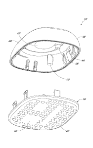
[0021] Figs. 1A and 1B are perspective and exploded views, respectively,
of a
physiological monitoring device, according to one embodiment;
[0022] Figs. 2A and 2B are top perspective and bottom perspective views,
respectively, of a printed circuit board assembly of the physiological
monitoring device;
[0023] Figs. 3A-E are perspective and exploded views of a flexible body
and
gasket of the physiological monitoring device;
[0024] Fig. 4 is an exploded view of a rigid housing of the
physiological
monitoring device;
[0025] Fig. 5A-B is a perspective view of a battery holder of the
physiological
monitoring device;
[0026] Fig. 6A and 6B are cross sectional views of the physiological
monitoring
device;
[0027] Fig. 7 is an exploded view of the physiological monitoring device
including a number of optional items, according to one embodiment;
[0028] Figs. 8A and 8B are perspective views of two people wearing the
physiological monitoring device, illustrating how the device bends to conform
to body
movement and position; and
[0029] Figs. 9A-9F illustrate various steps for applying the
physiological monitor
to a patient's body, according to one embodiment.
DETAILED DESCRIPTION
[0030] The following description is directed to a number of various
embodiments.
The described embodiments, however, may be implemented and/or varied in many
different
ways without departing from the scope of the invention. For example, the
described
embodiments may be implemented in any suitable device, apparatus, or system to
monitor
any of a number of physiological parameters. For example, the following
discussion focuses
primarily on long-term, patch-based cardiac rhythm monitoring devices. In one
alternative
embodiment, a physiological monitoring device may be used, for example, for
pulse
oximetry and diagnosis of obstructive sleep apnea. In various alternative
embodiments, one
size of physiological monitor may be used for adult patients and another size
may be used for
-7-
CA 02898626 2015-07-17
WO 2014/116825 PCT/US2014/012749
pediatric patients. The method of using a physiological monitoring device may
also vary. In
some cases, a device may be worn for one week or less, while in other cases, a
device may be
worn for at least seven days and/or for more than seven days, for example
between fourteen
days and twenty-one days or even longer. Many other alternative embodiments
and
applications of the described technology are possible. Thus, the following
description is
provided for exemplary purposes only. Throughout the specification, reference
may be made
to the term "conformal." It will be understood by one of skill in the art that
the term
"conformal" as used herein refers to a relationship between surfaces or
structures where a
first surface or structure fully adapts to the contours of a second surface or
structure.
[0031] Referring to Figures 1A and 1B, perspective and exploded views of
one
embodiment of a physiological monitoring device 100 are provided. As seen in
Figure 1A,
physiological monitoring device 100 may include a flexible body 110 coupled
with a
watertight, rigid housing 115. Flexible body 110 (which may be referred to as
"flexible
substrate" or "flexible construct") typically includes two wings 130, 131,
which extend
laterally from rigid housing 115, and two flexible electrode traces 311, 312,
each of which is
embedded in one of wings 130, 131. Each electrode trace 311, 312 is coupled,
on the bottom
surface of flexible body 110, with a flexible electrode (not visible in Figure
1A). The
electrodes are configured to sense heart rhythm signals from a patient to
which monitoring
device 100 is attached. Electrode traces 311, 312 then transmit those signals
to electronics
(not visible in Figure 1A) housed in rigid housing 115. Rigid housing 115 also
typically
contains a power source, such as one or more batteries.
[0032] As will be explained in further detail below, the combination of
a highly
flexible body 110, including flexible electrodes and electrode traces 311,
312, with a very
rigid housing 115 may provide a number of advantages. For example, flexible
body 110
includes a configuration and various features that facilitate comfortable
wearing of device
100 by a patient for fourteen (14) days or more without removal. Rigid housing
115, which
typically does not adhere to the patient in the embodiments described herein,
includes
features that lend to the comfort of device 100. Rigid housing 115 also
protects the
electronics and power source contained in housing 120, enhances the ability of
a patient to
provide an input related to a perceived cardiac event, and allows for simple
manufacturing
-8-
CA 02898626 2015-07-17
WO 2014/116825 PCT/US2014/012749
and reusability of at least some of the contents of housing 115. These and
other features of
physiological monitoring device 100 are described in greater detail below.
[0033] Referring now to Figure 1B, a partially exploded view of
physiological
monitoring device 100 illustrates component parts that make up, and that are
contained
within, rigid housing 115 in greater detail. In this embodiment, rigid housing
115 includes an
upper housing member 140, which detachably couples with a lower housing member
145.
Sandwiched between upper housing member 140 and lower housing member 145 are
an
upper gasket 370, and a lower gasket 360 (not visible on Figure 1B but just
below upper
gasket 370). Gaskets 370, 360 help make rigid housing member 115 watertight
when
assembled. A number of components of monitoring device 100 may be housed
between
upper housing member 140 and lower housing member 145. For example, in one
embodiment, housing 115 may contain a portion of flexible body 110, a printed
circuit board
assembly (PCBA) 120, a battery holder 150, and two batteries 160. Printed
circuit board
assembly 120 is positioned within housing 115 to contact electrode traces 311,
312 and
batteries 160. In various embodiments, one or more additional components may
be contained
within or attached to rigid housing 115. Some of these optional components are
described
further below, in reference to additional drawing figures.
[0034] Battery holder 150, according to various alternative embodiments,
may
hold two batteries (as in the illustrated embodiment), one battery, or more
than two batteries.
In other alternative embodiments, other power sources may be used. In the
embodiment
shown, battery holder 150 includes multiple retain tabs 153 for holding
batteries 160 in
holder 150. Additionally, battery holder 150 includes multiple feet 152 to
establish correct
spacing of batteries 160 from the surface of PCBA 120 and ensure proper
contact with spring
fingers 235 and 236. Spring fingers 235 and 236 are used in this embodiment
rather than
soldering batteries 160 to PCBA 120. Although soldering may be used in
alternative
embodiments, one advantage of spring fingers 235 and 236 is that they allow
batteries 160 to
be removed from PCBA 120 and holder 150 without damaging either of those
components,
thus allowing for multiple reuses of both. Eliminating solder connections also
simplifies and
speeds up assembly and disassembly of monitoring device 100.
[0035] In some embodiments, upper housing member 140 may act as a
patient
event trigger. When a patient is wearing physiological monitoring device 100
for cardiac
-9-
CA 02898626 2015-07-17
WO 2014/116825 PCT/US2014/012749
rhythm monitoring, it is typically advantageous for the patient to be able to
register with
device 100 (i.e., log into the device's memory) any cardiac events perceived
by the patient. If
the patient feels what he/she believes to be an episode of heart arrhythmia,
for example, the
patient may somehow trigger device 100 and thus provide a record of the
perceived event. At
some later time, the patient's recorded perceived event could be compared with
the patient's
actual heart rhythm, recorded by device 100, and this may help determine
whether the
patient's perceived events correlate with actual cardiac events. One problem
with patient
event triggers in currently available wearable cardiac rhythm monitoring
devices, however, is
that a small trigger may be hard to find and/or activate, especially since the
monitoring
device is typically worn under clothing. Additionally, pressing a trigger
button may affect the
electronics and/or the electrodes on the device in such a way that the
recorded heart rhythm
signal at that moment is altered simply by the motion caused to the device by
the patient
triggering. For example, pressing a trigger may jar one or both of the
electrodes in such a
way that the recorded heart rhythm signal at that moment appears like an
arrhythmia, even if
no actual arrhythmia event occurred. Additionally, there is a chance that the
trigger may be
inadvertently activated, for instance while sleeping or laying on the
monitoring device.
[0036] In the embodiment shown in Figures 1A and 1B, however, rigid
housing
115 is sufficiently rigid, and flexible body 110 is sufficiently flexible,
that motion applied to
housing 115 by a patient may rarely or ever cause an aberrant signal to be
sensed by the
electrodes. In this embodiment, the central portion of upper housing member
140 is slightly
concave and, when pressed by a patient who is wearing device 100, this central
portion
depresses slightly to trigger a trigger input on PCBA 120. Because the entire
upper surface of
rigid housing 115 acts as the patient event trigger, combined with the fact
that it is slightly
concave, it will generally be quite easy for a patient to find and push down
the trigger, even
under clothing. Additionally, the concave nature of the button allows it to be
recessed which
protects it from inadvertent activations. Thus, the present embodiment may
alleviate some of
the problems encountered with patient event triggers on currently available
heart rhythm
monitors. These and other aspects of the features shown in Figures 1A and 1B
will be
described in further detail below.
[0037] Referring now to Figures 2A and 2B, printed circuit board
assembly 120
(or "PCBA") may include a top surface 220, a bottom surface 230, a patient
trigger input 210
-10-
CA 02898626 2015-07-17
WO 2014/116825 PCT/US2014/012749
and spring contacts 235, 236, and 237. Printed circuit board assembly 120 may
be used to
mechanically support and electrically connect electronic components using
conductive
pathways, tracks or electrode traces 311, 312. Furthermore, because of the
sensitive nature of
PCBA 120 and the requirement to mechanically interface with rigid body 115, it
is beneficial
to have PCBA 120 be substantially rigid enough to prevent unwanted deflections
which may
introduce noise or artifact into the ECG signal. This is especially possible
during patient
trigger activations when a force is transmitted through rigid body 115 and
into PCBA 120.
One way to ensure rigidity of the PCBA is to ensure that the thickness of the
PCBA is
relatively above a certain value. For example, a thickness of at least about
0.08 cm is
desirable and, more preferably, a thickness of at least about 0.17 cm is
desirable. In this
application, PCBA 120 may also be referred to as, or substituted with, a
printed circuit board
(PCB), printed wiring board (PWB), etched wiring board, or printed circuit
assembly (PCA).
In some embodiments, a wire wrap or point-to-point construction may be used in
addition to,
or in place of, PCBA 120. PCBA 120 may include analog circuits and digital
circuits.
[0038] Patient trigger input 210 may be configured to relay a signal
from a patient
trigger, such as upper housing member 140 described above, to PCBA 120. For
example,
patient trigger input 210 may be a PCB switch or button that is responsive to
pressure from
the patient trigger (i e , the upper surface of upper housing portion 140). In
various
embodiments, patient trigger input 210 may be a surface mounted switch, a
tactile switch, an
LED illuminated tactile switch, or the like. In some embodiments, patient
trigger input 210
may also activate an indicator, such as an LED.
[0039] One important challenge in collecting heart rhythm signals from a
human
or animal subject with a small, two-electrode physiological monitoring device
such as device
100 described herein, is that having only two electrodes can sometimes provide
a limited
perspective when trying to discriminate between artifact and clinically
significant signals.
For example, when a left-handed patient brushes her teeth while wearing a
small, two-
electrode physiological monitoring device on her left chest, the tooth
brushing may often
introduce motion artifact that causes a recorded signal to appear very similar
to Ventricular
Tachycardia, a serious heart arrhythmia. Adding additional leads (and, hence,
vectors) is the
traditional approach toward mitigating this concern, but this is typically
done by adding extra
wires adhered to the patient's chest in various locations, such as with a
Holter monitor. This
-11-
CA 02898626 2015-07-17
WO 2014/116825 PCT/US2014/012749
approach is not consistent with a small, wearable, long term monitor such as
physiological
monitoring device 100.
[0040] An alternate approach to the problem described above is to
provide one or
more additional data channels to aid signal discrimination. In some
embodiments, for
example, device 100 may include a data channel for detecting patch motion. In
certain
embodiments, an accelerometer may provide patch motion by simply analyzing the
change in
magnitude of a single axis measurement, or alternatively of the combination of
all three axes.
The accelerometer may record device motion at a sufficient sampling rate to
allow
algorithmic comparison of its frequency spectrum with that of the recorded ECG
signal. If
there is a match between the motion and recorded signal, it is clear that the
device recording
in that time period is not from a clinical (e.g., cardiac) source, and thus
that portion of the
signal can be confidently marked as artifact. This technique may be
particularly useful in the
tooth brushing motion example aforementioned, where the rapid frequency of
motion as well
as the high amplitude artifact is similar to the heart rate and morphology,
respectively, of a
potentially life-threatening arrhythmia like Ventricular Tachycardia.
[0041] In some embodiments, using the magnitude of all three axes for
such an
analysis would smooth out any sudden changes in values due to a shift in
position rather than
a change in activity. In other embodiments, there may be some advantage in
using a specific
axis of measurement such as along the longitudinal axis of the body to focus
on a specific
type of artifact introduced by upward and downward movements associated with
walking or
running. In a similar vein, the use of a gyroscope in conjunction with the
accelerometer may
provide further resolution as to the nature of the motion experienced. While
whole body
movements may be sufficiently analyzed with an accelerometer on its own,
specific motion
of interest such as rotational motion due to arm movement is sufficiently
complex that an
accelerometer alone might not be able to distinguish.
[0042] In addition to detecting motion artifact, an accelerometer tuned
to the
dynamic range of human physical activities may provide activity levels of the
patient during
the recording, which can also enhance accuracy of algorithmic true arrhythmia
detection.
Given the single-lead limitation of device 100, arrhythmias that require
observation of less
prominent waves (e.g. P-wave) in addition to rate changes such as
Supraventricular
Tachycardia pose challenges to both computerized algorithms as well as the
trained human
-12-
CA 02898626 2015-07-17
WO 2014/116825 PCT/US2014/012749
eye. This particular arrhythmia is also characterized by the sudden nature of
its onset, which
may be more confidently discriminated from a non-pathological Sinus
Tachycardia if a
sudden surge in the patient's activity level is detected at the same time as
the increase in
heart rate. Broadly speaking, the provision of activity information to
clinical professionals
may help them discriminate between exercise-induced arrhythmia versus not. As
with
motion artifact detection, a single-axis accelerometer measurement optimized
to a particular
orientation may aid in more specifically determining the activity type such as
walking or
running. This additional information may help explain symptoms more
specifically and
thereby affect the subsequent course of therapeutic action.
[0043] In certain embodiments, an accelerometer with 3 axes may confer
advantages beyond what magnitude of motions can provide. When the subject is
not rapidly
moving, 3-dimensional accelerometer readings may approximate the tilt of PCBA
120, and
therefore body orientation relative to its original orientation. The original
body orientation
can be assumed to be in either an upright or supine position which is required
for appropriate
positioning and application of the device to the body. This information may
aid in ruling out
certain cardiac conditions that manifest as beat-to-beat morphology changes,
such as cardiac
alternans where periodic amplitude changes are observed, often in heart
failure cases.
Similar beat-to-beat morphology changes are observable in healthy subjects
upon shift in
body position due to the shift in heart position relative to the electrode
vector, for example
from an upright to a slouching position. By design, the single-channel device
100 does not
have an alternate ECG channel to easily rule out potential pathological shifts
in morphology,
however, correlation with shifts in body orientation will help explain these
normal changes
and avoid unnecessary treatment due to false diagnosis.
[0044] In other embodiments, the accelerometer may also be used as a
sleep
indicator, based on body orientation and movement. When presenting clinical
events (e.g.,
pauses), it is diagnostically helpful to be able to present information in a
manner that clearly
separates events that occurred during sleep from those during waking hours. In
fact, certain
algorithms such as for ECG-derived respiratory rate only make sense to run
when the patient
is in a relatively motionless state and therefore subtle signal modulation
introduced by chest
movement due to breathing is observable. Respiratory rate information is
useful as one
channel of information necessary to detect sleep apnea in certain patient
populations.
-13-
CA 02898626 2015-07-17
WO 2014/116825 PCT/US2014/012749
[0045] In certain embodiments, the accelerometer may also be used to
detect free-
falls, such as fainting. With an accelerometer, device 100 may be able to mark
fainting
(syncope) and other free-fall events without relying on patient trigger. In
order to allow
timely detection of such critical events, yet considering the battery and
memory limitations of
a small, wearable device such as device 100, acquisition of accelerometer
readings may be
done in bursts, where only interesting information such as a potential free
fall is written to
memory at a high sampling rate. An expansion of this event-trigger concept is
to use specific
tapping motions on device 100 as a patient trigger instead of or in
conjunction with the
button previously described. The use and detection of multiple types of
tapping sequences
may provide better resolution and accuracy into what exactly the patient was
feeling, instead
of relying on the patient to manually record their symptom and duration in a
trigger log after
the fact. An example of such added resolution is to indicate the severity of
the symptom by
the number of sequential taps.
[0046] Alternatively, in other embodiments, an optical sensors may be
used to
distinguish between device motion and patient body motion. Further, in
additional
embodiments, the device may not require a button or trigger.
[0047] Another optional data channel that may be added to physiological
monitoring device 100 is a channel for detecting flex and/or bend of device
100. In various
embodiments, for example, device 100 may include a strain gauge, piezoelectric
sensor or
optical sensor to detect motion artifact in device 100 itself and thus help to
distinguish
between motion artifact and cardiac rhythm data. Yet another optional data
channel for
device 100 may be a channel for detecting heart rate. For example, a pulse
oximeter,
microphone or stethoscope may provide heart rate information. Redundant heart
rate data
may facilitate discrimination of ECG signals from artifact. This is
particularly useful in cases
where arrhythmia such as Supraventricular Tachycardia is interrupted by
artifact, and
decisions must be made whether the episode was actually multiple shorter
episodes or one
sustained episode. Another data channel may be included for detecting ambient
electrical
noise. For example, device 100 may include an antenna for picking up
electromagnetic
interference. Detection of electromagnetic interference may facilitate
discrimination of
electrical noise from real ECG signals. Any of the above-described data
channels may be
-14-
CA 02898626 2015-07-17
WO 2014/116825 PCT/US2014/012749
stored to support future noise discrimination or applied for immediate
determination of
clinical validity in real-time.
[0048] With reference now to Figures 3A and 3B, flexible body 110 is
shown in
greater detail. As illustrated in Figure 3A, flexible body 110 may include
wings 130, 131, a
thin border 133 (or "rim" or "edge") around at least part of each wing 130,
131, electrode
traces 311, 312õ and a hinge portion 132 (or "shoulder") at or near a junction
of each wing
130, 131 with rigid housing 115. Also shown in Figure 3A is upper gasket 370,
which is not
considered part of flexible body 110 for this description, but which
facilitates attachment of
flexible body 110 to rigid housing 115.
[0049] Hinge portions 132 are relatively thin, even more flexible
portions of
flexible body 110. They allow flexible body 110 to flex freely at the area
where it is joined to
rigid housing 115. This enhances comfort, since when the patient moves,
housing 115 can
freely lift off of the patient's skin. Electrode traces 311, 312 are also very
thin and flexible,
to allow for patient movement without signal distortion. Borders 133 are
portions of flexible
body 110 that is thinner than immediately adjacent portions and that provide
for a smooth
transition from flexible body 110 to a patient's skin, thus preventing edge-
lift and penetration
of dirt or debris below flexible body 110.
[0050] As shown in greater detail in Figure 3B, flexible body 110 may
include
multiple layers. As mentioned previously, upper gasket 370 and lower gasket
360 are not
considered part of flexible body 110 for the purposes of this description but
are shown for
completeness of description. This distinction is for ease of description only,
however, and
should not be interpreted to limit the scope of the claimed invention.
Flexible body 110 may
include a top substrate layer 300, a bottom substrate layer 330, an adhesive
layer 340, and
flexible electrodes 350. Top and bottom substrate layers 300, 330 may be made
of any
suitable, flexible material, such as one or more flexible polymers. Suitable
flexible polymers
can include, but are not limited to, polyurethane, polyethylene, polyester,
polypropylene,
nylon, teflon and carbon impregnated vinyl. The material of substrate layers
300, 330 may be
selected based on desired characteristics. For example, the material of
substrate layers 300,
330 may be selected for flexibility, resilience, durability, breathability,
moisture
transpiration, adhesion and/or the like. In one embodiment, for example, top
substrate layer
300 may be made of polyurethane, and bottom substrate layer 330 may be made of
-15-
CA 02898626 2015-07-17
WO 2014/116825 PCT/US2014/012749
polyethylene or alternatively polyester. In other embodiments, substrate
layers 300, 330 may
be made of the same material. In yet another embodiment, substrate layer 330
may contain a
plurality of perforations in the area over adhesive layer 340 to provide for
even more
breathability and moisture transpiration. In various embodiments,
physiological monitoring
device 100 may be worn continuously by a patient for as many as 14-21 days or
more,
without removal during the time of wear and with device 100 being worn during
showering,
exercising and the like. Thus, the material(s) used and the thickness and
configuration of
substrate layers 300, 330 may be essential to the function of physiological
monitoring device
100. In some embodiments, the material of substrate layers 300, 330 acts as an
electric static
discharge (ESD) barrier to prevent arcing.
[0051] Typically, top and bottom substrate layers 300, 330 are attached
to one
another via adhesive placed on one or both layers 300, 330. For example, the
adhesive or
bonding substance between substrate layers 300, 330 may be an acrylic-based,
rubber-based,
or silicone-based adhesive. In other alternative embodiments, flexible body
110 may include
more than two layers of flexible material.
[0052] In addition to the choice of material(s), the
dimensions¨thickness, length
and width¨of substrate layers 300, 330 may be selected based on desired
characteristics of
flexible body 110. For example, in various embodiments, the thickness of
substrate layers
300, 330 may be selected to give flexible body 110 an overall thickness of
between about 0.1
mm to about 1.0 mm. According to various embodiments, flexible body 110 may
also have a
length of between about 7 cm and 15 cm and a width of about 3 cm and about 6
cm.
Generally, flexible body 110 will have a length sufficient to provide a
necessary amount of
separation between electrodes 350. For example, a distance from the center of
one electrode
350 to the center of the other electrode 350 should be at least about 6.0 cm
and more
preferably at least about 8.5 cm. This separation distance may vary, depending
on the
application. In some embodiments, substrate layers 300, 330 may all have the
same
thickness. Alternatively, the two substrate layers 300, 330 may have different
thicknesses.
[0053] As mentioned above, hinge portions 132 allow the rigid body 115
to lift
away from the patient while flexible body 110 remains adhered to the skin. The
functionality
of hinge portions 132 is critical in allowing the device to remain adhered to
the patient
throughout various activities that may stretch and compress the skin.
Furthermore, hinge
-16-
CA 02898626 2015-07-17
WO 2014/116825 PCT/US2014/012749
portions 132 allow for significantly improved comfort while wearing the
device. Generally,
hinge portions 132 will be sufficiently wide enough to provide adequate lift
of rigid body 115
without creating too large of a peel force on flexible body 110. For example,
in various
embodiments, the width of hinge portion 132 should be at least about 025 cm
and more
preferably at least about 0.75 cm.
[0054] Additionally, the shape or footprint of flexible body 110 may be
selected
based on desired characteristics. As seen in Figure 3A, wings 130, 131 and
borders 133 may
have rounded edges that give flexible body 110 an overall "peanut" shape.
However, wings
130, 131 can be formed in any number of different shapes such as rectangles,
ovals, loops, or
strips. In the embodiment shown in Figures 3A and 3B, the footprint top
substrate layer 300
is larger than the footprint of bottom substrate layer 330, with the extension
of top substrate
layer 300 forming borders 133. Thus, borders 133 are made of the same
polyurethane
material that top layer 300 is made of Borders 133 are thinner than an
adjacent portion of
each wing 130, 131, since they includes only top layer 300. The thinner,
highly compliant
rim 133 will likely enhance adherence of physiologic monitoring device 100 to
a patient, as it
provides a transition from an adjacent, slightly thicker portion of wings 130,
131 to the
patient's skin and thus helps prevent the edge of device 110 from peeling up
off the skin.
Border 133 may also help prevent the collection of dirt and other debris under
flexible body
110, which may help promote adherence to the skin and also enhance the
aesthetics of device
110. In alternative embodiments, the footprint of substrate layers 300, 330
may be the same,
thus eliminating borders 133.
[0055] While the illustrated embodiments of Figures 1A-3B include only
two
wings 130, 131, which extend from rigid housing 115 in approximately opposite
directions
(i.e., at a 180-degree angle relative to each other), other configurations are
possible in
alternative embodiments. For example, in some embodiments, wings 130, 131 may
be
arranged in an asymmetrical orientation relative to one another and/or one or
more additional
wings may be included. As long as sufficient electrode spacing is provided to
permit
physiological signal monitoring, and as long as wings 130, 131 are configured
to provide
extended attachment to the skin, any suitable configuration and number of
wings 130, 131
and electrode traces 311, 312 may be used. The embodiments described above
have proven
to be advantageous for adherence, patient comfort and accuracy of collected
heart rhythm
-17-
CA 02898626 2015-07-17
WO 2014/116825 PCT/US2014/012749
data, but in alternative embodiments it may be possible to implement
alternative
configurations.
[0056] Adhesive layer 340 is an adhesive that is applied to two portions
of the
bottom surface of bottom substrate layer 330, each portion corresponding to
one of wings
130, 131. Adhesive layer 340 thus does not extend along the portion of bottom
substrate
layer 330 upon which rigid housing 115 is mounted. Adhesive layer 340 may be
made of any
suitable adhesive, although certain adhesives have been found to be
advantageous for
providing long term adhesion to patient skin with relative comfort and lack of
skin irritation.
For example, in one embodiment, adhesive layer 340 is a hydrocolloid adhesive.
In another
embodiment, the adhesive layer 340 is comprised of a hydrocolloid adhesive
that contains
naturally-derived or synthetic absorbent materials which take up moisture from
the skin
during perspiration.
[0057] Each of the two portions of adhesive layer 340 includes a hole,
into which
one of electrodes 350 fits. Electrodes 350 made of flexible material to
further provide for
overall conformability of flexible body 110. In one embodiment, for example,
flexible
electrodes 350 may be made of a hydrogel 350. Electrodes 350 generally provide
conformal,
non-irritating contact with the skin to provide enhanced electrical connection
with the skin
and reduce motion artifact. In some embodiments, hydrogel electrodes 350 may
be punched
into adhesive layer 340, thus forming the holes and filling them with hydrogel
electrodes
350. In one alternative embodiment, electrodes 350 and adhesive 340 may be
replaced with
an adhesive layer made of a conductive material, such that the entire adhesive
layer on the
underside of each wing 130, 131 acts as an electrode. Such an adhesive layer
may include a
hybrid adhesive/conductive substance or adhesive substance mixed with
conductive elements
or particles. For example, in one embodiment, such an adhesive layer may be a
hybrid of a
hydrogel and a hydrocolloid adhesive.
[0058] As discussed above, in some embodiments, adhesive layer 340 may
cover
a portion of the underside of lower substrate layer 330, such that at least a
portion of the
bottom side of flexible body 110 does not include adhesive layer 340. As seen
in Figure 3A,
hinges 132 may be formed in the flexible body 110 as portions of each wing
130, 131 on
which adhesive layer 340 is not applied. Hinge portions 132 are generally
located at or near
the junction of flexible body 110 with rigid housing 115, and thus provide for
flexing of
-18-
CA 02898626 2015-07-17
WO 2014/116825 PCT/US2014/012749
device 100 to accommodate patient movement. In some embodiments, hinge
portions 132
may have a width that is less than that of adjacent portions of wings 130,
131, thus giving
device 100 its "peanut" shape mentioned above. As shown in Figure 8, as a
subject moves,
device 100 flexes along with patient movement. Device flexion may be severe
and is likely
to occur many times during long term monitoring. Hinge portions 132 may allow
for
dynamic conformability to the subject, while the rigidity of rigid housing 115
may allow
housing 115 to pop up off the patient's skin during device flexion, thus
preventing peeling of
the device 100 off of the skin at its edge.
[0059] Flexible body 110 further includes two electrode traces 311, 312
sandwiched between upper substrate layer 300 and lower substrate layer 330.
Each electrode
trace 311, 312 may include an electrode interface portion 310 and an
electrocardiogram
circuit interface portion 313. As illustrated in Figures 3C and 3D, ECG
circuit interface
portions 313 are in physical contact with spring fingers 237 and provide
electrical
communication with PCBA 120 when device 100 or zoomed-in device portion 101 is
assembled. Electrode interface portions 310 contact hydrogel electrodes 350.
Thus, electrode
traces 311, 312 transmit cardiac rhythm signals (and/or other physiological
data in various
embodiments) from electrodes 350 to PCBA 120.
[0060] The material and thickness of electrode traces 311, 312 are
important for
providing a desired combination of flexibility, durability and signal
transmission. For
example, in one embodiment, electrode traces 311, 312 may include a
combination of silver
(Ag) and silver chloride (AgC1). The silver and silver chloride may be
disposed in layers. For
example, one embodiment of electrode traces 311, 312 may include a top layer
of silver, a
middle layer of carbon impregnated vinyl, and a bottom (patient-facing) layer
of silver
chloride. In another embodiment, both top and bottom layers of electrode
traces 311, 312
may be made of silver chloride. In one embodiment, the top and bottom layers
may be
applied to the middle layer in the form of silver ink and silver chloride ink,
respectively. In
an alternative embodiment, each electrode trace may include only two layers,
such as a top
layer of silver and a bottom layer of silver chloride. In various embodiments,
the material of
a bottom layer of each electrode trace 311, 312, such as AgC1, may be selected
to match the
chemistry of the hydrogel electrodes 350 and create a half-cell with the body
of the subject.
-19-
CA 02898626 2015-07-17
WO 2014/116825 PCT/US2014/012749
[0061] The thickness of the electrode traces 311, 312 may be selected to
optimize
any of a number of desirable properties. For example, in some embodiments, at
least one of
the layers of electrode traces 311, 312 can be of a sufficient thickness to
minimize or slow
depletion of the material from an anode/cathode effect over time.
Additionally, the thickness
may be selected for a desired flexibility, durability and/or signal
transmission quality.
Flexible electrode traces 311, 312 generally may help provide conformal
contact with the
subject's skin and may help prevent electrodes 350 from peeling or lifting off
of the skin,
thereby providing strong motion artifact rejection and better signal quality
by minimizing
transfer of stress to electrodes 350.
[0062] As mentioned above, in some embodiments, top gasket 370 and
bottom
gasket 360 may be attached upper substrate 300 and lower substrate 330 of
flexible body
110. Gaskets 360, 370 may be made of any suitable material, such as urethane,
which
provides a water tight seal between the upper housing member 140 and lower
housing
member 145 of rigid housing 115. In one embodiment, top gasket 370 and/or
bottom gasket
360 may include an adhesive surface. Figure 3E depicts yet another embodiment
where top
gasket 370 includes tabs 371 that protrude away from the profile of top
housing 140 while
still being adhered to upper substrate 300. The tabs 371 cover a portion of
electrode traces
311, 312 and provide a strain relief for the traces at the point of highest
stress where the
flexible body meets the rigid housing.
[0063] With reference now to Figure 4, upper housing member 140 and
lower
housing member 145 of rigid housing 115 are shown in greater detail. Upper and
lower
housing members 140, 145 may be configured, when coupled together with gaskets
360, 370
in between, to form a watertight enclosure for containing PCBA 120, battery
holder 150,
batteries 160 and any other components contained within rigid housing 115.
Housing
members 140, 145 may be made of any suitable material to protect internal
components, such
as water resistant plastic. In one embodiment, upper housing member 140 may
include a
rigid sidewall 440, a light pipe 410 to transmit visual information from the
LEDs on the
PCBA through the housing member, a slightly flexible top surface 420, and an
inner trigger
member 430 extending inward from top surface 420. Top surface 420 is
configured to be
depressed by a patient when the patient perceives what he or she believes to
be an arrhythmia
or other cardiac event. When depressed, top surface 420 depresses inner
trigger member 430,
-20-
CA 02898626 2015-07-17
WO 2014/116825 PCT/US2014/012749
which contacts and activates trigger input 210 of PCBA 120. Additionally, as
discussed
previously, top surface 420 may have a concave shape (concavity facing the
inside of
housing 115) to accommodate the shape of a finger. It is believed that the
design of upper
housing member 140 isolates activation of the trigger input 210 from
electrodes 350, thereby
minimizing artifact in the data recording.
[0064] With continued reference to Figure 4, lower housing member 145
may be
configured to detachably connect with upper housing member 140 in such a way
that housing
members 140, 145 may be easily attached and detached for reusability of at
least some of the
component parts of monitoring device 100. In some embodiments, a bottom
surface 445
(patient facing surface) of lower housing member 145 may include multiple
dimples 450 (or
"bumps," "protrusions" or the like), which will contact the patient's skin
during use. Dimples
450 may allow for air flow between bottom surface 445 and the patient's skin,
thus
preventing a seal from forming between bottom surface 445 and the skin. It is
believed that
dimples 450 improve comfort and help prevent a perception in currently
available devices in
which the patient feels as if monitoring device 100 is falling off when it
housing 115 lifts off
the skin and breaks a seal with the skin. In yet another embodiment the bottom
surface 445 of
lower housing member 450 may include multiple divots (recesses instead of
protrusions) to
prevent a seal from forming.
[0065] Referring now to Figure 5A, battery holder 150 is shown in
greater detail.
Battery holder 150 may be made of plastic or other suitable material, is
configured to be
mounted to PCBA 120 and subsequently attached to rigid housing 115, and is
capable of
holding two batteries 160 (Figure 1B). In alternative embodiments, battery
holder 150 may
be configured to hold one battery or more than two batteries. A plurality of
protrusions 152
provide a stable platform for batteries 160 to be positioned a fixed distance
above the surface
of PCBA 120, avoiding unwanted contact with sensitive electronic components
yet providing
for adequate compression of spring contacts 235 (Figure 5B). Protrusions 153
lock batteries
160 into position and resist the upward force on the batteries from spring
contacts 235.
Battery holder 150 also positions batteries appropriately 160 to provide for
adequate
compression of spring contacts 236. Use of battery holder 150 in conjunction
with spring
contacts 235 and 236 allows for batteries 160 to be electrically connected to
PCBA 120 while
still having additional electronic components between batteries 160 and PCBA
120 and
-21-
CA 02898626 2015-07-17
WO 2014/116825 PCT/US2014/012749
maintain a very compact assembly. Battery holder 150 may include a flexible
hook 510
which engages a corresponding rigid hook 440 of upper housing member 140.
Under normal
assembly conditions the flexible hook 510 remains securely mated with rigid
hook 440. For
disassembly, flexible hook 510 can be pushed and bent using an appropriate
tool passed
through top housing 140 causing it to disengage from rigid hook 440 and
subsequently allow
top housing 140 to be removed.
[0066] With reference now to Figure 6A and 6B, physiological monitoring
device
100 is shown in side view cross-section. As shown in 6A, physiological
monitoring device
100 may include flexible body 110 coupled with rigid housing 115. Flexible
body 110 may
include top substrate layer 300, bottom substrate layer 330, adhesive layer
340 and electrodes
350. Electrode traces 311, 312 are also typically part of flexible body 110
and are embedded
between top substrate layer 300 and bottom substrate layer 330, but they are
not shown in
Figure 6. Flexible body 110 forms two wings 130, 131, extending to either side
of housing
115, and a border 133 surrounding at least part of each wing 130, 131. Rigid
housing 115
may include an upper housing member 140 coupled with a lower housing member
145 such
that it sandwiches a portion of flexible body 110 in between and provides a
watertight, sealed
compartment for PCBA 120. Upper housing member 140 may include inner trigger
member
430, and PCBA may include patient trigger member 210. As discussed previously,
lower
housing member 145 may include multiple dimples 450 or divots to enhance the
comfort of
the monitoring device 100.
[0067] It is desirable that PCBA 120 is sufficiently rigid to prevent
bending and
introducing unwanted artifact into the signal. In certain embodiments, an
additional
mechanism to reduce and prevent unwanted bending of PCBA 120 may be used. This
mechanism is shown in figure 6B. Support post 460 is integral to lower housing
145 and is
positioned directly under patient trigger input 210. During patient symptom
triggering, upper
housing member 140 is depressed, engaging inner trigger mechanism 430 and
transmitting a
force through patient trigger input 210 into PCBA 120. The force is further
transmitted
through PCBA 120 and into support post 460 without creating a bending moment,
thus
avoiding unwanted artifact.
[0068] Referring to Figure 7, in some embodiments, physiological
monitoring
device 100 may include one or more additional, optional features. For example,
in one
-22-
CA 02898626 2015-07-17
WO 2014/116825 PCT/US2014/012749
embodiment, monitoring device 100 may include a removable liner 810, a top
label 820, a
device identifier 830 and a bottom label 840. Liner 810 may be applied over a
top surface of
flexible member 110 to aid in the application of device 100 to the subject. As
is described in
further detail below, liner 810 may help support borders 133 of flexible body
110, as well as
wings 130, 131, during removal of one or more adhesive covers (not shown) that
cover
adhesive surface 340 before use. Liner 810 may be relative rigid and/or firm,
to help support
flexible body 110 during removal of adhesive covers. In various embodiments,
for example,
liner 810 may be made of cardboard, thick paper, plastic or the like. Liner
810 typically
includes an adhesive on one side for adhering to the top surface of wings 130,
131 of flexible
body 110.
[0069] Labels 820, 840 may be any suitable labels and may include
produce
name(s), manufacturer name(s), logo(s), design(s) and/or the like. They may be
removable or
permanently attached upper housing member 140 and/or lower housing member 145,
although typically they will be permanently attached, to avoid unregulated
reuse and/or
resale of the device by an unregistered user. Device identifier 830 may be a
barcode sticker,
computer readable chip, RF1D, or the like. Device identifier 830 may be
permanently or
removably attached to PCBA 120, flexible body 110 or the like. In some
embodiments, it
may be beneficial to have device identifier 830 stay with PCB A 120.
[0070] Referring now to Figures 8A and 8B, physiological monitoring
device 100
generally includes hinge portions 132 at or near the juncture of each wing
130, 131 with rigid
housing 115. Additionally, each wing 130, 131 is typically adhered to the
patient via
adhesive layers 340, while rigid body 115 is not adhered to the patient and is
thus free to
"float" (i.e., move up and down) over the patient's skin during movement and
change of
patient position. In other words, when the patient's chest contracts, rigid
housing pops up or
floats over the skin, thus minimizing stress on device 100, enhancing comfort,
and reducing
the tendency of wings 130, 131 to peel off of the skin. The advantage provided
by the
combination of the floating rigid body 115 and the adhered wings 130, 131 is
illustrated in
Figures 8A and 8B. In Figure 8A, a patient is sleeping, and in Figure 8B, a
patient is playing
golf In both examples, monitoring device 100 is squeezed together by the
patient's body,
causing rigid housing 115 to float above the skin as wings 130, 131 move
closer together.
This advantage of a floating, non-attached portion of a physiological
monitoring device is
-23-
CA 2898626 2017-05-10
described in further detail in U.S. Patent 8,560,046.
[0071] Referring now to Figures 9A-9F, one embodiment of a method for
applying physiological monitoring device 100 to the skin of a human subject is
described. In
this embodiment, before the first step shown in Figure 9A, the patient's skin
may be prepared,
typically by shaving a small portion of the skin on the left chest where
device 100 will be
placed and then abrading and/or cleaning the shaved portion. As shown in
Figure 9A, once the
patient's skin is prepared, a first step of applying device 100 may include
removing one or
both of two adhesive covers 600 from adhesive layers 340 on the bottom surface
of device
100, thus exposing adhesive layers 340. As illustrated in Figure 9B, the next
step may be to
apply device 100 to the skin, such that adhesive layer 340 adheres to the skin
in a desired
location. In some embodiments, one adhesive cover 600 may be removed, the
uncovered
adhesive layer 340 may be applied to the skin, and then the second adhesive
cover 600 may
be removed, and the second adhesive layer 340 may be applied to the skin.
Alternatively, both
adhesive covers 600 may be removed before applying device 100 to the skin.
While adhesive
covers 600 are being removed, liner 810 acts as a support for flexible body
110, provides the
physician or other user with something to hold onto, and prevents flexible
body 110 and
borders 133 of flexible body 110 from folding in on themselves, forming
wrinkles, etc. As
described above, liner 810 may be made of a relatively stiff, firm material to
provide support
for flexible body 110 during application of device 100 to the skin. Referring
to Figure 9C,
after device 100 has been applied to the skin, pressure may be applied to
flexible body 110 to
press it down onto the chest to help ensure adherence of device 100 to the
skin.
[0072] In a next step, referring to Figure 9D, liner 810 is removed
from (peeled off
of) the top surface of flexible body 110. As shown in Figure 9E, once liner
810 is removed,
pressure may again be applied to flexible body 110 to help ensure it is
adhered to the skin.
Finally, as shown in Fig. 9F, upper housing member 140 may be pressed to turn
on
physiological monitoring device 140. This described method is only one
embodiment. In
alternative embodiments, one or more steps may be skipped and/or one or more
additional
steps may be added.
24
CA 02898626 2015-07-17
WO 2014/116825 PCT/US2014/012749
[0073] When a desired monitoring period has ended, such as about 14-21
days in
some cases, a patient (or physician, nurse or the like) may remove
physiological monitoring
device 100 from the patient's skin, place device 100 in a prepaid mailing
pouch, and mail
device 100 to a data processing facility. At this facility, device 100 may be
partially or
completely disassembled, PCBA 120 may be removed, and stored physiological
data, such as
continuous heart rhythm information, may be downloaded from PCBA 120. The data
may
then be analyzed by any suitable method and then provided to a physician in
the form of a
report. The physician may then discuss the report with the patient. PCBA 120
and/or other
portions of device 100, such as rigid housing 115, may be reused in the
manufacture of
subsequent devices for the same or other patients. Because device 100 is built
up as a
combination of several removably coupled parts, various parts may be reused
for the same
embodiment or different embodiments of device 100. For example, PCBA 120 may
be used
first in an adult cardiac rhythm monitor and then may be used a second time to
construct a
monitor for sleep apnea. The same PCBA 120 may additionally or alternatively
be used with
a differently sized flexible body 110 to construct a pediatric cardiac
monitor. Thus, at least
some of the component parts of device 100 may be interchangeable and reusable.
[0074] Advantageously, physiological monitoring device 100 may provide
long
term adhesion to the skin. The combination of the configuration of flexible
and conformal
body 110, the watertight, low profile configuration of rigid housing 115, and
the interface
between the two allows device 100 to compensate for stress caused as the skin
of the subject
stretches and bends. As a result, device 100 may be worn continuously, without
removal, on
a patient for as many as 14-21 days or more. In some cases, device 100 may be
worn for
greater or less time, but 14-21 days may often be a desirable amount of time
for collecting
heart rhythm data and/or other physiological signal data from a patient.
[0075] In various alternative embodiments, the shape of a particular
physiological
monitoring device may vary. The shape, footprint, perimeter or boundary of the
device may
be circular, an oval, triangular, a compound curve or the like, for example.
In some
embodiments, the compound curve may include one or more concave curves and one
or more
convex curves. The convex shapes may be separated by a concave portion. The
concave
portion may be between the convex portion on the rigid housing and the convex
portion on
-25-
CA 02898626 2015-07-17
WO 2014/116825 PCT/US2014/012749
the electrodes. In some embodiments, the concave portion may correspond at
least partially
with a hinge, hinge region or area of reduced thickness between the body and a
wing.
[0076] While described in the context of a heart monitor, the device
improvements described herein are not so limited. The improvements described
in this
application may be applied to any of a wide variety of physiological data
monitoring,
recording and/or transmitting devices. The improved adhesion design features
may also be
applied to devices useful in the electronically controlled and/or time
released delivery of
pharmacological agents or blood testing, such as glucose monitors or other
blood testing
devices. As such, the description, characteristics and functionality of the
components
described herein may be modified as needed to include the specific components
of a
particular application such as electronics, antenna, power supplies or
charging connections,
data ports or connections for down loading or off loading information from the
device,
adding or offloading fluids from the device, monitoring or sensing elements
such as
electrodes, probes or sensors or any other component or components needed in
the device
specific function. In addition or alternatively, devices described herein may
be used to detect,
record, or transmit signals or information related to signals generated by a
body including but
not limited to one or more of ECG, EEG and/or EMG.
[0077] While the above embodiments disclose the invention with respect
to a data
channel for collecting a single physiological signal, it is contemplated that
additional data
channels can be include to collect additional data, for example, device
motion, device flex or
bed, heart rate and/or ambient electrical noise.
[0078] Various embodiments of a physiological monitoring device and
methods
for using it have been disclosed above. These various embodiments may be used
alone or in
combination, and various changes to individual features of the embodiments may
be altered,
without departing from the scope of the invention. For example, the order of
various method
steps may in some instances be changed, and/or one or more optional features
may be added
to or eliminated from a described device. Therefore, the description of the
embodiments
provided above should not be interpreted as unduly limiting the scope of the
invention as it is
set forth in the claims.
[0079] Various modifications to the implementations described in this
disclosure
may be made, and the generic principles defined herein may be applied to other
-26-
CA 02898626 2015-07-17
WO 2014/116825 PCT/US2014/012749
implementations without departing from the spirit or scope of this disclosure.
Thus, the
claims are not intended to be limited to the implementations shown herein, but
are to be
accorded the widest scope consistent with this disclosure, the principles and
the novel
features disclosed herein.
[0080] Certain features that are described in this specification in the
context of
separate embodiments also can be implemented in combination in a single
embodiment.
Conversely, various features that are described in the context of a single
embodiment also
can be implemented in multiple embodiments separately or in any suitable
subcombination.
Moreover, although features may be described above as acting in certain
combinations and
even initially claimed as such, one or more features from a claimed
combination can in some
cases be excised from the combination, and the claimed combination may be
directed to a
subcombination or variation of a subcombination.
[0081] Similarly, while operations are depicted in the drawings in a
particular
order, such operations need not be performed in the particular order shown or
in sequential
order, or that all illustrated operations be performed, to achieve desirable
results. Further, the
drawings may schematically depict one more example processes in the form of a
flow
diagram. However, other operations that are not depicted can be incorporated
in the example
processes that are schematically illustrated. For example, one or more
additional operations
can be performed before, after, simultaneously, or between any of the
illustrated operations.
Moreover, the separation of various system components in the embodiments
described above
should not be interpreted as requiring such separation in all embodiments.
Additionally,
other embodiments are within the scope of the following claims. In some cases,
the actions
recited in the claims can be performed in a different order and still achieve
desirable results.
-27-