Note: Descriptions are shown in the official language in which they were submitted.
CA 02899216 2015-07-23
WO 2014/115075 PCT/1B2014/058428
TITLE
"Device, system and method for detection and processing of heartbeat signals"
DESCRIPTION
The present invention relates to innovative devices, systems and methods for
detecting
the heart beat.
Heart beat detection systems (also called BVP - Blood Volume Pulse - detection
systems) which function optically are known. These systems usually employ a
light
emitter which by means of reflection or transparency illuminates a suitable
receiver
after the emitted light has struck or passed through a zone of the body.
Basically, these heart rate monitors are detection systems which are able to
measure the
way in which the blood volume changes over time in a specific zone of the
body.
Generally, the reflection devices are placed on a zone of the body, such as
the wrist,
where there is a variation in the quantity of light reflected depending on the
superficial
blood flow in this zone. The transparency devices are instead applied in the
vicinity of
relatively thin parts of the body (such as the fingers or the ear lobes) so
that the light is
able to pass through them and detect the variation in the light passing
through owing to
the blood flow in said parts.
Both systems, however, are subject to disturbances of the useful signal, for
example due
to both the surrounding light conditions and the movement of the person
undergoing
measurement.
For example, the sensor operates by means of contact with a deformable medium -
the
skin - inside which blood flows. This medium is subject to mechanical
deformation
SUBSTITUTE SHEET (RULE 26)
CA 02899216 2015-07-23
WO 2014/115075 PCT/1B2014/058428
which corrupts the measurement, adding an unwanted signal, namely noise.
Reflection devices are more practical for prolonged use, but the variation in
reflected
light produced by the variations in blood flow following the heart beat is
very small and
moreover is generally affected by a large amount of noise.
For example, although the wrist is one of the most convenient positions for
wearing a
reflection sensor for detecting the pulse, the noise on the signal, created by
the
movement of the tissues underneath the sensor following, for example, the
movement of
the limb, the wrist or the fingers, is one of the major obstacles to optical
detection of the
pulse in this zone. Also the act of moving or walking produces relative
movements of
the sensor and tissues which produce further disturbances of a significant
nature.
In the art various solutions have been proposed in order to try to improve the
signal/noise ratio during reflection detection, attempting to filter the
various
disturbances superimposed on the useful signal.
For example it has been proposed using movement sensors arranged together with
optical sensors for detecting relatively wide amplitude movements of the body
to which
the sensor is applied. This detection arrangement, however, does not provide
data about
the relative displacement of sensor and underlying tissue and is usually used
to prevent
reading of the optical sensor in the case of excessive movements on the part
of the
person, which it is assumed a priori may produce a large amount of disturbance
which
cannot be effectively filtered. In the case of prolonged physical activity,
the sensor
remains, however, deactivated for a long period and precisely when detection
of the
heart beat is of most interest.
It has also been proposed using two light sources with a suitable different
wavelength.
2
CA 02899216 2015-07-23
WO 2014/115075
PCT/1132014/058428
The first wavelength has been chosen from among those wavelengths which are
not
absorbed by the oxyhaemoglobin (for example red), while the second wavelength
is
chosen from among those which are better absorbed by the oxyhaemoglobin (for
example green). This results in a first signal which is better related to the
movement of
the tissues and a second signal which is better related to the blood flow.
Filtering of the
noise is then performed by suitably subtracting the first signal from the
second signal,
so as to mitigate the effect of the relative movements of tissues and sensor.
Such a
system is described for example in EP2462866.
A filtering system of this type provides an output signal with reduced noise.
However,
most often the signal/noise ratio is still very unfavourable. Moreover, not
always does
the response to the specific wavelength chosen remain constant with the
passing of time
and/or change of person undergoing the measurement.
Mixed methods also provide results which are not entirely satisfactory. For
example, the
noise is very high both when running and when working on a computer (finger
movements). In the first case accelerometers are most useful for eliminating
the noise,
while in the second case it is preferable to make use of the system with two
wavelengths. The simultaneous use of both methods as proposed in the prior art
(tbr
example, as described again in US2012150052) compensates, however, only for a
number of noise sources and still does not provide a satisfactory signal/noise
ratio for
special applications or where the person is free to perform any daily
activity. Moreover,
the two systems may interfere with each other, further hindering detection.
A general object of the present invention is to provide a system able to
ensure
satisfactory detection of the heart beat even in the presence of disturbances
caused by
3
CA 02899216 2015-07-23
WO 2014/115075 PCT/1B2014/058428
various sources. A further object is to provide an innovative system for
processing heart
beat signals.
In view of these objects the idea which has occurred, according to the
invention, is to
provide a heart beat detection device comprising at least one optical
reflection sensor
unit to be placed on the person's skin, the sensor unit being provided with a
light emitter
and a corresponding light receiver which converts the light reflected by the
skin into an
electric signal, characterized in that it comprises electrically adjustable
optical filters
which are connected to the emitter, to the receiver or to both of them in
order to select,
upon operation, a desired wavelength of the light.
The idea has also occurred to provide a system for detecting and processing
physiological data, comprising at least one device according to any one of the
preceding
claims connected by means of a wireless interface to a data processing and
transmission
unit which receives the data from the device and processes it.
The idea has also occurred to provide a method for increasing the signal/noise
ratio of
an electric signal for detecting the heart beat optically by means of at least
one optical
reflection sensor unit, comprising differentiating between the effects of at
least two light
wavelengths by means of electrically adjustable optical filters and processing
the
corresponding signals received from the at least one optical reflection unit
in order to
obtain the electric signal representing the heart beat.
As will become clear from the description and the drawings, according to the
invention
a device for detecting or monitoring the heart rate may comprise a sensor
system which
is in contact with the skin and which communicates with a central processing
system.
The remote system may comprise one or more optical detection systems for
measuring
4
CA 02899216 2015-07-23
WO 2014/115075 PCT/1B2014/058428
the variations in the blood volume making use of the physical principles of
absorption
and fluorescence. The optical systems may comprise:
- one or more wide-band light emitters (for example LEDs);
- one or more wide-band light receivers (for example photodiodes or
phototransistors)
and
- one or more tunable monochromators which can be connected to the light
emitters, to
the light receivers or to both of them, in order to select a certain
wavelength.
The heart rate monitor may also comprise one or more of the following:
- one or more optical detection systems situated at a fixed distance along the
direction of
the blood flow in order to estimate the propagation time of the blood;
- an electrical detection system comprising two or more electrodes in contact
with the
skin for measuring the galvanic response of the skin, and
- a mechanical detection system for measuring the three-dimensional
acceleration and
the orientation of the system.
The heart frequency monitor may also envisage that one or more monochromators
allow
the optical detection system to work both in absorption mode and in
fluorescence mode
at two or more wavelengths.
Still according to the invention a method for maximizing the signal/noise
ratio of the
blood volume signal may comprise the steps of:
- differentiating between the effects of two (or more) wavelengths in
absorption mode
and fluorescence mode on signals of the optical detection systems;
- dynamically tuning the two (or more) wavelengths in order to maximize the
signal
levels of the optical detection systems.
CA 02899216 2015-07-23
WO 2014/115075
PCT/1B2014/058428
Moreover the method may comprise one or more of the following steps:
- combining the effects of the absorption mode and fluorescence mode on the
signals of
=
the optical detection systems;
- combining the signals from the optical detection systems with the signal of
the
electrical detection system;
- eliminating the effect of deformation of the medium on the blood propagation
time,
and
- eliminating the effect of deformation of the medium resulting from other
mechanical
effects which are contained in the signal supplied by the mechanical detection
system
The heart rate monitor may also comprise a remote system in contact with a
user's skin
and communicating with a central processing system.
The remote system may also comprise one or more of the following elements:
- a remote processor;
- a detection system connected to the remote processor;
- a remote memory connected to the remote processor;
- a clock signal generator connected to the remote processor;
- a remote user interface connected to the remote processor;
- a remote transceiver connected to the remote processor;
- a remote antenna connected to the remote transceiver, and
- a remote battery connected to the remote processor, to the detection system,
to the
remote memory, to the clock signal generator, and to the remote transceiver.
The central processing system may comprise:
- a central processor;
6
CA 02899216 2015-07-23
WO 2014/115075 PCT/1B2014/058428
- a central memory connected to the central processor;
- a central transceiver connected to the central processor, and
- a central antenna connected to ie central transceiver.
The said central memory may further comprise a set of instructions which can
be carried
out on the central processor, the instructions comprising:
- an algorithm for maximizing the signal/noise ratio of the blood volume
signal received
from the remote system, and
- an algorithm for processing the optimized blood volume signal for
determining a
pulsation signal based on detection of the peak value.
In order to illustrate more clearly the innovative principles of the present
invention and
its advantages compared to the prior art, examples of embodiment applying
these
principles will be described below with the aid of the accompanying drawings.
In the
drawings:
- Figure I shows a block diagram of a first reflection detection device
provided in
accordance with the principles of the present invention;
- Figure 2 shows a graph of signals detected by an apparatus according to the
invention;
- Figure 3 shows a block diagram of a second reflection detection device
provided in
accordance with the principles othe present invention;
- Figure 4 shows a further graph illustrating signals detected by an apparatus
according
to the invention;
- Figure 5 shows a block diagram of a possible system for remote processing of
the data
detected by the sensors according to the invention;
- Figure 6 shows a schematic view of a bracelet detection system and
intelligent
7
CA 02899216 2015-07-23
WO 2014/115075 PCT/1B2014/058428
portable terminal for processing (or initial processing) of the signals
detected.
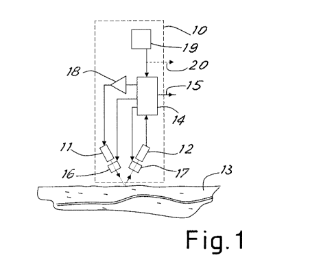
With reference to the figures, Figure 1 shows a first reflection detector
according to the
invention for detecting the heart beat.
Such a detector, which is generally denoted by 10, comprises a light emitter
11 (for
example an LED diode emitter) and a corresponding receiver 12 (for example a
photodiode or phototransistor) which receives the light of the emitter 11
after reflection
on the skin 13 of the person undergoing heart beat detection. Advantageously,
as will be
clarified below, the detector or device 10 may be positioned on the rear part
of the wrist,
for example in the manner of a wrist-watch.
The receiver 12 converts the light received into an electric signal sent to an
electronic
processing block 14 which emits a corresponding signal 15 (also called BVP -
i.e.
Blood Volume Pulse - signal) which depends on the heart beat of the person.
The block
14 may he a combination of an analog amplification circuit and programmable
microprocessor device for processing a signal, as may be easily imagined by
the person
skilled in the art in the light of the description provided here.
Advantageously, the emitter 11 emits light in a wide spectrum (for example
white light)
and the device 10 comprises an adjustable optical filter 16 and/or an
adjustable optical
filter 17, which are arranged respectively in front of the transmitter 11 and
the receiver
12. These optical filters may be controlled by the processing block 14 so as
to be tuned
to a desired wavelength for filtering the light sent and/or reflected.
Advantageously, these optical filters comprise so-called "monochromators" and
allow
dynamic selection of a specific wavelength from a wide-spectrum light. In
particular, it
has been found to be advantageous to use tunable Fabry-Perot monochromators,
known
8
CA 02899216 2015-07-23
WO 2014/115075 PCT/1B2014/058428
per se, which can be easily miniaturized.
Again advantageously, the device may comprise a circuit 18 for powering the
emitter 11
which is controlled by the processing block 14 so as to time the emission
luminosity of
the emitter II to a desired value.
For reasons which will become clear below, the device 10 may also comprise a
known
accelerometer 19 which sends movement signals to the processing block 14.
Advantageously, the accelerometer is chosen to measure the three-dimensional
acceleration and the orientation of the system.
As is known, the oxyhaemoglobin present in the blood absorbs given
wavelengths. This
effect is referred to as "absorption".
Moreover, the oxyhaemoglobin re-emits part of the energy absorbed in the form
of light
at a wavelength different from that absorbed. This effect is referred to as
"fluorescence".
Owing to the use of adjustable filters, it is possible to configure the system
in order to
make use first of one effect and then the other effect. In the first mode the
wavelength
which maximizes absorption is provided and the same wavelength is "observed"
by
means of the receiver 12. In the second mode the wavelength which maximizes
the
fluorescence is provided and the fluorescence wavelength characteristic of
oxyhaemoglobin (wavelength which is always greater than the incident
wavelength for
energy balance reasons) is observed by means of the receiver 11.
By combining the signal read by the receiver in the two different modes, i.e.
"absorption" mode and "fluorescence" mode, it is possible to improve the
signal/noise
ratio.
9
CA 02899216 2015-07-23
WO 2014/115075 PCT/1B2014/058428
Moreover, owing to the adjustability of the filters it is possible to adapt
the fluorescence
and/or absorption wavelength to the characteristics of the skin of the person
whose heart
beat is being detected (for example age, degree of tanning, skin complexion,
presence of
fat, presence of hair).
In fact the skin situated between detector and oxyhaernoglobin creates optical
interference which may alter the light emitted and/or received. Therefore, it
has been
found useful to attempt to find, possibly whenever the device is switched on,
the
wavelengths which maximize the amplitude of the BVP signal, depending on the
characteristics of the skin, both in fluorescence mode and in absorption mode.
For example, extremely fair skin favours the penetration of light and
therefore, in
absorption mode, wavelengths close to the UV band may be effectively used. On
the
contrary, tanned or dark skins do not allow small wavelengths to reach the
receiver
except in the case where the intensity is such that it adversely affects the
battery life.
A similar situation exists in fluorescence mode, where a maximum response of
the
oxyhaemoglobin is obtained by performing stimulation in the violet-blue band
and
detection in the orange band.
In other words, during operation, the processing block 14 may tune the filters
to
wavelengths considered suitable for detecting the heart beat using the
"absorption"
method (for example in the range of 530-580 nin for dark skin and 410-450 nm
for
extremely fair skin) and acquire the corresponding signal reflected and
captured by the
receiver 12. The processing block 14 may also tune the filters to a wavelength
considered suitable for detecting the heart beat using the "fluorescence"
method (for
example in the range 410-450 nm for the emission filter and 590-630 nrn for
the
CA 02899216 2015-07-23
WO 2014/115075 PCT/1B2014/058428
reception filter) and acquire the corresponding fluorescence signal captured
by the
receiver 12. By superimposing the two signals received (suitably compensating
for the
temporal delay between the two measurements) it is possible to obtain a BVP
signal
with a greater amplitude than the background noise.
Moreover, during the two measurements (or, advantageously, during a
calibration step
which may take place upon swit Thing on the device following application onto
the skin,
or cyclically during operation) the device may vary the wavelength of the
filter in the
region of the basic wavelengths defined for fluorescence and absorption,
attempting to
maximize the signal peak received in the two modes. After defining the
wavelengths for
which the greater signal is obtained, the device may use these wavelengths for
the
subsequent measurements until the subsequent calibration operation is
performed.
By periodically repeating calibration during operation of the device it is
possible to
compensate also for the varying conditions of the skin (for example, variation
in the
degree of tanning, sweating or change in temperature) which may influence the
measurement.
By way of a further advantage it is also possible to compensate for
disturbances on the
signal due to relative movements of the skin and device, for example caused by
movements of the person or movements of the muscles and tendons of the body
zone on
which the sensor is placed (for example movement of the fingers). In fact it
is possible
to tune the filter (or filters, in the case of a device with both filters) so
that the light
emitted by the emitter 11 is characterized by a wavelength which is less
sensitive to
flowing of the blood, hut more sensitive to movements on or under the skin
(for
example the wavelength 650-750 nm). The corresponding signal captured by the
CA 02899216 2015-07-23
WO 2014/115075 PCT/1B2014/058428
detector 12 may be used by the processing block 14 as a noise signal to be
subtracted
from the electric signal obtained by the detection of the BVP signal, via an
adaptive
numerical filter, so as to eliminate an important noise component.
Filtering may also take place for selecting green light or red light for the
uses
substantially of the prior art or also for filtering (using suitable emitters)
in the infrared
range or other ranges.
Advantageously, the detector 10 may also use the signal supplied by the
accelerometer
19 in order to compensate for disturbances due to major movements of the
device (for
example, as a result of physical activity performed by the person). The
accelerometer
signal may be supplied to the block 14 in order to provide an adaptive
numerical filter
which intervenes in the case of sudden accelerations (for example when
running).
The signal of the accelerometer 19 may also be used to prevent emission of the
BVP
signal by the device when the acceleration detected is above a threshold which
has been
determined beforehand as corresponding to a movement noise source which is too
great
for effective compensation of the noise on the BVP detected by the optical
system.
In order to reduce the noise on the output signal, the device 10 may also
advantageously
act on the luminous intensity of the light emitted by the sensor 11. However,
in the case
of battery-powered devices, a greater light intensity may negatively affect
the duration
of the battery charge.
Figure 2 shows a graph which schematically illustrates the relationship
between light E
emitted by the emitter (axis X) and amplitude of the signal received R (axis
Y).
As can be seen from the graph, there exists essentially a linear relationship
between
light emitted and reflected light measured by the receiver. The noise signal,
or BR
12
CA 02899216 2015-07-23
WO 2014/115075 PCT/1B2014/058428
(Background Reflection) signal, which may also comprise any ambient light
captured
by the sensor, and the BPR (Blood Pulse Reflection) signal are therefore both
incremented with an increase in the luminous emission intensity E. The slope
of the two
straight lines in the graph, which define the light reflection curve, may vary
from person
to person.
All the above means that, given a certain emitted light E. a first person may
have a
certain dR (namely a certain amplitude dR of periodic variation of reflected
light which
carries blood pulsation information). A second person may have, for the same
value E,
a value dR smaller or greater than the value dR of the first person.
If a value dRmin is established (namely a minimum useful signal value
received) the
emitter will be advantageously controlled by the block 14 so as to have in any
case an
emission E which allows the signal dR to be kept above the value dRrnin.
Although it
is possible to emit constantly a light at a value (for example, 1000mcd) which
ensures
that this condition always exists, such a solution may result in an
unnecessary premature
wear of the battery power.
Advantageously, it is instead preferable that the signal dR should always be
only
slightly higher than the value dRinin. A value of E, which may be called Eopt
(i.e. E
optimum), is thus obtained, this value satisfying this condition and being
variable
depending on the person to another or different conditions of the person. All
this is
shown by way of example in Figure 2 (where Eopl and Eopt2 give rise to the
same
dR1=c1R2--,--dRmin for two sample persons).
The block 14 may therefore advantageously vary the emission E by means of the
power
supply element 18 as mentioned above so as to keep the signal dR slightly
above dRmin
13
CA 02899216 2015-07-23
WO 2014/115075 PCT/I32014/058428
(optionally with a small safety margin) so as to optimize the amplitude of the
useful
signal, maximizing at the same time the battery life.
It is thus possible to use high-luminosity emitters (LEDs) also in battery
systems, using
the higher light emissions only in the case of need and only for the time
needed.
The power supply of the sources may also be provided pulsed and/or alternating
between the emitters, both in order to reduce the battery consumption and so
as to share
part or all of the drive circuit among the emitters.
Figure 3 shows a second embodiment of a detector according to the invention.
In this
second embodiment, which is denoted overall by 110, two detectors or devices
10, as
described above, denoted by 10a and 10b, are used, the BVP signal outputs 15a
and 15b
thereof being further processed by a processing and comparison block 120.
The two detectors 10a and 10b are arranged with the corresponding optical
units
(formed by emitter 11, receiver 12 and any optical filters 16 and/or 17)
arranged
generally along the main direction of flow of the blood in the part of the
body where the
device 110 is positioned. For example, in the case of positioning on a limb,
the direction
will be along the axis of the limb itself. In particular, in the case of
positioning on a
wrist, the direction may be advantageously that of elbow-to-hand.
The distance between the optical units may be a few centimetres or even less,
also
depending on the sensitivity of the detector and the position chosen on the
body.
Owing to the use of the two devices 10a and 10b, two signals 15a and 15b which
are
slightly phase-displaced in relation to each other (depending on the mutual
distance)
will be obtained, as shown schematically in Figure 4.
By means of calculation of the correlation, upon variation of the time of the
two signals,
14
CA 02899216 2015-07-23
WO 2014/115075
PCT/1B2014/0513428
performed by the block 120, it is possible to calculate the time "delta-f' for
transit of the
blood between the two optical units.
By detecting the variations of this time (or the apparent speed of
displacement of the
blood between the two optical units) it has been found to be possible to
obtain
information about the movement of the tissues underneath and between the two
optical
units. In other words it has been found that these movements may vary the
length of the
blood vessels and therefore alter the speed value detected or, rather, the
transit time
between the two optical units (which are at a fixed distance from each other).
It is thus possible to obtain furtly:r information about the noise which has
been produced
by the muscular movement and which may be subtracted from the BVP signal,
obtaining an improved BVP signal at the output 121 of the processing block
120.
Advantageously, the device 110 also comprises a system for measuring the
conductivity
of the skin, preferably in the ventral zone of the wrist, which has a
pronounced electro-
.
dermal activity.
The system for measuring the conductivity (or the galvanic effect of the skin)
comprises
advantageously two metal electrodes 122, 123 which make contact with the skin
in the
chosen zone and are connected to a measurement block 124 which detects the
electrical
resistance between the two electrodes.
The measurement of the resistance may be simply performed by causing a low or
very
low strength current to flow across the skin. A compensation algorithm may
also be
used to control the current which flows through the skin so as to balance a
base line for
the person, taking it as a zero line. In order to avoid polarization and/or
electrolysis
phenomena, the power supply on the electrodes may be periodically inverted.
Moreover,
CA 02899216 2015-07-23
WO 2014/115075 PCT/1B2014/058428
the electrodes may be silver-lined so as to prevent possible damage to the
skin and
deterioration of the electrode.
The inversion of polarity drastically reduces the risk of deposition of Ag+
ions on the
outer layers of the skin. The ions which may have been deposited on the skin
are
combined again with the surface of the electrode after each inversion of
polarity.
The resistance value measured by the detector is present on the output 125 of
the block
124 and is sent to a further processing block 126 which performs further
processing of
the BPV signal 121 so as to reduce further the noise associated with it, using
variations
in the conductivity at the output 125
In fact it has been found that the variation in conductivity measured on the
skin in the
region of the optical unit has a progression similar to the BVP added to the
slow
progression of sweating. The variation in conductivity observed, similar to
the BVP, is
due in particular to the wave of blood which travels along superficial vessels
and which
tends to constrict the sweat glands, which release small amounts of fluid at
the same
frequency as the heart beat.
This signal is in general very small and cannot be easily used alone to obtain
an
indication of the heart beat, but if combined with the signal detected
optically as
described above, allows a further improvement in the signal/noise ratio of the
BVP
signal output by the device according to the invention.
Even though not shown in Figure 1, this system for measuring the conductivity
may be
used in the same way also to reduce the noise in the device 10 according to
Figure 1,
using a processing block 125, to the input of which the signal 15 of the block
14 is sent
(instead of the signal 121 of the block 120), as may be now easily imagined by
the
16
CA 02899216 2015-07-23
WO 2014/115075 PC111B2014/058428
person skilled in the art.
The slow variation in the conductivity of the skin may also be sent outside of
the device
110 (or the device 10 which uses such a conductivity detector) in order to be
used to
provide further physiological information about the person, as is indicated by
way of
example by a broken line 127 in Figure 3.
The device 110 may also use an accelerometer 19 as described for the device in
Figure
1. The accelerometer is in this case advantageously connected to the last
processing
block 126 which is positioned before the BVP signal output 128 of the device.
In both
the devices 10 and 110, the three-dimensional acceleration signal may also be
sent
externally so as to used to provide further information about the person, as
indicated by
way of example by the broken lines 20 and 129 in Figures 1 and 3.
Figure 5 shows schematically an advantageous complete system, indicated
generally by
200, for detecting and processing the physiological data of a person.
The system 200 comprises a remote device 201 which comprises in turn a
detector of
the same type as the detector 10 or 110 described here, the BVP signals (15 or
128) and
any conductivity signal 127 of which are sent to a data processing and
transmission unit
202.
This unit 202 is advantageously formed as a microprocessor unit which is
suitably
programmed and therefore comprises advantageously a processor 203 which
receives
the signals from the device 10, 110, a program memory 204, a data memory 205
and a
transmitting unit 206 which are connected to the processor 203.
The unit 202 may be incorporated in the remote device 201 or be entirely or
partly
designed as a separate device and may also comprise known systems for
introducing
17
CA 02899216 2015-07-23
WO 2014/115075 PCT/1B2014/058428
commands and for displaying data and information (for example by means of a
touch-
screen display).
The unit 202 may be designed to communicate (advantageously via a wireless or
mobile
phone connection for connection to the Internet) with a remote server 208
which may in
turn be in communication with one or more terminals 209.
In this way the physiological data processed or pre-processed by the device
201 may be
sent (also after further processing by the server 208) to a remote display and
control
terminal 209. A remote examination of a person wearing the device 201 is thus
possible.
The data processed by the server (or also by the remote terminal 209 upon
operation by
an operator) may be sent to the unit 202 for local display, for example, by
the said
person on whom the measurement is being performed.
The signals 15, 128 and 127 may be sent directly to the unit 202 or pass via a
communication interface 207 known per se (of the cabled or advantageously
wireless
type).
In the case of a wireless connection, the detector 10, 110, together with the
suitable
communication interface 207, may be incorporated in a small portable device
(for
example in the form of a wrist-watch) which communicates wirelessly with the
processing and communication unit 202, kept in a pocket or hand-held.
Figure 6 shows an advantageous embodiment of the device 201 according to
Figure 5.
In this embodiment the detector 10, 110 is designed in the form of a device
210 to be
worn on the wrist, with the optical sensors arranged on a side intended to be
placed in
contact with the skin when the device is fastened to the wrist by means of a
strap 211.
Preferably, the electrical sensors are arranged on the strap itself.
Advantageously, a
18
CA 02899216 2015-07-23
WO 2014/115075 PCT/1B2014/058428
sealing ring 212 may be provided around the optical sensors and is pressed
against the
skin and prevents the entry of ambient light and/or external moisture into the
area
monitored by the sensors.
The device 210 communicates N,virelessly (for example via the interface 207
advantageously of the low energy Bluetooth type) with an intelligent terminal
(such as
advantageously a smart phone or a tablet) which, by means of suitable
programming,
which may be now easily imagined, performs the functions of a processing and
communications unit 202. The terminal may in turn communicate with the
Internet or
mobile phone network wirelessly as mentioned above, in the case where remote
processing or display of the data is required.
The input of commands and display of information locally may be easily
performed by
means of a touch screen 213 of the terminal 202.
An interesting application of the system shown in Figures 5 and/or 6 may be
that of
indicating to the person wearing the device 210 and/or to a remote operator
via the
terminal 209 various physiological parameters such as the stress state, the
level of
physical activity and physical condition, the quality of sleep, the excitation
level, etc.
These parameters may be determined on the basis of the signals detected by the
device
210. The operator may also receive the data from a plurality of remote
detectors worn
by several persons.
At this point it is clear how the predefined objects have been achieved. Using
the
methods and the devices according to the invention it is possible to obtain
precise and
reliable signals in many conditions where there is disturbance. For example,
it is
possible to change the operating mode of the system, choosing the colour of
the light
19
CA 02899216 2015-07-23
WO 2014/115075 PC171B2014/058428
depending on the external conditions and the condition and the type of skin,
while also
switching between absorption mode or fluorescence mode.
A heart rate monitor according to the invention may advantageously comprise a
sensor
system in contact with the skin and communicating with a central processing
system.
Moreover, the sensor system may comprise one or more optical detection systems
for
measuring the variations in the blood volume using the physical principles of
absorption
and/or fluorescence. The optical systems advantageously comprise one or more
wide-
band light emitters (LEDs) and one or more wide-band light receivers and one
or more
tunable monochromator filters which are connected to light emitters, light
receivers or
both of them, in order to select a certain wavelength.
Owing to the principles of the invention, if required, it is possible to
remove the effect
of deformation of the tissues on the blood propagation time. The propagation
speed is
altered partly by the beat itself, but more so by stretching of the tissues.
By making
suitable use of the signal obtained by means of the system described it is
possible to
remove a further noise component. Moreover, if required, it is possible to
remove the
effect of the "macroscopic" movement measured by the accelerometers. It is
also
possible to use effectively high-luminosity emitters.
Obviously the description provided above of embodiments applying the
innovative
principles of the present invention is provided by way of example of these
innovative
principles and must therefore not be regarded as limiting the scope of the
rights claimed
herein.
For example, the various processing blocks described above as separate may
also be
combined with each other in a single processing block (for example a suitably
CA 02899216 2015-07-23
WO 2014/115075
PCT/1132014/058428
=
programmed microeontroller unit) as may now be easily imagined by the person
skilled
in the art. For example, the block 14 of the detector 10 or the two detectors
10a and 10b
may also be designed as a single processing block, which may also comprise the
block
120 and, possibly, the blocks 124 and 126. Advantageously, the various blocks
may be
realized by means of algorithms which comprise at least one of the following:
an
algorithm for controlling the optical detection systems and receiving signals
from the
optical detection systems during the absorption mode; an algorithm for
controlling the
optical detection systems and receiving signals from the optical detection
systems
during the fluorescence mode; an algorithm for controlling the system for
detection of
the electrical conductivity of the skin and receiving signals from the system
for
detecting the electrical conductivity of the skin; an algorithm for
controlling the system
for detecting the acceleration (or mechanical movement) and receiving signals
from the
acceleration detection system.
These algorithms may be realized by means of a suitable program which can be
performed by the processor contained in the device according to the invention,
as may
be imagined by the person skilled in the art on the basis of the present
description.
Advantageously, the filters may be all adaptive numerical filters.
In the case of remote transmission it is also possible to envisage an
algorithm for
encoding signals received from the detection device or devices, for
transmission thereof
via the transceiver of the device to the external processing unit, and an
algorithm lbr
decoding signals received via the transceiver from the external processing
unit. Further
program parts may manage status commands for controlling a status light
emitter (for
example an LED) on a user interface of the device.
21
CA 02899216 2015-07-23
WO 2014/115075 PCT/1B2014/058428
Thanks to the system which uses selectable filters for the wavelengths of the
light, it is
also possible to use, compare and process signals obtained at more than two
wavelengths (for example blue, green, infrared) in order to optimize
particular aspects
of the detection operation.
The various innovative solutions according to the present invention which are
described
as being incorporated simultaneously in the examples of embodiment described
above,
may also be used separately in devices and systems according to the invention
or may
be differently combined.
The device according to the invention (for example in its device configuration
210) may
also comprise further elements useful for practical operation such as a three-
colour
status light emitter (LED) connected to the processor for indicating the
system status
and a pushbutton connected to the remote processor for interacting with the
detection
device. The status indicated by the status indicator may be at least one of
the following:
battery power low, battery charging, data acquisition mode. The device may
also
comprise a port for recharging an internal battery.
22