Note: Descriptions are shown in the official language in which they were submitted.
1
THROMBORESISTANT/BACTERICIDAL S-NITROSO-N-ACETYLPENICILLAMINE
(SNAP)-DOPED NITRIC OXIDE RELEASE POLYMERS WITH ENHANCED STABILITY
STATEMENT REGARDING FEDERALLY SPONSORED
RESEARCH OR DEVELOPMENT
[0002] This invention was made with government support under EB-004527 and
K251IL111213 awarded by the National Institutes of Health. The government has
certain rights
in the invention.
BACKGROUND
[0003] Nitric oxide (NO) has been shown to have several important
physiological functions,
including its unique vasodilating properties, cancer-fighting potency,
antibacterial properties,
and anti-platelet activity. Although NO is a stable radical, it may be highly
reactive with
hemoglobin and oxygen, thus making delivery of NO to the target site
challenging. Stable
hydrophilic, as well as hydrophobic NO donors may be best to take advantage of
the potency of
NO for a wide range of biomedical applications. These include NO-releasing
pharmaceuticals
and the preparation of thromboresistive hydrophobic polymeric coatings for
medical devices
such as intravascular catheters and extracorporeal circuits (based on NO's
antiplatelet activity).
However, despite the benefits of NO, the use of NO donors in polymeric systems
has been
relatively limited for various reasons.
[0003a] In accordance with an aspect of the present invention, there is
provided a polymeric
film, consisting of: a polymer matrix selected from the group consisting of a
polyurethane
polymer matrix, a silicone rubber polymer matrix, a thermoplastic silicone-
polycarbonate-
urethane matrix, a poly(vinyl chloride) polymer matrix, and a siloxane-based
polyurethane
elastomer matrix; and S-nitroso-N-acetylpenicillamine (SNAP) in the polymer
matrix in an
amount ranging from about 5 wt% to about 10 wt%, wherein the SNAP may release
nitric oxide
(NO); wherein the polymeric film exhibits stability under dry conditions at 37
C and
controllable NO release rates over at least a 7 day period, and further
wherein when exposed to
moisture or light said polymeric film is capable of photolyzing an S-
nitrosothiol bond from the
SNAP.[0003b]In accordance with a further aspect of the present invention,
there is provided a
CA 2899477 2018-11-14
la
method for making an NO-releasing polymeric composition, comprising the steps
of: selecting a
polymer matrix to at least one of increase, prolong, and control NO release
rates from S-nitroso-
N-acetylpenicillamine (SNAP) the selected polymer matrix acting to stabilize
the SNAP; and
dispersing the SNAP within the polymer matrix by solvent evaporation
including: dissolving the
selected polymer matrix in a solvent to form a solution; adding the SNAP to
the solution;
stirring the solution for a predetermined time; and drying the solution in
ambient conditions and
in the dark; wherein the SNAP is capable of releasing nitric oxide (NO) over
at least a 7 day
period.
[0003c] In accordance with a further aspect of the present invention, there
is provided a
polymeric composition, comprising: (i) a base polymer layer; (ii) at least one
active intermediate
layer on the base polymer layer, the at least one active intermediate layer
including a polymeric
film comprising: a siloxane-based polyurethane elastomer polymeric matrix;
about 10 wt% S-
nitroso-N-acetylpenicillamine (SNAP), said SNAP releasing NO over at least 20
days; the
polymeric film exhibits stability under dry conditions at 37 C and further
when exposed to
moisture or light said polymeric film is capable of photolyzing an S-
nitrosothiol bond from the
SNAP; and (iii) a top polymer layer disposed on the at least one active
intermediated polymer
layer.
BRIEF DESCRIPTION OF THE DRAWINGS
[0004] Features and advantages of examples of the present disclosure will
become apparent
by reference to the following detailed description and drawings, in which like
reference
numerals correspond to similar, though perhaps not identical, components. For
the sake of
CA 2899477 2018-11-14
CA 02899477 2015-07-27
WO 2014/124125 PCT/US2014/015086
2
brevity, reference numerals or features having a previously described function
may or may not
be described in connection with other drawings in which they appear.
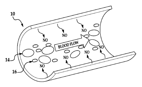
[0005] Fig. lA is a schematic cut-away view showing thrombus formation on a
siloxane-
based polyurethane elastomer (e.g., Elast-EonTM E2As) control coated
extracorporeal
circulation (ECC) circuit;
[0006] Fig. 1B is a schematic cut-away view showing a S-nitroso-N-
acetylpenicillamine
(SNAP)/E2As coated ECC circuit, which releases nitric NO and reduces thrombus
formation;
[0007] Fig. 2A shows the structure of S-nitroso-N-acetylpenicillamine
(SNAP);
[0008] Fig. 2B shows a scheme of S-nitrosothiol (RSNO) decomposition, which
can be
catalyzed by metal ions (e.g., Cut), light, and heat, yielding the disulfide
(RSSR) product and
nitric oxide (NO);
[0009] Figs. 3A and 3B show diffusion of SNAP from various polymer films
containing
wt% SNAP, monitored at 340 nm, films were immersed in 4 mt, phosphate buffered
saline
(PBS) in the dark at room temperature, 22 C (Fig. 3A) or 37 C (Fig. 3B) (the
data is the mean
SEM (n=3));
[0010] Fig. 4A shows NO release behavior of 10 wt% SNAP/E2As film at 37 C
in the dark,
ambient light, and 100W floodlight (the data is the mean SEM (n=3));
[0011] Fig. 4B shows NO release from 10 wt% SNAP in silicone rubber (SR),
CarboSil,
and ElastEonTM E2As films at 37 C and continuously irradiated with the 100W
floodlight (the
data is the mean + SEM (n=3));
[0012] Fig. 4C shows NO release from 5 wt% and 10 wt% SNAP in Elast-Eon'm
E2As
films at 37 C continuously under ambient light (amb) or the 100W floodlight
(the data is the
mean SEM (n=3));
[0013] Fig. 5A shows UV-vis spectra of a 10 wt% SNAP/E2As film, 1.0 mM
SNAP, and
E2As dissolved in N,N-dimethylacetamide (DMAc) (the data is the mean SEM
(n=3));
[0014] Fig. 5B shows cumulative NO release from 10 wt% SNAP/E2As films
incubated in
PBS under various conditions: room temperature (22 C) with ambient light, 37 C
in the dark,
37 C under ambient light, and 37 C under the 100W floodlight (the data is the
mean SEM
(n=3));
CA 02899477 2015-07-27
WO 2014/124125 PCT/US2014/015086
3
[0015] Fig. 6A shows diffusion of SNAP from 10 wt% SNAP-doped E2As films
soaking in
1 mi. PBS in the dark, as monitored at 340 nm, at room temperature (RT, 22 C)
or 37 C;
[0016] Fig. 6B shows a comparison of the cumulative SNAP leaching and
cumulative NO
release (from NOA-based or SNAP-based NO release data) from the 10 wt% SNAP-
doped
E2As films soaking in PBS at 37 C in the dark. Nitric oxide release from SNAP-
doped E2As
films can occur from thermal and/or photochemical decomposition of SNAP within
the
polymer phase, or from SNAP that leached into the aqueous phase. For the SNAP-
doped E2As
films, approximately 26% of the total NO release is attributed to the SNAP
leaching;
[0017] Fig. 7 is a graph showing diffusion of SNAP from 10 wt% SNAP in E2As
films
with 0, 2, or 4 top coats of E2As as monitored at 340 nm by UV-vis, the films
were soaked in
mM PBS containing 100 !AM EDTA, which was replaced daily after the UV-vis
measurement, at 37 C in the dark (the data is the mean SEM (n=3));
[0018] Fig. 8 is a graph showing stability of 10 wt% SNAP in E2As films
stored dry with
desiccant under various temperature and light conditions, the films were
dissolved in DMAc to
determine the amount of SNAP remaining at various times (compared to the
initial level) as
monitored at 340 nm by UV-vis (the data is the mean SEM (n=3));
[0019] Fig. 9 is a graph showing NO release behavior from 10 wt% SNAP/E2As
dry film at
37 C in the dark;
[0020] Fig. 10 is a schematic diagram of the extracorporeal circuit (ECC)
tubing coated
with 5 wt% SNAP/E2As followed by a top coat of E2As;
[0021] Figs. 11A and 11B are graphs showing representative NO surface flux
profiles from
a section of ECC tubing coated with 5 wt% SNAP in E2As before (Fig. 11A) and
after (Fig.
11B) blood exposure, NO release was measured via chemiluminescence at 37 C
under ambient
light;
[0022] Figs. 12A and 12B are graphs showing time-dependent effects of the 5
wt%
SNAP/E2As coating on platelet count (e.g., consumption) (Fig. 12A) and plasma
fibrinogen
levels (Fig. 12B) during the 4 hour blood exposure in the rabbit
thrombogenicity model (the
data is the mean SEM (n=4));
CA 02899477 2015-07-27
WO 2014/124125 PCT/US2014/015086
4
[0023] Fig. 13 is a graph showing results of in vitro fibrinogen adsorption
assays on the 5
wt% SNAP/E2As and E2As control coatings (fluorescence assay in a 96-well plate
that used
goat anti-human fibrinogen-FITC conjugated antibody to measure the level of
adsorbed human
fibrinogen (3 mg/mL) on the coatings, the data is the mean SEM (n=24));
[0024] Fig. 14 is a two-dimensional representation of thrombus formation on
the
SNAP/E2As and control ECCs after 4 hour blood exposure in the rabbit
thrombogenicity
model, as quantified using ImageJ software from N1H (the data is the mean +
SEM (n=4));
[0025] Fig. 15 is a graph showing an NO release profile of polyurethane
tubing (a micro-
polyurethane tubing available from Scientific Commodities, Inc.) impregnated
with SNAP, the
tubing having been soaked in a SNAP/acetone solution for either 1 or 2 days;
[0026] Fig. 16A is a schematic illustration of a catheter tubing coated
with an active layer
of 5 wt% or 10 wt% SNAP/E2As followed by a top coat of E2As;
[0027] Fig. 16B is a cross-sectional view of the catheter tubing of Fig.
16A, taken along
line 16B-16B of Fig. 16A;
[0028] Fig. 17 shows NO release profiles of 5 wt% and 10 wt% SNAP/E2As
catheters at
37 C in the dark (n = 5);
[0029] Fig. 18 is a graph showing quantitation of thrombus area on
SNAP/E2As catheters
and E2As control catheters after 7 day implantation in sheep, as calculated
with NIH ImageJ
software using a 2D representation of thrombus (the data are means + SEM
(n=5)),
* =p < 0.05; and
[0030] Fig. 19 is a comparison of bacterial adhesion (CFU/cm2) on 1 cm
piece of explanted
SNAP/E2As catheters and E2As control catheters (the data are means SEM
(n=5)),
* =p < 0.05.
DETAILED DESCRIPTION
[0031] Examples according to the present disclosure include a novel RSNO-
doped polymer
formulation useful for making biomedical devices. The novel polymer
formulations form
homogeneous films and exhibit RSNO stability even at 37 C for 4 months (with
only about a
10%-15% loss of NO). The novel polymer formulations may be used as coatings to
prevent
CA 02899477 2015-07-27
WO 2014/124125 PCT/1JS2014/015086
thrombus (i.e., blood clot) formation in, e.g., extracorporeal circulation
(ECC) circuits. Fig. lA
is a schematic cut-away view showing a siloxane-based polyurethane elastomer
(e.g., Elast-
EOIITM E2As) control coated ECC circuit 12 that exhibits thrombus formation.
As illustrated,
the red blood cells 14 and platelets 16 clot together. In contrast, Fig. 1B is
a schematic cut-
away view showing an example 10 according to the present disclosure of a SNAP-
doped
siloxane-based polyurethane elastomer (e.g., SNAP/E2As) coated ECC circuit
that does not
exhibit thrombus formation. As depicted in Fig. 1B, NO is generated, which
contributes to the
red blood cells 14 and platelets 16 not clotting together.
[0032] Blood/material interaction is important to the success of
implantable medical
devices, ranging from simple catheters, stents and grafts, to complex
extracorporeal artificial
organs that are used in thousands of patients every day. Thrombosis is one of
the primary
problems associated with clinical application of blood contacting materials.
Despite a thorough
understanding of the mechanisms of blood-surface interactions and decades of
bioengineering
research effort, the ideal non-thrombogenic prosthetic surface remains an
unsolved problem.
Over the last 50 years, much has been learned about surface-induced thrombosis
and attempts
to prevent it with systemic anticoagulation and surface modifications. Surface
modifications
have included using pure, very smooth silicone rubber or polyurethane, pre-
exposure of the
surfaces to albumin and other coating proteins, and surface binding of heparin
in an ionic as
well as a covalent fashion. Despite extensive research to develop a non-
thrombogenic surface
that mimics the endothelium, none of these modifications have been successful.
[0033] Nitric oxide (NO) has been found to be one of two potent
vasodilators secreted by
normal endothelium that has the ability to inhibit platelet
adhesion/activation and aggregation
to the blood vessel wall. The NO-flux from a normal and stimulated endothelium
has been
estimated to be in the range of 0.5x10⁻¹ mol cm-2 min-1 to 4x10⁻¹ mol cm-2
min-1. Nitric oxide
has been extensively studied for its inhibitory effects on circulating
platelet and monocyte
activation that leads to aggregation and ultimately initiation of thrombosis.
A wide range of
NO donors such as S-nitrosothiols (RSNOs), N-Hydroxy-N-nitrosoamines, N-
diazeniumdiolates and nitrosyl metal complexes have been studied at least over
the past decade.
CA 02899477 2015-07-27
WO 2014/124125 PCT/US2014/015086
6
[0034] Nitric oxide (NO) can be released from an NO adduct/donor species
appended to
polymers within a polymer coating. `Nitric oxide adducts" (NO adducts) and "NO-
donors"
refer to compounds and functional groups which, under physiological
conditions, can donate
and/or release NO such that biological activity of the NO is expressed at the
intended site of
action/target site.
[0035] Some examples according to the present disclosure include the NO
donor/adduct
within a polymer coating. The NO donor/adduct may be integrated into the
polymer coating in
any suitable manner, an example of which is doping. Suitable NO adducts
(examples of which
include discrete adducts) are generally those exhibiting capability of
embedding (either by
covalent attachment and/or dispersion) into the polymer matrix and exhibiting
process
preparation stability.
[0036] "Discrete NO adducts" as referred to herein are those NO adducts
(examples of
which are RSNOs) which, when placed into a polymer matrix, release
therapeutically relevant
fluxes of NO, ranging from about 0.2 x 10¹⁰mo1 cm-2min-1 to about 20 x 10-10
mol cm-2 min-1
of NO from the polymer phase. Those compounds that have their NO-releasing
moiety
covalently attached to a polymer backbone are generally referred to as
"polymeric NO
adducts." Examples of suitable polymeric NO adducts include, but are not
limited to, S-
nitrosothiolated polyurethanes, S-nitrosothiolated silicone rubbers, and/or
mixtures thereof.
Some examples of the discrete NO adducts exhibit some lipophilicity, but may
be made more
lipophilic by derivatization with one or more alkyl groups.
[0037] As such, examples of the present disclosure are novel nitric oxide
(NO) releasing
coatings formed from polymers doped with S-nitroso-N-acetylpenicillamine
(SNAP) to prevent
thrombus formation in, e.g., extracorporeal circulation (ECC) circuits and
catheter tubing.
[0038] Various hydrophobic polymer materials may be employed in examples of
the
material, method, and device as disclosed herein. These include, but are not
limited to
materials such as polyurethanes (PU), silicone rubbers (SR), copolymers of
polyurethane and
silicone rubber (e.g., E2A), poly(vinyl chloride) (PVC), polymethacrylates,
polyacrylates,
polycaprolactones, and/or mixtures thereof. In other examples, the polymer
material may
include both hydrophobic and hydrophilic domains. The polymer of choice will
be one capable
CA 02899477 2015-07-27
WO 2014/124125 PCT/US2014/015086
7
of releasing NO from, for example, covalently attached and/or dispersed S-
nitrosothiol (RSNO)
type NO-adducts within the polymer. The polymer of choice may also depend upon
the
application in which polymer coating/film will be used and the desired NO
release rate for that
application. As examples, a polymer having higher water uptake may be suitable
in
applications where quick NO release is desirable, while a polymer having lower
water uptake
may be suitable in applications were slow NO release is desirable. In
instances where
prolonged NO release is desirable, poly(lactic-co-glycolic acid) (PLGA)
additives may also be
included in the polymer coating/film to create an acidic environment to
further stabilize the
RSNO species.
[0039] Further, a system is contemplated as being within the purview of the
present
disclosure that includes discrete RSNOs doped into a polymer, with the polymer
also having
RSNO appended thereto (e.g., by covalent attachment). For example, previously
prepared
polyurethane polymers with appended RSNO functional groups can be mixed with
discrete
RSNOs or similar species to create the long-term NO release polymers enabled
by the present
disclosure.
[0040] In some examples, the NO adduct of choice is one capable of
spontaneous release of
NO when the polymer is exposed to solutions and/or blood under physiological
conditions. In
other examples, the NO adduct of choice is one capable of spontaneous release
of gas phase
NO when the polymer is exposed to certain light conditions. Some examples of
NO adducts
include discrete 5-nitrosothiols (RSNOs).
[0041] It is believed that examples of the present disclosure including
SNAP doped into
siloxane-based polyurethane elastomers (one example of which is E2As) may help
stabilize the
RSNO adduct, thus advantageously allowing longer NO release from the RSNO
species and
enhanced storage stability, even at higher temperatures (e.g., 37 C).
[0042] Spontaneous release of NO from the polymer may be governed by at
least one
process occurring between the NO adduct and the surrounding environment. For
RSNO
species, these include, but are not limited to temperature, moisture, and the
presence of certain
wavelengths of light. For example, photolysis of the S-N bond in the RSNO
species liberates
NO gas. Photolysis can occur with light in either the 300 nm to 400 nm
wavelength range or
CA 02899477 2015-07-27
WO 2014/124125 PCT/US2014/015086
8
the 500 nm to 600 nm wavelength range. In this example, the efficiency of NO
release is
generally greater in the higher wavelength range.
[0043] It is to be understood that discrete nitric oxide adducts may be
either covalently
attached to the polymer matrix or may be dispersed therein, or both. Some
examples of
discrete RSNOs include, but are not limited to S-nitrosoglutathione (GSNO), S-
nitroso-N-
acetylpenicillamine (SNAP, shown in Fig. 2A), 5-nitrosocysteine (CysNO), etc.,
and
derivatized discrete RSNOs. Derivatized RSNOs may be modified with alkyl
group(s). As
examples, a derivative may have an alkyl group attached to the free carboxyl
group of SNAP
and/or may have a longer alkyl (i.e., longer than acetyl) attached to the
amine group of S-
nitrosopenicillamine. As an example, an ester linkage may be formed between
the desired
alkyl group and the free carboxyl group of SNAP. As another example, a long
chain alkyl
(including from 4 to 10 carbon atoms) may replace the acetyl group of SNAP so
that the long
chain alkyl is attached to the amine nitrogen. As other examples, a sugar may
be attached to
the carboxyl group of SNAP (e.g., glucose-SNAP, mannose-SNAP, fructose-SNAP,
etc.).
[0044] The SNAP-doped NO release siloxane-based polyurethane elastomer
coatings
according to examples of the present disclosure were evaluated in vitro and
within a short-term
in vivo rabbit model of thrombogenicity. The novel coatings according to
examples of the
present disclosure continuously released from 0.5 to 1 x 10⁻¹ mol cm-2min-1
NO for 20 days in
the dark, soaking at 37 C in PBS. Additionally, the novel coatings retained
about 78% of the
SNAP after 4 months at 37 C in the dark (i.e., not exposed to wavelengths that
could photolyze
RSNO bonds) and in dry conditions (i.e., in the presence of a desiccant). As
discussed further
herein, examples of the novel coating materials were employed as inner wall
coatings of
extracorporeal circuits used for 4 hours of extracorporeal circulation (ECC)
in a rabbit model of
thrombogenicity to examine the effect of the coatings on platelet function,
clotting and
fibrinogen adsorption. The SNAP-doped NO release coatings were also used to
fabricate
catheters, which were implanted in sheep veins for 7 days to evaluate the
effects on thrombus
and bacterial adhesion.
[0045] As mentioned above, nitric oxide (NO) is an endogenous gas molecule
that plays
several key physiological roles, including prevention of platelet adhesion and
activation,
CA 02899477 2015-07-27
WO 2014/124125 PCT/US2014/015086
9
inhibiting bacterial adhesion and proliferation, enhancing vasodilation,
promoting
angiogenesis, and aiding in wound healing. The effects of NO are highly
dependent on the
location of the NO and its concentration in the physiological system. For
example, endothelial
cells that line the inner walls of healthy blood vessels produce an estimated
NO surface flux
ranging from 0.5 mol cm 2 min to 4.0x10' mol cm 2 min I. The function of many
blood-
contacting devices, including vascular grafts, stents, intravascular sensors,
intravascular
catheters, and extracorporeal life support circuits, can be impaired due to
platelet activation and
thrombus formation. One approach to improve the hemocompatibility of such
devices is the
use of coating materials that mimic the endothelial cells with respect to NO
release. Indeed, in
recent years there has been considerable interest in developing NO-release and
NO-generating
materials that can be used to improve the biocompatibility of such devices.
[0046] Nitric oxide also exhibits antimicrobial activity, including killing
bacteria and
preventing biofilm formation. Bacterial infections and biofilm formation are
problems that can
cause complications with biomedical devices. Bacteria also possess the ability
to form biofilms
on surfaces when the organism secretes a polysaccharide matrix in which the
bacteria will live.
This matrix provides both nutrients and protection against the host defense
and antibiotics.
Biofilms can act as a source of chronic infection, thereby prolonging the
recovery time.
Among its many biological roles, nitric oxide functions as an antimicrobial
agent and as an
accelerant to the wound healing process. Nitric oxide has broad-spectrum
antibacterial
properties, killing both gram-positive and gram-negative bacteria. Low levels
of nitric oxide
are also reported to efficiently disperse biofilms that have formed on the
surface of indwelling
medical devices.
[0047] Nitric oxide is highly reactive under physiological conditions, and
thus a wide range
of NO donor molecules with functional groups that can store and release NO
have been studied
for potential biomedical applications. Such molecules include organic
nitrates, metal-NO
complexes, N-diazeniumdiolates, and S-nitrosothiols (RSNOs). Physiological
RSNOs, such as
S-nitrosohemoglobin and 5-nitrosoglutathione (GSNO), are considered an
endogenous
reservoir of NO in vivo. Other synthetic RSNOs, such as S-nitroso-N-acetyl-L-
cysteine
(SNAC) and S-nitroso-N-acetylpenicillamine (SNAP, Fig. 2A) have been shown to
exhibit
CA 02899477 2015-07-27
WO 2014/124125 PCT/US2014/015086
significant antimicrobial and antithrombotic effects. It has also been
demonstrated that RSNOs
are both vasodilators and potent inhibitors of platelet aggregation. RSNOs
undergo thermal
decomposition, releasing NO and producing a corresponding disulfide species
(RSSR), as
shown in Fig. 2B. The NO release from RSNOs can be catalyzed by metal ions
(e.g., Cut) and
by light, through the irradiation at energies that correspond to the S-nitroso
absorption bands at
340 nm and/or 590 nm. It has been suggested that the more potent activity of
RSNOs vs. NO
as antiplatelet agents arises from the enhanced stability of RSNOs vs. NO, and
generation of
NO from RSNOs locally at the surface of platelets by membrane proteins that
contain catalytic
sites to convert RSNO to NO.
[0048] Incorporation of RSNOs into polymers can extend the utility of these
NO donors to
be applicable as coatings in biomedical devices, providing localized NO
release at the
blood/device interface. Several NO-release polymers consisting of small-
molecule RSNOs
dispersed in various polymer matrices, including polyethylene glycol (PEG),
poly(vinyl
alcohol), poly(vinyl pyrrolidone), and Pluronic0 F127 hydrogel, have been
suggested. These
materials have potential applications for topical NO delivery on wounds via
the diffusion of the
hydrophilic RSNOs from the polymer to the tissue. In fact, daily application
of a GSNO-
containing hydrogel has been shown to accelerate the wound healing process.
However, the
rapid leaching of the RSNOs from such polymers can significantly shorten the
NO/RSNO
release lifetime, lasting only several hours. An alternate approach has been
to synthesize
RSNO-modified materials, where the RSNO functionality is covalently bound to
the matrix.
Fumed silica particles, dendrimers, polyurethanes, polyesters,
polydimethylsiloxane (PDMS),
xerogels, self-assembled monolayers, and poly(vinyl methyl ether-co-maleic
anhydride)
(PVMMA) have all been modified with RSNO functionalities. RSNO-modified
xerogels were
found to release NO for up to 14 days and exhibit reduced platelet and
bacterial adhesion.
However, such RSNO-modified xerogels suffer from synthesis complications
leading to
cracking and non-uniform films, low RSNO conversion efficiency (maximum of 40%
for the
tertiary RSNO-modified xerogels), and thermal instability at room temperature
that would limit
their shelf-life. Many of the other RSNO modified materials reported to date
exhibit both
thermal and photoinitiated NO release, but many of these materials have not
proven clinically
CA 02899477 2015-07-27
WO 2014/124125 PCT/US2014/015086
11
useful due to their limited NO release lifetimes or lack of the RSNO
functionality stability
during storage, or low conversion to RSNO during synthesis.
[0049] Another approach reported to achieve localized NO delivery at a
polymer/blood
interface is to use NO-generating coatings, in which immobilized catalysts
(Cu(I/II) or
organoselenium species) can generate NO from endogenous RSNOs. For example, a
NO
generating coating containing Cu nanoparticles was evaluated recently using a
rabbit model
for extracorporeal circulation (ECC). However, to achieve good efficacy in
reducing thrombus
formation, continuous infusion of SNAP was required to supplement the
endogenous RSNO
levels.
[0050] In order to avoid the continuous infusion of RSNO species, the
present disclosure
includes several biomedical polymers that are capable of storing RSNO species.
The RSNO-
doped coatings according to the present disclosure can advantageously release
NO, as well as
potentially supplement the endogenous RSNO levels, if NO generating catalysts
are also
employed.
[0051] In the present disclosure, five biomedical grade polymers doped with
S-nitroso-N-
acetylpenicillamine (SNAP) were investigated for their potential to control
the release of NO
from the SNAP within the polymers, and further control the release of SNAP
itself. As
discussed further herein, SNAP is quite stable in the ElastEonTM E2As polymer,
creating a
homogeneous coating that can locally deliver NO (via thermal and photochemical
reactions) as
well as slowly release SNAP. E2As is an example of suitable siloxane-based
polyurethane
elastomers contemplated as being within the purview of the present disclosure.
E2As is a
solution grade of E2A (see Table 1 below). The E2As polymer containing SNAP
was coated
on the walls of extracorporeal circuits (ECC) and exposed to 4 hour blood flow
in a rabbit
model of extracorporeal circulation to examine the effects on platelet count,
platelet function,
clot area, and fibrinogen adsorption. After 4 hours, platelet count was
preserved at 100 7% of
baseline for the SNAP/E2As coated loops, compared to 60 6% for E2As control
circuits (n=
4). The SNAP/E2As coating also reduced the thrombus area when compared to the
control
(2.3 0.6 and 3.4 1.1 cm2, respectively). As will be discussed further herein,
the SNAP/E2As
catheters were also able to significantly reduce the thrombus area and
bacterial adhesion after 7
CA 02899477 2015-07-27
WO 2014/124125 PCT/US2014/015086
12
day implantation in sheep veins. All of the results suggest that the new
SNAP/E2As coatings
have potential to improve the thromboresistance of intravascular catheters,
grafts, and other
blood contacting medical devices.
[0052] The present inventors also examined the five biomedical polymers
(silicone rubber
(SR), ElastEonTM E2As (a siloxane-base polyurethane elastomer commercially
available from
Aortech Biomatcrials, Scoresby Victoria, Australia), CarboSil (a
thermoplastic silicone-
polycarbonate-urethane commercially available from DSM Biomedical Inc.,
Berkeley, CA),
TecoflexTm SG 0A and TecophillicTm SP-60D-60 (both polyurethanes commercially
available
from The Lubrizol Corporation, Wickliffe, Ohio)) for their potential to act as
a storage
reservoir for SNAP. The ElastEonTM polymer has excellent intrinsic
biocompatibility and
biostability properties, and exhibits low levels of blood protein adsorption.
Each of the SNAP-
doped polymers is characterized for its in vitro NO/SNAP release. The present
inventors have
found that SNAP itself is stable for at least 4 months in the ElastEonTM E2As
polymer,
creating a coating that releases NO thermally (at physiological temperature)
and can also serve
as a reservoir to supplement endogenous RSNO levels (by SNAP diffusion into
blood from the
polymer). The new SNAP/E2As polymer was tested for potential biomedical
applications via,
e.g., an ECC rabbit model of thrombogenicity to assess preservation of
platelet count and
function, and thrombus area after 4 hours of ECC.
[0053] It is to be understood that other siloxane-based polyurethane
elastomers (aside from
E2As) are also contemplated as being suitable for use in the present
disclosure. Further, it is to
be understood that other grades of Elast-EonTM siloxane-based polyurethane
elastomers
(commercially available from Aortech Biomaterials, Scoresby Victoria,
Australia) are
contemplated as being suitable for use in the present disclosure. Table 1
below is a table of
properties of various grades of ElastEonTM polymers. In addition to those
examples shown in
Table 1, it is believed that other suitable polymers include CarboSi10,
PuriSilTM, or silicone
rubber.
CA 02899477 2015-07-27
WO 2014/124125 PCT/1JS2014/015086
13
Table 1
Elast-Eonr" Properties
TEST E5-130 E5-
3258 E2A* F2-945 E2-852 E2-860 E2-862 E4
Durometer Hardness 77A 82A 90A 50D 55D 65D 68D 80D
Tensile Strength, MPa 21 23 76 28 30 34 34 60
Elongation at Break, % >700 >500 >450 >400 >300 >200
>200 25
Tensile Stress, 100% E, MPa 4 5 8 12 15 23 - -
Tensile Stress, 200% E, MPa 5 7 10 15 18 - - -
Tensile Stress, 300% E, MPa 6 9 13 18 23 - - -
Modulus of Elasticity, MPa 11 15 35 115 360 650 650 -
Tear Strength, kN/m 45 60 80 97 129 - - -
Physical Form Pellets Pellets** Pellets** Pellets Pellets
Pellets Pellets Pellets
Melt Temperature C 175- 180-185 195-200 220 210- 210-
210- 185
185 215 215 215
*Can also be supplied in solution grade and is soluble in both THF and DMAc.
** Also supplied in dispersion form.
[0054] Preliminary
In vitro Characterization of Various SNAP-Doped Polymer Films
[0055] SNAP doped into all of the five biomedical polymers produced
homogeneous and
transparent films of green color, without any observable phase separation. The
10 wt% SNAP
films stored approximately 0.42 [tmol of SNAP per mg polymer film (or 6
umol/cm2), while
the 5 wt% SNAP films stored approximately 0.21 umol of SNAP per mg polymer
film (or 3
umol/cm2).
[0056] The 5 wt% and 10 wt% SNAP/polymer films were immersed in 4 mL PBS in
the
dark at room temperature (i.e., 22 C) or at 37 C. The diffusion of SNAP into
the PBS from the
various polymer films containing 5 wt% and 10 wt% SNAP was monitored using UV-
Vis
absorption. As shown in Figs. 3A and 3B, all of the SNAP diffused out of the
SG80A and SP-
60D-60 polymer films during the first day of soaking in PBS at room
temperature (see Fig. 3A)
and at 37 C (see Fig. 3B). The SP-60D-60 polymer is hydrophilic with a water
uptake of about
60 wt%, while the SG80A is more hydrophobic, having a water uptake of about 6
wt% (see
Table 2 below).
CA 02899477 2015-07-27
WO 2014/124125 PCT/US2014/015086
14
[0057] Table 2 illustrates the water uptake of the 5 biomedical polymers
used in the present
disclosure. Polymer films (200 mg polymer) were cast in Teflon ring (d=2.5
cm) on Teflon
plates. Small disks (d=0.7 cm) were cut from the parent films, weighed, and
immersed in PBS for
48 hours at 37 C. The wet films were wiped dry and weighed again. The water
uptake of the
polymer films is reported in Table 2 in weight percent as follows: water
uptake (wt%) = (Wwei ¨
Wdry)/Wdry x 100, where Wõet and Wthy are the weights of the wet and dry
films, respectively.
Table 2
Polymer Water uptake [wit A]
Silicone Rubber 1.2 0.3
CarboSil 1.5 0.3
ElastEonTM E2As 1.2 0.1
Tecoflex SG80A 6.2 0.7
Tecophilic SP-60D-60 64.5 0.1
[0058] As shown in Figs. 3A and 3B, all of the SNAP leaves the more
hydrophilic SP-60D-
60 polymer during the initial 2 hours of soaking, while the more hydrophobic
SG80A leaches
all of the SNAP after 24 hours. Due to the rapid loss of the SNAP from the SP-
60D-60 and
SG80A polymers, a very large initial burst of NO was observed via
chemiluminescence (with a
Nitric Oxide Analyzer (NOA)) during the first day of soaking (Day 0), and the
films exhibited
no SNAP/NO release after one day (data not shown). Therefore, these two
polymers provide a
quick burst of NO/SNAP and were found not to be suitable for longer-term
release of
NO/SNAP.
[0059] In contrast, the silicone rubber, CarboSil , and E2As polymers
exhibit significantly
lower amounts of SNAP diffusing into the soaking buffer after one day (see
Figs. 3A and 3B).
For all three of these polymers, an initial burst of SNAP leaching was
observed during the first
day of soaking, corresponding to rapid water uptake by the polymer. This
initial burst was
about 10% of the total SNAP molecules incorporated into the films. Small
amounts of SNAP
continued to leach from these polymers during the subsequent days of soaking.
Silicone
rubber, CarboSil (a thermoplastic silicone-polycarbonate-urethane), and E2As
(a siloxane-
base polyurethane elastomer) all are hydrophobic polymers due to their high
PDMS content
and also have the lowest water uptake (see Table 1 above). SNAP is slightly
hydrophobic.
CA 02899477 2015-07-27
WO 2014/124125 PCT/US2014/015086
Therefore, SNAP should have a preference for remaining in these more
hydrophobic polymer
films. In addition, it is believed that the hydrophobic property of these
polymers also plays a
significant role in limiting the diffusion of SNAP into the buffer, due, at
least in part, to the
minimal water uptake of these polymers.
[0060] The thermal and photoinitiated NO release from the three SNAP-doped
polymers
(i.e., silicone rubber, CarboSi10, and E2As polymers) was also studied by NOA
measurements.
Nitric oxide release can be turned on/off using the 100W floodlight for all 3
film types. Fig.
4A illustrates the NO release behavior of the 10 wt% SNAP/E2As film at 37 C in
the dark, in
the ambient light, and in the 100W floodlight. As shown in Fig. 4A, there is
little difference in
the NO release from the 10 wt% SNAP/E2As film in the dark or under the ambient
lab lights,
since ambient fluorescent lighting does not emit the wavelengths responsible
for decomposing
RSNOs (340 nm or 590 nm). While the data for the 10 wt% SNAP/E2As film is
shown, it is to
be understood that for all three polymers, the total NO release detected by
the NOA for films
continuously irradiated with the 100W floodlight was about 100% of the SNAP
doped into the
films. The photoinitiated NO release from these three films was examined by
continuously
irradiating with a 100W floodlight at 37 C and monitoring the NO released with
the NOA (see
Fig. 4B). The SNAP-doped E2As and CarboSil0 films exhibited a gradual decrease
in the
photo-induced NO flux over a 3 day period, while the SR-based films released
NO for only 2
days under the same conditions. All three types of films incubated at 37 C
under ambient light
yielded an initial burst of NO on the first day of soaking, corresponding to
release of SNAP
into the solution, and on subsequent days, the NO flux ranged from 1 x 10⁻¹⁰
mol cm-2 minito
2 x 10⁻¹⁰ mol cm-2 min-1. This NO flux is still potentially useful to inhibit
platelet function and
kill bacteria.
[0061] It appears that the NO release may be more promising from the film
composed of 10
wt% SNAP in E2As under the 100W floodlight. Therefore, the wt% of SNAP in E2As
was
varied to 5 wt% and examined in more detail (see Fig. 4C). The NO release and
SNAP
leaching pattern is similar for the 5 wt% SNAP/E2As film, but the NO release
takes place over
a shorter time period. The biostability and biocompatibility of the E1astEonTM
polymers in
CA 02899477 2015-07-27
WO 2014/124125 PCT/US2014/015086
16
combination with the NO release from SNAP makes this formulation attractive
for further in
vitro and possible biomedical applications.
[0062] Long-term NO Release of SNAP/E2As Formulation
[0063] In vitro studies were conducted with the SNAP/E2As films to examine
the long-
term NO release and SNAP leaching from these films. The NO release from the
SNAP/E2As
films over time was determined based on the amount of SNAP decomposed within
the polymer
phase (i.e., by measuring the SNAP remaining after dissolving the films at
given time points).
The initial concentration of SNAP in the 10 wt% films was 420 nmol SNAP/mg
film. Fig. 5A
shows the UV-Vis spectra of 1.0 mM SNAP solution, a 10 wt% SNAP in E2As film
redissolved in N,N-dimethylacetamide (DMAc), and E2As dissolved in DMAc.
[0064] Due to thermal and/or photochemical decomposition of SNAP, a
decrease in the 340
nm absorbance band was observed as the 10 wt% SNAP/E2As films were soaked in
PBS under
various conditions, and the cumulative NO release based on that absorbance
decrease is shown
in Fig. 5B. The 10 wt% SNAP/E2As films displayed an initial burst of NO during
the first day
of soaking (see Figs. 3A and 3B), which corresponds to the thermal
decomposition as well as
diffusion of SNAP out of the film. Films soaked at room temperature had the
lowest flux of
NO release. However, films incubated at 37 C in the dark or under ambient
light exhibited a
higher NO release than the films at room temperature. This is due to the
increased thermal
decomposition of SNAP. The films that were exposed to ambient light yield
essentially the
same NO release profiles as the films that were soaked in the dark. Nitric
oxide release from
the SNAP/E2As films that are continuously irradiated with the 100W floodlight
at 37 C only
release NO for 3 days due to their higher NO fluxes that rapidly deplete the
SNAP reservoir.
These films provide NO release via both a thermal and photoinitiated
decomposition of SNAP.
[0065] Nitric oxide release from the SNAP-doped E2As can occur from thennal
and/or
photochemical decomposition of SNAP either within the polymer phase, or after
SNAP enters
the aqueous phase by diffusion out of the polymer. In order to better
understand the NO release
mechanism of the SNAP/E2As coating, the SNAP diffusion into PBS was monitored
over a 20
day period. As shown in Fig. 6A, the films containing 10 wt% SNAP at 37 C
exhibit an initial
burst of SNAP leaching on the first day. After this initial burst, the SNAP
continues to slowly
CA 02899477 2015-07-27
WO 2014/124125 PCT/US2014/015086
17
diffuse from the E2As until the SNAP reservoir is nearly depleted (with still
measurable
amounts of SNAP leaching on day 20). The total moles of SNAP that leach from
the film
accounts for about 27% of the total NO released (as detected by NOA
measurements) during
the 20 day period (see Fig. 6B).
[0066] Additionally, the effect of the number of polymer top coats on loss
of SNAP was
also evaluated. SNAP-doped E2As films without any top coat exhibit higher
levels of SNAP
diffusion into the buffer than films with at least 2 topcoats (see Fig. 7).
The thickness of the
top coat allows control of the diffusion rate of SNAP from the polymer
reservoir. As such, in
the examples disclosed herein the SNAP-doped films may be coated with a
polymer top coat.
Examples of suitable polymers for the top coat include the siloxane-based
polyurethane
elastomers, poly(vinyl chloride), crosslinked polyurethanes, crosslinked
silicone rubber,
polytetrafluoroethylene, etc. (without the NO donor therein). In some
examples, the polymer
used for the top coat is the same polymer used for the underlying film.
[0067] Stability Study of the SNAP/E2As Films
[0068] The stability of SNAP doped in the E2As polymer during dry storage
was also
evaluated. SNAP incorporated in E2As can potentially undergo thermal or
photochemical
decomposition during storage, thus limiting the available NO release capacity
at the time of
use. Therefore, 10 wt% SNAP/E2As films were stored with desiccant in the
freezer in the
dark, dry in the dark or in ambient light with desiccant at room temperature,
and dry in the dark
with desiccant at 37 C and 50 C. These stability studies were conducted in a
similar manner as
the cumulative NO release experiments, where films were dissolved in DMAc to
determine the
amount of SNAP remaining in the polymer at various time points (as described
herein). The
results indicate that SNAP is stable within the E2As polymer matrix after 4
months. After 2
months, for example, the 10 wt% SNAP films stored in the freezer (-20 C) in
the dark maintain
about 96% of the initial SNAP species, compared to 89% for films stored at
room temperature
and 82% for films stored at 37 C (see Fig. 8). The results shown in Fig. 8
illustrate the
enhanced stability of SNAP during storage. Tertiary RSNOs, such as SNAP, have
greater
stability than primary RSNOs due to steric hindrance surrounding the sulfur
atom. The
CA 02899477 2015-07-27
WO 2014/124125 PCT/US2014/015086
18
increased thermal stability of SNAP in combination with the stabilization
effect of the E2As
polymer provides excellent storage stability of the SNAP/E2As material.
[0069] Stability of RSNOs has been studied for viscous polymer matrices
containing such
NO donors, including poly(ethylene glycol), Pluronic0 F127 hydrogel,
poly(vinyl alcohol) and
poly(vinyl pyrroloidone). RSNOs decompose according to the following sequence
of
reactions:
RSNO ¨> RS = + NO (1)
RS = + RSNO RSSR + NO. (2)
With the overall reaction: 2RSNO ¨> RSSR + 2N0. (3).
[0070] The viscosity of the polymer matrix provides a cage effect on the
bond cleavage and
radical pair recombination. In addition, a viscous polymer matrix also limits
the diffusion of
the radical species, favoring geminate recombination to reform RSNO. Thus, the
E2As
polymer not only limits the diffusion of SNAP into the PBS, but it also
appears to provide an
additional stabilization effect to reduce the rate of SNAP decomposition.
[0071] An experiment was performed to test the storage stability of SNAP in
the E2A
matrix. Fig. 9 illustrates the results. In particular, Fig. 9 shows NO release
behavior from 10
wt% SNAP in E2As dry film at 37 C in the dark (n=1). The film was dried at 37
C and then
stored at 37 C. Approximately 5% of the total NO in the film was released
during the first
hours of storage, followed by very low levels of NO release. This corresponds
with other
storage/stability data disclosed herein (see Fig. 8), which shows that the
SNAP loses its NO
slowly during the 37 C, dry storage (losing only 10-15% of the SNAP after two
months of
storage in a dry state).
[0072] SNAP/E2As Coated ECC Loops and Effects on Rabbit Hernodynamics
[0073] The active ECC loops coated with 5 wt% SNAP in E2As and a top coat
of E2As (a
schematic cross-section of which is shown in Fig. 10) and control loops coated
with E2As only
were prepared. 5 wt% SNAP was used in these tests due, in part, to the short
duration of the
ECC experiment. As described above, the SNAP/E2As coating has an initial burst
of SNAP
CA 02899477 2015-07-27
WO 2014/124125 PCT/US2014/015086
19
diffusing into solution during the first day of soaking. To reduce the effects
of this burst during
the short-term ECC experiments, all loops were first soaked overnight in
saline, and the
soaking solution was discarded prior to the ECC experiments. Nitric oxide
released from
samples of the coated ECC loops were measured with the NOA for NO release
before blood
exposure (after overnight soaking in saline). The NO release of the SNAP/E2As
coated loops
maintains an average flux of about 2 x 10⁻¹⁰ mol cm-2 min-1 for 4 hours (at 37
C with ambient
light) (see Fig. 11A). After 4 hours of exposure to flowing blood, the ECC
loops still exhibit a
NO flux of at least 1.5 x 10⁻¹ mol cm-2 min-1 for at an additional 1 hour
period (see Fig. 11B).
[0074] The hemodynamic effects of the SNAP/E2As coated ECC circuits were
also
monitored over the 4 hours of blood exposure in the rabbit ECC model. The mean
arterial
pressure (MAP) dropped significantly for both SNAP/E2As and control loops
within the first
hour, dropping to 35+2 mmHg and 46+2 mmHg, respectively. The MAP was
maintained at
these levels for the 4 hours by continuous IV fluid maintenance. The ECC blood
flow dropped
and remained at 64+5 mL/min for SNAP/E2As ECC, but maintained at baseline
levels over the
4 hours (76+6 mL/min) for controls. The MAP drop and slower blood flow for the
SNAP/E2As circuits is likely due to the vasodilatory effects of SNAP diffusing
from the
coating into the blood. The heart rate was maintained over the 4 hours and no
significant
difference was noted between the SNAP/E2As and control ECC loops, averaging
205+2
beats/min. The activated clotting time increased over the 4 hour period for
both SNAP/E2As
and control circuits, likely due to the increase in intravascular fluids (the
hemodilution effect).
Similar effects on MAP and flow rate have been observed with SNAP infusion.
[0075] Effects of SNAP/E2As Coatings on Rabbit Platelet Function and
Thrombus
Formation
[0076] Platelet activation and function throughout the 4 hour ECC was
assessed by
recording the platelet count (e.g., consumption, see Fig. 12A) and plasma
fibrinogen levels (see
Fig. 12B), which were both corrected for hemodilution due to the added IV
fluids, as well as %
platelet aggregation. The baseline platelet counts (x108 platelets/mL) were
3.5+0.6 and 4.8+0.5
for the 5 wt% SNAP/E2As and E2As control circuits, respectively. For the
SNAP/E2As
circuits, the platelet count initially rose slightly and was maintained at
100+7% of baseline
CA 02899477 2015-07-27
WO 2014/124125 PCT/US2014/015086
levels at the end of 4 hours on ECC. The platelet count for control circuits
exhibited a time-
dependent loss in platelets, dropping to 60+6% of baseline after 4 hours. The
percent of
platelet functional aggregation was determined by ex vivo collagen stimulation
of PRP and
measured by optical turbidity. The platelets from blood taken from circulation
through the
SNAP/E2As and control circuits showed similar response to collagen-stimulated
platelet
aggregation during the 4 hour blood exposure, both maintaining 56+12% (with
baseline values
at 68+6%).
[0077] As shown in Fig. 12B, the plasma fibrinogen levels were maintained
at baseline
levels for the control circuits. For the 5 wt% SNAP/E2As circuits, the plasma
fibrinogen levels
during the first hour of ECC dropped to 83% of baseline levels and remained at
that level for
the 4 hour ECC. This decrease in plasma fibrinogen levels can be attributed to
fibrinogen
binding to the surfaces, as shown by the in vitro fibrinogen assay results
(see Fig. 13).
Surprisingly, even with the enhanced adsorption of fibrinogen on the SNAP/E2As
coatings,
these materials still exhibited significantly less platelet loss than
controls, suggesting that the
levels of NO produced overcome the enhanced fibrinogen adsorption that would
normally
enhance activation of platelets.
[0078] To determine the differential formation of thrombus in the
thrombogenicity chamber
of the ECC circuit (i.e., the 3/8 inch ID Tygon tubing, 8 cm in length within
the ECC loop), 2-
dimensional (2D) image analysis was performed after 4 hours of blood exposure.
The
thrombus area was analyzed by using Image J software and represents the 2D
area of thrombus
formation (pixels/cm2) in each thrombogenicity chamber. The thrombus area was
quantitated
and data are shown in Fig. 14. The thrombus area is significantly reduced for
the SNAP/E2As
circuits when compared to controls, although the E2As controls also had
relatively low
thrombus area, likely resulting from the enhanced intrinsic biocompatibility
of the E2As
polymer.
[0079] One of the effects of the new SNAP/E2As coating is the hypotension
caused by the
diffusion of SNAP into the blood stream. The co-administration of intravenous
fluids
counteracts this, but may in some instances pose difficulties in a clinical
situation.
Applications of SNAP have been reported to cause hypotension, hyperglycemia
and impaired
CA 02899477 2015-07-27
WO 2014/124125 PCT/US2014/015086
21
insulin secretion, and decreased cell viability. However, when used as
catheters for coatings
for small implantable devices, the surface area to volume (of blood) ratios
will be quite small,
and thus the amounts of SNAP lost to the blood will generally not be a
significant issue.
Furthermore, endogenous thiols and superoxide dismutase will reduce many of
the adverse
effects. The parent thiol, N-Acetyl-DL-penicillamine (NAP), however, has been
used clinically
to treat mercury poisoning and cystinuria with minimal side effects. Although
the SNAP/E2As
coatings disclosed herein do exhibit a hypotension effect, the daily levels of
SNAP delivered by
the coating are well below the reported levels causing the other potential
adverse side effects
described above.
[0080] Impregnation Method for Making SNAP-doped Polymers
[0081] The present disclosure further includes a method for loading
commercial SR or PU
tubing with SNAP. Commercial SR and PU includes medical grade tubing,
including those
available from US plastics, Cole Palmer, Professional Plastics, ICORally, and
Thermedics, Inc.
Fig. 15 shows an NO release profile of polyurethane (PU) tubing that was
soaked in a
SNAP/acetone solution for either 1 or 2 days. The tubing was then rinsed with
acetone and
dried prior to the NOA testing. The tubing was soaked in PBS at 37 C for the
NOA testing.
[0082] This impregnation approach enables the incorporation of the SNAP
species within
the walls of existing commercial catheters/tubings. This approach avoids
problems that may
arise when attempting to extrude SNAP into a polymer tubing under normal hot
extrusion
conditions due to the thermal instability of NO donors (e.g., SNAP and related
species) at high
temperatures. While acetone was used in the impregnation approach described
herein, it is
believed that other solvents (or mixtures of solvents) that may be used
include ethyl acetate,
cyclohexane, isopropanol, methanol, butanone, etc.
[0083] SNAP-doped Polymers for Catheter Tubing Applications
[0084] Fig. 16A is a schematic illustration of an E2As catheter tubing
prepared with 5 wt%
or 10 wt% SNAP/E2As according to an example of the present disclosure followed
by the
application of a top coat of E2As. Fig. 16B is a cross-section of the catheter
tubing, illustrating
the various layers. In general, the trilayer catheters were prepared by dip-
coating 5 base coats
of an E2As solution, 25 coats of the respective active solutions, and 5 top
coats of the E2As
CA 02899477 2015-07-27
WO 2014/124125 PCT/US2014/015086
22
solution. The SNAP/E2As catheters were kept in phosphate buffered saline (PBS)
in the dark
at 37 C.
[0085] Fig. 17 shows 20 day NO release of the SNAP/E2As catheters at 37 C
in the dark in
the phosphate buffered saline (PBS). The results in Fig. 17 illustrate that
the 10 wt%
SNAP/E2As catheters were able to release NO for up to 20 days and that the 5
wt%
SNAP/E2As catheters were able to release NO for up to 7 days at the specified
conditions.
[0086] E2As control catheters and the 10 wt% SNAP/E2As catheters were
implanted in
sheep veins for 7 days. After explantation, the SNAP/E2As catheters were found
to have
significantly less thrombus (Fig. 18) and a 90% reduction of bacterial
adhesion (Fig. 19) than
the E2As control catheters. Together Figs. 17 through 19 demonstrate the
potential application
of the SNAP-doped polymers in the catheter configuration.
[0087] To further illustrate the present disclosure, various examples are
given herein. It is
to be understood that these examples are provided for illustrative purposes
and are not to be
construed as limiting the scope of the present disclosure.
EXAMPLES
[0088] It is to be understood that the SNAP doped polymers and the data
previously
described herein were made using the materials and techniques described in the
EXAMPLES
section. The various testing procedures described in the EXAMPLES section were
also used.
[0089] Materials
[0090] N-Acetyl-DL-penicillamine (NAP), sodium chloride, potassium
chloride, sodium
phosphate dibasic, potassium phosphate monobasic, ethylenediaminetetraacetic
acid (EDTA),
tetrahydrofuran (THF), sulfuric acid and N,N-dimethylacetamide (DMAc) were
purchased
from Sigma-Aldrich (St. Louis, MO). Methanol, hydrochloric acid and sulfuric
acid were
obtained from Fisher Scientific (Pittsburgh, PA). TecophilicTm SP-60D-60 and
TecoflexTm 5g
80A were products of Lubrizol Advanced Materials Inc. (Cleveland, OH). Dow
Corning RTV
3140 Silicone Rubber (SR) was purchased from Ellsworth Adhesives (Germantown,
WI).
CarboSil0 20 90A was from the Polymer Technology Group (Berkeley, CA).
ElastEonTM
E2As was obtained from AorTech International, PLC (Scoresby, Victoria,
Australia). Human
CA 02899477 2015-07-27
WO 2014/124125 PCT/US2014/015086
23
plasma fibrinogen containing > 90% clottable proteins was a product of
Calbiochem (La Jolla,
CA), and fluorescein-labeled goat IgG (polyclonal) against denatured human
fibrinogen was
purchased from MP Biomedicals, LLC (Solon, OH). Black, polypropylene 96-well
microtiter
plates used for fluorescence measurements were obtained from Nalge Nunc
International
(Rochester, NY). All aqueous solutions were prepared with 18.2 win deionized
water using a
Milli-Q filter (Millipore Corp., Billerica, MA). Phosphate buffered saline
(PBS), pH 7.4,
containing 138 mM NaCl, 2.7 m_M KC1, 10 mM sodium phosphate, 100pM EDTA was
used for
all in vitro experiments.
[0091] Synthesis of SNAP
[0092] SNAP was synthesized using a modified version of a previously
reported method (I.
Chipinda, R. H. Simoyi, Journal of Physical Chemistry B 2006, 110, 5052).
Briefly, an
equimolar ratio of NAP and sodium nitrite was added to a 1:1 mixture of water
and methanol
containing 2 M HC1 and 2 M H2SO4. After 30 minutes of stifling, the reaction
vessel was
cooled in an ice bath to precipitate the green SNAP crystals. The crystals
were collected by
filtration, washed with water, and allowed to air dry. The reaction and
crystals were protected
from light at all times.
[0093] Preparation of SNAP-doped films
[0094] The polymer films containing 5 wt% and 10 wt% SNAP were prepared by
solvent
evaporation. For the 10 wt% SNAP films, the casting solutions were prepared by
dissolving
180 mg of the respective polymer in THF. The polyurethanes (SP-60D-60, SG-80A,
CarboSil and Elast-Eonim E2As) were dissolved in 3 mL THF, and SR was
dissolved in 1
mL THF. SNAP (20 mg) was then added to the polymer solution, and the mixture
was stirred
for 10 minutes. The 5 wt% SNAP films were prepared similarly with SNAP (10 mg)
and
polymer (190 mg). The film solution was cast in a Teflon ring (d=2.5 cm) on a
Teflon plate
and dried overnight under ambient conditions. Small disks (d=0.7 cm) were cut
from the
parent films and were dip coated 2 times with a topcoat solution (200 mg of
the respective
polymer (no SNAP added) in 4 mL THF) and dried overnight. As such, the topcoat
for each
sample was made of the same polymer as the parent film. The weight of each
small disk was
recorded prior to topcoating. All films and film solutions were protected from
light. The final
CA 02899477 2015-07-27
WO 2014/124125 PCT/US2014/015086
24
films had a SNAP-doped layer that was about150 m thick and a top coat that was
about 50 pm
thick.
[0095] Preparation of SNAP/E2As coated ECC loops
[0096] The ECC configuration employed in the in vivo rabbit study consisted
of 16-gauge
and 14-gauge IV polyurethane angiocatheters (Kendall Monoject Tyco Healthcare,
Mansfield,
MA), two 16 cm in length 1/4 inch inner diameter (ID) Tygon0 tubing, and an 8
cm length of
3/8 inch ID Tygon tubing that created a thrombogenicity chamber where
thrombus could
form more easily due to more turbulent blood flow.
[0097] As previously mentioned, due to the short duration of the ECC
experiments (4
hours), the NO release ECC loops were coated with only 5 wt% SNAP in E2As. The
SNAP/E2As solution was prepared by dissolving SNAP (125 mg) and E2As (2375 mg)
in THF
(15 mL). The E2As control solution consisted of E2As in (2500 mg in 15 mL).
SNAP/E2As
loops were first coated with 2 layers of the SNAP/E2As solution, followed by 1
coat of the
E2As control solution. E2As control loops were coated with 2 coats of the E2As
control
solution. ECC loops were allowed to air dry for 1 hour in the dark between
each coat. The
completely coated ECC was welded together using THF, starting at the left
carotid artery side,
with the 16-gauge angiocatheter, one 15 cm length 1/4 inch ID tubing, the 8 cm
length
thrombogenicity chamber, the second 15 cm length 1/4 inch ID tubing and
finally the 14-gauge
angiocatheter. The angiocatheters were interfaced with tubing using two luer-
lock PVC
connectors. The assembled ECC loops were dried under vacuum while protected
from light for
at least 48 hours. Prior to the ECC experiment, the loops were filled with
saline solution for
overnight soaking, and this solution was discarded immediately before the
rabbit experiment.
[0098] In vitro characterization of SNAP-doped films
[0099] UV-Vis spectra
[0100] All UV-Vis spectra were recorded in the wavelength range of 200 nm -
700 nm
using a UV-Vis spectrophotometer (Lambda 35, Perkin-Elmer, MA) at room
temperature. The
presence of the S-NO group of SNAP provides characteristic absorbance maxima
at 340 nm
and 590 nm, corresponding to the it -> 7r* and nN ¨> 7r* electronic
transitions.
CA 02899477 2015-07-27
WO 2014/124125 PCT/US2014/015086
[0101] Diffusion of SNAP from SNAP-doped Polymer Films Immersed in PBS
[0102] Top coated films were placed in individual vials soaked in 10 mM
PBS, pH 7.4,
containing 100 [iM EDTA to minimize any trace metal ion catalyzed
decomposition of SNAP.
Films were incubated in the dark at room temperature (22 C) or 37 C. At
various time points,
the UV-Vis spectra of a 1 mL aliquot of the PBS was taken for rapid
determination of the
SNAP concentration. The aliquots were protected from light and were
immediately returned to
the sample vials for the duration of the experiment. The films were placed in
fresh PBS buffer
daily. The molar absorption coefficient for SNAP in PBS at 340 nm was
determined to be:
ESNAP= I 024 M-1 cm-1.
[0103] Cumulative NO release from SNAP/E2As Films
[0104] After the 10 wt% SNAP in E2As films were prepared, the UV-Vis
spectra were
recorded of individual films dissolved in DMAc to determine the initial
concentration of SNAP
within the films (nmol SNAP/mg film). Equivalent films were then placed in
individual vials
containing 3 mL PBS (pH 7.4) containing 100 [iM EDTA. Films were incubated
under various
conditions: RT under ambient light, 37 C under ambient light, 37 C in dark,
and 37 C under a
100W floodlight. The fluorescent lights in the laboratory are referred to as
ambient light.
Films were placed in fresh PBS daily. At various time points, the films were
dissolved in
DMAc for rapid determination of the SNAP present in the film. The amount of NO
released
was determined indirectly from the amount of SNAP decomposed at various time
points. The
cumulative NO released over time ([NO]) was calculated by the difference
between the initial
amount of SNAP in the film ([SNAP]0) and the amount of SNAP at time t
([SNAP]): [NO] i =
[SNAP]0 ¨ [SNAP] t (where concentrations are in nmol/mg film). This
calculation was based on
the fact that the decay of the 340 nm absorption band of SNAP is directly
associated with the
hemolytic cleavage of the S-NO bond and concomitant NO release. The molar
absorption
coefficient for SNAP in DMAc at 340 nm was determined to be: ESNAP=1025 M-1 cm-
1.
[0105] NO Release Measurements
[0106] Nitric oxide released from the films was measured using a Sievers
chemiluminescence Nitric Oxide Analyzer (NOA) 280 (Boulder, CO). Films were
placed in
the sample vessel immersed in PBS (pH 7.4) containing 100 i.tM EDTA. Nitric
oxide was
CA 02899477 2015-07-27
WO 2014/124125 PCT/US2014/015086
26
continuously purged from the buffer and swept from the headspace using an N2
sweep gas and
bubbler into the chemiluminescence detection chamber. Clear glass sample
vessels were used
for the ambient light and photoinitiated NO release experiments. A 100W
halogen floodlight
(GE model 17986) was placed about 60 cm from the sample cell for the
photolysis
experiments. Films were incubated in the PBS under the same conditions as the
NOA
measurements (ambient light or 100W floodlight irradiation at 37 C).
[0107] SN4P/E2As Stability Study
[0108] SNAP/E2As films (consisting of 10 wt% SNAP) were placed under the
following
conditions in vials with desiccant: room temperature with ambient light, room
temperature in
dark, 37 C in dark, and in the freezer (-20 C) in dark. At various time points
over a 4 month
period, films were dissolved in DMAc, and the UV-Vis spectra was recorded to
determine the
% SNAP remaining in the film, as compared to the initial 10 wt% SNAP.
[0109] In vitro Fibrinogen Adsorption Assay
[0110] The in vitro fibrinogen adsorption immunofluorescence assay was
performed in a
96-well format. The SNAP/E2As and E2As control polymer solutions used to
prepare the ECC
circuits were also employed to coat microwells of the 96-well microtiter
plates and were dried
under the same conditions as the ECC loops. Briefly, human fibrinogen was
diluted to 3
mg/mL with Dulbecco's phosphate-buffered saline (dPBS) without CaCl2 and MgC12
(Gibco
Invitrogen, Grand Island, NY), equivalent to the human plasma concentration,
and then used
for adsorption experiments. One hundred IAL of this solution were added to
each well and the
coated wells were incubated with this solution for 1.5 hours at 37 C. This was
followed by
eight washing steps using wash buffer (100 [iL) for each wash, which consisted
of a 10-fold
dilution of the AbD Serotec Block ACE buffer (Raleigh, NC) containing 0.05%
Tween 20
(Calbiochem La Jolla, CA). To block nonspecific antibody binding, coated wells
were
incubated with 100 ILL of blocking buffer (4-fold dilution of Serotec Block
ACE buffer) for 30
minutes at 37 C. After rinsing 3 times with wash buffer (100 uL per well), a
background
fluorescence measurement of the plates was performed at 485 nm (excitation)
and 528 nm
(emission) on a Synergy 2 fluorescence microplate reader (Biotek Winooski,
VT). To detect
the adsorbed fibrinogen, fluorescein-labeled goat anti-human fibrinogen
antibody was diluted
CA 02899477 2015-07-27
WO 2014/124125 PCT/US2014/015086
27
(1 : 10) in a 10-fold dilution of the Serotec Block ACE buffer and 100 [t1_,
of this final solution
was added to each well. The antibody was allowed to bind to the surface-
adsorbed fibrinogen
for 1.5 hours at 37 C. Human fibrinogen adsorption to non-coated polypropylene
was used as
an internal control to normalize the fluorescence signals within different
plates. All
measurements were conducted in triplicate.
[0111] Rabbit ECC Thrombogenicity Experiments
[0112] All animal handling and surgical procedures employed were approved
by the
University Committee on the Use and Care of Animals in accordance with
university and
federal regulations. A total of 8 New Zealand white rabbits (Covance, Battle
Creek, MI) were
used in this study. All rabbits (2.5 kg - 3.5 kg) were initially anesthetized
with intramuscular
injections of 5 mg/kg xylazine injectable (AnaSed0 Lloyd Laboratories
Shenandoah, Iowa)
and 30 mg/kg ketamine hydrochloride (Hospira, Inc., Lake Forest, IL).
Maintenance anesthesia
was administered via isoflurane gas inhalation at a rate of 1.5% - 3% via
mechanical ventilation
which was done via a tracheotomy and using an A.D.S. 2000 Ventilator (Engler
Engineering
Corp. Hialeah, FL). Peek inspiratory pressure was set to 15 cm of H20, and the
ventilator flow
rate set to 8 L/min. In order to aid in maintenance of blood pressure
stability, IV fluids of
Lactated Ringer's were given at a rate of 10 mL/kg/h. For monitoring blood
pressure and
collecting blood samples, the rabbits' right carotid artery were cannulated
using a 16-gauge IV
angiocatheter (Jelco , Johnson & Johnson, Cincinnati, OH). Blood pressure and
derived heart
rate were monitored with a Series 7000 Monitor (Marquette Electronics
Milwaukee, WI).
Body temperature was monitored with a rectal probe and maintained at 37 C
using a water-
jacketed heating blanket. Prior to placement of the arteriovenous (AV) custom-
built
extracorporeal circuit (ECC), the rabbit left carotid artery and right
external jugular vein were
isolated and baseline hemodynamics as well as arterial blood pH, pCO2, p02,
total hemoglobin
and methemoglobin were measured using an ABL 825 blood-gas analyzer and an
OSM3
Hemoximeter (Radiometer Copenhagen, DK). In addition, baseline blood samples
were
collected for platelet and total white blood cell (WBC) counts which were
measured on a
Coulter Counter Z1 (Coulter Electronics Hialeah, FL). Plasma fibrinogen levels
were
determined using a Dade Behring BCS Coagulation Analyzer (Siemens, Deerfield,
IL),
CA 02899477 2015-07-27
WO 2014/124125 PCT/US2014/015086
28
activated clotting times (ACT) were monitored using a Hemochron Blood
Coagulation System
Model 801 (International Technidyne Corp., Edison, NJ), and platelet function
was assessed
using a Chrono-Log optical aggregometer model 490 (Havertown, PA).
[0113] After baseline blood measurements, the AV custom-built ECC was
placed into
position by cannulating the left carotid artery for ECC inflow and the right
external jugular vein
for ECC outflow. The flow through the ECC was initiated by unclamping the
arterial and
venous sides of ECC, and blood flow in circuit was monitored with an
ultrasonic flow probe
and flow meter (Transonic HT207, Ithaca, NY). Animals were not systemically
anticoagulated
during the experiments.
[0114] After 4 hours on ECC, the circuits were clamped, removed from
animal, rinsed with
60 mL of saline and drained. Any residual thrombus in the larger tubing of ECC
(i.e.,
thrombogenicity chamber) was photographed, and the degree of thrombus was
quantitated
using Image J imaging software from National Institutes of Health (Bethesda,
MD). Prior to
euthanasia, all animals were given a dose of 400 U/kg sodium heparin to
prevent necrotic
thrombosis. The animals were euthanized using a dose of Fatal Plus (130 mg/kg
sodium
pentobarbital) (Vortech Pharmaceuticals, Dearborn, MI). All animals underwent
gross
necropsy after being euthanized, including examination of the lungs, heart,
liver and spleen for
any signs of thromboembolic events.
[0115] Blood sampling
[0116] Rabbit whole blood samples were collected in non-anticoagulated 1 cc
syringes for
ACT, and in 3.2% sodium citrate vacutainers (Becton, Dickinson, Franklin
Lakes, NJ) with 3
cc volumes for cell counts and aggregometry, and 1 cc syringes containing 40
UtmL of sodium
heparin (APP Pharmaceuticals, LLC, Schaumburg, IL) for blood-gas analysis.
Following the
initiation of ECC blood flow, blood samples were collected every hour for 4
hours for these in
vitro measurements. Samples were used within 2 hours of collection to avoid
any activation of
platelets, monocytes or plasma fibrinogen.
[0117] Platelet Aggregometry
[0118] Rabbit platelet aggregation was assayed based on the Born's
turbidimetric method
using a Chrono-Log optical aggregometer. Briefly, citrated blood (1:10 blood
to 3.2% sodium
CA 02899477 2015-07-27
WO 2014/124125 PCT/US2014/015086
29
citrate solution) was collected (6 mL), and platelet-rich plasma (PRP) was
obtained by
centrifugation at 110 x g for 15 minutes. Platelet-poor plasma (PPP) was
obtained by another
centrifugation of the PRP-removed blood sample at 2730 x g for 15 minutes and
was used as
the blank for aggregation.
[0119] PRP was incubated for 10 minutes at 37 C and then 25 [tg/mL collagen
(Chrono-
PAR #385 Havertown, PA) was added. The percentage of aggregation was
determined 3
minutes after the addition of collagen using Chrono-Log Aggrolink software.
[0120] Preparation of SNAP-doped E2As and E2As control Catheters
[0121] Catheters were prepared by dip-coating polymer solutions on 18 cm
long stainless
steel mandrels of 2 mm diameter (purchased from McMaster Can). For the E2As
control
catheters, the polymer solution consisted of E2As dissolved in THF (150mg/mL).
Thirty-five
coats of the E2As solution was applied on the mandrel by dip-coating at an
interval of 2 min
between each coat. For the SNAP/E2As catheters, two different solutions,
namely a top/base
coat solution and an active solution, were prepared to make the trilayer
catheters (see Fig. 16).
The top/base coat solution consisted of E2As dissolved in THF (150mg/mL). The
active
solution was made up of 10 wt% SNAP and 90 wt% E2As was dissolved in THF with
overall
concentration of 150 mg/mL. Trilayer catheters were prepared by dip-coating 5
base coats of
E2As solution, 25 coats of active solution, and 5 top coats of E2As solution.
Catheters used for
sheep studies were 15 cm long.
[0122] Long-term (7 day) Implantation of Catheters in Sheep
[0123] Sheep Catheter Implantation
[0124] All animals received care compliant with the "Principles of
Laboratory Animal
Care" formulated by the National Society for Medical Research and the
"Guideline for the Care
of Use of Laboratory Animals" prepared by the National Academy of Sciences and
published
by the NIH. This study was approved by the University of Michigan Committee on
Use and
Care of Animals.
[0125] Five adult sheep were utilized in the large animal model. All
experiments were
performed under sterile conditions. The first 3 sheep procedures were
performed using 1%
Lidocaine subcutaneously for local anesthetic. Small (1 cm to 2 cm) vertical
and transverse
CA 02899477 2015-07-27
WO 2014/124125 PCT/US2014/015086
incisions were created over the right and left jugular vein. Cannulas were
then placed using a
modified Seldinger technique with one cannula either control (E2As) or
experimental
(SNAP/E2As) placed in either the right or left jugular vein. The skin was then
re-approximated
using skin staples.
[0126] The final 2 sheep experiments were performed under general
anesthetic. Propofol
(1 mg/kg) was used for induction followed by Isoflurane (0.1-4%) anesthetic
for maintenance.
A small 2-3 cm incision was created overlying the jugular vein. The right and
left jugular veins
were then isolated and either control (E2As) or experimental (SNAP/E2As)
cannulas were
placed under direct visualization. Sheep were then recovered and returned to
animal housing.
[0127] Animals remained in animal housing throughout the remainder of the
experiment.
Catheters were tested on a daily basis for 'latency. Cannulas were initially
attempted to be
aspirated and then flushed with 15mL of saline. If aspiration was initially
difficult, 15mL of
normal saline was attempted to be infused, and the process and aspiration and
flushing were
again tested. Patency data was recorded every 24 hour for 7 days.
[0128] Necropsy was performed on day 7. Sheep were anesthetized using the
same
anesthetic protocol described above. The right and left jugular veins were
dissected along their
length and isolated. The sheep were heparinized using approximately 100-150
IU/kg bolus
dose and activated clotting time of >200 seconds was confirmed. The jugular
veins were then
ligated and opened longitudinally. Catheters were removed and placed in
sterile saline for
further analysis.
[0129] Catheter Evaluation
[0130] After explanting, the catheters were rinsed in PBS. Pictures were
taken of the
exterior of the whole catheter and the interior of a 1 cm piece cut
longitudinally using a Nikon
L24 digital camera. The degree of thrombus was quantitated using Image J
imaging software
from NIH. To quantitate the viable bacteria, a 1 cm piece was cut
longitudinally and was
homogenized in lmL PBS buffer. The optimal homogenizing speed was found using
a
separate experiment where different homogenizing speeds and times were
compared. The
resulting homogenate was serially diluted in sterile PBS. Triplicate aliquots
of each dilution
CA 02899477 2015-07-27
WO 2014/124125 PCT/US2014/015086
31
(10 4) were plated on agar plates. The agar plates were incubated at 37 C for
24 hours,
followed by calculation of colony forming units per catheter surface area
(CFU/cm2).
[0131] Statistical Analysis
[0132] Throughout this disclosure, data are expressed as mean SEM
(standard error of the
mean). Comparison between the various SNAP/E2As and E2As control polymer
groups were
analyzed by a comparison of means using student's t-test. Values of p<0.05
were considered
statistically significant for all tests.
[0133] Conclusions
[0134] Examples of the present disclosure have shown that hydrophobic
polyurethanes,
e.g., siloxane-based polyurethane elastomers (one example of which is the
ElastEonTM E2As
polymer) are excellent matrices to act as a reservoir for SNAP, and the
resulting films can be
used for the controlled release of NO and SNAP. SNAP slowly diffuses from the
polymer film,
and NO release from the film/coating can be initiated by light and/or thermal
decomposition
when blood flows through an ECC loop. A stability study demonstrates that SNAP
is quite
stable within the E2As matrix, even during storage for 4 months at 37 C. While
the E2As
polymer has excellent innate biocompatible properties on its own,
incorporating SNAP into the
E2As polymer matrix provides controlled delivery of NO/SNAP to further improve
polymer
hemocompatibility. The SNAP/E2As coated ECC loops significantly preserved
platelet count
and function during 4 hours of ECC blood flow, while also reducing the clot
area when
compared to corresponding E2As coated control loops. In addition, the NO
released from
SNAP/E2As catheters was able to significantly reduce thrombus and bacterial
adhesion during
7 day implantation in sheep, thereby improving catheter patency. Incorporating
SNAP within
EIastEonTM E2As polymer films/coatings provides a simple way to locally
deliver NO/SNAP,
and has potential for improving the hemocompatibility of a wide variety of
blood-contacting
medical devices.
[0135] In summary, examples as disclosed herein include novel nitric oxide
(NO) releasing
coatings including siloxane-based polyurethane elastomers doped with S-nitroso-
N-
acetylpenicillamine (SNAP) to prevent thrombus formation in, e.g.,
extracorporeal circulation
CA 02899477 2015-07-27
WO 2014/124125 PCT/US2014/015086
32
(ECC) circuits and catheter tubing. In addition, the NO release from these
formulations is
likely to serve as a very effective bacterial agent.
[0136] It is to be understood that the ranges provided herein include the
stated range and
any value or sub-range within the stated range. For example, a range of about
0.2 x 10 1 mol
2 = 1
CM MM to about 20 x 1010 mol cm 2 min I should be interpreted to include not
only the
explicitly recited limits of 0.2 x 10' mol cm-2 mini
to about 20 x 10-1 mol cm-2 min-1, but also
to include individual values therebetween, such as 1 x 10⁻¹⁰ mol cm-2 mi11-1,
14.5 x 10⁻¹⁰
mol cm-2 min-1, etc., as well as sub-ranges therebetween, such as from 0.75 x
10-1 mol cm-2
min-1 to about 17 x 10-1 mol cm12 min-1, from about 5 x 10⁻¹ mol cm-2 min-1
to about 15 x 10-10
mol cm-2 min-1, etc. Furthermore, when "about" or "approximately" or the like
is/are utilized to
describe a value, this is meant to encompass minor variations (up to +1- 10%)
from the stated
value.
[0137] Reference throughout the specification to "one example", "another
example", "an
example", and so forth, means that a particular element (e.g., feature,
structure, and/or
characteristic) described in connection with the example is included in at
least one example
described herein, and may or may not be present in other examples. In
addition, it is to be
understood that the described elements for any example may be combined in any
suitable
manner in the various examples unless the context clearly dictates otherwise.
[0138] Furthermore, in describing and claiming the examples disclosed
herein, the singular
forms "a", "an", and "the" include plural referents unless the context clearly
dictates otherwise.
[0139] While several examples have been described in detail, it will be
apparent to those
skilled in the art that the disclosed examples may be modified. Therefore, the
foregoing
description is to be considered non-limiting.