Note: Descriptions are shown in the official language in which they were submitted.
CA 02899514 2015-07-27
WO 2014/124241 PCT/US2014/015281
SYSTEM AND METHOD FOR LUNG DENERVATION
CROSS REFERENCE TO RELATED APPLICATIONS
[0001] The present application claims the benefit of and priority to U.S.
Provisional
Patent Application Serial No. 61/762,741 filed on February 8, 2013, and U.S.
Provisional Patent
Application Serial No. 61/936,933 filed on February 7, 2014, the entire
contents of which are
incorporated herein by reference.
BACKGROUND
Technical Field
[0002] The present disclosure relates to systems and methods for treating
lung diseases.
More particularly, the present disclosure relates to systems and methods that
identify one or
more target nerves of a lung and treats the target nerves non-invasively based
on a three
dimensional model of the lung.
Discussion of Related Art
[0003] Standard of care for lung diseases, such as asthma, chronic
obstructive pulmonary
disease (COPD), and chronic obstructive lung disease (COLD), or for lung-
related diseases, such
as emphysema, chronic bronchitis, gastro esophageal reflux, cardiovascular
disease, and
rhinosinusitis, has been focused largely on medical and/or drug management
which are highly
invasive to patients in general. For example, it has been reported for decades
that lung
denervation via localized and invasive means (e.g., surgery) may provide
therapeutic benefit for
asthma or emphysema.
[0004] Poor airflow generally results in breakdown of lung which causes
lung diseases.
Sometimes, walls of alveoli are damaged and, in result, air is trapped inside
of the damaged
CA 02899514 2015-07-27
WO 2014/124241 PCT/US2014/015281
alveoli of the lung so that amount air during inhalation and exhalation
decreases and symptoms
of lung diseases increase. One way to treat damaged alveoli may be denervation
of a nerve so as
to disable whole or parts of functions of the nerve that affects contraction
of the damaged alveoli.
Some medical devices have been developed to denervate nerves by inserting an
ablation device
to the target. For a variety of reasons, including the infirmity of pulmonary
patients, and
technical challenges the adoption of such devices for denervation has been
relatively weak. The
present disclosure provides an alternative methodology for treatment which may
be applicable to
a broader range of patients.
SUMMARY
[0005] In an aspect, the present disclosure features a method for
treating a lung disease.
The method includes capturing a first set of images of at least a portion of a
lung displaying
symptoms of a lung disease, generating a three dimensional model from the
first set of images,
locating a target nerve proximate the portion of the lung, generating a
treatment plan, and non-
invasively denervating the target nerve based on the treatment plan such that
the function of the
portion of the lung is affected.
[0006] In an aspect, the treatment plan includes one or more of a
treatment size, a
treatment vector, a nerve location, an amount of energy, or a treatment
period. The treatment
size is calculated based on one or more of the severity of the symptoms of the
lung disease, a
location of the target nerve, a size of the target nerve, and whether the
denervation is to be
temporary or permanent. Non-invasively denervating the target nerve includes
radiating the
amount of energy to the target nerve for the treatment period.
[0007] In an aspect, the method further includes determining an
initiation time to start
denervating the target nerve during a breathing cycle of a patient. The
initiation time is the time
2
CA 02899514 2015-07-27
WO 2014/124241 PCT/US2014/015281
when the target nerve moves the least during the breathing cycle. The
treatment time is a period
from a time when the patient has substantially completed inhalation to a time
when the patient
starts exhalation, a period from a time when the patient has substantially
completed exhalation to
a time when the patient starts inhalation, or a period while the patient holds
a breath. The
method further includes a plurality of treatment periods until the nerve has
been radiated with the
amount of energy of the treatment plan.
[0008] In an aspect, non-invasively denervating the target nerve includes
generating a
breathing model for the patient and compensating for the movement of the
target nerve based on
the breathing model during denervation. Compensating for the movement of the
target nerve
includes compensating for respiratory movement, cardiac motion, and movement
of a patient.
[0009] In an aspect, generating the breathing model includes locating the
patient on a
treatment bed and placing a movement tracking sensor on the patient to monitor
movement of
the patient with respect to the treatment bed during the patient's breathing
cycle. The breathing
model is based on the movement of the movement tracking sensor during the
patient's breathing
cycle.
[0010] In an aspect, the three dimensional model is generated from the
first set of images
captured by one or more imaging device selected from the group consisting of a
computed
tomography (CT), magnetic resonance imaging (MRI), and an ultrasound imaging
device.
[0011] In an aspect, the method further includes generating enhanced
images which are
taken by one or more imaging device selected from the group consisting of
tissue spectroscopy,
optical coherence tomography, confocal microendoscopy, and fluorescence
microendoscopy.
[0012] In an aspect, generation of enhanced images includes determining a
pathway for
the portion of the lung based on the three dimensional model and the images,
inserting an
3
CA 02899514 2015-07-27
WO 2014/124241 PCT/US2014/015281
ultrasound device into the lungs of the patient following the pathway, and
imaging at least the
portion of the lung displaying symptoms of the lung disease. The method
further includes
placing one or more fiducial markers proximate the portion of the lung imaged
with the
ultrasound device, obtaining a second image set of images of at least a
portion of a lung
displaying symptoms of a lung disease and combining the ultrasound images with
the second set
of images, and identifying the target nerve for denervation based on the
combined images. The
one or more fiducial markers enable registration of the ultrasound images and
the second set of
images.
[0013] In an aspect, the method further includes employing a fluorescent
marker to mark
the target nerve prior to capturing the first set of images.
[0014] In an aspect, non-invasively denervating the target nerve includes
placing the
patient on a treatment bed and capturing additional images to register a
location of the target
nerve with respect to the treatment bed for non-invasive treatment.
[0015] Any of the above aspects and embodiments of the present disclosure
may be
combined without departing from the scope of the present disclosure.
BRIEF DESCRIPTION OF THE DRAWINGS
[0016] Objects and features of the presently disclosed systems and
methods will become
apparent to those of ordinary skill in the art when descriptions of various
embodiments are read
with reference to the accompanying drawings, of which:
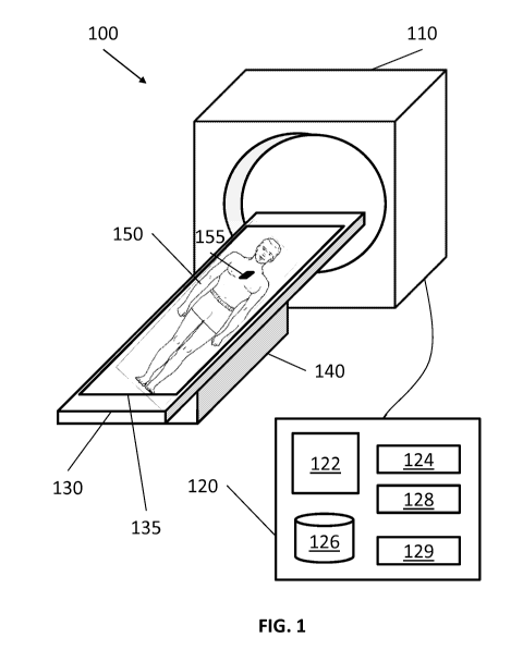
[0017] FIG. 1 is a perspective view of a system for treating lung
diseases of a patient in
accordance with an embodiment of the present disclosure;
[0018] FIG. 2A is a view of a computed tomography (CT) scan image of a
patient's lungs
taken from the transverse plane in accordance with an embodiment of the
present disclosure;
4
CA 02899514 2015-07-27
WO 2014/124241 PCT/US2014/015281
[0019] FIG. 2B is perspective view a patient's body illustrating the
transverse plane in
accordance with an embodiment of the present disclosure;
[0020] FIG. 2C is a view of a CT scan image of a patient's lungs taken
from the coronal
plane in accordance with an embodiment of the present disclosure;
[0021] FIG. 2D is perspective view of a patient's body illustrating the
coronal plane in
accordance with an embodiment of the present disclosure;
[0022] FIG. 2E is a view of a CT scan image of a patient's lungs taken
from the sagittal
plane in accordance with an embodiment of the present disclosure;
[0023] FIG. 2F is perspective view of a patient's body illustrating the
sagittal plane in
accordance with an embodiment of the present disclosure;
[0024] FIG. 3 is an anatomical illustration of a three dimensional model
for a lung in
accordance with an embodiment of the present disclosure;
[0025] FIG. 4 is an illustration of a user interface for adding a target
to a pathway plan in
accordance with an embodiment of the present disclosure;
[0026] FIG. 5A is an two dimensional illustration of the lung of FIG. 3;
[0027] FIG. 5B is an illustration of finding a pathway from a target to
an entry point of a
patient in accordance with an embodiment of the present disclosure;
[0028] FIG. 5C is an illustration of navigating the pathway of FIG. 5B
from the entry
point to the target in accordance with an embodiment of the present
disclosure;
[0029] FIG. 5D is an illustration of an imaging device inserted into the
lung following
the pathway;
[0030] FIG. 5E is an enlarged detail view of the circled area 530 of FIG.
5D;
[0031] FIG. 6 is a cross-sectional view of the lung of FIG. 5A with
respect to A-A
CA 02899514 2015-07-27
WO 2014/124241 PCT/US2014/015281
direction;
[0032] FIG. 7 is a flowchart of a method for generating a treatment plan
to treat a lung
disease in accordance with an embodiment of the present disclosure; and
[0033] FIG. 8 is a flowchart of a method for treating the lung disease
based on the
treatment plan of FIG. 7 in accordance with an embodiment of the present
disclosure.
DETAILED DESCRIPTION
[0034] The present disclosure is related to systems and methods for
treating lung diseases
using images of the lung to identify and locate a target nerve for denervation
treatment. One or
more imaging modalities may be used to provide sufficient resolution to locate
the target nerve.
Treatments are performed from outside of a patient's body and thus are not
invasive to the
patient.
[0035] Although the present disclosure will be described in terms of
specific illustrative
embodiments, it will be readily apparent to those skilled in this art that
various modifications,
rearrangements and substitutions may be made without departing from the spirit
of the present
disclosure. The scope of the present disclosure is defined by the claims
appended hereto.
[0036] FIG. 1 shows a system 100 that is generally directed to treating
lung diseases by
denervation. The system includes a treatment device 110, a treatment board
120, a support 130,
and a computing device 140. The treatment device 110 may use radiation
technique, such as
stereotactic body radiation therapy (SBRT), to non-invasively treat a portion
of a lung from
outside of the patient's body. In embodiments, the treatment device 110 may
use other forms of
medical techniques or energy, such as high intensity focused ultrasound
(HIFU), proton therapy,
and others suitable for non-invasive treatment for lung diseases known to
those of skill in the art.
[0037] In particular, the system 100 non-invasively treats the lung
diseases by utilizing a
6
CA 02899514 2015-07-27
WO 2014/124241 PCT/US2014/015281
three dimensional model of a lung to identify and locate a target for
denervation. The system
100 includes a treatment device 110, a computing device 120, a treatment bed
130, and a support
140. A patient 150 is lying on the treatment bed 130, awaiting entry into the
treatment device
110.
[0038] As noted above, the treatment device 110 is preferentially of the
type which
enables treatment of the patient 150, and particularly the lungs of the
patient 150 in a non-
invasive fashion. In other words, the treatment device 110 radiates treatment
energy and focuses
the treatment energy on the target. Thus, no incision of the tissue of the
patient 150 and no
insertion of catheter through a body opening, such as mouth, nose, or medical
incision of the
body, are necessary for treatment.
[0039] In some instances, the treatment device 110 may also be used as an
imaging
device in accordance with an embodiment of the present disclosure. It has been
reported that
such devices featuring combined imaging and treatment experience fewer
exporting errors,
which are generally caused by exporting image data from an imaging device to a
treatment
device, and may be reduced and localization errors, which are generally caused
by different
location of a patient between an imaging device and a treatment device.
Further, it may be
possible to conduct treatment immediately after or even while imaging a
portion of the patient
150 (e.g., the lungs).
[0040] The treatment device 110 may also be used for repeated or follow-
up procedures.
For example, in some situations, once a denervation treatment for one or more
nerves has been
done, the treated nerves are not completely severed and may regenerate. This
incomplete
severing of the nerve may be part of the treatment plan, or may be an
unintended. For this
reason, repeated or follow-up treatments may be made to provide additional
treatments to the
7
CA 02899514 2015-07-27
WO 2014/124241 PCT/US2014/015281
previously treated nerves to either obtain or to maintain the intended
therapeutic effect. .
[0041] The computing device 120, such as, a laptop, desktop, tablet, or
other similar
computing device, includes a display 122, one or more processors 124, memory
126, a network
card 128, and an input device 129. The system 100 may also include multiple
computing devices
120, wherein separate computing devices 120 are employed for procedure
planning and
treatment. The display 122 may be touch-sensitive and/or voice-activated,
enabling the display
122 to serve as both an input and output device. The display 122 may display a
two dimensional
or three dimensional model of a lung to locate and identify a portion of the
lung that displays
symptoms of the lung diseases. The display 122 may further display options to
select, add, and
remove a target to be treated and settable items for the treatments.
[0042] The one or more processors 124 execute computer-executable
instructions. The
processors 124 may perform image-processing functions so that the two
dimensional or three
dimensional model of the lung can be displayed on the display 122. In
embodiments, the
computing device 120 may further include a separate graphic accelerator that
performs only the
image-processing functions so that the one or more processors 124 may be
available for other
programs.
[0043] The memory 126 stores data and programs. For example, data may be
image data
for a two or three dimensional model or any other related data such as
patients' medical records,
prescriptions and/or history of the patient's diseases. The memory 126 may
include one or more
solid-state storage devices such as flash memory chips, mass storage, tape
drive, or any
computer-readable storage medium which is connected to a processor through a
storage
controller and a communications bus. Computer readable storage media include
non-transitory,
volatile and non-volatile, removable and non-removable media implemented in
any method or
8
CA 02899514 2015-07-27
WO 2014/124241 PCT/US2014/015281
technology for storage of information such as computer-readable instructions,
data structures,
program modules or other data. For example, computer-readable storage media
includes random
access memory (RAM), read-only memory (ROM), erasable programmable read only
memory
(EPROM), electrically erasable programmable read only memory (EEPROM), flash
memory or
other solid state memory technology, CD-ROM, DVD or other optical storage,
magnetic
cassettes, magnetic tape, magnetic disk storage or other magnetic storage
devices, or any other
medium which can be used to store desired information and which can be
accessed by the
computing device 120.
[0044] As noted above, one type of program stored in the memory 126 is a
pathway
planning module. As an initial step of pathway planning, image data of a
patient (typically in
DCOMM format) from for example a CT image data set (or other imaging modality)
is imported
into the pathway planning module. Imaging may be done by CT imaging, magnetic
resonance
imaging (MRI), functional MRI, ultrasound imaging, X-ray, and/or any other
imaging modalities.
[0045] The pathway planning module processes images of a patient and
creates a three-
dimensional model of a desired portion of the CT image, for example the lungs.
To generate the
3D model, the pathway planning module employs segmentation, surface rendering,
and/or
volume rendering. Details of these processes and the pathway planning module
can be found in
commonly assigned U.S. Patent Application number XX/XXXXXX (having attorney
docket no.
H-IL-00099) and U.S. Patent Application number 13/838,805, the entire contents
of which are
incorporated herein by reference. Such pathway planning modules permit the
user to view
individual slices of the CT image data set, and to identify one or more
targets. These targets may
be, for example, lesions or the location of a nerve which affects the actions
of tissue where lung
disease has rendered the lung function compromised or others. Having
identified these targets,
9
CA 02899514 2015-07-27
WO 2014/124241 PCT/US2014/015281
the pathway planning module enables the user to develop a plan to either
achieve access to the
target, for example by extending a biopsy or other tools through a natural
orifice or an incision to
be made by a clinician, or to pin-point the location and identify the
coordinates of the target such
that they can be employed by a treatment device 110, as will be described
below. The pathway
planning module guides a clinician through a series of steps to develop a
pathway plan for later
use for obtaining images with refined resolution. A clinician communicates
with the pathway
planning module via the display device 122 which displays interactive features
to receive inputs
from the clinician. The pathway planning module may be employed to further
refine the
resolution of one or more targets to identify and locate a nerve to be
denervated. The term,
clinician, includes doctor, surgeon, nurse, medical assistant, or any user of
the pathway planning
module involved in planning, performing, monitoring and/or supervising a
medical procedure.
[0046] The network interface 128 enables other computing devices 120
and/or the
treatment device 110 to communicate with each other through a wired and/or
wireless network
connection. In FIG. 1, the treatment device 110 may transmit or receive
medical images,
medical data, and control data with the computing device 120 via a wired
connection. In a case
where the network interface 128 connects to other computing devices 120 or the
treatment device
110 wirelessly, the network interface 128 uses a frequency for communication,
which is different
from the frequencies that the treatment device 110 uses for treatment.
[0047] The input device 129 is used for inputting data or control
information, such as
setting values, text information, and/or controlling the treatment device 110.
The input device
129 includes a keyboard, mouse, scanning devices, or other data input devices.
[0048] The treatment bed 130 receives the patient to be treated. The
support 140
supports the treatment bed 130 and may have mechanical structures to make the
treatment bed
CA 02899514 2015-07-27
WO 2014/124241 PCT/US2014/015281
130 movable horizontally and vertically. For example, when a patient lies down
her body on the
treatment board 130, the support 140 adjusts the height of the treatment board
130 and moves the
treatment board 130 to and from the treatment device 110, so that a target of
the lung to be
treated is placed at an optimal height and under the treatment device 110 for
treatment.
[0049] In embodiments, in case that the treatment device 110 is also used
for imaging the
lung to make a three dimensional model, the support 140 may move the treatment
bed 130 in
three transversal directions, namely, transversally, coronally, and
sagittally. Or, the treatment
device 110 may have imaging sensors that capture slices of image of the
patient's body in the
three directions without moving the patient 160.
[0050] The treatment bed 130 includes a field generator 135. The field
generator 135
may be employed for a number of functions. The primary function is to enable
the registration
of the CT image data, and particularly the targets identified therein during
the pathway planning
steps, with the location of a patient 150 lying on the treatment bed 130. As
will be appreciated,
in instances where the imaging and treatment are performed on different
machines, at different
times, or at different locations, registration of the patient with the image
data is important to
ensure that treatment is occurring at the proper locations within the patient.
Registration of the
patient 150 with the image data may be undertaken in a variety of ways.
[0051] One methodology for registration is to traverse a probe with a
sensor through two
or three bifurcations of a patient's bronchial tree. The sensor may be placed
for example on a
bronchoscope. The sensor detects the electromagnetic field generated by the
field generator 135,
and outputs a signal representative of its location. This signal is used in
combination with image
registration software, to match bronchoscopic image data with an internal view
of the 3D model
generated in the pathway planning steps described above. A variety of factors
are employed in
11
CA 02899514 2015-07-27
WO 2014/124241 PCT/US2014/015281
the registration process and its details are described in greater detail in
commonly assigned U.S.
Patent No. 8,218,846, the entirety of which is incorporated herein by
reference. Once registered,
the location of the patient within the electromagnetic field is known relative
to the location of the
target identified during pathway planning, and the coordinates of the target
can be used to
conduct treatment.
[0052] In embodiments, when respiratory movements of the patient 160 are
to be
monitored, the field generator 135 may be coupled with one or more sensors
located on the
patient 150 so that the patient's respiration and particularly the patient's
movements can be
monitored and accounted for during treatment. For example, movement tracking
sensor 155 may
be electromagnetically coupled with the treatment bed 130 or the field
generator 135. When the
patient 150 breathes in and out, air flows in and out of the lung so as to
inflate or deflate the lung,
respectively. The movement tracking sensor 155 also moves accordingly and
senses changes in
location with respect to the treatment bed 130. The movement tracking sensor
155 may be
placed on at least two body parts to consider comparative movements of
different body parts
(e.g., the width and depth of the chest).
[0053] Since the movement tracking sensor 155 does not actually track the
movement of
the target nerve, a breathing model is selected to correlate the respiratory
movement and the
movement of the target nerve. In this way, movements of different body parts
are considered
and are registered to CT images so that the accurate location of the target
nerve during
denervation treatment may be identified.
[0054] In embodiments, the field generator 135 may cover the whole
treatment bed 130
and may activate a portion of the field generator 135 so that only such
portion can be monitored.
The field generator 135 may generate a field other than the electromagnetic
field, which can be
12
CA 02899514 2015-07-27
WO 2014/124241 PCT/US2014/015281
used for monitoring a location of sensors located on the patient 150 and which
is known to a
person of ordinary skill in this area.
[0055] FIGS. 2A-2F show one of effective imaging modalities of
identifying targets, i.e.,
computed tomographic (CT) technique. The use of CT images as diagnostic tools
has become
routine and CT results are frequently the primary source of information
available to a clinician
regarding the size and location of a target lesion, tumor, or other similar
target of interest. CT
images are typically obtained by digitally imaging a patient in slices in each
of the transversal,
coronal and sagittal directions. For example, FIG. 2A illustrates a slice of a
CT image taken
from the transversal direction. In other words, CT images are cross-sectional
views taken at a
plane perpendicular to the transversal direction or perpendicular to the spine
of the patient as
illustrated in FIG. 2B. FIG. 2C illustrates a slice of a CT image taken from
the coronal direction.
In other words, CT images are cross-sectional views taken at a plane
perpendicular to the coronal
direction as illustrated in FIG. 2D. FIG. 2E illustrates a slice of a CT image
taken from the
sagittal direction. In other words, CT images are cross-sectional views taken
at a plane
perpendicular to the sagittal direction as illustrated in FIG. 2F. A clinician
may review the CT
image data slice by slice from each direction when attempting to identify or
locate a target, as
described above during the pathway planning phase.
[0056] In embodiments, these slices of images captured in the three
directions, i.e.,
transversal, coronal, and sagittal directions, are input to the computing
device 120 which, in turn,
generates a three dimensional model of the patient's lung. Generally, CT
images include images
of all organs inside of the patient's body. The computing device 120 processes
the CT images so
that images of most of organs are included in the three dimensional model. The
three
dimensional model may selectively show only the left and right lobes of the
lung, bronchial trees,
13
CA 02899514 2015-07-27
WO 2014/124241 PCT/US2014/015281
or the trachea. Nevertheless, two dimensional images (i.e., CT images) are
used to see images as
is taken.
[0057] FIG. 3 illustrates a three dimensional model 300 for a patent's
bronchial trees and
the trachea together with the lung according to an embodiment of the present
disclosure. The
three dimensional model may include information of most of the organs so that
a clinician may
selectively see particular organs or portions of organs of interest as shown
in FIG. 3. In this case,
these selected organs are the lungs including right lobe 310, the left lobe
320, the trachea 330
and bronchial trees 340. The right lobe 310 has three sub-lobes, i.e.,
superior lobe 312, middle
lobe 314, and inferior lobe 316, and the left lobe 320 has two sub-lobes,
i.e., superior lobe 322
and inferior lobe 324.
[0058] The trachea 330 is a tube that connects the pharynx and larynx to
the lung 310 and
320. At the lower end of the trachea 330, left or right primary bronchus 342
is divided.
Secondary bronchus 344 also divides at the lower end of the primary bronchus
342. The
circumference of the primary bronchus 342 is greater than that of the
secondary bronchus 344.
In the same manner, tertiary bronchus 346 divides at the lower end of the
secondary bronchus
344 and terminal bronchiole 348 divides at the lower end of the tertiary
bronchus 346. The
primary bronchus 342, the secondary bronchus 344, and the tertiary bronchus
346 are supported
by cartilaginous plates. However, when the size of the tertiary bronchus 346
becomes smaller
and smaller, the cartilaginous plates disappear and outer wall is dominated by
smooth muscle.
The outer wall of the terminal bronchiole 348 is also dominated by smooth
muscle.
[0059] A target nerve may exist on any bronchial trees, the primary
bronchus 342, the
secondary bronchus 344, the tertiary bronchus 346, and the terminal
bronchioles 348. Effects of
denervation of a target may be based on severity of symptoms or the location
of the target nerve.
14
CA 02899514 2015-07-27
WO 2014/124241 PCT/US2014/015281
If symptoms of the lung diseases are severe, plastic denervation may be
performed, or if the
symptoms are mild, elastic denervation may be performed. Plastic denervation
wholly disables
functions of the target nerve and elastic denervation partly disables the
functions. If a target
nerve is located on the primary bronchus 342, functions of nerves which are
connected to and
located below the target nerve, which is on the following secondary, tertiary,
and terminal
bronchial trees, may be disabled wholly or partly. In the same way, when a
target nerve is
located on the terminal bronchioles 348, only the functions of the target
nerve is disabled wholly
or partly but nerves connected to and located above the target nerve perform
their functions well
without being affected by the denervation.
[0060] Additionally, if symptoms of the lung diseases are severe, a
treatment size may be
greater than the size of the target nerve and, if not, the treatment size may
be equal to or smaller
than the size of the target nerve. Thus, the treatment size of the target
nerve depends on severity
of the symptoms of the lung disease, a location of the target nerve, and a
size of the target nerve.
[0061] According to some embodiments, a further refinement for the slices
of images is
necessitated when a selected imaging modality does not give sufficient
resolution to locate a
target nerve. This may be particularly true when seeking to treat the tertiary
bronchus 346 or the
terminal bronchiole 348. In this case, another imaging modality may be used to
provide further
refined resolution of the slices of images so that target nerves can be
identified and located.
[0062] In accordance with one embodiment, an ultrasound imaging modality
may be
employed to provide greater specificity and greater accuracy in identifying
the target nerve's
location in the patient 150. In one such embodiment, a radial ultrasound probe
is employed
following the pathway plan described above and images are taken of the
pathway. These images
may be registered to those of the CT image data and/or the 3D model to provide
greater clarity
CA 02899514 2015-07-27
WO 2014/124241 PCT/US2014/015281
with respect to the location of a target nerve. For example, this data may
also be used
diagnostically to help the clinician confirm that all likely candidates for
targeting have been
identified. As will be appreciated, other imaging modalities may be employed
to enhance the
first image data collected (e.g., the CT image data), these modalities
includes various forms of
ultrasound both internal and external to the patient, magnetic resonance
imaging (MRI),
fluoroscopy, and others without departing from the scope of the present
disclosure.
[0063] FIG. 4 illustrates a user interface 400 of the pathway planning
module for adding
a target and ultimately for developing a pathway plan in accordance with an
embodiment of the
present disclosure. If a clinician selects to create a new pathway plan, the
user interface 400 is
displayed on a display. The user interface 400 includes a localizer 410 and a
main window 420.
[0064] The localizer 410 shows a view orthogonal to the main image of the
screen, here
the main view is the axial view, thus the localizer is in the coronal view
showing the left and
right lobes 412 of a patient's lung and a location bar 414. As depicted in
FIG. 4, a clinician can
move the location bar 414 vertically, which has the effect of changing the
slice of the CT image
the axial direction as shown in FIG. 2A, to scroll through the CT images taken
at a plane
perpendicular to the axial direction as shown in FIG. 2B. The clinician may
also or alternatively
scroll through the CT images of the patient's lungs via an input device such
as a mouse wheel or
other device without directly moving the location bar 414. When another
direction is selected
for display on the three dimensional model, for example, the coronal
direction, the localizer 414
may display a coronal view of the organ requiring treatment (here shown as the
lungs). The
localizer 414 provides the clinician with a general reference for where the CT
slice 430 the
clinician is currently viewing is located in the organ being considered. The
localizer 414 may
also display one or more previously identified targets for the clinician's
reference.
16
CA 02899514 2015-07-27
WO 2014/124241 PCT/US2014/015281
[0065] The main window 420 shows an image 430 which corresponds to a CT
image
taken at a plane where the location bar 414 is located in the left and right
lobes 412. Title 432
indicates that the image 430 is a CT image taken in the direction of the
transversal or axial
direction. Date and time section 446 indicates the date and time when the CT
image 430 was
taken. Thus, a clinician can determine whether the image 430 was sufficiently
recent for
planning a pathway for a target. In case when the clinician determines that
the image 430 is too
outdated for the pathway planning, new images should be taken as shown in
FIGS. 2A-2F for
generation of a new 3D model and the pathway planning.
[0066] Target selection tools such as the cross hairs 434 helps the
clinician to select a
target 436. Direction indicators 438 and 440 indicate which direction is right
and left. As shown
in FIG. 4, the target is selected in the right lobe of the lung based on the
direction indicators 438
and 440.
[0067] Zoom slider bar 442 is used to zoom in and out to see details of
or general view of
the image 430. For example, if the slier of the zoom slider bar 442 is close
to zoom-out icon, a
particular portion of the image 430 is reduced and, if the slider of the zoom
slider bar 442 is
close to zoom-in icon, the particular portion of the image 430 is enlarged.
Window icon 444
may be used with the zoom slider bar 442 to refine a selection size of the
target 436. When the
target 436 is located by the target selection 434, the clinician may use the
zoom slider bar 442 to
zoom in the selected area and closely identify the target by resizing and/or
relocating the target
window using the window icon 444.
[0068] When the target 436 and its size are identified, the clinician
clicks plan button 448
to make a pathway plan to the target. The pathway plan may be reviewed and
exported by
clicking review & click button 450. If the clinician determines that the
pathway plan is
17
CA 02899514 2015-07-27
WO 2014/124241 PCT/US2014/015281
acceptable, the pathway plan is finished and exported by clicking finish &
export button 452. If
there are more than one target, the clinician can add more target by clicking
add a target button
454 and doing the same things as described above. Detailed method for planning
a pathway is
described in commonly assigned U.S. Patent Application Serial No. 13/838,805
to Baker and
U.S. Patent Application Serial No XX/XXXXX (having attorney docket no, H-IL-
00099), as
well as the references cited therein, all of which are incorporated by
reference in the present
disclosure.
[0069] FIGS. 5A shows a planar view of bronchial trees of a three
dimensional model or
of the slices of images of the lung such as the bronchial trees of FIG. 3.
Assuming a target area
is located at the tip of the bottom left end of the terminal bronchiole of
FIG. 5A, FIG. 5B
illustrates a pathway from the target area of the three dimensional model,
which corresponds to a
portion of the lung displaying symptoms of the lung disease, to a second area
of the three
dimensional model, which corresponds to the trachea. FIG. 5C illustrates an
ultrasound
transducer inserted into the lungs of the patient to the target following the
pathway of the three
dimensional model. When the ultrasound transducer reaches the portion of the
lung, the
ultrasound transducer transmits ultrasounds and receives sound reflects so
that the tissue in that
area can be more clearly defined and ultimately one or more nerves to be
denervated around the
target can be located and identified. In this way, CT imaging modality and the
ultrasound
imaging modality give sufficient resolution to identify sufficiently accurate
location of one or
more nerves to be denervated in the patient's lung.
[0070] FIGS. 5D and 5E illustrate an extended working channel 510
including an
ultrasound transducer 525 that is position at the distal end of the extended
working channel 510.
The clinician navigates a luminal network of the bronchial trees and the
trachea by following a
18
CA 02899514 2015-07-27
WO 2014/124241 PCT/US2014/015281
pathway plan as shown in FIG. 5C so that the ultrasound transducer 525 can
reach the identified
portion of the lung tissue.
[0071] FIG. 5E is an enlarged detail view of a circled area 530 of FIG.
5D. while the
distal tip of the extended working channel 510 or the ultrasound transducer
525 is navigated
through the luminal network toward the identified portion, the ultrasound
transducer 525 may
radiate ultrasound waves and receives reflects to capture images of the
luminal network and the
identified portion, which has a greater resolution than that of the slices of
images. It is described
in greater detail in commonly assigned U.S. Patent Application Serial No.
13/836,203, the
entirety of which is incorporated herein by reference.
[0072] FIG. 6 is a cross-sectional view 600 of the terminal bronchiole in
the direction of
A-A of FIG. 5A. The terminal bronchiole is surrounded by soft muscle 610.
Nerves 620 and
veins 630 are located on the soft muscle. The ultrasound imaging modality, as
described above,
provides a local view of the airways even out to the terminal bronchiole so
that even the thin
nerves 620 on the soft muscle 610 can be identified.
[0073] The lungs and tissue associated with the lungs are constantly in
motion. As a
result the nerves 620, move during a treatment because the thickness or size
of the nerves 620 is
relatively small compared to a movement of any patient's body part (e.g., the
lung, diaphragm, or
vascular tissue) or any operational movement of clinician (e.g., the treatment
device 110 or the
treatment bed 130). Thus, such movements should be compensated for to
accurately identify,
locate, and treat a target nerve.
[0074] The target nerve 620 may be cholinergic-parasympathetic nerve,
which mediates
contractions of muscle, or adrenergic-sympathetic nerve, which mediates
relaxation. The target
nerve 620 may also be a pre- or post-ganglionic nerve.
19
CA 02899514 2015-07-27
WO 2014/124241 PCT/US2014/015281
[0075] FIG. 7 shows a flowchart illustrating a method 700 for generating
a treatment plan
to treat a lung disease by denervation. The method 700 locates and identifies
one or more targets
and then generates a treatment plan for the targets. In step 705, a clinician
diagnoses a lung
disease by inspection, palpation, percussion, and/or auscultation.
[0076] After the lung disease is diagnosed, an imaging device takes
images of the patient
150 using for example a MRI or CT imaging device in step 710. Typical MRI or
CT imaging
devices render images of the patient in three axes, i.e., transversal,
coronal, and sagittal
directions. In embodiments, the clinician may use imaging enhancing agents to
fluorescently
dye the lung before taking images so as to identify the location of the lung
in either the images or
under visualization. Some of the imaging enhancing agents may be transportable
axonally
anterogradely or retrogradely to help visualize the white matter track (axon)
or gray matter
nucleus in brain. In other words, the imaging enhancing agents may help to
visualize nerves
located in and around the bronchial tree. This may be provided to the patient
even before the
image of the lung is taken so that, when the images of the lung are taken, the
fluorescent marker
is depicted on the images clearly. Imaging enhancement agents may be
fluorescent dye or
FLUOROGOLDTM. For example, FLUOROGOLDTM is a neuronal retrograde tracer which
stains the dendrites of nerve completely. When FLUOROGOLDTM is injected the
nerve
becomes fluorescently dyed and as a result emits frequencies of fluorescent
light when excited
by a specific frequency of light. In this way, an imaging device or
fluorescence microscopy
detects the fluorescent light so that a clinician can differentiate the nerve
from other organs with
clarity. Other markers for identifying the location of nerve tissue may be
employed by those of
ordinary skill in the art without departing from the present disclosure.
[0077] These images are combined and processed to generate a three
dimensional model
CA 02899514 2015-07-27
WO 2014/124241 PCT/US2014/015281
of the bronchial tree of the patient's lung in step 715. Generally, the more
images taken in each
direction, the more refined model may be created. Nevertheless, at some point,
more slice
images do not help enhancing resolution of the three dimensional model due to
limitations of the
selected image modality. Thus, an optimum number of slice images is taken in
each of the three
directions and is pre-determined in consideration of the specification of the
imaging modality
and a required resolution.
[0078] In step 720, a clinician reviews the three dimensional model/and
or the MRI or
CT images to identify the portions of the lung suffering from disease and
requiring treatment.
This is a gross determination and focuses the clinician's attention on the
appropriate portion of
the lung, wherein identification of specific nerves will be targeted and
treated as described in
detail below.
[0079] In embodiments, the three dimensional model may have information
of most of
the internal organs and other physiology in or around the lung, for example,
heart, ribs, spine,
and lung, bronchial trees, and diaphragm. The clinician may see organs
selectively and may
rotate the three dimensional model around any direction so that the clinician
can decide which
way is a more suitable direction to avoid hard tissues such as bones while
treating a target nerve.
Depending on the resolution of the three dimensional model, nerves for
treatment may be visible
in the model and the clinician can use tools in the user interface to mark
these nerves for targeted
treatment. In such an embodiment, it may not be necessary to review the
individual CT images.
[0080] In step 725, it is determined whether the three dimensional model
and CT images
have sufficient resolution to identify a target nerve proximate the identified
portion of the lung.
For example, if the identified portion of the lung for treatment is on a
primary or secondary
bronchial tree, then the three dimensional model and CT images may provide
sufficient
21
CA 02899514 2015-07-27
WO 2014/124241 PCT/US2014/015281
resolution to identify a target nerve. However, if the identified portion is
on a tertiary or terminal
bronchial tree, the three dimensional model and CT images may not provide
sufficient resolution
to do such.
[0081] When it is determined that the three dimensional model or CT
images provides
sufficient resolution, the clinician may identify a target nerve to be treated
proximate the
identified portion in step 748. The identification of a target refers to the
placement of a target on
the images and/or three dimensional model by the clinician. The target and
specifically the
coordinates of the target in the image and three dimensional model are used to
direct the
treatment device, as will be described in detail below.
[0082] In step 750, a location or fiducial marker may be optionally
placed in the lung
tissue proximate the target nerve. Generally, treatment of the target nerve
takes place in a
different time and space from identifying the target nerve. Thus, at a later
check-up or another
imaging of the lung for treatment, the clinicians may have to confirm the
location of the target
nerve. In this case, the location marker is used to guide the clinicians back
to the same location
which is proximate the portion of the lung displaying the symptoms of lung
disease. The fiducial
markers may also being employed in one or more registration process for
treatment of the target
nerve. In embodiments, a plurality of markers may be placed in the lung tissue
so that, when the
plurality of markers are imaged at a later time for treatment, a clinician may
identify the size and
depth of the target nerve based on the image showing topology of the plurality
of markers.
[0083] In step 755, it is determined whether there are more nerves to
target in the
identified portion of the lung suffering from lung disease. When it is
determined that there are
more nerves to target in the identified portion in step 755, steps 748, 750,
and 755 are repeated
until there are no more nerves to target. If it is determined that there are
no more nerves to target,
22
CA 02899514 2015-07-27
WO 2014/124241 PCT/US2014/015281
in step 760, the clinician further determines whether there are more portions
of the lung that
displays symptoms of the lung disease, which is different from the portion of
the lung identified
in step 720. When there are more portions, the method returns to step 720
until there are no
more portions that display the symptoms of the lung disease.
[0084] In step 725, when it is determined that the three dimensional
model and the
images do not provide sufficient resolution to identify a target nerve,
another imaging modality
may be necessary to generate further refined images to provide a sufficient
resolution to identify
a target nerve. In embodiments, a radial ultrasound imaging modality may
provide such
resolution of the identified portion of the lung. In order to obtain such
refined images of the
identified portion, the ultrasound imaging device is to be inserted into the
identified portion.
Here, the three dimensional model is used to determine which pathway the
ultrasound imaging
device is to follow to reach the identified portion of the lung. Such guidance
is called as a
pathway plan.
[0085] The pathway plan, as an option to obtain further refined images of
the identified
portion, is determined to guide a radial ultrasound transducer of the
ultrasound imaging device to
the identified portion in step 730. As described in Patent Application Serial
No. 13/838,805
which is incorporated by reference, the pathway plan is determined starting
from the identified
portion of the lung to a bodily opening such as mouth, nose, or incision.
[0086] In step 732, the patient is located on a location board and a
clinician inserts the
radial ultrasound transducer starting from the bodily opening and ending to
the identified portion
of the lung by following the pathway plan of the three dimensional model in
step 734. The
clinician may use the pathway planning module stored on the memory 126 of the
computing
device 120 of FIG. 1. The pathway planning module displays the three
dimensional model on
23
CA 02899514 2015-07-27
WO 2014/124241 PCT/US2014/015281
the display device 122 such that the clinician can confirm that the radial
ultrasound transducer
follows the pathway plan determined from the three dimensional model in an
order reverse to the
pathway plan, i.e., starting from a bodily opening to the identified portion
of the lung.
[0087] When the radial ultrasound transducer reaches the identified
portion of the lung,
the radial ultrasound transducer transmits high frequency sound waves
radially. The sound
waves are reflected from body organs in which density changes. In step 736,
the radial
ultrasound transducer detects the sound reflects and also transmits the
detected sound reflects to
the radial ultrasound imaging device which then processes the sound reflects
and generates
images
[0088] In embodiments, tissue spectroscopy based on near infra-red, infra-
red, or Raman
light scattering, optical coherence tomography, confocal microendoscopy, or
fluorescence
microendoscopy may be employed to provide sufficient resolution of the
identified portion of the
lung. Further, FLUOROGOLDTM may also be used to spectroscopically confirm
nerve location.
[0089] In step 738, the clinician may place a location marker near the
areas imaged using
the radial ultrasound. These location markers help to identify approximately
the location of the
target nerve for a later use. As in step 750, a plurality of location markers
may be used to
identify the location. The location markers may be placed at the same time
while imaging is
undertaken or as part of an iterative process where imaging and marker
placement are taken
alternatively, such that a marker is placed at each area where radial
ultrasound imaging is
undertaken. At a minimum location markers will be placed in and around the
portions of the
lung tissue suffering lung disease as previously identified in the CT images
or three dimensional
model.
[0090] The target may be one or more points along a nerve length, meaning
that targets
24
CA 02899514 2015-07-27
WO 2014/124241 PCT/US2014/015281
are located along and down the length of a nerve, on a single plane, e.g.,
circumference of a
bronchial tree, or along and down the length of a nerve in a different plane
with a different
distance apart from each other.
[0091] In step 740, it is determined whether there are more portions of
the lung tissue to
image with radial ultrasound. When it is determined that there are more
portions to image, steps
736 and 738 are repeated using the generated ultrasound images until there are
no more portions
require imaging. Once all the portions of the lungs are imaged using radial
ultrasound, the radial
ultrasound images are exported to the computing device 120 and stored in
memory 126 at step
742.
[0092] At this point the clinician has a decision to make. The radial
ultrasound images
taken in step 736 provide greater localized detail than the original CT images
taken in step 710.
Thus the radial ultrasound images may be registered to and combined with CT
images to
generate a high resolution image set. The decision to be made is whether to
generate a new CT
image at step 744. The benefit is that by generating a new CT image, the
fiducial markers which
were placed in step 738 will now also be imaged and provide for greater
ability to register and
clearly identify the location of targets for both treatment planning and
treatment of the patient.
However, in some instances it may be sufficient to forego the second CT
imaging step and
simply register the ultrasound images generated in step 736 with the original
CT images
generated in step 710. Accordingly, whether using the original CT images from
step 710 or
newly generated CT images from step 744, the CT images and the ultrasound
images are
registered to one another and a high resolution image set is generated in step
746.
[0093] From step 746, the process loops back to step 715 where a three
dimensional
model is generated, but this time using the high resolution image set. This
process continues
CA 02899514 2015-07-27
WO 2014/124241 PCT/US2014/015281
through step 760, as described above, to identify the locations of target
nerves in the high
resolution image set, until it is determined that there are no more nerves and
no more portions of
the lung tissue to review in steps 755 and 760.
[0094] When it is determined that there are no more nerves to identify in
step 755 and no
more portions of the lung t review in step 760, a treatment plan is generated
in step 765. The
treatment plan includes information which is necessary to treat all the
targets identified in the
method. For example, the treatment plan may include the size, depth, and
location of each target
nerve. Based on the information of each target nerve, the treatment plan may
further include
operational information on how to treat each target. The operational
information may include an
amount of energy to be radiated, a treatment period, a treatment vector, and
the number of
treatments to denervate a target nerve. The treatment period is a period
during which the
treatment device is applying energy to the tissue. Radiation of an amount of
energy at the
treatment vector for the treatment period may be determined such that it is
not likely to harm
tissues other than the intended target. Here, the treatment vector may be an
angle at which
treating energy is radiated to the target nerve. When the size or depth of the
target nerve is larger
or deeper than a predetermined size or depth, multiple treatments may be
necessary to fully treat
the target nerve. Even though an individual treatment may not harm tissues
other than the
intended target nerve, multiple treatments in the same location may harm the
tissues other than
the intended target. Thus, the treatment vectors may include a series of
angles in a case of
multiple treatments so that treating energy is not radiated via only one angle
during the multiple
treatments. The treatment plan may be dependent upon the severity of the lung
disease. During
treatment of target nerves, some tissues, such as hard tissues or bones, may
absorb or reflect the
treating energy. Such absorption or reflection of treating energy may cause an
ineffective
26
CA 02899514 2015-07-27
WO 2014/124241 PCT/US2014/015281
treatment or result in harms to some tissues other than the target nerves.
Thus the treatment plan
must be developed to avoid, to the extent possible interference from these
structures.
[0095] The three dimensional model may be utilized to determine a
treatment vector.
Since the three dimensional model has most of the organs and can be rotated in
any direction, the
clinician may determine a treatment vector by looking at organs selectively
and rotating the three
dimensional model in any directions. In embodiments, the three dimensional
model may be used
to automatically provide several treatment vectors for multiple unit
operations for one target
nerve. Once generated, this treatment plan can be exported to a memory device
126 or directly
to the treatment device 110 for use in treatment of the patient.
[0096] In embodiments, neuro-functional imaging modality may be used to
provide the
sufficient resolution of the identified portion of the lung. The neuro-
functional imaging modality
generates images of white matter anterogradely or retrogradely along long
tracks of nerves or
axons and may be registered with the MRI or CT images. Clinicians may identify
the size of
target nerves and make a treatment plan including a treatment period and
energy based on the
size.
[0097] FIG. 8 shows a flowchart illustrating a method 800 for treating
target nerves. In
810, a clinician imports the treatment plan together with the three
dimensional model, the images
(including CT, ultrasound, and high resolution image set) to the treatment
device. In step 815,
the patient is placed on the treatment bed 130 of FIG. 1. In instances where
the treatment device
110 is also an imaging device, in step 820, the clinician may performs a
follow-up imaging of the
patient's lung, this follow-up imaging may be used for registration purposes
to determine the
location of the patient 150 with respect to the treatment device. The
clinician compares the new
set of images with the previously taken images (e.g., the previous CT images
or the enhanced
27
CA 02899514 2015-07-27
WO 2014/124241 PCT/US2014/015281
resolution images) by looking at the location markers and registers the
patient's location on the
treatment device (i.e. the current image) with the prior images so that the
treatment device can
resolve the locations of target nerves in space with respect to the treatment
bed and the patient
150. Step 820 is optional, there are other methods for registering the patient
to the treatment
device which are known to those of skill in the art including performing a
bronchoscopy
procedure with a sensor located in a bronchoscope to sense the electromagnetic
filed emanating
from field generator 135 of FIG. 1. The generated field is sensed by the
sensor in the
bronchoscope (not shown) so that the relative location of the sensor with
respect to the patient
and the treatment bed can be registered to the CT or enhanced resolution
images. Here, the field
generated by the field generator may be an electromagnetic field or may be
other field that a
person of ordinary skill in the art can implement so that a sensor can sense
its location with
respect to the treatment bed. In this way, the patient's location in space may
be compensated for
so that the treatment device can identify and verify the location of target
nerves within the
treatment device. Regardless of whether additional images are taken, in step
825, location of the
patient in space on the treatment device 110 is registered to the treatment
plan.
[0098] In step 830, one or more movement tracking sensors may be
optionally placed on
the patient to track the movement of the patient. The movement tracking
sensors may be a
sensor that can sense the field generated by the field generator 135. While
the patient is placed
on the treatment bed, the patient's lung moves due to respiration, movement of
other organs such
as diaphragm, or movement of the patient. Such movements should be considered
and
compensated before actual treatment begins. The movement tracking sensor may
be a fiducial
marker, location sensor, or beacon. The movements of the lung may be caused by
respiratory
movement, cardiac motion, and/or movement of the patient. The movement
tracking marker
28
CA 02899514 2015-07-27
WO 2014/124241 PCT/US2014/015281
may be electromagnetically coupled with the treatment bed so that movement of
the patient with
respect to the treatment board may be recorded.
[0099] In a case when the movement tracking sensor may be placed on the
patient's body,
more than one movement tracking sensor may be placed to find a breathing model
which fits to
the patient's breathing pattern and the lung movement. The breathing model
shows relationship
between movement of the lung and the patient's breathing pattern. Accurate
estimation of a
tertiary or terminal bronchus tree while the patient is breathing may not be
easily obtained by a
generic breathing model because breathing causes the lungs to move cyclically,
meaning that the
lung movement varies by amplitude and direction during the breathing cycle
from 5mm to 30mm
depending on such breathing characteristics as patient size, age, altitude,
health, etc. U.S. PCT
International Application No. PCT/IB2008/003728, entirety of which is
incorporated by
reference herein, describes a method to build a dynamic breathing model that
can be used to
accurately estimate movements of a small bronchial tree during a patient's
breathing cycle.
[00100] Based on the breathing model, the clinician may estimate movement
of the lung
while patient is breathing. Since inhalation enlarges the chest, meaning that
depth and width of
the chest increase and exhalation deflates the chest, at least two movement
tracking sensors are
necessary to track changes in depth and width of the chest with reference to
the treatment bed,
one for depth and the other one for width.
[00101] Outputs of the movement tracking sensor is then transmitted to the
treatment
device. In step 835, the treatment device compensates the movement of the
patient so that the
treatment device may identify and locate the target nerve with respect to the
treatment bed during
a breathing cycle.
[00102] In embodiments, when the three dimensional model has pertinent
information for
29
CA 02899514 2015-07-27
WO 2014/124241 PCT/US2014/015281
treatment, the movement information may be displayed in the three dimensional
model to assist
the clinician in evaluating the treatment. In this case, the three dimensional
model may be used
throughout the end of the treatment for target nerves.
[00103] In step 840, the clinician can determine an initiation time for
starting treatment
during a breathing cycle. The treatment time included in the treatment plan
starts from the
initiation time. The target nerve of the lung moves the least starting from
the initiation time for
the operation period. The treatment period may be less than or equal to a
period from a time
when the patient is close to complete exhalation to a time when the patient is
close to start
inhalation or a period from a time when the patient is close to complete
inhalation to a time when
the patient is close to start exhalation. The treatment period may be a period
while the patient
holds a breath. If the treatment period determined in the treatment plan
method 700 is larger
than a period during which the lung moves the least, a number of treatments
may be employed
and, and the treatment vector may be adjusted based on the breathing model or
pattern of the
patient so that the adjusted operation information can apply to treating the
patient.
[00104] In step 845, the treatment bed or the treatment device moves so
that treatment can
be performed according to the treatment plan. For example, if the location of
a target nerve is in
the superior lobe of the right lung, the treatment bed may move in the
transversal direction so
that the target nerve is under the treatment device. Or if the treatment angle
for a target nerve is
at an angle of 30 degree from the right hand side, the treatment device may
rotate around the
transversal direction so that the treatment device can treat the target nerve
at that angle. While
doing this, the clinician should verify and confirm that the registered
location of the target in the
treatment device matches the actual location of the target with respect to the
treatment bed.
[00105] In step 850, denervation treatment is performed starting from the
initiation time
CA 02899514 2015-07-27
WO 2014/124241 PCT/US2014/015281
for the operation period such that the function of the identified target nerve
of the lung is affected.
The clinician may use SBRT to radiate stereotactic radiation from outside of
the patient's body
to denervate the target nerve at an angle defined in the operation vector. Or
any other non-
invasive treatment technique may be employed. Such treatment may be plastic or
elastic
denervation based on the severity of the lung disease.
[00106] In step 855, it is determined whether the target nerve is
sufficiently treated.
Determination may be performed based on real time imaging of the target nerve,
or based on a
calculation involving the target treatment volume, the amount of energy
applied, and the duration
of the treatment. If the target nerve is not sufficiently treated, another
denervation process is
performed until the target nerve is sufficiently treated. In this case, the
treatment plan for the
target nerve may also be adjusted so that consecutive treatments may not harm
tissues other than
the target nerve by changing angles listed in the operation vector, amount of
energy, and/or the
operation period included in the treatment plan. Here, the treatment may be
used in conjunction
with medical treatment (e.g., SPIRIVAO or lung function medications) to
accelerate effects of
the treatment or to compensate for lack of medical compliance.
[00107] If the target nerve is sufficiently treated, the clinician
determines whether there
are untreated target nerves in the treatment plan in step 860. If it is
determined that there are no
more target nerves in the treatment plan in step 860, treatments for all of
the target nerves are
completed. If there is an untreated target nerve, it is further determined
whether or not changing
treatment vector for another target nerve is required in step 865. This may
happen when the new
target nerve is in a location different from the previously treated target
nerve or when the new
target is treated at an angle different from that of the previously treated
target nerve. If that is
required, the treatment method returns to step 835 to compensate the movement
of the patient
31
CA 02899514 2015-07-27
WO 2014/124241 PCT/US2014/015281
because a different portion of the lung including the new target nerve may
move differently from
the portion of the lung including the previous target nerve. If not required,
the treatment method
returns to step 850 to treat the new target nerve non-invasively.
[00108] Although embodiments have been described in detail with reference
to the
accompanying drawings for the purpose of illustration and description, it is
to be understood that
the inventive processes and apparatus are not to be construed as limited
thereby. It will be
apparent to those of ordinary skill in the art that various modifications to
the foregoing
embodiments may be made without departing from the scope of the disclosure.
32