Note: Descriptions are shown in the official language in which they were submitted.
CA 02899693 2015-07-29
WO 2014/118297 PCT/EP2014/051845
Novel chimeric polypeptides for screening and drug discovery purposes
FIELD OF THE INVENTION
The present invention relates to novel polypeptides and their use for
screening and drug discovery.
More specifically, the invention provides chimeric polypeptides comprising a
membrane protein, in
particular a GPCR, fused to a binding domain, wherein the binding domain is
directed against and/or
specifically binds to said membrane protein. In particular, the chimeric
polypeptides of the invention
are single proteins wherein, in an intramolecular reaction, the binding domain
stabilizes the membrane
protein in a conformation of interest. Also provided are nucleic acid
sequences encoding such chimeric
polypeptides, cells capable of expressing such chimeric polypeptides as well
as cellular compositions
derived thereof. Also envisaged are screening methods for compounds using the
chimeric polypeptides
of the invention.
BACKGROUND
Drug discovery efforts generally focus on the identification of compounds that
modulate - inhibit or
enhance - the activity of the target of interest. Conventional lead
identification efforts proceed via
biochemical or cell based screening or in silico compound design. These
methods have identified and
validated a multitude of viable therapeutics in use today. However, as
reflected by the high failure rate
of new drug compounds (only an estimated 8% of phase I clinical therapeutics
eventually gain Food
and Drug Administration approval, at a conservative cost of $800 million per
drug), many efforts are
unsuccessful and often targets are abandoned once they are deemed undruggable
(Lee et al. 2009). A
considerable part of these failures are due to the fact that most biochemical
or cell based assays are
performed on targets in their prominent conformation, also referred to as the
basal conformation.
However, we now know that conformational flexibility is key to the function
and the pharmacology of
the majority of the current and future drug targets including GPCRs, ion
channels, (nuclear) receptors,
kinases and phosphatases. And for many of these targets, the most stable
conformation -
corresponding to the prominent structural species in the absence of ligands or
accessory proteins (the
basal conformation) - does not correspond to the druggable conformation to
which a drug must bind
to be most effective for the therapeutic indication.
Today, the most commonly targeted protein class for medicinal intervention are
G protein-coupled
receptors (GPCRs), also called seven-transmembrane receptors (7TMRs). They
play essential roles in
physiological responses to a diverse set of ligands such as biogenic amines,
amino acids, peptides,
proteins, prostanoids, phospholipids, fatty acids, nucleosides, nucleotides,
Ca2+ ions, odorants, bitter
CA 02899693 2015-07-29
WO 2014/118297 PCT/EP2014/051845
and sweet tastants, pheromones and protons (Heilker et al. 2009). Orthosteric
ligands that act on a
GPCR can induce a spectrum of effects on down-stream signaling pathways. In
general, GPCRs require
agonist binding for activation. Full agonists maximally activate the receptor.
Partial agonists elicit a
submaximal stimulation even at saturating concentrations. In some cases, a
GPCR may exhibit basal
activity towards a specific signaling pathway even in the absence of an
agonist (constitutive activity).
Inverse agonists can inhibit this basal activity. Notably, whereas neutral
antagonists can inhibit binding
of agonists, partial agonists, and inverse agonists at the orthosteric binding
site of GPCRs, they do not
alter the basal receptor activity. In recent years, advances have been made in
the discovery of novel
ligands for GPCRs that act at allosteric sites to regulate receptor function,
including positive and
negative allosteric modulators (PAMs and NAMs, respectively) and neutral
ligands, which offer novel
modes of action over orthosteric ligands (Christopoulos 2002).
It is now well established that GPCRs can signal through several distinct
mechanisms including those
mediated by G proteins or the multifunctional adaptor proteins 13-arrestins
(Rajagopal et at. 2010).
With the structures of several GPCRs solved in complex with various ligands
including inverse agonists,
antagonists and agonists (Cherezov et al. 2007, Rasmussen et al. 2011b,
Rosenbaum et al. 2011,
Shimamura et al. 2011, Xu 2011, Granier et al. 2012, Naga et al. 2012, Hanson
et al. 2012, Kruse et al.
2012, Manglik et at. 2012, Wu et al. 2012, Zhang et al. 2012) and the G-
protein (Rasmussen et al.
2011a), we now know that GPCRs are conformationally complex molecules with
specific conformations
causing G protein activation. Of special significance in the context of this
invention is the observation
that in comparison to the basal conformation, only relatively small changes in
the structure of the
agonist binding pocket led to substantial movement (up to 14A) and
rearrangements in three of the
transmembrane segments (Lebon et al. 2012).
Mass-spectrometry-based strategies (Kahsai et al. 2011), biophysical analysis
(Yao et al. 2006, Mary et
al. 2012) and NMR spectroscopy (Liu et al. 2012; Bokoch et al. 2010) provide
direct evidence for the
presence of other distinct ligand-specific conformations that lead to arrestin
mediated signalling. It
follows that different ligands can have differential effects on the
conformation and the diverse
signaling and regulatory repertoire of a single receptor. The importance of
these multiple
conformational states is their pharmacological relevance. As illustrated in
Figure 1, each of these
receptor conformations can be considered as a separate therapeutic drug target
because each of these
conformations promotes distinct relative efficacies toward the different
effector systems including G
proteins and arrestins.
2
CA 02899693 2015-07-29
WO 2014/118297 PCT/EP2014/051845
Drug discovery approaches can take considerable advantage from capturing the
target in a
therapeutically relevant - 'druggable' - conformation. Stabilizing a receptor
in a particular functional
conformation would inherently freeze the receptor in a single, disease
relevant druggable
conformation revealing new structural features that are suitable for targeting
with small molecules or
biologicals and may enable the identification of compounds that are selective
for that druggable
conformation. The stabilization of a unique druggable conformation, including
inactive states
corresponding to effector systems below basal activity or particular
functional states that activate
individual effector systems could not only lead to compounds with better
therapeutic efficacies but
could also benefit the identification of compounds with less undesirable side
effects that result from
triggering undesired pathways (Galandrin et al. 2007).
Further to that, conformational flexibility is an issue in high-throughput
screening (HTS) and fragment-
based drug discovery (FBDD) (Lawson 2012). In HTS, issues of different
conformations of a target can in
some cases be overcome by using whole-system assays with a functional readout
rather than
reductionist recombinant systems assays (Kenakin, 2009; Rajagopal et al,
2010). In FBDD however,
whole-system assays cannot be used because of the low efficacy/potency of the
initial hits, often
requiring mM concentrations of the fragments for biological activity and
causing toxicity. It follows that
high-throughput primary screens would benefit considerably from target
receptors that are stabilized
in the desired functional conformation in the absence of ligands or accessory
proteins. Access to such
conformationally stabilized receptors would allow the identification of the
subset of ligands that are
specific for that conformation with its particular structural features (Figure
1). In this way, a first
selection of the potentially biologically active compounds can be made using
simple assessment of
binding before establishing their efficacy profiles in a variety of (whole-
system) signaling assays.
Conformational flexibility also obstructs structure based drug discovery
starting from fragments. First,
many of the potential hits of fragment-based screening (FBS) are not potent
enough to quantitatively
displace the conformational equilibrium into a single conformation of the
protein-ligand complex that
can be crystallized in a diffracting crystal. If a complex cannot be
crystallized, soaking existing crystals
of ligand-free protein with (small) ligands is often the method of choice to
obtain crystals of the
complex. However, if these ligands displace the conformational equilibrium of
a conformationally
complex protein, these induced conformational changes will in many cases
destroy the crystals (Danley
2006) .
With the structures of the first GPCRs solved in 2007 (Rasmussen et al. 2007,
Rosenbaum et al. 2007),
we entered the new era of GPCR structural biology raising the possibility of
applying structure-based
3
CA 02899693 2015-07-29
WO 2014/118297 PCT/EP2014/051845
approaches to GPCR drug discovery efforts (Shoichet and Kobilka 2012). For a
large number of GPCR
drug targets relating to several therapeutic indications, the agonist-bound
active-state is often the
druggable conformation. Resolving the structure at high resolution of a GPCR
in this therapeutically
relevant 'druggable' conformation remains a challenge. Efforts to obtain an
agonist-bound active-state
GPCR structure have proven difficult due to the inherent instability of this
state in the absence of a G
protein. Structures of GPCRs in complex with full agonists have been solved
but not without difficulties.
First, natural agonists generally do not sufficiently stabilize the receptor
for the formation of
diffraction-quality crystals. In an attempt to solve this problem, agonists
have been covalently bound to
GPCRs for crystallization purposes. However, for example, the crystal
structure of a covalent agonist-
bound 132AR reveals a conformation closely resembling an inactive state rather
than the active state
with only little rearrangements in the transmembrane segments (Rosenbaum et
al. 2011) (Lebon et al.
2012).
Another approach towards determining agonist-bound conformations of a GPCR is
thermostabilization
of the receptor via systematic mutagenesis followed by measuring increased
thermostability in the
.. presence of bound agonist (e.g. W02008114020, W02009071914, W02010149964,
W02012098413).
For example, thermostabilising mutations have been discovered for the agonist-
bound A2AAR (Lebon et
al. 2011), the agonist bound 31-adrenergic receptor (Warne et at. 2011) and
the agonist bound
neurotensin receptor (White et al. 2012). However the structures of these
agonist bound stabilized
receptors are likely not in the fully active conformation, judged on the small
displacement of
transmembrane helix 6. More important and in contrast to the active state that
is stabilized by a G-
protein or a 6-protein mimic, these thermostabilized receptors show no
significant increase in the
affinities for their respective agonists (Serrano-Vega et al. 2008, Shibata et
al. 2009, Lebon et al. 2011).
Only recently, it became possible to obtain structures of an agonist-bound
active state of a GPCR,
making use of conformationally selective Nanobodies (XaperoneTM) that mimic G
protein function and
increase the affinity for agonists at the orthosteric site (Rasmussen et al.
2011b). Xaperones are useful
tools to lock the structure of GPCRs in a therapeutically relevant
conformation (Steyaert & Kobilka,
2011) and facilitate the discovery of drug candidates by increasing the
sensitivity and selectivity of
existing screening methods (W02012007593). However, this technological
approach also has its
limitations. Because the binding of the agonist at the orthosteric site
increases the affinity for the G-
protein mimicking XaperoneTM at the allosteric intracellular side of the
receptor and vice versa
(Rasmussen et al. 2011b), the GPCR-XaperoneTM complex is much more stable in
the presence of an
agonist. It follows that a GPCR, a stabilizing XaperoneTm and agonist have to
be co-crystallized to obtain
crystals of the GPCR in its active conformation and that the invention
described in W02012007593 is
4
CA 02899693 2015-07-29
WO 2014/118297 PCT/EP2014/051845
not very well suited for structure based drug discovery approaches involving
for example soaking of
ligands in existing crystals because the agonist that has been used to grow
the crystals will compete
with the ligand that is soaked in subsequently.
Thus, the development of new methods that constitutively stabilize GPCRs in a
particular druggable
conformation, even in the absence of agonists, would be an important asset to
improve drug discovery
via compound screening and/or structure based drug design.
SUMMARY OF THE INVENTION
The present invention essentially provides a drug target that is stabilized in
a therapeutically relevant
conformation. Our novel approach captures the target in a conformation of
interest to reveal novel
structural features that are suitable for targeting with small molecules or
biologicals for therapeutic
intervention. The constitutive stabilization of a unique conformation of the
drug target is obtained by
fusing the drug target of interest with a conformation-selective binding
domain, optionally separated
by a linker, and is the result of an intramolecular reaction of both moieties
within a single protein. The
resulting fusion polypeptides, herein also referred to as chimeric
polypeptides, are particularly
interesting since they may have structural properties that differ
significantly from the drug target in the
non-chimeric form and as such may serve as novel innovative tools for the
development of new, more
potent or more selective drugs. It will be appreciated that while the
invention has been exemplified
with GPCRs, it is equally applicable to any membrane protein.
One key advantage of the chimeric polypeptides of the present invention is
that a defined 1:1
stoichiometry of GPCR to binding domain is ensured in a single protein,
forcing the physical proximity
of the fusion partners, while maintaining the properties of the binding domain
to stabilize the receptor
in a particular druggable conformation. To illustrate this, and without the
purpose of being !imitative,
several genetic fusions of the gene segments encoding a GPCR and a
conformation-selective Nanobody
have been constructed (see Examples 1, 9, 13, 17, 20) and the pharmacological
properties of the
expressed GPCR-Nanobody fusion polypeptides have been analysed (see Examples
6, 8, 12, 16, 19, 22,
24-25). We show that the properties of particular panels of Nanobodies, such
as those that stabilize
active conformations of GPCRs or those that stabilize inactive conformations
of GPCRs, are maintained
in such 1:1 genetic fusions. For example, the 132 adrenergic receptor (I32AR)
fused to N b80, a Nanobody
that mimics G protein function and stabilizes the active conformation coupled
to G protein signaling
(W02012007593), shows a 2072 fold increased affinity for the natural agonist
epinephrine and exhibits
a 43 fold decreased affinity for the inverse agonist 1C1118,551, compared to
the receptor fused to a
mock Nanobody that is not directed against and does not specifically bind to
[32AR (see Example 6).
5
CA 02899693 2015-07-29
WO 2014/118297 PCT/EP2014/051845
The finding that the [32AR-Nb80 fusion binds the natural agonist epinephrine
2000-fold tighter than
P2AR fused to a mock Nanobody indicates that these conformationally
constrained fusion proteins
show increase in ligand affinities relating to a particular effector system
like G-protein mediated
signaling. The observation that the p2AR-Nb80 fusion also shows an increased
affinity for synthetic
agonists such as isoproterenol or salbutamol indicates that such
conformationally constrained fusion
proteins may provide a better starting point to screen for synthetic compounds
or biologicals that
trigger/inhibit particular signaling pathways. Accordingly, small molecules or
fragments that selectively
recognize structural features of orthosteric or allosteric sites that are
unique to the active
conformation of [32AR (leading to G protein coupled signaling) may bind up to
3 orders of magnitude
tighter to the 132AR-Nb80 1:1 fusion compared to the receptor not constrained
in its active state. For
HTS purposes, this means that the sensitivity to pick up test compounds that
induce G protein coupled
signaling may be increased with 3 orders of magnitude if such screening is
performed on a GPCR-Nb
fusion such as the [32AR-Nb80 fusion, compared to HTS efforts on [32AR not
constrained in its active
state.
The well-defined 1:1 stoichiometry of the intramolecular interaction between
the GPCR and the
binding domain of the chimeric polypeptides of the present invention cannot be
maintained
/guaranteed as efficiently in intermolecular interactions, nor with a GPCR
that is stabilized in a specific
conformation by exogenous addition of a conformation-selective binding domain
(W02012007593),
neither in a cellular system whereby a GPCR is co-expressed with a
conformation-selective binding
domain. A particular advantage related to the defined 1:1 stoichiometry is
that the binding domain
moiety can constrain a particular receptor conformation at a high, effective
intramolecular
concentration, even in the absence of any conformation-selective ligand (see
Example 8).
HTS based on classical receptor binding techniques such as radioligand
competition (inhibition or
displacement) assays to selectively screen for a subset of ligands that are
specific for a particular
.. druggable conformation of a given GPCR may benefit considerably from the
chimeric polypeptides of
the invention as compared to non-covalent complexes of the GPCR with a
Nanobody that stabilizes the
druggable conformation (W02012007593) (Rasmussen et al. 2011b). This is due to
the fact that
radioligand competition assays (or competition assays of ligands labeled by
another means) critically
rely on the physical separation of free ligand and receptor-bound ligand. This
is commonly done by
filtration on a filter, by centrifugation, by size exclusion chromatography or
by other biophysical
methods. Separating free ligand (labeled by any means) from labeled ligand
bound to the GPCR-
binding domain fusion can be easily achieved using the same conventional
methods because the
chimer is covalently linked in a defined 1:1 stoichiometry of GPCR to binding
domain. In case the
6
CA 02899693 2015-07-29
WO 2014/118297 PCT/EP2014/051845
binding domain is not covalently linked to the GPCR (W02012007593),
radioligand competition assays
and the subsequent analysis of the data are much more difficult to perform
because the 1:1
stoichiometry of GPCR to binding domain does not hold, implying much more
molecular species to be
separated and analyzed: free ligand, free binding domain, ligand-receptor
complex, receptor-binding
domain complex, ligand-receptor-binding domain complex versus free ligand,
free chimer and ligand-
chimer complex in the case of a GPCR-binding domain fusion (see Example 8).
Further, the innovative features of the GPCR-binding domain fusion proteins of
the present invention
also allows particular functional conformations of GPCRs to be screened for
the binding of lead
compounds or fragments using emerging biophysical methods including surface
plasmon resonance
(Rich et al. 2011) and target immobilized NMR screening (Fruh et al. 2010),
because the GPCR can be
immobilized as a single protein fusion in the desired functional conformation
on the solid phase in a
defined 1:1 stoichiometry of GPCR to binding domain.
Furthermore, it is particularly surprising that the GPCR-binding domain fusion
proteins of the present
invention allow to screen for and discriminate between agonists, antagonists
and inverse agonists in
one single comparative radioligand competition assay. The ability to
discriminate and predict the mode
of action of tested compounds at nM-p.M concentrations without the need for a
cellular receptor
signaling assay is an additional advantage for compound screening (see
Examples 24-26).
Another advantage of these GPCR-binding domain fusion proteins in a defined
1:1 stoichiometry
relates to X-ray crystallography of GPCRs and its applications in structure
based drug design. More
.. particularly, one advantage of the GPCR-binding domain fusion is that the
fusion protein can be
purified and crystallized in a defined 1:1 stoichiometry in the presence or
absence of any ligand.
Multiple ligand-receptor co-complex structures can thus be determined, just by
soaking compounds
into the ligand free GPCR-binding domain fusion, with the additional advantage
that this crystal system
is trapped in a predefined receptor conformation such as, for example, an
active conformation leading
to G protein and/or I3-arrestin coupled signaling, or an inactive conformation
unable to promote G
protein and/or arrestin-coupled signaling. This possibility to soak/co-
crystallize ligand free receptor
with (low affinity) lead compounds is a prerequisite for lead generation
involving virtual screening and
during the lead optimization part of drug discovery, for example to address
selectivity and solubility
issues.
.. Thus, according to a first aspect, the invention relates to a chimeric
polypeptide comprising a GPCR
moiety and a binding domain moiety. More particularly, the chimeric
polypeptide of the invention
comprises a GPCR fused to a binding domain, wherein said binding domain is
directed against and/or
7
CA 02899693 2015-07-29
WO 2014/118297 PCT/EP2014/051845
capable of specifically binding to the GPCR in an intramolecular interaction.
According to a particular
embodiment, the binding domain is fused to the GPCR either directly or through
a linker. In a particular
embodiment, the binding domain is not a naturally-occurring binding partner,
such as a G protein or an
arrestin.
Preferably, the binding domain as comprised in the chimeric polypeptide is a
conformation-selective
binding domain. According to one particular embodiment, the invention relates
to a chimeric
polypeptide comprising a GPCR fused to a binding domain that is selective for
the active conformation,
so that the GPCR is stabilized in an active conformation upon binding of the
binding domain. According
to specific embodiments, the active conformation is an agonist-bound active
conformation, such as a
full agonist-bound active conformation or a partial agonist-bound active
conformation. Alternatively,
the invention also relates to a chimeric polypeptide comprising a GPCR fused
to a binding domain that
is selective for an inactive conformation, so that the GPCR is stabilized in
an inactive conformation
upon binding of the binding domain. According to one embodiment, the inactive
conformation is an
inverse agonist-bound inactive conformation. Preferably, the chimeric
polypeptide of the invention
comprising a GPCR fused to a conformation-selective binding domain has an
increased affinity for a
conformation-selective ligand as compared to the corresponding GPCR alone.
According to more specific embodiments, the invention provides for chimeric
polypeptides as
described above, wherein the binding domain specifically binds to an
intracellular conformational
epitope of the GPCR, more particularly wherein said intracellular epitope is
comprised in a binding site
for a downstream signaling protein (e.g. a G protein binding site).
Alternatively, the invention provides
for chimeric polypeptides as described above, wherein the binding domain binds
to an extracellular
conformational epitope of the GPCR.
According to other specific embodiments, the binding domain that form part of
the chimeric
polypeptide as described above comprises an amino acid sequence that comprises
4 framework
regions (FR1 to FR4) and 3 complementary determining regions (CDR1 to CDR3),
or any suitable
fragment thereof. More specifically, the binding domain may be an
immunoglobulin single variable
domain, preferably an immunoglobulin single variable domain that is derived
from a heavy chain only
antibody. Most preferably, the immunoglobulin single variable domain comprises
a VHH sequence or a
Nanobody sequence.
.. In another aspect, the invention envisages a complex comprising a chimeric
polypeptide as described
above and a receptor ligand. Such a complex may be in solution or crystalline.
8
CA 02899693 2015-07-29
WO 2014/118297 PCT/EP2014/051845
Further, nucleic acid molecules comprising a nucleic acid sequence encoding
the chimeric polypeptide
as described above, as well as host cells comprising such nucleic acids are
also encompassed. Host cells
can be of prokaryotic or eukaryotic origin, such as a bacterial cell, a yeast
cell, a mammalian cell, an
insect cell. Also envisaged are membrane compositions comprising the chimeric
polypeptide or the
complex as described herein.
One other aspect of the invention relates to a method to produce a chimeric
polypeptide comprising a
GPCR fused to a binding domain, the method comprising the steps of culturing a
host cell comprising
the chimeric polypeptide as described hereinbefore under suitable conditions
and optionally isolating
the chimeric polypeptide.
Further, the invention also provides for a method to display a GPCR in an
active or inactive
conformation at the cellular surface or in a particular cellular membrane
fraction of a host cell, the
method comprising the steps of providing a host cell comprising a chimeric
polypeptide as described
above and culturing the cell under suitable conditions to express the chimeric
polypeptide.
A further aspect of the invention is a method of identifying conformation-
selective compounds, the
-- method comprising the steps of
(i) Providing a chimeric polypeptide as described above, and
(ii) Providing a test compound, and
(iii) Evaluating the selective binding of the test compound to the GPCR
comprised in the
chimeric polypeptide.
The conformation-selective compound (a small molecule, an antibody, an
antibody derivative, a
peptide or any other protein scaffold that may interact with a GPCR) that is
identified by the above
method may be an unbiased agonist, an unbiased inverse agonist, or biased
ligands that trigger or
inhibit G-protein coupled or arrestin coupled signaling, respectively.
According to a particular
embodiment, the method as described above further comprises the step of
classifying the test
compounds according to biological activity (agonist activity, inverse agonist,
antagonist activity). Or in
other words, the above described method allows discriminating between
agonists, inverse agonists
and antagonist activity.
Also encompassed is the use of a chimeric polypeptide as described above or a
complex as described
-- above to crystallize and/or to solve the structure of the GPCR; to capture
a GPCR in a functional
conformational state.
9
81789963
The invention as claimed relates to:
- a chimeric polypeptide comprising a G-protein coupled receptor (GPCR)
fused to a
conformation-selective binding domain, wherein said binding domain is directed
against and/or
capable of specifically binding to the GPCR and wherein the GPCR is stabilized
in an active or
inactive conformation upon binding of the binding domain, wherein said binding
domain
comprises an amino acid sequence that comprises 4 framework regions (FR1 to
FR4) and 3
complementary determining regions (CDR1 to CDR3), or a fragment thereof that
is directed
against and/or is capable of specifically binding to the GPCR; wherein said
binding domain is an
immunoglobulin single variable domain comprising a VHH sequence or a
NanobodyTM sequence;
- a complex comprising the chimeric polypeptide as described herein and a
ligand of the
G-protein coupled receptor (GPCR) which is part of the chimeric polypeptide;
- a nucleic acid molecule comprising a nucleic acid sequence encoding the
chimeric polypeptide
as described herein;
- a host cell comprising the nucleic acid molecule as described herein;
- a cellular composition comprising the chimeric polypeptide as described
herein or the complex
as described herein;
- a method to produce a chimeric polypeptide comprising a G-protein coupled
receptor (GPCR)
fused to a binding domain, the method comprising the steps of culturing the
host cell as
described herein under conditions suitable for the host cell to express the
chimeric polypeptide
and optionally isolating the chimeric polypeptide;
- a method to display a G-protein coupled receptor (GPCR) in an active or
inactive conformation
at the cellular surface or in a particular cellular membrane fraction of a
host cell, the method
comprising the steps of providing the host cell as described herein and
culturing the cell under
suitable conditions to express the chimeric polypeptide, wherein the GPCR is
displayed in an
active conformation if the binding domain stabilizes the GPCR in an active
conformation, and
wherein the GPCR is displayed in an inactive conformation if the binding
domain stabilizes the
GPCR in an inactive conformation;
- a method of identifying conformation-selective compounds, the method
comprising the steps
of (i) Providing the chimeric polypeptide as described herein, and (ii)
Providing a test compound,
and (iii) Evaluating the selective binding of the test compound to the G-
protein coupled receptor
(GPCR) comprised in the chimeric polypeptide;
9a
Date recue/ date received 2022-02-17
81789963
- use of the chimeric polypeptide as described herein or the complex as
described herein to
crystallize and/or to solve the structure of the G-protein coupled receptor
(GPCR) comprised in
the chimeric polypeptide;
- use of the chimeric polypeptide as described herein or the complex as
described herein to
capture the G-protein coupled receptor (GPCR) comprised in the chimeric
polypeptide in a
functional conformational state; and
- a kit comprising the chimeric polypeptide as described herein or the host
cell as described
herein and a reagent.
9b
Date recue/ date received 2022-02-17
CA 02899693 2015-07-29
WO 2014/118297 PCT/EP2014/051845
Further aspects and preferred embodiments of the invention are defined in the
description below and
in the appended claims.
BRIEF DESCRIPTION OF THE DRAWINGS
Figure 1: Conformational complexity of GPCR signalling. Schematic
representation of ligand-biased
efficacy, where three different ligands can induce and/or stabilize different
receptor conformations
that will each promote distinct relative efficacies toward different effector
systems. Abbreviations: R,
receptor; A, B and C, proteins or group of proteins implicated in a specific
signaling pathway. (Adapted
from Galandrin et al. 2007).
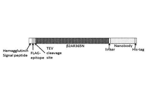
Figure 2: The p2AR-Nanobody fusions described in Example 1 contain two
different proteins connected
with a peptide linker: the GPCR 132AR365N, the linker GGGGSGGGS (underlined
and highlighted in
bold), a Nanobody. Underlined is the HA signal peptide and the His6 peptide
tag. The FLAG-tag is
represented in bold, the TEV cleavage site is indicated in grey shade. A.
Cartoon representation of the
p2AR-Nanobody fusion constructs. B. The amino acid sequence of the open
reading frame encoded by
pFastBac 132AR365N-Nb80. C. The amino acid sequence of the open reading frame
encoded by
pFastBac p2AR365N-N b/1. D. !he amino acid sequence of the open reading frame
encoded by
pFastBac 132AR365N-Nb69. E. The amino acid sequence of the open reading frame
encoded by
pFastBac 132AR365N-Nb60.
Figure 3: Expression of recombinant [32AR-Nanobody fusion proteins in Sf9
cells analyzed by Western
Blot. The presence of fusion protein was detected by anti-FLAG detection as
explained in Example 4.
Membranes of non-infected Sf9 cells (lane 1). Membranes of Sf9 cells
expressing recombinant
p2AR365N that was not fused to any Nb (lane 2: cells were infected with a
1:250 dilution of P2 and
cultured for 48 hours; lane 3: cells were infected with a 1:250 dilution of P2
and cultured for 55 hours;
lane 5: cells were infected with a 1:100 dilution of P2 and cultured for 48
hours; lane 6: cells were
infected with a 1:100 dilution of P2 and cultured for 55 hours). Protein
marker (PageRulerTM
Prestained Protein Ladder, Fermentas cat. Nr SM0671) (lane 4). Membranes of
Sf9 cells expressing
recombinant 132AR365N-Nb80 (lane 7: cells were infected with a 1:100 dilution
of P2 and cultured for
SS hours). Membranes of Sf9 cells expressing recombinant 132AR365N-Nb71 (lane
8: cells were infected
with a 1:100 dilution of P2 and cultured for 55 hours). Membranes of Sf9 cells
expressing recombinant
132AR365N-Nb69 (lane 9: cells were infected with a 1:100 dilution of P2 and
cultured for 55 hours).
Figure 4: Ligand binding properties of [32AR365N-Nb80 fusion compared to
(32AR365N-Nb69 mock
fusion. Radioligand displacement assays of different ligands competing with
[31-1]-dihydroalprenolol
CA 02899693 2015-07-29
WO 2014/118297 PCT/EP2014/051845
([3H]-DHA) for binding to 132AR365N-Nb80 (open circles), and using the
132AR365N-Nb69 chimer (closed
circles) as an internal reference for the non-constrained 132 adrenergic
receptor as described in
Example 6. Competition assays were performed on both receptors using the
natural agonist
epinephrine (A), the agonist (-)-isoproterenol (B), the neutral antagonist
alprenolol (C), the partial
agonist salbutamol (D), the inverse agonist ICI-118,551 (E), and the
antagonist carvedilol (F) as the
competing ligand, respectively. Curves have been fitted by non-linear
regression to a model for
competitive binding using the standard settings of Graphpad Prism 6Ø1050
labeled values have been
fitted to a one binding site competitive model. IC5Ohl6h labeled values have
been fitted to a two binding
site competitive binding model and correspond to the IC50 of the highest
affinity site.
Figure 5: Ligand binding properties of 132AR365N-Nb80 fusion compared to non-
fused 132AR365N.
Radioligand displacement assays of different ligands competing with N-
dihydroalprenolol ([3H]-DHA)
for binding to 132AR365N-Nb80 (open squares), and using the non-fused
132AR365N receptor (closed
squares) as an internal reference for the non-constrained 02 adrenergic
receptor as described in
Example 6. Competition assays were performed on both receptors using the
natural agonist
epinephrine (A), and the antagonist carvedilol (B) as the competing ligand,
respectively. Curves have
been fitted by non-linear regression to a model for competitive binding using
the standard settings of
Graphpad Prism 6Ø IC50 labeled values have been fitted to a one binding site
competitive model.
IC5Oh1gh labeled values have been fitted to a two binding site competitive
binding model and
correspond to the IC50 of the highest affinity site.
Figure 6: Ligand binding properties of 02AR365N-Nb71 fusion compared to
132AR365N-Nb69 fusion.
Radioligand displacement assays of different ligands competing with [3F1]-
dihydroalprenolol ([3H]-DHA)
for binding to 132AR365N-Nb71 (open triangles), and using the 132AR365N-Nb69
chimer (closed
triangles) as an internal reference for the non-constrained 02 adrenergic
receptor as described in
Example 6. Competition assays were performed on both receptors using the
natural agonist
epinephrine (A), the full agonist isoproterenol (B), the neutral antagonist
alprenolol (C), the partial
agonist salbutamol (D), the inverse agonist ICI-118,551 (E) and the antagonist
carvedilol (F) as the
competing ligand, respectively. Curves have been fitted by non-linear
regression to a model for
competitive binding using the standard settings of Graphpad Prism 6Ø1050
labeled values have been
fitted to a one binding site competitive model. IC5Oh15h labeled values have
been fitted to a two binding
site competitive binding model and correspond to the IC50 of the highest
affinity site.
Figure 7: Thermostability of the 02AR365N-Nb80 fusion compared to 132AR365N-
Nb69 fusion.
Thermostabilities of the ligand free DDM solubilized fusion proteins 132AR365-
Nb80 (open diamonds)
11
CA 02899693 2015-07-29
WO 2014/118297 PCT/EP2014/051845
and 32AR365N-Nb69 (closed diamonds). Thermostability assays were performed in
the absence of any
ligand on solubilized receptors in 0,08% DDM as described in Example 7. Curves
have been fitted by
non-linear regression with the log(agonist) vs response - variable slope (four
parameters) equation
using Prism using Graphpad Prism 6.0 (GraphPad Software, San Diego, CA).
Figure 8: Ligand binding properties of 132AR365N-Nb80 fusion compared to the
non-fused B2AR365N in
complex with exogenously added Nb80. Radioligand displacement assays of
epinephrine competing
with [3H]-dihydroalprenolol ([31-1]-DHA) for binding to non-fused (32AR365N
receptor in the presence of
different concentrations of Nb80 (500nM Nb80: closed triangles, 50nM Nb80:
closed diamonds, 5nM
Nb80: closed inverted triangles and 50pM Nb80: closed squares; dashed lines)
or in the presence of
different concentrations of Nb69 (500nM Nb69: open triangle, 50pM Nb69 open
squares; dotted lines).
The [32AR365N-Nb80 fusion (closed circles, full line) and the 132AR365N-Nb69
fusion (open circles, full
line) were used as internal reference in the assay as described in Example 8.
Curves have been fitted by
non-linear regression to a model for competitive binding using the standard
settings of Graphpad
Prism 6Ø IC50 labeled values have been fitted to a one binding site
competitive model. IC5Ohigh labeled
values have been fitted to a two binding site competitive binding model and
correspond to the IC50 of
the highest affinity site.
Figure 9: The M2R-Nanobody fusion described in Example 9 contains two
different proteins connected
with a peptide linker: the GPCR M2Ai3R, the linker GGGSGGGGSGGGGSGGGGSGGGS
(underlined and
highlighted in bold) (SEQ ID NO: 49) and a Nanobody. Underlined is the HA
signal peptide and the Hiss
peptide tag. The FLAG-tag is represented in bold, the TEV cleavage site is
indicated in grey shade. A.
Cartoon representation of the M2R-Nanobody fusion construct. B. The amino acid
sequence of the
open reading frame encoded by pFastBacl M2Ai3R. C. The amino acid sequence of
the open reading
frame encoded by pFastBac1 M21i3R-Nb9-1.
Figure 10: Ligand binding properties of M2Ai3R-Nb9-1 compared to the free non-
fused M21si3R.
Radioligand displacement assays of different ligands competing with [31-1]-N
methyl scopolamine ([31-I]-
NMS) for binding to M2Ai3R-Nb9-1 (closed circles), and using the M2Ai3R
(crossed circles) as an
internal reference for the non-constrained muscarinic receptor M2 as described
in Example 12.
Competition assays were performed on both receptors using the agonist
carbachol (A) or the agonist
oxotremorineM (6) as the competing ligand, respectively. Curves have been
fitted by non-linear
regression to a model for competitive binding using the standard settings of
Graphpad Prism 6Ø IC50
labeled values have been fitted to a one binding site competitive model.
IC5Oh15h labeled values have
12
CA 02899693 2015-07-29
WO 2014/118297 PCT/EP2014/051845
been fitted to a two binding site competitive binding model and correspond to
the IC50 of the highest
affinity site.
Figure 11: The 131AR-Nanobody fusions described in Example 13 contains two
different proteins
connected with a peptide linker: the GPCR human [31AR, the linker
GGGGSGGGGSGGGGSGGGGSGGGS
.. (SEQ ID NO: 60) (underlined and highlighted in bold) and a Nanobody.
Underlined is the HA signal
peptide and the His6 peptide tag. The FLAG-tag is represented in bold, the TEV
cleavage site is indicated
in grey shade. A. Cartoon representation of the 131AR-Nanobody fusion
constructs. B. The amino acid
sequence of the open reading frame encoded by pFastBac h131AR-Nb80. C. The
amino acid sequence of
the open reading frame encoded by pFastBac h131AR-Nb69.
Figure 12: Ligand binding properties of h131AR-Nb80 fusion compared to h131AR-
Nb69 fusion.
Radioligand displacement assays of different ligands competing with [3H]-
dihydroalprenolol ([31-1]-DHA)
for binding to h131AR-Nb80 (open circles), and using the h131AR-Nb69 chimer
(closed circles) as an
internal reference for the non-constrained 131 adrenergic receptor as
described in Example 16.
Competition assays were performed on both 131-receptors fusions using the
natural agonist
epinephrine (A), the inverse agonist ICI-118,551 (B) and the neutral
antagonist alprenolol (C) as the
competing ligand, respectively. Curves have been fitted by non-linear
regression to a model for
competitive binding using the standard settings of Graphpad Prism 6Ø1050
labeled values have been
fitted to a one binding site competitive model. 1C5OhI6h labeled values have
been fitted to a two binding
site competitive binding model and correspond to the IC50 of the highest
affinity site.
Figure 13: Ligand binding properties of 132AR365N-Nb60 fusion compared to
132AR365N-Nb69 mock
fusion. Radioligand displacement assays of different ligands competing with
[3H]-dihydroalprenolol
( [3F1] - D H A ) for binding to [32AR365N-Nb80 (open circles), 132AR365N-Nb60
(crossed open circles), using
the 132AR365N-Nb69 chimer (closed circles) as an internal reference for the
non-constrained [32
adrenergic receptor as described in Example 19. Competition assays were
performed on the 132-
receptors fusions using the natural agonist epinephrine (A), the agonist H-
isoproterenol (B) and the
inverse agonist ICI-118,551 (C) as the competing ligand, respectively. Curves
have been fitted by non-
linear regression to a model for competitive binding using the standard
settings of Graphpad Prism 6Ø
IC50 labeled values have been fitted to a one binding site competitive model.
IC5Ohi8h labeled values
have been fitted to a two binding site competitive binding model and
correspond to the IC50 of the
highest affinity site.
Figure 14. AA sequence alignment of full length wild type human Mon 1 (Uniprot
code P35372; SEQ ID
NO: 46) and mouse Mor1 (Uniprot code P42866; SED ID NO: 45). For the
purification of mouse Mor1,
13
CA 02899693 2015-07-29
WO 2014/118297 PCT/EP2014/051845
expressing in sf9 cells, an N-terminal TEV (N-terminus; insect cell expression
of mouse Mor1) protease
cleavage site has been introduced between the two underlined Glycines. A C-
terminal 3C protease
cleavage site has also been introduced between the underlined isoleucine and
the underlined glutamic
acid in mouse and human Mori expressed in 5f9. The theoretical intracellular
loops (ICL) and the
intracellular C-terminus according to the Uniprot database are depicted in
grey shade. AA residues that
have been deleted in the Mor-Na nobody fusion constructs are striked-through.
Figure 15: The Mor1-Nanobody fusions described in Example 20 contain two
different proteins
connected with a peptide linker: the GPCR Mori., the 34 GS linker (underlined
and highlighted in bold)
and a Nanobody. Underlined is the HA signal peptide and the His6 peptide tag.
The FLAG-tag is
represented in bold. The 3C cleavage site is indicated in grey shade. A.
Cartoon representation of the
Mor1-Nanobody fusion constructs. B-E. The amino acid sequence of the four Mor-
Nb open reading
frames.
Figure 16: Ligand binding properties of hMor1-Nb33 fusion compared to hMor1-
Nb10 mock fusion.
Radioligand displacement assays of different ligands competing with
radioligand for binding to hMor1-
Nb33 (circles) using the hMor1-Nb10 chimer (squares) as an internal reference
for the non-constrained
receptor. Competition assays were performed on both receptors using agonists
Dmt1-Dalda (A),
KG0P01 (B), and antagonist naloxone (C) as the competing ligand.
Figure 17. Comparative fragment binding with different activity profiles to
2AR-fusions.
Representative example of binding of 6 fragments to the active-state
stabilized 02AR365N-Nb80 fusion
(open bars) versus the prominent, non-constrained conformation 02AR365N-Nb69
fusion (black bars),
measured by radioligand displacement assays using [31-1]-dihydroalprenolol
([31-11-DHA) as the
radioligand. 2 fragments (AC23506, CC56213) show an agonist profile, 2
fragments (CC46746,
CC44914) have an antagonistic profile and 2 fragments KM08985) show an inverse
agonist profile.
Figure 18. Dose respons curves of 6 fragments to 02AR-Nb80 fusion compared to
02AR-Nb69 fusion.
Radioligand displacement assays of 6 different fragments competing with [3H]-
dihydroalprenolol ([3F1]-
DHA) for binding to 02AR365N-Nb80 (open circles) and using the 02AR365N-Nb69
chimer (closed
circles) as an internal reference for the non-constrained 02 adrenergic
receptor as described in
Example 24. Competition assays were performed on the 02-receptors fusions
using 2 fragments with
agonist profile (A), 2 fragments with antagonist profile (B) and 2 fragments
with the inverse agonist
profile (C) as the competing ligand, respectively. Curves have been fitted by
non-linear regression with
14
CA 02899693 2015-07-29
WO 2014/118297 PCT/EP2014/051845
the log(agonist) vs response - variable slope (four parameters) equation using
Prism using Graphpad
Prism 6Ø (GraphPad Software, San Diego, CA).
Figure 19: Binding properties of elaborated fragments to the 32AR-Nb80 fusion
compared to the 32AR-
Nb69 mock fusion. Radioligand displacement assays of different elaborated and
original parent
fragments competing with [3F1]-dihydroalprenolol ([3F1]-DHA) for binding to
P2AR365N-Nb80 (closed
symbols), using the 32AR365N-Nb69 chimer (open symbols) as an internal
reference for the non-
constrained [32 adrenergic receptor. Competition assays were performed on the
32-receptors fusions
using (A) the CC40246 (full line), the elaborated fragments: compound 2
(dashed line) and compound 3
(dotted line) as the competing ligand, respectively and (B) the CC56213 (full
line), the elaborated
fragments: compound 9 (dashed line) and compound 10 (dotted line) as the
competing ligand,
respectively. Curves have been fitted by non-linear regression to a model for
competitive binding using
the standard settings of Graphpad Prism 6Ø IC50 labeled values have been
fitted to a one binding site
competitive model.
DETAILED DESCRIPTION OF THE INVENTION
Definitions
The present invention will be described with respect to particular embodiments
and with reference to
certain drawings but the invention is not limited thereto but only by the
claims. Any reference signs in
the claims shall not be construed as limiting the scope. The drawings
described are only schematic and
are non-limiting. In the drawings, the size of some of the elements may be
exaggerated and not drawn
on scale for illustrative purposes. Where the term "comprising" is used in the
present description and
claims, it does not exclude other elements or steps. Where an indefinite or
definite article is used when
referring to a singular noun e.g. "a" or "an'', "the", this includes a plural
of that noun unless something
else is specifically stated. Furthermore, the terms first, second, third and
the like in the description and
in the claims, are used for distinguishing between similar elements and not
necessarily for describing a
sequential or chronological order. It is to be understood that the terms so
used are interchangeable
under appropriate circumstances and that the embodiments of the invention
described herein are
capable of operation in other sequences than described or illustrated herein.
Unless otherwise defined herein, scientific and technical terms and phrases
used in connection with
the present invention shall have the meanings that are commonly understood by
those of ordinary skill
in the art. Generally, nomenclatures used in connection with, and techniques
of molecular and cellular
biology, structural biology, biophysics, pharmacology, genetics and protein
and nucleic acid chemistry
CA 02899693 2015-07-29
WO 2014/118297 PCT/EP2014/051845
described herein are those well-known and commonly used in the art. The
methods and techniques of
the present invention are generally performed according to conventional
methods well known in the
art and as described in various general and more specific references that are
cited and discussed
throughout the present specification unless otherwise indicated. See, for
example, Sambrook et al.
Molecular Cloning: A Laboratory Manual, 3th ed., Cold Spring Harbor Laboratory
Press, Cold Spring
Harbor, N.Y. (2001); Ausubel et al., Current Protocols in Molecular Biology,
Greene Publishing
Associates (1992, and Supplements to 2002); Rup, Biomolecular crystallography:
principles, Practice
and Applications to Structural Biology, 15t edition, Garland Science, Taylor &
Francis Group, LLC, an
informa Business, N.Y. (2009); Limbird, Cell Surface Receptors, 3d ed.,
Springer (2004).
The terms "chimeric polypeptide", "chimeric protein", "fusion polypeptide",
"fusion protein" are used
interchangeably herein and refer to a protein that comprises at least two
separate and distinct
polypeptide components that may or may not originate from the same protein.
The polypeptide
components, while typically unjoined in their native state, are joined by
their respective amino and
carboxyl termini through a peptide linkage to form a single continuous
polypeptide. For example, a
protein of interest fused to an antibody is an example of a chimeric protein.
A convenient means for
linking or fusing two polypeptides is by expressing them as a fusion protein
from a recombinant nucleic
acid molecule, which comprises a first polynucleotide encoding a first
polypeptide operably linked to a
second polynucleotide encoding the second polypeptide. Otherwise, the
polypeptides comprised in a
fusion protein can be linked through peptide bonds that result from intein-
mediated protein splicing
(Muralidharan and Muir 2006) or sortagging (Popp et al. 2007) or may be
chemically linked by any
other means. Typically, a chimeric polypeptide will not exist as a contiguous
polypeptide in a protein
encoded by a gene in a non-recombinant genome. The term "chimeric polypeptide"
and grammatical
equivalents refer to a non-naturally occurring molecule which means that it is
man-made. The term
"fused to", and other grammatical equivalents, when referring to a chimeric
polypeptide (as defined
herein) refers to any chemical or recombinant mechanism for linking two or
more polypeptide
components. The fusion of the two or more polypeptide components may be a
direct fusion of the
sequences or it may be an indirect fusion, e.g. with intervening amino acid
sequences or linker
sequences. Examples will be provided further herein.
The term "membrane protein", as used herein, refers to a protein that is
attached to or associated with
a membrane of a cell or an organelle. They are often subdivided into several
categories including
integral membrane proteins, peripheral membrane proteins and lipid-anchored
proteins. Preferably,
the membrane protein is an integral membrane protein that is permanently bound
to the lipid bilayer
and which requires a detergent or another apolar solvent to be removed.
Integral membrane proteins
16
CA 02899693 2015-07-29
WO 2014/118297 PCT/EP2014/051845
include transmembrane proteins that are permanently attached to the lipid
membrane and span
across the membrane one or several times. Examples of suitable membrane
proteins include receptors
such as GPCRs, amongst others.
As used herein, the terms "polypeptide", "protein", "peptide" are used
interchangeably herein, and
refer to a polymeric form of amino acids of any length, which can include
coded and non-coded amino
acids, chemically or biochemically modified or derivatized amino acids, and
polypeptides having
modified peptide backbones. Throughout the application, the standard one
letter notation of amino
acids will be used. Typically, the term "amino acid" will refer to
"proteinogenic amino acid", i.e. those
amino acids that are naturally present in proteins. Most particularly, the
amino acids are in the L
isomeric form, but D amino acids are also envisaged.
As used herein, the terms "nucleic acid molecule", "polynucleotide",
"polynucleic acid", "nucleic acid"
are used interchangeably and refer to a polymeric form of nucleotides of any
length, either
deoxyribonucleotides or ribonucleotides, or analogs thereof. Polynucleotides
may have any three-
dimensional structure, and may perform any function, known or unknown. Non-
limiting examples of
polynucleotides include a gene, a gene fragment, exons, introns, messenger RNA
(mRNA), transfer
RNA, ribosomal RNA, ribozymes, cDNA, recombinant polynucleotides, branched
polynucleotides,
plasmids, vectors, isolated DNA of any sequence, control regions, isolated RNA
of any sequence,
nucleic acid probes, and primers. The nucleic acid molecule may be linear or
circular.
Any of the peptides, polypeptides, nucleic acids, etc., disclosed herein may
be "isolated" or "purified".
"Isolated" is used herein to indicate that the material referred to is (i)
separated from one or more
substances with which it exists in nature (e.g., is separated from at least
some cellular material,
separated from other polypeptides, separated from its natural sequence
context), and/or (ii) is
produced by a process that involves the hand of man such as recombinant DNA
technology, chemical
synthesis, etc.; and/or (iii) has a sequence, structure, or chemical
composition not found in nature.
"Purified" as used herein denote that the indicated nucleic acid or
polypeptide is present in the
substantial absence of other biological macromolecules, e.g., polynucleotides,
proteins, and the like. In
one embodiment, the polynucleotide or polypeptide is purified such that it
constitutes at least 90% by
weight, e.g., at least 95% by weight, e.g., at least 99% by weight, of the
polynucleotide(s) or
polypeptide(s) present (but water, buffers, ions, and other small molecules,
especially molecules
having a molecular weight of less than 1000 Dalton, can be present).
The term "sequence identity" as used herein refers to the extent that
sequences are identical on a
nucleotide-by-nucleotide basis or an amino acid-by-amino acid basis over a
window of comparison.
17
81789963
Thus, a "percentage of sequence identity" is calculated by comparing two
optimally aligned sequences
over the window of comparison, determining the number of positions at which
the identical nucleic
acid base (e.g., A, T, C, G, I) or the identical amino acid residue (e.g.,
Ala, Pro, Ser, Thr, Gly, Val, Leu, Ile,
Phe, Tyr, Trp, Lys, Arg, His, Asp, Glu, Asn, Gln, Cys and Met) occurs in both
sequences to yield the
number of matched positions, dividing the number of matched positions by the
total number of
positions in the window of comparison (i.e., the window size), and multiplying
the result by 100 to yield
the percentage of sequence identity. Determining the percentage of sequence
identity can be done
manually, or by making use of computer programs that are available in the art.
Examples of useful
algorithms are PILEUP (Higgins & Sharp, CABIOS 5:151 (1989), BLAST and BLAST
2.0 (Altschul et al. J.
Mol. Biol. 215: 403 (1990). Software for performing BLAST analyses is publicly
available through the
National Center for Biotechnology Information.
"Similarity" refers to the percentage number of amino acids that are identical
or constitute
conservative substitutions. Similarity may be determined using sequence
comparison programs such as
GAP (Deveraux et al. 1984). In this way, sequences of a similar or
substantially different length to those
cited herein might be compared by insertion of gaps into the alignment, such
gaps being determined,
for example, by the comparison algorithm used by GAP. As used herein,
"conservative substitution" is
the substitution of amino acids with other amino acids whose side chains have
similar biochemical
properties (e.g. are aliphatic, are aromatic, are positively charged, ...) and
is well known to the skilled
person. Non-conservative substitution is then the substitution of amino acids
with other amino acids
whose side chains do not have similar biochemical properties (e.g. replacement
of a hydrophobic with
a polar residue). Conservative substitutions will typically yield sequences
which are not identical
anymore, but still highly similar. By conservative substitutions is intended
combinations such as gly,
ala; val, lie, leu, met; asp, glu; asn, gin; ser, thr; lys, arg; cys, met; and
phe, tyr, trp.
A "deletion" is defined here as a change in either amino acid or nucleotide
sequence in which one or
more amino acid or nucleotide residues, respectively, are absent as compared
to an amino acid
sequence or nucleotide sequence of a parental polypeptide or nucleic acid.
Within the context of a
protein, a deletion can involve deletion of about 2, about 5, about 10, up to
about 20, up to about 30
or up to about 50 or more amino acids. A protein or a fragment thereof may
contain more than one
deletion. Within the context of a GPCR, a deletion may also be a loop
deletion, or an N- and/or C-
terminal deletion, or a combination thereof. As will be clear to the skilled
person, an N- and/or C-
terminal deletion of a GPCR is also referred to as a truncation of the amino
acid sequence of the GPCR
or a truncated GPCR.
18
CA 2899693 2020-03-16
CA 02899693 2015-07-29
WO 2014/118297 PCT/EP2014/051845
An "insertion" or "addition" is that change in an amino acid or nucleotide
sequences which has resulted
in the addition of one or more amino acid or nucleotide residues,
respectively, as compared to an
amino acid sequence or nucleotide sequence of a parental protein. "Insertion"
generally refers to
addition to one or more amino acid residues within an amino acid sequence of a
polypeptide, while
"addition" can be an insertion or refer to amino acid residues added at an N-
or C-terminus, or both
termini. Within the context of a protein or a fragment thereof, an insertion
or addition is usually of
about 1, about 3, about 5, about 10, up to about 20, up to about 30 or up to
about 50 or more amino
acids. A protein or fragment thereof may contain more than one insertion.
A "substitution", as used herein, results from the replacement of one or more
amino acids or
nucleotides by different amino acids or nucleotides, respectively as compared
to an amino acid
sequence or nucleotide sequence of a parental protein or a fragment thereof.
It is understood that a
protein or a fragment thereof may have conservative amino acid substitutions
which have substantially
no effect on the protein's activity. By conservative substitutions is intended
combinations such as gly,
ala; vat, ile, leu, met; asp, glu; asn, gin; ser, thr; lys, arg; cys, met; and
phe, tyr, trp.
The term "recombinant" when used in reference to a cell, nucleic acid, protein
or vector, indicates that
the cell, nucleic acid, protein or vector, has been modified by the
introduction of a heterologous
nucleic acid or protein or the alteration of a native nucleic acid or protein,
or that the cell is derived
from a cell so modified. Thus, for example, recombinant cells express nucleic
acids or polypeptides that
are not found within the native (non-recombinant) form of the cell or express
native genes that are
otherwise abnormally expressed, under expressed, over expressed or not
expressed at all.
As used herein, the term "expression" refers to the process by which a
polypeptide is produced based
on the nucleic acid sequence of a gene. The process includes both
transcription and translation.
The term "operably linked" as used herein refers to a linkage in which the
regulatory sequence is
contiguous with the gene of interest to control the gene of interest, as well
as regulatory sequences
that act in trans or at a distance to control the gene of interest. For
example, a DNA sequence is
operably linked to a promoter when it is ligated to the promoter downstream
with respect to the
transcription initiation site of the promoter and allows transcription
elongation to proceed through the
DNA sequence. A DNA for a signal sequence is operably linked to DNA coding for
a polypeptide if it is
expressed as a pre-protein that participates in the transport of the
polypeptide. Linkage of DNA
sequences to regulatory sequences is typically accomplished by ligation at
suitable restriction sites or
adapters or linkers inserted in lieu thereof using restriction endonucleases
known to one of skill in the
art.
19
CA 02899693 2015-07-29
WO 2014/118297 PCT/EP2014/051845
The term "regulatory sequence" as used herein, and also referred to as
"control sequence", refers to
polynucleotide sequences which are necessary to affect the expression of
coding sequences to which
they are operably linked. Regulatory sequences are sequences which control the
transcription, post-
transcriptional events and translation of nucleic acid sequences. Regulatory
sequences include
appropriate transcription initiation, termination, promoter and enhancer
sequences; efficient RNA
processing signals such as splicing and polyadenylation signals; sequences
that stabilize cytoplasmic
mRMA; sequences that enhance translation efficiency (e.g., ribosome binding
sites); sequences that
enhance protein stability; and when desired, sequences that enhance protein
secretion. The nature of
such control sequences differs depending upon the host organism. The term
"regulatory sequence" is
intended to include, at a minimum, all components whose presence is essential
for expression, and can
also include additional components whose presence is advantageous, for
example, leader sequences
and fusion partner sequences.
The term "vector" as used herein is intended to refer to a nucleic acid
molecule capable of transporting
another nucleic acid molecule to which it has been linked. The vector may be
of any suitable type
including, but not limited to, a phage, virus, plasmid, phagemid, cosmid,
bacmid or even an artificial
chromosome. Certain vectors are capable of autonomous replication in a host
cell into which they are
introduced (e.g., vectors having an origin of replication which functions in
the host cell). Other vectors
can be integrated into the genome of a host cell upon introduction into the
host cell, and are thereby
replicated along with the host genome. Moreover, certain preferred vectors are
capable of directing
the expression of certain genes of interest. Such vectors are referred to
herein as "recombinant
expression vectors" (or simply, "expression vectors"). Suitable vectors have
regulatory sequences, such
as promoters, enhancers, terminator sequences, and the like as desired and
according to a particular
host organism (e.g. bacterial cell, yeast cell). Typically, a recombinant
vector according to the present
invention comprises at least one "chimeric gene" or "expression cassette".
Expression cassettes are
generally DNA constructs preferably including (5' to 3' in the direction of
transcription): a promoter
region, a polynucleotide sequence, homologue, variant or fragment thereof of
the present invention
operably linked with the transcription initiation region, and a termination
sequence including a stop
signal for RNA polymerase and a polyadenylation signal. It is understood that
all of these regions
should be capable of operating in biological cells, such as prokaryotic or
eukaryotic cells, to be
transformed. The promoter region comprising the transcription initiation
region, which preferably
includes the RNA polymerase binding site, and the polyadenylation signal may
be native to the
biological cell to be transformed or may be derived from an alternative
source, where the region is
functional in the biological cell.
CA 02899693 2015-07-29
WO 2014/118297 PCT/EP2014/051845
The term "host cell", as used herein, is intended to refer to a cell into
which a recombinant vector has
been introduced. It should be understood that such terms are intended to refer
not only to the
particular subject cell but also to the progeny of such a cell. Because
certain modifications may occur in
succeeding generations due to either mutation or environmental influences,
such progeny may not, in
fact, be identical to the parent cell, but are still included within the scope
of the term "host cell" as
used herein. A host cell may be an isolated cell or cell line grown in culture
or may be a cell which
resides in a living tissue or organism. In particular, host cells are of
bacterial or fungal origin, but may
also be of plant or mammalian origin. The wordings "host cell", "recombinant
host cell", "expression
host cell", "expression host system", "expression system", are intended to
have the same meaning and
are used interchangeably herein.
An "epitope", as used herein, refers to an antigenic determinant of a
polypeptide. An epitope could
comprise 3 amino acids in a spatial conformation, which is unique to the
epitope. Generally an epitope
consists of at least 4, 5, 6, 7 such amino acids, and more usually, consists
of at least 8, 9, 10 such amino
acids. Methods of determining the spatial conformation of amino acids are
known in the art, and
include, for example, x-ray crystallography and multi-dimensional nuclear
magnetic resonance.
A "conformational epitope", as used herein, refers to an epitope comprising
amino acids in a spacial
conformation that is unique to a folded 3-dimensional conformation of a
polypeptide. Generally, a
conformational epitope consists of amino acids that are discontinuous in the
linear sequence but that
come together in the folded structure of the protein. However, a
conformational epitope may also
consist of a linear sequence of amino acids that adopts a conformation that is
unique to a folded 3-
dimensional conformation of the polypeptide (and not present in a denatured
state). In protein
complexes, conformational epitopes consist of amino acids that are
discontinuous in the linear
sequences of one or more polypeptides that come together upon folding of the
different folded
polypeptides and their association in a unique quaternary structure.
Similarly, conformational epitopes
may here also consist of a linear sequence of amino acids of one or more
polypeptides that come
together and adopt a conformation that is unique to the quaternary structure.
The term "conformation" or "conformational state" of a protein refers
generally to the range of
structures that a protein may adopt at any instant in time. One of skill in
the art will recognize that
determinants of conformation or conformational state include a protein's
primary structure as
.. reflected in a protein's amino acid sequence (including modified amino
acids) and the environment
surrounding the protein. The conformation or conformational state of a protein
also relates to
structural features such as protein secondary structures (e.g., a-helix, 13-
sheet, among others), tertiary
21
CA 02899693 2015-07-29
WO 2014/118297 PCT/EP2014/051845
structure (e.g., the three dimensional folding of a polypeptide chain), and
quaternary structure (e.g.,
interactions of a polypeptide chain with other protein subunits). Post-
translational and other
modifications to a polypeptide chain such as ligand binding, phosphorylation,
sulfation, glycosylation,
or attachments of hydrophobic groups, among others, can influence the
conformation of a protein.
Furthermore, environmental factors, such as pH, salt concentration, ionic
strength, and osmolality of
the surrounding solution, and interaction with other proteins and co-factors,
among others, can affect
protein conformation. The conformational state of a protein may be determined
by either functional
assay for activity or binding to another molecule or by means of physical
methods such as X-ray
crystallography, NMR, or spin labeling, among other methods. For a general
discussion of protein
conformation and conformational states, one is referred to Cantor and
Schimmel, Biophysical
Chemistry, Part I: The Conformation of Biological. Macromolecules,.W.H.
Freeman and Company, 1980,
and Creighton, Proteins: Structures and Molecular Properties, W.H. Freeman and
Company, 1993.
A "functional conformation" or a "functional conformational state" refers to
the fact that proteins, in
particular GPCRs, possess different conformational states having a dynamic
range of activity, in
particular ranging from no activity to maximal activity. It should be clear
that "a functional
conformational state", as used herein, is meant to cover any conformational
state of a protein, in
particular a GPCR, having any activity, including no activity, and is not
meant to cover the denatured
states of proteins. Non-limiting examples of functional conformations of GPCRs
include active
conformations, inactive conformations or basal conformations. A particular
class of functional
conformations is defined as "druggable conformation" and generally refers to a
unique therapeutically
relevant conformational state of a target protein. As an illustration, the
agonist-bound active
conformation of the [32 adrenergic receptor corresponds to the druggable
conformation of this
receptor relating to smooth muscle relaxation, dilation of bronchial passages
(asthma), vasodilation in
muscle and liver, relaxation of uterine muscle, and release of insulin. It
will thus be understood that
druggability is confined to particular conformations depending on the
therapeutic indication. More
details are provided further herein.
The term "stabilizing" or "stabilized", with respect to a functional
conformational state of a GPCR, as
used herein, refers to the retaining or holding of a GPCR in a subset of the
possible conformations that
it could otherwise assume, due to the effects of the intramolecular
interaction of the GPCR moiety
with the binding domain moiety of the chimeric polypeptide of the present
invention. Within this
context, a binding domain that specifically or selectively binds to a
particular conformation of a GPCR
refers to a binding domain that binds with a higher affinity to the GPCR in a
subset of conformations
than to other conformations that the GPCR may assume. One of skill in the art
will recognize that
22
CA 02899693 2015-07-29
WO 2014/118297 PCT/EP2014/051845
binding domains that specifically or selectively bind to a particular
conformation of a GPCR will stabilize
this particular conformation, and its related activity. More details are
provided further herein.
The term "affinity", as used herein, generally refers to the degree to which a
ligand (as defined further
herein) binds to a target protein so as to shift the equilibrium of target
protein and ligand toward the
presence of a complex formed by their binding. Thus, for example, where a
chimeric polypeptide and a
ligand are combined in relatively equal concentration, a ligand of high
affinity will bind to the chimeric
polypeptide so as to shift the equilibrium toward high concentration of the
resulting complex. The
dissociation constant Kd is commonly used to describe the affinity between a
ligand and a target
protein. Typically, the dissociation constant has a value that is lower than
le M. Preferably, the
dissociation constant is lower than 10-6 M, more preferably, lower than 10-7
M. Most preferably, the
dissociation constant is lower than le M. Other ways of describing the
affinity between a ligand and
its target protein are the association constant (Ka), the inhibition constant
(Ki), or indirectly by
evaluating the potency of ligands by measuring the half maximal inhibitory
concentration (IC50) or half
maximal effective concentration (EC50). It will be appreciated that within the
scope of the present
invention, the term "affinity" is used in the context of a binding domain that
binds a conformational
epitope of a target GPCR as well as in the context of a test compound (as
defined further herein) that
binds to the chimeric polypeptide of the present invention, more particularly
to an orthosteric or
allosteric site of a target GPCR comprised in the chimeric polypeptide.
It should be noted that, in intramolecular interactions, such as the specific
interaction of the binding
domain moiety to the GPCR moiety of the chimeric polypeptide of the invention,
the two interacting
groups are contained within a single molecule. The dissociation constant
between the binding domain
and the target, might be known when they are individual molecules, but for an
intramolecular
interaction, their concentration within the solution is irrelevant. Instead,
it is the "effective
concentration" of the two groups within the macromolecule that is important
for the intramolecular
interaction. The term "effective intramolecular concentration" or "effective
molarity" (Meff), as used
herein, refers to the effective concentration and can be calculated as
Kdinter/Kdintra, wherein the
dissociation constant KdIntra, which is dimensionless, is the dissociation
constant that is used to describe
an intramolecular reaction, which is a fusion of a target GPCR and a binding
domain, and wherein
Kdinter, which has units of concentration (e.g. molarity), is the dissociation
constant for an analogous
intermolecular interaction, which is a non-covalent complex of a binding
domain and a target GPCR.
The effective molarity will depend upon the structure of the macromolecule, in
particular the extent to
which it brings the two reactants together or keeps them apart, plus the
environment in which the two
groups are kept. The effective molarity for the intramolecular reaction will
be essentially zero if the
23
CA 02899693 2015-07-29
WO 2014/118297 PCT/EP2014/051845
macromolecular structure keeps the two groups apart, which is the case when a
binding domain is not
directed against and/or is not capable of specifically binding (as defined
further herein) to a target
GPCR. At the other extreme, when the macromolecular structure keeps the two
groups in the correct
proximity and orientation for reaction, their effective molarity can be
extremely high, up to iem,
concentrations that are not feasible with two independent molecules. According
to the present
invention, this is the case when a binding domain is capable of specifically
binding (as defined further
herein) to a target GPCR. Such values are predicted by theoretical
considerations and are also observed
experimentally, from the ratio of the rate or equilibrium constants for the
same reaction when the
groups are on the same molecule and on separate molecules.
The term "specificity", as used herein, refers to the ability of a binding
domain, in particular an
immunoglobulin or an immunoglobulin fragment, such as a VHH or Nanobody, to
bind preferentially to
one antigen, versus a different antigen, and does not necessarily imply high
affinity.
The terms "specifically bind" and "specific binding", as used herein,
generally refers to the ability of a
binding domain, in particular an immunoglobulin, such as an antibody, or an
immunoglobulin
fragment, such as a VHH or Nanobody, to preferentially bind to a particular
antigen that is present in a
homogeneous mixture of different antigens. In certain embodiments, a specific
binding interaction will
discriminate between desirable and undesirable antigens in a sample, in some
embodiments more
than about 10 to 100-fold or more (e.g., more than about 1000- or 10,000-
fold). Within the context of
the spectrum of conformational states of GPCRs, the terms particularly refer
to the ability of a binding
domain (as defined herein) to preferentially recognize and/or bind to a
particular conformational state
of a GPCR as compared to another conformational state. Accordingly, as used
herein, the term
"conformation-selective binding domain" in the context of the present
invention refers to a binding
domain that binds to a target GPCR in a conformation-selective manner. A
binding domain that
selectively binds to a particular conformation of a GPCR refers to a binding
domain that binds with a
higher affinity to a GPCR in a subset of conformations than to other
conformations that the GPCR may
assume. One of skill in the art will recognize that binding domains that
selectively bind to a particular
conformation of a GPCR will stabilize or retain the GPCR it this particular
conformation. For example,
an active state conformation-selective binding domain will preferentially bind
to a GPCR in an active
conformational state and will not or to a lesser degree bind to a GPCR in an
inactive conformational
.. state, and will thus have a higher affinity for said active conformational
state; or vice versa. The terms
"specifically bind", "selectively bind", "preferentially bind", and
grammatical equivalents thereof, are
used interchangeably herein. The terms "conformational specific" or
"conformational selective" are
also used interchangeably herein.
24
CA 02899693 2015-07-29
WO 2014/118297 PCT/EP2014/051845
The term "compound" or "test compound" or "candidate compound" or "drug
candidate compound"
as used herein are intended to have the same meaning and describe any
molecule, either naturally
occurring or synthetic that is tested in an assay, such as a screening assay
for drug discovery purposes.
As such, these compounds comprise organic or inorganic compounds. The
compounds include small
molecules, polynucleotides, lipids or hormone analogs that are characterized
by low molecular
weights. Other biopolymeric organic test compounds include small peptides or
peptide-like molecules
(peptidomimetics) comprising from about 2 to about 40 amino acids and larger
polypeptides
comprising from about 40 to about 500 amino acids, such as antibodies,
antibody fragments or
antibody conjugates. Test compounds can also be protein scaffolds or synthetic
scaffolds. For high-
throughput purposes, test compound libraries may be used, such as
combinatorial or randomized
libraries that provide a sufficient range of diversity. Examples include, but
are not limited to, natural
compound libraries, allosteric compound libraries, peptide libraries, antibody
fragment libraries,
synthetic compound libraries, fragment-based libraries, phage-display
libraries, and the like. A more
detailed description can be found further in the specification.
As used herein, the term "ligand" means a molecule that binds to a
polypeptide, in particular a GPCR. A
ligand may be, without the purpose of being limitative, a protein, a
(poly)peptide, a lipid, a small
molecule, a protein scaffold, a nucleic acid, an ion, a carbohydrate, an
antibody or an antibody
fragment. A ligand may be synthetic or naturally occurring. A ligand also
includes a "native ligand"
which is a ligand that is an endogenous, natural ligand for a native protein.
Within the context of the
present invention, a ligand means a molecule that binds to a GPCR, either at
the intracellular site or
extracellular site and/or at the transmembrane domains. Usually, but not
necessarily, a GPCR will
adopt a particular conformation upon binding of a ligand. Thus, a ligand may
also be a "conformation-
selective ligand" or "conformation-specific ligand". The term includes
agonists, full agonists, partial
agonists, and inverse agonists, binding at either the orthosteric site or at
an allosteric site. For the sake
of clarity, the term "conformation-selective ligand" or "conformation-specific
ligand" does not include
a neutral antagonist, since a neutral antagonist does not selectively bind a
particular conformation of a
GPCR.
An "orthosteric ligand", as used herein, refers to a ligand (both natural and
synthetic), that binds to the
active site of a receptor, such as a GPCR, and are further classified
according to their efficacy or in
other words to the effect they have on signaling through a specific pathway.
As used herein, an
"agonist" refers to a ligand that, by binding a receptor protein, increases
the receptor's signaling
activity. Full agonists are capable of maximal protein stimulation; partial
agonists are unable to elicit
full activity even at saturating concentrations. Partial agonists can also
function as "blockers" by
CA 02899693 2015-07-29
WO 2014/118297 PCT/EP2014/051845
preventing the binding of more robust agonists. An "antagonist", also referred
to as a "neutral
antagonist", refers to a ligand that binds a receptor without stimulating any
activity. An "antagonist" is
also known as a "blocker" because of its ability to prevent binding of other
ligands and, therefore,
block agonist-induced activity. Further, an "inverse agonist" refers to an
antagonist that, in addition to
blocking agonist effects, reduces a receptor's basal or constitutive activity
below that of the unliganded
protein.
Ligands as used herein may also be "biased ligands" with the ability to
selectively stimulate a subset of
a receptor's signaling activities, for example in the case of GPCRs the
selective activation of G-protein
or 13-arrestin function. Such ligands are known as "biased ligands", "biased
agonists" or "functionally
selective agonists". More particularly, ligand bias can be an imperfect bias
characterized by a ligand
stimulation of multiple receptor activities with different relative efficacies
for different signals or can
be a perfect bias characterized by a ligand stimulation of one receptor
protein activity without any
stimulation of another known receptor protein activity.
Another kind of ligands is known as allosteric regulators. "Allosteric
regulators" or otherwise "allosteric
modulators", 'allosteric ligands" or "effector molecules", as used herein,
refer to ligands that bind at
an allosteric site (that is, a regulatory site physically distinct from the
protein's active site) of a receptor
protein such as a GPCR. In contrast to orthosteric ligands, allosteric
modulators are non-competitive
because they bind receptor proteins at a different site and modify their
function even if the
endogenous ligand also is binding. Allosteric regulators that enhance the
protein's activity are referred
to herein as "allosteric activators" or "positive allosteric modulators"
(PAMs), whereas those that
decrease the protein's activity are referred to herein as "allosteric
inhibitors" or otherwise "negative
allosteric modulators" (NAMs).
As used herein, the terms "determining", "measuring", "assessing",
"monitoring" and "assaying" are
used interchangeably and include both quantitative and qualitative
determinations.
As used herein, the terms "complementarity determining region" or "CDR" within
the context of
antibodies refer to variable regions of either H (heavy) or L (light) chains
(also abbreviated as VH and
VL, respectively) and contains the amino acid sequences capable of
specifically binding to antigenic
targets. These CDR regions account for the basic specificity of the antibody
for a particular antigenic
determinant structure. Such regions are also referred to as "hypervariable
regions." The CDRs
represent non-contiguous stretches of amino acids within the variable regions
but, regardless of
species, the positional locations of these critical amino acid sequences
within the variable heavy and
light chain regions have been found to have similar locations within the amino
acid sequences of the
26
CA 02899693 2015-07-29
WO 2014/118297 PCT/EP2014/051845
variable chains. The variable heavy and light chains of all canonical
antibodies each have 3 CDR regions,
each non- contiguous with the others (termed L1, L2, L3, H1, H2, H3) for the
respective light (L) and
heavy (H) chains. lmmunoglobulin single variable domains, in particular VHHs
or Nanobodies, generally
comprise a single amino acid chain that can be considered to comprise 4
"framework sequences or
regions" or FRs and 3 "complementarity determining regions" or CDRs. The
Nanobodies have 3 CDR
regions, each non-contiguous with the others (termed CDR1, CDR2, CDR3). The
delineation of the FR
and CDR sequences can, for example, be based on the IMGT unique numbering
system for V-domains
and V-like domains (Lefranc et al. 2003,).
Detailed description
Chimeric polypeptides
A first aspect of the invention relates to a chimeric polypeptide comprising a
G protein-coupled
receptor (GPCR) fused to a binding domain, wherein the binding domain is
directed against and/or
specifically binds to the GPCR. The invention thus provides a chimeric
polypeptide which is a fusion
protein of at least two moieties, in particular at least a GPCR moiety and a
binding domain moiety,
wherein the binding domain is directed against and/or specifically binds to
the GPCR.
In general, the choice of the GPCR moiety forming part of the chimeric
polypeptide is not critical to the
invention, and will typically be selected according to the intended use and
application. A prerequisite
of the binding domain moiety is its capability to specifically bind (as
defined herein) to the GPCR of
choice. Thus, the binding domain moiety may generally be directed against any
desired GPCR, and may
in particular be directed against any conformational epitope (as defined
herein) of any GPCR. A binding
domain that specifically binds to a "conformational epitope" specifically
binds to a tertiary (i.e., three
dimensional) structure of a folded protein, and binds at much reduced (i.e.,
by a factor of at least 2, 5,
10, 50 or 100) affinity to the linear (i.e., unfolded, denatured) form of the
protein. In particular, said
conformational epitope can be part of an intracellular or extracellular
region, or an intramennbraneous
region, or a domain or loop structure of any desired GPCR. Thus, according to
particular embodiments,
the binding domain moiety may be directed against an extracellular region,
domain, loop or other
extracellular conformational epitope of any desired GPCR, but is preferably
directed against the
extracellular parts of the transmembrane domains or more preferably against
the extracellular loops
that link the transmembrane domains. Alternatively, the binding domain moiety
may be directed
against an intracellular region, domain, loop or other intracellular
conformational epitope of any
desired GPCR, but is preferably directed against one of the intracellular
parts of the transmembrane
domains or more preferably against the intracellular loops that link the
transmembrane domains. In
27
CA 02899693 2015-07-29
WO 2014/118297 PCT/EP2014/051845
other specific embodiments, the binding domain moiety may be directed against
a conformational
epitope that forms part of the binding site of a natural ligand, including but
limited to an endogenous
orthosteric agonist. In still other embodiments, the binding domain moiety may
be directed against a
conformational epitope, in particular an intracellular epitope, that is
comprised in a binding site for a
downstream signaling protein, including but not-limited to a G protein binding
site, a 13-arrestin binding
site.
According to a preferred embodiment, the chimeric polypeptide of the invention
comprises a GPCR
fused to a binding domain wherein, upon binding of the binding domain to the
GPCR in an
intramolecular reaction, the GPCR is stabilized in a particular conformation
without the need for an
additional ligand. Preferably, the chimeric polypeptide of the invention
comprises a GPCR that is
stabilized by the binding domain in a functional conformation (as defined
herein). With the term
"stabilized", as defined hereinbefore, is meant an increased stability of a
GPCR with respect to the
structure (e.g. conformational state) and/or particular biological activity
(e.g. intracellular signaling
activity, ligand binding affinity, ...). In relation to increased stability
with respect to structure and/or
biological activity, this may be readily determined either by means of
physical methods such as X-ray
crystallography, NMR, or spin labeling, among other methods or by a functional
assay for activity (e.g.
Ca2+ release, cAMP generation or transcriptional activity, p-arrestin
recruitment, ...) or ligand binding,
among other methods. The term "stabilize" also includes increased
thermostability of the GPCR under
non-physiological conditions induced by denaturants or denaturing conditions.
The term
"thermostabilize", "thermostabilizing", "increasing the thermostability of",
as used herein, refers to the
functional rather than to the thermodynamic properties of a GPCR and to the
protein's resistance to
irreversible denaturation induced by thermal and/or chemical approaches
including but not limited to
heating, cooling, freezing, chemical denaturants, pH, detergents, salts,
additives, proteases or
temperature. Irreversible denaturation leads to the irreversible unfolding of
the functional
conformations of the protein, loss of biological activity and aggregation of
the denaturated protein. In
relation to an increased stability to heat, this can be readily determined by
measuring ligand binding or
by using spectroscopic methods such as fluorescence, CD or light scattering
that are sensitive to
unfolding at increasing temperatures. It is preferred that the binding domain
moiety is capable of
increasing the stability as measured by an increase in the thermal stability
of a GPCR in a functional
conformational state with at least 2 C, at least 5 C, at least 8 C, and more
preferably at least 10 C or
15 C or 20 C. In relation to an increased stability to a detergent or to a
chaotrope, typically the GPCR is
incubated for a defined time in the presence of a test detergent or a test
chaotropic agent and the
stability is determined using, for example, ligand binding or a spectroscoptic
method, optionally at
increasing temperatures as discussed above. Otherwise, the binding domain
moiety is capable of
28
CA 02899693 2015-07-29
WO 2014/118297 PCT/EP2014/051845
increasing the stability to extreme pH of a functional conformational state of
a GPCR. In relation to an
extreme of pH, a typical test pH would be chosen for example in the range 6 to
8, the range 5.5 to 8.5,
the range 5 to 9, the range 4.5 to 9.5, more specifically in the range 4.5 to
5.5 (low pH) or in the range
8.5 to 9.5 (high pH). The term "(thermo)stabilize", "(thermo)sta bilizing",
"increasing the
(thermo)stability of", as used herein, applies to GPCRs embedded in lipid
particles or lipid layers (for
example, lipid monolayers, lipid bilayers, and the like) and to GPCRs that
have been solubilized in
detergent.
It is thus particularly envisaged that the chimeric polypeptides of the
invention comprise a GPCR
moiety that is stabilized in a functional conformation upon binding of the
binding domain moiety.
According to a preferred embodiment of the invention, the GPCR moiety is
stabilized in an active
conformation upon binding of a binding domain that is selective for an active
conformation. The term
"active conformation", as used herein, refers to a spectrum of receptor
conformations that upon
binding of a ligand allows signal transduction towards an intracellular
effector system, including G
protein dependent signaling and G protein-independent signaling (e.g. p-
arrestin signaling). An "active
conformation" thus encompasses a range of ligand-specific conformations,
including an agonist-bound
active conformation, a partial agonist-bound active conformation or a biased
agonist conformation
that induces the cooperative binding of an intracellular effector protein. In
another preferred
embodiment, the GPCR moiety is stabilized in an inactive conformation upon
binding of a binding
domain that is conformation-selective for an inactive conformation. The term
"inactive conformation",
as used herein, refers to a spectrum of receptor conformations that does not
allow or blocks signal
transduction towards an intracellular effector system. An "inactive
conformation" thus encompasses a
range of ligand-specific conformations, including an inverse agonist-bound
inactive conformation that
prevents the cooperative binding of an intracellular effector protein. It will
be understood that the site
of binding of the ligand is not critical for obtaining an active or inactive
conformation. Hence,
orthosteric ligands as well as allosteric modulators may equally be capable of
stabilizing a GPCR in an
active or inactive conformation. Thus, according to a particular embodiment of
the present invention,
the binding domain moiety that is capable of stabilizing the GPCR moiety may
bind at the orthosteric
site or at an allosteric site. In other specific embodiments, the binding
domain moiety that is capable of
stabilizing the GPCR moiety may be a binding domain that is selective for an
agonist-bound active
.. conformation, or that is selective for a partial agonist-bound active
conformation or that is selective for
a biased agonist-bound functional conformation binding domain, or that is
selective for an inverse
agonist-bound inactive conformation.
29
CA 02899693 2015-07-29
WO 2014/118297 PCT/EP2014/051845
It will thus be understood that the chimeric polypeptides of the present
invention are conformationally
constrained proteins since the GPCR moiety of the fusion is stabilized in a
particular functional
conformation, due to the high local concentration of the tethered binding
domain. To illustrate this
further, without the purpose of being !imitative, a chimeric polypeptide of
the invention comprising a
GPCR that is stabilized in an active conformation will have an increased or
enhanced affinity for an
agonist, more particularly for a full agonist, a partial agonist or a biased
agonist, as compared to the to
the corresponding non-fused GPCR or as compared to a chimeric polypeptide of
the corresponding
GPCR fused to a mock binding moiety (also referred to as control binding
moiety or irrelevant binding
moiety, and that is not directed to and/or does not specifically bind to the
GPCR). Also, a chimeric
polypeptide of the invention comprising a GPCR that is stabilized in an active
conformation will have a
decreased affinity for an inverse agonist, as compared to the corresponding
non-fused GPCR or to a
chimeric polypeptide of the corresponding GPCR fused to a mock binding moiety.
In contrast, a
chimeric polypeptide of the invention comprising a GPCR that is stabilized in
an inactive conformation
will have an enhanced affinity for an inverse agonist and/or will have a
decreased affinity for an agonist
as compared to the corresponding non-fused GPCR or to a chimeric polypeptide
of the corresponding
GPCR fused to a mock binding moiety. An increase or decrease in affinity for a
ligand may be directly
measured by and/or calculated from d decrease or increase, respectively ill
EC50, iC50, Kd, K, or any
other measure of affinity or potency known to one of skill in the art.
Preferably, the affinity of a
chimeric polypeptide of the invention for a ligand is increased or decreased
at least 2 fold, at least 5
fold, at least 10 fold, at least 50 fold, and more preferably at least 100
fold, at least 200 fold, at least
300 fold, at least 400 fold, at least 500 fold, even more preferably at least
1000 fold or 2000 fold or
more, upon binding to the constrained GPCR. It will be appreciated that
affinity measurements for
conformation-selective ligands that trigger/inhibit particular signaling
pathways may be carried with
any type of ligand, including natural ligands, small molecules, as well as
biologicals; with orthosteric
ligands as well as allosteric modulators; with single compounds as well as
compound libraries; with
lead compounds or fragments; etc.
Further, the way the different moieties that form part of the chimeric
polypeptides as described are
fused to each other will typically depend on both the type of GPCR and the
characteristics of the
binding domain (e.g. conformational epitope to which it binds). As is known by
the person skilled in the
art, GPCRs are characterized by an extracellular N-terminus, followed by seven
transmembrane ct-
helices connected by three intracellular and three extracellular loops, and
finally an intracellular C-
terminus (see also further herein). Depending on the site of binding
(extracellular or intracellular or
transmembrane), the binding domain moiety will preferably be fused to the N-
terminal end of the
GPCR or to the C-terminal end. Preferably, a binding domain that binds the
GPCR at an intracellular site
81789963
will be fused to the C-terminus of the GPCR. Likewise, a binding domain that
binds the GPCR at an
extracellular site will be fused to the N-terminus of the GPCR. The binding
domain moiety will be fused
with its C-terminal end to N-terminal end of the GPCR or the binding domain
can be fused with its N-
terminal end to the C-terminal end of the GPCR. Further, the fusion may be a
direct fusion of the
sequences or it may be an indirect fusion, e.g. with intervening amino acid
sequences or linker
sequences (as described further herein). A person skilled in the art will know
how to design a fusion
construct. If available, one will make use of the atomic structure of the GPCR
separately and/or the
atomic structure of the binding domain separately and/or the atomic structure
of the GPCR in complex
with the binding domain. Alternatively, the GPCR could be linked to the
binding domain using intein-
mediated protein splicing (Muralidharan and Muir 2006) or sortagging (Popp et
al. 2007) or other
chemoenzymatic methods for site-specific labeling or engineering of proteins
with small molecules or
other proteins, for example as described in Chen and Ting (2005) and Rabuka
(2010).
Particular embodiments of the different moieties that form part of the
chimeric polypeptides of the
invention will be further detailed here below.
GPCR moiety
The chimeric polypeptide of the invention comprises at least two polypeptide
components, one of
which is a GPCR. As a GPCR, any GPCR of interest can be used. The skilled
person can choose a suitable
GPCR depending on the intended application (as described further herein). In
the same line, and
depending on the intended use, a GPCR from any organism may be selected, such
as fungus (including
yeast), nematode, virus, insect, plant, bird (e.g. chicken, turkey), reptile,
or mammal (e.g., a mouse, rat,
rabbit, hamster, gerbil, dog, cat, goat, pig, cow, horse, whale, monkey,
camelid, or human).
"G-protein coupled receptors", or "GPCRs", as used herein, are well-known by
the skilled person and
refer to polypeptides that share a common structural motif, having seven
regions of between 22 to 24
hydrophobic amino acids that form seven alpha helices, each of which spans the
membrane. Each span
is identified by number, i.e., transmembrane-1 (TM1), transmembrane-2 (TM2),
etc. The
transmembrane helices are joined by regions of amino acids between
transmembrane-2 and
transmembrane-3, transmembrane-4 and transmembrane-5, and transmembrane-6 and
transmembrane-7 on the exterior, or "extracellular" side, of the cell
membrane, referred to as
"extracellular" regions 1, 2 and 3 (EC1 , EC2 and EC3), respectively. The
transmembrane helices are
also joined by regions of amino acids between transmembrane-1 and
transmembrane-2,
transmembrane-3 and transmembrane-4, and transmembrane-5 and transmembrane-6
on the
31
CA 2899693 2020-03-16
CA 02899693 2015-07-29
WO 2014/118297 PCT/EP2014/051845
interior, or "intracellular" side, of the cell membrane, referred to as
"intracellular" regions 1 , 2 and 3
(IC1 , IC2 and IC3), respectively. The "carboxy" ("C") terminus of the
receptor lies in the intracellular
space within the cell, and the "amino" ("N") terminus of the receptor lies in
the extracellular space
outside of the cell. Any of these regions are readily identifiable by analysis
of the primary amino acid
sequence of a GPCR.
GPCR structure and classification is generally well known in the art and
further discussion of GPCRs
may be found in Probst et al. 1992; Marchese et al. 1994; Lagerstrom &
Schioth, 2008; Rosenbaum et
al. 2009; and the following books: Jurgen Wess (Ed) Structure-Function
Analysis of G Protein- Coupled
Receptors published by Wiley-Liss (1 st edition; October 15, 1999); Kevin R.
Lynch (Ed) Identification
and Expression of G Protein-Coupled Receptors published by John Wiley & Sons
(March 1998) and
Tatsuya Haga (Ed), G Protein-Coupled Receptors, published by CRC Press
(September 24, 1999); and
Steve Watson (Ed) G-Protein Linked Receptor Factsbook, published by Academic
Press (1st edition;
1994). GPCRs can be grouped on the basis of sequence homology and functional
similarity into several
families, each of which can be used in the creation and use of the chimeric
polypeptides of the
invention. Most of the human GPCRs can be found in five main families, termed
Glutamate, Rhodopsin,
Adhesion, Frizzled/Taste2 and Secretin (Fredriksson et al., 2003). Members of
the Rhodopsin family
corresponding to class A (Kolakowski, 1994) or Class 1 (Foord et al 2005) in
older classification systems
only have small extracellular loops and the interaction of the ligands occurs
with residues within the
transmembrane cleft. This is by far the largest group (>90% of the GPCRs) and
contains receptors for
odorants, small molecules such as catecholamines and amines, (neuro)peptides
and glycoprotein
hormones. Rhodopsin, a representative of this family, is the first GPCR for
which the structure has
been solved (Palczewski et al., 2000). 132AR, the first receptor interacting
with a diffusible ligand for
which the structure has been solved (Rosenbaum et al, 2007) also belongs to
this family. Based on
phylogenetic analysis, class B GPCRs or Class 2 (Foord et al, 2005) receptors
have recently been
subdivided into two families: adhesion and secretin (Fredriksson et al.,
2003). Adhesion and secretin
receptors are characterized by a relatively long amino terminal extracellular
domain involved in ligand-
binding. Little is known about the orientation of the transmembrane domains,
but it is probably quite
different from that of rhodopsin. Ligands for these GPCRs are hormones, such
as glucagon, secretin,
gonadotropin-releasing hormone and parathyroid hormone. The Glutamate family
receptors (Class C
or Class 3 receptors) also have a large extracellular domain, which functions
like a "Venus fly trap"
since it can open and close with the agonist bound inside. Family members are
the metabotropic
glutamate, the Ca2+-sensing and the y- aminobutyric acid (GABA)-B receptors.
32
81789963
Thus, according to particular embodiments of the invention, the chimeric
polypeptide of the invention
comprises a GPCR that is chosen from the group comprising a GPCR of the
Glutamate family of GPCRs,
a GPCR of the Rhodopsin family of GPCRs, a GPCR of the Adhesion family of
GPCRs, a GPCR of the
Frizzled/Taste2 family of GPCRs, and a GPCR of the Secretin family of GPCRs.
Preferably, the GPCR
comprised in the chimeric polypeptide is a mammalian protein, or a plant
protein, or a microbial
protein, or a viral protein, or an insect protein. Even more preferably, the
GPCR is a human protein.
More specifically, GPCRs include, without limitation, 5-hydroxytryptamine
receptors, acetylcholine
receptors (muscarinic), adenosine receptors, adrenoceptors, anaphylatoxin
receptors, angiotensin
receptors, apelin receptors, bile acid receptors, bombesin receptors,
bradykinin receptors, cannabinoid
receptors, chemokine receptors, cholecystokinin receptors, dopamine receptors,
endothelin receptors,
estrogen (G protein coupled) receptors, formylpeptide receptors, free fatty
acid receptors, galanin
receptors, ghrelin receptors, glycoprotein hormone receptors, gonadotrophin-
releasing hormone
receptors, histamine receptors, KiSS1-derived peptide receptor, leukotriene
receptors,
lysophospholipid receptors, melanin-concentrating hormone receptors,
melanocortin receptors,
melatonin receptors, motilin receptors, neuromedin U receptors, neuropeptide
FF/neuropeptide AF
receptors, neuropeptide S receptors, neuropeptide W/neuropeptide B receptors,
neuropeptide Y
receptors, neurotensin receptors, nicotinic acid receptor family, opioid
receptors, orexin receptors, P2Y
receptors, peptide P518 receptor, platelet-activating factor receptor,
prokineticin receptors, prolactin-
releasing peptide receptor, prostanoid receptors, protease-activating
receptors, relaxin family peptide
receptors, somatostatin receptors, tachykinin receptors, thyrotropin-releasing
hormone receptor,
trace amine receptor, urotensin receptor, vasopressin and oxytocin receptors,
class A orphans, non-
signaling 71M chemokine-binding proteins, calcitonin receptors, corticotropin-
releasing factor
receptors, glucagon receptor family, parathyroid hormone receptors, VIP and
PACAP receptors, class B
orphans, calcium-sensing receptors, GABA B receptors, GPRC5 receptors,
metabotropic glutamate
receptors, class C orphans, Frizzled receptors, the rhodopsins and other G-
protein coupled seven
transmembrane segment receptors. GPCRs also include these GPCR receptors
associated with each
other as homomeric or heteromeric dimers or as higher-order oligomers. The
amino acid sequences
(and the nucleotide sequences of the cDNAs which encode them) of GPCRs are
readily available, for
example by reference to GenBank. HGNC standardized nomenclature to human
genes; accession
numbers of different isoforms from different organisms are available from
Uniprot. Moreover, a
comprehensive overview of receptor nomenclature, pharmacological, functional
and pathophysiological
information on GPCRs can be retrieved from the IUPHAR database.
33
CA 2899693 2020-03-16
CA 02899693 2015-07-29
WO 2014/118297 PCT/EP2014/051845
According to a preferred embodiment, the GPCR that forms part of the chimeric
polypeptide of the
invention, is chosen from the group comprising the adrenergic receptors,
preferably the a adrenergic
receptors, such as the al adrenergic receptors and the a2 adrenergic
receptors, and the p adrenergic
receptors, such as the pi adrenergic receptors, the 132 adrenergic receptors
and the 133 adrenergic
receptors; or from the group comprising the muscarinic receptors, preferably
the M1 muscarinic
receptors, the M2 muscarinic receptors, the M3 muscarinic receptors, the M4
muscarinic receptors
and the M5 muscarinic receptors; or from the group comprising the opioid
receptor family, preferably
the [1. opioid receptors (mu or MOP or Morl), the 6 opioid receptors (delta or
DOP), the -lc opioid
receptors (kappa or KOP), the NOP opioid receptors, all of which are well
known in the art.
.. It will be understood that, depending on the purpose and application, the
GPCR comprised in the
fusion protein may be a naturally occurring or non-naturally occurring (i.e.,
altered by man) receptor.
The term "naturally-occurring" in reference to a GPCR means a GPCR that is
naturally produced. In
particular, wild type polymorphic variants and isoforms of GPCRs, as well as
orthologs across different
species are examples of naturally occurring proteins, and are found for
example, and without
limitation, in a mammal, more specifically in a human, or in a virus, or in a
plant, or in an insect,
amongst others). Such GPCRs are found in nature. In addition, the term is
intended to encompass wild-
type polymorphic variants and certain other variants of the 132 adrenergic
receptor from a particular
species, including mutants. For example, a "human 132 adrenergic receptor" has
an amino acid
sequence that is at least 95% identical to (e.g., at least 95% or at least 98%
identical to) the naturally
occurring "human 132 adrenoreceptor" of Genbank accession number NP_000015. Or
also, a "human
muscarinic acetylcholine receptor M2" has an amino acid sequence that is at
least 95% identical to
(e.g., at least 95% or at least 98% identical to) the naturally occurring
"human muscarinic acetylcholine
receptor M2" of Genbank accession number AAA51570.1. Or also, a "human mu
opioid receptor" has
an amino acid sequence that is at least 95% identical to (e.g., at least 95%
or at least 98% identical to)
the naturally occurring "human mu opioid receptor" of Genbank accession number
NP_000905. The
term "non-naturally occurring" in reference to a GPCR means a GPCR that is not
naturally-occurring.
Wild-type GPCRs that have been mutated and variants of naturally-occurring
GPCRs are examples of
non-naturally occurring GPCRs. Non-naturally occurring GPCR may have an amino
acid sequence that is
at least 80% identical to, at least 90% identical to, at least 95% identical
to or at least 99% identical to,
a naturally-occurring GPCR. In certain circumstances, it may be advantageous
that the GPCR comprised
in the chimeric polypeptide is a non-naturally occurring protein. For example,
and for illustration
purposes only, to increase the probability of obtaining crystals of the
chimeric polypeptide comprising
the GPCR stabilized in a particular conformation, it might be desired to
perform some protein
engineering without or only minimally affecting the conformation (e.g. active
conformation with
34
CA 02899693 2015-07-29
WO 2014/118297 PCT/EP2014/051845
increased affinity for agonists). Or, alternatively or additionally, to
increase cellular expression levels of
a GPCR, or to increase the stability, one might also consider introducing
certain mutations in the GPCR
of interest. Non-limiting examples of non-naturally occurring GPCRs include,
without limitation, GPCRs
that have been made constitutively active through mutation, GPCRs with a loop
deletion, GPCRs with
an N- and/or C-terminal deletion, GPCRs with a substitution, an insertion or
addition, or any
combination thereof, in relation to their amino acid or nucleotide sequence,
or other variants of
naturally-occurring GPCRs. Also comprised within the scope of the present
invention are target GPCRs
comprising a chimeric or hybrid GPCR, for example a chimeric GPCR with an N-
and/or C-terminus from
one GPCR and loops of a second GPCR, or comprising a GPCR fused to a moiety,
such as 14 lysozyme,
Flavodoxin, Xylanase, Rubredoxin or cytochrome b as an utility in GPCR
crystallization (Chun et al. 2012
and also described in patent applications W02012/158555, W02012/030735,
W02012/148586).
According to specific embodiments within the scope of the present invention, a
non-naturally occurring
GPCR, as comprised in the chimeric polypeptide, may have an amino acid
sequence that is at least 80%
identical to, at least 90% identical to, at least 95% identical to, at least
97% identical to, or at least 99%
identical to, a naturally-occurring GPCR. Further, it will be appreciated that
the present invention also
envisages GPCRs with a loop deletion, or an N- and/or C-terminal deletion, or
a substitution, or an
insertion or addition in relation to its amino acid or nucleotide sequence, or
any combination thereof
(as defined hereinbefore, and see also Example section).
Binding domain moiety
The chimeric polypeptide of the invention comprises at least two polypeptide
moieties, one of which is
a GPCR (as described herein before), and another one which is a binding domain
that is directed against
and/or specifically binds to the GPCR.
The term "binding domain", as used herein, means the whole or part of a
proteinaceous (protein,
protein-like or protein containing) molecule that is capable of binding using
specific intermolecular
interactions to a GPCR. According to a particular embodiment, the term
"binding domain" does not
include a naturally-occurring binding partner of a GPCR, e.g. a G protein, an
arrestin, an endogenous
ligand; or variants or derivatives (including fragments) thereof. More
specifically, the term "binding
domain" refers to a polypeptide, more particularly a protein domain. A protein
binding domain is an
element of overall protein structure that is self-stabilizing and often folds
independently of the rest of
the protein chain. Binding domains vary in length from between about 25 amino
acids up to 500 amino
acids and more. Many binding domains can be classified into folds and are
recognizable, identifiable, 3-
D structures. Some folds are so common in many different proteins that they
are given special names.
CA 02899693 2015-07-29
WO 2014/118297 PCT/EP2014/051845
Non-limiting examples are binding domains selected from a 3- or 4-helix
bundle, an armadillo repeat
domain, a leucine-rich repeat domain, a PDZ domain, a SUMO or SUMO-like
domain, a cadherin
domain, an immunoglobulin-like domain, phosphotyrosine-binding domain,
pleckstrin homology
domain, src homology 2 domain, amongst others. A binding domain can thus be
derived from a
naturally occurring molecule, e.g. from components of the innate or adaptive
immune system, or it can
be entirely artificially designed.
In general, a binding domain can be immunoglobulin-based or it can be based on
domains present in
proteins, including but limited to microbial proteins, protease inhibitors,
toxins, fibronectin, lipocalins,
single chain antiparallel coiled coil proteins or repeat motif proteins.
Particular examples of binding
domains which are known in the art include, but are not limited to:
antibodies, heavy chain antibodies
(hcAb), single domain antibodies (sdAb), minibodies, the variable domain
derived from camelid heavy
chain antibodies (VHH or Nanobodies), the variable domain of the new antigen
receptors derived from
shark antibodies (VNAR), alphabodies, protein A, protein G, designed ankyrin-
repeat domains
(DARPins), fibronectin type III repeats, anticalins, knottins, engineered CH2
domains (nanoantibodies),
engineered SH3 domains, affibodies, peptides and proteins, lipopeptides (e.g.
pepducins) (see, e.g.,
Gebauer & Skerra, 2009; Skerra, 2000; Starovasnik et al., 1997; Binz et at.,
2004; Koide et al., 1998;
Dimitrov, 2009; Nygren et al. 2008; W02010066740). Frequently, when generating
a particular type of
binding domain using selection methods, combinatorial libraries comprising a
consensus or framework
sequence containing randomized potential interaction residues are used to
screen for binding to a
molecule of interest, such as a protein.
According to a preferred embodiment, it is particularly envisaged that the
binding domain that forms
part of the chimeric polypeptide of the invention is derived from an innate or
adaptive immune
system. Preferably, said binding domain is derived from an immunoglobulin.
Preferably, the binding
domain according to the invention is derived from an antibody or an antibody
fragment. The term
"antibody" (Ab) refers generally to a polypeptide encoded by an immunoglobulin
gene, or a functional
fragment thereof, that specifically binds and recognizes an antigen, and is
known to the person skilled
in the art. An antibody is meant to include a conventional four-chain
immunoglobulin, comprising two
identical pairs of polypeptide chains, each pair having one "light" (about 25
kDa) and one "heavy" chain
(about 50 kDa). Typically, in conventional immunoglobulins, a heavy chain
variable domain (VH) and a
light chain variable domain (VL) interact to form an antigen binding site. The
term "antibody" is meant
to include whole antibodies, including single-chain whole antibodies, and
antigen-binding fragments. In
some embodiments, antigen-binding fragments may be antigen-binding antibody
fragments that
include, but are not limited to, Fab, Fab' and F(ab')2, Fd, single-chain Fvs
(scFv), single-chain antibodies,
36
CA 02899693 2015-07-29
WO 2014/118297 PCT/EP2014/051845
disulfide-linked Fvs (dsFv) and fragments comprising or consisting of either a
VL or VH domain, and any
combination of those or any other functional portion of an immunoglobulin
peptide capable of binding
to the target antigen. The term "antibodies" is also meant to include heavy
chain only antibodies, or
fragments thereof, including immunoglobulin single variable domains, as
defined further herein.
The term "immunoglobulin single variable domain" defines molecules wherein the
antigen binding site
is present on, and formed by, a single immunoglobulin domain (which is
different from conventional
immunoglobulins or their fragments, wherein typically two immunoglobulin
variable domains interact
to form an antigen binding site). It should however be clear that the term
"immunoglobulin single
variable domain" does comprise fragments of conventional immunoglobulins
wherein the antigen
binding site is formed by a single variable domain. Preferably, the binding
domain moiety within the
scope of the present invention is an immunoglobulin single variable domain.
Generally, an immunoglobulin single variable domain will be an amino acid
sequence comprising 4
framework regions (FR1 to FR4) and 3 complementary determining regions (CDR1
to CDR3), preferably
according to the following formula (1): FR1-CDR1-FR2-CDR2-FR3-CDR3-FR4 (1), or
any suitable
fragment thereof (which will then usually contain at least some of the amino
acid residues that form at
least one of the complementarity determining regions). Immunoglobulin single
variable domains
comprising 4 FRs and 3 CDRs are known to the person skilled in the art and
have been described, as a
non-limiting example, in Wesolowski et al. 2009. Typical, but non-limiting,
examples of
immunoglobulin single variable domains include light chain variable domain
sequences (e.g. a VL
domain sequence) or a suitable fragment thereof, or heavy chain variable
domain sequences (e.g. a VH
domain sequence or VHH domain sequence) or a suitable fragment thereof, as
long as it is capable of
forming a single antigen binding unit. Thus, according to a preferred
embodiment, the binding domain
moiety is an immunoglobulin single variable domain that is a light chain
variable domain sequence (e.g.
a VL domain sequence) or a heavy chain variable domain sequence (e.g. a VH
domain sequence); more
specifically, the immunoglobulin single variable domain is a heavy chain
variable domain sequence that
is derived from a conventional four-chain antibody or a heavy chain variable
domain sequence that is
derived from a heavy chain antibody. The immunoglobulin single variable domain
may be a domain
antibody, or a single domain antibody, or a "dAB" or dAb, or a Nanobody (as
defined herein), or
another immunoglobulin single variable domain, or any suitable fragment of any
one thereof. For a
general description of single domain antibodies, reference is made to the
following book: "Single
domain antibodies", Methods in Molecular Biology, Eds. Saerens and
Muyldermans, 2012, Vol 911. The
immunoglobulin single variable domains, generally comprise a single amino acid
chain that can be
considered to comprise 4 "framework sequences" or FR's and 3 "complementary
determining regions"
37
CA 02899693 2015-07-29
WO 2014/118297 PCT/EP2014/051845
or CDR's (as defined hereinbefore). It should be clear that framework regions
of immunoglobulin single
variable domains may also contribute to the binding of their antigens
(Desmyter et al 2002; Korotkov
et al. 2009). The delineation of the CDR sequences (and thus also the FR
sequences) can be based on
the IMGT unique numbering system for V-domains and V-like domains (Lefranc et
al. 2003).
Alternatively, the delineation of the FR and CDR sequences can be done by
using the Ka bat numbering
system as applied to VHH domains from Camelids in the article of Riechmann and
Muyldermans
(2000).
It should be noted that the immunoglobulin single variable domains as binding
domain moiety in their
broadest sense are not limited to a specific biological source or to a
specific method of preparation.
The term "immunoglobulin single variable domain" encompasses variable domains
of different origin,
comprising mouse, rat, rabbit, donkey, human, shark, camelid variable domains.
According to specific
embodiments, the immunoglobulin single variable domains are derived from shark
antibodies (the so-
called immunoglobulin new antigen receptors or IgNARs), more specific from
naturally occurring heavy
chain shark antibodies, devoid of light chains, and are known as VNAR domain
sequences. Preferably,
the immunoglobulin single variable domains are derived from camelid
antibodies. More preferably, the
immunoglobulin single variable domains are derived from naturally occurring
camelid heavy chain only
antibodies, devoid of light chains, and are known as VHH domain sequences or
Nanobodies.
According to a particularly preferred embodiment, the binding domain moiety as
comprised in the
chimeric polypeptide of the invention is an immunoglobulin single variable
domain that is a Nanobody
(as defined further herein, and including but not limited to a VHH). The term
"Nanobody" (Nb), as used
herein, is a single domain antigen binding fragment. It particularly refers to
a single variable domain
derived from naturally occurring heavy chain only antibodies and is known to
the person skilled in the
art. Nanobodies are usually derived from heavy chain only antibodies (devoid
of light chains) seen in
camelids (Hamers-Casterman et al. 1993; Desmyter et al. 1996) and consequently
are often referred to
as VHH antibody or VHH sequence. Camelids comprise old world camelids (Came/us
bactrianus and
Came/us dromedarius) and new world camelids (for example Lama paccos, Lama
glama, Lama
guanicoe and Lama vicugna). Nanobody and Nanobodies are registered
trademarks of Ablynx NV
(Belgium). For a further description of VHHs or Nanobodies, reference is made
to the book "Single
domain antibodies", Methods in Molecular Biology, Eds. Saerens and
Muyldermans, 2012, Vol 911, in
particular to the Chapter by Vincke and Muyldermans (2012), as well as to a
non-limiting list of patent
applications, which are mentioned as general background art, and include: WO
94/04678, WO
95/04079, WO 96/34103 of the Vrije Universiteit Brussel; WO 94/25591, WO
99/37681, WO 00/40968,
WO 00/43507, WO 00/65057, WO 01/40310, WO 01/44301, EP 1 134 231 and WO
02/48193 of
38
81789963
Unilever; WO 97/49805, WO 01/21817, WO 03/035694, WO 03/054016 and WO
03/055527 of the
Vlaams Instituut voor Biotechnologie (VIB); WO 04/041867, WO 04/041862, WO
04/041865, WO
04/041863, WO 04/062551, WO 05/044858, WO 06/40153, WO 06/079372, WO
06/122786, WO
06/122787 and WO 06/122825, by Ablynx N.V. and the further published patent
applications by Ablynx
N .V. As will be known by the person skilled in the art, the Nanobodies are
particularly characterized by
the presence of one or more Camelidae "hallmark residues" in one or more of
the framework
sequences (according to Kabat numbering), as described for example in WO
08/020079, on page 75,
Table A-3). It should be noted that the Nanobodies, of the invention in their
broadest sense are not
limited to a specific biological source or to a specific method of
preparation. For example,
Nanobodies, can generally be obtained: (1) by isolating the VHH domain of a
naturally occurring heavy
chain only antibody; (2) by expression of a nucleotide sequence encoding a
naturally occurring VHH
domain; (3) by "humanization" of a naturally occurring VHH domain or by
expression of a nucleic
acid encoding a such humanized VHH domain; (4) by "camelization" of a
naturally occurring VH
domain from any animal species, and in particular from a mammalian species,
such as from a human
being, or by expression of a nucleic acid encoding such a camelized VH domain;
(5) by "camelisation"
of a "domain antibody" or ''Dab" as described in the art, or by expression of
a nucleic acid encoding
such a camelized VH domain; (6) by using synthetic or semi-synthetic
techniques for preparing proteins,
polypeptides or other amino acid sequences known per se; (7) by preparing a
nucleic acid encoding a
Nanobody using techniques for nucleic acid synthesis known per se, followed by
expression of the
nucleic acid thus obtained; and/or (8) by any combination of one or more of
the foregoing. A further
description of Nanobodies, including humanization and/or camelization of
Nanobodies, can be
found e.g. in W008/101985 and W008/142164, as well as further herein. A
particular class of
Nanobodies that interacts with conformational epitopes of native targets and
that stabilizes the target
in a unique non-prominent conformation (different than the basal conformation)
are called Xaperones
and are particularly envisaged here. Xaperones are unique tools in structural
biology. XaperoneTM
is a trademark of VIB and VUB (Belgium). By rigidifying flexible regions and
obscuring aggregative
surfaces, XaperoneTM complexes warrant conformationally uniform samples that
are key to protein
structure determination by X-ray crystallography. Major advantages for the use
of camelid antibody
fragments as crystallization aid are that Xaperones (1) bind cryptic epitopes
and lock proteins in unique
native conformations, (2) increase the stability of soluble proteins and
solubilized membrane proteins,
(3) reduce the conformational complexity of soluble proteins and solubilized
membrane proteins,
(4) increase the polar surface enabling the growth of diffracting crystals,
(5) sequester aggregative
or polymerizing surfaces, (6) allow to affinity-trap active protein.
39
CA 2899693 2020-03-16
CA 02899693 2015-07-29
WO 2014/118297 PCT/EP2014/051845
Within the scope of the present invention, the term "immunoglobulin single
variable domain" also
encompasses variable domains that are "humanized" or "camelized", in
particular Nanobodies that are
"humanized" or "camelized". For example both "humanization" and "camelization"
can be performed
by providing a nucleotide sequence that encodes a naturally occurring VHH
domain or VH domain,
respectively, and then changing, in a manner known per se, one or more codons
in said nucleotide
sequence in such a way that the new nucleotide sequence encodes a "humanized"
or "camelized"
immunoglobulin single variable domains of the invention, respectively. This
nucleic acid can then be
expressed in a manner known per se, so as to provide the desired
immunoglobulin single variable
domains of the invention. Alternatively, based on the amino acid sequence of a
naturally occurring VHH
domain or VH domain, respectively, the amino acid sequence of the desired
humanized or camelized
immunoglobulin single variable domains of the invention, respectively, can be
designed and then
synthesized de novo using techniques for peptide synthesis known per se. Also,
based on the amino
acid sequence or nucleotide sequence of a naturally occurring VHH domain or VH
domain, respectively,
a nucleotide sequence encoding the desired humanized or camelized
immunoglobulin single variable
domains of the invention, respectively, can be designed and then synthesized
de novo using
techniques for nucleic acid synthesis known per se, after which the nucleic
acid thus obtained can be
expressed in d Fild111lei known per se, so as to provide the desired
immunoglobulin single variable
domains of the invention. Other suitable methods and techniques for obtaining
the immunoglobulin
single variable domains of the invention and/or nucleic acids encoding the
same, starting from
naturally occurring VH sequences or preferably VHH sequences, will be clear
from the skilled person,
and may for example comprise combining one or more parts of one or more
naturally occurring VH
sequences (such as one or more FR sequences and/or CDR sequences), one or more
parts of one or
more naturally occurring VHH sequences (such as one or more FR sequences or
CDR sequences),
and/or one or more synthetic or semi-synthetic sequences, in a suitable
manner, so as to provide a
Nanobody of the invention or a nucleotide sequence or nucleic acid encoding
the same.
Also within the scope of the invention are natural or synthetic analogs,
mutants, variants, alleles, parts
or fragments (herein collectively referred to as "variants") of the
immunoglobulin single variable
domains, in particular the Nanobodies, as defined herein, and in particular
variants of the
immunoglobulin single variable domains of SEQ ID NOs: 13-20 (see Tables 1-2).
Thus, according to one
embodiment of the invention, the term "immunoglobulin single variable domain
of the invention" or
"Nanobody of the invention" in its broadest sense also covers such variants.
Generally, in such variants,
one or more amino acid residues may have been replaced, deleted and/or added,
compared to the
immunoglobulin single variable domains of the invention as defined herein.
Such substitutions,
insertions or deletions may be made in one or more of the FRs and/or in one or
more of the CDRs, and
81789963
in particular variants of the FRs and CDRs of the immunoglobulin single
variable domains of SEQ ID
NOs: 13-20 (see Tables 1-2). Variants, as used herein, are sequences wherein
each or any framework
region and each or any cornplementarity determining region shows at least 80%
identity, preferably at
least 85% identity, more preferably 90% identity, even more preferably 95%
identity or, still even more
preferably 99% identity with the corresponding region in the reference
sequence (i.e. FRl_variant
versus FR1 _reference, CDR1_variant versus CDR1 _reference, FR2_variant versus
FR2_reference,
CDR2_variant versus CDR2 _reference, FR3_variant versus FR3 _reference,
CDR3_variant versus
CDR3_reference, FR4_variant versus FR42eference), as can be measured
electronically by making use
of algorithms such as PILEUP and BLAST. Software for performing BLAST analyses
is publicly available
through the National Center for Biotechnology Information. It will be
understood that for determining
the degree of amino acid identity of the amino add sequences of the CDRs of
one or more sequences
of the immunoglobulin single variable domains, the amino acid residues that
form the framework
regions are disregarded. Similarly, for determining the degree of amino acid
identity of the amino
acid sequences of the FRs of one or more sequences of the immunoglobulin
single variable
domains of the invention, the amino acid residues that form the
complementarity regions are
disregarded. Such variants of immunoglobulin single variable domains may be of
particular advantage
since they may have improved potency/affinity.
By means of non-limiting examples, a substitution may for example be a
conservative substitution (as
described herein) and/or an amino acid residue may be replaced by another
amino acid residue that
naturally occurs at the same position in another VHH domain. Thus, any one or
more substitutions,
deletions or insertions, or any combination thereof, that either improve the
properties of the
immunoglobulin single variable domains or that do not detract from the desired
properties or from the
balance or combination of desired properties of the immunoglobulin single
variable domain (i.e. to the
extent that the immunoglobulin single variable domains is no longer suited for
its intended use) are
included within the scope of the invention. A skilled person will generally be
able to determine and
select suitable substitutions, deletions or insertions, or suitable
combinations of thereof, based on the
disclosure herein and optionally after a limited degree of routine
experimentation, which may for
example involve introducing a limited number of possible substitutions and
determining their influence
on the properties of the immunoglobulin single variable domains thus obtained.
Also encompassed within the scope of the present invention are immunoglobulin
single variable
domains that are in a "multivalent" form and are formed by bonding, chemically
or by recombinant
DNA techniques, together two or more monovalent immunoglobulin single variable
domains. Non-
limiting examples of multivalent constructs include "bivalent" constructs,
"trivalent" constructs,
41
CA 2899693 2020-03-16
CA 02899693 2015-07-29
WO 2014/118297 PCT/EP2014/051845
"tetravalent" constructs, and so on. The immunoglobulin single variable
domains comprised within a
multivalent construct may be identical or different. In another particular
embodiment, the
immunoglobulin single variable domains of the invention are in a
"multispecific" form and are formed
by bonding together two or more immunoglobulin single variable domains, of
which at least one with a
different specificity. Non-limiting examples of multi-specific constructs
include "bi-specific" constructs,
"tri-specific" constructs, "tetra-specific" constructs, and so on. To
illustrate this further, any multivalent
or multispecific (as defined herein) immunoglobulin single variable domain of
the invention may be
suitably directed against two or more different epitopes on the same antigen,
for example against two
or more different epitopes of the GPCR; or may be directed against two or more
different antigens, for
example against an epitope of the GPCR and an epitope of a natural binding
partner of the GPCR (e.g.
G protein, [3-arrestin). In particular, a monovalent immunoglobulin single
variable domain of the
invention is such that it will bind to the target GPCR with an affinity less
than 500 nM, preferably less
than 200 nM, more preferably less than 10 nM, such as less than 500 pM.
Multivalent or multispecific
immunoglobulin single variable domains of the invention may also have (or be
engineered and/or
selected for) increased avidity and/or improved selectivity for the desired
GPCR, and/or for any other
desired property or combination of desired properties that may be obtained by
the use of such
multivalent or multispecific immurioglobulin single variable domain's. In d
particular ernibodimerit, such
multivalent or multispecific binding domains of the invention may also have
(or be engineered and/or
selected for) improved efficacy in modulating signaling activity of a GPCR
(see also further herein).
Further, and depending on the host organism used to express the chimeric
polypeptide of the
invention, deletions and/or substitutions within the binding domain moiety may
be designed in such a
way that e.g. one or more sites for post-translational modification (such as
one or more glycosylation
sites) are removed, as will be within the ability of the person skilled in the
art. Alternatively,
substitutions or insertions may be designed so as to introduce one or more
sites for attachment of
functional groups (as described further herein).
Screening and selection of binding domain moieties suitable for the invention
A preferred class of binding domains, in particular immunoglobulin single
variable domains that form
part of the chimeric polypeptide of the invention, is directed against and/or
specifically binds to a
functional conformational state of a GPCR (as described hereinbefore).
Conformationally-selective
binding domains, in particular immunoglobulin single variable domains, can be
identified in several
ways, and will be illustrated hereafter in a non-limiting way for VHHs.
Preferably, conformation-
selective binding domains can be selected after the step of immunization of a
Camelidae with a GPCR
42
CA 02899693 2015-07-29
WO 2014/118297 PCT/EP2014/051845
in a functional conformational state, optionally bound to a receptor ligand,
to expose the immune
system of the animal with the conformational epitopes that are unique to the
GPCR (for example,
agonist-bound GPCR so as to raise antibodies directed against a GPCR in its
active conformational
state). Optionally, a particular ligand can be coupled to the GPCR of interest
by chemical cross-linking.
Thus, as further described herein, such VHH sequences can preferably be
generated or obtained by
suitably immunizing a species of Camelid with a GPCR, preferably a GPCR in a
functional
conformational state (i.e. so as to raise an immune response and/or heavy
chain only antibodies
directed against said GPCR), by obtaining a suitable biological sample from
said Camelid (such as a
blood sample, or any sample of 13-cells), and by generating VHH sequences
directed against said GPCR,
starting from said sample. Such techniques will be clear to the skilled
person. Yet another technique for
obtaining the desired VHH sequences involves suitably immunizing a transgenic
mammal that is
capable of expressing heavy chain only antibodies (i.e. so as to raise an
immune response and/or heavy
chain only antibodies directed against a GPCR in a functional conformational
state), obtaining a
suitable biological sample from said transgenic mammal (such as a blood
sample, or any sample of B-
cells), and then generating VHH sequences directed against said GPCR starting
from said sample, using
any suitable technique known per se. For example, for this purpose, the heavy
chain antibody-
expressing [nice arid the further methods and techniques described iii
W002085945 and in
W004049794 can be used.
For the immunization of an animal with a GPCR, the GPCR may be produced and
purified using
conventional methods that may employ expressing a recombinant form of the GPCR
in a host cell, and
purifying the GPCR using affinity chromatography and/or antibody-based
methods. In particular
embodiments, the baculoyirus/Sf-9 system may be employed for expression,
although other
expression systems (e.g., bacterial, yeast or mammalian cell systems) may also
be used. Exemplary
methods for expressing and purifying GCPRs are described in, for example,
Kobilka (1995), Eroglu et al
(2002), Chelikani et al (2006) and the book "Identification and Expression of
G Protein-Coupled
Receptors" (Kevin R. Lynch (Ed.), 1998), among many other references. A GPCR
may also be
reconstituted in phospholipid vesicles. Likewise, methods for reconstituting
an active GPCR in
phospholipid vesicles are known, and are described in: Luca et al (2003),
Mansoor et al (2006), Niu et
al. (2005), Shimada et al. (2002), and Eroglu et al. (2003), among others. In
certain cases, the GPCR and
phospholipids may be reconstituted at high phospholipid density (e.g., 1 mg
receptor per mg of
phospholipid). In particular embodiments, the phospholipids vesicles may be
tested to confirm that the
GPCR is active. In many cases, a GPCR may be present in the phospholipid
vesicle in both orientations
(in the normal orientation, and in the "inside-out" orientation in which the
intracellular loops are on
the outside of the vesicle). Other immunization methods with a GPCR include,
without limitation, the
43
CA 02899693 2015-07-29
WO 2014/118297 PCT/EP2014/051845
use of complete cells expressing a GPCR or fractions thereof, vaccination with
a nucleic acid sequence
encoding a GPCR (e.g. DNA vaccination), immunization with viruses or virus
like particles expressing a
GPCR, amongst others (e.g. as described in W02010070145, W02011083141).
Any suitable animal, in particular a mammal such as a rabbit, mouse, rat,
camel, sheep, cow, shark, pig,
amongst others, or a bird such as a chicken or turkey, may be immunized using
any of the techniques
well known in the art suitable for generating an immune response.
The selection for Nanobodies, as a non-limiting example, specifically binding
to a conformational
epitope of a functional conformational state of said GPCR may for example be
performed by screening
a set, collection or library of cells that express heavy chain only antibodies
on their surface (e.g. B-cells
obtained from a suitably immunized Camelid), or bacteriophages that display a
fusion of genIll and
Nanobody at their surface, or yeast cells that display a fusion of the mating
factor protein Aga2p, by
screening of a (naive or immune) library of VHH sequences or Nanobody
sequences, or by screening of
a (naIve or immune) library of nucleic acid sequences that encode VHH
sequences or Nanobody
sequences, which may all be performed in a manner known per se, and which
method may optionally
further comprise one or more other suitable steps, such as, for example and
without limitation, a step
of affinity maturation, a step of expressing the desired amino acid sequence,
a step of screening for
binding and/or for activity against the desired antigen (in this case, the
GPCR), a step of determining
the desired amino acid sequence or nucleotide sequence, a step of introducing
one or more
humanizing substitutions, a step of formatting in a suitable multivalent
and/or multispecific format, a
step of screening for the desired biological and/or physiological properties
(i.e. using a suitable assay
known in the art), and/or any combination of one or more of such steps, in any
suitable order.
Various methods may be used to determine specific binding (as defined
hereinbefore) between the
binding domain and a target GPCR, including for example, enzyme linked
immunosorbent assays
([LISA), flow cytometry, radioligand binding assays, surface plasnnon
resonance assays, phage display,
and the like, which are common practice in the art, for example, in discussed
in Sambrook et al. (2001),
Molecular Cloning, A Laboratory Manual. Third Edition. Cold Spring Harbor
Laboratory Press, Cold
Spring Harbor, NY, and are further illustrated in the Example section. It will
be appreciated that for this
purpose often a unique label or tag will be used, such as a peptide label, a
nucleic acid label, a chemical
label, a fluorescent label, or a radio isotope label, as described further
herein.
A particularly preferred way of selecting for conformation-selective binding
agents is as described in
e.g. WO 2012/007593. In an alternative preferred embodiment, selection for
conformation-selective
binding agents can also be performed by using cell sorting to select, from a
population of cells
44
CA 02899693 2015-07-29
WO 2014/118297 PCT/EP2014/051845
comprising a library of cell-surface tethered extracellular binding agents,
cells that are specifically
bound to either the GPCR in its active conformation or the GPCR in its
inactive conformation, but not
both, for example as described in Kruse et al. (2013). Without the purpose of
being !imitative, selection
for conformation-selective binding agents is also further illustrated in the
Example section.
It is also expected that the binding domain will generally be capable of
binding to all naturally occurring
or synthetic analogs, variants, mutants, alleles, parts, fragments, and
isoforms of a particular GPCR as
comprised in the chimeric polypeptide of the invention; or at least to those
analogs, variants, mutants,
alleles, parts, fragments, and isoforms of a particular GPCR that contain one
or more antigenic
determinants or epitopes that are essentially the same as the antigenic
determinant(s) or epitope(s) to
which the binding domains of the invention bind to a particular GPCR.
Linker moiety
Within the context of the present invention, the binding domain moiety and the
GPCR moiety (or
eventually to still other moieties, as described further herein) may be fused
to each other directly or
indirectly, whereby indirect coupling usually occurs through the use of
intervening amino acid
sequences or linker moieties. Preferred "linker molecules" or "linkers" are
peptides of 1 to 100 amino
acids length, and are typically, but not necessarily, chosen or designed to be
unstructured and flexible.
For instance, one can choose amino acids that form no particular secondary
structure. Or, amino acids
can be chosen so that they do not form a stable tertiary structure. Or, the
amino acid linkers may form
a random coil. Such linkers include, but are not limited to, synthetic
peptides rich in Gly, Ser, Thr, Gln,
Glu or further amino acids that are frequently associated with unstructured
regions in natural proteins
(Dosztanyi, Z., Csizmok, V., Tompa, P., & Simon, I. (2005). IUPred: web server
for the prediction of
intrinsically unstructured regions of proteins based on estimated energy
content. Bioinformatics
(Oxford, England), 21(16), 3433-4.). Non-limiting examples include (GS)5 or
(GS)10. Other non-limiting
examples of suitable linker sequences are also described in the Example
section.
In many cases, though not necessarily, the effective intramolecular
concentration (as defined herein)
will depend on the linker length in a GPCR-binding domain fusion. Accordingly,
an optimal linker length
will be chosen so that the effective intramolecular concentration reaches a
maximum value.
Preferably, the amino acid linker sequence has a low susceptibility to
proteolytic cleavage and does not
interfere with the biological activity of chimeric polypeptide.
Thus, according to specific embodiments, the amino acid (AA) linker sequence
is a peptide of between
0 and 200 AA, between 0 and 150 AA, between 0 and 100 AA, between 0 and 90 AA,
between 0 and 80
81789963
AA, between 0 and 70 AA, between 0 and 60 AA, between 0 and 50 AA, between 0
and 40 AA, between
0 and 30 amino acids, between 0 and 20 AA, between 0 and 10 amino acids,
between 0 and 5 amino
acids. Examples of sequences of short linkers include, but are not limited to,
PPP, PP or GS.
For certain applications, it may be advantageous that the linker molecule
comprises or consists of one
or more particular sequence motifs. For example, a proteolytic cleavage site
can be introduced into the
linker molecule such that GPCR moiety and binding domain moiety can be
released. Useful cleavage
sites are known in the art, and include a protease cleavage site such as
Factor Xa cleavage site having
the sequence IEGR (SEQ ID NO: 47), the thrombin cleavage site having the
sequence LVPR (SEQ ID NO:
48), the enterokinase cleaving site having the sequence DDDDK (SEQ ID NO: 49),
or the PreScissionTM
(or 3C) cleavage site LEVLFQGP (SEQ ID NO: 50).
In case the binding domain moiety and the GPCR moiety are linked using
chemoenzymatic methods for
protein modification, the linker moiety may exist of different chemical
entities, depending on the
enzymes or the synthetic chemistry that is used to produce the covalent chimer
in vivo or in vitro
(Rabuka 2010).
Other moieties and modifications
The chimeric polypeptide of the invention may be further modified and/or may
comprise (or can be
further fused to) other moieties, as described further herein. Examples of
modifications, as well as
examples of amino acid residues within the chimeric polypeptide of the
invention that can be modified
(i.e. either on the protein backbone but preferably on a side chain), methods
and techniques that can
be used to introduce such modifications and the potential uses and advantages
of such modifications
will be clear to the skilled person. For example, such a modification may
involve the introduction (e.g.
by covalent linking or in another suitable manner) of one or more functional
groups, residues or
moieties into or onto the chimeric polypeptide, in particular into or onto the
binding domain moiety or
into or onto the GPCR moiety, and/or optionally into or onto the linker
moiety. Examples of such
functional groups and of techniques for introducing them will be clear to the
skilled person, and can
generally comprise all functional groups and techniques mentioned in the art
as well as the functional
groups and techniques known per se for the modification of pharmaceutical
proteins, and in particular
for the modification of antibodies or antibody fragments (including ScFv's and
single domain
antibodies), for which reference is for example made to Remington's
Pharmaceutical Sciences, 16th
ed., Mack Publishing Co., Easton, PA (1980). Such functional groups may for
example be linked directly
(for example covalently) to the chimeric polypeptide, or optionally via a
suitable linker or spacer, as will
again be clear to the skilled person. A usually less preferred modification
comprises N-linked or 0-
46
CA 2899693 2020-03-16
CA 02899693 2015-07-29
WO 2014/118297 PCT/EP2014/051845
linked glycosylation, usually as part of co-translational and/or post-
translational modification,
depending on the host cell used for expressing the chimeric polypeptide of the
invention. Yet another
modification may comprise the introduction of one or more detectable labels or
other signal-
generating groups or moieties, depending on the intended use of the labelled
chimeric polypeptide.
Suitable labels and techniques for attaching, using and detecting them will be
clear to the skilled
person, and for example include, but are not limited to, fluorescent labels
(such as fluorescein,
isothiocyanate, rhodannine, phycoerythrin, phycocyanin, allophycocyanin, o-
phthaldehyde, and
fluorescamine and fluorescent metals such as Eu or others metals from the
lanthanide series),
phosphorescent labels, chemiluminescent labels or bioluminescent labels (such
as lumina!, isoluminol,
theromatic acridinium ester, imidazole, acridinium salts, oxalate ester,
dioxetane or GFP and its
analogs ), radio-isotopes, metals, metals chelates or metallic cations or
other metals or metallic cations
that are particularly suited for use in in vivo, in vitro or in situ detection
and imaging, as well as
chromophores and enzymes (such as malate dehydrogenase, staphylococcal
nuclease, delta- V- steroid
isonnerase, yeast alcohol dehydrogenase, alpha-glycerophosphate dehydrogenase,
triose phosphate
isomerase, biotinavidin peroxidase, horseradish peroxidase, alkaline
phosphatase, asparaginase,
glucose oxidase, beta-galactosidase, ribonuclease, urease, catalase, glucose-
VI-phosphate
dehydrogenase, glucoarnylase and acetylcholine esterase). Other suitable
labels will be clear to the
skilled person, and for example include moieties that can be detected using
NMR or [SR spectroscopy.
Such labelled chimeric polypeptides of the invention may for example be used
for in vitro, in vivo or in
situ assays (including immunoassays known per se such as ELISA, RIA, EIA and
other "sandwich assays",
etc.) as well as in vivo targeting and imaging purposes, depending on the
choice of the specific label. As
will be clear to the skilled person, another modification may involve the
introduction of a chelating
group, for example to chelate one of the metals or metallic cations referred
to above. Suitable
chelating groups for example include, without limitation, diethyl-
enetriaminepentaacetic acid (DTPA)
or ethylenediaminetetraacetic acid (EDTA). Yet another modification may
comprise the introduction of
a functional group that is one part of a specific binding pair, such as the
biotin-(strept)avidin binding
pair. Such a functional group may be used to link the chimeric polypeptide of
the invention to another
protein, polypeptide or chemical compound that is bound to the other half of
the binding pair, i.e.
through formation of the binding pair. For example, a chimeric polypeptide of
the invention may be
conjugated to biotin, and linked to another protein, polypeptide, compound or
carrier conjugated to
avidin or streptavidin. For example, such a conjugated chimeric polypeptide
may be used as a reporter,
for example in a detection system where a detectable signal-producing agent is
conjugated to avidin or
streptavidin. Such binding pairs may for example also be used to bind the
chimeric polypeptide of the
invention to a carrier, including carriers suitable for pharmaceutical
purposes. One non-limiting
47
CA 02899693 2015-07-29
WO 2014/118297 PCT/EP2014/051845
example are the liposomal formulations described by Cao and Suresh, Journal of
Drug Targeting, 8, 4,
257 (2000). Such binding pairs may also be used to link a therapeutically
active agent to the chimeric
polypeptide of the invention.
Compositions comprising a chimeric polypeptide
The chimeric polypeptide of the invention may also comprise (or can be further
fused to) other
molecules, in particular ligands, including a receptor ligand (such as a full
agonist, partial agonist,
antagonist, inverse agonist, biased agonist, natural binding partner, and the
like). Thus, according to
one aspect, the invention also envisages a complex comprising a chimeric
polypeptide as described
hereinbefore and a receptor ligand. As a non-limiting example, a stable
complex may be purified by
size exclusion chromatography. In a preferred embodiment, the complex
according to the invention is
in a solubilized form, such as in a detergent. In an alternative preferred
embodiment, the complex
according to the invention is immobilized to a solid support. Non-limiting
examples of solid supports as
well as methods and techniques for immobilization are described further in the
detailed description. In
still another embodiment, the complex according to the invention is in a
cellular composition, including
an organism, a tissue, a cell, a cell line, or in a membrane composition or
liposomal composition
derived from said organism, tissue, cell or cell line. Examples of membrane or
liposomal compositions
include, but are not limited to organelles, membrane preparations, viruses,
virus like lipoparticles
(VLPs), and the like. It will be appreciated that a cellular composition, or a
membrane or liposomal
composition may comprise natural or synthetic lipids. In yet another preferred
embodiment, the
complex is crystalline. So, a crystal of the complex is also provided, as well
as methods of making said
crystal, which are described in greater detail below. Preferably, a
crystalline form of a complex
comprising a chimeric polypeptide according to the invention and a receptor
ligand is envisaged.
Accordingly, the present invention also relates to a membrane or liposomal
composition comprising a
chimeric polypeptide or a complex as described above. Membrane compositions
may be derived from
a tissue, cell or cell line and include organelles, membrane extracts or
fractions thereof, VLPs, viruses,
and the like, as long as sufficient functionality of the chimeric polypeptides
is retained.
Expression systems
In another aspect, the invention relates to a nucleic acid molecule comprising
a nucleic acid sequence
encoding the chimeric polypeptide of the invention and as described
hereinbefore. According to a
particular embodiment, the invention relates to a nucleic acid molecule
comprising a nucleic acid
48
CA 02899693 2015-07-29
WO 2014/118297 PCT/EP2014/051845
sequence encoding the chimeric polypeptide of the invention, wherein the
nucleic acid encodes from
5' to 3':
= a signal peptide,
= an epitope tag,
= a protease cleavage site,
= a GPCR of interest,
= a conformation-selective binding domain targeting the GPCR of interest
Alternatively, according to another particular embodiment, the invention
relates to a nucleic acid
molecule comprising a nucleic acid sequence encoding the chimeric polypeptide
of the invention,
wherein the nucleic acid encodes from 5' to 3':
= a signal peptide,
= an epitope tag,
= a protease cleavage site,
= a conformation-selective binding domain targeting a GPCR of interest,
= the GPCR of interest
Such nucleic acid molecules are exemplified further in the Example section.
Further, the present invention also envisages expression vectors comprising
nucleic acid sequences
encoding any of chimeric polypeptides of the invention, as well as host cells
expressing such expression
vectors. Suitable expression systems include constitutive and inducible
expression systems in bacteria
or yeasts, virus expression systems, such as baculovirus, semliki forest virus
and lentiviruses, or
transient transfection in insect or mammalian cells. The cloning and/or
expression of the chimeric
polypeptides of the invention can be done according to techniques known by the
skilled person in the
art.
The "host cell" according to the present invention can be of any prokaryotic
or eukaryotic organism.
According to a preferred embodiment, the host cell is a eukaryotic cell and
can be of any eukaryotic
organism, but in particular embodiments yeast, plant, mammalian and insect
cells are envisaged. The
nature of the cells used will typically depend on the ease and cost of
producing the chimeric
polypeptide, the desired glycosylation properties, the origin of the chimeric
polypeptide, the intended
application, or any combination thereof. Mammalian cells may for instance be
used for achieving
complex glycosylation, but it may not be cost-effective to produce proteins in
mammalian cell systems.
Plant and insect cells, as well as yeast typically achieve high production
levels and are more cost-
49
CA 02899693 2015-07-29
WO 2014/118297 PCT/EP2014/051845
effective, but additional modifications may be needed to mimic the complex
glycosylation patterns of
mammalian proteins. Yeast cells are often used for expression of proteins
because they can be
economically cultured, give high yields of (medium-secreted) protein, and when
appropriately
modified are capable of producing proteins having suitable glycosylation
patterns. Further, yeast offers
established genetics allowing for rapid transformations, tested protein
localization strategies, and
facile gene knock-out techniques. Insect cells are also an attractive system
to express GPCRs because
insect cells offer an expression system without interfering with mammalian
GPCR signaling. Eukaryotic
cell or cell lines for protein production are well known in the art, including
cell lines with modified
glycosylation pathways, and non-limiting examples will be provided hereafter.
Animal or mammalian host cells suitable for harboring, expressing, and
producing proteins for
subsequent isolation and/or purification include Chinese hamster ovary cells
(CHO), such as CHO-K1
(ATCC CCL-61), DG44 (Chasin et al., 1986; Kolkekar et al., 1997), CHO-K1 Tet-
On cell line (Clontech),
CHO designated ECACC 85050302 (CAMR, Salisbury, Wiltshire, UK), CHO clone 13
(GEIMG, Genova, IT),
CHO clone B (GEIMG, Genova, IT), CHO-K1/SF designated ECACC 93061607 (CAMR,
Salisbury, Wiltshire,
UK), RR-CHOK1 designated ECACC 92052129 (CAMR, Salisbury, Wiltshire, UK),
dihydrofolate reductase
negative CHO cells (CH0/-DHFR, Urlaub and Chasin, 1980), and dp12.CHO cells
(U.S. Pat. No.
5,721,121); monkey kidney CV1 cells transformed by SV40 (COS cells, COS-7,
ATCC CRL-1651); human
embryonic kidney cells (e.g., 293 cells, or 2931 cells, or 293 cells subcloned
for growth in suspension
culture, Graham et al., 1977, J. Gen. Virol., 36:59, or GnTI KO HEK293S cells,
Reeves et al. 2002); baby
hamster kidney cells (BHK, ATCC CCL-10); monkey kidney cells (CV1, ATCC CCL-
70); African green
monkey kidney cells (VERO-76, ATCC CRL-1587; VERO, ATCC CCL-81); mouse sertoli
cells (TM4, Mather,
1980, Biol. Reprod., 23:243-251); human cervical carcinoma cells (HELA, ATCC
CCL-2); canine kidney
cells (MDCK, ATCC CCL-34); human lung cells (W138, ATCC CCL-75); human
hepatoma cells (HEP-G2, HB
8065); mouse mammary tumor cells (MMT 060562, ATCC CCL-51); buffalo rat liver
cells (BRL 3A, ATCC
CR1-1442); TRI cells (Mather, 1982); MCR 5 cells; FS4 cells. According to a
particular embodiment, the
cells are mammalian cells selected from Hek293 cells or COS cells.
Exemplary non-mammalian cell lines include, but are not limited to, insect
cells, such as Sf9
cells/baculovirus expression systems (e.g. review Jarvis, Virology Volume 310,
Issue 1, 25 May 2003,
Pages 1-7), plant cells such as tobacco cells, tomato cells, maize cells,
algae cells, or yeasts such as
Saccharomyces species, Schizosaccharomyces species, Hansenula species,
Yarrowia species or Pichia
species. According to particular embodiments, the eukaryotic cells are yeast
cells from a
Saccharomyces species (e.g. Saccharomyces cerevisiae), Schizosaccharomyces sp.
(for example
Schizosaccharomyces pombe), a Hansenula species (e.g. Hansenula polymorpha), a
Yarrowia species
CA 02899693 2015-07-29
WO 2014/118297 PCT/EP2014/051845
(e.g. Yarrowia lipolytica), a Kluyveromyces species (e.g. Kluyveromyces
lactis), a Pichia species (e.g.
Pichia pastoris), or a Komagataella species (e.g. Komagataella pastoris).
According to a specific
embodiment, the eukaryotic cells are Pichia cells, and in a most particular
embodiment Pichia pastoris
cells.
Transfection of target cells (e.g. mammalian cells) can be carried out
following principles outlined by
Sambrook and Russel (Molecular Cloning, A Laboratory Manual, 3rd Edition,
Volume 3, Chapter 16,
Section 16.1-16.54). In addition, viral transduction can also be performed
using reagents such as
adenoviral vectors. Selection of the appropriate viral vector system,
regulatory regions and host cell is
common knowledge within the level of ordinary skill in the art. The resulting
transfected cells are
maintained in culture or frozen for later use according to standard practices.
Accordingly, another aspect of the invention relates to a method for producing
a chimeric polypeptide
according to the invention, the method comprising at least the steps of:
a) Expressing in a suitable cellular expression system (as defined
hereinabove) a nucleic acid
encoding a chimeric polypeptide according to the invention, and optionally
b) isolating and/or purifying said chimeric polypeptide.
The above described conformationally constrained chimeric polypeptides can
thus be considered as
novel single-protein tools that are particularly useful for screening and drug
discovery (in its broadest
sense), all of which is now detailed further herein.
Applications
The herein described chimeric polypeptides and nucleic acids encoding the
same, complexes, cells, and
cellular compositions derived thereof, can be used in a variety of contexts
and applications, for
example and without limitation, (1) for direct separation and/or purification
of a chimeric polypeptide
as a single protein, wherein the GPCR of interest is stabilized in a
constitutive manner in a
conformation of interest; (2) for crystallization studies and high-resolution
structural analysis of GPCRs
by making use of a chimeric polypeptide of the invention; (3) for ligand
screening and (structure-based)
drug discovery, (4) for stably and constitutively expressing a GPCR as a
single fusion protein in a
conformation of interest at a cellular surface or in another cellular membrane
fraction, all of which will
be described into further detail below.
51
CA 02899693 2015-07-29
WO 2014/118297 PCT/EP2014/051845
Separation and purification methods
One key advantage of the invention is that a GPCR can be separated and
purified in a conformation of
interest in a defined 1:1 stoichiometry of GPCR to binding domain. A
particular advantage of the
present invention is that the binding domain in the chimeric polypeptide can
stabilize a particular
receptor conformation at a very high effective intramolecular concentration,
even in the absence of
any ligand. In addition, persons of ordinary skill in the art will recognize
that binding domains that
selectively bind a folded state of a GPCR will protect this GPCR against
chemical or thermal
denaturation, thus increasing the conformational stability and/or
thermostability. Therefore, the
chimeric polypeptide can be separated from a mixture, and optionally purified
as a single protein
wherein the receptor adopts the conformation that is stabilized by the binding
domain. This separated
or purified chimeric polypeptide is a very useful and direct tool for
subsequent crystallization, ligand
characterization and compound screening, immunizations, amongst others.
Accordingly, the present invention envisages a method of separating and/or
purifying a chimeric
polypeptide comprising a GPCR fused to a binding domain that specifically
binds to the GPCR, the
method comprising the step of separating and/or purifying the chimeric
polypeptide by any suitable
means.
Alternatively, it may be desired to first generate a complex of a chimeric
polypeptide as described
hereinbefore and a receptor ligand (e.g. an orthosteric ligand, an allosteric
ligand, a natural binding
protein such as a G protein, and the like), which can then subsequently be
separated and eventually
.. purified.
Thus, the present invention also envisages a method of separating and/or
purifying a complex of a
chimeric polypeptide and a receptor ligand, the method comprising the step of
separating and/or
purifying the complex by any suitable means.
In essence, the same methods that are commonly used to produce and purify
GPCRs (see above) can
be equally used for the production and purification of the chimeric
polypeptides of the present
invention. Methods for isolating/purifying chimeric polypeptides as described
above include, without
limitation, affinity-based methods such as affinity chromatography, affinity
purification,
immunoprecipitation, protein detection, immunochemistry, surface-display, size
exclusion
chromatography, ion exchange chromatography, amongst others, and are all well-
known in the art. For
example, the chimeric polypeptide can be expressed in recombinant form in a
host cell, and
subsequently purified using affinity chromatography and/or antibody-based
methods. In particular
52
CA 02899693 2015-07-29
WO 2014/118297 PCT/EP2014/051845
embodiments, the baculovirus/S1-9 system may be employed for expression,
although other
expression systems (e.g., bacterial, yeast or mammalian cell systems) may also
be used (as described
further herein).
Crystallography and applications in structure-based drug design
One aspect of the present invention relates to the usefulness of the chimeric
polypeptides of the
invention in X-ray crystallography of GPCRs and its applications in structure-
based drug design.
Crystallization of membrane proteins including GPCRs remains a challenge.
Although expression and
purification methods are appearing that allow for the generation of milligram
quantities, achieving
stability with these molecules is perhaps the most difficult hurdle to
overcome, especially in view of
the multiple conformations these proteins can adopt. Increased receptor
stability of detergent
solubilized GPCRs protects them from proteolytic degradation and/or
aggregation and facilitates the
purification and concentration of homogenous samples of correctly folded
proteins. Persons of
ordinary skill in the art will recognize that such samples are the preferred
starting point for the
generation of diffracting crystals.
The crystallization process itself is another major bottleneck in the process
of macromolecular
structure determination by X-ray crystallography. Successful crystallization
requires the formation of
nuclei and their subsequent growth to crystals of suitable size. Crystal
growth generally occurs
spontaneously in a supersaturated solution as a result of homogenous
nucleation. Proteins may be
crystallized in a typical sparse matrix screening experiment, in which
precipitants, additives and protein
.. concentration are sampled extensively, and supersaturation conditions
suitable for nucleation and
crystal growth can be identified for a particular protein. Related to the
sparse matrix screening
approach is to generate structural variation in the protein itself, for
example by adding ligands that
bind the protein, or by making different mutations, preferentially in surface
residues of the target
protein or by trying to crystallize different species orthologues of the
target protein.
Because crystallization involves an unfavorable loss of conformational entropy
in the molecule to be
assembled in the crystal lattice, methods that reduce the conformational
entropy of the target while
still in solution should enhance the likelihood of crystallization by lowering
the net entropic penalty of
lattice formation. The 'surface entropy reduction' approach has proved to be
highly effective
(Derewenda 2004). Likewise, binding partners such as ions, small molecule
ligands, and peptides can
reduce the conformational heterogeneity by binding to and stabilizing a subset
of conformational
states of a protein. Although such binding partners are effective, not all
proteins have a known binding
53
CA 02899693 2015-07-29
WO 2014/118297 PCT/EP2014/051845
partner, and even when a binding partner is known, its affinity, solubility,
and chemical stability may
not be compatible with crystallization trials.
Crystallisation of GPCRs for high-resolution structural studies is
particularly difficult because of the
amphipathic surface of these membrane proteins. Embedded in the membrane
bilayer, the contact
.. sites of the protein with the acyl chains of the phospholipids are
hydrophobic, whereas the polar
surfaces are exposed to the polar head groups of the lipids and to the aqueous
phases. To obtain well-
ordered three-dimensional crystals ¨ a prerequisite to X-ray structural
analysis at high resolution ¨
GPCRs are solubilised with the help of detergents and purified as
protein¨detergent complexes. The
detergent micelle covers the hydrophobic surface of the membrane protein in a
belt-like manner
.. (Hunte and Michel 2002; Ostermeier et al. 1995). GPCR¨detergent complexes
form three-dimensional
crystals in which contacts between adjacent protein molecules are made by the
polar surfaces of the
protein protruding from the detergent micelle (Day et al. 2007). Obviously,
the detergent micelle
requires space in the crystal lattice. Although attractive interactions
between the micelles might
stabilise the crystal packing (Rasmussen et al. 2007), these interactions do
not lead to rigid crystal
contacts. Because many membrane proteins, including GPCRs contain relatively
small or highly flexible
hydrophilic domains, a strategy to increase the probability of getting well-
ordered crystals is to enlarge
the polar surface of the protein and/or to reduce their flexibility. The most
physiologic approach is to
use a native signaling partner such as a G protein or arrestin. Unfortunately,
interactions of GPCRs with
G proteins or arrestins are highly lipid dependent, and it has been difficult
to form complexes of
sufficient stability for crystallography.
It is thus a particular advantage of the chimeric polypeptide of the invention
that the binding domain
binds a conformational epitope on the GPCR at a very high effective
intramolecular concentration, thus
stabilizing the receptor, reducing its conformational flexibility and
increasing its polar surface,
facilitating the crystallization of a rigid 1 to 1 receptor-binding domain
fusion. It is a particular
.. advantage that the chimeric polypeptide of the invention can be
crystallized in the absence of any
ligand.
It was surprisingly found that the chimeric polypeptides of the present
invention can be used as tools
to increase the probability of obtaining well-ordered crystals by minimizing
the conformational
heterogeneity in the GPCR of choice by fusion to a conformationally-selective
binding domain in a
defined 1:1 stoichiometry. Thus, according to one embodiment, it is envisaged
to use the chimeric
polypeptides of the invention for crystallization purposes. Advantageously,
crystals can be formed of
fusion proteins wherein the GPCR is trapped in a particular receptor
conformation, more particularly a
54
81789963
therapeutically relevant receptor conformation (e.g. an active conformation),
as ensured by the choice
of a conformationally-selective binding domain that forms part of the fusion
protein. The binding
domain will also reduce the flexibility of extracellular regions upon binding
the GPCR to grow well-
ordered crystals. Immunoglobulin single variable domains, including
Nanobodies, are especially suited
for this purpose because they bind conformational epitopes and are composed of
one single rigid
globular domain, devoid of flexible linker regions unlike conventional
antibodies or fragments derived
such as Fab's.
Thus, according to a preferred embodiment, the present invention provides for
chimeric polypeptides
comprising a GPCR fused to a binding domain useful as direct tools for
crystallizing a GPCR, and
eventually to solve the structure. According to a specific embodiment, the
present invention also
envisages to crystallize a complex of a chimeric polypeptide and a receptor
ligand (as defined
hereinbefore). In a particularly preferred embodiment of the above method, the
GPCR comprised in
the chimeric polypeptide or complex is in an active state conformation
revealing new structural
features that are suitable for targeting with small molecules or biologicals
and may enable the
identification of compounds that are selective for the active conformation. In
particular, these new
structural features may occur at the orthosteric site or at an allosteric
site, allowing the development
of new conformation-specific orthosteric or allosteric compounds or fragments
thereof, respectively. In
another embodiment the GPCR comprised in the chimeric polypeptide or complex
is in an inactive
state conformation (characterized by an increased affinity for inverse
agonists and/or by a decreased
affinity for agonists as compared to the non-constrained GPCR) or in a
functional conformation that
leads to B-arrestin dependent signaling (characterized by an increased
affinity for B-arrestin biased
agonists as compared to the non-constrained GPCR).
Thus, the chimeric polypeptide of the invention, optionally in complex with a
receptor ligand, may be
crystallized using any of a variety of specialized crystallization methods for
membrane proteins, many
of which are reviewed in Caffrey (2003 & 2009). In general terms, the methods
are lipid-based
methods that include adding lipid to the complex prior to crystallization.
Such methods have previously
been used to crystallize other membrane proteins. Many of these methods,
including the lipidic cubic
phase crystallization method and the bicelle crystallization method, exploit
the spontaneous self-
assembling properties of lipids and detergent as vesicles (vesicle-fusion
method), discoidal micelles
(bicelle method), and liquid crystals or mesophases (in meso or cubic-phase
method). Lipidic cubic
phases crystallization methods are described in, for example: Landau et al.
1996; Gouaux 1998;
Rummel et al. 1998; NoIlert et al. 2004, Rasmussen et al. 2011a,b. BiceIle
crystallization methods are
described in, for
CA 2899693 2020-03-16
81789963
example: Faham et al. 2005; Faham et al. 2002.
According to another embodiment, the invention relates to the use of a
chimeric polypeptide as
described herein to solve a structure of a GPCR-binding domain fusion, and
optionally further
comprising a receptor ligand. "Solving the structure" as used herein refers to
determining the
arrangement of atoms or the atomic coordinates of a protein, and is often done
by a biophysical
method, such as X-ray crystallography.
In many cases, obtaining a diffraction-quality crystal is the key barrier to
solving its atomic-resolution
structure. Thus, according to specific embodiments, the herein described
chimeric polypeptides can be
used to improve the diffraction quality of the crystals so that the crystal
structure of the GPCR-binding
domain fusion can be solved.
Further, obtaining structural information of GPCR targets, for example to help
guide GPCR drug
discovery, is highly desired. Beyond the crystallization of more GPCRs,
especially methods for acquiring
structures of receptors bound to lead compounds that have pharmacological or
biological activity and
whose chemical structure is used as a starting point for chemical
modifications in order to improve
potency, selectivity, or pharmacokinetic parameters. Persons of ordinary skill
in the art will recognize
that the chimeric polypeptide of the invention is particularly suited for co-
crystallization with lead
compounds that are selective for a non-prominent functional conformation
(different from the basal
conformation) induced by the binding domain because this binding domain is
able to substantially
increase the affinity for conformation-selective compounds.
According to another embodiment, the invention allows to crystallize the
chimeric polypeptide in the
absence of any ligand starting from a defined 1:1 stoichiometry between
receptor and binding domain
whereby the high effective intramolecular concentration of the conformation-
selective binding domain
can stabilize the receptor in the desired conformation within the crystal
lattice. Persons of ordinary
skill in the art will recognize that such preformed crystals of a free
receptor, stabilized in a particular
functional conformation are an ideal starting point for soaking experiments,
aiming at solving
structures of the receptor in complex with fragments or compounds that bind to
the receptor in the
conformation induced by the binding domain.
According to another embodiment, the present invention encompasses a method of
determining the
crystal structure of a chimeric polypeptide comprising a GPCR-binding domain
fusion, the method
comprising the steps of:
56
CA 2899693 2020-03-16
CA 02899693 2015-07-29
WO 2014/118297 PCT/EP2014/051845
a) Providing a chimeric polypeptide according to the invention, and
b) Allowing the chimeric polypeptide to crystallize
In particular embodiments of the above method of determining the crystal
structure, the chimeric
polypeptide comprising a GPCR-binding domain fusion further comprises a
receptor ligand, more
specifically an agonist, an inverse agonist, etc. bound to the GPCR.
Said determining of the crystal structure may be done by a biophysical method
such as X-ray
crystallography. The method may further comprise a step for obtaining the
atomic coordinates of the
crystal (as defined hereinbefore).
Ligand screening and drug discovery
Other applications are particularly envisaged by making use of the chimeric
polypeptides of the
invention, or advantageously, by making direct use of the host cells or cell
cultures comprising chimeric
polypeptides according to the invention, or by using membrane preparations
derived thereof, including
compound screening and immunizations, which will be described further herein.
In the process of compound screening, lead optimization and drug discovery
(including antibody
discovery), there is a requirement for faster, more effective, less expensive
and especially information-
rich screening assays that provide simultaneous information on various
compound characteristics and
their effects on various cellular pathways (i.e. efficacy, specificity,
toxicity and drug metabolism). Thus,
there is a need to quickly and inexpensively screen large numbers of compounds
in order to identify
new specific ligands of a protein of interest, preferably conformation-
selective ligands, which may be
potential new drug candidates. The present invention solves this problem by
providing GPCR drug
targets that are stabilized in a non-prominent druggable conformation, due to
the high effective
intramolecular concentration of the conformation-selective binding domain that
is covalently bound in
a 1 to 1 stoichiometry. This allows to quickly and reliably identify and
differentiate between ligands of
different functional profiles, including ligands with an agonist profile, an
inverse agonist profile, an
antagonist profile, a biased agonist profile, etc., in a single assay, so
increasing the speed and
likelihood of identifying a ligand with the desired pharmacological
properties. In particular, the
chimeric polypeptides and host cells comprising the same, as well as host cell
cultures or membrane
preparations derived thereof are provided, for which specific preferences have
been described
hereinbefore, are particularly suitable for this purpose. Thus, these chimeric
polypeptides, host cells, as
well as host cell cultures or membrane preparations derived thereof, can then
be used as immunogens
or selection reagents for screening in a variety of contexts.
57
81789963
Thus, according to a preferred embodiment, the present invention encompasses
the use of the
chimeric polypeptides, host cells, host cell cultures, or membrane
preparations derived thereof,
according to the invention and as described hereinbefore, in screening and/or
identification programs
for conformation-selective compounds of a GPCR, which ultimately might lead to
potential new drug
candidates.
According to one embodiment, the invention envisages a method of identifying
conformation-selective
compounds, the method comprising the steps of
(i) Providing a chimeric polypeptide according to the invention, and
(ii) Providing a test compound, and
(iii) Evaluating the selective binding of the test compound to the GPCR
comprised in the
chimeric polypeptide, and
(iv) Selecting a conformationally-selective compound.
Specific preferences for the chimeric polypeptides, host cells, host cell
cultures and membrane
preparations thereof are as defined above with respect to earlier aspects of
the invention.
In a particular embodiment of the above method, the conformation-selective
compound is a
compound selective for an active conformation of the GPCR comprised in the
chimeric polypeptide. In
another particular embodiment of the above method, the conformation-selective
compound is a
compound selective for an inactive conformation of the GPCR comprised in the
chimeric polypeptide.
In still another embodiment of the above method, the conformation-selective
compound is a
compound selective for a functional conformation that leads to (3-arrestin
dependent signaling or any
other functional conformation of interest.
In a preferred embodiment, the chimeric polypeptides as used in any of the
screening methods
described further herein, are provided as whole cells, or cell (organelle)
extracts such as membrane
extracts or fractions thereof, or may be incorporated in lipid layers or
vesicles (comprising natural
and/or synthetic lipids), high-density lipoparticles, or any nanoparticle,
such as nanodisks, or are
provided as virus or virus-like particles (VLPs), so that sufficient
functionality of the respective proteins
is retained. Methods for preparations of GPCR-containing chimeric polypeptides
from membrane
fragments or membrane-detergent extracts are reviewed in detail in Cooper
(2004). Alternatively,
the chimeric polypeptides may also be solubilized in detergents.
Screening assays for drug discovery can be solid phase (e.g. beads, columns,
slides, chips or plates) or
solution phase assays, e.g. a binding assay, such as radioligand binding
assays. In high-throughput
58
CA 2899693 2020-03-16
CA 02899693 2015-07-29
WO 2014/118297 PCT/EP2014/051845
assays, it is possible to screen up to several thousand different compounds in
a single day in 96-, 384-
or 1536-well formats. For example, each well of a microtiter plate can be used
to run a separate assay
against a selected potential modulator, or, if concentration or incubation
time effects are to be
observed, every 5-10 wells can test a single modulator. Thus, a single
standard microtiter plate can
assay about 96 modulators. It is possible to assay many plates per day; assay
screens for up to about
6.000, 20.000, 50.000 or more different compounds are possible today.
Various methods may be used to determine binding between the stabilized GPCR
and a test
compound, including for example, flow cytometry, radioligand binding assays,
enzyme linked
immunosorbent assays ([LISA), surface plasmon resonance assays, chip-based
assays,
imnnunocytofluorescence, yeast two-hybrid technology and phage display which
are common practice
in the art, for example, in Sambrook et al. (2001), Molecular Cloning, A
Laboratory Manual. Third
Edition. Cold Spring Harbor Laboratory Press, Cold Spring Harbor, NY. Other
methods of detecting
binding between a test compound and a membrane protein include ultrafiltration
with ion spray mass
spectroscopy/HPLC methods or other (bio)physical and analytical methods.
Fluorescence Energy
Resonance Transfer (FRET) methods, for example, well known to those skilled in
the art, may also be
used. It will be appreciated that a bound test compound can be detected using
a unique label or tag
associated with the compound, such as a peptide label, a nucleic acid label, a
chemical label, a
fluorescent label, or a radio isotope label, as described further herein.
In one embodiment, it is determined whether the compound alters the binding of
the GPCR as
comprised in the chimeric polypeptide to a receptor ligand (as defined
herein). Binding of a GPCR to its
ligand can be assayed using standard ligand binding methods known in the art
as described herein. For
example, a ligand may be radiolabelled or fluorescently labeled. The assay may
be carried out on whole
cells or on membranes obtained from the cells or aqueous solubilized receptor
with a detergent. The
compound will be characterized by its ability to alter the binding of the
labeled ligand (see also
Example section). The compound may decrease the binding between the GPCR and
its ligand, or may
increase the binding between the GPCR and its ligand, for example by a factor
of at least 2 fold, 3 fold,
4 fold, 5 fold, 10 fold, 20 fold, 30 fold, 50 fold, 100 fold.
According to a particularly preferred embodiment, the above described method
of identifying
conformation-selective compounds is performed by a ligand binding assay or
competition assay, even
more preferably a radioligand binding or competition assay. Most preferably,
the above described
method of identifying conformation-selective compounds is performed in a
comparative assay, more
59
CA 02899693 2015-07-29
WO 2014/118297 PCT/EP2014/051845
specifically, a comparative ligand competition assay, even more specifically a
comparative radioligand
competition assay, which is illustrated further in the Example section.
In case the above described method is performed in a comparative assay, it
will be understood that the
method will comprise the step of comparing the binding of a test compound for
a GPCR comprised in a
chimeric polypeptide wherein the GPCR is stabilized by a conformation-
selective binding domain
moiety in a functional conformation of interest (e.g. an active conformation
or an inactive
conformation) with the binding of the test compound to a control. Within the
scope of the invention,
the control can be the corresponding non-fused GPCR or a chimeric polypeptide
of the corresponding
GPCR fused to a mock binding domain moiety (also referred to as control
binding moiety or irrelevant
binding moiety) which is a binding domain moiety that is not directed to
and/or does not specifically
bind to the corresponding GPCR.
In a particular preferred embodiment, the step of evaluating the selective
binding of the test
compound to the GPCR comprised in the chimeric polypeptide in any of the above
described methods
is done by measuring and/or calculating the affinity (as defined herein) of
the test compound for the
chimeric polypeptide, as is also further illustrated in the Example section.
Often high-throughput screening of GPCR targets for conformation-selective
compounds will be
preferred. This will be facilitated by immobilization of a chimeric
polypeptide according to the
invention onto a suitable solid surface or support that can be arrayed or
otherwise multiplexed. Non-
limiting examples of suitable solid supports include beads, columns, slides,
chips or plates.
More particularly, the solid supports may be particulate (e. g. beads or
granules, generally used in
extraction columns) or in sheet form (e. g. membranes or filters, glass or
plastic slides, microtitre assay
plates, dipstick, capillary fill devices or such like) which can be flat,
pleated, or hollow fibres or tubes.
The following matrices are given as examples and are not exhaustive, such
examples could include
silica (porous amorphous silica), i. e. the FLASH series of cartridges
containing 60A irregular silica (32-
63 um or 35-70 urn) supplied by Biotage (a division of Dyax Corp.), agarose or
polyacrylamide supports,
for example the Sepharose range of products supplied by Annershann Pharmacia
Biotech, or the Affi-Gel
supports supplied by Bio-Rad. In addition there are macroporous polymers, such
as the pressure-stable
Affi-Prep supports as supplied by Bio-Rad. Other supports that could be
utilised include; dextran,
collagen, polystyrene, methacrylate, calcium alginate, controlled pore glass,
aluminium, titanium and
porous ceramics. Alternatively, the solid surface may comprise part of a mass
dependent sensor, for
example, a surface plasmon resonance detector. Further examples of
commercially available supports
81789963
are discussed in, for example, Protein Immobilisation, R.F. Taylor ed..,
Marcel Dekker, Inc., New York,
(1991).
Immobilization may be either non-covalent or covalent. In particular, non-
covalent immobilization or
adsorption on a solid surface of chimeric polypeptide according to the
invention may occur via a
surface coating with any of an antibody, or streptavidin or avidin, or a metal
ion, recognizing a
molecular tag attached to the chimeric polypeptide, according to standard
techniques known by the
skilled person (e.g. Strep tag, Histidine tag, etc.).
In particular, the chimeric polypeptide may be attached to a solid surface by
covalent cross-linking
using conventional coupling chemistries. A solid surface may naturally
comprise cross- linkable residues
.. suitable for covalent attachment or it may be coated or derivatised to
introduce suitable cross-linkable
groups according to methods well known in the art. In one particular
embodiment, sufficient
functionality of the immobilised protein is retained following direct covalent
coupling to the desired
matrix via a reactive moiety that does not contain a chemical spacer arm.
Further examples and more
detailed information on immobilization methods of antibody (fragments) on
solid supports are
discussed in Jung et al. (2008); similarly, membrane receptor immobilization
methods are reviewed in
Cooper (2004).
Advances in molecular biology, particularly through site-directed mutagenesis,
enable the mutation of
specific amino acid residues in a protein sequence. The mutation of a
particular amino acid (in a
protein with known or inferred structure) to a lysine or cysteine (or other
desired amino acid) can
provide a specific site for covalent coupling, for example. It is also
possible to reengineer a specific
protein to alter the distribution of surface available amino acids involved in
the chemical coupling
(Kallwass et al, 1993), in effect controlling the orientation of the coupled
protein. A similar approach
can be applied to the chimeric polypeptides according to the invention, so
providing a means of
oriented immobilisation without the addition of other peptide tails or domains
containing either
.. natural or unnatural amino acids. It will be understood that a mutation can
be introduced in the GPCR
moiety and/or the binding domain moiety of the chimeric polypeptide and needs
to be carefully
chosen so not to interfere with structural conformations and/or biological
activity. For example, in case
a mutation will be introduced in the binding domain moiety that is an antibody
or an antibody
fragment, such as a Nanobody, introduction of mutations in the framework
region is preferred,
minimising disruption to the antigen-binding activity of the antibody
(fragment).
Conveniently, the immobilised proteins may be used in immunoadsorption
processes such as
immunoassays, for example ELISA, or immunoaffinity purification processes by
contacting the
61
CA 2899693 2020-03-16
CA 02899693 2015-07-29
WO 2014/118297 PCT/EP2014/051845
immobilised proteins according to the invention with a test sample according
to standard methods
conventional in the art. Alternatively, and particularly for high-throughput
purposes, the immobilized
proteins can be arrayed or otherwise multiplexed. Preferably, the immobilised
proteins according to
the invention are used for the screening and selection of compounds that
selectively bind to a
particular conformation of a GPCR.
It will be appreciated that either the binding domain moiety or the GPCR
moiety that form part of the
fusion may be immobilized, depending on the type of application or the type of
screening that needs to
be done. Also, the choice of the GPCR-stabilizing binding domain (targeting a
particular conformational
epitope of the GPCR), will determine the orientation of the GPCR and
accordingly, the desired outcome
of the compound identification, e.g. compounds specifically binding to
extracellular parts,
intramembranal parts or intracelllular parts of said conformationally
stabilized GPCR.
In an alternative embodiment, the test compound (or a library of test
compounds) may be immobilized
on a solid surface, such as a chip surface, whereas the chimeric polypeptide
are provided, for example,
in a detergent solution or in a membrane-like preparation.
Accordingly, in one specific embodiment, a solid support to which is
immobilized a chimeric
polypeptide according to the invention is provided for use in any of the above
screening methods.
Most preferably, neither the chimeric polypeptide, nor the test compound is
immobilized, for example
in phage-display selection protocols in solution, or radioligand binding
assays.
The compounds to be tested can be any small chemical compound, or a
macromolecule, such as a
protein, a sugar, nucleic acid or lipid. Typically, test compounds will be
small chemical compounds,
peptides, antibodies or fragments thereof. It will be appreciated that in some
instances the test
compound may be a library of test compounds. In particular, high-throughput
screening assays for
therapeutic compounds such as agonists, antagonists or inverse agonists and/or
modulators form part
of the invention. For high-throughput purposes, compound libraries or
combinatorial libraries may be
used such as allosteric compound libraries, peptide libraries, antibody
libraries, fragment-based
libraries, synthetic compound libraries, natural compound libraries, phage-
display libraries and the like.
Methodologies for preparing and screening such libraries are known to those of
skill in the art.
The test compound may optionally be covalently or non-covalently linked to a
detectable label.
Suitable detectable labels and techniques for attaching, using and detecting
them will be clear to the
skilled person, and include, but are not limited to, any composition
detectable by spectroscopic,
62
CA 02899693 2015-07-29
WO 2014/118297 PCT/EP2014/051845
photochemical, biochemical, immunochemical, electrical, optical or chemical
means. Useful labels
include magnetic beads (e.g. dynabeads), fluorescent dyes (e.g. all Alexa
Fluor dyes, fluorescein
isothiocyanate, Texas red, rhodamine, green fluorescent protein and the like),
radiolabels (e.g. 3H, 1251,
35s, 14¨u, or - 32
0, enzymes (e.g. horse radish peroxidase, alkaline phosphatase), and
colorimetric labels
such as colloidal gold or colored glass or plastic (e.g. polystyrene,
polypropylene, latex, etc.) beads.
Means of detecting such labels are well known to those of skill in the art.
Thus, for example,
radiolabels may be detected using photographic film or scintillation counters,
fluorescent markers may
be detected using a photodetector to detect emitted illumination. Enzymatic
labels are typically
detected by providing the enzyme with a substrate and detecting the reaction
product produced by
the action of the enzyme on the substrate, and colorimetric labels are
detected by simply visualizing
the colored label. Other suitable detectable labels were described earlier
within the context of the first
aspect of the invention relating to the chimeric polypeptide of the invention.
Thus, according to specific embodiments, the test compound as used in any of
the above screening
methods is selected from the group comprising a polypeptide, a peptide, a
small molecule, a natural
.. product, a peptidomimetic, a nucleic acid, a lipid, lipopeptide, a
carbohydrate, an antibody or any
fragment derived thereof, such as Fab, Fab and F(ab')2, Fd, single-chain Fvs
(scFv), single-chain
antibodies, disulfide-linked Ns (dsFv) and fragments comprising either a VL or
VH domain, a heavy
chain antibody (hcAb), a single domain antibody (sdAb), a minibody, the
variable domain derived from
camelid heavy chain antibodies (VHH or Nanobody), the variable domain of the
new antigen receptors
.. derived from shark antibodies (VNAR), a protein scaffold including an
alphabody, protein A, protein G,
designed ankyrin-repeat domains (DARPins), fibronectin type III repeats,
anticalins, knottins,
engineered CH2 domains (nanoantibodies), as defined hereinbefore.
In one preferred embodiment, high throughput screening methods involve
providing a combinatorial
chemical or peptide library containing a large number of potential therapeutic
ligands. Such
"combinatorial libraries" or "compound libraries" are then screened in one or
more assays, as
described herein, to identify those library members (particular chemical
species or subclasses) that
display a desired characteristic activity. A "compound library" is a
collection of stored chemicals usually
used ultimately in high-throughput screening A "combinatorial library" is a
collection of diverse
chemical compounds generated by either chemical synthesis or biological
synthesis, by combining a
number of chemical "building blocks". Preparation and screening of
combinatorial libraries are well
known to those of skill in the art. The compounds thus identified can serve as
conventional "lead
compounds" or can themselves be used as potential or actual therapeutics.
Thus, in one further
embodiment, the screening methods as described herein above further comprises
modifying a test
63
CA 02899693 2015-07-29
WO 2014/118297 PCT/EP2014/051845
compound which has been shown to selectively bind to a chimeric polypeptide
comprising a chimeric
polypeptide comprising a GPCR in a particular conformation, and determining
whether the modified
test compound binds to the GPCR when residing in the particular conformation.
Thus, according to more specific embodiments, a complex comprising a chimeric
polypeptide of the
invention and a receptor ligand may be used in any of the above screening
methods. Preferably, the
receptor ligand is chosen from the group comprising a small molecule, a
polypeptide, an antibody or
any fragment derived thereof, a natural product, and the like. More
preferably, the receptor ligand is a
full agonist, or a partial agonist, a biased agonist, an antagonist, or an
inverse agonist, as described
herein before.
It may be desirable to identify and characterize natural or endogenous ligands
of target GPCRs. In
particular, there is a need to "de-orphanise" GPCRs for which a natural
activating ligand has not been
identified. Such ligands may be recovered from biological samples such as
blood or tissue extract or
from libraries of ligands. Thus, according to a particular embodiment, the
test compound as used in
any of the above screening methods is provided as a biological sample. In
particular, the sample can be
.. any suitable sample taken from an individual. For example, the sample may
be a body fluid sample
such as blood, serum, plasma, spinal fluid.
Although the screening methods as described above are sufficient to determine
the functional profile
of compounds, the functional effect of a compound on downstream signaling of
the target GPCR may
be additionally evaluated in secondary assays. Therefore, the efficacy of the
compounds and/or
compositions comprising the same, can be tested using any suitable in vitro
assay, cell-based assay, in
vivo assay and/or animal model known per se, or any combination thereof,
depending on the specific
disease or disorder involved.
It will be appreciated that the chimeric polypeptides, host cells and
derivatives thereof, according to
the present invention, may be further engineered and are thus particularly
useful tools for the
development or improvement of cell-based assays. Cell-based assays are
critical for assessing the
mechanism of action of new biological targets and biological activity of
chemical compounds. For
example, without the purpose of being !imitative, current cell-based assays
for GPCRs include
measures of pathway activation (Ca2+ release, cAMP generation or
transcriptional activity);
measurements of protein trafficking by tagging GPCRs and downstream elements
with GFP; and direct
measures of interactions between proteins using Fluorescence resonance energy
transfer (FRET),
bioluminescence resonance energy transfer (BRET) or yeast two-hybrid
approaches.
64
CA 02899693 2015-07-29
WO 2014/118297 PCT/EP2014/051845
Further, it may be particularly advantageous to immunize an animal with a
chimeric polypeptide of the
invention, or a host cell or derivative thereof comprising the same, in order
to raise antibodies,
preferably conformationally-selective antibodies against the target GPCR.
Thus, such immunization
methods are also envisaged here. methods for raising antibodies in vivo are
known in the art, and are
also described hereinbefore. Any suitable animal, e.g., a mammal such as a
rabbit, mouse, rat, camel,
sheep, cow, pig, or a bird such as a chicken or turkey, or a fish, such as a
shark, may be immunized
using any of the techniques well known in the art suitable for generating an
immune response.
Following immunization, expression libraries encoding immunoglobulin genes, or
portions thereof,
expressed in bacteria, yeast, filamentous phages, ribosomes or ribosomal
subunits or other display
systems, can be made according to well-known techniques in the art. Further to
that, the antibody
libraries that are generated comprise a collection of suitable test compounds
for use in any of the
screening methods as described hereinbefore. The antibodies that have been
raised as described
herein above may also be useful diagnostic tools to specifically detect GPCRs
in a particular
conformational state, and thus also form part of the present invention.
In one embodiment, the immobilized chimeric polypeptides (as described
hereinbefore) may be used
for the selection of binding domains including antibodies or antibody
fragments or peptides that bind
the confornnationally stabilized receptor. Persons of ordinary skill in the
art will recognize that such
binding domains, as a non-limiting example, can be selected by screening a
set, collection or library of
cells that express binding domains on their surface, or bacteriophages that
display a fusion of genii!
and Nanobody at their surface, or yeast cells that display a fusion of the
mating factor protein Aga 2p,
or by ribosome display amongst others.
One particular embodiment relates to the use of such immobilized chimeric
polypeptides for the
isolation of binding domains that are selective for the conformation of the
receptor that is induced by
the high effective intramolecular concentration of the conformation-selective
binding domain that is
covalently bound to the receptor in a 1 to 1 stoichiometry
Still another aspect of the invention relates to a kit comprising a chimeric
polypeptide of the invention,
or a host cell or a host cell culture or a membrane preparation according to
the invention. The kit may
further comprise a combination of reagents such as buffers, molecular tags,
vector constructs,
reference sample material, as well as a suitable solid supports, and the like.
Such a kit may be useful
for any of the applications of the present invention as described herein. For
example, the kit may
comprise (a library of) test compounds useful for compound screening
applications.
81789963
The following examples are intended to promote a further understanding of the
present invention.
While the present invention is described herein with reference to illustrated
embodiments, it should
be understood that the invention is not limited hereto. Those having ordinary
skill in the art and access
to the teachings herein will recognize additional modifications and
embodiments within the scope
thereof. Therefore, the present invention is limited only by the claims
attached herein.
EXAMPLES
Example 1: Generation of 2AR-Nanobody fusion protein constructs
The GPCR-Nanobody fusions described in this example are chimeric polypeptides
that contain two
different proteins connected with a peptide linker: the GPCR P2AR, the linker
GGGGSGGGS (SEQ ID NO:
51) and a Nanobody, all of which were fused in this order from the amino to
the carboxy terminus.
Genes encoding these proteins were fused as described below (Figure 2) and
cloned in the pFastBac1
vector (Invitrogen, cat. Nr 10359-016).
The GPCR part was amplified from DNA encoding the cleavable hemagglutinin (HA)
protein signal
peptide (SP) derived from influenza virus (MKTIIALSYIFCLVFA; SEQ ID NO: 52)
followed by the FlagTM
epitope (DYKDDDDA; SEQ ID NO: 53) followed by a TEV cleavage site (ENLYFQGF;
SEQ ID NO: 54)
followed by the coding sequence of human 02 adrenergic receptor encompassing
Gly2 to Gly365 (SEQ
ID NO: 55). A point mutation of N187E was also introduced to the construct to
disrupt this unwanted
glycosylation site (132AR365N; Rasmussen et al., 2011b). The 32 adrenergic
receptor engineering to
32AR365N was performed to increase the receptor's cellular expression levels
in Sf9 cells.
Nanobody gene segments were amplified from phagemids encoding the respective
Nanobodies. Nb80
(SEQ ID NO: 13) is a 1.32AR specific Nanobody that stabilizes the active state
of the 132AR bound to full
agonists such as BI-167107 (Rasmussen et al. 2011b). Nb71 (SEQ ID NO: 14) is
another P2AR specific
Xaperone that stabilizes an active state (W02012007593). Nb69 (SEQ ID NO: 15)
is specific for the
Muscarinic3 receptor and has no detectable affinity for 132AR.
The GPCR and the Nanobody were genetically fused in frame by an overlap
extension PCR. Therefore
20ng of plasmid containing the GPCR cDNA was used as the template in a 50 I
PCR reaction (Pfu
polymerase, Fermentas, cat. Nr EP0501) containing 4% of DMSO to amplify the
P2AR encoding DNA
using primer EP211 (5'-GCGGAATTCGAGCTCGCC-3'; SEQ ID NO: 56) and primer EP207
(5'.
CCTCCGCCGGATCCGCCACCTCCTCCACTCTGCTCCCCTGTG-3'; SEQ ID NO: 57). Primer EP211
harbors an
EcoRI restriction site at the 5' end of the coding sequence. Primer EP207
incorporates part of the
66
CA 2899693 2020-03-16
CA 02899693 2015-07-29
WO 2014/118297 PCT/EP2014/051845
GGGGSGGGS (SEQ ID NO: 51) linker at the C-terminus of the receptor. The
amplification conditions for
this reaction were 2min 95 C, 35 cycles of 30sec 94 C, 30sec 50 C, 1min30sec
72 C followed by 10 min
at 72 C to amplify the GPCR encoding part of the open reading frame of the
fusion.
Nb69, Nb71 and Nb80 all have identical N-terminal and C-terminal sequences.
Nanobody DNA's were
amplified with a Pfu polymerase in a PCR in 500 using 5ng of each construct
with primer EP206
(GGCGGATCCGGCGGAGGTTCGCAGGTGCAGCTGCAGGAGTCTGGGGGAGG; SEQ ID NO: 58) to
incorporate an overlapping part of the GGGGSGGGS linker at the N-terminus of
the Nanobody and
EP202 (TGGAATTCTAGATTAGTGATGGTGATGGTGGTGTGAGGAGACGGTGACCTGGGT; SEQ ID NO: 59)
to
add the sequence of a His6-tag at the C-terminal end of the fusion protein
incorporating an Xbal
cloning site at the 3' end of the gene. The following amplification conditions
were used 2min 95 C, 30
cycles of 30sec 94 C, 30sec 50 C, 1min 72 C followed by 10 min at 72 C to
amplify the Nanobody
encoding part of the hybrids.
PCR fragments encoding the GPCR or a Nanobody were purified using the PCR
purification kit
(Promega) and used as templates in a new 501.11 PCR reaction: 20ng of the
EP211-P2AR-EP207 (EP211
and EP207 refers to the primers that have been used for PCR amplification)
amplified PCR fragment
was mixed with approximately 20ng of amplified EP206-Nanobody-EP202 fragment
and fused with
KapaTaq polymerase (Sopaghem, cat. Nr BK1002) by overlap extension as follows.
After melting for
2min at 95 C and 5 cycles of 305ec 95 C, 30sec 55 C , lmin 45sec, the fused
2AR-Nanobody open
reading frames were amplified using primers EP202 and EP211 in 28 cycles
(305ec 95 C, 30sec 55 C,
1min 45sec 72 C) followed by 10nnin at 72 C. PCR fragments containing the
different 2AR-Nanobody
open reading frames were purified separately, cloned as an EcoRI-Xbal fragment
in pFastBac1 and
transformed in E. coli Top10. Plasmid DNA was prepared from single colonies
and the sequence of the
open reading frames was confirmed by sequencing. Constructs were designated
pFastBac p2AR365N-
Nb80 (Lab reference CA6836), pFastBac 32AR365N-Nb69 (Lab reference CA6833),
and pFastBac
p2AR365N-Nb71 (Lab reference CA6835). The amino acid sequences encoded by the
different
32AR365N-Nb fusion constructs are given in Figure 2 (SEQ ID NOs: 1-4).
Example 2: Expression of 2AR-Nanobody fusions in baculovirus-infected Sf9
cells
To produce bacmids encoding 132AR-Nanobody fusions, one ng of each pFastBac
P2AR365N-Nanobody
fusion was transformed into the DH10BacTM cells using the Bac-to-Bac
Baculovirus Expression system
according to the manufacturer's instructions (Invitrogen, cat. Nr 10359-016)
and plated overnight on a
fresh LB agar plate with 50 g/m1 kanamycin, 7.1g/mlgentamicin, 101.tg/m1
tetracycline, 100p.g/mIX-gal
and 40 g/m1 IPTG. White colonies were picked, bacmids were purified and the
sequences of the 32AR-
67
CA 02899693 2015-07-29
WO 2014/118297 PCT/EP2014/051845
Nanobody open reading frames confirmed. A plasmid encoding the 132AR open
reading frame, devoid
of the linker and a Nanobody (pFastBac p2AR365N) was also transformed into the
DH10BacTM cells to
produce a bacmid encoding non-fused receptor as a control. Recombinant
baculovirus was produced
by transfecting the132AR-Nanobody bacmids and the132AR365N bacmid in Sf9
cells.
.. For each bacmid a transfection mix was prepared by mixing 1m1 Grace's
unsupplemented insect cell
culture medium (Sigma, cat. Nr G8142), 154 of Cellfectin II (Invitrogen, cat.
Nr 10362-100) and 54 of
bacmid DNA (approximately 31.4). This transfection mix was preincubated for 20
minutes at RT. Next,
1x10⁷ pelleted Sf9 cells were resuspended in the transfection mix and shaken
at 27 C. After 4 hours
4m1 of protein free [SF 921 Sf9 medium (Expression systems LLC, cat. Nr 96-
001) was added and cells
were grown for 48hrs at 27 C and 130rpnn. An extra 5m1 of [SF 921 Sf9 medium
was added and cells
were incubated for another 24h to 40h before harvesting the P1 recombinant
baculovirus stock (P1) by
centrifugation. A P2 recombinant baculovirus stock was made by diluting P1 100
times in a fresh Sf9
culture at a density of 3x10⁶ cells/m1 and culturing at 27 C and 130rpm. P2
virus stocks were harvested
by centrifugation 72h after infection. Recombinant expression of the different
[32AR365N-Nb fusions
was accomplished through the infection of freshly grown Sf9 cells at a density
of 4 million/ml (1:100 to
1:250) with the P2 baculovirus stocks. Infected cells were cultured for 48h to
55h at 27 C (130rpm)
before harvesting. The expression of GPCR-Nanobody hybrids was confirmed by
fluorescence
microscopy on live cells (Example 3). Cells expressing the recombinant protein
were washed twice with
ice cold PBS (Life technologies, cat. Nr 10010-023) supplemented with 1.5mM
EDTA, pelleted and
stored at -80 C.
Example 3: Confirming expression of 132AR-Nanobody fusion proteins by
fluorescence microscopy
To monitor the expression of the different [32AR365N-Nb fusions, non-infected
Sf9 cells and Sf9 cells
infected with a P1 recombinant baculovirus stock (harvested after 72hrs of
infection) were analyzed for
receptor expression using fluorescence microscopy. For this purpose 151..11 of
each cell culture was
diluted in 8-well 1i-slides (Ibidi, cat. Nr 80821) into 200111 of PBS
containing 11.4 of a mouse anti Flag
(M2) monoclonal antibody (Sigma, cat. Nr F3165) and 1 g of a rat anti mouse-
IgG conjugated to FITC
(ebioscience, cat. Nr 11-4011) and incubated for 15min in the dark. Once cells
were surface attached,
excess of staining solution was carefully removed. Images of infected cells
and non-infected cells were
taken with the Eclipse TE2000 (Nikon) inverted microscope. Images were taken
using transmission
microscopy, and using epifluorescence transmission microscopy using the FITC
filter. All fluorescence
measurements were performed using the same exposure settings.
68
81789963
Comparing the fluorescence images of non-infected Sf9 cells with cells
expressing 132AR365N-Nb80,
132AR365N-Nb71, 132AR365N-Nb69 or non-fused 132AR365N demonstrate that the
chimeric
polypeptides are mainly expressed on the cell surface of the insect cells,
similar to the non-fused
receptor (data not shown).
Example 4: Preparation of membranes from insect cells expressing the 132AR-
Nanobody fusions.
Cell pellets obtained by centrifugation of fresh 50m1 cultures of Sf9 cells
expressing recombinant132AR-
Nb fusions were resuspended in 8m1 lysis buffer containing protease inhibitors
(10mM Tris/HCI pH7.4,
1mM EDTA, 1014/m1 leupeptine, 0,2mM PMSF). Cells were lysed by thoroughly
crushing the
resuspended pellets using a small glass grinder and a teflonTM pestle.
Membranes were recovered by
centrifugation at 26000g. Membrane pellets were resuspended in 1.5m1 of
storage buffer (10mM
Tris/HCI pH7.4, 1mM EDTA, 10% saccharose) and stored in aliquots at 80 C.
Total protein
concentrations were measured with the BCA protein assay kit (Thermo Scientific
Pierce, cat. Nr 23225)
according to the manufacturer's instructions. The expression of GPCR-Nanobody
hybrids in
baculovirus-infected Sf9 cells was further confirmed by analyzing the protein
content of these Sf9
membranes by western blot analysis (Example 5). The pharmacological properties
of these GPCR-
Nanobody hybrids have been analysed by radioligand competition assays (Example
6).
Example 5: Expression of p2AR-Nanobody fusion proteins confirmed by western
Blot
Membrane preparations (25 g total protein) of the different baculovirus
infected Sf9 cells were loaded
on a 12,5% SDS-PAGE gel. After electrophoresis, proteins were transferred to a
nitrocellulose sheet and
the membrane was blocked with 4% skimmed milk. Expression of the recombinant
proteins was
detected using the anti-flag M2 (Sigma, cat. Nr F3165) as the primary antibody
and an anti-mouse
alkaline phosphatase conjugate (Sigma, cat. Nr A3562) in combination with NBT
and BCIP to develop
the blot (Figure 3). The detection of bands with the appropriate molecular
weight (approximately 57
and 43 kDa for the Nanobody-fused and non-fused 132AR, respectively) confirms
expression of the
fusion protein for all constructs generated.
Example 6: Analysis of the pharmacological properties of the p2AR-Nanobody
fusions by
comparative radioligand competition assays.
To analyse the pharmacological properties of the different RAR365N-Nanobody
fusions we performed
comparative radioligand competition experiments using the natural agonist
epinephrine (Sigma cat. Nr
E4250), (-)-isoproterenol hydrochloride (full agonist, Sigma cat. Nr 16504),
salbutamol (partial agonist,
69
CA 2899693 2020-03-16
CA 02899693 2015-07-29
WO 2014/118297 PCT/EP2014/051845
Sigma cat. Nr S8260), ICI-118,551 hydrochloride (inverse agonist, Sigma cat.
Nr 1127) or alprenolol
hydrochloride (neutral antagonist, Sigma cat. Nr A8676) and carvedilol
(antagonist, Sigma cat. Nr
C3993) as the competitor and the neutral antagonist [3H]-dihydroalprenolol as
the radioligand. The
pharmacological effect of the different ligands is according to Kahsai et al.
(2011).
Radioligand competition binding experiments were performed on Sf9 insect cell
membranes
expressing either P2AR365N (no Nanobody fused), p2AR365N-Nb80, P2AR365N-Nb71
or I32AR365N-
Nb69. Membranes of sf9 cells expressing the different recombinant proteins
(Examples 2 & 4, 51..tg total
protein) were mixed with either (-)-isoproterenol, epinephrine, salbutamol,
IC1118,551 or alprenolol at
concentrations ranging from 10-um to 10-4M in binding buffer (75 mM Iris
pH7.5, 12.5 mM MgCl2, 1
mM EDTA, 0.05% BSA). Next, the radioligand [3H]-dihydroalprenolol was added (2
nM final
concentration) to each dilution and samples were incubated for 2 hours at room
temperature on a
shaking platform (total reaction volume per assay point was 2500). Receptor-
bound radioligand was
separated from free radioligand by filtration over Whatman GF/C unifilters
(Perkin Elmer, cat. Nr
6005174) using a 96 well FilterMate harvester (Perkin Elmer). After
filtration, membranes retained on
the filter plates were washed with ice-cold wash buffer (20mM Tris-HCI pH7.4),
and filters were dried
for 1 hour at 50 C. After adding 40 141 of scintillation fluid (MicroScintTm-
0, Perkin Elmer, cat.
Nr 6013611), radioactivity (cpm) retained on the filters was measured in a
Wallac MicroBeta TriLux
scintillation counter. Data represent the mean SEM (standard error of the
mean) of each experiment
performed in triplicate. The IC50 values were determined by nonlinear
regression analysis using Prism
(GraphPad Software, San Diego, CA).
In a first series of experiments, we compared the pharmacological properties
of the 132AR365N-Nb80
fusion with the properties of the 82AR365N-Nb69 chimer (Figure 4). Nb80 is a
Xaperone that
selectively binds to agonist bound P2AR and exhibits G protein-like behaviour,
thus stabilizing the
active-state conformation of the receptor in the agonist=p2AR=Nb 80 complex
(Rasmussen et al.
2011b). Nb69 is a mock Nanobody that specifically binds to the rat muscarinic
receptor M3 with no
detectable affinity for p2AR. We found that the pharmacological properties of
the 132AR365N-Nb80
chimer are profoundly different from the properties of the control Nb69-fused
receptor. Compared to
the Nb69 chimer, 132AR fused to the Nanobody with G protein-like behaviour
(Nb80) exhibits increased
affinities for agonists (epinephrine, isoproterenol, salbutamol) and decreased
affinities for inverse
agonists (ICI-118,551), exemplified by the modulated 1050s of the tested
ligands on the Nb80 and mock
fusion (Table 3). This proves that the 132AR365N-Nb80 chimer -but not the
control mock p2AR365N-
Nb69 chimer- adopts an active-state conformation characterized by increased
affinities for agonists
and decreased affinities for inverse agonists.
CA 02899693 2015-07-29
WO 2014/118297 PCT/EP2014/051845
The increased affinity of the 32AR365N-Nb80 chimer, compared to control
132AR365N-Nb69 for the
natural agonist epinephrine can be calculated from the ratio of the IC50
values from the competitive
binding experiments depicted in Figure 4A by dividing the IC50 of I32AR365N-
Nb69 by the IC5Oh1gh of
p2AR365N-Nb80, resulting in an apparent potency shift of
2080. The increased affinity of the
p2AR365N-Nb80 chimer for the synthetic agonist isoprotenerol can be calculated
from the ratio of the
IC50 values from the competitive binding experiments by dividing the IC50 of
32AR365N-Nb69 by the
IC5Oh1gh of [32AR365N-Nb80 (Figure 48), resulting in an apparent potency shift
of L- 670. The increased
affinity of the 132AR365N-Nb80 chimer for the synthetic partial agonist
salbutamol can be calculated
from the ratio of the IC50 values from the competitive binding experiments
depicted in Figure 4D by
.. dividing the IC50 of 32AR365N-Nb69 by the IC5Ohrgh of 32AR365N-Nb80,
resulting in an apparent
potency shift of 370.
Similarly, the decreased affinity of the p2AR365N-Nb80 chimer, compared to
control 132AR365N-Nb69
for the inverse agonist ICI-118,551 can be calculated from the ratio of the
IC50 values from the
competitive binding experiments depicted in Figure 4E by dividing the IC50 of
32AR365N-Nb69 by the
IC50 of (32AR365N-Nb80, resulting in an apparent potency shift of L=.: 0,023.
Notably, the [32AR-Nb80 fusion has little effect on the affinity for the
neutral antagonist alprenolol as
compared to the control 132AR365N-Nb69 chimer. The IC50's p2AR365N-Nb80 and
32AR365N-Nb69 for
alprenolol (Figure 4C) are indeed very similar, consistent with the fact that
neutral antagonists are non-
selective ligands that bind different functional conformations.
Remarkably, the 132AR-Nb80 fusion has little effect on the affinity for the
antagonist carvedilol as
compared to the control 132AR365N-Nb69 chimer. The IC50's of carvedilol
detected on 132AR365N-
Nb80 and 32AR365N-Nb69 (Figure 4F) are indeed very similar (difference less
than a factor 2).
In a second series of experiments, we compared the pharmacological properties
of the 32AR365N-
Nb80 fusion with the properties of the non-fused 32AR365N receptor (Figure 5).
P2AR fused to the
Nanobody with G protein-like behaviour (Nb80) exhibits 3 orders of magnitude
higher affinities for the
natural agonist epinephrine, indicating that the 132AR365N-Nb80 chimer -but
not the non-fused control
receptor- adopts an active-state conformation characterized by increased
affinities for agonists. The
increased affinity of the 32AR365N-Nb80 chimer, compared to control non-fused
132AR365N for the
natural agonist epinephrine can be calculated from the ratio of the IC50
values from the competitive
binding experiments depicted in Figure 5A by dividing the IC50 of non-fused
132AR365N by the IC50high
of p2AR365N-Nb80, resulting in an apparent potency shift of 1370 (Table 3).
71
CA 02899693 2015-07-29
WO 2014/118297 PCT/EP2014/051845
Remarkably, the f32AR-Nb80 fusion has little effect on the affinity for the
antagonist carvedilol as
compared to the control non-fused I32AR365N. The IC50's of 132AR365N-Nb80 and
non-fused
p2AR365N-Nb69 for carvedilol (Figure 58 and Table 3) are indeed very similar.
In a third experiment, we compared the pharmacological properties of the
[32AR365N-Nb71 fusion
with the properties of the 132AR365N-Nb69 chimer (Figure 6 and Table 3). Nb71
is another Nanobody
that selectively binds the active conformation of I32AR (W02012007594). We
found that the
pharmalogical properties of the 82AR365N-Nb71 chimer also exhibits increased
affinities for agonists
(isoproterenol, salbutamol) and decreased affinities for the inverse agonist
ICI-118,551, showing that
the 82AR365N-Nb71 chimer -but not the control [32AR365N-Nb69 chimer- adopts an
active-state
conformation characterized by increased affinities for agonists and decreased
affinities for inverse
agonists. The increased affinity of the [32AR365N-Nb71 chimer, compared to
control p2AR365N-Nb69
for the natural agonist epinephrine can be calculated from the ratio of the
IC50 values from the
competitive binding experiments depicted in Figure 6A by dividing the 1650 of
p2AR365N-Nb69 by the
IC5Oh1gh of I32AR365N-Nb71, resulting in an apparent potency shift of 2030.
The increased affinity of
the 82AR365N-Nb71 chimer for the synthetic agonist isoprotenerol can be
calculated from the ratio of
the IC50 values from the competitive binding experiments depicted in Figure 68
by dividing the IC50 of
p2AR365N-Nb69 by the IC5Ohigh of 82AR365N-Nb71, resulting in an apparent
potency shift of L- 80. The
increased affinity of the 82AR365N-Nb71 chimer for the synthetic partial
agonist salbutamol can be
calculated from the ratio of the IC50 values from the competitive binding
experiments depicted in
Figure 6D by dividing the IC50 of 132AR365N-Nb69 by the IC5Ohigh of [32AR365N-
Nb71, resulting in an
apparent potency shift of 450.
Similarly, the decreased affinity of the p2AR365N-Nb71 chimer, compared to
control 132AR365N-Nb69
for the inverse agonist ICI-118,551 can be calculated from the ratio of the
IC50 values from the
competitive binding experiments depicted in Figure 6E by dividing the IC50 of
[32AR365N-Nb69 by the
IC50 of (32AR365N-Nb71, resulting in an apparent potency shift of 0,01.
Notably, the 132AR-Nb71 fusion has little effect on the affinity for the
neutral antagonist alprenolol as
compared to the control p2AR365N-Nb69 chimer. The IC50's 82AR365N-Nb71 and
[32AR365N-Nb69 for
al prenolol (Figure 6C) are indeed very similar, consistent with the fact that
neutral antagonists are non-
selective ligands that bind different functional conformations.
.. Remarkably, the 132AR-Nb71 fusion has little effect on the affinity for the
antagonist carvedilol as
compared to the control 132AR365N-Nb69 chimer. The IC50's 132AR365N-Nb71 and
132AR365N-Nb69 for
carvedilol (Figure 6F) are indeed very similar.
72
81789963
Example 7: Conformational thermostability of the p2AR-Nanobody fusion.
One of the strategies for the stabilisation of GPCRs, namely the
thermostabilisation of GPCRs is based
on the systematic mutagenesis coupled with thermostability assays in the
presence of (radio)ligands to
screen for mutants with improved thermostability (Tate 2012). Unfortunately,
receptors that are
thermostabilized in the presence of an agonist show no significant increase in
the affinities for their
respective agonists (Serrano-Vegaet al. 2008, Shibata et al. 2009, Lebon et
al. 2011), indicating that
these thermostabilized receptors do not adopt the fully active conformation of
the receptor.
To test if the 132AR365N-Nb80 fusion, a fusion protein with all properties of
the active-state 132AR
receptor has an increased thermostability, we performed thermostability assays
on (32AR365N-Nb80
and 132AR365N-Nb69 to measure if a Nanobody with G protein-like behaviour
(Nb80) that stabilizes the
active conformation of the receptor also increases its thermostability,
leading to a genuine
conformational thermostabilized receptor.
To solubilize the receptors, we incubated membranes (approximately 1mg of
total membrane protein)
from Sf9 cells expressing either (32AR365N-Nb80 or p2AR365N-Nb69 for one hour
on ice in 1% DDM
(20mM Iris HCl, pH7,4, 500mM NaCI, 1% n-Dodecyl P-D-maltoside, 10p.g/m1
leupeptine, 0,2mM
PMSF). Next insoluble material was removed by centrifugation for 5min at
17000g. Next, solubilized
receptors were diluted with buffer (20mM Tris HCI, pH7,4, 500mM NaCI) to a DDM
concentration of
0,08% DDM and insoluble material was removed again by centrifugation (20000g
for 20 min at 4 C).
20p.I aliquots of the solubilized receptor were diluted to 90 I with assay
buffer (20mM Iris HCI, pH7,4,
500mM NaCI, 1% n-Dodecyl p-D-maltoside) and left on ice for 30min.
Thermostability was assessed by incubating diluted aliquots in the absence of
any ligand at a specified
temperature for 30 min. After 30 minutes, excess radioligand (5nM final
concentration)
was added to each sample and samples were left on ice for 45min.
To quantify the remaining fraction of functional receptor, receptor-bound
radioligand was separated
.. from free radioligand by gelfiltration on a 2m1 sephadeim G50 column
equilibrated with ice cold assay
buffer. The receptor bound radioligand was collected in a volume of lml and
diluted in 6m1 of
scintillation fluid (Optiphase H1safe2, PerkinElmer, cat. Nr 1200-436) to
count the radioactivity (cpm) in
a liquid scintillation analyser Tri-carb2810TR (Perkin Elmer). The nonspecific
binding was estimated by
measuring the radioactivity remaining in the protein fraction in the presence
of 1 M of cold
alprenolol.
Data represent the mean SEM (experiments performed in triplicate) of the
radioligand that
specifically binds to the receptor at a given temperature, relative to the
receptor bound ligand at 0 C.
These experiments indicate that the solubilized (32AR365N-Nb80 chimer (Tm =33
C) has a higher
thermostability than P2AR365N-Nb69 (Tm = 29 C) in 0,08% DDM (Figure 7).
Because Nb80 stabilizes
73
CA 2899693 2020-03-16
CA 02899693 2015-07-29
WO 2014/118297 PCT/EP2014/051845
the active conformation of [32AR we conclude that the [32AR365N-Nb80 chimer
represents a
conformational thermostabilized receptor.
Example 8: Comparison of the pharmacological properties of the 132AR-Nanobody
fusion to the non-
fused 132AR in complex with exogenously added Nanobody.
To compare the efficiency of fused versus non-fused Nb80 to stabilize the
active state of 132AR, the
pharmacological properties of the [32AR365N-Nb80 fusion was compared to the
non-fused [32AR365N
in complex with exogenously added Nb80. We performed similar comparative
radioligand competition
experiments as described in Example 6. The natural agonist epinephrine (Sigma
cat. Nr E4250) was
used as the competitor and [31-1]-dihydroalprenolol as the radioligand. The
132AR365N-Nb69 fusion, and
p2AR365N in complex with exogenously added Nb69 were assayed in parallel as
controls.
Radioligand competition binding experiments were performed on Sf9 insect cell
membranes (51.1.g total
protein) expressing either 32AR365N (no Nanobody fused), [32AR365N-Nb80 or
[32AR365N-Nb69.
Membranes of Sf9 cells expressing the [32AR365N-Nb80 fusion or the 132AR365N-
Nb69 fusion were
mixed with different concentrations of epinephrine ranging from 10-1 M to 10-
4M in binding buffer (75
mM Tris pH7.5, 12.5 mM MgCl2, 1 mM EDTA, 0.05% BSA). In parallel, different
concentrations of
purified Nb80 or Nb69, ranging from 50pM to 500nM, were added to membranes of
Sf9 cells
expressing the non-fused p2AR365N before mixing them with the different
concentrations of
epinephrine ranging from 1040 M to le M in binding buffer. Next, the
radioligand [31-1]-
dihydroalprenolol was added (2 nM final concentration) to each condition and
samples were incubated
.. for 2 hours at room temperature on a shaking platform (total reaction
volume per assay point was
2500). Ligand bound membranes were harvested and radioactivity was measured as
described in
Example 6. Data represent the mean SEM of each data point performed in
triplicate.
Data represent the mean SEM. of each experiment performed in triplicate. The
IC50 values were
determined by nonlinear regression analysis using Prism (GraphPad Software,
San Diego, CA).
As can be observed in Figure 8, the affinity of non-fused 132AR365N for the
natural agonist epinephrine
is depending on the concentration of Nb80, with increasing apparent affinity
concomitant with
increasing concentrations of exogenously added Nb80. The epinephrine potency
shift of 132AR365N in
presence of 500nM Nb80 is only 87-fold, while 534-fold for the 132AR365N-N b80
fusion (calculated by
dividing the IC50 of the mock fusion by the respective 1C5Os of the non-fused
132AR365N in presence of
exogenously added Nb80 and 32AR365N-Nb80). Only in the presence of 511M
exogenous Nb80, the
IC50 obtained with the non-fused [32AR365N is identical to the one obtained
with the 132AR365N-Nb80
fusion. 5 piM of free Nb80 has to be added to the non-fused receptor to
measure a comparable affinity
74
CA 02899693 2015-07-29
WO 2014/118297 PCT/EP2014/051845
of non-fused [32AR365N and the f32AR365N-Nb80 fusion for the natural agonist
epinephrine. The
effective intramolecular concentration of Nb80 in the PAR365N-Nb80 fusion thus
appears to be
higher than 500nM.
The amount of B2AR in the Sf9 membranes was determined to be approximately 20
pmol/mg total
membrane protein for the I32AR365N-Nb80 fusion. Five lig of the p2AR365N-Nb80
Sf9 membranes
thus contain approximately 0.1pmol of I32AR. Five p.M of exogenously added
Nb80 (in 250111 of reaction
volume) corresponds to approximately 1.25 nmol of Nb80. A 12500 fold molar
excess of exogenously
added Nb80 over [32AR is required to constrain the receptor in its epinephrine
bound active state with
the same efficiency as compared to the [32AR-Nb80 fusion under the conditions
tested and assuming
equal expression of [32AR whether or not fused to Nb80.
The results obtained via the comparative radioligand competition assay on the
active state constrained
vs the non-constrained [32AR (Examples 6 and 8) allow to discriminate
(partial) agonists (IC50 ratios >
10) from antagonists and inverse agonists (IC50 ratios < 1) (Table 3). The
ability to discriminate and
predict the mode of action of tested compounds at nM-p.M concentrations
(agonist, partial agonist,
antagonist, inverse agonist) without the need for a cellular receptor
signalling assay is an asset for
compound screening.
Example 9: Generation of M2R-Nanobody fusion protein constructs
The GPCR-Nanobody fusion described in this example is a chimeric polypeptide
that contains two
different proteins connected with a peptide linker: the GPCR M2 muscarinic
acetylcholine receptor
(M2R), the linker GGGSGGGGSGGGGSGGGGSGGGS (SEQ ID NO: 49) and a Nanobody, all
of which were
fused in this order from the amino to the carboxy terminus. Genes encoding
these proteins were fused
as described below (Figure 9) and cloned in the pFastBac1 vector.
The GPCR part, synthetically synthesized, encodes the cleavable hemagglutinin
(HA) protein signal
peptide (SP) derived from influenza virus (MKTIIALSYIFCLVFA; SEQ ID NO: 52)
followed by the Flag
epitope (DYKDDDD; SEQ ID NO: 50) followed by a TEV cleavage site (ENLYFQG; SEQ
ID NO: 51) followed
by the coding sequence of human M2 muscarinic acetylcholine receptor
encompassing Met1 to Arg466
with a deletion of the intracellular loop 3: replacing the amino acids Pro233
till Lys374 with Thr and
Arg. Point mutations (N2D, N3D, N6D, N9D) were introduced to the construct to
disrupt glycosylation
sites (M2Ai3R; SEQ ID NO: 5). This muscarinic receptor engineering was
performed to increase the
receptor's cellular expression levels in 519ce11s.
CA 02899693 2015-07-29
WO 2014/118297 PCT/EP2014/051845
Nb9-1 (SEQ ID NO: 16) is a Nanobody that selectively binds to agonist bound
M2Ai3R and exhibits G
protein-like behaviour (also referred to as a G protein mimetic), thus
stabilizing the active-state
conformation of the receptor in the agonist=M2Ai3R=Nb9-1 complex. The M2Ai3R-
Nb9-1 fusion was
was optimized for insect cell expression and synthesized by Geneart (pMK-
RQM21i3-Nb9-1; Lab
reference CA7908; Life technologies). The receptor-Nanobody fusion was cloned
as a BgIII-Xbal
fragment in the BamHI-Xbal opened pFastBac1 vector resulting in pFastBac1-
M2Ai3R-Nb9-1 (Lab
reference CA7911). A control construct encoding only the M2Ai3R open reading
frame without linker
and a Nanobody (pFastBac1-M2Ai3R; Lab reference CA7914) was constructed by
cloning the BgIII-Xbal
fragment from pMK-RQM2Ai3R (synthesized by Geneart, Lab reference 7909) in the
pFastBac1 vector.
All constructs were transformed in E. coil Top10 cells. The amino acid
sequences encoded by the
M2Ai3R control construct and the M2Ai3R-Nb fusion construct are shown in
Figure 9B and C (SEQ ID
NO: 5 and 6). The M2Ai3R- Nb9-8 fusion construct has been made in a similar
way. Nb9-8 (SEQ ID NO:
17) is another example of a M2R specific Nanobody with G protein-like behavior
(Kruse, 2013).
Example 10: Expression of MThi3R-Nanobody fusion and of M2Ai3R in baculovirus-
infected Sf9 cells
To produce bacmids encoding the M2Ai3R-Nanobody fusion, one ng of pFastBac1-
M2Ai3R-Nb9-1 was
transformed into DH1OBacTM cells using the Bac-to-Bac Baculovirus Expression
system according to
the manufacturer's instructions and plated overnight as described in Example
2. White colonies were
picked, bacmids were purified and the sequences of the open reading frames
reconfirmed.
Recombinant baculovirus was produced in Sf9 cells by transfecting the M2Ai3R-
Nanobody9-1 bacmid
and the M2Ai3R bacnnid.
P1 and P2 baculovirus stocks were prepared as described in Example 2.
Recombinant expression of
M2Ai3R or the M21i3R-Nb9-1 fusion was accomplished through the infection of
freshly grown Sf9 cells
at a density of 4x10⁶ /ml with 100fold diluted P2 baculovirus stocks. Infected
cells were cultured for
48h to 60h at 27 C (130rpm) before harvesting. The expression of GPCR-Nanobody
hybrids was
confirmed by fluorescence microscopy on live cells. Cells expressing the
recombinant protein were
washed twice with ice cold PBS (pH 7.4, 1.5mM EDTA), pelleted and stored at -
80 C.
Example 11: Preparation of membranes from insect cells expressing the M2Ai3R
or M2Ai3R-Nb
fusion.
Cell pellets obtained by centrifugation of fresh 50m1 cultures of Sf9 cells
expressing recombinant
M2Ai3R or M2Ai3R-Nb fusion were processed into membrane extracts and stored
exactly as described
in Example 4. Total protein concentrations were measured with the BCA protein
assay kit according to
76
CA 02899693 2015-07-29
WO 2014/118297 PCT/EP2014/051845
the manufacturer's instructions. The pharmacological properties of these GPCR-
Nanobody hybrids
were analysed by radioligand competition assays (Example 12).
Example 12: Analysis of the pharmacological properties of the M2Ai3R-Nanobody
fusion by
comparative radioligand competition assays.
To analyse the pharmacological properties of the M2Ai3R-Nanobody fusion we
performed comparative
radioligand competition experiments using carbachol (full agonist, Sigma cat.
Nr C4382) and
oxotremorine M (agonist, Sigma cat. Nr 0100) as the competitor and [31-1]-N
methyl scopolamine
(Perkin Elmer cat. Nr NET636001MC) as the radioligand.
Radioligand competition binding experiments were performed on Sf9 insect cell
membranes
expressing either M2Ai3R (no Nanobody fused) or M21i3R-Nb9-1. Membranes of Sf9
cells expressing
the different recombinant proteins (Examples 10 & 11, 1014 total protein) were
mixed with either
carbachol or oxotremorine M at concentrations ranging from 10-9M to 10-2M in
binding buffer (75
mM Tris pH7.5, 12.5 mM MgCl2, 1 mM EDTA, 0.05% BSA). Next, the radioligand [31-
1]-N methyl
scopolamine was added (0,5 nM final concentration) to each dilution and
samples were incubated for 2
hours at room temperature on a shaking platform (total reaction volume per
assay point was 2500).
Receptor-bound radioligand was separated from free radioligand by filtration
over Whatman GF/C
unifilters (Perkin Elmer, cat nr 6005174 ) using a 96 well FilterMate
harvester (Perkin Elmer). After
filtration, membranes retained on the filter plates were washed with ice-cold
wash buffer (20mM Tris-
HCI pH7.4), and filters were dried for 1 hour at 50'C. After adding 35 [11 of
scintillation fluid
(MicroScintIm-0, Perkin Elmer, cat. Nr 6013611), radioactivity (cpm) retained
on the filters was
measured in a Wallac MicroBeta TriLux scintillation counter.
We compared the pharmacological properties of the M2Ai3R-Nb9-1 fusion with the
properties of
M21i3R (Figure 10). We found that the pharmacological properties of the M2Ai3R-
Nb9-1 chimer is
different from the properties of the control M2Ai3R, exemplified by the 1050s
and 1050 ratio of the
tested ligands on the M2Ai3R-Nb9-1 fusion and M2Ai3R (Table 4). The increased
affinity of the
M2Ai3R-Nb9-1 chimer, compared to control non-fused M2Ai3R for the synthetic
agonist carbachol is
calculated as the ratio 1050m2.6.13R IC50highM2A13R-Nb9-1 (Figure 10A),
resulting in an apparent potency shift
of L- 67. The ratio obtained for the synthetic agonist oxotremorine M is 316,
showing that the M2Ai3R-
Nb9-1 chimer adopts an active-state conformation (Figure 10B). Similarly, the
M2Ai3R-Nb9-8 fusion
has a higher affinity for the synthetic agonist carbachol as compared to non-
fused M2Ai3R (data not
shown).
77
CA 02899693 2015-07-29
WO 2014/118297 PCT/EP2014/051845
Example 13: Generation of I31AR-Nanobody fusion protein constructs
The GPCR-Nanobody fusion described in this example is a chimeric polypeptide
that contains two
different proteins connected with a peptide linker: the 131 adrenergic
receptor (01AR), the linker
GGGGSGGGGSGGGGSGGGGSGGGS (SEQ ID NO: 60) and a Nanobody, all of which were
fused in this
order from the amino to the carboxy terminus. Genes encoding these proteins
(Figure F11A) were
fused as described below.
A synthetic (31AR gene was synthesized by Geneart (pMK-RQ hbeta1AR; Lab
reference CA CA7910). It
encodes the cleavable hemagglutinin (HA) protein signal peptide (SP) derived
from influenza virus
(MKTIIALSYIFCLVFA; SEQ ID NO: 52) followed by the Flag epitope (DYKDDDDA; SEQ
ID NO: 53) followed
by a TEV cleavage site (ENLYFQG; SEQ ID NO: 51) followed by the coding
sequence of human [31
adrenergic receptor encompassing Pro50 to Arg401 (hp1AR, Uniprot P08588). The
[31AR coding
sequence contains a deletion of the intracellular loop 3 (Ser260 till Gly304)
and two point mutations
C392S, C393S to improve the receptor's cellular expression levels in Sf9
cells. Alignments of the amino
acid sequences of (31AR and 32AR indicate that the amino acid side-chains that
constitute the Nb80
epitope in the 132AR=Gs complex (Rasmussen et al. 2011a) are identical amongst
P1AR and 32AR
except R318 (131AR numbering; Uniprot P08588) that is replaced by lysine in
I32AR. Based on this
observation, one extra point mutation R318K was introduced to create the full
Nb80 epitope in the
plAR.
Nanobody gene segments encoding the respective Nanobodies were cloned from the
plasmids
described in Example 1. Nb80 (SEQ ID NO: 13) is a 32AR Nanobody that
stabilizes the active state of the
32AR bound to full agonists such as BI-167107 (Rasmussen et al. 2011b). Nb69
(SEQ ID NO: 15) is
specific for the Muscarinic3 receptor and has no detectable affinity for
132AR.
The (31AR receptor was cloned as a BgIII-BamHI fragment from pMK-RQ hbeta1AR
(Lab reference
CA7910) in the BamH1 opened pFastBac p2AR365N-Nb80 (CA6836) vector to replace
the p2AR receptor
resulting in pFastBac-hp1AR-Nb80 (Lab reference CA7923). A control construct
pFastBac-h31AR-Nb69
(Lab reference CA7924) encoding the (31AR open reading frame linked to Nb69
was constructed by
cloning the same BgIII-BamHI fragment from pMK-RQ hbeta1AR in the pFastBac
P2AR365N-Nb69
(CA6833) to replace the (32AR receptor. All constructs were transformed in E.
coli Top10 cells. The
amino acid sequences encoded the hbeta1AR-Nb fusion constructs are shown in
Figure 11B-C (SEQ ID
NO: 7 and 8).
78
CA 02899693 2015-07-29
WO 2014/118297 PCT/EP2014/051845
Example 14: Expression of P1AR-Nanobody fusions in baculovirus-infected Sf9
cells.
To produce bacmids encoding one of the 131AR-Nanobody fusions, one ng of
pFastBac-h[31AR-Nb80 or
pFastBac-131AR-Nb69 was transformed into DH10BacT1 cells using the Bac-to-Bac
Baculovirus
Expression system according to the manufacturer's instructions and plated
overnight as described in
Example 2. White colonies were picked, bacmids were purified and the sequences
of the open reading
frames reconfirmed. Recombinant baculovirus was produced in Sf9 cells by
transfecting the I31AR-N b80
bacmid and the [31AR-Nb69 bacmid.
P1 and P2 baculovirus stocks were prepared as described in Example 2.
Recombinant expression of the
131AR-Nb80 or the p1AR-Nb69 fusion was accomplished through the infection of
freshly grown Sf9 cells
at a density of 4x10⁶/m1 with 100fold diluted P2 baculovirus stocks. Infected
cells were cultured for 48h
to 60h at 27 C (130rpm) before harvesting. Cells expressing the recombinant
protein were washed
twice with ice cold PBS (pH 7.4, 1.5mM EDTA), pelleted and stored at -80 C.
Example 15: Preparation of membranes from insect cells expressing the hp1AR-
Nb80 or hp1AR-Nb69
fusion.
Cell pellets obtained by centrifugation of fresh 50m1 cultures of Sf9 cells
expressing recombinant
h[31AR-Nb80 or h[31AR-Nb69 fusion were processed into membrane extracts and
stored exactly as
described in Example 4. Total protein concentrations were measured with the
BCA protein assay kit
according to the manufacturer's instructions. The pharmacological properties
of these GPCR-Nanobody
hybrids were analysed by radioligand competition assays (Example 16).
Example 16: Analysis of the pharmacological properties of the hP1AR-Nanobody
fusion by
comparative radioligand competition assays.
To analyse the pharmacological properties of the different h[31AR-Nanobody
fusions, we performed
comparative radioligand competition experiments using the natural agonist
epinephrine, then inverse
agonist ICI-118,551 hydrochloride or the neutral antagonist alprenolol
hydrochloride as the competitor
and the neutral antagonist [3H]-dihydroalprenolol as the radioligand. The
pharmacological effect of the
different ligands is according to Kahsai et al. (2011).
Membranes of Sf9 cells expressing the different recombinant proteins (Examples
14 & 15, 51..tg total
protein) were mixed with either epinephrine, IC1118,551 or alprenolol at
concentrations ranging from
10-11m to 10-4m in binding buffer (75 mM Tris pH7.5, 12.5 mM MgCl2, 1 mM EDTA,
0.05% BSA). Next,
the radioligand [3H]-dihydroalprenolol was added (2 nM final concentration) to
each dilution and
79
CA 02899693 2015-07-29
WO 2014/118297 PCT/EP2014/051845
samples were incubated for 2 hours at room temperature on a shaking platform
(total reaction volume
per assay point was 250 1). Receptor-bound radioligand was separated from free
radioligand as
described in Example 6. Data represent the mean SEM (standard error of the
mean) of each
experiment performed in triplicate. The IC50 values were determined by
nonlinear regression analysis
using Prism (GraphPad Software, San Diego, CA).
In this series of experiments, we compared the pharmacological properties of
the h131AR-Nb80 fusion
with the properties of the h[31AR-Nb69 fusion (Figure 12A). We found that the
pharmacological
properties of the h[31AR-Nb80 chimer are profoundly different from the
properties of the control
Nb69-fused receptor. Compared to the Nb69 chimer, hf31AR fused to the Nanobody
with G protein-like
behaviour (Nb80) exhibits increased affinity for the agonist epinephrine and a
decreased affinity for
the inverse agonist (1C1-118,551), exemplified by the modulated 1050s of the
tested ligands on the
h[31AR-Nb80 fusion and h[31AR-Nb69 fusion (Table 5). This proves that the
h131AR-Nb80 chimer - but
not the control mock 132AR365N-Nb69 chimer - adopts an active-state
conformation characterized by
increased affinities for agonists and decreased affinities for inverse
agonists.
The increased affinity of the h[31AR-Nb80 chimer, compared to control h131AR-
Nb69 for the natural
agonist epinephrine can be calculated from the ratio of the IC50 values from
the competitive binding
experiments depicted in Figure 12A by dividing the IC50 of h131AR-Nb69 by the
IC5Oh1gh of h[31AR-Nb80,
resulting in an apparent potency shift of =1,' 68. The decreased affinity of
the h[31AR-Nb80 chimer,
compared to control h[31AR-Nb69 for the inverse agonist ICI-118,551 can be
calculated from the ratio
of the IC50 values from the competitive binding experiments depicted in Figure
128 by dividing the
IC50 of h[31AR-Nb69 by the 1C50hIgh of h131AR-Nb80, resulting in an apparent
potency shift of 0,077.
Notably, the h[31AR-Nb80 fusion has little effect on the affinity for the
neutral antagonist alprenolol as
compared to the control h[31AR-Nb69 chimer. The IC50's 132AR365N-N b80 and
132AR365N-Nb69 for
alprenolol (Figure 12C) are indeed very similar, consistent with the fact that
neutral antagonists are
non-selective ligands that bind different functional conformations.
Example 17: Generation of 132AR-Nb60 fusion construct.
The GPCR-Nanobody fusions described in this Example is a chimeric polypeptides
that contain two
different proteins connected with a peptide linker: the GPCR [32AR, the linker
GGGGSGGGS (SEQ ID NO:
51) and N b60, all of which were fused in this order from the amino to the
carboxy terminus. The genes
encoding this protein were fused as described in Example 1 with minor
adjustments.
CA 02899693 2015-07-29
WO 2014/118297 PCT/EP2014/051845
The GPCR part was amplified from DNA encoding the cleavable hemagglutinin (HA)
protein signal
peptide (SP) derived from influenza virus (MKTIIALSYIFCLVFA; SEQ ID NO: 52)
followed by the Flag
epitope (DYKDDDDA; SEQ ID NO: 53) followed by a TEV cleavage site (ENLYFQGF;
SEQ ID NO: 54)
followed by the coding sequence of human 32 adrenergic receptor encompassing
Gly2 to Gly365 (SEQ
ID NO: 55). A point mutation of N187E was also introduced to the construct to
disrupt this unwanted
glycosylation site ([32AR365N; Rasmussen et al., 2011b). The 32 adrenergic
receptor engineering to
[32AR365N was performed to increase the receptor's cellular expression levels
in Sf9 cells.
The Nanobody gene segment was amplified from a phagemids encoding the Nb60.
Nb60 (SEQ ID NO:
18) is a 2AR-specific Nanobody that is described to stabilize an inactive
[32AR conformation and
significantly inhibit G-protein activation and P-arrestin recruitment (Staus
et al. 2013).
The GPCR and the Nanobody were genetically fused in frame by an overlap
extension PCR. Therefore
2ng of plasmid containing the GPCR cDNA was used as the template in a 50111
PCR reaction (Phusion
polymerase, (Biolabs, cat. Nr M05305) to amplify the [32AR encoding DNA
(CA6817) using primer EP232
(5'-GCAGATCTCGGICCGAAG-3'; SEQ ID NO: 61) and primer
EP207 (5'-
CCTCCGCCGGATCCGCCACCTCCTCCACTCTGCTCCCCTGTG-3'; SEQ ID NO: 57). Primer EP232
anneals 5' of
the EcoRI restriction site at the 5' end of the coding sequence. Primer EP207
incorporates part of the
GGGGSGGGS (SEQ ID NO: 51) linker at the C-terminus of the receptor. The
amplification conditions for
this reaction were 305ec 98 C, 25 cycles of 10sec 98 C, 305ec 64,5 C, 30sec 72
C followed by 5min at
72 C to amplify the GPCR encoding part of the open reading frame of the
fusion.
Nb60 has an identical N-terminal and C-terminal sequence as the Nanobodies in
Example 1. This
Nanobody DNA was amplified as described in Example 1 using 2ng of template
(CA2760) and Phusion
polymerase in a 50111 reaction. The following amplification conditions were
used 30min 98 C, 25 cycles
of 10sec 98 C and 10sec 72 C followed by 5 min at 72 C to amplify the Nanobody
encoding part of the
hybrids.
PCR fragments encoding the GPCR or a Nanobody were purified using the PCR
purification kit
(Promega) and used as templates in a new 50[11 PCR reaction: 6ng of the EP232-
32AR-EP207 (EP232
and EP207 refers to the primers that have been used for PCR amplification)
amplified PCR fragment
was mixed with approximately 2ng of amplified EP206-Nanobody-EP202 fragment
and fused with
Phusion polymerase by overlap extension as follows. After melting for 30sec at
98 C and 5 cycles of
10sec at 98 C, 305ec at 72 C , the fused 32AR-Nanobody open reading frames
were amplified using
primers EP232 and EP202 in 25 cycles (10sec 98 C, 305ec 67 C, 405ec 72 C)
followed by 5min at 72 C.
The PCR fragment containing the 32AR-Nb60 open reading frame was purified,
cloned as an EcoRI-Xbal
81
CA 02899693 2015-07-29
WO 2014/118297 PCT/EP2014/051845
fragment in pFastBac1 and transformed in E. coli Top10. Plasmid DNA was
prepared from a single
colony and the sequence of the open reading frame was confirmed by sequencing.
Constructs were
designated pFastBac 132AR365N-Nb60 (Lab reference CA8235). The amino acid
sequences encoded by
the different I32AR365N-Nb fusion constructs are given in Figure 2E (SEQ ID
NO: 4).
Example 18: Expression of 82AR-Nb60 in baculovirus-infected Sf9 cells and
preparation of
membranes from insect cells expressing the 82AR-Nb60 fusion
To express the r32AR365N-Nb60 fusion in Sf9 cells, a bacmid encoding 132AR-
Nb60 fusion was made and
Sf9 cells were infected as described in Example 2. Membranes from insect cells
expressing the 132AR-
N b60 fusion were prepared as described in Example 4. The pharmacological
properties of these GPCR-
Nanobody hybrids have been analysed by radioligand competition assays (Example
19).
Example 19: Analysis of the pharmacological properties of the 82AR¨Nb60 fusion
by comparative
radioligand competition assays.
To analyse the pharmacological properties of the 132AR365N-Nb60 fusion we
performed comparative
radinligand rnmpetitinn experiments using the natural agonist epinephrine, (-)-
isnprnterenol
hydrochloride (full agonist), and ICI-118,551 hydrochloride (inverse agonist)
as the competitor and the
neutral antagonist [3H]-dihydroalprenolol as the radioligand. The
pharmacological effect of the
different ligands is according to Kahsai et al. (2011).
Radioligand competition binding experiments were performed on Sf9 insect cell
membranes
expressing either [32AR365N-Nb80, [32AR365N-Nb60 or [32AR365N-Nb69. Membranes
of Sf9 cells
expressing the different recombinant proteins (Examples 2 & 4, 5pig total
protein) were mixed with
either, epinephrine, (-)-isoproterenol or IC1118,551 concentrations ranging
from 10-11M to 10-2M in
binding buffer (75 mM Tris pH7.5, 12.5 mM MgCl2, 1 mM EDTA, 0.05% BSA). Next,
the radioligand [3H]-
dihydroalprenolol was added (2 nM final concentration) to each dilution and
samples were incubated
for 2 hours at room temperature on a shaking platform (total reaction volume
per assay point was
2500). Receptor-bound radioligand was separated from free radioligand as
described in Example 6.
Data represent the mean SEM (standard error of the mean) of each experiment
performed in
triplicate. The IC50 values were determined by nonlinear regression analysis
using Prism (GraphPad
Software, San Diego, CA).
In these experiments, we compared the pharmacological properties of the
(32AR365N-Nb60 fusion with
the properties of the 132AR365N-Nb80 and the 132AR365N-Nb69 chimer (Figure
13A). Nb60 is a
82
CA 02899693 2015-07-29
WO 2014/118297 PCT/EP2014/051845
Nanobody that is claimed to stabilize an inactive 132AR conformation (Staus et
al. 2013), Nb80 is a
Nanobody that selectively binds to agonist bound 132AR and exhibits G protein-
like behaviour, thus
stabilizing the active-state conformation of the receptor in the
agonist=132AR=Nb 80 complex
(Rasmussen et al. 2011b). Nb69 is a mock Nanobody that specifically binds to
the rat muscarinic
receptor M3 with no detectable affinity for 2AR. We found that the
pharmacological properties of the
132AR365N-Nb60 chimer are profoundly different from the properties of the
control Nb69-fused
receptor. Compared to the Nb69 chimer,132AR fused to Nb60 exhibits decreased
affinities for agonists
(epinephrine, isoproterenol) and has little effect on the affinity for the
inverse agonist ICI-118,551,
exemplified by the modulated 1050s of the tested ligands on the Nb60 and Nb69
fusion (Table 6). This
proves that the 132AR365N-Nb60 chimer -but not the control mock 132AR365N-Nb69
chimer - adopts a
conformation that differs from the agonist-bound active state conformation and
differs from the basal
conformation of the receptor. This conformation is characterized by decreased
affinities for agonists.
Our results clearly show that different Nanobodies are able to stabilize
different conformational states
of a receptor.
The decreased affinity of the [32AR365N-Nb60 chimer (versus the increased
affinity of the B2AR365N-
Nb80 chimer), compared to control i32AR365N-Nb69 for the natural agonist
epinephrine can be
calculated from the ratio of the IC50 values from the competitive binding
experiments depicted in
Figure 13A by dividing the IC50 of 132AR365N-Nb69 by the IC50 of [32AR365N-
Nb60, resulting in an
apparent potency shift of 0,0046 (versus :-4855 for Nb80 fusion compared to
Nb69 fusion). The
decreased affinity of the [32AR365N-Nb60 chimer for the synthetic agonist
isoprotenerol can be
calculated from the ratio of the IC50 values from the competitive binding
experiments by dividing the
IC50 of 132AR365N-Nb69 by the IC50 of B2AR365N-Nb60 (Figure 13B), resulting in
an apparent potency
shift of 0,012 (versus -L,155 for Nb80 fusion compared to Nb69 fusion).
Example 20: Generation of mu-opioid receptor-Nanobody fusion constructs.
The Mon 1 AA sequence is highly conserved between different mammalian species.
More specifically,
the cross species sequence homology for the predicted intracellular
topological domains (ICLs + C-
terminus) between mouse, bovine, pig and human Mon 1 is > 90%. Knowing that
Nb33 interacts with
intracellular epitopes of mouse Mor1, we anticipated that Nb33 also stabilizes
the active conformation
of hMor1, as there is only one conserved substitution in ICL2 (indicated by
in Figure 14) and one
semi-conserved substitution in the C-terminus (indicated by '.' in Figure 14).
The GPCR-Nanobody fusions described in this example encode chimeric
polypeptides similar to those
described in Example 1. Here we describe the human mu-opioid receptor (hMor1,
NCB! reference
83
CA 02899693 2015-07-29
WO 2014/118297 PCT/EP2014/051845
sequence NM_000914.1) genetically fused to Nanobody Nb33 (Lab reference
XA8633) and separated
by a 34 AA Gly-Ser linker (GGGGSGGGGSGGGGSGGGGSGGGGSGGGGSGGGS; SEQ ID NO: 62).
An open
reading frame encoding hMor1 was optimized for insect cell expression and was
custom synthesized as
an EcoRI-Hind111 fragment at GeneArt (Life Technologies). A cartoon of the
synthetic hybrid gene is
represented in Figure 15. Compared to the 82AR fusions depicted in Figure 2A,
for the opioid receptor
fusions i) the TEV cleavage site is omitted and ii) 82AR365N is replaced by
the human Mon 1 nt
sequence encoding for AA residues 1-360 with an extra deletion of AA residues
2-5 (DSSA) and
immediately followed by a 3C cleavage site insertion (LEVLFQGP) before the
34G5 linker. Nb33 (Lab
reference XA8633; SEQ ID NO: 19) exhibitsGrio protein-like behavior (also
referred to as G11. protein
mimetic), and selectively stabilizes the active state conformation of Mon 1 in
the agonist=Mor1=Nb33
complex. Similar to G1, adding excess amounts of Nb33 increases the affinity
of mouseMor1 for agonist
Dmt1-Dalda at least 10-fold. In the fusion construct, the coding sequence of
Nb33 was engineered to
introduce a unique Pstl restriction site in FR1. The synthetic gene encoding
hMOR1-34GS-Nb33 was
cloned as an EcoRI-Hind111 fragment in pFastBac1 and transformed to E. coli
TG1, resulting in
pFastBac1::hMOR1-34GS-Nb33 (Lab reference XA8901). The encoding AA sequence of
the fusion is
depicted in Figure 15A. In parallel, another fusion of hMOR1 with an
irrelevant Nanobody was
generated as the control construct expressing the prominent conformation of
Mort Therefore, the
Nb33 encoding sequence in pFastBac1::hMOR1-34GS-Nb33 was exchanged by a gene
segment coding
for Nanobody Nb10 (Lab reference CA4910). Nb10 (SEQ ID NO: 20) is a well
characterized Nanobody
specific for extracellular epitopes of a chemokine GPCR and has no detectable
affinity for Mon. To
prepare the Nb10 coding gene segment for the fusion, Nb10 was reamplified by
PCR using primers A6E
(5'-GATGTGCAGCTGCAGGAGICTGGRGGAGG-3'; SEQ ID 63) introducing a Pstl site in
FR1 (changing V
into Q) of the Nanobody and GIII (5'-CCACAGACAGCCCTCATAG-3'; SEQ ID 64), the
latter oligo
hybridizing to gene III. As template, 5ng of a DNA prep of the phagemid
pXAP100 expressing the Nb10
gene III fusion was used. The amplification conditions for this reaction were
3min 94 C and 25 cycles of
305ec 94 C, 305ec 55 C, 45sec 72 C followed by 10 min at 72 C. After
purification, the amplicon was
double digested with Pstl (introduced via oligo A6E) and BstEll (a naturally
occurring unique site in
FR4), purified and ligated into the 6.2 kbp gel purified Pstl-BstEll fragment
derived of
pFastBac1::hMOR1-34GS-Nb33, resulting in pFastBac1::hMOR1-34GS-Nb10. The
encoding AA sequence
of the fusion hMOR1-34G5-Nb10 (SEQ. ID NO: 20) is depicted in Figure 15C.
Following cloning, plasmid
DNA of both fusion constructs was prepared from single colonies and the
sequence of the hybrid gene
was confirmed by sequencing.
84
CA 02899693 2015-07-29
WO 2014/118297 PCT/EP2014/051845
Example 21: Expression of mu opioid receptor-Nanobody fusions in baculovirus-
infected Sf9 cells.
To produce bacmids encoding Mor1-Nanobody fusions, five ng of pFastBac1::hMOR1-
34GS-Nb33 or
pFastBac1::hMOR1-34GS-Nb10 was transformed into the DH10BacTM cells.
Recombinant baculovirus
was produced by transfecting the Mor1-Nanobody bacmids in Sf9 cells. Each
bacmid was used for
Cellfectin II mediated Sf9 transfection as described in Example 2, except that
the P1 recombinant
baculovirus was harvested 72h after the transfection for the Nb10 fusion and
48h for the Nb33 fusion.
P1 virus stocks were stored at -80 C as aliquots of snap frozen culture
supernatant adding 5% (final
concentration) fetal calf serum (FCS). A P2 recombinant baculovirus stock was
made by diluting P1 50
to 1000 fold in a fresh Sf9 culture at a density of 1x10⁵ cells/ml and
culturing at 27 C and 130rpm. P2
virus stocks were harvested by centrifugation 48 and 72h after infection and
stored as described above
at -80 C until further use. The cell pellets were washed twice with ice cold
PBS (Life Technologies, cat
nr 10010-023), supplemented with 1.5mM EDTA), stored at -80 C to prepare
membranes.
Recombinant expression of the different Mon1-Nb fusions was accomplished
through the infection of
freshly grown Sf9 cells at a density of 2e6 cells/ml with 50, 200 and 1000
fold diluted P2 baculovirus
stocks. As a negative control expression, Sf9 cells were infected in parallel
with P2 of Flag tagged non-
related M2R. Infected cells were cultured for 48h at 27 C (130rpm) before
harvesting. The cell pellets
were washed twice with ice cold PBS (pH 7.4, 1.5mM EDTA) and stored at -80 C
until membranes were
prepared. Overexpression of GPCR-Nanobody chimers was confirmed via flow
cytometry (data not
shown).
Example 22: Analysis of the pharmacological properties of the hMOR1-Nb fusion
by comparative
radioligand competition assays.
Overexpression of hMOR1-34GS-Nb33 or hMOR1-34GS-Nb10 in 519 cells was assessed
via a radioligand
assay on membranes (P2) to verify native Mon 1 folding and stabilization of
the active conformer by
Nb33. To prepare membranes, aliquots of 1E7 hMor1 Sf9 cells were resuspended
in 1m1 of ice cold lysis
buffer containing protease inhibitors (Example 4). The cell suspension was
homogenized on ice
applying 6 10-sec pulses with a small volume Ultraturrax cell mixer (IKA). The
cell homogenate was
centrifuged for 35 min at 15000 x g in a pre-cooled centrifuge. The
supernatant was discarded and the
membrane pellet was resuspended in 75mM Tris HCI pH 7.4, 1mM EDTA, 5mM MgC12,
10% sucrose
and stored at -80 C until further use. The total protein content of the
membrane prep was determined
using the BCA protein assay kit according to the manufacturer's instructions.
The overexpression of recombinant Mori as a Nanobody fusion (hMOR1-34GS-Nb33
and hMOR1-
34G5-Nb10 membranes; P2, 72h incubation) was confirmed in a radioligand assay
by measuring total
CA 02899693 2015-07-29
WO 2014/118297 PCT/EP2014/051845
(TB) and non-specific (NS) binding of [3H]-Diprenorphine antagonist
radioligand (Perkin Elmer, cat nr
NET1121).
The pharmacological properties of recombinant Mon 1 is assessed in a
radioligand competition assay
(Manglik et al, 2012) on hMOR1-34GS-Nb33 or hMOR1-34GS-Nb10 membranes (P2, 72h
expression).
Fifteen lig of hMor1-34G5-Nb33 or hMor1-34GS-Nb10 Sf9 membranes in TBB (75mM
Tris-HCI pH 7.4,
1mM EDTA, 5mM MgCl2, 100mM NaCI) containing 1% BSA as binding buffer are
transferred to a 96
well plate. Subsequently, a serial dilution of cold agonist competitor (Dmt1-
Dalda or KG0P01) or
antagonist (naloxone) is added. After adding one nM [3H]-diprenorphine (final
concentration), the
reaction mix (total volume of 125[11) is incubated for 1hr at room
temperature. Membrane bound [3F1]-
diprenorphine is separated from unbound radioligand on 96 well FilterMate
harvester by passing over
a Whatman GF/C filter presoaked in TBB with 1% BSA and washed in ice cold TBB.
Filters were dried for
1 hour at 50 C. After adding 35 ill of scintillation fluid (MicroScintTm-0),
radioactivity (cpm) retained on
the filters was measured in a Wallac MicroBeta TriLux scintillation counter.
The percentage of residual
radioligand binding is calculated and the curves of a representative
experiment are represented in
Figure 16. Each value in the graph represents the average of 3 data points.
The IC50 values were
determined by nonlinear regression analysis, with the one site ¨ fit log IC50
equation using Prism
(GraphPad Software, San Diego, CA). The 1C5Os obtained for all ligands tested
are indicated in Table x.
The IC50 of agonist Dmt-DALDA on hMor1-34GS-Nb33 and hMor1-34GS-Nb10 Sf9
membranes are 655
and 18, respectively. For each agonist ligand tested (Dmt1-Dalda or KG0P01),
the highest affinities
(lowest IC50s) are demonstrated on the hMor1-34GS-Nb33 membranes compared to
those obtained
on hMor1-34GS-Nb10 membranes. For each agonist, an IC50 ratio 20
(Table 7) indicate their
preference for the active state constrained Mon 1 receptor. For the antagonist
naloxone, the 1C5Os do
not significantly differ whether determined on hMor1-34GS-Nb33 or on hMor1-
34GS-Nb10
membranes. This data supports that the Nb33 fusion expresses Mon 1 as the
active conformer and has
a higher affinity for agonists than for the antagonist naloxone.
Example 23: Fragment library screening using a comparative radioligand
displacement assay on the
I32AR-Nb fusions.
Example 6 illustrates that the pharmacological properties of the 132AR365N-
Nb80 fusion is
fundamentally different compared to the 132AR365 N-N b69 fusion: the Nb80
fusion preferentially binds
.. natural and synthetic agonists, whereas it shows lower affinity for inverse
agonists. Examples 12, 16,
19 further illustrate the modified pharmacological properties of particular
GPCR-Nb fusions. Building
on this knowledge, we tested if these pharmacological differences can be
exploited for drug discovery.
86
CA 02899693 2015-07-29
WO 2014/118297 PCT/EP2014/051845
As an example, we screened a fragment library to identify fragments that bind
with different affinities
to (32AR365N-Nb80 versus I32AR365N-Nb69.
To identify fragments that bind with different affinities toi32AR365N-Nb80
versus 132AR365N-Nb69, we
screened 1000 fragments of the Maybridge R03 fragment library containing
fragments with a
molecular weight in the range of 80 to 300 Da, for conformation selective
fragments. All fragments
were dissolved in 100% DMSO at a concentration of 200mM. The 1000 different
Maybridge fragments
were first diluted to 20mM in 100% DMSO, next they were diluted 40 times in
binding buffer (75 mM
Tris pH7.5, 12.5 mM MgCl2, 1 mM EDTA, 0.05% BSA). In a comparative radioligand
competition binding
experiment, membranes of Sf9 cells (5 g total protein in 1000 binding buffer)
expressing the
132AR365N-Nb80 fusion were mixed with 100[11 of each of the 1000 diluted
fragments in 12 96-well
plates. In parallel, membranes of Sf9 cells (51..tg total protein in 100 1
binding buffer) expressing the
I32AR365N-Nb69 fusion were mixed with 100111 of each of the same 1000 diluted
fragments in separate
series of 96-well plates. Next, radioligand [31Ll]-dihydroalprenolol was added
(50111, 2 nM final
concentration) to each sample (2000 samples in total) and all were incubated
for 2 hours at room
temperature on a shaking platform (total reaction volume per assay point was
250111). The final
screening concentration of each fragment was 200u.M. Each 96-well plate also
included membranes
incubated with epinephrine (1E-7M) and alprenolol (1E-6M, 1E-8M) as controls.
Membranes were
harvested and remaining [31-1]-dihydroalprenolol radioactivity was measured as
described in Example 6.
Reasoning that most fragments would not displace the radioligand, we
calculated for each 96-well
plate the average number of counts per well (excluding the controls) and used
this average value to
normalize all data points on that plate (expressed in %). These normalized
data were used to compare
the binding of each fragment for B2AR365N-Nb80 versus 132AR365N-Nb69 by
dividing the normalized
value obtained on B2AR365N-Nb69 by the normalized value obtained on 32AR365N-
Nb80. Ratios
higher than 1 indicate that a fragment is selective (has a higher affinity)
for B2AR365N-Nb80. Ratios
lower than 1 indicate that a fragment is selective for B2AR365N-Nb69.
To measure the reproducibility of this assay, the 44 fragments most selective
for 132AR365N-Nb80, the
44 fragments most selective for B2AR365N-Nb69 and the 44 fragments that
displace the radioligand on
132AR365N-Nb80 and 32AR365N-Nb69 to a similar extend were reassayed in
duplicate and ranked
once more using the data of the three independent assays. This comparative
assay enables to triage
132AR fragment hits into three separate functional profiles:
87
CA 02899693 2015-07-29
WO 2014/118297 PCT/EP2014/051845
= Fragments that are more selective for [32AR365N-Nb80 compared to
[32AR365N-Nb69, similar
to the natural and synthetic [32AR agonist epinephrine and isoproterenol,
respectively (agonist
profile).
= Fragments that displace radioligand on 132AR365N-Nb80 and 132AR365N-Nb69
to a similar
extend, similar to alprenolol (antagonist profile)
= Fragments that are more selective for [32AR365N-Nb69 compared to
[32AR365N-Nb80, similar
to IC1118551 (inverse agonist profile).
As a representative example, the % [3H]-dihydroalprenolol binding of 6
fragments is presented in
Figure 17 showing that we can profile the different fragments according to
their efficacy: 2 fragments,
represented by AC23506, CC56213, from the 12 higest ranked fragments having an
agonist profile,
preferentially bind the active conformation of 132AR, 2 fragments (KM08985,
AW00189) from the best
ranked fragments having an inverse agonist profile preferentially bind the
prominent conformation
(inverse agonist profile) and 2 fragments (CC46746, CC44914) bind both
conformations with the same
affinity (antagonists profile). Remarkably, only one fragment of the top 12
hits with agonist profile has
a catecholamine structure, characteristic for the endogenous and several
synthetic 132AR agonists.
Example 24: Analysis of the activity profiles of the selected fragments by
comparative radioligand
competition assays.
The profiles of the selected 18 fragment hits, 12 with the agonist activity
profile, 3 with the
antagonistic activity profile and the 3 with the inverse agonist activity
profile were subsequently
confirmed by comparing the dose response curves on the I32AR365N-Nb80 fusion
versus the
I32AR365N-Nb69 fusion as described in Example 6. Briefly, membranes of Sf9
cells expressing the
different recombinant fusion proteins were mixed with each of the 18 fragment
hits, concentrations
ranging from 4 x 10⁻⁷M to 4 x 10⁻⁴M in binding buffer, the radioligand [3H]-
dihydroalprenolol was
added to each mixture and samples were incubated for 2 hours at room
temperature on a shaking
platform. Receptor-bound radioligand was separated from free radioligand as
described in Example 6.
Data represent the mean SD (standard deviation) of each experiment performed
in triplicate. Graphs
were generated by nonlinear regression analysis using Prism (GraphPad
Software, San Diego, CA). The
dose response curves on the active state stabilized 132AR365N-Nb80 fusion
versus the 132AR365 N-N b69
fusion (the prominent conformation of the receptor) confirms the activity
profiles of the 18 fragments
choosen in Example 23. Surprisingly, 11 out of the 12 identified fragments
belonging to the agonist
profile have never been associated with [32AR ligands with agonist activity.
(A representative example
of a dose response curve of 2 fragments for each activity profile (agonist,
antagonist, inverse agonist) is
88
81789963
shown in Figure 18). Fragments AC23506, CC56213 clearly bind with a higher
affinity the active state
stabilized p2AR365N-Nb80 fusion then the 132AR365N-Nb69 fusion (Figure 18A).
For fragments
KM08985, AW00189 we see the opposite (Figure 18C) while the fragments CC46746,
CC44914 with an
antagonistic profile bind both conformations with the same affinity (Figure
1813).
Example 25: Analysis of pharmacological properties of elaborated fragments by
comparative
radioligand assays.
To demonstrate that the comparative radioligand assay can guide elaboration of
fragments with an
agonistic profile, a fragment CC56213 was chosen on the basis of their dose
respons curves to
elaborate. Multiple chemical variants were subsequently tested in the
comparative radioligand assay
.. as (Figure 19) as described in Example 6. Briefly, membranes of Sf9 cells
expressing the 132AR365N-
Nb80 fusion or the 132AR365N-Nb69 fusion proteins were mixed with each of the
elaborated fragments
(called compound 8, 9, 10) as well as with the original fragment CC56213,
concentrations ranging from
10-9M to 10-3M in binding buffer, the radioligand [3F1]-dihydroalprenolol was
added to each mixture
and samples were incubated for 2 hours at room temperature on a shaking
platform. Receptor-bound
radioligand was separated from free radioligand as described in Example 6.
Data represent the mean
SEM (standard error of Mean) of each experiment performed in triplicate.
Graphs were generated by
nonlinear regression analysis using Prism (GraphPad Software, San Diego, CA).
Compound 8 had a decreased affinity compared to the parent fragment (data not
shown) while
compound 9 and 10 had increased potency compared to the parent fragment
CC56213. The improved
.. affinity of the elaborated fragments (compounds 9, 10) versus the parent
compounds were
demonstrated by calculating the IC50 values for each 132AR conformer. The
increase affinity of the
132AR365N-Nb80 chimer for the elaborated compounds 9, 10 and the parent
fragment CC56213
compared to the control PAR365N-Nb69 can be calculated from the ratio of the
IC50 values from the
competitive binding experiments depicted in Figure 19 by dividing the IC50 of
132AR365N-Nb69 by the
IC50 of 132AR365N-Nb80, resulting in an apparent potency shift of 71, 94, 22
for compound 9, 10,
CC56213, respectively. Notably, compound 10 is not only more potent than its
parent fragment
CC56213 and but is a fivefold more selective for the active state stabilized
2AR.
Example 26: Profiling for agonism with the ADRB2 cAMP Biosensor Assays
(HitHunter - DiscoveRx)
.. To test if the fragments with an 'agonist profile' (12 fragments described
in Example 23 and 24) are
able to induce 132AR signaling, the 18 fragments from Example 23 were tested
at the DiscoveRx facility
at 2 concentrations (2mM and 100 M) in a cellular assay, measuring G protein
mediated (32AR
activation. The % of DMSO in the HitHunter' assay was 1%, a concentration that
was demonstrated
89
CA 2899693 2020-03-16
CA 02899693 2015-07-29
WO 2014/118297 PCT/EP2014/051845
not to be toxic for the 132AR cells used. As controls epinephrine (WM & 50nM)
and alprenolol (0,1u.M
& 5nM) were included in the assay. The HitHunter cAMP assay (DiscoveRx)
monitors the activation of
a GPCR via secondary messenger cAMP accumulation. The maximal synthesis of
cAMP caused by the
control agonist isoproterenol was set as 100% and the agonistic behavior of
the tested compounds was
expressed relative to isoproterenol agonism (data not shown). Remarkably, at
the concentrations
tested, the HitHunter bio-assay only identifies three fragments, including
CC56213, that cause a
detectable and dose depending 132AR signaling activity. It is a well-known
feature of fragment libraries
that many fragments are toxic for cells at mM concentrations and will not be
identified in cellular
assays. It thus appears that we can identify multiple fragments with a
particular biological activity
.. profile that cannot be identified in cellular assays, currently used for
fragment screening and HTS.
Table 1. List of Nanobodies
Nanobody Nanobody SEQ ID NO Sequence
reference short
number notation
CA2780 Nb80 13 QVQLQESGGGLVQAGGSLRLSCAASGSIFSINTMGWY
RQAPGKQRELVAAIHSGGSTNYANSVKGRFTISRDNAA
NTVYLQMNSLKPEDTAVYYCNVKDYGAVLYEYDYWG
QGTQVTVSS
CALM Nb/1 14 CIA/CILCMS666LVC).P66SLI-
itSCAA56FAFSSYLLKWY
RQAPGKQHELVAGITTGGNTYYADSVKGRFTISRDNAK
NTVYLQMSNLRPEDTAVYACNANWDLLSDYWGQGT
QVTVSS
CA5669 Nb69 15 QVQLQESGGGLVQAGGSLRLSCTASGLTLSNYAMGW
FRQAPGKEREFVAADTWNGNTYHQDSVKGRFTISRD
NAKNTVYLQMNYLKPEDTAVYYCAARGSRRSAYYSSS
DYTYRGQGTQVTVSS
N b9-1 16 QVQLQESGGGLVQAGGSLRLSCAASGHTFSSARMYW
VRQAPGKEREFVAAISRSGFTYSADSVKGRFTISRDIAN
NTVYLQMNSLQPEDTAIYTCYAAYLDEFYNDYTHYWG
LGTQVIVSS
Nb9-8 17 QVQLQESGGGLVQAGDSLRLSCAASGFDFDNFDDYAI
GWFRQAPGQEREGVSCIDPSDGSTIYADSAKGRFTISS
DNAENTVYLQMNSLKPEDTAVYVCSAWTLFHSDEYW
GQGTQVTVSS
CA2760 Nb60 18 QVQLQESGGGLVQAGGSLRLSCAASGSIFSLNDMGW
YRQAPGKLRELVAAITSGGSTKYADSVKGRFTISRDNAK
NTVYLQMNSLKAEDTAVYYCNAKVAGTFSIYDYWGQ
GTQVTVSS
XA8633 Nb33 19 QVQLQESGGGLVRPGGSRRLSCVDSERTSYPMGWFR
RAPGKEREFVASITWSGIDPTYADSVADRFTISRDVAN
CA 02899693 2015-07-29
WO 2014/118297 PCT/EP2014/051845
Nanobody Nanobody SEQ ID NO Sequence
reference short
number notation
NTLYLQM NS LKH EDTAVYYCAARAPVGQSSSPYDYDY
WGQGTQVTVSS
CA4910 N b10 20
QVQLQESGGGLVQPGGSLRLSCAASGSFRSIVSMAWY
RQAPGKQRELVASSNSGGSTNYADSVKGRFTISRDNA
KNTVYLQMNSLKPEDTAVYWCNVQNRLPGFDAFSGR
SIAETYWGQGTQVTVSS
Table 2. CDRs of listed Nanobodies
Nanobody Nanobody CDR1 CDR2 CDR3
reference short
number notation
CA2780 Nb80 GSIFSINT IHSGGST NVKDYGAVLYEYDY
(SEQ ID NO: 21) (SEQ ID NO: 29) (SEQ ID NO:37)
CA2771 Nb71 GFAFSSYE ITTGGNT NANWDLLSDY
(SEQ ID NO: 22) (SEQ ID NO: 30) (SEQ ID NO: 38)
CA5669 Nb69 GLTLSNYA DTWNGNT AARGSRRSAYYSSSDYTY
(SEQ ID NO: 23) (SEQ ID NO: 31) (SEQ ID NO: 39)
Nb9-1 (JFITF5SAR 151i5LJFT YAAYLDEFYNDYTHY
(SEQ ID NO: 24) (SEQ ID NO: 32) (SEQ ID NO: 40)
Nb9-8 GFDFDNFDDYA IDPSDGST SAWTLFHSDEY
(SEQ ID NO: 25) (SEQ ID NO: 33) (SEQ ID NO: 41)
Nb60 GSIFSLND ITSGGST NAKVAGTFSIYDY
(SEQ ID NO: 26) (SEQ ID NO: 34) (SEQ ID NO: 42)
XA8633 Nb33 ERTSYP ITWSGIDP AARAPVGQSSSPYDYDY
(SKI ID NO: 27) (SEQ ID NO: 35) (SEQ ID NO: 43)
CA4910 Nb10 GSFRSIVS SNSGGST NVQNRLPGFDAFSGRSIAETY
(SEQ ID NO: 28) (SEQ ID NO: 36) (SEQ ID NO: 44)
91
CA 02899693 2015-07-29
WO 2014/118297 PCT/EP2014/051845
Table 3. Ligand dependent potency shifts on active state constrained [32AR.
Values represent the ratio
of the IC50 of the indicated non-constrained 132AR (as a fusion to mock Nb69
or as non-fused receptor)
to the IC50 of the receptor constrained in the active state (Nb80 or Nb71
fusion)
Ligand Biological IC50 ratios
activity
132AR365N-Nb80 [32AR365N-Nb80 [32AR365N-Nb71
(vs [32AR365N¨Nb69 (vs [32AR365N) (vs
[32AR365N¨Nb69
mock fusion) mock fusion)
Full agonist 2072 1375 2032
epinephrine (endogenous
ligand)
Full agonist 670 ND 80
isoproterenol
Neutral 4 ND 1.5
alprenolol antagonist
Partial 373 ND 454
salbutamol agonist
Inverse 0.023 ND 0.01
1C1418,551 agonist
Antagonist 0.4 0.22 0.6
carvedilol
ND: not determined
Table 4.1050s of ligands for M2Ai3R-Nb9-1 compared to M2Ai3R
Ligand Biological activity IC50 (p.M) IC50
ratio
M2Ai3R- M2Ai3R IC50m2Ai3pi
N b9-1 I C5 high M25135-
Nb9-1
carbachol Full agonist 7,17 482 67,22
oxotremorine M Full agonist 0,31 98,4 316
Table 5.1C5Os of ligands for h131AR-Nb80 compared to 2AR-Nb69
Ligand Biological activity IC50 (nM) IC50
ratio
h[31AR- h[31AR- 1C50hp1AR-Nb69/
Nb80 Nb69 IC50h1gh
I-01AR
Nb80
epinephrine Full agonist 5,48 376 68,67
(endogenous
ligand)
alprenolol Neutral antagonist 14,61 14,44
0,988
1C1-118,551 Inverse agonist 10450 805
0,077
92
CA 02899693 2015-07-29
WO 2014/118297 PCT/EP2014/051845
Table 6.1050s of ligands for the [32AR-Nb60 compared to [32AR-Nb69
Ligand Biological activity IC50 (nM) IC50 ratio
132AR- 132AR-Nb69 IC50p2AR-vbG9i
Nb60 IC50 VAR b60
epinephrine Full agonist 284000 1310 0,0046
(endogenous
ligand)
isoprotenerol Full agonist 69200 836 0,012
ICI-118,551 Inverse agonist 22,8 11,4 0,5
Table 7. IC50s and fold preference of ligands for prominent (hMor1-34GS-Nb10)
and active state
stabilized (hMor1-34GS-Nb33) Mon 1 conformers.
IC50 (nM)
Ligand hMor1-34GS- hMor1-34GS- IC50Nb33/1C5ONb10
Nb33 Nb10
Dmt1-Dalda 18 655 36
(agonist)
KG0P01 3,8 76,7 20
(agonist)
Naloxone 21 49 2
(antagonist)
93
CA 02899693 2015-07-29
WO 2014/118297 PCT/EP2014/051845
REFERENCES
= Binz et al., Nature Biotech., 22: 575-582 (2004)
= Bokoch, M. P., Y. Zou, S. G. Rasmussen, C. W. Liu, R. Nygaard, D. M.
Rosenbaum, J. J. Fung, H. J.
Choi, F. S. Thian, T. S. Kobilka, J. D. Puglisi, W. I. Weis, L. Pardo, R. S.
Prosser, L. Mueller and B. K.
Kobilka (2010). "Ligand-specific regulation of the extracellular surface of a
G-protein-coupled
receptor." Nature 463(7277): 108-112.
= Caffrey (2003). Membrane protein crystallization. J Struct. Biol. 2003
142:108-32.
= Caffrey, M. & Cherezov, V. Crystallizing membrane proteins using I ipidic
mesophases. Nat Protoc 4,
706-731, (2009).
= Chasin et al., 1986, Som. Cell Molec. Genet., 12:555-556.
= Chelikani et al Protein Sci. 2006 15:1433-40
= Chen, I. and A. Y. Ting (2005). "Site-specific labeling of proteins with
small molecules in live cells."
Curr Opin Biotechnol 16(1): 35-40.
= Cherezov, V. et al. High-resolution crystal structure of an engineered
human beta2-adrenergic G
protein-coupled receptor. Science 318, 1258-1265, doi:1150577 [pi]
10.1126/science.1150577
(2007).
= Christopoulos, A. (2002). "Allosteric binding sites on cell-surface
receptors: novel targets for drug
discovery." Nat Rev Drug Discov 1(3): 198-210.
= Chun, E., A. A. Thompson, W. Liu, C. B. Roth, M. T. Griffith, V.
Katritch, J. Kunken, F. Xu, V.
Cherezov, M. A. Hanson and R. C. Stevens (2012). "Fusion partner toolchest for
the stabilization
and crystallization of G protein-coupled receptors." Structure 20(6): 967-976.
= Danley, D. E. (2006). "Crystallization to obtain protein-ligand complexes
for structure-aided drug
design." Acta Crystallogr D Biol Crystallogr 62(Pt 6): 569-575.
= Day P.W., Rasmussen S.G., Parnot C., Fung J.J., Masood A., Kobilka T.S.,
Yao X.J., Choi H.J., Weis
W.I. and Rohrer D.K. et al., A monoclonal antibody for G protein-coupled
receptor crystallography,
Nat Methods 4 (2007), pp. 927-929.
= Derewenda Z. S. Rational protein crystallization by mutational surface
engineering, Structure
(Camb) 12 (2004), pp. 529-535.
= Desmyter A, Spinelli S, Payan F, Lauwereys M, Wyns L, Muyldermans S,
Cambillau C. Three camelid
VHH domains in complex with porcine pancreatic alpha-amylase. Inhibition and
versatility of
binding topology. J Biol Chem. 2002 Jun 28;277(26):23645-50.
= Deveraux et al. 1984, Nucleic Acids Research 12, 387-395
= Dimitrov DS. Engineered CH2 domains (nanoantibodies). MAbs. 2009 Jan-
Feb;1(1):26-8.
= Eroglu et al EMBO 2002 3: 491^96
= Eroglu et al Proc. Natl. Acad. Sci. 2003 100: 10219-10224
= Faham et al Crystallization of bacteriorhodopsin from bicelle
formulations at room temperature.
Protein Sci. 2005 14:836-40. 2005
= Faham et al, Bicelle crystallization: a new method for crystallizing
membrane proteins yields a
monomeric bacteriorhodopsin structure. J Mol Biol. 2002 Feb 8;316(1): 1-6.
= Foord, S. M., T. I. Bonner, et al. (2005). "International Union of
Pharmacology. XLVI. G protein-
coupled receptor list." Pharmacological reviews 57(2): 279-288.
= Fredriksson, R., M. C. Lagerstrom, et al. (2003). The G-protein-coupled
receptors in the human
genome form five main families. Phylogenetic analysis, paralogon groups, and
fingerprints."
Molecular Pharmacology 63(6): 1256-1272.
= Frith V, Zhou Y, Chen D, Loch C, Ab E, Grinkova YN, Verheij H, Sligar
SG,Bushweller JH, Siegal G.
Application of fragment-based drug discovery to membrane proteins:
identification of ligands of
the integral membrane enzyme DsbB. Chem Biol. 2010 Aug 27;17(8):881-91.
= Galandrin S, Oligny-Longpre G, Bouvier M. The evasive nature of drug
efficacy: implications for
drug discovery. Trends Pharmacol Sci. 2007 Aug;28(8):423-30.
94
CA 02899693 2015-07-29
WO 2014/118297 PCT/EP2014/051845
= Gebauer M, Skerra A. Engineered protein scaffolds as next-generation
antibody therapeutics. Curr
Opin Chem Biol. 2009 Jun;13(3):245-55.
= George et al. 2002, Nat Rev Drug Discov 1:808.
= Gouaux, It's not just a phase: crystallization and X-ray structure
determination of
bacteriorhodopsin in lipidic cubic phases. Structure. 1998 6:5-10;
= Granier S, Manglik A, Kruse AC, Kobilka TS, Thian FS, Weis WI, Kobilka
BK. Structure of the 6-opioid
receptor bound to naltrindole. Nature. 2012 May 16;485(7398):400-4.
= Haga K, Kruse AC, Asada H, Yurugi-Kobayashi T, Shiroishi M, Zhang C, Weis
WI, Okada T, Kobilka BK,
Haga T, Kobayashi T. Structure of the human M2 muscarinic acetylcholine
receptor bound to an
antagonist. Nature. 2012 Jan 25;482(7386):547-51.
= Hamers-Casterman, C., T. Atarhouch, S. Muyldermans et al. Naturally
occurring antibodies devoid
of light chains. Nature 363, 446-448, doi:10.1038/363446a0 (1993).
= Hanson MA, Roth CB, Jo E, Griffith MT, Scott FL, Reinhart G, Desale H,
Clemons B, Cahalan SM,
Schuerer SC, Sanna MG, Han GW, Kuhn P, Rosen H, Stevens RC. Crystal structure
of a lipid G
protein-coupled receptor. Science. 2012 Feb 17;335(6070):851-5.
= Heilker et al 2009. Drug Discovery Today 14:231-240.
= Hunte C. and Michel H., Crystallisation of membrane proteins mediated by
antibody fragments,
Curr Opin Struct Biol 12 (2002), pp. 503-508.
= Kahsai, A. W., K. Xiao, S. Rajagopal, S. Ahn, A. K. Shukla, J. Sun, T. G.
Oas and R. J. Lefkowitz (2011).
"Multiple ligand-specific conformations of the beta2-adrenergic receptor." Nat
Chem Biol 7(10):
692-700.
= Kallwass et al, Biotechnol. Lett., 15 (1), 29-34,1993
= Kenakin 2002, Trends Pharmacol Sci 25:186.
= Kenakin TP (2009) Cellular assays as portals to seven-transmembrane
receptor-based drug
discovery. Nature reviews Drug discovery 8: 617-626
= Kobilka, B. K. Amino and carboxyl terminal modifications to facilitate
the production and
purification of a G protein-coupled receptor. Anal Biochem 231, 269-271
(1995).
= Koide et al, J. Mol Biol, 284: 1141-1151 (1998)
= Kolakowski, L. F. (1994). "GCRDB - A G-PROTEIN-COUPLED RECEPTOR
DATABASE." Receptors &
Channels 2(1): 1-7.
= Kolkekar et al., 1997, Biochemistry, 36:10901-10909.
= Korotkov KV, Pardon E, Steyaert 1, Hol WG. Crystal structure of the N-
terminal domain of the
secretin GspD from ETEC determined with the assistance of a nanobody.
Structure. 2009 Feb
13;17(2):255-65.
= Kruse AC, Hu J, Pan AC, Arlow DH, Rosenbaum DM, Rosemond E, Green HF, Liu
T,Chae PS, Dror RO,
Shaw DE, Weis WI, Wess J, Kobilka BK. Structure and dynamics of the M3
muscarinic acetylcholine
receptor. Nature. 2012 Feb 22;482(7386):552-6.
= Kruse AC, Ring AM, Manglik A, Hu J, Hu K, Eitel K, Hubner H, Pardon E,
Valant C, Sexton PM,
Christopoulos A, Felder CC, Gmeiner P, Steyaert 1, Weis WI, Garcia KC, Wess J,
Kobilka BK.
Activation and allosteric modulation of a muscarinic acetylcholine receptor.
Nature. 2013 Dec
5;504(7478).
= Lagerstrom, M. C. and H. B. Schioth (2008). "Structural diversity of G
protein-coupled receptors and
significance for drug discovery." Nature reviews. Drug discovery 7: 339-357.
= Landau et al, Lipidic cubic phases: a novel concept for the
crystallization of membrane proteins.
Proc. Natl. Acad. Sci. 1996 93:14532-5
= Lawson, A. D. (2012). "Antibody-enabled small-molecule drug discovery."
Nat Rev Drug Discov
11(7): 519-525.
= Lebon, G., K. Bennett, A. Jazayeri and C. G. Tate (2011).
"Thermostabilisation of an agonist-bound
conformation of the human adenosine A(2A) receptor." J Mol Biol 409(3): 298-
310.
CA 02899693 2015-07-29
WO 2014/118297 PCT/EP2014/051845
= Lebon, G., T. Warne and C. G. Tate (2012). "Agonist-bound structures of G
protein-coupled
receptors." Curr Opin Struct Biol 22(4): 482-490.
= Lee GM, Craik CS (2009). Trapping moving targets with small molecules.
Science. Apr
10;324(5924):213-5.
= Lefranc, M. P., C. Pommie, et al. (2003). "IMGT unique numbering for
immunoglobulin and T cell
receptor variable domains and Ig superfamily V-like domains." Developmental
and Comparative
Immunology 27(1): 55-77.
= Liu JJ, Horst R, Katritch V, Stevens RC, Wahrich K. Biased signaling
pathways in 32-adrenergic
receptor characterized by 19F-N MR. Science. 2012 Mar 2;335(6072):1106-10.
= Luca et al Proc. Natl. Acad. Sci. 2003 100 :10706-11
= Manglik A, Kruse AC, Kobilka TS, Thian FS, Mathiesen JM, Sunahara RK,
Pardo L,Weis WI, Kobilka
BK, Granier S. Crystal structure of the -opioid receptor bound to a morphinan
antagonist. Nature.
2012 Mar 21;485(7398):321-6.
= Mansoor et al Proc. Natl. Acad. Sci. 2006 103: 3060-3065
-- = Marchese et al. Genomics 23: 609-618, 1994
= Mary, S., M. Damian, M. Louet, N. Floquet, J. A. Fehrentz, J. Marie, J.
Martinez and J. L. Baneres
(2012). "Ligands and signaling proteins govern the conformational landscape
explored by a G
protein-coupled receptor." Proc Natl Acad Sci U S A 109(21): 8304-8309.
= Mather, 1982, Annals NYAcad. Sci., 383:44-68
= Muralidharan, V. and T. W. Muir (2006). "Protein ligation: an enabling
technology for the
biophysical analysis of proteins." Nat Methods 3(6): 429-438.
= Niu et al, Biophys J. 2005 89: 1833-1840
= Nollert et al Lipidic cubic phases as matrices for membrane protein
crystallization Methods. 2004
34:348-53
= Nygren, P-A. (2008) Alternative binding proteins: affibody binding proteins
developed from a small
three-helix bundle scaffold. FEBS J. 275, 2668-2676.
= Ostermeier C., lwata S., Ludwig B. and Michel H., Fv fragment-mediated
crystallization of the
membrane protein bacterial cytochrome c oxidase, Nat Struct Biol 2 (1995), pp.
842-846.
= Palczewski, K. et al. Crystal structure of rhodopsin: A G protein-coupled
receptor. Science 289, 739-
745 (2000).
= Popp, M. W., J. M. Antos, G. M. Grotenbreg, E. Spooner and H. L. Ploegh
(2007). "Sortagging: a
versatile method for protein labeling." Nature chemical biology 3: 707-708.
= Probst et al. 1992, DNA Cell Biol. 1992 1 1: 1 -20
= Rabuka, D. (2010). "Chemoenzymatic methods for site-specific protein
modification." Curr Opin
Chem Biol 14(6): 790-796.
= Rajagopal, S., K. Rajagopal and R. J. Lefkowitz (2010). "Teaching old
receptors new tricks: biasing
seven-transmembrane receptors." Nature reviews. Drug discovery 9: 373-386.
= Rasmussen SG, DeVree BT, Zou Y, Kruse AC, Chung KY, Kobilka IS, Thian FS,
Choc PS, Pardon E,
Calinski D, Mathiesen JM, Shah ST, Lyons JA, Caffrey M, Gellman SH, Steyaert
J, Skiniotis G, Weis
WI, Sunahara RK, Kobilka BK. Crystal structure of the f32 adrenergic receptor-
Gs protein complex.
Nature. 2011a Jul 19;477(7366):549-55.
= Rasmussen, S. G., H. J. Choi, J. J. Fung, E. Pardon, P. Casarosa, P. S.
Chae, B. T. Devree, D. M.
Rosenbaum, F. S. Thian, T. S. Kobilka, A. Schnapp, I. Konetzki, R. K.
Sunahara, S. H. Gellman, A.
Pautsch, J. Steyaert, W. I. Weis and B. K. Kobilka (2011b). "Structure of a
nanobody-stabilized active
state of the beta(2) adrenoceptor." Nature 469(7329): 175-180.
= Rasmussen, S. G. F., H.-J. Choi, D. M. Rosenbaum, T. S. Kobilka, F. S.
Thian, P. C. Edwards, M.
Burghammer, V. R. P. Ratnala, R. Sanishvili, R. F. Fischetti, G. F. X.
Schertler, W. I. Weis and B. K.
Kobilka (2007). "Crystal structure of the human beta2 adrenergic G-protein-
coupled receptor."
Nature 450: 383-387.
-- = Reeves et al. 2002, PNAS, 99: 13419
96
CA 02899693 2015-07-29
WO 2014/118297 PCT/EP2014/051845
= Rich RL, Errey J, Marshall F, Myszka DG. Biacore analysis with stabilized
G-protein-coupled
receptors. Anal Biochem. 2011 Feb 15;409(2):267-72.
= Riechmann and Muyldermans J. Immunol. Methods 2000; 240: 185-195.
= Rios et al. 2001, Pharmacol Ther 92:71
= Rosenbaum, D. M., V. Cherezov, M. A. Hanson, S. G. Rasmussen, F. S.
Thian, T. S. Kobilka, H. J. Choi,
X. J. Yao, W. I. Weis, R. C. Stevens and B. K. Kobilka (2007). "GPCR
engineering yields high-
resolution structural insights into beta2-adrenergic receptor function."
Science 318(5854): 1266-
1273.
= Rosenbaum, D. M. et al. Structure and function of an irreversible agonist-
beta(2) adrenoceptor
1.0 complex. Nature 469, 236-240 (2011).
= Rosenbaum D. M., S. G. Rasmussen, and B. K. Kobilka, Nature 459 (7245),
356 (2009).
= Rummel et al, Lipidic Cubic Phases: New Matrices for the Three-
Dimensional Crystallization of
Membrane Proteins. J. Struct. Biol. 1998 121 :82-91;
= Serrano-Vega, M. J., F. Magnani, Y. Shibata and C. G. Tate (2008).
"Conformational
thermostabilization of the betal-adrenergic receptor in a detergent-resistant
form." Proceedings
of the National Academy of Sciences of the United States of America 105(3):
877-882.
= Shibata, Y., J. F. White, M. J. Serrano-Vega, F. Magnani, A. L. Aloia, R.
Grisshannnner and C. G. Tate
(2009). "Thermostabilization of the neurotensin receptor NTS1." Journal of
Molecular Biology
390(2): 262-277.
= Shimada et al J. Biol. Chem. 2002 277:31774-80
= Shimamura T, Shiroishi M, Weyand S, Tsujimoto H, Winter G, Katritch V,
Abagyan R, Cherezov V, Liu
W, Han GW, Kobayashi T, Stevens RC, lwata S. Structure of the human histamine
H1 receptor
complex with doxepin. Nature. 2011 Jun 22;475(7354):65-70.
= Shoichet, B. K. and B. K. Kobilka (2012). "Structure-based drug screening
for G-protein-coupled
receptors." Trends Pharmacol Sci 33(5): 268-272.
= Skerra, J. Molecular Recognition, 13:167-187 (2000).
= Starovasnik MA, Braisted AC, Wells JA. Structural mimicry of a native
protein by a minimized
binding domain.Proc Natl Acad Sci USA. 1997 Sep 16; 94(19):10080-5.
= Staus DP, Wingler LM, Strachan RT, Rasmussen SGF, Pardon E, Ahn S, et al.
Regulation of Beta-2-
Adrenergic Receptor Function by Conformationally Selective Single-domain
Intrabodies. Mol
Pharmacol. 2013 Dec 6.
= Steyaertil, Kobilka BK (2011). Nanobody stabilization of G protein-
coupled receptor conformational
states. Curr Opin Struct Biol. Aug;21(4):567-72.
= Tate, C. G. (2012). "A crystal clear solution for determining 6-protein-
coupled receptor structures."
Trends Biochem Sci.
= Urlaub and Chasin, 1980, Proc. Natl. Acad. Sci. USA, 77:4216
= Warne, T., R. Moukhametzianov, J. G. Baker, R. Nehme, P. C. Edwards, A.
G. W. Leslie, G. F. X.
Schertler and C. G. Tate (2011). "The structural basis for agonist and partial
agonist action on a 81-
adrenergic receptor." Nature 469: 241-244.
= Wesolowski et al. (2009), Med. Microbiol. Immunol. 198:157
= White, J. F., N. Noinaj, Y. Shibata, J. Love, B. Kloss, F. Xu, J.
Gvozdenovic-Jeremic, P. Shah, J.
Shiloach, C. G. Tate and R. Grisshammer (2012). "Structure of the agonist-
bound neurotensin
receptor." Nature 490(7421): 508-513.
= Wu H, Wacker D, Mileni M, Katritch V, Han GW, Vardy E, Liu W, Thompson
AA,Huang XP, Carroll Fl,
Mascarella SW, Westkaemper RB, Mosier PD, Roth BL, Cherezov V, Stevens RC.
Structure of the
human K-opioid receptor in complex with JDTic. Nature. 2012 Mar
21;485(7398):327-32.
= Xu, F., et al. (2011). "Structure of an agonist-bound human A2A adenosine
receptor." Science
332(6027): 322-327.
97
r CA 02899693 2015-07-29
= Yao, X., C. Parnot, X. Deupi, V. R. P. Ratnala, G. Swaminath, D. Farrens
and B. Kobilka (2006).
"Coupling ligand structure to specific conformational switches in the beta2-
adrenoceptor." Nature
chemical biology 2: 417-422.
= Zhang C, Srinivasan Y, Arlow DH, Fung A, Palmer D, Zheng Y, Green HF,
Pandey A, Dror RO, Shaw
DE, Weis WI, Coughlin SR, Kobilka BK. High-resolution crystal structure of
human protease-
activated receptor 1. Nature. 2012 Dec 20;492(7429):387-92.
SEQUENCE LISTING IN ELECTRONIC FORM
In accordance with Section 111(1) of the Patent Rules, this
description contains a sequence listing in electronic form in ASCII
text format (file: 78471-29 Seq 23-07-2015 vl.txt).
A copy of the sequence listing in electronic form is available from
the Canadian Intellectual Property Office.
9 8