Note: Descriptions are shown in the official language in which they were submitted.
CA 02900904 2015-08-11
WO 2014/125431 1
PCT/IB2014/058969
PERSONAL HEALTH DATA COLLECTION
FIELD OF THE INVENTION
The present invention relates to means for collecting personal health data. In
particular, the invention relates a personal hand-held monitor (hereafter "a
PHEIM")
comprising a signal acquisition device for acquiring signals which can be used
to derive one
or more measurements of a parameter related to the health of a user.
In one aspect, the signal acquisition device is integrated with a personal
hand-held
computing device (hereafter "a PHHCD"). Such a PHHM is primarily intended for
use by
consumers. The PHI-1M uses the processor of the PHHCD to control and analyse
signals
received from the signal acquisition device. The present invention also
relates to a signal
acquisition device adapted to be integrated with such a PHHCD.
In another aspect, the PHHM is integrated with a dedicated computing device
for
general use. Such a PHHM is a stand-alone device primarily intended for use by
healthcare
professionals.
The present invention further relates to systems for operating the PHEIM and
for
handling the signals acquired by the signal acquisition device. The present
invention yet
further relates to a system for analysing, storing and transmitting signals
acquired by the
PHEIM via the internet or for regulating the uses to which the data derived
from those signals
may be put.
BACKGROUND TO THE INVENTION
Cellphones (also known as mobile phones) are a part of everyday life. In the
developed world, a large majority of adults have a cellphone. The use of
cellphones is also
becoming much more prevalent in developing countries as it enables such
countries to
develop a communications system without the need to install cabling. There
have been
various proposals for using cellphones in healthcare. However, all of these
proposals have
drawbacks.
Leslie, I et at., "Mobile Communications for medical care", Final Report, 21st
April
2011, reports on a major study by the University of Cambridge which identified
the crucial
contribution that cellphone networks will make to healthcare in developed, low
income and
emerging countries by transferring "vital signs" and other data from local
measurement
devices to a central data collection and processing computer. It identified
two separate
industrial communities ¨ those who make cellphones and those who make medical
devices.
Ladeira D et at., "Strategic Applications Agenda Version 3", Working Group on
Leading Edge Applications, January 2010, www.emobility.eu.org, is an e-
mobility study
CA 02900904 2015-08-11
WO 2014/125431 2
PCT/IB2014/058969
which considered the wide implications of networked health care and stated:
"Smart phones
can collect measurement results automatically and wirelessly from the
measuring devices
and seamlessly transfer the collected data to the doctor for further
analysis".
"Healthcare unwired ¨ new business models delivering care anywhere"
PricewaterhouseCoopers' Health Research Institute, September 2010, is a study
which
addresses the opportunity presented by wide access to communications but from
the
perspective of the medical profession and its impact on the medical business
model.
In a review in 2009, the Apple Company identified a growing demand for using
its
iPhone as part of a communications chain from medical devices to
practitioners and others
(see http ://in edi cal conn e et i vitv. o m/200 9/0311 Wappl e-t argets-
health-eare-with-i phone-3 (l-
os!).
These reports are based on the use of existing medical devices and existing
cellphone
technology and therefore require the presence of both a medical device
industry and a
cellphone industry.
Tablet computers and portable personal computers are also becoming small
enough to
be used as PHHCDs. Many such devices also include communications facilities
such as
WiFi or wireless telephone connectivity.
Personal digital assistant devices ("PDAs") are also now well-known and
include a
processor for enabling a user to store and retrieve personal data.
Hand-held games consoles are also now well-known and are used to enable games
to
be played by a user holding the console. The console includes a processor
which derives
signals from various sensors in the console and transmits these to a remote
station for
analysis and control of the game display.
Hand-held devices are frequently used to control televisions and other
domestic
electronic appliances. Such hand-held devices include electronics to detect
the actions of the
user and communicate them to the appliance.
There is growing recognition of the importance of allowing people to take
greater
control of their health. The internet has given access to extensive medical
and diagnostic
information so that patients may interact more effectively with their doctors
but there is little
direct exploitation of personal measurements. For example, a review of 23,000
medical
"apps" that run on smartphones or tablet computers found that only 159 of them
use data
from sensors (Walsh, Medtech Summit, Dublin 2013). There is a shortage of
measurement
devices that are accurate, affordable, easy to use and readily available.
Effective integration
of measurements of vital signs, such as pulse rate, blood pressure (hereafter
"BP"), body
CA 02900904 2015-08-11
WO 2014/125431
PCT/IB2014/058969
3
temperature, blood oxygen and respiration rate, with PHHCDs would greatly
enhance the
value of these apps and the ability of people to manage their health.
BLOOD PRESSURE MEASUREMENT
BP is a fundamental diagnostic, used throughout the world to assess health. A
recent
review (Smulyan H et al., "BP measurement: retrospective and prospective
Views",
American Journal of Hypertension, advance online publication 24th February,
2011;
doi:10.1038/ajh.2011.22) opens with the words: "Measurement of the arterial
blood pressure
(BP) is a time-honored, vital piece of medical information whose accuracy is
seldom
questioned". The basic measurements are the diastolic BP (DBP), the lowest
pressure
observed during the pulse cycle, and systolic BP (SBP), the highest pressure
observed during
the pulse cycle.
There are three established methods for measuring arterial BP without
inserting a
measurement device into the artery: the ausculatory, oscillometric and volume
clamp
methods. There are also relative measurement methods that detect changes in BP
but which
require calibration for each user.
The Riva-Ricci Ausculatory Method
In this method, an inflatable cuff is inflated to occlude the flow in an
artery, usually
the brachial (upper arm) artery. The cuff is then more slowly deflated to
allow blood to begin
to flow again. During deflation, Korotkoff sounds are detected using a
stethoscope and the
occurrence of those sounds is correlated with the pressure in the cuff as
shown by a mercury
sphygmomanometer attached to the cuff Smulyan (/c. cit.) reported that
comparisons of
ausculatory measurements with invasive measurements show that: "For SBP, the
average
differences between the two methods in the five studies ranged from 0.9 to
12.3 mm Hg with
standard deviations that ranged from 1.3 to 13.0 mm Hg (Figure 1). For DBP,
the average
differences ranged from 8.3 to 18 mm Hg with standard deviations that ranged
from 1.1 to
9.3 mm Hg ... The reasons for the inaccuracy are multiple and include both
observer errors
and methodological errors. Some common observer errors are digit preferences,
inattention,
too rapid cuff deflation and hearing deficits. Methodological errors involve
selection of a
single beat for measurement when there are beat-to-beat variations in the
pulses and
sequential rather than simultaneous comparisons".
The method has other limitations: it uses a mercury column to measure pressure
and
there are strong environmental objections to the use of mercury; it requires a
trained
practitioner; putting on a cuff is inconvenient and time consuming; and the
measurements are
not available digitally.
CA 02900904 2015-08-11
WO 2014/125431
PCT/IB2014/058969
4
The Automatic Oscillometric Method
In this method, an inflatable cuff is inflated to occlude the flow in an
artery, usually
the brachial or radial (wrist) artery. The cuff is then more slowly deflated
to allow blood to
begin to flow again. During deflation, the flow is detected by observing small
pressure
fluctuations introduced into the cuff by the pulse. Smulyan (/c. cit.)
reported that
comparisons of oscillometric measurements with ausculatory measurements show
that, for an
automatic device: "... 73, 87, and 96% of the automatic measurements must lie
within 5, 10,
and 15 mm Hg of the auscultatory values ... but, there is no standardized
algorithm for
identifying either the oscillometric SBP or DBP Each device manufacturer has
its awn
algorithm for BP detection, all are proprietary and unavailable for
independent study. other
problems with the measurement include errors related to irregular cardiac
rhythms,
variations in the rate of cuff deflation, volume of air within the cuff, and
compressibility of the
BA".
This method al so requires the use of an inconvenient cuff.
The Volume Clamp Method
This method also uses an inflatable cuff which is inflated to hold an artery,
usually a
finger artery, at a constant cross-sectional area throughout the pulse cycle.
The volume
clamp method is less accepted and less well known than the other two methods
but has the
potential to be more accurate and objective than the other two.
A review by Imholz et al., (Cardiovascular Research 38 [1998] 605-616) of the
Finapres device, which operates according to the volume clamp method, found
that: "Many
papers report on the accuracy of the device in comparison with intra-arterial
or with
noninvasive but intermittent BP measurements. We compiled the results of 43
such papers
and found systolic, diastolic and mean accuracies, in this order, ranging from
-48 to 30
mmHg, from -20 to 18 mmHg, and from -13 to 25 mmHg. ... We conclude that
Finapres
accuracy and precision usually suffice for reliable tracking of changes in BP.
Diagnostic
accuracy may be achieved with future application of corrective measures".
There has been further development of this method and its accuracy has
improved but
it is not seen as a viable alternative to the other two established methods.
This may be in part
because the volume clamp method is technically more complicated than the other
two
methods.
Relative Measurements
There are several ways of detecting changes in BP which require calibration
(and
sometimes frequent re-calibration) by one of the other methods in order to
give an absolute
CA 02900904 2015-08-11
WO 2014/125431
PCT/IB2014/058969
value. These include applanation tonometry and measurement of pulse wave
velocity. The
volume clamp method may also fall into this category.
W02013/001265
W02013/001265 discloses a PHEIM comprising a signal acquisition device for
5 .. acquiring signals which can be used to derive a measurement of a
parameter related to the
health of the user, the signal acquisition device being integrated with a
PHHCD.
The PHEIM of W02013/001265 must be of such a size and weight that it can
readily
be manipulated by a normal adult using one hand to hold the PHEIM and the
other hand to
enter or retrieve data. Preferably, the PHHCD includes communications
facilities, such as
WiFi or wireless telephone connectivity.
By "integrated" in W02013/001265 and in the present application is meant that
the
signal acquisition device and the PHHCD form a single physical unit wherein
the signal
acquisition device and the PHHCD remain in fixed relationship when either is
moved. All
electrical connections are provided within the PEIHM.
The acquired signals may be analogue or digital and, if analogue, may be
converted to
digital form for subsequent analysis by the processor of the PHHCD or for
analysis by a
remote data processing facility with which the PHHCD communicates using the
internet or
other data communication means.
The PHHCD with which the signal acquisition device of W02013/001265 is
integrated may be a cellphone, a tablet computer, a PDA, a games console or
any other
computing device which can readily be manipulated by a normal adult using one
hand to hold
the device and the other hand to enter or retrieve data.
The disclosure in W02013/001265 shows the merging of medical technology with
PHHCD technology by combining proven technological principles with novel
implementation to create a PHEIM which allows its user to acquire measurements
of personal
health data solely by using the PEIHM. If desired, the user may communicate
those
measurements to other parties.
The use of the PHHM of W02013/001265 is a significant improvement over the use
of the systems described in the studies referred to above because the signal
acquisition device
is integrated with the PHHCD. Since the signal acquisition device must be
small enough to
be integrated with the PHHCD without reducing its portability and is able to
make use of the
infrastructure of the PHHCD, such as its display and battery, it will be
significantly less
expensive than many of the known medical devices, which are too expensive for
most users
in low income or emerging countries and would deter even those in developed
countries. The
CA 02900904 2015-08-11
WO 2014/125431 6
PCT/IB2014/058969
signal acquisition device exploits micro-electronic technology to reduce size
and cost to a
level at which the signal acquisition device integrated with a PHHCD can
become ubiquitous
and personal to the user.
Preferably, the signal acquisition device of the PHHM of W02013/001265 is
adapted
to acquire signals while in contact with or very close to one or more parts of
the user's body.
In particular, the signal acquisition device may be adapted to acquire signals
while at least a
part of it is in contact with:
= one or more of the user's digits, especially one or more fingers;
= the skin near the carotid artery;
= the user's chest, advantageously close to the heart; and/or
= the inside of a user's ear or mouth.
The signal acquisition device of the PHHM of W02013/001265 includes one or
more
sensors for acquiring signals which can be used to derive a measurement of a
parameter
which is useful in relation to personal health. Preferably, the one or more
sensors is/are for
acquiring signals related to BP, pulse wave velocity, BP waveform,
temperature, blood
oxygen partial pressure, electrocardiogram, heart rate and/or respiratory
rate. The signal
acquisition device may include sensors for acquiring signals from which
measurements of
more than one of the above-mentioned parameters can be derived. The signal
acquisition
device preferably includes one or more sensor(s) for acquiring signals from
which
measurements of BP, using, for instance, one or more of sphygmomanometry,
photoplesthysmography and measurement of pulse wave velocity, can be derived.
The PHEIM of W02013/001265 may include one or more of the following sensors
and means. Particularly preferred combinations of these sensors and means are
referred to
below.
Temperature Sensor
The signal acquisition device of the PHEIM of W02013/001265 may include a
temperature sensor for acquiring signals from which a measurement of local
body
temperature (i.e. the temperature near the location at which the sensor is
applied to the body)
can be derived by the processor of the PHHCD. Advantageously, the signal
acquisition
device also includes a sensor for acquiring signals from which a measurement
of ambient
temperature can be derived by the processor. This may be the same sensor as is
used in
connection with measuring local temperature or may be a separate sensor.
Preferably, the
CA 02900904 2015-08-11
WO 2014/125431
PCT/IB2014/058969
7
processor is adapted to derive the user's core body temperature from the
signals acquired by
the temperature sensor.
As is well known, the temperature of a surface may be estimated by measuring
the
thermal radiation it emits. For typical body temperatures, the radiation is
concentrated at far
infra-red wavelengths. It may be detected by a bolometer, in which a target is
heated by the
incident radiation and its temperature measured, either directly by detecting
the change in its
resistance or indirectly using a thermocouple, thermistor or other similar
device. The field of
view may be defined by a lens or window. The temperature sensor may be adapted
to receive
radiation from the inside of the ear or the temporal artery on the forehead as
in existing
medical devices using this technique.
The temperature sensor is preferably positioned so as to be able to sense the
temperature of the user's ear, whether or not the user is making a telephone
call.
Alternatively, the temperature sensor may be positioned so that it is able to
make
measurements of the surface temperature of the body part on which any other
measurement
made by the PHEIM, such as a measurement of BP, is to be made.
Alternatively, the temperature sensor may be located such that the user may
orientate
its direction by manipulating the PHEIM such that it is able to sense the
temperature of the
body part or other item chosen, for example an item of the user's clothing.
The processor of
the PHEIM may in this case be adapted to derive a signal indicative of ambient
temperature
and/or to provide instructions to the user to orient the PHEIM so that signals
indicative of
body temperature and ambient temperature are obtained.
The signal acquisition device may include more than one temperature sensor for
sensing temperature at different locations.
The temperature sensor may be used for measurement of the temperature of other
items, for example food, domestic heating systems or wine.
Electrical Sensor
The heart is triggered by electrical signals that can be detected on the skin,
which is
the basis of the electrocardiogram (ECG). A simple version of this can detect
the time at
which the electrical signal that initiates a heartbeat occurs by measuring the
potential
difference between two separated parts of the body. With appropriate
electronic processing,
the time of occurrence of each initiation signal can be measured to within a
few milliseconds.
The signal acquisition device of the PHHM of W02013/001265 may include an
electrical sensor comprising two electrodes which are electrically isolated
from each other but
which can be contacted by two different parts of a user's body. Preferably,
the two electrodes
CA 02900904 2015-08-11
WO 2014/125431 8
PCT/IB2014/058969
can be contacted by one finger from each hand of the user. Preferably, one of
the electrodes
of the electrical sensor is associated with a blood flow occlusion means (see
below). The
other electrode will be located on a separate part of the PHI-1M. Preferably,
the blood flow
occlusion means is constructed with a surface that gives a good electrical
connection, such as
an array of micro-pyramids.
Preferably, the signal which is acquired by the electrical sensor is a measure
of the
potential difference between the two electrodes which is related to the
potential difference
between the two different body parts. Preferably, the processor of the PHHCD
is adapted to
amplify the signals from the electrical sensor and, if desired, to filter the
signals before,
during or after amplification. An amplified and filtered signal produced by
the processor will
generally have the form shown in Figure 1 in the attached drawings where the x
axis
represents time and the y axis represents potential difference. The arrows in
Figure 1 indicate
the time at which the electrical signal stimulates the heart to initiate
systole.
Blood Flow Occlusion Means
The signal acquisition device of the PHHM of W02013/001265 may comprise a
blood flow occlusion means for restricting or completely blocking the flow of
blood through
a part of a user's body and a pressure sensor for determining the pressure
applied by or to the
blood flow occlusion means.
The signal acquisition device of the PHEIM of W02013/001265 preferably
includes a
blood flow occlusion means which can be used by pressing it against a body
part, such as a
toe or finger, preferably a finger, where arterial blood flow through the body
part is affected
by pressure exerted on only one side of the body part, or vice versa.
The degree of occlusion may be detected by an oscillometric method or by
analysis of
the signals from a blood photosensor as described below.
The blood flow occlusion means may comprise a button that is pressed against
the
body part. Preferably, the button is a region of a plate, which region may
move
independently from the remainder of the plate and is connected to a force
sensor. The force
sensor is adapted to measure the force applied to the button but minimise the
distance the
button may move. Typically, the plate is of 10 mm by 20 mm with a circular
button of
typically of 3 to 5 mm in diameter or a non-circular button of similar area.
Preferably, the
distance the button moves when subject to the force of the body part is no
more than 0.1 mm.
Pressing the button against the body part creates a pressure within the body
part. The
body part in contact with the button pushes against the button with a force
approximately
CA 02900904 2015-08-11
WO 2014/125431
PCT/IB2014/058969
9
equal to the pressure within the body part multiplied by the area of the
button. By measuring
the force, the PHHM can make an accurate estimate of the pressure within the
body part.
The signal acquisition device may include a plurality of buttons, each of
which is
connected to a separate force sensor.
Blood Photosensor for Photoplethysmography (PPG)
Pulse oximeters using PPG have been on the market since the 1980s. They are
used
to estimate the degree of oxygenation in arterial blood. Red and infra-red
light is transmitted
towards a body part. The infra-red light is more strongly absorbed by
oxygenated blood than
by non-oxygenated blood; red light is more strongly absorbed by non-oxygenated
blood than
by oxygenated blood. The change in the infra-red absorption during systole is
a measure of
the amount of oxygenated blood. The level of red light absorption between
systoles is a
measure of the total amount of blood being illuminated and is used for
calibration.
Available pulse oximeters suffer from the disadvantage that they are stand-
alone
devices, unable to work cooperatively with other measurement devices, and
required to
include all of the necessary measurement infrastructure, such as batteries and
displays. A
pulse oximeter may be incorporated with the other aspects of the PHEIM of
W02013/001265
so as to share the costs and volume of the said infrastructure and to allow it
to work with
those other aspects at the same time, thus providing more useful information
to the user.
Preferably, the signal acquisition device of the PHHM of W02013/001265
includes a
PPG sensor. This uses one or more photosensors. The photosensor(s) may be
arranged for
transmission or scattering measurement. In transmission mode, the photosensor
comprises
one or more photo-emitters arranged to transmit light through the body part
and one or more
photo-detectors arranged to detect light transmitted from the photo-emitter(s)
through that
part. In scattering mode, the photosensor comprises one or more photo-emitters
arranged to
transmit light towards the body part and one or more photo-detectors arranged
to detect light
from the photo-emitter(s) scattered by the body part. Preferably, in
scattering mode, the
photo-detector(s) is(are) arranged in close proximity to the photo-emitter(s).
Preferably, in either case, the photosensor(s) is/are adapted to emit and
detect light at
two or more wavelengths. There may be a single, multiplexed photo-emitter
adapted to emit
light of two selected, different wavelengths or at least two photo-emitters,
each of which is
adapted to emit light of a selected, different wavelength. For either
alternative of the photo-
emitter(s), in one alternative, there is one multiplexed photo-detector which
can detect light at
the selected wavelengths. In another alternative, there are two or more photo-
detectors, each
of which is adapted to detect light of a selected, different wavelength.
CA 02900904 2015-08-11
WO 2014/125431 10
PCT/IB2014/058969
Preferably, one of the wavelengths is chosen so that the light is absorbed
more
strongly by oxygenated blood than by deoxygenated blood. A suitable wavelength
is 940 nm.
Another wavelength is chosen so that the light is absorbed more strongly by
deoxygenated
blood than by oxygenated blood. A suitable wavelength is 660 nm.
Preferably, the signal acquisition device is adapted to acquire a signal from
the photo-
detector(s) when no light is emitted from the photo-emitter(s). This allows a
further
calibration of the signals obtained at the first and, if used, second
wavelength(s).
Figure 2 in the attached drawings shows schematically the variation in
oxygenated
blood signal (top line), deoxygenated blood signal (middle line) and ambient
light signal
(bottom line).
Acoustic Sensor
The PHEIM of W02013/001265 may include an acoustic sensor for acquiring
signals
related to the sounds produced by the heartbeat. The acoustic sensor may be a
separate
microphone, geophone or vibration sensor or may be the microphone provided in
a standard
.. cellphone or tablet computer for speech reception or it may be the force or
pressure sensor
used to measure the pressure in the body part during arterial occlusion.
Preferably the
processor of the PHEIM is adapted to process the signals acquired by the
acoustic sensor to
determine the time at which the heart beats.
Figure 3 in the attached drawings shows a typical waveform of the "lub-dub"
beat of
.. the heart which would be acquired by the acoustic sensor. Two successive
pulses are shown.
The signal consists of an audio signal within an envelope of amplitude
generally of the form
shown in Figure 4 in the attached drawings.
Movement Sensor
The PHEIM of W02013/001265 may also include a movement sensor which is
adapted to detect the location of the part of the user's body on which the
signal acquisition
device is located. Preferably, the processor of the PHEIM is adapted to
correlate the signal
from the movement sensor with the signal from a pressure sensor to enable
calibration of BP
measurement. Preferably, the processor of the PHHM is adapted to issue
instructions audibly
or visibly to the user to move the body part so that such calibration can take
place. The
movement sensor may be an existing component of the PHHCD. It may detect
inertial forces
due to the acceleration of the PHHCD or pressure changes with altitude.
Ultrasonic Sensor
The signal acquisition device of the PHHM of W02013/001265 may include an
ultrasonic sensor for forming an image of the cross-section of the artery
and/or to use
CA 02900904 2015-08-11
WO 2014/125431 11
PCT/IB2014/058969
Doppler interferometry to estimate the flow velocity of the blood within the
artery. Said
ultrasonic sensor may consist of a set of individual elements that form an
array.
Personal Data Entry Means
Preferably, the PHEIM of W02013/001265 includes a personal data entry means
and
is adapted to store other personal data. The personal data entry means is
preferably a keypad
or touchscreen, advantageously the normal keypad or touchscreen of the PHHCD.
The data
which can be entered by these means may include but are not restricted to:
height, weight,
waist circumference, finger diameter and age.
Further Sensors and Means
The PHEIM of W02013/001265 may further include means for applying electrical
signals to the user's body and for detecting the signals produced in response
to those signals,
for instance to measure body properties such as body mass index.
The PHEIM of W02013/001265 may include a sensor adapted to acquire signals
from
which the identity of the user can be derived, such as for taking a
fingerprint of the user. This
makes it possible to ensure that the derived measurements relating to the
user's health can be
associated directly to the user. Such an identity sensor may be associated
with the blood flow
occlusion means or may be associated with an electrode of an electrical
sensor. It is possible
to locate the identity sensor in such a way that it is almost impossible for
the measured
medical indicators to be of any person other than the identified user.
Data Analysis
The sensors and means of the PHEIM of W02013/001265 may be used in various
combinations to allow for the acquisition of various health-related data. The
PHEIM may
include one or more of the temperature sensor, electrical sensor, blood flow
occlusion means,
blood photosensor for PPG, acoustic sensor, movement sensor, ultrasonic sensor
and
preferably includes at least the first four of these. Preferred combinations
of sensors and
means are set forth in the Table provided at the end of this description,
together with
indications of the health-related data that may be derived using these
combinations.
However, other combinations can be used to provide further health-related data
and
W02013/001265 is not to be limited to the combinations set forth in the Table
provided at
the end of this description.
Algorithms relating the combination of signals from any or all of the sensors
and
means contained in the PHHM of W02013/001265 and from other sensors that may
be part
of the PHHCD may be used to convert the acquired signals to the relevant
health-related data
or improve the accuracy of the deduced medical indicators ("vital signs"),
such as systolic
CA 02900904 2015-08-11
WO 2014/125431 12 PCT/IB2014/058969
and diastolic BP. Other medical indicators that are less well-known but which
are recognised
by medical specialists, such as arterial wall stiffness and pulse arrhythmia,
may also be
extracted. Any or all of these models may be coded as software and can be
loaded onto the
PHEIM or onto a remote computer for processing of the signals.
Preferably, the processor of the PHEIM of W02013/001265 is adapted to provide
audible or visual instructions to the user to enable the user to use the PHEIM
optimally. In
this case, it is preferred that the processor is adapted so that the
instructions are interactive
and based on signals received from the signal acquisition device, which can be
used to
determine whether the signal acquisition device is in the best position or
being used correctly.
It is preferred that the processor is adapted to take multiple measurements
and
correlate all those measurements to provide a better indication of the health
data.
Body Temperature
The accuracy of the estimate of core temperature can be improved by adapting
the
processor of the PHHCD of the PHEIM of W02013/001265 to provide audible or
visual
feedback for instructing the user to move the PHEIM so as to give the maximum
temperature
reading, for example when the PHEIM is against the user's ear and is moved to
ensure that the
sensor is directed to the warmest place.
Preferably, the temperature sensor is positioned in the PHEIM so that the
PHEIM is
able to cover the body part whose temperature is being measured, such as the
ear. In this
case, in use, the temperature may rise towards core temperature because drafts
are excluded
by the presence of the PHHM. The temperature sensor may be collocated or
combined with a
loudspeaker or other device used to reproduce sound in the PHHCD.
Preferably, the processor is adapted to record the measured temperature over a
period
of several seconds and to use a mathematical model to extrapolate to an
expected equilibrium
temperature.
The processor of the PHHM may be adapted to analyse the signals from the
temperature sensor to provide an estimate of the core body temperature of the
user. The
processor may be further adapted to carry out analysis to identify trends in
core temperature
and other derived information of diagnostic value.
Pulse Rate
The time of each pulse may be determined from the electrical signal, which
indicates
initiation of the systole, and also from the time of arrival of the systolic
pulse at the body part
against which the device is pressed, indicated by the pressure on the pressure
or force sensor
CA 02900904 2015-08-11
WO 2014/125431 13
PCT/IB2014/058969
in the occlusion means and by the absorption peak detected by the optical
sensor and/or by
the acoustic sensor, if present.
The average pulse rate most compatible with all of the data from each of those
sensors
is found by means of an optimising mathematical algorithm which the processor
of the
.. PHHCD of the PHEIM of W02013/001265 is adapted to operate. This may be a
simple least-
squares difference calculation with weighting or may use a Bayesian estimator
or other
optimising technique to find the most likely estimate.
Pulse Arrhythmia
Arrhythmia is a term used to refer to the variation of the interval between
pulses. The
patterns of such variations are a valuable diagnostic tool.
The variations may be obtained from the same data as is used to find the
average
pulse rate, again optionally using an optimising mathematical algorithm.
Blood Pressure
BP may be estimated by combining the data from four different types of
evidence:
pulse wave velocity, pulse volume, sphygmomanometry and pulse rate.
Sphygmomanometry
is itself derived from two different measurements, from the high frequency
signals from the
pressure sensor and from the blood photosensor(s). External data, such as
height, weight, age
and sex of the user, may also be exploited. There are thus five separate
measurements and
several pieces of data that may be combined using an optimising mathematical
algorithm
such as a Bayesian estimator to obtain the most reliable estimate of BP.
The resulting values are the systolic and diastolic BP at the location of the
body part
at which the measurement was made. Other diagnostic information may be
extracted from
the signals by means of further mathematical models. For example, the analysis
may
calculate the BP at another point on the body, such as the upper arm so as to
allow direct
comparison with the measurements by a conventional cuff-based
sphygmomanometer. It
may also calculate pressure at the aorta and also arterial stiffness.
Optionally the PHEIM of W02013/001265 may include a further temperature sensor
to detect the artery to be tested.
BLOOD PRESSURE MEASUREMENTS
Each of the measurements of BP is described below.
Pulse Wave Velocity
Pulse wave velocity (PWV) may be derived from pulse wave transition time
(PWTT).
The use of PWV to estimate BP is described in detail by Padilla et al.
(Padilla J et al., "Pulse
Wave Velocity and digital volume pulse as indirect estimators of BP: pilot
study on healthy
CA 02900904 2015-08-11
WO 2014/125431 14
PCT/IB2014/058969
volunteers" Cardiovasc. Eng. (2009) 9:104-112), which in turn references
earlier work on a
similar subject from 1995 and its specific use for estimating of BP in 2000.
The technique is
described in US Patent No. 5,865,755 dated February 2, 1999. It relies on the
observation
that the speed at which a blood pulse travels along the arteries is a function
of the arterial BP.
Preferably, the processor of the PHHM of W02013/001265 is adapted to derive an
estimate of PWV from the signals obtained from the electrical sensor and the
PPG sensor.
The processor is adapted to process the signal from the electrical sensor to
provide an
indication of the time at which systole (the heart beat) is initiated and to
process the signal
from the photosensor to determine the time of occurrence of the peak in the
oxygenated
signal, which indicates the time at which the pulse reaches the measurement
point. The
interval between these is a measure of the time taken for the pulse to travel
from the heart to
the measurement point (the PWTT). The processor is adapted to determine the BP
in relation
to this interval, which is typically 300 ms for measurements at the end of the
wrist or hand.
Preferably, the processor of the PHHM of W02013/001265 is adapted to make use
of
two further pieces of information to estimate PWV: the time delay between the
electrical
initiation signal and the initiation of systole by the heart; and the length
of the path between
the heart and the measurement point.
Preferably, the processor is adapted to analyse an acoustic signal to extract
the
envelope (analogous to detection in radio signals) and to use a threshold set
automatically to
identify the point that indicates the initiation of systole. In practice, this
could be at a defined
fraction of the change from background to peak, as shown in Figure 4 in the
attached
drawings, where the vertical arrows indicate the time at which the heart
responds to a
physiological electrical initiation signal and initiates systole. This is
typically a few tens of
milliseconds after the electrical initiation signal. Alternatively, the
processor is adapted to
match a curve to the waveform to make a more robust estimate.
Alternatively, the time delay may be estimated by measuring the PWTT to two
different parts of the body, such as the carotid artery and the finger. The
time delay can then
be found from knowledge of the typical ratio of the path lengths from the
heart to the two
different parts of the body.
Preferably, the PHEIM is adapted to store the time delay in non-volatile
memory. It
may be stored automatically when measured or entered into memory by user input
using a
keypad or touchscreen, advantageously the normal keypad or touchscreen of the
PHHCD.
Preferably, the PHEIM is adapted to store in non-volatile memory a value
related to
the length of the path between the heart and the measuring point. It may be
entered into
CA 02900904 2015-08-11
WO 2014/125431 15
PCT/IB2014/058969
memory by user input using a keypad or touchscreen. The value entered may be
an exact
measure of the length or may be a value which is approximately proportional to
the actual
length, such as the user's height.
Pulse Volume
Pulse volume may be derived from the blood photosensor (PPG) of the PHEIM of
W02013/001265. The use of PPG for estimating BP was reported by X. F. Teng and
Y. T.
Zhang at the IEEE EMBS, Cancun, Mexico, September 17-21, 2003. The basic
technique is
the subject of US Patent No. 5,140,990, dated August 25 1992. The change of
the infra-red
absorption during systole is a measure of the change in volume of the artery
being
illuminated, which is related to the pressure within the artery.
Further data may be derived from analysis of the shape of the absorption peak
during
systole, such as analysis of the total area under the peak.
Preferably, for the signal for oxygenated blood, the processor of the PHEIM is
adapted
to derive properties of the blood flow such as the relative amplitude and
timing of the direct
and reflected pressure wave from the shape of the curve such as from the area
under the peak,
its width at half-height and the height and width of the shoulder. Optionally,
the processor of
the PHEIM of W02013/001265 may be adapted to calculate ratios of these to
reduce the
effect of variations in illumination and location relative to the body part.
These ratios may be
used to characterise the properties of the blood flow.
The processor of the PHEIM is preferably adapted to analyse the signals from
the PPG
sensor to provide a direct estimate of systolic and diastolic BP at the point
of measurement.
Sphygmomanometry (Arterial Occlusion)
Sphygmomanometry is a mature technique for measuring BP which has been in use
for more than 100 years. Variable external pressure is applied with a cuff
around the body
part within which an artery runs. The pressure reduces the cross-section of
the artery and
restricts the flow of blood during systole.
Sphygmomanometry is conventionally conducted with a cuff that surrounds the
body
part and is inflated to a pressure at which all blood flow is stopped; the
pressure is then
slowly released. Systolic BP is measured by finding the smallest pressure that
completely
occludes the flow. Diastolic BP is measured by finding the largest pressure
that does not
cause any occlusion. The flow traditionally is detected by a skilled
practitioner using a
stethoscope to hear the sounds of the blood flowing (Korotkoff sounds).
Automatic sphygmomanometers detect the flow either by detecting fluctuations
in
pressure in the cuff caused by the flow (oscillometric method, see, for
example, the Freescale
CA 02900904 2015-08-11
WO 2014/125431 16
PCT/IB2014/058969
Application Note AN1571, "Digital BP Meter") or by optically sensing small
movements of
the skin. The magnitude of those fluctuations is an indicator of the degree of
occlusion. More
recently, PPG has been used by combining sphygmomanometry with the measurement
of
pulse volume (see Reisner et at., "Utility of the Photoplethysmogram in
Circulatory
Monitoring" Anesthesiology 2008; 108:950 ¨ 8).
The signal acquisition device of the PHHM of W02013/001265 may use any blood
flow occlusion means. It may use either or both of the pressure fluctuations
and the
measurement of pulse volume to determine the systolic and diastolic pressures.
Unlike conventional sphygmomanometry, flow may be detected at a range of
pressures in any order and the data fitted to a known mathematical equation.
It is preferred
that the processor of the PHI-1M of W02013/001265 is adapted to issue audible
or visual
instructions to the user to vary the force applied to the body part to cover a
wide enough
range of pressures to give a good fit to that mathematical equation. For
instance, if the user
has not pressed hard enough against the blood flow occlusion means to occlude
completely a
blood vessel during a systole, the device may be programmed to issue an
instruction to the
user to press harder on the occlusion means (or vice versa) so that the
required data can be
acquired.
This capability allows the pressure applied to the occlusion means to be
apparently
random. In carrying out BP monitoring, the user may vary the pressure applied
by or to the
blood flow occlusion means in a random manner. However, the data from the
blood flow
sensor can be correlated with the signal from the pressure sensor of the blood
flow occlusion
means to fit the measured data to a known theoretical relationship between
flow rate and
pressure (see, for example, the model shown on page 954 of Reisner (/c. cit.).
Pulse Rate
Pulse rate may be measured separately and can be used as an indicator of BP.
Al
Jaafreh ("New model to estimate mean BP by heart rate with stroke volume
changing
influence", Proc 28th IEEE EMBS Annual Intnl Conf 2006) concludes that: "The
relationship between heart rate (HR) and mean BP (MBP) is nonlinear". The
paper then
shows how allowance for stroke volume can compensate for some of that non-
linearity.
Stroke volume is estimated separately (see below) and personal data may also
be used.
OTHER MEASUREMENTS
Blood Oxygen
The blood photosensor of the PHI-1M of W02013/001265 can use PPG to estimate
blood oxygen levels. At least four variables may be derived from the measured
absorption at
CA 02900904 2015-08-11
WO 2014/125431 17
PCT/IB2014/058969
two wavelengths. These are the amplitude of the detected signal at each
wavelength at
systole and between systoles. The arrow in Figure 2 shows one of the values
that may be
derived from these, the height of the peak corresponding to the change in
oxygenated blood
signal at systole. It is established that these four values may be analysed to
estimate the
oxygenation of the blood (see for example Azmal et at., "Continuous
Measurement of
Oxygen Saturation Level using Photoplethysmography signal", Intl, Conf. on
Biomedical and
Pharmaceutical Engineering 2006, 504-7).
Pulse Wave Velocity
The pulse wave transition time may be measured as set out above and converted
into
an estimate of Pulse Wave Velocity. This information is of direct diagnostic
value to a skilled
medical practitioner, especially if considered with all the other data
obtainable from the
signal acquisition device of the PHEIM of W02013/001265.
Respiration Cycle
The state of the respiration cycle may be detected from several of the data
sets
measurable by the PHEIM of W02013/001265:
= pulse rate (measured by an electrical sensor and a blood photosensor,
see above);
= mean BP (see above); and
= amplitude of the systolic pulse (measured by PPG, see above).
The results of all of these measurements may be combined using an optimising
mathematical algorithm such as a Bayesian estimator to obtain the most
reliable description
of the amplitude and phase of the respiratory cycle.
Blood Flow Rate/Heart Stroke Volume
The volume pumped by the heart on each pulse is conventionally measured using
an
ultrasound scan. The cross-sectional area of the aorta is estimated from the
image and the
flow rate from the Doppler shift. This is a mature and inexpensive technique
but is only
available at the doctor's office.
Before ultrasound was readily available, a convenient and almost non-invasive
technique was to estimate the time taken for blood to circulate around the
body. This is
related to the pulse rate and the volume pumped on each pulse. The technique
used a strong-
tasting but harmless chemical that was injected into a vein in the arm and the
time measured
before it reached the patient's tongue and could be tasted.
CA 02900904 2015-08-11
WO 2014/125431 18
PCT/IB2014/058969
The PHHM of W02013/001265 can be adapted to allow a similar measurement to be
made by perturbing the respiratory cycle. The PHHCD may be adapted to instruct
the user to
hold his/her breath. The level of oxygen in the lungs starts to fall and the
oxygenation of the
blood in the lungs falls with it. Once this blood reaches the body point at
which
measurements are being made, the blood oxygen level will be seen to fall. The
time interval,
when combined with assumed or entered data as to the path length, is a measure
of flow
velocity. The PHHCD then instructs the user to start breathing again and the
time taken for
the blood oxygen level to start to rise again may also be measured.
Remote Data Processing
The PHHM of W02013/001265 is capable of making and displaying measurements
of any or any combination or all of the "vital signs" listed above without any
external data
processing. Additional features and improved accuracy may be provided by
external data
processing, using the communications capability of the PHHCD to connect to the
internet, a
cellular telephone network or other communications means.
Preferably, each PHHM of W02013/001265 has a unique, unalterable,
electronically-
readable identifier. This may be provided during manufacture or testing.
Furthermore, each
PHHM preferably includes circuitry to encrypt the measured data in a manner
which is
unique to that device.
The PHHCD of the PHHM of W02013/001265 may read the unique identifier when
the PHHM is first used and transmit that identifier to a remote secure data
service (RSDS) by
means of the Internet. The RSDS downloads to the PHHCD the necessary software,
calibration data and a decryption key to extract the data from the PHHM. This
is a more
reliable way of ensuring the proper calibration of the signal acquisition
device and minimises
the time required for installation and final test of the PHHM into the PHHCD.
The PHHCD is
preferably further programmed to communicate the measured data directly to the
user, for
instance via a visual display or audibly. Preferably, the communication is via
a visual
display. If desired, the processor may be programmed so that the display shows
not only the
measured parameter(s) but also trends in the measured parameter(s).
Optionally, the software may be time-limited, requiring the user to revalidate
it with
the RSDS after a fixed period of time. Optionally, the user may be required to
pay a licence
fee for some or all of the capability to be enabled.
Alternatively, the decryption key and calibration data may be retained by the
RSDS.
The PHHCD transmits the encrypted raw data from the PHHM to the RSDS for
analysis. The
CA 02900904 2015-08-11
WO 2014/125431 19
PCT/IB2014/058969
RSDS then returns the decrypted, calibrated data for further processing and
display to the
user.
The RSDS may carry out further processing of the measured data to obtain
greater
accuracy or to derive further diagnostic or indicative data. These data may be
retransmitted to
the PHHCD for display to the user.
The PHHCD may also be programmed by the RSDS to transmit the acquired signals
or the derived measurements to a remote location, for instance a user's,
clinician's, health
care provider's or insurance company's computer system where the acquired
signals or
measurements may be processed remotely, for instance to provide a more
accurate analysis,
or for the results of the analysis to be interpreted either automatically or
by a skilled doctor.
If the processor is so programmed, it may also be adapted to receive the
results of such
analysis and display such results to the user, as described above.
The PHHCD of the PHEIM of W02013/001265 may also be programmed by the
RSDS to permit third party applications (commonly known as "apps") access to
the data from
the PHEIM. Such permission may be made subject to the payment of a licence fee
or to the
app having been endorsed by the relevant regulatory authorities.
The PHHCD of the PHEIM of W02013/001265 may also be programmed to provide
information related to the derived measurement(s), such a normal ranges or
recommendations
for action.
The RSDS can offer a service to store many measurements from a PHEIM and
analyse
trends and other derived information for the user. This may be linked to an
automatic alert
service in the event of any significant change in the data. In addition, the
signals or
measurements can be anonymized and gathered from groups of or all PEIRMs of
W02013/001265 so that they can be used for research purposes.
Physical Construction
A number of different sensors and means, as referred to above, can be
incorporated
into the PHEIM of W02013/001265. They can be incorporated individually or in
any
combination of two or more sensors. For instance, a combination of a sensor
for measuring
the pressure applied by or to a blood flow occlusion means, a photosensor for
measuring
blood flow in a body part to which the pressure is applied and an electrical
sensor for
measuring pulse rate is particularly useful for providing more accurate data
for determining
BP. Preferably, the PHEIM of W02013/001265 integrates one or more Application
Specific
Integrated Circuits (ASIC), one or more Micro-Engineered Measurement Systems
(MEMS)
and/or photo-emitters and/or photo-detectors. They may be integrated as
separate silicon
CA 02900904 2015-08-11
WO 2014/125431 20
PCT/IB2014/058969
devices in a single package or, preferably, some or all of them may be
incorporated on one or
more silicon devices. Such integration will bring several benefits, included
reduced cost,
improved reliability, reduced size and mass and reduced power consumption.
Preferably, the PHHM of W02013/001265 exploits the other capabilities of PHHCD
for calibration and operation.
THE PRESENT INVENTION
The present invention provides significant improvements over aspects of the
PHHM
disclosed in W02013/001265. The PHHM of the present invention addresses the
weaknesses
of the established methods in that: it provides objective, precise,
repeatable, absolute and
accurate results; it does not use toxic materials; it is easy to use without
specialist training;
and it uses only inexpensive, simple and reliable technology.
The invention disclosed in W02013/001265 includes many advances on the
previous
state of the art but still has limitations. This application discloses several
enhancements and
refinements to the PHHM as described therein that can improve its usability,
cost,
effectiveness and/or ease of integration with the PHHCD.
There are several aspects to the present invention. For convenience, they are
described separately but it is apparent to a person skilled in the art that
they may be used
cooperatively to create a unified device in which the various aspects work
cooperatively to
share data and/or enhance their mutual performance and/or reduce costs and
complexity. It
will also be appreciated that the aspects of the present invention described
below can be used
together in any combination of two or more of the aspects, in particular for
the purposes set
out in the Table at the end of this description and may be used in combination
with the
features described above in connection with W02013/001265.
For convenience, the description herein describes the body part as a finger,
but it will
be apparent to a person skilled in the art that the device may be applied to
other body parts.
First Aspect
In a first aspect, the present invention relates to a PHHM as defined in
W02013/001265 and as set out above which can be applied by a user, such as the
subject
whose BP is to be measured or a medical practitioner who wishes to measure the
BP of the
subject, to a subject's body part and wherein the PHHCD is primarily intended
to provide a
processing means for the PHHM rather than having some other function, such as
mobile
telephony, to which the PHHM is added. This is referred to as a dedicated
computing
module. The PHHM may also be connected to a computer. The elements of the PHHM
or
CA 02900904 2015-08-11
WO 2014/125431 21
PCT/IB2014/058969
the module in which they are integrated may be incorporated in a device
designed specifically
for the purpose.
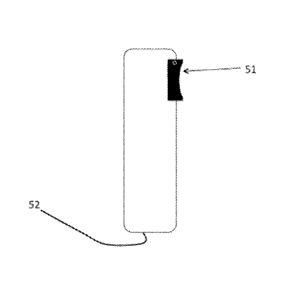
In one embodiment, shown in Figure 5, the signal acquisition device is shown
at 51.
The PHEIM of Figure 5 is connected to a computer by means of a cable 52.
However, the
PHHM of Figure 5 may alternatively be connected to a computer by wireless
means such as
Bluetooth.
The PHEIM of Figure 5 may also be equipped with data entry means and a display
(not shown in Figure 5) which may be combined as a touchscreen and used to
communicate
with the user (the subject or a health care professional) and used by the user
to activate the
PHEIM and to enter personal data or identifying data, such as a personal
identification
number.
In another form, shown in three views in Figure 6 in the attached drawings,
the
PHEIM is ergonomically curved to fit into the hand. The open surface 61 of the
signal
acquisition device and the top shape 62 is designed to hold a finger
comfortably along it.
Second Aspect
In a second aspect, the present invention relates to a PHEIM as defined in
W02013/001265 and as set out above comprising a signal acquisition device for
acquiring
signals which can be used to derive a measurement of a parameter related to
the health of the
user, the signal acquisition device being integrated with a PHHCD, which PHEIM
includes an
electrical sensor comprising at least three electrodes electrically isolated
from each other but
which can be contacted by different parts of a user's body. Preferably, two
electrodes can be
contacted by one finger from each hand of the user and a third electrode can
be contacted by
a hand. Preferably, one of the electrodes of the electrical sensor is
associated with a blood
flow occlusion means as described above in connection with W02013/001265 or as
described below in connection with the present invention. The other electrodes
will be
located on a separate part of the PHEIM. Preferably, the electrodes are
constructed with a
surface that gives a good electrical connection, such as an array of micro-
pyramids or a
material such as silver/silver chloride.
Preferably, the signal which is acquired by the electrical sensor is a measure
of the
potential difference between two of the electrodes, preferably in contact with
the fingers,
which is related to the potential difference between the two different body
parts. A third
electrode is used to provide an earth reference. Preferably, the processor of
the PHHCD is
adapted to amplify the signals from the electrical sensor and, if desired, to
filter the signals
before, during or after amplification. An amplified and filtered signal
produced by the
CA 02900904 2015-08-11
WO 2014/125431 22
PCT/IB2014/058969
processor will generally have the form shown in Figure 1 in the attached
drawings where the
x axis represents time and the y axis represents potential difference. The
arrows in Figure 1
indicate the time at which the electrical signal stimulates the heart to
initiate systole.
Third and Fourth Aspects
According to a third aspect of the present invention, there is provided a PHHM
adapted to provide a BP measurement comprising:
a housing, the housing including an open surface against which, in use, a
subject's
body part can be pressed or which, in use, can be pressed against a subject's
body part so that
pressure can be applied to the subject's body part to occlude an artery in the
body part;
a pressure sensor associated with the open surface for providing an electrical
signal
related to the pressure exerted by the open surface on the body part or vice
versa;
an optical sensor associated with the open surface for providing an electrical
signal
related to the luminal area of the artery which, in use, is occluded by the
open surface; and
processing means for controlling the device and for receiving and analysing
electrical
signals from the pressure sensor and the optical sensor to provide a
measurement of the
subject's SBP and/or DBP.
According to a fourth aspect of the present invention, there is provided a
PHHM
adapted to provide a BP measurement comprising:
a housing, the housing including an open surface against which, in use, a
subject's
body part can be pressed or which, in use, can be pressed against a subject's
body part so that
pressure can be applied to the subject's body part to occlude an artery in the
body part;
a pressure sensor associated with the open surface for providing an electrical
signal
related to the pressure exerted by the open surface on the body part or vice
versa; and
processing means for controlling the device and for receiving and analysing
the
electrical signals from the pressure sensor to provide a measurement of the
subject's SBP
and/or DBP.
The open surface must be suitable for applying pressure to a part of a
subject's body
or for having a part of the subject's body pressed against it. It is therefore
located on an outer
surface of the PHHM. If the open surface is in a dedicated module, it is
located on the face of
the module which, when the module is connected to the remainder of the PHHM,
is an outer
face of the PHHM. Preferably the open surface is sized so that it can interact
with a subject's
finger. The open surface may be flat. However, preferably, the open surface is
a concave
area in one face of the housing. The concave area may be part circular in
cross-section.
Preferably, the concave area has a radius of from 5 to 15 mm, more preferably
from 7 to 13
CA 02900904 2015-08-11
WO 2014/125431 23
PCT/IB2014/058969
mm, most preferably from 9 to 11 mm, and an arc length of from 5 to 15 mm,
more
preferably from 7 to 13 mm, most preferably from 9 to 11 mm. Preferably, the
open surface
is saddle-shaped, i.e. it has a central part with a constant radius and has
parts on either side of
the central part which slope away from the central part as shown in Figure 7
of the attached
drawings (see below).
In either of the third and fourth aspects of the invention, the PHEIM may also
include
an electrical sensor as described above with reference to W02013/001265 or
with reference
to the second aspect of the present invention for providing an electrical
signal related to the
time at which an electrical signal which initiates a heartbeat in the subject
occurs.
Fifth, Sixth and Seventh Aspects
According to a fifth aspect of the present invention, there is provided a
PHEIM
comprising a signal acquisition device for acquiring signals which can be used
to derive a
measurement of a parameter related to the health of the user, the signal
acquisition device
being integrated with a PHHCD, which PHEIM includes a blood flow occlusion
means which
is a button which is approximately rectangular with a length of 5 to 10 mm and
a width of 5
to 10 mm or is a circular button of 3 to 5 mm in diameter or is a non-circular
button of similar
area.
According to a sixth aspect of the present invention, there is provided a
PHEIM
comprising a signal acquisition device for acquiring signals which can be used
to derive a
measurement of a parameter related to the health of the user, the signal
acquisition device
being integrated with a PHHCD, which PHEIM includes a blood flow occlusion
means which
is a button, which is at least a part of an external surface of the PHHM which
is saddle-
shaped, i.e. approximately of the shape shown in Figure 7 in the attached
drawings, wherein
the button is physically separated from the surrounding surface. In Figure 7
there is a curved
surface 73 in one plane and, in the other plane, a central flat area 71 with
curved sides 72.
Preferably, the button is covered by a thin continuous membrane to exclude
contaminants. In this case, preferably, the button is co-planar with the
remainder of the
surface. However, the button may comprise the whole of the saddle-shaped
surface.
Preferably, the saddle-shaped external surface is continuous and sealed.
Preferably, the distance the button moves when subject to the force of the
body part is
no more than 0.01 mm.
According to a seventh aspect of the present invention, there is provided a
MEM
comprising a signal acquisition device for acquiring signals which can be used
to derive a
measurement of a parameter related to the health of the user, the signal
acquisition device
CA 02900904 2015-08-11
WO 2014/125431 24
PCT/IB2014/058969
being integrated with a PHHCD, which PHEIM includes a blood flow occlusion
means that is
a sealed vessel containing an essentially incompressible fluid in which is
immersed a pressure
sensor which is adapted to provide electrical signals to the processor of the
PHHCD. The
fluid may be a quasi-solid gel or may be a liquid. The incompressible fluid is
preferably
covered by a flexible membrane forming some or all of the occlusion means.
Preferably, the processor is adapted to extract a waveform from the electrical
signals,
typically similar in shape to the waveform shown in the top line of Figure 2.
Eighth, Ninth and Tenth Aspects
Infra-red light is preferentially absorbed by oxygenated haemoglobin so the
amount of
absorption is approximately proportional to the amount of arterial blood
through which the
light passes. For a given length of artery, the amount of arterial blood is
proportional to the
luminal area of the artery so the absorption signal is also approximately
proportional to the
luminal area.
As the artery expands on each systole and contracts on diastole, the
absorption of
infra-red light varies with the pulse.
The optical sensor used in the device of W02013/001265 is required to transmit
only
one wavelength of light, preferably in the infra-red range, to enable a
measurement of BP to
be taken. Thus, the optical sensor may comprise only a single photoemitter and
a single
photodetector. However, as the additional cost of enabling the optical sensor
to transmit a
second wavelength of light is small, according to a seventh aspect of the
present invention,
the optical sensor is able to transmit light at two wavelengths so that an
estimate of blood
oxygenation can be made at the same time as a measurement of BP.
According to an eighth aspect of the present invention, the processing means
is
adapted to correlate signals received from a pressure sensor with the signals
received from
the optical sensor so that the pressure exerted between an occlusion means and
the body part
can be correlated with the change of luminal area of the artery with each
pulse as a function
of the applied pressure. The correlated values can then be fitted to a curve
to provide
measurements of the subject's SBP and/or DBP.
According to a ninth aspect of the present invention, there is provided a PHHM
comprising a signal acquisition device for acquiring signals which can be used
to derive a
measurement of a parameter related to the health of the user, the signal
acquisition device
being integrated with a PHHCD, which PHEIM includes PPG sensor including one
or more
photosensors, wherein the or each photo-emitter and/or the or each photo-
detector is/are
provided with one or more lenses to narrow the field of view.
CA 02900904 2015-08-11
WO 2014/125431 25
PCT/IB2014/058969
According to a tenth aspect of the present invention, there is provided a
PHEIM
comprising a signal acquisition device for acquiring signals which can be used
to derive a
measurement of a parameter related to the health of the user, the signal
acquisition device
being integrated with PHHCD, which PHEIM includes a PPG sensor including two
or more
photosensors, wherein there are either two photo-emitters or two photo-
detectors arranged
such that light emitted in two different directions can be detected, and the
processor of the
PHHCD is adapted to process the signals received from each direction to locate
a blood
vessel, preferably an artery, in the user's body. Figure 8 in the attached
drawings shows such
an arrangement with one photo-detector 80 and two photo-emitters 81 and 82.
The difference between the signals received from each of the photo-emitters is
indicative of the displacement of the artery with respect to them.
Advantageously, the PHHCD is adapted to provide a visible or audible signal to
the
user to move a body part to optimise the position of the photo-sensor with
respect to the
blood vessel, preferably an artery. Alternatively, the PHHCD is adapted to
compensate the
signals from the photo-detector(s) when the blood vessel is not optimally
positioned with
respect to the photo-sensor.
Preferably, in either of the ninth and tenth aspects of the invention, the
optical axes of
the photo-emitter(s) and photo-detector(s) are aligned: to maximise the
sensitivity of the
signal produced by the photo-detectors to absorption of the emitted light by
blood. Figure 9a
shows such a configuration of one photo-emitter 90 and one photo-detector 91.
The two
optical components are aligned so that they are both directed towards the
artery 92 in order to
maximise its effect on the detected signal.
Alternatively, the optical axes are aligned to minimise the sensitivity of the
signal
produced by the photo-detectors to the location of the blood vessel. Figure 9b
shows such a
configuration of one photo-emitter 93 and one photo-detector 94. The two
optical
components are aligned so that a small movement of an artery relative to the
optical
components causes one to be better aligned and the other worse, thus reducing
the effect of
such a movement on the returned signal.
In a further alternative embodiment, the PHEIM is adapted to detect the
optical signals
and pressure signals at a range of pressures and determine if these correspond
to the signals
of a correctly-located artery. The PHEIM is adapted such that, if they are
not, the PHHM will
issue visible and/or audible signals instructing the user to reposition the
body part and try
again.
Eleventh Aspect
CA 02900904 2015-08-11
WO 2014/125431 26
PCT/IB2014/058969
According to an eleventh aspect of the present invention, there is provided a
PHHM
comprising a signal acquisition device for acquiring signals which can be used
to derive a
measurement of a parameter related to the health of the user, the signal
acquisition device
being integrated with a PHHCD, which PHHM is adapted to detect the presence of
a body
part, such as a finger and adapted to initiate operation of the PHHM and,
optionally, the
provision of instructions to the user on receipt of a signal by the sensor.
Twelfth Aspect
According to a twelfth aspect of the present invention, there is provided a
PHHM
comprising a signal acquisition device for acquiring signals which can be used
to derive a
measurement of a parameter related to the health of the user, the signal
acquisition device
being integrated with a PHHCD, which PHHM includes a photo-sensor for
detecting the flow
of blood through a blood vessel and an electrical sensor for detecting
electrical signals
relating to the action of the heart and the PHHM is adapted to use the timing
of events
detected by the electrical sensor to determine the time or times at which to
detect events in
the signals produced by the photo-sensor(s).
Thirteenth Aspect
According to a thirteenth aspect of the present invention, there is provided a
PHHM
comprising a signal acquisition device for acquiring signals which can be used
to derive a
measurement of a parameter related to the health of the user, the signal
acquisition device
being integrated with a PHHCD, which PHHM includes a blood flow occlusion
means which
provides an instantaneous estimate of the pressure in the artery by acting as
an applanation
tonometer.
Such tonometers do not usually produce an absolute measure of pressure and
have to
be calibrated by another means, such as occlusion. The PHHM disclosed herein
can be
adapted to combine both occlusion and applanation tonometry and so permit the
blood
occlusion means to be calibrated for use as an applanation tonometer by means
of occlusion.
This permits, for example, routine measurements to be made quickly using the
applanation
tonometer mode and occasional calibration measurements to be made using the
occlusion
mode.
The estimate of BP may be further refined by the use of other measurements.
The
Pulse Wave Velocity may be used to make a direct independent estimate of BP as
described
in detail by Padilla (/c. cit.), which in turn references earlier work on a
similar subject from
1995 and its specific use for estimating of BP in 2000. The technique is
described in US
Patent No. 5,865,755 dated 2nd February, 1999.
CA 02900904 2015-08-11
WO 2014/125431 27
PCT/IB2014/058969
Fourteenth and fifteenth Aspects
According to a fourteenth aspect of the present invention, there is provided
PHHM
comprising a signal acquisition device for acquiring signals which can be used
to derive a
measurement of a subject's BP, the signal acquisition device being integrated
with a PHHCD,
wherein the signal acquisition device comprises a blood flow occlusion means
adapted to be
pressed against one side only of a body part or to have one side only of a
body part pressed
against it, a means for measuring the pressure applied by or to the body part,
and a means for
detecting the flow of blood through the body part in contact with the blood
flow occlusion
means, wherein the PHHCD is a computer pointing device, commonly referred to
as a mouse,
or a controller for a television or other domestic electronic appliance so
that the subject may,
by holding the mouse, or the controller for a television or other domestic
electronic appliance
enable a measurement of his or her BP and, if desired, some or all of his or
her blood oxygen
concentration, pulse rate and respiration rate or other physiological vital
signs. The PHHM
may communicate with the computer with which the pointing device or controller
is being
used or another computer, either by means of a cable or by wireless means such
as Bluetooth.
Fifteenth Aspect
As disclosed in W02013/001265, the PHHM creates a quasi-static pressure on the
artery within the body part either by pressing the body part against the PHHM
or by pressing
the PHHM against the body part (W02013/001265, page 8, line 19).
According to a fifteenth aspect of the present invention, there is provided a
MEM
which comprises a signal acquisition device for acquiring signals which can be
used to derive
a measurement of a subject's BP, the signal acquisition device being
integrated with a
PHHCD, wherein the signal acquisition device comprises a blood flow occlusion
means
adapted to be pressed against one side only of a body part or to have one side
only of a body
part pressed against it, a means for measuring the pressure applied by or to
the body part, and
a means for detecting the flow of blood through the body part in contact with
the blood flow
occlusion means, wherein the blood flow occlusion means and its associated
electronic
components are included in a weight for producing a force between the blood
flow occlusion
means and the body part by acceleration of the weight.
For example, this could be a weight adapted to press against a finger and be
subject to
acceleration when the subject walks and swings his or her arms. As an
illustration, a swing
of 50 cm in 0.5 seconds would create a peak acceleration of around 10 m S-2 so
a mass of
around 50 g would create a pressure of around 200 mm Hg on an area of 20 mm2.
It would
CA 02900904 2015-08-11
WO 2014/125431 28
PCT/IB2014/058969
therefore be possible to occlude the artery by the random movements of the arm
when
walking with a steel ring 30 mm in diameter and 10 mm wide around the index
finger.
Figure 10 in the attached drawings shows a PHHM according to this aspect of
the
invention in which the elements of the PHEIM are integrated. Figure 10a shows
the PHEIM
on a finger. The PHEIM comprises a thick ring 101 for location around the
middle phalanx of
the index finger 102. Figure 10b shows the cross-section of the MEM. The ring
103 is
adapted to surround the finger 104 with a soft foam padding 105 between them.
A hard
region 106 causes the ring 101 to press against the finger near an artery 107
and a pressure
sensor and its associated electronics 108 are embedded in this hard region.
Not shown is the
cable or wireless connection from the pressure sensor to the PHHCD.
Sixteenth and Seventeenth Aspects
The BP measured by the PHEIM of W02013/001265 is affected by the difference in
height between the PHEIM and the subject's heart. The magnitude of the effect
is
approximately 1 mm Hg for every 13.6 mm of height difference.
Conventional measurements of BP use a brachial cuff, typically more than 100
mm
wide. This uncertainty in the measurement height leads to an uncertainty of
the measured
pressure of the order of 7 mm Hg, although this is not apparent in testing
because the ISO
standard test for automatic sphygmomanometers uses the same cuff for reference
as for the
automatic device. There may however be a significant difference between the
measured BP
and the intra-arterial pressure or aortic pressure. Also, the effective
height of the
measurement may depend on how the cuff is fitted and how far it is above the
elbow, thus
reducing the repeatability of the measurements.
According to a sixteenth aspect of the present invention, there is provided a
PHEIM
which comprises a signal acquisition device for acquiring signals which can be
used to derive
a measurement of a subject's BP, the signal acquisition device being
integrated with a
PHHCD, wherein the signal acquisition device comprises a blood flow occlusion
means
adapted to be pressed against one side only of a body part or to have one side
only of a body
part pressed against it, a means for measuring the pressure applied by or to
the body part, and
a means for detecting the flow of blood through the body part in contact with
the blood flow
occlusion means, wherein PHEIM is adapted to estimate the height of the PHEIM
with respect
to a fixed point on the subject's body.
The PHEIM thus includes a system for making more accurate and repeatable
measurements than can be achieved with conventional cuffs by making it
possible to
CA 02900904 2015-08-11
WO 2014/125431 29
PCT/IB2014/058969
determine accurately the height of the PHI-1M. The system may operate with
respect to the
height of the aortic valve of the heart.
The pressure sensor or a further pressure sensor of the PHHM may be used to
measure atmospheric pressure. The user may take such a measurement when the
device is at
the same height as the subject's heart and again when the sensor is used to
measure BP. The
difference between these may be used to correct the measured values of BP for
the effect of
hydrostatic pressure.
However, the system may alternatively operate with respect to a fixed point of
the
subject's anatomy. Such a fixed point is the midpoint between the centres of
the pupils of the
eyes. Many PHHCDs have a camera for creating an image of the subject's face.
Recent
PHHCDs include software to analyse that image to detect the pupils and even
the direction in
which the subject is looking. Many PHCCDs also include a tilt sensor to detect
the angle at
which the PHHCD is being held. According to an seventeenth aspect of the
invention, a
combination of these devices and software is used to determine the distance of
the PHHCD
from the eyes by estimating the angular distance between the pupils. The angle
below
horizontal of the PHHCD is estimated from the position in the image of the
subject's face at
which the pupils are detected and the tilt of the PHHCD. These are combined by
simple
trigonometry to estimate the vertical distance that the PHHCD is below the
subject's eyes.
Figure 11 in the attached drawings illustrates this. The dotted line shows the
direction from
the camera in the PHHCD 111 to the eyes, at an angle marked 113 with respect
to the body of
the PHHCD. The tilt angle of the PHHCD is marked 112. The angle 112 at which
the
PHHCD 111 is oriented is measured by a tilt sensor. The angle 113 between the
PHHCD and
the direction to the eyes is also measured. The estimate is then used to
improve the accuracy
of the determination of the subject's BP.
Eighteenth Aspect
A housing may be an integral part of device PHHM or may be part of a module
which
is adapted to be integrated into or attached to the remainder of the PHI-1M.
Such a module for
attachment to the remainder of the PHHM includes the housing, a blood flow
occlusion
means, such as described in W02013/001265 or as described herein, a pressure
sensor, an
optical sensor if present, at least one of the electrodes of an electrical
sensor if present and
electrical connections for connecting these components to the remainder of the
PHHM. Such
a module for integration with the remainder of the PHHM includes the housing,
an occlusion
means, a pressure sensor, an optical sensor if present, at least one of the
electrodes of an
electrical sensor if present and mechanical and electrical connections for
connecting these
CA 02900904 2015-08-11
WO 2014/125431 30
PCT/IB2014/058969
components to the remainder of the PHEIM. Such modules form an eighteenth
aspect of the
present invention.
Nineteenth Aspect
According to a nineteenth aspect of the invention, the PHEIM also includes a
bolometric thermometer for measuring body temperature. W02013/001265 shows how
the
clinical accuracy of such a bolometer may be improved by combining it with
some or all of
the other aspects of the PHEIM.
Preferably, the temperature of the cold junction of the bolometer is
determined from
the temperature sensing of the other aspects of the PHHM, such as the
temperature of the
bridge in a pressure sensor or the temperature of an ASIC (described below)
sensed by a
component included within it. Alternatively, the cold junction sensor of a
conventional
bolometer provides an indication of the temperature of the pressure sensor
bridge and/or the
ASIC.
Alternatively, the temperature sensor may be located such that the user may
orientate
its direction by manipulating the PHEIM such that it is able to sense the
temperature of the
body part or other item chosen, for example an item of the user's clothing.
Preferably the PHM is incorporated into a PHHCD which includes a camera and
display. These may be used to display an image of the field of view over which
the
temperature being detected is marked.
Twentieth Aspect
As indicated above, a PHHM has been disclosed which comprises a signal
acquisition
device for acquiring signals which can be used to derive a measurement of a
parameter
related to the health of a subject, the signal acquisition device being
integrated with a
PHHCD, wherein the parameter is BP and the signal acquisition device comprises
a blood
flow occlusion means adapted to be pressed against one side only of a body
part or to have
one side only of a body part pressed against it, a means for measuring the
pressure applied by
or to the body part, and a means for detecting the flow of blood through the
body part in
contact with the blood flow occlusion means, wherein the blood flow occlusion
means
comprises a button which is at least part of an external surface of the PHEIM
which is saddle
shaped and the button takes the form of a flexible membrane forming a wall of
a sealed
vessel containing an essentially incompressible fluid in which is immersed a
pressure sensor
which is adapted to provide electrical signals to the processor of the PHHCD.
According to a twentieth aspect of the present invention, there is provided a
PHEIM
which comprises a signal acquisition device for acquiring signals which can be
used to derive
CA 02900904 2015-08-11
WO 2014/125431 31
PCT/IB2014/058969
a measurement of a subject's BP, the signal acquisition device being
integrated with a
PHHCD, wherein the signal acquisition device comprises a blood flow occlusion
means
adapted to be pressed against one side only of a body part or to have one side
only of a body
part pressed against it, a means for measuring the pressure applied by or to
the body part, and
a means for detecting the flow of blood through the body part in contact with
the blood flow
occlusion means, wherein the blood flow occlusion means comprises at least
part of an
external surface of the PHHM wherein the pressure is sensed by means of a
flexible and
essentially incompressible gel in which is immersed a pressure sensor which is
adapted to
provide electrical signals to the processor of the PHHCD.
Twenty first Aspect
A PHHM has been disclosed which comprises a signal acquisition device for
acquiring signals which can be used to derive a measurement of a parameter
related to the
health of a subject, the signal acquisition device being integrated with a
PHHCD, wherein the
parameter is BP and the signal acquisition device comprises a blood flow
occlusion means
adapted to be pressed against one side only of a body part or to have one side
only of a body
part pressed against it, a means for measuring the pressure applied by or to
the body part, and
a means for detecting the flow of blood through the body part in contact with
the blood flow
occlusion means, wherein the blood flow occlusion means comprises a button
which is at
least part of an external surface of the PHHM which is saddle shaped, wherein
the PHHM
also serves as the on/off switch or some other subject-operated switch for the
PHHCD.
This may be achieved by means of a physical action, such as by applying force
to the
PHHM, or by placing the body part in the path of a photo-detector, which may
detect the
presence of the body part from the change in light intensity detected.
According to the a twenty first aspect of the present invention, there is
provided a
PHHM which comprises a signal acquisition device for acquiring signals which
can be used
to derive a measurement of a parameter related to the health of a subject, the
signal
acquisition device being integrated with a PHHCD, wherein the PHHM is used to
provide a
continuously variable means for the subject to control some function of the
PHHCD other
than the measurements that are made by the PHHM. Such a PHHM is adapted for
continuous, or analogue, control by varying the force applied to pressure
sensor, which is
then used to control a property of the PHHCD, such as volume or screen
brightness.
Twenty second Aspect
It is well-known that accurate blood pressure measurements require the subject
to be
calm and relaxed for a few minutes before taking the measurement. Many PHHCDs
have
CA 02900904 2015-08-11
WO 2014/125431 32
PCT/IB2014/058969
sensors built in that allow them to detect movement and vibration. In the
twenty second
aspect of the present invention, if the PHM is incorporated in such a PHHCD,
said sensor
may be used to detect movement of the PHHCD which, if the user is holding the
PHHCD,
can be used to warn the user to sit calmly for a few minutes or even to
prevent any
measurements being made until the PHHCD has been at rest for such a time.
Twenty third aspect
In the twenty third aspect of the present invention, the PHHM is adapted to
detect the
optical signals and pressure signals at a range of pressures and determine if
these correspond
to the signals of a correctly located artery. If they do not, the PHHM is
adapted to issue
visible and/or audible signals instructing the user to reposition the body
part and try again.
This aspect may be further adapted to provide multiple passes through a
measurement
sequence to refine the accuracy of the measurement or to reduce the time
required to obtain a
measurement.
The PHHM
Preferably, the PHHM according to any of the aspects of the present invention
is of
such a size and weight that it can readily be manipulated by a subject using
one hand to hold
the device against a finger of the other hand to make a measurement or by a
medical
practitioner holding the device against the subject. Preferably, the PHHM is
rectanguloid,
having an upper and a lower face connected by four side faces, wherein the
upper face is of
sufficient area to accommodate a display means and a data entry means, the
distance between
the upper and lower faces is small enough to be located on the subject's body
part and the
open surface is located on one of the side faces. The PHHM may have a width of
from 5 to
20 cm, a length of from 10 to 30 cm and a depth of from 0.5 to 2.0 cm. The
PHHM may
have rounded corners and edges.
Pressure Sensor
Pressing the occlusion means against a body part, such as a finger, or vice
versa
creates a pressure within the body part. The pressure sensor measures,
directly or indirectly,
the pressure between the occlusion means and the body part.
The pressure sensor may measure the pressure directly. For instance, the
pressure
sensor may comprise a pressure-responsive device immersed within a sealed
vessel
containing an essentially incompressible fluid. The fluid may be a quasi-solid
gel or may be
a liquid. The incompressible fluid is preferably covered by a flexible
membrane forming
some or all of the occlusion means.
CA 02900904 2015-08-11
WO 2014/125431
PCT/IB2014/058969
33
Alternatively, the pressure sensor may measure the pressure indirectly. For
instance,
the pressure sensor may comprise a force-responsive device connected to a
region of the
occlusion means, which region may move independently from the remainder of the
occlusion
means. Typically, the region is circular and from 3 to 5 mm in diameter or is
non-circular
and of similar area. Preferably, the distance the region moves when subject to
interaction
with a body part, such as a finger, is no more than 0.01 mm. Preferably, the
region is co-
planar with the remainder of the occlusion means and may be covered by a thin
continuous
membrane to exclude contaminants. In use, when a body part, such as a finger,
is in contact
with the region, the pressure between the body part and the occlusion means is
approximately
.. the force measured by the force-responsive device divided by the area of
the movable region.
The pressure sensor may include a plurality of pressure-responsive or force-
responsive devices.
By measuring the pressure or force, the device can make an accurate estimate
of the
pressure within the subject's body part. As explained below, provided that the
occlusion
means and the body part are in contact for a sufficient period of time and the
pressure
between the occlusion means and the body part is varied sufficiently, the
processing means
can analyse the signals received over a period of time and varying over a
range of pressures
from the pressure sensor to determine the SBP and/or DBP of the subject. It
has been found
that it is possible to fit the signals received from the pressure sensor, in
whatever order they
are received, to a curve from which SBP and/or DBP can be determined.
Optical Sensor
The optical sensor provides an electrical signal related to the luminal area
of the artery
by means of the absorption of light. It draws on the experience of pulse
oximeters using
photoplethysmography (PPG). As noted above, such pulse oximeters have been on
the
market since the 1980s. They are used to estimate the degree of oxygenation in
arterial
blood. The same principles as are described above in connection with the
disclosure in
W02013/001265 apply equally to all the aspects of the present invention. Red
and infra-red
light is transmitted by one or more photoemitters towards a body part and
detected by one or
more photodetectors after the light has passed through or been reflected by
the body part.
The infra-red light is more strongly absorbed by oxygenated blood than by non-
oxygenated
blood (a suitable wavelength is 940 nm); the red light is more strongly
absorbed by non-
oxygenated blood than by oxygenated blood (a suitable wavelength is 660 nm).
The ratio of
the fractional changes in red and infra-red intensity is monotonically related
to the percentage
CA 02900904 2015-08-11
WO 2014/125431
PCT/IB2014/058969
34
of oxygenation of the blood. It is also possible to use green light (a
suitable wavelength is
520 nm) in place of the red or infra-red light.
Infra-red light is preferentially absorbed by oxygenated haemoglobin so the
amount of
absorption is approximately proportional to the amount of arterial blood
through which the
light passes. For a given length of artery, the amount of arterial blood is
proportional to the
luminal area of the artery so the absorption signal is also approximately
proportional to the
luminal area.
As the artery expands on each systole and contracts on diastole, the
absorption of
infra-red light varies with the pulse.
The processing means correlates the signals received from the pressure sensor
with
the signals received from the optical sensor so that the pressure exerted
between the occlusion
means and the body part is correlated with the luminal area of the artery. The
correlated
values can then be fitted to a curve to provide measurements of the subject's
SBP and/or
DBP.
Artery Location
Preferably, the PHEIM is adapted to use the optical sensor as described in
W02013/001265 or as described above, in particular in connection with the
eighth, ninth,
tenth and nineteenth aspects of the invention for locating the artery.
Processing Means
The electrical signals produced by the sensor(s) of the PHEIM may be analogue
or
digital and, if analogue, the signals may be converted to digital form, by an
analogue-to-
digital converter in the sensor or in the processing means, for subsequent
analysis.
Preferably, the processing means includes one or more amplifiers for
amplifying the
electrical signals received from the sensor(s) in the device. The processing
means may also
include filtering means and/or conditioning means for filtering and/or
conditioning the
received electrical signals. The filtering and/or conditioning means may be
arranged operate
before, during or after amplification of the received electrical signals.
Preferably, the processing means includes one or more Application Specific
Integrated Circuits (ASIC) and/or one or more Micro-Engineered Measurement
Systems
(MEMS). The processing means will include any electronic circuitry associated
with the
sensor(s) in the device.
The processing means may comprise a number of separate electronic devices
which
are preferably integrated into a single package. However, advantageously, some
or all of the
electronic devices are integrated into a single unit. Such integration will
bring several
CA 02900904 2015-08-11
WO 2014/125431
PCT/IB2014/058969
benefits, including reduced cost, improved reliability, reduced size and mass
and reduced
power consumption.
Preferably, the processing means is adapted to cause the photoemitter(s), when
present, to be switched so that a single multiplexed photo-detector can detect
light at the
5 selected wavelengths. Preferably, the processing means is adapted to
allow an electrical
signal to be acquired from the photodetector(s) for a period in which no light
is emitted from
the photoemitter(s) to allow a further calibration of the signals.
Preferably, the processing means is adapted to:
control and receive electrical signals from the sensor(s) in the device;
10 analyse the electrical signals from the sensor(s) in order to determine
the BP and,
preferably, other diagnostic information; or
control the display means, if present, for communicating the result of the
measurement to the user.
The processing means may also be adapted to receive and process electrical
signals
15 from the data entry device, if present.
Preferably, the activities associated with artery location are carried out
during the
initial phase of a two-phase measurement cycle. In the initial phase, the
PHEIM makes
measurements to locate the artery and/or other measurements that ensure that
the subsequent
phase will be accurate and efficient, such as approximately determining SBP
and DBP. In
20 the second phase, the PHHM makes the accurate measurements.
Preferably, the processing means includes one or more storage devices, such as
a flash
memory, for storing the electrical signals received from the sensor(s) and/or
input from the
data entry device and any electrical signals derived from the received
signals. In particular, a
storage device is preferably provided for storing the historical BP data
derived by the
25 processing means for each subject.
The processing means may also be adapted to communicate with a remote
computer,
preferably wirelessly via the internet, to allow the output of the processing
means to be
further analysed, archived and/or communicated.
Preferably, the processing means is adapted to provide audible or visual
instructions,
30 advantageously via the display means, if present, to the user to enable
the user to use the
device optimally. This includes instructions to vary the force applied to the
body part to
cover a wide enough range of pressures to give a good fit to the mathematical
equation.
For instance, if the occlusion means has not been pressed hard enough against
the body
part to occlude completely an artery during a systole, the device may be
programmed to
CA 02900904 2015-08-11
WO 2014/125431 36
PCT/IB2014/058969
issue an instruction to the user to press harder on the open surface (or vice
versa) so that
the required electrical signals can be acquired. In this case, it is preferred
that the
processing means is adapted so that the instructions are interactive and based
on signals
received from the sensor(s) which can be used to determine whether the device
is in the best
position or being used correctly.
The PHHM of any of the aspects of the present invention may also include: a
display
means for displaying measurements of the subject's SBP and/or DBP; and/or
communications means for transmitting the measurements of the subject's SBP
and/or DBP;
and/or storage means for storing the measurements of the subject's SBP and/or
DBP. If
present, the storage means may also store other data sent to or generated by
the processing
means.
The PHHM of any of the aspects of the present invention may also include a
data
entry device adapted to be operated by the user so that the user can enter
information into the
device. The data entry device may be a keypad or a touchscreen. The data entry
device may
be used to input data for identifying a subject or other user so that
different subjects and/or
users can use the device. The data that can be entered by use of the data
entry means may
include, but are not restricted to, the subject's height, weight, waist
circumference, finger
diameter and age.
Operation of the PHHM
The PHHM is operated by holding the occlusion means against a body part, such
as a
finger, or holding the body part against the occlusion means and varying the
force exerted by
the body part on the occlusion means or exerted by the occlusion means on the
body part to
achieve a range of pressures in the body part from below DBP to above SBP.
While the force
of interaction between the body part and the occlusion means is being varied,
the sensor(s) in
the device are switched on and the electrical signals generated by the
sensor(s) are received
and processed by the processing means.
Unlike conventional sphygmomanometry, flow may be detected at a range of
pressures in any order and the data fitted to a mathematical equation.
Analysis of the Electrical Signals
The waveforms of typical electrical signals received from optical, pressure
and
electrical sensors are shown in Figure 12 in the attached drawings. The
primary signals that
the processing means extracts from these are the change in absorption on
systole (from the
optical sensor) and the instantaneous measured pressure at systole and
diastole (from the
pressure sensor). From these, it is adapted to compute an estimate of the
change in the
CA 02900904 2015-08-11
WO 2014/125431
PCT/IB2014/058969
37
optical signal as a function of pressure. Preferably, the processing means is
adapted to use
the timing of events detected by the electrical sensor to determine the time
or times at which
to detect events in the optical and pressure signals.
The change in the optical signal has already been shown to be approximately
proportional to the luminal area of the artery. The relationship between the
luminal area and
pressure is referred to as the Arterial Optical/Pressure Curve (AOPC).
In order to explain the form of the AOPC, it is necessary to consider how the
artery
behaves. The relationship between luminal area and pressure is as shown in
Figure 13 where
TMP is the TransMural Pressure, which is the instantaneous pressure in the
artery minus the
External Applied Pressure (EAP), which is the pressure generated by the
occlusion means
and measured by the pressure sensor. Such curves have been reported by several
researchers,
such as Drzewiecki et at., "Theory of the oscillometric maximum and the
systolic and
diastolic detection ratios", Annals of Biomedical Engineering, 1994, 22, 88-96
and
Langeworters et at., "Pressure-diameter relationships of segments of human
finger arteries"
Clin. Phys. Physiol. Meas., 1986, 7, 43-55, both using in vitro measurements
of
representative arteries.
Where the applied pressure is less than DBP, the artery remains open
throughout the
pulse cycle. The change in luminal area that is approximately proportional to
the electrical
signal produced by the optical sensor is caused by the stretching of the
artery wall as the
pressure difference between the inside and outside rises. Where the applied
pressure is
greater than DBP and less than SBP, the artery collapses during every pulse
and, when open,
stretches as in the previous case. When the applied pressure is greater than
SBP, the artery
remains closed throughout the pulse cycle. This is illustrated in Figure 14.
The quantitative form of the AOPC is found by fitting the measured values of
the
optical signal to a parametric representation of the AOPC, such as that
proposed by
Langeworters et at. (loc. cit.). The parameters of the AOPC may also be
informed by an
estimate of the arterial stiffness from the Pulse Wave Velocity, derived from
the Pulse Wave
Transit Time, which in turn is related to the time interval between the peak
of the electrical
signal and the peak of the optical signal. This technique is described in
detail by Padilla (/oc.
cit.).
The amplitude of the AOPC at each of DBP and SBP, corresponding to the two
ends
of the bars marked D... S in Figure 14 is plotted against EAP to give a curve
of the form
shown in Figure 15. This Figure shows a simulation for SBP = 150 mm Hg and DBP
= 80
mm Hg. SDB and DBP are marked respectively by the arrows "S" and "D". Figure
16
CA 02900904 2015-08-11
WO 2014/125431 38
PCT/IB2014/058969
shows a measured version of this curve. The processing algorithms use curve
fitting routines
to estimate DBP and/or SBP to high precision. In particular, there is a
clearly visible
transition at DBP, a feature absent from measurements made by all other non-
invasive
sphygmomanometers.
The electrical signals received by the processing means may be further
analysed to
extract an estimate of the pressure waveform throughout the pulse cycle.
Preferably, the
analysis uses one or both of two independent methods: the pressure deficit
method and the
pulse timing method.
The pressure deficit method exploits the instantaneous balance between the
pressure
within the artery and the sum of the pressure applied by the occlusion means
(EAP) and the
pressure caused by the tension in the artery wall (TMP). Measured values of
the optical
signal are used to find the corresponding TMP from the AOPC. The instantaneous
arterial
pressure is then found by adding the TMP to the measured instantaneous EAP.
The curve in
Figure 17 shows the result of such a calculation.
The pulse timing method identifies the times during the pulse cycle at which
the
optical signal changes from a large signal (small absorption) to a small
signal (large
absorption) and back, each time being measured with respect to the time of the
peak of the
electrical signal. The artery opens when the pressure within it exceeds the
pressure applied
by the occlusion means and collapses when the pressure falls below it. The
pressure applied
by the occlusion means at the time of these events allows the instantaneous
pressure to be
mapped through the pulse cycle.
Preferably, the instantaneous pressure wave derived from either or both of
these
methods is then used to model the effect of the reflection of the pulse wave
from the body
part, which in turn may be used to estimate the pressure at other parts of the
body including,
but not restricted to, the wrist, upper arm and aorta (see, for example,
Stergiopolus et at.,
"Physical basis of pressure transfer from periphery to aorta: a model-based
study" Am. J.
Physiol., 1998, 274, H1386-H1392).
Preferably, the models used to analyse the data make use of information
provided by
the subject such as height, weight, waist circumference, finger diameter and
age.
The estimate of blood pressure may be further refined by the use of other
measurements. The Pulse Wave Velocity may be used to make a direct independent
estimate
of blood pressure as described in detail by Padilla (/c. cit.), which in turn
references earlier
work on a similar subject from 1995 and its specific use for estimating of BP
in 2000. The
technique is described in US Patent No. 5,865,755 dated 2nd February, 1999.
Once the form
CA 02900904 2015-08-11
WO 2014/125431
PCT/IB2014/058969
39
of the AOPC is found, it is possible to compute the instantaneous pressure
throughout the
pulse cycle. This allows the PHEIM to perform the functions of a tonometer. It
also permits
a rapid estimate to be made of SBP and DBP, within one cycle, so allowing beat-
to-beat
monitoring of blood pressure.
In a further aspect of the analysis, it is well known that respiration
modulates the
timing of the heartbeat, the amplitude of the ECG signal, the mean and pulse
blood pressure
and possibly also the Pulse Wave Velocity. The analysis exploits all of these
to make several
independent measurements, using: the pulse period derived separately from the
red and infra-
red channels of the optical sensor and from the electrical sensor, the phase
difference between
said optical and electrical signals, the amplitude and mean values of the PPG
signal and the
amplitude of the ECG signal. All of these may be subject to noise or
inaccuracy. Each is
independently analysed to establish its quality, measured using parameters
such as the
repeatability of the periodicity and the signal/noise ratio. The independent
measurements are
then combined to give a robust estimate of respiration rate and depth by
including all where
the quality exceeds an empirically determined threshold.
Some or all of the data analysis of the signals concerning blood pressure may
be
conducted on the remote computers. This allows more demanding calculations to
be
provided, such as the analysis required to find the AOPC and to enable the
PHHM to be used
as a tonometer. Communication with the remote computers also permits the
results to be
archived and, if the user so instructs, transferred electronically to third
parties such as the
user's personal doctor, a medical specialist or a medical or life insurer.
Further aspects of the aspects of the invention are defined in the dependent
claims.
The various aspects of the invention disclosed herein may be used in any
combination.
ILLUSTRATIONS
A number of embodiments of the present invention will now be described by way
of
example only with reference to the accompanying drawings. It should be clearly
understood
that the following description is provided purely by way of illustration and
that the scope of
the invention is not limited to this description; rather the scope of the
invention is set out in
the attached claims.
In the attached drawings:
Figure 1 shows a generalised amplified and filtered signal acquired by an
electrical
sensor;
CA 02900904 2015-08-11
WO 2014/125431 40 PCT/IB2014/058969
Figure 2 shows schematically the variation in oxygenated blood signal (top
line),
deoxygenated blood signal (middle line) and ambient light signal (bottom line)
acquired from
a PPG sensor;
Figure 3 shows a typical signal waveform of the "lub-dub" beat of a heart
acquired by
an acoustic sensor;
Figure 4 shows the envelope derived from the acoustic signal of Figure 3;
Figures 5 and 6 show PHIRMs according to embodiments of the present invention;
Figure 7 shows a saddle shaped surface useful as part of a blood occlusion
means as
disclosed herein;
Figures 8 and 9 show possible configurations of photo-emitters and/or photo-
detectors;
Figure 10 shows a dynamic pressure device;
Figure 11 shows the geometry for estimating the height of a PHHCD relative to
a
subject's eyes;
Figure 12 shows typical signals produced by optical, pressure and electrical
sensors;
Figure 13 shows an illustration of a BP measurement derived from the
electrical
signals illustrated in Figure 12;
Figure 14 illustrates the range of arterial pressures from diastolic to
systolic on the
curve of Figure 13 at various values of External Applied Pressure (EAP);
Figure 15 shows a theoretical Arterial Optical/Pressure Curve (AOPC);
Figure 16 shows a measured AOPC;
Figure 17 shows a reconstructed arterial pressure wave compared with the
measured
optical signal;
Figure 18 shows a cellphone adapted to operate according to the present
invention;.
Figure 19 shows a sketch of one embodiment of a PHIRM according to the
invention;
Figure 20 shows the way in which a subject holds a computer pointing device
("mouse");
Figure 21 shows a cross-section through the fingers of a subject and computer
pointing device;
Figure 22 shows a representation of a module having components of a PHIRM
integrated therein;
Figure 23 shows a different shape of module similar to that shown in Figure
22; and
Figure 24 shows a module including the elements of a PEIHM incorporated within
a
PHHCD.
CA 02900904 2015-08-11
WO 2014/125431 41
PCT/IB2014/058969
All of the illustrated embodiments of the PHHM include one or more electronic
components (not shown) that can include: one or more pressure sensors, one or
more
analogue to digital convertors, one or more temperature sensors, a unique
identifier and an
interface to the electronic circuits of a PHHCD such as a cellphone. All of
the sensors and
these components are preferably built within a single module, typically 10 mm
x 6 mm x 4
mm in dimensions. The module is adapted to be installed in the top corner of a
cellphone, as
shown in Figure 18. The module 150 replaces the on/off switch in the cellphone
151 and the
user is informed via the cellphone's screen 152.
Figure 19 shows a sketch of one embodiment of a PHHM according to the
invention,
comprising a housing 191 including a blood flow occlusion means in the form of
a part-
circular open surface 192, which has a width of approximately 10 mm and a
depth of
approximately 3 mm. A pressure sensor 193, in this case a pressure-responsive
device, is
located in the centre of the open surface 192. A processing means 194 is
connected to the
pressure sensor (193) and to one or more photoemitters 195, one or more
photodetectors 197
and an electrode 196 forming part of a electrical sensor. Separate cables run
to a second
electrode 198 forming another part of the electrical sensor. The illustrated
PHHM includes a
data entry means in the form of a touchscreen 190 which is connected to the
processing
means by a cable 199. Alternatively, the touchscreen may be connected by a
wireless system
such as Bluetooth. The touchscreen 190 can be used by the user (the subject or
a health care
professional) to activate the PHHM and to enter personal data or identifying
data, such as a
personal identification number.
Figure 20 shows the way in which a subject holds a computer pointing device
("mouse"). Figure 21 shows a cross-section through the fingers of a subject
and computer
pointing device, where there is the index finger 212, middle finger 213, ring
finger 214 and
little finger 215. The sensors 216 of the PHHM are incorporated in the body of
the pointing
device and the index finger rests against them.
Figure 22 shows a representation of a module having components of a PHHM
integrated therein which may be incorporated by others into their products.
Figure 22a shows
a cross-section and Figure 22b a plan of the module, intended for use where
the body part is a
finger and incorporating aspects of the invention. The length of the module is
approximately
10 mm. The module includes a housing 221 with electrical connectors 222 to
connect the
module to another device, a pressure sensor 223 embedded in gel 224, infra-red
and visible
light emitting diodes 105 and photo-detector 226. They access the body part
via the windows
227 and 228. The module includes an occlusion means in the form of an open
surface 229
CA 02900904 2015-08-11
WO 2014/125431 42
PCT/IB2014/058969
against which a finger is pressed. The module includes an ASIC 220 and a
bolometric
temperature sensor 231, shown as a separate device from the ASIC.
Alternatively, it could be
incorporated as part of the same ASIC. The bolometric temperature sensor 231
has a window
232 in the side of the module. The module includes two electrodes 223 adapted
to be touched
by the body part when it is pressed against the open surface 229. Not shown is
a further
electrode which is adapted to make contact with another body part.
Figure 23 shows a different shape of module similar to that shown in Figure 22
but
shaped for use where the body part is the wrist.
Figure 24 shows a module including elements of a PHEIM incorporated within a
PHHCD such as a cellphone. Figure 24 shows a possible arrangement, with the
module 241
located in the position where the on/off switch would normally be in the
PHHCD. The
PHHCD includes a touch-screen display 242 and a third electrode 243 for the
electrical
sensor.
CA 02900904 2015-08-11
WO 2014/125431
PCT/IB2014/058969
43
Health- Measure- Relevant Notes
related ment sensors
parameter technique
Bolonnetry is a mature technique. ThePHHM preferably uses
feedback to guide the user to obtain the highest value (for
Body Temperature
Bolonnetry example by moving around over the ear) and a
model to
temperature sensor extrapolate the changes as measured to
estimate an asymptotic
value and to correct for ambient.
Timing of Electrical sensor The signal from the electrical sensor will be the
most reliable and
Pulse rate pulses precisely timed. The signals from
the two sensors will also be
Pressure sensor analysed to provide confirmation of the data
and to improve
Pulse Timing of Blood accuracy. The analysis will, like that for BP,
seek the most likely
arrhythmia pulses photosensor value in the light of all of
the available evidence.
Pulse Wave Velocity (PWV) is a direct
measure of BP. The electrical sensor may be
Electrical sensor used to detect the time of initiation of the
Pulse pulse. The acoustic sensor may be used when
Blood
Wave the user first calibrates the device to
measure
Transition photosensor the time between electrical signal and start
of
Time Acoustic sensor systole. The PPG sensor may be used to
(PWTT) detect the time at which the pulse reaches
Personal data the finger. The personal data may be used to
estimate the path length from heart to PPG The actual BP
sensor.
may be estimated
by combining the
five
separate
Pulse Blood The magnitude of the PPG signal is a measure
measurements
of the change in arterial volume, which is (or as many as are
volume photosensor related to BP. available).
The
combination
The applied pressure is measured
might not just be
U,
BP Both
by the pressure in the pad or the
a simple average;
c
o force on the
strap or button. The techniqu the processing
(systolic IL) ',7, Pressure sensor blood flow rate
can be detected es may may seek to find by small changes in the pressure
and D ro use
the most likely feedback value in the light
diastolic) .,
a., v, D
v, 4-,
E _) `3' caused by changes in volume of to guide of all
available
o r:,¨,= the artery. the
user information,
C
to push using a technique
harder or such as
a
o 0 .2 Pressure sensor The pulse volume depends on the softer
to Bayesian
E 7 , - 8 _' external pressure, unaffected if it map the
estimator to take
Blood is less than diastolic and falling to pressure
account of all
-C u 0_28 photosensor zero at systolic. space. data
including
0-o
(f)-._.- 0 co variations
between pulses.
There is a correlation between pulse rate and
Timing of As pulse rate BP. Personal data, including records or
pulses previous measurements, will add to its
relevance.
The pressure sensor may also be used as an
Applanaen Pressure sensor applanation tononneter, with calibration
by
tononnetryocclusion
Blood PPG Blood Standard PPG technique, combining measurements
of infra-red
oxygen photosensor and visible absorption when the pulse
reaches the finger.
Pulse
Wave PWTT As above, in BP measurement
Velocity
The respiration cycle is manifested in The
actual
Blood changes to the interval between pulses, the
respiration cycle
Effect on photosensor mean level of BP and the magnitude of the may
be obtained by
Respiration BP and PPG signal
combining the four
cycle pulse
separate estimates
The respiration cycle is manifested in (or as many as are
Electrical sensor available).
changes to the interval between pulses.
Perturbation The user may be instructed to hold his/her
breath. The level of
Blood flow of Blood blood oxygen falls after the less oxygenated
blood has reached the
rate respiratory photosensor measurement point, and rises
again after a breath is taken and the
cycle more oxygenated blood arrives