Note: Descriptions are shown in the official language in which they were submitted.
CA 02901368 2015-08-14
WO 2014/132072 PCT/GB2014/050595
1
CSF1 THERAPEUTICS
The present invention relates to compositions of matter and methods of using
the same in
enhancing regeneration or restoring function of an injured liver. The
compositions of
matter are useful in the treatment of hepatic disorders, for example, in the
prevention
and/or treatment of acute or chronic liver disease or as a supportive therapy
to improve the
outcomes following liver resection or liver transplantation.
BACKGROUND
Liver disease is a major cause of morbidity and mortality worldwide but
despite this there is
currently no effective therapy to enhance regeneration of the diseased or
injured liver. A
therapy to enhance regeneration of the liver could be applied across a range
of medical
and surgical contexts for indications including acute, acute-on-chronic or
chronic liver
failure. In the medical setting acute liver failure can arise from a range of
aetiologies, but
most commonly due to infection (viral hepatitis), alcohol ingestion, or toxin
overdose (such
as Paracetamol overdose). In acute liver failure widespread necrosis of the
liver tissue
may occur which can rapidly result in death. Acute liver failure can arise on
a background
of chronic liver disease (acute-on-chronic) where pre-existing liver disease
(due to viral
hepatitis, alcohol, non-alcoholic fatty liver disease and other causes)
further impairs the
liver's ability to regenerate. Chronic liver failure can result from a gradual
deterioration in
liver function (causes as above) until the point at which the liver is unable
to maintain
homeostasis. In life threatening liver failure the only option is liver
transplantation,
however the shortfall between potential donors and recipients means many
patients will
die while awaiting liver transplantation.
Liver regeneration is a complex process involving many growth factors,
cytokines and cell
types. Liver macrophages perform a range of vital homeostatic roles and are
critical to
effective liver regeneration. Macrophage colony stimulating factor (M-CSF)
also referred
to as colony stimulating factor 1 (CSF1) and used interchangeably, is
expressed in the
liver and is the principle factor responsible for production and maintenance
of cells of the
monocyte/macrophage lineage, including liver macrophages. Depletion of
macrophages
and deficiency of CSF1 lead to impaired liver regeneration following partial
hepatectomy.
It is known from the prior art in a M-CSF null mouse model after partial
hepatectomy that
M-CSF induced Kupffer cells play a key role in liver regeneration (Amemiya et
al., J.Surg.
Res. 165, 59-67, 2011). However the potential of CSF1 supplementation to
enhance liver
regeneration has hitherto not been considered.
CA 02901368 2015-08-14
WO 2014/132072 PCT/GB2014/050595
2
A therapy to enhance regeneration and/or restore function of the liver could
be applied
across a range of medical and surgical contexts for indications including
acute, acute-on-
chronic or chronic liver failure and would offer immediate benefit to
patients, clinicians and
health services alike.
A therapy to enhance regeneration of the liver could be applied as a rescue
therapy to
facilitate regeneration following transplantation or in the context of
overwhelming failure or
used to prevent decline in chronic liver disease would offer immediate benefit
to patients,
clinicians and health services alike.
BRIEF SUMMARY OF THE DISCLOSURE
According to a first aspect of the invention there is provided a biologically
active fragment
of CSF1 protein or a homolog or a variant or derivative thereof for use in
enhancing liver
regeneration and/or restoring liver function and/or modulating liver
homeostasis.
Also include is the nucleic acid encoding the biologically active fragment of
CSF1 protein
or a homolog or a variant or derivative thereof.
The inventors have surprisingly found that administration of additional or
extra or
supplemental CSF-1 to subjects having normal CSF-1 levels increases the size
of the liver
in healthy animals and improves the ability to repair the liver following loss
of function from
various causes. It was an unexpected finding that a supplement of CSF-1, to
already
functioning CSF-1 in an individual, would improve hepatic regeneration or
function. The
liver is under very strict homeostasis and to date no agent has been
identified that can
successfully modulate hepatic homeostasis in the clinical setting and increase
the size of
liver above the normal relative total body weight. However, the present
invention provides
evidence for use of CSF-1 as an appropriate hepatic trophic and homeostatic
agent in
mammalian species. Furthermore, the present invention is based upon the
observation
that CSF-1 can restore the phagocytic capacity of the liver, and thus use of
CSF-1 proteins
for restoring this aspect of liver function is of particular interest in the
present invention.
According to a further aspect of the invention there is provided a fusion
protein comprising:
(i) a biologically active fragment of CSF-1 or a homolog or a
variant or a
derivative thereof; and
(ii) a biologically active antibody fragment.
CA 02901368 2015-08-14
WO 2014/132072 PCT/GB2014/050595
3
Preferably, for the purpose of human therapy the biologically active fragment
of CSF-1 is
residues 33-182 of human CSF-1 (SEQ ID NO:5) or a biologically active portion
thereof, or
the biological equivalent fragment of CSF-1 from any mammalian species.
The biologically active fragment of CSF-1 may be native or it may be
recombinant.
Preferably, the antibody is an immunoglobulin selected from the group
comprising IgA,
IgD, IgE, IgG and IgM more preferably it is IgG.
Preferably, the antibody fragment is selected from the group comprising
F(ab')2, Fab', Fab,
Fv, Fc and rIgG and more preferably it is an FC fragment.
Preferably, the biologically active fragment of CSF-1 or a homolog or a
variant or derivative
thereof and the biologically active antibody fragment of the fusion protein
are covalently
linked directly or through a linker moiety.
According to a further aspect of the invention there is provided a nucleic
acid encoding the
fusion protein.
According to a yet further aspect of the invention there is provided a vector
comprising the
isolated nucleic acid of the invention.
According to a yet further aspect of the invention there is provided a host
cell comprising
the vector of the invention.
According to a yet further aspect of the invention there is provided a method
of making the
fusion protein of the first aspect of the invention, the method comprising:
(i) culturing the host cell of the present invention; and
(ii) collecting the fusion protein from said culture.
According to a yet further aspect of the invention there is provided a
composition
comprising:
(a) at least one fusion protein comprising (i) a biologically active
fragment of
CSF-1 or a homolog or a variant or a derivative thereof; and (ii) a
biologically active
antibody fragment; and
(b) a pharmaceutically acceptable carrier, excipient or diluents.
CA 02901368 2015-08-14
WO 2014/132072 PCT/GB2014/050595
4
In alternative embodiments the composition may include the nucleic acid or
vector of the
present invention.
According to a further aspect of the invention there is provided use of a
fusion protein
comprising:
(i) a biologically active fragment of CSF-1 or a homolog or a variant or a
derivative thereof; and
(ii) a biologically active antibody fragment
for enhancing liver regeneration and/or restoring liver function and/or
modulating liver
homeostasis.
In the surgical setting, surgical removal of the region of the liver
containing the liver cancer
is the mainstay of curative management. This can risk postoperative liver
failure, especially
if the patient has a background of chronic liver disease. In the context of
liver
transplantation, liver failure may ensue if the transplanted organ is
insufficient to meet the
demands of the recipient. Treatment with a therapy to enhance regeneration
could be
applied before, during or following surgery.
According to a further aspect of the invention there is provided use of the
fusion protein or
the nucleic acid or vector of the present invention for the manufacture of a
medicament for
enhancing liver regeneration and/or restoring liver function and/or modulating
liver
homeostasis.
According to a yet further aspect of the invention there is provided a method
of treatment
for an individual suffering from liver cancer and who is to undergoing
surgery, the method
comprising administering the fusion protein or the nucleic acid or vector of
the present
invention before, during or after the surgical procedure.
According to a yet further aspect of the invention there is provided a method
of treatment
for an individual who is undergoing liver transplant surgery, the method
comprising
administering the fusion protein or the nucleic acid or vector of the present
invention
before, during or after the surgical procedure.
According to a yet further aspect of the invention there is provided a kit
comprising one or
more containers having pharmaceutical dosage units comprising an effective
amount of
the fusion protein or nucleic acid or vector of the present invention, wherein
the container
is packaged with optional instructions for the use thereof.
CA 02901368 2015-08-14
WO 2014/132072 PCT/GB2014/050595
The various aspects of the present invention provide compositions and methods
of
enhancing regeneration or restoring function of an injured liver in humans and
other
mammalian species
5
Features ascribed to any aspect of the invention are applicable mutatis
mutandis to all
other aspects of the invention.
BRIEF DESCRIPTION OF THE DRAWINGS
Embodiments of the invention are further described hereinafter, by way of non-
limiting
example, with reference to the accompanying drawings, in which:
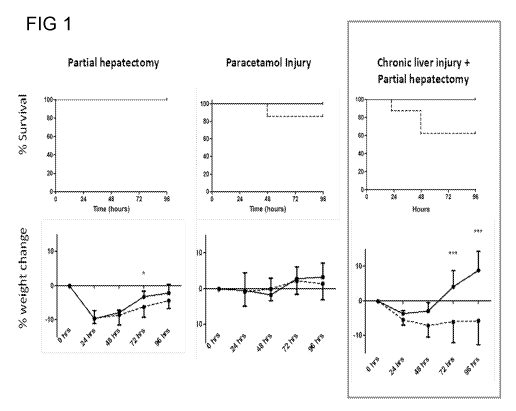
Figure 1 shows a Kaplein Meir of survival in three injury models and chart
shows
percentage weight change following intervention and treatment. ***p<0.001 Mann
Whitney
U. [Solid line = treatment with fc-CSF1; broken line = PBS control]
Figure 2 is a bar chart showing Liver weight / Body weight ratio expressed as
a percentage
and hepatocyte proliferation expressed as Ki67 positive hepatocytes per high-
powered
field. *p<0.05; **p<0.01; ***p<0.001 Mann Whitney U. [Treatment = fc-CSF1;
Control =
PBS]
Figure 3 shows the effect of CSF-1 administered as described above on body
weight.
Figure 3A compares CSF-1 (1mg/kg) with Fc-CSF-1 (1mg/kg). The unmodified
protein
has no effect at this dose, where Fc-CSF-1 clearly increased total body
weight. Figure 3B
shows a dose response curve, demonstrating detectable activity at 0.1mg/kg of
Fc-CSF-1.
FIG 3C shows the effect of 1mg/kg dose is confirmed in a larger experimental
series. The
animals in this series are analysed further in subsequent slides
Figure 4A shows the effect of CSF-1 (1mg/kg) and Fc-CSF-1 (1mg/kg) on mouse
spleen
weight, Figure 4B shows the effect of CSF-1 (1mg/kg) and Fc-CSF-1 (1mg/kg) on
mouse
liver weight.
Figure 5 shows the effect of Fc-CSF-1 on the numbers of macrophages in the
spleen,
detected with the csf1r-EGFP reporter, Figure 5A is the control and Figure 5B
shows the
treated sample.
CA 02901368 2015-08-14
WO 2014/132072 PCT/GB2014/050595
6
Figure 6A shows a dose response curve for Figures 5A and 5B, Figure 6A shows a
dose
response curve based upon immunohistochemical localisation of the macrophage-
specific
F4/80 antigen.
Figure 7 shows immunostaining for the macrophage-specific F4/80 antigen in
mice. Figure
7A shows PBS treated control liver; Figure 7B shows the liver of a mouse
treated with Fc-
CSF-1, Figure 70 shows PBS treated control spleen Figure 7D shows the spleen
of a
mouse treated with Fc-CSF-1.
Figure 8A shows a PBS treated control mouse liver with immunostaining for
proliferating
cell nuclear antigen (PCNA), Figure 8B shows a mouse liver following treatment
with Fc-
CSF-1.
Figure 9 shows the impact of pharmacokinetics of CSF-1 administered to weaner
pigs.
Figure 9A shows the clearance of unmodified CSF-1, Figures 9B and 90 show the
clearance of 1.2mg/kg of Fc-CSF-1 when administered intravenously and
subcutaneously
respectively.
Figure 10 shows the blood effects in weaner pigs administered 0.5 mg/Kg x6;
Figure 10A
shows the total white blood count, Figure 10B shows the monocyte count, Figure
100
shows the lymphocyte count and Figure 10D shows the neutophil count.
Figure lithe dose response curves of blood effects in weaner administered 0.5
mg/Kg x6;
Figure 11A shows the total white blood count, Figure 11B shows the monocyte
count,
Figure 110 shows the lymphocyte count and Figure 11D shows the neutrophil
count.
Figure 12 shows the effect on organ weights in weaner pigs administered 0.12
mg/Kg x3.
Figure 12A shows the effect on liver weight, Figure 12B shows the effect on
spleen weight,
Figure 110 shows the effect on lung weight and Figure 11D shows the effect on
kidney
weight.
Figure 13A shows serum CSF1 level in patients at admission in patients who
survived or
died/underwent liver transplantation with paracetamol induced liver failure.
Figure 13B
shows serum levels of a subset of patients who subsequently died or survived.
Figure 13
C shows receiver operating characteristic curve analysis assessing the
potential of
admission CSF1 to serve as a biomarker for survival without transplantation
following
paracetamol overdose.
CA 02901368 2015-08-14
WO 2014/132072 PCT/GB2014/050595
7
Figure 14A shows hepatic CSF1 gene expression following paracetamol
intoxication and
serum CSF1 level. Figure 14 B shows liver to bodyweight ratio and hepatocyte
proliferation assessed by Ki67 immunohistochemistry at Day 3 following
paracetamol
intoxication. Figure 140 shows serum analysis at Day 3 post paracetamol
intoxication
comparing control and CSF1 receptor inhibition.
Figure 15A shows mean liver weight to body weight ratio and hepatocyte
proliferation (ki67
immunohistochemistry) in mice following paracetamol intoxication comparing
CSF1-Fc
(solid line) or control (dotted line) administration. Figure 15B shows serum
parameters
post paracetamol intoxication. (n=8/group).
Figure 16A shows hepatic CSF1 gene expression following 2/3 partial
hepatectomy and
serum CSF1 level. Figure 16B shows liver to bodyweight ratio and hepatocyte
proliferation
assessed by Ki67 immunohistochemistry at Day 2 following 2/3 partial
hepatectomy with
CSF1 receptor inhibition (GW2580) or control. Figure 160 shows serum analysis
at Day 2
post paracetamol intoxication comparing control and CSF1 receptor inhibition
with
GW2580. (n=8 per group).
Figure 17A shows mean liver weight to body weight ratio and hepatocyte
proliferation (ki67
immunohistochemistry) in mice following 2/3 partial hepatectomy comparing CSF1-
Fc
(solid line) or control (dotted line) administration. Figure 17B shows serum
parameters
post paracetamol intoxication. (n=8/group) .Figure 170 shows relative gene
expression of
the proregenerative cytokines 116 and oncostatin M (OSM) and also a growth
factor
activator urokinase receptor (UR) with blockade of CSF1 receptor (GW2580) and
administration of CSF1-Fc versus controls.
Figure 18A shows Kaplan Meir plot showing trend to survival (p=0.07) and
increased body
weight postoperatively with CSF1-Fc treatment following partial hepatectomy in
the
chronically injured liver (solid line= CSF1-Fc, dotted line= control;
n=8/group at Day 4 and
Day 7). Figure 18B shows mean liver weight to body weight ratio, hepatocyte
proliferation
(ki67 immunohistochemistry) and fibrosis quantification via Sirius red
quantification. Figure
180 shows serum parameters.
Figure 19 shows A) gene expression relative to mean of control group (MARCO:
macrophage receptor with collagenous structure; MSR1: macrophage scavenger
receptor
1) (white = control; shaded = CSF1-Fc); B) Bead clearance assay showing flow
plot
CA 02901368 2015-08-14
WO 2014/132072 PCT/GB2014/050595
8
overlay gated on fluorescent beads from 1-15 minutes following intravascular
injection
(dotted line = control; solid line = CSF1-Fc; grey line = uninjured untreated
mouse); C) ex
vivo fluorescence organs 15 minutes following intravascular injection of
fluorescent beads.
(n=6 per group)
Figure 20 shows A) gene expression relative to mean of control group (MARCO:
macrophage receptor with collagenous structure; MSR1: macrophage scavenger
receptor
1) [white = control; shaded = CSF1-Fc]; B) bead clearance assay; C) ex vivo
fluorescence
organs 15 minutes following intravascular injection of fluorescent beads.
Figure 21 shows A) Serum CSF1 level of 55 patients undergoing partial
hepatectomy
taken preoperatively and on postoperative day 1 and postoperative day 3. B)
Cohort
segregated according to extent of liver resection. Two way ANOVA with post hoc
analysis
showing significant increase in CSF1 level in patients who had more than 5
segments
resected compared to patients who had less than 3 segments resected. C)
Patients who
developed postoperative liver failure shown in dots compared to rest of the
cohort (median
and range).
DETAILED DESCRIPTION
The terms "M-CSF", "macrophage colony stimulating factor", "CSF-1", "CSF1",
"colony
stimulating factor1" and "colony stimulating factor-1" are used
interchangeably herein.
By the term "supplementation", "supplement" or "supplementing", it is intended
that CSF-1
is administered to an individual in an additional or extra amount in excess of
the level that
the individual already has of functioning CSF-1.
By the terms "treat," "treating" or "treatment of," it is intended that the
severity of the
disorder or the symptoms of the disorder are reduced, or the disorder is
partially or entirely
eliminated, as compared to that which would occur in the absence of treatment.
Treatment
does not require the achievement of a complete cure of the disorder.
By the terms "restore," "restoring" or "restoration of," it is intended that
the severity of the
disorder or the symptoms of the disorder are reduced, or the disorder is
partially or entirely
eliminated, as compared to that which would occur in the absence of treatment.
Treatment
does not require the achievement of a complete cure of the disorder.
CA 02901368 2015-08-14
WO 2014/132072 PCT/GB2014/050595
9
A "therapeutically effective" or "effective" amount is intended to designate a
dose that
causes a relief of symptoms of a disease or disorder as noted through clinical
testing and
evaluation, patient observation, and/or the like. "Effective amount" or
"effective" can
further designate a dose that causes a detectable change in biological or
chemical activity.
The detectable changes may be detected and/or further quantified by one
skilled in the art
for the relevant mechanism or process. Moreover, "effective amount" or
"effective" can
designate an amount that maintains a desired physiological state, i.e.,
reduces or prevents
significant decline and/or promotes improvement in the condition of interest.
As is
generally understood in the art, the dosage will vary depending on the
administration
routes, symptoms and body weight of the patient but also depending upon the
compound
being administered.
Conditions which can be treated in the present invention include liver damage
or hepatitis
as the result of physical trauma, adverse action of pharmaceuticals or toxic
chemicals,
infection, autoimmunity, ischaemia, alcohol induced liver damage, or any other
cause of
liver damage. Liver injury is commonly caused by physical trauma such as road
traffic
accidents, falls, assault or the like. Paracetamol (acetaminophen) overdose is
a relatively
common cause of pharmaceutical-induced liver damage, but liver damage can also
be
caused by many other pharmaceuticals, e.g. methotrexate, statins, niacin,
amiodarone,
chemotherapy agents, and some antibiotics. Alcohol-induced liver disease is a
very
widespread cause of liver damage. Infections that cause liver damage include,
amongst
others, hepatitis A, B or C viral infections. While it is probable that CSF1
treatment will not
be appropriate in all cases of liver damage, in many cases it may have a
beneficial effect.
By the term "Fe" it is intended to refer to a region of an antibody molecule
that binds to
antibody receptors on the surface of cells such as macrophages and mast cells,
and to
complement protein. Fc (50,000 daltons) fragments contain the CH2 and CH3
region and
part of the hinge region held together by one or more disulfides and non-
covalent
interactions. Fc and Fe5p fragments are produced from fragmentation of IgG and
IgM,
respectively. The term Fc is derived from the ability of these antibody
fragments to
crystallize. Fc fragments are generated entirely from the heavy chain constant
region of an
immunoglobulin. The Fc fragment cannot bind antigen, but it is responsible for
the effector
functions of antibodies, such as complement fixation.
"Polypeptide" refers to a polymer of amino acids (dipeptide or greater) linked
through
peptide bonds. Thus, the term "polypeptide" includes proteins, oligopeptides,
protein
fragments, protein analogs and the like. The term "polypeptide" contemplates
CA 02901368 2015-08-14
WO 2014/132072 PCT/GB2014/050595
polypeptides as defined above that are encoded by nucleic acids, are
recombinantly
produced, are isolated from an appropriate source, or are synthesized.
As used herein, a "functional" polypeptide is one that retains at least one
biological activity
5 normally associated with that polypeptide. Preferably, a "functional"
polypeptide retains all
of the activities possessed by the unmodified peptide. By "retains" biological
activity, it is
meant that the polypeptide retains at least about 50%, 60%, 75%, 85%, 90%,
95%, 97%,
98%, 99%, or more, of the biological activity of the native polypeptide (and
can even have
a higher level of activity than the native polypeptide). A "non-functional"
polypeptide is one
10 that exhibits essentially no detectable biological activity normally
associated with the
polypeptide (e.g., at most, only an insignificant amount, e.g., less than
about 10% or even
5%).
"Fusion protein" as used herein, refers to a protein produced when two
heterologous
nucleotide sequences or fragments thereof coding for two (or more) different
polypeptides,
or fragments thereof, are fused together in the correct translational reading
frame. The
two or more different polypeptides, or fragments thereof, include those not
found fused
together in nature and/or include naturally occurring mutants.
As used herein, a "fragment" is one that substantially retains at least one
biological activity
normally associated with that protein or polypeptide. In particular
embodiments, the
"fragment" substantially retains all of the activities possessed by the
unmodified protein.
By "substantially retains" biological activity, it is meant that the protein
retains at least
about 50%, 60%, 75%, 85%, 90%, 95%, 97%, 98%, 99%, or more, of the biological
activity
of the native protein (and can even have a higher level of activity than the
native protein).
A "recombinant polypeptide" is one that is produced from a recombinant nucleic
acid.
An "isolated" polypeptide means a polypeptide that is separated or
substantially free from
at least some of the other components of the naturally occurring organism or
virus, for
example, the cell or viral structural components or other polypeptides or
nucleic acids
commonly found associated with the polypeptide. As used herein, the "isolated"
polypeptide is at least about 25%, 50%, 60%, 70%, 75%, 80%, 85%, 90%, 95%,
97%,
98%, 99% or more pure (w/w).
The term "derivative" is to be understood to refer to any molecule that is
derived
(substantially derived) or obtained (substantially obtained) from CSF-1, but
retains
CA 02901368 2015-08-14
WO 2014/132072 PCT/GB2014/050595
11
similarity, or substantial similarity, in biological function of CSF-1. In
certain aspects, the
biological function is the ability to promote liver organ development. A
derivative may, for
instance, be provided as a result of cleavage of CSF-1 to produce biologically-
active
fragments, cyclisation, bioconjugation and/or coupling with one or more
additional moieties
that improve, for example, solubility, stability or biological half-life, or
which act as a label
for subsequent detection or the like. A derivative may also result from post-
translational or
post-synthesis modification such as the attachment of carbohydrate moieties,
or chemical
reactions(s) resulting in structural modification(s) such as alkylation or
acetylation of an
amino acid(s) or other changes involving the formation of chemical bonds. In a
particularly
preferred embodiment of a derivative suitable for use in the present
invention, the
derivative is the mature domain of CSF-1. In another preferred embodiment of a
derivative
suitable for use in the methods disclosed herein, the derivative is a
biologically active, C-
terminal fragment of CSF-1 (e.g. a CSF-1 fragment comprising the C-terminal
amino acids
1 to 150 of the 536 amino acid protein). Further embodiments of a derivative
of CSF-1
include CSF-1 comprising chemically modified side chains (e.g. pegylation of
lysyl e-amino
groups), C- and/or N-termini (e.g. acylation of the N-terminal with acetic
anhydride), or
linked to various carriers (e.g. human serum albumin or histidine (His6) tag).
As generally used herein, a "homolog" shares a definable nucleotide or amino
acid
sequence relationship with another nucleic acid or polypeptide as the case may
be. A
"protein homolog" preferably shares at least 70% or 80% sequence identity,
more
preferably at least 85%, 90% and even more preferably at least 95%, 96%, 97%,
98% or
99% sequence identity with the amino acid sequences of polypeptides as
described
herein. Homologs of CSF may also be used in accordance with the invention.
Such CSF
homologs would preferably be characterized by biological activity about the
same or
greater than that of a CSF protein having a high or substantial biological
activity.
As used herein, "variant" proteins are proteins in which one or more amino
acids have
been replaced by different amino acids. Protein variants of CSF that retain
biological
activity of native or wild type CSF may be used in accordance with the
invention. It is well
understood in the art that some amino acids may be changed to others with
broadly similar
properties without changing the nature of the activity of the polypeptide
(conservative
substitutions). Generally, the substitutions which are likely to produce the
greatest changes
in a polypeptide's properties are those in which (a) a hydrophilic residue
(e.g., Ser or Thr)
is substituted for, or by, a hydrophobic residue (e.g. Leu, lie, Phe or Val);
(b) a cysteine or
proline is substituted for, or by, any other residue; (c) a residue having an
electropositive
side chain (e.g., Arg, His or Lys) is substituted for, or by, an
electronegative residue (e.g.,
CA 02901368 2015-08-14
WO 2014/132072 PCT/GB2014/050595
12
Glu or Asp) or (d) a residue having a bulky side chain (e.g., Phe or Trp) is
substituted for,
or by, one having a smaller side chain (e.g., Ala, Ser) or no side chain
(e.g., Gly).
Embodiments of the present invention further provide an isolated nucleic acid
(e.g., an
"isolated DNA" or an "isolated vector genome") that encodes the fusion protein
described
herein. The nucleic acid is separated or substantially free from at least some
of the other
components of the naturally occurring organism or virus, such as for example,
the cell or
viral structural components or other polypeptides or nucleic acids commonly
found
associated with the nucleic acid. The coding sequence for a polypeptide
constituting the
active agents of the present invention is transcribed, and optionally,
translated. According
to embodiments of the present invention, transcription and translation of the
coding
sequence will result in production of a fusion protein described.
It will be appreciated by those skilled in the art that there can be
variability in the nucleic
acids that encode the fusion polypeptides of the present invention due to the
degeneracy
of the genetic code. Further variation in the nucleic acid sequence can be
introduced by
the presence (or absence) of non-translated sequences, such as intronic
sequences and 5'
and 3' untranslated sequences. Moreover, the isolated nucleic acids of the
invention
encompass those nucleic acids encoding fusion proteins that have at least
about 60%,
70%, 80%, 90%, 95%, 97%, 98% or higher amino acid sequence similarity with the
polypeptide sequences specifically disclosed herein or to those known
sequences
corresponding to proteins included in aspects of the present invention (or
fragments
thereof) and further encode functional fusion proteins as defined herein
Isolated nucleic acids of this invention include RNA, DNA (including cDNAs)
and chimeras
thereof. The isolated nucleic acids can further comprise modified nucleotides
or nucleotide
analogs.
The isolated nucleic acids encoding the polypeptides of the invention can be
associated
with appropriate expression control sequences, e.g., transcription/translation
control
signals and polyadenylation signals.
It will be appreciated that a variety of promoter/enhancer elements can be
used depending
on the level and tissue-specific expression desired. The promoter can be
constitutive or
inducible (e.g., the metalothionein promoter or a hormone inducible promoter),
depending
on the pattern of expression desired. The promoter can be native or foreign
and can be a
natural or a synthetic sequence. By foreign, it is intended that the
transcriptional initiation
CA 02901368 2015-08-14
WO 2014/132072 PCT/GB2014/050595
13
region is not found in the wild-type host into which the transcriptional
initiation region is
introduced. The promoter is chosen so that it will function in the target
cell(s) of interest.
The present invention further provides methods of making fusion proteins
described
herein. Methods of making fusion proteins are well understood in the art. Such
methods
include growing a host cell including a vector that includes nucleic acids
encoding the
fusion protein under conditions appropriate for expression and subsequent
isolation of the
fusion protein. Accordingly, the isolated nucleic acids encoding a polypeptide
constituting
the fusion protein of the invention can be incorporated into a vector, e.g.,
for the purposes
of cloning or other laboratory manipulations, recombinant protein production,
or gene
delivery. Exemplary vectors include bacterial artificial chromosomes, cosmids,
yeast
artificial chromosomes, phage, plasmids, lipid vectors and viral vectors
(described in more
detail below).
In particular embodiments, the isolated nucleic acid is incorporated into an
expression
vector. In further embodiments of the present invention, the vector including
the isolated
nucleic acids described herein are included in a host cell. Expression vectors
compatible
with various host cells are well known in the art and contain suitable
elements for
transcription and translation of nucleic acids. Typically, an expression
vector contains an
"expression cassette," which includes, in the 5' to 3' direction, a promoter,
a coding
sequence encoding a polypeptide of the invention or active fragment thereof
operatively
associated with the promoter, and, optionally, a termination sequence
including a stop
signal for RNA polymerase and a polyadenylation signal for polyadenylase.
In addition to the regulatory control sequences discussed above, the
recombinant
expression vector can contain additional nucleotide sequences. For example,
the
recombinant expression vector can encode a selectable marker gene to identify
host cells
that have incorporated the vector and/or may comprise another heterologous
sequence of
interest.
Vectors can be introduced into prokaryotic or eukaryotic cells via
conventional
transformation or transfection techniques. As used herein, the terms
"transformation" and
"transfection" refer to a variety of art-recognized techniques for introducing
foreign nucleic
acids (e.g., DNA) into a host cell, including calcium phosphate or calcium
chloride co-
precipitation, DEAE-dextran-mediated transfection, lipofection,
electroporation,
microinjection, DNA-loaded liposomes, lipofectamine-DNA complexes, cell
sonication,
gene bombardment using high velocity microprojectiles, and viral-mediated
transfection.
CA 02901368 2015-08-14
WO 2014/132072 PCT/GB2014/050595
14
In terms of administration, the most suitable route in any given case will
depend on the
nature and severity of the liver condition being treated and on the fusion
protein, viral
vector, nucleic acid or pharmaceutical formulation being administered.
The fusion proteins, viral vectors and nucleic acids (e.g., DNA and/or RNA) of
the invention
can be formulated for administration in a pharmaceutical carrier in accordance
with known
techniques. See, e.g., Remington, The Science And Practice of Pharmacy (9th
Ed. 1995).
In the manufacture of a pharmaceutical formulation according to the invention,
the fusion
protein, viral vector or nucleic acid is typically admixed with, inter alia,
an acceptable
carrier. The carrier can be a solid or a liquid, or both, and is optionally
formulated as a
unit-dose formulation, which can be prepared by any of the well-known
techniques of
pharmacy.
The carriers and additives used for such pharmaceutical compositions can take
a variety of
forms depending on the anticipated mode of administration. Thus, compositions
for oral
administration may be, for example, solid preparations such as tablets, sugar-
coated
tablets, hard capsules, soft capsules, granules, powders and the like, with
suitable carriers
and additives being starches, sugars, binders, diluents, granulating agents,
lubricants,
disintegrating agents and the like. Because of their ease of use and higher
patient
compliance, tablets and capsules represent the most advantageous oral dosage
forms for
many medical conditions.
Similarly, compositions for liquid preparations include solutions, emulsions,
dispersions,
suspensions, syrups, elixirs, and the like with suitable carriers and
additives being water,
alcohols, oils, glycols, preservatives, flavoring agents, coloring agents,
suspending agents,
and the like.
In the case of a solution, it can be lyophilized to a powder and then
reconstituted
immediately prior to use. For dispersions and suspensions, appropriate
carriers and
additives include aqueous gums, celluloses, silicates or oils.
For injection, the carrier is typically a liquid, such as sterile pyrogen-free
water, pyrogen-
free phosphate-buffered saline solution, bacteriostatic water, or Cremophor
EL[R] (BASF,
Parsippany, N.J.), parenterally acceptable oil including polyethylene glycol,
polyvinyl
pyrrolidone, lecithin, arachis oil or sesame oil, with other additives for
aiding solubility or
CA 02901368 2015-08-14
WO 2014/132072 PCT/GB2014/050595
preservation may also be included. For other methods of administration, the
carrier can be
either solid or liquid.
For oral administration, the fusion protein, viral vector or nucleic acid can
be administered
5 in solid dosage forms, such as capsules, tablets, and powders, or in
liquid dosage forms,
such as elixirs, syrups, and suspensions. The fusion protein, viral vector or
nucleic acid
can be encapsulated in gelatin capsules together with inactive ingredients and
powdered
carriers, such as glucose, lactose, sucrose, mannitol, starch, cellulose or
cellulose
derivatives, magnesium stearate, stearic acid, sodium saccharin, talcum,
magnesium
10 carbonate and the like. Examples of additional inactive ingredients that
can be added to
provide desirable color, taste, stability, buffering capacity, dispersion or
other known
desirable features are red iron oxide, silica gel, sodium lauryl sulfate,
titanium dioxide,
edible white ink and the like. Similar diluents can be used to make compressed
tablets.
Both tablets and capsules can be manufactured as sustained release products to
provide
15 for continuous release of medication over a period of hours. Compressed
tablets can be
sugar coated or film coated to mask any unpleasant taste and protect the
tablet from the
atmosphere, or enteric- coated for selective disintegration in the
gastrointestinal tract.
Liquid dosage forms for oral administration can contain coloring and flavoring
to increase
patient acceptance.
Formulations of the present invention suitable for parenteral administration
can include
sterile aqueous and non-aqueous injection solutions of the fusion protein,
viral vector or
nucleic acid, which preparations are generally isotonic with the blood of the
intended
recipient. These preparations can contain anti-oxidants, buffers,
bacteriostats and solutes,
which render the formulation isotonic with the blood of the intended
recipient. Aqueous
and non-aqueous sterile suspensions can include suspending agents and
thickening
agents. The formulations can be presented in unindose or multi-dose
containers, for
example sealed ampoules and vials, and can be stored in a freeze-dried
(lyophilized)
condition requiring only the addition of the sterile liquid carrier, for
example, saline or
water-for-injection immediately prior to use.
Extemporaneous injection solutions and suspensions can be prepared from
sterile
powders, granules and tablets. For example, in one aspect of the present
invention, there
is provided an injectable, stable, sterile composition including a fusion
protein, viral vector
or nucleic acid of the invention, in a unit dosage form in a sealed container.
Optionally, the
composition is provided in the form of a lyophilizate, which is capable of
being
CA 02901368 2015-08-14
WO 2014/132072 PCT/GB2014/050595
16
reconstituted with a suitable pharmaceutically acceptable carrier to form a
liquid
composition suitable for injection thereof into a subject.
In particular embodiments of the invention, administration is by subcutaneous
or
intradermal administration. Subcutaneous and intradermal administration can be
by any
method known in the art including, but not limited to, injection, gene gun,
powderject
device, bioject device, microenhancer array, microneedles, and scarification
(i.e., abrading
the surface and then applying a solution including the fusion protein, viral
vector or nucleic
acid).
In other embodiments, the fusion protein, viral vector or nucleic acid is
administered
intramuscularly, for example, by intramuscular injection or by local
administration.
Nucleic acids (e.g., DNA and/or RNA) can also be delivered in association with
liposomes,
such as lecithin liposomes or other liposomes known in the art (for example,
as described
in WO 93/24640) and may further be associated with an adjuvant. Liposomes
including
cationic lipids interact spontaneously and rapidly with polyanions, such as
DNA and RNA,
resulting in liposome/nucleic acid complexes that capture up to 100% of the
polynucleotide. In addition, the polycationic complexes fuse with cell
membranes,
resulting in an intracellular delivery of polynucleotide that bypasses the
degradative
enzymes of the lysosomal compartment. PCT publication WO 94/27435 describes
compositions for genetic immunization including cationic lipids and
polynucleotides.
Agents that assist in the cellular uptake of nucleic acid, such as calcium
ions, viral proteins
and other transfection facilitating agents, may be included.
According to the present invention, methods of this invention include
administering an
effective amount of a composition of the present invention as described above
to the
subject. The effective amount of the composition, the use of which is in the
scope of
present invention, will vary somewhat from subject to subject, and will depend
upon factors
such as the age and condition of the subject and the route of delivery. Such
dosages can
be determined in accordance with routine pharmacological procedures known to
those
skilled in the art. For example, the active agents of the present invention
can be
administered to the subject in an amount ranging from a lower limit from about
0.01, 0.05,
0.10, 0.50, 1.0, 5.0, or 10% to an upper limit ranging from about 10, 20, 30,
40, 50, 60, 70,
80, 90, 95, 96, 97, 98, 99, or 100% by weight of the composition. In some
embodiments,
the active agents include from about 0.05 to about 95% by weight of the
composition. In
other embodiments, the active agents include from about 0.05 to about 60% by
weight of
CA 02901368 2015-08-14
WO 2014/132072 PCT/GB2014/050595
17
the composition. In still other embodiments, the active agents include from
about 0.05 to
about 10% by weight of the composition.
Cloning and expression of the pig Fc-fusion
The sequence corresponding to the active fragment of porcine CSF-1
(SENCSHMIGDGHLKVLQQLI DSQMETSCQIAFEFVDQEQLTDPVCYLKKAFLQVQDI LDE
TMRFRDNTPNANVIVQLQELSLRLNSCFTKDYEEQDKACVRTFYETPLQLLEKI KNVFN ET
KNLLKKDWNIFSKNCNNSFAKCSSQHERQPEGR) (SEQ ID NO:1) was linked to the
hinge-CH3 region of the porcine IgG1a sequence (GTKTKPPCPICPGCEVA
GPSVFI FPPKPKDTLM I SQTPEVTCVVVDVSKEHAEVQFSVVYVDGVEVHTAETRPKEEQF
NSTYRVVSVLPIQHQDWLKGKEFKCKVN NVDLPAPITRTISKAIGQSREPQVYTLPPPAEE
LSRSKVTVTCLVIGFYPPDI HVEWKSNGQPEPEGNYRTTPPQQDVDGTFFLYSKLAVDKA
RWDHGETFECAVMHEALHNHYTQKSISKTQGK) (SEQ ID NO:2). This entire region was
codon optimized for mammalian expression by GeneArt (Invitrogen, CA, USA) and
cloned
into the expression plasmid pS00524 using Hindi!! and Notl restriction sites
engineered
into the 5' and 3' ends respectively. The resulting plasmid was sequenced to
ensure ORF
integrity and protein was expressed from transfected HEK293F or CHO cells.
Isolation of pig CSF-1:Fc fusion
Porcine CSF-1 Fc fusion protein was isolated using Protein A affinity
chromatography.
Briefly, conditioned medium from cell culture was clarified and loaded onto
Protein A
Sepharose that was equilibrated with PBS. Following loading the column was
washed with
2 BV of PBS and 2 BV of 35 mM Na Acetate pH 5.5. Protein was eluted using a
step
gradient of 80% B Buffer (35 mM Acetic acid, no pH adjustment), 85% B buffer
and 100%
B buffer. The 80 and 85% B fractions were pooled based on lack of aggregated
protein
(analytical SEC) and the 100% B fraction was not included. Pooled protein was
pH
adjusted to 7.2 and dialyzed against PBS.
Porcine CSF-1 Fc-fusion quantitation in blood plasma by ELISA
Porcine CSF-1 Fc-fusion plasma levels were detected using an in-house
developed
conventional sandwich ELISA utilizing commercially available antibodies.
Capture
antibody was Abcam ab9693 (0.3 pg/mL) and detection antibody was Rabbit anti-
pig IgG
(Fe) biotinylated Alpha Diagnostic 90440 (1:5000 dilution).
Standard protein was
generated and purified in-house (lot 2/24/11 JAS). Standards were added to
each plate
along with the samples resulting in an 11 point standard range of 2700 ng/mL
to 0.046
pg/mL. This allowed for quantitation of each sample to a standard curve on
every assay
CA 02901368 2015-08-14
WO 2014/132072 PCT/GB2014/050595
18
plate. Assay detection was done using Pierce High Sensitivity Streptavidin-HRP
(1:10,000
dilution) and TMB Microwell Peroxidase Substrate System solution (KPL).
Pig PK
Weaner age barrows (<14kg) were assigned to three treatment groups receiving a
single
intravenous (IV) or subcutaneous (SC) dose as follows. Three pigs received 0.5
mg/kg
CSF-1 non-fusion dosed SC. Two pigs received 1.2mg/kg CSF-1:Fc fusion dosed
IV.
Two pigs received 1.2mg/kg CSF-1:Fc dosed SC. One ml plasma samples were
obtained
via the V. jugularis in EDTA anticoagulant tubes and placed on ice until
centrifuged. The
plasma was transferred to sterile tubes stored at -10 C until analysis. Serial
plasma
samples were obtained from each animal at pre-dose and 5 minutes, 30 minutes,
1 hour, 2
hours, 4 hours, 6 hours, 8, hours, 24 hours, 48 hours, and 72 hours post-dose.
CSF-1 and
CSF-1:Fc fusion protein levels were quantitated in plasma using ELISA assays.
MTT cell viability assay
Stable Ba/F3 cells expressing porcine CSF-1R were maintained in culture with
complete
RPM! supplemented with either 104 Units/ml rh-CSF-1 or 10% IL-3 conditioned
medium
prior to MTT assay. 2x10⁴ cells/well (Ba/F3 cells and Ba/F3 transfectants), or
5x10⁴
cells/well (pig BMM) of a 96 well plate were plated in triplicate or
quadruplicate and
appropriate treatment (serial dilutions of rh-CSF-1 or porcine Fc CSF-1 were
added to
make a total volume of 100p1 per well. Cells were incubated for 48 hours at 37
C with 5%
CO2. For Ba/F3 cells, 10p1 of MTT (Sigma Aldrich M5655) stock solution
(5mg/m1) was
added directly to each well to achieve a final concentration of 0.5mg/m1 and
incubated at
37 C for 3 hours prior to solubilisation overnight. For adherent mouse BMM
cells, culture
medium was replaced with 50p1 of 1mg/m1 MTT solution and incubated for 1 hour
at 37 C.
MTT solution was removed and tetrazolium salt solubilised with 100p1 of
solubilisation
agent (0.1M HCL, 10% Triton x -100 and isopropanol) followed by incubation at
37 C with
5% CO2 for 10 minutes. Plates were read at 570nm with reference wavelength of
405nm.
Mice Studies
Forty-eight male C57BI6 mice aged 10-12 weeks underwent either 2/3 partial
hepatectomy, paracetamol intoxication or chronic liver injury plus 2/3 partial
hepatectomy.
2/3 partial hepatectomy was performed by ligating the left lobe and left and
right median
lobes. Paracetamol intoxication was performed by intraperitoneal
administration of
350mg/kg paracetamol dissolved in phosphate buffered saline (PBS). Chronic
liver injury
plus 2/3 partial hepatectomy was performed by eight weeks intraperitoneal
carbon
CA 02901368 2015-08-14
WO 2014/132072 PCT/GB2014/050595
19
tetrachloride administration 1mcl/g twice weekly dissolved in olive oil
followed by 2/3 partial
hepatectomy as described above. The treatment group (n=8 per injury model)
received
0.75mcg/g FC-CSF1 administered subcutaneously immediately following either
partial
hepatectomy or paracetamol intoxication and subsequently every 24hours for
three further
doses. Control mice (n=8 per injury model) received subcutaneous PBS of
appropriate
volume. All mice were culled on day 4 following injury (partial hepatectomy or
paracetamol
intoxication).
Following a midline laparotomy the liver was excised and weighed. Liver weight
to body
weight ratio was calculated and expressed as a percentage. Livers were fixed
in 4%
formalin overnight then transferred to 70% ethanol. Livers were then embedded
in paraffin
blocks and 4pm sections cut. lmmunohistochemistry for Ki67 (a marker of
cellular
proliferation expressed throughout the cell cycle) was performed following
heat mediated
antigen retrieval in Tris/EDTA solution at pH9. Hepatocyte proliferation was
quantified by
counting ki67 positive hepatocytes in 20 high powered fields (400x) per animal
and the
mean calculated.
EXAMPLE 1
The effects of Fc-CSF1 on liver regeneration in murine models of acute liver
injury (partial
hepatectomy; paracetamol intoxication) and acute-on-chronic liver injury
(chronic liver
injury plus partial hepatectomy) were studieD. The treatment group (n=8 per
injury model)
received 0.5mcg/g Fc-CSF1 administered subcutaneously immediately following
either
partial hepatectomy or paracetamol intoxication and subsequently every 24hours
for three
further doses. Control mice (n=8 per injury model) received subcutaneous PBS
of
appropriate volume. Interventions and treatments were well tolerated. In the
most severe
injury model (chronic liver injury with 2/3 partial hepatectomy) there was a
significant
increase in mouse weight (p<0.001 at day 4) and a trend to improved survival
(p=0.08)
(Figure 1). Others findings included enhanced regenerative parameters of both
liver weight
and hepatocyte proliferation across the injury models (Figure 2). The results
of these
studies demonstrate that administration of Fc-CSF1 can enhance the
regenerative
response across a range of hepatic injury models. In the most severe model of
hepatic
injury (chronic liver injury plus partial hepatectomy) there was a trend to
improved survival
and a significant body weight increase indicating improved postoperative
course. Fc-CSF1
was found to have a growth promoting effect on hepatic weights and hepatocyte
proliferation in all injury models. While there is redundancy in many of the
pathways
leading to effective liver regeneration it appears that CSF1 is critical to
achieve optimal
recovery and the present studies have shown that supplementation of this
factor can
CA 02901368 2015-08-14
WO 2014/132072 PCT/GB2014/050595
further boost regeneration. It is envisaged these findings will translate to
improved
outcomes in the management of liver failure in the clinical setting.
EXAMPLE 2
5 Mice were injected with Fc-CSF-1 subcutaneously on each of 4 days and
sacrificed on the
5th day. The mice were csf1r-EGFP (MacGreen) mice on the C57BI/6
background.
Tissue processing and immunohistochemistry were carried out as described in
(Alikhan et
al Am J.Pathol. 179, 1243-1256, 2011 and Macdonald et al Blood. 116, 3955-
3963, 2010).
A comparison of the effect of recombinant pig CSF-1 or Fc-CSF-1 on the
proliferation of
10 mouse bone marrow cells or the Ba/F3 CS1R reporter cell line using the
assay described
in Gow et al Cytokine. 60, 793-805, 2012) showed that there was no difference
in
biological activity (data not shown), demonstrating that additional of the Fc
component to
the C terminus of CSF-1 does not interfere with binding to the receptor.
15 EXAMPLE 3
The effect of administered CFS-1 on body weight was assessed. Figure 3A
compares
CSF-1 (1mg/kg) with Fc-CSF-1 (1mg/kg). The unmodified protein has no effect at
this
dose, where Fc-CSF-1 clearly increase total body weight. Figure 3B shows a
dose
response curve, demonstrating detectable activity at 0.1mg/kg of Fc-CSF-1.
Figure 30
20 shows the effect of 1mg/kg dose is confirmed in a larger experimental
series. The animals
in this series are analysed further in subsequent studies. Organ weight
studies showed
that Fc-CSF-1 treatment at 1mg/kg almost doubled the weight of the spleen and
significantly increased total liver weight; whereas no effect of Fc-CSF-1 on
the weights or
the lung or kidney, either the whole group or segregated for male or female
was observed.
The effect of Fc-CSF-1 administered to mice on blood was assessed. Results
showed that
Fc-CSF-1 elevates the white blood cell count and the total blood monocyte
count. It was
noted that there is some variation between the male and female mice, the
former having
higher average counts than the latter, but the effect is seen in both sexes.
It was also
observed that that Fc-CSF-1 increases the segmented neutrophil counts. Again,
the
males can be distinguished from the females. Conversely, Fc-CSF-1 had no
effect on total
lymphocytes
The effect of Fc-CSF-1 administered to mice on the numbers of macrophages in
the
spleen, detected with the csf1r-EGFP reporter, was also assessed. Note the
greatly
CA 02901368 2015-08-14
WO 2014/132072 PCT/GB2014/050595
21
increased fluorescence in the treated Figure 5B. This is quantitated in Figure
6A, in a
dose response curve. In Figure 6B, the same result is demonstrated based upon
immunohistochemical localisation of the macrophage-specific F4/80 antigen.
The effect of Fc-CSF-1 administered to mice on bone resorbing osteoclasts in
bone was
assessed. Results showed an increase in the treatment group of osteoclasts in
the growth
plate, zone of resorption and shaft (data not shown)
EXAMPLE 4
Figure 7B shows immunostaining for the macrophage-specific F4/80 antigen in
the livers of
mice treated with Fc-CSF-1, demonstrating large increase in macrophage numbers
over
the control Figure 7A. Figure 7D shows immunostaining for the macrophage-
specific
F4/80 antigen in the spleen of mice treated with Fc-CSF-1, demonstrating large
increase in
macrophage numbers and also intensity of F4/80 over the control Figure 70.
Figure 8B
shows immunostaining for proliferating cell nuclear antigen (PCNA). No
staining is
observed in control mouse liver Figure 8A. Figure 8B shows that Fc-CSF-1
causes
extensive cell proliferation. Based upon size and nuclear morphology, the
proliferating
cells are identified as hepatocytes. In addition to the histochemical
staining, the livers of
control and Fc-CSF-1 treated mice have been examined using gene expression
profiling
on Affymetrix microarrays. The data confirm that there is a 4-8 fold
increase in the
abundance of known macrophage-specific genes (csf1r, emr1 (F4/80), and an even
greater increase in detection of cell cycle-associated genes. Importantly,
there was no
evidence of induction of classical inflammatory genes such as TNF-a, IL-6 or
IL-1.
EXAMPLE 5
Figure 9 demonstrates the impact of pharmacokinetics of CSF-1 administered to
weaner
pigs. Figure 9A shows the clearance of unmodified CSF-1. Note that the peak
plasma
level obtained is only around 10Ong/ml, and it is completely cleared by 20
hours. By
contrast, Fc-CSF-1 (Figures 9B and C) attains 100-fold higher plasma
concentrations and
remains elevated for up to 72 hours. A preliminary experiment on weaners
determined
that three treatments with 0.4mg/kg with Fc-CSF-1 every alternative day
produced a 2-3
fold increase in circulating monocyte numbers.
Further tests were conducted to assess Fc-CSF-1 efficacy/safety treatment
trial in
neonatal pigs. At the two doses tested (0.12 mg/Kg x3 and 0.5 mg/Kg x6) the Fc-
CSF-1
CA 02901368 2015-08-14
WO 2014/132072 PCT/GB2014/050595
22
treatment had little effect on body weight gain at any time point. A marginal
decrease in
weight gain was observed at the higher dose (data not shown). Figures 10A-D
and 11A-D
demonstrates the efficacy of the treatment. It was noted that in the control
animals, that
the total blood cell count, monocyte count and granulocyte count declines in
the first two
weeks of life in the pig. Fc-CSF-1 increased both the monocyte, lymphocyte and
granulocyte counts significantly. Figures 12A-D shows that at this dose and
timing, Fc-
CSF-1 did not alter the organ weights measure in the liver, spleen, lung or
kidney
respectively, at the end of the experiment. PCNA staining revealed that there
is extensive
proliferation of the pig liver in the control group, which may constrain any
effect at this age.
Pathology report described the presence of increased numbers of histiocytes in
the liver.
EXAMPLE 6
Serum macrophage colony stimulating (CSF1) was assessed using the MSDO
electrochemiluminescence platform in a cohort of 78 patients presenting with
acute liver
failure induced by paracetamol overdose. Patients who survived showed a
significantly
higher serum CSF1 level than those who died or required liver transplantation
(Figure
13A). Serial samples were analysed from a subset of patients (7 survivors, 7
died/Liver
transplant) demonstrating increase in serum CSF1 level in patients who
survived and
those who died showed a reduction in CSF1 level (Figure 13B). CSF1 level on
admission
demonstrated significant predictive value for survival (ROC-AUC 0.84) (Figure
130).
Hepatic CSF1 gene expression was assessed in mice following paracetamol
intoxication
(350mg/kg paracetamol IP following overnight fast) at time points up to 4
days, showing
peak CSF1 gene expression at day 2 (Figure 14A). Serum CSF1 level assessed via
Millipore Milliplex assay showed peak level at day 1 post paracetamol
intoxication (Figure
14A). Blockade of the CSF1 receptor (GW2580 180mg/kg via gavage, LC
laboratories)
with paracetamol intoxication resulted in impaired liver regeneration
demonstrated by
reduced liver weight to body weight ratio and impaired hepatocyte
proliferation at Day 3
post injury (Figure 14B). Serum analysis is shown in Figure 140, demonstrating
raised
ALT (marker of liver injury) with CSF1 receptor blockade at Day 3 post injury.
CSF1-Fc or control was administered to mice 12 hours following paracetamol
intoxication
significantly increasing liver weight to body weight ratio and increasing
hepatocyte
proliferation at Day 4 post paracetamol intoxication (Figure 15A), serum
analysis is shown
in Figure 15B.
CA 02901368 2015-08-14
WO 2014/132072 PCT/GB2014/050595
23
Results demonstrate that a higher level of serum CSF1 is associated with, and
predictive
of, survival in humans following acute liver failure induced by paracetamol
intoxication. In a
mouse model of paracetamol intoxication hepatic CSF1 gene expression increases
following partial hepatectomy. Blockade of the CSF1 receptor impairs liver
regeneration
and administration of CSF1-Fc 12 hours following paracetamol intoxication in
mice can
enhance regenerative parameters.
EXAMPLE 7
Hepatic CSF1 gene expression was assessed in mice following 2/3 partial
hepatectomy at
time points up to 7 days following surgery. There was an early reduction in
hepatic CSF1
gene expression at Day 1 (Figure 16A). Serum CSF1 level assessed via Millipore
Milliplex
assay was undetectable in this mouse model (Figure 16A). However blockade of
the CSF1
receptor (GW2580 180mg/kg via gavage, LC laboratories) with 2/3 partial
hepatectomy
resulted in impaired liver regeneration demonstrated by markedly impaired
hepatocyte
proliferation at Day 3 (Figure 16B). Serum analysis is shown in Figure 160,
demonstrating
raised ALT (marker of liver injury) with CSF1 receptor blockade.
CSF1-Fc or control was administered to mice immediately following 2/3 partial
hepatectomy significantly increasing liver weight to body weight ratio and
increasing
hepatocyte proliferation (Figure 17A). Serum analysis is shown in Figure 17B.
CSF1-Fc
administration significantly enhanced gene expression of pro-regenerative
cytokines 116
and oncostatin M (OSM) whereas CSF1 receptor inhibition with GW2580 resulted
in a
significant reduction in their expression at day 2 following partial
hepatectomy. Urokinase
receptor (UR), which is involved in growth factor activation was significantly
elevated with
CSF1-Fc administration with a reduction in urokinase receptor expression with
CSF1
receptor blockade (GW2580) (Figure 170)..
CSF1-Fc or control was administered to mice immediately following 2/3 partial
hepatectomy on a background of 8 weeks carbon tetrachloride induced chronic
liver injury
(1mcl/g carbon tetrachloride/mouse 2x/week). There was a trend to improved
survival with
CSF1-Fc treatment and significant increase in body weight (Figure 18A). Liver
weight to
body weight ratio and number of proliferating hepatocytes was increased
significantly and
there was a trend to reduction in fibrosis assessed by Sirius red
quantification
(Figure185B). Serum parameters are shown in Figure 180 demonstrating
significant
reduction in bilirubin and ALT at day 4 post hepatectomy with CSF1-Fc
treatment.
CA 02901368 2015-08-14
WO 2014/132072 PCT/GB2014/050595
24
In contrast to the situation in paracetamol intoxication it was found that
CSF1 gene
expression and serum level did not rise following partial hepatectomy. However
blockade
of the CSF1 receptor significantly impaired liver regeneration. Administration
of CSF1-Fc
significantly enhanced markers of regeneration in models of partial
hepatectomy in the
normal and chronically injured mouse liver.
EXAMPLE 8
CSF1-Fc enhances hepatic phagocytic ability following injury
Background
Situated downstream of the gut, the liver is constantly exposed to pathogenic
material and
it is in this context it performs detoxification and innate immune functions
central to
maintaining homeostasis. Hepatic macrophages represent the largest population
of
macrophages in direct circulatory contact, playing a major role in
phagocytosis of
pathogenic and other insoluble material. Liver injury places substantial
regenerative
demand on the liver, dramatically reducing phagocytic capacity and immune
function[1, 2].
At present there are no available therapies to enhance hepatic phagocytic
ability.
Methods
C57BI6 male mice (8-10 weeks) underwent either partial hepatectomy (2/3
resection) or
paracetamol intoxication (350mg/kg intraperitoneal following overnight fast).
CSF1-Fc was
administered as previous (0.75mg/kg). Gene analysis was performed using Qiagen
Quantitect Primers (MSR1 and MARCO) and related to GAPDH level for each
sample. For
the phagocytosis assay mice were anaesthetised with 2% isolfluorane and the
inferior
vena cava was cannulated. 0.1mIs of 50001U/m1 heparin solution was infused to
prevent
blockage of the catheter. 100p1 of red fluorescent bead solution (1:5 Latex
beads 1.0pm,
fluorescent red, SIGMA-ALDRICHO) was infused through the cannula (1:2 solution
for
assay following paracetamol injury). 20mcl of blood was removed from the
cannula every
two minutes starting from 1 minute post injection for 15 minutes. Blood was
immediately
fixed with 300p1 FACS-Lysing solution (BD Biosceinces). After 15 minutes mice
were
perfused with 15mIs 0.9% saline through the IVC cannula after dividing the
portal vein for
outflow. Organs were then removed (Liver, spleen, lungs, kidney, brain) and
imaged with a
Kodak In-Vivo Multispectral FX image station (Excitation: 550nm; Emission:
600nm;
Exposure 1 sec; f-stop 2.8). Subsequently blood samples were analysed using a
LSR-
CA 02901368 2015-08-14
WO 2014/132072 PCT/GB2014/050595
F0rteSSaTM flow cytometer (BD Biosciences) with fluorescent beads detected on
the blue
channel (B695/40) by a 1 minute sample collection on low flow rate setting.
Findings
5
CSF1-Fc and partial hepatectomy
Gene analysis of whole liver revealed upregulation of genes associated with
phagocytosis,
MSR 1 (macrophage scavenger receptor 1) and MARCO (macrophage receptor with
10 collagenous structure) at day 2 following partial hepatectomy with CSF1-
Fc treatment.
CSF1 receptor blockade resulted in a reciprocal decrease in expression of
these genes.
Treatment with CSF1-Fc following partial hepatectomy dramatically enhanced the
phagocytic ability of the liver as assessed by clearance from the circulation
of fluorescent
15 latex microbeads (Fig B). Fluorescent imaging of whole liver revealed
increased
fluorescent intensity of the liver consistent with the presence of enhanced
phagocytosis
(Figure C). Fluorescent imaging of other organs revealed that the liver was
the dominant
site of bead uptake.
20 CSF1-Fc and paracetamol intoxication
Gene analysis of whole liver revealed upregulation of genes associated with
phagocytosis,
MSR 1 (macrophage scavenger receptor 1) and MARCO (macrophage receptor with
collagenous structure) at day 2 following paracetamol intoxication.
The loss of hepatic tissue was markedly less severe in the paracetamol
intoxication model
compared to partial hepatectomy and consistent with this we did not notice
enhanced
clearance of the latex microbeads from the circulation in the paracetamol
model. We did
however see a significant increase in hepatic fluorescence following ex vivo
imaging,
suggestive of increased phagocytic capacity.
Conclusion
These findings demonstrate that following liver injury the phagocytic ability
of the liver can
be enhanced by treatment with CSF1-Fc.
EXAMPLE 9
CA 02901368 2015-08-14
WO 2014/132072 PCT/GB2014/050595
26
Partial hepatectomy and the effects of serum CSF1 level in humans was assessed
as
follows.
Methods
Serum macrophage colony stimulating (CSF1) was assessed using the MSDO
electrochemiluminescence platform in a cohort of 55 patients who underwent
partial
hepatectomy. Serum samples were taken preoperatively and on Day 1 and Day 3
postoperatively.
Findings
In humans following partial hepatectomy a significant decrease in serum CSF1
level was
seen at Day1 with a subsequent increase in CSF1 level on day 3 (Fig 21 A). The
greatest
increase was seen in patients who had the greatest number of segments removed
(more
than 5 segments compared to less than 3) (Fig 21 B). Of the whole cohort 2
patients
developed postoperative liver failure. The CSF1 level of these patients was in
the lowest
quartile (Fig 21 C).
Throughout the description and claims of this specification, the words
"comprise" and
"contain" and variations of them mean "including but not limited to", and they
are not
intended to (and do not) exclude other moieties, additives, components,
integers or steps.
Throughout the description and claims of this specification, the singular
encompasses the
plural unless the context otherwise requires. In particular, where the
indefinite article is
used, the specification is to be understood as contemplating plurality as well
as singularity,
unless the context requires otherwise.
Features, integers, characteristics, compounds, chemical moieties or groups
described in
conjunction with a particular aspect, embodiment or example of the invention
are to be
understood to be applicable to any other aspect, embodiment or example
described herein
unless incompatible therewith. All of the features disclosed in this
specification (including
any accompanying claims, abstract and drawings), and/or all of the steps of
any method or
process so disclosed, may be combined in any combination, except combinations
where at
least some of such features and/or steps are mutually exclusive. The invention
is not
restricted to the details of any foregoing embodiments. The invention extends
to any novel
one, or any novel combination, of the features disclosed in this specification
(including any
CA 02901368 2015-08-14
WO 2014/132072 PCT/GB2014/050595
27
accompanying claims, abstract and drawings), or to any novel one, or any novel
combination, of the steps of any method or process so disclosed.
The reader's attention is directed to all papers and documents which are filed
concurrently
with or previous to this specification in connection with this application and
which are open
to public inspection with this specification, and the contents of all such
papers and
documents are incorporated herein by reference.
References
1. Canalese, J., et al., Reticuloendothelial system and hepatocytic
function in
fulminant hepatic failure. Gut, 1982. 23(4): p. 265-9.
2. Schindl, M.J., et al., The adaptive response of the reticuloendothelial
system to
major liver resection in humans. Ann Surg, 2006. 243(4): p. 507-14.
HUMAN CSF-1 SEQUENCE
Full length (residues 1-554):
MTAPGAAGRCPPTTWLGSLLLLVCLLASRS IT EEVS EYCS HMIGSGHLQSLQRL IDSQME
TSCQ IT FE FVDQEQLKDPVCYLKKAELLVQDIMEDTMRFRDNIPNATAIVQLQELSLRLK
SC FT KDYE EHDKACVRT FYETPLQLLEKVKNVFNETKNLLDKDWNI FS KNCNNS FAECSS
QDVVTKPDCNCLY PKAI P S SDPASVS PHQPLAPSMAPVAGLTWE DS EGTEGS SLLPGEQP
LHTVDPGSAKQRPPRSTCQS FE PPET PVVKDST IGGSPQPRPSVGAFNPGMEDILDSAMG
TNWVPEEASGEASE I PVPQGTELS PS RPGGGSMQTE PARP SN FL SAS S PL PASAKGQQ PA
DVTGTALPRVGPVRPTGQDWNHT PQKTDHP SALLRDPPE PGS PRI S SLRPQGLSNP STLS
AQ PQLSRS HS SGSVLPLGELEGRRST RDRRSPAE PEGGPASEGAARPL PRENSVPLIDTG
HE RQ SEGS FS PQLQE SVFHLLVPSVILVLLAVGGLL FY RWRRRS HQE PQRADSPLEQPEG
SPLTQDDRQVELPV (SEQ ID NO:3)
Sequence with signal peptide removed (residues 33-554):
EEVSEYCSHMIGSGHLQSLQRL IDSQMETSCQ IT FE FVDQEQLKDPVCYLKKAFLLVQDI
ME DTMRFRDNT PNAIAIVQLQELSLRLKSC FT KDYE EHDKACVRT FYETPLQLLEKVKNV
FNETKNLLDKDWNI FSKNCNNS FAECSSQDVVTKPDCNCLYPKAIPSSDPASVSPHQPLA
CA 02901368 2015-08-14
WO 2014/132072
PCT/GB2014/050595
28
PSMAPVAGLTWEDSEGTEGSSLLPGEQPLHTVDPGSAKQRPPRSTCQS FE PPET PVVKDS
T IGGSPQPRPSVGAFNPGMEDILDSAMGTNWVPEEASGEASE I PVPQGTELS PSRPGGGS
MQTE PARP SN FL SASS PL PASAKGQQ PADVTGTAL P RVGPVRPT GQ DWNHT PQKT DHP SA
LLRDPPE PGS PRI S SLRPQGLSNP STLSAQ PQLSRS HS SGSVLPLGELEGRRST RDRRSP
AEPEGGPASEGAARPLPRENSVPLTDIGHERQSEGS FS PQLQE SVFHLLVPSVILVLLAV
GGLL FY RWRRRS HQE PQRADSPLEQPEGSPLTQDDRQVEL PV ( SEQ ID NO:4)
Active Fragment, i.e. residues 33-182:
EEVSEYCSHMIGSGHLQSLQRL IDSQMETSCQ IT FE FVDQEQLKDPVCYLKKAFLLVQDI
ME DTMRFRDNT PNATAIVQLQELSLRLKSC FT KDYE EHDKACVRT FYETPLQLLEKVKNV
FNETKNLLDKDWNI FSKNCNNS FAECSSQD (SEQ ID NO:5)