Note: Descriptions are shown in the official language in which they were submitted.
CA 02901551 2015-08-14
WO 2014/153232 PCT/US2014/029741
METHODS AND COMPOSITIONS FOR DIAGNOSING PREECLAMPSIA
CROSS-REFERENCE TO RELATED APPLICATION
[0001] This application claims the priority benefit to U.S. Provisional Patent
Application
Serial No. 61/785,934, filed March 14, 2013, the entire content of which is
incorporated herein
by reference.
BACKGROUND OF THE INVENTION
[0002] Preeclampsia (PEE) is a disorder that occurs during pregnancy and can
lead to
morbidity and mortality in both the mother and the fetus. This condition is
prevalent in pregnant
mothers both in the United States (U.S.) and abroad. PEE impacts 5-15% of all
births
worldwide. Worldwide, PEE is responsible for 20% of the 13 million preterm
births each year.
In the U.S. alone, 100,000 of the total annual 500,000 premature births are a
result of PEE
annually. Worldwide, of the 30 million intra-uterine growth retarded infants
born each year,
15% (4.5 million) are associated with PEE. Approximately 0.5 million babies
worldwide, and
10,500 babies in the U.S. die from maternal PEE each year. Stillbirths from
PEE in the U.S. are
approximately 2200 each year. Further compounding these numbers are that
worldwide, PEE is
poorly reported.
[0003] PEE involves pregnancy-induced hypertension in which protein is often
observed in a
subject's urine. At present, PEE is diagnosed by an elevation of the expectant
mother's blood
pressure, usually only after the 20th week of pregnancy, combined with the
appearance of
excessive protein in her urine. PEE is currently defined by an elevation in
blood pressure
(>140/90 mm Hg) and protein in the urine (>300 mg/24 hours) occurring in the
second half of
pregnancy in women without a history of high blood pressure, kidney disease,
diabetes, or other
significant disease. PEE may also include a myriad of other abnormalities.
[0004] There is no precise way to diagnose if the condition exists, and so the
possibility of
PEE is always considered for women displaying those particular symptoms beyond
20 weeks
gestation. PEE has proven particularly difficult to diagnose because its
symptoms mimic many
other diseases and includes symptoms such as headaches, abdominal pain, visual
disturbances,
confusion, anxiety, shortness of breath, nausea, and vomiting. If left
undiagnosed, or diagnosed
1
CA 02901551 2015-08-14
WO 2014/153232 PCT/US2014/029741
too late, there can be a serious impact on both women and their babies.
Moreover, PEE can
progress from mild to severe rapidly. Serious signs of the disease include
high blood pressure,
risk of brain injury, impaired kidney and liver function, blood clotting
problems, pulmonary
edema, and even seizures. Further adding to the complexity of diagnosis, some
women exhibit
preexisting hypertension, which is often difficult to discern from the onset
of PEE. To date, there
is no effective way to diagnose this potentially fatal condition prior to the
onset of symptoms.
[0005] PEE can result in maternal complications such as eclampsia and the
HELLP syndrome
(hemolysis, elevated liver enzymes, and lowered platelets). PEE can result in
fetal
complications such as prematurity, intrauterine growth restriction; acidosis,
ongoing life
challenges such as learning disorders, cerebral palsy, epilepsy, blindness,
and deafness, or can
even result in fetal death.
[0006] Early detection, diagnosis and/or prediction of the onset of PEE would
prevent or delay
many adverse maternal and fetal outcomes of PEE. The early molecular
abnormalities present in
preeclamptic women are only now being recognized but accurate and/or sensitive
ways of
diagnosing the women in need for treatment is still needed. Therefore, there
is an unmet need
for accurate testing for the early detection of mothers that will likely
experience PEE. The
invention described herein addresses this need and provides additional
benefits as well.
[0007] Throughout the specification, various references, publications,
patents, and/or patent
applications are cited. These references, publications, patents, and/or patent
applications are
incorporated by reference in their entirety for all purpose uses.
BRIEF SUMMARY OF THE INVENTION
[0008] The invention described herein provides, inter alia, methods,
compositions, and kits
useful for identifying the risk of developing preeclampsia (PEE), predicting
the onset of PEE,
monitoring the progression of PEE, monitoring the regression of PEE,
identifying a sub-
population of patients who should be treated for PEE or continue to be treated
for PEE, assessing
efficacy of treatment for PEE in pregnant women, and/or identifying a sub-
population of patients
who should be monitored for PEE symptoms.
[0009] In one aspect, the invention provides a method for detecting the risk
of developing
preeclampsia in a pregnant woman, the method comprising contacting a
biological sample from
2
CA 02901551 2015-08-14
WO 2014/153232 PCT/US2014/029741
the woman with a binding agent and detecting the binding of the binding agent
to at least three
PEE proteins present in the sample, wherein the binding agent binds the PEE
proteins with a Kd
of 10-12 M to 10-5 M, and wherein the binding of the binding agent to the PEE
proteins in the
sample is increased as compared to the binding of the binding agent to the PEE
proteins in a
biological sample from a healthy pregnant woman in the same trimester, whereby
the increase in
binding indicates the risk of developing preeclampsia to at least a 80% degree
of accuracy. In
some embodiments, the increase in binding indicates the risk of developing
preeclampsia to at
least a 90% degree of accuracy. In other embodiments, the increase in binding
indicates the risk
of developing preeclampsia to at least a 95% degree of accuracy. In one
embodiment, the
method is used for predicting the onset of preeclampsia, monitoring the
progression of
preeclampsia, monitoring the regression of preeclampsia, assessing efficacy of
treatment for
preeclampsia, identifying a sub-population of patients who should be treated
for preeclampsia or
identifying a sub-population of patients who should be monitored for
preeclampsia symptoms. In
one embodiment the at least three PEE proteins are selected from the PEE
proteins listed in
Table 1. In another embodiment, the at least three PEE proteins are selected
from the PEE
proteins listed in Table 2. In another embodiment, the at least three PEE
proteins are selected
from the combinations of PEE proteins in Table 3. In another embodiment, the
binding agent
further binds yet another PEE protein selected from Table 1 or Table 2. In
certain embodiments,
the binding agent binds no more than 48 PEE proteins. In certain embodiments,
the woman is in
the third trimester of her pregnancy. In certain embodiments, the woman is in
the second
trimester of her pregnancy. In certain embodiments, the woman is in the first
trimester of her
pregnancy. In some embodiments, the biological sample is a non-fetal maternal
sample. In
some embodiments, the biological sample is urine, blood, or placental tissue.
In some
embodiments, the binding agent comprises a mixture of individual antibodies to
the PEE
proteins GSN, THBS1 and PRG2. In yet other embodiments, the binding agent
further binds an
autoantibody present in the sample. In some such embodiments, the binding
agent further binds
an autoantibody selected from Table 4or Table 5. In one specific embodiment,
the binding agent
binds PEE autoantibodies to CSNK1D, CUEDC1 and ZRANB2. In another specific
embodiment, the binding agent binds PEE autoantibodies to CSNK1D, SULT4A1 and
Junction
Plakoglobin. In one embodiment of the method provided herein, the binding is
carried out by an
immunoassay. In another embodiment, the immunoassay is an enzyme-linked
immunosorbent
assay. In another embodiment, the immunoassay comprises beads. In another
embodiment, the
3
CA 02901551 2015-08-14
WO 2014/153232 PCT/US2014/029741
immunoassay comprises magnetic particles. In another embodiment, the
immunoassay does not
comprise magnetic particles. In another embodiment, the binding agent is a
nucleoprotein
comprising protein binding sites.
[0010] In another aspect, the invention provides a method for detecting the
risk of developing
preeclampsia in a pregnant woman, the method comprising contacting a
biological sample from
the woman with a binding agent, and detecting the binding of the binding agent
to an PEE
autoantibody present in the sample, wherein the binding agent binds the PEE
autoantibody with
a Kd of 10-12 M to le M, and wherein the binding of the binding agent to the
PEE autoantibody
in the sample is changed as compared to the binding of the binding agent to
the PEE
autoantibody in a biological sample from a healthy pregnant woman in the same
trimester,
whereby the change in binding indicates the risk of developing preeclampsia to
at least a 80%
degree of accuracy. In one embodiment, the increase in binding indicates the
risk of developing
preeclampsia to at least a 90% degree of accuracy. In another embodiment, the
increase in
binding indicates the risk of developing preeclampsia to at least a 95% degree
of accuracy. In
one embodiment, the PEE autoantibody is selected from Table 4 or Table 5. In
another
embodiment, the binding agent comprises a protein. In another embodiment, the
binding agent
comprises a protein array. In another embodiment, the binding of the binding
agent to the PEE
autoantibody in the sample is increased as compared to the binding of the
binding agent to the
autoantibody in a biological sample from a healthy pregnant woman. In another
embodiment,
the binding of the binding agent to the PEE autoantibody in the sample is
decreased as compared
to the binding of the binding agent to the autoantibody in a biological sample
from a healthy
pregnant woman. In another embodiment, the binding agent binds PEE
autoantibodies to
CSNK1D, CUEDC1 and ZRANB2. In another embodiment, the binding agent binds PEE
autoantibodies to CSNK1D, SULT4A1 and Junction Plakoglobin. In another
embodiment, the
binding agent further binds a PEE protein selected from Table lor Table 2. In
another
embodiment, the binding agent further binds a PEE protein selected from GSN,
THBS1 or
PRG2. In another embodiment, the binding agent binds no more than 48 PEE
proteins. In one
embodiment the binding agent comprises a protein. In another embodiment, the
binding agent
comprises a protein comprising a label. In a specific embodiment, the label
can be a fluorescent
label. In one embodiment, the woman is in the third trimester of her
pregnancy. In one
embodiment, the woman is in the second trimester of her pregnancy. In one
embodiment, the
woman is in the first trimester of her pregnancy. In another embodiment, the
biological sample is
4
CA 02901551 2015-08-14
WO 2014/153232 PCT/US2014/029741
a non-fetal maternal sample. In another embodiment, the biological sample is
urine, blood, or
placental tissue. In another embodiment, the binding is carried out by an
immunoassay. In
another embodiment, the immunoassay is an enzyme-linked immunosorbent assay.
In another
embodiment, the immunoassay comprises beads. In another embodiment, the
immunoassay
comprises magnetic particles. In another embodiment, the immunoassay does not
comprise
magnetic particles. In one embodiment the binding agent comprises an antibody.
In another
embodiment, the binding agent comprises a nucleoprotein. In yet another
embodiment, the
binding agent comprises a peptide. In another embodiment, the binding agent
comprises a
fluorescent label. In another embodiment, the binding agent comprises a
labeled antibody.
[0011] In yet another aspect, the invention provides a method for detecting
the risk of
developing preeclampsia in a pregnant woman, the method comprising contacting
a biological
sample from the woman with a binding agent, detecting the binding of the
binding agent to at
least one PEE protein and at least one PEE autoantibody present in the sample,
wherein the
binding agent binds the at least one PEE protein with a Kd of 10-12 M to 10-5
M, wherein the
binding agent binds the at least one PEE autoantibody with a Kd of 10-12 M to
1e M, and
wherein the binding of the binding agent to the at least one protein and the
at least one PEE
autoantibody in the sample is changed as compared to the binding of the
binding agent to the at
least one PEE protein and at least one PEE autoantibody in a biological sample
from a healthy
pregnant woman in the same trimester, whereby the change in binding indicates
the risk of
developing preeclampsia to at least a 80% degree of accuracy. In one
embodiment, the increase
in binding indicates the risk of developing preeclampsia to at least a 90%
degree of accuracy. In
another embodiment, the increase in binding indicates the risk of developing
preeclampsia to at
least a 95% degree of accuracy. In one embodiment, the method is further used
for predicting
the onset of preeclampsia, monitoring the progression of preeclampsia,
monitoring the
regression of preeclampsia, or assessing efficacy of treatment for
preeclampsia. In one
embodiment, the PEE autoantibody is selected from Table 4 or Table 5. In one
embodiment, the
binding of the binding agent to the PEE autoantibody in the sample is
increased as compared to
the binding of the binding agent to the autoantibody in a biological sample
from a healthy
pregnant woman. In another embodiment, the binding of the binding agent to the
PEE
autoantibody in the sample is decreased as compared to the binding of the
binding agent to the
autoantibody in a biological sample from a healthy pregnant woman. In one
embodiment, the
PEE protein is selected from Table 1 or Table 2. In another embodiment, the
binding agent
CA 02901551 2015-08-14
WO 2014/153232 PCT/US2014/029741
further binds a protein selected from GSN, THBS1 or PRG2. In one embodiment,
the binding
agent binds more than one PEE protein. In one specific embodiment, the binding
agent binds
the PEE proteins GSN, THBS1 and PRG2. In one embodiment, the binding agent
binds no more
than 48 PEE proteins. In one embodiment, the binding agent binds more than one
PEE
autoantibody. In one specific embodiment, the binding agent binds PEE
autoantibodies to
CSNK1D, CUEDC1 and ZRANB2. In another specific embodiment, the binding agent
binds
PEE autoantibodies to CSNK1D, SULT4A1 and Junction Plakoglobin. In one
embodiment, the
binding agent comprises a labeled antibody and a labeled protein. In a
particular embodiment the
label is a fluorescent label. In one embodiment, the woman is in the third
trimester of her
pregnancy. In another embodiment, the woman is in the second trimester of her
pregnancy. In
yet another embodiment, the woman is in the first trimester of her pregnancy.
In one
embodiment, the biological sample is a non-fetal maternal sample. In one
embodiment, the
biological sample is urine, blood, or placental tissue. In one embodiment, the
binding is carried
out by an immunoassay. In one specific embodiment, the immunoassay is an
enzyme-linked
immunosorbent assay. In another specific embodiment, the immunoassay comprises
beads. In
another specific embodiment, the immunoassay comprises magnetic particles. In
one specific
embodiment, the immunoassay does not comprise magnetic particles.
[0012] In another aspect, the invention provides a composition comprising one
or more solid
surfaces comprising binding agents for GSN, THBS1 and PRG2.
[0013] In another aspect, the invention provides a composition comprising one
or more solid
surfaces comprising binding agents for CSNK1D, CUEDC1 or ZRANB2
autoantibodies.
[0014] In another aspect, the invention provides a composition comprising one
or more solid
surfaces comprising binding agents for CSNK1D, SULT4A1 or Junction Plakoglobin
autoantibodies.
[0015] In another aspect, the invention provides a diagnostic assay kit
comprising: (a) reagents
for detecting GSN, THBS1 and PRG2 in a biological sample from a pregnant
woman; (b) a
composition comprising one or more solid surfaces that contain one or more
binding agents for
GSN, THBS1 and PRG2; and (c) instructions for use of the assay.
6
CA 02901551 2015-08-14
WO 2014/153232 PCT/US2014/029741
[0016] In another aspect, the invention provides a diagnostic assay kit
comprising: (a) reagents
for detecting an autoantibody in a biological sample from a pregnant woman;
(b) a composition
comprising one or more solid surfaces that contain one or more binding agents
for the PEE
autoantibody; and (c) instructions for use of the assay. In one embodiment,
the composition of
the kit comprises a solid surface capable of binding the CSNK1D, CUEDC1 and
ZRANB2 PEE
autoantibodies. In another embodiment, the composition of the kit comprises a
solid surface
capable of binding the CSNK1D, SULT4A1 and Junction Plakoglobin PEE
autoantibodies.
BRIEF DESCRIPTION OF THE DRAWINGS
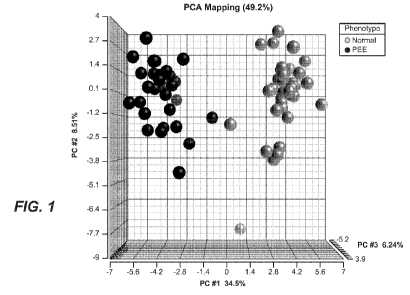
[0017] Figure 1 is a graphical representation of a principal component
analysis of 48 PEE
proteins that differentiate serum proteins in PEE (p<0.001 following ANOVA)
when compared
to sera from normal healthy pregnant women. The axes represent the
eigenvectors of the
covariance matrix scaled by the square root of the corresponding eigenvalue
for the spectral
counts of the 48 PEE proteins across each of the patients in the study (n=35
normal pregnancy,
n=25 pregnancy with PEE). Also see Table 1.
[0018] Figure 2 shows scatter plots for 8 proteins highly predictive of PEE.
These 8 PEE
proteins are a subset of the 48 PEE proteins in Figure 1: THBS1, PRG2, GSN,
ITIH4, HPX, TF,
APCS, and F5. Displayed are the mean spectral count values plus the SEM for
each protein from
healthy and PEE samples. Randomized combinations of three of these 8 proteins
yield an
average AUC of 0.95 or greater. Also see Table 2.
[0019] Figure 3 is a protein expression scheme for a biomarker pattern
analysis to identify
groups of proteins that show comparable patterns across trimesters. Identified
patterns of
proteins correspond to the column labeled "Biomarker Pattern Analysis" in
Tables 1 and 2.
[0020] Figure 4 shows scatter plots of the individual spectral counts for each
patient for the set
of 48 PEE proteins in Table 1; displayed are mean spectral counts plus SEM for
each protein.
[0021] Figure 5 is a graphical representation of a principal component
analysis following
ANOVA of 75 PEE autoantibodies, with a false discovery rate adjusted p-value
of qFDR<0.05
and a minimum fold change difference between Normal and PEE of 1.5, found to
be
differentially measured in serum samples from healthy pregnant women and
pregnant women
with PEE. Also see Table 4.
7
CA 02901551 2015-08-14
WO 2014/153232 PCT/US2014/029741
[0022] Figure 6 shows scatter plots of the individual mean fluorescent
intensity (MFI) counts
for each patient for a subset of 10 PEE autoantibodies from the set of 75 PEE
autoantibodies
(Figure 5 and Table 5) which were significantly different (qFDR<0.05; fold
change >1.5) in sera
taken from pregnant women with and without preeclampsia. Displayed are the
mean MFI counts
plus SEM of each autoantibody in serum samples from 25 healthy pregnant women
(Normal)
and 20 pregnant women with PEE. The light gray vertical bar on the left of
each graph shows
normal pregnant women in the first trimester (triangles) or the third
trimester (rectangles). The
dark gray vertical bar on the right of each graph shows the PEE women in the
third trimester
(rectangles). Biomarkers are chosen that can measured in normal pregnancy in
the first trimester,
with minimal overlap in the third trimester and with a significant (qFDR
<0.05) increase or
decrease in PEE over normal pregnancy with fold change >1.5.
[0023] Figure 7 shows scatter plots of the individual mean fluorescent
intensities (MFI) counts
for each patient forthe most significant 10 autoantibodies from the set of 75
PEE autoantibodies
which were significantly different (qFDR<0.05; fold change >1.5) in sera taken
from pregnant
women with and without preeclampsia; displayed are the mean MFI counts plus
SEM of each
autoantibody in serum samples from 25 healthy pregnant women and 20 pregnant
women with
PEE.
DETAILED DESCRIPTION OF THE INVENTION
[0024] The invention described herein provides, inter alia, methods,
compositions, and kits
useful for identifying the risk of developing preeclampsia (PEE), predicting
the onset of PEE,
monitoring the progression of PEE, monitoring the regression of PEE,
identifying a sub-
population of patients who should be treated for PEE or continue to be treated
for PEE, assessing
efficacy of treatment for PEE in pregnant women, and/or identifying a sub-
population of patients
who should be monitored for PEE symptoms. As further detailed below,
particular peptide,
protein and autoantibody biomarkers have been identified that may be utilized
to accurately
identify pregnant women during early to mid-pregnancy that may later develop
PEE. Such
markers may allow the diagnostic distinction between PEE and other conditions
that exhibit
similar symptoms. Early identification of subjects at greater risk for PEE
would be of
considerable value to help to save both the baby and the mother's lives and
improve their
welfare.
8
CA 02901551 2015-08-14
WO 2014/153232 PCT/US2014/029741
Definitions
[0025] For purposes of interpreting this specification, the following
definitions will apply and
whenever appropriate, terms used in the singular will also include the plural
and vice versa.
[0026] As used herein, "preeclampsia" or "PEE" refers to a condition when a
pregnant woman
develops high blood pressure (relative to her blood pressure before pregnancy)
and/or protein in
the urine during pregnancy. In many cases, the high blood pressure and/or
protein in the urine
occurs after the 20th week (late 2nd or 3rd trimester) of pregnancy. Symptoms
of preeclampsia
can include, but are not limited to, high blood pressure (hypertension),
excess protein in urine
(proteinuria), headaches, changes in vision (including temporary loss of
vision, blurred vision or
light sensitivity), abdominal pain (such as upper abdominal pain, e.g., under
the ribs on the right
side), nausea, vomiting, dizziness, decreased urine output, sudden weight
gain, visual
disturbances (e.g., oversensitivity to light, blurred vision, seeing flashing
spots or auras),
confusion, anxiety, and/or shortness of breath. In some cases, serious signs
of PEE include, but
are not limited to: high blood pressure, risk of brain injury, impaired kidney
and liver function,
blood clotting problems, pulmonary edema, and/or seizures. Other symptoms of
PEE are
described herein and known to one of skill in the art, including treating
physicians.
[0027] An individual "at risk" of developing PEE may or may not have
detectable disease or
symptoms of disease, and may or may not have displayed detectable disease or
symptoms of
disease prior to the treatment methods described herein. "At risk" denotes
that a individual has
one or more risk factors, which are measurable parameters that correlate with
development of
PEE, as described herein and known in the art. A subject having one or more of
these risk
factors has a higher probability of developing PEE than a subject without one
or more of these
risk factor(s). For example, in some embodiments, a subject "at risk" of
developing PEE shows
a change in the level of expression of one or more PEE proteins as shown in
Table 1 or Table 2.
[0028] An "individual" can be a "patient." A "patient," refers to an
"individual" who is under
the care of a treating physician. In one embodiment, the patient is a female.
In another
embodiment, the patient is a female who has not been diagnosed with PEE. In
yet other
embodiments, the patient is a female who has been diagnosed with PEE but has
not had any
treatment to address the PEE.
9
CA 02901551 2015-08-14
WO 2014/153232 PCT/US2014/029741
[0029] A "patient sub-population," and grammatical variations thereof, as used
herein, refers
to a patient subset characterized as having one or more distinctive measurable
and/or identifiable
characteristics that distinguishes the patient subset from others in the
broader disease category to
which it belongs. Such characteristics include having a PEE protein and/or
autoantibody profile
described herein as being characteristic of being at risk for developing PEE,
optionally in
combination with any of the symptoms described herein and known to one of
skill in the art,
including treating physicians.
[0030] The term "biological sample," as used herein, refers to a composition
that is obtained
or derived from an individual that contains a cellular and/or other molecular
entity that is to be
characterized and/or identified, for example based on physical, biochemical,
chemical and/or
physiological characteristics. In some embodiments, the biological sample is
blood, serum,
biological fluid or tissue from the mother. In other embodiments, the
biological sample contains
some material from the baby.
[0031] "Predicting" and "prediction" as used herein does not mean that the
event will happen
with 100% certainty. Instead it is intended to mean the event will more likely
than not happen.
Acts taken to "predict" or "make a prediction" can include the determination
of the likelihood
that an event will be more likely than not to happen. Assessment of multiple
factors described
herein can be used to make such a determination or prediction.
[0032] By "correlate" or "correlating" is meant comparing, in any way, the
performance
and/or results of a first analysis or protocol with the performance and/or
results of a second
analysis or protocol. For example, one may use the results of a first analysis
or protocol in
carrying out a second protocols and/or one may use the results of a first
analysis or protocol to
determine whether a second analysis or protocol should be performed. With
respect to the
embodiment of PEE protein analysis or PEE autoantibody analysis performed on
biological
samples from an individual, one may use the results to determine whether a
specific therapeutic
regimen should be performed for that individual.
[0033] The term "diagnosis" is used herein to refer to the identification or
classification of a
medical or pathological state, disease or condition. For example, "diagnosis"
may refer to
identification of PEE, "Diagnosis" may also refer to the classification of a
severity of PEE.
Diagnosis of PEE may be made according to any protocol that one of skill of
art (e.g.,
CA 02901551 2015-08-14
WO 2014/153232 PCT/US2014/029741
obstetrician) would use, for example, those set forth in "Pre-eclampsia:
Etiology and Clinical
Practice" (F. Lyall, Ed. and M. Belfort, Ed.), Cambridge University Press
(2007) and/or
"Chesley's Hypertensive Disorders in Pregnancy, 31d edition (M. Lindheimer, J.
Roberts, F.G.
Cunningham, Eds.), Elsevier (2009).
[0034] The term "aiding diagnosis" is used herein to refer to methods that
assist in making a
clinical determination regarding the presence, degree or other nature, of a
particular type of
symptom or condition of PEE. For example, a method of aiding diagnosis of PEE
can include
measuring the amount or detecting the presence or absence of one or more PEE
proteins and/or
PEE autoantibodies in a biological sample from an individual.
[0035] The term "prognosis" is used herein to refer to the prediction of the
likelihood of the
development of PEE (including recurrence of PEE). The predictive methods of
the invention
can be used clinically to make treatment decisions by choosing the most
appropriate treatment
modalities for any particular patient. The predictive methods of the present
invention are
valuable tools in predicting if and/or aiding in the diagnosis as to whether a
patient is likely to
develop PEE, have recurrence of PEE, and/or worsening of PEE symptoms.
[0036] "Treatment" refers to clinical intervention in an attempt to alter the
natural course of
the individual and can be performed before, during, or after the course of
clinical diagnosis or
prognosis. Desirable effects of treatment include preventing the occurrence or
recurrence of
PEE or a condition or symptom thereof, alleviating a condition or symptom of
PEE, diminishing
any direct or indirect pathological consequences of PEE, decreasing the rate
of PEE progression
or severity, and/or ameliorating or palliating the PEE. In some embodiments,
methods and
compositions of the invention are used on patient sub-populations identified
to be at risk of
developing PEE. In some cases, the methods and compositions of the invention
are useful in
attempts to delay development of PEE.
[0037] It is understood that aspects and embodiments of the invention
described herein
include "consisting" and/or "consisting essentially of' aspects and
embodiments.
[0038] As used herein, the term "peptide" may be used to refer to a natural or
synthetic
molecule comprising two or more amino acids linked by the carboxyl group of
one amino acid
to the alpha amino group of another. A peptide of the present invention is not
limited by length,
11
CA 02901551 2015-08-14
WO 2014/153232 PCT/US2014/029741
and thus "peptide" can be part of a longer polypeptide and/or of a protein or
can refer to the
longer polypeptide/protein itself The term peptide can be used interchangeably
with protein
and/or polypeptide.
[0039] As used herein, the term "detect" refers to the quantitative
measurement of
undetectable, low, normal, or high serum concentrations of one or more
biomarkers such as, for
example, proteins, peptides and other biological molecules.
[0040] As used herein, the terms "quantify" and "quantification" may be used
interchangeably, and refer to a process of determining the quantity or
abundance of a substance
in a sample (e.g., a biomarker), whether relative or absolute.
[0041] Reference to "about" a value or parameter herein includes (and
describes)
embodiments that are directed to that value or parameter per se. For example,
description
referring to "about X" includes description of "X." The term "about" is used
to provide
flexibility to a numerical range endpoint by providing that a given value may
be "a little above"
or "a little below" the endpoint without affecting the desired result.
Concentrations, amounts,
and other numerical data may be expressed or presented herein in a range
format. It is to be
understood that such a range format is used merely for convenience and brevity
and thus should
be interpreted flexibly to include not only the numerical values explicitly
recited as the limits of
the range, but also to include all the individual numerical values or sub-
ranges encompassed
within that range as if each numerical value and sub-range is explicitly
recited.
[0042] As used in the specification and the appended claims, the singular
forms "a," "an" and
"the" include plural referents unless the context clearly indicates otherwise.
[0043] "Optional" or "optionally" means that the subsequently described event
or
circumstance can or cannot occur, and that the description includes instances
where the event or
circumstance occurs and instances where it does not.
General Techniques
[0044] Unless defined otherwise, technical and scientific terms used herein
have the same
meaning as commonly understood by one of ordinary skill in the art to which
this invention
belongs.
12
CA 02901551 2015-08-14
WO 2014/153232 PCT/US2014/029741
[0045] The practice of the present invention will employ, unless otherwise
indicated,
conventional techniques of protein biology, protein chemistry, molecular
biology (including
recombinant techniques), microbiology, cell biology, biochemistry, and
immunology, which are
within the skill of the art. Such techniques are explained fully in the
literature, such as, "Pre-
eclampsia: Etiology and Clinical Practice" (F. Lyall, Ed. and M. Belfort,
Ed.), Cambridge
University Press (2007); "Chesley's Hypertensive Disorders in Pregnancy, 31d
edition (M.
Lindheimer, J. Roberts, F.G. Cunningham, Eds.), Elsevier (2009);" "Molecular
Cloning: A
Laboratory Manual", second edition (Sambrook et al., 1989); "Oligonucleotide
Synthesis" (M. J.
Gait, ed., 1984); "Current Protocols in Molecular Biology" (F. M. Ausubel et
al., eds., 1987,
periodic updates); "PCR: The Polymerase Chain Reaction", (Mullis et al., eds.,
1994); and
Singleton et al., Dictionary of Microbiology and Molecular Biology 2nd ed., J.
Wiley & Sons
(New York, N.Y. 1994).
Collection of Biological Samples from Pregnant Women
[0046] Typically, a biological sample is collected from the pregnant woman.
Any type of
biological sample may be collected, including but not limited to serum,
plasma, blood, urine,
mucus, saliva, cerebrospinal fluid, amniotic fluid, synovial fluid, cervical
vaginal fluid, lavage
fluid, placenta, other surrounding tissues, tissues, and combinations thereof.
In one
embodiment, the biological sample collected from the pregnant woman contains
only non-fetal
tissue. In another embodiment, some fetal tissue is collected along with the
maternal non-fetal
tissue.
[0047] Testing of women for PEE using the methods described herein may occur
at any time
during pregnancy when biomarkers such as proteins, peptides, and
autoantibodies indicative of
PEE are quantifiable. Biomarkers may be collected and tested for during the
first, second, or
third trimesters of pregnancy. In one embodiment biomarkers may be collected
and tested for at
from about 4 weeks to about 6 weeks gestation; at from about 6 weeks to about
8 weeks
gestation; at from about 8 weeks to about 10 weeks gestation; at from about 10
weeks to about
12 weeks gestation; at from about12 weeks to about 14 weeks gestation; at from
about 14 weeks
to about 16 weeks gestation; at from about 16 weeks to about 18 weeks
gestation; at from about
18 weeks to about 20 weeks gestation; at from about 20 weeks to about 22 weeks
gestation; at
from about 22 weeks to about 24 weeks gestation; at from about 24 weeks to
about 26 weeks
gestation; at from about 26 weeks to about 28 weeks gestation; at from about
28 weeks to about
13
CA 02901551 2015-08-14
WO 2014/153232 PCT/US2014/029741
30 weeks gestation; or at from about 30 weeks and beyond. These ranges should
not be seen as
limiting, as such testing may be performed at any point during pregnancy.
Rather these ranges
are provided to demonstrate periods of the gestational cycle where such
testing is most likely to
occur in a majority of pregnant women.
Identification of PEE Proteins and Peptides and Testing of Biological Samples
[0048] Methods for testing a pregnant woman for PEE may include detecting the
difference in
the concentration, expression, intracellular translocation, or activity of one
or more peptides,
proteins, and/or autoantibodies associated with PEE present in a biological
sample as compared
to a healthy pregnant woman who does not develop PEE. Various systems and
methods, as
further described herein, can be used to identify, characterize, and quantify
the peptides,
proteins, or autoantibodies. Non-limiting systems and methods are provided
herein.
[0049] In one embodiment, mass spectrometry can be used to identify peptides
or proteins that
are differentially expressed between pregnant women with PEE or suspected of
being at risk for
developing PEE and pregnant healthy women. In such an embodiment, comparing
multiple
mass spectra from different biological samples, locating mass ions that are
quantitatively
different after using approaches to compensate for non-biological variability,
isolating, and
characterizing the protein or peptide biomarker of interest can be used
herein.
[0050] In another embodiment, capillary liquid chromatography can be used to
identify
peptides or proteins that are differentially expressed between pregnant women
with PEE or
suspected of being at risk for developing PEE and pregnant healthy women.
Those of skill in
the art will appreciate that other techniques can be used to identify PEE
proteins and/or peptides.
The features of PEE proteins and/or peptides that enable them to be used as
diagnostic markers
for PEE is described infra.
[0051] The proteins or peptides that are differentially expressed can be
analyzed by one- or
multiple way-analysis of variance (AND VA) after quantile normalization to
identify
differentially expressed proteins between PEE and healthy pregnancies. See,
for example, Figure
1 and Table 1. In one embodiment, a protein upregulated with a fold change
greater than 1.5, or
a protein downregulated with a fold change greater than 1.5, with a p-value
with false discovery
rate of less than 0.05 can be considered to be significant and utilized to
build prediction models.
14
CA 02901551 2015-08-14
WO 2014/153232 PCT/US2014/029741
In another embodiment, a protein upregulated with a fold change greater than
2, or a protein
downregulated with a fold change greater than 2, with a p-value with false
discovery rate of less
than 0.05 can be considered to be significant and utilized to build prediction
models. Various
types of software can be used for statistical analysis. One example of such
software is Partek
Genomics Suite. The proteins or peptides can be subjected to statistical
analysis to select a
robust model for detection and/or prediction of PEE among the following
classification models:
K-nearest neighbor, nearest centroid, discriminate analysis, support vector
machine, partial least
squares, diagonal discriminant analysis, random forest and logistic
regression. As further
detailed in the Examples, multiple models can yield a receiver operator
characteristic curve
(ROC) with an area under the curve (AUC) of >0.9, with as few as 3 proteins.
Typically, the
area under the ROC curve is a measure of testing accuracy that takes into
account both measures
of sensitivity and specificity. An AUC of 1.0 means that there is 100%
accuracy in that cohort.
In various embodiments, the area under the ROC curve can be a statistical
measurement of the
accuracy of detection of PEE, the accuracy in the diagnosis of PEE, the
accuracy in the
prediction of PEE, the accuracy in the prediction of the onset of PEE, and the
like.
[0052] Using this methodology, exemplary peptides and/or proteins that were
found to be
associated with PEE are described in Example 1 and Table 1. These peptides
and/or proteins
can interchangeably be referred to as "PEE proteins" or "PEE peptides" or "PEE
polypeptides"
and are useful in the diagnosis, aiding of diagnosis, and/or treatment of PEE.
These PEE
proteins can also be used to identify patient sub-populations for treatment
for PEE.
Testing of Biological Samples and Identification of PEE Autoantibodies
[0053] Biological samples taken from pregnant women can be used to identify
autoantibodies
that can be used to assess whether a pregnant woman has or will develop PEE
(i.e., PEE
autoantibodies). Various techniques of measuring autoantibodies are known to
one of skill in
the art. One non-limiting method is to use a ProtoArray human protein
microarray V5
(microarray) (Invitrogen, Carlsbad, CA) to measure a variety of autoantibodies
in biological
samples from healthy pregnant women and PEE pregnant women. Relative
fluorescent units
representing the abundance of each autoantibody in the serum of healthy and
PEE pregnancies
can be uploaded to and analyzed using any type of statistical software, such
as Partek's
Genomics Suite (Partek Inc, St. Louis, MO). The data can be quantile
normalized followed by
CA 02901551 2015-08-14
WO 2014/153232 PCT/US2014/029741
one- or multiple way analysis of variance (ANOVA) to identify differentially
expressed
autoantibodies between PEE and healthy pregnancies.
[0054] Exemplary differentially expressed autoantibodies between PEE and
healthy
pregnancies are shown in Table 4, Table 5, and in Figures 5-7. In one
embodiment, an
autoantibody upregulated with a fold change greater than 1.5, or an
autoantibody downregulated
with a fold change greater than 1.5, with a p-value with false discovery rate
of less than 0.05 can
be considered significant and utilized to build prediction models. In one
embodiment, at least
about 8 antibodies can provide explanation of 70% of the variation between PEE
and healthy
pregnancy samples. In another embodiment about 15 antibodies can provide
explanation of all
of the 95% of the differences between the samples. The PEE autoantibodies
disclosed here in
can be used for various methods of assessing risk of developing PEE,
monitoring the
development of PEE and selecting patients for treatment as well as other uses
described herein.
Binding Agents and Methods of Using PEE Proteins and/or PEE Autoantibodies for
Detecting
PEE or Diagnosing the Risk of Developing Preeclampsia
[0055] Binding agents of the invention may be used to identify proteins,
peptides, and/or
autoantibodies present in the biological samples taken from a pregnant woman
suspected of
being at risk for developing PEE, already suffering from PEE, or from a
healthy pregnant
woman. The binding agent can be one or more proteins, one or more peptides,
one or more
antibodies, one or more nucleic acids, or one or more nucleoproteins. The
binding agent can
comprise a plurality of binding sites for proteins, peptides, and
autoantibodies. In one
embodiment, the binding agent can be used to identify a protein or peptide
that would predict if
a pregnant woman will develop PEE or has PEE, or is recovering from PEE. In
such cases, the
binding agent can be used to aid in the diagnosis of PEE or PEE status.
[0056] Binding agents of the invention may be labeled or modified in a manner
know to those
with skill in the art. For example binding agents may comprise a label. The
label may include,
but is not limited to a fluorescent label, an immunolabel, a magnetic label, a
DNA label, a small
molecule label, or a radio label.
[0057] A protein binding agent may be labeled or modified in some manner. For
example
protein binding agents may comprise a label. The label may include, but is not
limited to a
16
CA 02901551 2015-08-14
WO 2014/153232 PCT/US2014/029741
fluorescent label, an immunolabel, a magnetic label, a DNA label, a small
molecule label, or a
radio label.
[0058] A peptide binding agent may be labeled or modified in some manner. For
example
peptide binding agents may comprise a label. The label may include, but is not
limited to a
fluorescent label, an immunolabel, a magnetic label, a DNA label, a small
molecule label, or a
radio label.
[0059] A nucleic acid binding agent may be labeled or modified in some manner.
For example
nucleic acid binding agents may comprise a label. The label may include, but
is not limited to a
fluorescent label, an immunolabel, a magnetic label, a DNA label, a small
molecule label, or a
radio label.
[0060] A nucleoprotein binding agent may be labeled or modified in some
manner. For
example nucleoprotein binding agents may comprise a label. The label may
include, but is not
limited to a fluorescent label, an immunolabel, a magnetic label, a DNA label,
a small molecule
label, or a radio label.
[0061] The binding agent can bind one or more proteins, peptides, or
autoantibodies with a
dissociation constant (Li )of 10-15 M, 10-14K 10-13 M, 10-12 r,µ47 10-" M, HIM
M, 10-9 M, 10-8 M,
10-7 M, 10-6 M, i0 m, 10-4m, io M, or 10-2M. In certain embodiments, the
binding agent
binds the one or more proteins, peptides, or autoantibodies with a Kd range of
10-12M to 105M,
1010M to 105M, 10-8M 10 10-5M, 10-7M 10 105M, 10-1 M 10 10-8M, 10-9M 10 1Ã17M,
or 10-8
M to 10-6M.
[0062] In one specific embodiment the binding agent can bind 1 protein,
peptide, and/or
autoantibody. In another embodiment the binding agent can bind 2 proteins,
peptides, and/or
autoantibodies. In yet another embodiment, the binding agent can bind 3
proteins, peptides,
and/or autoantibodies. In related embodiments, the binding agent can bind 1 or
more, 2 or more,
3 or more, 4 or more, 5 or more, 6 or more, 7 or more, 8 or more, 9 or more,
10 or more, 11 or
more, 12 or more, 13 or more, 14 or more, 15 or more, 16 or more, 17 or more,
18 or more, 19 or
more, 20 or more, 21 or more, 22 or more, 23 or more, 24 or more, 25 or more,
proteins,
peptides, and/or autoantibodies, up to a maximum of 30, 40, 50, 60, 70, 80,
90, or 100 proteins,
peptides, and/or autoantibodies. In one specific embodiment the binding agent
can bind a
17
CA 02901551 2015-08-14
WO 2014/153232 PCT/US2014/029741
maximum of 48 PEE proteins and/or peptides. In another specific embodiment the
binding
agent can bind any combination of 2, 3, 4, 5, 6, 7, 8, 9, 10 or more
combinations of proteins,
peptides and antibodies.
[0063] The PEE proteins and PEE autoantibodies as described herein can be used
to diagnose
or aid in the diagnosis of individuals who are at risk of developing PEE. The
PEE proteins and
PEE autoantibodies can also be used to identify the risk of developing
preeclampsia, predict the
onset of PEE, monitor the progression of PEE, monitor the regression of PEE,
identify a sub-
population of patients who should be treated for PEE or continue to be treated
for PEE, assess
efficacy of treatment for PEE in pregnant women, and/or identify a sub-
population of patients
who should be monitored for PEE symptoms.
[0064] In one embodiment, the binding agents of the invention are selected
from antibodies,
autoantibodies, peptides, polypeptides, oligonucleotides, small molecules, and
the like. In a
specific embodiment, binding agents of the invention comprise a fluorescent
label, or a
fluorescent immunolabel. In another specific embodiment, the binding agents of
the invention
comprise a magnetic label or magnetic immunolabel. In another specific
embodiment, the
binding agents of the invention comprise a radio label or magnetic radio
label. In yet another
embodiment, the binding agents are labeled with a oligonucleotide or a small
molecule.
[0065] In one embodiment a binding agent comprises an immune assay. An
immunoassay can
be used to detect, identify and/or quantify proteins or peptides present in
the biological samples
taken from a pregnant woman suspected of being at risk for developing PEE and
a healthy
pregnant woman. In certain embodiments, the immunoassay can be an enzyme-
linked
immunosorbant assay (ELISA). Any immunoassay used herein can incorporate
fluorescent,
magnetic, or radio immunolabels.
[0066] In another embodiment, a binding agent comprises a diagnostic array. A
diagnostic
array can be used to detect, identify and/or quantify proteins, peptides, or
autoantibodies present
in the biological samples taken from a pregnant woman suspected of being at
risk for developing
PEE and a healthy pregnant woman. The array can include a protein or antibody-
coated
substrate comprising a plurality of discrete, known regions on the substrate.
The arrays can
comprise particles, nanoparticles, beads, nanobeads, or other solid surfaces
which can be porous
or non-porous, and can range in size. In one embodiment, the diagnostic array
does not
18
CA 02901551 2015-08-14
WO 2014/153232 PCT/US2014/029741
comprise fluorescent particles. In another embodiment, the diagnostic array
comprises
fluorescent particles. In one embodiment, the diagnostic array does not
comprise magnetic
particles. In another embodiment, the diagnostic array comprises magnetic
particles.
[0067] In a related embodiment, the binding agents of the invention comprise
particles,
nanoparticles, beads, nanobeads. In one embodiment, the nanoparticles, beads,
or nanobeads are
fluorescently labeled. In another embodiment, the nanoparticles, beads, or
nanobeads are
magnetically labeled. In another embodiment, the nanoparticles, beads, or
nanobeads are radio
labeled. In yet another embodiment, the nanoparticles, beads, or nanobeads are
labeled with a
oligonucleotide or a small molecule.
[0068] In another embodiment, a binding agent comprises a magnetic-based
protein assay
component and/or nanotags. In such an embodiment, a magnetic multiplex protein
assay is used
to detect, identify, and/or quantify proteins or peptides present in a
biological sample with the
use of magnetic nanotags. (Osterfeld et al., "Multiplex Protein Assays Based
on Real-Time
Magnetic Nanotag Sensing," PNAS, 105, 20637-20640 (published online Dec.12,
2008) For
example, a MagArray protein chip can be utilized for the diagnostic array. In
this embodiment,
protein or peptide detection is used carried out in three steps. First, probes
on the surface
specifically bind to proteins or peptides in the sample. Second, nanotag-
labeled antibodies bind
to the bound proteins or peptides, forming sandwich-like structures. Finally,
an external
magnetic field is applied to the chip and the stray magnetic field produced by
the nanotags is
measured electrically to determine the presence of the target molecule in the
sample.
[0069] In a related embodiment, the binding agents of the invention comprise
nanotags. In one
embodiment, the nanotags are fluorescently labeled. In another embodiment, the
nanotags are
magnetically labeled. In another embodiment, the nanotags are radio labeled.
In yet another
embodiment, the nanotags are labelled with a oligonucleotide or a small
molecule.
[0070] In another embodiment, carboxyl bead sets can be used to measure
proteins, peptides
of interest. Here, any protein or peptide can be covalently attached to a
stable microbead surface
followed by fluorescent labeling and fluorescence intensity measurement. The
VeraCode
Technology by Illumina (Illumina Inc., Hayward, CA) allows one to perform up
to 48
immunoassays in varying combinations in a single reaction in a standard 96-
well microplate.
19
CA 02901551 2015-08-14
WO 2014/153232 PCT/US2014/029741
[0071] In yet another embodiment proteins, peptides and autoantibodies can be
measured by
electrochemiluminesence ELISA. The multiplexed electrochemiluminesence ELISA
platform by
Meso Scale Discovery (MSD, Gaithersburg, MD) is a high throughput multiplexed
ELISA,
custom designable, with the capability to simultaneously measure up to several
analytes in the
same well.
[0072] In another embodiment, functional protein-based assays can be used to
detect
differences in activity, binding, intracellular translocation, or post-
translational processing of a
protein, peptide, or autoantibody biomarker of interest. Such assays include
competitive binding
assays, western blot immunoblot assays, liposome immunoassays, and the like.
In one specific
embodiment, and by way of example only, an assay such as Invitrogen's Prot
Array
Microarray can be used to detect protein-protein interactions of interest.
This array allows for
profiling a biological sample such as serum or urine from a pregnant woman
suspected for being
at risk for PEE and can be used for identifying biologically relevant protein
kinase substrates,
small molecule binding partners, ubiquitin ligase substrates, and proteins
interactors of
antibodies. In one embodiment, spectral imaging can be used to detect
differences in the
characteristics of proteins.
[0073] The PEE proteins and/or PEE autoantibodies can be detected by a binding
agent with
the functional parameter as described in the sections above. In other
embodiments, the binding
agent can be used to quantify PEE proteins, peptides and/or autoantibodies.
This may be useful
to predict the onset of PEE, the risk of developing PEE, to diagnose PEE, or
to determine the
severity of PEE symptoms.
[0074] One benefit of using the PEE proteins and/or PEE autoantibodies as
disclosed herein is
that determination of the risk of developing preeclampsia can be done with a
high level of
accuracy. Accuracy can be portrayed by sensitivity (the accuracy of the
preeclampsia positive
patients correctly identified) and by specificity (the accuracy of the
preeclampsia negative
patients correctly identified); positive predictive value (PPV) and negative
predictive value
(NPV) respectively.
[0075] In the embodiments provided herein, determination of the risk of
developing PEE using
the PEE proteins and/or PEE autoantibodies or a combination of both
peptides/proteins and
autoantibodies for a pregnant woman suspected to be at risk for developing PEE
is highly
CA 02901551 2015-08-14
WO 2014/153232 PCT/US2014/029741
accurate for the detection or prediction of PEE. In the embodiments provided
herein, the
methods provide at least 60%, at least 65%, at least 70%, at least 75%, at
least 80%, at least
85%, at least 90%, at least 95%, at least 96%, at least 97%, at least 98%, at
least 99%, or 100%
accuracy. Furthermore, in the embodiments provided herein, the methods provide
at least 60%,
at least 65%, at least 70%, at least 75%, at least 80%, at least 85%, at least
90%, at least 95%, at
least 96%, at least 97%, at least 98%, at least 99%, or 100% accuracy for the
detection, or
prediction of PEE.
[0076] In the embodiments provided herein, determination of the risk of
developing PEE using
the PEE proteins and/or PEE autoantibodies or a combination of both
peptides/proteins and
autoantibodies for a pregnant woman suspected to be at risk for developing PEE
is highly
sensitive for the detection or prediction of PEE. In the embodiments provided
herein, the
methods provide at least 60%, at least 65%, at least 70%, at least 75%, at
least 80%, at least
85%, at least 90%, at least 95%, at least 96%, at least 97%, at least 98%, at
least 99%, or 100%
sensitivity. Furthermore, in the embodiments provided herein, the methods
provide at least 60%,
at least 65%, at least 70%, at least 75%, at least 80%, at least 85%, at least
90%, at least 95%, at
least 96%, at least 97%, at least 98%, at least 99%, or 100% sensitivity for
the detection, or
prediction of PEE.
[0077] Furthermore in the embodiments provided herein, analysis of protein,
peptide, and/or
autoantibody biomarkers from a pregnant woman suspected to be at risk for
developing PEE is
highly specific for the detection or prediction of PEE. In the embodiments
provided herein, the
methods provide at least 60%, at least 65%, at least 70%, at least 75%, at
least 80%, at least
85%, at least 90%, at least 95%, at least 96%, at least 97%, at least 98%, at
least 99%, or 100%
specificity. Furthermore, in the embodiments provided herein, the methods
provide at least
60%, at least 65%, at least 70%, at least 75%, at least 80%, at least 85%, at
least 90%, at least
95%, at least 96%, at least 97%, at least 98%, at least 99%, or 100%
specificity for the detection,
or prediction of PEE.
[0078] Moreover, in the embodiments provided herein, analysis of protein,
peptide, and/or
autoantibody biomarkers from a pregnant woman suspected to be at risk for
developing PEE has
a positive predictive value (PPV; the proportion of positive test results that
are true
positives/correct diagnoses) for the detection or prediction of PEE. In the
embodiments
provided herein, the methods provide at least 60%, at least 65%, at least 70%,
at least 75%, at
21
CA 02901551 2015-08-14
WO 2014/153232 PCT/US2014/029741
least 80%, at least 85%, at least 90%, at least 95%, at least 96%, at least
97%, at least 98%, at
least 99%, or 100% PPV for the detection or prediction of PEE. Also, in the
embodiments
provided herein, analysis of biomarkers from a pregnant woman suspected to be
at risk for
developing PEE has a negative predictive value (NPV; the proportion of
subjects with a negative
test result who are correctly diagnosed) for the detection or prediction of
PEE. In the
embodiments provided herein, the methods provide at least 60%, at least 65%,
at least 70%, at
least 75%, at least 80%, at least 85%, at least 90%, at least 95%, at least
96%, at least 97%, at
least 98%, at least 99%, or 100% NPV, for the detection or prediction of PEE.
[0079] In the embodiments provided herein, the analysis of biomarkers from a
pregnant
woman suspected to be at risk for developing PEE provides an area under the
curve (AUC),
which is a statistical measurement of the probability of the detection of PEE,
or a statistical
measurement of the probability for predicting the development of PEE. In the
embodiments
provided herein, the methods provide an AUC of at least 0.80, at least 0.81,
at least 0.82, at least
0.83, at least 0.84, at least 0.85, at least 0.86, at least 0.87, at least
0.88, at least 0.89, at least
0.90, at least 0.91, at least 0.92, at least 0.93, at least 0.94, at least
0.95, at least 0.96, at least
0.97, at least 0.98, at least 0.99, and 1.0 for the detection of PEE or for
predicting the
development of PEE.
[0080] The analysis of biological samples taken from either a pregnant woman
suspected to be
at risk for developing PEE or from a healthy pregnant woman include testing
for only 1, testing
for combinations of 1 or more, 2 or more, 3 or more, 4 or more, 5 or more, 6
or more, 7 or more,
8 or more, 9 or more, 10 or more, 11 or more, 12 or more, 13 or more, 14 or
more, 15 or more,
16 or more, 17 or more, 18 or more, 19 or more, 20 or more, 21 or more, 22 or
more, 23 or more,
24 or more, 25 or more proteins, peptides, and/or autoantibodies, up to a
maximum of 30, 40, 50,
60, 70, 80, 90, or 100 PEE proteins, PEE peptides, and/or PEE autoantibodies
disclosed herein.
[0081] In one embodiment, the analysis of biomarkers from a pregnant woman
suspected to be
at risk for developing PEE comprises detecting an increase or decrease in at
least one protein
selected from Table 1. In such an embodiment, the risk for developing PEE can
comprise a
change in the protein concentration of IGLC1, C8G, C2, AP0A4, TF, SERPIND1,
CIS,
THBS1, APCS, CRP, IGFBP3, IGKC V-IV, ITIH1, CLU, APOC2, PROS1, PAPPA, AFM,
VWF, PRG2, APOB, FCN3, C8A, IGHG1, CD14, IGHG3, HPX, IGHG1, IGHG1, CLEC3B,
APOE, PSG9, CP, GSN, FN1, FBLN1, KRT86, A2M, APOB, F5, ITIH4, hCG, KRT31,
22
CA 02901551 2015-08-14
WO 2014/153232 PCT/US2014/029741
APOL1, HP, MBL2, C4BPB, and/or F13B. In another embodiment, the analysis of
biomarkers
from a pregnant woman suspected to be at risk for developing PEE comprises a
change in at
least one protein selected from Table 2. In such an embodiment, the risk for
developing PEE
can comprise a change in the protein concentration of APCS, THBS1, PRG2,
ITIH4, GSN,
HPX, F5, and/or TF.
[0082] In one embodiment, pregnant women who are at risk for developing PEE
can be
identified with a high level of accuracy (e.g., at least about 90%, 95%, 99%,
or 100%) using 3
proteins. In one embodiment, the 3 proteins are selected from Table 1. In
another embodiment,
any 3 proteins are selected from the group consisting of APCS, THBS1, PRG2,
ITIH4, GSN,
HPX, F5, TF, APOB, A2M, C4BPB, FN1, PAPPA, and VWF. In a related embodiment, a
combination of 3 proteins is selected from the combinations presented in Table
3.
[0083] In another embodiment, the analysis can include testing for up to any
one or
combination (of 2 or more, or of 3 or more) of the 48 PEE proteins as
disclosed in Table 1; or
any one or combination (of 2 or more, or 3 more more) of the 8 proteins as
disclosed in Table 2.
Combinations of the 48 PEE proteins of Table 1 can provide a minimal set of
proteins for
differentiating the risk of developing PEE from healthy pregnancy. Table 3
provides exemplary
combinations of 3 proteins that can be used for detecting the risk of
developing PEE in a
pregnant woman. In any of these embodiments, the testing can optionally can be
done with one
or more PEE autoantibodies further disclosed herein, infra.
[0084] In some embodiments, a minimum set of 3 proteins (the identities can
vary for the
combinations of 3 proteins) can give an AUC of > 0.96 for separation of women
suspected to be
at risk for developing PEE from women who will undergo a normal pregnancy. In
one
embodiment, any combination of 3 proteins can be selected from APCS, THBS1,
PRG2, ITIH4,
GSN, HPX, F5, TF, APOB, A2M, C4BPB, FN1, PAPPA, and VWF. For example, one
combination of 3 proteins can be GSN, THBS1 and PRG2. In other embodiments, a
minimum
set of 2 proteins can give an AUC of > 0.95 for separation of women suspected
to be at risk for
developing PEE from women who will undergo a normal pregnancy. For example,
one
combination of 2 proteins can be THBS1 and PRG2.
[0085] In the embodiments provided herein, the analysis of biomarkers from a
pregnant
woman suspected to be at risk for developing PEE comprises a change (increase
or decrease) in
23
CA 02901551 2015-08-14
WO 2014/153232 PCT/US2014/029741
at least one autoantibody selected from Table 4 or Table 5. Table 4 provides
75 PEE
autoantibodies differentially expressed between PEE and healthy pregnancies.
Table 5 provides
one subset of 10 autoantibodies differentially expressed between PEE and
healthy pregnancies.
In one embodiment, the risk for developing PEE comprises an increase in the
concentration of
an autoantibody to RNF111, ACY3, GSTZ1, SCAMPI, KIAA1826, OSBPL1A, NDUFB2,
JAK3, PI16, SCP2, SULT4A1, L0C283861, DLX1, KRT33B, PRKD2, BNIP1, U1snRNP68,
KLHL29, PSPH, MEIS3, C16orf28, JUP, CTNNA3, CUEDC1, EMILIN1, CCDC53,
UBE1DC1, JAK2, CD40, CAPZA2, DSN1, L0C284837, IGHAl, DACT3, CLIP3, TEC,
PSMA1, SNURF, CSNK2A1, CSNK1D, or combinations thereof. In another embodiment,
the
risk for developing PEE comprises a decrease in the concentration of an
autoantibody to AKT1,
GAGE7B, SUGT1L1, SLC5A2, HMGB1, GPBP1L1, APEX1, RPS28, RPS10, RBMX,
RPL39L, C18orf22, TSLP, GPR45, PIM1, RBMS3, IGLV2-14, CCL19, FGFR3, ANKHD1,
ACP1, BEX5, GL01, PIK3CG, MGC16075, PRC1, ZRANB2, HSPA5, CDK5, HOXC8,
TNFSF13B, SUPT4H1, ORC6L, FKSG44, or combinations thereof In one exemplary
embodiment, the risk for developing PEE comprises an increase in the
concentration of an
autoantibody to glutathione transferase zeta 1 (Maleylacetoacetate isomerase,
GSTZ1),
sulfotransferase family 4A, member 1 (SULT4A1), Junction plakoglobin (JUP),
CUE domain
containing 1 (CUEDC1), hypothetical protein L0C284837, and combinations
thereof. In
another exemplary embodiment, the risk for developing PEE comprises a decrease
in the
concentration of an autoantibody to casein kinase 1, delta (CSNK1D), pim-1
oncogene (PIM1),
protein regulator of cytokinesis 1 (PRC1), zinc finger, RAN-binding domain
containing 2
(ZRANB2), cyclin-dependent kinase 5 (CDK5), or combinations thereof. CSNK1D,
CUEDC1
and ZRANB2, is one exemplary set of 3 autoantibodies that allows for detection
of PEE with an
accuracy of 93%, with an area under the curve of 0.93. CSNK1D, SULT4A1 and
Junction
Plakoglobin is another exemplary set of 3 autoantibodies that allows for
detection of PEE with
an accuracy of 94%, with an area under the curve of 0.94.
[0086] In other embodiments, the assessment of a pregnant woman suspected to
be at risk for
developing PEE involves testing for the combination of a change in at least
one protein selected
from Table 1 or 2, a change in at least one autoantibody selected from Table 4
or Table 5, and
combinations thereof, including combinations of proteins and autoantibodies.
24
CA 02901551 2015-08-14
WO 2014/153232 PCT/US2014/029741
[0087] The PEE proteins and/or PEE antibodies of the invention can also be
used to identify a
patient sub-population of pregnant women who are at risk for preeclampsia for
treatment
purposes. In some embodiments, this sub-population is monitored for
development, progression,
or regression of PEE symptoms.
[0088] In some embodiments, this sub-population is treated for PEE prior to or
at the onset of
PEE symptoms. In some embodiments, the treatment is delivery of the baby. In
other
embodiments, the treatment can be administering to the pregnant women suitable
anti-
hypertensive medications to lower the blood pressure, corticosteroids and/or
anti-convulsive
medication. In other embodiments, the treatment is bed rest for the pregnant
woman to lower
the expectant mother's blood pressure and/or to increase blood flow to the
placenta. This sub-
population of patients can be monitored for various physiological parameters
known to a treating
physician at all stages to ensure their safety and their child's safety. In
some cases, the
monitoring is done to determine if the treatment should be continued or to see
if the treatment is
efficacious.
[0089] Therefore, using the PEE proteins and/or PEE antibodies of the
invention and the
methodology described herein, one of skill in the art can determine the risk
of developing PEE,
can determine the onset of PEE, monitor the progression of PEE, monitoring the
regression of
PEE, identify a sub-population of patients who should be treated for PEE or
continue to be
treated for PEE, assess efficacy of treatment for PEE in pregnant women,
and/or identify a sub-
population of patients who should be monitored for PEE symptoms.
Kits for the Diagnosis, Detection, or Prediction of PEE
[0090] The invention further provides for assay kits for the diagnosis,
detection and prediction
of PEE. A kit comprises reagents for detecting a protein, peptide, or
autoantibody or a
combination of protein, peptide and autoantibody implicated in PEE, in a
biological sample from
a pregnant woman. The reagents can comprise binding agents of the invention.
The binding
agents and reagents found in the kit may be labeled. They may be labeled, for
example, with a
fluorescent label, a radiolabel, an immunolabel, a magnetic label, a small
molecule label, a
DNA-based label, and/or any labels known to those in the art. The kit further
comprises a
composition comprising one or more solid surfaces that contain at least
binding agent, capable of
specifically binding a protein, peptide, or autoantibody biomarker (or
combinations thereof) of
CA 02901551 2015-08-14
WO 2014/153232 PCT/US2014/029741
interest in the biological sample. The kit also comprises instructions for the
use of the assay. In
one embodiment the binding agent is capable of binding to a peptide or
protein. In another
embodiment the binding agent is capable of binding to an autoantibody. In one
specific
embodiment, the kit comprises a composition comprising one or more solid
surfaces comprising
binding agents for a PEE protein selected from APCS, THBS1, PRG2, ITIH4, GSN,
HPX, F5,
TF, APOB, A2M, C4BPB, FN1, PAPPA, and VWF. In another specific embodiment, the
kit
comprises a composition comprising solid surfaces comprising binding agents
for the PEE
proteins THBS1 and PRG2. In another specific embodiment, the kit comprises a
composition
comprising solid surfaces comprising binding agents for the PEE proteins GSN,
THBS1 and
PRG2. In another specific embodiment, the kit comprises a composition
comprising solid
surfaces comprising binding agents for the PEE autoantibodies CSNK1D, CUEDC1
and
ZRANB2. In another specific embodiment, the kit comprises a composition
comprising solid
surfaces comprising binding agents for the PEE autoantibodies CSNK1D, SULT4A1
and
Junction Plakoglobin.
Compositions for the Diagnosis, Detection, or Prediction of PEE
[0091] The present invention provides for compositions comprising one or more
solid surfaces
which include binding agents, specific for PEE proteins and PEE
autoantibodies. In certain
embodiments, the solid surface comprises binding agents for at least 1, 1 or
more, 2 or more, 3
or more, 4 or more, 5 or more, 6 or more, 7 or more, 8 or more, 9 or more, 10
or more, 11 or
more, 12 or more, 13 or more, 14 or more, 15 or more, 16 or more, 17 or more,
18 or more, 19 or
more, 20 or more, 21 or more, 22 or more, 23 or more, 24 or more, 25 or more,
proteins,
peptides, and/or autoantibodies, up to a maximum of 30, 40, 50, 60, 70, 80,
90, or 100 proteins,
peptides, and/or autoantibodies. In one embodiment, a maximum of 48 PEE
proteins and/or
peptides is measured for risk assessment. In another embodiment, the binding
agent(s) can bind
a maximum of 48 PEE proteins and/or peptides. In another embodiment, the solid
surface can
include a binding agent which can bind any combination of 2, 3, 4, 5, 6, 7, 8,
9, 10 or more
combinations of PEE proteins, peptides and antibodies. In another specific
embodiment, the
invention provides a composition which includes one or more solid surfaces
comprising binding
agents for APCS, THBS1, PRG2, ITIH4, GSN, HPX, F5, TF, APOB, A2M, C4BPB, FN1,
PAPPA, and VWF. In another specific embodiment, the invention provides a
composition which
includes solid surfaces comprising binding agents for the PEE proteins THBS1
and PRG2. In
26
CA 02901551 2015-08-14
WO 2014/153232 PCT/US2014/029741
another specific embodiment, the invention provides a composition which
includes solid
surfaces comprising binding agents for the PEE proteins GSN, THBS1 and PRG2.
In another
specific embodiment, the invention provides a composition which includes solid
surfaces
comprising binding agents for the PEE autoantibodies CSNK1D, CUEDC1 and
ZRANB2. In
another specific embodiment, the invention provides a composition which
includes solid
surfaces comprising binding agents for the PEE autoantibodies CSNK1D, SULT4A1
and
Junction Plakoglobin.
[0092] In some embodiments the solid surface comprises a label. In one
embodiment the label
is an immunolabel, for example, one or more antibodies or autoantibodies
comprising labels. In
another embodiment, the label is a magnetic label. In another embodiment, the
label is a
fluorescent label.
[0093] The following examples are provided for illustrative purposes. These
are intended to
show certain aspects and embodiments of the present invention but are not
intended to limit the
invention in any manner.
EXAMPLES
Example 1: Identification of Serum Protein Biomarkers by Mass Spectrometry
Based
Proteomics
[0094] Serum samples from healthy and PEE pregnancies were depleted of the 20
most
abundant serum proteins with ProteoPrep20 Plasma Immunodepletion Kit (Sigma-
Aldrich,
Cat.no PROT20). The eluate from each sample was subsequently subjected to
trypsin digestion
with a standard trypsin digestion protocol. Tryptic peptides were
reconstituted in buffer
containing 0.2% formic acid, 2% acetonitrile, and 97.8% water prior to mass
spectrometry. The
high-performance liquid chromatography (HPLC) utilized was an Eksigent nano2D
(Eksigent)
with a self-packed 150 uM ID C18, 15 cm column. The electrospray source was a
Michrom
Advance operated at 600 nL/min on a LTQ Orbitrap Velos (Thermo Fisher). Data
acquisition
was performed in a data dependent fashion in which the top 12 (Velos) most
intense charged
peptide ions were selected for MS/MS fragmentation of charge state 2+ and 3+.
Data were
subsequently extracted with msconvert script into a mzXML format prior to
Sorcerer (SAGE-N)
analysis with the Sequest algorithm. The International Protein Index (IPI)
human database was
27
CA 02901551 2015-08-14
WO 2014/153232 PCT/US2014/029741
searched, using a 50 ppm mass window on the precursor ion. The static
modification of
propionamide on Cys and variable modifications of Met oxidation and Lys
acetylation was
allowed for. All searches were compiled and displayed in a Scaffold (Proteome
Software)
interface, which listed identified proteins with cumulative spectral counts
for each protein
identified.
[0095] Protein IDs along with corresponding spectral counts were uploaded to
and analyzed
with Partek's Genomics Suite (Partek Inc, St. Louis, MO). The data were
quantile normalized
followed by analysis of variance (ANOVA) to identify differentially expressed
proteins between
PEE and healthy pregnancies. Figure 1 and Table 1. Proteins with fold change
greater than or
less than 1.5 with a p-value with false discovery rate of less than 0.05 were
considered
significant and utilized to build prediction models. The Partek Genomics Suite
was utilized to
select one robust model for prediction of PEE among the following
classification models: K-
nearest neighbor, nearest centroid, discriminate analysis, support vector
machine, partial least
squares, diagonal discriminant analysis, random forest and logistic
regression. Multiple models
yield a receiver operator characteristic curve (ROC) with an area under the
curve (AUC) of 0.95-
1.0, with as few as 3 proteins. Any of these models are useful for the
diagnosis of PEE and
provides a Sensitivity of 0.9 or greater, Specificity of 1.0 (100%), PPV 1.0
(100%), NPV 0.92
(92%) with as few as 2 proteins (for example THSB1 and PRG2) or with 3
proteins (for
example: GSN, THBS1, PRG2). Table 2 provides for one set of 8 differentially
expressed
proteins in pregnant women with PEE as compared to healthy pregnant women.
Table 3
provides exemplary combinations of 3 proteins that can be used for detecting
the risk of
developing PEE in a pregnant woman.
28
Table 1: Differentially expressed proteins between PEE and healthy
pregnancies.
0
Healthy Healthy Overall PEE/
Healthy PEE/ Healthy PEE/ Overall p-value following ANOVA
PEE 1" Trimester 3rd
Trimester Healthy 1" Trimester 3rd Trimester Healthy (E
represents a power of 10)
Healthy
PEE vs
Trimester
Bio-
Healthy PEE vs vs
marker
1" Healthy Healthy
IPI Gene Other Pattern Media
Trimeste 3rd 3rd
. Identification Name
Aliases Analysis Mean n Mean Median Mean Median Mean Median
Mean Median Mean Median Mean Median r Trimester Trimester
1P100154742 IGLC1 IGLC 1
22.01 17.07 19.44 18.55 55.22 56.14 29.66
20.66 1.13 -1.09 -2.51 -3.29 -1.35 -1.21 1.83E-01 1.13E-03 1.86E-02
RP11-
229P 13.14-
1P100011261 C8G 003, C8C 1 6.07 6.00 6.59 6.71 8.57
9.63 7.16 7.37 -1.09 -1.12 -1.41 -
1.61 -1.18 -1.23 3.06E-01 9.92E-02 3.15E-01
DADB-
122G4.1,
1P100303963 C2 CO2 1
15.73 15.18 16.98 16.68 21.36 24.94 18.23
16.91 -1.08 -1.10 -1.36 -1.64 -1.16 -1.11 3.72E-01 6.39E-02 2.11E-01 n.)
apo-AIV;
0
apoA-IV;
apolipoprotei
1P100847179 AP0A4 n A4 1 21.67 24.58
25.06 27.11 36.52 51.89 28.34 27.11 -1.16 -1.10 -1.69 -2.11 -1.31 -1.10
8.11E-01 9.41E-06 4.34E-05 n.)
PRO1400,
0
PRO1557,
PRO2086,
co
1P100022463 TF TFQTL1 1 175.93 150.40 120.15 100.80 422.88 486.54
206.64 150.40 1.46 1.49 -2.40 -3.23 -1.17 1.00 1.25E-
02 1.87E-07 2.43E-08
D22S673,
HC2, HCF2,
HCII, HLS2,
LS2,
1P100292950 SERPIND1 THPH10
1 20.38 20.66 22.96 22.05 31.39 29.91
25.37 23.34 -1.13 -1.07 -1.54 -1.45 -1.25 -1.13 5.51E-02 1.12E-04 7.35E-03
1P100017696 Cl S 1 6.08 5.62 8.09 7.82 26.72
13.55 13.41 8.31 -1.33 -1.39 -4.39 -
2.41 -2.21 -1.48 3.72E-01 3.09E-02 1.26E-01
THBS,
THBS-1,
TSP, TSP-1,
1P100296099 THBS1 TSP1 1 1.67 0.84 2.98 2.72
11.80 11.29 5.50 3.91 -1.78 -3.25 -
7.06 -13.47 -3.29 -4.66 4.45E-03 2.13E-06 1.63E-03
i#DIV/0
ci)
1P100022391 APCS PTX2, SAP 1 1.76 0.00 0.32 0.00
12.72 11.60 3.86 0.49 5.49 !
-7.24 0.00 -2.20 0.00 9.52E-01 2.75E-05 1.43E-04
RP11 -
419N10.4,
1P100022389 CRP PTX1 1 1.30 0.60 1.96 1.49 4.23 4.03
2.61 2.72 0.66 0.40 0.31 0.15 0.50 0.22
8.36E-02 2.38E-01 8.01E-01
29
Healthy Healthy Overall PEE/
Healthy PEE/ Healthy PEE/ Overall p-value following ANOVA
PEE 1st Trimester 3rd
Trimester Healthy rt Trimester 3rd Trimester Healthy (E represents
a power of 10) 0
Healthy
1st
PEE vs
Trimester
Bio-
Healthy PEE vs vs
marker
1st Healthy Healthy
IPI Gene Other Pattern Media
Trimeste 3rd 3rd
. Identification Name
Aliases Analysis Mean n Mean Median Mean Median Mean Median
Mean Median Mean Median Mean Median r Trimester Trimester
tcag7.703,
1P100018305 IGFBP3 BP-53, IBP3 1 1.60 1.31 1.07 0.75
3.22 3.24 1.68 1.00 1.49 1.73 0.50
0.40 0.95 1.31 9.53E-01 8.71E-02 1.21E-01
1P100386133 IGKC \T-IV 1 1.95 0.72 1.53 1.00 12.39
11.28 4.63 2.49 1.28 -1.38 -6.34
-15.61 -2.37 -3.45 2.51E-01 1.47E-02 9.45E-02
H1P, IATIH,
IGHEP1, ITI-
HC1, ITIH,
1P100292530 ITIH1 SHAP 1 66.29
63.35 50.43 48.00 74.41 76.00 57.28 51.08 1.31 1.32 -1.12 -1.20 1.16
1.24 6.64E-03 9.68E-03 2.83E-01
CLI; AAG4;
APOJ;
n.)
KUB1;
SGP2; APO-
J; SGP-2; SP-
40; TRPM2;
TRPM-2;
n.)
1P100291262 CLU NA1/NA2 1 22.52 19.03
16.34 16.18 36.83 34.18 22.19 17.23 1.38 1.18 -1.64 -1.80 1.01
1.10 9.56E-02 1.45E-02 1.50E-01
oI
APO-CII,
1P100021856 APOC2 APOC-II 1 4.84 2.83 1.71 1.00
12.07 12.52 4.67 2.49 2.83 2.83 -2.49 -4.42 1.04
1.14 8.63E-02 7.70E-02 4.28E-01 co
PROS, PS21,
PS22, PS23,
PS24, PS25,
PSA, THPH5,
1P100294004 PROS1 THPH6 1 1.38 0.80 1.43 1.22
5.06 4.98 2.46 1.78 -1.03 -1.52
-3.67 -6.22 -1.79 -2.23 6.76E-02 2.94E-01 9.31E-01
RP11-
45A16.1,
ASBABP2,
DIPLA1,
1-0
IGFBP-4ase,
PAPA,
PAPP-A,
ti)
IPI00001869 PAPPA PAPPA1 2 15.17 13.91 0.00 0.00 6.34
4.42 1.81 0.00 n/a* n/a* 2.39 3.15
8.37 n/a* 4.63E-09 5.60E-02 1.67E-01
ALB2,
1P100019943 AFM ALBA, ALF 2 45.82
43.57 33.19 32.58 37.98 37.06 34.56 33.65 1.38 1.34 1.21 1.18
1.33 1.29 1.37E-04 1.26E-03 2.59E-01 7:B;
Healthy Healthy Overall PEE/
Healthy PEE/ Healthy PEE/ Overall p-value following ANOVA
PEE 1st Trimester 3rd Trimester Healthy
1st Trimester 3rd Trimester Healthy (E represents a power
of 10) 0
Healthy n.)
o
1st
PEE vs
Trimester .6.
1-,
Bio-
Healthy PEE vs vs un
(....)
marker
1st Healthy Healthy n.)
(....)
IPI Gene Other Pattern Media
Trimeste 3rd 3rd N
. Identification Name
Aliases Analysis Mean n Mean Median Mean Median Mean Median
Mean Median Mean Median Mean Median r Trimester Trimester
F8VWF,
1P100023014 VWF VWD 2 13.50 12.31 5.36 5.28 6.38 6.92
5.65 5.38 2.52 2.33 2.11 1.78 2.39 2.29 1.81E-
08 4.38E-02 2.57E-01
BMPG, MBP,
IPI00010341 PRG2 MBP1 2 5.88 6.18 0.00 0.00 1.71
0.69 0.49 0.00 n/a* n/a* 3.43 8.92
12.02 n/a* 4.13E-18 8.51E-02 1.37E-04
FLDB,
1P100894122 APOB LDLCQ4 2 47.72 51.11
14.38 16.91 27.71 16.82 18.19 16.91 3.32 3.02 1.72
3.04 2.62 3.02 6.98E-10 3.35E-01 8.40E-05
RP11-
n
4K3 A.8,
FCNH,
o
n.)
1P100293925 FCN3 HAKA1 3 0.84 0.54 3.42 3.66
2.68 3.09 3.21 3.35 -4.08 -
6.80 -3.20 -5.74 -3.83 -6.23 3.40E-07 7.63E-02 3.10E-01 ko
o
1P100011252 C8A 3 6.33 6.17 10.31 9.97
10.13 9.88 10.26 9.97 -1.63 -
1.62 -1.60 -1.60 -1.62 -1.62 1.58E-05 2.28E-03 5.01E-01 H
in
IgGl, Igh-4,
in
H
VH7183 -
1P100645363 IGHG1 IGHG1-2
3 57.04 48.73 118.56 118.79
68.19 58.03 104.17 108.71 -2.08 -2.44 -1.20 -1.19 -1.83 -2.23 2.62E-14
1.00E+00 2.68E-05 n.)
o
1P100029260 CD14 3 1.76 1.58 3.31 3.12 2.61
2.82 3.11 3.12 -1.88 -1.98 -
1.48 -1.79 -1.77 -1.98 1.13E-03 8.94E-01 9.67E-02 H
in
o1
1P100418153 IGHG3 IgG3 5 46.09 45.75 62.48 62.45
18.95 0.00 50.04 54.25 -1.36 -1.36 2.43 n/a* -1.09 -1.19 2.82E-03 1.05E-
02 3.66E-04
1P100022488 HPX HX 5 107.96 118.79 177.09 202.43 47.60 45.24 140.10
150.40 -1.64 -1.70 2.27 2.63 -1.30 -1.27 1.96E-09
5.72E-01 8.34E-03 co
1
IgGl, Igh-4,
H
11.
VH7183 -1P100423463 IGHG1 IGHG1-1 5 42.73 46.33 133.44
129.83 0.00 0.00 95.32 108.71 -3.12 -2.80 n/a* n/a* -2.23 -2.35
2.62E-14 1.00E+00 2.68E-05
IgGl, Igh-4,
VH7183 -1P100784842 IGHG1 IGHG1-3 5 43.05 42.80 105.74
108.71 0.00 0.00 75.53 94.17 -2.46 -2.54 n/a* n/a* -1.75 -2.20
2.62E-14 1.00E+00 2.68E-05
1P100792115 CLEC3B TN, TNA 5 3.54 3.85 7.90 7.82 1.54
0.79 6.08 7.23 -2.23 -2.03 2.30 4.85 -
1.72 -1.88 1.48E-13 7.40E-01 1.95E-05
AD2,
IV
LDLCQ5,
n
1P100021842 APOE LPG 6 25.41
20.35 12.75 12.89 36.27 36.48 19.47 14.95 1.99 1.58 -1.43 -1.79
1.31 1.36 3.59E-04 2.37E-04 8.75E-02
PS34, PSBG-
ci)
11, PSBG-9,
t..)
o
IP100293461 PSG9 PSG11, PSGI 6 7.91 7.65 0.00 0.00
12.10 11.75 3.46 0.00 n/a* n/a* -1.53 -1.54
2.29 n/a* 2.01E-15 4.76E-03 3.80E-10
.6.
1P100017601 CP CP-2 6 66.63 65.60 23.80 20.66 86.84 88.97 41.81 26.32
2.80 3.17 -1.30 -1.36 1.59 2.49 6.33E-13
4.38E-03 5.21E-09 7O-;
n.)
o
--.1
.6.
31
,-,
Healthy Healthy Overall PEE/
Healthy PEE/ Healthy PEE/ Overall p-value following ANOVA
PEE 1st Trimester 3rd
Trimester Healthy 1st Trimester 3rd Trimester Healthy (E
represents a power of 10) 0
Healthy
1st
PEE vs
Trimester
Bio-
Healthy PEE vs vs
marker
1st Healthy Healthy
IPI Gene Other Pattern Media
Trimeste 3rd 3rd
. Identification Name
Aliases Analysis Mean n Mean Median Mean Median Mean Median
Mean Median Mean Median Mean Median r Trimester Trimester
RP11-
477J21.1,
1P100026314 GSN ADF, AGEL
7 24.95 23.78 37.68 37.06 35.55 35.89
37.07 37.06 -1.51 -1.56 -1.42 -1.51 -1.49 -1.56 2.34E-10 4.56E-01 1.07E-04
CIG, ED-B,
FINC, FN,
FNZ, GFND,
GFND2,
1P100022418 FN1 LETS, MSF 8 72.41
74.33 57.22 56.05 55.55 53.98 56.74 56.05 1.27 1.33 1.30 1.38
1.28 1.33 2.89E-03 6.89E-07 8.87E-04
CTA-
n.)
941F9.7,
1P100296537 FBLN1 FBLN, FIBL1 8 9.89 10.28 3.83
3.91 10.00 10.18 5.59 4.66 2.59 2.63 -
1.01 1.01 1.77 2.20 2.37E-08 5.49E-01 1.92E-02 0
HB6, Hbl,
KRTHB1,
KRTHB6,
n.)
1P100182655 KRT86 MINX, hHb6 9 0.62 0.00 0.03 0.00
5.60 3.09 1.62 0.00 23.31 n/a* -9.08
0.00 -2.62 n/a* 5.75E-01 5.78E-03 6.83E-03
oI
A2MD,
CPAMD5,
co
FWP007,
1P100478003 A2M S863-7 9 317.37 279.48 279.48 279.48 166.86 150.40
247.30 279.48 1.14 1.00 1.90 1.86 1.28 1.00
2.74E-01 4.75E-05 1.25E-03
apo B-
100; apoB-
100; apoB-
48;
apolipoprotei
n B-100;
apolipoprotei
n B48; mutant
1P100022229 APOB Apo B 100 9
208.63 176.95 56.18 56.05 179.25 129.83 91.34 69.57 3.71 3.16 1.16
1.36 2.28 2.54 6.98E-10 3.35E-01 8.40E-05
FVL, PCCF,
ci)
RPRGL1 ,
1P100022937 F5 THPH2 9 1.00 0.00 0.25 0.00 0.58 0.00
0.34 0.00 4.07 n/a* 1.73 n/a* 2.94 n/a*
4.33E-01 2.98E-05 5.51E-04
32
Healthy Healthy Overall PEE/
Healthy PEE/ Healthy PEE/ Overall p-value following ANOVA
PEE 1st Trimester 3rd
Trimester Healthy 1st Trimester 3rd Trimester Healthy (E
represents a power of 10) 0
Healthy
1st
PEE vs
Trimester
Bio-
Healthy PEE vs vs
marker
1st Healthy Healthy
IPI Gene Other Pattern Media
Trimeste 3rd 3rd
. Identification Name
Aliases Analysis Mean n Mean Median Mean Median Mean Median
Mean Median Mean Median Mean Median r Trimester Trimester
PRO1851,
GP120, H4P,
IHRP, ITI-
HC4,
ITIHL1, PK-
IPI00218192 ITIH4 120, PKI20 9 11.29 0.00 0.00 0.00
0.00 0.00 0.00 0.00 n/a* n/a* n/a* n/a*
n/a* n/a* 3.59E-01 6.78E-05 1.28E-03
AWI08023,
IP100736256 hCG HCG 9 0.28 0.00 0.18 0.00 0.16 0.00
0.17 0.00 1.52 n/a* 1.74 n/a* 1.58 n/a*
6.90E-01 2.24E-03 3.70E-03
HAL Ha-1,
n.)
KRTHAl,
1P100032513 KRT31 hHal 9 0.16 0.00 0.00 0.00
1.40 0.00 0.40 0.00 n/a*
n/a* -8.66 n/a* -2.47 n/a* 5.24E-01 2.01E-02 1.88E-02
RP1-6802.2,
APO-L,
APOL,
n.)
APOL-I,
1P100186903 APOL1 FSGS4 9 7.48 5.57 3.61 3.91
7.62 7.15 4.76 4.52 2.07
1.43 -1.02 -1.28 1.57 1.23 6.93E-02 1.14E-07 3.25E-05
BP,
co
HP2ALPHA2
1P100641737 HP HPAlS 9 50.82 55.15 55.60 59.95 84.13 90.98 63.75
64.26 -1.09 0.92 -1.66 -1.65 -1.25 -1.17 5.98E-01 2.75E-
02 8.28E-02
RP11-
94H3.1,
COLECI,
HSMBPC,
MBL,
MBL2D,
MBP, MBP-
1P100004373 MBL2 C, MBP1 9 0.41 0.00 0.60 0.00 2.48
1.72 1.14 0.00 -1.48 n/a* -6.10
0.00 -2.80 n/a* 4.81E-01 6.04E-02 1.74E-01
RP11-
164023.3,
ci)
IP100025862 C4BPB C4BP 9 0.23 0.00 0.22 0.00 1.24
0.00 0.51 0.00 1.08 n/a* -5.32
n/a* -2.18 n/a* 9.15E-01 7.78E-02 1.28E-01
1P100007240 Fl3B FXIIIB 9 0.26 0.00 0.08 0.00 1.02
0.00 0.35 0.00 3.28 n/a* -3.93
n/a* -1.34 n/a* 8.21E-01 7.70E-01 7.16E-01
*n/a indicates value not available, as the protein was not detectable in
either PEE or healthy pregnancy
33
Table 2: One set of 8 differentially expressed proteins in pregnant women with
PEE as compared to healthy pregnant women 0
Healthy Healthy
PEE/ Overall p value following ANOVA
PEE 1st Trimester 3rd Trimester Overall
Healthy PEE/ Healthy 1 PEE/ Healthy 3 Healthy (E represents a power of
10)
Healthy
1¶ Trim `ttt
Biomarker
PEE vs PEE vs vs
IPI Gene Other Pattern
Healthy Healthy Healthy
ntification Name
Aliases Analysis Mean Median Mean
Median Mean Median Mean Median Mean Median Mean Median Mean Median lst Trim.
3rd Trim 3rd Trim
PRO1400,
PRO1557,
PRO2086,
30022463 TF TFQTL1 1 175.93
150.40 120.15 100.80 422.88 486.54 206.64 150.40 1.5 1.5 -2.4 -3.2
-1.2 1.0 1.25E-02 1.87E-07 2.43E-08
THBS,
THBS-1,
TSP, TSP-
30296099 THES I I, TSPI 1 1.67 0.84 2.98 2.72
11.80 11.29 5.50 3.91 -1.8 -3.2 -7.1 -13.5
-3.3 -4.7 4.45E-03 2.13E-06 1.63E-03
30022391 APCS PTX2, SAP 1 1.76 0.00 0.32 0.00
12.72 11.60 3.86 0.49 5.5 #DIV/0! -7.2 0.0
-2.2 0.0 9.52E-01 2.75E-05 1.43E-04
BMPG,
MBP,
30010341 PRG2 MBP1 2 5.88 6.18 0.00 0.00
1.71 0.69 0.49 0.00 n/a* n/a* 3.43
8.92 12.02 n/a* 4.13E-18 8.51E-02 1.37E-04
30022488 HPX HX 5 107.96 118.79 177.09 202.43 47.60 45.24 140.10
150.40 -1.64 -1.70 2.27 2.63 -1.30 -
1.27 1.96E-09 5.72E-01 8.34E-03 n.)
RP11-
477J21.1,
ADF,
co
30026314 GSN AGEL 7 24.95
23.78 37.68 37.06 35.55 35.89 37.07 37.06 -1.51 -1.56 -1.42 -1.51 -1.49 -
1.56 2.34E-10 4.56E-01 1.07E-04
FVL,
PCCF,
RPRGL1,
30022937 F5 THPH2 9 1.00 0.00 0.25 0.00 0.58
0.00 0.34 0.00 4.1 n/a* 1.7 n/a* 2.9
n/a* 4.33E-01 2.98E-05 5.51E-04
PRO1851,
GP120,
H4P, IHRP,
ITI-HC4,
ITIHL1,
PK-120,
30218192 ITIH4 PK120 9 11.29 0.00 0.00
0.00 0.00 0.00 0.00 0.00 n/a* n/a* n/a* n/a* n/a* n/a* 3.59E-01
6.78E-05 1.28E-03 cp
*n/a indicates value not available, as the protein was not detectable in
either PEE or healthy pregnancy
34
CA 02901551 2015-08-14
WO 2014/153232
PCT/US2014/029741
Table 3 Exemplary combinations of 3 PEE proteins that can be used for
diagnosing PEE,
detecting the risk of developing PEE, or monitoring the progression or
regression of PEE in
pregnant women.
Protein 1 Protein 2 Protein 3
Combination 1 APCS THBS 1 PRG2
Combination 2 APCS THBS 1 ITIH4
Combination 3 APCS THBS 1 GSN
Combination 4 APCS THBS 1 HPX
Combination 5 APCS THBS 1 F5
Combination 6 APCS THBS 1 TF
Combination 7 APCS PRG2 ITIH4
Combination 8 APCS PRG2 GSN
Combination 9 APCS PRG2 HPX
Combination 10 APCS PRG2 F5
Combination 11 APCS PRG2 TF
Combination 12 APCS ITIH4 GSN
Combination 13 APCS ITIH4 HPX
Combination 14 APCS ITIH4 F5
Combination 15 APCS ITIH4 TF
Combination 16 APCS GSN HPX
Combination 17 APCS GSN F5
Combination 18 APCS GSN TF
Combination 19 APCS HPX F5
Combination 20 APCS HPX TF
Combination 21 APCS F5 TF
Combination 22 THBS 1 PRG2 ITIH4
Combination 23 THBS 1 PRG2 GSN
Combination 24 THBS 1 PRG2 HPX
Combination 25 THBS 1 PRG2 F5
Combination 26 THBS 1 PRG2 TF
Combination 27 THBS 1 ITIH4 GSN
Combination 28 THBS 1 ITIH4 HPX
Combination 29 THBS 1 ITIH4 F5
Combination 30 THBS 1 ITIH4 TF
Combination 31 THBS 1 GSN HPX
Combination 32 THBS 1 GSN F5
Combination 33 THBS 1 GSN TF
Combination 34 THBS 1 HPX F5
Combination 35 THBS 1 HPX TF
Combination 36 THBS 1 F5 TF
CA 02901551 2015-08-14
WO 2014/153232
PCT/US2014/029741
Protein 1 Protein 2 Protein 3
Combination 37 PRG2 ITIH4 GSN
Combination 38 PRG2 ITIH4 HPX
Combination 39 PRG2 ITIH4 F5
Combination 40 PRG2 ITIH4 TF
Combination 41 PRG2 GSN HPX
Combination 42 PRG2 GSN F5
Combination 43 PRG2 GSN TF
Combination 44 PRG2 HPX F5
Combination 45 PRG2 HPX TF
Combination 46 PRG2 F5 TF
Combination 47 ITIH4 GSN HPX
Combination 48 ITIH4 GSN F5
Combination 49 ITIH4 GSN TF
Combination 50 ITIH4 HPX F5
Combination 51 ITIH4 HPX TF
Combination 52 ITIH4 F5 TF
Combination 53 GSN HPX F5
Combination 54 GSN HPX TF
Combination 55 GSN F5 TF
Combination 56 HPX F5 TF
36
CA 02901551 2015-08-14
WO 2014/153232 PCT/US2014/029741
Example 2: Identification of Antibody-based Biomarkers by High-density Protein
Microarrays
[0096] The ProtoArray human protein microarray V5 (microarray) (Invitrogen,
Carlsbad,
CA) was utilized to identify serum IgG interactions with a panel of antigens.
Each microarray
contained 9400 separate protein antigens printed in duplicate with N-terminal
GST epitopes
expressed in Baculovirus and affinity purified under native conditions
maintaining their cellular
enzymatic activities/native conformations. Each protein was derived from the
Ultimate ORF
collection (Invitrogen, Carlsbad, CA). Microarrays were stored at -80 C,
equilibrated to 4 C for
20 min before use. The microarray slides were blocked with 5.0 mL Blocking
Buffer (100 mM
Sod. Phosphate pH 7.4, 200 mM NaC1, 0.08% Triton X-100, 25% Glycerol, 20 mM
Reduced
Glutathione, 1.0 mM DTT, 1% Hammarstein Grade Caesin) with gentle agitation in
4-well trays
for 1 hr at 4 C. After the blocking step, the Blocking Buffer was removed by
aspiration and a
5.0 mL diluted serum (1:150) in PBST Buffer (1X PBS, 1% Hammerstein Grade
casein, 0.1%
Tween 20) was added onto the plate to incubate for 90 min with gentle
agitation at 4 C. In the
next step, the serum sample was removed and the plates were washed 5 times
with 5.0 mL fresh
PBST Buffer with 5 minutes incubations per wash with gentle agitation. A 5.0
mL portion of
secondary antibody (anti-human antibody-Alexa Fluor 647 conjugate) diluted in
PBST Buffer
was added and the plates were incubated for 90 minutes with gentle agitation
at 4 C. The
secondary antibody solution was removed by aspiration. The plates were then
washed for 5
times with 5.0 mL fresh PBST Buffer, 5 minutes incubations per wash with
gentle agitation. At
the end, the slides were dried by centrifugation and scanned using Axon
GenePix 4000B
Scanner (Molecular Devices, Sunnyvale, CA). The data was acquired by using
microarray
analysis software GenePix pro 6.0 (Molecular devices, Sunnyvale, CA). The
slides were scanned
at 635nm with a PMT gain of 600, a laser power of 100% and a focus point of 0
gm. The ".gal"
files were obtained from a ProtoArray central portal on the Invitrogen website
(www.invitrogen.com/ProtoArray) by submitting the barcode of each protein
microarray. Data
was extracted from the .gpr files with using Prospector Analyzer 5.2
(Invitrogen, Carlsbad,
CA) according to the manufacturer's instructions.
[0097] Relative fluorescent units representing the abundance of each
autoantibody in the
serum of healthy and PEE pregnancies were uploaded to and analyzed with
Partek's Genomics
Suite (Partek Inc, St. Louis, MO). The data were quantile normalized followed
by analysis of
variance (ANOVA) to identify differentially expressed autoantibodies between
PEE and healthy
37
CA 02901551 2015-08-14
WO 2014/153232 PCT/US2014/029741
pregnancies. (Tables 4-5, Figures 5-7). Figure 5 is a graphical representation
of a principal
component analysis following ANOVA of 75 PEE autoantibodies, with a p value of
<0.05 found
to be differentially measured in serum samples from healthy pregnant women and
pregnant
women with PEE. From this set various combinations of autoantibodies can
accurately predict
PEE. If a fold change for the autoantibody is set at a threshold of >2 fold
then the following 10
autoantibodieesare identified as one exemplary set to distinguish PEE: CSNK1D,
GSTZ1,
CDK5, PIM1, ZRANB2, PRC1, CUEDC1, SULT4A1, L0C283861, Junction Plakoglobin.
Sets
of 3 autoantibodies can be selected by statistical modeling to give predictive
rates of PEE.
CSNK1D, CUEDC1 and ZRANB2, is one exemplary set of 3 autoantibodies that gives
a
predictive rate of PEE with an AUC of 0.93. Another set of 3 autoantibodies,
CSNK1D,
SULT4A1 and Junction Plakoglobin, gives an AUC of 0.94 for PEE.
[0098] Autoantibodies with a fold change greater than 2 with a p-value with
false discovery
rate of less than 0.05 were considered significant and utilized to build
prediction models. The
Partek Genomics Suite was utilized to select a robust model for prediction of
PEE among the
following classification models: K-nearest neighbor, nearest centroid,
discriminate analysis,
support vector machine, partial least squares, diagonal discriminant analysis,
random forest and
logistic regression. Multiple models yielded a ROC with AUC of 0.9 or greater
with as few as 3
autoantibodies. Any one or more of these models can be utilized for the
diagnosis of PEE.
38
Table 4: Differentially expressed autoantibodies in PEE and healthy
pregnancies.
0
t,..)
o
Overall
75 PEE autoantibodies Healthy 1st
Healthy 3rd .6.
Healthy
PEE PEE vs. Healthy 3rd Trimester
(fc >,<1.5; pFDR0.05) Trimester
Trimester un
Pregnancy
c,.)
r..)
p-value c,.)
Accession ProtoArray Spot Ultimate
Abbreviated (E is Fold r..)
No. Full Name Mean SEM Mean SEM
Mean SEM Mean SEM Up/Down
Number ID ORF ID Name
Power of change
10)
E3 ubiquitin-protein
1 BC010369.1 B31R13C17(18) 10H13610 RNF111
ligase Arkadia 10.39 0.10 10.23 0.16 10.35 0.09
11.05 0.08 1.65E-04 1.76 up
aspartoacylase
2 NM 080658.1 B02R14C07(8) I0H3352 ACY3 (aminocyclase) 3
13.58 0.13 12.02 0.20 13.27 0.17 12.58 0.18 1.04E-01
1.48 up
glutathione
0
transferase zeta 1
(Maleylacetoacetate
0
iv
3 NM 145870.1 B48R05C07(8) 10H4046 GSTZ1 isomerase)
13.17 0.14 12.50 0.20 13.03 0.13 14.19 0.22 8.82E-05
3.22 up q3.
0
secretory carrier
H
Ui
4 NM_052822.1 B32R13C17(18) I0H12952 SCAMPI membrane protein
1 10.16 0.13 9.68 0.05 10.06 0.11 10.91
0.15 1.95E-04 2.34 up in
H
Coiled-coil domain-
1.)
XM_370653.2 B36R02C13(14) I0H26103 KIAA1826 containing protein K
10.50 0.11 10.00 0.23 10.40 0.11 11.24 0.16 2.00E-04
2.36 up 0
H
in
oxysterol binding
I
0
6 BC041563.1 B32R16C15(16) I0H28055 OSBPL1A
protein-like 1A 9.72 0.05 9.97 0.18 9.77 0.06
10.46 0.15 4.85E-02 1.41 up co
1
NADH
H
FP
dehydrogenase
7 NM_004546.1 B47R03CO3(4) I0H4818 NDUFB2 (ubiquinone) 1
beta 10.95 0.08 11.30 0.14 11.02 0.07 11.61 0.13 1.88E-
01 1.24 up
Tyrosine-protein
8 PV3855 B43R15C21(22) PV3855 JAK3 kinase JAK3
10.07 0.09 10.41 0.09 10.14 0.08 10.73 0.12 1.70E-01
1.25 up
Peptidase inhibitor
9 BC035634.1 B12R17C11(12) 10H27582 P116 16
10.89 0.11 10.66 0.20 10.84 0.09 11.60 0.15 2.24E-
03 1.92 up
sterol carrier protein
IV
BC005911.1 B44R14C17(18) 10H7548 SCP2 2 8.62
0.12 9.20 0.31 8.74 0.12 9.56 0.17 2.98E-01 1.28 up
n
1-3
sulfotransferase
family 4A, member
cp
n.)
o
11 BCO22459.1 B44R12C09(10) 10H11064 SULT4A1
1 12.53 0.14 12.48 0.13 12.52 0.11 13.60
0.23 8.59E-03 2.17 up 1--,
.6.
Unknown (protein
-1
12 NM 175899.2 B32R06C19(20) 10H14092 L0C283861 for MGC:16479)
9.90 0.16 10.40 0.25 10.00 0.14 11.28 0.28 8.28E-02
1.84 up n.)
-.1
.6.
39 ,-,
75 PEE autoantibodies Healthy 1st
Healthy 3rd
Overall
Healthy
PEE PEE vs. Healthy 3rd Trimester 0
(fc >,<1.5; pFDR0.05) Trimester
Trimester
Pregnancy
r..)
o
p-value
.6.
Accession ProtoArray Spot Ultimate
Abbreviated (E is Fold
Full Name Mean SEM Mean SEM
Mean SEM Mean SEM Up/Down
Number ID ORF ID Name
Power of change uvi
cA)
10)
r..)
cA)
distal-less homeobox
t.)
13 BC053351.1 B24R17C07(8) 10H28952 DLX1
1 9.44 0.09 10.22 0.20 9.60 0.10 10.34 0.20
7.36E-01 1.08 up
14 NM 002279.3 BO1R05C13(14) 10H13096 KRT33B keratin 33B
11.45 0.15 10.68 0.10 11.29 0.14 10.81 0.08 5.97E-01
1.10 up
protein kinase D2,
15 PV3758 B28R15C15(16) PV3758 PRKD2 transcript v
10.57 0.11 10.63 0.30 10.58 0.10 11.55 0.22 2.10E-02
1.90 up
BCL2/adenovirus
ElB 191(Da
16 BC010959.1 B47R13C09(10) I0H13742 BNIP1
interacting protein 10.32 0.07 10.21 0.14 10.30
0.06 10.91 0.15 7.39E-03 1.62 up n
Hs-IVGN:PM Small nuclear
2136-Ext:U1sn ribonucleoprotein
0
iv
17 RNP68 B18R15C13(14) NA U1snRNP68 complex 68
9.14 0.06 8.35 0.07 8.98 0.08 8.39 0.19 9.10E-
01 1.02 up l0
0
kelch-like 29
H
in
Ui
18 BC015667.2 B19R14C19(20) 10H14654 KLHL29 (Drosophila)
12.71 0.13 12.29 0.22 12.63
0.12 13.40 0.17 1.78E-03 2.16 up H
phosphoserine
iv
0
19 N1v1_004577.3 B32R05C21(22) 10H39828 PSPH phosphatase
13.18 0.14 13.02 0.27 13.15 0.12 13.91
0.15 7.82E-03 1.85 up H
C-,,
Meis homeobox 3,
O
20 BCO25404.1 B08R06C21(22) I0H11756 MEIS3
transcript var 9.94 0.16 9.68 0.11 9.89 0.13 10.89 0.23 6.30E-
03 2.30 up co
1
RING fmger protein
H
FP
21 NM 023076.2 B35R03C17(18) 10H2885 C16orf28 unkempt-like
11.02 0.12 10.76 0.05 10.97 0.10 11.58 0.13 3.36E-03
1.76 up
22 NM_002230.1 B08R19C19(20) I0H54131 JUP Junction
plakoglobin 10.23 0.19 10.44 0.13 10.27 0.15 11.37 0.24 5.39E-02
1.90 up
catenin (cadherin-
associated protein),
23 BCO22004.2 B31R04C15(16) 10H11222 CTNNA3
alpha 3 10.52 0.11 10.52 0.13 10.52 0.09 11.21
0.16 2.29E-02 1.62 up
CUE domain
24 NM 017949.1 B28R09C13(14) 10H29099 CUEDC1 containing 1
12.64 0.15 13.68 0.51 12.85 0.18 13.74 0.24 8.90E-01
1.05 up IV
n
elastin microfibril
1-3
25 BC007530.1 B44R16C17(18) 10H6875 EMILIN1
interfacer 1 10.24 0.12 10.51 0.19 10.30 0.10
11.00 0.16 1.26E-01 1.40 up
cp
coiled-coil domain
t.)
o
26 BC010889.1 B31R13C09(10) 10H12754 CCDC53
containing 53 9.57 0.10 10.13 0.36 9.69 0.11 10.32
0.16 5.44E-01 1.14 up
.6.
CB;
t.)
--.1
.6.
40 ,-,
75 PEE autoantibodies Healthy 1st
Healthy 3rd
Overall
Healthy
PEE PEE vs. Healthy 3rd Trimester 0
(fc >,<1.5; pFDR0.05) Trimester
Trimester
Pregnancy
r..)
o
p-value
.6.
Accession ProtoArray Spot Ultimate
Abbreviated (E is Fold
Full Name Mean SEM Mean SEM
Mean SEM Mean SEM Up/Down
Number ID ORF ID Name
Power of change uvi
cA)
10)
r..)
cA)
ubiquitin- activating
n.)
enzyme El-domain
27 NM 024818.1 B16R06C17(18) I0H9860 UBEIDCI containing 1
9.85 0.13 10.32 0.27 9.94 0.12 10.73 0.20 2.68E-01
1.33 up
Janus kinase 2 (a
protein tyrosine
28 PV4393 B08R15C19(20) PV4393 JAK2 kinase) 10.05 0.16 10.55
0.32 10.15 0.14 10.91 0.16 3.27E-01 1.28 up
CD40 molecule,
TNF receptor
n
29 NM_001250.2 B32R14C21(22) 10H10427 CD40 superfamily 10.65
0.13 10.48 0.18 10.62 0.11 11.30 0.14 1.01E-02 1.76 up
capping protein
0
iv
(actin filament)
ko
0
30 NM_006136.1 B24R04C07(8) 10H7253 CAPZA2 muscle
11.00 0.11 11.35 0.23 11.07 0.10 11.69 0.15 2.44E-01
1.27 up H
Ui
DSN1, MIND
in
F-,
kinetochore complex
iv
0
31 BC058899.1 B2ORO7C01(2) I0H29087 DSNI component
11.69 0.13 11.41 0.19 11.64 0.11 12.43
0.20 7.68E-03 2.02 up H
Ui
hypothetical protein
O
32 NM 194310.1 B48R07C09(10) 10H35456 L0C284837 L0C284837
11.98 0.10 11.77 0.12 11.93 0.09 12.57
0.16 9.20E-03 1.74 up co
1
immunoglobulin
H
.i.
heavy constant alpha
33 BC005951.1 B48R04C19(20) 10H7490 IGHAl 1 10.25 0.15
10.10 0.09 10.22 0.12 10.89 0.14 1.38E-02 1.74 up
34 BC034052.1 B36R11C21(22) 10H22224 DACT3
Dapper homolog 3 11.77 0.17 11.86 0.23 11.79 0.14
12.58 0.17 5.75E-02 1.65 up
CAP-Gly domain-
containing linker
35 BC013116.1 B37R07C17(18) 10H28618 CLIP3 protein 11.74
0.18 12.79 0.13 11.95 0.17 11.06 0.09 1.34E-06 -3.32 down
tee protein tyrosine
IV
n
36 PV3269 B25R16C01(2) PV3269 TEC kinase
9.66 0.19 10.74 0.40 9.87 0.19 8.87 0.09 2.42E-06 -
3.67 down 1-3
Proteasome subunit
cp
37 NM_148976.1 B17R21C15(16) I0H41126 PSMA1 alpha type-1
10.53 0.16 11.56 0.41 10.74 0.17 9.74
0.13 3.67E-06 -3.52 down n.)
o
SNRPN upstream
.6.
38 NM_005678.3 BO5R09C11(12) I0H45840 SNURF reading frame
12.61 0.15 13.46 0.39 12.78 0.16
11.86 0.14 2.81E-05 -3.04 down CB;
n.)
--.1
.6.
41
,-,
75 PEE autoantibodies Healthy 1st
Healthy 3rd
Overall
Healthy
PEE PEE vs. Healthy 3rd Trimester 0
(fc >,<1.5; pFDR0.05) Trimester
Trimester
Pregnancy
r.)
o
p-value
.6.
Accession ProtoArray Spot Ultimate
Abbreviated (E is Fold
Full Name Mean SEM Mean SEM
Mean SEM Mean SEM Up/Down
Number ID ORF ID Name
Power of change uvi
cA)
10)
r..)
cA)
casein kinase 2,
n.)
39 PV3248 B05R15C17(18) PV3248 CSNK2A1 alpha 1
polypeptide 9.33 0.11 9.95 0.48 9.45 0.13
8.68 0.12 9.29E-05 -2.41 down
40 PV3665 BO5R16C01(2) PV3665 CSNK1D casein kinase
1, delta 10.76 0.27 11.40 0.24 10.89 0.22 9.58 0.11 1.71E-04 -
3.53 down
RAC-beta
serine/threonine-
41 NM_005163.1 B25R16C05(6) PV3685 AKT1 protein kinase
8.59 0.18 9.11 0.18 8.69 0.15 7.83 0.10 2.65E-04
-2.43 down
42 NM_021123.1 BO5R13C01(2) 10H27363 GAGE7B G antigen 7
10.12 0.18 10.60 0.46 10.21 0.17 9.32
0.10 7.00E-04 -2.43 down
SGT 1, suppressor of
n
43 BCO20814.1 BO6R12C01(2) 10H12692 SUGT1L1
G2 allele of SKP1 1 11.57 0.14 11.94
0.36 11.65 0.13 10.92 0.10 8.78E-04 -2.03 down
Sodium/glucose
0
iv
44 NM_003041.1 BO5R21C13(14) 10H38149 SLC5A2 cotransporter 2
9.55 0.11 9.79 0.14 9.60 0.09 8.99 0.10 1.03E-03
-1.74 down l0
0
H
high-mobility group
in
in
45 NM_002128.2 B1OR15CO3(4) 10H2937 HMGB1 box 1
11.63 0.11 11.94 0.20 11.69 0.10
10.98 0.15 1.61E-03 -1.95 down H
GC-rich promoter
iv
0
binding protein 1-
H
Ui
46 NM 021639.2 B29R14CO3(4) 10H10045 GPBP1L1 like
12.44 0.14 12.79 0.63 12.51 0.16 11.58 0.13 1.79E-
03 -2.31 down 01
APEX nuclease
co
1
(multifunctional
H
FP
47 BC008145.1 B13R14C21(22) 10H3304
APEX1 DNA repair enzyme) 10.99 0.11
11.00 0.05 10.99 0.09 10.37 0.09 5.93E-03 -1.55 down
40S ribosomal
48 NM_001031.4 B1OR18CO3(4) 10H58930 RPS28 protein S28
12.63 0.12 12.73 0.17 12.65 0.10 11.90 0.16
9.25E-03 -1.78 down
ribosomal protein
49 NM 001014.2 BO1R16C19(20) 10H4063 RPS10 S10 11.94
0.11 12.12 0.10 11.97 0.09 11.39 0.14 9.59E-03
-1.67 down
RNA binding motif
50 BC006550.1 B13R16C13(14) 10H6089 RBMX
protein, X-linked 10.33 0.15 10.44 0.14
10.35 0.12 9.77 0.08 1.16E-02 -1.60 down IV
n
ribosomal protein
1-3
51 NM 052969.1 B1ORO8C07(8) 10H12514 RPL39L L39-like
13.06 0.10 13.22 0.13 13.09 0.09 12.48 0.16
1.17E-02 -1.68 down
ci)
chromosome 18
n.)
o
open reading frame
1--,
.6.
52 BC057772.1 BO5R19C11(12) 10H29154 C18orf22
22 10.22 0.15 10.30 0.09 10.24 0.12 9.58
0.10 1.24E-02 -1.64 down
n.)
--.1
.6.
42 ,-,
75 PEE autoantibodies Healthy 1st
Healthy 3rd
Overall
Healthy
PEE PEE vs. Healthy 3rd Trimester 0
(fc >,<1.5; pFDR0.05) Trimester
Trimester
Pregnancy
r..)
o
p-value
.6.
Accession ProtoArray Spot Ultimate
Abbreviated (E is Fold
Full Name Mean SEM Mean SEM
Mean SEM Mean SEM Up/Down
Number ID ORF ID Name
Power of change uvi
cA)
10)
r..)
cA)
thymic stromal
n.)
53 NM_138551.1 BO5R05C05(6) I0H13700 TSLP
lymphopoietin 11.74 0.10 11.88 0.02 11.77
0.08 11.18 0.15 1.27E-02 -1.63 down
G protein-coupled
54 NM_007227.3 B06R19C05(6) 10H40151 GPR45 receptor 45
8.97 0.16 8.95 0.10 8.97 0.13 8.23 0.09
1.37E-02 -1.65 down
55 PV3503 BO1R16C01(2) PV3503 PIM1 pim-1 oncogene
8.89 0.20 8.58 0.21 8.83 0.16 7.46 0.21
1.59E-02 -2.17 down
RNA-binding motif,
single-stranded-
56 BC030290.1 B13R14C11(12) I0H21571 RBMS3
interacting protein 3 11.12 0.15 11.13 0.12
11.12 0.13 10.46 0.11 2.64E-02 -1.59 down n
immunoglobulin
57 BC018749.1 B02R20009(10) I0H14788
IGLV2-14 lambda variable 2-14 10.63
0.12 10.65 0.06 10.63 0.09 10.05 0.13 2.65E-02 -1.52 down o
iv
chemokine (C-C
l0
0
58 PHC1244 B26R16C05(6) PHC1244 CCL19 motif) ligand 19
12.16 0.17 12.12 0.19 12.15 0.14 11.36
0.14 3.26E-02 -1.69 down H
Ul
Ul
fibroblast growth
H
59 PV3145 B09R15C15(16) PV3145 FGFR3 factor receptor 3
8.87 0.10 8.65 0.21 8.83 0.09 8.03 0.15 3.66E-02
-1.53 down 1.)
0
ankyrin repeat and
H
Ul
KH domain
O
60 NM_024668.1 B15R12C09(10) I0H5012 ANKHD1 containing
10.69 0.12 10.38 0.26 10.62 0.11
9.81 0.13 4.60E-02 -1.48 down co
1
acid phosphatase 1,
H
FP
61 NM 007099.1 B48R03C09(10) 10H6098 ACP1 soluble
12.08 0.13 13.41 0.33 12.35 0.16 12.88 0.16 1.12E-
01 -1.45 down
NM_00101297
62 8.1 B19R21C19(20) I0H42168 BEX5 Protein BEX5
9.44 0.18 10.71 0.29 9.69 0.19 10.24 0.12 1.77E-
01 -1.39 down
Lactoylglutathione
63 NM_006708.1 B08R21C07(8) 10H10090 GLO1
lyase 11.00 0.14 12.00 0.13 11.20 0.14
11.66 0.11 2.14E-01 -1.27 down
class IB
phosphatidylinositol
IV
n
64 PV4786 B11R16CO3(4) PV4786 PIK3CG 3-kinase
8.83 0.18 8.27 0.13 8.71 0.15 7.89 0.11 2.47E-
01 -1.30 down 1-3
hypothetical protein
65 XM_498434.1 B20R19C19(20) 10H57016 MGC16075 MGC16075, mRNA
10.67 0.16 11.89 0.35 10.92 0.17 11.52
0.16 2.97E-01 -1.29 down cp
n.)
o
protein regulator of
1--,
.6.
66 NM_003981.2 B45R09C17(18) 10H5138 PRC1 cytokinesis 1
15.21 0.22 14.44 0.15 15.06 0.19 14.07
0.16 3.60E-01 -1.30 down -1
n.)
--.1
.6.
43 ,-,
75 PEE autoantibodies Healthy 1st
Healthy 3rd
Overall
Healthy
PEE PEE vs. Healthy 3rd Trimester 0
(fc >,<1.5; pFDR0.05) Trimester
Trimester
Pregnancy
p-value
Accession ProtoArray Spot Ultimate
Abbreviated (E is Fold
Full Name Mean SEM Mean SEM
Mean SEM Mean SEM Up/Down
Number ID ORF ID Name
Power of change uvi
cA)
10)
cA)
zinc finger, RAN-
binding domain
67 NM_203350.1 B17R05C21(22) 10H36832 ZRANB2 containing 2
12.45 0.19 11.78 0.27 12.31 0.17 11.45 0.15 3.90E-01
-1.26 down
zinc finger, RAN-
binding domain
68 BC039814.1 B09R18C09(10) 10H26283 ZRANB2
containing 2 11.93 0.21 11.08 0.35 11.76 0.19
10.73 0.23 4.69E-01 -1.28 down
heat shock 70kDa
protein 5 (glucose-
69 NM_005347.2 B18R14C07(8) 10H14762 HSPA5 reg 10.81 0.13
10.33 0.07 10.71 0.11 10.19 0.08 5.34E-01 -1.10 down
cyclin-dependent
0
70 PV4676 B39R16C05(6) PV4676 CDK5 kinase 5
8.66 0.20 10.85 0.42 9.10 0.25 10.53 0.35 6.08E-01 -
1.25 down
0
71 BC053898.1 BO1R19C17(18) 10H29029 HOXC8 homeobox
C8 10.35 0.08 9.82 0.12 10.24 0.08 9.71 0.12 6.28E-01
-1.08 down
tumor necrosis factor
72 NM _006573.2 B05R03C13(14) 10H12947 TNFSF13B (ligand) superfam
13.67 0.11 13.14 0.15 13.56 0.10 13.04 0.14 7.00E-01
-1.07 down
0
suppressor of Ty 4
(DI
73 NM_003168.1 B23R03C05(6) 10H5411 SUPT4H1 homolog 1
14.05 0.18 13.26 0.25 13.89 0.16 13.21 0.14 9.00E-01
-1.03 down
origin recognition
co
74 NM_014321.2 B22R05C13(14) 10H39827 ORC6L complex, subunit 61
12.31 0.19 11.48 0.08 12.15 0.17 11.45 0.12 9.21E-01
-1.02 down
FERM domain
75 BC051695.1 B17R09C11(12) 10H26532 FKSG44
containing 8 13.67 0.11 13.03 0.27 13.54 0.11 13.01
0.14 9.43E-01 -1.01 down
,4z
44
Table 5: One set of 10 autoantibodies differentially expressed in PEE and
healthy pregnancies
0
Healthy Overall
Differentially Expressed Autoantibodies Healthy 1st
PEE vs. Healthy 3rd
3rd Healthy
PEE
(fc >,<1.5; pFDR0.05) Trimester
Trimester
Trimester Pregnancy
p-value
Fold
Accession ProtoArray Ultimate Abbreviated
Full Name Mean sEm Mean sEm Mean sEm
Mean sEm (E is
change Up/Down
Number Spot ID ORF ID Name (log2) (log2) (log2)
(log2) Power
(log2)
of 10)
glutathione
transferase zeta 1
(Maleylacetoacetate
NM 145870.1 B48R05C07(8) 10H4046 GSTZ1 isomerase)
13.17 0.14 12.50 0.20 13.03 0.13 14.19 0.22 8.82E-05 3.22 up
sulfotransferase
0
family 4A, member
1.)
BCO22459.1 B44R12C09(10) 10H11064 SULT4A1 1
12.53 0.14 12.48 0.13 12.52 0.11 13.60 0.23 8.59E-03 2.17 up
0
Junction
01
NM 002230.1 B08R19C19(20) 10H54131 JUP plakoglobin
10.23 0.19 10.44 0.13 10.27 0.15 11.37 0.24 5.39E-02 1.90 up
CUE domain
0
NM 017949.1 B28R09C13(14) 10H29099 CUEDC1 containing 1
12.64 0.15 13.68 0.51 12.85 0.18 13.74 0.24 8.90E-01 1.05 up
hypothetical protein
0
NM_194310.1 B48R07C09(10) 10H35456 L0C284837 L0C284837
11.98 0.10 11.77 0.12 11.93 0.09 12.57 0.16 9.20E-03 1.74 up
co
casein kinase 1,
PV3665 BO5R16C01(2) PV3665 CSNK1D delta 10.76 0.27 11.40
0.24 10.89 0.22 9.58 0.11 1.71E-04 -3.53 down
PV3503 BO1R16C01(2) PV3503 PIM1
pim-1 oncogene 8.89 0.20 8.58 0.21 8.83 0.16 7.46
0.21 1.59E-02 -2.17 down
protein regulator of
NM_003981.2 B45R09C17(18) 10H5138 PRC1 cytokinesis 1
15.21 0.22 14.44 0.15 15.06 0.19 14.07 0.16 3.60E-01 -1.30
down
zinc finger, RAN-
binding domain
BC039814.1 B09R18C09(10) I0H26283 ZRANB2 containing 2 11.93 0.21
11.08 0.35 11.76 0.19 10.73 0.23 4.69E-01 -1.28 down
cyclin-dependent
1-3
PV4676 B39R16C05(6) PV4676 CDK5 kinase 5
8.66 0.20 10.85 0.42 9.10 0.25 10.53 0.35 6.08E-01 -
1.25 down
CA 02901551 2015-08-14
WO 2014/153232 PCT/US2014/029741
Example 3: Prediction of PEE in a sample from a pregnant woman
[0099] A 50 milliliter blood sample is taken from a pregnant woman who
presents with high
blood pressure at week 13 or later of her pregnancy, and is therefore
suspected of either having
or developing PEE. Blood is coagulated to yield serum. The serum sample is
depleted of 20 of
the most abundant serum proteins with ProteoPrep20 Plasma Immunodepletion Kit
(Sigma-
Aldrich, Cat.no PROT20).
[0100] Serum proteins are then identified and quantitate using an enzyme-
linked
immunosorbant assay (ELISA), using standard protocols. In one embodiment
antibodies
specific for the following proteins can be used in the ELISA assay: APCS,
THBS1, PRG2,
ITIH4, GSN, HPX, FS, and TF.
[0101] In another alternative assay, the serum proteins are identified and
quantified using a
spectral imager.
[0102] In yet another assay, the serum proteins are identified and quantitated
using a
MagArray protein chip. For the MagArray protein chip, the serum is applied
onto the chip where
probes on the surface specifically bind to serum proteins in the sample.
Second, nanotag-labeled
antibodies specific to proteins of interest bind to the serum proteins bound
to the probes, forming
sandwich-like structures. Third, an external magnetic field is applied to the
chip and the stray
magnetic field produced by the nanotags is measured electrically to determine
the presence and
amount of each of the proteins of interest. In one embodiment antibodies
specific for the
following target proteins can be used in the MagArray assay: APCS, THBS1,
PRG2, ITIH4,
GSN, HPX, F5, and TF. In one assay format, 3 targets from the preceding list
are selected for
detection and quantification and used in the assay to form the sandwich like
structures. Binding
data for the 3 proteins are quantified. Concentrations of the proteins are
determined, and
compared to reference sample concentration values, taken from the serum of a
healthy pregnant
woman of approximately the same gestational age.
[0103] In still another assay, carboxyl bead sets can be used to measure
proteins, peptides, and
autoantibodies of interest. In this assay, any protein or peptide can be
covalently attached to a
highly stable microbead surface followed by fluorescent labeling and
fluorescence intensity
measurement. The VeraCode Technology by Illumina (Illumina Inc., Hayward, CA)
allows one
46
CA 02901551 2015-08-14
WO 2014/153232 PCT/US2014/029741
to perform up to 48 immunoassays in varying combinations in a single reaction
in a standard 96-
well microplate.
[0104] In yet another alternative, proteins, peptides and autoantibodies can
be measured by a
electrochemiluminesence ELISA. The sensitive multiplexed
electrochemiluminesence ELISA
platform by Meso Scale Discovery (MSD, Gaithersburg, MD) is a high throughput
multiplexed
ELISA for custom designed use with the capability to simultaneously measure up
to 10 analytes
in the same well.
[0105] Any change in the concentration (upregulation or downregulation) of the
3 proteins in
the serum sample taken from a woman suspected of having PEE, as compared to
the level of
those proteins in the serum sample from a healthy pregnant woman, is
indicative of a diagnosis
of PEE. This result is then transmitted back to the pregnant woman's physician
and appropriate
treatment measures can be taken to protect the pregnant mother and the growing
fetus.
47