Note: Descriptions are shown in the official language in which they were submitted.
CA 02902221 2015-08-21
WO 2014/153410 PCT/US2014/031224
1
BIOPSY DEVICE
Cross-Reference To Related Applications
[0001] This application claims priority to U.S. provisional patent application
serial no.
61/803,626 entitled "Improved biopsy device" filed March 20, 2013, which is
incorporated herein by reference.
BACKGROUND OF THE INVENTION
1. Field of the Invention
[0002] The present invention relates to a biopsy device for obtaining tissue
samples
from human or animal tissue. The invention is particularly, but not
exclusively, aimed at
percutaneous biopsy, in which it is desirable to gain access to suspect tissue
mass in a
minimally invasive manner. The invention relates to a biopsy device that is
optimized for
the sampling of tissues that are resilient and difficult to cut using
conventional approaches.
Furthermore, a biopsy device is disclosed that is optimized to deliver the
highest possible
tissue yield.
2. Description of the Related Art
[0003] For diagnostic purposes it may be desirable to obtain a tissue sample
of a human
or animal body for cytological or histological in vitro examination. The
tissue sample can
be examined for specific qualities based on which a diagnosis can be made and
therapy
can be administered. For the harvesting of tissue samples, several approaches
exist. The
conventional open biopsy is increasingly being replaced by less-invasive
biopsy methods,
and especially the field of breast biopsy has seen rapid development of novel
biopsy
device types that reduce the invasiveness of the tissue sampling procedure.
[0004] In the percutaneous technique, a needle is used to gain access to the
suspect
tissue mass in a less invasive fashion. This needle may be hollow, permitting
the
aspiration of single cells and tissue fragments into a lumen by application of
a vacuum
(aspiration biopsy). Alternatively, larger tissue cores may be harvested by
means of a
needle containing an inner movable trocar with a notch formed to receive
tissue cores, and
an outer, slidable cutting cannula with a sharpened distal end used to sever
these cores
from the surrounding tissue (core needle biopsy). By advancing the inner
trocar into a
suspect lesion and subsequently advance the outer slidable cannula to cover
the notch
CA 02902221 2015-08-21
WO 2014/153410 PCT/US2014/031224
2
completely, a tissue sample may be severed and held in the notch. The needle
may then be
retracted from the body of the patient, and the tissue sample may be collected
and stored
for further analysis.
[0005] Several parameters define whether a tissue sample is useful for
analysis, and one
of the more important is the sample size. Core needles, while representing a
less-invasive
approach to tissue sampling, are often incapable of delivering samples of an
adequate size
for reliable diagnosis. Using vacuum to engage and draw tissue towards the
sample notch
can significantly increase tissue sample sizes for a given biopsy needle
diameter thereby
improving diagnostic accuracy. Another well-known technique to increase sample
size is
to harvest multiple samples in order to obtain sufficient tissue for a
reliable diagnosis.
Instead of multiple insertions biopsy systems have been developed that enable
the
extraction of multiple samples with a single biopsy device insertion, the so
called SIMS
biopsy devices: "Single Insertion ¨ Multiple Samples". These devices are
typically
vacuum assisted and may include a tissue-collecting portion that can be moved
from an
advanced position at the sampling site to a retracted position where the
tissue sample may
be collected. Exemplary SIMS biopsy devices are disclosed in prior art
documents WO
2006/005342, WO 2006/005343, WO 2006/005344 and WO 2006/005345 employing a
spring-loaded linear cutting cannula.
SUMMARY OF THE INVENTION
[0006] In a first aspect the present disclosure relates to a biopsy device for
harvesting at
least one tissue sample from a suspect tissue mass in a body of a living
being, comprising
a cutting cannula that is hollow, an inner member comprising a sharpened
distal tip
configured to be introduced into the body and a sample notch for receiving the
at least one
severed tissue sample, the inner member receivable in the cutting cannula, and
a cutting
mechanism configured for causing the cutting cannula to be longitudinally
displaced in a
distal direction from a first position at the proximal end of the sample notch
exposing the
sample notch, to a second position at the distal end of the sample notch, so
as to sever said
tissue sample from remaining body tissue at the harvesting site.
[0007] In one embodiment of the invention the inner member is a rigid and/or
rotatable
toothed rack that is longitudinally displaceable in the cutting cannula
between a first
advanced position in which the sample notch of the toothed rack projects from
the distal
CA 02902221 2015-08-21
WO 2014/153410 PCT/US2014/031224
3
end portion of the cutting cannula, and a second retracted position in which
the sample
notch is in a proximal position with respect to the distal end portion of the
cutting cannula
in which the at least one tissue sample can be transferred from said sample
notch. The
cutting cannula and/or the toothed rack with the sample notch are preferably
independently movable in response to directions from a user of the biopsy
device. A
transport mechanism, e.g. in the form of an actuator system, may be provided
to move the
toothed rack. The transport mechanism may comprise a toothed wheel configured
for
engagement with the toothed rack.
[0008] In a further embodiment of the invention the inner member forms a
hollow
needle wherein the biopsy device is configured to longitudinally displace a
severed tissue
sample inside the hollow needle in a proximal direction from the sample notch
to a
collection position where the tissue sample can be collected, e.g. transferred
into a tissue
collection tank. The longitudinal displacement may be provided by means of a
vacuum
delivered through the hollow needle.
[0009] The biopsy device according to the invention is preferably adapted for
being
handheld by the user during harvesting of a tissue sample.
[0010] A further embodiment of the invention relates to a disposable unit for
a biopsy
device for harvesting at least one tissue sample from a suspect tissue mass in
a body of a
living being comprising a cutting cannula that is hollow, an inner member
comprising a
sharpened distal tip configured to be introduced into the body and a sample
notch for
receiving the at least one severed tissue sample, the inner member receivable
in the cutting
cannula, and a cutting mechanism configured for causing the cutting cannula to
be
longitudinally displaced in a distal direction from a first position at the
proximal end of the
sample notch exposing the sample notch, to a second position at the distal end
of the
sample notch, so as to sever said tissue sample from remaining body tissue at
the
harvesting site. The disposable unit may further comprise an interface for
connecting the
disposable unit to a handle unit.
BRIEF DESCRIPTION OF THE DRAWINGS
[0011] The invention will be described in further detail with reference to the
drawings in
which:
CA 02902221 2015-08-21
WO 2014/153410 PCT/US2014/031224
4
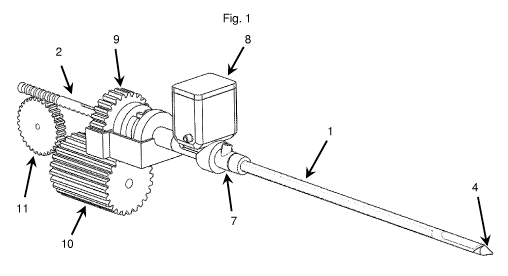
[0012] Fig. 1 is an exemplary embodiment of a biopsy device according to the
invention.
[0013] Fig. 2 is an exploded view of the components in Fig. 1.
[0014] Fig. 3 is a detailed view of a rigid toothed rack with a sharpened tip
and sample
notch at the distal end and a rotation zone in the proximal end.
[0015] Fig. 4a shows a cutting cannula in an advanced position covering a
sample notch.
[0016] Fig. 4b shows a cutting cannula in a retracted position exposing a
sample notch.
[0017] Fig. 5 shows a damper spring for use in connection with overshoot of a
spring-
loaded cutting cannula.
[0018] Fig. 6 shows a counter-rotation cutting interface between a sample
notch and a
cutting cannula.
[0019] Fig. 7 is a cross-sectional view of a cutting cannula featuring a
longitudinal air
channel having a lateral vent hole.
[0020] Fig. 8a shows a tissue collection tank.
[0021] Fig. 8b is a cut through illustration of the tissue collection tank in
Fig. 8a.
[0022] Fig. 9 illustrates the cutting interface between a cutting cannula and
a protrusion
at the inner member forming a cutting board for the cutting cannula. A cut-out
in the
drawing shows the longitudinal air channel and a plurality of vent holes in
the cutting
cannula.
[0023] Corresponding reference characters indicate corresponding parts
throughout the
several views. The exemplifications set out herein illustrate embodiments of
the invention
and such exemplifications are not to be construed as limiting the scope of the
invention in
any manner.
DETAILED DESCRIPTION OF THE INVENTION
[0024] The drawings illustrate exemplary biopsy devices which are provided
with a
needle portion comprising a cutting cannula 1, 1' and a sample notch 3 with a
sharpened
distal tip 4 for piercing tissue. The cutting cannula 1 is provided with a
slanted cutting
profiled as illustrated e.g. in Figs. 2 and 6, whereas the cutting cannula 1'
is provided with
a straight cutting profile 24 as illustrated e.g. in Figs. 5, 7 and 9. The
sample notch 3 is
part of a rigid toothed rack 2, and is movable between a first advanced and a
second
retracted position when actuated by a suitable source of mechanical motion.
The source of
CA 02902221 2015-08-21
WO 2014/153410 PCT/US2014/031224
mechanical motion may be a motor that may be powered by a battery and
operatively
connected to the rigid toothed rack 2 by means of one or more gear wheels 11.
[0025] The operative connection between the rigid toothed rack 2 and the gear
wheels
11 is configured to permit full 360 degree rotation of the toothed rack 2,
including the
sample notch 3, about its longitudinal axis. Such rotation may for instance be
permitted by
providing a proximal rotation zone 12 with a series of cut-outs that run
around the entire
circumference of the toothed rack. A rotation control gear 9, that is in
operative
connection with the rigid toothed rack, is engaged by a rotation driver gear
10 to support
the rotation of the rigid toothed rack 2 about its longitudinal axis. Another
set of
gearwheels may be in operative engagement with the cutting cannula 1 to
provide full
360-degree rotation of the cutting cannula 1 either independently or in step
with the
rotation of the rigid toothed rack 2.
[0026] The cutting cannula 1, l' may be retracted when actuated by a suitable
source of
mechanical motion. In the first embodiment, the source of mechanical motion
may be a
second motor that is powered by a battery and operatively connected to the
cutting cannula
1, l' by means of a series of gears driving an actuator rod. Retraction of the
cutting
cannula 1, l' exposes the sample notch 3, 3', and permits tissue to prolapse
into the lateral
opening of the sample notch 3, 3'.
[0027] During or after retraction of the cutting cannula 1, l', a vacuum may
be switched
on to support the prolapsing of tissue into the sample notch 3, 3'. Vacuum is
communicated from a vacuum pump and a hose through a vacuum gasket 7 that is
in
operative connection with the cutting cannula 1, 1' and into the inner lumen
of cutting
cannula 1, 1'. The rigid toothed rack 2 is provided with at least one vacuum
cut-out 16 that
run along the length of the rigid toothed rack 2, and end in sample notch 3,
and the
vacuum from the vacuum pump is communicated through these vacuum cut-outs 16
to the
sample notch 3 as soon as the pump is turned on.
[0028] A vacuum accumulator/reservoir may be configured to build and store a
vacuum,
is also in fluid communication with the sample notch 3, 3', and may provide a
transient
boost to the vacuum strength immediately prior to firing of the cutting
cannula 1, l' to
increase sample size.
[0029] Retraction of the cutting cannula 1, l' cocks a spring-loaded firing
mechanism
that is capable of powering the cutting cannula forward (i.e. in a distal
direction) at high
CA 02902221 2015-08-21
WO 2014/153410 PCT/US2014/031224
6
speed. As the cutting cannula 1, 1' moves forward at high speed, the sharpened
distal end
of the cannula 1, 1' makes contact with the tissue that has prolapsed into the
sample notch
3, 3' and severs it from the surrounding connecting tissue.
[0030] As illustrated in Fig. 5 the cutting cannula 1' may be permitted to
continue its
travel by a damper spring 13 that is placed in a damper spring housing 14 and
is in
operative connection with a rear flange 15 of the cutting cannula 1'. The
inertia of the
cutting cannula l' will allow it to proceed 1-2 mm beyond the permissible
traveling
distance of the spring-loaded firing mechanism, and will ensure that the
sharpened distal
end of the cutting cannula 1' has achieved a suitable overlap with the distal
section of the
sample notch 3'. Following the overthrow, the damper spring 13 ensures that
the cutting
cannula l' is returned to its neutral position in preparation for the next
tissue sample.
[0031] As illustrated in Fig. 6 the user of the biopsy device has the option
of rotating the
toothed rack 2 with the sample notch 3 relative to the stationary cutting
cannula 1 to sever
any connective tissue that has not been completely severed by the cutting
cannula 1.
Connective tissue that has not been completely severed may restrict retraction
of the tissue
sample and cause pain to the patient. The rotation causes connective tissue
that has not
been completely severed to saw against the sharpened distal end of cutting
cannula 1 for as
long as needed to complete the severing. Rotation may be step-wise and may
interchange
between a clockwise and a counter-clockwise direction and take place over a
rotation
angle of e.g. +/- 20 degrees relative to a neutral position. Furthermore the
cutting cannula
1 may be retracted and advanced in steps of 1-2 mm during rotation to further
support the
severing of tissue. When unrestricted movement of the sample notch 3 has been
restored,
the toothed rack 2 may continue its motion from the first advanced to the
second retracted
position to transport the tissue sample in sample notch 3 out of the body of
the patient.
[0032] The tissue sample may be collected in a tissue collection tank 8
comprising a
vacuum spout 21 through which a vacuum from a vacuum pump or vacuum
accumulator
may be communicated into a collection tank chamber 22. From the collection
tank
chamber, the vacuum may be communicated through a tissue collection spout 23
for
enhanced collection of the tissue sample. As illustrated in Fig. 8b the
collection spout 23
forms a collection tube 23' inside the collection tank 22 extending with a
certain length
from the bottom of the tank 22. Following collection of the tissue sample from
the sample
CA 02902221 2015-08-21
WO 2014/153410 PCT/US2014/031224
7
notch 3, said sample notch 3 may be returned to the sampling site for
collection of the next
tissue sample.
[0033] As illustrated in Figs. 7 and 9 the cutting cannula may have an inner
tube 17 and
an outer tube 18 forming between them a longitudinal air channel 19 that is at
a proximal
end in fluid communication with a first vacuum pump through a two-way valve
that may
be switched between a vacuum position and a position that permits entry of
atmospheric
air into the air channel. At the distal end the air channel 19 is in fluid
communication with
the lateral opening of the sample notch 3 through at least one vent hole 20
that is formed
in the inner tube 17.
[0034] As illustrated in Fig. 9 a plurality of the vent holes 20 may be
distributed
circumferentially around the inside of the inner tube 17. As illustrated in
Fig. 9 a
protrusion 25 formed as a collar may be provided adjacent to the sharpened
distal end 4'.
The interface 26 between the protrusion 25 and the cutting cannula l' forms a
cutting
board to ensure that connective tissue is cut properly during severing.
[0035] A frequently encountered complication in the harvesting of tissue
samples is the
presence of fibrous or connective tissue. Such tissue is characterized by
being highly
resilient and difficult to cut. The typical manifestation of malfunctions
related to
connective tissue is that the biopsy device gets stuck in the body of the
patient and has to
be removed by force or surgical intervention. This may be stressful to both
physician and
patient and may additionally be very painful for the patient. Inadequately
severed
connective tissue is a known problem for all kinds of biopsy devices and the
problem is
highly undesirable.
[0036] The use of a linear cutter requires a very precise interplay between
the sharpened
distal end of the cutting cannula and the distal section of the sample notch
if appropriate
severing of connective tissue is to occur. For this reason it is important
that the position of
the sample notch is very precisely controlled relative to the position of the
cutting cannula.
SIMS devices featuring a linear, spring-loaded cutting cannula typically
employ a sample
notch that is attached to a flexible bendable elongate member (e.g. a non-
rigid toothed
rack), and this toothed rack may not always produce the desired control of
position of the
sample notch due to the flexibility, design and material chosen. Some prior
art devices
employ toothed racks made of thermoplastic elastomers with significant
longitudinal
elasticity. By having the sample notch in a rigid toothed rack, which is
longitudinally
CA 02902221 2015-08-21
WO 2014/153410 PCT/US2014/031224
8
inelastic, a better control of position is provided. Thereby an appropriate
overlap of the
sharpened end of the cutting cannula with the distal section of the sample
notch can be
provided. Failure to establish a precise position of the sample notch may
result in the
incomplete closing of the sample notch opening. A rigid toothed rack provides
the
necessary lateral inelasticity and stability to ensure that the sharpened
distal end of the
cutting cannula completely closes the opening of the sample notch. Employing a
rigid
toothed rack therefore provides an improved control of the longitudinal and
lateral
position of the distal sharpened end of the cutting cannula relative to the
distal section of a
sample notch.
[0037] In one embodiment the proximal end of the rigid toothed rack is
configured to
operatively connect with a retraction gear wheel, and is furthermore
configured to permit
360 degree rotation of the toothed rack about its longitudinal axis without
requiring that
the operative connection with the retraction gearwheel is interrupted. This
may be
provided by means of a rotation mechanism.
[0038] In a further embodiment of the invention the rigid toothed rack
comprises a
rotation zone in the proximal end with circumferential teeth, e.g. in the form
of a series of
cut-outs that run around the entire circumference of the toothed rack, thereby
permitting
rotation of the rigid toothed rack. The rigid toothed rack may be rotatable
within the
cutting cannula and/or the rigid toothed rack and the cutting cannula are
rotatable
simultaneously relative to the biopsy device. The permitted rotation may be
360 degrees.
The biopsy device may further comprise a rotation control gear attached to the
rigid
toothed rack. A rotation driver gear may be provided and configured to engage
with the
rotation control gear for rotation of the rigid toothed rack. The cutting
cannula may also be
configured to rotate, such as 360 degrees, about its longitudinal axis.
[0039] In a further embodiment of the invention the rigid toothed rack is
configured
such that longitudinal displacement of the rigid toothed rack to the second
retracted
position can only be provided in a predefined rotational orientation of the
rigid toothed
rack. Thus, the rigid toothed rack may be rotatable within the cutting cannula
only in the
first advanced position, and/or the rigid toothed rack and the cutting cannula
are rotatable
simultaneously relative to the biopsy device only in the first advanced
position.
[0040] Whether the rigid toothed rack and/or the cutting cannula is rotated
simultaneously or independently may at least partly be controlled by means of
an interlock
CA 02902221 2015-08-21
WO 2014/153410 PCT/US2014/031224
9
mechanism configured for fixing the rigid toothed rack and the cutting cannula
relative to
each other. E.g. the interlock mechanism may have two states, one state that
allows free
movement of the cutting cannula and the toothed rack relative to each other
and one state
that fixes the two to each other.
[0041] This may help to ensure that the sample notch is always oriented
correctly with
respect to a tissue collection tank when a tissue sample is transferred to the
tank. This may
be provided if the toothing of the toothed rack is only located at one side of
the rigid
toothed rack. If there is a proximal rotation zone of the toothed rack as
mentioned above,
the toothing that extends in the distal direction beyond the rotation zone is
only located at
one side of said rigid toothed rack. A control system may help to ensure that
the rigid
toothed rack has the correct rotational orientation before retracting to the
retracted
position.
[0042] Rotation of the rigid toothed rack relative to the cutting cannula (or
vice versa)
may be advantageous during severing of a tissue sample and may thereby be an
improvement of the cutting mechanism. Rotation of the toothed rack, and
thereby the
sample notch, relative to the cutting cannula with the sharpened distal end,
may result in a
"sawing" motion that may complete the severing of incompletely severed
connective
tissue. Counter-rotation of the cutting cannula and the rigid toothed rack may
further be
provided during cutting which allows for enhanced cutting of e.g. connective
tissue.
[0043] Thus, in one embodiment of the invention the rigid toothed rack is
rotatable
within the cutting cannula during severing of the at least one tissue sample.
The cutting
mechanism may be configured to rotate the rigid toothed rack within the
cutting cannula
during severing of the at least one tissue sample. The rotation may be either
stepwise or
continuous. The rigid toothed rack and/or the cutting cannula may be rotatable
in
clockwise and/or in counter-clockwise directions. During severing the rotation
angle of the
rigid toothed rack relative to the cutting cannula may oscillate between -5
and +5 degrees
during severing, more preferably between -10 and +10 degrees, more preferably
between -
15 and +15 degrees, more preferably between -20 and +20 degrees, more
preferably
between -25 and +25 degrees, more preferably between -30 and +30 degrees, i.e.
like a
sawing motion oscillating between clock-wise and counter clock-wise
directions.
[0044] When taking a biopsy it is often necessary to rotate the entire biopsy
device
inside the patient in order to position the sample notch against the suspect
tissue mass.
CA 02902221 2015-08-21
WO 2014/153410 PCT/US2014/031224
This may lead to awkward handling situations during harvesting of tissue
samples. A
further advantage of rotational capability is therefore that the rigid toothed
rack and the
cutting cannula can be rotated simultaneously, preferably controlled by the
user, about
their longitudinal axis relative to the biopsy device in order to orientate
the sample notch
towards the suspect tissue mass, e.g. prior to activation of the firing
mechanism. Thus, the
biopsy device can be held in a steady position while the rigid toothed rack
and the cutting
cannula are rotated into the correct angular orientation relative to the
suspect tissue mass.
[0045] Another way to enhance the correct severing of tissue is if the cutting
mechanism
is configured to interchangeably retract and advance the cutting cannula in
small
longitudinal steps during severing of a tissue sample. The size of the steps
may between 0
and 3 mm, or between 0 and 1 mm, or between 1 and 2 mm or between 2 and 3 mm.
This
corresponds to a sawing motion in the longitudinal direction.
[0046] The cutting mechanism may also be improved if it is configured to
provide a
predefined overlap and/or overshoot during severing of a tissue sample such
that the distal
end of the cutting cannula passes beyond the distal end of the sample notch
temporarily
before returning to said second position. The length of said overshoot may be
between 0.5
and 5 mm, or between 0.5 and 1 mm, or between 1 and 2 mm, or between 2 and 3
mm, or
between 3 and 4 mm, or between 4 and 5 mm. This overshoot of the cutting
cannula may
help to apply further stress to incompletely severed tissue. The overshoot may
be provided
by means of an elastic element provided in connection with the cutting
cannula. One
solution could be in the form of at least one damper spring mounted in a
damper spring
housing. The damping may also be provided by using a damping element formed in
rubber. The elastic element may be configured to work along with a firing
mechanism of
the cutting cannula effected during severing of a tissue sample. If the firing
mechanism is
stopped by the elastic element the inertia of the cutting cannula and the
elasticity of the
elastic element will allow the sharpened end of the cutting cannula to proceed
a certain
length beyond the traveling distance of the spring-loaded firing mechanism,
and thereby
ensure that the sharpened distal end of the cutting cannula achieves a
suitable overlap with
the distal section of the sample notch. Subsequent to this overshoot, the
elastic element
ensures that the cutting cannula can be returned to its neutral position in
preparation for
the next tissue sample.
CA 02902221 2015-08-21
WO 2014/153410 PCT/US2014/031224
11
[0047] As an alternative, or supplement to, an overlap or overshoot between
the distal
sharpened end of the cutting cannula and the distal section of the sample
notch, the inner
member may further comprise a circumferential protrusion and/or collar located
between
the sharpened distal end and the sample notch, said circumferential protrusion
formed to
match the distal end of the cutting cannula. The circumferential protrusion
may thus be
configured to form a cutting surface for the cutting cannula during severing
of a tissue
sample. The cutting board (protrusion) may be disposed about the outer
periphery of the
sample notch and serve the purpose of ensuring that the tissue sample is
completely and
cleanly severed by the cutting cannula. The cutting mechanism may be
configured such
that the cutting cannula and the circumferential protrusion encounter during
severing of a
tissue sample. The protrusion is then preferentially formed in a material that
is softer than
the cutting cannula in order not to blunt the cutting cannula and preserve the
sharpness of
the cutting cannula. The cutting mechanism may alternatively be configured
such that the
cutting cannula and the circumferential protrusion does not encounter during
severing of a
tissue sample. Thus, the circumferential protrusion may be brought into close
proximity
without encountering during severing of a tissue sample. I.e. direct physical
contact
between the protrusion and the sharpened distal end of the cutting cannula is
avoided but
established at the material surface in close proximity to said sharpened
distal end. With
such a protrusion the transport of the tissue sample must be provided through
the inside of
the inner member, typically by means of vacuum, if SIMS functionality is
desired.
[0048] In a further embodiment of the invention the cutting cannula comprises
at least
one longitudinal vacuum channel (aka longitudinal air channel or passage)
formed inside
the external shell / wall of the cutting cannula. The longitudinal vacuum
channel may be
circumferential. This air channel may be provided by forming the cutting
cannula as an
inner and an outer tube forming between them an air passage that runs
longitudinally
along the length of the inner and outer tube. Fluid communication from this
air channel
and into the inner lumen of the cutting cannula may be provided by one or more
lateral
vent holes extending from the inside of the cutting cannula to the
longitudinal air channel.
A plurality of said lateral vent holes may be distributed circumferentially in
the cutting
cannula. The longitudinal vacuum channel may then, in its distal end, be in
fluid
communication with the sample notch when the rigid toothed rack is in its
first advanced
position. Thereby the cutting cannula may be configured such that a vacuum or
air flow
CA 02902221 2015-08-21
WO 2014/153410 PCT/US2014/031224
12
can be provided and/or established inside the cutting cannula, e.g. an airflow
from the air
channel and into the inner lumen of the cutting cannula. Fluid communication
from this air
channel and to the external of the cutting cannula may be provided by at least
one vacuum
spout and may be controlled by at least one vacuum valve. A vacuum pump may
then be
connected to the air channel via this vacuum valve, in which case a vacuum may
be
communicated through the air channel and the air vent holes and into the inner
lumen of
the cutting cannula. Thus, air may be sucked out of the inner lumen of the
cutting cannula.
Such evacuation may be useful for reducing or eliminating problems with air
that has been
accidentally introduced in the biopsy cavity and disturbs image quality in an
ultrasound-
guided biopsy procedure. Unwanted air may be introduced in the biopsy cavity
when the
rigid toothed rack is being advanced from the second retracted position and to
the first
advanced position. This advancement of the rigid toothed rack inside the
cutting cannula
may function as a piston that compresses the air inside the cutting cannula
and this air is
consequently blown into the biopsy cavity disturbing the ultrasound picture.
If air is
evacuated from the cutting cannula through the longitudinal vacuum channel
inside the
sidewall of the cutting cannula during advancement of the rigid toothed rack
this problem
can be addressed and solved.
[0049] A further embodiment of the invention comprises a tissue collection
tank for
collecting the at least one tissue sample transferred from the sample notch.
The tank may
comprise a tissue-collecting spout that may be configured to slide into the
sample notch
chamber and scoop the tissue sample into a sample tank. To enhance the
collection of the
tissue sample the tissue collection tank may be configured to be vacuumized,
e.g. by
connection to a vacuum pump via a vacuum port at the tank. The collecting
spout may be
elongated to form a pipe (aka collection pipe) to enhance the vacuum assisted
collection of
a tissue sample into the tank. At the outside the collection spout / pipe
forms a small spout
but at the inside of the tissue collection tank the collection pipe extends
and/or protrudes
into the tissue collection tank, i.e. the collection pipe may protrude from
the bottom or side
of the inside of the tissue collection tank. Thus, the collection pipe has a
certain length
inside the tissue collection tank. This length of the collection pipe may be
at least 2 mm, or
at least 4 mm, or at least 6 mm, or at least 8 mm, or at least 10 mm, or at
least 12 mm, or
at least 14 mm, or at least 16 mm, or at least 18 mm, or at least 20 mm, or at
least 22 mm,
CA 02902221 2015-08-21
WO 2014/153410 PCT/US2014/031224
13
or at least 24 mm, or at least 26 mm, or at least 28 mm, or at least 30 mm, or
at least 32
mm, or at least 34 mm, or at least 36 mm, or at least 38 mm, or at least 40
mm.
[0050] Some biopsy devices are constantly connected to external vacuum pumps
via
external vacuum hoses. These pumps can deliver a powerful and constant vacuum
to the
biopsy device but the necessary vacuum hoses reduce the manageability of the
biopsy
device for the user. A solution to that problem has until now been to provide
one or more
local battery driven small vacuum pumps integrated in the biopsy device.
However, such
small vacuum pumps can only provide a limited airflow which sometimes is not
sufficient
to maintain a constant vacuum level. A solution to that problem can be a
vacuum reservoir
integrated in the biopsy device that can deliver a boost to the (negative)
airflow for one or
more short periods of time, this additional airflow provided by the vacuum
reservoir can
thereby maintain a certain vacuum level. The biopsy device can thereby be
provided with
one or more small vacuum pumps supplied by the vacuum reservoir when
necessary. A
further embodiment of the invention therefore comprises a vacuum reservoir
(aka vacuum
accumulator) configured for accumulating a volume of vacuum that can be
delivered as a
transient boost in the airflow so as to maintain a level of vacuum present in
the system.
Such a vacuum reservoir can for instance be powered by a battery. The vacuum
reservoir
may be in fluid communication with the sample notch and configured to provide
an
increased suction to maintain the vacuum level in the sample notch during
severing of a
tissue sample, e.g. immediately before release of the cutting cannula in order
to increase
the amount of tissue that prolapses into the sample chamber and thereby
maximize the size
of the severed tissue sample. The vacuum reservoir may also be in fluid
communication
with the inside of the hollow inner member and configured to provide a
transient boost of
airflow when a tissue sample is being sucked through the inner member.
Furthermore, the
vacuum reservoir may be in fluid communication with the tissue collection tank
and
configured to provide a vacuum to or an increased suction in the tissue
collection tank to
main a vacuum level when a tissue sample is transferred from the sample notch
and into
the tissue collection tank. The vacuum reservoir may have a volume of 5-100
mL, or 5-10
mL, or 10-20 mL, or 20-30 mL, or 30-40 mL, or 50-100 mL.
[0051] Retraction of the cutting cannula to expose the sample notch may for
instance be
actuated by a motor that is powered by a battery and connected to one or more
gearwheels,
but other power sources and means of mechanical actuation are also envisioned.
This
CA 02902221 2015-08-21
WO 2014/153410 PCT/US2014/031224
14
retraction of the cutting cannula may facilitate the cocking of a firing
mechanism that may
for instance be spring-loaded. Other firing mechanisms, including electric,
pneumatic and
chemical, may also be provided. The cutting movement of the cutting cannula
during the
actual severing of tissue may be powered by the energy that is stored in a
firing
mechanism and happens as a high-speed linear passage across the laterally
facing opening
of the sample notch. During this passage, the sharpened distal end of the
cutting cannula
makes contact with the tissue that has prolapsed into the sample notch chamber
and severs
it from the surrounding tissue, thus creating a tissue sample in the sample
notch. The firing
mechanism may be replaced with a linear actuator that allows the controlled
advancement
of the cutting cannula during severing. In this case advancement of the
cutting cannula is
more controlled and it may be desirable to rotate the cutting cannula during
advancement
to adequately sever the tissue as described previously.
[0052] To provide for SIMS functionality retraction of the sample notch may be
provided by means of a motor that is operatively connected to the rigid
toothed rack by
means of one or more gearwheels. When activated, this motor causes the rigid
toothed
rack and the sample notch to travel from the first advanced position to the
second retracted
position, where the sample may be retrieved, e.g. by means of a tissue
collection tank, but
other means of retrieval ¨ including manual retrieval ¨ may also be
envisioned. After
completion of sample retrieval, the sample notch may be returned to the
sampling site by
reversing the direction of rotation of the motor.
[0053] The firing mechanism may be configured for causing the cutting cannula
and the
inner member to be longitudinally displaced in a distal direction, so as to
penetrate body
tissue at or near the suspect tissue mass prior to the cutting operation when
harvesting a
sample.
[0054] In one embodiment of the invention the inner member comprises a vacuum
port
in fluid communication with the sample notch. The inner member may thus be
configured
such that the sample notch can be vacuumized. A vacuum pump may be provided
for
generating a suction effect in the sample notch to increase the size of the
tissue sample that
prolapses into the sample notch, the vacuum pump being in fluid communication
with the
sample notch through a longitudinally extending passage in the inner member.
[0055] A further embodiment of the invention comprises a handle unit with a
power
source and at least one motor for driving the cutting mechanism and the
displacement of
CA 02902221 2015-08-21
WO 2014/153410 PCT/US2014/031224
the inner member and wherein at least the cutting cannula and the inner member
are
comprised in a disposable unit, which is releasably secured to the handle
unit.
[0056] To ensure that the cutting cannula and the sample notch achieve an
overlap that
is sufficient to cleanly sever the tissue to be sampled, the cutting cannula
is preferably
characterized by very tight length tolerances. Such tolerances may be achieved
by the use
of materials with low creep that are processed using high-precision milling or
molding,
and possibly result in total length variations of no more than +/- 0.5 mm
depending on the
overall total length of the cutting cannula. A preferred material for the
cutting cannula is
stainless steel which is made into tubes. These tubes are typically made by
rolling and
welding sheet metal to form a tubular structure which is then drawn through a
tool with a
diamond insert to achieve the desired diameter. Multiple drawings may be
employed to
achieve high precision. By utilizing stainless steel low creep for the cutting
cannula, none
or minimal elongation and achievable manufacturing tolerances are possible.
Other
materials, including titanium, are also envisioned for the making of the
cutting cannula.
[0057] To further support appropriate overlap between cutting cannula and
sample
notch, also the rigid toothed rack may be characterized by very tight length
tolerances.
Such tolerances may in some embodiments be achieved by the use of materials
with low
creep that are processed using high-precision milling or molding, and possibly
result in
total length variations of no more than +/- 0.5 mm depending on the overall
total length of
the rigid toothed rack. A preferred material for the rigid toothed rack is
stainless steel. The
rigid tooted rack would typically be made by milling a turned stainless steel
metal rod in
order to achieve the desired geometry. Other materials suited for the rigid
tooted rack are
titanium or similar metals with a high modulus of elasticity. Alternative
materials include
thermoplastic elastomers with suitable fillers for increased modulus of
elasticity. Suitable
types for a rigid toothed rack would be LCP (Liquid Crystal Polymer), PEEK
(Polyetheretherketone) in any grade. Thermoplastic elastomers have the benefit
of being
relatively easy to process and manufacture, but they are less rigid and will
also tend to
creep and shrink more than metal.
[0058] While this invention has been described with respect to at least one
embodiment,
the present invention can be further modified within the spirit and scope of
this disclosure.
This application is therefore intended to cover any variations, uses, or
adaptations of the
invention using its general principles. Further, this application is intended
to cover such
CA 02902221 2015-08-21
WO 2014/153410
PCT/US2014/031224
16
departures from the present disclosure as come within known or customary
practice in the
art to which this invention pertains and which fall within the limits of the
appended
claims.