Note: Descriptions are shown in the official language in which they were submitted.
CA Application No. 2,902,450
LOCATOR DEVICE FOR MEDICAL PROCEDURES ON THE BODY SURFACE AND
METHOD OF ITS USE
TECHNICAL FIELD
The present invention relates generally to medical devices and methods. More
specifically, the invention relates to devices and methods, including those
for delineating
portions of a body surface during a procedure, creating tension on the body
surface during
procedure and/or providing fiducials in image-guided procedures.
BACKGROUND
There are numerous surgical, cosmetic, therapeutic and dermatological
procedures
that involve precise placement of medical instruments on a body surface and/or
the need to
repeat a procedure multiple times at various locations on a body surface. Hair
transplantation
surgery is one example of such procedures, and it typically involves
harvesting donor hair grafts
from "donor areas," and implanting them in one or more bald areas ("recipient
areas''). Hair
transplantation surgery is a very labor-intensive and complex procedure that
requires great skill
and precision. When performed completely manually, hair transplantation
surgery typically
requires multiple, lengthy surgical procedures performed over time. As such,
the assignee of the
present application has developed an image-guided system for harvesting
follicular units from a
body surface, as described for example in U.S. Patent Publication Number
2007/0106306. Image
guidance is often used to direct movement of automated systems, such as a
system for harvesting
and implanting follicular units and/or performing other procedures on the skin
or other body
surfaces. One example of an image-guided, automated method and system is
described in U.S.
Patent Publication Number 2012/0158019.
In performing a procedure on a body surface of a patient, it is often
necessary or
desirable to perform the procedure on multiple portions of the body surface,
with each
subsequent portion located immediately adjacent to the prior portion so that
there are no gaps
between, or overlap of, the multiple body surface portions. In other
procedures, it may be
desirable to have specific and/or consistent amounts of gaps or overlaps
between the multiple
body surface portions. Also, using some automated systems, such as those
described in
1
CA 2902450 2018-05-15
CA 02902450 2015-08-25
WO 2014/158505 PCT/US2014/017514
RR-041 PCT
reference to certain embodiments of the above-referenced patent applications,
it may also be
necessary to use fiducial markers to guide the system to perform the
procedure. In any of these
cases, it can be challenging performing a procedure on multiple body surface
portions located in
desired locations relative to one another. Typically, for example, this may
involve manually
moving measuring devices, manually marking skin surfaces, approximating
locations where
prior procedures were performed, and the like. It can also be challenging to
assure proper, stable
and consistent positioning of fiducial markers in a treatment area. Various
embodiments
described below seek to address at least some of these challenges.
SUMMARY
The various embodiments described herein are directed to devices and methods
for
performing a procedure on multiple portions of a body surface. Any of a number
of different
procedures or portions of procedures may be performed, using devices and
methods described
herein. In some embodiments, the devices and methods may simply facilitate or
enhance a
procedure. In general, as used herein, the phrase "performing a procedure" is
meant to also
include facilitating and/or enhancing a procedure and/or performing,
facilitating and/or
enhancing part of a procedure.
The embodiments described herein may be used to perform a procedure on
multiple
portions of a body surface, where the portions are adjacent and non-
overlapping. Alternatively,
the same or other embodiments may be used to perform a procedure on multiple
portions of a
body surface, where the portions are overlapping, for example, by a uniform,
desired amount or
where a desired amount of gap is present between the body surface portions.
The various
embodiments described herein typically make it easier to perform procedures on
multiple body
surface portions at consistent locations relative to one another, such as
immediately adjacent to
one another.
According to one aspect, to facilitate a procedure on multiple, typically
adjacent and
non-overlapping, body surface segments, the devices and methods described
herein typically
involve a locator device with a first portion and a second portion. When the
portions are coupled
together, they delineate a body surface segment or area on which the procedure
may be
performed. The first portion may also be detached (fully or partially) from
the second portion
and moved to a new location, while the second portion remains stationary on
the body surface
2
CA 02902450 2015-08-25
WO 2014/158505 PCT/US2014/017514
RR-041 PCT
and acts as a reference. Once the first portion is repositioned on the body
surface, the second
portion can be moved to rejoin the first portion, thereby delineating a second
body surface
segment on which the procedure may be performed. This process may be repeated
as often as
desired to perform a procedure on a desired number of body surface segments or
areas.
In some embodiments, the locator device simply acts as a locator (or -
positioner")
for helping delineate multiple body surface portions for the procedure.
Optionally, the locator
device may also act as a skin/scalp tensioner. In other embodiments, the
locator device may
include multiple fiducials (or "fiducial markers") for guiding an image-guided
system that
performs the procedure. In some embodiments, the locator device may be a
skin/scalp tensioner
and also include fiducials.
In some embodiments, the locator device may remain in position while a
procedure
is performed on a delineated body surface portion. In alternative embodiments,
the locator device
may be used for marking the body surface, the locator may then be moved, and
the procedure
may be performed on the marked portion of the body surface. In some
embodiments, the locator
device may include a frame that has a central opening, and the opening
delineates the body
surface portions. Alternatively, an outer edge or some other feature(s) of the
locator device may
delineate the body surface portions in other embodiments.
According to one aspect of the present application, a method for performing a
procedure on a body surface of a patient is provided. The method comprising:
positioning a
locator device on the body surface to delineate a first segment of a body
surface; petforming the
procedure on the first segment of the body surface; moving a first portion of
the locator device
while leaving a second portion of the locator device stationary, the second
portion providing a
reference to guide movement of the first portion relative to the second
portion; and moving the
second portion of the locator device to reform the locator device and
delineate a second segment
of the body surface. In some embodiments, the first portion of the locator
device, or the second
portion of the locator device or both comprises a free end when delineating a
first segment of the
body surface. In other embodiments, at least one of the first portion and the
second portion of
the locator device is moved from first location on the body surface to a
second location on the
body surface, and the other of the first or the second portion of the location
device is moved to
reform at the second location.
3
CA 02902450 2015-08-25
WO 2014/158505 PCT/US2014/017514
RR-041 PCT
According to another aspect of the present application, a method for
performing a
procedure on a body surface of a patient is provided. The method comprising:
positioning a
locator device on the body surface in a first location to delineate a first
body surface segment,
wherein the locator device includes a plurality of fiducials; performing the
procedure on the first
body surface segment, using an image-guided system guided at least in part by
the plurality of
the fiducials; moving a first portion of the locator device on the body
surface while leaving a
second portion of the locator device stationary, wherein the second portion of
the locator device
provides a reference to guide movement of the first portion relative to the
second portion;
moving the second portion of the locator device to reform the locator device
and delineate a
second body surface segment.
In one embodiment, for example, the locator device may include at least four
fiducial
markers disposed on four opposing sides of the locator device such that the
markers form a grid
pattern on the body surface. In some embodiments, the locator device includes
a central opening
that delineates the first and second body surface portions. The opening may be
square-shaped,
and the fiducial markers may be disposed along each side of the square-shaped
opening. In one
embodiment, for example, at least four fiducial markers are disposed along
each side of the
opening.
In another embodiment, a method for performing a procedure on a body surface
of a
patient may involve: performing a procedure on a body surface of a patient,
the method
comprising: positioning a locator device on the body surface in a first
location; performing the
procedure on a first segment of the body surface delineated by the locator
device; moving a first
portion of the locator device a selected distance from a second portion of the
locator device to a
second location on the body surface while leaving the second portion of the
locator device on the
body surface in the first location, wherein the selected distance is dictated
by one or more
features of the locator device; and moving the second portion of the locator
device to reform the
locator device in the second location and thus delineate a second body surface
segment. In some
embodiments, the method may also include performing the procedure on the
second body surface
portion. Optionally, the method may further involve: moving the first portion
of the locator
device to a third location on the body surface while leaving the second
portion of the locator
device on the body surface in the second location; moving the second portion
of the locator
4
CA 02902450 2015-08-25
WO 2014/158505 PCT/US2014/017514
RR-041 PCT
device to reform the locator device in the third location and thus delineate a
third body surface
portion; and performing the procedure on the third body surface portion.
In various embodiments, the first and second portions of the locator device
may be
moved relative to one another in any of a number of suitable ways. For
example, the first portion
may be moved relative to the second portion by rotating, turning around,
flipping over,
swiveling, pivoting, or lifting and repositioning the first portion of the
locator device relative to
the second portion of the locator device.
In some embodiments, the body surface may be scalp, and the procedure may
involve harvesting a hair graft, making a site for hair implantation or
implanting a hair graft. The
procedure may include tattooing skin, removing a skin graft, attaching a skin
graft, or any other
suitable procedure.
In some embodiments, the method further involves disconnecting the first
portion of
the locator device from the second portion of the locator device before moving
the first portion to
the second location. In one embodiment, one or both of the first and the
second portions of the
locator device may comprise a free end, which can be used as a reference
feature. In such an
embodiment, for example, one end of the first portion of the locator device,
when moved to the
second location, may abut the free end of the second portion of the locator
device. In other
words, the previously free end of the second portion acts as a reference for
positioning the first
portion. In an alternative embodiment, the reference feature of the locator
device may comprise
at least one connector, connecting the first portion with the second portion,
such that the first
portion can only be moved the selected distance from the second portion due to
the at least one
connector.
In some embodiments, the method may also involve attaching the locator device
to
the body surface, such as skin, in the first and second locations such that
the locator device is
immobile relative to the body surface. For example, attaching the frame to the
body surface may
include adhering the frame to the skin using at least one of adhesive, pins,
hooks, barbs or
needles.
In another aspect of the application, a device for facilitating a procedure on
a body
surface of a patient is provided. The device comprising: a frame configured to
delineate a
segment of the body surface on which the procedure is performed, the frame
comprising; a first
portion; a second portion operatively connected to, and at least partially
detachable from, the first
5
CA 02902450 2015-08-25
WO 2014/158505 PCT/US2014/017514
RR-041 PCT
portion; a coupling member on at least one of the first and second portions
configured to allow
the first and second portions to connect detachably to one another; a bottom
surface; and a top
surface; and at least one reference feature on or connected to at least one of
the first portion or
the second portion of the frame so that the first and second portions of the
frame can be moved to
different locations on the body surface to delineate multiple segments of the
body surface.
In some embodiments, the frame may be at least partially flexible to conform
to the
body surface of the patient. In some embodiments, the frame may include a
central opening,
where the central opening delineates a segment of the body surface where the
procedure is
performed through the opening with the frame in place on the body surface.
In some embodiments, the at least one reference feature may include an edge of
the
first portion of the frame and a corresponding edge of the second portion of
the frame, such that
when the first portion is moved from a first location on the body surface to a
second location, the
edge of the first portion is made to abut the corresponding edge of the second
portion. In another
embodiment, the at least one reference feature may include at least one
connector configured to
connect the first portion of the frame to the second portion while the first
portion is being moved
relative to the second portion. For example, in one embodiment, the at least
one connector
includes two hinges on opposite sides of the first and second portions.
As mentioned above, in some embodiments, the device may include at least three
fiducial markers attached to the top surface and/or the bottom surface of the
frame and
configured to guide an image-guided system to perform at least part of the
procedure. For
example, in one embodiment, the procedure may be performed at least in part by
a robotic hair
transplantation system, and the fiducial markers guide the robotic system in
performing the
procedure. In some embodiments, the frame includes at least four fiducial
markers disposed on
four opposing sides of the frame such that the markers form a grid. In some
embodiments, the
frame may include a central opening shaped as a square or rectangle, and the
fiducial markers
may be disposed along each side of the opening. In some embodiments, at least
four fiducial
markers are disposed along each side of the opening.
In some embodiments, the frame also acts as a skin tensioner. The frame may
include at least one skin adhering member coupled with the bottom surface of
the frame. For
example, the at least one skin adhering member may include an adhesive, pins,
hooks, barbs
and/or needles. The device may also include at least two attachment members,
where at least one
6
CA 02902450 2015-08-25
WO 2014/158505 PCT/US2014/017514
RR-041 PCT
of the attachment members is coupled with each of the first and second
portions of the frame to
help attach the first and second portions to at least one of the patient or a
chair or table on which
the patient is sitting or lying.
According to another aspect of the application, a device for facilitating a
procedure on a
.. body surface of a patient is provided. The device comprising: a frame
forming an opening for
delineating a segment of the body surface on which the procedure is performed,
the frame
comprising; a first portion; a second portion connected to, and at least
partially detachable from,
the first portion; and a coupling member on the first and second portions
configured to allow the
first and second portions to detachably couple with one another; at least one
reference feature on
or connected to at least one of the first portion or the second portion of the
frame so that the first
and second portions of the frame can be moved to different locations on the
body surface to
delineate multiple segments of the body surface; and a plurality of fiducial
markers attached to
the frame and configured to guide an image-guided system to perform at least
part of the
procedure.
These and other aspects and embodiments will be described in greater detail
below.
BRIEF DESCRIPTION OF THE DRAWINGS
FIG. 1 is a block diagram illustrating one example of a method for performing
a hair transplant
procedure using fiducials and an image-guided system;
FIG. 2 is a schematic representation of an example of a robotic system that
may be used in
implementing various embodiments of the present application:
FIG. 3 is a block diagram illustrating a method for performing a procedure (or
portion thereof),
according to one embodiment;
FIGS. 4A and 4B are perspective views of an example of a frame with fiducials;
FIGS. 5A-5D are top views of a locator device, according to one embodiment,
illustrating a
method for moving the locator device from a first location to a second
location on a body
surface;
7
CA 02902450 2015-08-25
WO 2014/158505 PCT/US2014/017514
RR-041 PCT
FIGS. 6A-6D are top views of a locator device, according to an alternative
embodiment, also
illustrating a method for moving the locator device from a first location to a
second location on a
body surface, according to another alternative embodiment; and
FIGS. 7A-7E are side views of the locator device of FIGS. 6A-6D, illustrating
the various steps
of the method for moving the locator device, according to one embodiment.
DETAILED DESCRIPTION
In the following Detailed Description, reference is made to the accompanying
drawings that show, by way of illustration, some examples of embodiments in
which the
invention may be practiced. In this regard, directional terminology, such as
"right," "left,"
"upwards," "downwards," "vertical," "horizontal." etc., are used with
reference to the orientation
of the Figure(s) being described. Because components or embodiments of the
present invention
can be positioned or operated in a number of different orientations, the
directional terminology is
used for purposes of illustration and is in no way limiting. Other embodiments
may be used, and
structural or logical changes may be made, without departing from the scope of
the present
invention.
The term "tool," as used herein, refers to any number of tools or end
effectors that
are capable of performing an action, procedure or operation in various medical
procedures or
applications. For example, the tool may be a needle, a surgical scalpel,
blades, various types of
forceps, hemostats, surgical instruments, retractors, electrosurgical tools,
radio-frequency
ablation tools. suturing devices, tattoo placement or removal tools, cannula,
drills or lasers. With
reference to hair transplantation procedures, a "tool" may comprise a
"harvesting tool" or an
-implantation tool," and is capable of dissecting, harvesting or implanting
follicular units
(-FUs") from or into a skin or body surface, for example, a scalp. Such tools
may have many
different forms and configurations. In many embodiments, the tool comprises a
hollow tubular
shaft and thus may be labeled, for example, a cannula, a needle, or a punch.
The distal end of
such tools (for example, punches, coring devices, cutting and/or trimming
devices, needles), are
typically sharpened, to various degrees, to penetrate tissue and extract or
implant the follicular
unit. The terms "operatively connected," "coupled," or "mounted," or
"attached" as used herein,
means directly or indirectly coupled, attached, or mounted through one or more
intervening
components.
8
CA Application No. 2,902,450
Embodiments of the methods of the present invention may be implemented using
computer software, firmware or hardware. Various programming languages and
operating
systems may be used to implement the present invention.
Hair transplantation procedures that are carried out using automated
(including
robotic) systems or computer-controlled systems have been described, for
example, in the
Publication No. US 2007/0106306 commonly owned by the assignee of the present
application.
Robotic systems, such as robotic hair transplantation systems generally
require accurate
positioning of a tool under robotic control. When implementing a semi-
automated or a fully
automated procedure that requires precise control of the position, such as
hair transplantation, it
is desirable to be able to maintain such precise control despite patient
motion or temporary
interruptions.
According to the various embodiments described herein, a variety of devices
and
methods are provided, which enable a tool (or more generally a procedure) to
proceed from
where it left off and/or to facilitate performing a procedure (or a portion of
a procedure) on
multiple portions of adjacent body surface. For example, in reference to hair
transplantation, a
procedure may be performed manually, semi-automatically or in a fully
automated manner,
including using image guidance in some embodiments. In any of these cases,
when a portion of
the procedure is finished on a particular location on the body surface, it may
be necessary or
desirable precisely and accurately move to a next procedure area where hair
grafts will be
harvested or implanted, so that there are no gaps between, or overlapping of,
the first and second
procedure areas. Such gaps may result in underharvesting or underimplanting,
and overlapping
may result in overharvesting or overimplanting in the overlapped area. Using
currently available
methods, it is necessary to manually move fiducials from one location on the
scalp to another (in
the case of image-guided systems) or to manually estimate where a second
procedure location
should be placed relative to a first, and so on. This kind of manual movement
from one location
to another can be very challenging and very dependent upon the skill of the
user performing the
procedure. Furthermore, factors like bleeding can obscure the procedure area
and make the task
of moving from one area to the next without gaps or overlaps even more
difficult.
The various embodiments described herein seek to alleviate these challenges.
The
devices and methods described below generally include a locator device that is
used for guiding
a procedure. The locator device includes two moveable and at least partially
detachable portions,
9
CA 2902450 2018-05-15
CA 02902450 2015-08-25
WO 2014/158505 PCT/US2014/017514
RR-041 PCT
such that one of the two portions remains fixed to the body surface while the
other of the two
portions is being moved and serves as a reference for correct alignment. The
devices and
methods described herein thus provide self-alignment for performing procedures
on multiple
segments of skin, scalp or other body surface. Additionally, in some
embodiments, the devices
may also act as skin tensioners, fiducial carriers, or both.
Although the various examples and embodiments are often described herein with
relation to follicular units (naturally occurring aggregates of 1 to 4 hair
follicles) or hair grafts, in
various alternative embodiments, the various concepts discussed can be applied
more broadly to
other appropriate applications. Additionally, although the methods described
herein are
especially suited for use with image-guided systems (including robotic
systems) for hair
harvesting and/or implanting. they can be applied to other computer-
implemented or image-
guided applications. For example, devices and methods described herein may be
used in various
ablation procedures, biopsy procedures, spinal procedures, dermatological
procedures (e.g..
tattooing or tattoo removal, or treating various dermatological conditions,
such as skin cancers)
and other procedures that could benefit from the locator device described
herein. Therefore, the
examples provided herein are for the purposes of illustration and example
only, and this
description is not intended to be exhaustive or limiting.
FIG. 1 is a block diagram illustrating an example of a methodology of using
image
guidance and fiducials in performing a procedure on a body surface that could
be implemented
.. with the locator device and method of its use according to the present
disclosure. At step 110
(which may be a preliminary step and it is shown in dotted line), one or more
images of the body
surface with one or more reference points, such as a plurality of fiducials,
may be obtained, for
example, using an image acquisition device. This may be accomplished by any
technique known
in the art. For example, in some embodiments, an image acquisition device may
be attached to a
robotic arm, and the robotic arm with the attached image acquisition device
may be positioned so
that the harvesting or implantation region is in focus for the cameras. In
other embodiments, the
image acquisition device may be incorporated into the automated (e.g.,
robotic) system but not
attached to the robotic arm. Alternatively, in further embodiments, the image
acquisition device
could be a device separate from the robotic system.
As used in this application, a "fiducial" (or "fiducial marker") is an object
that may
act as a reference, and may be identifiable in a field of view of an imaging
device. Fiducials can
CA 02902450 2015-08-25
WO 2014/158505
PCT/US2014/017514
RR-041 PCT
take many forms, for example, a single artificial reference point that
uniquely identifies both
position and orientation may be used as a fiducial. Take for example, a set of
coordinate axes
printed on a surface. A set of reference points that each uniquely specifies a
position can be used
as fiducials. The combination of three or more such reference points can
specify a unique frame
of reference specifying both position and orientation. An example would be
spheres with
different colors. One sphere uniquely specifies a position in space, but not
orientation. Two more
spheres can be used to specify both position and orientation.
Although dots placed directly on the skin or natural features, such as
anatomical
landmarks or skin markings, may be used as fiducials in some procedures, they
generally will not
work in procedures discussed herein. Such fiducials are often obscured by
blood and other fluids,
may be washed away (in the case of dots marked on skin with a marker, for
example). Therefore,
various embodiments of a locator device are described herein that include
fiducials, to overcome
some of the drawbacks associated with natural fiducials and fiducials marked
directly on the
skin.
At step 115, a processor or an image processor, an example of which is
described
later in reference to FIG. 2, processes and records an identity and a location
of each of the
fiducials in a frame of reference of an image acquisition device (e.g., in a
camera field of view).
Such initial recording of fiducials could be referred to as "fiducial
registration." The fiducials
could be recorded in various coordinate systems, for example, in a fixed
"world" coordinate
system. In situations in which an image acquired by the image acquisition
device includes only a
subset of the fiducials, such that images of additional fiducials are needed,
step 120 provides for
acquiring additional images as needed, for example, including other subsets of
the fiducials, until
all fiducials have been identified. In an optional step 125 (shown in dotted
line), based on the
location of the each of the plurality of fiducials, a boundary of an area,
such an area within which
hair grafts or follicular units are intended to be harvested from or implanted
into, may be
determined. The boundaries may be determined automatically, for example, by
drawing lines
between various fiducials. The boundaries may be also adjusted to eliminate
certain portions of
the bound area where harvesting or implantation is difficult.
In order to accommodate for patient motion, temporary interruptions, and any
other
incident that may cause a shift in location of the fiducials in the camera
reference frame, as often
as required (as may be determined by the user), updated images of the body
surface are acquired,
11
CA 02902450 2015-08-25
WO 2014/158505 PCT/US2014/017514
RR-041 PCT
the images containing an image of the plurality of fiducials or a subset
thereof. Due to patient
motion, or another such temporary interruption, the locations of the fiducials
in these updated
images may be in a revised location with respect to the frame of reference of
the image
acquisition device.
The processor, in step 130, processes the revised location of each of the
plurality of
fiducials in the frame of reference of the image acquisition device, the
revised locations of each
of the plurality of fiducials which may be different from the locations
previously processed.
Having acquired the revised locations of the fiducials, and with the knowledge
of the original
locations of the fiducials. an offset for at least some or all of the fiducial
locations may be
determined in step 130. Based on this offset information, the processor, also
in step 130, may
process revised locations for each of the locations of interest, such as
locations from which
follicular units have already been harvested (if harvesting has already
started in a region of
interest within the boundary) or into which follicular units have already been
implanted (if such
implanting has been started).
Optionally, step 130 may also comprise determining the revised boundary, for
example, of the harvesting/implanting area based on the revised locations of
the fiducials.
However, it is not necessary, in some embodiments, to determine the whole
revised boundary, as
this information may be automatically ascertained simply based on the offset
of the minimum
number of the fiducials. In reference to the example of hair transplantation,
having determined
the offsets, and with the knowledge of the locations of the follicular units
that have been
harvested or implanted (if any) with respect of the fiducials, it is possible
in step 135 to
determine or select a location from where the next hair follicle is to be
harvested such that hair
follicles are not taken from an already harvested location, or to determine a
location into which
the next hair follicle is to be implanted such that hair follicles are not
implanted into locations
into which hair follicles have already been implanted. Such selection may be
made using a
processor programmed to perform the above-described step, such as a processor
described in
reference to FIG. 2.
In step 140, a hair graft or follicular unit is harvested from or implanted
into the
selected location, or a site for implantation is created. When the next hair
follicle is harvested or
implanted, the location from where it has been harvested from, or implanted
into, or where a new
site is created, may be registered or recorded by the processor in step 145.
This registration may
12
CA 02902450 2015-08-25
WO 2014/158505 PCT/US2014/017514
RR-041 PCT
include information on the location of the harvest or implant with respect to
at least one of the
plurality of fiducials, or the determined boundary.
Optionally, in step 150, the method may comprise creating and displaying a
virtual
representation on the image of the location from which the follicular unit has
been harvested (or
at least dissected from the surrounding tissue for further removal using
forceps or vacuum), or
the location for creating an implantation site, or the location into which a
follicular unit has been
implanted. Such visual representation, for example. on a monitor (e.g. a
computer screen) is
especially beneficial for the user to easily and quickly identify locations
where hair grafts have
been dissected or harvested, and also to differentiate between the previously
existing follicular
units and the newly implanted ones. The visual representations of step 150 may
be implemented
by using different colors, shapes or other appropriate differentiating
features.
In step 155, the processor determines, based on the information it has
recorded with
respect to the area and the locations of the follicular units that have been
harvested or implanted,
if follicular units have been harvested from all desired sites, or if
follicular units have been
implanted into all desired sites, or if all desired implantation sites have
been created. In the event
that all follicular units have been harvested or implanted, or all sites have
been created, the
processor may communicate this information, for example, to the image
acquisition device. In
addition, the processor may communicate this information to the user,
typically providing an
indication to the user (via the monitor, voice command, or any other
appropriate technique), for
example, that step 110 may begin again at a new donor or recipient region.
In the event there are still follicular units to harvest or implant, or sites
to create, the
processor continues to repeat steps 130-155 until all desired sites are
created. or all desired
follicular units are harvested or implanted. For example, updated images with
the updated
fiducial information are processed, offsets determined, the next harvest site
or implant site is
selected, etc. In this manner, a methodology is provided to enable hair
follicles to continue to be
harvested from or implanted into a body surface in a continuous and automatic
fashion despite
potential patient movements and interruptions. The tool is able to be moved to
each new
harvesting or implantation location with respect to fiducials, the fiducials
providing a mechanism
of recognizing the location of the harvesting/implanting area on the body
surface, despite
movement of the patient, or the image acquisition device.
13
CA 02902450 2015-08-25
WO 2014/158505 PCT/US2014/017514
RR-041 PCT
Referring now to FIG. 2, an example of a system that may be used to implement
various embodiments of the method described herein is schematically shown.
FIG. 2 is a
schematic perspective view of an example of a robotic system 200 for hair
harvesting (and/or
implantation). The system 200 includes a robotic arm 205 to which is coupled a
tool 210.
Various motors and other movement devices may be incorporated to enable fine
movements of
an operating tip of the tool 210 in multiple directions. The robotic system
200 further includes at
least one image acquisition device 215, which is described in more detail
below. The image
acquisition device may be mounted in a fixed position, or it may be coupled
(directly or
indirectly) to a robotic arm 205 or other controllable motion device. The
operating tip of the tool
210 is shown positioned over a body surface 220, in this case a part of the
patient scalp having
hair follicles thereon. In some embodiments, an image acquisition device may
be provided
separately and not included in the system. In those embodiments, an interface
may be provided
that allows various other components or modules of the system, such as image
processing
component, to interact with the separate image acquisition device.
A processor 225 may include an image processor 230 for processing images
obtained
from the image acquisition device 215. The image processor 230 may be a
separate device, or it
may be incorporated as a part of the processor 225. The processor 225 may also
instruct the
various movement devices of the robotic arm 205, including the tool 210 that
may be operatively
connected to the robotic arm. The processor 225 may act, for example, through
a controller 235.
The controller 235 may be operatively coupled to the robotic arm and
configured to control the
motion of the robotic arm, including the motion based on the images or data
acquired by the
image acquisition device. Alternatively, controller 235 may be incorporated as
a part of the
processor 225, so that all processing and controls of all movements of all the
tools, the robotic
arm and any other moveable parts of the assembly, including those based on the
images or data
acquired by the image acquisition device, are concentrated in one place. The
system 200 may
further comprise a monitor 240, keyboard 245, and mouse 250. A magnified image
of the body
surface 220 can be seen on the monitor 240. In addition, the system 200 may
include other tools,
devices and components, for example, those useful in harvesting, and/or
implantation of the hair
follicles, or in hair treatment planning. The system further includes an
interface adapted to
receive an image data, various parts of the system allow an operator to
monitor conditions and
provide instructions, as needed. The processor 225 may interact with the
imaging device 215 via
14
CA 02902450 2015-08-25
WO 2014/158505 PCT/US2014/017514
RR-041 PCT
the interface (not shown). The interface may include hardware ports, cables,
leads, and other data
transmission means, or it may include a computer program.
Some non-limiting examples of the image acquisition device 215 include one or
more cameras, such as any commercially available cameras. Of course, various
image capture
devices (or imaging devices) could be used with any of the embodiments of the
systems and
methods described herein. For example, the imaging device may be one or more
cameras, such
as any commercially available cameras. While stereo or multi-view imaging
devices are typically
very useful in the system 200, it is not necessary to employ such geometries
or configurations in
all embodiments. Likewise, although it is preferred that the image acquisition
device be a digital
device, it is not required. For example, the image acquisition device could be
an analog TV
camera that acquires an initial image, which is then processed into a digital
image (for example,
via an analog-to-digital device such as a commercial-off-the-shelf frame
grabber).
The image acquisition device 215 may be coupled to a processing system, shown
incorporated in the processor 225 in FIG. 2, to control the imaging operation
and process image
data. The processor 225 may comprise any suitable device programmed and
configured to
perform various methods, including methods directed to automated movement of
the hair
harvesting/implantation tool to maintain or change a desired direction of
travel within a hair
donor or hair recipient area. For example, the processor 225 may include a set
of instructions for
executing operations for: processing one or more images of a body surface to
determine locations
of a plurality of distinctive fiducials appearing in the one or more images,
(in some
embodiments, the plurality of the distinctive fiducials may define a
boundary); operating a tool
to harvest or implant a first follicular unit at a first location; identifying
a direction of travel of
the tool relative to a body surface based on the first location and on the
locations of at least one
of the plurality of the distinctive fiducials; causing the tool to travel in
the identified direction of
travel; and/or operating the tool to harvest or implant a second follicular
unit at a second location
on the body surface in the direction of travel. The image processor may be
programmed and
configured to perform various known image processing techniques, for example,
segmentation,
edge detection, object recognition and selection.
By way of example, and not limitation, a suitable processor or image processor
may
be a digital processing system that includes one or more processors or other
type(s) of device.
For example, a processor (image processor) may be a controller or any type of
personal computer
CA 02902450 2015-08-25
WO 2014/158505 PCT/US2014/017514
RR-041 PCT
("PC"). Alternatively, the processor (image processor) may comprise an
Application Specific
Integrated Circuit (ASIC) or Field Programmable Gate Array (FPGA). The
processor may also
include memory, storage devices, and other components generally known in the
art (and thus not
described in detail herein). The above-described processor could be used in
conjunction with
various partially automated and fully automated (including robotic) hair
transplantation and
treatment systems and devices, including but not limited to systems for hair
harvesting, or hair
transplantation.
The foregoing description, related to Figure 2, is merely one example of a
potential
system or apparatus that could be used with the methods and devices discussed
in the present
application. In alternative embodiments, any other suitable image-guided
systems may be used.
Moreover, the described methodology may be implemented in manually performed
or partially
automated procedures. For example, in some embodiments, a user may manually
place a locator
device on a body surface and, by reconfiguring the locator device as described
in more detail
below, may precisely identify one or more subsequent areas for performing a
relevant procedure.
Referring now to FIG. 3, in a number of embodiments, it may be desirable to
perform a procedure sequentially on multiple adjacent overlapping or non-
overlapping portions
of a body surface, such as skin. With reference to hair transplantation, for
example, three
procedures that are part of a hair transplantation procedure and that may be
performed on
multiple sections of the scalp are harvesting (taking donor follicular units),
"site-making"
(preparing a portion of the scalp for implantation of donor follicular units)
and implanting
(placing the harvested follicular units into the prepared site(s)). The
methods and devices
described herein may be applied to any or all of these procedures (or "sub-
procedures") in any
given embodiment. Additionally, although this description focuses on hair
transplantation
procedures performed on the scalp, various embodiments may be applied to other
skin on other
parts of the body and/or for other procedures, such as skin grafting,
tattooing and many other
procedures. Thus, the description of hair transplantation procedures herein
should not be
interpreted as limiting the scope of the invention as it is set forth in the
claims.
FIG. 3 is a flow chart illustrating one general method 300 of performing a
procedure
on skin (or another suitable body surface), such as but not limited to a hair
transplantation related
procedure performed on the scalp. In one embodiment, the method 300 may
involve positioning
a locator device on skin in a first location (step 310), performing the
procedure on a first skin
16
CA 02902450 2015-08-25
WO 2014/158505 PCT/US2014/017514
RR-041 PCT
segment delineated by the locator device (step 320), moving a first portion of
the locator device
(step 330). In various embodiments, the first and the second portions of the
locator device may
be disconnected from each other, either partially or completely depending on
the particular
embodiment) prior to movement of the first portion. Also, the movement of the
first portion may
be accomplished, for example, by simply rotating the first portion (e.g., 180
degrees), by flipping
or turning over the first portion from its front side to its back side, by
moving the first portion to
a different location (e.g., a certain distance away from its previous
position), or any combination
of the above movements. The above-mentioned examples of the various movements
are not
intended to be limiting, but rather provided as examples only. In step 340, a
second portion of
the device may be moved to reconnect with the first portion to delineate a
second skin segment.
The movement of the second portion may involve various movements, including
combination of
movements as described in reference to the first portion. Optionally, in step
350 a procedure
may be performed on a second skin segment and steps 330-350 may be repeated
for one or more
subsequent skin segments, if desired (step 360). Generally, while one of the
two portions of the
locator device is moved, the other portion of the device stays in place, in
its first location, on the
body surface of the patient. For example, when the first portion is moved, the
second portion is
used as a reference to show the user or manipulator (for example, a human or a
computer-
assisted device) where to move the first portion, and the other way around,
when the second
portion is moved, the first portion stays and may be used as a reference. In
this way, the locator
device may be used to move across a body surface, sequentially delineating
portions of the body
surface on which a procedure may be performed. As will be described further
below, in some
embodiments, the first and second portions may be attached to one another,
such that the first
portion may only move a certain distance and/or in a certain direction
relative to the second
portion. In other embodiments, the second portion may include a reference
feature (or features)
that mates with one or more reference features of the first portion, so that
the first portion can
contact the second portion to help the user determine the position.
In various embodiments of the method 300, it may be desirable to move the
locator
device from the first location to the second location on the skin in such a
way that the first and
second skin segments are immediately adjacent (or "abutting") one another. As
used herein, the
phrase "immediately adjacent" means touching but not overlapping. In
alternative embodiments,
first and second body surface segments may be overlapping by a known, desired
amount. In yet
17
CA Application No. 2,902,450
other alternative embodiments, first and second body surface segments may be
separated by a
gap of a known, desired amount. The locator device may be designed, in these
various
embodiments, with a shape and dimensions to delineate the immediately
adjacent, overlapping or
gapped body surface segments.
As discussed above, moving a locator device from one body surface segment or
area to
one or more subsequent, immediately adjacent segments or areas (or
consistently overlapping
or gapped segments) can be very challenging. In a procedure on the scalp, for
example, if a
reference device is picked up off of the patient's scalp completely and then
repositioned in the
second location, it may often be difficult to visualize or determine where the
second location is,
relative to the first location. For example, if a skin tensioner is picked up
by a doctor or other
user from a body surface and then placed again in a second location, there may
be no reference
point on the body of the exact previous location, and the accuracy of the
placement will depend
solely on the skill and judgment of the doctor or other user. The devices and
methods described
in the present application address these challenges.
Referring now to FIGS. 4A and 4B, a simplified example of a fiducial-carrying
device 400 is illustrated on a model of a scalp 420 (FIG. 4A) and by itself
(FIG. 4B). This device
400 is shown for illustrative purposes only. While it does not apply to the
method described
above in reference to FIG. 3 because it is a one-piece device, it is provided
primarily to illustrate,
generally, the shape, size and some features of one embodiment of the device
400, which could
be adapted with modifications for use in the presently described methods, for
example by
separating the device 400 into two parts.
In the embodiment shown, the device 400 includes frame 402, with an inner edge
404
that defines a treatment area 422, multiple fiducial markers 406 disposed
along the frame 402,
pins 408 at the corners of the frame 402 for stabilizing the frame 402 on the
scalp, and flexible
restraints 410 for attaching the frame 402 to the head or some piece of
equipment, such as a
procedure chair on which the patient is sitting or lying. In some embodiments,
the locator device
400 may also be a skin tensioner, such as but not limited to the skin
tensioners described in U.S.
Patent Application Pub. No. 2012/0158019 or U.S. Patent Application Pub. No.
2010/0191253.
In the embodiment shown, however, the locator device 400 simply rests on the
scalp
18
CA 2902450 2018-05-15
CA 02902450 2015-08-25
WO 2014/158505
PCT/US2014/017514
RR-041 PCT
420 (or other skin in some embodiments) and serves as a procedure locator and
fiducial frame,
without tensioning the skin.
The frame 402 may be made of any suitable material and may be any suitable
size. In
an actual embodiment for use with the presently described method, the frame
402 would be
dividable into at least two pieces, so that a first piece could be moved to a
different location on
the scalp while a second piece could remain stationary and act as a reference
for the first piece.
The frame 402 may be made of silicone, plastic or rubberized material and may
be sized such
that the inner edge 404 delineates a treatment area that is small enough to
have several treatment
areas fit adjacently on a head of a patient. Furthermore, the inner edge 404
may form any
suitable shape, such as but not limited to a square, a rectangle, a circle and
a triangle. In an
alternative embodiments, the frame 402 may be made of flexible or a non-
flexible (i.e., rigid)
material, may delineate multiple treatment areas simultaneously, and/or the
like. The frame 402
may also hold any suitable number of fiducial markers 406, such as between 1
fiducial and 100
fiducials. The embodiment shown includes 18 fiducials 406.
FIGS. 4A and 4B illustrate an embodiment in which a set of unique or
distinctive
(meaning that they are distinguishable or different from each other) fiducials
406 are either
formed on or affixed to the frame 402, which be used in a hair transplant
procedure. The frame
402 may lie generally in a plane and may comprise a single element, typically
molded material,
possibly configured such that it may be compressed inward from a relaxed
position. The frame
402, in this embodiment, has four sides arranged substantially in a square,
although they may be
arcuate and otherwise arranged in various geometrical patterns in alternative
embodiments.
In some embodiments, the fiducials 406 may include a single feature, for
example a
dot, a square, rectangle, a combination of the above, etc.; and each fiducial
406 may be
distinguishable from the others by the size of the feature. Alternatively, the
fiducials 406 may
include a feature (such as a dot, a square, rectangle, a combination of the
above, etc.) that may be
of the same (or different) size on each fiducial 406, but the fiducials 406
may be further
distinguishable from the one another, for example, by the number of the
features that it has on it.
In further alternative embodiments, each fiducial 406 may comprise a different
feature or
features. The fiducials 406 can be of any shape or configuration, provided the
imaging system is
capable of identifying and/or distinguishing them.
19
CA 02902450 2015-08-25
WO 2014/158505 PCT/US2014/017514
RR-041 PCT
Fiducials 406 may be used to assist in image guidance of a device for
performing a
procedure, for example, a robotic system, such as a follicular unit harvesting
or implanting
system. In some embodiments, one or more of the fiducials 406 are
distinguishable from others.
In alternative embodiments, all of the fiducials 406 are distinguishable from
each other. The
fiducials 406 serve as objects, or reference marks in a field of view of an
image acquisition
device. These fiducials 406, when viewed in an image, can be recognized in the
image, and may
be individually recognizable from each other in subsequent images. Fiducials
406 may be
physically identified by a 1-D bar code. a 2-D data matrix code, known
markings such as
alphanumeric characters, a series of dots, a series of bars, or any other type
of unique identifier
or custom scheme.
Although the embodiment of the device 400 shown in FIGS. 4A and 4B is not
usable
in its exact form for the methods described herein, any of the features
described in reference to
FIGS. 4A and 4B, however, may be employed and applied to any of the
embodiments of the
systems and methods according to the present inventions described further
below.
Referring now to FIGS. 5A-5D, one embodiment of a locator device 500 is
illustrated, along with a method for using it. In this embodiment, the locator
device 500 includes
a frame 502 that has two detachable or partially detachable (for example, as
described in
reference to Figures 6-7) pieces or portions ¨a portion 506 (that may be
referred to, for
example, as a first portion) and a portion 504 (that may be referred to, for
example, as a second
portion). Of course, either one of the portions could be a first portion and
the other a second
portion. The frame 502 may be made of rigid, semi-rigid or flexible material,
as discussed above.
Each piece 504, 506 may include multiple fiducials 508 attached to its top
side, optional flexible
attachment members 512, 514 for attaching to a patient's head or a chair or
other piece of
equipment for stabilization, and coupling members 510, 516 for removably
attaching the two
portions or pieces 504, 506 to one another. In various alternative
embodiments, the coupling
members 510. 516 may include hooks, latches, indents, recesses, springs,
magnets, pressure fit
components, male/female mating features that fit together like puzzle pieces,
Velcro, snap fit
pieces, or the like. Any type of feature for removably coupling the two pieces
504, 506 may be
used. Additionally, in various embodiments, the frame 502 may include more
than two pieces
504, 506. Although it may not be necessary and may increase complexity of the
device 500, for
CA 02902450 2015-08-25
WO 2014/158505 PCT/US2014/017514
RR-041 PCT
example, the frame 502 may be divided, for example, into three pieces in one
alternative
embodiment, where the pieces may be moved from location to location
sequentially.
Referring first to FIG. 5A, in a first step by way of example, the locator
device 500
may be positioned on a patient's skin at a first location to delineate a first
scalp segment 518 (or
"procedure area") for performing a procedure. A procedure may then be
performed on the first
scalp segment 518, such as but not limited to tattoo placement, tattoo
removal, hair or tissue
harvesting, site-making or implanting.
Retelling to FIG. 5B, the first piece 506 may then be detached from the second
piece
504 of the frame, by detaching the coupling members 516 on the first piece 506
from
corresponding coupling members on the second piece 504 (in this embodiment,
recessed areas in
the frame 502 for hooking onto the coupling members 516). The second piece 504
may be left in
place on the patient's skin at this stage, and the first piece 506 may be
rotated (or "turned 180
degrees") so that a formerly free end 522 of the first piece 506 abuts a
formerly coupled end 520
of the second piece 504.
Referring to FIG. 5C, the formerly free end 522 of the first piece 506 now
abuts the
formerly coupled end 520 of the second piece 504. At this stage, the open end
of the first piece
506 has begun to form a second scalp segment 524, which is immediately
adjacent the first scalp
segment 518. Because the second piece 504 of the frame 502 has not yet been
moved, it acts as a
registering or locating device for the first piece 506, which thus ensures
that one edge of the first
scalp segment 518 lines up at least approximately with one edge of the second
scalp segment
524. The edges of the first portion and the second portion of the frame may
provide reference
features, such that when the first portion is moved from a first location on
the body surface to a
second location, the relevant edge of the first portion is made to abut the
corresponding edge of
the second portion. Generally, the first piece 506 and the second piece 504
will not be attached
at this point but will simply abut one another. In alternative embodiments, if
there is an
advantage to attaching the two pieces 504, 506, they may be attached at this
stage.
Referring finally to FIG. 5D, the second piece 504 is now rotated (or -turned
180
degrees") around, such that the end 520 is now attached again to the first
piece 506 by the
coupling members 516. The frame 502 is thus reformed, and the second skin
segment 524 is
delineated. At this point, the procedure may be performed on the second skin
segment 524. This
process may be repeated as many times as desired, to pelform the procedure on
as many skin
21
CA 02902450 2015-08-25
WO 2014/158505 PCT/US2014/017514
RR-041 PCT
segments as desired. Again, as illustrated best in FIG. 5C, the successive
scalp segments 518,
524 are thus positioned immediately adjacent one another easily and simply,
without requiring
estimation by a human or machine. In alternative embodiments, the skin
segments 518, 524 may
be designed to overlap or may be designed with a small gap between them.
The width and the overall size and shape of the frame 502 may be designed such
that
when the frame is disassembled into the first and second portions 506, 504 and
then reassembled
at the subsequent body surface location, there will be no overlap between the
body surface
segments. In particular, the width of the sides of the frame 502 surrounding a
central opening
may be made proportional to the skin segments 518, 524 (which may be defined
by the central
opening of the frame), to prevent overlap.
In the embodiment illustrated in FIGS. 5A-5D, the fiducials 508 are located on
only
one side (referred to as the "top side") of the frame 502. In alternative
embodiments, one of
which is described below, the fiducials 508 may be positioned on both sides of
the frame 502,
and the method may involve flipping at least one part of the frame completely
over from a "top
side" to a "bottom side" during use (described further below in reference to
FIGS. 6A-6D). In
other alternative embodiments, no fiducials may be included. For example, the
frame 502 may
simply be used as an outline or guide for a procedure, to demarcate a portion
of the body surface,
and the procedure may be performed inside the frame 502 by a human user or
computer-assisted
device. In other embodiments, the frame 502 may be a skin/scalp tensioner but
again may not
include fiducials. Therefore, although the embodiment described in FIGS. 5A-5D
and those
described below include fiducials, alternative embodiments may not have
fiducials.
In an alternative embodiment, the locator device 500 as illustrated, may
utilize a
variation of the method described in reference to FIGS. 5A-D to delineate the
skin segment. In
the alternative method, as above, the locator device 500 may be positioned on
a patient's skin at
a first location to delineate a first skin segment 518 (or "procedure area")
for performing a
procedure. However, after detaching the portions 504 and 506 of the frame of
the device from
each other, the piece or portion 506 may be left in place on the patient's
skin at this stage, and
the portion 504 may be rotated (or "turned -180 degrees") and moved to the
other side of the
portion 506 so that a free end of the portion 506 now abuts a formerly coupled
end of the portion
504.
22
CA 02902450 2015-08-25
WO 2014/158505 PCT/US2014/017514
RR-041 PCT
At this stage, the coupled end of the second piece 504 has begun to define a
more
distant edge of a second scalp segment 524, the segment 524 being immediately
adjacent the first
scalp segment 518. Because the portion 506 of the frame 502 has not yet been
moved, it acts as a
registering or locating device for the portion 504. Finally, the portion 506
is now rotated in place
(or "turned 180 degrees"), such that the end 520 of the portion 504 is now
attached again to the
portion 506 by the coupling members 516. The frame 502 is thus reformed, and
the second scalp
segment 524 is delineated. At this point, the procedure may be performed on
the second scalp
segment 524. As before, this process may be repeated as many times as desired,
to perform the
procedure on as many scalp portions as desired.
When reference is made to a "procedure" performed on a body surface, this is
meant
to refer generally to any process performed on a body surface, including a
portion of a procedure,
a preparation step for a procedure, or a complete procedure. In various
alternative embodiments,
any of a number of method steps may be substituted, added or deleted from the
method described
above. For example, in one embodiment, the device 500 may be used to delineate
sequential
body surface portions, and each new portion may be marked on the body surface,
using the
device 500 as a guide. After all the portions are marked, the procedure may be
performed
sequentially on the body surface portions. In one embodiment, for example, the
body surface
portions may be marked using a tattoo. Alternatively, the portions may be
marked using a
surgical marker. In some embodiments, the device 500 may not include
fiducials, while in other
embodiments the device 500 could include fiducials to guide a surgical marking
device. In
another alternative embodiment where the body surface is marked using the
device 500, the
marking may be made along the outside edge of the device 500 rather than
inside the frame 502.
In such an embodiment, for example, the frame 502 might be a solid shape
without a central
opening.
In some embodiments, flexible attachment members 512. 514 may not be
necessary.
For example, the frame 502 may adhere to the scalp or other skin surface using
barbs,
microneedles, adhesive, suction and/or any of a number of adhering members or
devices, thus
eliminating the need for attachment members 512, 514. In the embodiment shown,
the
attachment members 512, 514 may attach to a chair or table on which the
patient is sitting or
lying. Alternatively, the attachment members may attach to the patient or to
some other piece of
equipment, so long as they assist in keeping the frame 502 stabilized on the
patient's head.
23
CA 02902450 2015-08-25
WO 2014/158505 PCT/US2014/017514
RR-041 PCT
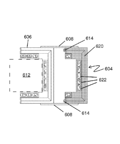
Referring now to FIGS. 6A-6D, yet another embodiment of a locator device 600
is
illustrated, along with a method of using it. In this embodiment, the locator
device 600 again
includes a frame 602, having a first portion 604 and a second portion 606,
attached to one
another with a hinge 608, to delineate a first scalp segment 612 (or
"procedure area"). The first
.. portion 604 has a top side 616 with a first set of fiducials 610 attached
to it, and the second
portion 606 has a top side 618 with a first set of fiducials 613 attached to
it. As with the method
described previously, initial steps of the method may involve positioning the
locator device 600,
for example, on the scalp at a desired location to delineate the first scalp
segment 612. In this
embodiment, the locator device 600 does not include straps to attach the
locator device 600 the
.. patient's head or a chair or the like. Instead, the top and bottom sides of
the frame 602 may
include two or more small pins, hooks, barbs, adhesive, or the like, to attach
the frame 602 to the
patient's scalp in a relatively stable manner. In alternative embodiments, one
or more straps or
other attachment devices may be used. It should be understood that small pins,
barbs, adhesive,
or the like may be used with the embodiments shown in Figures 5A-D.
Referring now to FIG. 6B, a second phase of the method may involve detaching
the
first portion 604 from the second portion 606 and flipping the first portion
604 over the second
portion 606 to a new location on the scalp. When flipped over, a bottom side
620 of the first
portion 604 is now facing up, along with fiducials 622 attached to the bottom
side 620. At this
stage, coupling members 614 (optional) on the first portion 604 are exposed.
Also at this stage,
the first portion 604 may not be directly secured to the scalp in some
embodiments, but may
simply be secured to the first portion via the hinges 608. Alternatively, the
first portion 604 may
be secured to the scalp using pins, hooks, needles or the like, located, for
example, at each of the
two corners of the first portion 604, in one embodiment. In this particular
configuration, the
hinges 608 additionally provide at least one connector, in the form of a
pivot, connecting the first
portion with the second portion, such that the first portion can only be moved
a selected distance
from the second portion due to the at least one connector 608.
Referring to FIG. 6C, in a next step, the second portion 606 may now be
flipped over
so that a bottom side 628 and bottom fiducials 624 on the second portion 606
are facing up. The
coupling members 614 of the first portion 604 may then be connected with
(e.g., inserted into)
corresponding coupling members 626 (such as receptacles) of the second portion
606 to reform
the frame 602. In some embodiments, the frame 602 may be reformed, and the two
portions 604,
24
CA 02902450 2015-08-25
WO 2014/158505 PCT/US2014/017514
RR-041 PCT
606 may be placed together without coupling members 614. 626. In these
embodiments, at least
one reference feature may include an edge of the first portion of the frame
and a corresponding
edge of the second portion of the frame, such that when the first portion is
moved from a first
location on the body surface to a second location, the edge of the first
portion is made to abut the
corresponding edge of the second portion.
Referring to FIG. 6D, the frame 602 is now reformed in a second location on
the
scalp, thus delineating a second scalp portion 630, which is immediately
adjacent the first scalp
portion 612. The frame 602 may be secured to the scalp via pins, hooks,
needles or the like, and
the procedure may be performed on the second scalp segment 630. These steps
may be repeated
as many times as desired to cover a desired total area of scalp. As with all
of the above-described
methods. the fiducials 610, 611, 622, 624 may be used to guide a robotic or
computer-automated
system to perform the procedure on the various body portions. As also
described above, in
alternative embodiments, fiducials may not be included.
Referring now to FIGS. 7A-7E, a side view illustrates the way in which the
first
portion 604 and the second portion 606 fit together during the method
described above. FIG. 7A
shows the first portion 604 and the second portion 606 coupled together in the
first body surface
location (delineating the first body surface segment). As discussed above, and
as illustrated in
FIGS. 7B and 7C, the first portion 604 of the locator device 600 may flip over
the second portion
606, for example, via the hinge 608, to position the first portion 604 in the
new, second location
(FIG. 7C). The hinge 608 is sized to position the first portion 604 at a
distance from the second
portion 606 that will make subsequent body surface portions immediately
adjacent one another.
In the next step, as shown in FIG. 7D, the second portion 606 is flipped over
on itself
(or in place). Finally, as shown in FIG. 7E, the first portion 604 and the
second portion 606 are
rejoined to reform the frame 602 in the second location and the coupling
members 614 may fit
into the recesses 626 (or "corresponding coupling members") on the first
portion 604. In
alternative embodiments, any of a number of alternative connecting structures
may be used, such
as hooks, pins, détentes, magnets or the like. FIGS. 7A-7E illustrate various
stages of the
operation of the device according to one embodiment.
Numerous changes, variations, and substitutions will occur to those skilled in
the art
without departing from the invention. Various alternatives to the embodiments
of the invention
described herein may be employed in practicing the invention. It should be
understood that the
CA 02902450 2015-08-25
WO 2014/158505 PCT/US2014/017514
RR-041 PCT
invention generally, as well as the specific embodiments described herein, are
not limited to the
particular forms or embodiments disclosed, but to the contrary cover all
modifications,
equivalents and alternatives falling within the scope of the appended claims.
By way of non-
limiting example, it will be appreciated by those skilled in the art that
particular features or
characteristics described in reference to one figure or embodiment may be
combined as suitable
with features or characteristics described in another figure or embodiment.
Similarly, the
devices and methods described herein may be used in manual, semi-automated and
fully
automated procedures, including image-guided and robotic procedures.
26