Note: Descriptions are shown in the official language in which they were submitted.
CA 02902560 2015-08-25
WO 2014/153087
PCT/US2014/029000
Nanoparticle-Based Compositions
CLAIM OF PRIORITY
This application claims priority to U.S. Patent Application Serial No.
61/783,439, filed on March 14, 2013, the entire contents of which are hereby
incorporated by reference.
STATEMENT AS TO FEDERALLY SPONSORED RESEARCH
This invention was made with Government support under NIH/R01 AI069259
and NIH/R01 AI072252, awarded by the National Institutes of Health. The
io Government has certain rights in the invention.
TECHNICAL FIELD
This invention relates to compositions and adjuvant compositions comprising
nanoparticles.
BACKGROUND
The mucosa' membranes are one of the largest organs in the body, and
comprise the linings of the gastrointestinal, urogenital, and respiratory
tracts. These
mucosa' membranes, while located in the body, are actually physical barriers
between
the external environment and the sterile internal body cavity known as the
systemic
environment. Thus, an important function of the mucosa' membranes is to keep
invading pathogens out of the sterile body cavity. Indeed, a vast majority of
human
pathogens, including bacteria, viruses, parasites and fungi, initiate
infections at the
mucosa' surfaces (Ogra et al., Clin Microbiol Rev. 14(2):430-45, 2001).
Mucosal immunity is important because stimulation of the mucosa' immune
response can result in the production of protective B cells and T cells in
both mucosa'
and systemic environments so that infections are stopped before the pathogens
enter
into the interior body cavity (see, e.g., McCluskie et al., Microbes Infect.
1(9):685-98;
1999; Rosenthal et al., Semin Immunol. 9(5):303-14, 1997). Despite its
important
role, very few vaccines specifically target the mucosa' immune system.
1
CA 02902560 2015-08-25
WO 2014/153087
PCT/US2014/029000
Vaccinations can be either passive or active. Canonically, active vaccinations
involve the exposure of an individual's immune system to one or more foreign
molecules that elicit an endogenous immune response resulting in the
activation of
antigen-specific naive lymphocytes that subsequently leads to antibody-
secreting B
cells or antigen-specific effector and memory T cells. This approach can
result in
long-lived protective immunity that can be boosted from time to time by
renewed
exposure to the same antigenic material. The prospect of longevity of a
successful
immune response to active vaccination makes this strategy more desirable in
most
clinical settings than passive vaccination whereby a recipient is injected
with
preformed antibodies or with antigen-specific effector lymphocytes, which can
confer
rapid protection, but typically do not establish persistent immunity.
SUMMARY
The present disclosure is based, at least in part, on the development of new
compositions including one or more adjuvant-loaded polymeric nanoparticles
attached
to an inactivated pathogen. For example, the new compositions include an
inactivated pathogen, e.g., a bacterium, such as a Chlamydia trachomatis,
Francisella
tularensis , Mycobacterium tuberculosis, Streptococcus pneumoniae, Listeria
monocytogenes, Vibrio cholera, Shigella sonnei, Shigella flexneri, or
Salmonella
typhimurium, or a virus, such as a human respiratory syncytial virus (RSV), an
Influenza virus, human immunodeficiency virus (HIV), or a Hepatitis C virus,
and
one or more polymeric nanoparticles that are loaded with one or more
adjuvants, such
as a Toll-like receptor agonist, e.g., the imidazoquinoline resiquimod (R-
848),
monophosphoryl lipid A, or an unmethylated CpG oligodeoxynucleotide, or an
endosomal membrane targeting agent, e.g., the Endo-Porter peptide. The
polymeric
nanoparticles can be formed by biodegradable polymers, e.g., poly(lactic-co-
glycolic
acid)-block-poly(L-histidine)-block-poly(ethylene glycol) (PLGA-PLH-PEG)
triblock
copolymers. One or more of the adjuvant-loaded nanoparticles are bound to each
of
the inactivated pathogens. These compositions are useful as vaccines for
preventing
and/or treating diseases caused by the specific pathogens, especially when
administered to a subject's mucosa' membranes.
Provided herein are also methods for stimulating in a subject a mucosa'
immune response against a pathogen, e.g., a bacterium, virus, parasite, or
fungus, by
2
CA 02902560 2015-08-25
WO 2014/153087
PCT/US2014/029000
administering to the subject the new vaccine compositions described herein
through
mucosa' administration, e.g., by an ocular, intranasal, oral, buccal,
sublingual,
tonsilar, by inhalation, e.g., pulmonary or bronchial, gastric, intestinal,
rectal, vaginal,
or urinary tract route.
In some embodiments, the one or more adjuvant-loaded polymeric
nanoparticles are surface charged and attached to the inactivated pathogen
through
electrostatic attraction. In some embodiments, the one or more adjuvant-loaded
polymeric nanoparticles are attached to the inactivated pathogen through a
linker, e.g.,
an attachment mechanism such as a monoclonal antibody, aptamer, antibiotic,
lectin,
io or antimicrobial peptide that binds specifically to a surface molecule
on the
inactivated pathogen.
For example, a Chlamydia trachomatis vaccine composition including an
inactivated Chlamydia trachomatis attached to one or more R848-loaded
polymeric
nanoparticles was made and evaluated in mouse models. While inactivated
Chlamydia trachomatis alone induce immune tolerance, the new Chlamydia
trachomatis vaccine compositions, when administered through a mucosa' route,
e.g.,
intranasally or intrauterinely, were effective in preventing subsequent
Chlamydia
trachomatis infection. Currently there are no vaccines available for use in
humans
against Chlamydia trachomatis infection. Thus, these new Chlamydia trachomatis
vaccine compositions are promising new prophylactic and therapeutic vaccines
against Chlamydia trachomatis infection in humans.
In general, in one aspect the disclosure features methods of stimulating a
mucosa' immune response against one or more different types of pathogen, e.g.,
Chlamydia trachomatis or Francisella tularensis in a subject in need thereof
The
methods include administering to the subject a composition that includes an
inactivated form of the pathogen, and one or more adjuvant-loaded polymeric
nanoparticles, wherein the one or more adjuvant-loaded polymeric nanoparticles
are
each attached to the inactivated pathogen.
In these methods, the pathogen can be a bacterium, virus, parasite, and/or
fungus, and the compositions can be administered to the subject through one or
more
mucosa' routes, e.g., an ocular, intranasal, oral, buccal, sublingual,
tonsilar,
pulmonary, gastric, intestinal, rectal, vaginal, and/or urinary tract route.
3
CA 02902560 2015-08-25
WO 2014/153087
PCT/US2014/029000
In some implementations of these methods, the one or more adjuvant-loaded
polymeric nanoparticles can include an adjuvant that targets an endosomal
membrane,
and/or the adjuvant-loaded polymeric nanoparticles can include a Toll-like
receptor
agonist, e.g., R848, monophosphoryl lipid A, or an unmethylated CpG
oligodeoxynucleotide.
In certain embodiments the one or more adjuvant-loaded polymeric
nanoparticles can be made of biodegradable polymers, such as poly(lactic-co-
glycolic
acid)-block-poly(L-histidine)-block-poly(ethylene glycol) (PLGA-PLH-PEG)
triblock
copolymers. In some embodiments the one or more adjuvant-loaded polymeric
nanoparticles are attached to the inactivated pathogen through electrostatic
attraction.
In other embodiments the one or more adjuvant-loaded polymeric nanoparticles
are
attached to the inactivated pathogen through one or more linkers, e.g., an
attachment
mechanism such as a monoclonal antibody, an aptamer, an antibiotic, a lectin,
and/or
an antimicrobial peptide that binds specifically to a surface molecular of the
inactivated pathogen.
In some embodiments, disclosed herein are methods for stimulating in a
subject a mucosa' immune response against bacteria selected from the group
consisting of Actinomyces, Anabaena, Bacillus, Bacteroides, Bdellovibrio,
Bordetella,
Borrelia, Brucella, Campylobacter, Caulobacter, Chlamydia, Chlorobium,
Chromatium, Clostridium, Corynebacterium, Cytophaga, Deinococcus,
Enterococcus,
Escherichia, Francisella, Halobacterium, Heliobacter, Haemophilus,
Hyphomicrobium, Legionella, Leptspirosis, Listeria, Meningococcus A, B, and C,
Methanobacterium, Micrococcus , Mycobacterium, Mycoplasma, Myxococcus,
Neisseria, Nitrobacter, Oscillatoria, Prochloron, Proteus, Pseudomonas,
Phodospirillum, Rickettsia, Salmonella, Shigella, Spirillum, Spirochaeta,
Staphylococcus, Streptococcus, Streptomyces, Sulfolobus , Thermoplasma,
Thiobacillus, Treponema, Vibrio, and Yersinia. In some embodiments, methods
for
stimulating in a subject a mucosa' immune response against Chlamydia
trachomatis
are provided. In some embodiments, methods for stimulating in a subject a
mucosa'
immune response against Francisella tularensis are provided. In some
embodiments,
methods for stimulating in a subject a mucosa' immune response against
Mycobacterium tuberculosis are provided.
4
CA 02902560 2015-08-25
WO 2014/153087
PCT/US2014/029000
In some embodiments, disclosed herein are methods for stimulating in a
subject a mucosa' immune response against viruses selected from the group
consisting
of Adenoviridae, Arenaviridae, Arterivirus, Astroviridae, Baculoviridae,
Badnavirus,
Bamaviridae, Bimaviridae, Bromoviridae, Bunyaviridae, Caliciviridae,
Capillovirus,
Carlavirus, Caulimovirus, Circoviridae, Closterovirus, Comoviridae,
Coronaviridae,
Corticoviridae, Cystoviridae, Deltavirus, Dianthovirus, Enamovirus,
Flaviviridae,
Filoviridae, Flaviviridae, Hepadnaviridae, Herpesviridae, Hypoviridae,
Iridoviridae,
Leviviridae, Lipothrixviridae, Microviridae, Orthomyxoviridae,
Papillomaviridae,
Papovaviridae, Paramyxoviridae, Parvoviridae, Picornaviridae, Polyomaviridae,
Poxviridae, Reoviridae, Retroviridae, Rhabdoviridae, Togaviridae, and
Totiviridae. In
some embodiments, methods for stimulating in a subject a mucosa' immune
response
against human respiratory syncytial viruses are provided. In some embodiments,
methods for stimulating in a subject a mucosal immune response against SARS
coronaviruses are provided. In some embodiments, methods for stimulating in a
subject a mucosa' immune response against Noroviruses are provided. In some
embodiments, methods for stimulating in a subject a mucosa' immune response
against human immunodeficiency viruses are provided.
In another general aspect, the disclosure includes compositions that include
one or more different types of inactivated pathogens, e.g., Chlamydia
trachomatis or
Francisella tularensis; and one or more adjuvant-loaded polymeric
nanoparticles,
wherein the one or more adjuvant-loaded polymeric nanoparticles are each
attached to
the inactivated pathogen through an attachment mechanism. In these
compositions
the inactivated pathogen can be an inactivated bacterium, an inactivated
virus, an
inactivated parasite, and/or an inactivated fungus. For example, the
inactivated
pathogen can be an inactivated bacterium selected from the group consisting of
Chlamydia trachomatis, Francisella tularensis, Mycobacterium tuberculosis,
Streptococcus pneumoniae, Listeria monocytogenes, Vibrio cholera, Shigella
sonnei,
Shigella flexneri, and/or Salmonella typhimurium. In some embodiments, the
compositions disclosed herein consist of, or consist essentially of, one or
more
different types of inactivated pathogens, e.g., Chlamydia trachomatis or
Francisella
tularensis; and one or more adjuvant-loaded polymeric nanoparticles, wherein
the one
5
CA 02902560 2015-08-25
WO 2014/153087
PCT/US2014/029000
or more adjuvant-loaded polymeric nanoparticles are each attached to the
inactivated
pathogen through an attachment mechanism.
In some implementations, the one or more adjuvant-loaded polymeric
nanoparticles include an adjuvant that targets an endosomal membrane and/or a
Toll-
like receptor agonist. For example, the Toll-like receptor agonist can be R848
or an
unmethylated CpG oligodeoxynucleotide.
In various implementations, the one or more adjuvant-loaded polymeric
nanoparticles can be made of biodegradable polymers, e.g., poly(lactic-co-
glycolic
acid)-block-poly(L-histidine)-block-poly(ethylene glycol) (PLGA-PLH-PEG)
triblock
copolymers. In certain embodiments, the one or more adjuvant-loaded polymeric
nanoparticles are attached to the inactivated pathogen through electrostatic
attraction.
In other embodiments, the one or more adjuvant-loaded polymeric nanoparticles
are
attached to the inactivated pathogen through a linker, e.g., an attachment
mechansim
such as one or more of a monoclonal antibody, an aptamer, an antibiotic, a
lectin, or
an antimicrobial peptide that binds specifically to a surface molecular of the
inactivated pathogen.
The compositions can be designed in a form suitable for mucosa'
administration, e.g., via an ocular, intranasal, oral, buccal, sublingual,
tonsilar,
pulmonary, gastric, intestinal, rectal, vaginal, or urinary tract route as
described in
further detail herein.
In some embodiments, the compositions disclosed herein include inactivated
bacteria selected from the group consisting of Actinomyces, Anabaena,
Bacillus,
Bacteroides, Bdellovibrio, Bordetella, Borrelia, Brucella, Campylobacter,
Caulobacter, Chlamydia, Chlorobium, Chromatium, Clostridium, Corynebacterium,
Cytophaga, Deinococcus, Enterococcus , Escherichia, Francisella,
Halobacterium,
Heliobacter, Haemophilus, Hyphomicrobium, Legionella, Leptspirosis , Listeria,
Meningococcus A, B, and C, Methanobacterium, Micrococcus, Mycobacterium,
Mycoplasma, Myxococcus, Neisseria, Nitrobacter, Oscillatoria, Prochloron,
Proteus,
Pseudomonas , Phodospirillum, Rickettsia, Salmonella, Shigella, Spirillum,
Spirochaeta, Staphylococcus, Streptococcus, Streptomyces, Sulfolobus ,
Thermoplasma, Thiobacillus, Treponema, Vibrio, and Yersinia. In some
embodiments,
the compositions disclosed herein include inactivated Chlamydia trachomatis .
In
6
CA 02902560 2015-08-25
WO 2014/153087
PCT/US2014/029000
some embodiments, the compositions disclosed herein include inactivated
Francisella
tularensis. In some embodiments, the compositions disclosed herein include
inactivated Mycobacterium tuberculosis.
In some embodiments, the compositions disclosed herein include inactivated
viruses selected from the group consisting of Adenoviridae, Arenaviridae,
Arterivirus,
Astroviridae, Baculoviridae, Badnavirus, Bamaviridae, Bimaviridae,
Bromoviridae,
Bunyaviridae, Caliciviridae, Capillovirus, Carlavirus, Caulimovirus,
Circoviridae,
Closterovirus, Comoviridae, Coronaviridae, Corticoviridae, Cystoviridae,
Deltavirus,
Dianthovirus, Enamovirus, Flaviviridae, Filoviridae, Flaviviridae,
Hepadnaviridae,
Herpesviridae, Hypoviridae, Iridoviridae, Leviviridae, Lipothrixviridae,
Microviridae,
Orthomyxoviridae, Papillomaviridae, Papovaviridae, Paramyxoviridae,
Parvoviridae,
Picomaviridae, Polyomaviridae, Poxviridae, Reoviridae, Retroviridae,
Rhabdoviridae,
Togaviridae, and Totiviridae. In some embodiments, the compositions disclosed
herein include inactivated human respiratory syncytial viruses. In some
embodiments,
the compositions disclosed herein include inactivated SARS coronaviruses. In
some
embodiments, the compositions disclosed herein include inactivated
Noroviruses. In
some embodiments, the compositions disclosed herein include inactivated human
immunodeficiency viruses.
In some embodiments, the disclosure includes compositions that include
inactivated Chlamydia trachomatis; and one or more adjuvant-loaded polymeric
nanoparticles, wherein the one or more adjuvant-loaded polymeric nanoparticles
are
each attached to the inactivated Chlamydia trachomatis. In some
implementations,
the one or more adjuvant-loaded polymeric nanoparticles include an adjuvant
that
targets an endosomal membrane and/or a Toll-like receptor agonist. For
example, the
adjuvant can be one or more of R848, unmethylated CpG oligodeoxynucleotide,
and
monophosphoryl lipid A. In some embodiments, the adjuvant is R848. In various
implementations, the one or more adjuvant-loaded polymeric nanoparticles can
be
made of biodegradable polymers, e.g., poly(lactic-co-glycolic acid)-block-
poly(L-
histidine)-block-poly(ethylene glycol) (PLGA-PLH-PEG) triblock copolymers. In
certain embodiments, the one or more adjuvant-loaded polymeric nanoparticles
are
attached to the inactivated Chlamydia trachomatis through electrostatic
attraction. In
other embodiments, the one or more adjuvant-loaded polymeric nanoparticles are
7
CA 02902560 2015-08-25
WO 2014/153087
PCT/US2014/029000
attached to the inactivated Chlamydia trachomatis through a linker, e.g., an
attachment mechansim such as one or more of a monoclonal antibody, an aptamer,
an
antibiotic, a lectin, or an antimicrobial peptide that binds specifically to a
surface
molecular of the inactivated pathogen. The compositions can be designed in a
form
suitable for mucosa' administration, e.g., via an ocular, intranasal, oral,
buccal,
sublingual, tonsilar, pulmonary, gastric, intestinal, rectal, vaginal, or
urinary tract
route. In some embodiments, methods for stimulating in a subject a mucosa'
immune
response against Chlamydia trachomatis by adminitering the compositions
described
herein are provided.
In some embodiments, the disclosure includes compositions that include
inactivated Francisella tularensis; and one or more adjuvant-loaded polymeric
nanoparticles, wherein the one or more adjuvant-loaded polymeric nanoparticles
are
each attached to the inactivated Francisella tularensis. In some
implementations, the
one or more adjuvant-loaded polymeric nanoparticles include an adjuvant that
targets
an endosomal membrane and/or a Toll-like receptor agonist. For example, the
adjuvant can be one or more of R848, unmethylated CpG oligodeoxynucleotide,
and
monophosphoryl lipid A. In some embodiments, the adjuvant is R848. In various
implementations, the one or more adjuvant-loaded polymeric nanoparticles can
be
made of biodegradable polymers, e.g., poly(lactic-co-glycolic acid)-block-
poly(L-
histidine)-block-poly(ethylene glycol) (PLGA-PLH-PEG) triblock copolymers. In
certain embodiments, the one or more adjuvant-loaded polymeric nanoparticles
are
attached to the inactivated Francisella tularensis through electrostatic
attraction. In
other embodiments, the one or more adjuvant-loaded polymeric nanoparticles are
attached to the inactivated Francisella tularensis through a linker, e.g., an
attachment
mechansim such as one or more of a monoclonal antibody, an aptamer, an
antibiotic,
a lectin, or an antimicrobial peptide that binds specifically to a surface
molecular of
the inactivated pathogen. The compositions can be designed in a form suitable
for
mucosal administration, e.g., via an ocular, intranasal, oral, buccal,
sublingual,
tonsilar, pulmonary, gastric, intestinal, rectal, vaginal, or urinary tract
route. In some
embodiments, methods for stimulating in a subject a mucosa' immune response
against Francisella tularensis by adminitering the compositions described
herein are
provided.
8
CA 02902560 2015-08-25
WO 2014/153087
PCT/US2014/029000
In some embodiments, the disclosure includes compositions that include
inactivated Mycobacterium tuberculosis; and one or more adjuvant-loaded
polymeric
nanoparticles, wherein the one or more adjuvant-loaded polymeric nanoparticles
are
each attached to the inactivated Mycobacterium tuberculosis. In some
implementations, the one or more adjuvant-loaded polymeric nanoparticles
include an
adjuvant that targets an endosomal membrane and/or a Toll-like receptor
agonist. For
example, the adjuvant can be one or more of R848, unmethylated CpG
oligodeoxynucleotide, and monophosphoryl lipid A. In some embodiments, the
adjuvant is monophosphoryl lipid A. In various implementations, the one or
more
adjuvant-loaded polymeric nanoparticles can be made of biodegradable polymers,
e.g., poly(lactic-co-glycolic acid)-block-poly(L-histidine)-block-
poly(ethylene glycol)
(PLGA-PLH-PEG) triblock copolymers. In certain embodiments, the one or more
adjuvant-loaded polymeric nanoparticles are attached to the inactivated
Mycobacterium tuberculosis through electrostatic attraction. In other
embodiments,
the one or more adjuvant-loaded polymeric nanoparticles are attached to the
inactivated Mycobacterium tuberculosis through a linker, e.g., an attachment
mechansim such as one or more of a monoclonal antibody, an aptamer, an
antibiotic,
a lectin, or an antimicrobial peptide that binds specifically to a surface
molecular of
the inactivated pathogen. The compositions can be designed in a form suitable
for
mucosa' administration, e.g., via an ocular, intranasal, oral, buccal,
sublingual,
tonsilar, pulmonary, gastric, intestinal, rectal, vaginal, or urinary tract
route. In some
embodiments, methods for stimulating in a subject a mucosa' immune response
against Mycobacterium tuberculosis by adminitering the compositions described
herein are provided.
In some embodiments, the disclosure includes compositions that include
inactivated human respiratory syncytial viruses (RSV); and one or more
adjuvant-
loaded polymeric nanoparticles, wherein the one or more adjuvant-loaded
polymeric
nanoparticles are each attached to the inactivated human respiratory syncytial
viruses.
In some implementations, the one or more adjuvant-loaded polymeric
nanoparticles
include an adjuvant that targets an endosomal membrane and/or a Toll-like
receptor
agonist. For example, the adjuvant can be one or more of R848, unmethylated
CpG
oligodeoxynucleotide, and monophosphoryl lipid A. In some embodiments, the
9
CA 02902560 2015-08-25
WO 2014/153087
PCT/US2014/029000
adjuvant is R848. In various implementations, the one or more adjuvant-loaded
polymeric nanoparticles can be made of biodegradable polymers, e.g.,
poly(lactic-co-
glycolic acid)-block-poly(L-histidine)-block-poly(ethylene glycol) (PLGA-PLH-
PEG) triblock copolymers. In certain embodiments, the one or more adjuvant-
loaded
polymeric nanoparticles are attached to the inactivated human respiratory
syncytial
viruses through electrostatic attraction. In other embodiments, the one or
more
adjuvant-loaded polymeric nanoparticles are attached to the inactivated human
respiratory syncytial viruses through a linker, e.g., an attachment mechansim
such as
one or more of a monoclonal antibody, an aptamer, an antibiotic, a lectin, or
an
antimicrobial peptide that binds specifically to a surface molecular of the
inactivated
pathogen. The compositions can be designed in a form suitable for mucosa'
administration, e.g., via an ocular, intranasal, oral, buccal, sublingual,
tonsilar,
pulmonary, gastric, intestinal, rectal, vaginal, or urinary tract route. In
some
embodiments, methods for stimulating in a subject a mucosa' immune response
against human respiratory syncytial viruses by adminitering the compositions
described herein are provided.
In some embodiments, the disclosure includes compositions that include
inactivated SARS coronaviruses; and one or more adjuvant-loaded polymeric
nanoparticles, wherein the one or more adjuvant-loaded polymeric nanoparticles
are
each attached to the inactivated SARS coronaviruses. In some implementations,
the
one or more adjuvant-loaded polymeric nanoparticles include an adjuvant that
targets
an endosomal membrane and/or a Toll-like receptor agonist. For example, the
adjuvant can be one or more of R848, unmethylated CpG oligodeoxynucleotide,
and
monophosphoryl lipid A. In some embodiments, the adjuvant is unmethylated CpG
oligodeoxynucleotide. In various implementations, the one or more adjuvant-
loaded
polymeric nanoparticles can be made of biodegradable polymers, e.g.,
poly(lactic-co-
glycolic acid)-block-poly(L-histidine)-block-poly(ethylene glycol) (PLGA-PLH-
PEG) triblock copolymers. In certain embodiments, the one or more adjuvant-
loaded
polymeric nanoparticles are attached to the inactivated SARS coronaviruses
through
electrostatic attraction. In other embodiments, the one or more adjuvant-
loaded
polymeric nanoparticles are attached to the inactivated SARS coronaviruses
through a
linker, e.g., an attachment mechansim such as one or more of a monoclonal
antibody,
CA 02902560 2015-08-25
WO 2014/153087
PCT/US2014/029000
an aptamer, an antibiotic, a lectin, or an antimicrobial peptide that binds
specifically
to a surface molecular of the inactivated pathogen. The compositions can be
designed
in a form suitable for mucosa' administration, e.g., via an ocular,
intranasal, oral,
buccal, sublingual, tonsilar, pulmonary, gastric, intestinal, rectal, vaginal,
or urinary
tract route. In some embodiments, methods for stimulating in a subject a
mucosa'
immune response against SARS coronaviruses by adminitering the compositions
described herein are provided.
In some embodiments, the disclosure includes compositions that include
inactivated Noroviruses; and one or more adjuvant-loaded polymeric
nanoparticles,
wherein the one or more adjuvant-loaded polymeric nanoparticles are each
attached to
the inactivated Noroviruses. In some implementations, the one or more adjuvant-
loaded polymeric nanoparticles include an adjuvant that targets an endosomal
membrane and/or a Toll-like receptor agonist. For example, the adjuvant can be
one
or more of R848, unmethylated CpG oligodeoxynucleotide, and monophosphoryl
lipid A. In some embodiments, the adjuvant is unmethylated CpG
oligodeoxynucleotide. In various implementations, the one or more adjuvant-
loaded
polymeric nanoparticles can be made of biodegradable polymers, e.g.,
poly(lactic-co-
glycolic acid)-block-poly(L-histidine)-block-poly(ethylene glycol) (PLGA-PLH-
PEG) triblock copolymers. In certain embodiments, the one or more adjuvant-
loaded
polymeric nanoparticles are attached to the inactivated Noroviruses through
electrostatic attraction. In other embodiments, the one or more adjuvant-
loaded
polymeric nanoparticles are attached to the inactivated Noroviruses through a
linker,
e.g., an attachment mechansim such as one or more of a monoclonal antibody, an
aptamer, an antibiotic, a lectin, or an antimicrobial peptide that binds
specifically to a
surface molecular of the inactivated pathogen. The compositions can be
designed in a
form suitable for mucosa' administration, e.g., via an ocular, intranasal,
oral, buccal,
sublingual, tonsilar, pulmonary, gastric, intestinal, rectal, vaginal, or
urinary tract
route. In some embodiments, methods for stimulating in a subject a mucosa'
immune
response against Noroviruses by adminitering the compositions described herein
are
provided.
In some embodiments, the disclosure includes compositions that include
inactivated human immunodeficiency viruses; and one or more adjuvant-loaded
11
CA 02902560 2015-08-25
WO 2014/153087
PCT/US2014/029000
polymeric nanoparticles, wherein the one or more adjuvant-loaded polymeric
nanoparticles are each attached to the inactivated human immunodeficiency
viruses.
In some implementations, the one or more adjuvant-loaded polymeric
nanoparticles
include an adjuvant that targets an endosomal membrane and/or a Toll-like
receptor
agonist. For example, the adjuvant can be one or more of R848, unmethylated
CpG
oligodeoxynucleotide, and monophosphoryl lipid A. In some embodiments, the
adjuvant is monophosphoryl lipid A. In various implementations, the one or
more
adjuvant-loaded polymeric nanoparticles can be made of biodegradable polymers,
e.g., poly(lactic-co-glycolic acid)-block-poly(L-histidine)-block-
poly(ethylene glycol)
(PLGA-PLH-PEG) triblock copolymers. In certain embodiments, the one or more
adjuvant-loaded polymeric nanoparticles are attached to the inactivated human
immunodeficiency viruses through electrostatic attraction. In other
embodiments, the
one or more adjuvant-loaded polymeric nanoparticles are attached to the
inactivated
human immunodeficiency viruses through a linker, e.g., an attachment mechansim
such as one or more of a monoclonal antibody, an aptamer, an antibiotic, a
lectin, or
an antimicrobial peptide that binds specifically to a surface molecular of the
inactivated pathogen. The compositions can be designed in a form suitable for
mucosa' administration, e.g., via an ocular, intranasal, oral, buccal,
sublingual,
tonsilar, pulmonary, gastric, intestinal, rectal, vaginal, or urinary tract
route. In some
embodiments, methods for stimulating in a subject a mucosa' immune response
against human immunodeficiency viruses by adminitering the compositions
described
herein are provided.
A "pathogen" as used herein is an infectious agent that causes diseases in its
host. A pathogen can be a bacterium, virus, parasite, fungus, or other
microbial
infectious agent.
As used herein, a "nanoparticle" is a particle in the range of between 500 nm
to less than 0.5 nm, e.g., having a diameter that is between 50 and 500 nm.
As used herein, the term "adjuvant" refers to an immunological adjuvant. By
this is meant a compound or composition that is able to enhance or facilitate
the
immune system's response to a pathogen, thereby inducing an immune response or
series of immune responses in the subject. The adjuvant can facilitate the
effect of the
12
CA 02902560 2015-08-25
WO 2014/153087
PCT/US2014/029000
compositions, for example, by forming depots (prolonging the half-life of the
composition), provide additional T-cell help, and/or stimulate cytokine
production.
As used herein, a "subject" is an animal, e.g., a mammal, e.g., a human,
monkey, dog, cat, horse, cow, pig, goat, rabbit, or mouse.
As used herein, "treatment" can be prophylactic or therapeutic. Prophylactic
treatment can be used to treat a subject at a risk of developing disease from
an
infectious pathogen. An individual traveling to or living in an area of
endemic
infectious disease may be considered to be at risk and a candidate for
prophylactic
vaccination against the particular infectious pathogen. Therapeutic treatment
with
vaccines can be used to initiate or enhance a subject's immune response to a
contracted pathogen.
As generally used herein, an "effective amount" is the amount that is
sufficient
to induce a protective immune response in the treated subject. The actual
effective
amounts of vaccine can vary according to the specific pathogen and adjuvant
being
utilized, the particular vaccine composition formulated, the mode of
administration,
and the age, weight, condition of the subject being vaccinated, as well as the
route of
administration and the disease or disorder.
As used herein, "immunostimulatory" means that a substance has a
stimulating effect on the immune system. Such substances can be readily
identified
using standard assays which indicate various aspects of the immune response,
such as
cytokine secretion, antibody production, NK cell activation and T cell
proliferation.
See, e.g., WO 97/28259; WO 98/16247; WO 99/11275; Krieg et al. (1995) Nature
374:546-549; Yamamoto et al. (1992) J. Immunol. 148:4072-76; Ballas et al.
(1996)
J. Immunol. 157:1840-45; Klinman et al. (1997) J. Immunol. 158:3635-39; Sato
et al.
(1996) Science 273:352-354; Pisetsky (1996) J. Immunol. 156:421-423; Shimada
et
al. (1986) Jpn. J. Cancer Res. 77:808-816; Cowdery et al. (1996) J. Immunol.
156:4570-75; Roman et al. (1997) Nat. Med. 3:849-854; Lipford et al. (1997a)
Eur. J.
Immunol. 27:2340-44; WO 98/55495 and WO 00/61151. Accordingly, these and
other methods can be used to identify, test and/or confirm immunostimulatory
substances, such as immunostimulatory nucleotides, e immunostimulatory
isolated
nucleic acids.
13
CA 02902560 2015-08-25
WO 2014/153087
PCT/US2014/029000
As used herein, "couple" or "coupled" or "couples" (and the like) means to
chemically associate one entity (for example a moiety) with another. In some
implementations, the coupling is covalent, meaning that the coupling occurs in
the
context of the presence of a covalent bond between the two entities. In non-
covalent
implementations, the non-covalent coupling is mediated by non-covalent
interactions
including but not limited to charge interactions, affinity interactions, metal
coordination, physical adsorption, host-guest interactions, hydrophobic
interactions,
TT stacking interactions, hydrogen bonding interactions, van der Waals
interactions,
magnetic interactions, electrostatic interactions, dipole-dipole interactions,
and/or
1 o combinations thereof In certain implementations, encapsulation is a
form of
coupling.
As used herein "encapsulate" means to enclose within a synthetic
nanoparticle, preferably enclose completely within a synthetic nanoparticle.
Most or
all of a substance that is encapsulated is not exposed to the local
environment external
to the synthetic nanoparticle. Encapsulation is distinct from absorption,
which places
most or all of a substance on a surface of a synthetic nanoparticle, and
leaves the
substance exposed to the local environment external to the synthetic
nanoparticle.
Unless otherwise defined, all technical and scientific terms used herein have
the same meaning as commonly understood by one of ordinary skill in the art to
which this invention belongs. Although methods and materials similar or
equivalent
to those described herein can be used in the practice or testing of the
present
invention, suitable methods and materials are described below. All
publications,
patent applications, patents, and other references mentioned herein are
incorporated
by reference in their entirety. In case of conflict, the present
specification, including
definitions, will control. In addition, the materials, methods, and examples
are
illustrative only and not intended to be limiting.
Other features and advantages of the invention will be apparent from the
following detailed description, and from the claims.
DESCRIPTION OF DRAWINGS
FIG. lA is a dot plot showing intrauterine immunization with UV-inactivated
Ct (UV-Ct) resulted in enhanced susceptibility to subsequent live Chlamydia
challenge, indicating immune tolerance is induced by UV-Ct.
14
CA 02902560 2015-08-25
WO 2014/153087
PCT/US2014/029000
FIG. 1B is a dot plot showing that co-administration of adjuvants does not
overcome UV-Ct induced immune tolerance.
FIGs. 1C-1D are bar graphs showing that the UV-Ct induced immune
tolerance is mediated by FoxP3+ Treg cells.
FIG lE is a dot plot showing that treatment with anti-CD25 monoclonal
antibody overcomes UV-Ct induced immune tolerance, indicating that CD4+ FoxP3+
Treg cells play a critical role in mediating UV-Ct induced immune tolerance.
FIG. 1F is a dot plot showing IL-10 deficiency abrogated the inactivated
Chlamydia induced immune tolerance, confirming that Treg-secreted IL-10 plays
a
critical role in the inactivated Chlamydia induced immune tolerance.
FIG. 2A is a schematic drawing illustrating that the surface charge-switching
synthetic adjuvant particles (cSAP) bind to inactivated Chlamydia trachomatis.
FIG 2B is a cryo-TEM (cryogenic transmission electron microscope) image
showing the binding of R848-loaded nanoparticles to the surface of inactivated
Chlamydia trachomatis.
FIG. 2C is a dynamic light scattering graph confirming the binding of
nanoparticles to inactivated Chlamydia trachomatis.
FIGs. 2D-2E are a set of flow cytometry graphs and corresponding histograms
showing the binding of BacLight-stained UV-Ct with either Alexa Fluor 488-
labeled
cSAP or control particles lacking the PLH group (SAP) at pH of 7.4 and 6.0
FIG. 2F is a dot plot showing immunization with a vaccine composition
including UV-Ct-cSAPs protects the mice against subsequent live Chlamydia
challenges.
FIG. 2G is a dot plot showing titration or neutralization of the R848-loaded
cSAP before attachment to UV-Ct does not change the immune protective property
of
the vaccine composition.
FIG. 2H is a dot plot showing IgG induction by a vaccine composition
including UV-Ct-cSAPs. The data were pooled from two independent experiments.
n.d. = not detected.
FIGs. 3A-3D are a set of dot plots showing that the protective immunity
stimulated by the new Chlamydia trachomatis vaccine composition is mediated by
CD4+ T cells.
CA 02902560 2015-08-25
WO 2014/153087
PCT/US2014/029000
FIG. 4A is a bar graph showing significantly more Chlamydia-specific
transgenic CD4+ T cells were present in lymph nodes of the mice challenged
with the
new vaccine composition (UV-Ct-cSAPs) or infectious Chlamydia (Ct) when
compared with mice immunized with UV-inactivated Chlamydia (UV-Ct) or the
uninfected control mice (Naive).
FIG. 4B is a set of flow cytometry graphs showing that the number of
Chlamydia-specific CD90.1+ CD4+ T cells in mice immunized with Ct or UV-Ct-
cSAPs greatly exceeded those in the uninfected mice or mice immunized with UV-
Ct,
indicating Ct and UV-Ct-cSAPs, but not UV-Ct induce Chlamydia-specific CD4+ T
io cell proliferation.
FIG 4C is a set of pie charts showing that the number of CD90.1+ CD4+ T
cells producing all three cytokines (TNF-a, IFN-y, and IL-2) was significantly
higher
in mice immunized with Ct or UV-Ct-cSAPs when compared with mice immunized
with UV-Ct or the uninfected control mice.
FIG. 5A is a set of flow cytometry graphs showing that F4/80+ CD103-
macrophages express high level of CD1lb and CX3CR1 but low level of CD11c;
F4/80- CD103- dendritic cells express high level of CD11c, CD11b, and CX3CR1;
F4/80- CD103+ dendritic cells express low level of CD1lb and CX3CR1, but high
level of CD1 1 c.
FIGs. 5B and 5C are a set of dot plots showing CD103- dendritic cells had a
significantly higher Chlamydia loads than F4/80+ macrophages and CD103+
dendritic
cells in both uteri (5B) and lymph nodes (5C), indicating CD103- dendritic
cells play
important roles in recognizing and presenting Chlamydia.
FIGs. 5D and 5E are a set of bar graphs showing CD103- dendritic cells
isolated from uteri of mice immunized with infectious Chlamydia (Ct), or the
new
vaccine composition (UV-Ct-cSAPs), but not the other antigen presenting cells,
induced proliferation of Chlamydia-specific CD90.1+ CD4+ transgenic T cells
(NR1
cells) both in vitro (5D) and in vivo (5E).
FIG. 5F is a bar graph showing CD103+ dendritic cells increased the number
of FoxP3+CD25+NR1 cells following UV-Ct immunization.
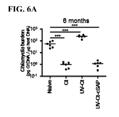
FIG. 6A is a dot plot showing intrauterine immunization with the new vaccine
composition or the infectious Chlamydia, but not the inactivated Chlamydia,
resulted
16
CA 02902560 2015-08-25
WO 2014/153087
PCT/US2014/029000
in protection against subsequent genital Chlamydia infection for six months
after
immunization.
FIG. 6B is a dot plot showing intranasal, but not subcutaneous, immunization
with the new vaccine composition resulted in protective immunity against
subsequent
genital Chlamydia infection, indicating that cross-mucosal protective immunity
was
induced by the new vaccine composition.
FIG. 6C is a set of flow cytometry graphs showing that tissue homing into the
uterus of Chlamydia-specific transgenic CD4+ T cells was induced by
intrauterine or
intranasal, but not subcutaneous immunization.
FIG. 6D is a set of bar graphs showing that immunization with UV-Ct-cSAP
by either intrauterine (i.u.) or intranasal (i.n.) route, but not by
subcutaneous (s.c.)
route, induced the recruitment and retention of protective NR1 cells in the
genital
mucosa and in lung. The numbers of NR1 cells in liver, lymph nodes, spleen, or
blood are comparable among the different routes of immunization.
FIG. 7A is a schematic drawing showing the experiment protocol for Example
5.
FIGs. 7B-7C are bar graphs showing that blocking alpha 4 integrin efficiently
prevented T cell accumulation in uterus (7C), but had no effect on the number
of NR1
cells in the spleen (7B).
FIG. 7D is a bar graph showing that the systemic NR1 cells present in the
spleen were not affected by a4 antibody injections.
FIG. 7E is a bar graph showing that accumulation of NR1 cells was observed
in Gr.1 mice that were treated with IgG, and Gr. 3 mice that were treated with
anti-a4
mAb only after the Chlamydia challenge, but not in Gr. 2 mice that were
treated with
anti-a4 mAb after both vaccination and challenge.
FIG. 7F is a dot plot showing that Gr.3 mice treated with anti-a4 mAb only
after the Chlamydia challenge (the group containing uterine-resident memory T
cells
but no additionally recruited circulatory memory cells) were protected against
genital
Chlamydia challenge, compared to the naïve control mice and the Gr. 2 mice
treated
with anti-a4 mAb after both immunization and challenge.
FIG. 8A is a schematic drawing showing the parabiosis experiment protocol
for Example 6.
17
CA 02902560 2015-08-25
WO 2014/153087
PCT/US2014/029000
FIGs. 8B-8C are dot plots showing that both partners of the Group A mice
were protected against subsequent genital Chlamydia challenge (8B); but only
the
immunized partner (CD45.2), not the other partner (CD45.1) of the Group B mice
was
protected against subsequent genital Chlamydia challenge (8C).
FIGs. 8D-8E are bar graphs showing that more NR-1 cells were present in
mice that were protected against subsequent genital Chlamydia challenge
compared
with mice that were not protected.
FIGs. 8F-8G are dot plots showing that immunization with UV-Ct induced
immune tolerance that are independent of the timing of parabiosis.
io FIG. 9A is a line graph showing that UV-LVS-cSAP-immunized mice were
fully protected against subsequent challenge with an attenuated LVS strain of
Francisella tularensis.
FIG. 9B is a line graph showing that UV-LVS-cSAP-immunized mice were
partially protected against subsequent challenge with a fully virulent SchuS4
strain of
Francisella tularensis.
FIGs. 9C-9D are line graphs showing that full protection against subsequent
challenge with an attenuated LVS strain of Francisella tularensis was obtained
after
immunization with UV-LVS-cSAP by intraperitoneal route (9C), but not by the
subcutaneous route (9D).
FIGs. 9E-9F are line graphs showing that the levels of induced IgG (9E) and
IgM (9F) antibodies were higher in UV-LVS-cSAP-immunized mice than in live
LVS-infected mice.
For all figures, *=p<0.05, **=p<0.01, ***=p < 0.001.
DETAILED DESCRIPTION
The present disclosure is based, at least in part, on the development of new
vaccine compositions comprising one or more adjuvant-loaded polymeric
nanoparticles attached to an inactivated pathogen. For example, the new
vaccine
compositions comprise an inactivated pathogen, e.g., a bacterium, such as a
Chlamydia trachomatis, Francisella tularensis, Mycobacterium tuberculosis,
Streptococcus pneumoniae, Listeria monocytogenes, Vibrio cholera, Shigella
sonnei,
Shigella flexneri, or Salmonella typhimurium, or a virus, such as an Influenza
virus, a
human respiratory syncytial virus (RSV), human immunodeficiency virus (HIV),
18
CA 02902560 2015-08-25
WO 2014/153087
PCT/US2014/029000
Hepatitis C virus, and one or more polymeric nanoparticles that are loaded
with
adjuvants, such as a Toll-like receptor agonist, e.g., the imidazoquinoline
resiquimod
(R-848), monophosphoryl lipid A, or an unmethylated CpG oligodeoxynucleotide,
or
an endosomal membrane targeting agent, e.g., the Endo-Porter peptide. One or
more
of the adjuvant-loaded nanoparticles are bound to each of the inactivated
pathogens.
These vaccine compositions are useful for preventing and/or treating diseases
caused
by the specific pathogens, especially when administered to a subject's mucosa'
membranes.
The vaccine compositions disclosed herein include one or more adjuvant-
loaded nanoparticles attached to each of the inactivated whole pathogens,
e.g., via an
attachment mechanism. This attachment mechanism can be an electrostatic
attraction,
covalent coupling, or a hydrophobic interaction. The adjuvants can be a
dendritic cell
targeting molecule, for example, a Toll-like receptor agonist, e.g., R-848,
which is
recognized as a potent synthetic agonist of TLR7/TLR8, or an unmethylated CpG
oligodeoxynucleotide, which is immunostimulatory agonist of TLR-9, or
monophosphoryl lipid A, which is immunostimulatory agonist of TLR-4, or an
endosomal membrane targeting agent, e.g., the Endo-Porter peptide.
A vast majority of vaccines available today target the systemic immune system
and block disease progression after the pathogens have crossed the mucosa'
barrier
and entered into the normally sterile systemic environment. The vaccine
compositions disclosed herein can target the mucosa' membranes and stimulate
mucosa' immunity in an immunized subject that protects the subject from
infection by
an active form of the inactivated pathogens included in the vaccine. These
vaccine
compositions achieve immune protection either by preventing initial
colonization and
replication of the pathogens or by blocking further infection progression.
Thus, these
vaccine compositions are both prophylactic and therapeutic.
Inactivated Pathogens
A "pathogen" as used herein is an infectious agent that causes diseases in its
host. A pathogen can be a bacterium, virus, parasite, fungus, or other
microbial
infectious agent. Many vaccines against pathogens comprise live or attenuated
microorganisms. However, live or attenuated vaccines can sometimes cause
19
CA 02902560 2015-08-25
WO 2014/153087
PCT/US2014/029000
infectious pathologies, especially when administered to immune-compromised
recipients. Other vaccines utilize one or more purified components of pathogen
lysates, such as one or more surface carbohydrates or recombinant pathogen-
derived
proteins. However, incomplete protection can be seen in this type of vaccines
due to
partial presentation of pathogenic antigens. Those pathogenic antigens not
included
in the vaccines can still cause infectious pathologies in an immunized
individual.
The vaccine compositions disclosed herein include one or more inactivated
whole pathogens, for example, inactivated bacteria, inactivated viruses,
inactivated
parasites, or inactivated fungi. Recipients of the vaccine compositions
disclosed
herein are presented with a full spectrum of pathogenic antigens of a
particular
pathogen, and thus gain complete immune protection against that pathogen.
Whole pathogens can be inactivated by a physical or chemical treatment
known in the art, for example, by exposure to UV light, elevated temperature,
fixation, ionizing radiation, paraformaldehyde, formalin, hydroxylamine,
phenol,
polysorbate, and the like. The type of inactivation method can be chosen with
a view
to retain the immunogenicity of the whole pathogen.
Bacterial pathogens cause bacterial diseases such as Anthrax, Bacterial
Meningitis, Botulism, Brucellosis, Cat Scratch Disease, Cholera, Diphtheria,
Epidemic Typhus, Gonorrhea, Impetigo, Leprosy (Hansen's Disease), Listeriosis,
Rheumatic Fever; Nocardiosis, Pertussis (Whooping Cough), Plague, Pneumococcal
pneumonia, Psittacosis, Q fever, Rocky Mountain Spotted Fever (RMSF),
Salmonellosis, Scarlet Fever, Shigellosis, Syphilis, Tetanus, Trachoma,
Tuberculosis,
Tularemia, Typhoid Fever, Typhus and Urinary Tract Infections.
One or more inactivated whole bacteria can be used as pathogens in the
vaccine compositions disclosed herein and can be derived from any of the
following
bacterial genera: Actinomyces , Anabaena, Bacillus (e.g. Bacillus anthracis),
Bacteroides, Bdellovibrio, Bordetella, Borrelia, Brucella, Campylobacter,
Caulobacter, Chlamydia, Chlorobium, Chromatium, Clostridium, Corynebacterium,
Cytophaga, Deinococcus, Enterococcus, Escherichia, Francisella (e.g.
Francisella
tularensis), Halobacterium, Heliobacter, Haemophilus (e.g., Hemophilus
influenza
type B), Hyphomicrobium, Legionella, Leptspirosis , Listeria, Meningococcus A,
B,
and C, Methanobacterium, Micrococcus , Mycobacterium (e.g. Mycobacterium
CA 02902560 2015-08-25
WO 2014/153087
PCT/US2014/029000
tuberculosis), Mycoplasma, Myxococcus, Neisseria, Nitrobacter, , Oscillatoria,
Prochloron, Proteus, Pseudomonas (e.g. Pseudomonas pneumonia), Phodospirillum,
Rickettsia, Salmonella, Shigella, Spirillum, Spirochaeta, Staphylococcus,
Streptococcus (e.g. Streptococcus pneumonia), Streptomyces , Sulfolobus ,
Thermoplasma, Thiobacillus, Treponema, Vibrio (e.g. Vibrio cholera), and
Yersinia.
Viral pathogens cause viral diseases such as AIDS, AIDS-related complex,
chickenpox, common cold-Influenza (Flu), dengue fever, foot and mouth disease,
hepatitis, herpes simplex, HPY, Lassa fever, measles, mumps, poliomyelitis,
rabies,
SARS, Smallpox, viral encephalitis, viral gastroenteritis, viral meningitis,
viral
pneumonia, West Nile disease and Yellow fever.
One or more inactivated viruses can be used as pathogens in the vaccine
compositions disclosed herein and can be derived from any of the following
viral
families: Adenoviridae, Arenaviridae, Arterivirus, Astroviridae,
Baculoviridae,
Badnavirus, Bamaviridae, Bimaviridae, Bromoviridae, Bunyaviridae,
Caliciviridae,
Capillovirus, Carlavirus, Caulimovirus, Circoviridae, Closterovirus,
Comoviridae,
Coronaviridae (e.g., Coronavirus, such as severe acute respiratory syndrome
(SARS)
virus), Corticoviridae, Cystoviridae, Deltavirus, Dianthovirus, Enamovirus,
Flaviviridae, Filoviridae (e.g., Marburg virus and Ebola virus (e.g., Zaire,
Reston,
Ivory Coast, or Sudan strain)), Flaviviridae (e.g., Hepatitis C virus, Dengue
virus 1,
Dengue virus 2, Dengue virus 3, and Dengue virus 4), Hepadnaviridae,
Herpesviridae
(e.g., Human herpes virus I, 3, 4, S, and 6, and Cytomegalovirus),
Hypoviridae,
Iridoviridae, Leviviridae, Lipothrixviridae, Microviridae, Orthomyxoviridae
(e.g.,
Influenza virus A and B and C), Papillomaviridae, Papovaviridae,
Paramyxoviridae
(e.g., measles, mumps, and human respiratory syncytial virus), Parvoviridae,
Picomaviridae (e.g., poliovirus, rhinovirus, hepatovirus, and aphthovirus),
Polyomaviridae, Poxviridae (e.g., vaccinia and smallpox virus), Reoviridae
(e.g.,
rotavirus), Retroviridae (e.g., lentivirus, such as human immunodeficiency
virus HIV
I and HIV 2), Rhabdoviridae (for example, rabies virus, measles virus,
respiratory
syncytial virus, etc.), Togaviridae (for example, rubella virus, dengue virus,
etc.), and
Totiviridae.
21
CA 02902560 2015-08-25
WO 2014/153087
PCT/US2014/029000
Viral-based vaccines can also be made using virus-like particles or
pseudotyped viruses that contain antigenic viral proteins, e.g., RSV, HIV, or
Norovirus.
Parasitic pathogens cause parasitic diseases such as parasitic diseases such
as
African trypanosomiasis, Amebiasis, Ascariasis, Babesiosis, Chagas Disease,
Clonorchiasis, Cryptosporidiosis, Cysticercosis, Diphyllobothriasis,
Dracunculiasis,
Echinococcosis, Enterobiasis, Fascioliasis, Fasciolopsiasis, Filariasis, Free-
living
amebic infection, Giardiasis, Gnathostomiasis, Hymenolepiasis, Isosporiasis,
Kala-
azar, Leishmaniasis, Malaria, Metagonimiasis, Myiasis, Onchocerciasis,
Pediculosis,
Pinworm Infection, Scabies, Schistosomiasis, Taeniasis, Toxocariasis,
Toxoplasmosis,
Trichinellosis, Trichinosis, Trichuriasis, Trichomoniasis and Trypanosomiasis.
One or more inactivated parasites can be used as pathogens in the vaccine
compositions disclosed herein and can be derived from: e.g., Ascaris
lumbricoides,
Babesia microti, Babesia duncani, Brugia malayi, Brugia timori, Clonorchis
sinensis,
Cryptosporidium, Diphyllobothrium, Dracunculus medinensis , Echinococcus
granulosus, Entamoeba histolytica, Enterobius vermicularis, Fasciola hepatica,
Fasciola gigantica, Fasciolopsis buski, Gardia lamblia, Gnathostoma,
Hymenolepis ,
Isospora belli, Leishmania, Mansonella, Metagonimus , Naegleria fowleri,
Onchocerca volvulus , Plasmodium Jalciparum, Sarcoptes scabiei, Schistosoma
mansoni, Taenia solium, Toxocara, Toxoplasma gondii, Trichinella spiralis,
Trichuris
trichiura, Trichomonas vaginalis , Trypanosoma brucei, Trypanosoma cruzi,
Toxoplasma gondii, Trichomonas vaginalis , or Wuchereria bancrofti.
Pathogenic fungi cause fungal diseases such as Aspergillosis, Blastomycosis,
Candidiasis, Coccidioidomycosis, Cryptococcosis, Histoplasmosis and Tinea
pedis, in
a host. One or more inactivated fungi can be used as pathogens in the vaccine
compositions disclosed herein and can be derived from the fungal genera, e.g.,
Aspergillus, Blastomyces, Candida, Coccidioides, Cryptococcus, Histoplasma,
Pneumocystis, Stachybotrys, Trichophyton.
Polymeric Nanoparticles
The vaccine and adjuvant compositions disclosed herein include one or more
adjuvant-loaded nanoparticles or nanocarriers. The polymer that forms the
22
CA 02902560 2015-08-25
WO 2014/153087
PCT/US2014/029000
nanoparticles can be any biodegradable or non-biodegradable synthetic or
natural
polymer. Preferably, the polymer is a biodegradable polymer. Examples of
useful
biodegradable polymers include polylactic acid (PLA), poly(glycolic acid)
(PGA), or
poly(lactic-co-glycolic acid) (PLGA). These polymers have an established
safety
record and can be used in human subjects (Jiang, et al., Adv. Drug Deliv.
Rev.,
57(3):391-410, 2005; Aguado and Lambert, Immunobiology, 184(2-3): 113-25,
1992;
Bramwell, et al., Adv. Drug Deliv. Rev., 57(9):1247-65, 2005). Other
amphiphilic
poly(amino acid) nanoparticles, amphiphilic polysaccharide nanoparticles, or
polyion
nanoparticles can be used in the vaccine composition disclosed herein (see,
Akagi et
al., Adv Polym Sci. 247:31-64, 2012).
The foregoing polymers can be used alone, as physical mixtures, or by
forming copolymers. In certain embodiments, the nanoparticles are formed by a
mixture of poly(lactic-co-glycolic acid)-block-poly(L-histidine)-block-
poly(ethylene
glycol) (PLGA-PLH-PEG) triblock copolymer; PLGA-PEG diblock copolymer, and
PLA. These copolymers can be synthesized using standard techniques. For
example,
the copolymer PLGA-PLH-PEG can be synthesized using a block end-grafting
strategy.
A linear structure PLGA-PLH-PEG can provide the nanoparticles several
characteristics compatible with extended circulation and charge-mediated
targeting.
First, the PLH segment becomes positively charged under acidic conditions,
yielding
an overall positive potential on the nanoparticle surface, facilitating
interactions with
negatively charged pathogens and producing strong multivalent electrostatic
mediated
binding. Second, the PLGA segment can form a solid core matrix without having
the
destabilizing force of the PLH at acidic pH. Third, the PLH segment rises to
near the
nanoparticle surface during polymer self-assembly, due to its intrinsic
hydrophilicity
under typical formulation conditions as well as its close association with the
PEG,
which would preferentially rise to the surface due to its relative
hydrophilicity. This is
significant, because it increases cationic charges at the nanoparticle
surface. Third,
having the PEG portion at the distal end of the polymer facilitates
nanoparticle
colloidal stability and circulation time at physiologic pH (Radovic-Moreno, et
al.,
ACS Nano 6: 4279-4287, 2012; Gref et al., Science 263: 1600-1603, 1994).
23
CA 02902560 2015-08-25
WO 2014/153087
PCT/US2014/029000
In some embodiments, natural polymers can be used. Examples of natural
polymers include alginate and other polysaccharides, collagen, albumin and
other
hydrophilic proteins, zein and other prolamines and hydrophobic proteins,
copolymers
and mixtures thereof In general, these materials degrade either by enzymatic
hydrolysis or exposure to water in vivo, by surface or bulk erosion.
Other suitable biodegradable polymers include, but are not limited to,
poly(hydroxy acids), such as polymers and copolymers of lactic acid and
glycolic
acid, polyanhydrides, poly(ortho)esters, polyesters, polyurethanes, poly(butic
acid),
poly(valeric acid), poly(caprolactone), poly(hydroxyalkanoates), and
poly(lactide-co-
1 o caprolactone).
The polymer can be a bioadhesive polymer that is hydrophilic or hydrophobic.
Hydrophilic polymers include CARBOPOLTM (a high molecular weight, crosslinked,
acrylic acid-based polymers manufactured by Noveon), polycarbophil, cellulose
esters, and dextran.
These polymers can be obtained from sources such as Sigma Chemical Co.,
St. Louis, Mo.; Polysciences, Warrenton, Pa.; Aldrich, Milwaukee, Wis.; Fluka,
Ronkonkoma, N.Y.; and BioRad, Richmond, Calif, or can be synthesized from
monomers obtained from these or other suppliers using standard techniques.
A wide variety of polymers and methods for forming polymeric matrices
therefrom are known conventionally. In general, a polymeric matrix comprises
one or
more polymers. Polymers can be natural or unnatural (synthetic) polymers.
Polymers
can be homopolymers or copolymers comprising two or more monomers. In terms of
sequence, copolymers can be random, block, or comprise a combination of random
and block sequences. Typically, polymers in accordance with the present
invention
are organic polymers.
Examples of polymers suitable for use in the present invention include, but
are
not limited to polyethylenes, polycarbonates (e.g. poly(1,3-dioxan-2one)),
polyanhydrides (e.g. poly(sebacic anhydride)), polypropylfumarates, polyamides
(e.g., polycaprolactam), polyacetals, polyethers, polyesters (e.g.,
polylactide,
polyglycolide, polylactide-co-glycolide, polycaprolactone, polyhydroxyacid
(e.g.
po1y(13-hydroxya1kanoate))), poly(orthoesters), polycyanoacrylates, polyvinyl
alcohols, polyurethanes, polyphosphazenes, polyacrylates, polymethacrylates,
24
CA 02902560 2015-08-25
WO 2014/153087
PCT/US2014/029000
polyureas, polystyrenes, and polyamines, polylysine, polylysine-PEG
copolymers,
and poly(ethyleneimine), poly(ethylene imine)-PEG copolymers.
In some implementations, polymers in accordance with the present invention
include polymers that have been approved for use in humans by the U.S. Food
and
Drug Administration (FDA) under 21 C.F.R. 177.2600, including but not
limited to
polyesters (e.g., polylactic acid, poly(lactic-co-glycolic acid),
polycaprolactone,
polyvalerolactone, poly(1,3-dioxan-2one)); polyanhydrides (e.g., poly(sebacic
anhydride)); polyethers (e.g., polyethylene glycol); polyurethanes;
polymethacrylates;
polyacrylates; and polycyanoacrylates.
In some implementations, polymers can be hydrophilic. For example,
polymers can comprise anionic groups (e.g., phosphate group, sulfate group,
carboxylate group); cationic groups (e.g., quaternary amine group); or polar
groups
(e.g., hydroxyl group, thiol group, amine group). In some implementations,
polymers
can be hydrophobic. Selection of the hydrophilicity or hydrophobicity of the
polymer
can have an impact on the nature of materials that are incorporated (e.g.,
coupled)
within the synthetic nanoparticle.
In some implementations, polymers can be modified with one or more
moieties and/or functional groups. A variety of moieties or functional groups
can be
used in accordance with the present invention. In some implementations,
polymers
can be modified with polyethylene glycol (PEG), with a carbohydrate, and/or
with
acyclic polyacetals derived from polysaccharides (Papisov, 2001, ACS Symposium
Series, 786:301). Certain implementations can be made using the general
teachings of
US Patent No. 5,543,158 to Gref et al., or WO publication W02009/051837 by Von
Andrian et al.
In some implementations, polymers can be modified with a lipid or fatty acid
group. In some implementations, a fatty acid group can be one or more of
butyric,
caproic, caprylic, capric, lauric, myristic, palmitic, stearic, arachidic,
behenic, or
lignoceric acid. In some implementations, a fatty acid group can be one or
more of
palmitoleic, oleic, vaccenic, linoleic, alpha-linoleic, gamma-linoleic,
arachidonic,
gadoleic, arachidonic, eicosapentaenoic, docosahexaenoic, or erucic acid.
In some implementations, polymers can be polyesters, including copolymers
comprising lactic acid and glycolic acid units, such as poly(lactic acid-co-
glycolic
CA 02902560 2015-08-25
WO 2014/153087
PCT/US2014/029000
acid) and poly(lactide-co-glycolide), collectively referred to herein as
"PLGA"; and
homopolymers comprising glycolic acid units, referred to herein as "PGA," and
lactic
acid units, such as poly-L-lactic acid, poly-D-lactic acid, poly-D,L-lactic
acid, poly-L-
lactide, poly-D-lactide, and poly-D,L-lactide, collectively referred to herein
as "PLA."
In some implementations, exemplary polyesters include, for example,
polyhydroxyacids; PEG copolymers and copolymers of lactide and glycolide
(e.g.,
PLA-PEG copolymers, PGA-PEG copolymers, PLGA-PEG copolymers, and
derivatives thereof In some implementations, polyesters include, for example,
poly(caprolactone), poly(caprolactone)-PEG copolymers, poly(L-lactide-co-L-
lysine),
io poly(serine ester), poly(4-hydroxy-L-proline ester), poly[a-(4-
aminobuty1)-L-glycolic
acid], and derivatives thereof The degradation rate of PLGA can be adjusted by
altering the lactic acid: glycolic acid ratio. In some implementations, PLGA
to be
used in accordance with the present invention is characterized by a lactic
acid:
glycolic acid ratio of approximately 85:15, approximately 75:25, approximately
60:40, approximately 50:50, approximately 40:60, approximately 25:75, or
approximately 15:85.
In some implementations, polymers can be one or more acrylic polymers. In
certain implementations, acrylic polymers include, for example, acrylic acid
and
methacrylic acid copolymers, methyl methacrylate copolymers, ethoxyethyl
methacrylates, cyanoethyl methacrylate, aminoalkyl methacrylate copolymer,
poly(acrylic acid), poly(methacrylic acid), methacrylic acid alkylamide
copolymer,
poly(methyl methacrylate), poly(methacrylic acid anhydride), methyl
methacrylate,
polymethacrylate, poly(methyl methacrylate) copolymer, polyacrylamide,
aminoalkyl
methacrylate copolymer, glycidyl methacrylate copolymers, polycyanoacrylates,
and
combinations comprising one or more of the foregoing polymers. The acrylic
polymer can comprise fully-polymerized copolymers of acrylic and methacrylic
acid
esters with a low content of quaternary ammonium groups.
In some implementations, polymers can be cationic polymers. In general,
cationic polymers are able to condense and/or protect negatively charged
strands of
nucleic acids (e.g., DNA, or derivatives thereof). Amine-containing polymers
such as
poly(lysine) (Zauner et al., 1998, Adv. Drug Del. Rev., 30:97; and Kabanov et
al.,
1995, Bioconjugate Chem., 6:7), poly(ethylene imine) (PEI; Boussif et al.,
1995,
26
CA 02902560 2015-08-25
WO 2014/153087
PCT/US2014/029000
Proc. Natl. Acad. Sci., USA, 1995, 92:7297), and poly(amidoamine) dendrimers
(Kukowska-Latallo et al., 1996, Proc. Natl. Acad. Sci., USA, 93:4897; Tang et
al.,
1996, Bioconjugate Chem., 7:703; and Haensler et al., 1993, Bioconjugate
Chem.,
4:372) are positively-charged at physiological pH, form ion pairs with nucleic
acids,
and mediate transfection in a variety of cell lines.
In some implementations, polymers can be degradable polyesters bearing
cationic side chains (Putnam et al., 1999, Macromolecules, 32:3658; Barrera et
al.,
1993, J. Am. Chem. Soc., 115:11010; Kwon et al., 1989, Macromolecules,
22:3250;
Lim et al., 1999, J. Am. Chem. Soc., 121:5633; and Zhou et al., 1990,
Macromolecules, 23:3399). Examples of these polyesters include poly(L-lactide-
co-
L-lysine) (Barrera et al., 1993, J. Am. Chem. Soc., 115:11010), poly(serine
ester)
(Zhou et al., 1990, Macromolecules, 23:3399), poly(4-hydroxy-L-proline ester)
(Putnam et al., 1999, Macromolecules, 32:3658; and Lim et al., 1999, J. Am.
Chem.
Soc., 121:5633), and poly(4-hydroxy-L-proline ester) (Putnam et al., 1999,
Macromolecules, 32:3658; and Lim et al., 1999, J. Am. Chem. Soc., 121:5633).
The properties of these and other polymers and methods for preparing them
are well known in the art (see, for example, U.S. Patents 6,123,727;
5,804,178;
5,770,417; 5,736,372; 5,716,404; 6,095,148; 5,837,752; 5,902,599; 5,696,175;
5,514,378; 5,512,600; 5,399,665; 5,019,379; 5,010,167; 4,806,621; 4,638,045;
and
4,946,929; Wang et al., 2001, J. Am. Chem. Soc., 123:9480; Lim et al., 2001,
J. Am.
Chem. Soc., 123:2460; Langer, 2000, Acc. Chem. Res., 33:94; Langer, 1999, J.
Control. Release, 62:7; and Uhrich et al., 1999, Chem. Rev., 99:3181). More
generally, a variety of methods for synthesizing certain suitable polymers are
described in Concise Encyclopedia of Polymer Science and Polymeric Amines and
Ammonium Salts, Ed. by Goethals, Pergamon Press, 1980; Principles of
Polymerization by Odian, John Wiley & Sons, Fourth Edition, 2004; Contemporary
Polymer Chemistry by Allcock et al., Prentice-Hall, 1981; Deming et al., 1997,
Nature, 390:386; and in U.S. Patents 6,506,577, 6,632,922, 6,686,446, and
6,818,732.
In some implementations, polymers can be linear or branched polymers. In
some implementations, polymers can be dendrimers. In some implementations,
polymers can be substantially cross-linked to one another. In some
implementations,
polymers can be substantially free of cross-links. In some implementations,
polymers
27
CA 02902560 2015-08-25
WO 2014/153087
PCT/US2014/029000
can be used in accordance with the present invention without undergoing a
cross-
linking step. It is further to be understood that inventive synthetic
nanoparticles can
comprise block copolymers, graft copolymers, blends, mixtures, and/or adducts
of any
of the foregoing and other polymers. Those skilled in the art will recognize
that the
polymers listed herein represent an exemplary, not comprehensive, list of
polymers
that can be of use in accordance with the present invention.
In some implementations, synthetic nanoparticles can optionally comprise one
or more amphiphilic entities. In some implementations, an amphiphilic entity
can
promote the production of synthetic nanoparticles with increased stability,
improved
uniformity, or increased viscosity. In some implementations, amphiphilic
entities can
be associated with the interior surface of a lipid membrane (e.g., lipid
bilayer, lipid
monolayer, etc.). Many amphiphilic entities known in the art are suitable for
use in
making synthetic nanoparticles in accordance with the present invention. Such
amphiphilic entities include, but are not limited to, phosphoglycerides;
phosphatidylcholines; dipalmitoyl phosphatidylcholine (DPPC);
dioleylphosphatidyl
ethanolamine (DOPE); dioleyloxypropyltriethylammonium (DOTMA);
dioleoylphosphatidylcholine; cholesterol; cholesterol ester; diacylglycerol;
diacylglycerolsuccinate; diphosphatidyl glycerol (DPPG); hexanedecanol; fatty
alcohols such as polyethylene glycol (PEG); polyoxyethylene-9-lauryl ether; a
surface
active fatty acid, such as palmitic acid or oleic acid; fatty acids; fatty
acid
monoglycerides; fatty acid diglycerides; fatty acid amides; sorbitan trioleate
(Span085) glycocholate; sorbitan monolaurate (Span020); polysorbate 20
(Tween020); polysorbate 60 (Tween060); polysorbate 65 (Tween065); polysorbate
80 (Tween080); polysorbate 85 (Tween085); polyoxyethylene monostearate;
surfactin; a poloxomer; a sorbitan fatty acid ester such as sorbitan
trioleate; lecithin;
lysolecithin; phosphatidylserine; phosphatidylinositol;sphingomyelin;
phosphatidylethanolamine (cephalin); cardiolipin; phosphatidic acid;
cerebrosides;
dicetylphosphate; dipalmitoylphosphatidylglycerol; stearylamine; dodecylamine;
hexadecyl-amine; acetyl palmitate; glycerol ricinoleate; hexadecyl sterate;
isopropyl
myristate; tyloxapol; poly(ethylene glycol)5000-phosphatidylethanolamine;
poly(ethylene glycol)400-monostearate; phospholipids; synthetic and/or natural
detergents having high surfactant properties; deoxycholates; cyclodextrins;
chaotropic
28
CA 02902560 2015-08-25
WO 2014/153087
PCT/US2014/029000
salts; ion pairing agents; and combinations thereof An amphiphilic entity
component
can be a mixture of different amphiphilic entities. Those skilled in the art
will
recognize that this is an exemplary, not comprehensive, list of substances
with
surfactant activity. Any amphiphilic entity can be used in the production of
synthetic
nanoparticles to be used in accordance with the present invention.
In some implementations, synthetic nanoparticles can optionally comprise one
or more carbohydrates. Carbohydrates can be natural or synthetic. A
carbohydrate
can be a derivatized natural carbohydrate. In certain implementations, a
carbohydrate
comprises monosaccharide or disaccharide, including but not limited to
glucose,
fructose, galactose, ribose, lactose, sucrose, maltose, trehalose, cellbiose,
mannose,
xylose, arabinose, glucoronic acid, galactoronic acid, mannuronic acid,
glucosamine,
galatosamine, and neuramic acid. In certain implementations, a carbohydrate is
a
polysaccharide, including but not limited to pullulan, cellulose,
microcrystalline
cellulose, hydroxypropyl methylcellulose (HPMC), hydroxycellulose (HC),
methylcellulose (MC), dextran, cyclodextran, glycogen, hydroxyethylstarch,
carageenan, glycon, amylose, chitosan, N,0-carboxylmethylchitosan, algin and
alginic acid, starch, chitin, inulin, konjac, glucommannan, pustulan, heparin,
hyaluronic acid, curdlan, and xanthan. In implementations, the inventive
synthetic
nanoparticles do not comprise (or specifically exclude) carbohydrates, such as
a
polysaccharide. In certain implementations, the carbohydrate can comprise a
carbohydrate derivative such as a sugar alcohol, including but not limited to
mannitol,
sorbitol, xylitol, erythritol, maltitol, and lactitol.
Adjuvants
The vaccine and adjuvant compositions disclosed herein include adjuvant-
loaded nanoparticles. One or more adjuvants can be encapsulated or otherwise
entrapped in the nanoparticles, or can be associated with the surface of the
nanoparticles.
As used herein, the term "adjuvant" refers to an immunological adjuvant. By
this is meant a compound or composition that is able to enhance or facilitate
the
immune system's response to a pathogen, thereby inducing an immune response or
series of immune responses in the subject. The adjuvant can facilitate the
effect of the
29
CA 02902560 2015-08-25
WO 2014/153087
PCT/US2014/029000
vaccine compositions, for example, by forming depots (prolonging the half-life
of the
vaccine), provide additional T-cell help, and/or stimulate cytokine
production.
Dendritic cells are the most potent antigen-presenting cells in the body and
are
responsible for initiating all pathogen-specific immune responses by binding
to the
pathogenic antigens. Dendritic cells also communicate to T cells about the
nature of
the pathogen encountered through chemotactic signals, and induce proper T cell
response. Thus, targeting dendritic cells can enhance the delivery and
presentation of
pathogenic antigens and control the nature of the immune responses induced by
the
vaccination.
In response to the different types of pathogens encountered, dendritic cells
utilize different surface receptors to bind to the exposed pathogenic
antigens. During
migration, dendritic cells undergo a process of maturation in which they lose
phagocytic capacity and develop an increased ability to communicate with T-
cells in
the lymph nodes. This maturation process is dependent on signaling from
pathogen-
associated molecular pattern (PAMP) molecules through pattern recognition
receptors,
such as the members of the Toll-like receptor family (TLR). PAMPs target the
TLR
on the surface of the dendritic cell and signal internally, thereby
potentially increasing
dendritic cell antigen uptake, maturation, and T-cell stimulatory capacity.
TLR
agonists therefore are potent dendritic cell activators and can be included in
the
vaccine compositions describe herein, e.g., CpG oligodeoxynucleotides
(bacterial),
double-stranded RNAs (viral), lipopolysaccharides (bacterial), peptidoglycans
(bacterial), lipoarabinomannins (bacterial), zymosans (yeast), mycoplasmal
lipoproteins such as MALP-2 (bacterial), flagellins (bacterial), poly(inosinic-
cytidylic) acids (bacterial), lipoteichoic acids (bacterial) or
imidazoquinolines
(synthetic).
R848 (Resiquimod) is a guanosine derivative of imidazoquinoline and is an
agonist for TLR7 and TLR8. R848 is an effective adjuvant that activates
dendritic
cells and B cells to produce cytokines optimal for T helper 1 (Thl) cell
immunity and
antibody production. Thus, R848 can be included as an adjuvant in the vaccine
compositions disclosed herein to augment both humoral and cell mediated immune
responses. Methods of using this adjuvant are described in detail in the
examples
below.
CA 02902560 2015-08-25
WO 2014/153087
PCT/US2014/029000
TLR9 specifically recognizes unmethylated CpG motifs, hallmark of
microbial DNA, which can be mimicked by synthetic oligodeoxynucleotides
containing CpG motifs. TLR9 stimulation by CpG DNA or CpG
oligodeoxynucleotides triggers intracellular signaling leading to the
activation of
macrophages, dendritic cells and B cells, and the production of cytokines,
chemokines, and immunoglobulins. Subsequently, cytokines produced by dendritic
cells, such as IL-12, induce the differentiation of naive T cells into Thl and
cytotoxic
T-cells (CTL). Studies have shown that CpG oligodeoxynucleotides as vaccine
adjuvants can potentiate immune protection against a variety of viral,
bacterial, and
parasitic diseases, for example, hepatitis B (Krieg et al., Proc Am Thorac
Soc.
4(3):289-94, 2007; Schmidt et al., Nat. Biotechnol. 25(8):825-6, 2007). Thus
unmethylated CpG oligodeoxynucleotides can be included as adjuvants in the
vaccine
compositions disclosed herein to augment both humoral and cell mediated immune
responses.
Lipid A, the biologically active portion of the gram-negative bacterial cell
wall
constituent lipopolysaccharide (LPS), is known to possess strong
immunostimulatory
properties and has been evaluated as an adjuvant for promoting immune
responses.
TLR4 was identified as the signaling receptor for lipid A. Monophosphoryl
lipid A
(MPLA) comprises the lipid A portion of Salmonella minnesota LPS. LPS and MPLA
induce similar cytokine profiles, but MPLA is less toxic. Combining MPLA with
other immunostimulants can facilitate eliciting an effective immune response.
In specific implementations, the inventive compositions incorporate a ligand
for Toll-like receptor (TLR)-9, such as immunostimulatory DNA molecules
comprising CpGs, which induce type I interferon secretion, and stimulate T and
B cell
activation leading to increased antibody production and cytotoxic T cell
responses
(Krieg et al., CpG motifs in bacterial DNA trigger direct B cell activation.
Nature.
1995. 374:546-549; Chu et al. CpG oligodeoxynucleotides act as adjuvants that
switch on T helper 1 (Thl) immunity. J. Exp. Med. 1997. 186:1623-1631; Lipford
et
al. CpG-containing synthetic oligonucleotides promote B and cytotoxic T cell
responses to protein antigen: a new class of vaccine adjuvants. Eur. J.
Immunol. 1997.
27:2340-2344; Roman et al. "Immunostimulatory DNA sequences function as T
helper-1-promoting adjuvants," Nat. Med. 1997. 3:849-854; Davis et al. CpG DNA
is
31
CA 02902560 2015-08-25
WO 2014/153087
PCT/US2014/029000
a potent enhancer of specific immunity in mice immunized with recombinant
hepatitis
B surface antigen. J. Immunol. 1998. 160:870-876; Lipford et al., "Bacterial
DNA as
immune cell activator," Trends Microbiol. 1998. 6:496-500; US Patent 6,207,646
to
Krieg et al.; US Patent 7,223,398 to Tuck et al.; US Patent 7,250,403 to Van
Nest et
al.; or US Patent 7,566,703 to Krieg et al.
Dendritic cell targeting molecules can also include monoclonal or polyclonal
antibodies or fragments thereof that recognize and bind to epitopes displayed
on the
surface of dendritic cells. For example, lectin DEC-205, a dendritic cell
surface
epitope, has been targeted by an anti-DEC205 recombinant antibody in mice, and
io boosted both humoral and cellular responses to an antigen attached to
the heavy chain
of the antibody (Hawiger, et al., J. Exp. Med., 194(6):769-79, 2001; Bonifaz,
et al., J.
Exp. Med., 196(12):1627-38, 2002; Bonifaz, et al., J. Exp. Med., 199(6):815-
24,
2004). A variety of other endocytic receptors, including a mannose-specific
lectin
(mannose receptor) and IgG Fc receptors, have also been targeted in this way
with
similar enhancement of antigen presentation efficiency. Other suitable
receptors that
can be targeted include, but are not limited to, DC-SIGN, 33D1, SIGLEC-H,
DCIR,
CD11 c, heat shock protein receptors, and scavenger receptors.
Many receptors used for targeting vaccines to dendritic cells, such as lectin
DEC-205, have the property of delivering antigens to late endosomes where
immunogenic peptides are formed and loaded onto MHC class II molecules (which
are needed for CD4 T cell and antibody responses) (Mellman, Adv. Exp. Med.
Biol.
560:63-7, 2005; Mellman and Steinman, Cell 106(3):255-8, 2001). Effective
vaccination, however, often requires the production of CD8 cytotoxic T cell
response,
which occurs only when antigen is present in the cytoplasm. Dendritic cells
effect
this function by cross-presentation, where exogenous antigens escape endocytic
vesicles and enter the cytoplasm where they are cleaved into peptides by the
proteosome, imported into the endoplasmic reticulum, and loaded onto newly
synthesized MHC class I molecules (which are required for stimulation of CD8 T
cells). Efficiency of cross presentation can be artificially enhanced by
limited
disruption of endosome-lysosome membranes during antigen uptake. Endosomal
membrane disrupting agents therefore can serve as effective adjuvants, and can
include, e.g., small molecule drugs, peptides, polypeptides, including
elastin, and
32
CA 02902560 2015-08-25
WO 2014/153087
PCT/US2014/029000
synthetic agents that disrupt intracellular pH or vesicular membranes. In
certain
embodiments, the endosome-disrupting agent is a low pH-activated, amphipathic,
pore-forming peptide, e.g., the Endo-Porter peptide (Endo-Porter; GeneTools,
Philomath, Oreg.) (Summerton, Ann. N.Y. Acad. Sci., 1058:1-14, 2005). Thus
Endo-
Porter peptide can be included as adjuvants in the vaccine compositions
disclosed
herein to augment cross-presentation of pathogenic antigens.
Various adjuvants are described, for example, in PCT W02012/068295. Such
adjuvants can include, but are not limited to, stimulators of pattern
recognition
receptors, such as RIG-1 and NOD-like receptors (NLR), mineral salts, such as
alum,
io alum combined with monphosphoryl lipid (MPL) A of Enterobacteria, such
as
Escherichia coli, Salmonella minnesota, Salmonella typhimurium, or Shigella
flexneri
or specifically with MPL (AS04), MPL A of above-mentioned bacteria
separately,
saponins, such as QS-21,Quil-A, ISCOMs, ISCOMATRIXTm, emulsions such as
MFS9TM, Montanide0 ISA 51 and ISA 720, A502 (Q521+squalene+ MPL ),
liposomes and liposomal formulations such as AS01, synthesized or specifically
prepared microparticles and microcarriers such as bacteria-derived outer
membrane
vesicles (OMV) of N. gonorrheae, Chlamydia trachomatis, and others, or
chitosan
particles, depot-forming agents, such as Pluronic0 block co-polymers,
specifically
modified or prepared peptides, such as muramyl dipeptide, aminoalkyl
glucosaminide
4-phosphates, such as RC529, or proteins, such as bacterial toxoids or toxin
fragments.
In addition to the Toll-Like Receptors noted above, adjuvants can include
agonists for pattern recognition receptors (PRR), including, but not limited
to Toll-
Like Receptors (TLRs), specifically TLRs 2, 3, 4, 5, 7, 8, 9 and/or
combinations
thereof In other implementations, adjuvants comprise agonists for Toll-Like
Receptors 3, agonists for Toll-Like Receptors 7 and 8, or agonists for Toll-
Like
Receptor 9; adenine derivatives such as those disclosed in US patent 6,329,381
(Sumitomo Pharmaceutical Company); immunostimulatory DNA; or
immunostimulatory RNA. In specific implementations, the inventive compositions
incorporate as adjuvants compounds that are agonists for toll-like receptors
(TLRs) 7
& 8 ("TLR 7/8 agonists"). Of utility are the TLR 7/8 agonist compounds
disclosed in
U.S. Patent 6,696,076 to Tomai et al., including but not limited to,
imidazoquinoline
33
CA 02902560 2015-08-25
WO 2014/153087
PCT/US2014/029000
amines, imidazopyridine amines, 6,7-fused cycloalkylimidazopyridine amines,
and
1,2-bridged imidazoquinoline amines.
In specific implementations, an adjuvant can be an agonist for the surface
molecule CD40. In certain implementations, to stimulate immunity rather than
tolerance, an inventive composition incorporates an adjuvant that promotes DC
maturation (needed for priming of naive T cells) and the production of
cytokines, such
as type I interferons, which promote antibody immune responses. In
implementations, adjuvants can also include immunostimulatory RNA molecules,
such as, but not limited to, dsRNA or poly I:C (a TLR3 stimulant), and/or
those
disclosed in F. Heil et al., "Species-Specific Recognition of Single-Stranded
RNA via
Toll-like Receptor 7 and 8" Science 303(5663), 1526-1529 (2004); J. Vollmer et
al.,
"Immune modulation by chemically modified ribonucleosides and
oligoribonucleotides" WO 2008033432 A2; A. Forsbach et al., "Immunostimulatory
oligoribonucleotides containing specific sequence motif(s) and targeting the
Toll-like
receptor 8 pathway" WO 2007062107 A2; E. Uhlmann et al., "Modified
oligoribonucleotide analogs with enhanced immunostimulatory activity" U.S.
Pat.
Appl. Publ. US 2006/0241076; G. Lipford et al., "Immunostimulatory viral RNA
oligonucleotides and use for treating cancer and infections" WO 2005097993 A2;
G.
Lipford et al., "Immunostimulatory G,U-containing oligoribonucleotides,
compositions, and screening methods" WO 2003086280 A2. In some
implementations, an adjuvant can be a TLR-4 agonist, such as bacterial
lipopolysaccharide (LPS), VSV-G, and/or HMGB-1. In some implementations,
adjuvants can comprise TLR-5 agonists, such as flagellin, or portions or
derivatives
thereof, including but not limited to those disclosed in US Patents 6,130,082,
6,585,980, and 7,192,725.
In some implementations, adjuvants can be proinflammatory stimuli released
from necrotic cells (e.g., urate crystals). In some implementations, adjuvants
can be
activated components of the complement cascade (e.g., CD21, CD35, etc.). In
some
implementations, adjuvants can be activated components of immune complexes.
The
adjuvants can also include complement receptor agonists, such as a molecule
that
binds to CD21 or CD35. In some implementations, the complement receptor
agonist
induces endogenous complement opsonization of the synthetic nanoparticle. In
some
34
CA 02902560 2015-08-25
WO 2014/153087
PCT/US2014/029000
implementations, adjuvants are cytokines, which are small proteins or
biological
factors (in the range of 5 kD ¨ 20 IcD) that are released by cells and have
specific
effects on cell-cell interaction, communication and behavior of other cells.
In some
implementations, the cytokine receptor agonist is a small molecule, antibody,
fusion
protein, or aptamer.
In various implementations, at least a portion of the dose of adjuvant can be
coupled to synthetic nanoparticles, e.g., all of the dose of adjuvant can be
coupled to
synthetic nanoparticles. In other implementations, at least a portion of the
dose of the
adjuvant is not coupled to the synthetic nanoparticles. In certain
implementations, the
io dose of adjuvant comprises two or more types of adjuvants. For instance,
and without
limitation, adjuvants that act on different TLR receptors can be combined. As
an
example, in an implementation a TLR 7/8 agonist can be combined with a TLR 9
agonist. In another implementation, a TLR 7/8 agonist can be combined with a
TLR 4
agonist. In yet another implementation, a TLR 9 agonist can be combined with a
TLR
3 agonist.
In some other embodiments, the adjuvant can include one or more of the
following: glycolipid alpha-galactosylceramide; oil emulsions (e.g., Freund's
adjuvant); saponin formulations; virosomes and viral-like particles; bacterial
and
microbial derivatives including, but not limited to carbohydrates such as
lipopolysaccharide (LPS); immunostimulatory oligonucleotides; ADP-ribosylating
toxins and detoxified derivatives; alum; BCG; mineral-containing compositions
(e.g.,
mineral salts, such as aluminum salts and calcium salts, hydroxides,
phosphates,
sulfates, etc.); bioadhesives and/or mucoadhesives; microparticles; liposomes;
polyoxyethylene ether and polyoxyethylene ester formulations; polyphosphazene;
muramyl peptides; imidazoquinolone compounds; and surface active substances
(e.g.
lysolecithin, pluronic polyols, polyanions, peptides, oil emulsions, keyhole
limpet
hemocyanin, and dinitrophenol).
Adjuvants can also include immunomodulators such as cytokines, interleukins
(e.g., IL-1, IL-2, IL-4, IL-5, IL-6, IL-7, IL-12, etc.), interferons (e.g.,
interferon-y),
macrophage colony stimulating factor, and tumor necrosis factor; and co-
stimulatory
molecules, such as those of the B7 family.
CA 02902560 2015-08-25
WO 2014/153087
PCT/US2014/029000
Methods for Assembling Adjuvant-Loaded Nanoparticles
Many known processes can be used to form the adjuvant-loaded nanoparticles.
For example, adjuvant-loaded nanoparticles can be formed by solvent
evaporation
techniques (as described in Mathiowitz, et al., J. Scanning Microscopy 4:329,
1990;
Beck et al., Fertil. Steril. 31:545, 1979; Benita, et al., J. Pharm. Sci.,
73:1721, 1984;
and U.S. Pat. No. 3,960,757). The one or more polymers are dissolved in a
volatile
organic solvent, such as methylene chloride. Adjuvants can be added to the
solution,
and the mixture is suspended in an aqueous solution that contains a surface
active
agent such as poly(vinyl alcohol). The resulting emulsion is stirred until
most of the
organic solvent is evaporated, leaving solid nanoparticles.
In other examples, adjuvant-loaded nanoparticles can be formed by using
phase inversion wherein a polymer is dissolved in a solvent, fine particles of
the
adjuvant are mixed or dissolved in the polymer solution, and the mixture is
poured
into a strong non-solvent for the polymer, to spontaneously produce, under
favorable
conditions, polymeric microspheres, wherein the polymer is either coated with
the
particles or the particles are dispersed in the polymer.
Certain adjuvants can be coupled non-covalently to the nanoparticles, such as
by adsorption. For example, adsorption of nucleic acids to the surface of a
nanoparticle can be accomplished by salt formation. When using this method,
the
nanoparticle is prepared in such a manner that the nanoparticle comprises a
material
that introduces a charge to the nanoparticle. Often the use of a charged
surfactant,
e.g., a cationic surfactant that is used to adsorb the negatively charged
nucleic acids
during the nanoparticle preparation, is sufficient to provide surface charge
to the
nanoparticle. Contacting the charged nanoparticles with a solution of nucleic
acids
causes adsorption of the nucleic acids. This method is described in the patent
application in Published International Patent Application WO 00/06123 of
O'Hagen
et al.
Some adjuvants can be encapsulated by the nanoparticles. For example,
encapsulation of nucleic acids, such as unmethylated CpG
oligodeoxynucleotides, can
be accomplished by dissolving the nucleic acids in an aqueous buffer and then
using
this solution in the single or double emulsion process to form nanoparticles
by self-
assembly. This process is described in Tse, et al International Journal of
36
CA 02902560 2015-08-25
WO 2014/153087
PCT/US2014/029000
Pharmaceutics, 370 (1-2), 33 (2009). In addition, various materials can be
encapsulated into synthetic nanoparticles using a variety of methods including
but not
limited to C. Astete et al., "Synthesis and characterization of PLGA
nanoparticles" J.
Biomater. Sci. Polymer Edn, Vol. 17, No. 3, pp. 247-289 (2006); K. Avgoustakis
"Pegylated Poly(Lactide) and Poly(Lactide-Co-Glycolide) Nanoparticles:
Preparation,
Properties and Possible Applications in Drug Delivery" Current Drug Delivery
1:321-
333 (2004); C. Reis et al., "Nanoencapsulation I. Methods for preparation of
drug-
loaded polymeric nanoparticles" Nanomedicine 2:8¨ 21 (2006).
Other methods suitable for encapsulating materials, such as nucleic acids,
into
io synthetic nanoparticles can be used, including without limitation
methods disclosed in
United States Patent 6,632,671 to Unger issued October 14, 2003; H. Martimprey
et
al., "Polymer nanocarriers for the delivery of small fragments of nucleic
acids:
Oligonucleotides and siRNA" European Journal of Pharmaceutics and
Biopharmaceutics 71:490-504 (2009); or P. Malyala, et al., "Enhancing the
therapeutic efficacy of CpG oligonucleotides using biodegradable
microparticles"
Advanced Drug Delivery Reviews 61: 218-225 (2009).
Covalent coupling can be accomplished by a variety of methods, e.g., as
described in Bioconjugate Techniques, 2nd edition, Elsevier (2008) by
Hermanson.
One method that is useful for coupling nucleic acids to polymers or
nanoparticles
carrying amine functional groups is to activate the 5' phosphate of the
nucleic acid
with 1-(3-dimethylamino)propy1-3-ethylcarbodiimide methiodide (EDC) and
imidazole and then reacting the activated nucleic acid with the amine
substituted
polymer or nanoparticle (Shabarova et al, FEBS Letters, 154 288, (1983)). A
schematic of this process is shown below for surface amine functionalized
nanoparticles.
37
CA 02902560 2015-08-25
WO 2014/153087 PCT/US2014/029000
0 0
11 c-NH
EDC 11
CpG-DNA-0-P-OH + CpG-DNA-0-P-OH
1 1
0-
µ_
NH2 0
H2N NH2
+CpG-DNA-0-P-OH
1
H2N NH2
0
11
CpG-DNA-0-P-OH
0 NH 0
11 H H 11
CpG-DNA-0-P-N N-1?-0-CpG-DNA
H O
H
HN N-P-O-CpG-DNA
H
CpG-DNA-0-P-OH OH
0
In certain embodiments, covalent coupling can be made via a covalent linker.
For example, the covalent linker can be or comprise an amide linker, a
disulfide
linker, a thioether linker, a hydrazone linker, a hydrazide linker, an imine
or oxime
linker, a urea or thiourea linker, an amidine linker, an amine linker, and a
sulfonamide
linker.
An amide linker is formed via an amide bond between an amine on one
element with the carboxylic acid group of a second element such as the
nanoparticle.
The amide bond in the linker can be made using any of the conventional amide
bond
forming reactions with suitably protected amino acids or antigens or adjuvants
and
activated carboxylic acid such N-hydroxysuccinimide-activated ester.
A disulfide linker is made via the formation of a disulfide (S-S) bond between
two sulfur atoms of the form, for instance, of R1-S-S-R2. A disulfide bond can
be
formed by thiol exchange of an antigen or adjuvant containing thiol/mercaptan
group
(-SH) with another activated thiol group on an element containing
thiol/mercaptan
groups with an element containing an activated thiol group.
38
CA 02902560 2015-08-25
WO 2014/153087
PCT/US2014/029000
RI
Yi
A triazole linker, e.g., a 1,2,3-triazole of the form , wherein R1 and R2
can be any chemical entities, can be made by the 1,3-dipolar cycloaddition
reaction of
an azide attached to a first element with a terminal alkyne attached to a
second
element. The 1,3-dipolar cycloaddition reaction is performed with or without a
catalyst, preferably with Cu(I)-catalyst, which links the two elements through
a 1,2,3-
triazole function. This chemistry is described in Sharpless et al., Angew.
Chem. Int.
Ed. 41(14), 2596, (2002) and Meldal, et al, Chem. Rev., 2008, 108(8), 2952-
3015 and
is often referred to as a "click reaction" or CuAAC.
A thioether linker is made by the formation of a sulfur-carbon (thioether)
bond
in the form, for instance, of Ri-S-R2. Thioether can be made by either
alkylation of a
thiol/mercaptan (-SH) group on one component such as the element with an
alkylating
group such as halide or epoxide on a second element. Thioether linkers can
also be
formed by Michael addition of a thiol/mercaptan group on one element to an
electron-
deficient alkene group on a second element such as a polymer containing a
maleimide
group as the Michael acceptor. In another way, thioether linkers can be
prepared by
the radical thiol-ene reaction of a thiol/mercaptan group on one element with
an
alkene group on a second element such as a polymer or nanoparticle.
A hydrazone linker is made by the reaction of a hydrazide group on one
element with an aldehyde/ketone group on the second element.
A hydrazide linker is formed by the reaction of a hydrazine group on one
element with a carboxylic acid group on the second element. Such reaction is
generally performed using chemistry similar to the formation of amide bond
where
the carboxylic acid is activated with an activating reagent.
An imine or oxime linker is formed by the reaction of an amine or N-
alkoxyamine (or aminooxy) group on one element with an aldehyde or ketone
group
on a second element.
A urea or thiourea linker is prepared by the reaction of an amine group on one
element with an isocyanate or thioisocyanate group on a second element.
An amidine linker is prepared by the reaction of an amine group on one
element with an imidoester group on a second element.
39
CA 02902560 2015-08-25
WO 2014/153087
PCT/US2014/029000
An amine linker is made by the alkylation reaction of an amine group on one
element with an alkylating group such as halide, epoxide, or sulfonate ester
group on
the second element. Alternatively, an amine linker can also be made by
reductive
amination of an amine group on one element with an aldehyde or ketone group on
the
second element with a suitable reducing reagent such as sodium
cyanoborohydride or
sodium triacetoxyborohydride.
A sulfonamide linker is made by the reaction of an amine group on one
element with a sulfonyl halide (such as sulfonyl chloride) group on a second
element.
Various adjuvants can also be coupled via non-covalent coupling methods.
For examples, a negative charged adjuvant can be coupled to a positively
charged
carrier through electrostatic adsorption. An adjuvant containing a metal
ligand can
also be coupled to a carrier containing a metal complex via a metal-ligand
complex.
In certain embodiments, adjuvants can be attached to a polymer, for example
polylactic acid-block-polyethylene glycol, prior to the assembly of a
synthetic
nanoparticle or a synthetic nanoparticle can be formed with reactive or
activatable
groups on its surface. In the latter case, the adjuvant can be prepared with a
group
that is compatible with the attachment chemistry that is presented by the
synthetic
nanoparticles' surface. In other implementations, a peptide adjuvant can be
attached
to virus-like particles (VLPs) or liposomes using a suitable linker.
In certain embodiments, the linker can be a homobifunctional or
heterobifunctional reagent as described in Hermanson 2008. For example, a VLP
or
liposome synthetic nanoparticle containing a carboxylic group on the surface
can be
treated with a homobifunctional linker, adipic dihydrazide (ADH), in the
presence of
EDC to form the corresponding synthetic nanoparticle with the ADH linker. The
resulting ADH linked synthetic nanoparticle is then conjugated with a peptide
antigen
containing an acid group via the other end of the ADH linker on NC to produce
the
corresponding VLP or liposome peptide conjugate.
Attachment Mechanisms
One or more adjuvant-loaded nanoparticles can be attached to each inactivated
pathogen through a variety of attachment mechanisms including, but not limited
to,
CA 02902560 2015-08-25
WO 2014/153087
PCT/US2014/029000
electrostatic attractions, covalent coupling directly or through a linker, or
hydrophobic
interactions.
In some embodiments, the inactivated pathogens are attached to adjuvant-
loaded nanoparticles through electrostatic attractions. Most protozoans,
bacteria, and
viruses have a negative surface charge at physiologic pH (Robert A. Freitas
Jr.,
Nanomedicine, Volume IIA: Biocompatibility, Landes Bioscience, Georgetown, TX,
2003). For example, Gram-negative bacteria have an outer membrane composed of
lipopolysaccharides, which impart a strongly negative charge to their surface.
Almost
all Gram-positive bacteria cell walls are made up of thick peptidoglycan
layer, which
is rich in negatively charged Teichoic acids (Knox, et al., Bacteriol. Rev.
37(2):215,
1973). Cationic nanoparticles can be used to effectively target those
negatively
charged pathogens through electrostatic attractions (Liu et al., Nature
Nanotechnology
4, 457-463, 2009).
In some embodiments, nanoparticles formed by PLGA-PLH-PEG have
sufficiently strong cationic charges on their surface that they can bind to
negatively
charged inactivated pathogen through electrostatic attractions.
In some embodiments, the inactivated pathogens can be attached to adjuvant-
loaded nanoparticles through a linker conjugated on the nanoparticle surface.
The
linker can be a monoclonal antibody that binds specifically to a surface
antigen of the
inactivated pathogen, e.g. monoclonal antibody that targets E. coli 0157
surface
antigen (Zhao et al., Proc Natl Acad Sci USA. 101(42):15027-32, 2004). The
linker
can be an aptamer that binds specifically to a surface target of the
inactivated
pathogen, e.g., aptamer NK2 that binds to virulent strain M. tuberculosis
(H37Ry)
with high affinity and specificity (Chen et al., Biochem Biophys Res Commun.
8;357(3):743-8, 2007). The linker can be an antibiotic, e.g., vancomycin,
which binds
specifically to a surface target of the inactivated pathogen (Kell et al., ACS
Nano 2:
1777-1788, 2008). The linker can be a lectin that binds specifically to a
surface
polysaccharide of the inactivated pathogen, e.g., Ulex Europaeus Agglutinin I
(UEA I)
or Conconavalin A (Con A) lectins that binds to carbohydrate receptors of H.
pylori
strains (Umamaheshwari et al., J Drug Target. 11(7):415-23, 2003). The linker
can
also be an antimicrobial peptide, e.g., Sushi 1, which binds specifically to a
surface
41
CA 02902560 2015-08-25
WO 2014/153087
PCT/US2014/029000
target of the inactivated pathogen (Leptihn et al., BMC Biol 7: 22, 2009).
Other
linkers described herein can also be used.
In some embodiments, the inactivated pathogens attach to adjuvant-loaded
nanoparticles through a pair of binding partners that form highly specific,
non-
covalent interactions with each another. Suitable binding pairs are well known
in the
art, for example, biotin and avidin, biotin and streptavidin, biotin and
neutravidin,
glutathione-S-transferase and glutathione, maltose-binding protein and
amylase, and
maltose-binding protein and maltose.
Methods of Using The Vaccine Compositions
The vaccine compositions disclosed herein are useful as prophylactic vaccines,
which confer immune protection in a subject against subsequent infection by
the
specific pathogens contained in the vaccine. For example, the pathogen can be
bacterium, such as a Chlamydia trachomatis, Francisella tularensis,
Mycobacterium
tuberculosis, Streptococcus pneumoniae, Listeria monocytogenes, Vibrio
cholera,
Shigella sonnei, Shigella flexneri, or Salmonella typhimurium; or a virus,
such as an
Influenza virus, human immunodeficiency virus (HIV), Hepatitis C virus.
Subjects at
a risk of developing disease from an infectious pathogen can be treated
prophylactically with the vaccine compositions disclosed herein. An individual
traveling to or living in an area of endemic infectious disease may be
considered to be
at risk and a candidate for prophylactic vaccination against the particular
infectious
pathogen. Preventative treatment can be applied to any number of pathogen-
related
diseases where there is a known relationship between the particular disease
and a
particular risk factor, such as geographical location or work environment.
The vaccine compositions are also useful as therapeutic vaccines, which can
be used to initiate or enhance a subject's immune response to a contracted
pathogen.
Subjects having contracted an infectious pathogen can be treated
therapeutically with
the vaccine compositions disclosed herein.
The ability of these vaccine compositions to elicit T-cell mediated mucosa'
immune responses makes these compositions especially useful for preventing and
treating infectious diseases caused by bacteria, viruses, parasites, fungi, or
other
microbial pathogens that enter a subject through the mucosa' membranes.
42
CA 02902560 2015-08-25
WO 2014/153087
PCT/US2014/029000
The desired outcome of a prophylactic or therapeutic immune response may
vary according to the disease, according to principles well known in the art.
For
example, an immune response against a pathogen may inhibit or prevent
colonization
and replication of the pathogen, effecting protective immunity and the absence
or
reduction of any disease symptoms. However, a vaccine against pathogens may be
considered effective if it reduces the number, severity, or duration of
symptoms; if it
reduces the number of individuals in a population with symptoms; or even if it
merely
reduces the transmission of an infectious pathogen. Treatment may be effected
in a
single dose or repeated at intervals. The appropriate dosage depends on
various
1 o parameters understood by skilled artisans such as the vaccine
composition itself, the
route of administration or the condition of the subject to be vaccinated
(weight, age
and the like).
Administration
In general, vaccines can be administered by a variety of routes including, but
not limited to: oral, inhalation (nasal, bronchial, or pulmonary),
intravenous,
intraperitoneal, intramuscular, transdermal, subcutaneous, topical,
sublingual, vaginal,
or rectal means.
The vaccine compositions and methods of preventing or treating infections
disclosed herein are particularly effective through mucosal administration to
the
oral/alimentary, respiratory, or genitourinary tracts. For example, the
vaccine
compositions can be administered through an ocular, intranasal, oral, buccal,
sublingual, tonsilar, bronchial, pulmonary, gastric, intestinal, rectal,
vaginal, or
urinary tract route. In some embodiments, the vaccine compositions can be
administered by intranasal vaccination. Methods of intranasal vaccination
include
administration of a droplet, spray, or dry powdered form of the vaccine, e.g.,
nebulized or aerosolized vaccine composition, into the nasopharynx of the
individual
to be immunized. Alternative administration routes include intravaginal and
intrarectal administration, and suppositories for rectal or vaginal
administration can be
utilized. The vaccine compositions can be administered by the vaginal route,
and
pharmaceutically acceptable excipients for vaginal administration can be used,
including emulsifiers, polymers such as CARBOPOLO, and other known stabilizers
43
CA 02902560 2015-08-25
WO 2014/153087
PCT/US2014/029000
of vaginal creams and suppositories. The vaccine compositions can be
administered
by the rectal route, and waxes and polymers known in the art for forming
rectal
suppositories can be included. In some embodiments, the vaccine composition
can be
administered orally or through other gastrointestinal route. Enteric
formulations such
as gastro resistant capsules and granules for oral administration are suitable
for such
administration.
Targeting the vaccine compositions directly to mucosa' membranes greatly
facilitates the ability of the vaccine compositions to induce mucosa'
immunity.
Mucosal immunity is essential for protection against infections by pathogens
before
io they cross the mucosa' barrier. Moreover, mucosa' vaccination through
one route,
such as intranasal vaccination, may induce mucosa' immunity not only in that
mucosal site, but also in distant mucosa' sites such as the genital mucosa
(Mestecky,
Journal of Clinical Immunology, 7:265-276, 1987). Besides its superiority in
inducing mucosa' immune responses, another attractive advantage of the mucosa'
vaccination relies on its ability to also induce good systemic immunity.
Mucosa'
administration also bypasses the painful injections and the associated
negative effect
on patients, especially when boosts are required to sustain a vigorous
immunity.
Administration of the vaccine compositions can be accomplished by any
acceptable method that allows an effective amount of the vaccine to reach its
target.
Penetrants appropriate for mucosa' administration can be included in the
vaccine
compositions, for example, detergents, bile salts, or fusidic acid
derivatives. The
particular mode selected will depend upon factors such as the particular
composition,
the severity of the state of the subject being treated, and the dosage
required to induce
an effective immune response in the subject. As generally used herein, an
"effective
amount" is the amount that is sufficient to induce an immune response in the
treated
subject. The actual effective amounts of vaccine can vary according to the
specific
pathogen and adjuvant being utilized, the particular vaccine composition
formulated,
the mode of administration, and the age, weight, condition of the individual
being
vaccinated, as well as the route of administration and the disease or
disorder.
EXAMPLES
The invention is further described in the following examples, which do not
limit the scope of the invention described in the claims.
44
CA 02902560 2015-08-25
WO 2014/153087
PCT/US2014/029000
Example 1. Immunization with Inactivated Chlamydia trachomatis
Induces Regulatory T Cell-Mediated Immune Tolerance
Bacteria of the genus Chlamydia cause a plethora of ocular, genital and
respiratory diseases, such as conjunctivitis and blinding trachoma, non-
gonococcal
urethritis, cervicitis, pelvic inflammatory disease, ectopic pregnancy, tubal
factor
infertility, and interstitial pneumonia (Igietseme et al., Expert Review of
Vaccines
10:1585-1596, 2011). Chlamydia trachomatis (thereafter "Chlamydia" or "Ct") is
an
obligate intracellular pathogen that alternates in its life cycle between an
infectious
elementary body (EB) and a metabolically active reticulate body (RB).
Currently
there are no vaccines available for use in humans against Chlamydia
trachomatis
infection (Igietseme et al., Expert Review of Vaccines 10:1585-1596, 2011).
A vaccine based on inactivated Chlamydia was first examined for its immune
protective effect. Live pathogenic Chlamydia elementary bodies were
inactivated by
exposure to UV light for 30 minutes. The inactivated Chlamydia bacteria were
isolated by infecting McCoy cells to exclude actively proliferating Chlamydia.
Mice were intrauterinely immunized with infectious or UV-inactivated
Chlamydia. Intrauterine inoculation was performed using the Non-Surgical
Embryo
Transfer Device (NSET, ParaTechs) following the accompanying instructions.
Mice
were briefly restrained while a single small plastic "speculum" was inserted
into the
vagina. This allowed a special micropipette tip (on a regular pipette) to be
positioned
for precise delivery of 10-20 pl of the Chlamydia bacteria across the cervix.
Four weeks after immunization, the immunized mice and naïve control mice
were challenged intrauterinely with 106 Infectious Unit (IFU) of live
Chlamydia. Six
days later, uteri were harvested from all challenged mice and RNA samples were
prepared from uteri. Quantitative PCR (qPCR) was performed to detect and
determine the amount of Chlamydia 16s RNA relative to the amount of mouse
glyceraldehyde-3-phosphate dehydrogenase (GAPDH). Normalized amount of
Chlamydia 16s rRNA was calculated for each mouse of four independent
experiments
and presented as Chlamydia load in FIG. 1A. Intrauterine immunization with
infectious Chlamydia resulted in reduced Chlamydia load and immune protection
against subsequent intrauterine challenge by live Chlamydia (FIG. 1A).
Intrauterine
CA 02902560 2015-08-25
WO 2014/153087
PCT/US2014/029000
immunization with UV-inactivated Chlamydia (UV-Ct), however, resulted in
enhanced susceptibility to subsequent live Chlamydia challenge, indicating
immune
tolerance had been induced by UV-inactivated Chlamydia (FIG.1A). This suggests
inactivated bacteria alone do not induce protection, but rather immune
tolerance.
Next, different types of adjuvants were co-administered with UV-inactivated
Chlamydia to see if they can overcome the immune tolerance induced by the
inactivated Chlamydia. Mice were intrauterinely immunized with infectious
Chlamydia, UV-inactivated Chlamydia, or UV-inactivated Chlamydia with one of
the
following adjuvants: aluminum hydroxide (Alum); imiquimod (IMQ); or CpG
oligodeoxynucleotide type C (CpG). Four weeks after immunization, the
immunized
mice and naïve controls mice were challenged either intrauterinely or
subcutaneously
with live Chlamydia as described above. Chlamydia loads were measured by qPCR
on Day 6 after the challenge, and data from a representative experiment were
shown
in FIG. 1B. Intrauterine immunization with UV-inactivated Chlamydia again
resulted
in immune tolerance and co-administration of adjuvants does not overcome the
immune tolerance effect (FIG. 1B). Subcutaneous immunization with UV-
inactivated
Chlamydia did not provide protection or tolerance for subsequent genital
Chlamydia
challenge even when co-administered with Alum, IMQ, or CpG (FIG. 1B).
The specific T cell type mediating the immune tolerance was investigated by
using Chlamydia-specific TCR transgenic mice. Wild-type CD90.1+ transgenic
CD4+
T cells (NR1 cells) were transferred into CD90.2+ host mice. One day later,
the
recipient mice were inoculated intrauterinely with 106 IFU of infectious or UV-
inactivated Chlamydia. At Day 4 following the infection, draining lymph nodes
were
harvested and cells were prepared for flow cytometry. The transferred CD90.1+
CD4+
T cells were analyzed for intracellular FoxP3 expression, which is a marker
for
regulatory T (Treg) cells. A significant increase of FoxP3-expressing CD25h1gh
NR1
cells was observed in the uterus and the draining LN following UV-Ct
immunization
(FIGs. 1C-1D). These FoxP3+ cells were generated by conversion rather than
proliferation because transfer of eGFP- NR1 cells, isolated by FACS, yielded
similar
numbers of Tregs (FIGs. 1C-1D). These data suggest that UV-Ct immunization
leads
to the induction of FoxP3+ Tregs and may explain why this vaccine approach is
ineffective and induces immune tolerance.
46
CA 02902560 2015-08-25
WO 2014/153087
PCT/US2014/029000
CD25 is an established marker for CD4+ FoxP3+ Treg cells in mice. To
determine if Tregs were responsible for tolerance, Treg cells were depleted by
anti-
CD25 monoclonal antibody treatments three days before and after Chlamydia
challenge in mice previously vaccinated with UV-Ct. Chlamydia load was
examined
as described above.. Specifically, mice were intrauterinely immunized with
106IFU
of UV-inactivated Chlamydia. Four weeks later, immunized mice and naïve
controls
mice were challenged with 106 IFU of infectious Chlamydia. Three days before
the
challenge and three days after the challenge, the immunized mice were treated
with an
anti-CD25 monoclonal antibody (PC61.5) or control IgG (500 mg). Chlamydia
loads
io were measured by qPCR on Day 6 after the challenge. Depletion of Tregs
reduced
the Chlamydia levels in the genital tract relative to control IgG-treated mice
(FIG.
1E)õ indicating that CD4+ FoxP3+ Treg cells play a critical role in mediating
the
inactivated Chlamydia induced immune tolerance.
When activated, Treg cells secrete large amounts of interleukin-10 (IL-10).
Therefore, the role of IL-10 in in the inactivated Chlamydia induced immune
tolerance was examined. Wild type or IL-10-/- mice were immunized
intrauterinely
with infectious or UV-inactivated Chlamydia. Challenged with live Chlamydia
and
determination of Chlamydia loads were performed as described above. IL-10
deficiency abrogated the inactivated Chlamydia induced immune tolerance (FIG.
1F),
confirming that Treg-secreted IL-10 plays a critical role in the inactivated
Chlamydia
induced immune tolerance.
These results indicate that UV-Ct vaccination stimulates tolerance through the
induction of Chlamydia-specific CD4+ Tregs in an IL-10-dependent fashion.
Example 2. Generation and Evaluation of Chlamydia trachomatis Vaccine
Compositions
Vaccine compositions including UV-inactivated Chlamydia attached to
adjuvant-loaded polymeric nanoparticles were prepared and evaluated. The TLR7
agonist R848 was used as an example of the suitable adjuvants that can be
included in
the vaccine composition. R848 was covalently linked to PLA, and assembled into
nanoparticles that were slightly negatively charged and surface PEGylated at
physiologic pH 7.4. Lowering the pH to 6 activates the surface-switching
47
CA 02902560 2015-08-25
WO 2014/153087
PCT/US2014/029000
mechanism, resulting in positive surface charges of the nanoparticles and
subsequent
binding to negatively charged Chlamydia (FIG. 2A).
Polymer Synthesis
Poly(L-histidine) (PLH) was custom synthesized by GenScript (Piscataway,
NJ) to contain an N-terminal lysine and C-terminal cysteine with 20 histidine
residues
in between (N- to C-terminus sequence: KH20C). This KH20C peptide (0.01 mmol)
was mixed with 0.01 mmol orthopyridyl-modified methoxy poly(ethylene glycol)
(mPEG-OPSS, purchased from Laysan Bio, Arab, AL) in water to generate PLH-PEG
diblock copolymer, which was purified by dialysis using Slide-A-Lyzer 2,000
io MWCO dialysis cassettes (Thermo Scientific) and lyophilized to dry
product.
Separately, 5 !Imo' poly(lactic-co-glycolic acid)-COOH (PLGA-COOH) with
an inherent viscosity of 0.67 (purchased from LACTEL Absorbables) was
activated
by 0.246 mmol 1-ethyl-3-(3-dimethylaminopropyl)carbodiimide (EDC) and 0.295
mmol N-Hydroxysuccinimide (NHS) in 2 mL dichloromethane. PLGA-NHS was
precipitated by -20 C methanol, and dried in a vacuum at 50 C. Dried PLGA-NHS
(104.9 mg) was then coupled to 24.8 mg PLH-PEG diblock copolymer (synthesized
as described above) by reaction for at least 24 hours in DMSO to generate a
PLGA-
PLH-PEG triblock copolymer. The PLGA-PEG diblock copolymer was purchased
from Boehringer Ingelheim GmbH.
The R848-PLA (polylactic acid) was synthesized by ring opening
polymerization. R-848, (100 mg, 3.18 X 10⁻⁴ moles, from InVivogen), D/L
lactide
(5.6 gm, 3.89 X 10⁻² moles, from Sigma Aldrich) and anhydrous sodium sulfate
(4.0
gm) were dried under vacuum at 50 C for 8 hours and subsequently toluene (100
mL)
was added. The reaction was stirred in an oil bath set at 120 C and then tin
ethylhexanoate (75 mg, 60 L) was added. Heating was then continued under argon
for 16 hours. The reaction was stopped by adding water and subsequently with
additional toluene (200 mL). The toluene solution was then washed in turn with
10%
sodium chloride solution containing 5% hydrochloric acid (200 mL) followed by
saturated sodium bicarbonate (200 mL). The solution was dried over magnesium
sulfate, filtered and evaporated under vacuum to give 3.59 grams of polylactic
acid-R-
848 conjugate. A portion of the polymer was hydrolyzed in base and examined by
HPLC for R-848 content. By comparison to a standard curve of R-848
concentration
48
CA 02902560 2015-08-25
WO 2014/153087
PCT/US2014/029000
with the HPLC response, it was determined that the polymer contained 5.6 mg of
R-
848 per gram of polymer. The molecular weight of the polymer was determined by
GPC to be about 27,000 (see US Patent Application Publication No.
20110268805).
R848-loaded Polymeric Nanoparticle Preparation
R848-loaded nanoparticles were prepared by emulsifying a polymer-
containing organic phase into an aqueous phase, as previously described
(Kamaly et
al., Chem. Soc. Rev., 41: 2971-3010, 2012). The organic phase was prepared by
mixing the following three polymers: (1) poly(lactic-co-glycolic acid)-block-
poly(L-
io histidine)-block-poly(ethylene glycol) (PLGA-PLH-PEG) triblock co-
polymers (40
wt%), (2) PLGA-PEG diblock copolymers (20 wt%), and (3) poly(lactic acid)-R848
conjugates (PLA-R848) (40 wt%). The PLGA-PEG copolymers were added to
improve stability of the organic phase. Forming conjugates of active
ingredients with
biodegradable polymers improves encapsulation and controls the release of
active
ingredients from PLGA- or PLA-based nanoparticles (Kolishetti et al., Proc.
Natl.
Acad. Sci. USA., 107:17939-17944, 2010).
Vaccine Formulation
In a typical formulation, nanoparticles designed to attach to Chlamydia
bacteria were prepared by mixing 5.33 mg of PLH-containing polymer with 2.66
mg
of PLGA-PEG and 5.33 mg of R848-PLA conjugate together in 400 uL of a 15/85
DMSO/ethyl acetate solution. Control nanoparticles that were not positively
charged
were formulated in a similar manner, only using 8.0 mg of PLGA-PEG and 5.33 mg
of R848-PLA. For nanoparticles that did not contain R848-PLA, an equal amount
of
PLGA was used (inherent viscosity of 0.67, LACTEL absorbables). The polymer-
containing organic solution was sonicated into 2 mL of pure water using a
probe tip
sonicator (Misonix Sonicator S-4000, Farmingdale, NY) for 30 second in
continuous
mode at 40% amplitude, and then diluted into 8 mL of pure water under magnetic
stirring in a fume hood. The solvent was allowed to evaporate for at least 2
hours, at
which point the nanoparticles were collected and purified by repeated
ultrafiltration
using Amicon Ultra-4 100,000 NMWL cutoff filters (Millipore, Billerica, MA).
49
CA 02902560 2015-08-25
WO 2014/153087
PCT/US2014/029000
The polymeric nanoparticles (40 wt% PLGA-PLH-PEG, 40 wt% PLA-R848,
20 wt% PLGA-PEG) (0.67 mg/dose) were incubated with 107 IFU per dose of UV-
inactivated Chlamydia in a dilute salt solution at pH 6Ø At this pH, the
nanoparticles
yield a positive surface charge (zeta potential 20.7 1.0 mV) and can bind to
the
surface of the negatively charged UV-inactivated bacteria (zeta potential -
12.9 0.6
mV), forming the desired Chlamydia-nanoparticle conjugates (FIG. 2A). As a
control
formulation, a formulation containing: (1) PLGA-PEG (60 wt%) and (2) PLA-R848
conjugate (40 wt%), was prepared and processed with the same steps as above.
The binding of R848-loaded nanoparticles (also called "charge-switching
io synthetic adjuvant particles" (cSAPs)) to inactivated Chlamydia was
examined by
atomic force microscopy. FIG 2B is a cryo-TEM (cryogenic transmission electron
microscope) image showing the binding of R848-loaded nanoparticles to the
surface
of inactivated Chlamydia trachomatis in low pH conditions, e.g., at pH 6.
Dynamic
light scattering confirms the binding of R848-loaded nanoparticles to
inactivated
Chlamydia bacteria by revealing size differences of UV-Ct-cSAP constructs
compared to UV-Ct or cSAPs alone (FIG. 2C).
UV inactivated Chlamydia (UV-Ct) were stained with a BacLight staining kit
and then incubated with Alexa Fluor 488 labeled cSAP or control particles
lacking the
PLH group (SAP) at pH of 7.4 and 6Ø Flow cytometry was used to examine
binding
between UV-Ct and cSAP or SAP. UV-Ct binds to cSAPs only at pH 6, not at pH
7.4
(FIGs. 2D-2E).
Evaluation of the Vaccine Compositions
Immune protective effect of a vaccine composition including UV-inactivated
Chlamydia attached to R848-loaded nanoparticles was examined. Mice were
intrauterinely immunized with 106 IFU of infectious Chlamydia, or a vaccine
composition including UV-inactivated Chlamydia (UV -Ct), UV-Ct attached to
R848-
loaded nanoparticles, UV-Ct attached to empty nanoparticles, or UV-Ct mixed
with
R848. Four weeks after immunization, immunized mice and naïve control mice
were
challenged with 106 IFU of live Chlamydia. Chlamydia loads were measured by
qPCR on Day 6 after the challenge. Immunization with a vaccine composition
including UV-inactivated Chlamydia attached to R848-loaded nanoparticles, but
not
CA 02902560 2015-08-25
WO 2014/153087
PCT/US2014/029000
the other compositions, protects the mice against subsequent live Chlamydia
challenges (FIG. 2F). In certain conditions, R848-loaded nanoparticles were
neutralized with sodium hydroxide (NaOH) before attached to UV-inactivated
Chlamydia. In some other conditions, R848-loaded nanoparticles were diluted
1:10
or 1:100 fold before attached to UV-inactivated Chlamydia. A 1:10 titration or
neutralization of the R848-loaded nanoparticles before attachment to UV-
inactivated
Chlamydia does not change the immune protective property of the vaccine
composition (FIG. 2G).
Induction of anti-Chlamydia IgG in the serum by live Chlamydia, UV-
io inactivated Chlamydia, and UV-Ct-cSAP were determined by ELISA. Data
were
presented in FIG. 2H as optical densities (OD) for serum from individual mice.
UV-
Ct-cSAP was able to induce IgG in mice at the similar level as live Chlamydia
(FIG.
2H).
Example 3. Chlamydia trachomatis Vaccine Compositions Induced
Protective Immunity is Mediated by CD4+ T Cells
To examine the effector cells that mediate the protective immunity, C57BL/6
wild type, DHLMP2a-/- (antibody-deficient), 1.1.Mt (B cell-deficient), CD8-/-,
MHC
class le- (lack antigen recognition by CD4+ T cells) and RAG-2-/- mice (lack T
and B
lymphocytes) were immunized with the new vaccine composition including R848-
loaded nanoparticles attached to UV-inactivated Chlamydia, and challenged one
month later in the genital tract with infectious Chlamydia. Protective
immunity
against subsequent Chlamydia infection was preserved in the DHLMP2a-/-
(antibody-
deficient), 1.1.Mt (B cell-deficient), and CD8-/- mice; but lost in MHC class
II-/- (lack
antigen recognition by CD4+ T cells) and RAG-2-/- (lack T and B lymphocytes)
mice
(FIG. 3A-3C). These findings indicate that CD4+ T cells may play an important
role
in mediating the protective immunity stimulated by new vaccine composition
including R848-loaded nanoparticles attached to UV-inactivated Chlamydia.
To confirm that CD4+ T cell mediate the protective immunity stimulated by
the new vaccine composition, mice were immunized with infectious Chlamydia
(Ct),
UV-inactivated Chlamydia (UV-Ct), or the new vaccine composition (UV-Ct-
cSAPs).
Four weeks later, CD4+ or CD8+ T cells, or T-cell depleted lymphocytes were
isolated
51
CA 02902560 2015-08-25
WO 2014/153087
PCT/US2014/029000
from the immunized mice or control mice and transferred to naïve mice. One day
after the transfer, recipient mice were challenged with 106 IFU of live
Chlamydia and
the uteri from those mice were harvested 6 days after the challenge. Chlamydia
loads
were determined as described above in Example 1.
Protective immunity against subsequent Chlamydia challenge was observed in
recipient mice that were transferred with CD4+ T cells obtained from mice
immunized
with infectious Chlamydia or the new vaccine composition, but not in other
recipient
mice (FIG. 3D). Transfer of CD4+ T cells from mice that had been immunized
with
inactivated Chlamydia alone, conferred enhanced susceptibility to subsequent
Chlamydia challenge, confirming that immune tolerance also depends on CD4+ T
cells (FIG. 3D). Thus FIG. 3D shows that protective immunity was mediated by
CD4+
T cells, not CD8+ T cells or other lymphocytes.
Induction of Chlamydia-specific CD4+ T cells by the new vaccine composition
was examined using Chlamydia-specific T cell receptor (TCR) transgenic mice.
Wild-type CD90.1+ transgenic CD4+ T cells (NR1 cells) were labeled with a
fluorescent dye carboxyfluorescein diacetate succinimidyl ester (CFSE) and
transferred into CD90.2 host mice one day before immunization. Recipient mice
were
intrauterinely challenged with 106 IFU of infectious Chlamydia (Ct), UV-
inactivated
Chlamydia (UV-Ct), or the new vaccine composition (UV-Ct-cSAPs). At Day 4
following the challenge, the draining lymph nodes and uteri were harvested
from the
recipient mice and analyzed by flow cytometry. The absolute number of
Chlamydia-
specific transgenic CD4+ T cells in the lymph node and uterus was enumerated.
Significantly more NR1 cells accumulated in the uterus and the draining LNs of
the
mice challenged with infectious Chlamydia or the new vaccine composition when
compared with mice challenged with UV-inactivated Chlamydia (FIG. 4A).
Proliferation of Chlamydia-specific transgenic CD90.1+ CD4+ T cells from the
lymph nodes was analyzed for CFSE dilution by flow cytometry. CD90.1+ CD4+ T
cells retained high levels of CFSE after transfer into uninfected mice,
showing that
those CD90.1+ CD4+ T cells did not proliferate in the absence of infection
(FIG. 4B).
Mice infected with UV-inactivated Chlamydia (UV-Ct) have comparable levels of
Chlamydia-specific CD4+ T cells as the control mice, indicating no stimulation
of
Chlamydia-specific CD4+ T in those mice as well (FIG. 4B). On the other hand,
the
52
CA 02902560 2015-08-25
WO 2014/153087
PCT/US2014/029000
number of CD90.1+ CD4+ T cells in mice challenged with infectious Chlamydia
(Ct)
or the new vaccine composition (UV-Ct-cSAPs) greatly exceeded those in the
uninfected control mice or mice challenged with UV-Ct (FIG. 4B). These
findings
indicate that the new vaccine composition induced Chlamydia-specific CD4+ T
cells
proliferation in a similar manner as live Chlamydia.
CD90.1+ transgenic CD4+ T cells were isolated from lymph nodes on Day 4
following immunization, re-stimulated in vitro by T cell stimulators phorbol
12-
myristate 13-acetate (PMA) and ionomycin, and stained for intracellular TNF-a,
IFN-
y, , and IL-2 production. The number of CD90.1+ CD4+ T cells producing all
three
cytokines were significantly higher in mice immunized with the new vaccine
composition or infectious Chlamydia when compared with mice immunized with UV-
inactivated Chlamydia or the control mice (FIG. 4C), indicating the new
vaccine
composition induced active Chlamydia-specific T cell response in a similar
manner as
live Chlamydia.
These data show vaccination with UV-Ct-cSAP resulted in NR1 cells
proliferation (FIG. 4B) and accumulation in the uterus and the draining LNs
(FIG.
4A), which produced the cytokines TNF-a, IFN-y, and IL-2 indistinguishably
from
mice infected with live Chlamydia (FIG. 4C). Therefore, direct linkage of
adjuvants
to inactivated bacteria promotes a multi-functional antigen-specific cellular
immune
response that protects the mucosa against subsequent bacterium infection.
Example 4. CD103- Dendritic Cells of the Uterus Induce Chlamydia-
specific T Cells
CD45+ MHC-IL antigen presenting cells were sorted into three subsets
according to F4/80 and CD103 expression: F4/80+ CD103- macrophages, F4/80-
CD103- dendritic cells, and F4/80- CD103+ dendritic cells, and analyzed for
CD11 c,
CD11b, and CX3CR1 expression. F4/80+ CD103- macrophages showed high CD1 lb
and CX3CR1 expression, but low CD11c expression; F4/80- CD103- dendritic cells
showed high CD1 lc, CD11b, and CX3CR1 expression; F4/80- CD103+ dendritic
cells
showed low CD1lb and CX3CR1, but high CD1 lc expression (FIG. 5A).
Mice were intrauterinely challenged with infectious (Ct), UV-inactivated (UV-
Ct) Chlamydia, or the new vaccine composition (UV-Ct-cSAPs). 18 or 24 hours
after
53
CA 02902560 2015-08-25
WO 2014/153087
PCT/US2014/029000
infection, CD45+ MHC-II cells were isolated from uteri or lymph nodes, and
sorted
by fluorescence-activated cell sorting (FACS) according their CD103 and F4/80
expression. Chlamydia loads were measured by qPCR per 1,000 sorted antigen-
presenting cells. Isolated CD326+ epithelial cells (EC) of the uterus served
as positive
controls. CD103- dendritic cells have a significantly higher Chlamydia loads
than
F4/80+ macrophages and CD103+ dendritic cells in both uteri (FIG. 5B) and
lymph
nodes (FIG. 5C), indicating CD103- dendritic cells play important roles in
recognizing
and presenting Chlamydia.
Chlamydia-specific CD4+ transgenic T cells (NR1 cells) were labeled with
CFSE and co-cultured for three days with F4/80+ macrophages, CD103- dendritic
cells, or CD103+ dendritic cells isolated from the uteri of mice
intrauterinely
challenged with infectious Chlamydia (Ct), UV-inactivated (UV-Ct) Chlamydia,
or
the new vaccine composition (UV-Ct-cSAPs). The proliferation of Chlamydia-
specific CD4+ transgenic T cells was measured by CFSE dilution. CD103-
dendritic
cells isolated from uteri of mice immunized with infectious Chlamydia, but not
the
other antigen presenting cells, induce proliferation of Chlamydia-specific
CD4+
transgenic T cells in vitro (FIG. 5D). The effect of CD103- dendritic cells on
Chlamydia-specific CD4+ transgenic T cells was then examined in vivo. Wild-
type
CD90.1+ transgenic CD4+ T cells were labeled with CFSE and transferred into
CD90.2 host mice. One day later, recipient mice were injected into the right
footpad
with isolated F4/80+ macrophages, CD103- dendritic cells, or CD103+ dendritic
cells
isolated from the uteri of mice immunized with infectious Chlamydia (CO, UV-
inactivated (UV-Ct) Chlamydia, or the new vaccine composition (UV-Ct-cSAPs).
Left and right popliteal lymph nodes were analyzed for CFSE-diluted CD90.1+
transgenic CD4+ T cells 3 days after cell injection. Again, CD103- dendritic
cells
isolated from the uteri of mice immunized with live Ct or UV-Ct-cSAPs, but not
other
antigen-presenting cells, induce proliferation of Chlamydia-specific CD4+
transgenic
T cells in right popliteal lymph nodes (FIG. 5E). Thus, CD103- dendritic cells
of the
uterus induce proliferation of Chlamydia-specific T cells both in vitro and in
vivo.
These data show that CD103- dendritic cells sorted from live Chlamydia
infected- or UV-Ct-cSAP immunized mice stimulated proliferation of NR1 cells
in
vitro and in vivo as measured by CFSE dilution (FIG. 5D-5E). CD103+ dendritic
cells
54
CA 02902560 2015-08-25
WO 2014/153087
PCT/US2014/029000
were able to promote significant NR1 proliferation following UV-Ct
immunization
only (FIG. 5D) by increasing the number FoxP3+CD25+ NR1 cells (FIG. 5F).
Together these results demonstrate that after Chlamydia infection or UV-Ct-
cSAP
immunization, CD103- dendritic cells from the genital mucosa traffic Chlamydia
antigens and directly activate Chlamydia-specific CD4+ T cells to promote
protective
immunity. Conversely, UV-Ct is not efficiently taken up by the CD103-
dendritic cell
population, rather, it is found in CD103+ dendritic cells, leading to the
induction of
Tregs that might influence the balance between protection and tolerance in the
genital
mucosa.
Example 5. Cross-Mucosal Protective Immunity Induced by Chlamydia
trachomatis Vaccine Compositions
The length of the protective immunity induced by the new vaccine
compositions was evaluated. Mice were intrauterinely challenged with live
Chlamydia six months after intrauterine immunization with nothing, infectious
or
inactivated Chlamydia, or the new vaccine composition (UV-Ct-cSAPs). Chlamydia
loads were determined as described above in Example 1. Intrauterine
immunization
with the new vaccine composition or the infectious Chlamydia, but no the
inactivated
Chlamydia, resulted in protection against subsequent genital Chlamydia
infection for
at least six months after immunization (FIGs. 6A), indicating the presence of
long-
term immunity.
Different routes of vaccine administration were also tested. The new vaccines
compositions were administered either through another mucosa' interface:
intranasal
mucosa' membrane, or by the traditional route: subcutaneously. For intranasal
challenge, mice were anesthetized and a drop of the vaccine compositions was
placed
on its nostril until it was inhaled. For subcutaneous inoculation, the vaccine
compositions were administered under the skin at the base of the tail or
flank. Four
weeks after immunization, mice were challenged intrauterinely with infectious
Chlamydia, and Chlamydia load were determined. Intranasal, but not
subcutaneous,
immunization with the new vaccine composition (UV-Ct-cSAP) resulted in
protective
immunity against subsequent genital Chlamydia infection (FIG. 6B). Thus, cross-
mucosa' protective immunity was induced by the new vaccine composition.
CA 02902560 2015-08-25
WO 2014/153087
PCT/US2014/029000
Next, wild-type CD90.1+ transgenic CD4+ T cells (NR1) were transferred into
CD90.2 host mice one day before they were immunized intrauterinely (i.u.),
intranasally (i.n.) or subcutaneously (s.c.) with UV-Ct-cSAPs. At Day 7
following
the immunization, the uteri were harvested and analyzed by flow cytometry. The
absolute number of Chlamydia-specific transgenic CD4+ T cells was enumerated.
Chlamydia-specific transgenic CD4+ T cells migrated into the genital mucosa
tissue
by intrauterine or intranasal, but not subcutaneous immunization (FIG. 6C).
Quantitative and/or qualitative differences between the Chlamydia-specific T
cell response after intranasal immunization and subcutaneous immunization was
investigated. Uterus-residing memory cells may be imprinted in the lungs
following
immunization, then traffic to the genital tract where they mediate protection.
To test
this hypothesis, mice were injected with NR1 cells and then immunized by
different
routes with the new vaccine composition UV-Ct-cSAP. The number of NR1 cells
was quantified in several organs 7 and 30 days post-vaccination. The induction
of
circulatory NR1 cells (e.g. in the blood, spleen) as well as the number of NR1
cells in
the lymph node and liver was comparable between the different routes of
immunization (FIG. 6D).
In contrast, intrauterine immunization led to the highest levels of NR1
recruitment and retention in the genital mucosa while subcutaneous vaccination
did
not lead to any NR1 retention (FIG. 6D). Interestingly, a significant number
of NR1
cells was present in the uterus even 30 days following intranasal immunization
(FIG.
6D). The number of NR1 cells in the lungs was reversed to the number of NR1
cells
in the uterus for intranasal and intrauterine vaccination (FIG. 6D). These
data show
that immunization by either intrauterine or intranasal routes is sufficient to
induce the
recruitment and retention of protective NR1 cells in the genital mucosa.
To dissect the function of mucosa' resident and circulation memory T cells,
mice were injected intravenously with NR1 cells and then vaccinated
intranasally
with UV-Ct-cSAP. These immunized mice were divided into three groups and
treated
with either anti-alpha-4 integrin (a4) monoclonal antibody (mAb) and/or a
control
IgG as described below. All three groups of mice were challenged with live
Chlamydia intrauterinely nine days after the immunization (D9). The first
group of
mice (Gr.1) was treated with the control IgG intravenously from day 3 to day
11 (D3-
56
CA 02902560 2015-08-25
WO 2014/153087
PCT/US2014/029000
11). The second group of mice (Gr.2) was treated with anti-a4 mAb during D3-11
to
completely block migration of Chlamydia-specific T cells. The third group of
mice
(Gr.3) was treated with the control IgG during D3-9 to allow tissue resident
memory
T cells to traffic into the genital tissue and then treated with anti-a4 mAb
during D9-
11 after the Chlamydia challenge to block additional recruitment of T cells.
Blocking
a4 integrin efficiently prevents T cell accumulation in uterus (FIG. 7C), but
had no
effect on the number of NR1 cells in the spleen (FIG. 7B). The systemic NR1
cells
present in the spleen were not affected by a4 antibody injections (FIG. 7D).
FIG. 7E shows that accumulation of NR1 cells was observed in Gr.1 mice that
io were treated with IgG and Gr. 3 mice that were treated with anti-a4 mAb
only after
the Chlamydia challenge, but not in Gr. 2 mice that were treated with anti-a4
mAb
after both vaccination and challenge. FIG. 7F shows that Gr.3 mice treated
with anti-
a4 mAb only after the Chlamydia challenge (the group containing uterine-
resident
memory T cells but no additionally recruited circulatory memory cells) were
significantly protected against genital Chlamydia challenge, compared to the
naïve
control mice and the Gr. 2 mice treated with anti-a4 mAb after both
immunization
and challenge.
Example 6. Resident Mucosal Memory T Cells are Induced by
Immunization at Distant Mucosal Surfaces and Confer Protection Against
Subsequent Bacterial Challenges
To test the role of resident mucosal memory T cells and recruited circulatory
memory T cells in vaccine-induced protection against subsequent bacteria
challenges,
pairs of parabiotic mice were generated and immunized as shown in FIG. 8A. One
of
the parabiotic partners (CD45.2) was immunized intranasally with UV-Ct-cSAP or
UV-Ct either before (Group B) or after the parabiosis (Group A), while the
other
partner (CD45.1) was not immunized. Two weeks after parabiosis, the chimerism
of
lymphocytes was greater than 45% (data not shown). Both partners were
challenged
with live Chlamydia two weeks after the parabiosis or the immunization,
whichever
occurred later. Level of Chlamydia bacteria in the uterus of each mouse was
determined by qPCR. Both partners of the Group A mice were protected against
subsequent genital Chlamydia challenge (FIG. 8B).
57
CA 02902560 2015-08-25
WO 2014/153087
PCT/US2014/029000
When two mice shared blood circulation prior to immunization with UV-Ct-
cSAP, the mucosa resident memory cells induced by the immunization can traffic
to
the uteri of both partners, and confer protection against subsequent bacteria
challenge
to both mice. For the Group B mice, only the immunized partner (CD45.2), not
the
other partner (CD45.1) was protected against subsequent genital Chlamydia
challenge
(FIG. 8C). The number of NR-1 cells was higher in mice that were protected
against
subsequent genital Chlamydia challenge when compared with mice that were not
protected (FIGs. 8D-8E). Importantly, intrauterine immunization with UV-Ct
induced
immune tolerance that was independent of the timing of parabiosis (FIGs. 8F-
8G).
These data suggest that resident mucosa' memory T cells can be induced by
immunization with the vaccine composition described herein at a distant
mucosa'
surface and can confer protection against subsequent bacterial challenge. In
contrast,
immunization with unmodified UV-Ct induces circulating Tregs that influence
immune tolerance. Together these data suggest cross-mucosal immunity can be
achieved using the Chlamydia vaccine compositions described herein.
Example 7. Generation and Evaluation of Francisella tularensis Vaccine
Composition
Francisella tularensis is a Gram-negative intracellular bacterium, which is
highly virulent and causes tularemia. Several types of tularemia exist,
depending on
how and where the bacteria enter the body. The ulceroglandular tularemia is
the most
common kind of tularemia, where a skin ulcer forms at the site of infection ¨
usually
an insect or animal bite. The pneumonic tularemia causes signs and symptoms
typical
of pneumonia and can be lethal. Due to its low infectious dose, ease of spread
by
aerosol and high virulence, Francisella tularensis is classified as a Class A
Select
Agent by the U.S. government, along with other potential agents of
bioterrorism such
as Yersinia pestis, Smallpox, and Ebola. There are two different lab strains
of
Francisella tularensis: the attenuated strain LVS and the fully virulent
strain SchuS4.
LVS is created more than 50 years ago by exhaustive in vitro passage of the
bacterium and has been used as a live vaccine. Serious adverse side effects,
incomplete immunity, and undefined immunogenic properties make LVS not an
ideal
vaccine.
58
CA 02902560 2015-08-25
WO 2014/153087
PCT/US2014/029000
Live LVS bacteria were inactivated by exposure to UV light for 30 minutes.
The inactivated LVS bacteria were isolated by infecting human embryonic kidney
(HEK) 293 cells to exclude actively proliferating bacteria. R848-loaded
nanoparticles
were prepared and attached to UV-inactivated Francisella tularensis (UV-LVS)
as
described in Example 2. The vaccine compositions including R848-loaded
nanoparticles attached to UV-inactivated LVS (UV-LVS-cSAP) were administered
to
mice intranasally. Control mice were intranasally administered with live LVS
or
control particles (cSAP, UV-LVS, UV-LVS+SAP). Four weeks later, the immunized
mice and control mice were challenged intranasally with a lethal dose of live
LVS,
and the survival of these mice were observed for 21 days. FIG. 9A shows that
UV-
LVS-cSAP-immunized mice were fully protected against subsequent challenge by
the
attenuated LVS strain of Francisella tularensis.
Protection conferred by the vaccine composition against subsequent challenge
by the fully virulent SchuS4 strain of Francisella tularensis was
investigated. Mice
were immunized intranasally with UV-LVS-cSAP, or treated with live LVS, or
control particles (UV-LVS or UV-LVS+SAP). Two booster immunizations or
treatments with the same composition were performed at two-week intervals.
Four
weeks later, the immunized mice and control mice were challenged intranasally
with
the fully virulent SchuS4 strain, and the survival of these mice were observed
for 30
days. FIG. 9B shows that UV-LVS-cSAP-immunized mice were partially protected
against subsequent challenge by the fully virulent SchuS4 strain of
Francisella
tularensis.
The effect of the administration route on the protective immunity was studied.
Mice were treated with UV-LVS-cSAP, live LVS, or UV-LVS+SAP, either
subcutaneously or intraperitoneally. Four weeks later, the immunized mice and
control mice were challenged intranasally with a lethal dose of live LVS, and
the
survival of these mice were observed for 21 days. Full protection against
subsequent
challenge with LVS was obtained after immunization with UV-LVS-cSAP by the
intraperitoneal route (FIG. 9C), but not by the subcutaneous route (FIG. 9D).
Induction of IgG and IgM antibodies four weeks after UV-LVS-cSAP
immunization was studied. The levels of induced IgG (FIG. 9E) and IgM (FIG.
9F)
antibodies were higher in UV-LVS-cSAP-immunized mice than in live LVS-infected
59
CA 02902560 2015-08-25
WO 2014/153087
PCT/US2014/029000
mice. Treatment with the control particles (UV-LVS+SAP or UV-LVS) did not
induce an increase in the IgG or IgM antibodies (FIGs. 9E-9F).
These data suggest that immunization with the Francisella tularensis vaccine
composition described herein is protective against subsequent Francisella
tularensis
challenges.
Example 8. Generation and Evaluation of Streptococcus pneumoniae
Vaccine Composition
Streptococcus pneumoniae, or pneumococcus, is a Gram-positive, alpha-
ie hemolytic, aerotolerant anaerobic bacterium. A significant human
pathogenic
bacterium, pneumococcus is a major cause of pneumonia, and can be isolated in
nearly 50% of pneumonia cases. Despite the name, pneumococcus can cause many
types of pneumococcal infections other than pneumonia, including acute
sinusitis,
otitis media, meningitis, bacteremia, sepsis, osteomyelitis, septic arthritis,
endocarditis, peritonitis, pericarditis, cellulitis, and brain abscess.
Live pneumococcus bacteria are inactivated by exposure to UV light for 30
minutes. The inactivated pneumococcus bacteria are isolated by infecting human
embryonic kidney (HEK) 293 cells to exclude actively proliferating bacteria.
R848-loaded nanoparticles are prepared and attached to UV-inactivated
pneumococcus as described in Example 2. The vaccine compositions including
R848-
loaded nanoparticles attached to UV-inactivated pneumococcus are administered
to
mice intranasally and evaluated by the methods described in Example 2. A month
later, mice immunized with pneumococcus vaccine composition and naïve control
mice are challenged with live pneumococcus, and RNA samples are prepared from
mice tissues. Pneumococcus loads are measured by qPCR of pneumococcus 16s
RNA normalized to mouse GAPDH. The pneumococcus vaccine composition is
determined to be effective when pneumococcus load is lower in mice immunized
with
the vaccine composition than the control mice.
Example 9. Generation and Evaluation of Methicillin-Resistant
Staphylococcus aureus Vaccine Composition
CA 02902560 2015-08-25
WO 2014/153087
PCT/US2014/029000
Methicillin-resistant Staphylococcus aureus (MRSA) is any strain of
Staphylococcus aureus that has developed, through the process of natural
selection,
resistance to beta-lactam antibiotics, including the penicillins (methicillin,
dicloxacillin, nafcillin, oxacillin, etc.) and the cephalosporins. Strains
unable to resist
these antibiotics are classified as methicillin-sensitive Staphylococcus
aureus, or
MSSA. Development of such antibiotic resistance does not cause the bacteria to
be
more intrinsically toxic than strains of Staphylococcus aureus that have no
antibiotic
resistance, but resistance does make MRSA infection more difficult to treat
with
standard types of antibiotics and thus more dangerous.
Staphylococcus aureus most commonly colonizes the nostrils, other
respiratory tract, open wounds, intravenous catheters, or the urinary tract.
Healthy
individuals may carry MRSA asymptomatically for periods ranging from a few
weeks
to many years. Patients with compromised immune systems are at a significantly
greater risk of symptomatic secondary infection. MRSA is especially
troublesome in
hospitals, prisons and nursing homes, where patients with open wounds,
invasive
devices, and weakened immune systems are at greater risk of infection than the
general public.
Live MRSA are inactivated by exposure to UV light for 30 minutes. The
inactivated MRSA are isolated by infecting human embryonic kidney (HEK) 293
cells to exclude actively proliferating bacteria.
R848-loaded nanoparticles are prepared and attached to UV-inactivated
MRSA described in Example 2. The vaccine compositions including R848-loaded
nanoparticles attached to UV-inactivated MRSA are administered to mice nasally
and
evaluated by the methods described in Example 2. A month later, mice immunized
with MRSA vaccine composition and naïve control mice are challenged with live
MRSA, and RNA samples are prepared from mice tissues. MRSA loads are
measured by qPCR of MRSA 16s RNA normalized to mouse GAPDH. The MRSA
vaccine composition is determined to be effective when MRSA load is lower in
mice
immunized with the vaccine composition than the control mice.
Example 10. Generation and Evaluation of Influenza A virus Vaccine
Composition
61
CA 02902560 2015-08-25
WO 2014/153087
PCT/US2014/029000
Influenza A virus is a genus of the Orthomyxoviridae family of viruses and
can cause influenza in birds and mammals. Some isolates of Influenza A virus
causes
severe disease both in domestic poultry and in humans. Transmission of
Influenza A
viruses from wild aquatic birds to domestic poultry can cause an outbreak and
give
rise to human influenza pandemics.
Live Influenza A viruses are inactivated by exposure to UV light for 30
minutes. The inactivated Influenza A viruses are isolated by infecting human
embryonic kidney (HEK) 293 cells to exclude actively proliferating viruses.
R848-loaded nanoparticles are prepared and attached to UV-inactivated
Influenza A viruses as described in Example 2. The vaccine compositions
including
R848-loaded nanoparticles attached to UV-inactivated Influenza A viruses are
administered to mice intranasally and evaluated by the methods described in
Example
2. A month later, mice immunized with Influenza A viruses vaccine compositions
and naïve control mice are challenged with live Influenza A viruses, and RNA
samples are prepared from mice blood samples. Influenza A virus loads are
measured
by qPCR of Influenza A virus RNA normalized to mouse GAPDH. The Influenza A
virus vaccine composition is determined to be effective when Influenza A virus
load
is lower in mice immunized with the vaccine composition than the control mice.
Example 11. Generation and Evaluation of Human Respiratory Syncytial
Virus (RSV) Vaccine Composition
Human respiratory syncytial virus (RSV) is a virus that causes respiratory
tract
infections. RSV is a negative-sense, single-stranded RNA virus of the family
Paramyxoviridae, which includes common respiratory viruses such as those
causing
measles and mumps. RSV is a member of the paramyxovirus subfamily
Pneumovirinae. It is a major cause of lower respiratory tract infections and
hospital
visits during infancy and childhood. In the United States, 60% of infants are
infected
during their first RSV season, and nearly all children will have been infected
with the
virus by 2-3 years of age. About 2-3% of the patients infected with RSV
develop
bronchiolitis, necessitating hospitalization.
Live RSV viruses are inactivated by exposure to UV light for 30 minutes. The
inactivated RSV viruses are isolated by infecting human embryonic kidney (HEK)
62
CA 02902560 2015-08-25
WO 2014/153087
PCT/US2014/029000
293 cells to exclude actively proliferating virus. An RSV vaccine also can be
made
using virus-like particles or pseudotyped viruses that contain antigenic RSV
proteins.
R848-loaded nanoparticles are prepared and attached to UV-inactivated RSV
as described in Example 2. The vaccine compositions including R848-loaded
nanoparticles attached to UV-inactivated RSV are administered to mice
intranasally
and evaluated by the methods described in Example 2. A month later, mice
immunized with RSV vaccine composition and naïve control mice are challenged
with live RSV, and RNA samples are prepared from mice tissues. RSV loads are
measured by qPCR of RSV 16s RNA normalized to mouse GAPDH. The RSV
io vaccine composition is determined to be effective when RSV load is lower
in mice
immunized with the vaccine composition than the control mice that are not
immunized.
Example 12. Generation and Evaluation of SARS Coronavirus Vaccine
Composition
Severe acute respiratory syndrome (SARS) is a viral respiratory disease of
zoonotic origin caused by the SARS coronavirus (SARS-CoV). Between November
2002 and July 2003, an outbreak of SARS in southern China caused 775 deaths in
multiple countries with a fatality rate of about 9.6%, according to the World
Health
Organization. Initial symptoms are flu-like and may include fever, myalgia,
lethargy
symptoms, cough, sore throat, and other nonspecific symptoms.
Live SARS-CoV viruses are inactivated by exposure to UV light for 30
minutes. The inactivated SARS-CoV viruses are isolated by infecting human
embryonic kidney (HEK) 293 cells to exclude actively proliferating virus.
CpG-loaded nanoparticles are synthesized by encapsulating CpG
oligodeoxynucleotide type C into nanoparticles using single or double emulsion
process as described in US 2012/0213812. Encapsulation is accomplished by
dissolving the CpG oligodeoxynucleotide type C in an aqueous buffer and then
using
this solution in the single or double emulsion process with the charge-
switching
copolymers described in Example 2 to form nanoparticles by self-assembly. CpG-
loaded nanoparticles are then attached to UV-inactivated SARS-CoV as described
in
Example 2. The vaccine compositions including CpG-loaded nanoparticles
attached
63
CA 02902560 2015-08-25
WO 2014/153087
PCT/US2014/029000
to UV-inactivated SARS-CoV are administered to mice intranasally and evaluated
by
the methods described in Example 2.
A month later, mice immunized with SARS-CoV vaccine composition and
naïve control mice are challenged with live SARS-CoV, and RNA samples are
prepared from mice tissues. SARS-CoV loads are measured by qPCR of SARS-CoV
16s RNA normalized to mouse GAPDH. The SARS-CoV vaccine composition is
determined to be effective when SARS-CoV load is lower in mice immunized with
the vaccine composition than the control mice that are not immunized.
io Example 13. Generation and Evaluation of Norovirus Vaccine
Composition
Norovirus is a genus of genetically diverse single-stranded RNA, non-
enveloped viruses in the Caliciviridae family. The viruses are transmitted by
fecally
contaminated food or water; by person-to-person contact; and via
aerosolization of the
virus and subsequent contamination of surfaces. Noroviruses are the most
common
cause of viral gastroenteritis in humans, and affect people of all ages. The
known
viruses in the genus are all considered to be the variant strains of a single
species
called Norwalk virus. This species causes approximately 90% of epidemic
nonbacterial outbreaks of gastroenteritis around the world and may be
responsible for
50% of all foodborne outbreaks of gastroenteritis in the United States.
Norovirus
infection is characterized by nausea, forceful vomiting, watery diarrhea, and
abdominal pain, and in some cases, loss of taste. General lethargy, weakness,
muscle
aches, headache, and low-grade fever may occur.
Live Noroviruses are inactivated by exposure to UV light for 30 minutes. The
inactivated Noroviruses are isolated by infecting human embryonic kidney (HEK)
293
cells to exclude actively proliferating virus.
CpG-loaded nanoparticles are synthesized as described in Example 12. CpG-
loaded nanoparticles are then attached to UV-inactivated Noroviruses as
described in
Example 2. The vaccine compositions including CpG-loaded nanoparticles
attached
to UV-inactivated Noroviruses are administered to mice intranasally and
evaluated by
the methods described in Example 2.
64
CA 02902560 2015-08-25
WO 2014/153087
PCT/US2014/029000
A month later, mice immunized with Norovirus vaccine composition and
naïve control mice are challenged with live Noroviruses, and RNA samples are
prepared from mice tissues. Noroviruse loads are measured by qPCR of Norovirus
16s RNA normalized to mouse GAPDH. The Norovirus vaccine composition is
determined to be effective when Norovirus load is lower in mice immunized with
the
vaccine composition than the control mice that are not immunized.
Example 14. Generation and Evaluation of human immunodeficiency
virus (HIV) Vaccine Composition
io The human immunodeficiency virus (HIV) is a lentivirus that causes the
acquired immunodeficiency syndrome (AIDS), a condition in humans in which
progressive failure of the immune system allows life-threatening opportunistic
infections and cancers to thrive. Infection with HIV occurs by the transfer of
blood,
semen, vaginal fluid, pre-ejaculate, or breast milk. Within these bodily
fluids, HIV is
present as both free virus particles and virus within infected immune cells.
HIV
infects vital cells in the human immune system such as helper T cells
(specifically
CD4+ T cells), macrophages, and dendritic cells, killing those cells.
HIV viruses are inactivated by exposure to UV light for 30 minutes. The
inactivated HIV viruses are isolated by infecting human embryonic kidney (HEK)
293
cells to exclude actively proliferating virus.
Monophosphoryl lipid A (MPLA)-loaded nanoparticles are synthesized by
encapsulating MPLA into nanoparticles using single or double emulsion process.
MPLA-loaded nanoparticles are then attached to UV-inactivated HIV as described
in
Example 2. The vaccine compositions including MPLA-loaded nanoparticles
attached to UV-inactivated HIV are administered to mice intranasally and
evaluated
by the methods described in Example 2.
A month later, mice immunized with HIV vaccine composition and naïve
control mice are challenged with live HIV, and RNA samples are prepared from
mice
tissues. HIV loads are measured by qPCR of HIV 16s RNA normalized to mouse
GAPDH. The HIV vaccine composition is determined to be effective when HIV load
is lower in mice immunized with the vaccine composition than the control mice
that
are not immunized.
CA 02902560 2015-08-25
WO 2014/153087
PCT/US2014/029000
Example 15. Generation and Evaluation of Tuberculosis Vaccine
Composition
Tuberculosis (TB) is an infectious disease caused by various strains of
mycobacteria, usually Mycobacterium tuberculosis. Tuberculosis typically
attacks the
lungs, but can also affect other parts of the body. It is spread through the
air when
people who have an active TB infection cough, sneeze, or otherwise transmit
respiratory fluids through the air. Most infections do not have symptoms,
known as
latent tuberculosis. About one in ten latent infections eventually progresses
to active
disease which, if left untreated, kills more than 50% of those infected. The
classic
symptoms of active TB infection are a chronic cough with blood-tinged sputum,
fever, night sweats, and weight loss. Infection of other organs causes a wide
range of
symptoms. Treatment is difficult and requires administration of multiple
antibiotics
over a long period of time. Antibiotic resistance is a growing problem in
multiple
drug-resistant tuberculosis (MDR-TB) infections.
Mycobacterium tuberculosis are inactivated by exposure to UV light for 30
minutes. The inactivated Mycobacterium tuberculosis are isolated by infecting
human embryonic kidney (HEK) 293 cells to exclude actively proliferating
virus.
Monophosphoryl lipid A (MPLA)-loaded nanoparticles are synthesized as
described in Example 14. MPLA-loaded nanoparticles are then attached to UV-
inactivated Mycobacterium tuberculosis as described in Example 2. The vaccine
compositions including MPLA-loaded nanoparticles attached to UV-inactivated
Mycobacterium tuberculosis are administered to mice intranasally and evaluated
by
the methods described in Example 2.
A month later, mice immunized with Mycobacterium tuberculosis vaccine
composition and naïve control mice are challenged with live Mycobacterium
tuberculosis, and RNA samples are prepared from mice tissues. Mycobacterium
tuberculosis loads are measured by qPCR of Mycobacterium tuberculosis 16s RNA
normalized to mouse GAPDH. The Mycobacterium tuberculosis vaccine composition
is determined to be effective when Mycobacterium tuberculosis load is lower in
mice
immunized with the vaccine composition than the control mice that are not
immunized.
66
CA 02902560 2015-08-25
WO 2014/153087
PCT/US2014/029000
OTHER EMBODIMENTS
It is to be understood that while the invention has been described in
conjunction with the detailed description thereof, the foregoing description
is intended
to illustrate and not limit the scope of the invention, which is defined by
the scope of
the appended claims. Other aspects, advantages, and modifications are within
the
scope of the following claims.
67