Note: Descriptions are shown in the official language in which they were submitted.
-1-
METHOD AND SYSTEM FOR ALTERING BODY MASS COMPOSITION
USING GALVANIC VESTIBULAR STIMULATION
FIELD OF THE INVENTION
The present invention relates to a device and method for vestibular
stimulation to
produce physiological changes in an individual's body mass composition.
BACKGROUND OF THE INVENTION
Obesity is a medical condition which involves the accumulation of excess body
fat. It is defined by body mass index (BMI), which is a measure of body weight
based
upon an individual's weight and height. (BMI = mass(kg)/(height(m))2). Obesity
is
defined, by both the World Health Organization and the National Institutes of
Health, as a
BMI greater than or equal to 30, and pre-obesity is defined as a BMI in the 25
to 30
range. Obesity is one of the leading preventable causes of death worldwide,
and is
thought to reduce life expectancy by around 7 years. Excess body fat in itself
can also
cause significant perceived issues with cosmesis in healthy individuals.
Many different techniques have been employed to assist individuals who are
overweight to lose weight. These include multiple different types of diet,
exercise
regimes, weight loss medications and weight loss surgery. There is currently
no easy or
universally effective weight loss solution.
Osteoporosis is a disease of bones that is characterized by a reduction in
bone
mineral density (BMD), with the result that there is an increased risk of
fracture. The
World Health Organization defines osteoporosis as a BMD that is 2.5 standard
deviations
or more below the mean peak bone mass (average of young, healthy adults) as
measured
by dual energy X-Ray absorptiometry. The development of osteoporosis is
determined by
the interplay of three factors: first, an individual's peak BMD; second the
rate of bone
resorption; third, the rate of formation of new bone during remodelling. It is
a particular
health concern with aging populations in the developed
Date Recue/Date Received 2020-08-21
CA 02903017 2015-08-28
WO 2014/134564
PCT/US2014/019658
-2-
world, especially in post-menopausal women. A variety of pharmacological
treatments have been employed to treat osteoporosis with the mainstay of
current
management being bisphosphonates, which alter the rate that bone is resorbed.
Centrifugation can in effect mimic a gravitational field greater than that
experienced on the surface of the Earth (1G), referred to as "hypergravity"
(Smith,
1992). It has been observed that chronic centrifugation of animals leads to an
alteration of body mass composition (Fuller et al., 2000; Fuller et al.,
2002). In
particular, animals subjected to hypergravity via centrifugation exhibit a
shift in "the
proportional distribution of body mass between fat and fat-free components"
(Fuller
et al., 2000), with a reduction in body fat that is proportional to field
strength (Fuller
et al., 2002).
Hypergravity has been reported to specifically bring about a reduction in the
body fat of chickens (Evans et al., 1969; Smith & Kelly, 1963; Smith & Kelly,
1965;
Burton & Smith, 1996), hamsters (Briney & Wunder, 1962), other domestic fowl
(Smith et al., 1975), rabbits (Katovich & Smith, 1978), mice (Oyama & Platt,
1967;
Keil, 1969; Fuller et al., 2000; Fuller et al., 2002) and rats (Oyama & Platt,
1967;
Oyama & Zeitman, 1967; Pitts et al., 1972; Roy et al., 1996; Warren et al.,
1998). The
observed decrease in body fat can be quite significant. For example, it has
been
reported that chickens will decrease from 30% body fat at 1G to 3% at 3G
(Burton &
Smith, 1996). Similarly, mice living at 2G showed approximately a 55%
reduction in
absolute and percentage carcass fat (Fuller et al., 2000). This seems to be
accompanied by an increased usage of fatty acids as a metabolic substrate, and
an
increased metabolic rate (Fuller et al., 2006).
While marked loss of fat appears to be the principal change in body mass
composition to hypergravity, and with it an increase in the relative size of
the body's
fat-free component, specific changes to the muscles and bones of animals
subjected to
chronic centrifugation have also been noted by some authors. Small laboratory
animals adapted to a 2G environment have been reported to increase their
skeletal
mass (as measured using body calcium content) by around 18% (Smith, 1992).
Jaekel
et al. (1977) also reported that prolonged centrifugation at 2.76G led to an
increased
bone mineral density in rat thigh bones.
CA 02903017 2015-08-28
WO 2014/134564
PCT/US2014/019658
-3-
The balance between flexor and extensor muscles has been observed to shift in
response to hypergravity to favor muscles with an anti-gravity function
(Smith, 1992).
In domestic fowl on Earth the leg extensor:flexor muscle mass ratio is 0.85
but 2G
altered this ratio to 1.17 (Burton & Smith, 1967; Smith, 1992). There also
appears to
be a functional difference in the muscles of animals exposed to hypergravity.
Animals
adapted to 2.5G have been reported to demonstrate a markedly increased
exercise
capacity (as measured by running to exhaustion), of about three-fold that of
non-
adapted controls, and an increased maximum oxygen uptake (Burton and Smith,
1967,
1996). Hamsters exposed to a 4G environment for 4 weeks were similarly found
to
have a greater resistance to fatigue in the gastrocnemius muscle and a 37%
increase in
the strength of its tetanic contraction (Canonica, 1966).
Functional adaptations in the muscles of rats adapted to hypergravity have
been examined by analysis of the protein called myosin heavy chain (MHC)
(Fuller et
al., 2006). Adult rats exposed to 2G for eight weeks were found to have
altered MHC
characteristics in their soleus and plantaris muscles (Fuller, 2006). Soleus
tends to
have more slow-twitch fibers, which are better at endurance activities, and
plantaris
has relatively more fast-twitch fibers, which are better for sprinting but
tend to fatigue
more rapidly (Gollnick et al., 1974; Fuller et al., 2006). Fuller et al.
(2006) found that
the rats adapted to 2G had an increase in the slow twitch form of MHC (MHC1)
in
their soleus muscles, and a converse increase in the fast twitch form of MHC
(MHC2b) in their plantaris muscles.
Several mechanisms have been proposed to explain these physiological
changes, either alone or in conjunction, including: alterations in
mitochondrial
uncoupling proteins; fluid volume shifts; alterations in intracranial
pressure; increased
loading of skeletal muscles; altered feeding behavior; and activation of the
vestibular
system (Fuller et al., 2000; Fuller et al., 2002). The vestibular system,
which is a
major contributor to our sense of balance and spatial orientation, consists in
each
inner ear of three semicircular canals (which detect rotational movement) and
the two
otolith organs, termed the utricle and saccule, which detect linear
acceleration and
gravity (Khan & Chang, 2013). They are called otolith organs as they are fluid
filled
sacs containing numerous free moving calcium carbonate crystals ¨ called
otoliths ¨
CA 02903017 2015-08-28
WO 2014/134564
PCT/US2014/019658
-4-
which move under the influence of gravity or linear acceleration to act upon
receptor
cells to alter vestibular afferent nerve activity.
Experiments using mutant mice have suggested that the otolith organs are of
particular importance in producing the physiological changes observed in
animals
subjected to chronic centrifugation. In the first experiment, wildtype mice
and a type
of mutant mice that lack otolith organs but have intact semicircular canals
were
subjected to 8 weeks of chronic centrifugation at 2G (Fuller et al., 2002). At
the end
of this period the percentage body fat was significantly reduced in the
wildtype mice
living at 2G compared to a control population living at 1G (8.5% cf 15.5%),
and the
percentage lean muscle mass was significantly increased compared to the
control
population (91.5% cf 83.1%). However, the mutant mice (lacking otolith organs)
living at 2G showed no significant change in their body mass composition
compared
to mutant mice living at 1G.
The second study involved subjecting wildtype and mutant mice (without
otolith organs) to just two hours of centrifugation at 2G (Fuller et al.,
2004). In the
wildtype mice, the authors reported widespread activation (as determined by c-
fos
upregulation) of a variety of brain structures known to be important in
homeostasis
and autonomic nervous system regulation including: the dorsomedial
hypothalamus (a
brain area thought to be of major importance in overseeing feeding behavior
and in
fixing a set point for body mass (Fuller et al., 2004)); the parabrachial
nucleus; the
bed nucleus of the stria terminalis; the amygdala; the dorsal raphe; and the
locus
cerulcus. These findings were not observed in the mutant mice.
The vestibular nuclei (which are located in the pons and medulla and receive
input via the vestibular nerve from the vestibular system) are thought to
project (both
directly and indirectly via the parieto-insular vestibular cortex (PIVC)) to
the
brainstem homeostatic sites of the parabrachial nucleus (PB) and the peri-
aqueductal
gray (PAG) (see Chapter 1 and Chapter 3, Section 8 in doctoral thesis by
McGeoch,
2010). The PB seems to act to maintain homeostasis ¨ i.e., a stable internal
physiological milieu ¨ by integrating this vestibular input with sympathetic
input (via
lamina 1 spino- and trigemino-thalamic tract fibers) and parasympathetic input
(via
the nucleus of the solitary tract) (Balaban and Yates, 2004; Craig, 2007;
Craig, 2009;
McGeoch et al., 2008, 2009; McGeoch, 2010).
CA 02903017 2015-08-28
WO 2014/134564
PCT/US2014/019658
-5-
It is thought that the PB then acts to maintain homeostasis by means of
behavioral, neuroendocrine, and autonomic nervous system efferent (i.e., both
sympathetic and parasympathetic) responses (Balaban and Yates, 2004; McGeoch,
2010). Anatomically the PB projects to the insula and anterior cingulate,
amygdala
and hypothalamus. The insula and anterior cingulate are areas of cerebral
cortex
implicated in emotional affect and motivation, and hence behavior (Craig,
2009). The
hypothalamus plays a vital role in coordinating the neuroendocrine system and,
particularly via its dorsomedial aspect, oversees feeding behavior and fixes a
set point
for body mass composition (Balaban and Yates, 2004; Fuller et al., 2004;
Craig,
2007). The amygdala (together again with the hypothalamus and insula) is
similarly
known to be important in autonomic nervous system control. The PB also outputs
to
the PAG and basal forebrain, which are also involved in homeostasis (Balaban
and
Yates, 2004).
The vestibular system is also known to input to the rostral ventro-lateral
medulla (RVLM), which is a major sympathetic control site, and it seems likely
that
any observed modulatory effect of vestibular stimulation on sympathetic
function
will, at least in part, be mediated via the RVLM (Bent et al., 2006; Grewal et
al.,
2009; James & Macefield 2010; James et al., 2010; Hammam et al., 2011).
However,
as the semicircular canals are not involved in modulating sympathetic outflow
during
vestibular stimulation (Ray et al., 1998), any sympathetic modulation arising
from
vestibular stimulation must be attributable to activation of the otolith
organs (i.e., the
utricle and sacculc). It is known that white adipose tissue, which constitutes
the vast
majority of adipose tissue in the human body, is innervated by the sympathetic
nervous system and that this innervation regulates the mass of the adipose
tissue and
the number of fat cells within it (Bowers et al., 2004).
The sympathetic nervous system is also known to innervate mature long bones
and by this means plays a modulatory role in bone remodelling (Denise et al.,
2006).
Bilateral vestibular lesions in rats lead to a decrease in the mineral density
of weight
bearing bones (Denise et al., 2006). However, this reduction is prevented by
the
adrenoceptor antagonist propranolol (Denise et al., 2006), which suggests a
direct
interaction between the vestibular inputs and the sympathetic nervous system.
Hence,
it appears that the reported increase in bone mineral density in response to
CA 02903017 2015-08-28
WO 2014/134564
PCT/US2014/019658
-6-
hypergravity (Jaekel et al., 1977; Smith, 1992), may also be mediated by a
vestibulo-
sympathetic effect.
There arc also data showing direct pathways connecting the vestibular nuclei
with the dorsomedial hypothalamus (Cavdar et al., 2001), which is the part of
the
hypothalamus already mentioned as being specifically involved in regulating
feeding
behavior and setting a fixed point for body mass (Fuller et al., 2004).
The hormone leptin is secreted by fat cells and acts upon the hypothalamus to
regulate food intake and energy expenditure. Leptin acts to suppress food
intake and
increase energy expenditure (Hwa et al., 1997), and as such plays a role in
regulating
body weight. Notably, vestibular stimulation has been found to cause an
increase in
leptin release (Sobhani, 2002; Sailesh & Mukkadan, 2014).
A chemical approach to vestibular stimulation may be based on betahistine, a
partial histamine-3 (H3) receptor antagonist that has been used for some time
to treat
Meniere's disease. It is also known that by blocking presynaptic H3 receptors,
betahistine causes an increased release of histamine and activation of H1
receptors,
which is the opposite action to antihistaminic vestibular suppressants (Barak
et al.,
2008; Baloh & Kerber, 2011). Some early reports have suggested that, at least
in
certain subgroups, betahistine may be an effective weight loss medication
(Barak et
al., 2008). Conversely vestibular suppressant medications often lead to weight
gain.
Various techniques have been used for research and clinical purposes to
stimulate some or all of the components of the vestibular system in humans
(Carter
and Ray, 2007). These include: (1) Caloric vestibular stimulation, which
involves
irrigating the outer canal of the ear with warm or cold water or air and
mainly
stimulates the lateral semicircular canal of that ear; (2) Yaw head rotations,
which
activates both lateral semicircular canals; (3) Head-down rotation to activate
otolith
organs and also, initially, semicircular canals; (4) Linear acceleration,
which activates
otolith organs; (5) Off-vertical axis rotation (OVAR), which activates otolith
organs;
(6) Galvanic vestibular stimulation ("GVS"), which activates all five
components of
the vestibular apparatus simultaneously using an electrical current
(Fitzpatrick & Day,
2004; St. George & Fitzpatrick, 2011); (7) Click induced vestibular
stimulation using
an auditory click (Watson & Colebatch, 1998); and (8) Neck muscle vibration
induced vestibular stimulation (Karnath et al., 2002). Of these techniques,
only one
CA 02903017 2015-08-28
WO 2014/134564
PCT/US2014/019658
-7-
offers the practical option of being produced commercially for home use
without
expert supervision -- GVS.
GVS involves stimulating the vestibular system through the transcutancous
application of a small electric current (usually between 0.1 to 3 milliamps
(mA)) via
two electrodes. The electrodes can be applied to a variety of locations around
the
head, but typically one is applied to the skin over each mastoid process,
i.e., behind
each ear. Some authors term this a "binaural application." If a cathode and an
anode
are used with one placed over each mastoid, which is the most common
iteration, then
this is termed a bipolar binaural application of GVS. The current can be
delivered in a
variety of ways, including a constant state, in square waves, a sinusoidal
(alternating
current) pattern and as a pulse train (Petersen et al., 1994; Carter & Ray,
2007;
Fitzpatrick & Day, 2004; St. George & Fitzpatrick, 2011).
An electronic appetite suppressant device known as the FOOD WATCHERTm
was available on the market in the United Kingdom until recently. The premise
behind the FOOD WATCHERTm was that it would act to electrically activate
acupuncture points on the ears, with the consequence that a user's appetite
would be
suppressed. Additionally it was argued that it may suppress appetite by
activating the
vagus nerve (Esposito et al., 2012).
The FOOD WATCHERTm electrodes were conically shaped plugs designed to
be inserted into the external auditory canals (Esposito et al., 2012). The
FOOD
WATCHERTm is reported to have generated a "signal with amplitude of 40V,
frequency of 50Hz and current of 40 in.A. through the ear plugs" (Esposito et
al.,
2012).
A study was carried out on 40 overweight and obese healthy volunteers to
investigate the effectiveness of the FOOD WATCHERTm (Esposito et al., 2012).
Ten
volunteers received the FOOD WATCHERTm and a hypocaloric diet, ten received a
hypocaloric diet alone, ten received the FOOD WATCHERIm and a high-protein
diet,
and ten a high protein diet alone. The authors found that "after 2 months of
simultaneous treatment with electric stimulation and diet there was an average
weight
loss of 7.07 kg in the hypocaloric group and 9.48 kg in the high-protein
group,
whereas an average weight loss of 5.9 kg and 7,17 kg were observed with
hypocaloric
and high-protein diet alone, respectively", leading the authors to conclude
that
CA 02903017 2015-08-28
WO 2014/134564
PCT/US2014/019658
-8-
electrical stimulation through the ears may help with weight loss,
particularly when
used with a high-protein diet, possibly acting via a Yin-yang acupuncture
energy
balance.
Muscle sympathetic nerve activity (MSNA) to the blood vessels in skeletal
muscle can be measured directly in man using microelectrodes. It has been
reported
that GVS delivered as square wave pulses (at 2 mA of 1 second duration) was
ineffective at altering MSNA (Bolton et al., 2004; Carter & Ray, 2007).
Conversely,
delivering GVS (with an electrode over each mastoid) more dynamically is
effective
at modulating MSNA. This has been shown using both pulse trains (specifically
10,
lms pulses across 30ms and time-locked to the R wave of the electrocardiogram)
(Voustianiouk et al., 2005), and sinusoidal GVS (-2 to 2 mA, 60-100 cycles,
applied
at administered bipolar binaural GVS ( 2mA, 200 cycles) at frequencies of
0.2, 0.5,
0.8, 1.1, 1.4, 1.7 & 2.0 Hz, to 11 human volunteers while measuring their MSNA
(Grevval et al., 2009).
Grewal et al. found a degree of cyclic modulation of MSNA at all frequencies,
however, vestibular modulation of MSNA was significantly stronger at 0.2 Hz
and
significantly weaker at 0.8 Hz. This suggested "that low-frequency changes in
vestibular input, such as those associated with postural changes,
preferentially
modulate MSNA." Conversely, it was proposed that vestibular inputs around the
frequency of the heart rate (i.e., 0.8 Hz, which is 48 beats per minute)
compete with,
and are inhibited by, the modulation of the MSNA by baroreceptors (pressure
detecting mechanoreceptors in the walls of blood vessels), which are activated
at the
frequency of the heart rate.
The baroreceptor reflex is believed to act via the parasympathetic nervous
system (including the vagus nerve and nucleus of the solitary tract) to
inhibit the
action of the RVLM. This inhibition may be mediated, at least in part, via the
caudal
ventrolateral medulla (Sved et al., 2000).
Additional evidence to support the argument that vestibular inputs with a
frequency distinct from the cardiac frequency are more potent at modulating
MSNA,
is found in a study in which 8 human subjects were given sinusoidal GVS at
their own
cardiac frequency, and at 0.1, 0.2, 0.3, 0.6 Hz from this frequency (James
&
CA 02903017 2015-08-28
WO 2014/134564
PCT/US2014/019658
-9-
Macefield, 2010). The authors report that the modulatory effect of the GVS on
MSNA
activity was impaired when its frequency was closer to the cardiac frequency.
The same authors also measured skin sympathetic nerve activity (SSNA),
using microelectrodes, in 11 volunteers subjected to bipolar binaural GVS over
the
mastoid processes ( 2 mA, 200 cycles) at 0.2, 0.5, 0.8, 1.1, 1.4, 1.7 and 2.0
Hz
(James et al., 2010). Marked entrainment of GVS was found at all frequencies,
although it was significantly weaker at 2.0 Hz. In contrast to the pattern
observed
with vestibular modulation of MSNA (Grewal et al., 2009), it was reported that
the
pulse related modulation of SSNA was greater at 0.8 Hz than at 0.2 Hz.
In a recent study, this group found that low frequency sinusoidal GVS (at
0.08,
0.13 and 0.18 Hz) caused two peaks of MSNA modulation (Hammam et al., 2011).
This suggested that the primary peak occurs from the positive peak of the
sinusoid in
which the right vestibular nerve is hyperpolarized and the left depolarized,
with the
secondary peak of MSNA modulation occurring during the reverse scenario. This
behavior was not observed at higher frequencies, possibly because there was
insufficient time for a secondary peak to be produced. The authors suggest
that this
finding indicates "convergence of bilateral inputs from vestibular nuclei onto
the
output nuclei from which MSNA originates, the rostral ventro-lateral medulla."
Various uses for vestibular stimulation have been described in related art,
including: treating motion sickness (US Pat. No. 4,558,703 to Mark); headsets
for
stimulation in a virtual environmental (US Pat. No. 6,077,237 to Campbell, et
al.);
counteracting postural sway (US Pat. No. 6,219,578 to Collins, ct al.); to
induce sleep,
control respiratory function, open a patient's airway and/or counteract
vertigo (US
Pat. No. 6,748,275 to Lattner, et al.); an in-ear caloric vestibular
stimulation apparatus
(US Pat. No. 8262717 to Rogers, et al.); and to alleviate anxiety (US Pat. No.
8,041,429 to Kirby).
Patent applications have been filed for the following: a method of delivering
caloric vestibular stimulation (US Patent Publication 2011/0313498 to Rogers,
et al.)
and a system and method for reducing snoring and/or sleep apnea in a sleeping
person, which may involve the use of GVS (US Patent Publication 2008/0308112
to
Bensoussan). Chan, et al. have filed several patent applications for a variety
of uses
of GVS including: an adaptive system and method for altering the motion of a
person
CA 02903017 2015-08-28
WO 2014/134564
PCT/US2014/019658
-10-
(US Patent Publication 2010/0114256); a system for altering motional responses
to
sensory input (US Patent Publication 2010/0114255); a system and method for
providing therapy by altering the motion of a person (US Patent Publication
2010/0114188); a system and method for providing feedback control in a
vestibular
stimulation system (US Patent Publication 2010/0114187); a system for altering
the
motional response to music (US Patent Publication 2010/011418); a system and
method for game playing using vestibular stimulation (US Patent Publication
2010/0113150); a system and method of altering the motions of a user to meet
an
objective (US Patent Publication 2010/0112535); and a system and method of
training
to perform specified motions by providing motional feedback (US Patent
Publication
2010/0112533).
GVS is also known to stimulate all components of the vestibular apparatus,
including the two otolith organs, and dynamic forms of GVS (i.e., pulse train
and
sinusoidal) appear to be effective at modulating sympathetic activity. If
bipolar
binaural sinusoidal GVS is used, the modulation of MSNA is greater when it is
administered at a frequency distinct from the cardiac frequency.
In spite of the many reported uses of GVS in the prior art, there has been no
teaching or suggestion to apply GVS to alteration of body mass composition in
humans. The present invention is directed to such an application.
SUMMARY OF THE INVENTION
According to the present invention, a system and method are provided for
galvanic vestibular stimulation to alter body mass composition in humans. In
an
exemplary embodiment, sinusoidal or pulse trains of galvanic current are
applied via
electrodes applied to a subject's scalp to stimulate the otolith organs and
activate the
vestibular system. The alteration of body mass composition may include one or
more
of the following effects: a decrease in body fat; a relative increase in lean
muscle
mass; and an increase in bone mineral density. The present invention may be
used to
treat obesity, diseases associated with obesity (e.g., type 2 diabetes
mellitus and
hypertension), osteoporosis, or it may be used as an aid in physical training
to
improve relative lean muscle mass and improve the exercise capacity of that
muscle.
In an exemplary embodiment, vestibular stimulation, preferably via GVS
(likely administered in a sinusoidal or pulse-train manner), is applied to
modulate
CA 02903017 2015-08-28
WO 2014/134564
PCT/US2014/019658
-11-
body mass composition in order to bring about: a decrease in total body fat;
an
increase in lean muscle mass; and an increase in bone mineral density. This
effect will
likely take place via activation of the otolith organs of the inner car by GVS
and
subsequent modulation of sympathetic nervous system activity, which is likely
to be
mediated via the RVLM. Additionally, this effect may also involve brain
structures
such as brainstem homeostatic sites (specifically the PB, PAG), the PIVC,
amygdala,
insula and the hypothalamus. The effect may also be mediated via an effect on
the
release of certain hormones, such as leptin. The efficacy of the invention is
likely to
be greater if bipolar binaural GVS (with an electrode over each mastoid
process) is
administered in a dynamic manner (e.g. sinusoidal or pulse train).
In one aspect of the invention, a device for altering body mass composition in
a human subject includes electrodes disposed in electrical contact with the
subject's
scalp at a location corresponding to each of the subject's left and right
vestibular
system; and a current source in electrical communication with the electrodes
for
applying galvanic vestibular stimulation (GVS) to the subject. In one
embodiment,
the current source produces a constant current within a predetermined voltage
range.
The current source may produce a current having alternating polarity. The
current
source may further include a feedback loop for measuring a resistance across
the
subject's scalp and adjusting a voltage output to maintain a constant current
across the
subject's scalp. The current produced by current source may be within a range
of
0.001 mA to 5 mA. The current produced by the current source may be sinusoidal
with a frequency that is less than the subject's cardiac frequency.
In another aspect of the invention, a method for altering body mass
composition in a human subject comprises applying galvanic vestibular
stimulation
(GVS) to the subject. The GVS can be applied by disposing an electrode on the
subject's scalp proximate to each mastoid process. The GVS may be a current
having
a constant level and an alternating polarity. In one embodiment, the constant
current
level can be maintained by a feedback loop adapted to measure a resistance
across the
subject's scalp and adjust a voltage output to maintain the current level. The
GVS
may be a sinusoidal current having a frequency that is less than the subject's
cardiac
frequency. The GVS may be applied for a predetermined period of time at a
regular
interval, which may be daily, weekly, or a combination thereof.
CA 02903017 2015-08-28
WO 2014/134564
PCT/US2014/019658
-12-
In yet another aspect of the invention, a method of decreasing total body fat
in
a human subject in need thereof comprises applying galvanic vestibular
stimulation
(GVS) to the subject. Still another aspect of the invention is a method of
increasing
relative percentage lean muscle mass in a human subject in need thereof by
applying
galvanic vestibular stimulation (GVS) to the subject. In a further aspect of
the
invention, a method of increasing bone mineral density in a human subject in
need
thereof includes applying galvanic vestibular stimulation (GVS) to the
subject.
BRIEF DESCRIPTION OF THE DRAWINGS
The present invention will be better understood from the following detailed
description of some preferred embodiments of the invention, taken in
conjunction
with the accompanying drawings, in which like numbers correspond to like
parts, and
in which:
FIG. 1 is a schematic diagram of an exemplary stimulator circuit.
FIG. 2 is a schematic diagram of an alternative embodiment of the stimulator
circuit with a gain control component.
FIG. 3 is a schematic diagram of a second alternative embodiment of the
stimulator device.
FIGs. 4A and 4B illustrate exemplary wave forms generated by the device.
FIG. 5 is a diagram showing an exemplary GVS electrode placement.
FIG. 6 is a diagram illustrating the vestibular system of the left inner ear.
FIG. 7 is a sample report showing the results of a first DXA scan of a human
subject.
FIG. 8 is a sample report showing the results of a second DXA scan of the
same human subject following a series of GVS stimulations.
DETAILED DESCRIPTION
FIGs. 1 and 2 illustrate one possible embodiment of the GVS circuitry that can
be employed to carry out the method of the present invention. The device 20
includes
a source of time-varying galvanic current that may be software programmable
using a
microcontroller.
FIG. 1 illustrates the basic components of an embodiment of the stimulation
device 20, which includes an operational-amplifier ("op-amp") based constant-
current
CA 02903017 2015-08-28
WO 2014/134564
PCT/US2014/019658
-13-
source. A voltage is placed across the scalp 10 through electrodes 4 and 6 and
measured by the op-amp 12. In the exemplary embodiment, op-amp 12 may be a
general purpose operational amplifier, an example of which is the LM741 series
op-
amp, which is widely commercially available. Selection of an appropriate
operational
amplifier will be within the level of skill in the art. If the voltage
returning from the
scalp 10 to pin 2 (inverting input) of op-amp 12 is different than the
reference voltage
+9V at pin 3 (non-inverting input), the operational amplifier draws from the
+18V
input through pin 7 to increase the amount of voltage output at pin 6, thereby
increasing the current across the scalp 10 to maintain a constant current
level. Load
resistor 16 is 250 ohms. Adjustment of potentiometer 14 provides gain control
by
decreasing the voltage input into op-amp 12 at pin 2, thus controlling the
amount of
current flowing across the scalp. In the preferred embodiment, the +9V and
+18V
inputs are provided by one or more batteries (not shown), or a conventional DC
converter may be used with appropriate safety provisions.
The schematic in FIG. 2 adds control components to the basic stimulator
circuit 20 of FIG. 1. Transistor 22, powered by the pulse-width-modulation
(PWM)
output (MOS1 (master output/slave input, pin 5) of an ATtiny13 microcontroller
24
(Atmel Corporation, San Jose, CA) or similar device, may be used to control
the gain
of the stimulator. The PWM causes the transistor to draw more or less of the
voltage
entering the Op-Amp 12 (pin 2) to ground, thus modulating the amount of
current
flowing across the scalp.
In a preferred embodiment, the device components and any external interfaces
will be enclosed within a housing 30 (shown in FIG. 5) with appropriate user
controls
32 for selecting stimulation parameters as appropriate. Note that a knob is
shown for
illustrative purposes only and that other types of controls, including
switches, buttons,
pressure bumps, slides, touch screens or other interface devices may be used.
Optional design components that may be added to expand the functionality of
the
device include a memory storage device, such as a memory card or electrically
erasable programmable read-only memory (EEPROM), which will allow the time,
duration, and intensity of stimulations to be recorded. This can be
accomplished by
programming the microcontroller 24 to output a logic-level 3.4V pulse (TTL
(transistor-transistor logic)) from the remaining digital out (MISO (master
input/slave
CA 02903017 2015-08-28
WO 2014/134564
PCT/US2014/019658
-14-
output, pin 6) to a secure digital (SD) memory card, EEPROM, USB flash drive
or
other data storage device via an appropriate port on the device housing.
Additionally,
the +18V input may be derived by integrating a charge pump, or DC-DC step-up
converter, such as the MAX629 or MAX1683 (not shown). This design feature
would
have the benefit of reducing the size of the device by producing the necessary
+18V
input from smaller batteries, which can be disposable or lithium ion
rechargeable.
Additional features may include wireless communication circuitry, as is known
in the
art, for programming and/or data collection from a remote computing device,
which
may include a personal computer, smart phone or tablet computer.
Other functions for implementing GVS in the present invention may include
the ability to pulse the current at precise intervals and durations, in a
sinusoidal wave
with adjustable amplitude and period, and even switch polarity at precise
intervals.
Additional options for facilitating and/or enhancing the administration of GVS
may include a built-in biofeedback capability to adjust the stimulation
parameters for
optimal effect based on signals generated by sensors that monitor the
subject's
activity and/or biometric characteristics, such as motion, position, heart
rate, etc. For
example, real-time heart measured by a heart-rate sensor or monitor can be
used as
input into the GVS device, triggering an automatic adjustment of the
sinusoidal GVS
frequency to an appropriate, possibly pre-programmed, fraction of the cardiac
frequency. Real-time data on the user's motion or position measured by
accelerometers may also be used as input to control stimulation, to improve
effectiveness and safety. For example, treatment could be terminated if
excessive
motion or change in the user's position is detected, or the user can be
alerted about
changes in position that could have adverse effects. The heart rate
sensor/monitor
and/or accelerometers may be separate devices that communicate with the
inventive
GVS device through a wired or wireless connection. Alternatively, sensors may
be
incorporated directly into the GVS device to form a wearable "sense-and-treat"
system. As new sensors are developed and adapted to mobile computing
technologies
for form "smart", wearable mobile health devices, a "sense-and-treat" GVS
device
may provide closely tailored stimulation based on a wide array of sensor data
input
into the device.
CA 02903017 2015-08-28
WO 2014/134564
PCT/US2014/019658
-15-
FIG. 3 schematically illustrates an exemplary prototype of the inventive
device 40 implemented using the widely commercially-available ARDUINO Uno
single board microcontroller 42 (Arduino, LLC, Cambridge, MA), which is based
on
the ATmega328 microcontroller (ATMEL Corporation, San Jose, CA).
Microcontroller 42 includes fourteen digital input/output pins (of which six
can be
used as pulse width modulation (PWM) outputs), six analog inputs, a 16 MHz
ceramic resonator, a USB connection, a power jack, an ICSP header, and a reset
button. The +14.8 V DC power to the circuit is provided by batteries 49. For
example, four lithium ion batteries, each providing 3.7V (1300mAh) are used,
and are
.. preferably rechargeable via charging port 51.
The PWM allows the output waveform to be accurately controlled. In this
case, the waveform takes a repeating half-sine wave pattern in a positive
deflection, as
shown in FIG. 4A. The frequency has been predefined as 0.5Hz, but may be set
to a
different value by manual control or in response to input from a sensor, such
as a
heart rate sensor (see, e.g., FIG. 5). The user can manually control the
amplitude by
adjusting the potentiometer 48, allowing a range of 0 to 14.8V to be supplied
to the
electrodes. This adjustment may be effected by rotating a knob, moving a slide
(physically or via a touch screen), or any other known user control mechanism.
Alternatively, the potentiometer setting can automatically adjust in response
to an
input signal from a sensor. Relay 44 communicates the voltage adjustment to a
graphical display 45 to provide a read-out of the selected voltage and/or
current.
A relay 46 may be employed to effectively reverse the polarity of the current
with every second pulse. The effect of this is shown in FIG. 4B, where the
sinusoidal
pattern changes polarity, thus generating a complete sine waveform to produce
alternating periods of stimulation, on the order of 1 second in duration, to
the left and
right mastoid electrodes 50L and 50R.
The device may optionally include a three color LED 52 that provides a visual
display of device conditions, i.e., diagnostic guidance, such as an indication
that the
device is working correctly or that the battery requires recharging.
Optional design components may include a touch screen configuration that
incorporates the potentiometer controls, a digital display of voltage and
current, plus
other operational parameters and/or usage history. For example, remaining
battery
CA 02903017 2015-08-28
WO 2014/134564
PCT/US2014/019658
-16-
charge, previous stimulation statistics and variations in resistance could be
displayed.
Additional features may include controls for alterations in the waveform such
as
change of frequency and change of wave type (for example square, pulse or
random
noise). The ARDUIN6 microprocessor platform (or any similar platform) is
ideally
suited to incorporate feedback control or manual control of frequency,
intensity or
other stimulation parameters based on an external signal source. For example,
the
ARDU1NO microprocessor platform, if provided with BLUETOOTH capability,
can be wirelessly controlled by an iPHONE , ANDROID , or other smart phone,
laptop or personal computer, tablet or mobile device, so that the touchscreen
of the
mobile device can be used to control and/or display the GVS stimulation
parameters
rather than requiring a dedicated screen on the device. The mobile device may
also be
configured to store and analyze data from previous stimulations, providing
trends and
statistics about long periods of stimulation, such as over 6 months.
Applications of
this could allow for programs to monitor and guide users on their progress and
goals,
highlighting body measurements and changes in weight relative to the periods
of
stimulation.
An exemplary operational sequence for the embodiment of FIG. 3 for use in
effecting an alteration in body mass composition may include the following
steps:
1. When the push button power switch 41 is activated, the battery(ies) 49
supply 5 volts DC to the microprocessor 42 through a 5 volt regulator and
a I amp fuse (shown in the figure but not separately labeled.)
2. The LED 52 will flash green three times to indicate the power is "on". If
the blue light flashes the battery needs charging. While the voltage is
supplied to the electrodes 50L and 50R, the LED 52 will flash red at
regular intervals, e.g., 30 seconds to a minute.
3. The microprocessor 42 generates a 0.75 VDC half wave sign wave. The
voltage is amplified to 14.8 volts by the amplifier. The sine wave
completes one-half cycle in 1 second (i.e., the frequency of the sine wave
is 0.5Hz). The voltage can be varied by the potentiometer 48 from 0 to
14.8 volts.
4. After a half cycle is completed, relay 46 switches polarity of the
electrodes
50L, 50R and the microprocessor 42 sends another half cycle. The relay 46
CA 02903017 2015-08-28
WO 2014/134564
PCT/US2014/019658
-17-
again switches polarity and continues for as long as the unit is "on". This
sends a full sine wave of up to +14.8 VDC to the electrodes, with the full
voltage swing modulated by the potentiometer 48.
5. A digital display 45
provides a visual indication of the voltage and current
delivered to the electrodes 50L, 50R. Depending on the size and
complexity of the display, voltage and current values may be displayed
simultaneously or alternately for a short duration, e.g., 3 seconds.
Other device options may include user controls to allow the current to be
pulsed at precise intervals and durations, a sinusoidal wave to be generated
with
adjustable amplitude and period, and/or to switch polarity at precise
intervals.
External control and monitoring via a smart phone or other mobile device as
described above may also be included. Further input and processing capability
for
interfacing and feedback control through external or internal sensors may be
included.
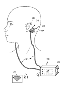
FIG. 5 illustrates an exemplary GVS electrode 34 positioned on the skin
behind the pinna of the left ear 36, and over the left mastoid process, of a
subject to be
treated. The mastoid process is represented by dashed line 38. The right
electrode
(not shown) would be placed in the same manner on the skin over the right
mastoid
process and behind the right pinna. It should be noted that the illustrated
placement of
the electrodes is provided as an example only. In fact, laterality of the
electrode
application, e.g., electrodes precisely over both mastoid processes, is not
believed to
be critical, as long as each electrode is in sufficient proximity to the
vestibular system
to apply the desired stimulation. The electrodes 34 are connected to
stimulation
device 40 (inside housing 30) by leads 33. Manual control means, illustrated
here as a
simple knob 32, may be operated to control the current or other parameters. As
described above, alternative control means include a slide, touch screen,
buttons or
other conventional control devices. External control signals, for example, a
signal
from a heart rate monitor 35, may be input into the device either wirelessly,
as
illustrated, or by leads running between the sensor and the device. Electrodes
such as
the widely commercially available 2x2 inch platinum electrodes used for
transcutaneous electrical nerve stimulation (TENS) may be used in order to
minimize
any possible skin irritation. A conducting gel 37 may be applied between the
CA 02903017 2015-08-28
WO 2014/134564
PCT/US2014/019658
-18-
subject's scalp and the contact surface of the electrodes to enhance
conduction and
reduce the risk of skin irritation.
The amount of current the subject actually receives depends on the scalp
resistance (Iscalp = Velectrodesascalp), which may vary as the user perspires,
if the
electrode position changes, or if contact with the skin is partially lost. It
appears that
the current levels quoted in the literature could only be delivered if the
scalp
resistance was much lower than it actually is. Measurements conducted in
conjunction with the development of the inventive method and device indicate
that the
trans-mastoid resistance is typically between 200 to 500 k-Ohm. Thus, if a GVS
device were actually being used to deliver 1 mA, the voltage would be between
200 to
500V according to Ohm's law. The battery-powered devices that are usually used
to
administer GVS are simply not capable of generating such an output. Hence, the
existing reports appear to be inaccurate with regard to the actual current
being
delivered in GVS.
Prior art designs lack consideration for each subject's unique scalp
resistance,
and therefore may not deliver an effective current to each patient. In the
present
invention, this limitation can be overcome by taking into account inter-
subject scalp
resistance variability as well as compensating for fluctuations in the scalp
resistance
that may occur throughout the procedure. To compensate for slight and
fluctuating
changes in scalp resistance during the administration of current, the
inventive GVS
device may include an internal feedback loop that continuously compares the
desired
current against the actual measured current across the scalp and automatically
compensates for any differences. If Rs,* increases, the Veiectrodes increases
to
compensate. Conversely, voltage decreases when Rscalp drops. This dynamic
feedback
compensation loop provides constant current across the scalp for the duration
of the
procedure regardless of fluctuating changes in electrode-scalp impedance.
FIG. 6 illustrates the vestibular system of the left inner ear. The cochlea
68,
which is the peripheral organ of hearing, is also shown. It demonstrates: the
anterior
62, posterior 67, and horizontal 63 semicircular canals, which transduce
rotational
movements; and the otolith organs (the utricle 66 and saccule 65), which
transduce
linear acceleration and gravity. Without intending to be bound by any theory,
it is
believed that the otolith organs mediate any change in body mass composition
that
CA 02903017 2015-08-28
WO 2014/134564
PCT/US2014/019658
-19-
GVS evokes. The vestibulocochlear nerve 64 (also known as the eighth cranial
nerve)
is composed of the cochlear nerve (which carries signals from the cochlea),
and the
vestibular nerve (which carries signals from the vestibular system).
Validation
Performance of the present invention was evaluated using dual energy x-ray
absoiptiometry (DXA), a technique that was originally developed to determine
bone
mineral density (BMD) and to aid in the management of osteoporosis. More
recently,
the technique has been expanded to include the analysis of fat mass and lean
body
mass in addition to BMD. The DXA machine emits alternating high and low energy
x-rays that produce precise, high quality images. The use of a fan beam allows
decreased scan times so that scans can be completed within seconds or minutes.
The basic principle of DXA data acquisition is based on the differences
between bone and soft tissue attenuation at the high and low x-ray levels. As
the x-ray
beam passes through the subject, detectors register the varying levels of x-
rays that
are absorbed by the anatomical structures of the subject. The raw scan data,
which
includes values of tissue and bone, are captured and sent to a computer. An
algorithm
interprets each pixel, and creates an image and quantitative measurement of
the bone
and body tissues.
Whole body DXA scans using a HOLOGIC Discovery W" DXA scanner
were conducted to determine bone mineral density, lean mass and whole body
fat.
The technique has a precision error (1SD) of 3% for whole body fat and 1.5%
for lean
mass. The in vivo precision for the measurement of bone density using the DXA
technique is 0.5 ¨ 1.5% at the lumbar spine and the standard deviation of the
lumbar
spine bone density is 0.01 gicm2. The radiation risk associated with the
proposed
protocol used is small and in cumulative total is equal to 0.26 mSv for each
subject.
This amount of radiation exposure is low, typically less than what one would
receive
from one year of natural exposure, i.e., around 1.6 mSv.
A comparable commercially available GVS device sold under the trademark
VESTIBULATOR" (Good Vibrations Engineering Ltd. of Ontario, Canada) has
previously been used in a number of research studies at other institutions.
(Barnett-
Cowan & Harris, 2009; Trainor et al., 2009.) This device functions with 8 AA
batteries, so that the voltage can never exceed 12 V. According to the
manufacturer's
CA 02903017 2015-08-28
WO 2014/134564
PCT/US2014/019658
-20-
specifications, the maximum current that this device can deliver is 2.5 mA.
The
present invention uses a more user-friendly device (e.g., the delivered
current can be
adjusted using a controller (knob, slide, or similar) on the side of the
housing, in
comparison to the VESTIBULATORTm, where a similar adjustment can only be
carried out by first writing a MATLAW) script and then uploading it remotely,
via
BLUETOOTHR', in order to reprogram the VESTIBULATOR'sIm settings.)
Due to the very small currents used during GVS, the technique is believed to
be safe (Fitzpatrick & Day, 2004; Hanson, 2009). In particular, although
electrical
current can lead to cardiac arrhythmias, including ventricular fibrillation,
the
threshold for such an occurrence is in the 75 to 400 mA range, well above the
current
levels the battery powered GVS devices can deliver. Furthermore, the
electrodes will
only be applied to the scalp, such as shown in FIG. 5, and nowhere near the
skin over
the chest.
Resistive heating can occur with high voltage electrical stimulation of the
skin.
However, the voltage and current (usually below lmA) delivered during GVS are
well below the levels that pose this risk. Nonetheless, skin irritation can
occur due to
changes in pH. This may be mitigated by using large surface area
(approximately 2
inch diameter) platinum electrodes and aloe vera conducting gels.
It may be desirable to monitor the subject's heart rate (HR) to determine the
cardiac frequency during GVS treatment. The cardiac frequency can then be used
to
alter the frequency of the sinusoidal GVS so as to maintain a certain ratio
between the
cardiac frequency and the frequency of the sinusoidal GVS to avoid
interference with
baroreceptor activity. For example, a sinusoidal GVS frequency to cardiac
frequency
ratio of 0.5 would be appropriate.
During administration of GVS, one platinum electrode is attached to the skin
over one mastoid and the other electrode attached to the skin over the other,
as shown
in FIG. 5. The electrodes may be coated with conducting gel containing aloe
vera.
The device is activated to deliver a current of approximately 0.1 mA (given a
trans-
mastoid resistance of about 500 kOhm) with a sinusoidal waveform at 0.5Hz. A
typical current range for the device would be around 0.001 mA to 5 mA. The
subject
should remain seated or lying flat throughout the session to avoid mishap due
to
altered balance during vestibular stimulation. The device is set up to
automatically
CA 02903017 2015-08-28
WO 2014/134564
PCT/US2014/019658
-21-
stop after one hour however, the subject may discontinue the treatment sooner
if
desired. The subject should remain seated until their balance has returned to
normal,
which should occur within a short period of time after the GVS device has been
turned off.
.. Example 1 ¨ 23 Year Old Female Subject
Data accrued for one human subject support the use of GVS as an effective
approach for altering body mass composition to reduce total body fat and
increase
lean muscle mass. The subject was a Hispanic female born in 1989 and at the
time of
the study was 23 years old. A cumulative total of 20 hours of GVS was
administered
between 8 October 2012 and 7 December 2012. Over this two month period, the
subject received one hour of GVS on each stimulation day. No GVS session
exceeded one hour on any stimulation day.
At the start and completion of the study (after providing a negative pregnancy
test), the subject underwent DXA scans as described above. The first DXA scan
was
carried out on the day of the first GVS session (before the session) and the
second
scan was carried out five days after the final GVS session. In order to ensure
a
constant hydration status, the subject was instructed not to exercise within
12 hours of
the DXA scans and to refrain from consumption of alcohol, nicotine, and
caffeinated
beverages. The subject reported that she was at the same stage of her
menstrual cycle
at the time of each scan. The subject was blinded as to whether she was
receiving an
experimental or placebo procedure.
The GVS was administered using the bipolar binaural method with an
electrode placed on the skin over each mastoid process (see FIG. 5). A linear
stimulus
isolator from World Precision Instruments (A395D) was used to administer the
stimulus, and a 0.5Hz sinusoidal waveform was imposed on this stimulus by a
signal
generator from BK Precision (Model 4010A). The subject was seated with her
eyes
open throughout the administration. The
subject's approximate trans-mastoid
resistance (after preparing the skin with micro-abrasive gel) was
approximately 500
kOhm. To achieve the desired level of stimulation, the current delivered
throughout
each of the GVS sessions was approximately 0.1mA. The subject reported being
aware of a swaying sensation during each stimulation session. The subject made
no
CA 02903017 2015-08-28
WO 2014/134564
PCT/US2014/019658
-22-
changes to her dietary habits and did not engage in exercise during the study
period.
She was on no regular medications.
The report for the initial baseline DXA scan is provided in FIG. 7. Prior to
treatment, testing indicated that the subject had a total body fat of 32947.4
g; a total
combined bone mineral content (BMC) and lean muscle mass of 49799.3 g; and a
percentage body fat of 39.8%. The second DXA scan performed after conclusion
of
the treatment period produced the results shown in FIG. 8. The post-treatment
results
indicated total body fat of 31839.9g; a total combined BMC and lean muscle
mass of
51890.4g; and a percentage body fat of 38.0%. (The BMC is directly
proportional to
the BMD, which as described above is used in the diagnosis of osteoporosis).
Between the two scans, the subject's combined BMC and lean muscle mass
increased by 2091.1g and total body fat decreased by 1107.5g. Compared to the
baseline scan, this represents an increase in combined BMC and lean muscle
mass of
4.2% and a decrease in total body fat of 3.4%. The subject's ratio of total
fat to
combined BMC and total lean muscle mass improved from 0.66 to 0.61. The data
from this subject are thus supportive of the method of using GVS to alter body
mass
composition as described.
The inventive system and method are based on a novel use of vestibular
stimulation, in particular, galvanic vestibular stimulation, to produce
physiological
changes in an individual human's body mass composition. The application of GVS
as
described herein simulates some of the effects of hypergravity, providing a
safe,
simple, drug-free approach to reduce body fat, increase lean muscle mass and
increase
bone density. The simplicity of the device and its operation makes it possible
for any
individual wishing to modify his or her body mass composition, regardless of
whether
for health, aesthetic, or athletic performance reasons, to administer
stimulation in the
privacy of their home. The device may also be used in a medical facility such
as a
doctor's office, clinic, or physical therapy facility to treat obesity and
associated
diseases, treat or prevent osteoporosis, and assist in physical training or
recovery from
injury.
REFERENCES
Balaban CD, Yates BJ. 2004. Vestibulo-autonomic interactions: a teleologic
CA 02903017 2015-08-28
WO 2014/134564
PCT/US2014/019658
-23-
perspective. In: Highstein SM, R. Fay RR, Popper AN, editors. Springer
handbook of auditory research: the vestibular system. New York: Springer-
Verlag. p. 286-342.
Baloh RW, Kerber KA. Clinical neurophysiology of the vestibular system. Oxford
University Press, Oxford, 2011.
Barak N, Greenway FL, Fujioka K, Aronne LJ, Kushner RF. Effect of
histaminergic
manipulation on weight in obese adults: a randomized placebo controlled trial.
Int J Obes (Lond) 2008; 32: 1559-1565.
Barnett-Cowan M, Harris LR. Perceived timing of vestibular stimulation
relative to
touch, light and sound. Exp Brain Res 2009; 198: 221-231.
Bent LR, Bolton PS, Macefield VG. Modulation of muscle sympathetic bursts by
sinusoidal galvanic vestibular stimulation in human subjects. Exp Brain Res
2006; 174: 701-711.
Bolton PS, Wardman DL, Macefield VG. Absence of short-term vestibular
modulation of muscle sympathetic outflow, assessed by brief galvanic
vestibular stimulation in awake human subjects. Exp Brain Res 2004; 154: 39-
43.
Bowers RR, Festuccia WT, Song CK, Shi H, Migliorini RH, Bartness TJ.
Sympathetic innervation of white adipose tissue and its regulation of fat cell
number. Am J Physiol Regul Integr Comp Physiol 2004; 286: R1167-R1175.
Briney SR, Wunder CC. Growth of hamsters during continual centrifugation. Am J
Physiol 1962; 203: 461-464.
Burton RR, Smith AH. Muscle size, gravity and work capacity. Proceedings of
the
XVI International Congress of Aviation and Space Medicine, Lisbon, Portugal
1967.
Burton RR, Smith AH. Adaptation to acceleration environments. In: Handbook of
Physiology. Environmental Physiology. Bethesda, MD, Am Physiol Soc 1996,
sect. 4, vol II, chapt. 40, p. 943-974.
Canonica PG. Effects of prolonged hypergravity stress on the myogenic
properties of
the gastrocnemius muscle. Masters dissertation, University of South Carolina
1966.
Craig AD. Mechanisms of thalamic pain. In: Henry JL, Panju A, Yashpal K,
editors.
CA 02903017 2015-08-28
WO 2014/134564
PCT/US2014/019658
-24-
Central neuropathic pain: focus on poststroke pain. Seattle: IASP Press. 2007
p. 81-99.
Craig AD. How do you feel ¨ now? The anterior insula and human awareness. Nat
Rev Neurosci 2009; 10: 59-70.
Carter JC, Ray CA. Sympathetic response to vestibular activation in humans. Am
J
Physiol Regul Integr Comp Physiol 2008; 294: R681-8.
Cavdar S, San T, Aker R, Sehirli U, Onat F. Cerebellar connections to the
dorsomedial and posterior nuclei of the hypothalamus in the rat. J Anat 2001;
198: 37-45.
Denise P, Normand H, Wood S. 2006. Interactions among the vestibular,
autonomic
and skeletal systems in artificial gravity. In: Clement G, Bukley A, editors.
Artificial gravity. New York: Springer. p. 233-47.
Esposito A, Fistetto G, Di Cerbo A, Palmieri B. Aural stimulation as add-on to
diet
for weight loss: a preliminary clinical study. J Obes Wt Loss Ther 2012; 2.
Evans JW, Smith AH, Boda JM. Fat metabolism and chronic acceleration. Am J
Physiol 1969; 216: 1468-1471.
Fitzpatrick RC, Day BL. Probing the human vestibular system with galvanic
stimulation. J Appl Physiol 2004; 96: 2301-16.
Fuller PM, Warden CH, Barry SJ, Fuller CA. Effects of 2-G exposure on
temperature
regulation, circadian rhythms, and adiposity in UCP2/3 transgenic mice. J
Appl Physiol 2000; 89: 1491-1498.
Fuller PM, Jones TA, Jones SM, Fuller CA. Neurovestibular modulation of
circadian
and homeostatic regulation: Vestibulohypothalamic connection? Proc Natl
Acad Sci USA 2002; 99: 15723-15728.
Fuller PM, Jones TA, Jones SM, Fuller CA. Evidence for macular gravity
receptor
modulation of hypothalamic, limbic and autonomic nuclei. Neuroscience
2004; 129: 461-471.
Fuller PM, Baldwin KM, Fuller CA. Parallel and divergent adaptations of rat
soleus
and plantaris to chronic exercise and hypergravity. Am J Physiol Regul Integr
Comp Physiol 2006; 290: R442-R448.
Gollnick PD, Sjoedin B, Karlsson J, Jansson E, Saltin B. Human soleus muscle:
A
comparison of fiber composition and enzyme activities with other leg muscles.
CA 02903017 2015-08-28
WO 2014/134564
PCT/US2014/019658
-25-
Pfluegers Archiv 1974; 348: 247-255.
Grewal T, James C, Macefield VG. Frequency-dependent modulation of muscle
sympathetic nerve activity by sinusoidal galvanic vestibular stimulation in
human subjects. Exp Brain Res 2009; 197: 379-386.
Hammam E, James C, Dawood T, Macefield VG. Low-frequency sinusoidal galvanic
stimulation of the left and right vestibular nerves reveals two peaks of
modulation in muscle sympathetic nerve activity. Exp Brain Res 2011; 213:
507-514.
Hanson J. Galvanic vestibular stimulation: applied to flight training. 2009.
Masters of
Science in Electrical Engineering Thesis. California Polytechnic State
University.
Hwa JJ, Fawzi AB, Graziano MP, Ghibaudi L, Williams P, Van Heek M, Davis H,
Rudinski M, Sybertz E, Strader CD. Leptin increases energy expenditure and
selectively promotes fat metabolism in ob/ob mice. Am J Physiol 1997; 272:
R1204-1209.
Jaekel E, Amtmann E, Oyama J. Effect of chronic centrifugation on bone density
in
the rat. Anat Embryol 1977; 151: 223-232.
James C, Macefield VG. Competitive interactions between vestibular and cardiac
rhythms in the modulation of muscle sympathetic nerve activity. Auton
Neurosci 2010; 158: 127-131.
James C, Stathis A, Macefield VG. Vestibular and pulse-related modulation of
skin
sympathetic nerve activity during sinusoidal galvanic vestibular stimulation
in
human subjects. Exp Brain Res 2010; 202: 291-298.
Karnath HO, Reich E, Rorden C, Fetter M, Driver J. The perception of body
orientation after neck-proprioceptive stimulation: Effects of time and of
visual
cuing. Exp Brain Res 2002; 143: 350-358.
Katovich M, Smith A. Body mass, composition, and food intake in rabbits during
altered acceleration fields. J Appl Physiol 1978; 45: 51-55.
Keil LC. Changes in growth and body composition of mice exposed to chronic
centrifugation. Growth 1969; 33: 83-88.
Khan S, Chang R. Anatomy of the vestibular system: a review.
NeuroRehabilitation
2013; 32: 437-443.
CA 02903017 2015-08-28
WO 2014/134564
PCT/US2014/019658
-26-
McGeoch PD. The modulation of central pain by vestibular stimulation and
another
study on human brain function. Doctoral thesis, University of Aberdeen, 2010.
McGeoch PD, Williams LE, Lee RR, Ramachandran VS. Behavioural evidence for
vestibular stimulation as a treatment for central post-stroke pain. J Neurol
Neurosurg Psychiatry 2008; 79:1 298-1301.
McGeoch PD, Williams LE, Song T, Lee RR, Huang M, Ramachandran VS. Post-
stroke tactile allodynia and its modulation by vestibular stimulation: a MEG
case study. Acta Neurol Scand 2009; 119: 404-409.
Oyama J and Platt WT. Reproduction and growth of mice and rats under
conditions of
simulated increased gravity. Am J Physiol 1967; 212: 164-166.
Oyama J, Zeitman B. Tissue composition of rats exposed to chronic
centrifugation.
Am J Physiol 1967;213: 1305-1310.
Petersen H, Magnusson M, Fransson PA, Johansson R. Vestibular disturbance at
frequencies above 1 Hz affects human postural control. Acta Otolaryngol
1994; 114: 225-230.
Pitts GC, Bull LS, Oyama J. Effect of chronic centrifugation on body
composition of
the rat. Am J Physiol 1972; 223: 1944-1948.
Ray CA, Hume KM, Steele SL. Sympathetic nerve activity during natural
stimulation
of horizontal semicircular canals in humans. Am J Physiol Regul Integr Comp
Physiol 1998; 275: R1274-R1278.
Roy RR, Roy ME, Talmadge RJ, Mendoza R, Grindeland RE, Vasques M. Size and
myosin heavy chain profiles of rat hindlimb extensor muscle fibers after 2
weeks at 2G. Aviat Space Environ Med 1996; 67 (9): 854-858.
Sailesh KS, Mukkadan JK. Vestibular modulation of endocrine secretions ¨ a
review.
Int J Res Health Sci 2014; 2(1): 0-0
St George RJ, Fitzpatrick RC. The sense of self-motion, orientation and
balance
explored by vestibular stimulation. J Physiol 2011; 589: 807-813.
Smith AH. Centrifuges: their development and use in gravitational biology.
ASGSB
Bulletin 1992; 5(2): 33-41.
Smith AH, Kelly CF. Influence of chronic acceleration upon growth and body
composition. Ann NY Acad Sci 1963; 110: 410-424.
CA 02903017 2015-08-28
WO 2014/134564
PCT/US2014/019658
-27-
Smith AH, Kelly CF. Biological effects of chronic acceleration Naval Res Rev
1965;
18: 1-10.
Smith AH, Sanchez 0, Burton RR. Gravitational effects on body composition in
birds. Life Sci Space Res 1975; 13: 21-27.
Sobhani I, Buyse M, Goiot H, Weber N, Laigneau JP, Henin D, Soul JC, Bado A.
Vagal stimulation rapidly increases leptin secretion in human stomach.
Gastroenterology 2002; 122: 259-263.
Sved AF, Ito S, Madden CJ. Baroreflex dependent and independent roles of the
caudal
ventrolaterl medulla in cardiovascular regulation. Brain Res Bull 2000; 51:
129-133.
Trainor LJ, Gao X, Lei JJ, Lehtovaara K, Harris LR. The primal role of the
vestibular
system in determining musical rhythm. Cortex 2009; 45: 35-43.
Voustianiouk A, Kaufmann H, Diedrich A, Raphan T, Biaggioni I, MacDougall H,
Ogorodnikov D, Cohen B. Electrical activation of the human vestibule-
sympathetic reflex. Exp Brain Res 2005; 171: 251-261.
Warren LE, Horwitz BA, Fuller CA. Effects of 2G on lean and obese Zucker rats
(Abstract). Fourteenth Annual Meeting Am Soc Gravitational and Space Biol.
1998.
Watson SRD, Colebatch JG. Vestibular-evoked electromyographic responses in
soleus: a comparison between click and galvanic stimulation. Exp Brain Res
1998;
119:504-510.