Note: Descriptions are shown in the official language in which they were submitted.
CA 02903480 2015-09-01
WO 2014/152358
PCT/US2014/027250
COMBINATIONS OF A MEK INHIBITOR COMPOUND WITH AN HER3/EGFR INHIBITOR
COMPOUND AND METHODS OF USE
FIELD OF THE INVENTION
The invention relates generally to pharmaceutical combinations of compounds
with activity
against hyperproliferative disorders such as cancer that include a combination
of a compound that
inhibits the MEK pathway with a compound that blocks HER3/EGFR. The invention
also relates to
methods of using the combinations for in vitro, in situ, and in vivo diagnosis
or treatment of
mammalian cells, or associated pathological conditions.
BACKGROUND OF THE INVENTION
Protein kinases (PK) are enzymes that catalyze the phosphorylation of hydroxy
groups on
tyrosine, serine and threonine residues of proteins by transfer of the
terminal (gamma) phosphate from
ATP. Through signal transduction pathways, these enzymes modulate cell growth,
differentiation and
proliferation, i.e., virtually all aspects of cell life in one way or another
depend on PK activity
(Hardie, G. and Hanks, S. (1995) The Protein Kinase Facts Book. Land IT
Academic Press, San
Diego, CA). Furthermore, abnormal PK activity has been related to a host of
disorders, ranging from
relatively non-life threatening diseases such as psoriasis to extremely
virulent diseases such as
glioblastoma (brain cancer). Protein kinases are an important target class for
therapeutic modulation
(Cohen, P. (2002) Nature Rev. Drug Discovery 1:309).
MEK is a dual-specificity kinase that phosphorylates tyrosines and threonines
required for
activation on ERK 1 and 2. Two related genes encode MEK1 and MEK2 which differ
in their binding
to ERKs. HER3 a receptor tyrosine kinase that can be bound and activated by
neuregulins and
NTAK. EGFR is a transmembrane glycoprotein that is a receptor for members of
the epidermal
growth factor family.
Currently, there remains a need for improved methods and compositions that can
be used to
treat hyperproliferative diseases such as cancer.
SUMMARY OF THE INVENTION
It has been determined that improved effects in inhibiting the growth of
cancer cells in vitro
and in vivo can be achieved by inhibiting MEK, HER3 and EGFR. It has been
found, for example,
that improved effects in inhibiting the growth of cancer cells in vitro and in
vivo can be achieved by
administering a combination of GDC-0973 or GDC-0623, or a pharmaceutically
acceptable salt
1
SUBSTITUTE SHEET (RULE 26)
CA 02903480 2015-09-01
WO 2014/152358
PCT/US2014/027250
combinations and methods will be useful in the treatment of hyperproliferative
disorders such as
cancer. In certain embodiments, administration of the combinations may provide
synergistic effects.
Accordingly, certain embodiments of the invention provide therapeutic
combinations
comprising the small-molecule MEK inhibitor GDC-0973 (Formula I), or a
pharmaceutically
acceptable salt thereof (see WO 2007/044515) and having the structure:
OH
H
N
F H
0 NIJO
to N
# (I)
I F
F
or the small-molecule MEK inhibitor GDC-0623 (Formula II), or a
pharmaceutically
acceptable salt thereof (see W02009/085983), and having the structure:
H
HO ...N 0
0 F
H
N
I # (11)
1 I
N
in combination with MEHD7945A, a dual-action antibody which comprises two
identical
antigen binding domains, each of which specifically binds to both HER3 and
EGFR (see DLllf in
WO 2010/108127 (e.g., Figure 33) and Schaefer et al., Cancer Cell, 20, 472-486
(2011)).
MEHD7945A and GDC-0973 or GDC-0623may be present in two separate
pharmaceutical
compositions or together in a single pharmaceutical composition.
Accordingly, certain embodiments of the invention are directed to a
combination of GDC-
0973 or GDC-0623, or a pharmaceutically acceptable salt thereof and MEHD7945A,
for the
therapeutic treatment of a hyperproliferative disorder.
In certain embodiments, the hyperproliferative disorder is cancer.
In certain embodiments, the cancer is associated with the KRAS mutation.
In certain embodiments, the cancer is selected from, colorectal, mesothelioma,
endometrial,
pancreatic, breast, lung, ovarian, prostate, melanoma, gastric, colon, renal,
head and neck, and
glioblastoma
In certain embodiments GDC-0973 or a pharmaceutically acceptable salt thereof
is
2
CA 02903480 2015-09-01
WO 2014/152358
PCT/US2014/027250
administered in combination with MEHD7945A.
In certain embodiments, GDC-0623or a pharmaceutically acceptable salt thereof
is
administered in combination with MEHD7945A.
In certain embodiments, GDC-0973 or GDC-0623, or a pharmaceutically acceptable
salt
thereof is administered simultaneously with MEHD7945A.
In certain embodiments, GDC-0973 or GDC-0623, or a pharmaceutically acceptable
salt
thereof and MEHD7945A are administered sequentially.
Certain embodiments of the invention are directed to a combination of GDC-0973
or GDC-
0623, or a pharmaceutically acceptable salt thereof and MEHD7945A for
therapeutic use for
improving the quality of life of a patient having a hyperproliferative
disorder.
Certain embodiments of the invention are directed to a combination of GDC-0973
or GDC-
0623, or a pharmaceutically acceptable salt thereof; and MEHD7945A, for
treating a
hyperproliferative disorder.
Certain embodiments of the invention are directed to a use of a combination of
GDC-0973 or
GDC-0623, or a pharmaceutically acceptable salt thereof; and MEHD7945A, in the
preparation of a
medicament for the treatment of a hyperproliferative disorder in a patient.
Certain embodiments of the invention are directed to a kit comprising GDC-0973
or GDC-
0623, or a pharmaceutically acceptable salt thereof; and MEHD7945A, a
container, and a package
insert or label indicating the administration GDC-0973 or GDC-0623, or a
pharmaceutically
acceptable salt thereof; and MEHD7945A, for treating a hyperproliferative
disorder.
Certain embodiments of the invention are directed to a product comprising GDC-
0973 or
GDC-0623, or a pharmaceutically acceptable salt thereof and MEHD7945A as a
combined
preparation for separate, simultaneous or sequential use in the treatment of a
hyperproliferative
disorder (e.g., cancer).
Certain embodiments of the invention are directed to a method for treating a
hyperproliferative disorder in a patient (e.g., cancer), comprising
administering to the patient a
combination of GDC-0973 and GDC-0623, or a pharmaceutically acceptable salt
thereof; and
MEHD7945A.
BRIEF DESCRIPTION OF THE DRAWINGS
Figure 1 is a graph demonstrating that MEHD7945A binds to both HER3-ECD and
EGFR-
ECD.
Figure 2A and B are graphs demonstrating that MEHD7945A inhibits EGFR and
HER2/HER3-dependent signaling.
Figure 3 is a graph showing inhibition of tumor growth in FaDu cancer model by
MEHD7945A.
3
CA 02903480 2015-09-01
WO 2014/152358
PCT/US2014/027250
Figure 4 is a summary of the tumor growth inhibitory effect of MEHD7945A
compared to
cetuximab or anti-HER3 in numerous murine xenograft models.
Figure 5 is a graph demonstrating that GDC-0973 and GDC-0623 are effective in
inhibiting
the growth of B-RAF mutant tumor cells.
Figure 6 is a graph demonstrating that GDC-0973 and GDC-0623 are effective in
inhibiting
the growth of KRAS mutant tumor cells.
Figure 7 is a graph demonstrating the effect of single agent and combination
treatment on
pAkt and pERK levels in a murine xenograft CRC KRAS DLD-1 (A) and LS180 (B)
models.
Figure 8 is a graph demonstrating the tumor growth inhibitory effect of single
agent and
combination treatments of MEHD7945A, GDC-0973 and GDC-0623.
Figure 9 demonstrates that TGFoc-stimulated LS180 or DLD-1 cells treated with
cobimetinib
showed increased phosphorylation of AKT.
Figure 10 is a graph demonstrating the inhibition of KRAS-mutant cell line,
LS180,
proliferation by MEHD7945A and cobimetinib combination.
Figure 11A is a graph demonstrating the effect of cobimetinib in combination
with
MEHD7945A on LS180 Colorectal Adenocarcinoma Tumor Xenografts in CD-1 Nude
Mice; Figure
11B is a table summarizing the data from Figure 11A.
Figure 12A is a graph demonstrating the effect of cobimetinib in combination
with
MEHD7945A on KRAS-Mutant DLD-1 Colorectal Adenocarcinoma Tumor Xenografts in
C.B-17
SCID beige mice; Figure 12B is a table summarizing the data from Figure 12A.
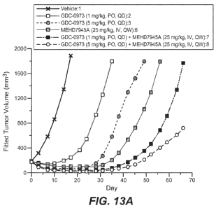
Figure 13A is a graph demonstrating the effect of cobimetinib in combination
with
MEHD7945A on BxPC3 Ductal Pancreatic Xenograft Tumors in NCr Nude Mice; Figure
13B is a
table summarizing the anti-tumor activity for this study; Figure 13C is a
table summarizing the Time
to Tumor Progression and Response for this study.
DETAILED DESCRIPTION OF EXEMPLARY EMBODIMENTS
I. Definitions
It must be noted that as used herein and in the appended claims, the singular
forms "a", "and",
and "the" include plural referents unless the context clearly dictates
otherwise.
Throughout this specification and claims, the word "comprise," or variations
such as
"comprises" or "comprising," will be understood to imply the inclusion of a
stated integer or group of
integers but not the exclusion of any other integer or group of integers.
The term "antibody" herein is used in the broadest sense and specifically
covers monoclonal
antibodies, polyclonal antibodies, multispecific antibodies, and antibody
fragments so long as they
exhibit the desired biological activity. The term "multispecific antibody" is
used in the broadest sense
and specifically covers an antibody comprising an antigen-binding domain that
has polyepitopic
4
CA 02903480 2015-09-01
WO 2014/152358
PCT/US2014/027250
specificity (i.e., is capable of specifically binding to two, or more,
different epitopes on one biological
molecule or is capable of specifically binding to epitopes on two, or more,
different biological
molecules). One specific example of an antigen-binding domain is a VHVL unit
comprised of a heavy
chain variable domain (VH) and a light chain variable domain (VL). Such
multispecific antibodies
include, but are not limited to, full length antibodies, antibodies having two
or more VL and VH
domains, antibody fragments such as Fab, Fv, dsFv, scFv, diabodies, bispecific
diabodies and
triabodies, antibody fragments that have been linked covalently or non-
covalently. A "bispecific
antibody" is a multispecific antibody comprising an antigen-binding domain
that is capable of
specifically binding to two different epitopes on one biological molecule or
is capable of specifically
binding to epitopes on two different biological molecules. The bispecific
antibody is also referred to
herein as having "dual specificity" or as being "dual specific".
In certain embodiments, an antibody of the invention has a dissociation
constant (Kd) of
< 1 M, < 100 nM, < 10 nM, < 1 nM, < 0.1 nM, < 0.01 nM, or < 0.001 nM (e.g. 10-
8M or less, e.g.
from 10-8M to 10-13M, e.g., from 10-9M to 10-13 M) for its target HER or HERs.
The basic 4-chain antibody unit is a heterotetrameric glycoprotein composed of
two identical
light (L) chains and two identical heavy (H) chains (an IgM antibody consists
of 5 of the basic
heterotetramer units along with an additional polypeptide called J chain, and
therefore contains 10
antigen-binding sites, while secreted IgA antibodies can polymerize to form
polyvalent assemblages
comprising 2-5 of the basic 4-chain units along with J chain). In the case of
IgGs, the 4-chain unit is
generally about 150,000 daltons. Each L chain is linked to an H chain by one
covalent disulfide bond,
while the two H chains are linked to each other by one or more disulfide bonds
depending on the H
chain isotype. Each H and L chain also has regularly spaced intrachain
disulfide bridges. Each H
chain has, at the N-terminus, a variable domain (VH) followed by three
constant domains (CH) for
each of the a and 7 chains and four CH domains for and E isotypes. Each L
chain has, at the N-
terminus, a variable domain (VL) followed by a constant domain (CL) at its
other end. The VL is
aligned with the VH and the CL is aligned with the first constant domain of
the heavy chain (CH1).
Particular amino acid residues are believed to form an interface between the
light chain and heavy
chain variable domains. The pairing of a VH and VL together forms a single
antigen-binding site.
For the structure and properties of the different classes of antibodies, see,
e.g., Basic and Clinical
Immunology, 8th edition, Daniel P. Stites, Abba I. Terr and Tristram G.
Parslow (eds.), Appleton &
Lange, Norwalk, CT, 1994, page 71 and Chapter 6.
The L chain from any vertebrate species can be assigned to one of two clearly
distinct types,
called kappa and lambda, based on the amino acid sequences of their constant
domains. Depending
on the amino acid sequence of the constant domain of their heavy chains (CH),
immunoglobulins can
be assigned to different classes or isotypes. There are five classes of
immunoglobulins: IgA, IgD,
5
CA 02903480 2015-09-01
WO 2014/152358
PCT/US2014/027250
IgE, IgG, and IgM, having heavy chains designated a, 6, y, E, and ,
respectively. The 7 and a classes
are further divided into subclasses on the basis of relatively minor
differences in CH sequence and
function, e.g., humans express the following subclasses: IgGl, IgG2, IgG3,
IgG4, IgAl, and IgA2.
The term "variable" refers to the fact that certain segments of the variable
domains differ
extensively in sequence among antibodies. The V domain mediates antigen-
binding and defines
specificity of a particular antibody for its particular antigen. However, the
variability is not evenly
distributed across the 110-amino acid span of the variable domains. Instead,
the V regions consist of
relatively invariant stretches called framework regions (FRs) of 15-30 amino
acids separated by
shorter regions of extreme variability called hypervariable regions" or HVR.
The variable domains of
native heavy and light chains each comprise four FRs, largely adopting a beta-
sheet configuration,
connected by three hypervariable regions, which form loops connecting, and in
some cases forming
part of, the beta-sheet structure. The hypervariable regions in each chain are
held together in close
proximity by the FRs and, with the hypervariable regions from the other chain,
contribute to the
formation of the antigen-binding site of antibodies (see Kabat et al.,
Sequences of Proteins of
Immunological Interest, 5th Ed. Public Health Service, National Institutes of
Health, Bethesda, MD.
(1991)). The constant domains are not involved directly in binding an antibody
to an antigen, but
exhibit various effector functions, such as participation of the antibody in
antibody dependent cellular
cytotoxicity (ADCC).
The term "hypervariable region," "HVR," or "HV," when used herein refers to
the regions of
an antibody variable domain which are hypervariable in sequence and/or form
structurally defined
loops. Generally, antibodies comprise six HVRs; three in the VH (HVR-H1, HVR-
H2, HVR-H3),
and three in the VL (HVR-L1, HVR-L2, HVR-L3). In native antibodies, H3 and L3
display the most
diversity of the six HVRs, and H3 in particular is believed to play a unique
role in conferring fine
specificity to antibodies. See, e.g., Xu et al., Immunity 13:37-45 (2000);
Johnson and Wu, in Methods
in Molecular Biology 248:1-25 (Lo, ed., Human Press, Totowa, NJ, 2003).
Indeed, naturally
occurring camelid antibodies consisting of a heavy chain only are functional
and stable in the absence
of light chain. See, e.g., Hamers-Casterman et al., Nature 363:446-448 (1993);
Sheriff et al., Nature
Struct. Biol. 3:733-736 (1996).
HVRs generally comprise amino acid residues from the hypervariable loops
and/or from the
"complementarity determining regions" (CDRs), the latter being of highest
sequence variability
and/or involved in antigen recognition. A number of HVR delineations are in
use and are
encompassed herein. The Kabat Complementarity Determining Regions (CDRs) are
based on
sequence variability and are the most commonly used (Kabat et al., Sequences
of Proteins of
Immunological Interest, 5th Ed. Public Health Service, National Institutes of
Health, Bethesda, MD.
(1991)). Chothia refers instead to the location of the structural loops
(Chothia and Lesk J. MoL Biol.
6
CA 02903480 2015-09-01
WO 2014/152358
PCT/US2014/027250
196:901-917 (1987)). The AbM HVRs represent a compromise between the Kabat
HVRs and
Chothia structural loops, and are used by Oxford Molecular's AbM antibody
modeling software. The
"contact" HVRs are based on an analysis of the available complex crystal
structures. The residues
from each of these HVRs are noted below.
Loop Kabat AbM Chothia Contact
L1 L24-L34 L24-L34 L26-L32 L30-L36
L2 L50-L56 L50-L56 L50-L52 L46-L55
L3 L89-L97 L89-L97 L91-L96 L89-L96
H1 H31-H35B H26-H35B H26-H32 H30-H35B
(Kabat Numbering)
H1 H31-H35 H26-H35 H26-H32 H30-H35
(Chothia Numbering)
H2 H50-H65 H50-H58 H53-H55 H47-H58
H3 H95-H102 H95-H102 H96-H101 H93-H101
HVRs may comprise "extended HVRs" as follows: 24-36 or 24-34 (L1), 46-56 or 50-
56 (L2)
and 89-97 or 89-96 (L3) in the VL and 26-35 (H1), 50-65 or 47-65 (H2) and 93-
102, 94-102, or 95-
102 (H3) in the VH. The variable domain residues are numbered according to
Kabat et al., supra, for
each of these definitions.
"Framework" or "FR" residues are those variable domain residues other than the
HVR residues
as herein defined.
The term "variable domain residue numbering as in Kabat" or "amino acid
position numbering
as in Kabat," and variations thereof, refers to the numbering system used for
heavy chain variable
domains or light chain variable domains of the compilation of antibodies in
Kabat et al., supra. Using
this numbering system, the actual linear amino acid sequence may contain fewer
or additional amino
acids corresponding to a shortening of, or insertion into, a FR or HVR of the
variable domain. For
example, a heavy chain variable domain may include a single amino acid insert
(residue 52a
according to Kabat) after residue 52 of H2 and inserted residues (e.g.
residues 82a, 82b, and 82c, etc.
according to Kabat) after heavy chain FR residue 82. The Kabat numbering of
residues may be
determined for a given antibody by alignment at regions of homology of the
sequence of the antibody
with a "standard" Kabat numbered sequence.
The Kabat numbering system is generally used when referring to a residue in
the variable
domain (approximately residues 1-107 of the light chain and residues 1-113 of
the heavy chain) (e.g,
Kabat et al., Sequences of Immunological Interest. 5th Ed. Public Health
Service, National Institutes
of Health, Bethesda, Md. (1991)). The "EU numbering system" or "EU index" is
generally used
7
CA 02903480 2015-09-01
WO 2014/152358
PCT/US2014/027250
when referring to a residue in an immunoglobulin heavy chain constant region
(e.g., the EU index
reported in Kabat et al., supra). The "EU index as in Kabat" refers to the
residue numbering of the
human IgG1 EU antibody. Unless stated otherwise herein, references to residue
numbers in the
variable domain of antibodies means residue numbering by the Kabat numbering
system. Unless
stated otherwise herein, references to residue numbers in the constant domain
of antibodies means
residue numbering by the EU numbering system (e.g., see WO 2006/073941).
"Affinity" refers to the strength of the sum total of noncovalent interactions
between a single
binding site of a molecule (e.g., an antibody) and its binding partner (e.g.,
an antigen). Unless
indicated otherwise, as used herein, "binding affinity" refers to intrinsic
binding affinity which
reflects a 1:1 interaction between members of a binding pair (e.g., antibody
and antigen). The affinity
of a molecule X for its partner Y can generally be represented by the
dissociation constant (Kd).
Affinity can be measured by common methods known in the art, including those
described herein.
An "affinity matured" antibody is one with one or more alterations in one or
more HVRs or
framework region thereof which result in an improvement in the affinity of the
antibody for antigen,
compared to a parent antibody which does not possess those alteration(s). In
one embodiment, an
affinity matured antibody has nanomolar or even picomolar affinities for the
target antigen. Affinity
matured antibodies may be produced using certain procedures known in the art.
For example, Marks
et al. Bio/Technology 10:779-783 (1992) describes affinity maturation by VH
and VL domain
shuffling. Random mutagenesis of HVR and/or framework residues is described
by, for example,
Barbas et al. Proc Nat. Acad. Sci. USA 91:3809-3813 (1994); Schier et a/. Gene
169:147-155 (1995);
Yelton et a/. J. Immunot. 155:1994-2004 (1995); Jackson et al., J. Immunot.
154(7):3310-9 (1995);
and Hawkins et al, J. Mot. Biol. 226:889-896 (1992).
The "class" of an antibody refers to the type of constant domain or constant
region possessed
by its heavy chain. There are five major classes of antibodies: IgA, IgD, IgE,
IgG, and IgM, and
several of these may be further divided into subclasses (isotypes), e.g.,
IgGi, IgG2, IgG3, IgG4, IgAi,
and IgA2. The heavy chain constant domains that correspond to the different
classes of
immunoglobulins are called a, 6, c, y, and 11., respectively.
The term "patient" (interchangeably termed "individual" and "subject") is a
human patient.
The patient may be a "cancer patient", i.e. one who is suffering or at risk
for suffering from one or
more symptoms of cancer.
The terms "treat" and "treatment" refer to therapeutic treatment, wherein the
object is to
prevent or slow down (lessen) an undesired physiological change or disorder,
such as the growth,
development or spread of cancer. For purposes of this invention, beneficial or
desired clinical results
include, but are not limited to, alleviation of symptoms, diminishment of
extent of disease, stabilized
(i.e., not worsening) state of disease, delay or slowing of disease
progression, amelioration or
8
CA 02903480 2015-09-01
WO 2014/152358
PCT/US2014/027250
palliation of the disease state, and remission (whether partial or total),
whether detectable or
undetectable. "Treatment" can also mean prolonging survival as compared to
expected survival if not
receiving treatment. Those in need of treatment include those already having
the condition or
disorder, e.g., a patient with cancer.
The phrase "therapeutically effective amount" means an amount that (i) treats
the particular
disease, condition, or disorder, (ii) attenuates, ameliorates, or eliminates
one or more symptoms of the
particular disease, condition, or disorder, or (iii) prevents or delays the
onset of one or more
symptoms of the particular disease, condition, or disorder described herein.
In the case of cancer, the
therapeutically effective amount may reduce the number of cancer cells; reduce
the tumor size; inhibit
(e.g., slow to some extent and preferably stop) cancer cell infiltration into
peripheral organs; inhibit
(e.g., slow to some extent and preferably stop) tumor metastasis; inhibit, to
some extent, tumor
growth; and/or relieve to some extent one or more of the symptoms associated
with the cancer. To the
extent the combination may prevent growth and/or kill existing cancer cells,
it may be cytostatic
and/or cytotoxic. For cancer therapy, efficacy can be measured, for example,
by assessing the time to
disease progression (TTP) and/or determining the response rate (RR).
The terms "cancer" and "cancerous" refer to or describe the physiological
condition in
mammals that is typically characterized by unregulated cell growth. A "tumor"
comprises one or
more cancerous cells. Examples of cancer include, but are not limited to,
carcinoma, lymphoma,
blastoma, sarcoma, and leukemia or lymphoid malignancies. More particular
examples of such
cancers include squamous cell cancer (e.g., epithelial squamous cell cancer),
lung cancer including
small- cell lung cancer, non-small cell lung cancer ("NSCLC"), adenocarcinoma
of the lung and
squamous carcinoma of the lung, cancer of the peritoneum, hepatocellular
cancer, gastric or stomach
cancer including gastrointestinal cancer, pancreatic cancer, glioblastoma,
cervical cancer, ovarian
cancer, liver cancer, bladder cancer, hepatoma, breast cancer, colon cancer,
rectal cancer, colorectal
cancer, endometrial or uterine carcinoma, salivary gland carcinoma, kidney or
renal cancer, prostate
cancer, vulval cancer, thyroid cancer, hepatic carcinoma, anal carcinoma,
penile carcinoma, as well as
head and neck cancer. Gastric cancer, as used herein, includes stomach cancer,
which can develop in
any part of the stomach and may spread throughout the stomach and to other
organs; particularly the
esophagus, lungs, lymph nodes, and the liver.
A "chemotherapeutic agent" is a biological (e.g., large molecule) or chemical
(e.g., small
molecule) compound useful in the treatment of cancer, regardless of mechanism
of action.
A "platinum agent" is a chemotherapeutic agent that comprises platinum, for
example
carboplatin, cisplatin, and oxaliplatin.
The term "mammal" includes, but is not limited to, humans, mice, rats, guinea
pigs, monkeys,
dogs, cats, horses, cows, pigs, sheep, and poultry. In one embodiment, the
mammal is a human.
The term "package insert" is used to refer to instructions customarily
included in commercial
9
CA 02903480 2015-09-01
WO 2014/152358
PCT/US2014/027250
packages of therapeutic products that contain information about the
indications, usage, dosage,
administration, contraindications and/or warnings concerning the use of such
therapeutic products.
The phrase "pharmaceutically acceptable salt" as used herein, refers to
pharmaceutically
acceptable organic or inorganic salts of a compound. Exemplary salts include,
but are not limited, to
bismesylate, sulfate, citrate, acetate, oxalate, chloride, bromide, iodide,
nitrate, bisulfate, phosphate,
acid phosphate, isonicotinate, lactate, salicylate, acid citrate, tartrate,
oleate, tannate, pantothenate,
bitartrate, ascorbate, succinate, maleate, gentisinate, fumarate, gluconate,
glucuronate, saccharate,
formate, benzoate, glutamate, methanesulfonate "mesylate", ethanesulfonate,
benzenesulfonate, p-
toluenesulfonate, and pamoate (i.e., 1,1'-methylene-bis -(2-hydroxy-3-
naphthoate)) salts. A
pharmaceutically acceptable salt may involve the inclusion of another molecule
such as an acetate ion,
a succinate ion or other counter ion. The counter ion may be any organic or
inorganic moiety that
stabilizes the charge on the parent compound. Furthermore, a pharmaceutically
acceptable salt may
have more than one charged atom in its structure. Instances where multiple
charged atoms are part of
the pharmaceutically acceptable salt can have multiple counter ions. Hence, a
pharmaceutically
acceptable salt can have one or more charged atoms and/or one or more counter
ion.
The desired pharmaceutically acceptable salt may be prepared by any suitable
method
available in the art. For example, treatment of the free base with an
inorganic acid, such as
hydrochloric acid, hydrobromic acid, sulfuric acid, nitric acid,
methanesulfonic acid, phosphoric acid
and the like, or with an organic acid, such as acetic acid, maleic acid,
succinic acid, mandelic acid,
fumaric acid, malonic acid, pyruvic acid, oxalic acid, glycolic acid,
salicylic acid, a pyranosidyl acid,
such as glucuronic acid or galacturonic acid, an alpha hydroxy acid, such as
citric acid or tartaric acid,
an amino acid, such as aspartic acid or glutamic acid, an aromatic acid, such
as benzoic acid or
cinnamic acid, a sulfonic acid, such as p-toluenesulfonic acid or
ethanesulfonic acid, or the like.
Acids which are generally considered suitable for the formation of
pharmaceutically useful or
acceptable salts from basic pharmaceutical compounds are discussed, for
example, by P. Stahl et al,
Camille G. (eds.) Handbook of Pharmaceutical Salts. Properties, Selection and
Use. (2002) Zurich:
Wiley-VCH; S. Berge et al, Journal of Pharmaceutical Sciences (1977) 66(1)
119; P. Gould,
International J. of Pharmaceutics (1986) 33 201 217; Anderson et al, The
Practice of Medicinal
Chemistry (1996), Academic Press, New York; Remington's Pharmaceutical
Sciences, 18th ed.,
(1995) Mack Publishing Co., Easton PA; and in The Orange Book (Food & Drug
Administration,
Washington, D.C. on their website). These disclosures are incorporated herein
by reference thereto.
The phrase "pharmaceutically acceptable" indicates that the substance or
composition is
compatible chemically and/or toxicologically with the other ingredients
comprising a formulation
and/or the patient being treated therewith.
The term "synergistic" as used herein refers to a therapeutic combination
which is more
effective than the additive effects of the two or more single agents. A
determination of a synergistic
CA 02903480 2015-09-01
WO 2014/152358
PCT/US2014/027250
interaction may be based on the results obtained from the assays known in the
art. The results of
these assays can be analyzed using the Chou and Talalay combination method and
Dose-Effect
Analysis with CalcuSyn software in order to obtain a Combination Index (Chou
and Talalay, 1984,
Adv. Enzyme Regul. 22:27-55). The combinations provided herein can be analyzed
utilizing a
standard program for quantifying synergism, additivism, and antagonism among
anticancer agents.
An example program is that described by Chou and Talalay, in "New Avenues in
Developmental
Cancer Chemotherapy," Academic Press, 1987, Chapter 2. Combination Index
values less than 0.8
indicates synergy, values greater than 1.2 indicate antagonism and values
between 0.8 to 1.2 indicate
additive effects. The combination therapy may provide "synergy" and prove
"synergistic", i.e., the
effect achieved when the active ingredients used together is greater than the
sum of the effects that
results from using the compounds separately. Thus, in embodiments, the
combined amount of the
active ingredients are effective in providing a synergistic effect (also
referred to herein as a
synergistically effective amount). A synergistic effect may be attained when
the active ingredients
are: (1) co-formulated and administered or delivered simultaneously in a
combined, unit dosage
formulation; (2) delivered by alternation or in parallel as separate
formulations; or (3) by some other
regimen. When delivered in alternation therapy, a synergistic effect may be
attained when the
compounds are administered or delivered sequentially, e.g., by different
injections in separate
syringes. In general, during alternation therapy, an effective dosage of each
active ingredient is
administered sequentially, i.e., serially, whereas in combination therapy,
effective dosages of two or
more active ingredients are administered together. Combination effects were
evaluated using both the
BLISS independence model and the highest single agent (HSA) model (Lehar et
al. 2007, Molecular
Systems Biology 3:80). BLISS scores quantify degree of potentiation from
single agents and a
positive BLISS score (greater than 0) suggests greater than simple additivity.
A cumulative positive
BLISS score greater than 250 is considered strong synergy observed within the
concentration ranges
tested. An HSA score (greater than 0) suggests a combination effect greater
than the maximum of the
single agent responses at corresponding concentrations.
In addition to providing improved treatment for a given hyperproliferative
disorder,
administration of certain combinations of the invention may improve the
quality of life for a patient
compared to the quality of life experienced by the same patient receiving a
different treatment. For
example, administration of a combination to a patient may provide an improved
quality of life
compared to the quality of life the same patient would experience if they
received only one of the
individual agents as therapy. For example, the combined therapy with a
combination described herein
may lower the dose of therapeutic agents needed. The combination therapy may
also decrease or
eliminate the need for the use of chemotherapeutic agents and the side-effects
associated with high-
dose chemotherapeutic agents (e.g. nausea, vomiting, hair loss, rash,
decreased appetite, weight loss,
etc.). The combination may also cause reduced tumor burden and the associated
adverse events, such
11
CA 02903480 2015-09-01
WO 2014/152358
PCT/US2014/027250
as pain, organ dysfunction, weight loss, etc. Accordingly, one aspect of the
invention provides a
combination for therapeutic use for improving the quality of life of a patient
treated for a
hyperproliferative disorder with an agent described herein.
One aspect includes a method of tumor growth inhibition (TGI) in a patient
suffering from a
cancer, comprising administering a combination described herein to the
patient. In certain
embodiments, the combination provides a synergistic effect.
In certain embodiments, the TGI of the combination is greater than the TGI of
any one of
GDC-0973 and GDC-0623 or MEHD7945A alone. In certain embodiments, the TGI of
the
combination is about 10, 15, 20, 25, 30, 35, 40, 45, 50, 55, 60, 65, 70 or 75
percent greater than the
TGI of the agents alone.
Methods of measuring TGI are known in the art. In one example method, average
tumor
volumes are determined and compared from the patient before and after
treatment. Tumor volumes
can be measured in two dimensions (length and width) using any method in the
art, for example
UltraCal IV calipers (Fred V. Fowler Company) or by PET (positron emission
tomography), or by
some other method. The formula tumor volume (mm3) = (length x width2) x 0.5
can be used.
Measuring tumor volumes over multiple time periods can be done using a mixed-
modeling Linear
Mixed Effects (LME) approach (Pinheiro et al. 2009). This approach can address
both repeated
measurements (and multiple patients). Cubic regression splines can be used to
fit a non-linear profile
to the time courses of tumor volume at each dose level. These non-linear
profiles can then be related
to dose within the mixed model. Tumor growth inhibition as a percent of
vehicle can be calculated as
a percent area under the fitted curve (AUC) per day in relation to the
vehicle, using the following
formula:
[
% TGI = 100 1 AUCtreatment / clay
"Cvehicle / day
Using this formula, a TGI value of 100% indicates tumor stasis, greater than
about 1% but less than
about 100% indicates tumor growth inhibition, and greater than about 100%
indicates tumor
regression.
II. MEK and HER3/EGFR INHIBITORS
A. MEK Inhibitors
The present invention relates to MEK inhibitors and their use in a combination
therapy with
HER3 and EGFR inhibitors. MEK inhibitors have been extensively reviewed (S.
Price, Putative
Allosteric MEK1 and MEK 2 inhibitors, Expert Opin. Ther. Patents, 2008
18(6):603; J.I. Trujillo,
MEK Inhibitors: a patent review 2008-2010 Expert Opin. Ther. Patents 2011
21(7):1045. Preferably
the MEK inhibitor is selected from GDC-0973 (cobimetinib), GDC-0623, AZD6244
(selumetinib),
12
CA 02903480 2015-09-01
WO 2014/152358
PCT/US2014/027250
AZD8330, BAY 86-9766 (refametinib), GSK-1120212 (trametinib), ARRY-162,
MSC1936369,
MK162, TAK733 and PD-325901. Most preferably the MEK inhibitor is GDC-0973
(cobimetinib) or
GDC-0623.
GDC-0973 is an orally available, potent and highly selective inhibitor of MEK1
and MEK2,
central components of the RAS/RAF pathway. GDC-0973 has the Chemical Abstract
Registration
Number (CAS) 934660-93-2 and the chemical structure:
OH H
F H
0 Ndt
N
I F
F
GDC-0623 has the Chemical Abstract Registration Number (CAS) 1168091-68-6 and
the chemical
structure:
H
HOo-N 0
F
N
I H * (II)
k I
N
A. Preparation of MEK Inhibitors: GDC-0973 and GDC-0623
The MEK inhibitor GDC-0973 (Formula I) , or a pharmaceutically acceptable salt
thereof,
can be prepared as described in in Example 22 of W02007044515 or,
alternatively, as described as
described by Rice, et al. (K. D. Rice et al., Novel Carboxamide-Based
Allosteric MEK inhibitors:
Discovery and Optimization Efforts toward XL518 (GDC-0973, Med. Chem. Lett.
2012 3:416).
The MEK inhibitor GDC-0623 (Formula II), or a pharmaceutically acceptable salt
thereof can
be prepared, e.g., as described in Example 5 of W02009/085983.
B. HER3/EGFR Inhibitors
The present invention relates to compounds which inhibit HER3, EGFR, or both
HER3 and
EGFR and their use in a combination therapy with a MEK inhibitor. The HER3,
EGFR, and dual
13
CA 02903480 2015-09-01
WO 2014/152358
PCT/US2014/027250
HER3/EGFR inhibitors can be an antibody or other antigen-binding protein, a
small molecule, a
nucleic acid (such as an siRNA), or any other such molecule.
In one embodiment, the combination therapy relates to HER3 inhibitors.
Exemplary anti-
HER3 antibodies are described in W02011076683 (Mab205.10.1, Mab205.10.2,
Mab205.10.3),
US7846440; US7705130 and US5968511.
In one embodiment, the combination therapy relates to EGFR inhibitors.
Examples of EGFR
inhibitors include MAb 579 (ATCC CRL HB 8506), MAb 455 (ATCC CRL HB8507), MAb
225
(ATCC CRL 8508), MAb 528 (ATCC CRL 8509) (see, US Patent No. 4,943, 533,
Mendelsohn et al.)
and variants thereof, such as chimerized 225 (C225 or Cetuximab; ERBITUX@) and
reshaped human
225 (H225) (see, WO 96/40210, Imclone Systems Inc.); IMC-11F8, a fully human,
EGFR-targeted
antibody (Imclone); antibodies that bind type II mutant EGFR (US Patent No.
5,212,290); humanized
and chimeric antibodies that bind EGFR as described in US Patent No.
5,891,996; and human
antibodies that bind EGFR, such as ABX-EGF or Panitumumab (see W098/50433,
Abgenix/Amgen);
EMD 55900 (Stragliotto et al. Eur. J. Cancer 32A:636-640 (1996)); EMD7200
(matuzumab) a
humanized EGFR antibody directed against EGFR that competes with both EGF and
TGF-alpha for
EGFR binding (EMD/Merck); human EGFR antibody, HuMax-EGFR (GenMab); fully
human
antibodies known as E1.1, E2.4, E2.5, E6.2, E6.4, E2.11, E6. 3 and E7.6. 3 and
described in US
6,235,883; MDX-447 (Medarex Inc); and mAb 806 or humanized mAb 806 (Johns et
al., J. Biol.
Chem. 279(29):30375-30384 (2004)). The anti-EGFR antibody may be conjugated
with a cytotoxic
agent, thus generating an immunoconjugate (see, e.g., EP659,439A2, Merck
Patent GmbH). EGFR
inhibitors include small molecules such as compounds described in US Patent
Nos: 5,616,582,
5,457,105, 5,475,001, 5,654,307, 5,679,683, 6,084,095, 6,265,410, 6,455,534,
6,521,620, 6,596,726,
6,713,484, 5,770,599, 6,140,332, 5,866,572, 6,399,602, 6,344,459, 6,602,863,
6,391,874, 6,344,455,
5,760,041, 6,002,008, and 5,747,498, as well as the following PCT
publications: W098/14451,
W098/50038, W099/09016, and W099/24037. Particular small molecule EGFR
inhibitors include
OSI-774 (CP-358774, erlotinib, TARCEVA@ Genentech/OSI Pharmaceuticals); PD
183805 (CI
1033, 2-propenamide, N-[4-[(3-chloro-4-fluorophenyl)amino]-7-[3-(4-
morpholinyl)propoxy]-6-
quinazoliny1]-, dihydrochloride, Pfizer Inc.); ZD1839, gefitinib (IRESSA@) 4-
(3'-Chloro-4'-
fluoroanilino)-7-methoxy-6-(3-morpholinopropoxy)quinazoline, AstraZeneca); ZM
105180 ((6-
amino-4-(3-methylphenyl-amino)-quinazoline, Zeneca); BIBX-1382 (N8-(3-chloro-4-
fluoro-pheny1)-
N2-(1-methyl-piperidin-4-y1)-pyrimido[5,4-d]pyrimidine-2,8-diamine, Boehringer
Ingelheim); PKI-
166 ((R)-4-[4-[(1-phenylethyl)amino]-1H-pyrrolo[2,3-d]pyrimidin-6-y1]-phenol);
(R)-6-(4-
hydroxypheny1)-4-[(1-phenylethyl)amino]-7H-pyrrolo[2,3-d]pyrimidine); CL-
387785 (N44-[(3-
bromophenyl)amino]-6-quinazoliny1]-2-butynamide); EKB-569 (N-[4-[(3-chloro-4-
fluorophenyl)amino]-3-cyano-7-ethoxy-6-quinoliny1]-4-(dimethylamino)-2-
butenamide) (Wyeth);
AG1478 (Sugen); and AG1571 (5U5271; Sugen).
14
CA 02903480 2015-09-01
WO 2014/152358
PCT/US2014/027250
In one embodiment, the combination therapy relates to bispecific HER3/EGFR
inhibitors. In
one embodiment, the bispecific HER3/EGFR inhibitor is a bispecific antibody.
In one embodiment,
the bispecific HER3/EGFR inhibitor is a bispecific antibody which comprises an
antigen binding
domain that specifically binds to both HER3 and EGFR. In one embodiment, the
bispecific
HER3/EGFR inhibitor is a bispecific antibody which comprises two identical
antigen binding
domains, each of which specifically binds to both HER3 and EGFR. Such
antibodies are described in
W02010108127, U520100255010 and Schaefer et al, Cancer Cell, 20: 472-486
(2011). One such
particular bispecific HER3/EGFR inhibitor comprising an antigen binding domain
that specifically
binds to both HER3 and EGFR is DL11f, also known as MEHD7945A. MEHD7945A is
capable of
binding to Domain III of EGFR and Domain III of HER3. MEHD7945A is also able
to bind to Fcy
receptors and has the potential to elicit antibody-dependent cell-mediated
cytotoxicity (ADCC).
MEHD7945A shows potent anti-tumor activity in various nonclinical models,
including models that
are unresponsive to anti-EGFR therapeutics.
The dual-action antibody MEHD7945A which comprises two identical antigen
binding
domains, each of which specifically binds to both HER3 and EGFR can be
prepared as described in
WO 2010/108127 (see DL11f, e.g., Figure 33) and Schaefer et al., Cancer Cell,
20, 472-486 (2011).
The amino acid sequence for the heavy chain variable domain of MEHD7945A is
provided as SEQ
ID NO: 1 and the amino acid sequence for the light chain variable domain of
MEHD7945A is
provided in SEQ ID NO: 2.
In one embodiment, the bispecific HER3/EGFR antibody comprises an antigen-
binding domain
that specifically binds to HER3 and EGFR where the antibody comprises a VH
comprising one, two,
and/or three of the HVRs of the amino acid sequence of SEQ ID NO: 1. In one
embodiment, the
bispecific HER3/EGFR antibody comprises an antigen-binding domain that
specifically binds to
HER3 and EGFR where the antibody comprises a VH comprising one, two, and/or
three of the HVRs
of the amino acid sequence of SEQ ID NO: 1 and a VL comprising one, two,
and/or three of the HVRs
of the amino acid sequence of SEQ ID NO: 2. In one embodiment, the bispecific
HER3/EGFR
antibody comprises an antigen-binding domain that specifically binds to HER3
and EGFR where the
antibody comprises a VH comprising all three HVRs of the amino acid sequence
of SEQ ID NO: 1 and
a VL comprising all three of the HVRs of the amino acid sequence of SEQ ID NO:
2. In some
embodiments, the HVRs are extended HVRs. In one specific embodiment, HVR-Hl
comprises the
amino acid sequence LSGDWIH (SEQ ID NO: 3), HVR-H2 comprises the amino acid
sequence
VGEISAAGGYTD (SEQ ID NO: 4), HVR-H3 comprises the amino acid sequence
ARESRVSFEAAMDY (SEQ ID NO: 5), HVR-Li comprises the amino acid sequence
NIATDVA
(SEQ ID NO: 6), HVR-L2 comprises the amino acid sequence SASF (SEQ ID NO: 7),
and HVR-L3
comprises the amino acid sequence SEPEPYT (SEQ ID NO: 8).
In one embodiment, the bispecific HER3/EGFR antibody comprises an antigen-
binding domain
CA 02903480 2015-09-01
WO 2014/152358
PCT/US2014/027250
that specifically binds to HER3 and EGFR where the antibody comprises a VH
having at least 80%,
85%, 90%, 91%, 92%, 93%, 94%, 95%, 96%, 97%, 98%, or 99% sequence identity to
the amino acid
sequence of SEQ ID NO: 1. In one specific embodiment, the bispecific HER3/EGFR
comprising a
VH having at least 80%, 85%, 90%, 91%, 92%, 93%, 94%, 95%, 96%, 97%, 98%, or
99% sequence
identity to the amino acid sequence of SEQ ID NO: 1 comprises a HVR-H1
comprising the amino
acid sequence LSGDWIH (SEQ ID NO: 3), HVR-H2 comprising the amino acid
sequence
VGEISAAGGYTD (SEQ ID NO: 4), and HVR-H3 comprising the amino acid sequence
ARESRVSFEAAMDY (SEQ ID NO: 5).
In one embodiment, the bispecific HER3/EGFR antibody comprises an antigen-
binding domain
that specifically binds to HER3 and EGFR where the antibody comprises a VL
having at least 80%,
85%, 90%, 91%, 92%, 93%, 94%, 95%, 96%, 97%, 98%, or 99% sequence identity to
the amino acid
sequence of SEQ ID NO: 2. In one specific embodiment, the bispecific HER3/EGFR
comprising a
VL having at least 80%, 85%, 90%, 91%, 92%, 93%, 94%, 95%, 96%, 97%, 98%, or
99% sequence
identity to the amino acid sequence of SEQ ID NO: 2 comprises a HVR-L1
comprising the amino
acid sequence NIATDVA (SEQ ID NO: 6), HVR-L2 comprising the amino acid
sequence SASF
(SEQ ID NO: 7), and HVR-L3 comprising the amino acid sequence SEPEPYT (SEQ ID
NO: 8).
In one embodiment, the bispecific HER3/EGFR antibody comprises an antigen-
binding
domain that specifically binds to HER3 and EGFR where the antibody comprises a
VH having at least
80%, 85%, 90%, 91%, 92%, 93%, 94%, 95%, 96%, 97%, 98%, or 99% sequence
identity to the
amino acid sequence of SEQ ID NO: 1 and a VL having at least 80%, 85%, 90%,
91%, 92%, 93%,
94%, 95%, 96%, 97%, 98%, or 99% sequence identity to the amino acid sequence
of SEQ ID NO: 2.
In one embodiment, the bispecific HER3/EGFR antibody comprising a VH having at
least 80%, 85%,
90%, 91%, 92%, 93%, 94%, 95%, 96%, 97%, 98%, or 99% sequence identity to the
amino acid
sequence of SEQ ID NO: 1 and a VL having at least 80%, 85%, 90%, 91%, 92%,
93%, 94%, 95%,
96%, 97%, 98%, or 99% sequence identity to the amino acid sequence of SEQ ID
NO: 2 comprises a
HVR-H1 comprising the amino acid sequence LSGDWIH (SEQ ID NO: 3), HVR-H2
comprising the
amino acid sequence VGEISAAGGYTD (SEQ ID NO: 4), and HVR-H3 comprising the
amino acid
sequence ARESRVSFEAAMDY (SEQ ID NO: 5), a HVR-L1 comprising the amino acid
sequence
NIATDVA (SEQ ID NO: 6), HVR-L2 comprising the amino acid sequence SASF (SEQ ID
NO: 7),
and HVR-L3 comprising the amino acid sequence SEPEPYT (SEQ ID NO: 8).
In one embodiment, the bispecific HER3/EGFR antibody comprises an antigen-
binding domain
that specifically binds to HER3 and EGFR where the antibody comprises a VH
comprising the amino
acid sequence of SEQ ID NO: 1. In one embodiment, the bispecific HER3/EGFR
antibody comprises
an antigen-binding domain that specifically binds to HER3 and EGFR where the
antibody comprises a
VL comprising the amino acid sequence of SEQ ID NO: 2. In one embodiment, the
bispecific
16
CA 02903480 2015-09-01
WO 2014/152358
PCT/US2014/027250
HER3/EGFR antibody comprises an antigen-binding domain that specifically binds
HER3 and EGFR
where the antibody comprises a VH comprising the amino acid sequence of SEQ ID
NO: 1 and a VL
comprising the amino acid sequence of SEQ ID NO: 2.
In one embodiment, the bispecific HER3/EGFR antibody comprises an antigen-
binding domain
that specifically binds to HER3 and EGFR where the antibody comprises a heavy
chain comprising
the amino acid sequence of SEQ ID NO: 9. In one embodiment, the bispecific
HER3/EGFR antibody
comprises an antigen-binding domain that specifically binds to HER3 and EGFR
where the antibody
comprises a light chain comprising the amino acid sequence of SEQ ID NO: 10.
In one embodiment,
the bispecific HER3/EGFR antibody comprises an antigen-binding domain that
specifically binds
HER3 and EGFR where the antibody comprises a heavy chain comprising the amino
acid sequence of
SEQ ID NO: 9 and a light chain comprising the amino acid sequence of SEQ ID
NO: 10.
In some embodiments, the bispecific HER3/EGFR antibody comprising an antigen-
binding
domain that specifically binds to EGFR and HER3 is a full length IgG1
antibody.
C. Antibody Preparation
1. Antibody Affinity
In certain embodiments, an antibody provided herein has a dissociation
constant (Kd) of
< 1 M, < 100 nM, < 10 nM, < 1 nM, < 0.1 nM, < 0.01 nM, or < 0.001 nM (e.g. 10-
8M or less, e.g.
from 10-8M to 10-13M, e.g., from 10-9M to 10-13 M).
In one embodiment, Kd is measured by a radiolabeled antigen binding assay
(RIA) performed
with the Fab version of an antibody of interest and its antigen as described
by the following assay.
Solution binding affinity of Fabs for antigen is measured by equilibrating Fab
with a minimal
concentration of (125I)-labeled antigen in the presence of a titration series
of unlabeled antigen, then
capturing bound antigen with an anti-Fab antibody-coated plate (see, e.g.,
Chen et al., J. Mol. Biol.
293:865-881(1999)). To establish conditions for the assay, MICROTITER multi-
well plates
(Thermo Scientific) are coated overnight with 5 [tg/m1 of a capturing anti-Fab
antibody (Cappel Labs)
in 50 mM sodium carbonate (pH 9.6), and subsequently blocked with 2% (w/v)
bovine serum albumin
in PBS for two to five hours at room temperature (approximately 23 C). In a
non-adsorbent plate
(Nunc #269620), 100 pM or 26 pM [1251]-antigen are mixed with serial dilutions
of a Fab of interest
(e.g., consistent with assessment of the anti-VEGF antibody, Fab-12, in Presta
et al., Cancer Res.
57:4593-4599 (1997)). The Fab of interest is then incubated overnight;
however, the incubation may
continue for a longer period (e.g., about 65 hours) to ensure that equilibrium
is reached. Thereafter,
the mixtures are transferred to the capture plate for incubation at room
temperature (e.g., for one
hour). The solution is then removed and the plate washed eight times with 0.1%
polysorbate 20
17
CA 02903480 2015-09-01
WO 2014/152358
PCT/US2014/027250
(TWEEN-20 ) in PBS. When the plates have dried, 150 [Ll/well of scintillant
(MICROSCINT-20 TM;
Packard) is added, and the plates are counted on a TOPCOUNT TM gamma counter
(Packard) for ten
minutes. Concentrations of each Fab that give less than or equal to 20% of
maximal binding are
chosen for use in competitive binding assays.
According to another embodiment, Kd is measured using surface plasmon
resonance assays
using a BIACORE -2000 or a BIACORE -3000 (BIAcore, Inc., Piscataway, NJ) at 25
C with
immobilized antigen CMS chips at ¨10 response units (RU). Briefly,
carboxymethylated dextran
biosensor chips (CMS, BIACORE, Inc.) are activated with N-ethyl-N'- (3-
dimethylaminopropy1)-
carbodiimide hydrochloride (EDC) and N-hydroxysuccinimide (NHS) according to
the supplier's
instructions. Antigen is diluted with 10 mM sodium acetate, pH 4.8, to 5
[tg/m1 (-0.2 [LM) before
injection at a flow rate of 5 [LI/minute to achieve approximately 10 response
units (RU) of coupled
protein. Following the injection of antigen, 1 M ethanolamine is injected to
block unreacted groups.
For kinetics measurements, two-fold serial dilutions of Fab (0.78 nM to 500
nM) are injected in PBS
with 0.05% polysorbate 20 (TWEEN-20Tm) surfactant (PBST) at 25 C at a flow
rate of approximately
25 [LI/min. Association rates (kon) and dissociation rates (koff) are
calculated using a simple one-to-
one Langmuir binding model (BIACORE Evaluation Software version 3.2) by
simultaneously fitting
the association and dissociation sensorgrams. The equilibrium dissociation
constant (Kd) is
calculated as the ratio koff/kon. See, e.g., Chen et al., J. Mol. Biol.
293:865-881 (1999). If the on-
rate exceeds 106 M-1 5-1 by the surface plasmon resonance assay above, then
the on-rate can be
determined by using a fluorescent quenching technique that measures the
increase or decrease in
fluorescence emission intensity (excitation = 295 nm; emission = 340 nm, 16 nm
band-pass) at 250C
of a 20 nM anti-antigen antibody (Fab form) in PBS, pH 7.2, in the presence of
increasing
concentrations of antigen as measured in a spectrometer, such as a stop-flow
equipped
spectrophometer (Aviv Instruments) or a 8000-series SLM-AMINCOlm
spectrophotometer
(ThermoSpectronic) with a stirred cuvette.
2. Antibody Fragments
In certain embodiments, an antibody provided herein is an antibody fragment.
Antibody
fragments include, but are not limited to, Fab, Fab', Fab'-SH, F(ab')2, Fv,
and scFv fragments, and
other fragments described below. For a review of certain antibody fragments,
see Hudson et al. Nat.
Med. 9:129-134 (2003). For a review of scFv fragments, see, e.g., Pluckthiin,
in The Pharmacology of
Monoclonal Antibodies, vol. 113, Rosenburg and Moore eds., (Springer-Verlag,
New York), pp. 269-
315 (1994); see also WO 93/16185; and U.S. Patent Nos. 5,571,894 and
5,587,458. For discussion of
Fab and F(ab)2 fragments comprising salvage receptor binding epitope residues
and having increased
in vivo half-life, see U.S. Patent No. 5,869,046.
18
CA 02903480 2015-09-01
WO 2014/152358
PCT/US2014/027250
Diabodies are antibody fragments with two antigen-binding sites that may be
bivalent or
bispecific. See, for example, EP 404,097; WO 1993/01161; Hudson et al., Nat.
Med. 9:129-134
(2003); and Hollinger et al., Proc. Natl. Acad. Sci. USA 90: 6444-6448 (1993).
Triabodies and
tetrabodies are also described in Hudson et al., Nat. Med. 9:129-134 (2003).
Single-domain antibodies are antibody fragments comprising all or a portion of
the heavy
chain variable domain or all or a portion of the light chain variable domain
of an antibody. In certain
embodiments, a single-domain antibody is a human single-domain antibody
(Domantis, Inc.,
Waltham, MA; see, e.g., U.S. Patent No. 6,248,516 B1).
Antibody fragments can be made by various techniques, including but not
limited to
proteolytic digestion of an intact antibody as well as production by
recombinant host cells (e.g. E. coli
or phage), as described herein.
3. Chimeric and Humanized Antibodies
In certain embodiments, an antibody provided herein is a chimeric antibody.
Certain chimeric
antibodies are described, e.g., in U.S. Patent No. 4,816,567; and Morrison et
al., Proc. Natl. Acad. Sci.
USA, 81:6851-6855 (1984)). In one example, a chimeric antibody comprises a non-
human variable
region (e.g., a variable region derived from a mouse, rat, hamster, rabbit, or
non-human primate, such
as a monkey) and a human constant region. In a further example, a chimeric
antibody is a "class
switched" antibody in which the class or subclass has been changed from that
of the parent antibody.
Chimeric antibodies include antigen-binding fragments thereof
In certain embodiments, a chimeric antibody is a humanized antibody.
Typically, a non-
human antibody is humanized to reduce immunogenicity to humans, while
retaining the specificity
and affinity of the parental non-human antibody. Generally, a humanized
antibody comprises one or
more variable domains in which HVRs, e.g., CDRs, (or portions thereof) are
derived from a non-
human antibody, and FRs (or portions thereof) are derived from human antibody
sequences. A
humanized antibody optionally will also comprise at least a portion of a human
constant region. In
some embodiments, some FR residues in a humanized antibody are substituted
with corresponding
residues from a non-human antibody (e.g., the antibody from which the HVR
residues are derived),
e.g., to restore or improve antibody specificity or affinity.
Humanized antibodies and methods of making them are reviewed, e.g., in Almagro
and
Fransson, Front. Biosci. 13:1619-1633 (2008), and are further described, e.g.,
in Riechmann et al.,
Nature 332:323-329 (1988); Queen et al., Proc. Nat'l Acad. Sci. USA 86:10029-
10033 (1989); US
Patent Nos. 5, 821,337, 7,527,791, 6,982,321, and 7,087,409; Kashmiri et al.,
Methods 36:25-34
(2005) (describing SDR (a-CDR) grafting); Padlan, Mol. Immunol. 28:489-498
(1991) (describing
"resurfacing"); Dall'Acqua et al., Methods 36:43-60 (2005) (describing "FR
shuffling"); and Osbourn
19
CA 02903480 2015-09-01
WO 2014/152358
PCT/US2014/027250
et al., Methods 36:61-68 (2005) and Klimka et al., Br. J. Cancer, 83:252-260
(2000) (describing the
"guided selection" approach to FR shuffling).
Human framework regions that may be used for humanization include but are not
limited to:
framework regions selected using the "best-fit" method (see, e.g., Sims et al.
J. Immunol. 151:2296
(1993)); framework regions derived from the consensus sequence of human
antibodies of a particular
subgroup of light or heavy chain variable regions (see, e.g., Carter et al.
Proc. Natl. Acad. Sci. USA,
89:4285 (1992); and Presta et al. J. Immunol., 151:2623 (1993)); human mature
(somatically
mutated) framework regions or human germline framework regions (see, e.g.,
Almagro and Fransson,
Front. Biosci. 13:1619-1633 (2008)); and framework regions derived from
screening FR libraries (see,
e.g., Baca et al., J. Biol. Chem. 272:10678-10684 (1997) and Rosok et al., J.
Biol. Chem. 271:22611-
22618 (1996)).
4. Human Antibodies
In certain embodiments, an antibody provided herein is a human antibody. Human
antibodies
can be produced using various techniques known in the art. Human antibodies
are described
generally in van Dijk and van de Winkel, Curr. Opin. Pharmacol. 5: 368-74
(2001) and Lonberg,
Curr. Opin. Immunol. 20:450-459 (2008).
Human antibodies may be prepared by administering an immunogen to a transgenic
animal
that has been modified to produce intact human antibodies or intact antibodies
with human variable
regions in response to antigenic challenge. Such animals typically contain all
or a portion of the
human immunoglobulin loci, which replace the endogenous immunoglobulin loci,
or which are
present extrachromosomally or integrated randomly into the animal's
chromosomes. In such
transgenic mice, the endogenous immunoglobulin loci have generally been
inactivated. For review of
methods for obtaining human antibodies from transgenic animals, see Lonberg,
Nat. Biotech.
23:1117-1125 (2005). See also, e.g., U.S. Patent Nos. 6,075,181 and 6,150,584
describing
XENOMOUSETI" technology; U.S. Patent No. 5,770,429 describing HuMABO
technology; U.S.
Patent No. 7,041,870 describing K-M MOUSE technology, and U.S. Patent
Application Publication
No. US 2007/0061900, describing VELociMousE0 technology). Human variable
regions from intact
antibodies generated by such animals may be further modified, e.g., by
combining with a different
human constant region.
Human antibodies can also be made by hybridoma-based methods. Human myeloma
and
mouse-human heteromyeloma cell lines for the production of human monoclonal
antibodies have
been described. (See, e.g., Kozbor J. Immunol., 133: 3001 (1984); Brodeur et
al., Monoclonal
Antibody Production Techniques and Applications, pp. 51-63 (Marcel Dekker,
Inc., New York,
1987); and Boerner et al., J. Immunol., 147: 86 (1991).) Human antibodies
generated via human B -
cell hybridoma technology arc also described in Li c.1 ai., Proc. NatL Acad,
ScL tJSA, 103:3557-3562
CA 02903480 2015-09-01
WO 2014/152358
PCT/US2014/027250
(2006). Additional methods include those described, for example, in U.S.
Patent No. 7,189,826
(describing production of monoclonal human IgM antibodies from hybridoma cell
lines) and Ni,
Xiandai Mianyixue, 26(4):265-268 (2006) (describing human-human hybridomas).
Human
hybridoma technology (Trioma technology) is also described in Vollmers and
Brandlein, Histology
and Histopathology, 20(3):927-937 (2005) and Vollmers and Brandlein, Methods
and Findings in
Experimental and Clinical Pharmacology, 27(3):185-91 (2005).
Human antibodies may also be generated by isolating Fv clone variable domain
sequences
selected from human-derived phage display libraries. Such variable domain
sequences may then be
combined with a desired human constant domain. Techniques for selecting human
antibodies from
antibody libraries are described below.
5. Library-Derived Antibodies
Antibodies of the invention may be isolated by screening combinatorial
libraries for
antibodies with the desired activity or activities. For example, a variety of
methods are known in the
art for generating phage display libraries and screening such libraries for
antibodies possessing the
desired binding characteristics. Such methods are reviewed, e.g., in
Hoogenboom et al. in Methods in
Molecular Biology 178:1-37 (O'Brien et al., ed., Human Press, Totowa, NJ,
2001) and further
described, e.g., in the McCafferty et al., Nature 348:552-554; Clackson et
al., Nature 352: 624-628
(1991); Marks et al., J. Mol. Biol. 222: 581-597 (1992); Marks and Bradbury,
in Methods in
Molecular Biology 248:161-175 (Lo, ed., Human Press, Totowa, NJ, 2003); Sidhu
et al., J. Mol. Biol.
338(2): 299-310 (2004); Lee et al., J. Mol. Biol. 340(5): 1073-1093 (2004);
Fellouse, Proc. Natl.
Acad. Sci. USA 101(34): 12467-12472 (2004); and Lee et al., J. Immunol.
Methods 284(1-2): 119-
132(2004).
In certain phage display methods, repertoires of VH and VL genes are
separately cloned by
polymerase chain reaction (PCR) and recombined randomly in phage libraries,
which can then be
screened for antigen-binding phage as described in Winter et al., Ann. Rev.
Immunol., 12: 433-455
(1994). Phage typically display antibody fragments, either as single-chain Fv
(scFv) fragments or as
Fab fragments. Libraries from immunized sources provide high-affinity
antibodies to the immunogen
without the requirement of constructing hybridomas. Alternatively, the naive
repertoire can be cloned
(e.g., from human) to provide a single source of antibodies to a wide range of
non-self and also self
antigens without any immunization as described by Griffiths et al., EMBO J,
12: 725-734 (1993).
Finally, naive libraries can also be made synthetically by cloning
unrearranged V-gene segments from
stem cells, and using PCR primers containing random sequence to encode the
highly variable CDR3
regions and to accomplish rearrangement in vitro, as described by Hoogenboom
and Winter, J. Mol.
Biol., 227: 381-388 (1992). Patent publications describing human antibody
phage libraries include,
21
CA 02903480 2015-09-01
WO 2014/152358
PCT/US2014/027250
for example: US Patent No. 5,750,373, and US Patent Publication Nos.
2005/0079574, 2005/0119455,
2005/0266000, 2007/0117126, 2007/0160598, 2007/0237764, 2007/0292936, and
2009/0002360.
Antibodies or antibody fragments isolated from human antibody libraries are
considered
human antibodies or human antibody fragments herein.
6. Multispecific Antibodies
In certain embodiments, an antibody provided herein is a multispecific
antibody, e.g. a
traditional bispecific antibody comprising two antigen binding domains each
specific for a distinct
target. Multispecific antibodies are monoclonal antibodies that have binding
specificities for at least
two different sites. In certain embodiments, one of the binding specificities
is for HER3 and the other
__ is for any other antigen. In certain embodiments, bispecific antibodies may
bind to two different
epitopes of HER3. Bispecific antibodies may also be used to localize cytotoxic
agents to cells which
express HER3. Bispecific antibodies can be prepared as full length antibodies
or antibody fragments.
Techniques for making multispecific antibodies include, but are not limited
to, recombinant
co-expression of two immunoglobulin heavy chain-light chain pairs having
different specificities (see
__ Milstein and Cuello, Nature 305: 537 (1983)), WO 93/08829, and Traunecker
et al., EMBO J. 10:
3655 (1991)), and "knob-in-hole" engineering (see, e.g., U.S. Patent No.
5,731,168). Multi-specific
antibodies may also be made by engineering electrostatic steering effects for
making antibody Fc-
heterodimeric molecules (WO 2009/089004A1); cross-linking two or more
antibodies or fragments
(see, e.g., US Patent No. 4,676,980, and Brennan et al., Science, 229:
81(1985)); using leucine
__ zippers to produce bi-specific antibodies (see, e.g., Kostelny et al., J.
Immunol., 148(5):1547-1553
(1992)); using "diabody" technology for making bispecific antibody fragments
(see, e.g., Hollinger et
al., Proc. Natl. Acad. Sci. USA, 90:6444-6448 (1993)); and using single-chain
Fv (sFv) dimers
(see,e.g. Gruber et al., J. Immunol., 152:5368 (1994)); and preparing
trispecific antibodies as
described, e.g., in Tutt et al. J. Immunol. 147: 60 (1991).
Engineered antibodies with three or more functional antigen binding sites,
including "Octopus
antibodies," are also included herein (see, e.g. US 2006/0025576A1).
The antibody or fragment herein also includes a "Dual Acting FAb" or "DAF"
comprising an
antigen binding site that binds to HER3 as well as another, different antigen
(see, US 2008/0069820,
for example). Examples of such a bispecific HER3/EGFR inhibitor are described
herein and include
__ the exemplary DLllf (MEHD7945A )antibody.
7. Antibody Variants
In certain embodiments, amino acid sequence variants of the antibodies
provided herein are
contemplated. For example, it may be desirable to improve the binding affinity
and/or other
biological properties of the antibody. Amino acid sequence variants of an
antibody may be prepared
22
CA 02903480 2015-09-01
WO 2014/152358
PCT/US2014/027250
by introducing appropriate modifications into the nucleotide sequence encoding
the antibody, or by
peptide synthesis. Such modifications include, for example, deletions from,
and/or insertions into
and/or substitutions of residues within the amino acid sequences of the
antibody. Any combination of
deletion, insertion, and substitution can be made to arrive at the final
construct, provided that the final
construct possesses the desired characteristics, e.g., antigen-binding.
a) Substitution, Insertion, and Deletion Variants
In certain embodiments, antibody variants having one or more amino acid
substitutions are
provided. Sites of interest for substitutional mutagenesis include the HVRs
and FRs. Conservative
substitutions are shown in Table 1 under the heading of "conservative
substitutions." More
substantial changes are provided in Table 1 under the heading of "exemplary
substitutions," and as
further described below in reference to amino acid side chain classes. Amino
acid substitutions may
be introduced into an antibody of interest and the products screened for a
desired activity, e.g.,
retained/improved antigen binding, decreased immunogenicity, or improved ADCC
or CDC.
TABLE 1
Original Exemplary
Preferred
Residue Substitutions
Substitutions
Ala (A) Val; Leu; Ile Val
Arg (R) Lys; Gln; Asn Lys
Asn (N) Gln; His; Asp, Lys; Arg Gln
Asp (D) Glu; Asn Glu
Cys (C) Ser; Ala Ser
Gln (Q) Asn; Glu Asn
Glu (E) Asp; Gln Asp
Gly (G) Ala Ala
His (H) Asn; Gln; Lys; Arg Arg
Ile (I) Leu; Val; Met; Ala; Phe; Norleucine Leu
Leu (L) Norleucine; Ile; Val; Met; Ala; Phe Ile
Lys (K) Arg; Gln; Asn Arg
Met (M) Leu; Phe; Ile Leu
Phe (F) Trp; Leu; Val; Ile; Ala; Tyr Tyr
Pro (P) Ala Ala
Ser (S) Thr Thr
Thr (T) Val; Ser Ser
23
CA 02903480 2015-09-01
WO 2014/152358
PCT/US2014/027250
Original Exemplary
Preferred
Residue Substitutions
Substitutions
Trp (W) Tyr; Phe Tyr
Tyr (Y) Trp; Phe; Thr; Ser Phe
Val (V) Ile; Leu; Met; Phe; Ala; Norleucine Leu
Amino acids may be grouped according to common side-chain properties:
(1) hydrophobic: Norleucine, Met, Ala, Val, Leu, Ile;
(2) neutral hydrophilic: Cys, Ser, Thr, Asn, Gin;
(3) acidic: Asp, Glu;
(4) basic: His, Lys, Arg;
(5) residues that influence chain orientation: Gly, Pro;
(6) aromatic: Trp, Tyr, Phe.
Non-conservative substitutions will entail exchanging a member of one of these
classes for
another class.
One type of substitutional variant involves substituting one or more
hypervariable region
residues of a parent antibody (e.g. a humanized or human antibody). Generally,
the resulting
variant(s) selected for further study will have modifications (e.g.,
improvements) in certain biological
properties (e.g., increased affinity, reduced immunogenicity) relative to the
parent antibody and/or
will have substantially retained certain biological properties of the parent
antibody. An exemplary
substitutional variant is an affinity matured antibody, which may be
conveniently generated, e.g.,
using phage display-based affinity maturation techniques such as those
described herein. Briefly, one
or more HVR residues are mutated and the variant antibodies displayed on phage
and screened for a
particular biological activity (e.g. binding affinity).
Alterations (e.g., substitutions) may be made in HVRs, e.g., to improve
antibody affinity.
Such alterations may be made in HVR "hotspots," i.e., residues encoded by
codons that undergo
mutation at high frequency during the somatic maturation process (see, e.g.,
Chowdhury, Methods
Mol. Biol. 207:179-196 (2008)), and/or SDRs (a-CDRs), with the resulting
variant VH or VL being
tested for binding affinity. Affinity maturation by constructing and
reselecting from secondary
libraries has been described, e.g., in Hoogenboom et al. in Methods in
Molecular Biology 178:1-37
(O'Brien et al., ed., Human Press, Totowa, NJ, (2001).) In some embodiments of
affinity maturation,
diversity is introduced into the variable genes chosen for maturation by any
of a variety of methods
(e.g., error-prone PCR, chain shuffling, or oligonucleotide-directed
mutagenesis). A secondary
library is then created. The library is then screened to identify any antibody
variants with the desired
affinity. Another method to introduce diversity involves HVR-directed
approaches, in which several
24
CA 02903480 2015-09-01
WO 2014/152358
PCT/US2014/027250
HVR residues (e.g., 4-6 residues at a time) are randomized. HVR residues
involved in antigen binding
may be specifically identified, e.g., using alanine scanning mutagenesis or
modeling. CDR-H3 and
CDR-L3 in particular are often targeted.
In certain embodiments, substitutions, insertions, or deletions may occur
within one or more
HVRs so long as such alterations do not substantially reduce the ability of
the antibody to bind
antigen. For example, conservative alterations (e.g., conservative
substitutions as provided herein)
that do not substantially reduce binding affinity may be made in HVRs. Such
alterations may be
outside of HVR "hotspots" or SDRs. In certain embodiments of the variant VH
and VL sequences
provided above, each HVR either is unaltered, or contains no more than one,
two or three amino acid
substitutions.
A useful method for identification of residues or regions of an antibody that
may be targeted
for mutagenesis is called "alanine scanning mutagenesis" as described by
Cunningham and Wells
(1989) Science, 244:1081-1085. In this method, a residue or group of target
residues (e.g., charged
residues such as arg, asp, his, lys, and glu) are identified and replaced by a
neutral or negatively
charged amino acid (e.g., alanine or polyalanine) to determine whether the
interaction of the antibody
with antigen is affected. Further substitutions may be introduced at the amino
acid locations
demonstrating functional sensitivity to the initial substitutions.
Alternatively, or additionally, a crystal
structure of an antigen-antibody complex to identify contact points between
the antibody and antigen.
Such contact residues and neighboring residues may be targeted or eliminated
as candidates for
substitution. Variants may be screened to determine whether they contain the
desired properties.
Amino acid sequence insertions include amino- and/or carboxyl-terminal fusions
ranging in
length from one residue to polypeptides containing a hundred or more residues,
as well as
intrasequence insertions of single or multiple amino acid residues. Examples
of terminal insertions
include an antibody with an N-terminal methionyl residue. Other insertional
variants of the antibody
molecule include the fusion to the N- or C-terminus of the antibody to an
enzyme (e.g. for ADEPT) or
a polypeptide which increases the serum half-life of the antibody.
b) Glycosylation variants
In certain embodiments, an antibody provided herein is altered to increase or
decrease the
extent to which the antibody is glycosylated. Addition or deletion of
glycosylation sites to an
antibody may be conveniently accomplished by altering the amino acid sequence
such that one or
more glycosylation sites is created or removed.
Where the antibody comprises an Fc region, the carbohydrate attached thereto
may be altered.
Native antibodies produced by mammalian cells typically comprise a branched,
biantennary
oligosaccharide that is generally attached by an N-linkage to Asn297 of the
CH2 domain of the Fc
region. See, e.g., Wright et al. TIBTECH 15:26-32 (1997). The oligosaccharide
may include various
CA 02903480 2015-09-01
WO 2014/152358
PCT/US2014/027250
carbohydrates, e.g., mannose, N-acetyl glucosamine (G1cNAc), galactose, and
sialic acid, as well as a
fucose attached to a GlcNAc in the "stem" of the biantennary oligosaccharide
structure. In some
embodiments, modifications of the oligosaccharide in an antibody of the
invention may be made in
order to create antibody variants with certain improved properties.
In one embodiment, antibody variants are provided having a carbohydrate
structure that lacks
fucose attached (directly or indirectly) to an Fc region. For example, the
amount of fucose in such
antibody may be from 1% to 80%, from 1% to 65%, from 5% to 65% or from 20% to
40%. The
amount of fucose is determined by calculating the average amount of fucose
within the sugar chain at
Asn297, relative to the sum of all glycostructures attached to Asn 297 (e. g.
complex, hybrid and high
mannose structures) as measured by MALDI-TOF mass spectrometry, as described
in
WO 2008/077546, for example. Asn297 refers to the asparagine residue located
at about position 297
in the Fc region (Eu numbering of Fc region residues); however, Asn297 may
also be located about
3 amino acids upstream or downstream of position 297, i.e., between positions
294 and 300, due to
minor sequence variations in antibodies. Such fucosylation variants may have
improved ADCC
function. See, e.g., US Patent Publication Nos. US 2003/0157108 (Presta, L.);
US 2004/0093621
(Kyowa Hakko Kogyo Co., Ltd). Examples of publications related to
"defucosylated" or "fucose-
deficient" antibody variants include: US 2003/0157108; WO 2000/61739; WO
2001/29246; US
2003/0115614; US 2002/0164328; US 2004/0093621; US 2004/0132140; US
2004/0110704; US
2004/0110282; US 2004/0109865; WO 2003/085119; WO 2003/084570; WO 2005/035586;
WO
2005/035778; W02005/053742; W02002/031140; Okazaki et al. J. Mol. Biol.
336:1239-1249
(2004); Yamane-Ohnuki et al. Biotech. Bioeng. 87: 614 (2004). Examples of cell
lines capable of
producing defucosylated antibodies include Lec13 CHO cells deficient in
protein fucosylation (Ripka
et al. Arch. Biochem. Biophys. 249:533-545 (1986); US Pat Appl No US
2003/0157108 Al, Presta,
L; and WO 2004/056312 Al, Adams et al., especially at Example 11), and
knockout cell lines, such
as alpha-1,6-fucosyltransferase gene, FUT8, knockout CHO cells (see, e.g.,
Yamane-Ohnuki et al.
Biotech. Bioeng. 87: 614 (2004); Kanda, Y. et al., Biotechnol. Bioeng.,
94(4):680-688 (2006); and
W02003/085107).
Antibodies variants are further provided with bisected oligosaccharides, e.g.,
in which a
biantennary oligosaccharide attached to the Fc region of the antibody is
bisected by GlcNAc. Such
antibody variants may have reduced fucosylation and/or improved ADCC function.
Examples of such
antibody variants are described, e.g., in WO 2003/011878 (Jean-Mairet et al.);
US Patent No.
6,602,684 (Umana et al.); and US 2005/0123546 (Umana et al.). Antibody
variants with at least one
galactose residue in the oligosaccharide attached to the Fc region are also
provided. Such antibody
variants may have improved CDC function. Such antibody variants are described,
e.g., in WO
1997/30087 (Patel et al.); WO 1998/58964 (Raju, S.); and WO 1999/22764 (Raju,
S.).
26
CA 02903480 2015-09-01
WO 2014/152358
PCT/US2014/027250
c) Fc region variants
In certain embodiments, one or more amino acid modifications may be introduced
into the Fc
region of an antibody provided herein, thereby generating an Fc region
variant. The Fc region variant
may comprise a human Fc region sequence (e.g., a human IgGl, IgG2, IgG3 or
IgG4 Fc region)
comprising an amino acid modification (e.g. a substitution) at one or more
amino acid positions.
In certain embodiments, the invention contemplates an antibody variant that
possesses some
but not all effector functions, which make it a desirable candidate for
applications in which the half
life of the antibody in vivo is important yet certain effector functions (such
as complement and
ADCC) are unnecessary or deleterious. In vitro and/or in vivo cytotoxicity
assays can be conducted
to confirm the reduction/depletion of CDC and/or ADCC activities. For example,
Fc receptor (FcR)
binding assays can be conducted to ensure that the antibody lacks FcyR binding
(hence likely lacking
ADCC activity), but retains FcRn binding ability. The primary cells for
mediating ADCC, NK cells,
express FcyRIII only, whereas monocytes express FcyRI, FcyRII and FcyRIII. FcR
expression on
hematopoietic cells is summarized in Table 3 on page 464 of Ravetch and Kinet,
Annu. Rev.
Immunol. 9:457-492 (1991). Non-limiting examples of in vitro assays to assess
ADCC activity of a
molecule of interest is described in U.S. Patent No. 5,500,362 (see, e.g.
Hellstrom, I. et al. Proc. Nat'l
Acad. Sci. USA 83:7059-7063 (1986)) and Hellstrom, I et al., Proc. Nat'l Acad.
Sci. USA 82:1499-
1502 (1985); 5,821,337 (see Bruggemann, M. et al., J. Exp. Med. 166:1351-1361
(1987)).
Alternatively, non-radioactive assays methods may be employed (see, for
example, ACTITm non-
radioactive cytotoxicity assay for flow cytometry (CellTechnology, Inc.
Mountain View, CA; and
CytoTox 96 non-radioactive cytotoxicity assay (Promega, Madison, WI). Useful
effector cells for
such assays include peripheral blood mononuclear cells (PBMC) and Natural
Killer (NK) cells.
Alternatively, or additionally, ADCC activity of the molecule of interest may
be assessed in vivo, e.g.,
in a animal model such as that disclosed in Clynes et al. Proc. Nat'l Acad.
Sci. USA 95:652-656
(1998). Clq binding assays may also be carried out to confirm that the
antibody is unable to bind Clq
and hence lacks CDC activity. See, e.g., Clq and C3c binding ELISA in WO
2006/029879 and
WO 2005/100402. To assess complement activation, a CDC assay may be performed
(see, for
example, Gazzano-Santoro et al., J. Immunol. Methods 202:163 (1996); Cragg,
M.S. et al., Blood
101:1045-1052 (2003); and Cragg, M.S. and M.J. Glennie, Blood 103:2738-2743
(2004)). FcRn
binding and in vivo clearance/half life determinations can also be performed
using methods known in
the art (see, e.g., Petkova, S.B. et al., Intl. Immunol. 18(12):1759-1769
(2006)).
Antibodies with reduced effector function include those with substitution of
one or more of
Fc region residues 238, 265, 269, 270, 297, 327 and 329 (U.S. Patent No.
6,737,056). Such Fc
mutants include Fc mutants with substitutions at two or more of amino acid
positions 265, 269, 270,
27
CA 02903480 2015-09-01
WO 2014/152358
PCT/US2014/027250
297 and 327, including the so-called "DANA" Fe mutant with substitution of
residues 265 and 297 to
alanine (US Patent No. 7,332,581).
Certain antibody variants with improved or diminished binding to FcRs are
described. (See,
e.g., U.S. Patent No. 6,737,056; WO 2004/056312, and Shields et al., J. Biol.
Chem. 9(2): 6591-6604
(2001).)
In certain embodiments, an antibody variant comprises an Fe region with one or
more amino
acid substitutions which improve ADCC, e.g., substitutions at positions 298,
333, and/or 334 of the Fe
region (EU numbering of residues).
In some embodiments, alterations are made in the Fe region that result in
altered (i.e., either
improved or diminished) Clq binding and/or Complement Dependent Cytotoxicity
(CDC), e.g., as
described in US Patent No. 6,194,551, WO 99/51642, and Idusogie et al. J.
Immunol. 164: 4178-4184
(2000).
Antibodies with increased half lives and improved binding to the neonatal Fe
receptor (FcRn),
which is responsible for the transfer of maternal IgGs to the fetus (Guyer et
al., J. Immunol. 117:587
(1976) and Kim et al., J. Immunol. 24:249 (1994)), are described in
U52005/0014934A1 (Hinton et
al.). Those antibodies comprise an Fe region with one or more substitutions
therein which improve
binding of the Fe region to FcRn. Such Fe variants include those with
substitutions at one or more of
Fe region residues: 238, 256, 265, 272, 286, 303, 305, 307, 311, 312, 317,
340, 356, 360, 362, 376,
378, 380, 382, 413, 424 or 434, e.g., substitution of Fe region residue 434
(US Patent No. 7,371,826).
See also Duncan & Winter, Nature 322:738-40 (1988); U.S. Patent No. 5,648,260;
U.S.
Patent No. 5,624,821; and WO 94/29351 concerning other examples of Fe region
variants.
d) Cysteine engineered antibody variants
In certain embodiments, it may be desirable to create cysteine engineered
antibodies, e.g.,
"thioMAbs," in which one or more residues of an antibody are substituted with
cysteine residues. In
particular embodiments, the substituted residues occur at accessible sites of
the antibody. By
substituting those residues with cysteine, reactive thiol groups are thereby
positioned at accessible
sites of the antibody and may be used to conjugate the antibody to other
moieties, such as drug
moieties or linker-drug moieties, to create an immunoconjugate, as described
further herein. In
certain embodiments, any one or more of the following residues may be
substituted with cysteine:
V205 (Kabat numbering) of the light chain; A118 (EU numbering) of the heavy
chain; and S400 (EU
numbering) of the heavy chain Fe region. Cysteine engineered antibodies may be
generated as
described, e.g., in U.S. Patent No. 7,521,541.
e) Antibody Derivatives
In certain embodiments, an antibody provided herein may be further modified to
contain
additional nonproteinaceous moieties that are known in the art and readily
available. The moieties
28
CA 02903480 2015-09-01
WO 2014/152358
PCT/US2014/027250
suitable for derivatization of the antibody include but are not limited to
water soluble polymers. Non-
limiting examples of water soluble polymers include, but are not limited to,
polyethylene glycol
(PEG), copolymers of ethylene glycol/propylene glycol, carboxymethylcellulose,
dextran, polyvinyl
alcohol, polyvinyl pyrrolidone, poly-1, 3-dioxolane, poly-1,3,6-trioxane,
ethylene/maleic anhydride
copolymer, polyaminoacids (either homopolymers or random copolymers), and
dextran or poly(n-
vinyl pyrrolidone)polyethylene glycol, propropylene glycol homopolymers,
prolypropylene
oxide/ethylene oxide co-polymers, polyoxyethylated polyols (e.g., glycerol),
polyvinyl alcohol, and
mixtures thereof Polyethylene glycol propionaldehyde may have advantages in
manufacturing due to
its stability in water. The polymer may be of any molecular weight, and may be
branched or
unbranched. The number of polymers attached to the antibody may vary, and if
more than one
polymer are attached, they can be the same or different molecules. In general,
the number and/or type
of polymers used for derivatization can be determined based on considerations
including, but not
limited to, the particular properties or functions of the antibody to be
improved, whether the antibody
derivative will be used in a therapy under defined conditions, etc.
In another embodiment, conjugates of an antibody and nonproteinaceous moiety
that may be
selectively heated by exposure to radiation are provided. In one embodiment,
the nonproteinaceous
moiety is a carbon nanotube (Kam et al., Proc. Natl. Acad. Sci. USA 102: 11600-
11605 (2005)). The
radiation may be of any wavelength, and includes, but is not limited to,
wavelengths that do not harm
ordinary cells, but which heat the nonproteinaceous moiety to a temperature at
which cells proximal
to the antibody-nonproteinaceous moiety are killed.
1.) Recombinant Methods and Compositions
Antibodies may be produced using recombinant methods and compositions, e.g.,
as described
in U.S. Patent No. 4,816,567. In one embodiment, isolated nucleic acid
encoding an anti-HER3/ anti-
EGFR antibody (including bispecific antibodies) described herein is provided.
Such nucleic acid
may encode an amino acid sequence comprising the VL and/or an amino acid
sequence comprising
the VH of the antibody (e.g., the light and/or heavy chains of the antibody).
In a further embodiment,
one or more vectors (e.g., expression vectors) comprising such nucleic acid
are provided. In a further
embodiment, a host cell comprising such nucleic acid is provided. In one such
embodiment, a host
cell comprises (e.g., has been transformed with): (1) a vector comprising a
nucleic acid that encodes
an amino acid sequence comprising the VL of the antibody and an amino acid
sequence comprising
the VH of the antibody, or (2) a first vector comprising a nucleic acid that
encodes an amino acid
sequence comprising the VL of the antibody and a second vector comprising a
nucleic acid that
encodes an amino acid sequence comprising the VH of the antibody. In one
embodiment, the host
cell is eukaryotic, e.g. a Chinese Hamster Ovary (CHO) cell or lymphoid cell
(e.g., YO, NSO, Sp20
cell). In one embodiment, a method of making an antibody (including bispecific
antibodies) is
29
CA 02903480 2015-09-01
WO 2014/152358
PCT/US2014/027250
provided, wherein the method comprises culturing a host cell comprising a
nucleic acid encoding the
antibody, as provided above, under conditions suitable for expression of the
antibody, and optionally
recovering the antibody from the host cell (or host cell culture medium).
For recombinant production of an antibody (including bispecific antibodies),
nucleic acid
encoding an antibody, e.g., as described above, is isolated and inserted into
one or more vectors for
further cloning and/or expression in a host cell. Such nucleic acid may be
readily isolated and
sequenced using conventional procedures (e.g., by using oligonucleotide probes
that are capable of
binding specifically to genes encoding the heavy and light chains of the
antibody).
Suitable host cells for cloning or expression of antibody-encoding vectors
include prokaryotic
or eukaryotic cells described herein. For example, antibodies may be produced
in bacteria, in
particular when glycosylation and Fc effector function are not needed. For
expression of antibody
fragments and polypeptides in bacteria, see, e.g., U.S. Patent Nos. 5,648,237,
5,789,199, and
5,840,523. (See also Charlton, Methods in Molecular Biology, Vol. 248 (B.K.C.
Lo, ed., Humana
Press, Totowa, NJ, 2003), pp. 245-254, describing expression of antibody
fragments in E. coli.) After
expression, the antibody may be isolated from the bacterial cell paste in a
soluble fraction and can be
further purified.
In addition to prokaryotes, eukaryotic microbes such as filamentous fungi or
yeast are suitable
cloning or expression hosts for antibody-encoding vectors, including fungi and
yeast strains whose
glycosylation pathways have been "humanized," resulting in the production of
an antibody with a
partially or fully human glycosylation pattern. See Gerngross, Nat. Biotech.
22:1409-1414 (2004),
and Li et al., Nat. Biotech. 24:210-215 (2006).
Suitable host cells for the expression of glycosylated antibody are also
derived from
multicellular organisms (invertebrates and vertebrates). Examples of
invertebrate cells include plant
and insect cells. Numerous baculoviral strains have been identified which may
be used in conjunction
with insect cells, particularly for transfection of Spodoptera frugiperda
cells.
Plant cell cultures can also be utilized as hosts. See, e.g., US Patent Nos.
5,959,177,
6,040,498, 6,420,548, 7,125,978, and 6,417,429 (describing PLANTIBODIES TM
technology for
producing antibodies in transgenic plants).
Vertebrate cells may also be used as hosts. For example, mammalian cell lines
that are
adapted to grow in suspension may be useful. Other examples of useful
mammalian host cell lines are
monkey kidney CV1 line transformed by 5V40 (COS-7); human embryonic kidney
line (293 or 293
cells as described, e.g., in Graham et al., J. Gen Virol. 36:59 (1977)); baby
hamster kidney cells
(BHK); mouse sertoli cells (TM4 cells as described, e.g., in Mather, Biol.
Reprod. 23:243-251
(1980)); monkey kidney cells (CV1); African green monkey kidney cells (VERO-
76); human cervical
carcinoma cells (HELA); canine kidney cells (MDCK; buffalo rat liver cells
(BRL 3A); human lung
cells (W138); human liver cells (Hep G2); mouse mammary tumor (MMT 060562);
TRI cells, as
CA 02903480 2015-09-01
WO 2014/152358
PCT/US2014/027250
described, e.g., in Mather et al., Annals N.Y. Acad. Sci. 383:44-68 (1982);
MRC 5 cells; and FS4
cells. Other useful mammalian host cell lines include Chinese hamster ovary
(CHO) cells, including
DHFR- CHO cells (Urlaub et al., Proc. Natl. Acad. Sci. USA 77:4216 (1980));
and myeloma cell lines
such as YO, NSO and Sp2/0. For a review of certain mammalian host cell lines
suitable for antibody
production, see, e.g., Yazaki and Wu, Methods in Molecular Biology, Vol. 248
(B.K.C. Lo, ed.,
Humana Press, Totowa, NJ), pp. 255-268 (2003).
III. COMPOSITIONS
Pharmaceutical compositions or formulations of the present invention include
combinations
as described herein.
The compounds described herein or a pharmaceutically acceptable salt thereof
may exist in
unsolvated as well as solvated forms with pharmaceutically acceptable solvents
such as water,
ethanol, and the like, and it is intended that the invention embrace both
solvated and unsolvated
forms.
The compound or a pharmaceutically acceptable salt thereof may also exist in
different
tautomeric forms, and all such forms are embraced within the scope of the
invention. The term
"tautomer" or "tautomeric form" refers to structural isomers of different
energies which are
interconvertible via a low energy barrier. For example, proton tautomers (also
known as prototropic
tautomers) include interconversions via migration of a proton, such as keto-
enol and imine-enamine
isomerizations. Valence tautomers include interconversions by reorganization
of some of the bonding
electrons.
Pharmaceutical compositions encompass both the bulk composition and individual
dosage
units comprised of more than one (e.g., two) pharmaceutically active agents,
along with any
pharmaceutically inactive excipients, diluents, carriers, or glidants. The
bulk composition and each
individual dosage unit can contain fixed amounts of the aforesaid
pharmaceutically active agents. The
bulk composition is material that has not yet been formed into individual
dosage units. An illustrative
dosage unit is an oral dosage unit such as tablets, pills, capsules, and the
like. Similarly, the herein-
described method of treating a patient by administering a pharmaceutical
composition of the present
invention is also intended to encompass the administration of the bulk
composition and individual
dosage units.
Pharmaceutical compositions also embrace isotopically-labeled compounds which
are
identical to those recited herein, but for the fact that one or more atoms are
replaced by an atom
having an atomic mass or mass number different from the atomic mass or mass
number usually found
in nature. All isotopes of any particular atom or element as specified are
contemplated within the
scope of the compounds of the invention, and their uses. Exemplary isotopes
that can be incorporated
31
CA 02903480 2015-09-01
WO 2014/152358
PCT/US2014/027250
into compounds include isotopes of hydrogen, carbon, nitrogen, oxygen,
phosphorus, sulfur, fluorine,
chlorine and iodine, such as 2H, 3H, 11C, 13C, 14C, 13N, 15N, 15o, 17o, 18o,
32p, 33p, 35 s, 18F, 36C1, 1231 and
1251. Certain isotopically-labeled compounds of the present invention (e.g.,
those labeled with 3H and
14C) are useful in compound and/or substrate tissue distribution assays.
Tritiated (3H) and carbon-14
¨
(NC) isotopes are useful for their ease of preparation and detectability.
Further, substitution with
heavier isotopes such as deuterium (2H) may afford certain therapeutic
advantages resulting from
greater metabolic stability (e.g., increased in vivo half-life or reduced
dosage requirements) and hence
may be preferred in some circumstances. Positron emitting isotopes such as
150, 13N, 11C and 18F are
useful for positron emission tomography (PET) studies to examine substrate
receptor occupancy.
The pharmaceutically acceptable salts of the compounds are formulated in
accordance with
standard pharmaceutical practice for use in a therapeutic combination for
therapeutic treatment of
hyperproliferative disorders (such as cancer, such as triple negative breast
cancer) in mammals
including humans (such as human males or females). The invention provides a
pharmaceutical
composition comprising a combination as described herein in association with
one or more
pharmaceutically acceptable carrier, glidant, diluent, or excipient.
Suitable carriers, diluents and excipients are well known to those skilled in
the art and include
materials such as carbohydrates, waxes, water soluble and/or swellable
polymers, hydrophilic or
hydrophobic materials, gelatin, oils, solvents, water and the like. The
particular carrier, diluent or
excipient used will depend upon the means and purpose for which the compound
of the present
invention is being applied. Solvents are generally selected based on solvents
recognized by persons
skilled in the art as safe (GRAS) to be administered to a mammal. In general,
safe solvents are non-
toxic aqueous solvents such as water and other non-toxic solvents that are
soluble or miscible in
water. Suitable aqueous solvents include water, ethanol, propylene glycol,
polyethylene glycols (e.g.,
PEG 400, PEG 300), etc. and mixtures thereof The formulations may also include
one or more
buffers, stabilizing agents, surfactants, wetting agents, lubricating agents,
emulsifiers, suspending
agents, preservatives, antioxidants, opaquing agents, glidants, processing
aids, colorants, sweeteners,
perfuming agents, flavoring agents and other known additives to provide an
elegant presentation of
the drug (i.e., a compound of the present invention or pharmaceutical
composition thereof) or aid in
the manufacturing of the pharmaceutical product (i.e., medicament).
The formulations may be prepared using conventional dissolution and mixing
procedures.
For example, the bulk drug substance (i.e., compound of the present invention
or stabilized form of
the compound (e.g., complex with a cyclodextrin derivative or other known
complexation agent) is
dissolved in a suitable solvent in the presence of one or more of the
excipients described above. The
compound of the present invention is typically formulated into pharmaceutical
dosage forms to
provide an easily controllable dosage of the drug and to enable patient
compliance with the prescribed
regimen.
32
CA 02903480 2015-09-01
WO 2014/152358
PCT/US2014/027250
The pharmaceutical composition (or formulation) for administration may be
packaged in a
variety of ways depending upon the method used for administering the drug.
Generally, an article for
distribution includes a container having deposited therein the pharmaceutical
formulation in an
appropriate form. Suitable containers are well known to those skilled in the
art and include materials
such as bottles (plastic and glass), sachets, ampoules, plastic bags, metal
cylinders, and the like. The
container may also include a tamper-proof assemblage to prevent indiscreet
access to the contents of
the package. In addition, the container has deposited thereon a label that
describes the contents of the
container. The label may also include appropriate warnings.
Pharmaceutical formulations of the compounds may be prepared for various
routes and types
of administration. For example, the compound or a pharmaceutically acceptable
salt thereof having
the desired degree of purity may optionally be mixed with pharmaceutically
acceptable diluents,
carriers, excipients or stabilizers (Remington's Pharmaceutical Sciences
(1995) 18th edition, Mack
Publ. Co., Easton, PA), in the form of a lyophilized formulation, milled
powder, or an aqueous
solution. Formulation may be conducted by mixing at ambient temperature at the
appropriate pH, and
at the desired degree of purity, with physiologically acceptable carriers,
i.e., carriers that are non-toxic
to recipients at the dosages and concentrations employed. The pH of the
formulation depends mainly
on the particular use and the concentration of compound, but may range from
about 3 to about 8.
The pharmaceutical formulation is preferably sterile. In particular,
formulations to be used
for in vivo administration must be sterile. Such sterilization is readily
accomplished by filtration
through sterile filtration membranes.
The pharmaceutical formulation ordinarily can be stored as a solid
composition, a lyophilized
formulation or as an aqueous solution.
The pharmaceutical formulations will be dosed and administered in a fashion,
e.g., amounts,
concentrations, schedules, course, vehicles and route of administration,
consistent with good medical
practice. Factors for consideration in this context include the particular
disorder being treated, the
particular mammal being treated, the clinical condition of the individual
patient, the cause of the
disorder, the site of delivery of the agent, the method of administration, the
scheduling of
administration, and other factors known to medical practitioners. The
"therapeutically effective
amount" to be administered will be governed by such considerations, and is the
minimum amount
necessary to prevent, ameliorate, or treat the coagulation factor mediated
disorder. Such amount is
preferably below the amount that is toxic to the host or renders the host
significantly more susceptible
to bleeding.
Acceptable diluents, carriers, excipients and stabilizers are nontoxic to
recipients at the
dosages and concentrations employed, and include buffers such as phosphate,
citrate and other
organic acids; antioxidants including ascorbic acid and methionine;
preservatives (such as
octadecyldimethylbenzyl ammonium chloride; hexamethonium chloride;
benzalkonium chloride,
33
CA 02903480 2015-09-01
WO 2014/152358
PCT/US2014/027250
benzethonium chloride; phenol, butyl or benzyl alcohol; alkyl parabens such as
methyl or propyl
paraben; catechol; resorcinol; cyclohexanol; 3-pentanol; and m-cresol); low
molecular weight (less
than about 10 residues) polypeptides; proteins, such as serum albumin,
gelatin, or immunoglobulins;
hydrophilic polymers such as polyvinylpyrrolidone; amino acids such as
glycine, glutamine,
asparagine, histidine, arginine, or lysine; monosaccharides, disaccharides and
other carbohydrates
including glucose, mannose, or dextrins; chelating agents such as EDTA; sugars
such as sucrose,
mannitol, trehalose or sorbitol; salt-forming counter-ions such as sodium;
metal complexes (e.g., Zn-
protein complexes); and/or non-ionic surfactants such as TWEENTm, PLURONICSTM
or polyethylene
glycol (PEG). The active pharmaceutical ingredients may also be entrapped in
microcapsules
prepared, for example, by coacervation techniques or by interfacial
polymerization, for example,
hydroxymethylcellulose or gelatin-microcapsules and poly-(methylmethacylate)
microcapsules,
respectively, in colloidal drug delivery systems (for example, liposomes,
albumin microspheres,
microemulsions, nano-particles and nanocapsules) or in macroemulsions. Such
techniques are
disclosed in Remington's Pharmaceutical Sciences 18th edition, (1995) Mack
Publ. Co., Easton, PA.
Sustained-release preparations may be prepared. Suitable examples of sustained-
release
preparations include semipermeable matrices of solid hydrophobic polymers
containing a compound
or a pharmaceutically acceptable salt thereof, which matrices are in the form
of shaped articles, e.g.,
films, or microcapsules. Examples of sustained-release matrices include
polyesters, hydrogels (for
example, poly(2-hydroxyethyl-methacrylate), or poly(vinyl alcohol)),
polylactides (US 3773919),
copolymers of L-glutamic acid and gamma-ethyl-L-glutamate, non-degradable
ethylene-vinyl acetate,
degradable lactic acid-glycolic acid copolymers such as the LUPRON DEPOTTm
(injectable
microspheres composed of lactic acid-glycolic acid copolymer and leuprolide
acetate) and poly-D (-)
3-hydroxybutyric acid.
The pharmaceutical formulations include those suitable for the administration
routes detailed
herein. The formulations may conveniently be presented in unit dosage form and
may be prepared by
any of the methods well known in the art of pharmacy. Techniques and
formulations generally are
found in Remington's Pharmaceutical Sciences 18th Ed. (1995) Mack Publishing
Co., Easton, PA.
Such methods include the step of bringing into association the active
ingredient with the carrier which
constitutes one or more accessory ingredients. In general the formulations are
prepared by uniformly
and intimately bringing into association the active ingredient with liquid
carriers or finely divided
solid carriers or both, and then, if necessary, shaping the product.
Formulations of combinations suitable for oral administration may be prepared
as discrete
units such as pills, hard or soft e.g., gelatin capsules, cachets, troches,
lozenges, aqueous or oil
suspensions, dispersible powders or granules, emulsions, syrups or elixirs
each containing a
predetermined amount GDC-0973 or GDC-0623, or a pharmaceutically acceptable
salt thereof; and
MEHD7945A. The amount of GDC-0973 or GDC-0623, or a pharmaceutically
acceptable salt
34
CA 02903480 2015-09-01
WO 2014/152358
PCT/US2014/027250
thereof; and MEHD7945A may be formulated in a pill, capsule, solution or
suspension as a combined
formulation. Alternatively, the combination may be formulated separately in a
pill, capsule, solution
or suspension for administration by alternation.
Formulations may be prepared according to any method known to the art for the
manufacture
of pharmaceutical compositions and such compositions may contain one or more
agents including
sweetening agents, flavoring agents, coloring agents and preserving agents, in
order to provide a
palatable preparation. Compressed tablets may be prepared by compressing in a
suitable machine the
active ingredient in a free-flowing form such as a powder or granules,
optionally mixed with a binder,
lubricant, inert diluent, preservative, surface active or dispersing agent.
Molded tablets may be made
by molding in a suitable machine a mixture of the powdered active ingredient
moistened with an inert
liquid diluent. The tablets may optionally be coated or scored and optionally
are formulated so as to
provide slow or controlled release of the active ingredient therefrom. Tablet
excipients of a
pharmaceutical formulation may include: Filler (or diluent) to increase the
bulk volume of the
powdered drug making up the tablet; Disintegrants to encourage the tablet to
break down into small
fragments, ideally individual drug particles, when it is ingested and promote
the rapid dissolution and
absorption of drug; Binder to ensure that granules and tablets can be formed
with the required
mechanical strength and hold a tablet together after it has been compressed,
preventing it from
breaking down into its component powders during packaging, shipping and
routine handling; Glidant
to improve the flowability of the powder making up the tablet during
production; Lubricant to ensure
that the tableting powder does not adhere to the equipment used to press the
tablet during
manufacture. They improve the flow of the powder mixes through the presses and
minimize friction
and breakage as the finished tablets are ejected from the equipment;
Antiadherent with function
similar to that of the glidant, reducing adhesion between the powder making up
the tablet and the
machine that is used to punch out the shape of the tablet during manufacture;
Flavor incorporated into
tablets to give them a more pleasant taste or to mask an unpleasant one, and
Colorant to aid
identification and patient compliance.
Tablets containing the active ingredient in admixture with non-toxic
pharmaceutically
acceptable excipient which are suitable for manufacture of tablets are
acceptable. These excipients
may be, for example, inert diluents, such as calcium or sodium carbonate,
lactose, calcium or sodium
phosphate; granulating and disintegrating agents, such as maize starch, or
alginic acid; binding agents,
such as starch, gelatin or acacia; and lubricating agents, such as magnesium
stearate, stearic acid or
talc. Tablets may be uncoated or may be coated by known techniques including
microencapsulation
to delay disintegration and adsorption in the gastrointestinal tract and
thereby provide a sustained
action over a longer period. For example, a time delay material such as
glyceryl monostearate or
glyceryl distearate alone or with a wax may be employed.
For treatment of the eye or other external tissues, e.g., mouth and skin, the
formulations are
CA 02903480 2015-09-01
WO 2014/152358
PCT/US2014/027250
preferably applied as a topical ointment or cream containing the active
ingredient(s) in an amount of,
for example, 0.075 to 20% w/w. When formulated in an ointment, the active
ingredients may be
employed with either a paraffinic or a water-miscible ointment base.
Alternatively, the active
ingredients may be formulated in a cream with an oil-in-water cream base.
If desired, the aqueous phase of the cream base may include a polyhydric
alcohol, i.e., an
alcohol having two or more hydroxyl groups such as propylene glycol, butane
1,3-diol, mannitol,
sorbitol, glycerol and polyethylene glycol (including PEG 400) and mixtures
thereof The topical
formulations may desirably include a compound which enhances absorption or
penetration of the
active ingredient through the skin or other affected areas. Examples of such
dermal penetration
enhancers include dimethyl sulfoxide and related analogs.
The oily phase of the emulsions of this invention may be constituted from
known ingredients
in a known manner, including a mixture of at least one emulsifier with a fat
or an oil, or with both a
fat and an oil. Preferably, a hydrophilic emulsifier is included together with
a lipophilic emulsifier
which acts as a stabilizer. Together, the emulsifier(s) with or without
stabilizer(s) make up an
emulsifying wax, and the wax together with the oil and fat comprise an
emulsifying ointment base
which forms the oily dispersed phase of cream formulations. Emulsifiers and
emulsion stabilizers
suitable for use in the formulation include Tween0 60, Span 80, cetostearyl
alcohol, benzyl alcohol,
myristyl alcohol, glyceryl mono-stearate and sodium lauryl sulfate.
Aqueous suspensions of the pharmaceutical formulations contain the active
materials in
admixture with excipients suitable for the manufacture of aqueous suspensions.
Such excipients
include a suspending agent, such as sodium carboxymethylcellulose,
croscarmellose, povidone,
methylcellulose, hydroxypropyl methylcellulose, sodium alginate,
polyvinylpyrrolidone, gum
tragacanth and gum acacia, and dispersing or wetting agents such as a
naturally occurring phosphatide
(e.g., lecithin), a condensation product of an alkylene oxide with a fatty
acid (e.g., polyoxyethylene
stearate), a condensation product of ethylene oxide with a long chain
aliphatic alcohol (e.g.,
heptadecaethyleneoxycetanol), a condensation product of ethylene oxide with a
partial ester derived
from a fatty acid and a hexitol anhydride (e.g., polyoxyethylene sorbitan
monooleate). The aqueous
suspension may also contain one or more preservatives such as ethyl or n-
propyl p-hydroxybenzoate,
one or more coloring agents, one or more flavoring agents and one or more
sweetening agents, such as
sucrose or saccharin.
Pharmaceutical compositions may be in the form of a sterile injectable
preparation, such as a
sterile injectable aqueous or oleaginous suspension. This suspension may be
formulated according to
the known art using those suitable dispersing or wetting agents and suspending
agents which have
been mentioned above. The sterile injectable preparation may be a solution or
a suspension in a non-
toxic parenterally acceptable diluent or solvent, such as a solution in 1,3-
butanediol or prepared from
a lyophilized powder. Among the acceptable vehicles and solvents that may be
employed are water,
36
CA 02903480 2015-09-01
WO 2014/152358
PCT/US2014/027250
Ringer's solution and isotonic sodium chloride solution. In addition, sterile
fixed oils may
conventionally be employed as a solvent or suspending medium. For this purpose
any bland fixed oil
may be employed including synthetic mono- or diglycerides. In addition, fatty
acids such as oleic
acid may likewise be used in the preparation of injectables.
The amount(s) of active ingredient(s) that may be combined with the carrier
material to
produce a single dosage form will vary depending upon the host treated and the
particular mode of
administration. For example, a time-release formulation intended for oral
administration to humans
may contain approximately 1 to 1000 mg of active material compounded with an
appropriate and
convenient amount of carrier material which may vary from about 5 to about 95%
of the total
compositions (weight:weight). The pharmaceutical composition can be prepared
to provide easily
measurable amounts for administration. For example, an aqueous solution
intended for intravenous
infusion may contain from about 3 to 500 [tg of the active ingredient per
milliliter of solution in order
that infusion of a suitable volume at a rate of about 30 mL/hr can occur.
Formulations suitable for parenteral administration include aqueous and non-
aqueous sterile
injection solutions which may contain anti-oxidants, buffers, bacteriostats
and solutes which render
the formulation isotonic with the blood of the intended recipient; and aqueous
and non-aqueous sterile
suspensions which may include suspending agents and thickening agents.
Formulations suitable for topical administration to the eye also include eye
drops wherein the
active ingredient is dissolved or suspended in a suitable carrier, especially
an aqueous solvent for the
active ingredient. The active ingredient is preferably present in such
formulations in a concentration
of about 0.5 to 20% w/w, for example about 0.5 to 10% w/w, for example about
1.5% w/w.
Formulations suitable for topical administration in the mouth include lozenges
comprising the
active ingredient in a flavored basis, usually sucrose and acacia or
tragacanth; pastilles comprising the
active ingredient in an inert basis such as gelatin and glycerin, or sucrose
and acacia; and
mouthwashes comprising the active ingredient in a suitable liquid carrier.
Formulations for rectal administration may be presented as a suppository with
a suitable base
comprising for example cocoa butter or a salicylate.
Formulations suitable for intrapulmonary or nasal administration have a
particle size for
example in the range of 0.1 to 500 microns (including particle sizes in a
range between 0.1 and 500
microns in increments microns such as 0.5, 1, 30 microns, 35 microns, etc.),
which is administered by
rapid inhalation through the nasal passage or by inhalation through the mouth
so as to reach the
alveolar sacs. Suitable formulations include aqueous or oily solutions of the
active ingredient.
Formulations suitable for aerosol or dry powder administration may be prepared
according to
conventional methods and may be delivered with other therapeutic agents such
as compounds
heretofore used in the treatment or prophylaxis disorders as described below.
Formulations suitable for vaginal administration may be presented as
pessaries, tampons,
37
CA 02903480 2015-09-01
WO 2014/152358
PCT/US2014/027250
creams, gels, pastes, foams or spray formulations containing in addition to
the active ingredient such
carriers as are known in the art to be appropriate.
The formulations may be packaged in unit-dose or multi-dose containers, for
example sealed
ampoules and vials, and may be stored in a freeze-dried (lyophilized)
condition requiring only the
addition of the sterile liquid carrier, for example water, for injection
immediately prior to use.
Extemporaneous injection solutions and suspensions are prepared from sterile
powders, granules and
tablets of the kind previously described. Preferred unit dosage formulations
are those containing a
daily dose or unit daily sub-dose, as herein above recited, or an appropriate
fraction thereof, of the
active ingredient.
The invention further provides veterinary compositions comprising a
combination described
herein together with a veterinary carrier therefore. Veterinary carriers are
materials useful for the
purpose of administering the composition and may be solid, liquid or gaseous
materials which are
otherwise inert or acceptable in the veterinary art and are compatible with
the active ingredient.
These veterinary compositions may be administered parenterally, orally or by
any other desired route.
IV. COMBINATION THERAPY
One aspect of the invention provides for a combination therapy for treatment
of cancer in a
patient wherein the combination therapy comprises administration of a MEK
inhibitor, an EGFR
inhibitor, and a HER3 inhibitor to the patient.
In one embodiment, the MEK inhibitor of the combination therapy is either GDC-
0973 or
GDC-0623. GDC-0973 and GDC-0623 are potent and highly selective small molecule
allosteric
inhibitors of MEK 1/2, the kinases that activate ERK 1/2. Inhibition of MEK
1/2 is a promising
strategy to control the growth of tumors that are dependent on aberrant
signaling in the MEK/ERK
pathway. Preclinical studies have demonstrated that both inhibitors are
effective in inhibiting the
growth of tumor cells bearing activating B-RAF mutations that are associated
with many tumor types,
with GDC-0973 showing more activity in this model. Figure 5. Preclinical
studies have demonstrated
that both inhibitors are effective in inhibiting the growth of tumor cells
bearing activating Ras
mutations that are associated with many tumor types, with GDC-0623 showing
more activity in this
model. Figure 6.
In one embodiment, the HER3 inhibitor and the EGFR inhibitor functions are
present in the
same molecule, for example, a bispecific antibody capable of binding to and
inhibiting the biological
activity of both HER3 and EGFR. In one embodiment, the HER3 and EGFR inhibitor
is a bispecific
antibody which specifically binds to both HER3 and EGFR. In one embodiment,
the HER3 and EGFR
inhibitor is a bispecific antibody which comprises two identical antigen
binding domains, each of
which specifically binds to both HER3 and EGFR.
38
CA 02903480 2015-09-01
WO 2014/152358
PCT/US2014/027250
In one embodiment, the HER3 and EGFR bispecific antibody which comprises two
identical
antigen binding domains, each of which specifically binds to both HER3 and
EGFR is antibody
MEHD7945A. MEHD7945A blocks ligand binding to its EGFR and HER3 targets. The
MEHD7945A antibody binds to EGFR with a Kd of about 1.9 nM and binds to HER3
with a Kd of
about 0.4 mM. (See WO 2010/108127 and Schaefer, et al. Cancer Cell, 20: 472-
486 (2011)).
MEHD7945A inhibits EGFR and HER2/HER3-dependent signaling. Furthermore
MEHD7945A, as a
single agent, inhibits MAPK and PI3K signaling.
The combination of a MEK inhibitor with a HER3 and EGFR inhibitor or
inhibitors provides
a method of inhibiting both RAS/MEK and PI3K/AKT pathways and thus provides a
more effective
anti-cancer therapy. The combination therapy would also serve to prevent or
delay the inherent or
acquired resistance attributable to activation of the PI3K/AKT pathway
observed with MEK
inhibition and to prevent or delay inherent or acquired resistance mediated
via RAS pathway
activation. Furthermore, the combination therapy would serve to block two
established EGFR-
resistance mechanisms - KRAS mutations and HER3 activation.
The MEK inhibitor, HER3 inhibitor and EGFR inhibitor may be formulated in a
single
pharmaceutical composition. Alternatively, the combination may be present as
two pharmaceutical
compositions wherein a first pharmaceutical composition includes one of a MEK
inhibitor, a HER3
inhibitor and an EGFR inhibitor and a second pharmaceutical composition
comprising two of the
MEK inhibitor, the HER3 inhibitor or the EGFR inhibitor, wherein the MEK
inhibitor, the HER3
inhibitor and the EGFR inhibitor are not present in both the first
pharmaceutical composition and the
second pharmaceutical composition. In embodiments, the combination may be
present as two
pharmaceutical compositions wherein a first pharmaceutical composition
includes a MEK inhibitor
and a second pharmaceutical composition comprises a HER3 inhibitor and an EGFR
inhibitor. In
embodiments, the combination may be present as three pharmaceutical
compositions, wherein each of
the three pharmaceutical compositions include one of a MEK inhibitor, a HER3
inhibitor or a EGFR
inhibitor.
When the combination comprises a dual HER3/EGFR inhibitor, such as MEHD7945A,
the
MEK inhibitor and the dual HER3/EGFR inhibitor may be formulated in a single
pharmaceutical
composition or the MEK inhibitor may be formulated in a first pharmaceutical
composition and the
dual HER3/EGFR inhibitor may be formulated in a second pharmaceutical
composition.
As demonstrated in the Examples, the combination of MEHD7945A and cobimetinib
(GDC-
0973 ) results in robust activity in vitro and in vivo. In vitro signaling
studies in colorectal cell lines
demonstrate that the effect of the combination of MEHD7945A and cobimetinib on
the inhibition of
AKT and ERK signaling is superior compared to single-agent activity.
Inhibition of proliferation was
also enhanced in the combination group. Increased in vivo efficacy was
demonstrated in the
combination group when compared to the single-agent groups in KRAS-mutant
xenograft models of
39
CA 02903480 2015-09-01
WO 2014/152358
PCT/US2014/027250
colon cancer, supporting the hypothesis that combined inhibition of the
signaling receptors EGFR and
HER3 and concurrent inhibition of the RAS/RAF/MEK pathway is necessary to
prevent
compensatory pathway activation and thereby enhance potency. Increased in vivo
efficacy was seen
in the combination group when compared to single-agent treatment in a
pancreatic wild-type KRAS
xenograft model, suggesting that the combination of MEHD7945A and cobimetinib
is also beneficial
in pancreatic cancers.
The combination may be employed in combination with chemotherapeutic agents
for the
treatment of a hyperproliferative disease or disorder, including tumors,
cancers, and neoplastic tissue,
along with pre-malignant and non-neoplastic or non-malignant
hyperproliferative disorders. In
certain embodiments, a combination is combined in a dosing regimen as
combination therapy, with
another compound that has anti-hyperproliferative properties or that is useful
for treating the
hyperproliferative disorder. The additional compound of the dosing regimen
preferably has
complementary activities to the combination, and such that they do not
adversely affect each other.
Such compounds may be administered in amounts that are effective for the
purpose intended. In one
embodiment, the therapeutic combination is administered by a dosing regimen
wherein the
therapeutically effective amount of a MEK inhibitor compound (such as GDC-0973
or GDC-0623),
or a pharmaceutically acceptable salt thereof is administered in a range from
twice daily to once every
three weeks (q3wk), and the therapeutically effective amount of HER3/EGFR
inhibitor or inhibitors
(such as MEHD7945A) is administered in a range from twice daily to once every
three weeks.
The combination therapy may be administered as a simultaneous or sequential
regimen.
When administered sequentially, the combination may be administered in two or
more
administrations. The combined administration includes coadministration, using
separate formulation,
and consecutive administration in either order, wherein preferably there is a
time period while both
(or all) active agents simultaneously exert their biological activities.
In one specific aspect of the invention, the MEK inhibitor compound (such as
GDC-0973 or
GDC-0623), or a pharmaceutically acceptable salt thereof can be administered
for a time period of
about 1 to about 10 days after administration of the HER3/EGFR inhibitor or
inhibitors (such as
MEHD7945A) begins. In another specific aspect of the invention, the MEK
inhibitor compound
(such as GDC-0973 or GDC-0623), or a pharmaceutically acceptable salt thereof
can be administered
for a time period of about 1 to 10 days before administration of the HER3/EGFR
inhibitor or
inhibitors (such as MEHD7945A) begins. In another specific aspect of the
invention, administration
of the compound of the MEK inhibitor compound (such as GDC-0973 or GDC-0623),
or a
pharmaceutically acceptable salt thereof and administration of the HER3/EGFR
inhibitor or inhibitors
(such as MEHD7945A) begin on the same day.
In one specific aspect of the invention the HER3/EGFR inhibitor or inhibitors
(such as
MEHD7945A) can be administered for a time period of about 1 to about 10 days
after administration
CA 02903480 2015-09-01
WO 2014/152358
PCT/US2014/027250
of the MEK inhibitor compound (such as GDC-0973 or GDC-0623), or a
pharmaceutically
acceptable salt thereof begins. In another specific aspect of the invention,
the HER3/EGFR inhibitor
or inhibitors (such as MEHD7945A) can be administered for a time period of
about 1 to 10 days
before administration of th the MEK inhibitor compound (such as GDC-0973 or
GDC-0623), or a
pharmaceutically acceptable salt thereof begins. In another specific aspect of
the invention,
administration of the HER3/EGFR inhibitor or inhibitors (such as MEHD7945A)
and administration
of the MEK inhibitor compound (such as GDC-0973 or GDC-0623), or a
pharmaceutically
acceptable salt thereof begin on the same day.
Suitable dosages for any of the above coadministered agents are those
presently used and may
be lowered due to the combined action (synergy) of the newly identified agent
and other
chemotherapeutic agents or treatments, such as to increase the therapeutic
index or mitigate toxicity or
other side-effects or consequences.
In a particular embodiment of anti-cancer therapy, the therapeutic combination
may combined
with surgical therapy and radiotherapy. The amounts of the combination and the
relative timings of
administration will be selected in order to achieve the desired combined
therapeutic effect.
V. DOSAGE REGIMES FOR THE COMBINATION THERAPY
A dose of MEK inhibitor compound of formula I or II, or a pharmaceutically
acceptable salt
thereof, to treat human patients may range from about 20 mg to about 1600 mg
of the compound. A
typical dose may be about 50 mg to about 800 mg of the compound. A dose may be
administered
once a day (QD), twice per day (BID), or more frequently, depending on the
pharmacokinetic (PK)
and pharmacodynamic (PD) properties, including absorption, distribution,
metabolism, and excretion
of the particular compound. In addition, toxicity factors may influence the
dosage and administration
dosing regimen. When administered orally, the pill, capsule, or tablet may be
ingested twice daily,
daily or less frequently such as weekly or once every two or three weeks for a
specified period of
time. The regimen may be repeated for a number of cycles of therapy.
A dose to treat human patients with an antibody, such as MEHD7945A, may range
from
about 0.05 mg/kg to about 30 mg/kg. Thus, one or more doses of about 0.5
mg/kg, 2.0 mg/kg, 4.0
mg/kg, 10 mg/kg, 12 mg/kg, 13 mg/kg, 14 mg.kg, 15 mg/kg, 20 mg/kg, 25 mg/kg,
or 30 mg/kg (or
any combination thereof) may be administered to the patient. Such doses may be
administered daily
or intermittently, e.g. every week, every two weeks, or every three weeks.
In one particular embodiment, the dose to a human patient is 1100 mg of
MEHD7945A
administered by IV every two weeks (Q2W) and 40 mg orally daily (QD) of GDC-
0973
(cobimetinib). In another particular embodiment, the dose to a human patient
is 1100 mg of
MEHD7945A IV Q2W and 50 mg orally QD of GDC-0973 (cobimetinib). In another
particular
embodiment, the dose to a human patient is 1100 mg of MEHD7945A IV Q2W and 60
mg orally QD
41
CA 02903480 2015-09-01
WO 2014/152358
PCT/US2014/027250
of GDC-0973 (cobimetinib). In another particular embodiment, the dose to a
human patient is
1100 mg of MEHD7945A IV Q2W and 70 mg orally QD of GDC-0973 (cobimetinib). In
another
particular embodiment, the dose to a human patient is 1100 mg of MEHD7945A IV
Q2W and 80 mg
orally QD of GDC-0973 (cobimetinib). In these particular embodiments, patients
receive 1100 mg of
MEHD7945A IV Q2W; GDC-0973 (cobimetinib) will be administered for 21
consecutive days
followed by 7 days off
In another particular embodiment, the dose to a human patient is 1100 mg of
MEHD7945A
administered by IV every two weeks (Q2W) and 40 mg of GDC-0973 (cobimetinib)
administered
orally once a week. In another particular embodiment, the dose to a human
patient is 1100 mg of
MEHD7945A IV Q2W and 50 mg orally QD of GDC-0973 (cobimetinib) administered
orally once a
week. In another particular embodiment, the dose to a human patient is 1100 mg
of MEHD7945A IV
Q2W and 60 mg orally QD of GDC-0973 (cobimetinib) administered orally once a
week. In another
particular embodiment, the dose to a human patient is 1100 mg of MEHD7945A IV
Q2W and 70 mg
orally QD of GDC-0973 (cobimetinib) administered orally once a week. In
another particular
embodiment, the dose to a human patient is 1100 mg of MEHD7945A IV Q2W and 80
mg orally QD
of GDC-0973 (cobimetinib) administered orally once a week.
In another particular embodiment, the dose to a human patient is 1100 mg of
MEHD7945A
administered by IV every two weeks (Q2W) and 40 mg of GDC-0973 (cobimetinib)
administered
orally twice a week. In another particular embodiment, the dose to a human
patient is 1100 mg of
MEHD7945A IV Q2W and 50 mg orally QD of GDC-0973 (cobimetinib) administered
orally twice a
week. In another particular embodiment, the dose to a human patient is 1100 mg
of MEHD7945A IV
Q2W and 60 mg orally QD of GDC-0973 (cobimetinib) administered orally twice a
week. In another
particular embodiment, the dose to a human patient is 1100 mg of MEHD7945A IV
Q2W and 70 mg
orally QD of GDC-0973 (cobimetinib) administered orally twice a week. In
another particular
embodiment, the dose to a human patient is 1100 mg of MEHD7945A IV Q2W and 80
mg orally QD
of GDC-0973 (cobimetinib) administered orally twice a week.
In another particular embodiment, the dose to a human patient is 1100 mg of
MEHD7945A
administered by IV every two weeks (Q2W) and 40 mg of GDC-0973 (cobimetinib)
administered
orally three times a week. In another particular embodiment, the dose to a
human patient is 1100 mg
of MEHD7945A IV Q2W and 50 mg orally QD of GDC-0973 (cobimetinib) administered
orally three
times a week. In another particular embodiment, the dose to a human patient is
1100 mg of
MEHD7945A IV Q2W and 60 mg orally QD of GDC-0973 (cobimetinib) administered
orally three
times a week. In another particular embodiment, the dose to a human patient is
1100 mg of
MEHD7945A IV Q2W and 70 mg orally QD of GDC-0973 (cobimetinib) administered
orally three
times a week. In another particular embodiment, the dose to a human patient is
1100 mg of
MEHD7945A IV Q2W and 80 mg orally QD of GDC-0973 (cobimetinib) administered
orally three
42
CA 02903480 2015-09-01
WO 2014/152358
PCT/US2014/027250
times a week.
In another particular embodiment, the dose to a human patient is 1100 mg of
MEHD7945A
administered by IV every two weeks (Q2W) and 40 mg of GDC-0973 (cobimetinib)
administered
orally four times a week. In another particular embodiment, the dose to a
human patient is 1100 mg
of MEHD7945A IV Q2W and 50 mg orally QD of GDC-0973 (cobimetinib) administered
orally four
times a week. In another particular embodiment, the dose to a human patient is
1100 mg of
MEHD7945A IV Q2W and 60 mg orally QD of GDC-0973 (cobimetinib) administered
orally four
times a week. In another particular embodiment, the dose to a human patient is
1100 mg of
MEHD7945A IV Q2W and 70 mg orally QD of GDC-0973 (cobimetinib) administered
orally four
times a week. In another particular embodiment, the dose to a human patient is
1100 mg of
MEHD7945A IV Q2W and 80 mg orally QD of GDC-0973 (cobimetinib) administered
orally four
times a week.
In another particular embodiment, the dose to a human patient is 1100 mg of
MEHD7945A
administered by IV every two weeks (Q2W) and 40 mg of GDC-0973 (cobimetinib)
administered
orally five times a week. In another particular embodiment, the dose to a
human patient is 1100 mg of
MEHD7945A IV Q2W and 50 mg orally QD of GDC-0973 (cobimetinib) administered
orally five
times a week. In another particular embodiment, the dose to a human patient is
1100 mg of
MEHD7945A IV Q2W and 60 mg orally QD of GDC-0973 (cobimetinib) administered
orally five
times a week. In another particular embodiment, the dose to a human patient is
1100 mg of
MEHD7945A IV Q2W and 70 mg orally QD of GDC-0973 (cobimetinib) administered
orally five
times a week. In another particular embodiment, the dose to a human patient is
1100 mg of
MEHD7945A IV Q2W and 80 mg orally QD of GDC-0973 (cobimetinib) administered
orally five
times a week.
In another particular embodiment, the dose to a human patient is 1100 mg of
MEHD7945A
administered by IV every two weeks (Q2W) and 40 mg of GDC-0973 (cobimetinib)
administered
orally six times a week. In another particular embodiment, the dose to a human
patient is 1100 mg of
MEHD7945A IV Q2W and 50 mg orally QD of GDC-0973 (cobimetinib) administered
orally six
times a week. In another particular embodiment, the dose to a human patient is
1100 mg of
MEHD7945A IV Q2W and 60 mg orally QD of GDC-0973 (cobimetinib) administered
orally six
times a week. In another particular embodiment, the dose to a human patient is
1100 mg of
MEHD7945A IV Q2W and 70 mg orally QD of GDC-0973 (cobimetinib) administered
orally six
times a week. In another particular embodiment, the dose to a human patient is
1100 mg of
MEHD7945A IV Q2W and 80 mg orally QD of GDC-0973 (cobimetinib) administered
orally six
times a week.
In another particular embodiment, the dose to a human patient is 400 mg of
MEHD7945A
administered by IV once a week (QW) and 40 mg of GDC-0973 (cobimetinib)
administered orally
43
CA 02903480 2015-09-01
WO 2014/152358
PCT/US2014/027250
once a week. In another particular embodiment, the dose to a human patient is
400 mg of
MEHD7945A administered by IV QW and 50 mg orally QD of GDC-0973 (cobimetinib)
administered orally once a week. In another particular embodiment, the dose to
a human patient is
400 mg of MEHD7945A administered by IV QW and 60 mg orally QD of GDC-0973
(cobimetinib)
administered orally once a week. In another particular embodiment, the dose to
a human patient is
400 mg of MEHD7945A administered by IV QW and 70 mg orally QD of GDC-0973
(cobimetinib)
administered orally once a week. In another particular embodiment, the dose to
a human patient is
400 mg of MEHD7945A administered by IV QW and 80 mg orally QD of GDC-0973
(cobimetinib)
administered orally once a week.
In another particular embodiment, the dose to a human patient is 400 mg of
MEHD7945A
administered by IV QW and 40 mg of GDC-0973 (cobimetinib) administered orally
twice a week. In
another particular embodiment, the dose to a human patient is 400 mg of
MEHD7945A administered
by IV QW and 50 mg orally QD of GDC-0973 (cobimetinib) administered orally
twice a week. In
another particular embodiment, the dose to a human patient is 400 mg of
MEHD7945A administered
by IV QW and 60 mg orally QD of GDC-0973 (cobimetinib) administered orally
twice a week. In
another particular embodiment, the dose to a human patient is 400 mg of
MEHD7945A administered
by IV QW and 70 mg orally QD of GDC-0973 (cobimetinib) administered orally
twice a week. In
another particular embodiment, the dose to a human patient is 400 mg of
MEHD7945A administered
by IV QW and 80 mg orally QD of GDC-0973 (cobimetinib) administered orally
twice a week.
In another particular embodiment, the dose to a human patient is 400 mg of
MEHD7945A
administered by IV QW and 40 mg of GDC-0973 (cobimetinib) administered orally
three times a
week. In another particular embodiment, the dose to a human patient is 400 mg
of MEHD7945A
administered by IV QW and 50 mg orally QD of GDC-0973 (cobimetinib)
administered orally three
times a week. In another particular embodiment, the dose to a human patient is
400 mg of
MEHD7945A administered by IV QW and 60 mg orally QD of GDC-0973 (cobimetinib)
administered orally three times a week. In another particular embodiment, the
dose to a human
patient is 400 mg of MEHD7945A administered by IV QW and 70 mg orally QD of
GDC-0973
(cobimetinib) administered orally three times a week. In another particular
embodiment, the dose to
a human patient is 400 mg of MEHD7945A administered by IV QW and 80 mg orally
QD of GDC-
0973 (cobimetinib) administered orally three times a week.
In another particular embodiment, the dose to a human patient is 400 mg of
MEHD7945A
administered by IV QW and 40 mg of GDC-0973 (cobimetinib) administered orally
four times a
week. In another particular embodiment, the dose to a human patient is 400 mg
of MEHD7945A
administered by IV QW and 50 mg orally QD of GDC-0973 (cobimetinib)
administered orally four
times a week. In another particular embodiment, the dose to a human patient is
400 mg of
MEHD7945A administered by IV QW and 60 mg orally QD of GDC-0973 (cobimetinib)
44
CA 02903480 2015-09-01
WO 2014/152358
PCT/US2014/027250
administered orally four times a week. In another particular embodiment, the
dose to a human patient
is 400 mg of MEHD7945A administered by IV QW and 70 mg orally QD of GDC-0973
(cobimetinib) administered orally four times a week. In another particular
embodiment, the dose to a
human patient is 400 mg of MEHD7945A administered by IV QW and 80 mg orally QD
of GDC-
0973 (cobimetinib) administered orally four times a week.
In another particular embodiment, the dose to a human patient is 400 mg of
MEHD7945A
administered by IV QW and 40 mg of GDC-0973 (cobimetinib) administered orally
five times a
week. In another particular embodiment, the dose to a human patient 400 mg of
MEHD7945A
administered by IV QW and 50 mg orally QD of GDC-0973 (cobimetinib)
administered orally five
times a week. In another particular embodiment, the dose to a human patient is
400 mg of
MEHD7945A administered by IV QW and 60 mg orally QD of GDC-0973 (cobimetinib)
administered orally five times a week. In another particular embodiment, the
dose to a human patient
is 400 mg of MEHD7945A administered by IV QW and 70 mg orally QD of GDC-0973
(cobimetinib) administered orally five times a week. In another particular
embodiment, the dose to a
human patient is 400 mg of MEHD7945A administered by IV QW and 80 mg orally QD
of GDC-
0973 (cobimetinib) administered orally five times a week.
In another particular embodiment, the dose to a human patient is 400 mg of
MEHD7945A
administered by IV QW and 40 mg of GDC-0973 (cobimetinib) administered orally
six times a week.
In another particular embodiment, the dose to a human patient is 400 mg of
MEHD7945A
administered by IV QW and 50 mg orally QD of GDC-0973 (cobimetinib)
administered orally six
times a week. In another particular embodiment, the dose to a human patient is
400 mg of
MEHD7945A administered by IV QW and 60 mg orally QD of GDC-0973 (cobimetinib)
administered orally six times a week. In another particular embodiment, the
dose to a human patient
is 400 mg of MEHD7945A administered by IV QW and 70 mg orally QD of GDC-0973
(cobimetinib) administered orally six times a week. In another particular
embodiment, the dose to a
human patient is 400 mg of MEHD7945A administered by IV QW and 80 mg orally QD
of GDC-
0973 (cobimetinib) administered orally six times a week.
VI. METHODS OF TREATMENT
Therapeutic combinations provided herein are useful for treating diseases,
conditions and/or
disorders including, but not limited to, those modulated by AKT kinase in a
patient. Cancers that can
be treated according to the methods of this invention include, but are not
limited to, colorectal,
mesothelioma, endometrial, pancreatic, breast, lung, ovarian, prostate,
melanoma, gastric, colon,
renal, head and neck, and glioblastoma.
Combinations of the invention may provide improved effects against certain
cancer
phenotypes. For example, certain combinations of the invention may provide
improved effects
CA 02903480 2015-09-01
WO 2014/152358
PCT/US2014/027250
against cancers associated with RAS mutation (such as KRAS mutations), EGFR
mutations (such as
T790M), PTEN mutation (or low or null status), AKT mutation (or high pAKT
expression or
amplification levels), PI3K mutation, or a combination of the above. In one
embodiment, the cancer
comprises a KRAS mutation at position 12 or 13. In certain embodiments, the
KRAS mutation is
G12A, G12C, G12D, G12R, G12S, G12V, G13C, or Gl3D.
Accordingly, certain combinations described herein may be particularly useful
against these
types of cancers. GDC-0973 has been shown to have improved efficacy against
KRAS driven tumors
which are common in colon, pancreatic and lung tumors.
Proliferation ECso (1-LM)
KRAsG13D/BRAFV600E
Compound A375 HCT115
ECso
(BRAFV600E) (KRAsG1 3D )
GDC-0973 0.005 0.520 104
GDC-0623 0.007 0.042 6
PTEN null (or low) status may be measured by any suitable means as is known in
the art. In
one example, IHC is used. Alternatively, Western blot analysis can be used.
Antibodies to PTEN are
commercially available (Cell Signaling Technology, Beverly, MA, Cascade
Biosciences, Winchester,
MA). Example procedures for IHC and Western blot analysis for PTEN status are
described in
Neshat, M. S. et al. Enhanced sensitivity of PTEN-deficient tumors to
inhibition of FRAP/mTOR,
Proc. Natl Acad. Sci. USA 98, 10314-10319 (2001) and Perren, A., et. al.
Immunohistochemical
Evidence of Loss of PTEN Expression in Primary Ductal Adenocarcinomas of the
Breast, American
Journal of Pathology, Vol. 155, No. 4, October 1999. Additionally, cancers
associated with AKT
mutation or with PI3K mutation can be identified using techniques that are
known in the art.
The level of activation or phosphorylation of AKT ("pAKT") compared to the
levrel of non-
activated or non-phosphorylated AKT in a given sample can be measured by
inetho d s known in the
art. The pAKT status can he expressed in terms of a ratio (e.g. amount of pAKT
in a tumor cell
divided by amount pAKT in a non-tumorous cell of the same type) or a
subtracfion (e.g. amount of
pAKT in a tumor cell minus amount pAKT in the cell or in a non-tumorous cell
of the same type).
The pAKT profile can also be expressed in terms of the level of activation of
the pathway by
measuring amounts of phosphorylated downstream targets of .AK-17 (for example,
pGSK 01 PRAS40).
A high pAKT refers to activation or phosphorylation levels of overall AKT in
the sample that are
higher than a baseline value. In one example, the baseline value is the basal
levels of pAKT for a
given cell type, In another example, the baseline value is average or mean
level of pAKT in a given
population of sample cells, for example non-cancerous or cells, in another
example, a high pAKT
refers to a tumor cell that over-expresses or -amplified phosphorylated or
activated AKT in the cell,
46
CA 02903480 2015-09-01
WO 2014/152358
PCT/US2014/027250
when compared to an average of normal, healthy (e.g. non-tumorous) cells of
the same type from
either the same mammal or a patient population. The pAKT profile can also be
used in conjunction
with other markers, for example FOXO:3a localization profiles, for predicting
efficacy of certain
P13kIAKT kinase pathway inhibitors. Kits for testing for the presence of P13k,
KRAS and AK'l-
Inutations are commercially available (Qiagen).
In one specific aspect, the invention provides a method for treating a patient
having a cancer
that is associated with PTEN mutation or loss of expression, AKT mutation or
amplification, PI3K
mutation or amplification, or a combination thereof comprising administering a
combination of the
invention to the patient. In another aspect, the invention provides a method
for identifying a patient
having a cancer that that can be treated with a combination of the invention
comprising determining if
the patient's cancer is associated with PTEN mutation or loss of expression,
AKT mutation or
amplification, PI3K mutation or amplification, or a combination thereof,
wherein association of the
patient's cancer with PTEN mutation or loss of expression, AKT mutation or
amplification, PI3K
mutation or amplification or amplification or a combination thereof is
indicative of a cancer that can
be treated with a combination of the invention. In a further aspect, the
invention provides a method
further comprising treating the patient so identified with a combination of
the invention. In one
embodiment, the cancer is ovarian, breast, melanoma, colon, head and neck, or
non-small cell lung
cancer.
VII. ARTICLES OF MANUFACTURE
In another embodiment of the invention, an article of manufacture, or "kit",
containing a
combination useful for the treatment of the diseases and disorders described
above is provided. In one
embodiment, the kit comprises a container and a combination described herein.
The kit may further comprise a label or package insert, on or associated with
the container.
The term "package insert" is used to refer to instructions customarily
included in commercial
packages of therapeutic products, that contain information about the
indications, usage, dosage,
administration, contraindications and/or warnings concerning the use of such
therapeutic products.
Suitable containers include, for example, bottles, vials, syringes, blister
pack, etc. The container may
be formed from a variety of materials such as glass or plastic. The container
may hold a combination,
or a formulation thereof, which is effective for treating the condition and
may have a sterile access
port (for example, the container may be an intravenous solution bag or a vial
having a stopper
pierceable by a hypodermic injection needle). The label or package insert
indicates that the
composition is used for treating the condition of choice, such as cancer. In
one embodiment, the label
or package inserts indicates that the composition comprising the combination
can be used to treat a
disorder resulting from abnormal cell growth. The label or package insert may
also indicate that the
composition can be used to treat other disorders. Alternatively, or
additionally, the article of
47
CA 02903480 2015-09-01
WO 2014/152358
PCT/US2014/027250
manufacture may further comprise a second container comprising a
pharmaceutically acceptable
buffer, such as bacteriostatic water for injection (BWFI), phosphate-buffered
saline, Ringer's solution
and dextrose solution. It may further include other materials desirable from a
commercial and user
standpoint, including other buffers, diluents, filters, needles, and syringes.
The kit may further comprise directions for the administration of the
combination, and, if
present, the second pharmaceutical formulation. For example, if the kit
comprises a first composition
comprising GDC-0973 or GDC-0623, or a pharmaceutically acceptable salt thereof
and a second
pharmaceutical formulation comprising MEHD7945A, the kit may further comprise
directions for the
simultaneous, sequential or separate administration of the first and second
pharmaceutical
compositions to a patient in need thereof
In another embodiment, the kits are suitable for the delivery of solid oral
forms of a
combination, such as tablets or capsules. Such a kit preferably includes a
number of unit dosages.
Such kits can include a card having the dosages oriented in the order of their
intended use. An
example of such a kit is a "blister pack". Blister packs are well known in the
packaging industry and
are widely used for packaging pharmaceutical unit dosage forms. If desired, a
memory aid can be
provided, for example in the form of numbers, letters, or other markings or
with a calendar insert,
designating the days in the treatment schedule in which the dosages can be
administered.
According to one embodiment, a kit may comprise (a) a first container with GDC-
0973 or
GDC-0623, or a pharmaceutically acceptable salt thereof contained therein; (b)
a second container
with MEHD7945A and (c) a third container with a third pharmaceutical
formulation contained
therein, wherein the third pharmaceutical formulation comprises another
compound with anti-
hyperproliferative activity. Alternatively, or additionally, the kit may
further comprise a third
container comprising a pharmaceutically-acceptable buffer, such as
bacteriostatic water for injection
(BWFI), phosphate-buffered saline, Ringer's solution and dextrose solution. It
may further include
other materials desirable from a commercial and user standpoint, including
other buffers, diluents,
filters, needles, and syringes.
Where the kit comprises a composition of GDC-0973 or GDC-0623, or a
pharmaceutically
acceptable salt thereof and MEHD7945A, the kit may comprise a container for
containing the separate
compositions such as a divided bottle or a divided foil packet, however, the
separate compositions
may also be contained within a single, undivided container. Typically, the kit
comprises directions
for the administration of the separate components. The kit form is
particularly advantageous when the
separate components are preferably administered in different dosage forms
(e.g., oral and parenteral),
are administered at different dosage intervals, or when titration of the
individual components of the
combination is desired by the prescribing physician.
48
CA 02903480 2015-09-01
WO 2014/152358
PCT/US2014/027250
VIII. EXAMPLES
In order to illustrate the invention, the following examples are included.
However, it is to be
understood that these examples do not limit the invention and are only meant
to suggest a method of
practicing the invention.
EXAMPLE 1
MEHD7945A is specific for both HER3 and EGFR
MEHD7945A is an antibody comprising an antigen-binding domain that has binding
specificity for both EGFR and HER3. WO 2010/108127 and Schaefer, et al. Cancer
Cell, 20: 472-486
(2011). Typically, bitargeting agents are constructed by linking two distinct
antigen-binding modules,
each module being able to bind to only one antigen. In contrast, in MEHD7945A,
each module (Fab)
can bind either of two antigens, thus having the potential to elicit enhanced
binding affinity from an
avidity effect. To confirm that each of the two identical Fabs of MEHD7945A
can bind either EGFR
or HER3, a competitive binding assay was performed. MEHD7945A binding to
immobilized HER3-
ECD was reduced in a dose-dependent manner with increasing amounts of EGFR-
ECD. Conversely,
MEHD7945A was competed from immobilized EGFR-ECD by soluble HER3-ECD protein.
As
expected, given their relative binding constants, higher concentrations of
soluble EGFR-ECD were
needed to compete with binding of MEHD7945A to immobilized HER3-ECD (Figure
1). The results
in Figure 1 are expressed as MEHD7945A concentration versus OD. The assays
examined the
binding of MEHD7945A to immobilized HER3-ECD or EGFR-ECD, as indicated, in the
presence of
indicated soluble competitor: lx = 0.02 [tg/ml, 10x = 0.2 [tg/ml, 100x = 2
[ig/ml, 1000x = 20 [tg/ml.
Results in Figure 1 are expressed as DL1 1 f concentration versus OD.
EXAMPLE 2
MEHD7945A inhibits EGFR and HER2/HER3-Dependent Signaling
The dual activity of MEHD7945A in cell signaling assays was determined. To
assess the
inhibitory function on HER3, MCF-7 cells for which NRG treatment potently
activates the
HER2/HER3 pathway were used. Treatment with MEHD7945A prior to NRG stimulation
potently
inhibited the phosphorylation of HER3 in a dose-dependent manner, and markedly
decreased the
phosphorylation of AKT and ERK1/2 (Figure 2A). MEHD7945A inhibited
phosphorylation of HER3
with an IC50 of 0.05 [tg/ml, phosphorylation of AKT with an IC50 value of 0.19
[tg/ml, and
phosphorylation of ERK1/2 with an IC50 value of 1.13 [tg/ml. Treatment with a
monospecific
antibody against HER3, anti-HER3, that has comparable binding affinity to HER3
achieved similar
results. Anti-HER3 inhibited phosphorylation of HER3 with an IC50 of 0.12
[tg/ml, phosphorylation
49
CA 02903480 2015-09-01
WO 2014/152358
PCT/US2014/027250
of AKT with an IC50 value of 0.74 [tg/ml, and phosphorylation of ERK1/2 with
an IC50 value of 1.83
[tg/ml. EGFR-NR6 cells were pretreated with MEHD7945A prior to ligand
stimulation and it was
determined that DLllf inhibited phosphorylation of EGFR and ERK1/2 with IC50
values of 0.03 and
0.16 [tg/ml, respectively (Figure 2B). The monospecific EGFR antibody
cetuximab was more
effective in inhibiting phosphorylation of EGFR and downstream signaling
molecules, which was
likely due to the higher binding affinity to EGFR Moreover, betacellulin- and
amphiregulin-induced
EGFR phosphorylation was also inhibited by MEHD7945A. MEHD7945A inhibited
ERK1/2 and
AKT pathways as potently as the combination of anti-HER3 and cetuximab in A431
and BxPC3 cells.
The assays were performed as follows. MCF-7 cells treated with indicated
concentrations of
MEHD7945A or anti-HER3 were stimulated with 0.5 nM NRG for 10 min. Cell
lysates were
immunoblotted to detect pHER3 (Tyr1289), pAKT (Ser473), pERK1/2
(Thr202/Tyr204), and total
HER3. Figure 2A. EGFR-NR6 cells treated with indicated concentrations of
MEHD7945A or
cetuximab for 1 hr prior stimulation with 5 nM TGF-a for 10 min. Cell lysates
were subjected to
immunoblotting to detect, pERK1/2 (Thr202/Tyr204), total EGFR, and
phosphorylated EGFR. Since
EGFR-NR6 cells only express EGFR all potential phosphorylation sites of EGFR
were detected using
a pTyr antibody.
EXAMPLE 3
MEHD7945A is active in numerous cancer models
In vivo activity in Fadu xenograft model, a head and neck squamous cell
carcinoma model
MEHD7945A, a commercially available anti-EGFR antibody, and an anti-HER3
antibody
were tested in mice with established tumors derived from Fadu cells (ATCC HTB-
43, Manassas, Va.)
5x10⁶ FaDu cells were inoculated subcutaneously in CB17 SCID mice. Animals
with similarly sized
tumors were randomized into treatment cohorts (n=9/group) as follows: Vehicle
(MEHD7945A
formulation buffer), anti-EGFR antibody (25 mg/kg), anti-HER3 antibody (50
mg/kg), and
MEHD7945A (25 mg/kg). Treatments were administered intraperitoneally,
beginning with a 2x
loading dose (50 or 100 mg/kg respectively) on the day of randomization and
continuing weekly for a
total of four treatments. As shown in Figure 3, MEHD7945A is active in the
FaDu head and neck
cancer model and is more effective in inhibiting tumor growth than either an
anti-EGFR specific or an
anti-HER3 specific antibody.
MEHD7945A is active in additional cancer types
Figure 4 provides a summary of the some of the additional cancer types in
which
MEHD7945A shows activity as well as the relative activity of cetuximab or a
monospecific anti-
HER3 antibody on the cancer types. Details of the assays used to generate this
summary are provided
in WO 2010/108127. In brief, mice were treated with 25mg/kg MEHD7945A, 25
mg/kg cetuximab,
CA 02903480 2015-09-01
WO 2014/152358
PCT/US2014/027250
50 mg/kg anti-HER3 or the combination of 25mg/kg cetuximab plus 50 mg/kg anti-
HER3, once a
week for 4 cycles. MAXF449, OVXF550 and LX983 were treated with 30 mg/kg
MEHD7945A, 30
mg/kg cetuximab, 60 mg/kg anti-HER3 or the combination of 30 mg/kg cetuximab
plus 60 mg/kg
anti-HER3, once a week for 4 cycles. Initial dose was a 2x loading dose for
all treatments. Percent of
tumor growth inhibition (TGI) was calculated for each study based on the last
day of study in which
the majority of mice remained in the vehicle group. TGI below 25% is indicated
as -, TGI between
25-50 % is indicated as +, TGI between 51-75% is indicated as ++, and TGI of
76% and above as
+++. NSCLC= non-small cell lung cancer, HNSSC= head and neck squamous cell
carcinoma, CRC=
colorectal cancer, n/a= non applicable. OVXF550, MAXF449 and LXF983 models are
human patient
derived transplant models.
EXAMPLE 4
Combination of MEHD7945A with either GDC-0973 or GDC-0623 results in pERK
suppression
that is better than pERK suppression provided by single agent therapy.
Treatment with either of the MEK inhibitors as single agents resulted in an
increase pAkt
level while the combination therapy reduced the pAKT levels to baseline
levels. Furthermore, the
combination of MEHD7945A with either GDC-0973 or GDC-0623 results in pERK
suppression in a
Kras mutant model better than single agent therapy. Figure 7. In this assay
MEHD7945A was present
in 10 mg/ml, GDC-0973 in 1 M, GDC-0623 in M, and heregulin (HRG) in 10 nM.
EXAMPLE 5
Combined therapy with MEHD7945A and either GDC-0973 or GDC-0623 was superior
to
monotherapy in preclinical models of CRC KRAS mutant cancer.
Mouse LS180 xenograft tumor models of KRAS mutant colorectal cancer were
treated with
MEHD7945A, GDC-0973 and GDC-0623 as single agents and in combinations
consisting of
MEHD7945A with GDC-0973 and MEHD7945A with GDC-0623. The treatment groups were
as
follows : 01 ¨ vehicle control; 03- GDC-0973 (10 mg/kg, PO, QD); 04-GDC-0623
(5 mg/kg, PO,
QD); 06¨ MEHD7945A (25 mg/kg, IV, QW); 08- GDC-0973 (10 mg/kg, PO, QD) +
MEHD7945A
(25 mg/kg, IV, QW); 09- GDC-0623 (5 mg/kg, PO, QD) + MEHD7945A (25 mg/kg, IV,
QW).
The tumor volume was measured over the course of treatment and the results are
shown in
Figure 8. As shown in Figure 8, the combination of MEHD7945A with either GDC-
0973 or GDC-
0623 was superior to single agent treatment.
51
CA 02903480 2015-09-01
WO 2014/152358
PCT/US2014/027250
EXAMPLE 6
In Vitro Effects of a Combination of MEHD7945A and GDC-0973 in KRAS Mutant
Colorectal
Cell Lines
Inhibition of the RAS/RAF/MEK and the PI3K/AKT pathways were explored in vitro
using
MEHD7945A and cobimetinib or the combination of both agents in KRAS-mutant
colorectal cell
lines. Two KRAS mutant colorectal cell lines were selected to assess the
potential upregulation of
phosphorylated AKT (pAKT) by cobimetinib due to the inhibition of negative
feedback loops.
Upregulation of pAKT upon MEK inhibition has been described in several cell
systems (Mirzoeva et
al. 2009; Diep et al 2011; Turke et al. 2012). Moreover, we investigated if
the addition of
MEHD7945A to cobimetinib treatment could enhance pAKT and pERK1/2 inhibition.
LS180 cells
were pretreated with 10 mg/mL MEHD7945A, 0.05 .1\4 cobimetinib, or the
combination thereof for
one hour before stimulation with 5 nM TGFa for twelve minutes. DLD-1 cells
were pretreated with
10 mg/mL MEHD7945A, 0.025 .1\4 cobimetinib, or the combination thereof for 1
hour before
stimulation with 5 nM TGFa for 12 minutes. Cell lysates were immunoblotted to
detect
phosphorylation of EGFR (pEGFR1068), phosphorylation of AKT (pAKTS473), and
phosphorylation
of ERK1/2 (pERK1/2 T202/Y204), and total protein levels of EGFR, AKT, or
ERK1/2. The results
are shown in Figure 9 (left panel = LS180 cells, right panel = DLD-1 cells)
(EGFR = epidermal
growth factor receptor; ERK = extracellular signal regulated kinase; p =
phosphorylated; TGFa =
transforming growth factor a)
TGFa-stimulated LS180 or DLD-1 cells that were treated with cobimetinib showed
increased
phosphorylation of AKT (Figure 9, Lane 4 compared to control lysate (Lane 2)
which suggests the
presence of a MEK inhibitor-induced feedback loop (Mirzoeva et al. 2009; Diep
et al 2011; Turke et
al. 2012).
Only partial inhibition of ERK1/2 phosphorylation with low doses of
cobimetinib (0.05 .1\4
for LS180 cells and 0.025 .1\4 for DLD-1 cells (see Figure 9 left and right
panels, respectively) was
achieved. However, low doses of combined cobimetinib plus MEHD7945A resulted
in strong
downregulation of pERK and pAKT in both cell lines (see Figure 9, Lane 5). In
Figure 9, the left
panel displays LS180 cells pretreated with 10 mg/mL MEHD7945A, 0.05 .1\4
cobimetinib, or the
combination for 1 hour before stimulation with 5 nM TGFa for 12 minutes. Right
panel displays
DLD-1 cells pretreated with 10 mg/mL MEHD7945A, 0.025 .1\4 cobimetinib, or
the combination for
1 hour before stimulation with 5 nM TGFa for 12 minutes. Cell lysates were
immunoblotted to detect
phosphorylation of EGFR (pEGFR1068), phosphorylation of AKT (pAKTS473), and
phosphorylation
of ERK1/2 (pERK1/2 T202/Y204), and total protein levels of EGFR, AKT, or
ERK1/2.
To test the anti-proliferative effect of combined inhibition of MEK1/2 and
EGFR/HER3 in a
52
CA 02903480 2015-09-01
WO 2014/152358
PCT/US2014/027250
KRAS-mutant cell line, LS180 cells were treated with increasing concentrations
of cobimetinib (0.17-
10,000 nM) in the presence or absence of 5 mg/mL of MEHD7945A. The combination
of
MEHD7945A and cobimetinib resulted in stronger reduction of cell viability
when compared to the
anti-proliferative effect of cobimetinib alone. The results are shown in
Figure 10 (results are
expressed as RFU (relative fluorescence unit) plotted against SMI (small
molecule inhibitor)
concentration. For data analysis, a 4-parameter curve-fitting program was
used. Data are
representative of three independent experiments).
EXAMPLE 7
Combination Studies of MEHD7945A with Cobimetinib in LS180 and DLD-1 Xenograft
Models
A combination study of MEHD7945A and cobimetinib was conducted in the KRAS-
mutant
colorectal xenograft models LS180 and DLD-1. Both of these models were
selected because of their
KRAS-mutant status and their EGFR and HER3 expression. Cobimetinib was
administered as an
aqueous solution orally at 3 or 10 mg/mL once daily for 21 days. MEHD7945A was
administered IV
once a week until Day 21 was reached. Tumor sizes and body weights were
recorded twice weekly
over the course of the study. Mice were promptly euthanized when tumor volume
exceeded 2000
mm3 or if body weight loss was 20% of their starting weight.
To appropriately analyze the repeated measurement of tumor volumes from the
same animals
over time, a mixed-modeling approach was used (Pinheiro et al. 2009). This
approach addressed both
repeated measurements and modest drop-out rates due to non¨treatment-related
termination of
animals before study end. This analysis was used to determine tumor growth
inhibition as a
percentage of vehicle (%TGI) or time to tumor progression (TTP).
LS180 Model
After randomization, LS180 tumor bearing mice were given oral (PO) gavage
doses of 0
(vehicle), 3, or 10 mg/kg cobimetinib (expressed as free-base equivalents)
once a day (QD) for 21
days. Mice were given 25 mg/kg of MEHD7945A via intravenous (IV) bolus
injection once per week
(QW) for a total of three injections. In groups that received both agents,
cobimetinib was administered
first and immediately followed by MEHD7945A.
Administration of cobimetinib at 3 or 10 mg/kg or MEHD7945A at 25 mg/kg
resulted in
28%, 63%, and 44% TGI, respectively. Cobimetinib and MEHD7945A in combination
had a stronger
anti-tumor activity compared to single-agent activity. Cobimetinib at 3 and 10
mg/kg with
MEHD7945A at 25 mg/kg resulted in 48% and 79% TGI, respectively. The data are
shown in Figure
11A and the study is summarized in Figure 11B. In Figure 11 CI =confidence
interval; HB#8 =
53
CA 02903480 2015-09-01
WO 2014/152358
PCT/US2014/027250
histidine buffer 8; MCT = 0.5% (w/v) methylcellulose, 0.2% (w/v) polysorbate
80; TGI =tumor
growth inhibition; w/v =weight per volume.
DLD-1
After randomization, DLD-1 tumor bearing mice were given PO gavage doses of 0
(vehicle),
3, or 10 mg/kg cobimetinib (expressed as free-base equivalents) QD for 21
days. Mice received 25
mg/kg of MEHD7945A via IV bolus injection QW for a total of three injections.
In groups that
received both agents, cobimetinib was administered first and immediately
followed by MEHD7945A.
Administration of cobimetinib at 3 and 10 mg/kg and MEHD7945A at 25 mg/kg
resulted in
39%, 62%, and 62% TGI, respectively. However, the combination of at 3 and 10
mg/kg with
MEHD7945A at 25 mg/kg resulted in 90% and 108% TGI. The data are shown in
Figure 12A and the
study is summarized in Figure 12B.
EXAMPLE 8
Combination Studies of MEHD7945A with Cobimetinib in Pancreatic BxPC3
Xenograft Model
After randomization, mice were given PO gavage doses of 0 (vehicle), 1, or 5
mg/kg
cobimetinib (expressed as free-base equivalents) QD for 21 days. Mice were
given 25 mg/kg of
MEHD7945A via IV bolus injection QW for a total of three injections. In groups
that received both
agents, cobimetinib was administered first and immediately followed by
MEHD7945A.
Administration of cobimetinib at 1 and 5 mg/kg and MEHD7945A at 25 mg/kg
resulted in
88%, 109%, and 107% TGI, respectively. Administration of cobimetinib at 1 or 5
mg/kg with
MEHD7945A at 25 mg/kg combined resulted in 113% and 114% TGI, respectively.
The data are
shown in Figure 13A and the study is summarized in Figure 13B. Time to tumor
progression (TTP) to
twice (2x) initial tumor volume was monitored for each group following 21 days
of dosing (see Figure
13C). In the vehicle-control arm, TTP 2x was 4.5 days. Treatment of mice with
the single agent
cobimetinib extended TTP 2x to 22 days at 1 mg/kg and 33 days at 5 mg/kg.
Treatment of mice with
single agent MEHD7945A extended TTP 2x to 39.5 days. Combination of MEHD7945A
plus
cobimetinib at 1 mg/kg extended TTP 2x to 50.5 days. Likewise, combination of
MEHD7945A plus
cobimetinib at 5 mg/kg extended TTP 2x to 56 days. A 100% decrease in tumor
volume, defined as
complete response (CR) was seen in 3 animals in the 5 mg/kg cobimetinib plus
MEHD7945A group,
but not in any other treatment groups (see Figure 13C). In Figure 13 CI =
confidence interval; CR =
complete response (100% decrease in tumor volume); HB#8 = histidine buffer;
MCT = 0.5% (w/v)
methylcellulose, 0.2% (w/v) polysorbate 80; NA = not achieved; PR = partial
response ( 50-99%
decrease in tumor volume); TTP = time to tumor progression to twice (2x) or
five times (5x) the
initial tumor volume represents the group average in days.
54
CA 02903480 2015-09-01
WO 2014/152358
PCT/US2014/027250
All documents cited herein are incorporated by reference. While certain
embodiments of
invention are described, and many details have been set forth for purposes of
illustration, certain of
the details can be varied without departing from the basic principles of the
invention. Since numerous
modifications and changes will be readily apparent to those skilled in the
art, it is not desired to limit
the invention to the exact construction and process shown as described herein.
Accordingly, all
suitable modifications and equivalents may be considered to fall within the
scope as defined by the
claims that follow.
References
Amin DN, Campbell MR, Moasser MM. The role of HER3, the unpretentious member
of the
HER family, in cancer biology and cancer therapeutics. Sem Cell Dev Biol
2010;21:944-50.
Bostrom J, Yu SF, Kan D, et al. Variants of the antibody herceptin that
interact with HER2
and VEGF at the antigen binding site. Science (New York, NY) 2009;323:1610-14.
Carracedo A, Ma L, Teruya-Feldstein J, et al. Inhibition of mTORC1 leads to
MAPK
pathway activation through a PI3K-dependent feedback loop in human cancer. J
Clin Invest
2008;118:3065-74.
Ciardiello F and Tortora G. EGFR antagonists in cancer treatment. N Engl J Med
2008;358:1160-74.
Diaz LA Jr, Williams RT, Wu J, et al. The molecular evolution of acquired
resistance to
targeted EGFR blockade in colorectal cancers. Nature 2012;486(7404):537-40.
doi:10.1038/nature11219.
Diep CH, Munoz RM, Choudhary A, et al. Synergistic effect between erlotinib
and MEK
inhibitors in KRAS wild-type human pancreatic cancer cells. Clin Cancer Res
2011;17:2744-56.
Downward J. Targeting RAS signalling pathways in cancer therapy. Nat Rev
Cancer
2003;3:11-22. Review.
Eastman A, Perez RP. New targets and challenges in the molecular therapeutics
of cancer. Br
J Clin Pharmacol 2006;62:5-14.
Engelman JA, Janne PA, Mermel C, et al. ErbB-3 mediates phosphoinositide 3-
kinase activity
in gefitinib-sensitive non-small cell lung cancer cell lines. Proceedings of
the National Academy of
Sciences of the United States of America 2005;102:3788-93.
Engelman JA, Zejnullahu K, Mitsudomi T, et al. MET amplification leads to
gefitinib
resistance in lung cancer by activating ERBB3 signaling. Science (New York,
NY) 2007;316:1039-
43.
Ferlay J, Steliarova-Foucher E, Lortet-Tieulent, et al. Cancer incidence and
mortality patterns
CA 02903480 2015-09-01
WO 2014/152358
PCT/US2014/027250
in Europe: Estimates for 40 countries in 2012. Eur J Cancer. 2013;49:1374-
1403.
Fremin C, Meloche S. From basic research to clinical development of MEK1/2
inhibitors for
cancer therapy. J Hematol Oncol. 2010; 3: 8. doi:10.1186/1756-8722-3-8.
Harandi A, Zaidi AS, Stoker AM et al.Clinical efficacy and toxicity of anti-
EGFR therapy in
common cancers. J Onco12009:1-14.
Hansen LA, Alexander N, Hogan ME, et al. Genetically null mice reveal a
central role for
epidermal growth factor receptor in the differentiation of the hair follicle
and normal hair
development. Am J PathHayes TK and Der CJ. Mutant and wild-type Ras: co-
conspirators in cancer.
Cancer Discov 2013;3:24-6.
Holbro T, Beerli RR, Maurer F, et al. The ErbB2/ErbB3 heterodimer functions as
an
oncogenic unit: ErbB2 requires ErbB3 to drive breast tumor cell proliferation.
Proceedings of the
National Academy of Sciences of the United States of America 2003a;100:8933-
38.
Holbro T, Civenni, G Hynes NE. The ErbB receptors and their role in cancer
progression.
Exp Cell Res 2003b;284:99-110.
Hynes NE and MacDonald G. ErbB receptors and signaling pathways in cancer.
Curr Opin
Cell Biol 2009;21:177-84.
Jemal A, Simard EP, Dorell C, et al. Annual report to the Nation on the Status
of cancer,
1975-2009, Featuring the burden and trends in human papillomavirus
(HPV)¨associated cancers and
HPV vaccination coverage levels. J Natl Cancer Inst, 2013;105(3):175-201.
doi:10.1093/jnci/djs491
Jean GW, Shah SR. Epidermal growth factor receptor monoclonal antibodies for
the treatment
of metastatic colorectal cancer. Pharmacotherapy 2008;28:742-54.
Johnson L, Mercer K, Greenbaum P, et al. Somatic activation of the k-ras
oncogene causes
early onset lung cancer in mice. Nature 2001;410:1111-1116.
Johnson GL, Lapadat R. Mitogen-activated protein kinase pathways mediated by
ERK, JNK,
and p38 protein kinases [review]. Science 2002; 298:1911-12.
Jost M, Huggett TM, Kari C, et al. Epidermal growth factor receptor-dependent
control of
keratinocyte survival and Bc1-xL expression through a MEK-dependent pathway. J
Biol Chem
2001;276:6320-6.
Jura N, Shan Y, Cao X, et al. Structural analysis of the catalytically
inactive kinase domain of
the human EGF receptor 3. Proceedings of the National Academy of Sciences of
the United States of
America 2009;106:21608-13.
Karapetis CS, Khambata-Ford S, Jonker DJ, et al. K-ras mutations and benefit
from
cetuximab in advanced colorectal cancer. N Engl J Med 2008;359:1757-65.
Kern F, Niault T, and Baccarini M. Ras and Raf pathways in epidermis
development and
carcinogenesis. British Journal of Cancer 2011:104;229-34.
Kitano H. Cancer as a robust system: implications for anticancer therapy. Nat
Rev Cancer
56
CA 02903480 2015-09-01
WO 2014/152358
PCT/US2014/027250
2004;4:227-35.
Menon SS, Whitfield LR, Sadis S, et al. Pharmacokinetics (PK) and
pharmacodynamics (PD)
of PD 0325901, a second generation MEK inhibitor after multiple oral doses of
PD 0325901 to
advanced cancer patients. 2005 ASCO Annual Meeting Proceedings.ol
1997;150:1959-75.
Mirzoeva OK, Das D, Heiser LM, et al. Basal subtype and MAPK/ERK kinase (MEK)-
phosphoinositide 3-kinase feedback signaling determine susceptibility of
breast cancer cells to MEK
inhibition. Cancer Res 2009;69:565-572.
Misale S, Yaeger R, Hobor S, et al. Emergence of KRAS mutations and acquired
resistance to
anti-EGFR therapy in colorectal cancer. Nature 2012;486(7404):532-6.
doi:10.1038/nature11156.
Moasser MM. Targeting the function of the HER2 oncogene in human cancer
therapeutics.
Oncogene 2007; 26:6577-92.
Natarajan M, Lin K-M, Hsueh RC, et al. A global analysis of cross-talk in a
mammalian
cellular signaling network. Nat Cell Biol 2006;8:571-80.
Olayioye MA, Neve RM, Lane HA, et al. The ErbB signaling network: receptor
heterodimerization in development and cancer. EMBO J 2000;19:3159-67.
Pinkas-Kramarski R, Soussan L, Waterman H, et al. Diversification of Neu
differentiation
factor and epidermal growth factor signaling by combinatorial receptor
interactions. EMBO J
1996;15:2452-67.
Pinheiro J, Bates D, DebRoy S, et al. nlme: linear and nonlinear mixed effects
models. 2009;
Version R package version 3.1-92.Riese DJ 2nd, van Raaij TM, Plowman GD, et
al. The cellular
response to neuregulins is governed by complex interactions of the erbB
receptor family. Mol Cell
Biol 1995;15:5770-76.
Roberts PJ and Der CJ. Targeting the RAF-MEK-ERK mitogen-activated protein
kinase
cascade for the treatment of cancer [review]. Oncogene 2007;26:3291-310.
Saijo N. What are the reasons for negative phase III trials of molecular-
target-based drugs?
Cancer Sci 2004;95:772-6.
Schaefer G, Haber L, Crocker LM, et al. A two-in-one antibody against HER3 and
EGFR has
superior inhibitory activity compared with monospecific antibodies. Cancer
Cell 2011;20:472-86.
Schoeberl B, West KA, Leszczyniecka MO, et al. MM-121: a human monoclonal
antibody
ErbB3 antagonist. American Association for Cancer Research 2008. [Poster
7006].
Sergina NV, Moasser MM. The HER family and cancer: emerging molecular
mechanisms
and therapeutic targets. Trends Mol Med 2007;13:527-34.
Sergina NV, Rausch M, Wang D, et al. Escape from HER-family tyrosine kinase
inhibitor
therapy by the kinase-inactive HER3. Nature 2007;445:437-41.
Shames DS, Carbon J, Walter K, et al. (2013). High heregulin expression is
associated with
activated HER3 and may define an actionable biomarker in patients with
squamous cell carcinomas of
57
CA 02903480 2015-09-01
WO 2014/152358
PCT/US2014/027250
the head and neck. PloS One 2013;8:e56765.
Shi F, Telesco SE, Liu Y, et al. ErbB3/HER3 intracellular domain is competent
to bind ATP
and catalyze autophosphorylation. Proceedings of the National Academy of
Sciences of the United
States of America 2010;107:7692-7.
Sierke SL, Cheng K, Kim HH, et al. Biochemical characterization of the protein
tyrosine
kinase homology domain of the ErbB3 (HER3) receptor protein. Biochem J
1997;322( Pt 3):757-63.
Smit VT, Boot AJ, Smits AM, et al. KRAS codon 12 mutations occur very
frequently in
pancreatic adenocarcinomas. Nucleic Acids Res 1988;16:7773-82.
Sunaga N, Kaira K, Imai H, et al. Oncogenic KRAS-induced epiregulin
overexpression
contributes to aggressive phenotype and is a promising therapeutic target in
non-small-cell lung
cancer. Oncogene 2012 Sep 10. doi: 10.1038/onc.2012.402. [Epub ahead of print]
Sunaga N, Shames DS, Girard L, et al. Knockdown of oncogenic KRAS in non-small
cell
lung cancers suppresses tumor growth and sensitizes tumor cells to targeted
therapy. Mol Cancer
Ther. 2011 Feb;10(2):336-346.
Threadgill DW, Dlugosz AA, Hansen LA, et al. Targeted disruption of mouse EGF
receptor:
effect of genetic background on mutant phenotype. Science 1995;269:230-4.
Troiani T, Martinelli E, Capasso A, et al. Targeting EGFR in pancreatic cancer
treatment.
Curr Drug Targets 2012;13:802-10.
Turke AB, Song Y, Costa C, et al. MEK inhibition leads to PI3K/AKT activation
by relieving
a negative feedback on ERBB receptors. Cancer Res 2012;72:3228-37.
Wheeler DL, Huang S, Kruser TJ, et al. Mechanisms of action acquired
resistance to
cetuximab: role of HER (ErbB) family members. Oncogene 2008;27:3944-56.
Wilson TR, Lee DY, Berry L, et al. Neuregulin-1 -mediated autocrine signaling
underlies
sensitivity to HER2 kinase inhibitors in a subset of human cancers. Cancer
Cell 2011;20:158-72.
Young A, Lou D, McCormick F. Oncogenic and wild-type Ras play divergent roles
in the
regulation of mitogen-activated protein kinase signaling. Cancer Discovery
2013;3:112-23.
58