Note: Descriptions are shown in the official language in which they were submitted.
EXPANDABLE BODY DEVICE AND METHOD OF USE
CROSS REFERENCE TO RELATED APPLICATIONS
[0001] The present application claims priority to: U.S. Provisional
Patent
application 61/793,737, which was filed on March 15, 2013, entitled
"Expandable Body
Device and Method of Use".
FIELD OF THE PRESENT DISCLOSURE
[0002] The present disclosure relates to devices and systems including
an
expandable body and a delivery catheter for the treatment of saccular
aneurysms of the
vascular system or the occlusion of blood vessel segments or other biological
conduits,
where the expandable body ultimately remains in the aneurysm, blood vessel
segment,
or biological conduit segment in an expanded state. Further, the present
disclosure
relates to methods and systems for delivering and positioning various
embodiments of
the expandable body, which are dimensioned and configured to fill and/or seal
at least a
portion of the saccular aneurysm, blood vessel segment, or biological conduit
segment
such that the expandable body remains in place in an expanded state while the
delivery
catheter is removed from the patient's body. The present disclosure also
relates to
devices, systems, and methods for treating saccular aneurysms wherein the
expandable body may be deployed in combination with one or more coiled wires
that
contact both the wall of the aneurysm and the expandable body and exert force
on the
expandable body to aid in sealing the aneurysm neck.
BACKGROUND OF THE PRESENT DISCLOSURE
[0003] An aneurysm is an abnormal outward bulging of a blood vessel
that
can occur anywhere in the body. This bulge weakens the blood vessel wall,
making it
susceptible to rupture, which can result in bleeding or hemorrhage. Aneurysms
are
common in the arterial circulation of the brain, where they are known as
cerebral or
intracranial aneurysms. When cerebral aneurysms rupture, this often leads to a
1
Date Recue/Date Received 2020-08-07
hemorrhagic stroke, brain damage, and sometimes death. Cerebral aneurysms are
a
common condition, affecting an estimated 2% of the adult population.
Approximately
90% of cerebral aneurysms are saccular with a rounded, sac, or pouch-like
shape.
Invasive surgery is the traditional mode of treatment, with the surgery
involving opening
the skull and sealing the aneurysms by placing a small surgical clip on the
outside of the
neck or body of the aneurysm, thereby limiting blood flow into the aneurysm
sac.
[0004] Alternatively, minimally invasive, catheter-based, endovascular
treatments have been used wherein a series of small metal coiled wires
("coils") are
used to fill aneurysm sacs, blood vessel segments, or biological conduit
segments to
effect occlusion. In order to occlude an aneurysm or blood vessel with coils,
a physician
inserts a catheter into a lumen of the vascular system and maneuvers the
catheter tip to
the location where occlusion is desired. With the catheter tip in position,
the physician
passes the coils through the catheter into the lumen or inner cavity of the
aneurysm,
blood vessel segment, or biological conduit segment.
[0005] Although effective, coiling of saccular cerebral aneurysms has
drawbacks. First, coil placement is difficult to control, often resulting in
coil protrusion
into the parent vessel or coil migration to non-target locations. Second,
coils only
partially fill and occlude the aneurysm sac. The accumulation of thrombus and
fibrous
tissue is required to seal the aneurysm, a process that often takes weeks to
months to
occur and is sometimes incomplete, which can reduce the effectiveness of coils
in the
treatment of acute aneurysm rupture with subarachnoid hemorrhage. Even when
the
use of coils is initially effective, recanalization of the aneurysm, blood
vessel, or
biological conduit is a common occurrence, resulting in a return of blood flow
to the
aneurysm and increasing the risk rupture over time. Incomplete filling of
saccular
aneurysms with coils is especially common in the neck region of saccular
aneurysms,
where coil density can be low and blood flow rates high. Third, numerous coils
are
usually required to treat the aneurysm, resulting in high costs and long
treatment times.
Fourth, coils are susceptible to compaction, further exposing the aneurysm
neck and
thereby contributing to the high rate of aneurysm recurrence.
[0006] More recently, traditional tubular stents have been adapted for
the
treatment of cerebral aneurysms. These stents are placed on catheter delivery
devices
2
Date Recue/Date Received 2020-08-07
and positioned in the parent vessel adjacent to the aneurysm. These stents are
then
expanded in the parent vessel with the delivery device, followed by removal of
the
delivery device. The expanded metal stent acts to reduce blood flow into the
aneurysm
sac and promote aneurysm thrombosis. Although effective, the use of these
"flow
diverting" stents has drawbacks. First, the stents may cover and divert blood
flow away
from important arterial branches adjacent to the aneurysm, sometimes resulting
in
ischemia and stroke ¨ a problem especially seen with the treatment of
bifurcation
aneurysms. Second, these stents are a source of thrombus and intimal
hyperplasia
formation in the parent vessel, which can result in narrowing in the parent
vessel lumen,
ischemia, and stroke.
[0007] In other clinical situations, patients can benefit from the
occlusion of
certain artery or vein segments. Clinical settings where endovascular vessel
occlusion
is beneficial include reducing bleeding from an injured vessel, reducing blood
flow to
tumors, and rerouting the path of blood in the vascular system for other
purposes such
as to reduce blood flow to vascular anomalies and malformations. Minimally
invasive,
catheter-based, endovascular treatments have been developed to occlude blood
vessel
segments. Endovascular medical devices for blood vessel occlusion include
balloon
catheters wherein the balloon can be inflated to fill the lumen of a blood
vessel segment
and detached from the catheter. There are two major drawbacks to the use of
detachable balloon catheters for blood vessel occlusion. First, the balloons
are made of
polymers that generally resist tissue incorporation. This limits fixation of
the devices
where they are placed and increases the risk of migration. Second, the
balloons are
configured with elastic walls, which are expanded with pressurization, and
valves
designed to maintain that pressure after detachment. Unfortunately, there is a
substantial rate of balloon and valve failure, resulting in deflation. Without
tissue
incorporation, balloon deflation can lead to blood vessel or biological
conduit
recanalization or balloon migration and occlusion of non-target vessel
segments.
[0008] More recently, endovascular medical devices for blood vessel
occlusion have been developed that include basket structures that are used to
fill a
portion of the lumen of a blood vessel segment to induce thrombosis and
occlusion of
the blood vessel segment. Although only a single basket structure is usually
required to
3
Date Recue/Date Received 2020-08-07
occlude a blood vessel segment, and the devices are generally easier to
control, these
devices only partially fill the blood vessel and require the accumulation of
thrombus and
fibrous tissue to occlude the blood vessel. As with coils, this process takes
weeks to
occur and is sometimes incomplete, often resulting in incomplete occlusion or
recanalization and a failed treatment.
[0009] Therefore, there remains a need for medical devices, systems,
and
methods for treating saccular aneurysms, including cerebral aneurysms, which
result in
a more effective and complete reduction of blood flow to saccular aneurysms
that is
more effective in sealing the neck, and more durable and permanent. It is
further
desired to have medical devices, systems, and methods that reduce the flow of
blood
into saccular aneurysms and seals the aneurysm neck more quickly. Finally, it
is
desired to have medical devices, systems, and methods for treating saccular
aneurysms that can be used more easily and in less time, with a lower risk of
complications, and at a lower cost when compared with existing treatments.
[0010] There also remains a need for catheter-based medical devices,
systems, and methods for the occlusion of segments of blood vessel segments
and
other biological conduits that are simple to perform, result in a rapid,
controlled, and
complete occlusion, have a low risk of recanalization, device migration, or
other
complications, and can be purchased at a reasonable cost.
SUMMARY OF THE PRESENT DISCLOSURE
[0011] Disclosed herein are medical systems and devices for the
treatment of
saccular aneurysms using an expandable body or structure or one or more
expandable
bodies or structures in combination to occlude saccular aneurysms. Also
disclosed are
medical systems and devices for the occlusion or blockage of blood vessel
segments,
including arteries, veins, other vascular conduits, and other biological
conduits using an
expandable body or structure, or one or more expandable bodies or structures
in
combination. The expandable body or bodies may be configured for use as a
balloon, a
ballstent, a blockstent, a self-expanding coil of wire, or other expandable
construction.
The terms "expandable body", "expandable structure", "expandable balloon",
"ballstent",
and "blockstent", as used herein, refer to an expandable body having a single-
layered or
4
Date Recue/Date Received 2020-08-07
multi-layered construction and wherein the expandable body may be first
introduced in a
non-expanded state into a patient using a delivery device; second, negotiated
in the
non-expanded state through the cardiovascular system of the patient to a
target
treatment site (i.e., implantation site); third, expanded at the target
treatment site into an
expanded state; and, fourth, detached from the delivery device to remain in
the patient's
body in an expanded configuration at the target or treatment site. Also
disclosed herein
are methods of manufacturing and methods of using the medical devices and
medical
systems.
[0012] A medical system disclosed herein may be used to fill a
biological
space of a patient. Such a medical system includes a single-lobed metallic
expandable
body and delivery device. Such a medical system may also include one or more
additional expandable bodies, including coiled wires that can be placed
immediately
adjacent to the single-lobed expandable body. Filling of a biological space
includes
occlusion of at least a portion of a lumen of a ruptured or non-ruptured
saccular
aneurysm or a lumen of a blood vessel segment, including arteries and veins,
or a
lumen of another type of biological conduit.
[0013] The single-lobed metallic expandable body includes a distal
region, a
proximal region generally opposite the distal region, and optionally an
intermediate
region transitioning from the distal region to the proximal region. A center
axis extends
proximal-distal between the proximal region and distal region of the single-
lobed
metallic expandable body. A wall of the single-lobed metallic expandable body
extends
generally continuously through from the proximal region, optionally through
the
intermediate region, to the distal region to define an exterior surface of the
expandable
body and an interior surface of the expandable body. The interior surface
defines an
interior volume of the expandable body. The expandable body is configured to
expand
from a deliverable (i.e., collapsed or non-expanded) configuration to an
expanded
configuration.
[0014] In various embodiments, the expandable body includes a proximal
region and distal region separated by an intermediate region that forms the
unitary
construct of the expandable body. The expandable body may further be defined
by a
first axis and a second axis transverse to the first axis. The first axis
extends between a
Date Recue/Date Received 2020-08-07
proximal neck and a distal neck of the expandable body. In one aspect, the
shape of
the intermediate region may be described and defined by an arc parallel to the
first axis.
In various embodiments, the width or length of the expandable body along the
second
axis is greater than the height or length of the expandable body along the
first axis. In
some embodiments, when expanded, a maximum radius of the distal region,
parallel to
the second axis, is less than or equal to a maximum radius of the proximal
region
parallel to the second axis. In some embodiments, when expanded, a maximum
radius
of the distal region, parallel to the first axis, is less than or equal to a
maximum radius of
the proximal region parallel to the first axis.
[0015] In various other embodiments, the expandable bodies may also be
defined and described as having a generally hemispherical proximal region
affixed to a
generally hemispherical distal region. Hemispheroids formed by each region may
be
further defined by a semi-major axis and semi-minor axis that align with the
first axis or
the second axis. Each region has a corresponding neck and may independently
define
an oblate hem ispheroid, a prolate hem ispheroid, or a hemisphere.
[0016] The delivery device has a longitudinally extending body that
includes a
proximal end and a distal end generally opposite the proximal end. The distal
end of
the delivery device is operably coupled to the proximal neck of the expandable
body. In
some embodiments, the distal end of the delivery device is also operably
coupled to the
distal neck of the expandable body. In one embodiment, when the expandable
body is
in the deliverable configuration, the wall assumes a pleated configuration
having a
plurality of pleats folded over in a clockwise direction relative to the first
or center axis,
or, alternately, in a counter-clockwise direction relative to the first or
center axis to form
a folded-over region of the expandable body. Conversely, when the expandable
body is
in the expanded configuration, the plurality of pleats is not folded over and
the pleated
configuration substantially ceases to exist.
[0017] In one embodiment, the system or medical system includes a
detachment system having an electrical circuit partially supported on the
delivery device
and configured to decouple an expandable body from a distal end of the
delivery device
by electrolysis.
6
Date Recue/Date Received 2020-08-07
[0018] Methods for filling at least a portion of a biological space of
a patient
are also disclosed herein. One method includes providing a single-lobed
metallic
expandable body configured to expand from a deliverable configuration to an
expanded
configuration. The expandable body is introduced to the biological space of
the patient
in a deliverable configuration via a delivery device having a distal end
operably engaged
to a proximal neck, proximal region, or distal neck of the expandable body. A
fluid
medium can be delivered into the interior volume of the expandable body via
the
delivery device to inflate or expand the expandable body, causing it to assume
an
expanded configuration. After expansion, the expandable body is detached from
the
delivery device. In some embodiments, the method includes using a detachment
system having an electrical circuit partially supported on the delivery device
to decouple
the expandable body from a distal end of the delivery device by electrolysis.
In some
embodiment a portion of the delivery device, including a portion of the
proximal neck,
undergoes electrolysis prior to detachment. In some embodiments, the portion
of the
proximal neck that undergoes electrolysis is ring shaped.
[0019] Methods for manufacturing a device or system for filling a
biological
space of a patient are also disclosed herein. One method includes
manufacturing a
single-lobed metallic expandable body having a distal region, a proximal
region
generally opposite the distal region, and an optional intermediate region
transitioning
from the distal region to the proximal region. A center or first axis extends
between the
proximal neck and the distal neck of the single lobed metallic expandable
body. A wall
of the single-lobed metallic expandable body extends generally continuously
from the
proximal region through the intermediate region, and to the proximal region to
define an
exterior surface of the expandable body and an interior surface of the
expandable body.
The interior surface defines an interior volume of the expandable body. The
method
also includes welding or joining all or a portion of one or two neck segments
to the
expandable body. The neck segments may be joined during an electroforming
process
to form the expandable body.
[0020] The methods also include manufacturing a delivery device having
a
longitudinally extending body that includes a proximal end and a distal end
generally
opposite the proximal end, operably coupling the distal end of the delivery
device to the
7
Date Recue/Date Received 2020-08-07
expandable body, including to the proximal neck or proximal region of the
expandable
body. The methods of manufacturing also include forming the wall of the
expandable
body into a pleated configuration. The pleated configuration includes a
plurality of
pleats folded over in a clockwise direction relative to the first or center
axis, or
alternately, a counter-clockwise direction relative to the first or center
axis to form a
folded-over region of the expandable body.
[0021] Another method of manufacturing a system for filling a
biological space
of a patient includes coupling a stainless steel ring to a proximal end of a
sacrificial
mandrel, depositing a metal layer over the sacrificial mandrel and at least
over a portion
of the stainless steel ring or tube, and eliminating the sacrificial mandrel
to leave behind
the metal layer in the form of a hollow body having the shape of the
sacrificial mandrel,
which can be fashioned into an expandable body. This embodiment of a method of
manufacturing includes a method wherein the metal is deposited by
electroforming, and
a method wherein the metal deposited is gold. The stainless steel ring is
therefore
joined to and extending from a proximal region of the hollow body, forming a
neck,
including forming a proximal neck. The stainless steel ring may also be added
by
welding a separate segment to the neck or main body of the expandable body,
the main
body defined as comprising the proximal region and the distal region, and
optionally the
intermediate region. In certain embodiments, a stainless steel ring or tube is
coupled to
a delivery device, and configured wherein the ring or tube can be severed by
electrolysis.
[0022] The method can include applying an electrical insulation
material to an
exterior surface and an interior surface of the expandable body and an
exterior surface
or interior surface of the stainless steel ring and creating an anode by
rendering a
portion of the exterior surface of the region of the neck composed of the
stainless steel
ring free of the electrical insulation material. The method further includes
coupling at
least a portion of the stainless steel ring to a distal end of a delivery
device and
electrically coupling an electrolysis system to the stainless steel ring to
form a potential
anode through a conduction path that travels through the delivery device. The
method
also includes affixing one or more end caps or nose cones to the necks of the
expandable body, or to the distal end of the delivery catheter. The end caps
or nose
8
Date Recue/Date Received 2020-08-07
cones may comprise a polymeric material. In addition, a polymer sheath or
coating may
be attached to the expandable body and end caps or nose cones, such that the
polymer
sheath encapsulates the expandable body when in a folded, wrapped, or
compressed
delivery configuration.
[0023] In the various embodiments of the devices, systems and methods
described above, the walls of the expandable body can include at least one
metal layer
having a thickness ranging between approximately 5 and 50 pm. In one example,
the
metal layer of the proximal, intermediate, and distal regions may include gold
or
platinum. The wall of the expandable body may also include an inner layer of a
non-
metallic coating extending over an inner surface of the metal layer and / or
an outer
layer of a non-metallic coating extending over an outer surface of the metal
layer. The
non-metallic coating may be an electrical insulation material, including, for
example,
Parylene. For example, an inner layer and outer layer of Parylene may coat the
gold or
platinum metal layer.
[0024] A surface of the metal layer may include rounded, pebbled, or
granular
surface structures that have a surface height of approximately 0.1 pm to
approximately
pm. The outer surface of the metal layer may include generally tubular
protrusions.
In one embodiment, some of the generally tubular protrusions are branched. In
another
embodiment, some are joined on both ends to the metal layer to form loops.
[0025] The metal layer of the expandable body may be produced by
electroforming on a mandrel, wherein optionally all or a portion of the
mandrel is
sacrificial. Portions of the mandrel may be formed of sacrificial aluminum
components,
as well as non-sacrificial components made of other metals, such as stainless
steel,
zinc, magnesium, or copper. The mandrel may have a surface finish of no more
than
approximately 0.1 pm Rt (i.e. maximum peak-to-valley height).
[0026] Alternately, the mandrel may have a pleated outer surface that
generally replicates a pleated configuration of the expandable body that is
intermediate
in shape between the deliverable configuration and the expanded configuration.
A non-
sacrificial stainless steel mandrel component may include a surface layer of
gold or
platinum that extends over at least a portion of one of an inner surface or an
outer
surface of the non-sacrificial mandrel component.
9
Date Recue/Date Received 2020-08-07
[0027] In various embodiments, the expandable body may undergo one or
more annealing processes. The expandable body may be annealed before and after
being folded into the deliverable configuration. Further, the expandable body
may
undergo an annealing process while comprising a non-metallic coating.
[0028] The wall of the expandable body may include pores that may extend
completely through the thickness of the wall from the interior to the exterior
surface.
The pores range from 0.1 to 500 pm in diameter. As such, the expandable body
may
be inflated by a fluid supply device in fluid communication with the interior
volume of the
expandable body via the delivery device. The fluid supply device is configured
to
provide a supply fluid flow rate to the interior volume that exceeds an escape
fluid flow
rate from a plurality of pores at a fluid delivery pressure. In one
embodiment, at the
time of expansion of the expandable body the pores are filled with a material
that is
biodegradable or bioerodible, such that the pores open some period of time
after
expansion in vivo.
[0029] When in the delivery or deliverable configuration, the folded-
over
region of the expandable body may define a wire-receiving channel. In one
embodiment, no portion of the delivery device or delivery catheter is found
within the
folded-over region of the expandable body. In another embodiment, a portion of
the
delivery device or delivery catheter is found within the folded-over region of
the
expandable body. Each pleat includes a ridge line extending proximal-distal
and
radially away from the center axis and each pleat is separated from any
immediately
adjacent pleat by an interposed trough extending proximal-distal, such that
the pleated
configuration has an alternating ridge-trough arrangement. When folded, each
pleat is
folded over an immediately adjacent pleat in a clockwise direction relative to
the first or
center axis, or in a counter-clockwise direction relative to the first or
center axis. In one
embodiment, no portion of the delivery device is found within the folded-over
region of
the expandable body. In another embodiment, the folded-over region of the
expandable
body may define a channel for receiving a guide wire. In another embodiment, a
portion
of the delivery device or delivery catheter is found within the folded-over
region of the
expandable body.
Date Recue/Date Received 2020-08-07
[0030] In various embodiments, the expandable body is inflated or
expanded
to achieve the expanded configuration. The expandable body is inflated or
expanded
via the delivery of a fluid medium to the interior volume of the expandable
body. The
fluid medium typically includes a liquid or gas. In various embodiments,
during
expansion, pressure within the expandable body is 5 atmospheres (atm) or less.
Other
suitable pressures include 3 atm or less, 2 atm or less, and 1 atm or less.
[0031] During expansion or inflation, the pleated configuration and
the
plurality of pleats of the expandable body that are present in the deliverable
configuration are substantially eliminated. When expanded, the expandable body
possesses sufficient strength to maintain itself in the expanded configuration
within a
biological space after detachment or separation from the delivery device.
[0032] The metallic expandable body and the delivery device are
configured
to allow the interior volume of the expandable body to, optionally, be at
least partially
filled with a solid or semi-solid support structure. The support structures
include metallic
or polymeric coils or wires, metallic or polymeric expansile structures,
beads, balls,
microspheres, a bioresorbable or bioerodible material, or combinations
thereof. In one
embodiment, solid or semi-solid material or members not derived from the
patient are
not required in the interior volume of the expandable body to cause the
expandable
body to assume or maintain the expanded configuration after separation of the
expandable body and the delivery device.
[0033] When in the expanded configuration, the expandable body may have
an overall shape that is spherical, spheroid, or ellipsoid. In various
embodiments, an
expandable body smaller than the biological space to be filled is selected. In
various
embodiments, when expanded, the expandable body has a maximal width, length,
or
diameter parallel to the second axis that is greater than the width of the
mouth or
opening into the biological space, such that the expanded form of the
expandable body
may reduce the flow of biological fluid into the biological space, or seal the
mouth or
opening into the biological space. For example, the expandable body may be
used to
seal a saccular aneurysm or at least reduce the flow of blood into a saccular
aneurysm.
To maintain contact with the mouth or opening of the aneurysm, the expandable
body
may be deployed in combination with a coiled wire that fills at least a
portion of the
11
Date Recue/Date Received 2020-08-07
remaining void in the biological space and applies force to the surface of the
expandable body to maintain its position within the space and maintain
continued
contact with the mouth or opening of the space. In certain embodiments the
coiled wire
is a form of an expandable body, such as when the coiled wire comprises
nitinol or
another self-expanding material. In particular, the coiled wire (or "coil" or
"accessory
coil") is deployed within the void of an aneurysm between the expandable body
and the
wall of the aneurysm opposite the mouth or opening from the parent vessel and
into the
aneurysm lumen or sac. As used herein, a parent vessel is a vessel from which
the
aneurysm has formed. The accessory coil contacts both the wall of the aneurysm
and
the expandable body and applies a force to press or hold the expandable body
against
the neck or mouth of the aneurysm. The size of the expandable body is selected
such
that the expandable body is larger or wider than the neck or mouth of the
aneurysm and
cannot be pushed out of the aneurysm and into the parent vessel in a manner
that
would occlude more than 50% of the lumen cross-sectional area of the parent
vessel.
In one embodiment, the accessory coil can be made with methods and materials
that
impart a self-expanding quality to the coil. For example, the accessory coil
may be a
spherically-shaped coil comprising nitinol. In other embodiments, the
accessory coil
may be of various other shapes, including but not limited to spherical,
spheroid,
ellipsoid, or cylindrical configurations. In other embodiments the accessory
coil may be
coated with a polymeric material, such as PTFE, to cushion the coil and
increase the
lubricity of the coil in a manner that may reduce trauma to the wall of the
aneurysm and
may reduce the force required to push the coil through and out of a coil
delivery
catheter.
[0034] In
various aspects, the accessory coil may have a diameter in a range
between approximately 0.05 and 0.3 mm. Preferably, the accessory coil has a
diameter
between approximately 0.1 and 0.2 mm. Similarly, the polymer coating on the
accessory
coil may have a thickness in a range between approximately 0.02 and 0.06 mm.
Preferably, the polymer coating has a thickness between approximately 0.03 and
0.05
mm. The accessory coil may be delivered to the biological space, such as the
lumen of
the aneurysm, using a delivery catheter that may be placed through the
guidewire
lumen of the delivery catheter that is coupled to the expandable body. This
coil delivery
12
Date Recue/Date Received 2020-08-07
catheter may have an outer diameter in a range between approximately 0.35 and
0.55
mm, and preferably, an outer diameter between approximately 0.4 and 0.5 mm.
Similarly, the coil delivery catheter may have an inner diameter in a range
between
approximately 0.2 and 0.4 mm, and preferably, an inner diameter between
approximately 0.25 and 0.35 mm.
[0035] The expandable body may include a proximal and distal neck that
each
extends away from the expandable body. In one embodiment, both the expandable
body and the neck are formed entirely from a malleable metal such as gold or
platinum.
In another embodiment, at least a portion of at least one neck comprises
stainless steel
that may be subsequently severed via electrolysis, including a stainless steel
ring.
[0036] The delivery device includes a longitudinally extending body,
which
may have the form and function of a catheter, and may have a hydrophilic or
lubricious
coating. This coating may also be present on the expandable body. The distal
segment
of the longitudinally extending body is operably coupled to the expandable
body,
including to the proximal neck and the proximal region. The distal segment of
the
longitudinally extending body may also be operably coupled to the distal neck.
For
example, the distal end of the longitudinally extending body may be received
in the neck
at the proximal region of the expandable body, such that the outer surface of
the distal
segment of the longitudinally extending body is in contact with an inner
surface of the
proximal neck of the expandable body. In another example, the distal segment
of the
longitudinally extending body terminates near a proximal edge of a ring-shaped
region
of exposed metal in the neck of the expandable body. In another example, the
distal
segment of the longitudinally extending body extends through the expandable
body and
is in contact with an inner surface of the distal neck of the expandable body.
In another
example, the distal segment of the longitudinally extending body extends
through the
expandable body and through the distal neck of the expandable body.
[0037] The various systems and methods may include or use an
electrolysis
system configured to deliver an electrical current to the expandable body,
including to
an exposed metal surface on a neck, including the proximal neck. In various
embodiments the electrical current comprises a constant current, a constant
voltage, or
a square-wave voltage. When the longitudinally extending body or delivery
catheter is
13
Date Recue/Date Received 2020-08-07
coupled to the expandable body, the delivery of the electrical current can
result in
separation or detachment of the delivery catheter from the expandable body.
The
separation can occur in a circumferential or ring-shaped non-coated or exposed
metal
surface region of the neck formed of stainless steel or gold and exposed by,
for
example, laser etching. During electrolysis, the circumferential non-coated or
exposed
metal surface region of the neck acts as an anode. When delivering a square-
wave
voltage, the voltage of the anode is modulated based on a comparison between
the
voltage of the anode and the voltage of a reference electrode supported on the
delivery
device or residing external to the delivery device, such as with a needle or
electrode
pad residing on or in the patient, or an electrode residing on the body of the
delivery
catheter.
[0038] One method of manufacturing the expandable body includes: a)
providing a sacrificial mandrel comprising a pleated outer surface; b)
depositing a metal
layer over the sacrificial mandrel; c) removing the sacrificial mandrel and
leaving behind
the metal layer in the form of a hollow pleated body; d) coating with a non-
metallic
material an interior surface and / or an exterior surface of metal layer of
the hollow
pleated body; and e) folding the hollow pleated body to further increase the
extent to
which the hollow pleated body is pleated, the folding comprising folding over
a plurality
of pleats in a clockwise direction relative to a center axis of the hollow
pleated body, or
a counter-clockwise direction relative to the center axis.
[0039] The portion of the electrolysis system supported on the
delivery device
includes one or more conductors embedded on or in the wall of the delivery
catheter
that act as electrical conductors for the electrical system. These conductors
may also
simultaneously provide structural reinforcement for the wall of the delivery
catheter.
The conductors are wires, cables, or other electrical conductors that may be
routed on
or through the catheter or catheter wall in a variety of configurations
including a spiral,
braided, or straight configuration. One of the conductors is in electrical
communication
with a portion of the expandable body that can function as an anode, such as
at or near
a circumferential region of the neck having an exposed metal surface, while
another of
the conductors is in electrical communication with a structure supported on
the delivery
device that can function as a cathode, such as a platinum metal electrode or
ring. In
14
Date Recue/Date Received 2020-08-07
one embodiment, one of the conductors is in electrical communication with a
structure
supported on the delivery device that can function as a reference electrode.
[0040] The present application is related to PCT International Patent
Application No. PCT/U512/47072, which was filed on July 17, 2012, entitled
"Expandable Body Device and Method of Use"; PCT International Patent
Application
No. PCT/US12/21620, which was filed on January 17, 2012, entitled "Detachable
Metal
Balloon Delivery Device and Method"; PCT International Patent Application No.
PCT/U512/21621, which was filed on January 17, 2012, entitled "Ballstent
Device and
Methods of Use," PCT International Patent Application No. PCT/US12/00030,
which
was filed on January 17, 2012, entitled "Blockstent Device and Methods of
Use," and
U.S. Provisional Application No. 61/433,305 ("the '305 Application) entitled
"Detachable
Metal Balloon Delivery Device and Method," filed on January 17, 2011. Each of
the
above-listed patent applications is commonly-owned, was commonly owned by the
same inventive entity at the time of filing.
DESCRIPTION OF FIGURES
[0041] FIGS. 1A-D are planar views of embodiments of an expandable
body.
[0042] FIG. 2A is a perspective view of an embodiment of an expandable
body.
[0043] FIGS. 2B-C are a partial interior view and a cross-sectional
view,
respectively, of an embodiment of the expandable body of FIG. 2A.
[0044] FIGS. 2D-E are a perspective view and a cross-sectional view,
respectively, of an embodiment of an expandable body.
[0045] FIG. 2F is a plan view of an embodiment of an expandable body.
[0046] FIG. 2G is a partial interior view of an embodiment of an
expandable
body of FIG. 2F.
[0047] FIGS. 2H-K are close-up cross-sectional views of an embodiment
of
the expandable body of FIG. 2F.
[0048] FIG. 2L is a perspective view of an embodiment of an expandable
body.
Date Recue/Date Received 2020-08-07
[0049] FIG. 2M is a plan view of an embodiment of the expandable body
of
FIG. 2L.
[0050] FIG. 2N is a cross-sectional view of an embodiment of the
expandable
body of FIG. 2L.
[0051] FIG. 20 is a close-up cross-sectional view of an embodiment of
an
embodiment of the expandable body of FIG. 2L.
[0052] FIG. 2P is a cross-sectional view illustrating a delivery
device and coil
traversing the interior of the expandable body of FIG. 2L.
[0053] FIG. 2Q is a partial interior view illustrating a delivery
device traversing
the interior of the expandable body of FIG. 2L.
[0054] FIGS. 3A-B are a cross-sectional view and a close-up cross-
sectional
view, respectively, of an embodiment of an expandable body.
[0055] FIGS. 4A-B are a planar view and a close-up cross-sectional
view,
respectively, of an embodiment of an expandable body.
[0056] FIGS. 5A-B are a planar view and a close-up cross-sectional
view,
respectively, of an electrolysis neck segment for an embodiment of an
expandable
body.
[0057] FIGS. 6A-B are a perspective view and a cross-sectional view,
respectively, of an embodiment of an expandable body and delivery device.
[0058] FIGS. 6C-D are a perspective view and a cross-sectional view,
respectively, of an embodiment of an expandable body.
[0059] FIG. 7 is perspective view of an embodiment of a dual catheter
delivery
device.
[0060] FIGS. 8A-F are planar views of various configurations for
embodiments
of an expandable body.
[0061] FIGS. 8G-V are views of various configurations for embodiments
of an
expandable body.
[0062] FIG. 9 is a plan view of an embodiment of a medical device.
[0063] FIGS. 10A-B are plan views of an embodiment of a medical
device.
16
Date Recue/Date Received 2020-08-07
[0064] FIGS. 11A-F are views of an embodiment of the medical device
illustrating a sequence of steps associated with the delivery of the
expandable body to
an aneurysm and deployment.
[0065] FIGS. 12A-B are perspective views of an embodiment of an
accessory
coil.
[0066] FIG. 13 is a plan view of an embodiment of a medical device.
[0067] FIGS. 14A-B are plan views of an embodiment of a medical
device.
[0068] FIGS. 15A-F are views of an embodiment of the medical device
illustrating a sequence of steps associated with the delivery of the
expandable body to
an aneurysm and deployment.
[0069] FIGS. 16A-D are hemispherical cross-sectional views taken along
a
diameter of embodiments of the expandable body.
[0070] FIG. 16E is a longitudinal cross-section of the expandable body
supported on a distal end of a delivery catheter, wherein the expandable body
is
spherical and may be employed as an embodiment of a ballstent.
[0071] FIG. 16F is a partial cross-section through the wall of the
ballstent of
FIG. 16E.
[0072] FIG. 16G is a longitudinal cross-section of the expandable body
supported on a distal end of a delivery catheter, wherein the expandable body
is
cylindrical with hemispherical ends and may be employed as an embodiment of a
ballstent or blockstent.
[0073] FIG. 16H is a partial cross-section through the wall of the
expandable
body of FIG. 16G.
[0074] FIG. 161 is a longitudinal cross-section of the expandable body
supported on a distal end of a delivery catheter, wherein the expandable body
is
spherical and may be employed as an embodiment of a ballstent.
[0075] FIG. 16J is a partial cross-section through the wall of the
ballstent of
FIG. 161.
[0076] FIG. 16K is a longitudinal cross-section of the expandable body
supported on a distal end of a delivery catheter, wherein the expandable body
is
17
Date Recue/Date Received 2020-08-07
cylindrical with hemispherical ends and may be employed as an embodiment of a
ballstent or blockstent.
[0077] FIG. 16L is a partial cross-section through the wall of the
expandable
body of FIG. 16K.
[0078] FIGS. 17A-B are views of the expandable body deployed in a
bifurcation aneurysm with an accessory coil according to one embodiment.
[0079] FIG. 17C is a plan view of the expandable body deployed in a
bifurcation aneurysm after the insertion of an accessory coil that is
positioned both
within the expandable body and the void of the biological space.
[0080] FIG. 17D is a plan view of the expandable body deployed in a
bifurcation aneurysm after the insertion of a magnetic internal support
structure and an
external magnetic coil.
[0081] FIG. 17E is a plan view of the expandable body after the
insertion of an
internal support structure.
[0082] FIG. 17F is a plan view of an embodiment of the expandable
body,
wherein the shape of the expanded body is being changed by applying an
external force
using a balloon catheter.
[0083] FIG. 17G is a plan view of the expandable body after insertion
in a
bifurcation aneurysm.
[0084] FIGS. 18A-E are plan views of embodiments of an expandable body
with a porous surface layer facilitating tissue ingrowths in an aneurysm.
[0085] FIG. 18F is a plan view of the expandable body after the
insertion of an
accessory coil that contacts and secures a thrombus within a bifurcation
aneurysm.
[0086] FIGS. 18G-H are plan views of embodiments of an expandable body
with external surface projections for anchoring the expanded body to the
surrounding
tissues.
[0087] FIG. 19A is a perspective view of an embodiment of an
expandable
body as compressed against a delivery catheter.
[0088] FIG. 19B is an end view of an embodiment of a compressed
expandable body.
18
Date Recue/Date Received 2020-08-07
[0089] FIG. 19C is an end view of an embodiment of a compressed
expandable body that defines an off-center channel.
[0090] FIG. 19D is an end view of an embodiment of a compressed
expandable body.
[0091] FIGS. 20A-B are transverse cross-sections of embodiments of the
delivery catheter of the medical device.
[0092] FIGS. 21A is a plan view of an embodiment of the medical device
with
a lumen configured to accept a guide catheter, rather than a guide wire.
[0093] FIG. 21B is a transverse cross section of the device as taken
along
section line A-A in FIG. 21A.
[0094] FIG. 22 is a perspective view of an arrangement for inflating
or
deflating an expandable body.
[0095] FIG. 23A is a plan view of an embodiment of the medical device
wherein the expandable body is attached to the delivery catheter with an
adhesive and
separated from the delivery catheter by electrolysis of a portion of the neck
of the
expandable body.
[0096] FIGS. 23B-F are transverse cross-sectional and plan views of
various
delivery catheters.
[0097] FIG. 23G is a plan view of a catheter supporting one or more
electrode
rings.
[0098] FIGS. 23H-I are partial cross-section and perspective views of
an
expandable body attached to a delivery device.
[0099] FIG. 24A illustrates various dimensions for an expandable body
having
a cylindrical intermediate portion and hemispherical ends.
[00100] FIGS. 24B-C illustrate various dimensions for a neck region of an
expandable body.
[0100] FIGS. 25A-C depict a sequence for electroforming an expandable
body
on a mandrel.
[0101] FIG. 26 depicts an embodiment of a mandrel for electroforming a
metal
expandable body.
19
Date Recue/Date Received 2020-08-07
[0102] FIG. 27 depicts another embodiment of a mandrel for
electroforming a
metal expandable body.
[0103] FIG. 28 is a partial cross-section of metal expandable body
produced
by electroforming.
[0104] FIGS. 29A-D are photographs of various embodiments of mandrel
models and metal expandable bodies formed thereon.
[0105] FIG. 29E shows an external surface of a metal expandable body
according to one embodiment.
[0106] FIGS. 30A-B respectively depict coatings on an exterior surface
and an
interior surface of a spherical embodiment of an expandable body.
[0107] FIGS. 30C-F are various plan views and cross-sections depicting
a
region of exposed metal surface wherein the metal expanded body is detached
from the
delivery catheter by electrolysis.
[0108] FIGS. 31A-B are plan views of embodiments of the medical
devices for
delivering various embodiments of the expandable body.
[0109] FIG. 32A is a cross-sectional view of a hub for use with a
medical
device wherein electrolytic detachment of the expanded body is performed by
passing
an electrical current into the medical device.
[0110] FIGS. 32B-C are partial see-through views of a hub for use with
a
medical device.
[0111] FIG. 33 is a top plan and side plan view of a handheld
controller for
use with a medical device wherein detachment of the expanded body is performed
by
passing an electrical current into the medical device.
[0112] FIGS. 34-36 are flowcharts illustrating the steps for
manufacturing the
expandable body, a delivery catheter, and a medical kit containing a medical
device,
respectively.
Date Recue/Date Received 2020-08-07
[0113] FIGS. 37A-D are illustrations of a process for surgically
constructing a
saccular aneurysm on a newly created carotid artery terminal bifurcation as
performed
during clinical testing of an embodiment of the expandable body.
[0114] FIG. 38 is an angiogram of a saccular aneurysm acquired during
clinical testing of an embodiment of the expandable body.
[0115] FIGS. 39A-B are angiograms of occluded saccular aneurysms
acquired during clinical testing of an embodiment of the expandable body.
[0116] FIG. 40 depicts a tissue samples collected during clinical
testing of an
embodiment of the expandable body.
[0117] FIG. 41 depicts results of angiography performed during
clinical testing
of an embodiment of the expandable body.
[0118] FIG. 42 depicts tissue samples collected during clinical
testing of an
embodiment of the expandable body.
DETAILED DESCRIPTION
[0119] The present disclosure relates to a medical device including a
delivery
device and an expandable structure or expandable body. The expandable body is
a
thin-walled, hollow metal structure that can be compressed and then expanded
into a
semi-rigid form that can remain in the body for an extended period. The terms
"expandable body", "expanded body", "expanded expandable body", "expandable
structure", "expandable balloon", "ballstent", and "blockstent" are all used
to describe
the hollow metal structure described herein for use in filling a biological
space. The
term "expanded" is generally used to describe an expandable body that is
expanded,
and not in the deliverable or delivery configuration. Particular embodiments
of the
expandable body may be referred to as a ballstent or blockstent according to
structure
and/or use of the body. In one example, the term "ballstent" is used at times
to describe
a generally rounded form of the expandable body and one that can be used for
the
treatment of saccular cerebral aneurysms. In another example, the term
"blockstent"
21
Date Recue/Date Received 2020-08-07
can be used at times to describe a generally oblong or cylindrical form the
expandable
body, and one that can be used to fill a portion of the lumen of an artery or
vein
segment, or a portion of the lumen of a segment of another form of biological
conduit.
Specifically, the expandable body, when acting as a ballstent, is configured
for use in
filling and occluding saccular aneurysms of blood vessels, especially saccular
cerebral
aneurysms and ruptured aneurysms. The expandable body may also be configured
as
a blockstent for use in blocking or occluding the lumen of segments of
arteries, veins,
and other biological conduits.
[0120] The delivery device is configured to deliver a ballstent to an
aneurysm
and to provide a pathway, through a hollow cylindrical member or lumen of a
cylindrical
member, for a fluid medium to move into the void of the ballstent expandable
body, in
order to expand it and fill at least a portion of the volume of the aneurysm
sac. The
delivery device can also be configured to deliver a second expandable body or
other
structures, such as a coiled wire or nitinol coiled wire, to an aneurysm by
providing a
pathway through a hollow cylindrical member or lumen of a cylindrical member
for the
coiled wire to pass from outside the patient into the lumen or cavity of the
aneurysm.
The delivery catheter also can be configured to deliver an expandable body in
the form
of a blockstent to a blood vessel segment and to provide a pathway, through a
cylindrical member or lumen of a cylindrical member, for fluid to move into
the central
void of the blockstent expandable body, in order to expand it and fill at
least a portion of
the lumen of the blood vessel segment. Expanding the expandable body, as used
herein, can refer to partial or complete expansion of the body using a fluid
(i.e., a liquid,
gas, gel, or combination thereof) or a solid (i.e., a solid body, a lattice,
granular
particles, etc., or a combination thereof).
[0121] In certain embodiments, the expandable body includes two necks
positioned at opposite ends of the expandable body. For example, one neck may
be
located at a proximal end of the expandable body and another neck may be
positioned
at the distal end of the expandable body. Optionally, at least one of the
necks may be
joined to a ring (such as through a weld), such as a stainless steel ring,
that can be
severed by electrolysis after placing the expandable body in a biological
space. In this
instance, the main body of the expandable body may comprise a material that is
less
22
Date Recue/Date Received 2020-08-07
susceptible to electrolysis or galvanic corrosion, such as noble metals
including but not
limited to gold, while a neck may comprise a material of less relative
nobility that is more
susceptible to electrolysis or galvanic corrosion, such as stainless steel.
Alternatively,
the body and a neck may comprise materials that are more similar in their
susceptibility
to electrolysis or galvanic corrosion and the body and optionally a portion of
the neck
may be coated with a material that functions as an electrical insulator to
limit the
electrolysis or galvanic corrosion to the neck or the coated portion of the
neck during
electrolysis. Such electrical insulator could include Parylene. Alternatively,
a neck may
comprise a material of less relative nobility that is more susceptible to
electrolysis or
galvanic corrosion, such as stainless steel, and a portion of this material
more
susceptible to electrolysis or galvanic corrosion may be coated with
additional material
that is less susceptible to electrolysis or galvanic corrosion, such as noble
metals
including but not limited to gold, such that electrolysis will be concentrated
in the portion
of the neck where the material of less relative nobility that is more
susceptible to
electrolysis or galvanic corrosion, such as stainless steel, is exposed or
uncoated.
[0122] Each of the necks may include a tip or nose cone to improve the
dynamic profile of the device that reduces resistance during the advancement
of the
device in a forward or backward direction within an artery, vein, or other
biological
conduit. In this manner the tip or nose cone could reduce the risk of injury
to the wall of
the artery, vein, or other biological conduit. The tip or nose cone may
comprise
polymeric, metallic, or other materials, including materials that are
biodegradable or
bioerodible. The presence of a tip or nose cone on the expandable body can
reduce
friction, reduce trauma caused by a proximal or distal end of the body, and
improve
trackability of the device as it is positioned and repositioned. This is
especially relevant
when placing the expandable body within an aneurysm, as the dome of an
aneurysm is
fragile and susceptible to wall rupture when probed with a sharp or fine-
pointed device.
The tip or nose cone may also provide an attachment point for a polymer wrap
that
surrounds the folded, wrapped, or compressed expandable body as the body is
positioned within the patient. The polymer wrap further increases the
trackability of the
body and reduces friction as the expandable body is delivered through the
vascular
23
Date Recue/Date Received 2020-08-07
system. The tip or nosecone may also be placed on the distal portion of a
delivery
catheter where it can serve a similar purpose.
[0123] The expandable body can be formed by depositing a metal layer over
a mandrel using an electroforming process. During the electroforming process,
a metal
ring or structure may be incorporated into the metal layer to create a neck
for the
expandable body. This ring or structure may comprise stainless steel, zinc,
copper or
gold, or other material susceptible to galvanic corrosion or electrothermal
separation.
The mandrel may be a sacrificial mandrel that can be eliminated from the
expandable
body after electroforming, to leave a hollow metallic structure that is, or
can be formed
into, an expandable body.
[0124] The hollow metallic expandable body may undergo one or more
annealing processes. The annealing process may occur before or after a neck
segment
that includes stainless steel is welded or otherwise joined to the expandable
body. The
interior and exterior surfaces of the metallic expandable body may be coated
with a
metallic or non-metallic material that is an electrically insulating material,
including
polymers such as Parylene TM. The interior and exterior surfaces of the
metallic
expandable body may be coated or partially coated with a metallic or non-
metallic
material that is less susceptible to electrolysis or galvanic corrosion, such
as noble
metals including but not limited to gold. The metallic expandable body may be
annealed before and after the metallic expandable body has been caused to
assume a
deliverable (i.e., collapsed or non-expanded) folded or pleated configuration.
The
metallic body may be annealed before or after a coating is applied, including
coatings of
an electrically insulating material.
[0125] The metallic expandable body can be folded into a deliverable
configuration for introduction into an aneurysm, an artery or vein segment, or
a segment
of another form of biological conduit. When folded into the deliverable
configuration, the
metallic expandable body can be formed into a pleated configuration, having a
number
of pleats, which may be wrapped around a central axis of the metallic
expandable body.
[0126] When used to fill an aneurysm, the catheter delivery device and
an
attached ballstent expandable body are advanced into the lumen or cavity of
the
24
Date Recue/Date Received 2020-08-07
aneurysm sac. Similarly, when used to occlude a blood vessel or other
biological
conduit, the delivery device and an attached blockstent expandable body are
advanced
into the lumen or void of the vessel or biological conduit. The delivery
device can also
deliver a fluid, a solid, or a combination thereof, to the interior void of
the expandable
body to expand the body in the lumen of the aneurysm sac or blood vessel
segment,
and to help maintain the expansion of the expanded body. The expanded body may
be
detached from the delivery device by one or more of a variety of arrangements
and
methods including mechanical, electrolytic, electrothermal, chemical,
hydraulic, or sonic
devices, systems, arrangements and methods.
[0127] The medical device can be used as part of various systems,
methods,
and medical kits. These systems, methods, and medical kits can be used to
treat
saccular arterial aneurysms, such as a saccular cerebral aneurysm, and to
occlude a
segment of an artery or vein, or other biological conduit, such as a ductus
arteriosus,
bronchus, pancreatic duct, bile duct, ureter, or fallopian tube. These
systems, methods,
and medical kits can be used to treat a variety of medical conditions.
The Expandable Body
[0128] In various embodiments, an expandable body configured for the
occlusion of saccular cerebral aneurysms is generally referred to as a
ballstent, and can
have many shapes including a spherical, spheroid, ellipsoid, or cardioid
shape. In
various other embodiments, the expandable body may be configured as a
blockstent for
the occlusion of the lumen of biological conduits, including artery and vein
segments,
and can have many shapes including an oblong or generally cylindrical shape,
including
a cylindrical shape with both flat and rounded ends.
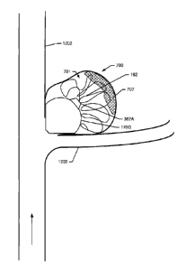
[0129] Generally, spherical ballstents 100 and 150 are shown in FIGS.
1A-D,
and 2A-4B. In particular, a spherical ballstent 100 is shown in an expanded
state, in
FIGS. 1A-4A. The ballstent 100 and 150 has a proximal neck 116, protruding
away
from the ballstent, that defines an opening 112 for the passage of fluids,
liquids, gases,
gels, or solids into or though the void of the ballstent. In the ballstent 100
shown in
Date Recue/Date Received 2020-08-07
FIGS. 1B, the neck 116 protrudes into the void to define the opening 112 for
the
passage of fluids, liquids, gases, gels, or solids into the ballstent 100.
[0130] Another spherical embodiment of the ballstent 100 is shown in
FIG. 1C
in an expanded state. This embodiment includes a proximal neck 116 that
defines an
opening 112 for the passage of fluids, liquids, gases, gels, or solids, into
or through the
ballstent. The ballstent 100 also includes a distal neck 118, protruding away
from the
ballstent, that defines an opening 114 for the passage of a guide wire 302 or
a coil 162,
as shown in FIGS. 2A-B and 3A-B, through the ballstent or from the interior of
the
ballstent to the exterior of the ballstent, including distal to the distal
neck. A similar
spherical embodiment of the ballstent 100 is shown in FIG. 1D in an expanded
state.
This embodiment includes the proximal neck 116 that defines the opening 112
and the
distal neck 118 that defines the opening 114, both which protrude into the
interior of the
ballstent 100, for the passage of fluids, liquids, gases, gels, or solids,
including a guide
wire 302 or a coil 162, into or through the interior of the ballstent.
[0131] Ultimately, the metallic expandable bodies disclosed herein may
have
a variety of configurations and any of the configurations may be employed for
a variety
of uses including occluding aneurysms, including saccular aneurysms, and
segments of
biological conduits, including arteries and veins. Generally speaking, some
configurations may lend themselves more readily or effectively to one
application or
another. For example, the spherical expandable bodies 100 of FIGS. 1A-D may be
particularly advantageous when acting as a ballstent for the filling of the
lumen (or void
or cavity) of a saccular aneurysm. Similarly, as explained further below, the
spherical
expandable bodies 100 and 150 of FIGS. 1A-D and 2A-4B and the expandable
bodies
140 and 170A-F of FIGS 6A-D, 8A-S, 16G, and 16K, for example, may be used with
a
coil or accessory coil 162 to fill at least a portion of the lumen (or void or
cavity) of a
saccular aneurysm and reduce or obstruct the flow of blood through opening
from the
parent vessel to the lumen of the aneurysm, or reduce or obstruct the flow of
blood
through the neck of a saccular aneurysm into the body of the aneurysm lumen
(or void,
26
Date Recue/Date Received 2020-08-07
or cavity). In various embodiments, the coil or accessory coil 162 comprises a
self-
expanding material, such as nitinol wire.
[0132] In some embodiments, as shown in FIGS. 8A-G and 8U, the
expandable bodies 170A-G can be characterized to include a proximal region
174A-G,
an intermediate region 173A-G, and a distal region 172A-G, wherein the
proximal region
and distal region are generally opposite each other. For each body 170A-G,
proximal
region 174A-G, the intermediate region 173A-G, and the distal region 172A-G
form the
unitary construction of the expandable body. For this characterization, the
proximal
region, the intermediate region, and the distal region together form a "main
body" of the
expandable body, which excludes the necks. The expandable bodies 170A-G may
further be defined by a first axis 176 and a second axis 178 transverse to the
first axis.
In one aspect, the first axis 176 extends between the necks 116 and 118.
[0133] In one embodiment, the shape of the intermediate region 173A-G
of
the expandable bodies 170A-G may be defined by the rotation, about the first
axis 176,
of a variable radius arc formed along the first axis, where the maximum radius
for the
variable arc is equal to either the maximum radius 181 of the distal region
172 or the
maximum radius 180 of the proximal region 174, as measured along the second
axis
178. For some embodiments, the expanded expandable body 170A-G has a total
length 179 along the first axis 176 that is less than or equal to the maximum
diameter
182 of the expanded expandable body along the second axis 178.
[0134] In some embodiments without an intermediate region, as shown in
FIGS. 8A-G and 8U, the expandable bodies 170A-G can be characterized to
include a
proximal region 174 and a distal region 172, wherein the proximal region and
distal
region are generally opposite each other. For each body 170A-G, proximal
region 174
and the distal region 172 form the unitary construction of the expandable
body. For this
characterization, the proximal region and the distal region together form a
"main body"
of the expandable body, which excludes the necks. The expandable bodies 170A-G
may also be further be defined by a first axis 176 and a second axis 178
transverse to
the first axis. In one aspect, the first axis 176 extends between the necks
116 and 118.
For some embodiments, the expanded expandable body 170A-G has a total length
179
27
Date Recue/Date Received 2020-08-07
along the first axis 176 that is greater than or equal to the maximum diameter
182 of the
expanded expandable body along the second axis 178.
[0135] In various other embodiments, the expandable bodies may be
defined
and described by the proximal region 174 and the distal region 172, where each
region
is generally a hem ispheroid. The hemispheroid formed by each region 172 and
174 is
further defined by a semi-major axis and semi-minor axis that may be parallel
with the
first axis 176 or the second axis 178, depending upon the lengths of each
axis. In
various embodiments, the hem ispheroid of the proximal region 174 has a semi-
major
axis and semi-minor axis different from that of the distal region 172. In
other
embodiments, the hemispheroid of the proximal region 174 has a semi-major axis
and
semi-minor axis the same as that in the distal region 176. Similarly, for each
of the
distal and proximal regions 172 and 174, respectively, the semi-major and semi-
minor
axis may differ from one another or be identical so that the corresponding
region may
have a generally shape of an oblate hem ispheroid, a prolate hem ispheroid, or
a
hemisphere. As shown, the expandable bodies 170A-G may also be fabricated in a
variety of other configurations that have generally spheroid or ellipsoid
shapes. The
expandable bodies 170A-G may also include a proximal neck 116 and a distal
neck
118.
[0136] In some embodiments, the expanded expandable bodies 170A-G have
a length 179 from the proximal neck 116 to the distal neck 118 of
approximately 4 mm
to approximately 16 mm or larger and a maximum diameter 182 of approximately 4
mm
to approximately 16 mm or larger. As shown in FIGS. 8A-F and 8U, the maximum
radius length for the proximal regions 174A-G and distal regions 172A-G are
equal,
such that the expandable bodies 170A-G have a generally circular cross-section
when
viewed in cross-section along the first axis 176. As shown in FIGS. 8A-E and
8U, the
radius length at any equivalent location for the proximal regions 174A-G and
distal
regions 172A-G may not be equal, such that the expandable bodies 170A-G may
not
have a generally circular cross-section when viewed in cross-section along the
second
axis 176. In other embodiments, as shown in FIG. 8F, the radius length at any
equivalent location for the proximal regions 174A-G and distal regions 172A-G
may be
28
Date Recue/Date Received 2020-08-07
equal, such that the expandable bodies 170A-G may have a generally circular
cross-
section when viewed in cross-section along the second axis 176.
[0137] In one aspect, the different configurations of the expandable
bodies
170A-G may be obtained by varying the maximum length ("height") along the
first axis
176 for the proximal region 174A-G and the distal region 172A-G,
independently. For
example as shown in FIGS. 8A, C, and E, the height 183 for the proximal region
174A
may be smaller than the height 184 for the distal region 172A. In other
examples as
shown in FIGS. 8B, D, and F, the height 183 for the proximal region 174A may
be equal
to the height 184 for the distal region 172A. In other examples, the height
183 for the
proximal region 174A may be larger than the height 184 for the distal region
172A.
While both expandable bodies 170A and 170B have the same maximum diameter, the
difference in the heights for the proximal and distal regions of each
expandable body
results in different overall shapes for the expandable body. As shown, the
expandable
body 170A is generally heart-shaped, while the expandable body 170B has a
spheroid
shape.
[0138] In other examples shown in FIGS. 8A-F and 8U, the heights 183
and
184 of the proximal portion 174A-F and distal portion 173A-F, respectively,
may be
varied independently to produce a wide variety of configurations of the
expandable
bodies 170A-G. The height 183 for the proximal region 174C may be
approximately 2
mm, while the height for the distal region 172C is approximately 4 mm.
Similarly, the
height 183 for the proximal region 174D may be approximately 3 mm, while the
height
for the distal region 172D is also approximately 3 mm. For the expandable body
170E,
the height 183 for the proximal region 174E may be approximately 2 mm, while
the
height 184 for the distal region 172E is approximately 3.5 mm, while for the
expandable
body 170F, the height 183 for the proximal region 174F may be approximately 3
mm,
while the height 184 for the distal region 172F is approximately 4 mm. As
shown, the
expandable bodies 170A-G may have a number of configurations that may be
generally
spheroid, generally spherical, or generally heart-shaped.
[0139] The metallic expandable body, such as the expanded spherical
ballstents 100 and 150 of FIGS. 1A-D and 2A-4B and the expanded expandable
bodies
29
Date Recue/Date Received 2020-08-07
140 and 170A-G of FIGS. 8A-U, 16G, and 16K, may have a wall 102 composed of a
single continuous layer 122, as shown in FIG. 16A. The wall 102 includes a
material,
preferably a metal that is biocompatible and ductile, that can be formed into
a thin wall,
and can assume a variety of shapes after expansion. By way of example and not
limitation, the metal can be selected from the group consisting of gold,
platinum, silver,
nickel, titanium, vanadium, aluminum, tantalum, zirconium, chromium, silver,
magnesium, niobium, scandium, cobalt, palladium, manganese, molybdenum, alloys
thereof, and combinations thereof. Preferred metals include gold, platinum,
and silver,
alloys thereof, and combinations thereof. Expandable bodies can also be made
from
alternative materials that can be formed into thin-walled structures that are
sufficiently
rigid or semi-rigid to tolerate compression and expansion, and can maintain an
expanded state in vivo. Alternative materials include polymers or plastics
that are
reinforced with metal coils or braids, and other materials with similar
properties. The
materials forming the wall 102 and the thickness of the wall are selected such
that the
expandable body 100, 140, 150, or 170A-G has sufficient rigidity to remain in
an
expanded state in vivo under typical physiologic conditions after expansion
and
separation from the delivery catheter, both when the pressure inside and
outside the
central void or space 108 is the same or similar and when the pressure outside
is
greater than the pressure inside.
[0140] Further, it is desirable that the materials used to form and
support the
expandable body 100, 140, 150, or 170A-G have sufficiently mechanical
properties of
ductility, malleability, and plasticity to be compressed or folded without
tearing and later
expanded without rupturing. In general, ductility is a measure of a material's
ability to
be deformed without breaking, while the malleability of the material
determines the ease
of deforming without breaking when the metal is subjected to pressure or
forces. The
ductility and malleability of a material factor into the plasticity of the
material, which
generally refers to a property of the material that permits it to undergo a
permanent
change in shape without rupture or breakage. As such, the expandable bodies
may be
Date Recue/Date Received 2020-08-07
composed of any biocompatible materials having sufficient ductility,
malleability, and
plasticity to undergo one or more compressions, folding processes, and
expansions.
[0141] The central layer 122 of the wall 102 has an interior surface
106 and
exterior surface 124 that define a wall thickness 120. In particular, for
FIGS. 16A and
16B, the distance between the interior surface 106 and the exterior surface
124 is the
overall wall thickness 120 of the wall 102. Preferably, the central layer 122
of the wall
102 has a thickness 120 from about 3 pm to about 50 pm and is preferably,
approximately 10 pm thick. The wall thickness 120 can be uniform. For example,
the
wall 102 may have a uniform thickness of 3 pm, 5 pm, 10 pm, 15 pm, 20 pm, 30
pm, 40
pm, or 50 pm. For example, the thickness 120 of the wall 102 may be selected
such
that the expandable body is strong enough to resist compression from blood
pulsation
but weak enough to yield and collapse during healing and involution of a
treated
saccular aneurysm or an occluded segment of artery or vein, or other form of
biological
conduit.
[0142] Alternatively, the thickness of the wall 102 at different
locations may
vary in thickness. Alternatively, the expandable body 100, 140, 150, or 170A-G
may be
composed of a single porous layer or wall 122, as shown in FIG. 16B, with
pores or
microperforations 1300 wherein at least some or all of the microperforations
extend all
the way from the internal surface 106 to the external surface 124. For this
embodiment,
the wall 102 may be of a uniform thickness or a varied thickness. During
expansion of
the ballstent 100 of this embodiment, the fluid medium may travel under
pressure from
the void or space 108, through the wall 102 and leave the ballstent at the
exterior
surface 124. For this embodiment, the microperforations 1300 may range from 1
¨500
pm in diameter. Another example range of microperforation diameters is 0.01 to
50 pm.
[0143] The expandable body 100, 140, 150, or 170A-G includes a central
wall
or layer 122, optionally with an exterior wall or layer 104, and optionally
with an interior
wall or layer 214, as shown in FIG. 16D. As mentioned, the construct of the
central
layer or wall 122 and the layers 104 and 214 can be uniform, porous, or
combinations
thereof. In one embodiment of the ballstent 100 used to treat a saccular
aneurysm, the
31
Date Recue/Date Received 2020-08-07
wall 102 includes a plurality of microperforations 1300 that extend completely
through
the thickness 120 of the wall 102.
[0144] In one construction, the central layer or wall 122 is
continuous and
formed of gold. Optionally, to this preferred construction, an exterior layer
104 formed
of porous gold can be added. Optionally, an interior layer 214 formed of
Parylene may
be present. Optionally, an exterior layer 104 formed of Parylene may be
present. In
certain embodiments where electrolysis is used to separate the expanded
expandable
body 100, 140, 150, or 170A-G from the delivery catheter, certain portions of
the
ballstent or the expanded expandable body (such as the neck or body) are
coated with
an insulator or polymer, such as Parylene. In certain embodiments where
electrolysis is
used to separate the expanded expandable body 100, 140, 150, or 170A-G from
the
delivery catheter, certain portions of the ballstent or the expanded
expandable body
(such as the neck or body) are coated with a metal that is relatively
resistant to
electrolysis, such as gold or platinum. These portions include the external
surface, the
internal surface, or both the internal and external surfaces, while a portion
of the neck or
body remains uncoated or non-insulated. In this instance, the uncoated or non-
insulated portion of the wall is electrolytically dissolved (i.e. corroded) by
the passage of
an electrical current from the exposed metal of the wall into the surrounding
electrolyte
(i.e. blood or serum). In certain embodiments, the uncoated or non-insulated
portions of
the wall are created by masking during the coating process. In other
embodiments, the
coating or insulation is removed from the uncoated or non-insulated portions
of the wall
or neck, as through etching or ablation, such as with laser etching or laser
ablation.
[0145] One embodiment of a generally spherical ballstent 150 is shown
in
FIGS. 1A-4B. The generally spherical ballstent 100 or 150 includes the wall
102 that
forms a spherical body when expanded. In one aspect, a distal region 152 of
the wall
102 includes one or more annular portions 154A-B. The annular portions 154A-B
have
a radius of curvature greater than the remainder of the wall 102 such that the
distal
region presents a flatter surface than the remainder of the wall. The
generally spherical
ballstent 150 also includes a proximal neck 116 and a distal neck 118
protruding away
32
Date Recue/Date Received 2020-08-07
from the distal region 152. In another embodiment, a distal neck can protrude
into the
interior void of the expanded expandable body.
[0146] In various embodiments, as shown in FIGS. 2B-C and 2E, a
bridging
catheter 160 extends through the proximal neck 116, through interior void of
the
expanded expandable body and into the distal neck 118. In one aspect, the
bridging
catheter 160 is an elongated tubular member component of the delivery catheter
that
provides structural support to the ballstent 150. In one embodiment, the
bridging
catheter 160 has an outer internal diameter in a range between approximately
0.5 and
2.0 mm and an inner diameter in a range between approximately 0.4 and 1.9 mm.
In
some embodiments, the bridging catheter is a component of the delivery
catheter, or is
operatively coupled to the delivery catheter.
[0147] In another aspect, the bridging catheter 160 provides a pathway
to
deliver a solid material, such as a guide wire 302 or a coil 162, as shown in
FIGS. 2B-C,
2E, 2G, 2N-P, 8H, 8J-0, and 8R-S, through the interior space 108 to the
exterior of the
ballstent via the distal neck 118. The bridging catheter 160 may also include
one or
more openings 164 for the passage of fluids, liquids, gases, gels, or even
solids into the
interior 108 of the ballstent 150. Thus, as explained more fully below, the
bridging
catheter 160 may be used to inflate or expand the expandable body while also
permitting a guide wire 302 or a coil 162 to pass into or through the interior
108 of the
ballstent 150 and to the exterior of the distal region 152.
[0148] In various embodiments, the openings 164 within the bridging
catheter
160 may have a diameter in a range between approximately 200 pm and 1 mm. As
shown in FIGS. 3A-3B, the bridging catheter 160 may be dimensioned such that
it can
receive a coil or accessory coil 162. The coil or accessory coil 162 may be
fed directly
through the lumen of the bridging catheter 160 or may be fed through a second
catheter
352B (a "coil delivery catheter") that is passed through the bridging catheter
160, as
shown in FIG. 7, and in this way comprises a dual catheter delivery system.
[0149] The bridging catheter 160 may also permit the passage of a
catheter
such as the catheter, or coil delivery catheter, 352B to pass through the
interior of the
expandable body 100, 140, 150, or 170A-G, to deliver the coil or accessory
coil 162 to
33
Date Recue/Date Received 2020-08-07
the lumen, cavity, or void of a saccular aneurysm. As shown, in FIGS. 2L-Q,
the
catheter 352B may be fed through the expandable body and the accessory coil
162 may
be simultaneously or subsequently fed through the catheter 352B.
The Expandable Body Exterior
[0150] As discussed, the expandable body 100, 140, 150, or 170A-G may
have one or more additional coating or layer(s) 104 on the exterior surface
124 of the
central layer 122, as shown in FIG. 16C-D. The wall 102 and any additional
exterior
layers define an exterior surface 110 that, when expanded, contacts the
internal wall of
the aneurysm or blood vessel. The exterior layer 104 can be of a uniform or
varied
thickness, preferably between about 1 pm and about 59 pm. In one embodiment,
the
exterior layer 124 has a thickness between 0.1 and 10 pm. In a specific
embodiment,
the exterior layer 124 has a thickness of about 1 pm.
[0151] The exterior layer 124 can be formed of polymers, latex,
elastomers, or
metals. The exterior layer 124 may be an electrical insulator, and in a
preferred
embodiment, the exterior layer 124 is formed of a Parylene coating. The
exterior layer
124 may be a metallic or non-metallic material that is less susceptible to
electrolysis or
galvanic corrosion, such as noble metals, and in preferred embodiments gold or
platinum. The exterior coating or layer 104 of the expandable body 100, 140,
150, or
170A-G may be porous and contain a plurality of pores 200, as shown in FIGS.
16C and
16D. Alternatively, the exterior layer 104 can be smooth, with limited
porosity or
protrusions. For example, the exterior layer 104 may be a polished metal
surface. In
one embodiment, portions of the exterior layer 104 can be smooth, while other
portions
can be porous or contain protrusions. In one embodiment, the surface
variations can
include a pattern. FIG. 29E depicts structures of the exterior surface 110
after
electroforming and Parylene coating. As shown, the exterior surface 110 of the
wall 102
may have rounded, pebbled, or granular structures. In various embodiments, the
rounded, pebbled, or granular surface structures have a height of
approximately 0.1 pm
to approximately 10 pm.
[0152] When configured as a porous or spongy layer, the exterior layer
104
can contain (or be configured to contain) solutions that include
pharmaceutical drugs,
34
Date Recue/Date Received 2020-08-07
pharmacologically active molecules, or pharmaceutical compositions within the
pores
200. As such, solutions such as pharmaceutical drugs, pharmacologically active
molecules, or pharmaceutical compositions can be delivered to the treatment
site.
Drugs, pharmacologically active molecules, or pharmaceutical compositions that
promote thrombosis, stimulate cell proliferation or extracellular matrix
production, or
tissue growth are examples of agents that can be placed in the pores 200 of
the exterior
layer 104. The pharmaceutical drugs, pharmacologically active molecules, or
pharmaceutical compositions are incorporated into the pores 200 of the wall or
the
exterior layer 104 prior to positioning the expandable body 100, 140, 150, or
170A-G at
the desired location. The drug compositions may be delivered into the pores
200 via
capillary or wicking action. The pores 200 range from about 0.01 pm to about
500 pm
in diameter. Pore diameters for each expandable body may vary according to the
specific drugs, pharmacologically active molecules, or pharmaceutical
compositions to
be incorporated and the desired rate of release in vivo. By way of example and
not
limitation, the expandable body 100, 140, 150, or 170A-G may have a porous
exterior
layer 104 where the pore diameter averages from about 0.01 pm to about 0.05
pm,
about 0.05 pm to about 0.5 pm, 0.5 pm to about 5 pm, about 5 pm to about 25
pm,
about 25 pm to about 500 pm, about 0.05 pm to about 500 pm, or about 0.01 pm
to
about 500 pm.
[0153] The pharmaceutical drugs, pharmacologically active molecules, or
pharmaceutical compositions may include thrombin, platelet-derived growth
factor,
EthiodoI0, SotradecoI0, or combinations thereof. Other pharmaceutical
compounds
and compositions that promote thrombosis, stimulate cell proliferation,
stimulate the
synthesis of extracellular matrix, or the growth of tissue into the porous
external wall of
the expandable body 100, 140, 150, or 170A-G may also be used. Such drugs or
pharmaceutical compositions may include molecules to promote cell
proliferation,
extracellular matrix production, or tissue growth, such that the expanded
expandable
body 100, 140, 150, or 170A-G will become more firmly attached to the tissue
at the
treatment location. The dosages and manner in which the pharmaceutical drugs,
pharmacologically active molecules, or pharmaceutical compositions are
incorporated
into the wall 102 or exterior layer 104 are a matter of choice depending upon
the
Date Recue/Date Received 2020-08-07
treatment performed. Other compounds may be used to promote blood clotting or
thrombosis around the expandable body. In various aspects, the pores 200 may
be
filled with a biodegradable or bioerodible material, such that the volume of
material in
the pores decreases over time and the pores are opened in vivo at a point in
time
subsequent to placement of the expandable body. For embodiments of the
expandable
body 100, 140, 150, or 170A-G with a porous layer 104, overtime, the
ballstent,
blockstent, or the expandable body remains expanded with the expanded body
eventually becoming affixed to the surrounding tissue.
[0154] As can be understood from FIGS. 18G-H, the exterior surface 110
of
the expandable body 100, 140, 150, or 170A-G may also include one or more
protrusions or projections 1800 (which may be generally tubular or have other
configurations) that can increase the strength of the attachment of the
expanded body
to the adjacent tissue, and thereby reduce the risk of movement or migration.
The
protrusions may have a length that ranges between about 0.01 pm to about 167
pm.
Some protrusions can have a branched construction, while others may be joined
on
both ends to the exterior surface 110 to form loops. In some embodiments, the
protrusions are rigid, or semi-rigid. In other embodiments, the protrusions
are flexible
and hair-like, and may further comprise globular ends, similar to the
protrusions on the
surface of the footpad of the gecko. The protrusions may be attached to the
expandable body 100, 140, 150, or 170A-G after formation. Alternatively, or
additionally,
the protrusions may be incorporated into the expandable body during
electroforming.
[0155] In another embodiment, the ballstent 100 may comprise a porous
external layer or wall 104 or a wall with external protrusions 1800 to promote
thrombus
formation on the external surface 110 or in the pores 200 and promote cell
proliferation,
extracellular matrix production, or tissue growth into or around the wall 102
of the
ballstent 100 such that the ballstent 100 will, over time, become more
strongly attached
to the tissue in the adjacent aneurysm wall.
[0156] As shown in FIGS. 18A-D, the central layer 122 and the porous
exterior layer 104 of the ballstent 100 placed into the aneurysm 700 may be
configured
to promote thrombus 1206 formation on the exterior layer. The thrombus may be
comprised of red blood cells 1208, platelets 1210, and fibrin 1212. Overtime,
the
36
Date Recue/Date Received 2020-08-07
thrombus 1206 may be partially absorbed into the exterior layer 104, as new
endothelial
cells 1214 are formed over the thrombus. The new endothelial cells may form a
seal of
connective tissue 1216 across the opening of aneurysm 700. In addition to
sealing the
opening of the aneurysm 700, connective tissue 1216 from the wall 704 of the
aneurysm may grow into the porous exterior layer 104 of the ballstent 100 to
adhere the
ballstent to the wall of the aneurysm, as shown in FIG. 18E.
[0157] In other embodiments, the projections or protrusions 1800 may
be
generally tubular, straight, curved, hook-shaped, or configured as pigtail
hooks as
shown in FIGS. 18G-H. In a macroscopic form, the projections may be composed
of
nitinol or any other suitable biocompatible material.
[0158] FIG. 18H depicts an expanded ballstent 100 that is anchored to
the
wall 704 of an aneurysm 700. The size and shape of the protrusions may be
selected
based upon the condition being treated, and may be designed and dimensioned to
provide sufficient anchoring support without causing excessive damage to the
wall of
the aneurysm or the surrounding tissue. Alternatively, microscopic protrusions
or
filaments may be used to anchor the ballstent. For some embodiments, these
microscopic protrusions range in length from 0.01 pm to about 57 pm, and can
be
straight or branching. In various embodiments, both ends of one or more of the
protrusions may be joined to the exterior surface 110 of the ballstent 100
and/ or the
exterior surface 216 of the wall 102 to form a loop.
[0159] The ballstent or expandable body 100, 140, 150, or 170A-G may
also
be used to contain or trap a thrombus, such as a mural thrombus, that has
formed
within an aneurysm or other biological space. As shown in FIG. 18F, an
expandable
body 170G may be placed within an aneurysm 700 having one or more thrombi,
including a mural thrombus 707, within the cavity 701 or dome of the aneurysm.
In one
aspect, an expandable body 170G having an expanded volume smaller than the
volume
of the aneurysm cavity 701 is selected. The expandable body is delivered to
the
aneurysm, inflated or expanded, and contacted by an inserted accessory coil
162, as
previously described. In this aspect, the accessory coil 162 simultaneously
contacts the
expandable body 170G, the thrombus 707, and the wall of the aneurysm. The
37
Date Recue/Date Received 2020-08-07
expandable body 170G in conjunction with the accessory coil 162 acts to trap
the
thrombus 707 within the aneurysm holding it in places until absorption by the
patient.
[0160] In various embodiments, an expandable body that does not
completely fill the cavity 701 of an aneurysm 700 that may potentially contain
a blood
clot is preferred. As such, a larger expandable body that more fully fills the
cavity 701,
is less desirable as it may force thrombus within the aneurysm 700 out into
the parent
blood vessel 1202 or 1203, where the thrombus may embolize, travel through the
vascular system, and cause a stroke.
[0161] In various embodiments, the expandable body 100 may include a
thin
polymer sheath that is wrapped around the entire body of the expandable body
when in
the delivery or deliverable configuration. The sheath may be added to the
exterior of
the expandable body 100 during fabrication of the expandable body. The sheath
may
be affixed to a proximal nose cone 362B, a distal nose cone 360 or 362A, or
both, such
as those shown in FIGS. 2A-Q. The polymer sheath increases trackability of the
expandable body 100 and reduces friction with the lining of blood vessels as
the
expandable body is delivered through the vascular system. During inflation or
expansion of the expandable body 100, the polymer sheath opens while remaining
affixed to the expandable body, the delivery catheter, the proximal nose cone
362B, or
the distal nose cone 360 or 362A. In one embodiment, the sheath may be
perforated or
partially scored before deployment to allow for easier expansion of the
expandable body
100.
The Expandable Body Interior
[0162] In some embodiments, the expandable body 100, 140, 150, or 170A-
G
may include an additional layer or liner 214 on the interior surface 106 of
the central
layer 122, as shown in FIGS. 16D, 16F, 16H, 16J, and 16L. The interior layer
may be
made from the same materials as the central layer, or can be made of different
materials. The interior layer may be formed of gold, platinum, silver, alloys
thereof, or
combinations thereof. The additional layer 214 on the interior surface 106 of
the central
layer 122 of the expandable body 100, 140, 150, or 170A-G may also be formed
of a
polymer, plastic, latex, rubber, woven or knitted fiber material, metal, or
another
38
Date Recue/Date Received 2020-08-07
material, or combinations thereof. Preferably, the interior layer 214 is an
elastomeric
coating that is bonded to the interior surface 106 of the central layer 122.
The interior
layer 214 can be a variety of thicknesses, preferably ranging between about
0.1 pm and
about 59 pm. In one embodiment, the interior layer 214 has a thickness between
about
0.1 pm and about 10 pm. The total thickness of the wall 102, including the
central layer
122, the exterior layer 104, and the interior layer 214 is preferably between
about 2 pm
and about 50 pm, regardless if the wall contains one, two, three, or more
layers. The
interior layer 214 can comprise polymers, latex, or elastomers. In a preferred
embodiment, the interior layer 214 comprises Parylene. The interior layer 214
also
adds mechanical properties (such as strength) to the wall 102. Further, the
interior
layer 214, optionally, can form a seal that prevents the escape of a fluid
medium from
the expandable body 100, 140, 150, or 170A-G, should the central layer 122
contain a
defect or hole. The central layer 122 and any additional layers define an
interior surface
106 or 218, respectively, such that when the ballstent or the expandable body
is
expanded, with a fluid, liquid, gas, or solid, a central void or space 108 is
defined. As
shown in FIG. 16D, the distance between the interior surface 218 and the
exterior
surface 110 is the overall wall thickness 120 of the wall 102.
The Expandable Body Neck(s) and Opening(s)
[0163] As illustrated in FIGS. 1A-D, 2A-4B, 8A-S, 8U, 16A-D, 16G, and
16K,
the expandable bodies 140, 150, or 170A-G have one or more openings 112 and
114
defined by the wall 102 or by the proximal neck 116 or the distal neck 118. In
various
embodiments, the ballstent, blockstent or expandable body has one or more
openings
112 and 114 defined by necks 116 or 118, respectively. In all embodiments, a
fluid
medium can enter the opening 112 and move into the central void or space 108
defined
by the interior surface 106 or 218, thereby inflating or expanding the
expandable body.
In various embodiments, one or both of the necks 116 and 118 may extend
outwardly
from its respective end region (proximal region or distal region) of the
expandable
bodies 100, 140, 150, or 170 A-G as shown in FIGS. 1A, 1C, 2A-4B, 8A-S, 8U,
16G and
16K. Alternately, one or both of the necks 116 and 118 may extend inwardly
from its
respective end region and into the interior void 108, as illustrated in FIGS.
1B and 1D.
The proximal necks 116 can be used for attaching the expandable body 100, 140,
150,
39
Date Recue/Date Received 2020-08-07
or 170A-G to the delivery catheter and may function in separating the
ballstent or the
expandable body from the delivery catheter. In various embodiments, the necks
116
and 118 and the wall 102 or main body may be formed from different metals. For
example, in one embodiment, the neck(s) 116 and 118 and the wall 102 or main
body
may be formed by gold. In other embodiments, the neck 116 and 118 may comprise
stainless steel, including but not limited to 304 series or 316L series
stainless steel and
the wall 102 or main body may be formed by gold, platinum, or another
malleable metal.
The neck 116 and 118 may comprise multiple metals, such as stainless steel and
another metal such as gold or platinum, including embodiments wherein the
various
regions of the expandable bodies 100, 140, 150, or 170A-G are distinct in
their metal
content and embodiments wherein the different metals are formed in layers in
the
various regions, including an embodiment wherein a neck comprises an interior
layer of
stainless steel with an exterior layer of gold and an embodiment wherein a
neck
comprises an central layer of stainless steel with interior and exterior
layers of gold,
including embodiments wherein at least a portion of the surface of the
exterior layer is
stainless steel, including embodiments wherein a portion of the gold exterior
layer is
absent through masking or through etching, including laser etching.
[0164] Additionally, the necks 116 and 118 can be designed and
dimensioned such that the opening 112 or 114, preferably the proximal opening
112,
can be closed or partially closed before, during, or after separation of the
expanded
body from the delivery catheter. One or more openings 112 or 114 may remain
open.
Optionally, before, during, or after separation, the necks 116 and 118 may be
folded,
pinched, or closed to form a seal. The necks 116 and 118, or alternatively the
stainless
steel ring 250, may have a length Ni, as shown in FIGS. 24A and 30C, ranging
between about 0.5 mm and about 20 mm, preferably a length between about 0.5 mm
and about 5 mm. In one embodiment, the neck length Ni is approximately 1.27 mm
0.08 mm.
[0165] In various embodiments, at least one of the necks 116 and 118
and the
stainless steel ring 250, as shown in FIGS. 2A-E, 24A, and 30D, have an outer
diameter
N2 and an inner diameter N3 that defines the openings 112 and 114,
respectively. The
outer diameter N2 is in a range between about 0.25 mm and about 2 mm and the
inner
Date Recue/Date Received 2020-08-07
diameter N3 is in a range between about 0.24 mm and about 1.95 mm. In one
embodiment, the neck outer diameter N2 is approximately 0.99 0.01 mm and the
neck
inner diameter N3 is approximately 0.89 0.01 mm.
[0166] The thickness of the walls of either or both of the necks 116
and 118
may be the same as the main body of the ballstent, blockstent, or the
expandable body
or may be thinner or thicker than the wall of main body. Preferably, either or
both of the
necks 116 and 118 have a wall thickness N4 between about 3 pm and about 60 pm,
as
shown in FIGS. 24B-C, 30D, and 30F. In one particular embodiment, the neck has
a
thickness of approximately 50 pm. In one embodiment of the ballstent 100 where
the
neck(s) 116 and 118 extend into the central void space 108 as indicated in
FIGS. 1B
and 1D, the external surface 110 of the expanded ballstent retains a more
rounded
surface contour, increasing the strength of the expanded ballstent and
reducing the risk
of damage to the aneurysm wall or the adjacent tissue during placement.
[0167] One or both of the necks 116 or 118 can be coated or insulated
on the
inner wall, outer wall, or both. This coating can include metals such as gold
or platinum
and polymers such as Parylene TM. In addition, the necks 116 and 118 may
include one
or more caps or nose cones 360, as shown in FIGS. 2A-C and 4A-B or nose cones
362A-B as shown in FIGS. 2D-Q, to improve trackability of the expandable body
100
during delivery and placement. In addition to improving the trackability of
the
expandable body 100 during placement, the nose cones 360 or 362A-B also serve
to
protect the necks 116 and 118 during positioning, as well as reducing the risk
of
damage to the walls or lining of any blood vessels or conduits traversed by
the
expandable body 100 during placement. In some embodiments, a nose cone affixed
to
the distal portion of the delivery catheter can serve the same purpose.
[0168] As shown in FIGS. 2C and 4B, the nose cones 360 or 362A-B include
a central channel 364 that encircles and engages the necks 116 and 118. In one
embodiment, the nose cone 360 is generally cylindrical as shown in FIGS. 2A-C
and
4A-B, while in other embodiments, the nose cones 362A-B may have a
frustoconical or
"bullet-shaped" configuration, as shown in FIGS. 2D-Q. The nose cones 360 or
362A-B
may be composed of any biocompatible material, including polymers and metals.
In
41
Date Recue/Date Received 2020-08-07
one embodiment, the nose cones 360 or 362A-B are composed of PTFE. In various
embodiments, the nose cones 360 or 362A-B hay have an outer diameter in a
range
between approximately 0.75 and 2.5 mm, an inner diameter in a range between
approximately 0.25 and 2 mm, with a length in a range between approximately 1
and 4
mm.
[0169]
In various embodiments, the necks 116 and 118 are further modified to
provide a detachment point for detaching the expandable body 100, 140, 150, or
170 A-
G from a delivery catheter. For example, a strip of electrically conductive
material,
including an uncoated or non-insulated section of a neck, weld, solder, or
other fixation
point, or a portion of the ballstent, blockstent or the expandable body
itself, is left
exposed, uncoated, or non-insulated or later exposed after coating, including
an
exposed, uncoated, or non-insulated region that in the shape of a
circumferential or
ring-shaped exposed surface of metal or conductive materials that can be
subjected to
electrolysis to achieve separation between the expanded expandable body and
the
distal end of the delivery device. Preferably, a stainless steel ring is
affixed to the wall
102 or the main body of the expandable body, as stainless steel is highly
sensitive to
galvanic corrosion and electrolysis. For example, as can be understood from
FIGS.
16E, 16G, 161, 16K, 28, and 30A-B, in one embodiment, at least a portion of an
inner
surface of the metal layer of the neck of the metallic expandable body is
electrically
insulated by having an outer surface of a distal portion of the delivery
device extending
along the inner surface of the metal layer of the neck of the metallic
expandable body.
In some embodiments, on the inner surface of the proximal neck 116, a proximal
boundary of the ring-shaped exposed metal surface may be defined by a distal
boundary of the delivery device in the neck region and a distal boundary of
the ring-
shaped exposed metal surface may be defined by a boundary of the inner
insulation
layer in the neck region. For the outer surface of the proximal neck 116, both
the
proximal and distal boundary of the ring-shaped exposed metal surface may be
defined
by a boundary of the outer insulation layer in the neck region. In such an
embodiment,
the distal end of the delivery catheter 300 or 400 may distally terminate near
a proximal
edge of the ring-shaped exposed metal surface of the neck. As indicated in
FIG. 23A, a
conductive wire can be engaged in electrical contact with the uncoated or non-
insulated
42
Date Recue/Date Received 2020-08-07
portion of the neck or a weld or solder between a neck and the delivery
catheter, or on
the expandable body itself 100, 140, 150, or 170A-G to allow the uncoated or
non-
insulated portion to be dissolved (corroded) or removed via electrolysis.
[0170] In other embodiments, one or both necks 116 and 118 may be
affixed
with a metallic ring 250, as shown in FIGS. 2A, 2B, 5A, and 5B, which may be
subsequently severed using electrolysis. The metallic ring 250 may be composed
of
stainless steel and, as explained below, may be subjected to one or more
heating
procedures to sensitize the steel to galvanic corrosion, thereby allowing for
faster
separation or severing via electrolysis.
Expandable Body Shapes and Dimensions
[0171] FIGS. 16E-F and 16I-J illustrate a ballstent 100 and a delivery
catheter
220 that may be used to deliver the ballstent. In one characterization, the
ballstent 100
includes a distal region 202 that includes the distal end 204 of the
ballstent. Adjacent to
the distal region 202 is an intermediate region 206 where the ballstent
transitions from
the distal region 202 to a proximal region 208 that includes a proximal end
210 of the
ballstent. The proximal region 208 is generally opposite the distal region
202. A center
axis 212 extends proximal-distal between the proximal region 208 and the
distal region
202. The ballstent wall 102 extends generally continuously through the
intermediate
region 206 from the proximal region 208 to the distal region 202, and the
ballstent 100 is
in the form of a single-lobed metallic expandable body. In another
characterization, the
ballstent 100 includes a distal region 222 that is joined directly to a
proximal region 228
that is generally opposite the distal region 222. A center axis 212 extends
proximal-
distal between the proximal region 208 and the distal region 202. The
ballstent wall 102
extends generally continuously from proximal region 208 to the distal region
202 and
the ballstent 100 is in the form of a single-lobed metallic expandable body.
[0172] In one embodiment, when the ballstent 100 is expanded, the
intermediate region 206, the proximal region 208, and the distal region 202
combine to
form a generally spherical shape. In various embodiments, the dimensions of
the
ballstents 100 are selected based upon the size and shape of the saccular
aneurysm
being treated. Preferred shapes of the ballstent 100 include round, oblong,
and
43
Date Recue/Date Received 2020-08-07
irregular. The diameter of the round expanded ballstent 100 ranges from about
2 mm to
about 30 mm, and preferably has an expanded diameter ranging from about 2 mm
to
about 20 mm. The expanded length of oblong ballstent or blockstent preferably
ranges
between about 2 mm to about 30 mm. The ballstent 100 may have an expanded
volume that ranges between about 0.001 m L to about 65 m L. In preferred
embodiments, the expanded diameter of the spherical ballstent 100 ranges from
about
2 mm to about 10 mm, while the preferred expanded volume ranges from about
0.004
m L to about 40 m L. In preferred embodiments, the expanded length of the
oblong
ballstent or blockstent 100 ranges between about 2 mm to about 30 mm.
[0173] FIGS. 16G-H and 16K-L illustrate an expandable body 140 and a
catheter 220 that may be used to deliver the expandable body. In some
embodiments,
the expandable body 140 can include a generally cylindrical intermediate
region 206
(where the longitudinal axis of the cylindrical portion is perpendicular to
the central axis
212), a generally hemispherical proximal region 208 and, a generally
hemispherical
distal region 208, as shown in FIG. 16G. In other embodiments, the expandable
body
140 can include a generally cylindrical intermediate region 206 (where the
longitudinal
axis of the cylindrical portion is aligned along a longitudinal axis of the
neck 116), a
generally hemispherical proximal region 208 and, a generally hemispherical
distal
region 208, as shown in FIG. 24 A. The intermediate region 206 may have a
radius R1
that is equal to the radius R2 of both the proximal region 208 and the distal
region 208,
as shown in FIG. 24A. In various embodiments, the delivery catheter 220 is
typically
engaged to the proximal neck 116 or proximal region 208 of the expandable
body.
[0174] In other embodiments, one or more portions of the expandable
body
wall 102 may be thicker than the remaining portions of the wall. By way of
example and
not limitation, the wall in the middle of the main body or the intermediate
region of the
expandable body may be thicker or thinner than the wall in the proximal and
distal
regions or portions of the expandable body, or the wall of a neck may be
thicker or
thinner than the main body of the expandable body. In various embodiments, the
wall
thickness 120, as shown in FIGS. 16A-D, may be scaled relative to the overall
diameter
of the expandable body to avoid undesired increases in wall stress with
increases in
diameter. In various embodiments of the expandable body 100, 140, 150, or 170A-
G, a
44
Date Recue/Date Received 2020-08-07
balance can be made between a wall thickness 120 that is thin enough to enable
the
various small compressed forms of the delivery configuration and to permit
expansion of
the expandable body at lower pressures, and a wall thickness that is thick
enough to
maintain structure integrity and resist compression after delivery and
detachment.
Therefore, the average wall thickness 120 is preferably in a range between
about 10 pm
and about 50pm. By way of example and not limitation, the wall thickness 120
for an
expandable body 100, 140, 150, or 170A-G having an expanded diameter of about
4
mm may be about 10 pm, while the wall thickness for an expandable body having
an
expanded diameter of about 10 mm may be about 25 pm.
[0175] As shown in FIG. 24A, the expandable body 140 may have a generally
cylindrical shape with rounded or hemispherical ends (where the longitudinal
axis of the
cylindrical shape is aligned with a longitudinal axis of the neck 116), such
that the total
length L1 of the main body of the expandable body parallel to the first axis
is greater
than the total width of the expandable body parallel to the second axis (i.e.
twice the
radial distance R1). In other embodiments, the expandable body 140 may have a
generally cylindrical shape with flattened or flat ends as shown in FIGS. 16G
and 16K,
such that the total length of the main body of the expandable body along the
central axis
212 is less than the total width of the expandable body perpendicular to the
central axis.
The expandable body 140 is in the form of a single-lobed metallic expandable
body.
[0176] In various embodiments, the expandable body 140 has an expanded
diameter (both along the center axis 212 and perpendicular to the center axis)
ranging
from about 2 mm to about 30 mm. Assuming no change in wall thickness 120, the
stress in the wall of expandable body 140 will increase, as the radius R1 (see
FIG. 24A)
of the intermediate region 206 increases. Therefore, in some embodiments, the
diameter of the expandable body 140 is limited by the ultimate tensile
strength of the
material (e.g. gold) used to form the expandable body and by the pressure
required to
expand the compressed expandable body. As can be understood from FIG. 24A, the
expandable body 140 may have an expanded length L1 of between about 2 mm to
about 120 mm, such length L1 comprising the proximal region, intermediate
region, and
distal region. Preferably, the length is between about 5 mm to about 60 mm,
and in a
particular embodiment the expanded length L1 is approximately 40 0.03 mm and
the
Date Recue/Date Received 2020-08-07
length L2 of the intermediate region 206 may be approximately 24 0.03 mm,
such
length L2 comprising only the intermediate region.
[0177] The concentration of stress between the neck 116 and the
proximal
region or end 208 of the expandable body 100, 140, 150, or 170A-G may be
reduced or
offset by increasing the radius R4 between the neck and the proximal region,
as shown
in FIGS. 24B-C. For example, the stress experienced by the wall 102 in FIG.
24B
having a radius of R4 is greater than the stress experienced by the wall in
FIG. 24C
having a radius of R4', where R4' is greater than R4. In addition, stress may
be
concentrated at the point where the neck 116 transitions to the wall of the
proximal
region 208 of the expandable body 100, 140, 150, or 170A-G due to a metallic
ring
incorporated into the neck 116 during formation of the expandable body. This
stress
concentration may be mitigated by reducing the overall wall thickness N4 of
the neck
116. By way of example and not limitation, the neck 116 shown in FIG. 24B may
have a
wall thickness N4 of approximately 25 pm, while the neck shown in FIG. 24C may
have
a wall thickness N4' of approximately 12.5 pm.
Expansion of the Expandable Body
[0178] The central void or space 108 of the expandable body 100, 140,
150,
or 170A-G can be filled with fluids, gels, solids, or combinations thereof to
expand or
inflate the expandable body 100, 140, 150, or 170A-G. The terms expand,
inflate, and
forms thereof may be used interchangeable to refer to the action of changing
the
expandable body from the delivery or deliverable configuration to an expanded
or at
least partially expanded configuration. A fluid medium is a substance having
particles
that easily move and change their relative position without a separation of
the mass.
Fluid media that may be used to expand the expandable body 100, 140, 150, or
170A-G
include liquids, gases, gels, and combinations thereof. By way of example and
not
limitation, the fluid medium may be water, a saline solution, a radiographic
contrast
solution, or a mixture thereof. In one embodiment, the fluid medium may
further include
a solution or suspension of a drug, pharmacologically active molecules, or a
pharmaceutical preparation.
46
Date Recue/Date Received 2020-08-07
[0179] In various embodiments, the shape and construction, including
multi-
layer constructions, of the expandable body 100, 140, 150, or 170A-G permits
the
expandable body to remain in an inflated or expanded configuration without the
use of
any support structures not derived from the patient. For example, the fluid
medium
used to inflate the expandable body 100, 140, 150, or 170A-G, and optionally
blood
from the patient, will fill the interior void 108 and cause the ballstent,
blockstent, or the
expandable body to remain in an expanded configuration. In addition, support
structures derived from the patient, including but not limited to blood clots
and tissue
ingrowths, may support and maintain the structural integrity of the expandable
body
100, 140, 150, or 170A-G, when expanded.
[0180] In one embodiment, as shown in FIGS. 17A-B, the expandable body
100, 140, 150, or 170A-G may be used to seal a saccular aneurysm 700 located
near
the junction of blood vessels 1202 and 1203. As shown, the expandable body
100, 140,
150, or 170A-G may be positioned and inflated by the delivery catheter 352A to
seal the
opening 703 of a saccular aneurysm 700 with the aid of a coil or accessory
coil 162 that
is introduced into the aneurysm by passage through the delivery catheter 352A
and
through the expanded expandable body. The coil or accessory coil 162 contacts
the
wall of the aneurysm 700 (including the wall opposite the opening from the
parent
vessels 1202 and 1203 to the aneurysm 703) as well as the exterior of the
expandable
body 100, 140, 150, or 170A-G, where the coil 162 exerts a force, as indicated
by 705
upon the expandable body towards the opening 703 to press the expandable body
against the opening. As a result, the expandable body 100, 140, 150, or 170A-G
prevents the flow of blood, as indicated by 706, from entering the aneurysm.
In one
aspect, the expandable body 100, 140, 150, or 170A-G may be fully expanded
before
introducing the accessory coil 162. In another aspect, the accessory coil 162
may be
introduced, at least partially, before inflation of the expandable body 100,
140, 150, or
170A-G. In yet another aspect, the expansion of the expandable body 100, 140,
150, or
170A-G and the introduction of the accessory coil 162 may occur simultaneously
or in
an alternating incremental fashion. In certain embodiments, after inflation or
expansion
of the expandable body 100, 140, 150, or 170A-G and insertion of the coil or
accessory
coil 162, the expandable body 100, 140, 150, or 170A-G is detached from the
delivery
47
Date Recue/Date Received 2020-08-07
catheter 352A by electrolysis that corrodes a portion of the proximal neck
250, including
a ring-shaped region of exposed stainless steel.
[0181] In one embodiment, multiple coils or accessory coil(s) 162 may
be
deployed within the aneurysm 700. In one embodiment, as shown in FIG. 17C, a
portion of one or more coil or accessory coil 162 is deployed within the
lumen, void, or
cavity of the aneurysm while another portion of the coil is deployed within
the void of the
expandable body 100, 140, 150, or 170A-G. For example, after inflating or
expanding
the expandable body, an accessory coil delivery catheter 352B may be fully
inserted
through delivery catheter 352A, through the expandable body 100, 140, 150, or
170A-G,
and into the lumen of the aneurysm 700 and the accessory coil 162 may be
inserted
into the unfilled portion of the aneurysm 700. The coil delivery catheter 352B
is then
retracted so that its distal end is located within the expandable body 100,
140, 150, or
170A-G and the remainder of the accessory coil 162 or another accessory coil
is
deployed with the expandable body. The deployment of the accessory coil 162
both
within and external to the expandable body 100, 140, 150, or 170A-G may serve
to
stabilize and maintain the position of the expandable body within the aneurysm
700.
[0182] In another embodiment, the accessory coil 162 may be magnetic,
such that multiple accessory coils may be deployed to stabilize the expandable
body
100, 140, 150, or 170A-G within an aneurysm through the magnetic attraction of
the
coils. For example, as shown in FIG. 17D, a first magnetic accessory coil 162A
may be
deployed within an inflated expandable body 100, 140, 150, 170A-G, as
previously
described. One or more other magnetic accessory coils 162B are then deployed
within
the neck or opening 703 of the aneurysm 700. The accessory coil 162B fills and
occludes any residual space in the neck or opening 703 after deploying the
expandable
body 100, 140, 150, or 170A-G. In one aspect, the accessory coils 162A-B are
attracted to and contact the exterior surface of the expandable body 100, 140,
150, or
170A-G. In another aspect, the accessory coils 162A-B are attracted to one
another
through the wall of the expandable body 100, 140, 150, or 170A-G.
[0183] In various other embodiments, the shape of an expanded
expandable
body 100, 140, 150, or 170A-G is maintained by placing solid material or
support
48
Date Recue/Date Received 2020-08-07
structures into the central void or space 108. Examples of this solid material
include
metal or polymeric coils or wires, metal or polymeric solid support
structures,
bioresorbable materials, radially expansile materials, beads, particles,
granules,
spheres, microspheres, or sponges. In certain embodiments, these solid
materials can
also be used to help expand the expandable body 100, 140, 150, or 170A-G. In
other
embodiments, these solid materials are added after expansion. In one
embodiment, as
shown in FIG. 17E, the aneurysm 700 within the parent blood vessel 1202 is
filled with a
ballstent 100 containing at least one coil or expansile wire 1204. In one
aspect, the
expandable body 100, 140, 150, or 170A-G may be expanded by the coil or
expansile
wire 1204 only. In other aspects, the expandable body 100, 140, 150, or 170A-G
may
be expanded by a fluid medium, and the solid materials may be added later to
provide
support to maintain the expanded shape of the expandable body, or vice versa.
Other
suitable biocompatible solid materials may also be used. The solid fill
members can
function as a lattice to insure the structural integrity of the expandable
body 100, 140,
150, or 170A-G. For example, the coil 1204 can promote the structural
integrity of the
expandable body 100, 140, 150, or 170A-G and reduce compression of the
expandable
body. In one embodiment, solid material may be designed and manufactured to
match
an expandable body 100, 140, 150, or 170A-G of a particular size or shape, and
may be
packaged as part of the medical device for use with the packaged expandable
body.
[0184] In the event that the expandable body 100, 140, 150, or 170A-G
is not
appropriately sized or positioned for the desired treatment, the expandable
body may be
intentionally collapsed and recaptured. In one embodiment, where the
expandable
body 100, 140, 150, or 170A-G is still attached to the delivery catheter, a
negative
pressure can be generated within the delivery catheter to assist in the
collapse of the
expandable body. In this embodiment, the expandable body 100, 140, 150, or
170A-G
may re-collapse due to the vacuum pressure alone.
[0185] In other embodiments, additional efforts are necessary to
collapse the
expandable body 100, 140, 150, or 170A-G after deployment due to the
inherently
stable geometry of expandable body. Additionally, structural features may be
incorporated into the expandable body 100, 140, 150, or 170A-G to facilitate
an
intentional collapse. For example, a series of vertical grooves may be created
in
49
Date Recue/Date Received 2020-08-07
expandable body 100, 140, 150, or 170A-G during the electroforming process to
create
geometric stress concentrations that encourage collapse under sufficient
vacuum
pressure. In another embodiment, the exterior surface of the expandable body
100,
140, 150, or 170A-G is coated with a polymer (including a thick polymer) and
then the
polymer coating is etched (including by laser etching) to leave a series of
"ribs",
channels or grooves along exterior surface 110 of the expandable body. The
grooves
may be formed laterally or longitudinally around the expandable body 100, 140,
150, or
170A-G.
[0186] In other embodiments, one or more tools designed to collapse
the
expandable body 100, 140, 150, or 170A-G may be used. In one example, an
elongated tubular collapsing tool having a number of outwardly biased or
splayed
"fingers" may be used. The fingers are collapsed inward when the collapsing
tool is
inserted into patient. When the collapsing tool is actuated, the fingers
spring out radially
and encircle the expanded expandable body 100, 140, 150, or 170A-G. The
collapsing
tool is then retracted such that the fingers engage and compress and deflate
the
expanded expandable body 100, 140, 150, or 170A-G. A vacuum may also be
applied
throughout the process to encourage collapse of the expandable body 100, 140,
150, or
170A-G.
The Expandable Body in Use
[0187] Advantageously, as illustrated in FIG. 17F, the ballstent 100
can be
delivered into the lumen, cavity, or dome 701 of a saccular aneurysm 700,
expanded,
and then separated from the delivery catheter 300, such that the delivery
catheter can
be removed while the expanded ballstent remains in place filling a portion,
substantially
all, or all of the lumen of the aneurysm in an expanded state. The expanded
ballstent
100 will typically conform to the shape of the saccular aneurysm cavity 701 in
which it is
placed. The expanded ballstent 100 can also be shaped with external force,
such as a
physical force applied by the inflated balloon portion 1102 of an adjacent
balloon
catheter 1100, as shown in FIG. 17F. With precise placement and shaping, the
ballstent 100 can be positioned such that the saccular aneurysm cavity 701 is
completely or substantially filled and sealed, and further with none of the
ballstent, or a
Date Recue/Date Received 2020-08-07
minimal amount of the ballstent, extending into the lumen of the parent vessel
1202
from which the saccular aneurysm has formed.
[0188] When treating saccular aneurysms of various shapes, a host of
expanded ballstent shapes are acceptable, including circular, oblong, and
irregular, so
long as the shape is generally rounded and the expanded ballstent includes a
single
lobe. Regardless of the formed shape, when a ballstent is expanded in the
cavity 701
of an aneurysm sac 700, in one embodiment, the ballstent is designed to
conform, at
least partially, to the shape of the cavity.
[0189] In one embodiment, the expandable body may be used to treat a
bifurcation aneurysm that is located at the intersection of two or more blood
vessels. As
shown in FIG. 17G, a bifurcation aneurysm 600 has a neck or opening 603 that
forms
an approximate right angle to the blood vessels 1202 and 1203. In one aspect,
the
bifurcation aneurysm 600 may be treated by an expandable body 170G as shown in
FIGS. 8T-V, where FIG. 8V is a view of the expandable body when the proximal
region
174G is viewed along the first axis 176, as indicated by 185. The expandable
body
170G includes a proximal region 174G that has generally frustoconical in
configuration
and a distal region 172G that has a configuration similar to any one of the
distal regions
172A-G of the expandable bodies 170A-G, shown in FIGS. 8A-F and 8U. The
expandable body 170G also includes proximal and distal necks 116 and 118,
respectively. As shown in FIG. 17G, the frustoconical configuration of the
expandable
body 170G permits the expandable body to make contact and seal the
perpendicular
surfaces of the blood vessels 1202 and 1203 at the opening 603 of the
bifurcation
aneurysm 600. The deployment of coils or accessory coil(s) 162 within and/or
external
to the expandable body 170G may further serve to stabilize and maintain the
position of
the expandable body 170G within the bifurcation aneurysm 600.
[0190] Research suggests that the presence of an intact endothelium
correlates with expansion of the lumen of blood vessels and aneurysms in
certain
clinical situations. In these settings, endothelial cells sense changes in the
lumen of
blood vessels or aneurysms and stimulate biological processes that lead to an
increase
in cellular and enzyme activity in the wall of blood vessel segments or
aneurysms
51
Date Recue/Date Received 2020-08-07
associated with changes in the extracellular and cellular components of the
wall and
expansion or enlargement of the lumen. Research has also shown that
endothelial cells
require flowing blood on their luminal surface to remain healthy and viable.
Therefore, a
medical device, system, or method that could reduce or eliminate flowing blood
over the
luminal surface of endothelial cells lining an aneurysm or blood vessel
segment could
thereby reduce endothelial cell viability, reduce biochemical signaling from
endothelial
cells, and cellular, and reduce enzymatic activity associated with blood
vessel or
aneurysm expansion or enlargement, which is an important goal in preventing or
treating aneurysms. Given this, in certain embodiments, the ballstent 100 is
fully
expanded to treat a saccular aneurysm. In addition to the physical nature of
the filling
and blocking effect of the expanded ballstent in the aneurysm sac, this
treatment also
reduces endothelial viability in the aneurysm sac. In other embodiments, the
ballstent
100 need not be fully expanded to treat a saccular aneurysm, but may
successfully seal
the aneurysm or reduce endothelial cell viability while partially expanded. In
all
embodiments, the ballstent remains in an expanded state (partially or
completely) after
detachment from the delivery catheter. An expanded state refers to the at
least partial
distention of the ballstent 100, such as at least 20%, 50%, 75%, or 90% and up
to 100%
of the maximum ballstent volume. In various aspects, the size of the
biological space
may be determined by any suitable method. The size and configuration of the
expandable body 100, 140, 150, and 170A-G is then selected to best fill the
space or
the desired portion of the space.
[0191] In various embodiments as explained below with reference to
FIGS.
11A-F and 15A-F, the expandable body 100 or 140 is positioned within the
saccular
aneurysm and inflated to an expanded state. In this embodiment, the expandable
body
100 or 140 is dimensioned to have an expanded width or diameter (as measured
transverse to the axis extending from the proximal nose cone 362B to the
distal nose
cone 362A) that is greater than the width of the opening 703 of the aneurysm
from the
parent vessel 1202. After inflation or expansion, the expandable body 100 or
140 is
retracted towards the opening 703 of the aneurysm, and a coil or accessory
coil 162, as
shown in FIGS. 11E and 15E, is delivered through the delivery catheter and
also
through the expandable body 100 or 140 and positioned within the aneurysm 700
in the
52
Date Recue/Date Received 2020-08-07
region of the dome 701 via the distal neck 118. The accessory coil 162
contacts both
the inner surface 704 of the wall of the aneurysm 700, as shown in FIGS. 11E
and 15E,
and the external surface of the expandable body 100 or 140, including the
distal surface
of the expandable body. The accessory coil 162 exerts a force against the
expandable
body 100 or 140 to push the expandable body against the opening 703 of the
aneurysm
700. In one embodiment, the accessory coil may be slightly magnetic such that
it is
attracted to and stays in contact with the expandable body 100 or 140, without
undesirable biological or physiological effects.
[0192] As shown in FIG. 11F, and 17B, the expandable body 100, in
conjunction with the accessory coil 162, function similar to a poppet valve to
seal the
aneurysm opening 703. In particular, the expandable body functions like a plug
that
covers the aneurysm opening 703, while the accessory coil 162 functions as a
spring to
apply constant force on the expandable body 100.
[0193] In various embodiments, the accessory coil 162 is composed of
nitinol.
In one aspect, the accessory coil 162 may be formed from wires having a
diameter in
the range of approximately 0.05 mm to approximately 0.20 mm. The nitinol wires
may
further be coated with a polymer 161, including but not limited to PTFE, as
shown in
FIG. 3B. In one aspect, the coated nitinol wires or fibers of the accessory
coil 162 may
include an end-cap 163, including a polymeric end-cap, as shown in FIGS. 3A,
to
minimize the potential for injury to aneurysm surface or other vessels
traversed by the
coil. The coating and the end caps may also reduce friction when inserting the
coil with
an accessory coil delivery catheter 352B, as shown in FIG. 7. In various
embodiments,
the accessory coil 162 may have a diameter in a range between approximately
0.05 and
0.3 mm. Preferably, the accessory coil 162 has a diameter between
approximately 0.1
and 0.2 mm. Similarly, the polymer coating 161 on the accessory coil 162 may
have a
thickness in a range between approximately 0.02 and 0.06 mm. Preferably, the
polymer
coating has a thickness between approximately 0.03 and 0.05 mm. The coil
delivery
catheter 352B may have an outer diameter in a range between approximately 0.35
and
0.55 mm, and preferably, an outer diameter between approximately 0.4 and 0.5
mm.
Similarly, the coil delivery catheter 352B may have an inner diameter in a
range
53
Date Recue/Date Received 2020-08-07
between approximately 0.2 and 0.4 mm, and preferably, an inner diameter
between
approximately 0.25 and 0.35 mm.
[0194] In one embodiment, the accessory coil is delivered into the
aneurysm
and allowed to fill the void in the aneurysm not occupied by the expandable
body. In
another embodiment, the accessory coil is pre-formed into a spherical shape
having
dimensions X1 x Y1 as shown in FIGS. 12A or is pre-formed into an oval shape
having
dimensions X1 x Y1 or X2 x Y2, as shown in FIGS. 12B. By way of example, the
accessory coil 162 may be formed into an approximately 8 mm diameter ball or
an
approximately 8 mm x 4 mm spheroid. In other examples, the accessory coil may
be
configured into three-dimensional construct having a volume between
approximately 50
mm3 and 300 mm3.
Forming the Expandable Body
[0195] In an exemplary method of forming the expandable body 100, 140,
150, or 170A-G, the central layer 122 of the wall 102 may be formed by vapor
deposition, wherein vapors from one or more polymers, pure metals, metal
alloys, or
layers thereof, are condensed upon a substrate or mold (e.g., mandrel). The
mold may
be removed to provide a hollow shell formed of the pure metal or metal alloy.
[0196] In a preferred embodiment, the central layer 122 of the wall
102 is
formed by electroforming or electroplating a metallic shell over a removable
form or
mold (e.g., mandrel). For example, as shown in FIGS. 25A-C, a multi-part
mandrel
3200 for electroforming the expandable body 100, 140, 150, or 170A-G is shown
in
partial cross-section. The mandrel 3200 includes a steel base 3202 and form
member
3204 that is removable from the base. Preferably, the form member 3204 is
composed
of a rigid material, including but not limited to aluminum or stainless steel.
Although
shown as a sphere, other embodiments of the form member 3204 may be other
shapes,
including but not limited to the shape of a partially pleated or partially
folded body 3204
that results in an expandable body 100, 140, 150, or 170A-G having a
configuration
intermediate to the deliverable (i.e., fully collapsed or pleated and folded)
configuration
and the fully expanded configuration, such a partially pleated mandrel 3204
being
depicted in FIG. 26. In addition, the protrusions 1800, as shown in FIGS. 18G-
H, may
54
Date Recue/Date Received 2020-08-07
be fashioned onto the form member 3204, such that the protrusions 1800 are
formed
during the electroforming or electroplating process. The form member 3204 may
be
spherical as shown in FIGS. 25A-B and 27 to form a spherical expandable body
100, or
150. Similarly, the form member 3204 may be oblong, a cylindrical body having
hemispherical ends, or any other shape to form the expandable bodies 140 and
170A-
G. In various embodiments, the mandrel 3200 or at least the removable form
3204 is
sacrificial, such that it is consumed during the process of forming the
expandable body
100, 140, 150, or 170A-G.
[0197] To form a metal expandable body, the form member 3204 is removed
from the base 3202. A portion of the form member 3204 may be threaded so that
it can
engage a threaded spindle 3206 extending from the base 3202. After the form
member
3204 is detached from the base 3202, a metallic ring 3208 is positioned on the
threaded
spindle 3206. In one embodiment shown in FIG. 27, the threaded spindle 3206
includes
a shoulder 3212 that has a diameter greater than that of the threaded spindle
3206,
such that the metallic ring 3208 can be seated in a desired position.
[0198] The metallic ring 3208 is a non-sacrificial component of the
mandrel
3200. In one embodiment, the metallic ring 3208 is any biocompatible metal
that is
reactive to electrolysis. For example, the metallic ring 3208 may be composed
of gold,
316L stainless steel, or 304 stainless steel. Preferably, the metallic ring
comprises 304
stainless steel, as 304 stainless steel has lower nickel content than 316L
stainless steel
and will minimize the risk of cytotoxicity during electrolysis. In some
embodiments, 304
stainless steel is preferred as it has a pitting potential (approximately 0.18
V - 0.38 V
relative to a reference electrode) that is lower than the hydrolysis potential
of water
(approximately 0.82 V). Therefore, electrolysis with 304 stainless steel may
be
performed under more controlled conditions with more repeatable results than
electrolysis performed with 316L stainless steel or gold, whose pitting
potentials
(approximately 0.98 V - 1.18 V and approximately 0.7 V - 0.9 V, respectively)
exceed
the hydrolysis potential of water.
[0199] In various embodiments, the metallic ring 3208 is between
approximately 0.6 mm and approximately 3.8 mm in length, with a wall that is
between
Date Recue/Date Received 2020-08-07
approximately 25.4 pm and approximately 254 pm thick. In one embodiment, the
metallic ring 3208 is 1.2 mm in length. A gold plating or coating may
optionally be
applied to at least a portion 3210 of the metallic ring 3208 to encourage the
deposition
of gold that will be used to form a gold expandable body. Similarly, a plating
or coating
composed of another metal, including but not limited to platinum, may be used
to
encourage the deposit of the other metal. As such, the metallic ring 3208 will
be
integrated into the expandable body 100, 140, 150, or 170A-G and form all or a
portion
of the neck 116 or 118 of the expandable body. A non-conductive polymer joint
may be
placed between the neck 116 or 118 and the rounded body portion of the
expandable
body 100. This joint will provide additional flexibility to the expandable
body 100, as
well as further insulating the expandable body from the electrolysis current
used to
detach various embodiments of the expandable body.
[0200] Once the metallic ring 3208 and the form member 3204 are
positioned
on the threaded spindle 3206, the mandrel 3200 is placed in an electrolyte
bath (not
shown) containing metallic ions, such as gold, where the gold ions are
deposited on the
form member and at least a portion of the metallic ring 3208. In particular,
the mandrel
3200 is positioned such that the expandable body 100, 140, 150, or 170A-G is
electroformed over the form member 3204 and the portion of the metallic ring
3208
having the gold flash, thereby bonding the metallic ring to the expandable
body. In
some embodiments, a portion of the metallic ring 3208 is not coated by gold,
including
methods that use masking before electroforming.
[0201] In various embodiments and as can be understood from FIGS. 16A-
D,
the thickness 120 of the ballstent wall 102 can be controlled by varying the
electroforming process. For example, by adjusting the duration of the
electroforming
process, walls of greater or lesser thickness may be formed. Similarly, the
wall
thickness 120 may be varied in certain locations by applying one or more masks
to the
mandrel 3200. In addition, the location of the mandrel 3200 relative to the
anode in the
solution bath will also affect the thickness of the wall. For example, an
internal feature
at the neck of the expandable body 100, 140, 150, or 170A-G may have a thinner
wall
than the rounded spherical portion of the expandable body. The expandable body
100,
140, 150, or 170A-G may be formed intentionally with a thinner, and therefore
weaker,
56
Date Recue/Date Received 2020-08-07
neck region that can be severed to separate the expandable body from the neck
116,
including a neck that includes the metallic ring 3208. Alternatively or
additionally, a
stress concentration ring in the form of a line or strip may be defined in the
neck or in
the proximal portion 208 of the expandable body 100, 140, 150, or 170A-G, more
specifically, a ring-shaped region of exposed metal (e.g., stainless steel
portion of the
ring 3208 or a gold portion of the neck 116) to help facilitate separation of
the delivery
device or catheter from the expanded expandable body at the ring-shaped region
of the
exposed metal. Such a stress concentration line may be formed into the ring-
shaped
region of the exposed metal by various methods including by laser etching, by
various
mechanical operations such as sawing or grinding, or by electrolysis.
[0202] After formation, the expandable body 100, 140, 150, or 170A-G
and
the form member 3204 are removed from the mandrel base 3202, where the form
member is removed to leave only the metallic ring 3208, which may form all or
a portion
of a the proximal neck and the remainder of the expandable body, which may
include
the main body and optionally a distal neck, as shown in a partial cross-
section in FIG.
28. In one embodiment, the aluminum form member 3204 is removed though the
neck
116 by chemical and/or thermal leaching or etching. In another embodiment, a
hole is
drilled into the aluminum form member 3204 though the neck 116 by mechanical
operations, such as, but not limited to, drilling with an auger bit. The hole
may be used
to accelerate and regulate the chemical etching process to remove the aluminum
form
member 3204 from the expandable body 100, 140, 150, or 170A-G. Preferably,
combinations of mechanical, chemical, and thermal methods are used to ensure
that all
of the constituents of the form member 3204 are removed. It is desirable to
completely
remove the form member 3204 from the expandable body 100, 140, 150, or 170A-G
to
ensure sufficient plasticity or malleability of the expandable body and to
minimize any
toxic effects after implantation, such as may be the case specifically when
the
expandable body comprises residual aluminum.
[0203] To reduce the presence of stress concentrations regions or
surface
variations of the expandable body 100, 140, 150, or 170A-G and to eliminate
the
transfer of concentric machine marks from the form member 3204, the mandrel
3200
and in particular the form member may be polished or lapped before
electroforming the
57
Date Recue/Date Received 2020-08-07
expandable body. An unpolished form member 3204 and a resulting gold
expandable
body 100, 140, 150, or 170A-G are shown in FIGS. 29A and 29B, respectively.
Conversely, a polished form member 3204 having a lapped finish and the
resulting gold
expandable body 100, 140, 150, or 170A-G are shown in FIGS. 29C and 29D,
respectively. In one embodiment, polishing the form member 3204 reduces the
distance between the highest and lowest points of surface imperfections or
features to
approximately 0.1 pm or less.
[0204] Once the form member 3204 has been removed from the expandable
body 100, 140, 150, or 170A-G, the expandable body may undergo an annealing
process to improve the pliability of the expandable body. In one embodiment,
the
expandable body is heated to approximately 300 C for approximately 1 hour and
then
immediately quenched in a bath of distilled water at room temperature. In
other
embodiments, the expandable body 100, 140, 150, or 170A-G is folded or
otherwise
deformed after a first annealing process and then subjected to one or more
additional
annealing processes. In further embodiments, the expandable body 100, 140,
150, or
170A-G is coated on the external surface, including coating with a polymer
such as
Parylene, and then subjected to one or more annealing processes.
[0205] The interior and exterior surfaces of the expandable body 100,
140,
150, or 170A-G may be cleaned to remove any contaminants remaining from
manufacture. For example, in one embodiment, the expandable body 100, 140,
150, or
170A-G is placed in an ultrasonic cleaner that contains an isopropyl alcohol
bath for
approximately 10 minutes. The expandable body 100, 140, 150, or 170A-G is then
removed from the bath and injected with distilled water to remove any
contaminants
remaining in the interior of the expandable body. Optionally, the expandable
body 100,
140, 150, or 170A-G may be dried in a vacuum oven held at approximately 90 C.
In
various embodiments, the exterior surface, and optionally the interior
surface, of the
expandable body may be plated with platinum to reduce the potential for
undesired
reactivity with a patient during deployment, including to reduce the potential
for
electrolysis on the surface of the main body or distal neck of the expanded
expandable
body.
58
Date Recue/Date Received 2020-08-07
[0206] As shown in FIGS. 16D, 30A, and 30B, the exterior surface 110 of
the
ballstent 100, the interior surface 106, or both can be coated with a polymer
such as
Parylene or an acrylic polymer. The polymer can be added by incorporating a
pre-
formed material into the desired orientation, by vapor deposition, or other
methods. In
some embodiments, at least a portion of the neck 116 or the interior surface
3304 of the
metallic ring 3208 is not coated. In one embodiment, the ballstent 100 may be
annealed, as previously described, at least once after the application of the
non-metallic
coating.
[0207] In embodiments of the expandable body 100, 140, 150, or 170A-G
where the wall 102 is composed of a material that his highly non-reactive
during
electrolysis, such as platinum, the interior and exterior of the neck 116 or
118 may be
coated, while the remaining surfaces are not coated. Similarly, in some
embodiments
where the expandable body 100, 140, 150, or 170A-G will be detached by an
operation
other than electrolysis, only the interior surface 106 may be coated with the
non-metallic
coating.
[0208] In some embodiments, after coating, a portion of the polymer
coating is
removed from the exterior surface 3300 to expose the metal surface in a strip
or ring
configuration, as shown in FIGS. 30C-F. In other embodiments, the exposed
metal
surface may be formed by masking this region before coating, and then removing
the
masking material. Electrolysis can be used to separate the expanded expandable
body
from the remainder of the neck 3300 and the delivery catheter at the region
comprising
the exposed metal surface. The width W of the detachment site (i.e. the
exposed metal
surface in a strip or ring configuration) 3302 may be in a range between about
0.1 mm
and about 0.4 mm. The detachment site W may be located anywhere along the
length
Ni of the neck 116. In some embodiments W may be located in the region of the
neck
formed by the metallic ring 3208. In one particular embodiment, the exposed
strip of the
detachment site 3302 has a width W of 0.25 mm 0.03 mm and is located at a
length
N5 of approximately 0.51 mm 0.03 mm from the end of the neck 116. The
metallic
strip may be exposed by any suitable method, including but not limited to
laser etching
or laser ablation. In other embodiments, the metallic strip of the detachment
site 3302
may be exposed before or after the folding or compression of the expandable
body 100,
59
Date Recue/Date Received 2020-08-07
140, 150, or 170A-G. By way of example and not limitation, in one embodiment,
the
exposed metal in the region 3302 is gold, while in other embodiments the
exposed
metal is stainless steel.
[0209] In various embodiments, the wall 102 of the expandable body
100,
140, 150, or 170A-G is perforated to create a plurality of microperforations
1300, as
shown in FIG. 16B. By way of example and not limitation, the microperforations
1300
may be created by laser perforating the wall 102. The microperforations 1300
or pores
may range from approximately 1 pm to approximately 500 pm in diameter and may
extend completely through the thickness of the wall 1022 from the interior
void 108 to
the exterior surface 110. Alternatively, a microperforated expandable body
100, 140,
150, or 170A-G may be formed during the electroforming process, such as with
the use
of a masking pattern.
[0210] After perforating, the expandable body surfaces 110 and 106 may
be
coated with a polymer that does not completely cover the microperforations
1300,
thereby leaving channels between the inner and outer surfaces. Alternately,
the
expandable body 100, 140, 150, or 170A-G may be laser perforated after
coating. The
microperforations 1300 permit the exchange of fluid between the interior void
108 of the
expandable body 100, 140, 150, or 170A-G and the environment exterior to the
expandable body.
[0211] In various embodiments, as shown in FIGS. 16C-D, the exterior
layer
104 may be formed on the outside of the central layer 122 of the expandable
body 100,
140, 150, or 170A-G by additional electroplating or electroforming, by vapor
deposition,
or by sputter deposition, wherein material is eroded from a target (e.g., a
metal or metal
alloy) and is then deposited onto a substrate (e.g., a mandrel or mold)
forming a thin
layer on the substrate. Similarly, an interior layer 214 may be formed on the
inside of
the central layer 122 of the expandable body 100, 140, 150, or 170A-G by
additional
electroplating or electroforming, or by vapor deposition, or by sputter
deposition.
[0212] In various embodiments, an additional polymer coating is
applied to the
expandable body 100, 140, 150, or 170A-G to modify the strength and
flexibility
characteristics of the wall 102. For example, polymer may be applied via dip,
spin, or
Date Recue/Date Received 2020-08-07
spray coating, or through deposition processes specialized for the specific
polymer to
provide additional strength or flexibility to the wall. The additional coating
may be
Parylene, biocompatible polyurethanes, PTFE, and silicone, among others. In
one
embodiment, this coating can be limited to the neck 116 or 118 of the
expandable body
100, 140, 150, or 170A-G by using a mechanical or chemical template. In
various
embodiments, detailed geometries and designs can be laser etched into the
reinforcement coating to further optimize the wall properties with the folding
geometry.
Further, the removal of the reinforcement coating in regions where it is not
needed
would also remove unnecessary material from the final diameter of the
collapsed and
wrapped expandable body 100, 140, 150, or 170A-G.
[0213] The wall 102 of the main body of the expandable body 100, 140,
150,
or 170A-G may be formed by different methods than the neck 116. As shown in
FIGS.
16C-D, the central layer 122 of the expandable body 100, 140, 150, or 170A-G
may be
formed by different methods than the exterior layer or coating 104 or the
interior layer or
coating 214. In various other embodiments, the expandable body 100, 140, 150,
or
170A-G may be formed by manipulating and securing one or more sheets of metal
in
the desired configuration to form the wall 102 and/or the exterior layer 104.
These two-
dimensional sheets may further comprise rubber, plastic, polymer, woven or
knitted fiber
materials, or other materials, or combinations thereof. By way of example and
not
limitation, one or more two-dimensional sheets of a metal may be folded into
an
expandable body shape and welded, soldered, glued, or bonded together.
Similarly,
two-dimensional sheets of material may be manipulated and secured to form the
exterior layer 104 or the interior layer 214.
[0214] In another embodiment, a stainless steel (SST) ring 250, as
shown in
FIGS. 2A, 2B, 5A, and 5B is attached to the proximal neck 116 via welding
after the
formation of the expandable body 100, 140, 150, or 170A-G. In other
embodiments, the
entire neck 116 may be stainless steel and may be incorporated during the
formation of
the expandable body or subsequently welded to the body. The SST ring 250 or
the
SST neck 116 may be composed of any biocompatible stainless steel alloy,
including
but not limited to 300 series stainless steel or 400 series stainless steel
and preferably
304, 316, 316L, or 316LVM stainless steel.
61
Date Recue/Date Received 2020-08-07
[0215] The SST ring 250 may be subjected to one or more heat-treating
processes to make the SST ring more sensitive to the galvanic corrosion caused
by
electrolysis. Therefore, the heat-treating processes allow the SST ring 250 to
be
severed more easily thereby decreasing the time necessary to detach the
expandable
body from the delivery catheter. In one aspect, the SST ring is heated by
laser etching
the surface of the SST ring. The SST ring 250 is also heated by the welding
process to
attach the ring to the proximal neck 116. It is believed that the heating
processes of
welding or laser etching can sensitize the SST ring 250 to the galvanic
corrosion of
electrolysis.
[0216] In one embodiment, the SST ring 250 may be included in an
elongated
electrolysis segment 260, as shown in FIGS. 2A-B, 2D-I, 2K-N, 2P-Q, 6A-D, 8G-
K, 8P,
10B, and 14B. In this embodiment, the electrolysis segment 260 is a coil
segment,
similar to a catheter or guide wire that is attached to the distal portion of
a delivery
catheter 400 that has been modified to include a cathode ring 262 and at least
a portion
of the SST ring 250 that serves as the anode for electrolysis. Similar to the
thermoset
polymer segment 1020, described below with reference to FIGS. 23H-I, the
electrolysis
segment 260 includes an insulating coating 264 that separates a ring cathode
electrode
262 and the SST ring anode 250. In another embodiment, the electrolysis
segment 260
may be fabricated independently and then affixed to the delivery catheter 400
using any
suitable method. By way of example and not limitation, the methods to affix
the
electrolysis segment 260 to the delivery catheter 400 may include welds,
solder, or an
adhesive.
The Delivery Device
[0217] The expandable body 100, 140, 150, or 170A-G is advanced and
positioned within human body by an elongated portion of the medical device
known as
the "delivery device" or "delivery catheter", with delivery catheter used
particularly when
the elongated portion of the medical device is flexible. In one embodiment, a
delivery
device is an elongated medical device that defines at least one lumen, or
potential
lumen. The delivery device has a proximal and a distal end and is dimensioned
to
deliver a fluid medium from a fluid medium source at the proximal end of the
device into
62
Date Recue/Date Received 2020-08-07
the central void or space 108 of the expandable body 100, 140, 150, or 170A-G,
which
is attached or coupled to the distal end of the delivery device. Further, any
medical
device or component of a medical device that can position the expandable body
100,
140, 150, or 170A-G at a desired location in the vascular system, such as the
lumen of
a saccular aneurysm or lumen of a target blood vessel, facilitate the
expansion of the
expandable body, and then facilitate the separation of the expandable body
from the
delivery device is generally acceptable as a delivery device. Typically, the
delivery
device is a flexible catheter (a "delivery catheter"). Preferably, the
delivery catheter may
be any flexible catheter, hollow wire, removable core wire, or combinations
thereof,
suitable for accessing locations with the vascular system including the
delivery
catheters 300, 352A-B, and 400, shown in FIGS. 7, 9, and 13. The delivery
device may
also be any other type of catheter, hollow wire, or removable core wire, or
alternatively a
needle or trochar, a stylet, or combinations thereof, suitable for accessing
locations
within the vascular system or in other biological conduits. In various
embodiments, the
delivery device is a catheter 300, 352A-B, or 400 that can carry an attached
compressed expandable body 100, 140, 150, or 170A-G to the lumen of a saccular
aneurysm or the lumen of a target artery or vein, or other form of biological
conduit.
[0218] A
catheter is a flexible, tubular, elongate medical device configured for
insertion into bodily compartments, including blood vessels, to permit the
injection or the
withdrawal of fluids, amongst other functions. Catheters are often formed of
polymers
or plastics and optionally further include metal, such as in a coil or braid
configuration
for reinforcement. Catheters can be configured to enable attachment to
expandable
bodies 100, 140, 150, or 170A-G, facilitate the delivery of compressed
expandable
bodies to the lumen of an aneurysm sac or lumen of a target blood vessel or
other
biological conduit, facilitate the inflation or expansion of compressed
expandable
bodies, and separate from expanded expandable bodies. In some embodiments, the
delivery catheter 300, 352A-B, or 400 can be configured to pass through the
vascular
system with the attached expandable body 100, 140, 150, or 170A-G in a
compressed
form, as shown in FIGS. 10A and 17A. After expansion, the expandable body 100,
140,
150, or 170A-G is separated from the delivery catheter 300, 352A-B, or 400,
thereby
allowing the expanded expandable body to remain in place while the delivery
catheter is
63
Date Recue/Date Received 2020-08-07
removed from the body. In this way, delivery catheters are similar to
angioplasty
balloon catheters, which are configured to enable attachment to traditional
rigid tubular
stents, to facilitate the delivery of attached compressed traditional tubular
stents to the
lumen of a specific segment of a blood vessel or other biological conduit,
enable
expansion of compressed traditional tubular stents, and separate from expanded
traditional tubular stents.
[0219] The delivery catheter 300, 352A-B, or 400 is composed of a
biocompatible material. By way of example and not limitation, the delivery
catheter 300,
352A-B, or 400 and various components thereof may be formed of silicone
rubber,
natural rubber, polyvinyl chlorides, polyurethane, copolyester polymers,
thermoplastic
rubbers, silicone-polycarbonate copolymers, polyethylene ethyl-vinyl-acetate
copolymers, woven polyester fibers, or combinations thereof. In one
embodiment, the
wall of the delivery catheter 300, 352A-B, or 400 may be reinforced with a
metal, such
as coiled or braided stainless steel or nitinol, to enhance control and reduce
kinking of
the delivery catheter during use. Metals suitable for delivery catheter
reinforcement
include stainless steel and nitinol.
[0220] As shown in FIGS. 7, 9, 10A-B, 13, 14A-B and 23A-B, the delivery
catheters 300, 352A-B, or 400 will have a hollow, or potentially hollow,
cylindrical
member that defines a lumen to allow for passage of a fluid medium from the
proximal
end of the delivery catheter to the distal end of the delivery catheter and
into the central
void 108 of the expandable body. The delivery catheter, 352A-B, or is designed
and
dimensioned such that it can be inserted in the body to deliver the compressed
expandable body 100, 140, 150, or 170A-G to a desired location, facilitate the
inflation
or expansion of the expandable body, and facilitate the separation of the
expanded
expandable body from the delivery catheter. When a single lumen delivery
catheter
300, 352A-B, or 400 is used, the compressed expandable body may be positioned
in
the lumen of a saccular aneurysm or lumen of the target blood vessel after
being
advanced through a separate larger catheter, guide catheter, or guide sheath
that is
positioned with its distal end within or near the aneurysm or target location
within the
target blood vessel. Once in the lumen of the aneurysm sac or lumen of the
target
blood vessel and out of the guide catheter, the compressed expandable body
100, 140,
64
Date Recue/Date Received 2020-08-07
150, or 170A-G can be expanded, and then the expanded expandable body and the
delivery catheter 300, 352A-B, or 400 can be separated, and the delivery
catheter and
the guide catheter can be removed from the body, while the expanded expandable
body
remains in place. The hollow, or potentially hollow, cylindrical member 306 of
delivery
catheter 300, 352A-B, or 400 has a wall thickness ranging from about 0.05 mm
to about
0.25 mm. Preferably, wall thickness of the hollow cylindrical member 306
ranges from
about 0.1 mm to about 0.2 mm. The lumen 312 defined by the hollow cylindrical
member 306 for the purpose of enabling the passage of a fluid medium into the
central
void or space of the expandable body 108 has a diameter ranging from about 0.4
mm to
about 1 mm. The proximal end of the hollow cylindrical member 306 includes a
port or
hub 3408 to communicate with a pressurized fluid medium source, such as a
syringe
314 or a pump (not shown) containing, for example, water, saline or a
radiographic
contrast solution. The fluid media for expanding the expandable body are
received into
the delivery catheter 300, 352A-B, or 400 through the hub or port 3408.
Single Lumen Catheters
[0221] FIG. 9 depicts a longitudinal view of a single lumen embodiment
of the
delivery catheter portion 400 of the medical device 500, and FIG. 20A depicts
a
transverse cross-section of the single lumen catheter. As shown in FIGS. 11A-
F, for the
single lumen embodiment, the delivery catheter 400 moves through the lumen of
a
guide catheter 800 to deliver the compressed ballstent 100 to the lumen 701 of
a
saccular aneurysm 700. For this single lumen embodiment, the delivery catheter
400
does not include a hollow cylindrical member that defines a lumen that is
dimensioned
to allow for the passage of a guidance member, or guide wire.
[0222] The dimensions of the delivery catheter 300, 352A-B, or 400 are
a
matter of design choice depending upon the size of aneurysm to be treated and
the
location of the aneurysm in the vascular system. The distance between the
aneurysm
to be treated and the site of insertion of the medical device into the
vascular system, will
determine, in part, the length of the delivery catheter 300, 352A-B, or 400.
Delivery
catheter lengths range between about 5 cm and about 300 cm, with preferable
ranges
between about 75 cm and about 225 cm. The smallest diameter blood vessel
segment
Date Recue/Date Received 2020-08-07
in the path between the site of insertion of the medical device into the
vascular system
and the aneurysm to be treated will determine, in part, the diameter of the
delivery
catheter 300, 352A-B, or 400. Delivery catheter diameters range between 2 Fr
and 7
Fr, with preferable ranges between 2 Fr and 5 Fr.
[0223] FIGS. 10A-B depict longitudinal views of a single lumen
embodiment of
the delivery catheter 400 portion of a medical device 500. FIG. 10A depicts a
longitudinal view of a single lumen embodiment of the medical device 500 with
the
ballstent 100 in a compressed form. FIG. 10B depicts a longitudinal view of a
single
lumen embodiment of the medical device 500 with the ballstent 100 in an
expanded
form.
[0224] In some embodiments, as shown in FIGS. 10A-B, the proximal end
of
the delivery catheter 400 is configured with a hub 3408 that may facilitate a
Luer-Lok or
Luer-Slip type connection for connecting a fluid medium source, such as a
syringe 314,
to the lumen 312 of a hollow cylindrical member configured to transmit the
fluid medium
from the proximal end of the delivery catheter to the central void or space of
the
expandable body 100, 140, 150, or 170A-G. As shown, in FIG. 22, the lumen 312
of a
delivery catheter 400 is connected to a fluid medium source, such as the
syringe 314,
through a female Luer fitting 2802. A stopcock 2804 or flow switch may be
positioned
between the fluid medium source and the delivery catheter 400 to enable
greater control
over the movement of the fluid medium into and out of the delivery catheter.
[0225] As shown in FIG. 17E, in one embodiment single lumen delivery
catheter can be used to place a ballstent 100 in the lumen 701 of the aneurysm
sac
700, For this embodiment, an optional removable wire or obturator 404 is
removed from
the delivery catheter. The removable wire or obturator 404 may include a
handle 408 or
other device to facilitate insertion and removal. Then, a fluid medium source,
such as
the syringe 314 can be connected to the hub 3408 and a fluid medium can be
moved
from the syringe 314 into the central void or space 108 of the ballstent 100
under
pressure, resulting in inflation or expansion of the ballstent within the
lumen 701 of the
aneurysm sac 700 and filling substantially all or a portion of the aneurysm
sac. Fluid
media such as water (including deionized water), saline, solutions of
radiographic
66
Date Recue/Date Received 2020-08-07
contrast agents, or solutions of drugs, such as thrombin, can be used to
expand the
compressed ballstent 100. As shown in FIG. 17E, after inflation or expansion
of the
ballstent 100, a coil, accessory coil, expansile wire, or expansile structure
1204 can be
placed into the central void of the ballstent 100.
[0226] A variety of methods and devices can be used to separate the delivery
catheter 400 from the ballstent, blockstent, or expandable body. In one
embodiment as
indicated in FIGS. 9, 10A-B, and 23A, the delivery catheter 300 or 400
comprises one or
more electrolysis wire(s) 320 or insulated conductor wire(s). For this
embodiment, after
the ballstent 100 is expanded, an electrical current is applied to the
electrolysis wire(s)
320 or the insulated conductor wire(s) to dissolve a portion of the proximal
neck of the
ballstent 100 by electrolysis (including a stainless steel portion). In
alternative
embodiments, the electrical current may be applied to dissolve a portion of a
stainless
steel ring 250 between the ballstent 100 and the delivery catheter 300 or 400
or to
dissolve a portion of the proximal region of the ballstent 100 by
electrolysis. A direct
current (DC) may be used for any of these embodiments. Once a portion of the
proximal neck, stainless steel ring 250, or proximal region of the ballstent
100 is
dissolved or corroded, the delivery catheter 300 or 400 is separated from the
expanded
ballstent and the delivery catheter and the guide catheter 800 are removed.
[0227] In various embodiment as illustrated in FIGS. 23B-C, a
single
lumen catheter 1000 has a coil-reinforced wall 1002 consisting of one, two, or
three
electrical conductor (e.g., wires or cables) to provide conductive path(s) for
performing
electrolysis, as explained more fully below. In one embodiment, the external
surface
1004 of the wall 1002 is composed of polyimide and has a hydrophilic or
lubricious
coating, while the conductive path(s) includes 0.001 inch x 0.003 inch flat
stainless steel
coils 1006. The conductor coil(s) 1006 can be configured in a one, two, or
three
conductor arrangement 1008 as shown in FIGS. 23B-F, as discussed below with
regard
to performing electrolysis. The conductors of the coil 1006 and any other
conductors
may be straight, braided, or coiled. The conductive path defined by the
conductor coils
1006 can be coated in an insulating polymer such as Parylene, while the
interior lumen
1012 can be lined with PTFE, including a PTFE composite.
67
Date Recue/Date Received 2020-08-07
[0228] In certain embodiments, a modified infusion wire having a
removable
core can be used as a single lumen delivery catheter. An infusion wire is a
modified
guide wire wherein the solid metal core can be removed to leave a lumen that
can be
used to inject the fluid media. An infusion wire with a removable core can be
modified
such that an expandable body 100, 140, 150, or 170A-G can be attached to the
distal
end and expanded through the wire lumen, after the removal of the core wire.
[0229] In some embodiments all or a portion of the interior and
exterior
surfaces of the delivery device can be further coated with a hydrophilic or
lubricious
coating. In other embodiments, all or a portion of the expandable body 100,
140, 150,
or 170A-G can also be coated with a hydrophilic or lubricious coating.
Dual Lumen Catheters
[0230] As shown in FIG. 13 and FIG. 20B, the delivery catheter 300 may
include an additional hollow cylindrical member that defines a second lumen
324 to
receive a guidance member, such as a guide wire 302, to assist in the guidance
of the
ballstent 100 component of the medical device to the desired location, as can
be
understood from FIGS. 14A-B and 15A-F. This second lumen 324 is generally
adjacent
and parallel to the first lumen 312. As shown in FIGS. 13 and 20B, the
delivery catheter
300 may be a double lumen catheter, with one lumen 312 configured to enable
the
passage of the fluid medium from a fluid medium source at the proximal end of
the
delivery catheter to the central void or space 108 of the ballstent at the
distal end of the
delivery catheter, and the other lumen 324 configured to accept a guidance
member,
such as a guide wire 302, to facilitate advancement and positioning of the
medical
device in the vascular system. In certain embodiments, the distal end of the
lumen 324
configured to accept a guidance member may be defined by a bridging catheter,
similar
to the bridging catheter 160 as shown in FIGS. 2B-C, 2E, 2G, 2L-N, 20-P, 8H,
8J-0,
and 8R-S, either as a part of the delivery catheter that passes from the
proximal hub to
the distal end of the delivery catheter, or as a distinct element coupled or
bonded to the
distal end of the delivery catheter. As described previously, this guidance
catheter can
pass through the proximal neck, through the void of the expandable body, and
operatively couple to the distal neck, such that a guide wire, guidance
member, coil,
68
Date Recue/Date Received 2020-08-07
accessory coil, or accessory coil catheter can be passed through the hub of
the delivery
catheter and out the distal end of the medical device, including for
positioning of a guide
wire or guidance member in an artery, vein or other biological conduit and
also including
for placement of a coil or accessory coil in the lumen of a saccular aneurysm.
[0231] As
shown in FIG. 20B, the delivery catheter 300 includes two hollow
cylindrical members, each with a lumen, wherein the hollow cylindrical members
304 or
306 have a wall thickness ranging from about 0.05 mm to about 0.25 mm.
Preferably,
the hollow cylindrical member 304 or 306 wall thickness ranges from about 0.1
mm to
about 0.2 mm. The lumen defined by the hollow cylindrical member 304 for the
accepting a guide wire 302 has a diameter ranging from about 0.25 mm to about
0.5
mm. The diameter of the lumen for the passage of the fluid medium into the
ballstent
100 and the diameter of the lumen for accepting a guidance member 324 may be
similarly dimensioned. Alternatively, the diameter of the lumen for the
passage of the
fluid medium into the ballstent, blockstent, or expandable member may be
larger or
smaller than the diameter of the lumen for accepting a guidance member, such
as the
guide wire 302 or for accepting a coil, accessory coil, or accessory coil
catheter.
[0232] For
a delivery catheter with two lumens, the first and second hollow
cylindrical members may be similarly dimensioned. Alternatively, the second
hollow
cylindrical member may have a larger diameter to accept the guide wire,
guidance
member, coil, accessory coil, or accessory coil catheter, or a smaller
diameter. The
proximal end of the second hollow cylindrical member 304 is engaged to the hub
3408.
The hub 3408 facilitates the insertion of the guide wire 302, guidance member,
coil,
accessory coil, or accessory coil catheter into the second hollow cylindrical
member
304. As can be understood from FIGS. 13, 14A-B, 15A-F, and 20B, in some
embodiments the guide wire 302, guidance member, coil, accessory coil, or
accessory
coil catheter can be fed through the second hollow cylindrical member 304 and
extended out of the distal end of the delivery catheter 300, and also out the
distal end of
the medical device. In other embodiments, including those embodiments lacking
a
bridging catheter component, the coil, accessory coil, or accessory coil
catheter can be
fed through the second hollow cylindrical member 304 and placed in the central
void of
the ballstent, blockstent, or expandable body. In some of the embodiments with
a
69
Date Recue/Date Received 2020-08-07
double lumen delivery catheter, the delivery catheter 300 is advanced over the
guide
wire 302 until the compressed ballstent 140 is positioned in the lumen of a
saccular
aneurysm. Once the compressed ballstent 140 is in the desired position, the
ballstent
140 is expanded by the fluid medium provided to the first hollow cylindrical
member 306
by the syringe 314 connected to the ballstent expansion hub 3408. Fluid media
such as
water, saline, solutions of radiographic contrast agents, or solutions of
drugs, such as
thrombin, can be used to expand the compressed ballstent. The guide wire 302
is
preferably an angiographic wire of sufficient length for the distal tip of the
guide wire to
reach the aneurysm, and a proximal end extending out and away from the point
of entry
into the vascular system. In some embodiments, the guide wire 302 has a
straight or
angled distal tip, while in other embodiments, the guide wire 302 has a curved
J-shaped
distal tip, typically constructed from a shape-memory alloy or a braided metal
that
causes the tip to return to the J-shape after any applied stress is removed.
The
materials and dimensions of the guide wire 302 may be selected based upon the
diameter, length, and tortuosity of the blood vessels being traversed.
Typically, the
guide wire 302 may be composed of any suitable biocompatible materials and
have an
outer diameter ranging between about 0.3 mm to about 0.95 mm.
[0233] FIGS. 14A-B depict longitudinal views of a double lumen
embodiment
of the delivery catheter portion 300 of the medical device 500. FIG. 14A
depicts a
longitudinal view of a double lumen embodiment of the medical device 500 with
the
expandable body 140 in a compressed form, while FIG. 14B depicts a
longitudinal view
of a double lumen embodiment of the medical device 500 with the ballstent 140
in an
expanded form. The delivery catheter 300 is used to advance the ballstent 140
over a
guide wire 302 and into the lumen of the aneurysm sac. The delivery catheter
300 is
also used to deliver a fluid, liquid, gas, solid, or a combination thereof, to
expand the
ballstent 140 in the lumen 701 of the aneurysm sac 700. In some embodiments,
the
delivery catheter 300 or 400 comprises one or more electrolysis wire(s) 320 or
insulated
conductor wire(s). For these embodiments, after the ballstent 100 is expanded,
an
electrical current is applied to the electrolysis wire(s) 320 or the insulated
conductor
wire(s) to dissolve a portion of the proximal neck of the ballstent 100 by
electrolysis
(including a stainless steel portion. In alternative embodiments, the
electrical current
Date Recue/Date Received 2020-08-07
may be applied to dissolve a portion of a stainless steel ring 250 between the
ballstent
100 and the delivery catheter 300 or 400 or to dissolve a portion of the
proximal region
of the ballstent 100 by electrolysis. A direct current (DC) may be used for
any of these
embodiments. . Once a portion of the proximal neck, stainless steel ring 250,
or
proximal region of the ballstent 100 is dissolved or corroded, the delivery
catheter 300
or 400 is separated from the expanded ballstent and the delivery catheter and
the guide
catheter 800 are removed.
[0234] In
one embodiment, an electrolysis wire 320 or an insulated conductor
wire is connected or electrically coupled to a portion of the proximal neck of
the
ballstent, including at an exposed metal surface 3302. In another embodiment,
an
electrolysis wire 320 or an insulated conductor wire is connected or
electrically coupled
to a weld, solder, or other form of bonding between the ballstent and the
delivery
catheter, including an adhesive. In another embodiment, an electrolysis wire
320 or an
insulated conductor wire is connected or electrically coupled to another
portion of the
ballstent 140, also including at an exposed metal surface 3302.
[0235] As shown in FIGS. 10A-B, 13, 14A-B, and 15A-F, in one embodiment
of the medical device 500, the delivery catheter 300 or 400 advances the
attached
compressed ballstent 100 or 140 over a guide wire 302 and into the lumen or
cavity 701
of the aneurysm sac 700. Once the compressed ballstent 100 or 140 has been
placed
in the lumen 701 of the aneurysm sac 700, the guide wire 302 is removed. Then,
a fluid
medium source, such as the syringe 314 is connected to the hub 3408 and a
fluid
medium is moved from the syringe 314 into the central void or space 108 of the
ballstent 100 or 140 resulting in expansion of the ballstent until it fills at
least a portion of
the lumen of the aneurysm sac 701. After inflation or expansion, the delivery
catheter
300 or 400 is pulled to back in the aneurysm sac 700 to pull the expanded
expandable
body 100 or 140 towards the opening 703 between the parent vessel and the
aneurysm,
including toward the neck or mouth, as indicated as 702 in FIG. 15D. This in
turn,
brings the expanded expandable body 100 or 140 into contact with the aneurysm
wall
704 in, near, or adjacent to the neck or mouth 703 of the aneurysm. The coil
or
accessory coil 162 is then fed through the catheter 300 or 400, through the
interior of
the expandable body 100 or 140 and delivered into the aneurysm lumen 701, as
shown
71
Date Recue/Date Received 2020-08-07
in FIG. 15E, including passing the coil or accessory coil through the guide
wire lumen.
The accessory coil 162 is inserted until the accessory coil contacts both the
aneurysm
wall 704 opposite the mouth 703 and the external surface of the expandable
body 100
or 140, where the accessory coil exerts a continuous force on the expandable
body
causing the expandable body to seal the mouth of the aneurysm 700. As shown in
FIG.
15F, after the expandable body 100 or 140 is expanded and the coil or
accessory coil
has been placed, the delivery catheter 300 or 400 and the expandable body 100
or 140
are detached or separated and the delivery catheter is removed while leaving
the
expanded body in the lumen 701 of the aneurysm where it seals the mouth 703 of
the
aneurysm, and the accessory coil in place in the lumen of the aneurysm behind
the
expanded body where it acts to hold the expanded ballstent in place.
[0236] A variety of methods and devices can be used to separate the delivery
catheter 400 from the ballstent, blockstent, or expandable body. In one
embodiment as
indicated in FIGS. 9, 10A-B, and 23A, the delivery catheter 300 or 400
comprises one or
more electrolysis wire(s) 320 or insulated conductor wire(s). For this
embodiment, after
the ballstent 100 is expanded, an electrical current is applied to the
electrolysis wire(s)
320 or the insulated conductor wire(s) to dissolve a portion of the proximal
neck of the
ballstent 100 by electrolysis (including a stainless steel portion). In
alternative
embodiments, the electrical current may be applied to dissolve a portion of a
stainless
steel ring 250 between the ballstent 100 and the delivery catheter 300 or 400
or to
dissolve a portion of the proximal region of the ballstent 100 by
electrolysis. A direct
current (DC) may be used for any of these embodiments. . Once a portion of the
proximal neck, stainless steel ring 250, or proximal region of the ballstent
100 is
dissolved or corroded, the delivery catheter 300 or 400 is separated from the
expanded
ballstent and the delivery catheter and the guide catheter 800 are removed.
[0237] In various embodiment, a double lumen catheter has a coil-
reinforced
wall consisting of one, two, or three electrical conductor (e.g., wires or
cables) to
provide conductive path(s) for performing electrolysis, as explained more
fully below. In
one embodiment, the external surface of the wall is composed of polyimide and
has a
hydrophilic or lubricious coating, while the conductive path(s) includes 0.001
inch x
0.003 inch flat stainless steel or copper coils. The conductor coils 1006 can
be
72
Date Recue/Date Received 2020-08-07
configured in a one, two, or three conductor arrangement, as discussed below
with
regard to performing electrolysis. The conductors of the coil and any other
conductors
may be straight, braided, or coiled. The conductive path defined by the
conductor coils
can be coated in an insulating polymer such as Parylene, while the interior
lumen can
be lined with PTFE, including a PTFE composite.
[0238] In some embodiments all or a portion of the interior and
exterior
surfaces of the delivery device or catheter can be further coated with a
hydrophilic or
lubricious coating. In other embodiments, all or a portion of the expandable
body 100,
140, 150, or 170A-G can also be coated with a hydrophilic or lubricious
coating.
Guidance Members
[0239] As shown in FIGS. 15A-F, for an embodiment using a double lumen
catheter, the delivery catheter 300 moves over a guidance member or guide wire
302 to
deliver the compressed ballstent 140 to the lumen 701 of a saccular aneurysm
700.
Examples of a guidance member include a flexible guide wire. The guide wire
302 can
comprise metal in the form of a flexible thread, coil, or slender rod. For
example, the
basic angiography guide wire consists of a fixed solid metal core covered by a
metal
spring coil. In other situations, a delivery catheter is advanced over a
needle or trochar.
The guide wire 302 occupies a lumen in the delivery catheter, with such lumen
defined
by the tubular portion of the delivery catheter. Once located in place, the
guide wire 302
can be removed in order to allow the injection or withdrawal of a fluid
medium.
[0240] As shown in FIGS. 21A-B, in another embodiment, the delivery
catheter of the medical device can be configured with a lumen that can accept
a guide
catheter 800 as a guidance member. With this configuration, the medical device
can be
advanced in a tri-axial configuration, with the medical device 500 advanced
over a guide
catheter 800, which is advanced over a guide wire. In certain embodiments, the
proximal hub on the guide catheter can be removed to allow the lumen of the
hollow
cylindrical member 304 of delivery catheter 300 of the medical device 500 to
accept the
guide catheter 800. In certain instances, this embodiment of the medical
device can
result in better control over the delivery of the compressed expandable body
to the
aneurysm or target blood vessel lumen and better trackability of the
compressed
73
Date Recue/Date Received 2020-08-07
expandable body 100, 140, 150, or 170A-G as it is advanced to the desired
location. As
shown, in one aspect, the hollow cylindrical member 304 of delivery catheter
300 may
be annular shaped and fully encircle the guidance catheter 800, while in other
aspects,
the delivery catheter may engage 60%, 70%, 80%, 90%, or more of the
circumference
of the guidance catheter.
Exemplary Ballstent Catheter and Expandable Body Catheter Medical Devices
[0241] FIG. 31A depicts an embodiment of an expandable body medical
device that can be used as a ballstent catheter 3400A. As shown, the ballstent
catheter
medical device 3400A includes a delivery catheter 3402 configured at a distal
end 3404
for engaging the ballstent 100. The proximal end 3406 of the delivery catheter
3402 is
engaged to a hub 3408 that permits electrical and fluid communication with the
ballstent
100 through the catheter. A syringe 314 may be used to deliver a fluid medium
to the
ballstent 100. The device 3400A also includes an electrical connector or port
3422 for
establishing electrical communication from a handheld controller 3418 to the
ballstent
100, including through electrolysis wires or conductors present in the wall of
the delivery
catheter.
[0242] FIG. 31B depicts an embodiment of an expandable body medical
device that can be used as a blockstent medical device 3400B. As shown, the
medical
device 3400B includes a delivery catheter 3402 configured at the distal end
3404 for
engaging the expandable body 100. The proximal end 3406 of the delivery
catheter
3402 is engaged to a hub that permits electrical and fluid communication with
the
expandable body 150 through the catheter. A syringe 314 may be used to deliver
a
fluid medium to the expandable body 150. The device 3400B also includes an
electrical
connector or port 3422 for establishing electrical communication from a power
source
(not shown) to the expandable body 150, including through electrolysis wires
or
conductors present in the wall of the delivery catheter.
[0243] A cross-sectional view of a hub 3408 for a medical device with
a single
lumen delivery catheter wherein the primary method of detachment is
electrolysis is
shown in FIG. 32A. The hub 3408 includes a first connection port 3410 that is
configured with a Luer hub or taper that may facilitate a Luer-Lok or Luer-
Slip type
74
Date Recue/Date Received 2020-08-07
connection for connecting a fluid medium source, such as a syringe 314, to the
lumen
312 of a hollow cylindrical member of the delivery catheter 3402 configured to
transmit
the fluid medium from the proximal end of the delivery catheter to the central
void or
space 108 of the expandable body 100, 140, 150, or 170A-G. Optionally, the
first
connection port 3410 may also accept a guide wire or guidance member. The hub
3408
is also configured with a second connection port 3422 is configured to allow
for
electrical communication with the catheter 3402. For example, one or more
electrolysis
wire(s) 320 in electrical communication with electrodes mounted on the
catheter 3402
and/or the ballstent, blockstent, or expandable member 100 may extend through
a
channel 3416 of the hub 3408 and into the second connection port 3422.
Alternatively,
one or more resistive wires may extend through the channel 3416 of the hub
3408 and
into the second connection port 3422. A power source or source of electricity,
such as
a handheld controller 3418 shown in FIGS. 31A and 33, may communicate with the
wire
320 to perform various functions including, but not limited to, electrolysis
or heating a
heat-sensitive material, such communication occurring through a coupling of
the
electrical connector portion 3424 of the handheld controller and the
connection port
3422 of the hub 3408.
[0244] A view of a hub 3408 for a medical device with a double lumen delivery
catheter wherein the primary method of detachment is electrolysis is shown in
FIG. 32B.
The hub 3408 includes a first connection port 3410 that is configured with a
Luer hub or
taper that may facilitate a Luer-Lok or Luer-Slip type connection for
connecting a fluid
medium source, such as a syringe 314, to the lumen 312 of a hollow cylindrical
member
of the delivery catheter 3402 configured to transmit the fluid medium from the
proximal
end of the delivery catheter to the central void or space 108 of the
expandable body
100, 140, 150, or 170A-G. The hub 3408 is also configured with a second
connection
port 3422 is configured to allow for electrical communication with the
catheter 3402. For
example, one or more electrolysis wire(s) 320 in electrical communication with
electrodes mounted on the catheter 3402 and/or the ballstent, blockstent, or
expandable
member 100 may extend through a channel 3416 of the hub 3408 and into the
second
connection port 3422. Alternatively, one or more resistive wires may extend
through the
channel 3416 of the hub 3408 and into the second connection port 3422. A power
Date Recue/Date Received 2020-08-07
source or source of electricity, such as a handheld controller 3418 shown in
FIGS. 31A
and 33, may communicate with the wire 320 to perform various functions
including, but
not limited to, electrolysis or heating a heat-sensitive material, such
communication
occurring through a coupling of the electrical connector portion 3424 of the
handheld
controller 3418 and the connection port 3422 portion of the hub 3408. A third
connection port 3410 is also configured to receive and engage a guide wire 302
or an
obturator wire 404.
[0245] A view of a hub 3408 for a medical device with a double lumen delivery
catheter wherein the primary method of detachment is mechanical is shown in
FIG.
32C. The hub 3408 includes a first connection port 3410 that is configured
with a Luer
hub or taper that may facilitate a Luer-Lok or Luer-Slip type connection for
connecting a
fluid medium source, such as a syringe 314, to the lumen 312 of a hollow
cylindrical
member of the delivery catheter 3402 configured to transmit the fluid medium
from the
proximal end of the delivery catheter to the central void or space 108 of the
expandable
body 100, 140, 150, or 170A-G. A second connection port 3410 is also
configured to
receive and engage a guide wire 302 or an obturator wire 404.
[0246] As shown in FIG32A, in a preferred embodiment, the second
connection port 3414 is bonded to a threaded nut 3420, such that an electrical
terminal
3422 may be secured to the nut and the hub 3408. The electrical terminal 3422
is in
electrical communication with the one or more conductive wires and configured
to
receive an electrical connector from an external power source, such as the
handheld
controller 3418. By way of example and not limitation, the electrical
connector 3424
may be a 3.5 mm audio jack. Other electrical connectors may also be used.
[0247] As shown in FIG. 33, the handheld controller 3418 can be connected
to the electrical terminal 3422 through a jack 3424 to deliver an electrical
current
through the catheter 3402 for detaching the expandable body 100, 140, 150, or
170A-G.
For example, in one embodiment, the catheter 3402 includes a conductive coil
1006
that may be arranged in a one, two, or three conductor arrangement 1007, 1008,
and
1010, respectively, as shown in FIGS. 23C and 23E and 23F. The various
conductor
arrangements 1008 and 1010 can provide both reinforcing strength and a
conductive
76
Date Recue/Date Received 2020-08-07
pathway along the length of the catheter 3402. The handheld controller 3418
provides
a current or a voltage potential to the electrodes 1014, 1016, and optionally
1026,
extending through the catheter 3402 to detach the expandable body 100, 140,
150, or
170A-G by electrolysis or thermal detachment, as explained below. In one
embodiment, the handheld controller 3418 includes a body 3426, a power supply
such
as a battery, one or more actuation buttons 3428, and one or more indicators
3430 to
indicate the status of the controller, the detachment of the expandable body
100, 140,
150, or 170A-G, and the status of the power source, such as the battery.
Foldinq the Expandable Body
[0248] In order to facilitate advancement of the expandable body
through the
vascular system, the expandable body 100, 140, 150, or 170A-G can be
compressed
into various shapes and dimensions. Optionally, this compression can include
various
forms and patterns of folding or pleating. For example, one or more pleats can
be made
in the expandable body 100, 140, 150, or 170A-G and then the pleats can be
wrapped
into a cylindrical shape. Alternatively, the expandable body 100, 140, 150, or
170A-G
may be flattened into a planar shape and then rolled into a cylindrical shape.
Alternatively, the expandable body 100, 140, 150, or 170A-G may be compressed
into a
compact spherical shape. Additionally, the portions of the expandable body
100, 140,
150, or 170A-G may be twisted during compression. In certain embodiments, the
expandable body may be compressed around the delivery catheter 300, as in FIG.
14k
In other instances, the expandable body may be compressed around the obturator
404,
as in FIG. 10A. In other embodiments, the expandable body may be compressed
around a guidewire, including embodiments wherein the medical device has a
delivery
catheter with single lumen, where the single lumen is used both to deliver
fluid to the
central void of the expandable body for inflation or expansion and to accept a
guide wire
or guidance member. In other embodiments, the expandable body 100, 140, 150,
or
170A-G may be compressed on itself, without a central catheter, obturator, or
guidewire.
[0249] In FIG. 19A, the expandable body 100, 140, 150, or 170A-G has
been
pleated, folded, and wrapped around a hollow cylindrical member 304 of the
delivery
77
Date Recue/Date Received 2020-08-07
catheter 300, such hollow cylindrical member including a bridging catheter,
similar to the
bridging catheter 160. Such embodiment may also comprise compression of the
folded
and wrapped expandable against the delivery catheter. In FIG. 19B, the
expandable
body 100, 140, 150, or 170A-G is pleated and wrapped without being wrapped
around a
hollow cylindrical member or delivery catheter. In another embodiment, the
expandable
body 100, 140, 150, or 170A-G is folded into pleats, then the pleats of the
folded
expandable body are wrapped around an obturator, removable wire, guidewire, or
guidance member 304, as shown in FIG. 19C. Such embodiment may also comprise
compression of the folded and wrapped expandable against the obturator,
removable
wire, guidewire, or guidance member 304. In another embodiment, the expandable
body 100, 140, 150, or 170A-G is folded into pleats, and then the pleated
folds are
rolled into a generally cylindrical shape without a removable wire, obturator,
guidewire,
guidance member or catheter acting as central fixation point, as shown in FIG.
19D.
[0250] In various embodiments, the expandable body 100, 140, 150, or
170A-
G is attached to the delivery catheter 300, 400, then the pleats are formed,
and then the
pleated folds are wrapped and compressed onto the delivery catheter 300,
obturator
404, or guidewire. In another embodiment, the expandable body 100, 140, 150,
or
170A-G is first folded to form pleats, and then attached to the delivery
catheter 300,
400, and then the pleated folds are wrapped and compressed onto the outer
surface of
the delivery catheter 300, obturator 404, or guidewire. In another embodiment,
the
expandable body 100, 140, 150, or 170A-G may be folded and compressed into a
variety of shapes in a manner similar to Japanese origami.
[0251] The expandable body 100, 140, 150, or 170A-G may be folded to
form
one or more pleats, which may be further folded, rolled, and compressed,
similar to the
folding of non-compliant angioplasty expandable bodies. In various other
embodiments,
the pleated expandable body is folded and compressed to fit on the end of a
flexible
guide wire and travel within a hollow cylindrical member of a separate
catheter. The
expandable body 100, 140, 150, or 170A-G may be folded and compressed using
any
suitable arrangements and methods. It is desired that the surface of the
expandable
body 100, 140, 150, or 170A-G be smooth when in the delivery configuration. In
certain
78
Date Recue/Date Received 2020-08-07
embodiments, it is desired that the folding of the expandable body 100, 140,
150, or
170A-G result in even folds.
Detachinq the Expandable Body
[0252] The expandable body 100, 140, 150, or 170A-G may be attached to, or
engaged with, the delivery catheter in a variety of ways. For example, the
expandable
body 100, 140, 150, or 170A-G may be affixed to the delivery catheter by a
friction fit,
using an adhesive, or glue, by a weld or solder, by a junction or uniting of
components,
or by the application of a compressive force from a clamp, ring, elastomer
sleeve or
wrap, or compressive balloon. Various methods and devices may be used to
separate
the expanded expandable body from the delivery catheter. By way of example and
not
limitation, these methods and devices may be broadly categorized as physical
or
mechanical, electrical, thermal, chemical, hydraulic, and sonic.
Detachment by Electrolysis
[0253] The expandable body 100, 140, 150, or 170A-G may be detached or
separated from the delivery catheter by electrolysis. When using electrolysis,
a
constant current, constant voltage, or square wave voltage potential may be
used.
Detachment of the expandable body 100, 140, 150, or 170A-G from the delivery
catheter may be performed using a medical device or system with one, two, or
three
electrical conductors, as shown in FIGS. 23B-F. In one embodiment, a conductor
arrangement 1010 includes three conductors incorporated into, or carried by, a
delivery
catheter 1000. In alternate embodiments of a three-conductor arrangement , two
conductors are incorporated into, or carried by, a delivery catheter 1000 and
a third
conductor is configured to make electrical contact with patient in another
manner, such
as with an electrode patch or electrode needle. Similarly, one conductor may
be is
incorporated into, or carried by, a delivery catheter 1000 and two conductors
that are
configured to make electrical contact with patient in another manner, such as
with an
electrode patch or electrode needle, such as the patch 3106 shown in FIG. 23A.
In a
two conductor arrangement 1008, two conductors are incorporated into, or
carried by, a
delivery catheter 1000. Alternatively, one conductor may be incorporated into,
or
carried by, a delivery catheter 1000 and one conductor is configured to make
electrical
79
Date Recue/Date Received 2020-08-07
contact with patient in another manner, such as with an electrode patch 3106
or
electrode needle, as shown in FIG. 23A. Another conductor arrangement 1007, as
shown in FIG. 23F, includes a single conductor arrangement, where a single
conductor
is incorporated into, or carried by, a delivery catheter 1000.
[0254] The medical device or system may further comprise a terminus such as
an electrode at the distal end of the conductor, including a terminus that is
a tubular or
ring shaped cathode ring 1028. In other embodiments, the terminus is a ring-
shaped
segment of exposed stainless steel in the proximal neck of the expandable
body, such
segment capable of functioning as an anode.
[0255] The two-conductor arrangement may be used to perform constant
current electrolysis, wherein one conductor is electrically coupled to an
anode and one
conductor is electrically coupled to a cathode, as shown in FIG. 23G. The
various
three-conductor arrangements may be used to perform constant voltage
electrolysis or
electrolysis using a square-wave voltage potential, wherein one conductor is
electrically
coupled to an anode, one conductor is electrically coupled to a cathode, and a
third
conductor is electrically coupled to a reference electrode. In any of these
arrangements, the electrical conductors or electrodes may be composed of any
biocompatible conductive material including platinum, stainless steel, gold,
or silver, and
alloys thereof. In one example, the electrical conductors or electrodes may be
comprised of a platinum-iridium alloy.
[0256] When using the two electrical conductor arrangement 1008 to perform
constant current electrolysis, there is less control over the voltage
potential in the anode
or working electrode 1014. As such, the voltage potential at the working
electrode 1014
and anode site or portion 3102, increases until the potential and current
flowing to the
working electrode, or anode, is sufficient to cause oxidation of ions in the
bloodstream
near the working electrode, or anode. For example, the electrical current may
break
down H20 molecules in the bloodstream to form H+ ions and electronegative 02
molecules. In one example, the 02 molecules can then bond to exposed gold at
the
working electrode, or anode, of a gold expandable body 100, 140, 150, or 170A-
G and
dissolve the exposed gold strip, thereby enabling detachment of the expandable
body
Date Recue/Date Received 2020-08-07
and the delivery catheter. In one embodiment, a polymer coating on the
expandable
body 100, 140, 150, or 170A-G can be an electrical insulator or dielectric
material that
prevents or retards the H+ ions and 02 molecules from reacting with the coated
portions
of the expandable body. In another example, electrolysis can occur in a ring-
shaped
strip of exposed stainless steel at the anode site 3102, in the neck of
expandable body
wherein the main body comprises gold, resulting in dissolution of the exposed
stainless
steel, thereby enabling detachment of the expandable body and the delivery
catheter.
In one embodiment, a polymer coating on the expandable body 100, 140, 150, or
170A-
G can be an electrical insulator or dielectric material that prevents or
retards electrolysis
the coated portions of the expandable body.
[0257] In one embodiment, approximately 0.01 to 5.0 mA of constant
current
is provided between the anode site 3102 or the working electrode and a cathode
or
ground electrode 3106 electrically engaged to an electrode patch 3106 on the
patient's
skin or a needle in the patient that functions as the cathode for the
electrolysis system
and process. In another embodiment, the cathode or ground electrode is mounted
on
the delivery catheter 300, as shown by 1028 on FIG. 23G, including in the form
of a
conductive cathode rings or tube. Another embodiment of the two electrical
conductor
arrangement is shown in FIGS. 23H-I. In this embodiment, the proximal end 1018
of a
thermoset polymer segment 1020 is bonded to a distal end 1022 of the catheter
1000,
while the distal end 1024 of the thermoset polymer segment is bonded to
metallic ring
3208 formed in the neck 116 or 3208 of the expandable body 100, 140, 150, or
170A-G.
An anode site 3102 is present in the neck 116 of the expandable body 100, 140,
150, or
170A-G. As shown in FIG. 23H, a conductor wire 1014 is embedded within the
polymer
segment 1020 and bonded to the neck 116 or 3208 of the expandable body 100,
140,
150, or 170A-G, resulting in an electrical connection to the ring-shaped anode
site 3102,
via the working electrode 1014. In one embodiment, the conductor wire may be
bonded
directly to the anode site 3102. In some embodiments, the conductor wire 1014
may be
bonded to the neck 116 or 3208 of the expandable body 100, 140, 150, or 170A-G
using a silver adhesive or any other suitable adhesive. In other embodiments,
the
conductor wire 1014 may be welded to the neck 116 or 3208 of the expandable
body
100, 140, 150, or 170A-G, including by laser welding.
81
Date Recue/Date Received 2020-08-07
[0258] As shown in FIG. 23H, a cathode, or ground electrode 1028 is
mounted on the delivery catheter 1000. Additionally, a conductor wire 1016 is
embedded within the wall of the delivery catheter and bonded to the cathode,
or ground
electrode 1028, resulting in an electrical connection to the cathode, or
ground electrode
1028, which is ring-shaped. In one embodiment, the conductor wire may be
bonded
directly to the cathode ring 1028. In some embodiments, the conductor wire
1016 may
be bonded to the cathode ring 1028 using a silver adhesive or any other
suitable
adhesive. In other embodiments, the conductor wire 1016 may be welded to the
cathode ring 1028, including by laser welding.
[0259] In another embodiment, the three electrical conductor
arrangements
may be used to provide more control and selectivity in the voltage potential
of the anode
site 3102. In addition to the working electrode 1014 and the ground electrode
1016, the
three electrical conductor arrangement includes a reference electrode and a
potentiostat (not shown) that are used to monitor and control the voltage
potential of the
and, or working electrode, relative to the reference electrode. In various
embodiments,
the reference electrode is preferably made of platinum, silver, or silver
chloride. By way
of example and not limitation, the three electrical conductor arrangement can
be used to
detach the expandable body 100, 140, 150, or 170A-G using a constant current,
a
constant voltage or an alternating square wave-potential voltage. The working
electrode 1014 is modulated based on a comparison between the voltage of the
anode
site 3102 via the working electrode1014 and the voltage of the reference
electrode,
which in some embodiment can be supported on the delivery catheter and in
other
embodiments can be configured to make electrical contact with patient in
another
manner, such as with an electrode patch or electrode needle. In one
embodiment, the
potentiostat is configured to provide a voltage in the range between
approximately +0.5
V and +1.5 Vat the working electrode 1014 relative to the reference electrode.
[0260] In various embodiments, the electrical current travels from the
cathode
ring 1028 that is supported on the delivery catheter 1000 to a location
outside the body
of the patient by a conductive wire 1016 embedded in the wall of the delivery
catheter.
The conductive wire 1016 can also simultaneously provide structural
reinforcement for
the wall of the delivery catheter 1000.
82
Date Recue/Date Received 2020-08-07
[0261] In another embodiment, the expandable body 100, 140, 150, or
170A-
G and the delivery catheter 300 may be joined by one or more non-insulated
welds 316,
solder, or an adhesive 318, as shown in FIG. 23A, including embodiments
wherein the
joining is between the proximal neck 116 and the distal end of the delivery
catheter 304
or 306. An electrical conductor 320, which may be in the form of a wire, or
cable that
relies on the surrounding electrical insulating material of the catheter wall
and/or a
dedicated electrical insulating jacket of the electrical conductor itself for
electrical
insulation, extends along the length of the delivery catheter from the
proximal end of the
delivery catheter 300 to the distal end of the delivery catheter. The proximal
end of the
electrical conductor 320 is electrically coupled to a power source or source
of electrical
current 3100 outside the patient's body. The power source 3100 is also in
electrical
communication with a needle or electrode patch 3106 on the patient's skin that
functions as the cathode for the electrolysis process. The distal end of the
electrical
conductor 320 is coupled to the proximal portion of the expandable body 100,
140, 150,
or 170A-G, which is also coupled to the distal portion of the delivery
catheter. In this
embodiment, a portion of the neck expandable body 100, 140, 150, or 170A-G is
functioning as the anode site 3102 for electrolysis. In this embodiment, the
electrolysis
electrical conductor 320 is in electrical communication with the portion 3102
of the
expandable body that is not electrically insulated and that is not bonded to
the delivery
catheter (i.e., the anode site). In various embodiments, the electrolysis
electrical
conductor 320 can lie within the wall of the delivery catheter 300 as shown in
FIG. 23A,
along the exterior surface of the delivery catheter, or within a lumen of the
delivery
catheter.
[0262] In some embodiments, as shown in FIG. 23A, the electrical
conductor
320 is insulated, wherein a proximal anode portion 3102 of the expandable body
100,
140, 150, or 170A-G is not insulated, including a portion of the proximal
neck, which is
similar to detachment site 3302, as shown in 30A-F. In some embodiments, the
electrical conductor 320 and the remainder of the expandable body 100, 140,
150, or
170A-G and 116, including the remainder of the necks, are insulated; while a
proximal
anode portion 3102 of the expandable body is not insulated, including a
portion of the
proximal neck in some embodiments. In some embodiments, the neck 116 of the
83
Date Recue/Date Received 2020-08-07
expandable body 100, 140, 150, or 170A-G is comprised of metal that can
readily
undergo electrolysis (such as stainless steel) wherein the remainder of the
expandable
body is comprised of a metal that does not as readily undergo electrolysis,
such as gold
or platinum. For this embodiment, the gold or platinum portion of the
expandable body
100, 140, 150, or 170A-G may not need to be insulated. An electrical current
or charge
is applied to the electrical conductor 320 after the expandable body 100, 140,
150, or
170A-G is expanded. The current is applied in an amount and for a time
sufficient to
dissolve at least a portion of the non-insulated anode portion 3102 of the
expandable
body 100, 140, 150, or 170A-G, enabling the separation of the delivery
catheter from
the expandable body, wherein the expanded expandable body remains in place at
the
desired position while the delivery catheter 300 is removed.
[0263] In another embodiment, an electrical current is applied to the
electrical
conductor 320 after the expandable body 100, 140, 150, or 170A-G is expanded.
The
current is applied in an amount and for a time sufficient to dissolve at least
a portion of a
weld or solder between the expandable body 100, 140, 150, or 170A-G and the
delivery
catheter 300, enabling the separation of the delivery catheter from the
expandable
body, wherein the expanded expandable body remains in place at the desired
position
while the delivery catheter 300 is removed. In another embodiment, the current
is
applied in an amount and for a time sufficient to dissolve at least a portion
of the main
body of the expandable body enabling the separation of the delivery catheter
from the
expandable body, wherein the expanded expandable body remains in place at the
desired position while the delivery catheter 300 is removed. In one embodiment
the
current is a direct current (DC) while in another embodiment, the current is
an
alternating current (AC).
[0264] Typically, during constant current electrolysis, gas bubbles
formed as a
byproduct of the electrolysis tend to form an insulating barrier at the
detachment site.
The gas bubble barrier in combination with an aggregation of non-ionic blood
constitutes (fats, proteins, and amino acids, among others) at the detachment
site tends
to increase impedance at the detachment site and increase the time necessary
for
detachment, as the rate of electrolysis is decreased. Similarly, blood may
begin to clot
at the detachment site 3302 further impeding the detachment processes.
84
Date Recue/Date Received 2020-08-07
[0265] Electrolysis is preferably performed when the expandable body
100,
140, 150, or 170A-G is positioned such that the detachment site 3302 shown in
FIGS.
30A-F is within a constant stream of ionic blood constituents. For example,
when the
ballstent 100 is positioned to fill an aneurysm, the detachment site 3302 can
be
positioned such that the detachment site protrudes into the adjacent parent
blood vessel
or near the adjacent parent blood vessel. While in or near the adjacent parent
blood
vessel, the detachment site 3302 is exposed to a constant stream of ionic
blood
constituents that aid in the electrolysis process to detach the ballstent 100.
The
constant stream of blood also minimizes the incidence of blood coagulation at
the
detachment site 3302 during electrolysis, thereby potentially reducing the
time required
to separate the expanded expandable body 100, 140, 150, or 170A-G and the
delivery
catheter, and reducing the risk of embolism of thrombus and stroke, when
cerebral
aneurysms are treated.
[0266] In another embodiment, voltage controlled electrolysis is
performed
using an alternating square wave potential voltage. By way of example and not
limitation, the potential at the anode site 3102 or working electrode 1014, as
shown in
FIGS. 23H-I, alternates between approximately +0.5 V and approximately +0.8 V,
relative to the reference electrode, at a frequency in a range between 0.1 Hz
and 10 Hz.
In one aspect, the rate at which the voltage potential of the anode site 3102
or working
electrode 1014 varies may be configured to allow for removal of oxides that
form on the
surface of the anode or working electrode and any aggregation of protein that
may form.
In this embodiment, oxides are removed during the "depassivation" period of
lower
voltage while aggregated proteins are removed during the "passivation or
hydrolysis"
period of higher voltage. The removal of both oxides and aggregated proteins
is
promoted by the voltage cycling. Therefore, the use of an alternating square
wave
potential voltage or the use of square wave voltage pulses may allow for a
shorter and
more consistent detachment times.
[0267] In various embodiments, the voltage ranges used to perform
voltage-
controlled electrolysis may vary in response to the composition of the
material at the
detachment site 3302 (e.g., anode portion 3102) and the reference electrode.
For
example, if the detachment site 3302 is composed of gold and the reference
electrode
Date Recue/Date Received 2020-08-07
1026 is composed of platinum then the voltage at the gold anode may alternate
between approximately +0.6 V and approximately +1.4 V relative to the
reference
electrode at approximately 1 Hz. Conversely, the voltage potential at a
detachment site
3302 composed of 304 stainless steel may alternate between approximately +0.1
V and
approximately +0.4 V relative to the platinum reference electrode at
approximately 1 Hz.
In one embodiment, the detachment site 3302, functioning as an anode site
3102, is
316L stainless steel. In this embodiment, electrolysis is performed such that
the
potential at the 316L stainless steel anode alternates between approximately
+0.7 V
and approximately +1.2 V relative to the platinum reference electrode at
approximately
1 Hz. In various embodiments, it is desirable for the lower voltage of the
alternating
square wave voltage potential to be below the hydrolysis potential of water.
Sealing the Detached Expandable Body
[0268] In one embodiment, the opening 112 and or 114 of the expanded
expandable body 100, 140, 150, or 170A-G is left open at the end of the
procedure,
including the opening in a proximal neck or a distal neck. In other
embodiments, the
openings 112 and/or 114 of the expanded expandable body 100, 140, 150, or 170A-
G is
closed prior to the end of the procedure. By way of example and not
limitation, the
opening 112 may be sealed by applying an external force with the inflation of
the
balloon portion 1102 of a balloon catheter 1100 adjacent to the expanded
expandable
body 100, 140, 150, or 170A-G, as shown in FIG. 17E. Alternatively, an opening
may
be sealed by snugging a loop of flexible material around the external surface
of the neck
of the expandable body 100, 140, 150, or 170A-G prior to separation of the
expanded
expandable body and the delivery catheter. In this method, the loop of
material may
comprise a wire, polymer strand, filament, string, thread, or snare.
[0269] In various embodiments, one or both necks 116 and 118 of the
expandable body 100, 140, 150, or 170A-G are plugged or otherwise sealed after
inflation. For example, the necks 116 and 118 may be plugged by the insertion
of a
solid structure dimensioned to fit securely within the necks. This material
may be a
sponge, a coil, or a metallic cap that is placed over or within the necks 116
and 118.
Radiopaque Marking of the Expandable Body
86
Date Recue/Date Received 2020-08-07
[0270] According to any of the methods where the expandable body 100, 140,
150, or 170A-G is detached or separated from delivery catheter, one or more
radiopaque markers may be incorporated into the appropriate portions of the
expandable body or delivery catheter, in addition to the nose cones 360 or
362A-B, to
assist in the positioning of the expandable body, expansion of the expandable
body,
detachment or separation of the expanded expandable body from the delivery
catheter,
and removal of the delivery catheter after detachment or separation. For
example, a
radiopaque marker band or spot may be incorporated into the medical device to
identify
the location where separation is intended or designed to occur. In addition,
radiopaque
material may be incorporated into the expandable bodies 100, 140, 150, or 170
A-G. In
addition, a radiopaque spot or marker band may be incorporated into distal end
of the
delivery catheter so that the tip of the delivery catheter can be visualized
under
fluoroscopy while pulling the delivery catheter away from the expanded
expandable
body 100, 140, 150, or 170A-G. A radiopaque spot or marker band may also be
placed
onto the detachment components, as need be. The radiopaque marker may comprise
various radiodense materials, including but not limited to a metal band, a
metal spot or
line, or spot or a line of barium.
[0271] In various embodiments, a saccular aneurysm 700 or a blood
vessel
may be visualized by using a radiopaque dye. The radiopaque dye may be
injected
prior to introducing the expandable body 100, 140, 150, or 170A-G and can be
used to
confirm the appropriate size and position for the compressed or expanded body.
Expandable Body Medical Kit
[0272] In various embodiments, a medical kit may be provided for
treating a
patient with the medical device. The medical kit may include the medical
device 500, a
guide wire 302, one or more guide catheters 800, one or more expandable body
support
structures, one or more accessory coils, and instructions for methods for
separating the
expanded expandable body 100, 140, 150, or 170A-G from the delivery catheter
300 or
400. In various embodiments, the medical kit may including medical devices
comprising
accessory coils or delivery catheters for accessory coils, and separate
medical devices
for separation, such as a power source and controller for performing
electrolysis or
87
Date Recue/Date Received 2020-08-07
heating a thermally-sensitive binding structure that joins the expandable
member 100,
140, 150, or 170A-G and the delivery device. The medical kit may further
include
instructions for use. The instructions for use may be provided on the
packaging of the
medical kit in the form of a label. The instructions for use may be provided
in any
tangible medium (e.g., paper, CD, or DVD) either separate from the medical kit
or
contained within the packaging of the medical kit. The instructions for use
may be
provided via an electronic data feed or via instructions posted on the
Internet.
[0273] The medical device 3400A can be used as part of various systems,
methods, and medical kits. These systems, methods, and medical kits can be
used to
treat saccular arterial aneurysms, such as a saccular cerebral aneurysm.
Alternatively,
these systems, methods, and medical kits can be used to treat a variety of
medical
conditions. In one embodiment, the systems, methods, and medical kits can be
used to
occlude biological conduits in patients in need thereof, the biological
conduits including
arteries, veins, vascular structures, ducts, airways, bile ducts, pancreatic
ducts,
enterocutaneous fistulas, ureters, fallopian tubes, and urethras, among
others. The
medical kit includes the medical device and instructions for use. The medical
kit may
also contain additional components for carrying out a variety of treatments
using the
medical device 500.
Example Methods for Manufacturing a Medical Kit
[0274]
FIGS. 34-36 are flowcharts of methods to manufacture the expandable
body 100, 140, 150, or 170A-G, a delivery catheter 1000, and a medical kit. In
one
embodiment, a method 4000 for making the expandable body 100, 140, 150, or
170A-G
includes forming the expandable body on a mandrel at step 4002 and coating the
expandable body at step 4004. At step 4006, the detachment site and the sites
where
the conductive wires are bonded to the expandable body 100, 140, 150, or 170A-
G are
exposed. The expandable body 100, 140, 150, or 170A-G is then annealed,
folded,
wrapped, and annealed again at steps 4008-4012.
[0275] A method 4100 to manufacture or otherwise prepare an existing
delivery catheter is provided. At step 4102, a coil-reinforced catheter 3402,
with
electrically conductive coils is obtained and the outer coating is removed
from the
88
Date Recue/Date Received 2020-08-07
catheter to expose a portion of the electrical conductors of the coil at step
4104. At step
4106 a portion of the exposed electrical conductors are unwrapped, a cathode
ring 1028
is bonded to the catheter 1000 and an electrical conductor thereof at step
4108, and the
exposed electrical conductors are then covered with an insulating material at
step 4110.
The bonding sites on the catheter 3402 are masked, and the catheter is coated
with a
hydrophilic or lubricious coating at steps 4112 and 4114. One end of the
catheter 3402
is configured for engagement to a fluid source and optionally a source of
electrical
current. By way example and not limitation, the catheter 1000 may be bonded to
a hub
that may further include a Luer fitting, an electrical jack, or a port for
passage of a guide
wire.
[0276] The electrical conductors 1014 and 1016 are bonded to the anode
and
cathode, respectively, and then the electrical conductors are extended from
the delivery
catheter and covered in insulating jackets at steps 4118 and 4120. At steps
4122 and
4124, the extension electrical conductors are soldered to electrical plugs,
such as the
electric terminal 3422, and the soldered joints are insulated.
[0277] As shown in FIG. 36, the method 4200 to assemble the medical
device 3400A and a medical kit includes bonding the expandable body 100, 140,
150,
or 170A-G to the catheter 3402 at step 4202. At step 4204, the electrical
conductor
1014 is bonded to the expandable body 100, 140, 150, or 170A-G to form an
anode and
the exposed conductive surfaces are further insulated at step 4206. Once
assembled,
the device 3400A is tested at step 4208 and packaged in a medical kit at step
4210.
Example Methods of Using the Expandable Body
[0278] A typical method for using the medical device 3400A to treat a
saccular
aneurysm includes accessing the vascular system of a human with a needle,
passing a
guidance member, or guide wire, 302 into the vessel, optionally placing a
vascular
sheath, advancing the medical device comprising a compressed ballstent 100 and
a
delivery catheter 300 or 400 and advancing it until the compressed ballstent
is located
in the lumen 701 of an aneurysm sac 700, such ballstent configured to occupy
only a
portion of the lumen or cavity of the saccular aneurysm. Then the ballstent
100 is
expanded by passing a fluid, liquid, gas, or solid material, or combinations
thereof,
89
Date Recue/Date Received 2020-08-07
through the delivery catheter and into the central void or space 108 of the
ballstent. The
guidewire is removed and a coil delivery catheter with a pre-loaded accessory
coil is
passed through the guide wire until its tip has exited the distal end of the
medical
device, including exiting from an expandable body, the neck of an expandable
body or a
nose cone affixed to an expandable body. The accessory coil is then expelled
from the
coil delivery catheter and into the unfilled portion of the lumen of the
aneurysm such that
the accessory coil makes contact with the wall of the aneurysm opposite the
opening
from the parent vessel into the aneurysm lumen and simultaneously makes
contact with
the exterior surface of the wall of the expanded expandable body. Optionally,
one or
more additional accessory coils can be placed, as needed. The delivery
catheter is
then separated from the expanded ballstent 100 are then the delivery catheter
is
removed from the body, while the expanded ballstent and the accessory coil(s)
remain
in place within the lumen 701 of the aneurysm sac 700. The position of the
ballstent
100 and accessory coil(s) during and after the procedure may be monitored by
any
suitable methods, including fluoroscopy, computed tomography, MRI, and
ultrasound,
including intravascular ultrasound. The degree of occlusion of the aneurysm
can be
evaluated using angiography before and after detachment of the expanded
ballstent
100 from the delivery catheter.
[0279] In various embodiments of the ballstent 100, the shape of a
ballstent
that has been expanded in the lumen of a saccular aneurysm is determined, in
part, by
the formed shape of the ballstent. For example, in some embodiments, the
ballstent
100 is manufactured into a round, oblong, irregular, or non-spherical
orientation to
match at least a portion of the contours of the cavity for a particular
saccular aneurysm
700, including the diameter of the opening into the saccular aneurysm from the
adjacent
parent vessel from which it arose. The expanded shape is also determined by
the size
and shape of the lumen of the saccular aneurysm. The expanded shape can also
be
determined by the application of an external force, such as by inflating the
balloon
portion of a balloon catheter adjacent to the expanded ballstent 100. In
certain
embodiments of the methods, the balloon portion 1102 of a balloon catheter
1100 is
inflated in the lumen of the parent blood vessel 1202 adjacent to the expanded
ballstent
100 in the lumen of the aneurysm sac, thereby pushing the wall 1104 of the
ballstent
Date Recue/Date Received 2020-08-07
100 toward the aneurysm, as shown in FIG. 17E. In other embodiments, the
ballstent
100 is manufactured into a non-spherical orientation to match the contours of
the cavity
for a particular saccular aneurysm 700.
[0280] In all embodiments, the expanded shape of the ballstent 100 is
determined by the following factors: 1) the manufactured shape of the
ballstent 100; 2)
the degree of ballstent expansion; 3) the size and shape of the aneurysm 700;
and 4)
the effect of any applied external force on the ballstent after expansion. By
way of
example and not limitation, the manufactured size and shape of the ballstent
100 may
be determined by making measurements of the aneurysm 700. The measurements can
be made by using medical images, including two-dimensional and three-
dimensional
reconstructions, and standard distance reference markers. Other methods of
measuring the aneurysm may also be used.
[0281] In another embodiment, the position, size, and shape of the
expanded
ballstent 100 can be manipulated while positioned within the aneurysm 700. In
this
embodiment, it is not necessary to determine the precise contours of the
aneurysm 700
prior to inserting the ballstent 100. The ballstent 100 is shaped by the
degree of
expansion of the ballstent and the application of external forces. For
example, an
external force may be applied by inflating the balloon portion of a balloon
catheter
adjacent to the expanded ballstent 100, or by tools inserted through or around
the
delivery catheter 400 or guide catheter 800. In other embodiments, the
ballstent 100
may be shaped in a step prior to or after the step of separating the expanded
ballstent
from the delivery catheter 400.
[0282] In various embodiments, the ballstent 100 is designed so that
the
exterior surface 110 or 124 of the expanded ballstent 100 makes contact with a
substantial portion of the inner surface 704 of the aneurysm 700, as shown in
FIGS.
11A-F and 15A-F. In some embodiment, the exterior surface 110 or 124 of the
ballstent
100 and 140 makes contact with at least 50%, 75%, 90% or more of the inner
surface
704 of the aneurysm 700, including up to 100%. In embodiments, the expanded
ballstent 100 and 140 is designed to completely or nearly completely fill the
lumen 701
of the aneurysm 700, including up to 100%. In some embodiments, the expanded
91
Date Recue/Date Received 2020-08-07
ballstent 100 and 140 fills at least 50%, 75%, 90% or more of the volume of
the lumen
701 of the aneurysm 700.
[0283] In various embodiments of the expandable body 100, 140, 150, or
170A-G, the shape of the expandable body that has been expanded in the lumen
of a
blood vessel segment is determined, in part, by the formed shape of the
expandable
body. For example, in some embodiments, the expandable body 100, 140, 150, or
170A-G is manufactured into a cylindrical, oblong, irregular, or non-spherical
orientation
to match the contours of the lumen, void, or cavity for a particular blood
vessel segment
or biological conduit segment. The expanded shape is also determined by the
size and
shape of the lumen, void, or cavity of the blood vessel segment, or biological
conduit
segment. The expanded shape can also be determined by the application of an
external force, such as by inflating the balloon portion of a balloon catheter
adjacent to
the expanded ballstent 100, 140, 150, or 170A-G. In other embodiments, the
expandable body 100, 140, 150, or 170A-G is manufactured into a non-spherical
orientation to match the contours of the lumen, void, or cavity for a
particular blood
vessel segment, or biological conduit segment.
[0284] In all embodiments, the expanded shape of the expandable body
100,
140, 150, or 170A-G is determined by the following factors: 1) the
manufactured shape
of the expandable body; 2) the degree of expandable body expansion; 3) the
size and
shape of the lumen, void, or cavity of the blood vessel segment, or biological
conduit
segment; and 4) the effect of any applied external force on the expandable
body after
expansion. By way of example and not limitation, the manufactured size and
shape of
the expandable body 100, 140, 150, or 170A-G may be determined by making
measurements of lumen, void, or cavity to be filled. The measurements can be
made
by using medical images, including two-dimensional and three-dimensional
reconstructions, and standard distance reference markers. Other methods of
measuring the lumen, void, or cavity may also be used.
[0285] In another embodiment, the position, size, and shape of the
expanded
expandable body 100, 140, 150, or 170A-G can be manipulated and configured or
changed in vivo or even in situ while positioned within the blood vessel
segment or
92
Date Recue/Date Received 2020-08-07
biological conduit. In this embodiment, it is not necessary to determine the
precise
contours of the lumen, void, or cavity to be filled prior to inserting the
expandable body
100, 140, 150, or 170A-G. The expandable body 100, 140, 150, or 170A-G is
shaped
by the degree of expansion of the expandable body and the application of
internal
and/or external forces. For example, an external force may be applied by
inflating the
balloon portion of a balloon catheter adjacent to the expanded expandable
body, or by
tools inserted through or around the delivery catheter 400 or guide catheter
800. In
other embodiments, the expandable body 100, 140, 150, or 170A-G may be shaped
in a
step prior to or after the step of separating the expanded expandable body
from the
delivery catheter 400.
[0286] In all embodiments, the expandable bodies 100, 140, 150, or
170A-G
are configured to maintain their expanded shapes. As such, the expanded bodies
are
not designed for or intended for flattening into disc-like structures before
or after
separation from the delivery catheter.
An Example Method of Treatment Using the Expandable Body
[0287] By way of example and not limitation, as can be understood from
FIGS. 9, 10A-B, and 11A-F, a first method of using the device 500 or 3400A to
treat a
patient may include the steps of examining a patient and collecting diagnostic
medical
images to identify a saccular aneurysm. The vascular system may be accessed
using
any suitable method including accessing an artery using the Seldinger
technique. A
guide wire 302 is then inserted into the vascular system. Then a guide
catheter 800 is
inserted into the vascular system and advanced into or near the lumen of the
saccular
aneurysm. The position and luminal dimensions of the saccular aneurysm are
then
visualized by an intra-arterial injection of radiographic contrast solution
under
fluoroscopy. The guide wire 302 is removed and the medical device 500 or 3400A
is
then inserted through the guide catheter 800 until the compressed ballstent
100 is
advanced into the lumen 701 of the aneurysm 700. The ballstent 100 is then
expanded
in the lumen 701 of the aneurysm 700. A radiographic contrast solution may be
injected
into the parent vessel 1202 of the aneurysm 700 to confirm that the size of
the
expanded ballstent 100 is appropriate and that it is properly positioned in
the aneurysm.
93
Date Recue/Date Received 2020-08-07
Once proper placement and sizing of the expanded ballstent 100 has been
confirmed,
the expanded ballstent is separated from the delivery catheter 400 by any of
the
methods disclosed herein, and the delivery catheter is removed. The expanded
ballstent 100 is left in the patient, where subsequent examination may be
conducted to
determine if additional treatment is necessary. The expanded ballstent 100 is
left in the
patient functions to reduce the flow of blood into the aneurysm, reduce the
risk of
bleeding of the aneurysm, or reduce the risk of expansion of the aneurysm, and
as such
it alleviates current medical problems the patient is experiencing or reduces
the risk of
future medical problems the patient might experience had the aneurysm 700 not
been
treated.
[0288] By way of example and not limitation, as can be understood from
FIGS. 13, 14A-B, and 15A-F, a second method of using the device 500 or 3400A
to
treat a patient may include the steps of examining a patient and collecting
diagnostic
medical images to identify a saccular aneurysm. The vascular system may be
accessed using any suitable method including accessing an artery using the
Seldinger
technique. A guide wire 302 is then inserted into the vascular system. Then a
guide
catheter 800 is inserted into the vascular system and advanced with the guide
wire 302
until the guide wire 302 is positioned in or near the lumen of the saccular
aneurysm.
The position and luminal dimensions of the saccular aneurysm are then
visualized by an
intra-arterial injection of radiographic contrast solution under fluoroscopy.
The guide
catheter 800 is removed and the medical device 500 or 3400A is then inserted
over the
guide wire 302 until the compressed ballstent 140 is advanced into the lumen
701 of the
aneurysm 700. The guide wire 302 is removed. The ballstent 140 is expanded in
the
lumen 701 of the aneurysm 700. A radiographic contrast solution may be
injected into
the parent vessel 1202 of the aneurysm 700 to confirm that the size of the
ballstent 140
is appropriate and that it is properly positioned in aneurysm. Once proper
placement
and sizing of the expanded ballstent 140 has been confirmed, the expanded
ballstent is
separated from the delivery catheter 300 by any of the methods disclosed
herein and
the delivery catheter is removed. The expanded ballstent 100 left in the
patient
functions to reduce the flow of blood into the aneurysm, reduce the risk of
bleeding of
the aneurysm, or reduce the risk of expansion of the aneurysm, and as such it
alleviates
94
Date Recue/Date Received 2020-08-07
current medical problems the patient is experiencing or reduces the risk of
future
medical problems the patient might experience had the aneurysm 700 not been
treated.
[0289] In another embodiment, the expandable bodies 100, 140, 150, or
170A-G may be rapidly deployed during an emergency. In particular, the
expandable
bodies 100, 140, 150, or 170A-G may be deployed rapidly to treat a ruptured
cerebral
aneurysm, to immediately reduce bleeding from the aneurysm.
An Exemplary Method of Treatinq a Patient Hayinq a Cerebral Aneurysm
[0290] A hypothetical method for using the medical device 500 or 3400A to
treat a patient having a saccular cerebral aneurysm may begin with one or more
pre-
surgical consultations, where a number of tests may be performed. The tests
may
include blood tests, urine tests, an electrocardiogram, and imaging tests
including a
head CT, a head MRI, and a cerebral angiogram, among others. From the
diagnostic
imaging tests, images, and measurements of the aneurysm may be obtained
demonstrating the position, size, and shape of the aneurysm. The consultations
may
occur several days before, or on the same day, that the procedure is
performed.
[0291] On the day of the procedure, the patient is prepared for the
procedure
and typically given local anesthesia. The patient's groin is then prepped and
draped in
an aseptic manner. Then a physician accesses a femoral artery in the patient
with a
micropuncture set. A soft tip guide wire 302 is inserted in a retrograde
fashion into the
femoral artery. A vascular sheath is placed. A diagnostic catheter is advanced
over the
guide wire until the tip of the diagnostic catheter is in the lumen of the
saccular cerebral
aneurysm, and the tip of the guidewire is placed in the aneurysm, while the
diagnostic
catheter is removed. While the physician is positioning guide wire, a surgical
assistant
prepares the ballstent portion 100 of the medical device by wetting the porous
exterior
layer 104 of the ballstent with a solution containing thrombin. The medical
device 500
or 3400A is advanced over the guide wire and positioned in the lumen 701 of
the
aneurysm sac 700. After the compressed ballstent 100 is in the desired
position, the
compressed ballstent is expanded by injecting a saline solution through the
lumen 312
of the delivery catheter 300 or 400 and into the central void 108 of the
ballstent until the
Date Recue/Date Received 2020-08-07
ballstent expands to fill at least a portion of the aneurysm. The physician
obtains an
angiogram of the aneurysm 700 and the parent artery 1202 by injection of
radiographic
contrast material in order to confirm that the expanded ballstent 100 is
positioned
properly within the lumen 701 of the saccular aneurysm 700 and fills the
aneurysm
adequately. The guidewire is removed and a coil delivery catheter with a pre-
loaded
accessory coil is passed through the guide wire until its tip has exited the
distal end of
the medical device, including exiting from an expandable body, the neck of an
expandable body or a nose cone affixed to an expandable body. The accessory
coil is
then expelled from the coil delivery catheter and into the unfilled portion of
the lumen of
the aneurysm such that the accessory coil makes contact with the wall of the
aneurysm
opposite the opening from the parent vessel into the aneurysm lumen and
simultaneously makes contact with the exterior surface of the wall of the
expanded
expandable body. Optionally, one or more additional accessory coils can be
placed, as
needed.
[0292] The physician then connects the proximal end of an electrolysis
wire
320 or the insulated conductor wire to a DC power source and applies a current
to the
electrolysis wire or insulated conductor wire which is electrically coupled to
the neck 116
of the ballstent 100 in an amount, and for a time sufficient, to result in the
dissolution of
a portion of the neck or proximal body 208 of the ballstent that is uncoated
and without
insulation, resulting in separation of the expanded ballstent and the delivery
catheter.
The physician obtains another angiogram of the aneurysm 700 and the parent
artery
1202 in order to confirm that the expanded, released ballstent 100 is
positioned properly
within the lumen of the saccular aneurysm and fills the aneurysm adequately.
The
physician removes the delivery catheter 400. The physician advances a balloon
catheter 1100 over the guide wire 302 until the balloon 1102 is adjacent to
the
expanded ballstent 100. The balloon portion 1102 of the balloon catheter 1100
is then
inflated with a saline solution until it fills the lumen of the parent artery
1202 and flattens
and pushes the wall 1104 of the expanded ballstent 100 toward the aneurysm
700. The
physician obtains another angiogram of the aneurysm 700 and the parent artery
1202 in
order to confirm that the expanded, released ballstent 100 is positioned
properly within
the lumen of saccular aneurysm, fills the aneurysm adequately, and that the
lumen of
96
Date Recue/Date Received 2020-08-07
the parent artery 1202 is free of obstruction. The physician withdraws the
balloon
catheter 1100, the guide wire 302, and the sheath and achieves hemostasis of
the
femoral artery puncture with compression. The patient is then transported to a
recovery
room. During and after recovery, the physician periodically monitors the
patient as well
as the position of the ballstent 100 and the completeness of the sealing of
the aneurysm
700.
Clinical Examples of Use
Ballstent Treatment
[0293] Using a canine model of a large, terminal, carotid artery,
venous pouch
aneurysm, a comparison was made between treatment with the ballstent (n = 2)
and
treatment with standard coils (n = 1).
Methods
[0294] The experimental model used Canis lupus familiaris hound cross dogs
weighing about 16 kg. In each dog, a single saccular aneurysm was surgically
constructed on a newly created carotid artery terminal bifurcation according
to FIGS.
37A-D, which illustrates transection of the carotid arteries (FIG. 37A),
construction of the
terminal bifurcation (FIG. 37B), addition of the saccular aneurysm (FIG. 37C),
and the
final configuration of the aneurysm fashioned from a transplanted segment of
excised
jugular vein (FIG. 37D). Contrast angiography was performed after aneurysm
creation
to verify integrity of the aneurysm.
[0295] Approximately 3 weeks after aneurysm creation, an appropriately
sized
sheath was placed in a femoral artery via surgical cut-down of the vessel.
Heparin was
administered to achieve a target activated clotting time (ACT) 300 seconds.
Under
fluoroscopic guidance, a guide sheath (6 Fr x 90 cm long) was advanced into
the
proximal right common carotid artery caudal to the aneurysm. Contrast
angiography
was then performed to visualize the lumen of the aneurysm and the parent
vessels. A
0.018" guide wire was then placed into the lumen of the aneurysm and the guide
sheath
was advanced toward the aneurysm.
97
Date Recue/Date Received 2020-08-07
[0296] For the ballstent test group, at the time of treatment the
aneurysm is
the first animal measured about 12 mm x 9 mm x 6 mm (FIG. 38), while the
aneurysm in
the second animal measured about 15 mm x 9 mm x 10 mm. The aneurysm in each
dog was treated with a system including: a first medical device further
comprising a
ballstent expandable body and one or more second medical device(s) comprising
an
accessory coil pre-loaded into an accessory coil delivery catheter. The
expanded form
of the ballstent was spherical. The main body and distal neck of the ballstent
comprised
gold while the proximal neck comprised stainless steel with a gold coating or
plating.
The main body of the ballstent measured 8 mm in diameter (in both the first
and second
axis) and was formed from a single layer of gold measuring 20 microns in
thickness. A
polymeric nose cone was attached to the proximal neck and also to the distal
end of the
delivery catheter. The delivery catheter had an outer diameter of 3.5 Fr and
comprised
two hollow cylindrical bodies or lumens, the first lumen configured for the
passage of an
0.018" guide wire or an accessory coil or accessory coil catheter, and the
second lumen
configured for the injection of fluid from the proximal hub of the delivery
catheter into the
central void of the ballstent, in order to cause inflation or expansion of the
ballstent from
the delivery configuration. The distal portion of the first lumen was defined
by a bridging
catheter. The wall of the delivery catheter was formed of polyimide with a
RIFE lining
of the lumens, and was reinforced with braided wire. Also embedded in the wall
of the
delivery catheter were two insulated conductive wires. One conductive wire was
electrically connected to the stainless steel portion of the proximal neck of
the ballstent
and was therefore electrically connected to a ring-shaped region of the
proximal neck
wherein the exterior surface of this region was comprised of exposed, non-
insulated
stainless steel, of the 304 series, further wherein the exposed region was
formed by
laser etching, to form an anode. A second conductive wire was electrically
connected to
a non-insulated ring-shaped electrode comprising platinum that was mounted on
the
delivery catheter, to form a cathode. Both conductive wires were connected to
an
electrical jack incorporated into the proximal hub of the delivery catheter.
The proximal
neck of the ballstent was coupled to the delivery catheter and held by
adhesive, folded
into pleats, and the pleats were wrapped around the distal end of the delivery
catheter
and the bridging catheter, and then compressed onto the delivery catheter.
98
Date Recue/Date Received 2020-08-07
[0297] The compressed ballstent and delivery catheter was advanced over a
0.018" guide wire, positioned in the aneurysm sac, and then inflated or
expanded. The
expanded ballstent was then pulled back to occlude the opening from the parent
vessels into the lumen of the aneurysm sac, including the neck. Expansion of
the
ballstent was achieved using saline infused through into a port on the hub and
through
the delivery catheter into the central void of the ballstent with an inflation
device, while
measuring inflation pressure. The guide wire was then removed and an accessory
coil
catheter with a pre-loaded 8 mm diameter accessory coil comprising nitinol was
advanced through the guide wire lumen until the tip of the accessory coil
catheter had
passed through the expanded ballstent, through the distal neck, and was in the
lumen of
an unfilled portion of the aneurysm between the expanded ballstent and the
inner lining
of a wall of the aneurysm generally opposite the opening from the parent
vessels into
the aneurysm lumen. The accessory coil was then expelled from the accessory
coil
catheter using a nitinol wire as a pusher. After placement, the accessory coil
made
contact with both the exterior surface of the expanded ballstent and the inner
lining of a
wall of the aneurysm generally opposite the opening from the parent vessels
into the
aneurysm lumen, and exerted a force on the expanded ballstent toward the
opening
from the parent vessels into the aneurysm lumen. In the first animal one
accessory coil
was placed. In the second animal three accessory coils were placed. To help
induce
thrombosis, a small amount of thrombin was injected through an empty coil
delivery
catheter and into the unfilled portion of the aneurysm lumen between the
expanded
ballstent and the inner lining of a wall of the aneurysm generally opposite
the opening
from the parent vessels into the aneurysm lumen. After this, the accessory
coil delivery
catheter was removed and angiography performed to evaluate the degree of
aneurysm
occlusion by injection of contrast through the guide catheter. The ballstent
was
detached by electrolysis with 2 mA of DC current provided to an electrical
jack
incorporated into a port on the hub of the delivery catheter, using a
galvanostat system.
Angiography was performed to evaluate the degree of aneurysm occlusion after
detachment of the expanded ballstent and the delivery catheter by injection of
contrast
through the guide catheter. The guide catheter and sheath were then removed
and the
animal recovered.
99
Date Recue/Date Received 2020-08-07
[0298] For the coil test group, the lumen of aneurysm was partially
filled with
multiple coils of various sizes (Axium TM, Covidien PLC, Dublin, Ireland)
sufficient to
reduce the flow of blood into the aneurysm sac, using standard microcatheters
and
guide wires, and standard coiling techniques. The position of the coils and
the degree
of occlusion of the experimental aneurysm were evaluated with angiography by
injection
of contrast through the guide catheter, including a final angiogram. For both
test
groups, contrast angiography was performed immediately after device
deployment.
Treatment time, device number and cost, and degree of occlusion at the end of
the
procedure were measured. The guide catheter and sheath were then removed and
the
animal recovered.
[0299] At 4 weeks, an appropriately sized sheath was placed in a femoral
artery via surgical cut-down to the vessel. Heparin was administered to
achieve a target
ACT 300 sec. Under fluoroscopic guidance, a catheter was advanced into the
proximal right common carotid artery caudal to the aneurysm. Contrast
angiography
was then performed to visualize the aneurysm. The animal was then euthanized
with
an overdose of pentobarbital and tissue samples collected for histopathology,
including
the aneurysm and adjacent portions of the parent vessels.
Results
[0300] For the first animal in the ballstent test group, one ballstent
and one
accessory coil were placed over a 30-minute treatment period at an estimated
cost of
$11,750. The degree of acute occlusion with this ballstent treatment was
estimated at
100% by angiography (FIG. 39A). Four weeks after treatment, the ballstent
showed
sustained occlusion of the aneurysm (FIG. 39B) with well organized, mature,
and fully
endothelialized neointima covering the entire aneurysm neck seen on
histopathology
(FIG. 40).
[0301] For the animal in the coil test group, 18 coils were placed
over a 60-
minute treatment period at a list price cost of $31,500. The degree of acute
occlusion at
the end of the coil treatment was estimated at 85 - 99% by angiography.
Histopathology is pending for this animal.
100
Date Recue/Date Received 2020-08-07
Blockstent Treatment
[0302] Using a canine subclavian artery occlusion model, a comparison
was
made between treatment with the blockstent (n = 3) and treatment with the
Amplatzer0
Vascular Plug II (AVP2, n = 3).
Methods
[0303] The experimental model used Canis lupus familiaris hound cross
dogs
weighing about 20 kg each. The study involved the use of a medical device to
place a 6
mm diameter blockstent expandable body in the subclavian / axillary artery on
one side
while a guide catheter was used to place a 6 mm AVP2 in the contralateral
subclavian /
axillary artery. An appropriately sized sheath was placed in a femoral artery
via surgical
cut-down of the vessel. Heparin was administered to achieve a target activated
clotting
time (ACT) of 250-300 sec. Under fluoroscopic guidance, a 0.018" guide wire
was
advanced beyond the intended occlusion site in the subclavian / axillary
artery. A guide
sheath (6 Fr x 90 cm long) was advanced over the guide wire into the
subclavian /
axillary artery. Contrast angiography was then performed to visualize the
subclavian /
axillary artery and its side branches.
[0304] The blockstent medical device includes a blockstent form of an
expandable body. The expanded form of the blockstent was cylindrical, with
rounded
ends. The blockstent had a proximal neck and a distal neck and comprised gold.
The
main body of the blockstent measured 8 mm in diameter and was formed from a
single
layer of gold measuring 20 microns in thickness. A polymeric nosecone was
attached
to the distal neck. The blockstent medical device further comprised a delivery
catheter
with an outer diameter of 3.25 Fr that comprised two hollow cylindrical bodies
or
lumens, the first lumen for the passage of an 0.018" guide wire and the second
lumen
for the injection of fluid from the proximal hub into the central void of the
blockstent to
cause inflation or expansion. The wall of the delivery catheter was formed of
polyimide
with a PTFE lining and was reinforced with braided wire. The proximal neck of
the
blockstent was coupled to the delivery catheter, folded into pleats, wrapped
around the
distal end of the delivery catheter and an obturator wire, and compressed. The
proximal
101
Date Recue/Date Received 2020-08-07
neck of the blockstent was held to the distal end of the delivery catheter by
an
elastomeric outer sleeve that gripped the neck of the blockstent and formed a
friction fit.
[0305] After placement of a guide sheath or guide catheter in the
proximal
subclavian artery, and the placement of the 0.018" guide wire, the compressed
blockstent and the delivery catheter were advanced over the guide wire
positioned in
the axillary / subclavian artery and then inflated or expanded. Angiography
performed
to evaluate the degree of artery occlusion by injection of contrast through
the guide
sheath or guide catheter. The tip of the guide sheath or guide catheter was
advanced
forward until it was touching the proximal end of the expanded blockstent. The
delivery
catheter was pulled back, resulting in mechanical detachment of the expanded
ballstent
from the delivery catheter by disengaging the proximal neck of the expanded
blockstent
from the elastic sleeve on the distal end of the delivery catheter. The
position of the
expanded, detached blockstent and the occlusion of the target vessel were
confirmed
with angiography and the guide wire removed.
[0306] For the AVP2 treatments, the guide wire was removed and exchanged
for the AVP2, with care taken not to twist the device's delivery wire. The
distal end of
the AVP2 was positioned at the distal edge of the intended occlusion site. The
guide
sheath or guide catheter was then pulled back to expose the AVP2, resulting in
expansion. The position of the expanded device was confirmed with angiography.
The
AVP2 was then detached by unscrewing its delivery wire. The position of the
expanded, detached AVP2 was confirmed with angiography and the guide sheath
removed along with the delivery wire.
[0307] For both treatments, contrast angiography was performed
immediately
after device deployment. The treated vessel segment was monitored with serial
angiography every 2.5 minutes for the first 30 minutes or until occlusion was
observed.
[0308] At 29 days, an appropriately sized sheath was placed in a
femoral
artery via surgical cut-down to the vessel. Heparin was administered to
achieve a target
ACT 300 sec. Under fluoroscopic guidance, a guide sheath (6 Fr x 90 cm
long) was
advanced into the subclavian artery. Contrast angiography was then performed
to
visualize the artery and its side branches. This process was then repeated on
the
102
Date Recue/Date Received 2020-08-07
contralateral side. The animal was then euthanized with an overdose of
pentobarbital
and tissue samples collected for histopathology, including the aneurysm, the
implanted
ballstents, accessory coils, and Axium coils, and adjacent portions of the
parent
vessels.
Results
[0309] A summary of the angiography results for each device is provided
in
FIG. 41. The blockstent demonstrated excellent fluoroscopic visibility, good
trackability,
low pressure (1-3 atm) expansion, and reliable detachment. Rapid occlusion
achieved
in 3/3 arteries with the blockstent and 3/3 arteries. All animals survived to
the
scheduled Day 29 termination. Complete occlusion was maintained at 29 days in
3 of 3
arteries with the blockstent (100%) and 0 of 3 arteries with the AVP2 (0%).
All of the
blockstent-treated arteries were also fully occluded by histopathology, with
little
inflammatory response or device-related damage to the vessel wall, as shown in
FIG.
42. Partial blockstent deformation occurred over time, possibly caused either
by issue
ingrowth or compression between dog's forelimb and chest wall, but this
deformation
had no effect on the blockstent's ability to completely and permanently
occlude the
target artery segment. None of the AVP2 treated arteries were fully occluded
at 29
days by histopathology.
[0310] It will be appreciated that the devices and methods of the
present
invention are capable of being incorporated in the form of a variety of
embodiments,
only a few of which have been illustrated and described above. The disclosures
herein
may be embodied in other specific forms without departing from its spirit or
essential
characteristics. The described embodiments are to be considered in all
respects only
as illustrative and not restrictive and the scope of the present invention is,
therefore
indicated by the appended claims rather than by the foregoing description. All
changes
that come within the meaning and range of equivalency of the claims are to be
embraced within their scope.
103
Date Recue/Date Received 2020-08-07