Note: Descriptions are shown in the official language in which they were submitted.
CA 02903772 2015-09-02
WO 2014/150937
PCT/US2014/024597
ANTIBODY DRUG CONJUGATES
FIELD OF THE INVENTION
[0001] The present disclosure is directed to anti-cKIT antibodies,
antibody fragments,
antibody drug conjugates, and their uses for the treatment of cancer.
BACKGROUND OF THE INVENTION
[0002] cKIT is a single transmembrane, receptor tyrosine kinase that binds
the ligand Stem
Cell Factor (SCF). SCF induces homodimerization of cKIT which activates its
tyrosine kinase activity
and signals through both the P13-AKT and MAPK pathways (Kindblom et al., Am J.
Path. 1998
152(5):1259). cKIT was initially discovered as an oncogene as a truncated form
expressed by a feline
retrovirus (Besmer et al., J Virol. 1986; 60(1): 194-203). Cloning of the
corresponding human gene
demonstrated that cKIT is a member of the type III class of receptor tyrosine
kinases, which count
among the family members; FLT3, CSF-1 receptor and PDGF receptor.
[0003] Mice that are mutant for cKIT have shown that cKIT is required for
the development
of hematopoietic cells, germ cells, mast cells and melanocytes. In the human,
cKIT loss of function
may lead to deathess and de-pigmentation of the skin and hair. A number of
gain of function
mutations for cKIT have been described in various cancers. Such cancers
include gastro-intestinal-
stromal tumors (GIST), acute myeloid leukemia (AML), small cell lung cancer
(SCLC), mast cell
leukemia (MCL) and pancreatic cancer (Hirota et al., Science 1998 (279):577;
Esposito et al., Lab.
Invets. 2002 82(11):1481).
[0004] Because of these preliminary indications that cKIT was an oncogene,
an antibody was
generated that identified cKIT as a marker of AML (Gadd et al., Leuk. Res.
1985 (9):1329). This
murine monoclonal, known as YB5.B8, was generated by using leukemic blast
cells from a human
patient and bound cKIT, which was abundantly expressed on the surface of the
AML cells, but did not
detect cKIT on normal blood or bone marrow cells (Gadd et al., supra). A
second cKIT antibody (SR-
I) was generated that blocked the binding of SCF to cKIT and thus blocked cKIT
signaling (Broudy et
al., Blood 1992 79(2):338). The biological effect of the SR-1 antibody was to
inhibit BFU-E and
CFU-GM growth, and based on this evidence, suggested using it for further
studies on hematopoiesis
or tumor cell growth (Broudy et al., supra).
1
CA 02903772 2015-09-02
WO 2014/150937
PCT/US2014/024597
[0005] In further cancer studies, investigators found that treatment with
Imatinib, a small
molecule inhibitor of cKIT, would significantly reduce proliferation of GIST
cell lines. However,
Imatinib treated cells become resistant over time due to secondary mutations
in cKIT (Edris et al.,
Proc. Nat. Acad. Sci. USA, Early On-line Edition 2013). However, if the GIST
cells were treated with
the SR-1 antibody as a second therapeutic, there was significant decrease in
cell proliferation, and a
decrease in cKIT expression on the cell surface (Edris et al., supra). Thus, a
naked SR-1 antibody was
efficacious in addressing the problem of Imatinib resistance in human GIST
lines, suggesting that an
Imatinib/anti-cKIT antibody combination may be useful.
Antibody drug conjugates
[0006] Antibody drug conjugates ("ADCs") have been used for the local
delivery of
cytotoxic agents in the treatment of cancer (see e.g., Lambert, Cuff. Opinion
In Pharmacology 5:543-
549, 2005). ADCs allow targeted delivery of the drug moiety where maximum
efficacy with minimal
toxicity may be achieved. As more ADCs show promising clinical results, there
is an increased need
to develop new therapeutics for cancer therapy.
SUMMARY OF THE INVENTION
[0007] The present disclosure is directed to an antibody drug conjugate of
the formula Ab-(L-
(D)m)n or a pharmaceutically acceptable salt thereof; wherein Ab is an
antibody or antigen binding
fragment thereof that specifically binds to an epitope of human cKIT; L is a
linker; D is a drug moiety;
m is an integer from 1 to 8; and n is an integer from 1 to 10.
[0008] The antibody drug conjugate, wherein said n is 3 or 4.
[0009] The antibody drug conjugate, wherein said antibody or antigen
binding fragment
thereof specifically binds the extracellular domain of cKIT (SEQ ID NO.160).
[0010] The antibody drug conjugate, wherein said antibody or antigen
binding fragment
specifically binds to an epitope of human cKIT at domains 1-3 (SEQ ID NO.155).
[0011] The antibody drug conjugate wherein said antibody or antigen
binding fragment
thereof specifically binds human cKIT at SEQ ID NO. 161 and SEQ ID NO. 162.
[0012] The antibody drug conjugate wherein said antibody or antigen
binding fragment
thereof specifically binds human cKIT at SEQ ID NO. 163 and SEQ ID NO. 164.
[0013] The antibody drug conjugate, wherein said antibody or antigen
binding fragment
thereof comprises:(i) a heavy chain variable region that comprises (a) a HCDR1
(CDR-
Complementarity Determining Region) of SEQ ID NO: 76, (b) a HCDR2 of SEQ ID
NO: 77, (c) a
HCDR3 of SEQ ID NO: 78; and a light chain variable region that comprises: (d)
a LCDR1 of SEQ ID
NO: 85, (e) a LCDR2 of SEQ ID NO: 86, and (f) a LCDR3 of SEQ ID NO: 87;
2
CA 02903772 2015-09-02
WO 2014/150937
PCT/US2014/024597
[0014] (ii) a heavy chain variable region that comprises (a) a HCDR1 of
SEQ ID NO: 22, (b)
a HCDR2 of SEQ ID NO: 23, (c) a HCDR3 of SEQ ID NO: 24; and a light chain
variable region that
comprises: (d) a LCDR1 of SEQ ID NO: 31, (e) a LCDR2 of SEQ ID NO: 32, and (f)
a LCDR3 of
SEQ ID NO: 33;
[0015] (iii) a heavy chain variable region that comprises (a) a HCDR1 of
SEQ ID NO: 130,
(b) a HCDR2 of SEQ ID NO: 131, (c) a HCDR3 of SEQ ID NO: 132; and a light
chain variable region
that comprises: (d) a LCDR1 of SEQ ID NO: 139, (e) a LCDR2 of SEQ ID NO: 140,
and (f) a LCDR3
of SEQ ID NO: 141;
[0016] (iv) a heavy chain variable region that comprises: (a) a HCDR1 of
SEQ ID NO: 58,
(b) a HCDR2 of SEQ ID NO: 59, (c) a HCDR3 of SEQ ID NO: 60; and a light chain
variable region
that comprises: (d) a LCDR1 of SEQ ID NO: 67, (e) a LCDR2 of SEQ ID NO: 68,
and (f) a LCDR3 of
SEQ ID NO: 69;
[0017] (v) a heavy chain variable region that comprises: (a) a HCDR1 of
SEQ ID NO: 40, (b)
a HCDR2 of SEQ ID NO: 41, (c) a HCDR3 of SEQ ID NO: 42; and a light chain
variable region that
comprises: (d) a LCDR1 of SEQ ID NO: 49, (e) a LCDR2 of SEQ ID NO: 50, and (f)
a LCDR3 of
SEQ ID NO: 51;
[0018] (vi) a heavy chain variable region that comprises: (a) a HCDR1 of
SEQ ID NO: 94,
(b) a HCDR2 of SEQ ID NO: 95, (c) a HCDR3 of SEQ ID NO: 96; and a light chain
variable region
that comprises: (d) a LCDR1 of SEQ ID NO: 103, (d) a LCDR2 of SEQ ID NO: 104,
and (f) a LCDR3
of SEQ ID NO: 105;
[0019] (vii) a heavy chain variable region that comprises: (a) a HCDR1 of
SEQ ID NO: 112,
(b) a HCDR2 of SEQ ID NO: 113, (c) a HCDR3 of SEQ ID NO: 114; and a light
chain variable region
that comprises: (d) a LCDR1 of SEQ ID NO: 121, (e) a LCDR2 of SEQ ID NO: 122,
and (f) a LCDR3
of SEQ ID NO: 123; or
[0020] (viii) a heavy chain variable region that comprises: (a) a HCDR1 of
SEQ ID NO: 3,
(b) a HCDR2 of SEQ ID NO: 4, (c) a HCDR3 of SEQ ID NO: 5; and a light chain
variable region that
comprises: (d) a LCDR1 of SEQ ID NO: 12, (e) a LCDR2 of SEQ ID NO: 13, and (f)
a LCDR3 of
SEQ ID NO: 14.
[0021] The antibody drug conjugate in which at least one amino acid within
a CDR is
substituted by a corresponding residue of a corresponding CDR of another anti-
cKIT antibody of
Table 1.
[0022] The antibody drug conjugate in which one or two amino acids within
a CDR have
been modified, deleted or substituted.
[0023] The antibody drug conjugate that retains at least 90, 91, 92, 93,
94, 95, 96, 97, 98 or
99% identity over either the variable light or the variable heavy region.
3
CA 02903772 2015-09-02
WO 2014/150937
PCT/US2014/024597
[0024] The antibody drug conjugate wherein the antibody is a monoclonal
antibody, a
chimeric antibody, a humanized antibody, a human engineered antibody, a human
antibody, a single
chain antibody(scFv) or an antibody fragment.
[0025] The antibody drug conjugate, wherein said linker (L) is selected
from the group
consisting of a cleavable linker, a non-cleavable linker, a hydrophilic
linker, a procharged linker and a
dicarboxylic acid based linker.
[0026] The antibody drug conjugate, wherein the linker is derived from a
cross-linking
reagent selected from the group consisting of N-succinimidy1-3-(2-
pyridyldithio)propionate (SPDP),
N-succinimidyl 4-(2-pyridyldithio)pentanoate (SPP), N-succinimidyl 4-(2-
pyridyldithio)butanoate
(SPDB), N-succinimidy1-4-(2-pyridyldithio)2-sulfo-butanoate (sulfo-SPDB), N-
succinimidyl
iodoacetate (SIA), N-succinimidy1(4-iodoacetyl)aminobenzoate (STAB), maleimide
PEG NHS, N-
succinimidyl 4-(maleimidomethyl) cyclohexanecarboxylate (SMCC), N-
sulfosuccinimidyl 4-
(maleimidomethyl) cyclohexanecarboxylate (sulfo-SMCC) or 2,5-dioxopyrrolidin-1-
y1 17-(2,5-dioxo-
2,5-dihydro-1H-pyrrol-1-y1)-5,8,11,14-tetraoxo-4,7,10,13-tetraazaheptadecan-1-
oate (CX1-1).
[0027] The antibody drug conjugate, wherein said linker is derived from
the cross-linking
reagent N-succinimidyl 4-(maleimidomethyl) cyclohexanecarboxylate (SMCC).
[0028] The antibody drug conjugate, wherein said drug moiety (D) is
selected from a group
consisting of a V-ATPase inhibitor, a pro-apoptotic agent, a Bc12 inhibitor,
an MCL1 inhibitor, a
HSP90 inhibitor, an TAP inhibitor, an mTor inhibitor, a microtubule
stabilizer, a microtubule
destabilizer, an auristatin, a dolastatin, a maytansinoid, a MetAP (methionine
aminopeptidase), an
inhibitor of nuclear export of proteins CRM1, a DPPIV inhibitor, proteasome
inhibitors, inhibitors of
phosphoryl transfer reactions in mitochondria, a protein synthesis inhibitor,
a kinase inhibitor, a CDK2
inhibitor, a CDK9 inhibitor, a kinesin inhibitor, an HDAC inhibitor, a DNA
damaging agent, a DNA
alkylating agent, a DNA intercalator, a DNA minor groove binder and a DHFR
inhibitor.
[0029] The antibody drug conjugate, wherein the drug moiety is a
maytansinoid.
[0030] The antibody drug conjugate, wherein the maytansinoid is N(2')-
deacetyl-N(2')-(3-
mercapto-l-oxopropy1)-maytansine (DM1) or N(2')-deacetyl-N2-(4- mercapto-4-
methyl- 1 -
oxopenty1)-maytansine (DM4).
[0031] The antibody drug conjugate in combination with another therapeutic
agent.
[0032] The antibody drug conjugate in combination with a therapeutic agent
listed in Table
16.
4
CA 02903772 2015-09-02
WO 2014/150937
PCT/US2014/024597
[0033] An antibody drug conjugate of the formula
0
0
oNs
0 0 N NH Ab
c 0
o m e N 0
=
0
N 0
0- HO H
or a pharmaceutically acceptable salt thereof; wherein; Ab is an antibody or
antigen binding fragment
thereof that specifically binds to human cKIT, and at least n number of
primary amines; and n is an
integer from 1 to 10.
[0034] The antibody drug conjugate, wherein said antibody or antigen
binding fragment
specifically binds to an epitope of human cKIT at domains 1-3 (SEQ ID NO.155).
[0035] The antibody drug conjugate, wherein said antibody or antigen
binding fragment
thereof specifically binds human cKIT at SEQ ID NO. 161 and SEQ ID NO. 162.
[0036] The antibody drug conjugate wherein said antibody or antigen
binding fragment
thereof specifically binds human cKIT at SEQ ID NO. 163 and SEQ ID NO. 164.
[0037] The antibody drug conjugate, wherein said Ab is an antibody or
antigen binding
fragment thereof comprises: (i) a heavy chain variable region that comprises
(a) a HCDR1 (CDR-
Complementarity Determining Region) of SEQ ID NO: 76, (b) a HCDR2 of SEQ ID
NO: 77, (c) a
HCDR3 of SEQ ID NO: 78; and a light chain variable region that comprises: (d)
a LCDR1 of SEQ ID
NO: 85, (e) a LCDR2 of SEQ ID NO: 86, and (f) a LCDR3 of SEQ ID NO: 87;
[0038] (ii) a heavy chain variable region that comprises (a) a HCDR1 of
SEQ ID NO: 22, (b)
a HCDR2 of SEQ ID NO: 23, (c) a HCDR3 of SEQ ID NO: 24; and a light chain
variable region that
comprises: (d) a LCDR1 of SEQ ID NO: 31, (e) a LCDR2 of SEQ ID NO: 32, and (f)
a LCDR3 of
SEQ ID NO: 33;
[0039] (iii) a heavy chain variable region that comprises (a) a HCDR1 of
SEQ ID NO: 130,
(b) a HCDR2 of SEQ ID NO: 131, (c) a HCDR3 of SEQ ID NO: 132; and a light
chain variable region
that comprises: (d) a LCDR1 of SEQ ID NO: 139, (e) a LCDR2 of SEQ ID NO: 140,
and (f) a LCDR3
of SEQ ID NO: 141;
[0040] (iv) a heavy chain variable region that comprises: (a) a HCDR1 of
SEQ ID NO: 58,
(b) a HCDR2 of SEQ ID NO: 59, (c) a HCDR3 of SEQ ID NO: 60; and a light chain
variable region
CA 02903772 2015-09-02
WO 2014/150937
PCT/US2014/024597
that comprises: (d) a LCDR1 of SEQ ID NO: 67, (e) a LCDR2 of SEQ ID NO: 68,
and (f) a LCDR3 of
SEQ ID NO: 69;
[0041] (v) a heavy chain variable region that comprises: (a) a HCDR1 of
SEQ ID NO: 40, (b)
a HCDR2 of SEQ ID NO: 41, (c) a HCDR3 of SEQ ID NO: 42; and a light chain
variable region that
comprises: (d) a LCDR1 of SEQ ID NO: 49, (e) a LCDR2 of SEQ ID NO: 50, and (f)
a LCDR3 of
SEQ ID NO: 51;
[0042] (vi) a heavy chain variable region that comprises: (a) a HCDR1 of
SEQ ID NO: 94,
(b) a HCDR2 of SEQ ID NO: 95, (c) a HCDR3 of SEQ ID NO: 96; and a light chain
variable region
that comprises: (d) a LCDR1 of SEQ ID NO: 103, (d) a LCDR2 of SEQ ID NO: 104,
and (f) a LCDR3
of SEQ ID NO: 105;
[0043] (vii) a heavy chain variable region that comprises: (a) a HCDR1 of
SEQ ID NO: 112,
(b) a HCDR2 of SEQ ID NO: 113, (c) a HCDR3 of SEQ ID NO: 114; and a light
chain variable region
that comprises: (d) a LCDR1 of SEQ ID NO: 121, (e) a LCDR2 of SEQ ID NO: 122,
and (f) a LCDR3
of SEQ ID NO: 123; or
[0044] (viii) a heavy chain variable region that comprises: (a) a HCDR1 of
SEQ ID NO: 3,
(b) a HCDR2 of SEQ ID NO: 4, (c) a HCDR3 of SEQ ID NO: 5; and a light chain
variable region that
comprises: (d) a LCDR1 of SEQ ID NO: 12, (e) a LCDR2 of SEQ ID NO: 13, and (f)
a LCDR3 of
SEQ ID NO: 14.
[0045] The antibody drug conjugate in which at least one amino acid within
a CDR is
substituted by a corresponding residue of a corresponding CDR of another anti-
cKIT antibody of
Table 1.
[0046] The antibody drug conjugate in which one or two amino acids within
a CDR have
been modified, deleted or substituted.
[0047] The antibody drug conjugate that retains at least 90, 91, 92, 93,
94, 95, 96, 97, 98 or
99% identity over either the variable light or variable heavy region.
[0048] The antibody drug conjugate wherein the antibody is a monoclonal
antibody, a
chimeric antibody, a humanized antibody, a human engineered antibody, a human
antibody, a single
chain antibody(scFv) or an antibody fragment.
[0049] The antibody drug conjugate, wherein said n is an integer from 2 to
8.
[0050] The antibody drug conjugate, wherein said n is an integer from 3 to
4.
[0051] The antibody drug conjugate in combination with another therapeutic
agent.
[0052] The antibody drug conjugate in combination with a therapeutic agent
listed in Table
16.
[0053] A pharmaceutical composition comprising the antibody drug conjugate
and a
pharmaceutically acceptable carrier.
6
CA 02903772 2015-09-02
WO 2014/150937
PCT/US2014/024597
[0054] The pharmaceutical composition, wherein said composition is
prepared as a
lyophilisate.
[0055] The pharmaceutical composition, wherein said lyophilisate comprises
the antibody
drug conjugate, sodium succinate, and polysorbate 20.
[0056] A method of treating an cKIT positive cancer in a patient in need
thereof, comprising
administering to said patient the antibody drug conjugate, or the
pharmaceutical composition.
[0057] The method of treating, wherein said cancer is selected from the
group consisting of
gastrointestinal stromal tumors (GIST), small cell lung cancer (SCLC), acute
myeloid leukemia
(AML), melanoma, mast cell leukemia (MCL), mastocytosis, neurofibromatosis,
breast cancer, non-
small cell lung cancer (NSCLC) and pancreatic cancer.
[0058] The method, wherein the antibody drug conjugate or the
pharmaceutical composition
is administered in combination with another therapeutic agent.
[0059] The method, wherein the antibody drug conjugate or the
pharmaceutical composition
is administered in combination with a therapeutic listed in Table 16.
[0060] The antibody drug conjugate for use as a medicament.
[0061] The antibody drug conjugate, or the pharmaceutical composition for
use in the
treatment of a cKIT positive cancer.
[0062] The antibody drug conjugate administered in combination with
another therapeutic
agent.
[0063] The antibody drug conjugate administered in combination with a
therapeutic agent
listed in Table 16.
[0064] A nucleic acid that encodes the antibody or antigen binding
fragment.
[0065] A vector comprising the nucleic acid.
[0066] A host cell comprising the vector.
[0067] A process for producing an antibody or antigen binding fragment
comprising
cultivating the host cell and recovering the antibody from the culture.
[0068] A process for producing an anti-cKIT antibody drug conjugate, the
process
comprising: (a) chemically linking SMCC to a drug moiety DM-1; (b) conjugating
said linker-drug to
the antibody recovered from the cell culture; and (c) purifying the antibody
drug conjugate.
[0069] The antibody drug conjugate having an average maytansinoid to
antibody ratio
(MAR), measured with a UV spectrophotometer, about 3.5.
[0070] An antibody or antigen binding fragment thereof that comprises:
[0071] (i) a heavy chain variable region that comprises (a) a HCDR1 (CDR-
Complementarity
Determining Region) of SEQ ID NO: 76, (b) a HCDR2 of SEQ ID NO: 77, (c) a
HCDR3 of SEQ ID
NO: 78; and a light chain variable region that comprises: (d) a LCDR1 of SEQ
ID NO: 85, (e) a
LCDR2 of SEQ ID NO: 86, and (f) a LCDR3 of SEQ ID NO: 87;
7
CA 02903772 2015-09-02
WO 2014/150937
PCT/US2014/024597
[0072] (ii) a heavy chain variable region that comprises (a) a HCDR1 of
SEQ ID NO: 22, (b)
a HCDR2 of SEQ ID NO: 23, (c) a HCDR3 of SEQ ID NO: 24; and a light chain
variable region that
comprises: (d) a LCDR1 of SEQ ID NO: 31, (e) a LCDR2 of SEQ ID NO: 32, and (f)
a LCDR3 of
SEQ ID NO: 33;
[0073] (iii) a heavy chain variable region that comprises (a) a HCDR1 of
SEQ ID NO: 130,
(b) a HCDR2 of SEQ ID NO: 131, (c) a HCDR3 of SEQ ID NO: 132; and a light
chain variable region
that comprises: (d) a LCDR1 of SEQ ID NO: 139, (e) a LCDR2 of SEQ ID NO: 140,
and (f) a LCDR3
of SEQ ID NO: 141;
[0074] (iv) a heavy chain variable region that comprises: (a) a HCDR1 of
SEQ ID NO: 58,
(b) a HCDR2 of SEQ ID NO: 59, (c) a HCDR3 of SEQ ID NO: 60; and a light chain
variable region
that comprises: (d) a LCDR1 of SEQ ID NO: 67, (e) a LCDR2 of SEQ ID NO: 68,
and (f) a LCDR3 of
SEQ ID NO: 69;
[0075] (v) a heavy chain variable region that comprises: (a) a HCDR1 of
SEQ ID NO: 40, (b)
a HCDR2 of SEQ ID NO: 41, (c) a HCDR3 of SEQ ID NO: 42; and a light chain
variable region that
comprises: (d) a LCDR1 of SEQ ID NO: 49, (e) a LCDR2 of SEQ ID NO: 50, and (f)
a LCDR3 of
SEQ ID NO: 51;
[0076] (vi) a heavy chain variable region that comprises: (a) a HCDR1 of
SEQ ID NO: 94,
(b) a HCDR2 of SEQ ID NO: 95, (c) a HCDR3 of SEQ ID NO: 96; and a light chain
variable region
that comprises: (d) a LCDR1 of SEQ ID NO: 103, (d) a LCDR2 of SEQ ID NO: 104,
and (f) a LCDR3
of SEQ ID NO: 105;
[0077] (vii) a heavy chain variable region that comprises: (a) a HCDR1 of
SEQ ID NO: 112,
(b) a HCDR2 of SEQ ID NO: 113, (c) a HCDR3 of SEQ ID NO: 114; and a light
chain variable region
that comprises: (d) a LCDR1 of SEQ ID NO: 121, (e) a LCDR2 of SEQ ID NO: 122,
and (f) a LCDR3
of SEQ ID NO: 123; or
[0078] (viii) a heavy chain variable region that comprises: (a) a HCDR1 of
SEQ ID NO: 3,
(b) a HCDR2 of SEQ ID NO: 4, (c) a HCDR3 of SEQ ID NO: 5; and a light chain
variable region that
comprises: (d) a LCDR1 of SEQ ID NO: 12, (e) a LCDR2 of SEQ ID NO: 13, and (f)
a LCDR3 of
SEQ ID NO: 14.
[0079] A diagnostic reagent comprising the antibody or antigen binding
fragment thereof
which is labeled.
[0080] The diagnostic reagent wherein the label is selected from the group
consisting of a
radiolabel, a fluorophore, a clu-omophore, an imaging agent, and a metal ion.
Definitions_
[0081] Unless stated otherwise, the following terms and phrases as used
herein are intended
to have the following meanings:
8
CA 02903772 2015-09-02
WO 2014/150937
PCT/US2014/024597
[0082] The term "alkyl" refers to a monovalent saturated hydrocarbon chain
having the
specified number of carbon atoms. For example, C1_6 alkyl refers to an alkyl
group having from 1 to
6 carbon atoms. Alkyl groups may be straight or branched. Representative
branched alkyl groups
have one, two, or three branches. Examples of alkyl groups include, but are
not limited to, methyl,
ethyl, propyl (n-propyl and isopropyl), butyl (n-butyl, isobutyl, sec-butyl,
and t-butyl), pentyl (n-
pentyl, isopentyl, and neopentyl), and hexyl.
[0083] The term "antibody" as used herein refers to a polypeptide of the
immunoglobulin
family that is capable of binding a corresponding antigen non-covalently,
reversibly, and in a specific
manner. For example, a naturally occurring IgG antibody is a tetramer
comprising at least two heavy
(H) chains and two light (L) chains inter-connected by disulfide bonds. Each
heavy chain is
comprised of a heavy chain variable region (abbreviated herein as VH) and a
heavy chain constant
region. The heavy chain constant region is comprised of three domains, CH1,
CH2 and CH3. Each
light chain is comprised of a light chain variable region (abbreviated herein
as VL) and a light chain
constant region. The light chain constant region is comprised of one domain,
CL. The VH and VL
regions can be further subdivided into regions of hypervariability, termed
complementarity
determining regions (CDR), interspersed with regions that are more conserved,
termed framework
regions (FR). Each VH and VL is composed of three CDRs and four FRs arranged
from amino-
terminus to carboxy-terminus in the following order: FR1, CDR1, FR2, CDR2,
FR3, CDR3, and FR4.
The variable regions of the heavy and light chains contain a binding domain
that interacts with an
antigen. The constant regions of the antibodies may mediate the binding of the
immunoglobulin to
host tissues or factors, including various cells of the immune system (e.g.,
effector cells) and the first
component (Clq) of the classical complement system.
[0084] The term "antibody" includes, but is not limited to, monoclonal
antibodies, human
antibodies, humanized antibodies, camelid antibodies, chimeric antibodies, and
anti-idiotypic (anti-Id)
antibodies (including, e.g., anti-Id antibodies to antibodies of the present
disclosure). The antibodies
can be of any isotype/class (e.g., IgG, IgE, IgM, IgD, IgA and IgY), or
subclass (e.g., IgGl, IgG2,
IgG3, IgG4, IgA 1 and IgA2).
[0085] "Complementarity-determining domains" or "complementary-determining
regions
("CDRs") interchangeably refer to the hypervariable regions of VL and VH. The
CDRs are the target
protein-binding site of the antibody chains that harbors specificity for such
target protein. There are
three CDRs (CDR1-3, numbered sequentially from the N-terminus) in each human
VL or VH,
constituting about 15-20% of the variable domains. CDRs can be referred to by
their region and order.
9
CA 02903772 2015-09-02
WO 2014/150937
PCT/US2014/024597
For example, "VHCDR1" or "HCDR1" both refer to the first CDR of the heavy
chain variable region.
The CDRs are structurally complementary to the epitope of the target protein
and are thus directly
responsible for the binding specificity. The remaining stretches of the VL or
VH, the so-called
framework regions, exhibit less variation in amino acid sequence (Kuby,
Immunology, 4th ed.,
Chapter 4. W.H. Freeman & Co., New York, 2000).
[0086] The positions of the CDRs and framework regions can be determined
using various
well known definitions in the art, e.g., Kabat, Chothia, and AbM (see, e.g.,
Johnson et al., Nucleic
Acids Res., 29:205-206 (2001); Chothia and Lesk, J. Mol. Biol., 196:901-917
(1987); Chothia et al.,
Nature, 342:877-883 (1989); Chothia et al., J. Mol. Biol., 227:799-817 (1992);
Al-Lazikani et al.,
J.Mol.Biol., 273:927-748 (1997)). Definitions of antigen combining sites are
also described in the
following: Ruiz et al., Nucleic Acids Res., 28:219-221 (2000); and Lefi-anc,
M.P., Nucleic Acids Res.,
29:207-209 (2001); MacCallum et al., J. Mol. Biol., 262:732-745 (1996); and
Martin et al., Proc. Natl.
Acad. Sci. USA, 86:9268-9272 (1989); Martin et al., Methods Enzymol., 203:121-
153 (1991); and
Rees et al., In Sternberg M.J.E. (ed.), Protein Structure Prediction, Oxford
University Press, Oxford,
141-172 (1996).
[0087] Both the light and heavy chains are divided into regions of
structural and functional
homology. The terms "constant" and "variable" are used functionally. In this
regard, it will be
appreciated that the variable domains of both the light (VL) and heavy (VH)
chain portions determine
antigen recognition and specificity. Conversely, the constant domains of the
light chain (CL) and the
heavy chain (CH1, CH2 or CH3) confer important biological properties such as
secretion,
transplacental mobility, Fc receptor binding, complement binding, and the
like. By convention, the
numbering of the constant region domains increases as they become more distal
from the antigen
binding site or amino-terminus of the antibody. The N-terminus is a variable
region and at the C-
terminus is a constant region; the CH3 and CL domains actually comprise the
carboxy-terminal
domains of the heavy and light chain, respectively.
[0088] The term "antigen binding fragment", as used herein, refers to one
or more portions of
an antibody that retain the ability to specifically interact with (e.g., by
binding, steric hindrance,
stabilizing/destabilizing, spatial distribution) an epitope of an antigen.
Examples of binding fragments
include, but are not limited to, single-chain Fvs (scFv), disulfide-linked Fvs
(sdFv), Fab fragments,
F(ab') fragments, a monovalent fragment consisting of the VL, VH, CL and CH1
domains; a F(ab)2
fragment, a bivalent fragment comprising two Fab fragments linked by a
disulfide bridge at the hinge
region; a Fd fragment consisting of the VH and CH1 domains; a Fv fragment
consisting of the VL and
CA 02903772 2015-09-02
WO 2014/150937
PCT/US2014/024597
VH domains of a single arm of an antibody; a dAb fragment (Ward et al., Nature
341:544-546, 1989),
which consists of a VH domain; and an isolated complementarity determining
region (CDR), or other
epitope-binding fragments of an antibody.
[0089] Furthermore, although the two domains of the Fv fragment, VL and
VH, are coded for
by separate genes, they can be joined, using recombinant methods, by a
synthetic linker that enables
them to be made as a single protein chain in which the VL and VH regions pair
to form monovalent
molecules (known as single chain Fv ("scFv"); see, e.g., Bird et al., Science
242:423-426, 1988; and
Huston et al., Proc. Natl. Acad. Sci. 85:5879-5883, 1988). Such single chain
antibodies are also
intended to be encompassed within the term "antigen binding fragment." These
antigen binding
fragments are obtained using conventional techniques known to those of skill
in the art, and the
fragments are screened for utility in the same manner as are intact
antibodies.
[0090] Antigen binding fragments can also be incorporated into single
domain antibodies,
maxibodies, minibodies, nanobodies, intrabodies, diabodies, triabodies,
tetrabodies, v-NAR and bis-
scFv (see, e.g., Hollinger and Hudson, Nature Biotechnology 23:1126-1136,
2005). Antigen binding
fragments can be grafted into scaffolds based on polypeptides such as
fibronectin type III (Fn3) (see
U.S. Pat. No. 6,703,199, which describes fibronectin polypeptide monobodies).
[0091] Antigen binding fragments can be incorporated into single chain
molecules
comprising a pair of tandem Fv segments (VH-CH1-VH-CH1) which, together with
complementary
light chain polypeptides, form a pair of antigen binding regions (Zapata et
al., Protein Eng. 8:1057-
1062, 1995; and U.S. Pat. No. 5,641,870).
[0092] The term "monoclonal antibody" or "monoclonal antibody composition"
as used
herein refers to polypeptides, including antibodies and antigen binding
fragments that have
substantially identical amino acid sequence or are derived from the same
genetic source. This term
also includes preparations of antibody molecules of single molecular
composition. A monoclonal
antibody composition displays a single binding specificity and affinity for a
particular epitope.
[0093] The term "human antibody", as used herein, includes antibodies
having variable
regions in which both the framework and CDR regions are derived from sequences
of human origin.
Furthermore, if the antibody contains a constant region, the constant region
also is derived from such
human sequences, e.g., human germline sequences, or mutated versions of human
germline sequences
or antibody containing consensus framework sequences derived from human
framework sequences
analysis, for example, as described in Knappik et al., J. Mol. Biol. 296:57-
86, 2000).
11
CA 02903772 2015-09-02
WO 2014/150937
PCT/US2014/024597
[0094] The human antibodies of the present disclosure can include amino
acid residues not
encoded by human sequences (e.g., mutations introduced by random or site-
specific mutagenesis in
vitro or by somatic mutation in vivo, or a conservative substitution to
promote stability or
manufacturing).
[0095] The term "recognize" as used herein refers to an antibody or
antigen binding fragment
thereof that finds and interacts (e.g., binds) with its epitope, whether that
epitope is linear or
conformational. The term "epitope" refers to a site on an antigen to which an
antibody or antigen
binding fragment of the disclosure specifically binds. Epitopes can be formed
both from contiguous
amino acids or noncontiguous amino acids juxtaposed by tertiary folding of a
protein. Epitopes
formed from contiguous amino acids are typically retained on exposure to
denaturing solvents,
whereas epitopes formed by tertiary folding are typically lost on treatment
with denaturing solvents.
An epitope typically includes at least 3, 4, 5, 6, 7, 8, 9, 10, 11, 12, 13, 14
or 15 amino acids in a unique
spatial conformation. Methods of determining spatial conformation of epitopes
include techniques in
the art, for example, x-ray crystallography and 2-dimensional nuclear magnetic
resonance (see, e.g.,
Epitope Mapping Protocols in Methods in Molecular Biology, Vol. 66, G. E.
Morris, Ed. (1996)). A
"paratope" is the part of the antibody which recognizes the epitope of the
antigen.
[0096] The phrase "specifically binds" or "selectively binds," when used
in the context of
describing the interaction between an antigen (e.g., a protein) and an
antibody, antibody fragment, or
antibody-derived binding agent, refers to a binding reaction that is
determinative of the presence of the
antigen in a heterogeneous population of proteins and other biologics, e.g.,
in a biological sample, e.g.,
a blood, serum, plasma or tissue sample. Thus, under certain designated
immunoassay conditions, the
antibodies or binding agents with a particular binding specificity bind to a
particular antigen at least
two times the background and do not substantially bind in a significant amount
to other antigens
present in the sample. In one aspect, under designated immunoassay conditions,
the antibody or
binding agent with a particular binding specificity binds to a particular
antigen at least ten (10) times
the background and does not substantially bind in a significant amount to
other antigens present in the
sample. Specific binding to an antibody or binding agent under such conditions
may require the
antibody or agent to have been selected for its specificity for a particular
protein. As desired or
appropriate, this selection may be achieved by subtracting out antibodies that
cross-react with
molecules from other species (e.g., mouse or rat) or other subtypes.
Alternatively, in some aspects,
antibodies or antibody fragments are selected that cross-react with certain
desired molecules.
12
CA 02903772 2015-09-02
WO 2014/150937
PCT/US2014/024597
[0097] The term "affinity" as used herein refers to the strength of
interaction between
antibody and antigen at single antigenic sites. Within each antigenic site,
the variable region of the
antibody "arm" interacts through weak non-covalent forces with antigen at
numerous sites; the more
interactions, the stronger the affinity.
[0098] The term "isolated antibody" refers to an antibody that is
substantially free of other
antibodies having different antigenic specificities. An isolated antibody that
specifically binds to one
antigen may, however, have cross-reactivity to other antigens. Moreover, an
isolated antibody may be
substantially free of other cellular material and/or chemicals.
[0099] The term "corresponding human germline sequence" refers to the
nucleic acid
sequence encoding a human variable region amino acid sequence or subsequence
that shares the
highest determined amino acid sequence identity with a reference variable
region amino acid sequence
or subsequence in comparison to all other all other known variable region
amino acid sequences
encoded by human germline immunoglobulin variable region sequences. The
corresponding human
germline sequence can also refer to the human variable region amino acid
sequence or subsequence
with the highest amino acid sequence identity with a reference variable region
amino acid sequence or
subsequence in comparison to all other evaluated variable region amino acid
sequences. The
corresponding human germline sequence can be framework regions only,
complementarity
determining regions only, framework and complementary determining regions, a
variable segment (as
defined above), or other combinations of sequences or subsequences that
comprise a variable region.
Sequence identity can be determined using the methods described herein, for
example, aligning two
sequences using BLAST, ALIGN, or another alignment algorithm known in the art.
The
corresponding human germline nucleic acid or amino acid sequence can have at
least about 90%, 92%,
93%, 94%, 95%, 96%, 97%, 98%, 9-0/0,
or 100% sequence identity with the reference variable region
nucleic acid or amino acid sequence.
[00100] A variety of immunoassay formats may be used to select antibodies
specifically
immunoreactive with a particular protein. For example, solid-phase ELISA
immunoassays are
routinely used to select antibodies specifically immunoreactive with a protein
(see, e.g., Harlow &
Lane, Using Antibodies, A Laboratory Manual (1998), for a description of
immunoassay formats and
conditions that can be used to determine specific immunoreactivity). Typically
a specific or selective
binding reaction will produce a signal at least twice over the background
signal and more typically at
least 10 to 100 times over the background.
13
CA 02903772 2015-09-02
WO 2014/150937
PCT/US2014/024597
[00101] The term "equilibrium dissociation constant (KD, M)" refers to the
dissociation rate
constant (kd, time-1) divided by the association rate constant (ka, time-1, M-
1). Equilibrium
dissociation constants can be measured using any known method in the art. The
antibodies of the
present disclosure generally will have an equilibrium dissociation constant of
less than about 10-7 or
10-8 M, for example, less than about 10-9 M or 10-1o m in some aspects, less
than about 10-11 M, 10-12
M or 10-13 M.
[00102] The term "bioavailability" refers to the systemic availability
(i.e., blood/plasma levels)
of a given amount of drug administered to a patient. Bioavailability is an
absolute term that indicates
measurement of both the time (rate) and total amount (extent) of drug that
reaches the general
circulation from an administered dosage form.
[00103] As used herein, the phrase "consisting essentially of' refers to
the genera or species of
active pharmaceutical agents included in a method or composition, as well as
any excipients inactive
for the intended purpose of the methods or compositions. In some aspects, the
phrase "consisting
essentially of' expressly excludes the inclusion of one or more additional
active agents other than an
antibody drug conjugate of the present disclosure. In some aspects, the phrase
"consisting essentially
of' expressly excludes the inclusion of one or more additional active agents
other than an antibody
drug conjugate of the present disclosure and a second co-administered agent.
[00104] The term "amino acid" refers to naturally occurring, synthetic, and
unnatural amino
acids, as well as amino acid analogs and amino acid mimetics that function in
a manner similar to the
naturally occurring amino acids. Naturally occurring amino acids are those
encoded by the genetic
code, as well as those amino acids that are later modified, e.g.,
hydroxyproline, y-carboxyglutamate,
and 0-phosphoserine. Amino acid analogs refer to compounds that have the same
basic chemical
structure as a naturally occurring amino acid, i.e., an a-carbon that is bound
to a hydrogen, a carboxyl
group, an amino group, and an R group, e.g., homoserine, norleucine,
methionine sulfoxide,
methionine methyl sulfonium. Such analogs have modified R groups (e.g.,
norleucine) or modified
peptide backbones, but retain the same basic chemical structure as a naturally
occurring amino acid.
Amino acid mimetics refers to chemical compounds that have a structure that is
different from the
general chemical structure of an amino acid, but that functions in a manner
similar to a naturally
occurring amino acid.
[00105] The term "conservatively modified variant" applies to both amino
acid and nucleic
acid sequences. With respect to particular nucleic acid sequences,
conservatively modified variants
refers to those nucleic acids which encode identical or essentially identical
amino acid sequences, or
14
CA 02903772 2015-09-02
WO 2014/150937
PCT/US2014/024597
where the nucleic acid does not encode an amino acid sequence, to essentially
identical sequences.
Because of the degeneracy of the genetic code, a large number of functionally
identical nucleic acids
encode any given protein. For instance, the codons GCA, GCC, GCG and GCU all
encode the amino
acid alanine. Thus, at every position where an alanine is specified by a
codon, the codon can be
altered to any of the corresponding codons described without altering the
encoded polypeptide. Such
nucleic acid variations are "silent variations," which are one species of
conservatively modified
variations. Every nucleic acid sequence herein which encodes a polypeptide
also describes every
possible silent variation of the nucleic acid. One of skill will recognize
that each codon in a nucleic
acid (except AUG, which is ordinarily the only codon for methionine, and TGG,
which is ordinarily
the only codon for tryptophan) can be modified to yield a functionally
identical molecule.
Accordingly, each silent variation of a nucleic acid that encodes a
polypeptide is implicit in each
described sequence.
[00106] For polypeptide sequences, "conservatively modified variants"
include individual
substitutions, deletions or additions to a polypeptide sequence which result
in the substitution of an
amino acid with a chemically similar amino acid. Conservative substitution
tables providing
functionally similar amino acids are well known in the art. Such
conservatively modified variants are
in addition to and do not exclude polymorphic variants, interspecies homologs,
and alleles. The
following eight groups contain amino acids that are conservative substitutions
for one another: 1)
Alanine (A), Glycine (G); 2) Aspartic acid (D), Glutamic acid (E); 3)
Asparagine (N), Glutamine
(Q); 4) Arginine (R), Lysine (K); 5) Isoleucine (I), Leucine (L), Methionine
(M), Valine (V); 6)
Phenylalanine (F), Tyrosine (Y), Tryptophan (W); 7) Serine (S), Threonine (T);
and 8) Cysteine (C),
Methionine (M) (see, e.g., Creighton, Proteins (1984)). In some aspects, the
term "conservative
sequence modifications" are used to refer to amino acid modifications that do
not significantly affect
or alter the binding characteristics of the antibody containing the amino acid
sequence.
[00107] The term "optimized" as used herein refers to a nucleotide sequence
that has been
altered to encode an amino acid sequence using codons that are preferred in
the production cell or
organism, generally a eukaryotic cell, for example, a yeast cell, a Pichia
cell, a fungal cell, a
Trichoderma cell, a Chinese Hamster Ovary cell (CHO) or a human cell. The
optimized nucleotide
sequence is engineered to retain completely or as much as possible the amino
acid sequence originally
encoded by the starting nucleotide sequence, which is also known as the
"parental" sequence.
[00108] The terms "percent identical" or "percent identity," in the context
of two or more
nucleic acids or polypeptide sequences, refers to the extent to which two or
more sequences or
CA 02903772 2015-09-02
WO 2014/150937
PCT/US2014/024597
subsequences that are the same. Two sequences are "identical" if they have the
same sequence of
amino acids or nucleotides over the region being compared. Two sequences are
"substantially
identical" if two sequences have a specified percentage of amino acid residues
or nucleotides that are
the same (i.e., 60% identity, optionally 65%, 70%, 75%, 80%, 85%, 90%, 95%,or
vv --
% identity over a
specified region, or, when not specified, over the entire sequence), when
compared and aligned for
maximum correspondence over a comparison window, or designated region as
measured using one of
the following sequence comparison algorithms or by manual alignment and visual
inspection.
Optionally, the identity exists over a region that is at least about 30
nucleotides (or 10 amino acids) in
length, or more preferably over a region that is 100 to 500 or 1000 or more
nucleotides (or 20, 50, 200
or more amino acids) in length.
[00109] For sequence comparison, typically one sequence acts as a reference
sequence, to
which test sequences are compared. When using a sequence comparison algorithm,
test and reference
sequences are entered into a computer, subsequence coordinates are designated,
if necessary, and
sequence algorithm program parameters are designated. Default program
parameters can be used, or
alternative parameters can be designated. The sequence comparison algorithm
then calculates the
percent sequence identities for the test sequences relative to the reference
sequence, based on the
program parameters.
[00110] A "comparison window", as used herein, includes reference to a
segment of any one
of the number of contiguous positions selected from the group consisting of
from 20 to 600, usually
about 50 to about 200, more usually about 100 to about 150 in which a sequence
may be compared to
a reference sequence of the same number of contiguous positions after the two
sequences are optimally
aligned. Methods of alignment of sequences for comparison are well known in
the art. Optimal
alignment of sequences for comparison can be conducted, e.g., by the local
homology algorithm of
Smith and Waterman, Adv. Appl. Math. 2:482c (1970), by the homology alignment
algorithm of
Needleman and Wunsch, J. Mol. Biol. 48:443 (1970), by the search for
similarity method of Pearson
and Lipman, Proc. Natl. Acad. Sci. USA 85:2444 (1988), by computerized
implementations of these
algorithms (GAP, BESTFIT, FASTA, and TFASTA in the Wisconsin Genetics Software
Package,
Genetics Computer Group, 575 Science Dr., Madison, WI), or by manual alignment
and visual
inspection (see, e.g., Brent et al., Current Protocols in Molecular Biology,
2003).
[00111] Two examples of algorithms that are suitable for determining
percent sequence
identity and sequence similarity are the BLAST and BLAST 2.0 algorithms, which
are described in
Altschul et al., Nuc. Acids Res. 25:3389-3402, 1977; and Altschul et al., J.
Mol. Biol. 215:403-410,
16
CA 02903772 2015-09-02
WO 2014/150937
PCT/US2014/024597
1990, respectively. Software for performing BLAST analyses is publicly
available through the
National Center for Biotechnology Information. This algorithm involves first
identifying high scoring
sequence pairs (HSPs) by identifying short words of length W in the query
sequence, which either
match or satisfy some positive-valued threshold score T when aligned with a
word of the same length
in a database sequence. T is referred to as the neighborhood word score
threshold (Altschul et al.,
supra). These initial neighborhood word hits act as seeds for initiating
searches to find longer HSPs
containing them. The word hits are extended in both directions along each
sequence for as far as the
cumulative alignment score can be increased. Cumulative scores are calculated
using, for nucleotide
sequences, the parameters M (reward score for a pair of matching residues;
always > 0) and N (penalty
score for mismatching residues; always < 0). For amino acid sequences, a
scoring matrix is used to
calculate the cumulative score. Extension of the word hits in each direction
are halted when: the
cumulative alignment score falls off by the quantity X from its maximum
achieved value; the
cumulative score goes to zero or below, due to the accumulation of one or more
negative-scoring
residue alignments; or the end of either sequence is reached. The BLAST
algorithm parameters W, T,
and X determine the sensitivity and speed of the alignment. The BLASTN program
(for nucleotide
sequences) uses as defaults a word length (W) of 11, an expectation (E) or 10,
M=5, N=-4 and a
comparison of both strands. For amino acid sequences, the BLASTP program uses
as defaults a word
length of 3, and expectation (E) of 10, and the BLOSUM62 scoring matrix (see
Henikoff and
Henikoff, (1989) Proc. Natl. Acad. Sci. USA 89:10915) alignments (B) of 50,
expectation (E) of 10,
M=5, N=-4, and a comparison of both strands.
[00112] The BLAST algorithm also performs a statistical analysis of the
similarity between
two sequences (see, e.g., Karlin and Altschul, Proc. Natl. Acad. Sci. USA
90:5873-5787, 1993). One
measure of similarity provided by the BLAST algorithm is the smallest sum
probability (P(N)), which
provides an indication of the probability by which a match between two
nucleotide or amino acid
sequences would occur by chance. For example, a nucleic acid is considered
similar to a reference
sequence if the smallest sum probability in a comparison of the test nucleic
acid to the reference
nucleic acid is less than about 0.2, more preferably less than about 0.01, and
most preferably less than
about 0.001.
[00113] The percent identity between two amino acid sequences can also be
determined using
the algorithm of E. Meyers and W. Miller, Comput. Appl. Biosci. 4:11-17, 1988)
which has been
incorporated into the ALIGN program (version 2.0), using a PAM120 weight
residue table, a gap
length penalty of 12 and a gap penalty of 4. In addition, the percent identity
between two amino acid
17
CA 02903772 2015-09-02
WO 2014/150937
PCT/US2014/024597
sequences can be determined using the Needleman and Wunsch, J. Mol. Biol.
48:444-453, 1970)
algorithm which has been incorporated into the GAP program in the GCG software
package (available
at www.gcg.com), using either a Blossom 62 matrix or a PAM250 matrix, and a
gap weight of 16, 14,
12, 10, 8, 6, or 4 and a length weight of 1, 2, 3, 4, 5, or 6.
[00114] Other than percentage of sequence identity noted above, another
indication that two
nucleic acid sequences or polypeptides are substantially identical is that the
polypeptide encoded by
the first nucleic acid is immunologically cross reactive with the antibodies
raised against the
polypeptide encoded by the second nucleic acid, as described below. Thus, a
polypeptide is typically
substantially identical to a second polypeptide, for example, where the two
peptides differ only by
conservative substitutions. Another indication that two nucleic acid sequences
are substantially
identical is that the two molecules or their complements hybridize to each
other under stringent
conditions, as described below. Yet another indication that two nucleic acid
sequences are
substantially identical is that the same primers can be used to amplify the
sequence.
[00115] The term "nucleic acid" is used herein interchangeably with the
term "polynucleotide"
and refers to deoxyribonucleotides or ribonucleotides and polymers thereof in
either single- or double-
stranded form. The term encompasses nucleic acids containing known nucleotide
analogs or modified
backbone residues or linkages, which are synthetic, naturally occurring, and
non-naturally occurring,
which have similar binding properties as the reference nucleic acid, and which
are metabolized in a
manner similar to the reference nucleotides. Examples of such analogs include,
without limitation,
phosphorothioates, phosphoramidates, methyl phosphonates, chiral-methyl
phosphonates, 2-0-methyl
ribonucleotides, peptide-nucleic acids (PNAs).
[00116] Unless otherwise indicated, a particular nucleic acid sequence also
implicitly
encompasses conservatively modified variants thereof (e.g., degenerate codon
substitutions) and
complementary sequences, as well as the sequence explicitly indicated.
Specifically, as detailed
below, degenerate codon substitutions may be achieved by generating sequences
in which the third
position of one or more selected (or all) codons is substituted with mixed-
base and/or deoxyinosine
residues (Batzer et al., (1991) Nucleic Acid Res. 19:5081; Ohtsuka et al.,
(1985) J. Biol. Chem.
260:2605-2608; and Rossolini et al., (1994) Mol. Cell. Probes 8:91-98).
[00117] The term "operably linked" in the context of nucleic acids refers
to a functional
relationship between two or more polynucleotide (e.g., DNA) segments.
Typically, it refers to the
functional relationship of a transcriptional regulatory sequence to a
transcribed sequence. For
example, a promoter or enhancer sequence is operably linked to a coding
sequence if it stimulates or
18
CA 02903772 2015-09-02
WO 2014/150937
PCT/US2014/024597
modulates the transcription of the coding sequence in an appropriate host cell
or other expression
system. Generally, promoter transcriptional regulatory sequences that are
operably linked to a
transcribed sequence are physically contiguous to the transcribed sequence,
i.e., they are cis-acting.
However, some transcriptional regulatory sequences, such as enhancers, need
not be physically
contiguous or located in close proximity to the coding sequences whose
transcription they enhance.
[00118] The terms "polypeptide" and "protein" are used interchangeably
herein to refer to a
polymer of amino acid residues. The terms apply to amino acid polymers in
which one or more amino
acid residue is an artificial chemical mimetic of a corresponding naturally
occurring amino acid, as
well as to naturally occurring amino acid polymers and non-naturally occurring
amino acid polymer.
Unless otherwise indicated, a particular polypeptide sequence also implicitly
encompasses
conservatively modified variants thereof
[00119] The term "immunoconjugate" or "antibody drug conjugate" as used
herein refers to
the linkage of an antibody or an antigen binding fragment thereof with another
agent, such as a
chemotherapeutic agent, a toxin, an immunotherapeutic agent, an imaging probe,
and the like. The
linkage can be covalent bonds, or non-covalent interactions such as through
electrostatic forces.
Various linkers, known in the art, can be employed in order to form the
immunoconjugate.
Additionally, the immunoconjugate can be provided in the form of a fusion
protein that may be
expressed from a polynucleotide encoding the immunoconjugate. As used herein,
"fusion protein"
refers to proteins created through the joining of two or more genes or gene
fragments which originally
coded for separate proteins (including peptides and polypeptides). Translation
of the fusion gene
results in a single protein with functional properties derived from each of
the original proteins.
[00120] The term "subject" includes human and non-human animals. Non-human
animals
include all vertebrates, e.g., mammals and non-mammals, such as non-human
primates, sheep, dog,
cow, chickens, amphibians, and reptiles. Except when noted, the terms
"patient" or "subject" are used
herein interchangeably.
[00121] The term "toxin," "cytotoxin" or "cytotoxic agent" as used herein,
refers to any agent
that is detrimental to the growth and proliferation of cells and may act to
reduce, inhibit, or destroy a
cell or malignancy.
[00122] The term "anti-cancer agent" as used herein refers to any agent
that can be used to
treat a cell proliferative disorder such as cancer, including but not limited
to, cytotoxic agents,
chemotherapeutic agents, radiotherapy and radiotherapeutic agents, targeted
anti-cancer agents, and
immunotherapeutic agents.
19
CA 02903772 2015-09-02
WO 2014/150937
PCT/US2014/024597
[00123] The term "drug moiety" or "payload" as used herein refers to a
chemical moiety that
is conjugated to an antibody or antigen binding fragment, and can include any
therapeutic or
diagnostic agent, for example, an anti-cancer, anti-inflammatory, anti-
infective (e.g., anti-fungal,
antibacterial, anti-parasitic, anti-viral), or an anesthetic agent. In certain
aspects, a drug moiety is
selected from a V-ATPase inhibitor, a HSP90 inhibitor, an TAP inhibitor, an
mTor inhibitor, a
microtubule stabilizer, a microtubule destabilizer, an auristatin, a
dolastatin, a maytansinoid, a MetAP
(methionine aminopeptidase), an inhibitor of nuclear export of proteins CRM1,
a DPPIV inhibitor, an
inhibitor of phosphoryl transfer reactions in mitochondria, a protein
synthesis inhibitor, a kinase
inhibitor, a CDK2 inhibitor, a CDK9 inhibitor, a proteasome inhibitor, a
kinesin inhibitor, an HDAC
inhibitor, a DNA damaging agent, a DNA alkylating agent, a DNA intercalator, a
DNA minor groove
binder and a DHFR inhibitor. Methods for attaching each of these to a linker
compatible with the
antibodies and method of the present disclosure are known in the art. See,
e.g., Singh et al., (2009)
Therapeutic Antibodies: Methods and Protocols, vol. 525, 445-457. In addition,
a payload can be a
biophysical probe, a fluorophore, a spin label, an infrared probe, an affinity
probe, a chelator, a
spectroscopic probe, a radioactive probe, a lipid molecule, a polyethylene
glycol, a polymer, a spin
label, DNA, RNA, a protein, a peptide, a surface, an antibody, an antibody
fragment, a nanoparticle, a
quantum dot, a liposome, a PLGA particle, a saccharide or a polysaccharide.
[00124] The term "maytansinoid drug moiety" means the substructure of an
antibody-drug
conjugate that has the structure of a maytansinoid compound. Maytansine was
first isolated from the
east African shrub Maytenus serrata (U.S. Pat. No. 3,896,111). Subsequently,
it was discovered that
certain microbes also produce maytansinoids, such as maytansinol and C-3
maytansinol esters (U.S.
Pat. No. 4,151,042). Synthetic maytansinol and maytansinol analogues have been
reported. See U.S.
Pat. Nos. 4,137,230; 4,248,870; 4,256,746; 4,260,608; 4,265,814; 4,294,757;
4,307,016; 4,308,268;
4,308,269; 4,309,428; 4,313,946; 4,315,929; 4,317,821; 4,322,348; 4,331,598;
4,361,650; 4,364,866;
4,424,219; 4,450,254; 4,362,663; and 4,371,533, and Kawai et al (1984) Chem.
Pharm. Bull. 3441-
3451), each of which are expressly incorporated by reference. Specific
examples of maytansinoids
useful for conjugation include DM1, DM3 and DM4.
[00125] "Tumor" refers to neoplastic cell growth and proliferation, whether
malignant or
benign, and all pre-cancerous and cancerous cells and tissues.
[00126] The term "anti-tumor activity" means a reduction in the rate of
tumor cell
proliferation, viability, or metastatic activity. A possible way of showing
anti-tumor activity is to
show a decline in growth rate of tumor cells, tumor size stasis or tumor size
reduction. Such activity
CA 02903772 2015-09-02
WO 2014/150937
PCT/US2014/024597
can be assessed using accepted in vitro or in vivo tumor models, including but
not limited to xenograft
models, allograft models, MMTV models, and other known models known in the art
to investigate
anti-tumor activity.
[00127] The term "malignancy" refers to a non-benign tumor or a cancer. As
used herein, the
term "cancer" includes a malignancy characterized by deregulated or
uncontrolled cell growth.
Exemplary cancers include: carcinomas, sarcomas, leukemias, and lymphomas.
[00128] The term "cancer" includes primary malignant tumors (e.g., those
whose cells have
not migrated to sites in the subject's body other than the site of the
original tumor) and secondary
malignant tumors (e.g., those arising from metastasis, the migration of tumor
cells to secondary sites
that are different from the site of the original tumor).
[00129] The term "cKIT" refers to a tyrosine kinase receptor that is a
member of the receptor
tyrosine kinase III family. The nucleic acid and amino acid sequences of cKIT
are known, and have
been published in GenBank Accession Nos. X06182.1, EU826594.1, GU983671.1,
HM015525.1,
HM015526.1, AK304031.1 and BC071593.1. See also SEQ ID NO:1 for the human cKIT
cDNA
sequence and SEQ ID NO.2 for the human cKIT protein sequence. Structurally,
cKIT receptor is a
type I transmembrane protein and contains a signal peptide, 5 Ig-like C2
domains in the extracellular
domain and has a protein kinase domain in its intracellular domain and has
over its full length at least
about 90%, 91%, 92%, 93%, 94%, 95%, 96%, 97%, 98%, 99%, or 100% sequence
identity with the
amino acid sequence of SEQ ID NO.2. Structurally, a cKIT nucleic acid sequence
has over its full
length at least about 90%, 91%, 92%, 93%, 94%, 95%, 96%, 97%, 98%, 99%, or
100% sequence
identity with the nucleic acid sequence of SEQ ID NO 1.
[00130] The terms "cKIT expressing cancer" or "cKIT positive cancer" refers
to a cancer that
express cKIT and/or a mutant form of cKIT on the surface of cancer cells.
[00131] As used herein, the terms "treat," "treating," or "treatment" of
any disease or disorder
refer in one aspect, to ameliorating the disease or disorder (i.e., slowing or
arresting or reducing the
development of the disease or at least one of the clinical symptoms thereof).
In another aspect, "treat,"
"treating," or "treatment" refers to alleviating or ameliorating at least one
physical parameter including
those which may not be discernible by the patient. In yet another aspect,
"treat," "treating," or
"treatment" refers to modulating the disease or disorder, either physically,
(e.g., stabilization of a
discernible symptom), physiologically, (e.g., stabilization of a physical
parameter), or both. In yet
another aspect, "treat," "treating," or "treatment" refers to preventing or
delaying the onset or
development or progression of the disease or disorder.
21
CA 02903772 2015-09-02
WO 2014/150937
PCT/US2014/024597
[00132] The term "therapeutically acceptable amount" or "therapeutically
effective dose"
interchangeably refers to an amount sufficient to effect the desired result
(i.e., a reduction in tumor
size, inhibition of tumor growth, prevention of metastasis, inhibition or
prevention of viral, bacterial,
fungal or parasitic infection). In some aspects, a therapeutically acceptable
amount does not induce or
cause undesirable side effects. A therapeutically acceptable amount can be
determined by first
administering a low dose, and then incrementally increasing that dose until
the desired effect is
achieved. A "prophylactically effective dosage," and a "therapeutically
effective dosage," of the
molecules of the present disclosure can prevent the onset of, or result in a
decrease in severity of,
respectively, disease symptoms, including symptoms associated with cancer.
[00133] The term "co-administer" refers to the simultaneous presence of two
active agents in
the blood of an individual. Active agents that are co-administered can be
concurrently or sequentially
delivered.
BRIEF DESCRIPTION OF THE DRAWINGS
[00134] Figure 1 shows activity of cKIT-MCC-DM1 ADCs in a subset of cancer
cell lines.
[00135] Figure 2 depicts activity of 9P3-MCC-DM1, 9P3-SPDB-DM4 and 9P3-CX1-
1-DM1
in a subset of cancer cell lines.
[00136] Figure 3 shows the activity of 9P3-MCC-DM1 in a panel of AML, GIST,
melanoma
and SCLC cell lines with varying levels of cKIT surface receptor expression.
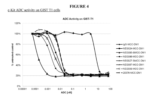
[00137] Figure 4 shows the ability of cKIT-MCC-DM1 ADCs to inhibit the
proliferation of
GIST-Ti (Imatinib-sensitive) cells.
[00138] Figure 5 shows the ability of cKIT-MCC-DM1 ADCs to inhibit the
proliferation of
GI5T430 (Imatinib-resistant) cells.
[00139] Figure 6 shows the ability of cKIT-MCC-DM1 ADCs to inhibit the
proliferation of
NCI-H526 (higher cKIT expressing SCLC) cells.
[00140] Figure 7 shows the ability of cKIT-MCC-DM1 ADCs to inhibit the
proliferation of
NCI-H1048 (lower cKIT expressing SCLC) cells.
[00141] Figure 8 shows the ability of cKIT-MCC-DM1 ADCs to inhibit the
proliferation of
CMK11-5 (high cKIT expressing AML) cells.
[00142] Figure 9 shows the ability of cKIT-MCC-DM1 ADCs to inhibit the
proliferation of
Uke-1 (lower cKIT expressing AML) cells.
[00143] Figure 10 is HDx-MS raw data plotted as the corrected difference
over the standard
error in measurement. A more negative value indicates more protection from
deuterium exchange
22
CA 02903772 2015-09-02
WO 2014/150937
PCT/US2014/024597
upon binding of 9P3 to cKIT antigen. The two most significant regions of
protection are denoted as
Region 1 and Region 2.
[00144] Figure 11 shows regions of HDx-MS protection are mapped using
surface fill: region
1 (black) and region 2 (dark grey). SCF binding sites are denoted as Site I
(light grey spheres), Site II
(medium grey spheres), and Site III (darker grey spheres).
[00145] Figure 12 is a Western blot showing the ability of SCF, NEG085-MCC-
DM1,
NEG024-MCC-DM1 and 20376-MCC-DM1 to modulate phosphorylation of cKIT in a
wildtype cKIT
cell line Mo7e Figure 12(A) or mutant cKIT cell line GIST-Ti Figure 12(B)
after 15 minutes.
[00146] Figure 13 shows that NEG085 and 20376 Abs mediate rapid
internalization of surface
cKIT on GIST-Ti cells (A) and on human bone marrow cells (B)
[00147] Figure 14 are Western blots showing the ability of SCF or NEG085-
MCC-DM1 to
accelerate cKIT degradation in a mutant cKIT cell line, GIST-Ti (Figure 14A)
and wildtype cKIT cell
line NCI-H526 (Figure 14B) over a timecourse.
[00148] Figure 15 shows the ability of NEG085, NEG024, 20376, NEG085-MCC-
DM1 to
inhibit the SCF-dependent proliferation of Mo7e cells.
[00149] Figure 16 shows the ability of NEG085 and NEG085-MCC-DM1 to inhibit
SCF-
independent proliferation of Mo7e cells.
[00150] Figure 17 shows the assessment of the ability of Campath (anti-CD52
Ab), NEG085
or 20376 antibodies to induce not ADCC in vitro in Uke-1 cells.
[00151] Figure 18 shows NEG085 and 20376 do not mediate primary human mast
cell
apoptosis.
[00152] Figure 19 shows NEG085 and 20376 do not mediate primary human mast
cell
degranulation.
[00153] Figure 20 shows co-localization of IgG1 and mitotic arrest of
NEG027-MCC-DM1 in
GIST Ti xenograft model.
[00154] Figure 21 shows tissue sections of mitotic arrest (p-histone H3)
and apoptosis
(caspase 3) after single dose of cKIT ADC.
[00155] Figure 22 graphically represents mitotic arrest and apoptosis
induction 8 days post
single dose of cKIT ADC.
[00156] Figure 23 shows (A) Dose response efficacy in GIST Ti mouse
xenograft and (B),
change in body weight over course of treatment.
[00157] Figure 24 graphically depicts (A) anti-DM1 ELISA after dosing in a
GIST Ti
xenograft model and (B) anti-human IgG1 ELISA after dosing in a GIST Ti
xenograft model.
[00158] Figure 25 is a table of NEG027-MCC-DM1 dose response in a GIST Ti
xenograft
mouse model.
23
CA 02903772 2015-09-02
WO 2014/150937
PCT/US2014/024597
[00159] Figure 26 are histology sections of NEG027-MCC-DM1 dose response
efficacy in
GIST Ti. (A) is Group 4 pooled tumors, (B) Group 5 pooled tumors.
[00160] Figure 27 depicts (A) Efficacy with 0.625mg/kg in a GIST Ti
xenograft mouse
model, (B) change of tumor volume vs control (% T/C) and (C) change in body
weight over course of
treatment.
[00161] Figure 28 shows clustering of day 41 after administration of single
dose of anti-cKIT
ADC to a GIST Ti xenograft mouse.
[00162] Figure 29 is a table of cKIT ADC efficacy at low effective dose in
GIST Ti xenograft
model.
[00163] Figure 30 shows (A) Anti-cKIT PK in a GIST Ti xenograft mouse
model, (left panel
is anti-DM1 ELISA) (B) Right panel is anti-human IgG1 ELISA.
[00164] Figure 31 A-C shows (A) NEG085-MCC-DM1, NEG024MCC-DM1 and NEG086-
MCC-DM1 activity in a SCLC model (B) change in body weight over course of
treatment (C)
expression of cKIT on tumor sample.
[00165] Figure 32 is a table of an anti-cKIT-ADC Efficacy Study in NCI-
H1048 SCLC
[00166] Figure 33 A-B shows (A) NEG085-MCC-DM1 dose response in NCI-H1048
(SCLC)
xenograft model, (B) Change in body weight over course of treatment.
[00167] Figure 34 is a table showing a NEG085-MCC-DM1 efficacy study in a
NCI-1048
(SCLC) xenograft mouse model.
[00168] Figure 35 A-C shows (A) Efficacy of 20376 and NEG024 in NCI-H526
(SCLC)
xenograft mouse model, (B) Antibody serum concentration after dosing and (C)
IHC for cKIT shows
expression of cKIT levels on H526 tumor.
[00169] Figure 36 shows anti-cKIT ADC in a small cell lung cancer (SCLC)
xenograft model.
[00170] Figure 37 shows anti-cKIT ADC efficacy in an AML xenograft model
(Kasumi-1).
[00171] Figure 38 shows anti-cKIT ADC efficacy in a HMC-1 mastocytosis
xenograft mouse
model.
[00172] Figure 39 A/B shows efficacy of mouse cross reactive 20376-MCC-DM1
in GIST Ti
xenograft mouse model with (A) dosage and tumor volume and (B) change in body
weight over course
of treatment.
[00173] Figure 40 A/B shows (A) Efficacy of mouse cross reactive 20376-MCC-
DM1 in
GIST Ti xenograft mouse model ¨PK and (B) Antibody serum concentration post
dosing.
[00174] Figure 41 shows dose response efficacy study in GIST Ti SCID-beige
mice.
24
CA 02903772 2015-09-02
WO 2014/150937
PCT/US2014/024597
[00175] Figure 42 A/B shows (A) efficacy in GIST Ti xenograft mouse model
(no efficacy
with unconjugated) and (B) change in body weight over course of treatment.
[00176] Figure 43 is a comparison of efficacy in a GIST Ti mouse xenograft
model
(unlabeled/MCC-DM1/SPDB-DM4).
[00177] Figure 44 A/B shows (A)Efficacy in a GIST 430 xenograft model
comparing SPDB-
DM4 and MCC-DM1 and (B) Change in body weight over course of treatment
[00178] Figure 45 shows efficacy in GIST 430 SCID-beige mouse model.
[00179] Figure 46 are photographs of p-Histone H3 immunostaining after
treatment with
NEG085-MCC-DM1.
[00180] Figure 47 is a graph of mitotic arrest shown by p-Histone H3
staining after
administration of NEG085-MCC-DM1.
[00181] Figure 48A shows cKIT staining of a GIST Ti tumor, Figure 48B shows
NEG085-
MCC-DM1 dose response in a GIST Ti xenograft model, Figure 48C shows the
change in body
weight of the mice treated with NEG085-MCC-DM1.
[00182] Figure 49A shows cKIT staining of a GIST 430 tumor, Figure 49B
shows NEG085-
MCC-DM1 dose response in a GIST 430 xenograft model, Figure 49C shows the
change in body
weight of the mice treated with NEG085-MCC-DM1.
[00183] Figure 50A shows cKIT staining of a NCI-H526 tumor (small cell lung
cancer
(SCLC), Figure 50B shows NEG085-MCC-DM1 dose response in a NCI-H526 xenograft
model,
Figure 50C shows the change in body weight of the mice treated with NEG085-MCC-
DM1.
[00184] Figure 51A shows the amount of IgG1 after NEG085-MCC-DM1 dosing in
a NCI-
H526 xenograft model, Figure 51B is a graph of an anti-DM1 ELISA in the NCI-
H526 xenograft
model after dosing with NEG085-MCC-DM1.
[00185] Figure 52A is a graph showing efficacy of NEG085-MCC-DM1 in a
primary AML
xenograft mouse model, Figure 52B shows the change in body weight of the mice
treated with
NEG085-MCC-DM1.
[00186] Figure 53 is a representation of the crystal structure of the
NEG085 Fab in complex
with cKIT domains 1 and 2. Fab heavy chains are in dark grey, Fab light chains
are in white and cKIT
domains are in light grey. Epitopes and paratopes are in black.
CA 02903772 2015-09-02
WO 2014/150937
PCT/US2014/024597
DETAILED DESCRIPTION
[00187] The present disclosure provides for antibodies, antibody fragments
(e.g., antigen
binding fragments), and antibody drug conjugates that bind to cKIT. In
particular, the present
disclosure is directed to antibodies and antibody fragments (e.g., antigen
binding fragments) that bind
to cKIT, and internalize upon such binding. The antibodies and antibody
fragments (e.g., antigen
binding fragments) of the present disclosure can be used for producing
antibody drug conjugates.
Furthermore, the present disclosure provides antibody drug conjugates that
have desirable
pharmacokinetic characteristics and other desirable attributes, and thus can
be used for treating cancer
expressing cKIT, without limitation, for example: gastrointestinal stromal
tumors (GIST), small cell
lung cancer (SCLC), acute myeloid leukemia (AML), melanoma, mast cell leukemia
(MCL),
mastocytosis, neurofibromatosis, breast cancer, non-small cell lung cancer
(NSCLC) and pancreatic
cancer. The present disclosure further provides pharmaceutical compositions
comprising the antibody
drug conjugates, and methods of making and using such pharmaceutical
compositions for the
treatment of cancer.
Antibody Drug Conjugates
[00188] The present disclosure provides antibody drug conjugates, where an
antibody, antigen
binding fragment or its functional equivalent that specifically binds to cKIT
is linked to a drug moiety.
In one aspect, the antibodies, antigen binding fragments or their functional
equivalents are linked, via
covalent attachment by a linker, to a drug moiety that is an anti-cancer
agent. The antibody drug
conjugates can selectively deliver an effective dose of an anti-cancer agent
(e.g., a cytotoxic agent) to
tumor tissues expressing cKIT, whereby greater selectivity (and lower
efficacious dose) may be
achieved.
[00189] In one aspect, the disclosure provides for an immunoconjugate of
Formula (I):
Ab¨(L¨(D)m)õ
Wherein Ab represents an cKIT binding antibody or antibody fragment (e.g.,
antigen binding
fragment) described herein;
L is a linker;
D is a drug moiety;
m is an integer from 1-8; and
n is an integer from 1-20. In one aspect, n is an integer from 1 to 10, 2 to
8, or 2 to 5. In a specific
aspect, n is 3 to 4. In some aspects, m is 1. In some aspects, m is 2, 3 or 4.
26
CA 02903772 2015-09-02
WO 2014/150937
PCT/US2014/024597
[00190] While the drug to antibody ratio has an exact integer value for a
specific conjugate
molecule (e.g., n multiplied by m in Formula (I)), it is understood that the
value will often be an
average value when used to describe a sample containing many molecules, due to
some degree of
inhomogeneity, typically associated with the conjugation step. The average
loading for a sample of an
immunoconjugate is referred to herein as the drug to antibody ratio, or "DAR."
In the aspect of
maytansinoids, this can be referred to as maytansinoid to antibody ratio or
"MAR." In some aspects,
the DAR is between about 1 and about 5, and typically is about 3, 3.5, 4, 4.5,
or 5. In some aspects, at
least 50% of a sample by weight is compound having the average DAR plus or
minus 2, and
preferably at least 50% of the sample is a conjugate that contains the average
DAR plus or minus 1.
Other aspects include immunoconjugates wherein the DAR is about 3.5. In some
aspects, a DAR of
'about n' means the measured value for DAR is within 20% of n.
[00191] The present disclosure provides immunoconjugates comprising the
antibodies,
antibody fragments (e.g., antigen binding fragments) and their functional
equivalents as disclosed
herein, linked or conjugated to a drug moiety. In one aspect, the drug moiety
D is a maytansinoid drug
moiety, including those having the structure:
0
0 0
CI \
0
ON
0
N/L
0
0 HO 1-1
where the wavy line indicates the covalent attachment of the sulfur atom of
the maytansinoid to a
linker of an antibody drug conjugate. R at each occurrence is independently H
or a C1-C6 alkyl. The
alkylene chain attaching the amide group to the sulfur atom may be methanyl,
ethanyl, or propanyl,
i.e. m is 1, 2, or 3. (U. S. Pat. No. 633,410, U.S. Pat. No. 5,208,020, Chari
et al. (1992) Cancer Res.
52;127-131, Lui et al. (1996) Proc. Natl. Acad. Sci. 93:8618-8623).
27
CA 02903772 2015-09-02
WO 2014/150937
PCT/US2014/024597
[00192] All stereoisomers of the maytansinoid drug moiety are contemplated
for the
immunoconjugates disclosed, i.e. any combination of R and S configurations at
the chiral carbons of
the maytansinoid. In one aspect the maytansinoid drug moiety has the following
stereochemistry.
¨ 0
0)
0 0
CI \
7 0
ON
0
N 0
c; Ha I-1
[00193] In one aspect, the maytansinoid drug moiety is N2'-deacetyl-N2-(3-
mercapto-1-
oxopropy1)-maytansine (also known as DM1). DM1 is represented by the following
structural
formula.
0
.E.-
CH2CH2S-
0 0
\ CI \ 0
ON
0
N 0
ci HO
DM1
[00194] In another aspect the maytansinoid drug moiety is N2'-deacetyl-N2-
(4-mercapto-1-
oxopenty1)-maytansine (also known as DM3). DM3 is respresented by the
following structural
formula.
28
CA 02903772 2015-09-02
WO 2014/150937
PCT/US2014/024597
0
Me
\ CI \ T
0
0
---=-
N
6 Ha H
DM3
[00195] In another aspect the maytansinoid drug moiety is N2'-deacetyl-NT-
(4-methyl-4-
mercapto-1-oxopenty1)-maytansine (also known as DM4). DM4 is respresented by
the following
structural formula.
0
'Z.--Me
0 0 I Me s¨
CI \ o
0
0
NO
0- HO H
DM4
[00196] The drug moiety D can be linked to the antibody through a linker L.
L is any
chemical moiety that is capable of linking the antibody Ab to the drug moiety
D. The linker, L
attaches the antibody Ab to the drug D through covalents bond(s). The linker
reagent is a bifunctional
or multifunctional moiety which can be used to link a drug moiety D and an
antibody Ab to form
antibody drug conjugates. Antibody drug conjugates can be prepared using a
linker having a reactive
functionality for binding to the drug moiety D and to the antibody Ab. A
cysteine, thiol or an amine,
e.g. N-terminus or amino acid side chain such as lysine of the antibody can
form a bond with a
funactional group of a linker reagent.
[00197] In one aspect, L is a cleavable linker. In another aspect, L is a
non-cleavable linker.
In some aspects, L is an acid-labile linker, photo-labile linker, peptidase
cleavable linker, esterase
29
CA 02903772 2015-09-02
WO 2014/150937
PCT/US2014/024597
cleavable linker, a disulfide bond reducible linker, a hydrophilic linker, a
procharged linker, or a
dicarboxylic acid based linker.
[00198] Suitable cross-linking reagents that form a non-cleavable linker
between the drug
moiety D, for example maytansinoid, and the antibody Ab are well known in the
art, and can form
non-cleavable linkers that comprise a sulfur atom (such as SMCC) or those that
are without a sulfur
atom. Preferred cross-linking reagents that form non-cleavable linkers between
the drug moiety D, for
example maytansinoid, and the antibody Ab comprise a maleimido- or haloacetyl-
based moiety.
According to the present disclosure, such non-cleavable linkers are said to be
derived from maleimido-
or haloacetyl-based moieties.
[00199] Cross-
linking reagents comprising a maleimido-based moiety include but not limited
to, N-succinimidy1-4-(maleimidomethyl)cyclohexanecarboxylate (SMCC),
sulfosuccinimidyl 4-(N-
maleimidomethyl) cyclohexane-l-carboxylate (sulfo-SMCC), N-succinimidy1-4-
(maleimidomethyl)cyclohexane-1 -carboxy-(6-amidocaproate), which is a "long
chain" analog of
SMCC (LC-SMCC), K-maleimidoundeconoic acid N-succinimidyl ester (KMUA), 7-
maleimidobutyric
acid N-succinimidyl ester (GMBS), E-maleimidocaproic acid N-succinimidyl ester
(EMCS), m-
maleimidobenzoyl-N-hydroxysuccinimide ester (MB S), N-(6c-maleimidoacetoxy)-
succinimide ester
(AMSA), succinimidy1-6-(13-maleimidopropionamido)hexanoate (SMPH), N-
succinimidy1-4-(p-
maleimidopheny1)-butyrate (SMPB), N-(-p-maleomidophenyBisocyanate (PMIP) and
maleimido-
based cross-linking reagents containing a polyethythene glycol spacer, such as
MAL-PEG-NHS.
These cross-linking reagents form non-cleavable linkers derived from maleimido-
based moieties.
Representative structures of maleimido-based cross-linking reagents are shown
below.
/JO
0 0
0
c N\01.r0,1;...
0 0
0 0 0
SMCC 0 Sulfo-SMCC
0
---1(0
PEG 0,...
,......./I N y N
\\ 0
0 0
MAL-PEG-NHS
CA 02903772 2015-09-02
WO 2014/150937
PCT/US2014/024597
[00200] In another aspect, the linker L is derived from N-succinimidy1-4-
(maleimidomethyl)cyclohexanecarboxylate (SMCC), sulfosuccinimidyl 4-(N-
maleimidomethyl)
cyclohexane-l-carboxylate (sulfo-SMCC) or MAL-PEG-NHS.
[00201] Cross-linking reagents comprising a haloacetyle-based moiety
include N-succinimidyl
iodoacetate (SIA), N-succinimidy1(4-iodoacetyl)aminobenzoate (STAB), N-
succinimidyl bromoacetate
(SBA) and N-succinimidyl 3-(bromoacetamido)propionate (SBAP). These cross-
linking reagents
form a non-cleavable linker derived from haloacetyl-based moieties.
Representative structures of
haloacetyl-based cross-linking reagents are shown below.
0
0
-----A 0 N
' 0
N, )1 <0
H
0 SIA SIAB
Or
[00202] In one aspect, the linker L is derived from N-succinimidyl
iodoacetate (SIA) or N-
succinimidy1(4-iodoacetyl)aminobenzoate (STAB).
[00203] Suitable cross-linking reagents that form a cleavable linker
between the drug moiety
D, for example maytansinoid, and the antibody Ab are well known in the art.
Disulfide containing
linkers are linkers cleavable through disulfide exchange, which can occur
under physiological
conditions. According to the present disclosure, such cleavable linkers are
said to be derived from
disulfide-based moieties. Suitable disulfide cross-linking reagents include N-
succinimidy1-3-(2-
pyridyldithio)propionate (SPDP), N-succinimidy1-4-(2-pyridyldithio)pentanoate
(SPP), N-
succinimidy1-4-(2-pyridyldithio)butanoate (SPDB) and N-succinimidy1-4-(2-
pyridyldithio)2-sulfo-
butanoate (sulfo-SPDB), the structures of which are shown below. These
disulfide cross-linking
reagents form a cleavable linker derived from disulfide-based moieties.
0
--A 0
I
0
N-succinimidy1-3-(2-pyridyldithio)propionate (SPDP),
31
CA 02903772 2015-09-02
WO 2014/150937
PCT/US2014/024597
0
0 N
N,
0
0
N-succinimidy1-4-(2-pyridyldithio)pentanoate (SPP),
0
0 N
N,
0
0
N-succinimidy1-4-(2-pyridyldithio)butanoate (SPDB) and
0
Na03SN. 0 N
0
N-succinimidy1-4-(2-pyridyldithio)2-sulfo-butanoate (sulfo-SPDB).
[00204] In one aspect, the linker L is derived from N-succinimidy1-4-(2-
pyridyldithio)butanoate (SPDB).
[00205] Suitable
cross-linking reagents that form a charged linker between the drug moiety D,
for example maytansinoid, and the antibody Ab are known as procharged cross-
linking reagents. In
one aspect, the linker L is derived from the procharged cross-linking reagent
is CX1-1. The structure
of CX1-1 is below.
0
I N=LI\IeNNNO,Ij.
H II H II
0 0 0 0
0
2,5-dioxopyrrolidin-1-y1 17-(2,5-dioxo-2,5-dihydro-1H-pyn-o1-1-y1)-5,8,11,14-
tetraoxo-4,7,10,13-
tetraazaheptadecan-1-oate (CX1-1)
32
CA 02903772 2015-09-02
WO 2014/150937
PCT/US2014/024597
[00206] In one aspect provided by the disclosure, the conjugate is
represented by any one of
the following structural formulae:
0 *-N
o
9,
'T 'N
o NH=4-Ab
CI
= tz 0
0.
0
\
= N
Hu H
n
Ab-MCC-DMI
0
ciNH _______________________________________________________ Ab
0 0 0
= 0
0
0
N 0
dHO H
Ab-SPD B-D M4
33
CA 02903772 2015-09-02
WO 2014/150937
PCT/US2014/024597
0
0
0 H 0
0 0 N,/NAN.,.N,)=LNN HAb
I CI \
- 0 0 H 0 H 8 0
0
=
0
-
d' H6 Fl
Ab-CX1-1-DM1
[00207] wherein:
[00208] Ab is an antibody or antigen binding fragment thereof that
specifically binds to human
cKIT;
[00209] n, which indicates the number of D-L groups attached the Ab through
the formation
of an amide bond with a primary amine of the Ab, is an integer from 1 to 20.
In one aspect, n is an
integer from 1 to 10, 2 to 8 or 2 to 5. In a specific aspect, n is 3 or 4.
[00210] In one aspect, the average molar ratio of drug (e.g., DM1 or DM4)
to the antibody in
the conjugate (i.e., average w value, also known as Maytanisnoid Antibody
Ratio (MAR)) is about 1 to
about 10, about 2 to about 8 (e.g., 1.9, 2.0, 2.1, 2.2, 2.3, 2.4, 2.5, 2.6,
2.7, 2.8, 2.9, 3.0, 3.1, 3.2, 3.3,
3.4, 3.5, 3.6, 3.7, 3.8, 3.9, 4.0, 4.1, 4.2, 4.3, 4.4, 4.5, 4.6, 4.7, 4.8,
4.9, 5.0, 5.1, 5.2, 5.3, 5.4, 5.5, 5.6,
5.7, 5.8, 5.9, 6.0, 6.1, 6.2, 6.3, 6.4, 6.5, 6.6, 6.7, 6.8, 6.9, 7.0, 7.1,
7.2, 7.3, 7.4, 7.5, 7.6, 7.7, 7.8, 7.9,
8.0, or 8.1), about 2.5 to about 7, about 3 to about 5, about 2.5 to about 4.5
(e.g., about 2.5, about 2.6,
about 2.7, about 2.8, about 2.9, about 3.0, about 3.1, about 3.3, about 3.4,
about 3.5, about 3.6, about
3.7, about 3.8, about 3.9, about 4.0, about 4.1, about 4.2, about 4.3, about
4.4, about 4.5), about 3.0 to
about 4.0, about 3.2 to about 4.2, or about 4.5 to 5.5 (e.g., about 4.5, about
4.6, about 4.7, about 4.8,
about 4.9, about 5.0, about 5.1, about 5.2, about 5.3, about 5.4, or about
5.5).
[00211] In one aspect provided by the disclosure, the conjugate has
substantially high purity
and has one or more of the following features: (a) greater than about 90%
(e.g., greater than or equal to
about 91%, 92%, 93%, 94%, 95%, 96%, 97%, 98%, 99%, or 100%), preferably
greater than about
95%, of conjugate species are monomeric, (b) unconjugated linker level in the
conjugate preparation is
less than about 10% (e.g., less than or equal to about 9%, 8%, 7%, 6%, 5%, 4%,
3%, 2%, 1%, or 0%)
34
CA 02903772 2015-09-02
WO 2014/150937
PCT/US2014/024597
(relative to total linker), (c) less than 10% of conjugate species are
crosslinked (e.g., less than or equal
to about 9%, 8%, 7%, 6%, 5%, 4%, 3%, 2%, 1%, or 0%), (d) free drug (e.g., DM1
or DM4) level in
the conjugate preparation is less than about 2% (e.g., less than or equal to
about 1.5%, 1.4%, 1.3%,
1.2%, 1.1%, 1.0%, 0.9%, 0.8%, 0.7%, 0.6%, 0.5%, 0.4%, 0.3%, 0.2%, 0.1%, or 0%)
(mol/mol relative
to total cytotoxic agent).
[00212] As used herein, the term "unconjugated linker" refers to the
antibody that is
covalently linked with a linker derived from a cross-linking reagent (e.g.,
SMCC, Sulfo-SMCC,
SPDB, Sulfo-SPDB or CX1-1), wherein the antibody is not covalently coupled to
the drug (e.g., DM1
or DM4) through a linker (i.e., the "unconjugated linker" can be represented
by Ab-SMCC, Ab-SPDB,
or Ab-CX1-1).
1. Drug- Moiety
[00213] The present disclosure provides immunoconjugates that specifically
bind to cKIT.
The immunoconjugates of the present disclosure comprise anti- cKIT antibodies,
antibody fragments
(e.g., antigen binding fragments) or functional equivalents that are
conjugated to a drug moiety, e.g.,
an anti-cancer agent, anti-hematological disorder agent, an autoimmune
treatment agent, an anti-
inflammatory agent, an antifungal agent, an antibacterial agent, an anti-
parasitic agent, an anti-viral
agent, or an anesthetic agent. The antibodies, antibody fragments (e.g.,
antigen binding fragments) or
functional equivalents can be conjugated to several identical or different
drug moieties using any
methods known in the art.
[00214] In certain aspects, the drug moiety of the immunoconjugates of the
present disclosure
is selected from a group consisting of a V-ATPase inhibitor, a pro-apoptotic
agent, a Bc12 inhibitor, an
MCL1 inhibitor, a HSP90 inhibitor, an TAP inhibitor, an mTor inhibitor, a
microtubule stabilizer, a
microtubule destabilizer, an auristatin, a dolastatin, a maytansinoid, a MetAP
(methionine
aminopeptidase), an inhibitor of nuclear export of proteins CRM1, a DPPIV
inhibitor, proteasome
inhibitors, an inhibitor of phosphoryl transfer reactions in mitochondria, a
protein synthesis inhibitor, a
kinase inhibitor, a CDK2 inhibitor, a CDK9 inhibitor, a kinesin inhibitor, an
HDAC inhibitor, a DNA
damaging agent, a DNA alkylating agent, a DNA intercalator, a DNA minor groove
binder and a
DHFR inhibitor.
[00215] In one aspect, the drug moiety of the immunoconjugates of the
present disclosure is a
maytansinoid drug moiety, such as but not limited to, DM1, DM3, or DM4.
CA 02903772 2015-09-02
WO 2014/150937
PCT/US2014/024597
[00216] Further, the antibodies, antibody fragments (e.g., antigen binding
fragments) or
functional equivalents of the present disclosure may be conjugated to a drug
moiety that modifies a
given biological response. Drug moieties are not to be construed as limited to
classical chemical
therapeutic agents. For example, the drug moiety may be a protein, peptide, or
polypeptide possessing
a desired biological activity. Such proteins may include, for example, a toxin
such as abrin, ricin A,
pseudomonas exotoxin, cholera toxin, or diphtheria toxin, a protein such as
tumor necrosis factor, a-
interferon, f3-interferon, nerve growth factor, platelet derived growth
factor, tissue plasminogen
activator, a cytokine, an apoptotic agent, an anti-angiogenic agent, or, a
biological response modifier
such as, for example, a lymphokine.
[00217] In one aspect, the antibodies, antibody fragments (e.g., antigen
binding fragments) or
functional equivalents of the present disclosure are conjugated to a drug
moiety, such as a cytotoxin, a
drug (e.g., an immunosuppressant) or a radiotoxin. Examples of cytotoxin
include but are not limited
to, taxanes (see, e.g., International (PCT) Patent Application Nos. WO
01/38318 and
PCT/U503/02675), DNA-alkylating agents (e.g., CC-1065 analogs), antlu-
acyclines, tubulysin analogs,
duocarmycin analogs, auristatin E, auristatin F, maytansinoids, and cytotoxic
agents comprising a
reactive polyethylene glycol moiety (see, e.g., Sasse et al., J. Antibiot.
(Tokyo), 53, 879-85 (2000),
Suzawa et al., Bioorg. Med. Chem., 8, 2175-84 (2000), Ichimura et al., J.
Antibiot. (Tokyo), 44, 1045-
53 (1991), Francisco et al., Blood 2003 15;102(4):1458-65), U.S. Pat. Nos.
5,475,092, 6,340,701,
6,372,738, and 6,436,931, U.S. Patent Application Publication No. 2001/0036923
Al, Pending U.S.
patent application Ser. Nos. 10/024,290 and 10/116,053, and International
(PCT) Patent Application
No. WO 01/49698), taxon, cytochalasin B, gramicidin D, ethidium bromide,
emetine, mitomycin,
etoposide, tenoposide, vincristine, vinblastine, t. colchicin, doxorubicin,
daunorubicin, dihydroxy
anthracin dione, mitoxantrone, mithramycin, actinomycin D, 1 -
dehydrotestosterone, glucocorticoids,
procaine, tetracaine, lidocaine, propranolol, and puromycin and analogs or
homologs thereof.
Therapeutic agents also include, for example, anti-metabolites (e.g.,
methotrexate, 6-mercaptopurine,
6-thioguanine, cytarabine, 5-fluorouracil decarbazine), ablating agents (e.g.,
mechlorethamine,
thiotepa chlorambucil, meiphalan, carmustine (BSNU) and lomustine (CCNU),
cyclophosphamide,
busulfan, dibromomannitol, streptozotocin, mitomycin C, and cis-
dichlorodiamine platinum (II)
(DDP) cisplatin, antlu-acyclines (e.g., daunorubicin (formerly daunomycin) and
doxorubicin),
antibiotics (e.g., dactinomycin (formerly actinomycin), bleomycin,
mithramycin, and antlu-amycin
(AMC)), and anti-mitotic agents (e.g., vincristine and vinblastine). (See
e.g., Seattle Genetics
U5200903 04721).
36
CA 02903772 2015-09-02
WO 2014/150937
PCT/US2014/024597
[00218] Other examples of cytotoxins that can be conjugated to the
antibodies, antibody
fragments (antigen binding fragments) or functional equivalents of the present
disclosure include
duocarmycins, calicheamicins, maytansines and auristatins, and derivatives
thereof
[00219] Various types of cytotoxins, linkers and methods for conjugating
therapeutic agents to
antibodies are known in the art, see, e.g., Saito et al., (2003) Adv. Drug
Deliv. Rev. 55:199-215; Trail
et al., (2003) Cancer Immunol. Immunother. 52:328-337; Payne, (2003) Cancer
Cell 3:207-212; Allen,
(2002) Nat. Rev. Cancer 2:750-763; Pastan and Kreitman, (2002) Curr. Opin.
Investig. Drugs 3:1089-
1091; Senter and Springer, (2001) Adv. Drug Deliv. Rev. 53:247-264.
[00220] The antibodies, antibody fragments (e.g., antigen binding
fragments) or functional
equivalents of the present disclosure can also be conjugated to a radioactive
isotope to generate
cytotoxic radiopharmaceuticals, referred to as radioimmunoconjugates. Examples
of radioactive
isotopes that can be conjugated to antibodies for use diagnostically or
therapeutically include, but are
not limited to, iodine-131, indium-111, yttrium-90, and lutetium-177. Methods
for preparing
radioimmunoconjugates are established in the art. Examples of
radioimmunoconjugates are
commercially available, including ZevalinTM (IDEC Pharmaceuticals) and
BexxarTM (Corixa
Pharmaceuticals), and similar methods can be used to prepare
radioimmunoconjugates using the
antibodies disclosed herein. In certain aspects, the macrocyclic chelator is
1,4,7,10-
tetraazacyclododecane-N,N',N",N' "-tetraacetic acid (DOTA) which can be
attached to the antibody
via a linker molecule. Such linker molecules are commonly known in the art and
described in
Denardo et al., (1998) Clin Cancer Res. 4(10):2483-90; Peterson et al., (1999)
Bioconjug. Chem.
10(4):553-7; and Zimmerman et al., (1999) Nucl. Med. Biol. 26(8):943-50, each
incorporated by
reference in their entireties.
[00221] The antibodies, antibody fragments (e.g., antigen binding
fragments) or functional
equivalents of the present disclosure can also conjugated to a heterologous
protein or polypeptide (or
fragment thereof, preferably to a polypeptide of at least 10, at least 20, at
least 30, at least 40, at least
50, at least 60, at least 70, at least 80, at least 90 or at least 100 amino
acids) to generate fusion
proteins. In particular, the present disclosure provides fusion proteins
comprising an antibody
fragment (e.g., antigen binding fragment) described herein (e.g., a Fab
fragment, Fd fragment, Fv
fragment, F(ab)2 fragment, a VH domain, a VH CDR, a VL domain or a VL CDR) and
a heterologous
protein, polypeptide, or peptide.
[00222] Additional fusion proteins may be generated through the techniques
of gene-shuffling,
motif-shuffling, exon-shuffling, and/or codon-shuffling (collectively referred
to as "DNA shuffling").
37
CA 02903772 2015-09-02
WO 2014/150937
PCT/US2014/024597
DNA shuffling may be employed to alter the activities of antibodies of the
present disclosure or
fragments thereof (e.g., antibodies or fragments thereof with higher
affinities and lower dissociation
rates). See, generally, U.S. Patent Nos. 5,605,793, 5,811,238, 5,830,721,
5,834,252, and 5,837,458;
Patten et al., (1997) Cuff. Opinion Biotechnol. 8:724-33; Harayama, (1998)
Trends Biotechnol.
16(2):76-82; Hansson et al., (1999) J. Mol. Biol. 287:265-76; and Lorenzo and
Blasco, (1998)
Biotechniques 24(2):308- 313 (each of these patents and publications are
hereby incorporated by
reference in its entirety). Antibodies or fragments thereof, or the encoded
antibodies or fragments
thereof, may be altered by being subjected to random mutagenesis by error-
prone PCR, random
nucleotide insertion or other methods prior to recombination. A polynucleotide
encoding an antibody
or fragment thereof that specifically binds to an antigen may be recombined
with one or more
components, motifs, sections, parts, domains, fragments, etc. of one or more
heterologous molecules.
[00223] Moreover, the antibodies, antibody fragments (e.g., antigen binding
fragments) or
functional equivalents of the present disclosure can be conjugated to marker
sequences, such as a
peptide, to facilitate purification. In preferred aspects, the marker amino
acid sequence is a hexa-
histidine peptide, such as the tag provided in a pQE vector (QIAGEN, Inc.,
9259 Eton Avenue,
Chatsworth, CA, 91311), among others, many of which are commercially
available. As described in
Gentz et al., (1989) Proc. Natl. Acad. Sci. USA 86:821-824, for instance, hexa-
histidine provides for
convenient purification of the fusion protein. Other peptide tags useful for
purification include, but
are not limited to, the hemagglutinin ("HA") tag, which corresponds to an
epitope derived from the
influenza hemagglutinin protein (Wilson et al., (1984) Cell 37:767), and the
"FLAG" tag (A. Einhauer
et al., J. Biochem. Biophys. Methods 49: 455-465, 2001). As described in the
present disclosure,
antibodies or antigen binding fragments can also be conjugated to tumor-
penetrating peptides in order
to enhance their efficacy.
[00224] In other aspects, the antibodies, antibody fragments (e.g., antigen
binding fragments)
or functional equivalents of the present disclosure are conjugated to a
diagnostic or detectable agent.
Such immunoconjugates can be useful for monitoring or prognosing the onset,
development,
progression and/or severity of a disease or disorder as part of a clinical
testing procedure, such as
determining the efficacy of a particular therapy. Such diagnosis and detection
can be accomplished by
coupling the antibody to detectable substances including, but not limited to,
various enzymes, such as,
but not limited to, horseradish peroxidase, alkaline phosphatase, beta-
galactosidase, or
acetylcholinesterase; prosthetic groups, such as, but not limited to,
streptavidin/biotin and
avidin/biotin; fluorescent materials, such as, but not limited to, Alexa Fluor
350, Alexa Fluor 405,
38
CA 02903772 2015-09-02
WO 2014/150937
PCT/US2014/024597
Alexa Fluor 430, Alexa Fluor 488, Alexa Fluor 500, Alexa Fluor 514, Alexa
Fluor 532, Alexa Fluor
546, Alexa Fluor 555, Alexa Fluor 568, Alexa Fluor 594, Alexa Fluor 610, Alexa
Fluor 633, Alexa
Fluor 647, Alexa Fluor 660, Alexa Fluor 680, Alexa Fluor 700, Alexa Fluor 750,
umbelliferone,
fluorescein, fluorescein isothiocyanate, rhodamine, dichlorotriazinylamine
fluorescein, dansyl chloride
or phycoerythrin; luminescent materials, such as, but not limited to, luminol;
bioluminescent materials,
such as but not limited to, luciferase, luciferin, and aequorin; radioactive
materials, such as, but not
limited to, iodine (1311, 1251, 1231, and 1211), carbon (14C), sulfur (35S),
tritium (3H), indium (115In, 1131n,
112In, and 111In,), technetium (99Tc), thallium (261Ti), gallium (68Ga, 67Ga),
palladium (163Pd),
molybdenum (99Mo), xenon (133Xe), fluorine (18F), 153Sm, 177Lu, 159Gd, 149pin,
140La, 175yb, 166H0, 90y,
47se, 186Re, 188Re, 142 Pr, 105Rn, 97Rn, 68-e,
57Co, 65Z11, 85Sr, 32P, 153Gd, 169
yb, 51cr, 54-n,
M 75Se, 64CU,
113Sn, and 117Sn; and positron emitting metals using various positron emission
tomographies, and non-
radioactive paramagnetic metal ions.
[00225] The antibodies, antibody fragments (e.g., antigen binding
fragments) or functional
equivalents of the present disclosure may also be attached to solid supports,
which are particularly
useful for immunoassays or purification of the target antigen. Such solid
supports include, but are not
limited to, glass, cellulose, polyacrylamide, nylon, polystyrene, polyvinyl
chloride or polypropylene.
2. Linker
[00226] As used herein, a "linker" is any chemical moiety that is capable
of linking an
antibody, antibody fragment (e.g., antigen binding fragments) or functional
equivalent to another
moiety, such as a drug moeity. Linkers can be susceptible to cleavage
(cleavable linker), such as,
acid-induced cleavage, photo-induced cleavage, peptidase-induced cleavage,
esterase-induced
cleavage, and disulfide bond cleavage, at conditions under which the compound
or the antibody
remains active. Alternatively, linkers can be substantially resistant to
cleavage (e.g., stable linker or
noncleavable linker). In some aspects, the linker is a procharged linker, a
hydrophilic linker, or a
dicarboxylic acid based linker.
[00227] In one aspect, the linker used is derived from a crosslinking
reagent such as N-
succinimidy1-3-(2-pyridyldithio)propionate (SPDP), N-succinimidyl 4-(2-
pyridyldithio)pentanoate
(SPP), N-succinimidyl 4-(2-pyridyldithio)butanoate (SPDB), N-succinimidy1-4-(2-
pyridyldithio)-2-
sulfo-butanoate (sulfo-SPDB), N-succinimidyl iodoacetate (SIA), N-
succinimidy1(4-
iodoacetyl)aminobenzoate (SIAB), maleimide PEG NHS, N-succinimidyl 4-
(maleimidomethyl)
cyclohexanecarboxylate (SMCC), N-sulfosuccinimidyl 4-(maleimidomethyl)
cyclohexanecarboxylate
39
CA 02903772 2015-09-02
WO 2014/150937
PCT/US2014/024597
(sulfo-SMCC) or 2,5-dioxopyrrolidin-l-y1 17-(2,5-dioxo-2,5-dihydro-1H-pyn-o1-1-
y1)-5,8,11,14-
tetraoxo-4,7,10,13-tetraazaheptadecan-1-oate (CX1-1). In another aspect, the
linker used is derived
from a cross-linking agent such as N-succinimidy1-3-(2-
pyridyldithio)propionate (SPDP), N-
succinimidyl 4-(maleimidomethyl) cyclohexanecarboxylate (SMCC), N-
sulfosuccinimidyl 4-
(maleimidomethyl) cyclohexanecarboxylate (sulfo-SMCC), N-succinimidy1-4-(2-
pyridyldithio)-2-
sulfo-butanoate (sulfo-SPDB) or 2,5-dioxopyrrolidin-l-y1 17-(2,5-dioxo-2,5-
dihydro-1H-pyrrol-1-y1)-
5,8,11,14-tetraoxo-4,7,10,13-tetraazaheptadecan-1-oate (CX1-1).
[00228] Non-cleavable linkers are any chemical moiety capable of linking a
drug, such as a
maytansinoid, to an antibody in a stable, covalent manner and does not fall
off under the categorties
listed above for cleaveable linkers. Thus, non-cleavable linkers are
substantialy resistant to acid-
induced cleavage, photo-induced cleavage, peptidase-induced cleavage, esterase-
induced cleavage and
disulfide bond cleavage. Furthermore, non-cleavable refers to the ability of
the chemical bond in the
linker or adjoining to the linker to withstand cleavage induced by an acid,
photolabile-cleaving agent,
a peptidase, an esterase, or a chemical or physiological compound that cleaves
a disulfide bond, at
conditions under which the drug, such as maytansionoid or the antibody does
not lose its activity.
[00229] Acid-labile linkers are linkers cleavable at acidic pH. For
example, certain
intracellular compartments, such as endosomes and lysosomes, have an acidic pH
(pH 4-5), and
provide conditions suitable to cleave acid-labile linkers.
[00230] Photo-labile linkers are linkers that are useful at the body
surface and in many body
cavities that are accessable to light. Furthermore, infrared light can
penetrate tissue.
[00231] Some linkers can be cleaved by peptidases, i.e. peptidase cleavable
linkers. Only
certain peptides are readily cleaved inside or outside cells, see e.g. Trout
et al., 79 Proc. Natl.
Acad.Sci. USA, 626-629 (1982) and Umemoto et al. 43 Int. J. Cancer, 677-684
(1989). Furthermore,
peptides are composed of cc-amino acids and peptidic bonds, which chemically
are amide bonds
between the carboxylate of one amino acid and the amino group of a second
amino acid. Other amide
bonds, such as the bond between a carboxylate and the E-amino group of lysine,
are understood not to
be peptidic bonds and are considered non-cleavable.
[00232] Some linkers can be cleaved by esterases, i.e. esterase cleavable
linkers. Again, only
certain esters can be cleaved by esterases present inside or outside of cells.
Esters are formed by the
condensation of a carboxylic acid and an alcohol. Simple esters are esters
produced with simple
alcohols, such as aliphatic alcohols, and small cyclic and small aromatic
alcohols.
CA 02903772 2015-09-02
WO 2014/150937
PCT/US2014/024597
[00233] Procharged linkers are derived from charged cross-linking reagents
that retain their
charge after incorporation into an antibody drug conjugate. Examples of
procharged linkers can be
found in US 2009/0274713.
3. Conjugation and Preparation of ADCs
[00234] The conjugates of the present disclosure can be prepared by any
methods known in
the art, such as those described in US Patent Nos. 7,811,572, 6,411,163,
7,368,565, and 8,163,888, and
US application publications 2011/0003969, 2011/0166319, 2012/0253021 and
2012/0259100. The
entire teachings of these patents and patent application publications are
herein incorporated by
reference.
One-Step Process
[00235] In one aspect, the conjugates of the present disclosure can be
prepared by a one-step
process. The process comprises combining the antibody, drug and cross-linking
agent in a
substantially aqueous medium, optionally containing one or more co-solvents,
at a suitable pH. In one
aspect, the process comprises the step of contacting the antibody of the
present disclosure with a drug
(e.g., DM1 or DM4) to form a first mixture comprising the antibody and the
drug, and then contacting
the first mixture comprising the antibody and the drug with a cross-linking
agent (e.g., SMCC, Sulfo-
SMCC, SPDB, Sulfo-SPDB or CX1-1) in a solution having a pH of about 4 to about
9 to provide a
mixture comprising (i) the conjugate (e.g., Ab-MCC-DM1, Ab-SPDB-DM4, or Ab-CX1-
1-DM1), (ii)
free drug (e.g., DM1 or DM4), and (iii) reaction by-products.
[00236] In one aspect, the one-step process comprises contacting the
antibody with the drug
(e.g., DM1 or DM4) and then the cross-linking agent (e.g., SMCC, Sulfo-SMCC,
SPDB, Sulfo-SPDB
or CX1-1) in a solution having a pH of about 6 or greater (e.g., about 6 to
about 9, about 6 to about 7,
about 7 to about 9, about 7 to about 8.5, about 7.5 to about 8.5, about 7.5 to
about 8.0, about 8.0 to
about 9.0, or about 8.5 to about 9.0). For example, the process comprises
contacting a cell-binding
agent with the drug (DM1 or DM4) and then the cross-linking agent (e.g., SMCC,
Sulfo-SMCC,
SPDB, Sulfo-SPDB or CX1-1) in a solution having a pH of about 6.0, about 6.1,
about 6.2, about 6.3,
about 6.4, about 6.5, about 6.6, about 6.7, about 6.8, about 6.9, about 7.0,
about 7.1, about 7.2, about
7.3, about 7.4, about 7.5, about 7.6, about 7.7, about 7.8, about 7.9, about
8.0, about 8.1, about 8.2,
about 8.3, about 8.4, about 8.5, about 8.6, about 8.7, about 8.8, about 8.9,
or about 9Ø In another
aspect, the process comprises contacting a cell-binding agent with the drug
(e.g., DM1 or DM4) and
then the cross-linking agent (e.g., SMCC, Sulfo-SMCC, SPDB, Sulfo-SPDB or CX1-
1) in a solution
having a pH of about 7.8 (e.g., a pH of 7.6 to 8.0 or a pH of 7.7 to 7.9).
[00237] The one-step process (i.e., contacting the antibody with the drug
(e.g., DM1 or DM4)
and then the cross-linking agent (e.g., SMCC, Sulfo-SMCC, SPDB, Sulfo-SPDB or
CX1-1) can be
carried out at any suitable temperature known in the art. For example, the one-
step process can occur
41
CA 02903772 2015-09-02
WO 2014/150937
PCT/US2014/024597
at about 20 C or less (e.g., about -10 C (provided that the solution is
prevented from freezing, e.g., by
the presence of organic solvent used to dissolve the cytotoxic agent and the
bifunctional crosslinking
reagent) to about 20 C, about 0 C to about 18 C, about 4 C to about 16 C), at
room temperature (e.g.,
about 20 C to about 30 C or about 20 C to about 25 C), or at an elevated
temperature (e.g., about
30 C to about 37 C). In one aspect, the one-step process occurs at a
temperature of about 16 C to
about 24 C (e.g., about 16 C, about 17 C, about 18 C, about 19 C, about 20 C,
about 21 C, about
22 C, about 23 C, about 24 C, or about 25 C). In another aspect, the one-step
process is carried out at
a temperature of about 15 C or less (e.g., about -10 C to about 15 C, or about
0 C to about 15 C).
For example, the process comprises contacting the antibody with the drug
(e.g., DM1 or DM4) and
then the cross-linking agent (e.g., SMCC, Sulfo-SMCC, SPDB, Sulfo-SPDB or CX1-
1) at a
temperature of about 15 C, about 14 C, about 13 C, about 12 C, about 11 C,
about 10 C, about 9 C,
about 8 C, about 7 C, about 6 C, about 5 C, about 4 C, about 3 C, about 2 C,
about 1 C, about 0 C,
about -1 C, about -2 C, about -3 C, about -4 C, about -5 C, about -6 C, about -
7 C, about -8 C, about
-9 C, or about -10 C, provided that the solution is prevented from freezing,
e.g., by the presence of
organic solvent(s) used to dissolve the cross-linking agent (e.g., SMCC, Sulfo-
SMCC, Sulfo-SPDB
SPDB, or CX1-1). In one aspect, the process comprises contacting the antibody
with the drug (e.g.,
DM1 or DM4) and then the cross-linking agent (e.g., SMCC, Sulfo-SMCC, SPDB,
Sulfo-SPDB or
CX1-1) at a temperature of about -10 C to about 15 C, about 0 C to about 15 C,
about 0 C to about
C, about 0 C to about 5 C, about 5 C to about 15 C, about 10 C to about 15 C,
or about 5 C to
about 10 C. In another aspect, the process comprises contacting the antibody
with the drug (e.g.,
DM1 or DM4) and then the cross-linking agent (e.g., SMCC, Sulfo-SMCC, SPDB,
Sulfo-SPDB or
CX1-1) at a temperature of about 10 C (e.g., a temperature of 8 C to 12 C or a
temperature of 9 C to
11 C).
[00238] In one aspect, the contacting described above is effected by
providing the antibody,
then contacting the antibody with the drug (e.g., DM1 or DM4) to form a first
mixture comprising the
antibody and the drug (e.g., DM1 or DM4), and then contacting the first
mixture comprising the
antibody and the drug (e.g., DM1 or DM4) with the cross-linking agent (e.g.,
SMCC, Sulfo-SMCC,
SPDB, Sulfo-SPDB or CX1-1). For example, in one aspect, the antibody is
provided in a reaction
vessel, the drug (e.g., DM1 or DM4) is added to the reaction vessel (thereby
contacting the antibody),
and then the cross-linking agent (e.g., SMCC, Sulfo-SMCC, SPDB, Sulfo-SPDB or
CX1-1) is added
to the mixture comprising the antibody and the drug (e.g., DM1 or DM4)
(thereby contacting the
mixture comprising the antibody and the drug). In one aspect, the antibody is
provided in a reaction
vessel, and the drug (e.g., DM1 or DM4) is added to the reaction vessel
immediately following
providing the antibody to the vessel. In another aspect, the antibody is
provided in a reaction vessel,
and the drug (e.g., DM1 or DM4) is added to the reaction vessel after a time
interval following
providing the antibody to the vessel (e.g., about 5 minutes, about 10 minutes,
about 20 minutes, about
42
CA 02903772 2015-09-02
WO 2014/150937
PCT/US2014/024597
30 minutes, about 40 minutes, about 50 minutes, about 1 hour, about 1 day or
longer after providing
the cell-binding agent to the space). The drug (e.g., DM1 or DM4) can be added
quickly (i.e., within a
short time interval, such as about 5 minutes, about 10 minutes) or slowly
(such as by using a pump).
[00239] The mixture comprising the antibody and the drug (e.g., DM1 or DM4)
can then be
contacted with the cross-linking agent (e.g., SMCC, Sulfo-SMCC, SPDB, Sulfo-
SPDB or CX1-1)
either immediately after contacting the antibody with the drug (e.g., DM1 or
DM4) or at some later
point (e.g., about 5 minutes to about 8 hours or longer) after contacting the
antibody with the drug
(e.g., DM1 or DM4). For example, in one aspect, the cross-linking agent (e.g.,
SMCC, Sulfo-SMCC,
SPDB, Sulfo-SPDB or CX1-1) is added to the mixture comprising the antibody and
the drug (e.g.,
DM1 or DM4) immediately after the addition of the drug (e.g., DM1 or DM4) to
the reaction vessel
comprising the antibody. Alternatively, the mixture comprising the antibody
and the drug (e.g., DM1
or DM4) can be contacted with the cross-linking agent (e.g., SMCC, Sulfo-SMCC,
SPDB, Sulfo-
SPDB or CX1-1) at about 5 minutes, about 10 minutes, about 20 minutes, about
30 minutes, about 1
hour, about 2 hours, about 3 hours, about 4 hours, about 5 hours, about 6
hours, about 7 hours, about 8
hours, or longer after contacting the antibody with the drug (e.g., DM1 or
DM4).
[00240] After the mixture comprising the antibody and the drug (e.g., DM1
or DM4) is
contacted with the cross-linking agent (e.g., SMCC, Sulfo-SMCC, SPDB, Sulfo-
SPDB or CX1-1) the
reaction is allowed to proceed for about 1 hour, about 2 hours, about 3 hours,
about 4 hours, about 5
hours, about 6 hours, about 7 hours, about 8 hours, about 9 hours, about 10
hours, about 11 hours,
about 12 hours, about 13 hours, about 14 hours, about 15 hours, about 16
hours, about 17 hours, about
18 hours, about 19 hours, about 20 hours, about 21 hours, about 22 hours,
about 23 hours, about 24
hours, or longer (e.g., about 30 hours, about 35 hours, about 40 hours, about
45 hours, or about 48
hrs).
[00241] In one aspect, the one-step process further comprises a quenching
step to quench any
um-eacted drug (e.g., DM1 or DM4) and/or um-eacted cross-linking agent (e.g.,
SMCC, Sulfo-SMCC,
SPDB, Sulfo-SPDB or CX1-1). The quenching step is typically performed prior to
purification of the
conjugate. In one aspect, the mixture is quenched by contacting the mixture
with a quenching reagent.
As used herein, the "quenching reagent" refers to a reagent that reacts with
the free drug (e.g., DM1 or
DM4) and/or cross-linking agent (e.g., SMCC, Sulfo-SMCC, SPDB, Sulfo-SPDB or
CX1-1). In one
aspect, maleimide or haloacetamide quenching reagents, such as 4-
maleimidobutyric acid, 3-
maleimidopropionic acid, N-ethylmaleimide, iodoacetamide, or
iodoacetamidopropionic acid, can be
used to ensure that any um-eacted group (such as thiol) in the drug (e.g., DM1
or DM4) is quenched.
The quenching step can help prevent the dimerization of the drug (e.g., DM1).
The dimerized DM1
can be difficult to remove. Upon quenching with polar, charged thiol-quenching
reagents (such as 4-
maleimidobutyric acid or 3-maleimidopropionic acid), the excess, unreacted DM1
is converted into a
polar, charged, water-soluble adduct that can be easily separated from the
covalently- linked conjugate
43
CA 02903772 2015-09-02
WO 2014/150937
PCT/US2014/024597
during the purification step. Quenching with non-polar and neutral thiol-
quenching reagents can also
be used. In one aspect, the mixture is quenched by contacting the mixture with
a quenching reagent
that reacts with the um-eacted cross-linking agent (e.g., SMCC, Sulfo-SMCC,
SPDB, Sulfo-SPDB or
CX1-1). For example, nucleophiles can be added to the mixture in order to
quench any unreacted
SMCC. The nucleophile preferably is an amino group containing nucleophile,
such as lysine, taurine
and hydroxylamine.
[00242] In another aspect, the reaction (i.e., contacting the antibody with
the drug (e.g., DM1
or DM4) and then cross-linking agent (e.g., SMCC, Sulfo-SMCC, SPDB, Sulfo-SPDB
or CX1-1)) is
allowed to proceed to completion prior to contacting the mixture with a
quenching reagent. In this
regard, the quenching reagent is added to the mixture about 1 hour to about 48
hours (e.g., about 1
hour, about 2 hours, about 3 hours, about 4 hours, about 5 hours, about 6
hours, about 7 hours, about 8
hours, about 9 hours, about 10 hours, about 11 hours, about 12 hours, about 13
hours, about 14 hours,
about 15 hours, about 16 hours, about 17 hours, about 18 hours, about 19
hours, about 20 hours, about
21 hours, about 22 hours, about 23 hours, about 24 hours, or about 25 hours to
about 48 hours) after
the mixture comprising the antibody and the drug (e.g., DM1 or DM4) is
contacted with the cross-
linking agent (e.g., SMCC, Sulfo-SMCC, SPDB, Sulfo-SPDB or CX1-1).
[00243] Alternatively, the mixture is quenched by lowering the pH of the
mixture to about 5.0
(e.g., 4.8, 4.9, 5.0, 5.1 or 5.2). In another aspect, the mixture is quenched
by lowering the pH to less
than 6.0, less than 5.5, less than 5.0, less than 4.8, less than 4.6, less
than 4.4, less than 4.2, less than
4Ø Alternatively, the pH is lowered to about 4.0 (e.g., 3.8, 3.9, 4.0, 4.1
or 4.2) to about 6.0 (e.g., 5.8,
5.9, 6.0, 6.1 or 6.2), about 4.0 to about 5.0, about 4.5 (e.g., 4.3, 4.4, 4.5,
4.6 or 4.7) to about 5Ø In
one aspect, the mixture is quenched by lowering the pH of the mixture to 4.8.
In another aspect, the
mixture is quenched by lowering the pH of the mixture to 5.5.
[00244] In one aspect, the one-step process further comprises a holding
step to release the
unstably bound linkers from the antibody. The holding step comprises holding
the mixture prior to
purification of the conjugate (e.g., after the reaction step, between the
reaction step and the quenching
step, or after the quenching step). For example, the process comprises (a)
contacting the antibody with
the drug (e.g., DM1 or DM4) to form a mixture comprising the antibody and the
drug (e.g., DM1 or
DM4); and then contacting the mixture comprising the antibody and drug (e.g.,
DM1 or DM4) with
the cross-linking agent (e.g., SMCC, Sulfo-SMCC, SPDB, Sulfo-SPDB or CX1-1),
in a solution
having a pH of about 4 to about 9 to provide a mixture comprising (i) the
conjugate (e.g., Ab-MCC-
DM1, Ab-SPDB-DM4 or Ab-CX1-1-DM1), (ii) free drug (e.g., DM1 or DM4), and
(iii) reaction by-
products, (b) holding the mixture prepared in step (a) to release the unstably
bound linkers from the
cell-binding agent, and (c) purifying the mixture to provide a purified
conjugate.
[00245] In another aspect, the process comprises (a) contacting the
antibody with the drug
(e.g., DM1 or DM4) to form a mixture comprising the antibody and the drug
(e.g., DM1 or DM4); and
44
CA 02903772 2015-09-02
WO 2014/150937
PCT/US2014/024597
then contacting the mixture comprising the antibody and the drug (e.g., DM1 or
DM4) with the cross-
linking agent (e.g., SMCC, Sulfo-SMCC, SPDB, Sulfo-SPDB or CX1-1), in a
solution having a pH of
about 4 to about 9 to provide a mixture comprising (i) the conjugate, (ii)
free drug (e.g., DM1 or
DM4), and (iii) reaction by-products, (b) quenching the mixture prepared in
step (a) to quench any
uireacted drug (e.g., DM1 or DM4) and/or uireacted cross-linking agent (e.g.,
SMCC, Sulfo-SMCC,
SPDB, Sulfo-SPDB or CX1-1), (c) holding the mixture prepared in step (b) to
release the unstably
bound linkers from the cell-binding agent, and (d) purifying the mixture to
provide a purified
conjugate (e.g., Ab-MCC-DM1, Ab-SPDB-DM4 or Ab-CX1-1-DM1).
[00246] Alternatively, the holding step can be performed after purification
of the conjugate,
followed by an additional purification step.
[00247] In another aspect, the reaction is allowed to proceed to completion
prior to the holding
step. In this regard, the holding step can be performed about 1 hour to about
48 hours (e.g., about 1
hour, about 2 hours, about 3 hours, about 4 hours, about 5 hours, about 6
hours, about 7 hours, about 8
hours, about 9 hours, about 10 hours, about 11 hours, about 12 hours, about 13
hours, about 14 hours,
about 15 hours, about 16 hours, about 17 hours, about 18 hours, about 19
hours, about 20 hours, about
21 hours, about 22 hours, about 23 hours, about 24 hours, or about 24 hours to
about 48 hours) after
the mixture comprising the antibody and the drug (e.g., DM1 or DM4) is
contacted with the cross-
linking agent (e.g., SMCC, Sulfo-SMCC, SPDB, Sulfo-SPDB or CX1-1).
[00248] The holding step comprises maintaining the solution at a suitable
temperature (e.g.,
about 0 C to about 37 C) for a suitable period of time (e.g., about 1 hour to
about 1 week, about 1 hour
to about 24 hours, about 1 hour to about 8 hours, or about 1 hour to about 4
hours) to release the
unstably bound linkers from the antibody while not substantially releasing the
stably bound linkers
from the antibody. In one aspect, the holding step comprises maintaining the
solution at about 20 C
or less (e.g., about 0 C to about 18 C, about 4 C to about 16 C), at room
temperature (e.g., about 20 C
to about 30 C or about 20 C to about 25 C), or at an elevated temperature
(e.g., about 30 C to about
37 C). In one aspect, the holding step comprises maintaining the solution at a
temperature of about
16 C to about 24 C (e.g., about 15 C, about 16 C, about 17 C, about 18 C,
about 19 C, about 20 C,
about 21 C, about 22 C, about 23 C, about 24 C, or about 25 C). In another
aspect, the holding step
comprises maintaining the solution at a temperature of about 2 C to about 8 C
(e.g., about 0 C, about
1 C, about 2 C, about 3 C, about 4 C, about 5 C, about 6 C, about 7 C, about 8
C, about 9 C, or about
C). In another aspect, the holding step comprises maintaining the solution at
a temperature of
about 37 C (e.g., about 34 C, about 35 C, about 36 C, about 37 C, about 38 C,
about 39 C, or about
40 C).
[00249] The duration of the holding step depends on the temperature and the
pH at which the
holding step is performed. For example, the duration of the holding step can
be substantially reduced
by performing the holding step at elevated temperature, with the maximum
temperature limited by the
CA 02903772 2015-09-02
WO 2014/150937
PCT/US2014/024597
stability of the cell-binding agent-cytotoxic agent conjugate. The holding
step can comprise
maintaining the solution for about 1 hour to about 1 day (e.g., about 1 hour,
about 2 hours, about 3
hours, about 4 hours, about 5 hours, about 6 hours, about 7 hours, about 8
hours, about 9 hours, about
hours, about 12 hours, about 14 hours, about 16 hours, about 18 hours, about
20 hours, about 22
hours, or about 24 hours), about 10 hours to about 24 hours, about 12 hours to
about 24 hours, about
14 hours to about 24 hours, about 16 hours to about 24 hours, about 18 hours
to about 24 hours, about
hours to about 24 hours, about 5 hours to about 1 week, about 20 hours to
about 1 week, about 12
hours to about 1 week (e.g., about 12 hours, about 16 hours, about 20 hours,
about 24 hours, about 2
days, about 3 days, about 4 days, about 5 days, about 6 days, or about 7
days), or about 1 day to about
1 week.
[00250] In one aspect, the holding step comprises maintaining the solution
at a temperature of
about 2 C to about 8 C for a period of at least about 12 hours for up to a
week. In another aspect, the
holding step comprises maintaining the solution at a temperature of about 2 C
to about 8 C overnight
(e.g., about 12 to about 24 hours, preferably about 20 hours).
[00251] The pH value for the holding step preferably is about 4 to about
10. In one aspect, the
pH value for the holding step is about 4 or more, but less than about 6 (e.g.,
4 to 5.9) or about 5 or
more, but less than about 6 (e.g., 5 to 5.9). In another aspect, the pH values
for the holding step range
from about 6 to about 10 (e.g., about 6.5 to about 9, about 6 to about 8). For
example, pH values for
the holding step can be about 6, about 6.5, about 7, about 7.5, about 8, about
8.5, about 9, about 9.5, or
about 10.
[00252] In other aspects, the holding step can comprise incubating the
mixture at 25 C at a pH
of about 6-7.5 for about 12 hours to about 1 week, incubating the mixture at 4
C at a pH of about 4.5-
5.9 for about 5 hours to about 5 days, or incubating the mixture at 25 C at a
pH of about 4.5-5.9 for
about 5 hours to about 1 day.
[00253] The one-step process can optionally include the addition of sucrose
to the reaction
step to increase solubility and recovery of the conjugates. Desirably, sucrose
is added at a
concentration of about 0.1% (w/v) to about 20% (w/v) (e.g., about 0.1% (w/v),
1% (w/v), 5% (w/v),
10% (w/v), 15% (w/v), or 20% (w/v)). Preferably, sucrose is added at a
concentration of about 1%
(w/v) to about 10% (w/v) (e.g., about 0.5% (w/v), about 1% (w/v), about 1.5%
(w/v), about 2% (w/v),
about 3% (w/v), about 4% (w/v), about 5% (w/v), about 6% (w/v), about 7%
(w/v), about 8% (w/v),
about 9% (w/v), about 10% (w/v), or about 11% (w/v)). In addition, the
reaction step also can
comprise the addition of a buffering agent. Any suitable buffering agent known
in the art can be used.
Suitable buffering agents include, for example, a citrate buffer, an acetate
buffer, a succinate buffer,
and a phosphate buffer. In one aspect, the buffering agent is selected from
the group consisting of
HEPPSO (N-(2-hydroxyethyl)piperazine-N'-(2-hydroxypropanesulfonic acid)),
POPSO (piperazine-
1,4-bis-(2-hydroxy-propane-sulfonic acid) dehydrate), HEPES (4-(2-
hydroxyethyl)piperazine-1-
46
CA 02903772 2015-09-02
WO 2014/150937
PCT/US2014/024597
ethanesulfonic acid), HEPPS (EPPS) (4-(2-hydroxyethyl)piperazine-1 -
propanesulfonic acid), TES (N-
[tris(hydroxymethyl)methy1]-2-aminoethanesulfonic acid), and a combination
thereof.
[00254] The one-step process can further comprise the step of purifying the
mixture to provide
purified conjugate (e.g., Ab-MCC-DM1, Ab-SPDB-DM4 or Ab-CX1-1-DM1). Any
purification
methods known in the art can be used to purify the conjugates of the present
disclosure. In one aspect,
the conjugates of the present disclosure use tangential flow filtration (TFF),
non-adsorptive
chromatography, adsorptive chromatography, adsorptive filtration, selective
precipitation, or any other
suitable purification process, as well as combinations thereof In another
aspect, prior to subjecting the
conjugates to purification process described above, the conjugates are first
filtered through one or
more PVDF membranes. Alternatively, the conjugates are filtered through one or
more PVDF
membranes after subjecting the conjugates to the purification process
described above. For example,
in one aspect, the conjugates are filtered through one or more PVDF membranes
and then purified
using tangential flow filtration. Alternatively, the conjugates are purified
using tangential flow
filtration and then filtered through one or more PVDF membranes.
[00255] Any suitable TFF systems may be utilized for purification,
including a Pellicon0 type
system (Millipore, Billerica, MA), a Sartocon0 Cassette system (Sartorius AG,
Edgewood, NY), and a
Centrasette0 type system (Pall Corp., East Hills, NY).
[00256] Any suitable adsorptive chromatography resin may be utilized for
purification.
Preferred adsorptive chromatography resins include hydroxyapatite
chromatography, hydrophobic
charge induction chromatography (HCIC), hydrophobic interaction chromatography
(HIC), ion
exchange chromatography, mixed mode ion exchange chromatography, immobilized
metal affinity
chromatography (IMAC), dye ligand chromatography, affinity chromatography,
reversed phase
chromatography, and combinations thereof. Examples of suitable hydroxyapatite
resins include
ceramic hydroxyapatite (CHT Type I and Type II, Bio-Rad Laboratories,
Hercules, CA), HA
Ultrogel0 hydroxyapatite (Pall Corp., East Hills, NY), and ceramic
fluoroapatite (CFT Type I and
Type II, Bio-Rad Laboratories, Hercules, CA). An example of a suitable HCIC
resin is MEP
Hyperce10 resin (Pall Corp., East Hills, NY). Examples of suitable HIC resins
include Butyl-
Sepharose, Hexyl-Sepaharose, Phenyl-Sepharose, and Octyl Sepharose resins (all
from GE Healthcare,
Piscataway, NJ), as well as Macro-prep Methyl and Macro-Prep t-Butyl resins
(Biorad
Laboratories, Hercules, CA). Examples of suitable ion exchange resins include
SP-Sepharose , CM-
Sepharose , and Q-Sepharose resins (all from GE Healthcare, Piscataway, NJ),
and Unosphere0 S
resin (Bio-Rad Laboratories, Hercules, CA). Examples of suitable mixed mode
ion exchangers
include Bakerbond0 ABx resin (JT Baker, Phillipsburg NJ). Examples of suitable
IMAC resins
include Chelating Sepharose 0 resin (GE Healthcare, Piscataway, NJ) and
Profinity0 IMAC resin
(Bio-Rad Laboratories, Hercules, CA). Examples of suitable dye ligand resins
include Blue Sepharose
resin (GE Healthcare, Piscataway, NJ) and Affi-gel Blue resin (Bio-Rad
Laboratories, Hercules, CA).
47
CA 02903772 2015-09-02
WO 2014/150937
PCT/US2014/024597
Examples of suitable affinity resins include Protein A Sepharose resin (e.g.,
MabSelect, GE
Healthcare, Piscataway, NJ) and lectin affinity resins, e.g. Lentil Lectin
Sepharose0 resin (GE
Healthcare, Piscataway, NJ), where the antibody bears appropriate lectin
binding sites. Examples of
suitable reversed phase resins include C4, C8, and C18 resins (Grace Vydac,
Hesperia, CA).
[00257] Any suitable non-adsorptive chromatography resin may be utilized
for purification.
Examples of suitable non-adsorptive chromatography resins include, but are not
limited to,
SEPHADEXTM G-25, G-50, G-100, SEPHACRYLTM resins (e.g., S-200 and S-300),
SUPERDEXTM
resins (e.g., SUPERDEXTM 75 and SUPERDEXTM 200), BIO-GEL resins (e.g., P-6, P-
10, P-30, P-
60, and P-100), and others known to those of ordinary skill in the art.
Two-Step Process and One-Pot Process
[00258] In one aspect, the conjugates of the present disclosure can be
prepared as described in
the U.S. Patent 7,811,572 and U.S. Patent Application Publication No.
2006/0182750. The process
comprises the steps of (a) contacting the antibody of the present disclosure
with the cross-linking
agent (e.g., SMCC, Sulfo-SMCC, SPDB, Sulfo-SPDB or CX1-1) to covalently attach
the linker (i.e.,
Ab-SMCC, Ab-SPDB or Ab-CX1-1) to the antibody and thereby prepare a first
mixture comprising
the antibody having the linker bound thereto; (b) optionally subjecting the
first mixture to a
purification process to prepare a purified first mixture of the antibody
having the linker bound thereto;
(c) conjugating the drug (e.g., DM1 or DM4) to the antibody having the linker
bound thereto in the
first mixture by reacting the antibody having the linker bound thereto with
the drug (e.g., DM1 or
DM4) in a solution having a pH of about 4 to about 9 to prepare a second
mixture comprising (i)
conjugate (e.g., Ab-MCC-DM1, Ab-SPDB-DM4 or Ab-CX1-1-DM1), (ii) free drug
(e.g., DM1 or
DM4); and (iii) reaction by-products; and (d) subjecting the second mixture to
a purification process to
purify the conjugate from the other components of the second mixture.
Alternatively, the purification
step (b) can be omitted. Any purification methods described herein can be used
for steps (b) and (d).
In one embodiment, TFF is used for both steps (b) and (d). In another
embodiment, TFF is used for
step (b) and absorptive chromatography (e.g., CHT) is used for step (d).
One-Step Reagent and In-situ Process
[00259] In one aspect, the conjugates of the present disclosure can be
prepared by conjugating
pre-formed drug-linker compound (e.g., SMCC-DM1, Sulfo-SMCC-DM1, SPDB-DM4 or
CX1-1-
DM1) to the antibody of the present disclosure, as described in U.S. Patent
6,441,163 and U.S. Patent
Application Publication Nos. 2011/0003969 and 2008/0145374, followed by a
purification step. Any
purification methods described herein can be used. The drug-linker compound is
prepared by reacting
the drug (e.g., DM1 or DM4) with the cross-linking agent (e.g., SMCC, Sulfo-
SMCC, SPDB, Sulfo-
48
CA 02903772 2015-09-02
WO 2014/150937
PCT/US2014/024597
SPDB or CX1-1). The drug-linker compound (e.g., SMCC-DM1, Sulfo-SMCC-DM1, SPDB-
DM4 or
CX1-1-DM1) is optionally subjected to purification before being conjugated to
the antibody.
4. Characterization and Selection of Desirable Antibodies and Antibody Druk
Coniukates
[00260] The antibodies, antibody fragments (e.g., antigen binding
fragments) or antibody drug
conjugates of the present disclosure can be characterized and selected for
their physical/chemical
properties and/or biological activities by various assays known in the art.
[00261] For example, an antibody of the present disclosure can be tested
for its antigen
binding activity by known methods such as ELISA, FACS, Biacore or Western
blot.
[00262] Transgenic animals and cell lines are particularly useful in
screening antibody drug
conjugates (ADCs) that have potential as prophylactic or therapeutic
treatments of cancer
overexpression of tumor-associated antigens and cell surface receptors.
Screening for a useful ADC
may involve administering a candidate ADC over a range of doses to the
transgenic animal, and
assaying at various time points for the effect(s) of the ADC on the disease or
disorder being evaluated.
Alternatively, or additionally, the drug can be administered prior to or
simultaneously with exposure to
an inducer of the disease, if applicable. The candidate ADC may be screened
serially and individually,
or in parallel under medium or high-throughput screening format.
[00263] One aspect is a screening method comprising (a) transplanting cells
from a stable
cancer cell line or human patient tumor expressing cKIT (e.g., a GIST cell
line or tumor fragment, a
melanoma cell line or tumor fragment, AML primary cells) into a non-human
animal, (b)
administering an ADC drug candidate to the non-human animal and (c)
determining the ability of the
candidate to inhibit the growth of tumors from the transplanted cell line. The
present disclosure also
encompasses a method of screening ADC candidates for the treatment of a
disease or disorder
characterized by the overexpression of cKIT comprising (a) contacting cells
from a stable cancer cell
line expressing cKIT with a drug candidate, and (b) evaluating the ability of
the ADC candidate to
inhibit the growth of the stable cell line.
[00264] Another aspect is a screening method comprising (a) contacting
cells from a stable
cancer cell line expressing cKIT with an ADC drug candidate and (b) evaluating
the ability of the
ADC candidate to block ligand activation of cKIT. In another aspect the
ability of the ADC candidate
to block ligand-stimulated tyrosine phosphorylation is evaluated.
[00265] A further aspect is a screening method comprising (a) contacting
cells from a stable
cancer cell line expressing cKIT with an ADC drug candidate and (b) evaluating
the ability of the
ADC candidate to induce cell death. In one aspect the ability of the ADC
candidate to induce
apoptosis is evaluated.
49
CA 02903772 2015-09-02
WO 2014/150937
PCT/US2014/024597
[00266] Candidate ADC can be screened by being administered to the
transgenic animal over a
range of doses, and evaluating the animal's physiological response to the
compounds over time. In
some cases, it can be appropriate to administer the compound in conjunction
with co-factors that
would enhance the efficacy of the compound. If cell lines derived from the
subject transgenic animals
are used to screen for ADCs useful in treating various disorders associated
with overexpression of
cKIT, the test ADCs are added to the cell culture medium at an appropriate
time, and the cellular
response to the ADCs is evaluated over time using the appropriate biochemical
and/or histological
assays.
[00267] Thus, the present disclosure provides assays for identifying ADC
which specifically
target and bind to cKIT, and cKIT overexpression on tumor cells.
cKIT Antibodies
[00268] The present disclosure provides for antibodies or antibody
fragments (e.g., antigen
binding fragments) that specifically bind to human cKIT. Antibodies or
antibody fragments (e.g.,
antigen binding fragments) of the present disclosure include, but are not
limited to, the human
monoclonal antibodies or fragments thereof, isolated as described, in the
Examples below.
[00269] The present disclosure in certain aspects provides antibodies or
antibody fragments
(e.g., antigen binding fragments) that specifically bind cKIT, said antibodies
or antibody fragments
(e.g., antigen binding fragments) comprise a VH domain having an amino acid
sequence of SEQ ID
NO: 9, 28, 46, 64, 82, 100, 118 or 136 (Table 1). The present disclosure also
provides antibodies or
antibody fragments (e.g., antigen binding fragments) that specifically bind to
cKIT, said antibodies or
antibody fragments (e.g., antigen binding fragments) comprise a VH CDR having
an amino acid
sequence of any one of the VH CDRs listed in Table 1. In particular aspects,
the present disclosure
provides antibodies or antibody fragments (e.g., antigen binding fragments)
that specifically bind to
cKIT, said antibodies comprising (or alternatively, consist of) one, two,
three, four, five or more VH
CDRs having an amino acid sequence of any of the VH CDRs listed in Table 1,
infra.
[00270] The present disclosure provides antibodies or antibody fragments
(e.g., antigen
binding fragments) that specifically bind to cKIT, said antibodies or antibody
fragments (e.g., antigen
binding fragments) comprise a VL domain having an amino acid sequence of SEQ
ID NO: 18, 37, 55,
73, 91, 109, 127 or 145 (Table1). The present disclosure also provides
antibodies or antibody
fragments (e.g., antigen binding fragments) that specifically bind to cKIT,
said antibodies or antibody
fragments (e.g., antigen binding fragments) comprise a VL CDR having an amino
acid sequence of
CA 02903772 2015-09-02
WO 2014/150937
PCT/US2014/024597
any one of the VL CDRs listed in Table 1, infra. In particular, the disclosure
provides antibodies or
antibody fragments (e.g., antigen binding fragments) that specifically bind to
cKIT, said antibodies or
antibody fragments (e.g., antigen binding fragments) comprise (or
alternatively, consist of) one, two,
three or more VL CDRs having an amino acid sequence of any of the VL CDRs
listed in Table 1.
[00271] Other antibodies or antibody fragments (e.g., antigen binding
fragments) of the
present disclosure include amino acids that have been mutated, yet have at
least 60, 70, 80, 90 or 95
percent identity in the CDR regions with the CDR regions depicted in the
sequences described in
Table 1. In some aspects, it includes mutant amino acid sequences wherein no
more than 1,2, 3,4 or
amino acids have been mutated in the CDR regions when compared with the CDR
regions depicted
in the sequence described in Table 1.
[00272] The present disclosure also provides nucleic acid sequences that
encode VH, VL, the
full length heavy chain, and the full length light chain of the antibodies
that specifically bind to cKIT.
Such nucleic acid sequences can be optimized for expression in mammalian
cells.
Table 1. Examples of anti- cKIT Antibodies
9P3
SEQ ID NO 3: (Kabat) HCDR1 DYYMA
SEQ ID NO 4: (Kabat) HCDR2 NINYDGSSTYYLDSLKS
SEQ ID NO 5: (Kabat) HCDR3 GDYYGTTYWYFDV
SEQ ID NO 6: (Chothia) HCDR1 GFTFSDY
SEQ ID NO 7: (Chothia) HCDR2 NYDGSS
SEQ ID NO 8: (Chothia) HCDR3 GDYYGTTYWYFDV
SEQ ID NO 9: VH EVREVESEGGLVQPRSSMKESCTASGFTFSDYYMAWVRQVPE
KGLEWVANINYDGSSTYYLDSLKSRFIISRDNAKNILYLQMSSL
KSEDTATYYCARGDYYGTTYWYFDVWGTGTTVTVSS
SEQ ID NO 10 Constant \TAM) S VFIFPFSDEQLKSUFASVNICELNNFYPREAKVQWKVDN
heavy ALQSGNSQESVTEQPSKDSTYSESSTLTLSK_ADYEKIIKVYACE
chain VTHQ(31,SSINTICSENRGEC
SEQ ID NO 11: Heavy EVQLVESCiGGINQPCiCiSLRLSCAASGITISDYYMAWVRQAPG
Chain KGLEWVANINYDGSSTYYLDSVKGRETISRDNAKNSLYWNIN
(humanized SERAEDTAWYCARGDYYGTTYWYFDVWCiQGTINTVSS.AST
KGP SVFPI, AP SSKSTSGCITAALGCINKDYFP EPVIVSWNSCiAll,
TSGVHTFPAVLQSSGLYSLSSVVTVPSSSLGFQTYICNVNHKPS
NTKVDKRVEPKSCDIMITCPPCPAPELLGGPSVFLFPPKTKDTL
SRY1.3 EsITCVVVDVS FLED PEVICIN WYITDCWEVIINAKTKP RE,
EQYNSTYRVVSVLTVIJIQDW1_.NGKEYKCKVSNKALPAPIEKTI
SKAKGQPREPONYTETPSREEMTKNQV SLTCLVICCIFYPSDIAV
51
CA 02903772 2015-09-02
WO 2014/150937
PCT/US2014/024597
1-3WESNGQPFNNYKTTPrv`LDSDGSFFLYSKLTVDKSRWQQGN
VESGSVMBEAEHNHYTQKSESISPGK
SEQ ID NO 12: (Kabat) LCDR1 RASQDISNYLN
SEQ ID NO 13: (Kabat) LCDR2 YTSRLQS
SEQ ID NO 14: (Kabat) LCDR3 QQGKKLWS
SEQ ID NO 15: LCDR1 SQDISNY
(Chothia)
SEQ ID NO 16: LCDR2 YTS
(Chothia)
SEQ ID NO 17: LCDR3 GKKLW
(Chothia)
SEQ ID NO 18: VL DIQMTQTTSSLSASLGDRVTISCRASQDISNYLNWYQQKPDGT
VKLLIYYTSRLQSGVPSRFSGSGSGTDYSLTISNLEQEDIATYFC
QQGKKLWSFGGGTKLEIKR
SEQ ID NO:19 Constant ASTKGPSVFPLAPSSKSTSGGTAALGCLVKDYFPEPVTVSWNS
light chain GALTSGVHTFPAVLQSSGLYSLSSVVTVPSSSLGTQTYICNVNH
KPSNTKVDKRVEPKSCDKTHTCPPCPAPELLGGPSVFLEPPKPK
DTLMISRTPEVTCVVVDVSHEDPEVKFNWYVDGVEVHNAKTK
PREEQYNSTYRVVSVLTVLHQDWLNGKEYKCKVSNKALPAPI
EKTISKAKGQPREPQVYTLPPSREEMTKNQVSLTCLVKGFYPSD
IAVEWESNGQPENNYKTTPPVLDSDGSFFLYSKLTVDKSRWQQ
GNVFSCSVMHEALHNHYTQKSLSLSPGK
SEQ ID NO 20: Light DIQMTQSPSSLSASVGDRVTITCRASQSISSYLNWYQQKPGKAP
Chain KLLIYYTSRLQSGVPSRFSGSGSGTDYTLTISSLQPEDFATYYCQ
(humanized QGKKLWSEGGGTKVEIKRTVAAPSVFIFPPSDEQLKSGTASVVC
VK1) LLNNFYPREAKVQWKVDNALQSGNSQESVTEQDSKDSTYSLS
STLTLSKADYEKHKVYACEVTHQGLSSPVTKSFNRGEC
'--SEQ ID NO 21: DNA Light EIVMTQSPATLSLSPGERATLSCRASQDISNYLNWYQQKPGQA
Chain PRLLIYYTSRLQSGIPARFSGSGSGTDYTLTISSLEPEDFAVYYC
(humanized QQGKKLWSEGGGTKVEIKRTVAAPSVFIFPPSDEQLKSGTASV
VK3 VCLLNNFYPREAKVQWKVDNALQSGNSQESVTEQDSKDSTYS
NEG009) LSSTLTLSKADYEKHKVYACEVTHQGLSSPVTKSFNRGEC
NEG024
SEQ ID NO 22: (Kabat) HCDR1 DYYMA
SEQ ID NO 23: (Kabat) HCDR2 NINQIAGSTYYLDSVRG
SEQ ID NO 24: (Kabat) HCDR3 GDYYGTTYWYFDV
SEQ ID NO 25: HCDR1 GFTFSDY
(Chothia)
SEQ ID NO 26: HCDR2 NQIAGS
(Chothia)
SEQ ID NO 27: HCDR3 GDYYGTTYWYFDV
(Chothia)
SEQ ID NO 28: VH EVQLVESGGGLVQPGGSLRLSCAASGFTESDYYMAWVRQAPG
KGLEWVANINQIAGSTYYLDSVRGRFTISRDNAKNSLYLQMNS
52
CA 02903772 2015-09-02
WO 2014/150937
PCT/US2014/024597
LRAEDTAVYYCARGDYYGTTYWYFDVWGQGTTVTVSS
SEQ ID NO 29: Heavy EVQLVESGGGLVQPGGSLRLSCAASGFTF SDYYMAWVRQAPG
Chain KGLEWVANINQIAGSTYYLD SVRGRFTISRDNAKNSLYLQMNS
LRAEDTAVYYCARGDYYGTTYWYFDVWGQGTTVTVSSASTK
GPSVFPLAPSSKSTSGGTAALGCLVKDYFPEPVTVSWNSGALTS
GVHTFPAVLQSSGLYSLSSVVTVP SSSLGTQTYICNVNHKPSNT
KVDKRVEPKSC DKTHTC P PC PAPELLGGP SVFLFPPKPKDTLMI
SRTPEVTCVVVDVSHEDPEVKFNWYVDGVEVHNAKTKPREEQ
YNSTYRVVSVLTVLHQDWLNGKEYKCKVSNKALPAPIEKTISK
AKGQP REP QVYTLPP SREEMTKNQVSLTCLVKGFYP SDIAVEW
ESNGQPENNYKTTPPVLD SDGSFFLYSKLTVDKSRWQQGNVFS
C SVMHEALHNHYTQKSL SL SP GK
I-
SEQ ID NO 30: DNA GAAGTGCAATTGGTGGAAAGCGGCGGAGGCCTGGTGCAGCC
Heavy TGGCGGCTCTCTGAGACTGAGCTGTGCCGCCAGCGGCTTCA
Chain CCTTCAGCGACTACTACATGGCCTGGGTCCGACAGGCCCCT
GGCAAGGGCCTGGAATGGGTGGCCAATATCAACCAAATCGC
CGGCAGCACCTACTACCTGGACAGCGTGAGAGGCCGGTTCA
CCATCAGCCGGGACAACGCCAAGAACAGCCTGTACCTGCAG
ATGAACAGCCTGCGGGCCGAGGACACCGCCGTGTACTACTG
CGCCAGAGGCGATTACTACGGCACCACCTACTGGTACTTCG
ACGTGTGGGGCCAGGGCACCACCGTGACCGTCAGCTCAGCT
AGCACCAAGGGCCCCAGCGTGTTCCCCCTGGCCCCCAGCAG
CAAGAGCACCAGCGGCGGCACAGCCGCCCTGGGCTGCCTGG
TGAAGGACTACTTCCCCGAGCCCGTGACCGTGTCCTGGAAC
AGCGGAGCCCTGACCTCCGGCGTGCACACCTTCCCCGCCGT
GCTGCAGAGCAGCGGCCTGTACAGCCTGTCCAGCGTGGTGA
CAGTGCCCAGCAGCAGCCTGGGCACCCAGACCTACATCTGC
AACGTGAACCACAAGCCCAGCAACACCAAGGTGGACAAGA
GAGTGGAGCCCAAGAGCTGCGACAAGACCCACACCTGCCCC
CCCTGCCCAGCCCCAGAGCTGCTGGGCGGACCCTCCGTGTTC
CTGTTCCCCCCCAAGCCCAAGGACACCCTGATGATCAGCAG
GACCCCCGAGGTGACCTGCGTGGTGGTGGACGTGAGCCACG
AGGACCCAGAGGTGAAGTTCAACTGGTACGTGGACGGCGTG
GAGGTGCACAACGCCAAGACCAAGCCCAGAGAGGAGCAGT
ACAACAGCACCTACAGGGTGGTGTCCGTGCTGACCGTGCTG
CACCAGGACTGGCTGAACGGCAAGGAATACAAGTGCAAGG
TCTCCAACAAGGCCCTGCCAGCCCCCATCGAAAAGACCATC
AGCAAGGCCAAGGGCCAGCCACGGGAGCCCCAGGTGTACA
CCCTGCCCCCCTCCCGGGAGGAGATGACCAAGAACCAGGTG
TCCCTGACCTGTCTGGTGAAGGGCTTCTACCCCAGCGACATC
GCCGTGGAGTGGGAGAGCAACGGCCAGCCCGAGAACAACT
ACAAGACCACCCCCCCAGTGCTGGACAGCGACGGCAGCTTC
TTCCTGTACAGCAAGCTGACCGTGGACAAGTCCAGGTGGCA
GCAGGGCAACGTGTTCAGCTGCAGCGTGATGCACGAGGCCC
53
CA 02903772 2015-09-02
WO 2014/150937
PCT/US2014/024597
TGCACAACCACTACACCCAGAAGAGCCTGAGCCTGTCCCCC
GGCAAG
SEQ ID NO 31: (Kabat) LCDRI RASQDISNYLN
---------------------- -a-
SEQ ID NO 32: (Kabat) LCDR2 YTSRLQ S
SEQ ID NO 33: (Kabat) LCDR3 QQGKKLWS
SEQ ID NO 34: LCDRI SQDISNY
(Chothia)
SEQ ID NO 35: LCDR2 YTS
(Chothia)
SEQ ID NO 36: LCDR3 GKKLW
(Chothia)
SEQ ID NO 37: VL EIVMTQ SPATLSLSPGERATLSCRASQDISNYLNWYQQKPGQA
PRLLIYYTSRLQSGIPARFSGSGSGTDYTLTISSLEPEDFAVYYC
QQGKKLWSFGGGTKVEIK
SEQ ID NO 38: Light EIVMTQSPATLSLSPGERATLSCRASQDISNYLNWYQQKPGQA
Chain PRLLIYYTSRLQSGIPARFSGSGSGTDYTLTISSLEPEDFAVYYC
QQGKKLWSFGGGTKVEIKRTVAAP SVFIFPP SDEQLKSGTASV
VCLLNNFYPREAKVQWKVDNALQSGNSQESVTEQDSKD STY S
LSSTLTLSKADYEKHKVYACEVTHQGLSSPVTKSFNRGEC
SEQ ID NO 39: DNA Light GAGATCGTGATGACCCAGAGCCCCGCCACCCTGAGCCTGAG
Chain CCCTGGCGAAAGAGCCACCCTGTCCTGCAGAGCCAGCCAGG
ACATCAGCAACTACCTGAACTGGTATCAGCAGAAGCCCGGC
CAGGCCCCCAGACTGCTGATCTACTACACCAGCCGGCTGCA
GAGCGGCATCCCCGCCAGATTTTCTGGCAGCGGCAGCGGCA
CCGACTACACCCTGACCATCAGCAGCCTGGAACCCGAGGAC
TTCGCCGTGTACTACTGCCAGCAGGGCAAGAAGCTGTGGTC
CTTCGGCGGAGGCACCAAGGTGGAAATCAAGCGTACGGTGG
CCGCTCCCAGCGTGTTCATCTTCCCCCCCAGCGACGAGCAGC
TGAAGAGCGGCACCGCCAGCGTGGTGTGCCTGCTGAACAAC
TTCTACCCCCGGGAGGCCAAGGTGCAGTGGAAGGTGGACAA
CGCCCTGCAGAGCGGCAACAGCCAGGAGAGCGTCACCGAG
CAGGACAGCAAGGACTCCACCTACAGCCTGAGCAGCACCCT
GACCCTGAGCAAGGCCGACTACGAGAAGCATAAGGTGTACG
CCTGCGAGGTGACCCACCAGGGCCTGTCCAGCCCCGTGACC
AAGAGCTTCAACAGGGGCGAGTGC
NEG026
SEQ ID NO 40: (Kabat) HCDRI DYYMA
SEQ ID NO 41: (Kabat) HCDR2 NINQNTGSTYYVD SVQG
SEQ ID NO 42: (Kabat) HCDR3 GDYYGTTYWYFDV
SEQ ID NO 43: HCDRI GFTF SDY
(Chothia)
SEQ ID NO 44: HCDR2 NQNTGS
(Chothia)
SEQ ID NO 45: HCDR3 GDYYGTTYWYFDV
-------------- .. ----------------------------------------------
54
CA 02903772 2015-09-02
WO 2014/150937
PCT/US2014/024597
(Chothia)
SEQ ID NO 46: VH EVQLVESGGGLVQPGGSLRLSCAASGFTF SDYYMAWVRQAPG
KGLEWVANINQNTGSTYYVDSVQGRFTISRDNAKNSLYLQMN
SLRAEDTAVYYCARGDYYGTTYWYFDVWGQGTTVTVSS
SEQ ID NO 47: Heavy EVQLVESGGGLVQPGGSLRLSCAASGFTF SDYYMAWVRQAPG
Chain KGLEWVANINQNTGSTYYVDSVQGRFTISRDNAKNSLYLQMN
SLRAEDTAVYYCARGDYYGTTYWYFDVWGQGTTVTVSSAST
KGP SVFP LAP SSKSTSGGTAALGCLVKDYFPEPVTVSWNSGAL
TSGVHTFPAVLQ SSGLYSLSSVVTVPSSSLGTQTYICNVNHKP S
NTKVDKRVEPKSC DKTHTC PP CPAPELLGGP SVF LF PP KP KDTL
MISRTPEVTCVVVDVSHEDPEVKFNWYVDGVEVHNAKTKPRE
EQYNSTYRVVSVLTVLHQDWLNGKEYKCKVSNKALPAPIEKTI
SKAKGQPREPQVYTLPPSREEMTKNQVSLTCLVKGFYPSDIAV
EWESNGQPENNYKTTPPVLD SDGSFFLYSKLTVDKSRWQQGN
VF SC SVMHEALHNHYTQ KSL SL SP GK
'--SEQ ID NO 48: DNA GAAGTGCAATTGGTGGAAAGCGGCGGAGGCCTGGTGCAGCC
Heavy TGGCGGCTCTCTGAGACTGAGCTGTGCCGCCAGCGGCTTCA
Chain CCTTCAGCGACTACTACATGGCCTGGGTCCGACAGGCCCCT
GGCAAGGGCCTGGAATGGGTGGCCAATATCAACCAAAACAC
CGGCAGCACCTACTACGTGGACAGCGTGCAAGGCCGGTTCA
CCATCAGCCGGGACAACGCCAAGAACAGCCTGTACCTGCAG
ATGAACAGCCTGCGGGCCGAGGACACCGCCGTGTACTACTG
CGCCAGAGGCGATTACTACGGCACCACCTACTGGTACTTCG
ACGTGTGGGGCCAGGGCACCACCGTGACCGTCAGCTCAGCT
AGCACCAAGGGCCCCAGCGTGTTCCCCCTGGCCCCCAGCAG
CAAGAGCACCAGCGGCGGCACAGCCGCCCTGGGCTGCCTGG
TGAAGGACTACTTCCCCGAGCCCGTGACCGTGTCCTGGAAC
AGCGGAGCCCTGACCTCCGGCGTGCACACCTTCCCCGCCGT
GCTGCAGAGCAGCGGCCTGTACAGCCTGTCCAGCGTGGTGA
CAGTGCCCAGCAGCAGCCTGGGCACCCAGACCTACATCTGC
AACGTGAACCACAAGCCCAGCAACACCAAGGTGGACAAGA
GAGTGGAGCCCAAGAGCTGCGACAAGACCCACACCTGCCCC
CCCTGCCCAGCCCCAGAGCTGCTGGGCGGACCCTCCGTGTTC
CTGTTCCCCCCCAAGCCCAAGGACACCCTGATGATCAGCAG
GACCCCCGAGGTGACCTGCGTGGTGGTGGACGTGAGCCACG
AGGACCCAGAGGTGAAGTTCAACTGGTACGTGGACGGCGTG
GAGGTGCACAACGCCAAGACCAAGCCCAGAGAGGAGCAGT
ACAACAGCACCTACAGGGTGGTGTCCGTGCTGACCGTGCTG
CACCAGGACTGGCTGAACGGCAAGGAATACAAGTGCAAGG
TCTCCAACAAGGCCCTGCCAGCCCCCATCGAAAAGACCATC
AGCAAGGCCAAGGGCCAGCCACGGGAGCCCCAGGTGTACA
CCCTGCCCCCCTCCCGGGAGGAGATGACCAAGAACCAGGTG
TCCCTGACCTGTCTGGTGAAGGGCTTCTACCCCAGCGACATC
GCCGTGGAGTGGGAGAGCAACGGCCAGCCCGAGAACAACT I
, ...............................................................
CA 02903772 2015-09-02
WO 2014/150937
PCT/US2014/024597
ACAAGACCACCCCCCCAGTGCTGGACAGCGACGGCAGCTTC
TTCCTGTACAGCAAGCTGACCGTGGACAAGTCCAGGTGGCA
GCAGGGCAACGTGTTCAGCTGCAGCGTGATGCACGAGGCCC
TGCACAACCACTACACCCAGAAGAGCCTGAGCCTGTCCCCC
GGCAAG
SEQ ID NO 49: (Kabat) LCDRI - RASQDISNYLN
....................... r ......................................
SEQ ID NO 50: (Kabat) LCDR2 YTSRLQS
SEQ ID NO 51: (Kabat) LCDR3 QQGKKLWS
----------------------- ¨ -----
SEQ ID NO 52: LCDRI SQDISNY
(Chothia)
----------------------- -,,-
SEQ ID NO 53: LCDR2 YTS
(Chothia)
SEQ ID NO 54: LCDR3 GKKLW
(Chothia)
----------------------- + --------------------------------------
SEQ ID NO 55: VL EIVMTQSPATLSLSPGERATLSCRASQDISNYLNWYQQKPGQA
PRLLIYYTSRLQSGIPARFSGSGSGTDYTLTISSLEPEDFAVYYC
QQGKKLWSFGGGTKVEIK
----------------------- , --------------------------------------
SEQ ID NO 56: Light EIVMTQSPATLSLSPGERATLSCRASQDISNYLNWYQQKPGQA
Chain PRLLIYYTSRLQSGIPARFSGSGSGTDYTLTISSLEPEDFAVYYC
QQGKKLWSFGGGTKVEIKRTVAAPSVFIFPPSDEQLKSGTASV
VCLLNNFYPREAKVQWKVDNALQSGNSQE
SVTEQDSKDSTYSLSSTLTLSKADYEKHKVYACEVTHQGLSSP
VTKSFNRGEC
....................... , ......................................
SEQ ID NO 57: DNA Light GAGATCGTGATGACCCAGAGCCCCGCCACCCTGAGCCTGAG
Chain CCCTGGCGAAAGAGCCACCCTGTCCTGCAGAGCCAGCCAGG
ACATCAGCAACTACCTGAACTGGTATCAGCAGAAGCCCGGC
CAGGCCCCCAGACTGCTGATCTACTACACCAGCCGGCTGCA
GAGCGGCATCCCCGCCAGATTTTCTGGCAGCGGCAGCGGCA
CCGACTACACCCTGACCATCAGCAGCCTGGAACCCGAGGAC
TTCGCCGTGTACTACTGCCAGCAGGGCAAGAAGCTGTGGTC
CTTCGGCGGAGGCACCAAGGTGGAAATCAAGCGTACGGTGG
CCGCTCCCAGCGTGTTCATCTTCCCCCCCAGCGACGAGCAGC
TGAAGAGCGGCACCGCCAGCGTGGTGTGCCTGCTGAACAAC
TTCTACCCCCGGGAGGCCAAGGTGCAGTGGAAGGTGGACAA
CGCCCTGCAGAGCGGCAACAGCCAGGAGAGCGTCACCGAG
CAGGACAGCAAGGACTCCACCTACAGCCTGAGCAGCACCCT
GACCCTGAGCAAGGCCGACTACGAGAAGCATAAGGTGTACG
CCTGCGAGGTGACCCACCAGGGCCTGTCCAGCCCCGTGACC
AAGAGCTTCAACAGGGGCGAGTGC
....................... r ......................................
NEG027
SEQ ID NO 58: (Kabat) HCDRI DYYMA
SEQ ID NO 59: (Kabat) HCDR2 SINQNTGSTYYLDSVRG
SEQ ID NO 60: (Kabat) HCDR3 GDYYGTTYWYFDV
SEQ ID NO 61: HCDRI GFTFSDY
56
CA 02903772 2015-09-02
WO 2014/150937
PCT/US2014/024597
.............. , ...............................................
(Chothia)
-------------- + -----------------------------------------------
SEQ ID NO 62: HCDR2 NQNTGS
(Chothia)
---------------------- -a-
SEQ ID NO 63: HCDR3 GDYYGTTYWYFDV
(Chothia)
SEQ ID NO 64: VH EVQLVESGGGLVQPGGSLRLSCAASGFTFSDYYMAWVRQAPG
KGLEWVASINQNTGSTYYLDSVRGRFTISRDNAKNSLYLQMNS
LRAEDTAVYYCARGDYYGTTYWYFDVWGQGTTVTVSS
'--SEQ ID NO 65: Heavy EVQLVESGGGLVQPGGSLRLSCAASGFTFSDYYMAWVRQAPG
Chain KGLEWVASINQNTGSTYYLDSVRGRFTISRDNAKNSLYLQMNS
LRAEDTAVYYCARGDYYGTTYWYFDVWGQGTTVTVSSASTK
GPSVFPLAPSSKSTSGGTAALGCLVKDYFPEPVTVSWNSGALTS
GVHTFPAVLQSSGLYSLSSVVTVPSSSLGTQTYICNVNHKPSNT
KVDKRVEPKSCDKTHTCPPCPAPELLGGPSVFLFPPKPKDTLMI
SRTPEVTCVVVDVSHEDPEVKFNWYVDGVEVHNAKTKPREEQ
YNSTYRVVSVLTVLHQDWLNGKEYKCKVSNKALPAPIEKTISK
AKGQPREPQVYTLPPSREEMTKNQVSLTCLVKGFYPSDIAVEW
ESNGQPENNYKTTPPVLDSDGSFFLYSKLTVDKSRWQQGNVFS
CSVMHEALHNHYTQKSLSLSPGK
I- ------------
SEQ ID NO 66: DNA GAAGTGCAATTGGTGGAAAGCGGCGGAGGCCTGGTGCAGCC
Heavy TGGCGGCTCTCTGAGACTGAGCTGTGCCGCCAGCGGCTTCA
Chain CCTTCAGCGACTACTACATGGCCTGGGTCCGACAGGCCCCT
GGCAAGGGCCTGGAATGGGTGGCCAGTATCAACCAAAACAC
CGGCAGCACCTACTACCTGGACAGCGTGCGAGGCCGGTTCA
CCATCAGCCGGGACAACGCCAAGAACAGCCTGTACCTGCAG
ATGAACAGCCTGCGGGCCGAGGACACCGCCGTGTACTACTG
CGCCAGAGGCGATTACTACGGCACCACCTACTGGTACTTCG
ACGTGTGGGGCCAGGGCACCACCGTGACCGTCAGCTCAGCT
AGCACCAAGGGCCCCAGCGTGTTCCCCCTGGCCCCCAGCAG
CAAGAGCACCAGCGGCGGCACAGCCGCCCTGGGCTGCCTGG
TGAAGGACTACTTCCCCGAGCCCGTGACCGTGTCCTGGAAC
AGCGGAGCCCTGACCTCCGGCGTGCACACCTTCCCCGCCGT
GCTGCAGAGCAGCGGCCTGTACAGCCTGTCCAGCGTGGTGA
CAGTGCCCAGCAGCAGCCTGGGCACCCAGACCTACATCTGC
AACGTGAACCACAAGCCCAGCAACACCAAGGTGGACAAGA
GAGTGGAGCCCAAGAGCTGCGACAAGACCCACACCTGCCCC
CCCTGCCCAGCCCCAGAGCTGCTGGGCGGACCCTCCGTGTTC
CTGTTCCCCCCCAAGCCCAAGGACACCCTGATGATCAGCAG
GACCCCCGAGGTGACCTGCGTGGTGGTGGACGTGAGCCACG
AGGACCCAGAGGTGAAGTTCAACTGGTACGTGGACGGCGTG
GAGGTGCACAACGCCAAGACCAAGCCCAGAGAGGAGCAGT
ACAACAGCACCTACAGGGTGGTGTCCGTGCTGACCGTGCTG
CACCAGGACTGGCTGAACGGCAAGGAATACAAGTGCAAGG
TCTCCAACAAGGCCCTGCCAGCCCCCATCGAAAAGACCATC 1
...................... , .......................................
57
CA 02903772 2015-09-02
WO 2014/150937
PCT/US2014/024597
.............. , ...............................................
AGCAAGGCCAAGGGCCAGCCACGGGAGCCCCAGGTGTACA
CCCTGCCCCCCTCCCGGGAGGAGATGACCAAGAACCAGGTG
TCCCTGACCTGTCTGGTGAAGGGCTTCTACCCCAGCGACATC
GCCGTGGAGTGGGAGAGCAACGGCCAGCCCGAGAACAACT
ACAAGACCACCCCCCCAGTGCTGGACAGCGACGGCAGCTTC
TTCCTGTACAGCAAGCTGACCGTGGACAAGTCCAGGTGGCA
GCAGGGCAACGTGTTCAGCTGCAGCGTGATGCACGAGGCCC
TGCACAACCACTACACCCAGAAGAGCCTGAGCCTGTCCCCC
GGCAAG
SEQ ID NO 67: (Kabat) LCDRI RASQDISNYLN
SEQ ID NO 68: (Kabat) LCDR2 YTSRLQS
SEQ ID NO 69: (Kabat) LCDR3 QQGKKLWS
---------------------- + ---------------------------------------
SEQ ID NO 70: LCDRI SQDISNY
(Chothia)
SEQ ID NO 71: LCDR2 YTS
(Chothia)
...................... + .......................................
SEQ ID NO 72: LCDR3 GKKLW
(Chothia)
SEQ ID NO 73: VL EIVMTQSPATLSLSPGERATLSCRASQDISNYLNWYQQKPGQA
PRLLIYYTSRLQSGIPARFSGSGSGTDYTLTISSLEPEDFAVYYC
QQGKKLWSFGGGTKVEIK
SEQ ID NO 74: Light EIVMTQSPATLSLSPGERATLSCRASQDISNYLNWYQQKPGQA
Chain PRLLIYYTSRLQSGIPARFSGSGSGTDYTLTISSLEP
EDFAVYYCQQGKKLWSFGGGTKVEIKRTVAAPSVFIFPPSDEQ
LKSGTASVVCLLNNFYPREAKVQWKVDNALQSGNSQESVTEQ
DSKDSTYSLSSTLTLSKADYEKHKVYACEVTHQGLSSPVTKSF
NRGEC
SEQ ID NO 75: DNA Light GAGATCGTGATGACCCAGAGCCCCGCCACCCTGAGCCTGAG '
Chain CCCTGGCGAAAGAGCCACCCTGTCCTGCAGAGCCAGCCAGG
ACATCAGCAACTACCTGAACTGGTATCAGCAGAAGCCCGGC ;
!
CAGGCCCCCAGACTGCTGATCTACTACACCAGCCGGCTGCA
GAGCGGCATCCCCGCCAGATTTTCTGGCAGCGGCAGCGGCA 1
CCGACTACACCCTGACCATCAGCAGCCTGGAACCCGAGGAC '
TTCGCCGTGTACTACTGCCAGCAGGGCAAGAAGCTGTGGTC
CTTCGGCGGAGGCACCAAGGTGGAAATCAAGCGTACGGTGG
CCGCTCCCAGCGTGTTCATCTTCCCCCCCAGCGACGAGCAGC
TGAAGAGCGGCACCGCCAGCGTGGTGTGCCTGCTGAACAAC
TTCTACCCCCGGGAGGCCAAGGTGCAGTGGAAGGTGGACAA
CGCCCTGCAGAGCGGCAACAGCCAGGAGAGCGTCACCGAG
CAGGACAGCAAGGACTCCACCTACAGCCTGAGCAGCACCCT
GACCCTGAGCAAGGCCGACTACGAGAAGCATAAGGTGTACG
CCTGCGAGGTGACCCACCAGGGCCTGTCCAGCCCCGTGACC
AAGAGCTTCAACAGGGGCGAGTGC
NEG085
58
CA 02903772 2015-09-02
WO 2014/150937
PCT/US2014/024597
SEQ ID NO 76: (Kabat) HCDR1 GYYMA
SEQ ID NO 77: (Kabat) HCDR2 NINYPGSSTYYLDSVKG
SEQ ID NO 78: (Kabat) HCDR3 GDYYGTTYWYFDV
SEQ ID NO 79: HCDR1 GFAFSGY
(Chothia)
.............. i-
SEQ ID NO 80: HCDR2 NYPGSS
(Chothia)
SEQ ID NO 81: HCDR3 GDYYGTTYWYFDV
(Chothia)
.............. + ...............................................
SEQ ID NO 82: VH EVQLVESGGGLVQPGGSLRLSCAASGFAFSGYYMAWVRQAPG
KGLEWVANINYPGSSTYYLDSVKGRFTISRDNAKNSLYLQMNS
LRAEDTAVYYCARGDYYGTTYWYFDVWGQGTTVTVSS
.............. i-
SEQ ID NO 83: Heavy EVQLVESGGGLVQPGGSLRLSCAASGFAFSGYYMAWVRQAPG
Chain KGLEWVANINYPGSSTYYLDSVKGRFTISRDNAKNSLYLQMNS
LRAEDTAVYYCARGDYYGTTYWYFDVWGQGTTVTVSSASTK
GPSVFPLAPSSKSTSGGTAALGCLVKDYFPEPVTVSWNSGALTS
GVHTFPAVLQSSGLYSLSSVVTVPSSSLGTQTYICNVNHKPSNT
KVDKRVEPKSCDKTHTCPPCPAPELLGGPSVFLFPPKPKDTLMI
SRTPEVTCVVVDVSHEDPEVKFNWYVDGVEVHNAKTKPREEQ
YNSTYRVVSVLTVLHQDWLNGKEYKCKVSNKALPAPIEKTISK
AKGQPREPQVYTLPPSREEMTKNQVSLTCLVKGFYPSDIAVEW
ESNGQPENNYKTTPPVLDSDGSFFLYSKLTVDKSRWQQGNVFS
CSVMHEALHNHYTQKSLSLSPGK
.............. + ...............................................
SEQ ID NO 84: DNA GAAGTGCAATTGGTGGAAAGCGGCGGAGGCCTGGTGCAGCC
Heavy TGGCGGCTCTCTGAGACTGAGCTGTGCCGCCAGCGGCTTCG
Chain CCTTCAGCGGCTACTACATGGCCTGGGTCCGACAGGCCCCT
GGCAAGGGCCTGGAATGGGTGGCCAACATCAACTACCCCGG
CAGCAGCACCTACTACCTGGACAGCGTGAAGGGCCGGTTCA
CCATCAGCCGGGACAACGCCAAGAACAGCCTGTACCTGCAG
ATGAACAGCCTGCGGGCCGAGGACACCGCCGTGTACTACTG
CGCCAGAGGCGATTACTACGGCACCACCTACTGGTACTTCG
ACGTGTGGGGCCAGGGCACCACCGTGACCGTCAGCTCAGCT
AGCACCAAGGGCCCCAGCGTGTTCCCCCTGGCCCCCAGCAG
CAAGAGCACCAGCGGCGGCACAGCCGCCCTGGGCTGCCTGG
TGAAGGACTACTTCCCCGAGCCCGTGACCGTGTCCTGGAAC
AGCGGAGCCCTGACCTCCGGCGTGCACACCTTCCCCGCCGT
GCTGCAGAGCAGCGGCCTGTACAGCCTGTCCAGCGTGGTGA
CAGTGCCCAGCAGCAGCCTGGGCACCCAGACCTACATCTGC
AACGTGAACCACAAGCCCAGCAACACCAAGGTGGACAAGA
GAGTGGAGCCCAAGAGCTGCGACAAGACCCACACCTGCCCC
CCCTGCCCAGCCCCAGAGCTGCTGGGCGGACCCTCCGTGTTC
CTGTTCCCCCCCAAGCCCAAGGACACCCTGATGATCAGCAG
GACCCCCGAGGTGACCTGCGTGGTGGTGGACGTGAGCCACG
AGGACCCAGAGGTGAAGTTCAACTGGTACGTGGACGGCGTG i
.............. .. ..............................................
59
CA 02903772 2015-09-02
WO 2014/150937
PCT/US2014/024597
.............. , ................................................
GAGGTGCACAACGCCAAGACCAAGCCCAGAGAGGAGCAGT
ACAACAGCACCTACAGGGTGGTGTCCGTGCTGACCGTGCTG
CACCAGGACTGGCTGAACGGCAAGGAATACAAGTGCAAGG
TCTCCAACAAGGCCCTGCCAGCCCCCATCGAAAAGACCATC
AGCAAGGCCAAGGGCCAGCCACGGGAGCCCCAGGTGTACA
CCCTGCCCCCCTCCCGGGAGGAGATGACCAAGAACCAGGTG
TCCCTGACCTGTCTGGTGAAGGGCTTCTACCCCAGCGACATC
GCCGTGGAGTGGGAGAGCAACGGCCAGCCCGAGAACAACT
ACAAGACCACCCCCCCAGTGCTGGACAGCGACGGCAGCTTC
TTCCTGTACAGCAAGCTGACCGTGGACAAGTCCAGGTGGCA
GCAGGGCAACGTGTTCAGCTGCAGCGTGATGCACGAGGCCC
TGCACAACCACTACACCCAGAAGAGCCTGAGCCTGTCCCCC
GGCAAG
SEQ ID NO 85: (Kabat) LCDR1 RASQSISSYLN
SEQ ID NO 86: (Kabat) LCDR2 YTSRLQS
-------------- ---t--
SEQ ID NO 87: (Kabat) LCDR3 QQGRRLWS
SEQ ID NO 88: LCDR1 SQSISSY
(Chothia)
...................... + ........................................
SEQ ID NO 89: LCDR2 YTS
(Chothia)
SEQ ID NO 90: LCDR3 GRRLW
(Chothia)
.............. 4-
SEQ ID NO 91: VL DIQMTQSPSSLSASVGDRVTITCRASQSISSYLNWYQQKPGKAP
KLLIYYTSRLQSGVPSRFSGSGSGTDFTLTISSLQPEDFATYYCQ
QGRRLWSFGGGTKVEIK
SEQ ID NO 92: Light DIQMTQSPSSLSASVGDRVTITCRASQSISSYLNWYQQKPGKAP
Chain KLLIYYTSRLQSGVPSRFSGSGSGTDFTLTISSLQPEDFATYYCQ
QGRRLWSFGGGTKVEIKRTVAAPSVFIFPPSDEQLKSGTASVVC
LLNNFYPREAKVQWKVDNALQSGNSQESVTEQDSKDSTYSLS
STLTLSKADYEKHKVYACEVTHQGLSSPVTKSFNRGEC
SEQ ID NO 93: DNA Light GATATCCAGATGACCCAGAGCCCCAGCAGCCTGAGCGCCAG
Chain CGTGGGCGACAGAGTGACCATCACCTGTCGGGCCAGCCAGA
GCATCAGCAGCTACCTGAACTGGTATCAGCAGAAGCCCGGC
AAGGCCCCCAAGCTGCTGATCTACTACACCAGCCGGCTGCA
GAGCGGCGTGCCCAGCAGATTTTCTGGCAGCGGCAGCGGCA
CCGACTTCACCCTGACCATCAGCAGCCTGCAGCCCGAGGAC
TTCGCCACCTACTACTGCCAGCAGGGCCGCCGCCTGTGGTCC
TTCGGCGGAGGCACCAAGGTGGAAATCAAGCGTACGGTGGC
CGCTCCCAGCGTGTTCATCTTCCCCCCCAGCGACGAGCAGCT
GAAGAGCGGCACCGCCAGCGTGGTGTGCCTGCTGAACAACT
TCTACCCCCGGGAGGCCAAGGTGCAGTGGAAGGTGGACAAC
GCCCTGCAGAGCGGCAACAGCCAGGAGAGCGTCACCGAGC
AGGACAGCAAGGACTCCACCTACAGCCTGAGCAGCACCCTG ,
ACCCTGAGCAAGGCCGACTACGAGAAGCATAAGGTGTACGC 1
...................... , ........................................
CA 02903772 2015-09-02
WO 2014/150937
PCT/US2014/024597
CTGCGAGGTGACCCACCAGGGCCTGTCCAGCCCCGTGACCA
AGAGCTTCAACAGGGGCGAGTGC
NEG086
---------------------- -,,-
SEQ ID NO 94: (Kabat) HCDRI DYYMA
...................... - .......................................
SEQ ID NO 95: (Kabat) HCDR2 NINQIAGSTYYVDSVQG
SEQ ID NO 96: (Kabat) HCDR3 GDYYGTTYWYFDV
SEQ ID NO 97: HCDR I GFTFSDY
(Chothia)
---------------------- -a-
SEQ ID NO 98: HCDR2 NQIAGS
(Chothia)
SEQ ID NO 99: HCDR3 GDYYGTTYWYFDV
(Chothia)
---------------------- + ---------------------------------------
SEQ ID NO 100: VH EVQLVESGGGLVQPGGSLRLSCAASGFTESDYYMAWVRQAPG
KGLEWVANINQIAGSTYYVDSVQGRFTISRDNAKNSLYLQMNS
LRAEDTAVYYCARGDYYGTTYWYFDVWGQGTTVTVSS
---------------------- , ---------------------------------------
SEQ ID NO 101: Heavy EVQLVESGGGLVQPGGSLRLSCAASGFTFSDYYMAWVRQAPG
Chain KGLEWVANINQIAGSTYYVDSVQGRFTISRDNAKNSLYLQMNS
LRAEDTAVYYCARGDYYGTTYWYFDVWGQGTTVTVSSASTK
GPSVFPLAPSSKSTSGGTAALGCLVKDYFPEPVTVSWNSGALTS
GVHTFPAVLQSSGLYSLSSVVTVPSSSLGTQTYICNVNHKPSNT
KVDKRVEPKSCDKTHTCPPCPAPELLGGPSVFLEPPKPKDTLMI
SRTPEVTCVVVDVSHEDPEVKFNWYVDGVEVHNAKTKPREEQ
YNSTYRVVSVLTVLHQDWLNGKEYKCKVSNKALPAPIEKTISK
AKGQPREPQVYTLPPSREEMTKNQVSLTCLVKGFYPSDIAVEW
ESNGQPENNYKTTPPVLDSDGSFELYSKLTVDKSRWQQGNVES
CSVMHEALHNHYTQKSLSLSPGK
...................... + .......................................
SEQ ID NO 102: DNA GAAGTGCAATTGGTGGAAAGCGGCGGAGGCCTGGTGCAGCC
Heavy TGGCGGCTCTCTGAGACTGAGCTGTGCCGCCAGCGGCTTCA
Chain CCTTCAGCGACTACTACATGGCCTGGGTCCGACAGGCCCCT
GGCAAGGGCCTGGAATGGGTGGCCAATATCAACCAAATCGC
CGGCAGCACCTACTACGTGGACAGCGTGCAAGGCCGGTTCA
CCATCAGCCGGGACAACGCCAAGAACAGCCTGTACCTGCAG
ATGAACAGCCTGCGGGCCGAGGACACCGCCGTGTACTACTG
CGCCAGAGGCGATTACTACGGCACCACCTACTGGTACTTCG
ACGTGTGGGGCCAGGGCACCACCGTGACCGTCAGCTCAGCT
AGCACCAAGGGCCCCAGCGTGTTCCCCCTGGCCCCCAGCAG
CAAGAGCACCAGCGGCGGCACAGCCGCCCTGGGCTGCCTGG
TGAAGGACTACTTCCCCGAGCCCGTGACCGTGTCCTGGAAC
AGCGGAGCCCTGACCTCCGGCGTGCACACCTTCCCCGCCGT
GCTGCAGAGCAGCGGCCTGTACAGCCTGTCCAGCGTGGTGA
CAGTGCCCAGCAGCAGCCTGGGCACCCAGACCTACATCTGC
AACGTGAACCACAAGCCCAGCAACACCAAGGTGGACAAGA
GAGTGGAGCCCAAGAGCTGCGACAAGACCCACACCTGCCCC
CCCTGCCCAGCCCCAGAGCTGCTGGGCGGACCCTCCGTGTTC
................................................................ ;
61
CA 02903772 2015-09-02
WO 2014/150937
PCT/US2014/024597
CTGTTCCCCCCCAAGCCCAAGGACACCCTGATGATCAGCAG 1
GACCCCCGAGGTGACCTGCGTGGTGGTGGACGTGAGCCACG
AGGACCCAGAGGTGAAGTTCAACTGGTACGTGGACGGCGTG
GAGGTGCACAACGCCAAGACCAAGCCCAGAGAGGAGCAGT
ACAACAGCACCTACAGGGTGGTGTCCGTGCTGACCGTGCTG
CACCAGGACTGGCTGAACGGCAAGGAATACAAGTGCAAGG
TCTCCAACAAGGCCCTGCCAGCCCCCATCGAAAAGACCATC
AGCAAGGCCAAGGGCCAGCCACGGGAGCCCCAGGTGTACA
CCCTGCCCCCCTCCCGGGAGGAGATGACCAAGAACCAGGTG
TCCCTGACCTGTCTGGTGAAGGGCTTCTACCCCAGCGACATC
GCCGTGGAGTGGGAGAGCAACGGCCAGCCCGAGAACAACT
ACAAGACCACCCCCCCAGTGCTGGACAGCGACGGCAGCTTC
TTCCTGTACAGCAAGCTGACCGTGGACAAGTCCAGGTGGCA
GCAGGGCAACGTGTTCAGCTGCAGCGTGATGCACGAGGCCC
TGCACAACCACTACACCCAGAAGAGCCTGAGCCTGTCCCCC
GGCAAG
SEQ ID NO 103: (Kabat) LCDR1 RASQSISSYLN
SEQ ID NO 104: (Kabat) LCDR2 ¨ YTSRLQS
SEQ ID NO 105: (Kabat) LCDR3 QQGRRLWS
SEQ ID NO 106: LCDR1 SQSISSY
(Chothia)
SEQ ID NO 107: LCDR2 YTS
(Chothia)
SEQ ID NO 108: LCDR3 GRRLW
(Chothia)
SEQ ID NO 109: VL DIQMTQSPSSLSASVGDRVTITCRASQSISSYLNWYQQKPGKAP
KLLIYYTSRLQSGVPSRFSGSGSGTDFTLTISSLQPEDFATYYCQ
QGRRLWSFGGGTKVEIK
SEQ ID NO 110: Light DIQMTQSPSSLSASVGDRVTITCRASQSISSYLNWYQQKPGKAP
Chain KLLIYYTSRLQSGVPSRFSGSGSGTDFTLTISSLQPEDFATYYCQ
QGRRLWSFGGGTKVEIKRTVAAPSVFIFPPSDEQLKSGTASVVC
LLNNFYPREAKVQWKVDNALQSGNSQESVTEQDSKDSTYSLS
STLTLSKADYEKHKVYACEVTHQGLSSPVTKSFNRGEC
SEQ ID NO 111: DNA Light GATATCCAGATGACCCAGAGCCCCAGCAGCCTGAGCGCCAG
Chain CGTGGGCGACAGAGTGACCATCACCTGTCGGGCCAGCCAGA
GCATCAGCAGCTACCTGAACTGGTATCAGCAGAAGCCCGGC
AAGGCCCCCAAGCTGCTGATCTACTACACCAGCCGGCTGCA
GAGCGGCGTGCCCAGCAGATTTTCTGGCAGCGGCAGCGGCA
CCGACTTCACCCTGACCATCAGCAGCCTGCAGCCCGAGGAC
TTCGCCACCTACTACTGCCAGCAGGGCCGCCGCCTGTGGTCC
TTCGGCGGAGGCACCAAGGTGGAAATCAAGCGTACGGTGGC
CGCTCCCAGCGTGTTCATCTTCCCCCCCAGCGACGAGCAGCT
GAAGAGCGGCACCGCCAGCGTGGTGTGCCTGCTGAACAACT
TCTACCCCCGGGAGGCCAAGGTGCAGTGGAAGGTGGACAAC 1
...................... , .......................................
62
CA 02903772 2015-09-02
WO 2014/150937
PCT/US2014/024597
GCCCTGCAGAGCGGCAACAGCCAGGAGAGCGTCACCGAGC
AGGACAGCAAGGACTCCACCTACAGCCTGAGCAGCACCCTG
ACCCTGAGCAAGGCCGACTACGAGAAGCATAAGGTGTACGC
CTGCGAGGTGACCCACCAGGGCCTGTCCAGCCCCGTGACCA
AGAGCTTCAACAGGGGCGAGTGC
---------------------- -a-
NEG087
...................... r .......................................
SEQ ID NO 112: (Kabat) HCDR1 DYYMA
SEQ ID NO 113: (Kabat) HCDR2 SINQNTGSTYYLDSVRG
SEQ ID NO 114: (Kabat) HCDR3 GDYYGTTYWYFDV
, ...............................................................
SEQ ID NO 115: HCDR1 GFTFSDY
(Chothia)
...................... + .......................................
SEQ ID NO 116: HCDR2 NQNTGS
(Chothia)
SEQ ID NO 117: HCDR3 GDYYGTTYWYFDV
(Chothia)
...................... r .......................................
SEQ ID NO 118: VH EVQLVESGGGLVQPGGSLRLSCAASGFTFSDYYMAWVRQAPG
KGLEWVASINQNTGSTYYLDSVRGRFTISRDNAKNSLYLQMNS
LRAEDTAVYYCARGDYYGTTYWYFDVWGQGTTVTVSS
- ...............................................................
SEQ ID NO 119: Heavy EVQLVESGGGLVQPGGSLRLSCAASGFTFSDYYMAWVRQAPG
Chain KGLEWVASINQNTGSTYYLDSVRGRFTISRDNAKNSLYLQMNS
LRAEDTAVYYCARGDYYGTTYWYFDVWGQGTTVTVSSASTK
GPSVFPLAPSSKSTSGGTAALGCLVKDYFPEPVTVSWNSGALTS
GVHTFPAVLQSSGLYSLSSVVTVPSSSLGTQTYICNVNHKPSNT
KVDKRVEPKSCDKTHTCPPCPAPELLGGPSVFLFPPKPKDTLMI
SRTPEVTCVVVDVSHEDPEVKFNWYVDGVEVHNAKTKPREEQ
YNSTYRVVSVLTVLHQDWLNGKEYKCKVSNKALPAPIEKTISK
AKGQPREPQVYTLPPSREEMTKNQVSLTCLVKGFYPSDIAVEW
ESNGQPENNYKTTPPVLDSDGSFFLYSKLTVDKSRWQQGNVFS
CSVMHEALHNHYTQKSLSLSPGK
...................... r .......................................
SEQ ID NO 120: DNA GAAGTGCAATTGGTGGAAAGCGGCGGAGGCCTGGTGCAGCC
Heavy TGGCGGCTCTCTGAGACTGAGCTGTGCCGCCAGCGGCTTCA
Chain CCTTCAGCGACTACTACATGGCCTGGGTCCGACAGGCCCCT
GGCAAGGGCCTGGAATGGGTGGCCAGTATCAACCAAAACAC
CGGCAGCACCTACTACCTGGACAGCGTGCGAGGCCGGTTCA
CCATCAGCCGGGACAACGCCAAGAACAGCCTGTACCTGCAG
ATGAACAGCCTGCGGGCCGAGGACACCGCCGTGTACTACTG
CGCCAGAGGCGATTACTACGGCACCACCTACTGGTACTTCG
ACGTGTGGGGCCAGGGCACCACCGTGACCGTCAGCTCAGCT
AGCACCAAGGGCCCCAGCGTGTTCCCCCTGGCCCCCAGCAG
CAAGAGCACCAGCGGCGGCACAGCCGCCCTGGGCTGCCTGG
TGAAGGACTACTTCCCCGAGCCCGTGACCGTGTCCTGGAAC
AGCGGAGCCCTGACCTCCGGCGTGCACACCTTCCCCGCCGT
GCTGCAGAGCAGCGGCCTGTACAGCCTGTCCAGCGTGGTGA
CAGTGCCCAGCAGCAGCCTGGGCACCCAGACCTACATCTGC
63
CA 02903772 2015-09-02
WO 2014/150937
PCT/US2014/024597
AACGTGAACCACAAGCCCAGCAACACCAAGGTGGACAAGA
GAGTGGAGCCCAAGAGCTGCGACAAGACCCACACCTGCCCC
CCCTGCCCAGCCCCAGAGCTGCTGGGCGGACCCTCCGTGTTC
CTGTTCCCCCCCAAGCCCAAGGACACCCTGATGATCAGCAG
GACCCCCGAGGTGACCTGCGTGGTGGTGGACGTGAGCCACG
AGGACCCAGAGGTGAAGTTCAACTGGTACGTGGACGGCGTG
GAGGTGCACAACGCCAAGACCAAGCCCAGAGAGGAGCAGT
ACAACAGCACCTACAGGGTGGTGTCCGTGCTGACCGTGCTG
CACCAGGACTGGCTGAACGGCAAGGAATACAAGTGCAAGG
TCTCCAACAAGGCCCTGCCAGCCCCCATCGAAAAGACCATC
AGCAAGGCCAAGGGCCAGCCACGGGAGCCCCAGGTGTACA
CCCTGCCCCCCTCCCGGGAGGAGATGACCAAGAACCAGGTG
TCCCTGACCTGTCTGGTGAAGGGCTTCTACCCCAGCGACATC
GCCGTGGAGTGGGAGAGCAACGGCCAGCCCGAGAACAACT
ACAAGACCACCCCCCCAGTGCTGGACAGCGACGGCAGCTTC
TTCCTGTACAGCAAGCTGACCGTGGACAAGTCCAGGTGGCA
GCAGGGCAACGTGTTCAGCTGCAGCGTGATGCACGAGGCCC
TGCACAACCACTACACCCAGAAGAGCCTGAGCCTGTCCCCC
GGCAAG
'¨SEQ ID NO 121: (Kabat) LCDRI RASQSISSYLN
SEQ ID NO 122: (Kabat) LCDR2 YTSRLQS
SEQ ID NO 123: (Kabat) LCDR3 QQGRRLWS
---------------------- -a-
SEQ ID NO 124: LCDRI SQSISSY
(Chothia)
SEQ ID NO 125: LCDR2 YTS
(Chothia)
SEQ ID NO 126: LCDR3 GRRLW
(Chothia)
SEQ ID NO 127: VL DIQMTQSPSSLSASVGDRVTITCRASQSISSYLNWYQQKPGKAP
KLLIYYTSRLQSGVPSRFSGSGSGTDFTLTISSLQPEDFATYYCQ
QGRRLWSFGGGTKVEIK
SEQ ID NO 128: Light DIQMTQSPSSLSASVGDRVTITCRASQSISSYLNWYQQKPGKAP
Chain KLLIYYTSRLQSGVPSRFSGSGSGTDFTLTISSLQPEDFATYYCQ
QGRRLWSFGGGTKVEIKRTVAAPSVFIFPPSDEQLKSGTASVVC
LLNNFYPREAKVQWKVDNALQSGNSQESVTEQDSKDSTYSLS
STLTLSKADYEKHKVYACEVTHQGLSSPVTKSFNRGEC
SEQ ID NO 129: DNA Light GATATCCAGATGACCCAGAGCCCCAGCAGCCTGAGCGCCAG
Chain CGTGGGCGACAGAGTGACCATCACCTGTCGGGCCAGCCAGA
GCATCAGCAGCTACCTGAACTGGTATCAGCAGAAGCCCGGC
AAGGCCCCCAAGCTGCTGATCTACTACACCAGCCGGCTGCA
GAGCGGCGTGCCCAGCAGATTTTCTGGCAGCGGCAGCGGCA
CCGACTTCACCCTGACCATCAGCAGCCTGCAGCCCGAGGAC
TTCGCCACCTACTACTGCCAGCAGGGCCGCCGCCTGTGGTCC
TTCGGCGGAGGCACCAAGGTGGAAATCAAGCGTACGGTGGC
...................... , .......................................
64
CA 02903772 2015-09-02
WO 2014/150937
PCT/US2014/024597
CGCTCCCAGCGTGTTCATCTTCCCCCCCAGCGACGAGCAGCT
GAAGAGCGGCACCGCCAGCGTGGTGTGCCTGCTGAACAACT
TCTACCCCCGGGAGGCCAAGGTGCAGTGGAAGGTGGACAAC
GCCCTGCAGAGCGGCAACAGCCAGGAGAGCGTCACCGAGC
AGGACAGCAAGGACTCCACCTACAGCCTGAGCAGCACCCTG
ACCCTGAGCAAGGCCGACTACGAGAAGCATAAGGTGTACGC
CTGCGAGGTGACCCACCAGGGCCTGTCCAGCCCCGTGACCA
AGAGCTTCAACAGGGGCGAGTGC
...................... r ......................................
20376
SEQ ID NO 130: (Kabat) HCDR1 SYAIS
---------------------- ¨ -------------
SEQ ID NO 131: (Kabat) HCDR2 GIIPMSGRTTYAQKFQG
SEQ ID NO 132: (Kabat) HCDR3 DYGPEAPDYGQSTSYFWYYAFDP
SEQ ID NO 133: HCDR1 GGTFSSY
(Chothia)
SEQ ID NO 134: HCDR2 IPMSGR
(Chothia)
SEQ ID NO 135: HCDR3 DYGPEAPDYGQSTSYFWYYAFDP
(Chothia)
SEQ ID NO 136: VH QVQLVQSGAEVKKPGSSVKVSCKASGGTFSSYAISWVRQAPG
QGLEWMGGIIPMSGRTTYAQKFQGRVTITADESTSTAYMELSS
LRSEDTAVYYCARDYGPEAPDYGQSTSYFWYYAFDPWGQGTL
VTVSS
SEQ ID NO 137: Heavy QVQLVQSGAEVKKPGSSVKVSCKASGGTFSSYAISWVRQAPG
Chain QGLEWMGGIIPMSGRTTYAQKFQGRVTITADESTSTAYMELSS
LRSEDTAVYYCARDYGPEAPDYGQSTSYFWYYAFDPWGQGTL
VTVSSASTKGPSVFPLAPSSKSTSGGTAALGCLVKDYFPEPVTV
SWNSGALTSGVHTFPAVLQSSGLYSESSVVTVPSSSLGTQTYIC
NVNHKPSNTKVDKRVEPKSCDKTHTCPPCPAPELLGGPSVFLFP
PKPKDTLMISRTPEVTCVVVDVSHEDPEVKFNWYVDGVEVHN
AKTKPREEQYNSTYRVVSVETVLHQDWENGKEYKCKVSNKA
LPAPIEKTISKAKGQPREPQVYTEPPSREEMTKNQVSLTCLVKG
FYPSDIAVEWESNGQPENNYKTTPPVLDSDGSFFLYSKLTVDKS
RWQQGNVFSCSVMHEALHNHYTQKSLSESPGK
SEQ ID NO 138: DNA CAGGTGCAGCTGGTGCAGTCAGGCGCCGAAGTGAAGAAACC
Heavy CGGCTCTAGCGTGAAAGTCAGCTGTAAAGCTAGTGGGGGCA
Chain CCTTCTCTAGCTACGCTATTAGCTGGGTCAGACAGGCCCCAG
GTCAAGGCTTGGAGTGGATGGGCGGAATTATCCCTATGAGC
GGTAGAACTACCTACGCTCAGAAATTTCAGGGTAGAGTGAC
TATCACCGCCGACGAGTCTACTAGCACCGCCTATATGGAAC
TGAGTTCTCTGAGGTCAGAGGACACCGCCGTCTACTACTGC
GCTAGAGACTACGGCCCCGAGGCCCCCGACTACGGTCAATC
AACTAGCTACTTCTGGTACTACGCCTTCGACCCTTGGGGTCA
AGGCACCCTGGTCACCGTGTCTTCAGCTAGCACTAAGGGCC
CAAGTGTGTTTCCCCTGGCCCCCAGCAGCAAGTCTACTTCCG
CA 02903772 2015-09-02
WO 2014/150937
PCT/US2014/024597
GCGGAACTGCTGCCCTGGGTTGCCTGGTGAAGGACTACTTC
CCCGAGCCCGTGACAGTGTCCTGGAACTCTGGGGCTCTGAC
TTCCGGCGTGCACACCTTCCCCGCCGTGCTGCAGAGCAGCG
GCCTGTACAGCCTGAGCAGCGTGGTGACAGTGCCCTCCAGC
TCTCTGGGAACCCAGACCTATATCTGCAACGTGAACCACAA
GCCCAGCAACACCAAGGTGGACAAGAGAGTGGAGCCCAAG
AGCTGCGACAAGACCCACACCTGCCCCCCCTGCCCAGCTCC
AGAACTGCTGGGAGGGCCTTCCGTGTTCCTGTTCCCCCCCAA
GCCCAAGGACACCCTGATGATCAGCAGGACCCCCGAGGTGA
CCTGCGTGGTGGTGGACGTGTCCCACGAGGACCCAGAGGTG
AAGTTCAACTGGTACGTGGACGGCGTGGAGGTGCACAACGC
CAAGACCAAGCCCAGAGAGGAGCAGTACAACAGCACCTAC
AGGGTGGTGTCCGTGCTGACCGTGCTGCACCAGGACTGGCT
GAACGGCAAAGAATACAAGTGCAAAGTCTCCAACAAGGCC
CTGCCAGCCCCAATCGAAAAGACAATCAGCAAGGCCAAGG
GCCAGCCACGGGAGCCCCAGGTGTACACCCTGCCCCCCAGC
CGGGAGGAGATGACCAAGAACCAGGTGTCCCTGACCTGTCT
GGTGAAGGGCTTCTACCCCAGCGATATCGCCGTGGAGTGGG
AGAGCAACGGCCAGCCCGAGAACAACTACAAGACCACCCC
CCCAGTGCTGGACAGCGACGGCAGCTTCTTCCTGTACAGCA
AGCTGACCGTGGACAAGTCCAGGTGGCAGCAGGGCAACGTG
TTCAGCTGCAGCGTGATGCACGAGGCCCTGCACAACCACTA
CACCCAGAAGTCCCTGAGCCTGAGCCCCGGCAAG
...................... + .......................................
SEQ ID NO 139: (Kabat) LCDRI SGDNIPSYFVH
SEQ ID NO 140: (Kabat) LCDR2 DDNDRPS
SEQ ID NO 141: (Kabat) LCDR3 SSWDQDTVV
SEQ ID NO 142: LCDRI DNIPSYF
(Chothia)
...................... + .......................................
SEQ ID NO 143: LCDR2 DDN
(Chothia)
SEQ ID NO 144: LCDR3 WDQDTV
(Chothia)
...................... + .......................................
SEQ ID NO 145: VL DIELTQPPSVSVSPGQTASITCSGDNIPSYFVHWYQQKPGQAPV
LVIYDDNDRPSGIPERFSGSNSGNTATLTISGTQAEDEADYYCSS
WDQDTVVFGGGTKLTVL
...................... , .......................................
SEQ ID NO 146: Light DIELTQPPSVSVSPGQTASITCSGDNIPSYFVHWYQQKPGQAPV
Chain LVIYDDNDRPSGIPERFSGSNSGNTATLTISGTQAEDEADYYCSS
WDQDTVVFGGGTKLTVLGQPKAAPSVTLFPPSSEELQANKATL
VCLISDFYPGAVTVAWKADSSPVKAGVETTTPSKQSNNKYAAS
SYLSLTPEQWKSHRSYSCQVTHEGSTVEKTVAPTECS
SEQ ID NO 147: DNA Light GATATCGAGCTGACTCAGCCCCCTAGCGTCAGCGTCAGCCC
Chain TGGTCAAACCGCCTCTATCACCTGTAGCGGCGATAATATCCC
TAGCTACTTCGTGCACTGGTATCAGCAGAAGCCCGGTCAAG
CCCCCGTGCTGGTGATCTACGACGATAACGATAGACCTAGC
...................... , .......................................
66
CA 02903772 2015-09-02
WO 2014/150937
PCT/US2014/024597
GGAATCCCCGAGCGGTTTAGCGGCTCTAATAGCGGTAACAC
CGCTACCCTGACTATTAGCGGCACTCAGGCCGAGGACGAGG
CCGACTACTACTGCTCTAGCTGGGATCAGGACACCGTGGTG
TTCGGCGGAGGCACTAAGCTGACCGTGCTGGGTCAACCTAA
GGCTGCCCCCAGCGTGACCCTGTTCCCCCCCAGCAGCGAGG
AGCTGCAGGCCAACAAGGCCACCCTGGTGTGCCTGATCAGC
GACTTCTACCCAGGCGCCGTGACCGTGGCCTGGAAGGCCGA
CAGCAGCCCCGTGAAGGCCGGCGTGGAGACCACCACCCCCA
GCAAGCAGAGCAACAACAAGTACGCCGCCAGCAGCTACCTG
AGCCTGACCCCCGAGCAGTGGAAGAGCCACAGGTCCTACAG
CTGCCAGGTGACCCACGAGGGCAGCACCGTGGAAAAGACC
GTGGCCCCAACCGAGTGCAGC
[00273] Other antibodies of the present disclosure include those where the
amino acids or
nucleic acids encoding the amino acids have been mutated, yet have at least
60, 70, 80, 90 or 95
percent identity to the sequences described in Table 1. In some aspects, it
includes mutant amino acid
sequences wherein no more than 1, 2, 3, 4 or 5 amino acids have been mutated
in the variable regions
when compared with the variable regions depicted in the sequence described in
Table 1, while
retaining substantially the same therapeutic activity.
[00274] Since each of these antibodies can bind to cKIT, the VH, VL, full
length light chain,
and full length heavy chain sequences (amino acid sequences and the nucleotide
sequences encoding
the amino acid sequences) can be "mixed and matched" to create other cKIT -
binding antibodies.
Such "mixed and matched" cKIT -binding antibodies can be tested using the
binding assays known in
the art (e.g., ELISAs, and other assays described in the Example section).
When these chains are
mixed and matched, a VH sequence from a particular VH/VL pairing should be
replaced with a
structurally similar VH sequence. Likewise a full length heavy chain sequence
from a particular full
length heavy chain / full length light chain pairing should be replaced with a
structurally similar full
length heavy chain sequence. Likewise, a VL sequence from a particular VH/VL
pairing should be
replaced with a structurally similar VL sequence. Likewise, a full length
light chain sequence from a
particular full length heavy chain / full length light chain pairing should be
replaced with a structurally
similar full length light chain sequence. Accordingly, in one aspect, the
disclosure provides for an
isolated monoclonal antibody or antigen binding region thereof having: a heavy
chain variable region
comprising an amino acid sequence selected from the group consisting of SEQ ID
NOs: 9, 28, 46, 64,
82, 100, 118 or 136 (Table 1); and a light chain variable region comprising an
amino acid sequence
67
CA 02903772 2015-09-02
WO 2014/150937
PCT/US2014/024597
selected from the group consisting of SEQ ID NOs: 18, 37, 55, 73, 91, 109, 127
or 145(Tablel);
wherein the antibody specifically binds to cKIT.
[00275] In another aspect, the disclosure provides (i) an isolated
monoclonal antibody having:
a full length heavy chain comprising an amino acid sequence that has been
optimized for expression in
the cell of a mammalian selected from the group consisting of SEQ ID NOs: 11,
29, 47, 65, 83, 101,
119, or 137; and a full length light chain comprising an amino acid sequence
that has been optimized
for expression in the cell of a mammalian selected from the group consisting
of SEQ ID NOs: 20, 21,
38, 56, 74, 92, 110, 128, or 146; or (ii) a functional protein comprising an
antigen binding portion
thereof
[00276] In another aspect, the present disclosure provides cKIT -binding
antibodies that
comprise the heavy chain and light chain CDR1s, CDR2s and CDR3s as described
in Table 1, or
combinations thereof. The amino acid sequences of the VH CDR1s of the
antibodies are shown in
SEQ ID NOs: 3, 22, 40, 58, 76, 94, 112 and 130. The amino acid sequences of
the VH CDR2s of the
antibodies and are shown in SEQ ID NOs: 4, 23, 41, 59, 77, 95, 113 and 131.
The amino acid
sequences of the VH CDR3s of the antibodies are shown in SEQ ID NOs: 5, 24,
42, 60, 78, 96, 114
and 132. The amino acid sequences of the VL CDR1s of the antibodies are shown
in SEQ ID NOs: 12,
31, 49, 67, 85, 103, 121 and 139. The amino acid sequences of the VL CDR2s of
the antibodies are
shown in SEQ ID NOs 13, 32, 50, 68, 86, 104, 122 and 140. The amino acid
sequences of the VL
CDR3s of the antibodies are shown in SEQ ID NOs:14, 33, 51, 69, 87, 105, 123
and 141.
[00277] Given that each of these antibodies can bind to cKIT and that
antigen-binding
specificity is provided primarily by the CDR1, 2 and 3 regions, the VH CDR1, 2
and 3 sequences and
VL CDR1, 2 and 3 sequences can be "mixed and matched" (i.e., CDRs from
different antibodies can
be mixed and match, although each antibody must contain a VH CDR1, 2 and 3 and
a VL CDR1, 2
and 3 to create other CS-binding binding molecules. Such "mixed and matched"
cKIT-binding
antibodies can be tested using the binding assays known in the art and those
described in the Examples
(e.g., ELISAs). When VH CDR sequences are mixed and matched, the CDR1, CDR2
and/or CDR3
sequence from a particular VH sequence should be replaced with a structurally
similar CDR
sequence(s). Likewise, when VL CDR sequences are mixed and matched, the CDR1,
CDR2 and/or
CDR3 sequence from a particular VL sequence should be replaced with a
structurally similar CDR
sequence(s). It will be readily apparent to the ordinarily skilled artisan
that novel VH and VL
sequences can be created by substituting one or more VH and/or VL CDR region
sequences with
68
CA 02903772 2015-09-02
WO 2014/150937
PCT/US2014/024597
structurally similar sequences from the CDR sequences shown herein for
monoclonal antibodies of the
present disclosure.
[00278] Accordingly, the present disclosure provides an isolated monoclonal
antibody or
antigen binding region thereof comprising a heavy chain CDR1 comprising an
amino acid sequence
selected from the group consisting of SEQ ID NOs: 3,22, 40, 58, 76, 94, 112
and 130; a heavy chain
CDR2 comprising an amino acid sequence selected from the group consisting of
SEQ ID NOs: 4, 23,
41, 59, 77, 95, 113 and 131; a heavy chain CDR3 comprising an amino acid
sequence selected from
the group consisting of SEQ ID NOs: 5, 24, 42, 60, 78, 96, 114 and 132; a
light chain CDR1
comprising an amino acid sequence selected from the group consisting of SEQ ID
NOs: 12, 31, 49, 67,
85, 103, 121 and 139; a light chain CDR2 comprising an amino acid sequence
selected from the group
consisting of SEQ ID NOs: 13, 32, 50, 68, 86, 104, 122 and 140; and a light
chain CDR3 comprising
an amino acid sequence selected from the group consisting of SEQ ID NOs: 14,
33, 51, 69, 87, 105,
123 and 141; wherein the antibody specifically binds cKIT.
[00279] In a specific aspect, an antibody or antibody fragment (e.g.,
antigen binding
fragments) that specifically binds to cKIT comprising a heavy chain CDR1 of
SEQ ID NO:3, a heavy
chain CDR2 of SEQ ID NO: 4; a heavy chain CDR3 of SEQ ID NO:5; a light chain
CDR1 of SEQ ID
NO:12; a light chain CDR2 of SEQ ID NO: 13; and a light chain CDR3 of SEQ ID
NO: 14.
[00280] In a specific aspect, an antibody or antibody fragment (e.g.,
antigen binding
fragments) that specifically binds to cKIT comprising a heavy chain CDR1 of
SEQ ID NO:22, a heavy
chain CDR2 of SEQ ID NO: 23; a heavy chain CDR3 of SEQ ID NO:24; a light chain
CDR1 of SEQ
ID NO:31; a light chain CDR2 of SEQ ID NO: 32; and a light chain CDR3 of SEQ
ID NO: 33.
[00281] In a specific aspect, an antibody or antibody fragment (e.g.,
antigen binding
fragments) that specifically binds to cKIT comprising a heavy chain CDR1 of
SEQ ID NO:40, a heavy
chain CDR2 of SEQ ID NO: 41; a heavy chain CDR3 of SEQ ID NO:42; a light chain
CDR1 of SEQ
ID NO:49; a light chain CDR2 of SEQ ID NO: 50; and a light chain CDR3 of SEQ
ID NO: 51.
[00282] In a specific aspect, an antibody or antibody fragment (e.g.,
antigen binding
fragments) that specifically binds to cKIT comprising a heavy chain CDR1 of
SEQ ID NO:58, a heavy
chain CDR2 of SEQ ID NO: 59; a heavy chain CDR3 of SEQ ID NO:60; a light chain
CDR1 of SEQ
ID NO:67; a light chain CDR2 of SEQ ID NO: 68; and a light chain CDR3 of SEQ
ID NO: 69.
[00283] In a specific aspect, an antibody or antibody fragment (e.g.,
antigen binding
fragments) that specifically binds to cKIT comprising a heavy chain CDR1 of
SEQ ID NO:76, a heavy
69
CA 02903772 2015-09-02
WO 2014/150937
PCT/US2014/024597
chain CDR2 of SEQ ID NO: 77; a heavy chain CDR3 of SEQ ID NO:78; a light chain
CDR1 of SEQ
ID NO:85; a light chain CDR2 of SEQ ID NO: 86; and a light chain CDR3 of SEQ
ID NO: 87.
[00284] In a specific aspect, an antibody or antibody fragment (e.g.,
antigen binding
fragments) that specifically binds to cKIT comprising a heavy chain CDR1 of
SEQ ID NO:94, a heavy
chain CDR2 of SEQ ID NO: 95; a heavy chain CDR3 of SEQ ID NO:96; a light chain
CDR1 of SEQ
ID NO:103; a light chain CDR2 of SEQ ID NO: 104; and a light chain CDR3 of SEQ
ID NO: 105.
[00285] In a specific aspect, an antibody or antibody fragment (e.g.,
antigen binding
fragments) that specifically binds to cKIT comprising a heavy chain CDR1 of
SEQ ID NO:112, a
heavy chain CDR2 of SEQ ID NO: 113; a heavy chain CDR3 of SEQ ID NO:114; a
light chain CDR1
of SEQ ID NO:121; a light chain CDR2 of SEQ ID NO: 122; and a light chain CDR3
of SEQ ID NO:
123.
[00286] In a specific aspect, an antibody or antibody fragment (e.g.,
antigen binding
fragments) that specifically binds to cKIT comprising a heavy chain CDR1 of
SEQ ID NO:130, a
heavy chain CDR2 of SEQ ID NO: 131; a heavy chain CDR3 of SEQ ID NO:132; a
light chain CDR1
of SEQ ID NO:139; a light chain CDR2 of SEQ ID NO: 140; and a light chain CDR3
of SEQ ID NO:
141.
[00287] In certain aspects, an antibody that specifically binds to cKIT is
an antibody or
antibody fragment (e.g., antigen binding fragment) that is described in Table
1.
1. Identification of Epitopes and Antibodies That Bind to the Same Epitope
[00288] The present disclosure provides antibodies and antibody fragments
(e.g., antigen
binding fragments) that bind to an epitope of within the extracelluar domain
of the cKIT receptor. In
certain aspects the antibodies and antibody fragments can bind to epitopes
with domains 1-3 of the
cKIT extracellular domain.
[00289] The present disclosure also provides antibodies and antibody
fragments (e.g., antigen
binding fragments) that bind to the same epitope as do the anti- cKIT
antibodies described in Table 1.
Additional antibodies and antibody fragments (e.g., antigen binding fragments)
can therefore be
identified based on their ability to cross-compete (e.g., to competitively
inhibit the binding of, in a
statistically significant manner) with other antibodies in cKIT binding
assays. The ability of a test
antibody to inhibit the binding of antibodies and antibody fragments (e.g.,
antigen binding fragments)
of the present disclosure to a cKIT protein (e.g., human cKIT) demonstrates
that the test antibody can
compete with that antibody or antibody fragment (e.g., antigen binding
fragments) for binding to
CA 02903772 2015-09-02
WO 2014/150937
PCT/US2014/024597
cKIT; such an antibody may, according to non-limiting theory, bind to the same
or a related (e.g., a
structurally similar or spatially proximal) epitope on the cKIT protein as the
antibody or antibody
fragment (e.g., antigen binding fragments) with which it competes. In a
certain aspect, the antibody
that binds to the same epitope on cKIT as the antibodies or antibody fragments
(e.g., antigen binding
fragments) of the present disclosure is a human or humanized monoclonal
antibody. Such human or
humanized monoclonal antibodies can be prepared and isolated as described
herein.
2. Further Alteration of the Framework of Fc Region
[00290] The present disclosure provides site-specific labeled
immunoconjugates. These
immunoconjugates can comprise modified antibodies or antigen binding fragments
thereof that further
comprise modifications to framework residues within VH and/or VL, e.g. to
improve the properties of
the antibody. Typically such framework modifications are made to decrease the
immunogenicity of
the antibody. For example, one approach is to "back-mutate" one or more
framework residues to the
corresponding germline sequence. More specifically, an antibody that has
undergone somatic
mutation may contain framework residues that differ from the germline sequence
from which the
antibody is derived. Such residues can be identified by comparing the antibody
framework sequences
to the germline sequences from which the antibody is derived. To return the
framework region
sequences to their germline configuration, the somatic mutations can be "back-
mutated" to the
germline sequence by, for example, site-directed mutagenesis. Such "back-
mutated" antibodies are
also intended to be encompassed.
[00291] Another type of framework modification involves mutating one or
more residues
within the framework region, or even within one or more CDR regions, to remove
T-cell epitopes to
thereby reduce the potential immunogenicity of the antibody. This approach is
also referred to as
"deimmunization" and is described in further detail in U.S. Patent Publication
No. 2003/0153043 by
Carr et al.
[00292] In addition or alternative to modifications made within the
framework or CDR regions,
antibodies can be engineered to include modifications within the Fc region,
typically to alter one or
more functional properties of the antibody, such as serum half-life,
complement fixation, Fc receptor
binding, and/or antigen-dependent cellular cytotoxicity. Furthermore, an
antibody can be chemically
modified (e.g., one or more chemical moieties can be attached to the antibody)
or be modified to alter
its glycosylation, again to alter one or more functional properties of the
antibody. Each of these
aspects is described in further detail below.
71
CA 02903772 2015-09-02
WO 2014/150937
PCT/US2014/024597
[00293] In one aspect, the hinge region of CH1 is modified such that the
number of cysteine
residues in the hinge region is altered, e.g., increased or decreased. This
approach is described further
in U.S. Patent No. 5,677,425 by Bodmer et al. The number of cysteine residues
in the hinge region of
CH1 is altered to, for example, facilitate assembly of the light and heavy
chains or to increase or
decrease the stability of the antibody.
[00294] In another aspect, the Fc hinge region of an antibody is mutated to
decrease the
biological half-life of the antibody. More specifically, one or more amino
acid mutations are
introduced into the CH2-CH3 domain interface region of the Fc-hinge fragment
such that the antibody
has impaired Staphylococcyl protein A (SpA) binding relative to native Fc-
hinge domain SpA binding.
This approach is described in further detail in U.S. Patent No. 6,165,745 by
Ward et al.
[00295] In yet other aspects, the Fc region is altered by replacing at
least one amino acid
residue with a different amino acid residue to alter the effector functions of
the antibody. For example,
one or more amino acids can be replaced with a different amino acid residue
such that the antibody has
an altered affinity for an effector ligand but retains the antigen-binding
ability of the parent antibody.
The effector ligand to which affinity is altered can be, for example, an Fc
receptor or the Cl
component of complement. This approach is described in, e.g., U.S. Patent Nos.
5,624,821 and
5,648,260, both by Winter et al.
[00296] In another aspect, one or more amino acids selected from amino acid
residues can be
replaced with a different amino acid residue such that the antibody has
altered Clq binding and/or
reduced or abolished complement dependent cytotoxicity (CDC). This approach is
described in, e.g.,
U.S. Patent Nos. 6,194,551 by Idusogie et al.
[00297] In another aspect, one or more amino acid residues are altered to
thereby alter the
ability of the antibody to fix complement. This approach is described in,
e.g., the PCT Publication
WO 94/29351 by Bodmer et al. In a specific aspect, one or more amino acids of
an antibody or
antigen binding fragment thereof of the present disclosure are replaced by one
or more allotypic amino
acid residues, for the IgG1 subclass and the kappa isotype. Allotypic amino
acid residues also include,
but are not limited to, the constant region of the heavy chain of the IgGl,
IgG2, and IgG3 subclasses
as well as the constant region of the light chain of the kappa isotype as
described by Jefferis et al.,
MAbs. 1:332-338 (2009).
[00298] In yet another aspect, the Fc region is modified to increase the
ability of the antibody
to mediate antibody dependent cellular cytotoxicity (ADCC) and/or to increase
the affinity of the
antibody for an Fcy receptor by modifying one or more amino acids. This
approach is described in,
72
CA 02903772 2015-09-02
WO 2014/150937
PCT/US2014/024597
e.g., the PCT Publication WO 00/42072 by Presta. Moreover, the binding sites
on human IgG1 for
FcyR1, FcyRII, FcyRIII and FcRn have been mapped and variants with improved
binding have been
described (see Shields et al., J. Biol. Chem. 276:6591-6604, 2001).
[00299] In still another aspect, the glycosylation of an antibody is
modified. For example, an
aglycosylated antibody can be made (i.e., the antibody lacks glycosylation).
Glycosylation can be
altered to, for example, increase the affinity of the antibody for "antigen."
Such carbohydrate
modifications can be accomplished by, for example, altering one or more sites
of glycosylation within
the antibody sequence. For example, one or more amino acid substitutions can
be made that result in
elimination of one or more variable region framework glycosylation sites to
thereby eliminate
glycosylation at that site. Such aglycosylation may increase the affinity of
the antibody for antigen.
Such an approach is described in, e.g., U.S. Patent Nos. 5,714,350 and
6,350,861 by Co et al.
[00300] Additionally or alternatively, an antibody can be made that has an
altered type of
glycosylation, such as a hypofucosylated antibody having reduced amounts of
fucosyl residues or an
antibody having increased bisecting GlcNac structures. Such altered
glycosylation patterns have been
demonstrated to increase the ADCC ability of antibodies. Such carbohydrate
modifications can be
accomplished by, for example, expressing the antibody in a host cell with
altered glycosylation
machinery. Cells with altered glycosylation machinery have been described in
the art and can be used
as host cells in which to express recombinant antibodies to thereby produce an
antibody with altered
glycosylation. For example, EP 1,176,195 by Hang et al. describes a cell line
with a functionally
disrupted FUT8 gene, which encodes a fucosyl transferase, such that antibodies
expressed in such a
cell line exhibit hypofucosylation. PCT Publication WO 03/035835 by Presta
describes a variant
CHO cell line, Lec13 cells, with reduced ability to attach fucose to Asn(297)-
linked carbohydrates,
also resulting in hypofucosylation of antibodies expressed in that host cell
(see also Shields et al.,
(2002) J. Biol. Chem. 277:26733-26740). PCT Publication WO 99/54342 by Umana
et al. describes
cell lines engineered to express glycoprotein-modifying glycosyl transferases
(e.g., beta(1,4)-N
acetylglucosaminyltransferase III (GnTIII)) such that antibodies expressed in
the engineered cell lines
exhibit increased bisecting GlcNac structures which results in increased ADCC
activity of the
antibodies (see also Umana et al., Nat. Biotech. 17:176-180, 1999).
[00301] In another aspect, the antibody is modified to increase its
biological half-life. Various
approaches are possible. For example, one or more of the following mutations
can be introduced:
T252L, T2545, T256F, as described in U.S. Patent No. 6,277,375 to Ward.
Alternatively, to increase
the biological half-life, the antibody can be altered within the CH1 or CL
region to contain a salvage
73
CA 02903772 2015-09-02
WO 2014/150937
PCT/US2014/024597
receptor binding epitope taken from two loops of a CH2 domain of an Fc region
of an IgG, as
described in U.S. Patent Nos. 5,869,046 and 6,121,022 by Presta et al.
[00302] In order to minimize the ADCC activity of an antibody, specific
mutations in the Fc
region result in "Fc silent" antibodies that have minimal interaction with
effector cells. In general, the
"IgG Fc region" is used to define the C-terminal region of an immunoglobulin
heavy chain, including
native sequence Fc region and variant Fc regions. The human IgG heavy chain Fc
region is generally
defined as comprising the amino acid residue from position C226 or from P230
to the carboxyl-
terminus of the IgG antibody. The numbering of residues in the Fc region is
that of the EU index of
Kabat. The C- terminal lysine (residue K447) of the Fc region may be removed,
for example, during
production or purification of the antibody.
[00303] Silenced effector functions can be obtained by mutation in the Fc
region of the
antibodies and have been described in the art: LALA and N297A (Strohl, W.,
2009, Cuff. Opin.
Biotechnol. vol. 20(6):685-691); and D265A (Baudino et al., 2008, J. Immunol.
181 : 6664- 69) see
also Heusser et al., W02012065950. Examples of silent Fc lgG1 antibodies are
the LALA mutant
comprising L234A and L235A mutation in the lgG1 Fc amino acid sequence.
Another example of a
silent lgG1 antibody is the DAPA (D265A, P329A) mutation (US 6,737,056).
Another silent lgG1
antibody comprises the N297A mutation, which results in aglycosylated/non-
glycosylated antibodies.
[00304] Fc silent antibodies result in no or low ADCC activity, meaning
that an Fc silent
antibody exhibits an ADCC activity that is below 50% specific cell lysis, No
ADCC activity means
that the Fc silent antibody exhibits an ADCC activity (specific cell lysis)
that is below 1 %.
3. Production of the cKIT Antibodies
[00305] Anti- cKIT antibodies and antibody fragments (e.g., antigen binding
fragments)
thereof can be produced by any means known in the art, including but not
limited to, recombinant
expression, chemical synthesis, and enzymatic digestion of antibody tetramers,
whereas full-length
monoclonal antibodies can be obtained by, e.g., hybridoma or recombinant
production. Recombinant
expression can be from any appropriate host cells known in the art, for
example, mammalian host cells,
bacterial host cells, yeast host cells, insect host cells, etc.
[00306] The disclosure further provides polynucleotides encoding the
antibodies described
herein, e.g., polynucleotides encoding heavy or light chain variable regions
or segments comprising
the complementarily determining regions as described herein. In some aspects,
the polynucleotide
encoding the heavy chain variable regions has at least 85%, 89%, 90%, 91%,
92%, 93%, 94%, 95%,
74
CA 02903772 2015-09-02
WO 2014/150937
PCT/US2014/024597
96%, 97%, -.0,,
Vi6 /o 99%, or 100% nucleic acid sequence identity with a polynucleotide
selected from the
group consisting of SEQ ID NOs: 30, 48, 66, 84, 102, 120 and 137. In some
aspects, the
polynucleotide encoding the light chain variable regions has at least 85%,
89%, 90%, 91%, 92%, 93%,
94%, 95%, 96%, 97%, 98%, vv/0 -0,,
or 100% nucleic acid sequence identity with a polynucleotide
selected from the group consisting of SEQ ID NOs:39, 57, 75, 93, 111, 129 and
147.
[00307] In some aspects, the polynucleotide encoding the heavy chain has at
least 85%, 89%,
90%, 91%, 92%, 93%, 94%, 95%, 96%, 97%, 98%, vv/0 -0,,
or 100% nucleic acid sequence identity with
a polynucleotide of SEQ ID NO: 30, 48, 66, 84, 102, 120. In some aspects, the
polynucleotide
encoding the light chain has at least 85%, 89%, 90%, 91%, 92%, 93%, 94%, 95%,
96%, 97%, 98%,
99%, or 100% nucleic acid sequence identity with a polynucleotide of SEQ ID
NO: 39, 57, 75, 93, 111,
129 and 147.
[00308] The polynucleotides of the present disclosure can encode only the
variable region
sequence of an anti- cKIT antibody. They can also encode both a variable
region and a constant
region of the antibody. Some of the polynucleotide sequences encode a
polypeptide that comprises
variable regions of both the heavy chain and the light chain of one of an
exemplified anti- cKIT
antibody. Some other polynucleotides encode two polypeptide segments that
respectively are
substantially identical to the variable regions of the heavy chain and the
light chain of one of the
mouse antibodies.
[00309] The polynucleotide sequences can be produced by de novo solid-phase
DNA synthesis
or by PCR mutagenesis of an existing sequence (e.g., sequences as described in
the Examples below)
encoding an anti-cKIT antibody or its binding fragment. Direct chemical
synthesis of nucleic acids
can be accomplished by methods known in the art, such as the phosphotriester
method of Narang et al.,
Meth. Enzymol. 68:90, 1979; the phosphodiester method of Brown et al., Meth.
Enzymol. 68:109,
1979; the diethylphosphoramidite method of Beaucage et al., Tetra. Lett.,
22:1859, 1981; and the solid
support method of U.S. Patent No. 4,458,066. Introducing mutations to a
polynucleotide sequence by
PCR can be performed as described in, e.g., PCR Technology: Principles and
Applications for DNA
Amplification, H.A. Erlich (Ed.), Freeman Press, NY, NY, 1992; PCR Protocols:
A Guide to Methods
and Applications, Innis et al. (Ed.), Academic Press, San Diego, CA, 1990;
Mattila et al., Nucleic
Acids Res. 19:967, 1991; and Eckert et al., PCR Methods and Applications 1:17,
1991.
[00310] Also provided in the present disclosure are expression vectors and
host cells for
producing the anti-cKIT antibodies described above. Various expression vectors
can be employed to
express the polynucleotides encoding the anti- cKIT antibody chains or binding
fragments. Both viral-
CA 02903772 2015-09-02
WO 2014/150937
PCT/US2014/024597
based and nonviral expression vectors can be used to produce the antibodies in
a mammalian host cell.
Nonviral vectors and systems include plasmids, episomal vectors, typically
with an expression cassette
for expressing a protein or RNA, and human artificial chromosomes (see, e.g.,
Harrington et al., Nat
Genet 15:345, 1997). For example, nonviral vectors useful for expression of
the anti-cKIT
polynucleotides and polypeptides in mammalian (e.g., human) cells include
pThioHis A, B & C,
pcDNA3.1/His, pEBVHis A, B & C (Invitrogen, San Diego, CA), MPSV vectors, and
numerous other
vectors known in the art for expressing other proteins. Useful viral vectors
include vectors based on
retroviruses, adenoviruses, adenoassociated viruses, herpes viruses, vectors
based on 5V40, papilloma
virus, HBP Epstein Barr virus, vaccinia virus vectors and Semliki Forest virus
(SFV). See, Brent et al.,
supra; Smith, Annu. Rev. Microbiol. 49:807, 1995; and Rosenfeld et al., Cell
68:143, 1992.
[00311] The choice of expression vector depends on the intended host cells
in which the vector
is to be expressed. Typically, the expression vectors contain a promoter and
other regulatory
sequences (e.g., enhancers) that are operably linked to the polynucleotides
encoding an anti-cKIT
antibody chain or fragment. In some aspects, an inducible promoter is employed
to prevent expression
of inserted sequences except under inducing conditions. Inducible promoters
include, e.g., arabinose,
lacZ, metallothionein promoter or a heat shock promoter. Cultures of
transformed organisms can be
expanded under noninducing conditions without biasing the population for
coding sequences whose
expression products are better tolerated by the host cells. In addition to
promoters, other regulatory
elements may also be required or desired for efficient expression of an anti-
cKIT antibody chain or
fragment. These elements typically include an ATG initiation codon and
adjacent ribosome binding
site or other sequences. In addition, the efficiency of expression may be
enhanced by the inclusion of
enhancers appropriate to the cell system in use (see, e.g., Scharf et al.,
Results Probl. Cell Differ.
20:125, 1994; and Bittner et al., Meth. Enzymol., 153:516, 1987). For example,
the 5V40 enhancer or
CMV enhancer may be used to increase expression in mammalian host cells.
[00312] The expression vectors may also provide a secretion signal sequence
position to form
a fusion protein with polypeptides encoded by inserted anti-cKIT antibody
sequences. More often, the
inserted anti-cKIT antibody sequences are linked to a signal sequences before
inclusion in the vector.
Vectors to be used to receive sequences encoding anti-cKIT antibody light and
heavy chain variable
domains sometimes also encode constant regions or parts thereof. Such vectors
allow expression of
the variable regions as fusion proteins with the constant regions thereby
leading to production of intact
antibodies or fragments thereof Typically, such constant regions are human.
76
CA 02903772 2015-09-02
WO 2014/150937
PCT/US2014/024597
[00313] The host cells for harboring and expressing the anti-cKIT antibody
chains can be
either prokaryotic or eukaryotic. E. coli is one prokaryotic host useful for
cloning and expressing the
polynucleotides of the present disclosure. Other microbial hosts suitable for
use include bacilli, such
as Bacillus subtilis, and other enterobacteriaceae, such as Salmonella, Sen-
atia, and various
Pseudomonas species. In these prokaryotic hosts, one can also make expression
vectors, which
typically contain expression control sequences compatible with the host cell
(e.g., an origin of
replication). In addition, any number of a variety of well-known promoters
will be present, such as the
lactose promoter system, a tryptophan (hp) promoter system, a beta-lactamase
promoter system, or a
promoter system from phage lambda. The promoters typically control expression,
optionally with an
operator sequence, and have ribosome binding site sequences and the like, for
initiating and
completing transcription and translation. Other microbes, such as yeast, can
also be employed to
express anti-cKIT polypeptides. Insect cells in combination with baculovirus
vectors can also be used.
[00314] In other aspects, mammalian host cells are used to express and
produce the anti-cKIT
polypeptides of the present disclosure. For example, they can be either a
hybridoma cell line
expressing endogenous immunoglobulin genes (e.g., the myeloma hybridoma clones
as described in
the Examples) or a mammalian cell line harboring an exogenous expression
vector (e.g., the 5P2/0
myeloma cells exemplified below). These include any normal mortal or normal or
abnormal immortal
animal or human cell. For example, a number of suitable host cell lines
capable of secreting intact
immunoglobulins have been developed, including the CHO cell lines, various COS
cell lines, HeLa
cells, myeloma cell lines, transformed B-cells and hybridomas. The use of
mammalian tissue cell
culture to express polypeptides is discussed generally in, e.g., Winnacker,
From Genes to Clones,
VCH Publishers, N.Y., N.Y., 1987. Expression vectors for mammalian host cells
can include
expression control sequences, such as an origin of replication, a promoter,
and an enhancer (see, e.g.,
Queen et al., Immunol. Rev. 89:49-68, 1986), and necessary processing
information sites, such as
ribosome binding sites, RNA splice sites, polyadenylation sites, and
transcriptional terminator
sequences. These expression vectors usually contain promoters derived from
mammalian genes or
from mammalian viruses. Suitable promoters may be constitutive, cell type-
specific, stage-specific,
and/or modulatable or regulatable. Useful promoters include, but are not
limited to, the
metallothionein promoter, the constitutive adenovirus major late promoter, the
dexamethasone-
inducible MMTV promoter, the 5V40 promoter, the MRP polIII promoter, the
constitutive MPSV
promoter, the tetracycline-inducible CMV promoter (such as the human immediate-
early CMV
promoter), the constitutive CMV promoter, and promoter-enhancer combinations
known in the art.
77
CA 02903772 2015-09-02
WO 2014/150937
PCT/US2014/024597
[00315] Methods for introducing expression vectors containing the
polynucleotide sequences
of interest vary depending on the type of cellular host. For example, calcium
chloride transfection is
commonly utilized for prokaryotic cells, whereas calcium phosphate treatment
or electroporation may
be used for other cellular hosts (see generally Sambrook et al., supra). Other
methods include, e.g.,
electroporation, calcium phosphate treatment, liposome-mediated
transformation, injection and
microinjection, ballistic methods, virosomes, immunoliposomes,
polycation:nucleic acid conjugates,
naked DNA, artificial virions, fusion to the herpes virus structural protein
VP22 (Elliot and O'Hare,
Cell 88:223, 1997), agent-enhanced uptake of DNA, and ex vivo transduction.
For long-term, high-
yield production of recombinant proteins, stable expression will often be
desired. For example, cell
lines which stably express anti-cKIT antibody chains or binding fragments can
be prepared using
expression vectors which contain viral origins of replication or endogenous
expression elements and a
selectable marker gene. Following introduction of the vector, cells may be
allowed to grow for 1-2
days in an enriched media before they are switched to selective media. The
purpose of the selectable
marker is to confer resistance to selection, and its presence allows growth of
cells which successfully
express the introduced sequences in selective media. Resistant, stably
transfected cells can be
proliferated using tissue culture techniques appropriate to the cell type.
Therapeutic and Dia2nostic Uses
[00316] The antibodies, antibody fragments (e.g., antigen binding
fragments), and antibody
drug conjugates of the present disclosure are useful in a variety of
applications including, but not
limited to, treatment of cancer, such as solid cancers. In certain aspects,
the antibodies, antibody
fragments (e.g., antigen binding fragments), and antibody drug conjugates are
useful for inhibiting
tumor growth, inducing differentiation, reducing tumor volume, and/or reducing
the tumorigenicity of
a tumor. The methods of use can be in vitro, ex vivo, or in vivo methods.
[00317] In one aspect, the antibodies, antibody fragments (e.g., antigen
binding fragments),
and antibody drug conjugates are useful for detecting the presence of cKIT in
a biological sample. The
term "detecting" as used herein encompasses quantitative or qualitative
detection. In certain aspects, a
biological sample comprises a cell or tissue. In certain aspects, such tissues
include normal and/or
cancerous tissues that express cKIT at higher levels relative to other
tissues.
78
CA 02903772 2015-09-02
WO 2014/150937
PCT/US2014/024597
[00318] In one aspect, the present disclosure provides a method of
detecting the presence of
cKIT in a biological sample. In certain aspects, the method comprises
contacting the biological
sample with an anti-cKIT antibody under conditions permissive for binding of
the antibody to the
antigen, and detecting whether a complex is formed between the antibody and
the antigen.
[00319] Also included is a method of diagnosing a disorder associated with
increased
expression of cKIT. In certain aspects, the method comprises contacting a test
cell with an anti-cKIT
antibody; determining the level of expression (either quantitatively or
qualitatively) of cKIT on the test
cell by detecting binding of the anti-cKIT antibody to the cKIT antigen; and
comparing the level of
expression of cKIT in the test cell with the level of expression of cKIT in a
control cell (e.g., a normal
cell of the same tissue origin as the test cell or a cell that expresses cKIT
at levels comparable to such
a normal cell), wherein a higher level of expression of cKIT on the test cell
as compared to the control
cell indicates the presence of a disorder associated with increased expression
of cKIT. In certain
aspects, the test cell is obtained from an individual suspected of having a
disorder associated with
increased expression of cKIT. In certain aspects, the disorder is a cell
proliferative disorder, such as a
cancer or a tumor.
[00320] In certain aspects, a method of diagnosis or detection, such as
those described above,
comprises detecting binding of an anti-cKIT antibody to cKIT expressed on the
surface of a cell or in a
membrane preparation obtained from a cell expressing cKIT on its surface. An
exemplary assay for
detecting binding of an anti-cKIT antibody to cKIT expressed on the surface of
a cell is a "FACS"
assay.
[00321] Certain other methods can be used to detect binding of anti-cKIT
antibodies to cKIT.
Such methods include, but are not limited to, antigen-binding assays that are
well known in the art,
such as western blots, radioimmunoassays, ELISA (enzyme linked immunosorbent
assay), "sandwich"
immunoassays, immunoprecipitation assays, fluorescent immunoassays, protein A
immunoassays, and
immunohistochemistry (IHC).
[00322] In certain aspects, anti-cKIT antibodies are labeled. Labels
include, but are not
limited to, labels or moieties that are detected directly (such as
fluorescent, clu-omophoric, electron-
dense, chemiluminescent, and radioactive labels), as well as moieties, such as
enzymes or ligands, that
are detected indirectly, e.g., through an enzymatic reaction or molecular
interaction.
[00323] In certain aspects, anti-cKIT antibodies are immobilized on an
insoluble matrix.
Immobilization entails separating the anti-cKIT antibody from any cKIT
proteins that remains free in
solution. This conventionally is accomplished by either insolubilizing the
anti-cKIT antibody before
79
CA 02903772 2015-09-02
WO 2014/150937
PCT/US2014/024597
the assay procedure, as by adsorption to a water-insoluble matrix or surface
(Bennich et al, U.S. Patent
No. 3,720,760), or by covalent coupling (for example, using glutaraldehyde
cross-linking), or by
insolubilizing the anti-cKIT antibody after formation of a complex between the
anti-cKIT antibody
and cKIT protein, e.g., by immunoprecipitation.
[00324] Any of the above aspects of diagnosis or detection can be carried
out using an
immunoconjugate of the present disclosure in place of or in addition to an
anti-cKIT antibody.
[00325] In one aspect, the disclosure provides for a method of treating,
preventing or
ameliorating a disease comprising administering the antibodies, antibody
fragments (e.g., antigen
binding fragments), and antibody drug conjugates to a patient, thereby
treating the disease. In certain
aspects, the disease treated with the antibodies, antibody fragments (e.g.,
antigen binding fragments),
and antibody drug conjugates is a cancer. Examples of diseases which can be
treated and/or prevented
include, but are not limited to, gastrointestinal stromal tumors (GIST), small
cell lung cancer (SCLC),
acute myeloid leukemia (AML), melanoma, mast cell leukemia (MCL),
mastocytosis,
neurofibromatosis, breast cancer, non-small cell lung cancer (NSCLC), and
pancreatic cancer. In
certain aspects, the cancer is characterized by cKIT expressing cells to which
the antibodies, antibody
fragments (e.g., antigen binding fragments), and antibody drug conjugates can
specifically bind.
[00326] The present disclosure provides for methods of treating cancer
comprising
administering a therapeutically effective amount of the antibodies, antibody
fragments (e.g., antigen
binding fragments), or antibody drug conjugates. In certain aspects, the
cancer is a solid cancer. In
certain aspects, the subject is a human.
[00327] In certain aspects, the method of inhibiting tumor growth comprises
administering to a
subject a therapeutically effective amount of the antibodies, antibody
fragments (e.g., antigen binding
fragments), or antibody drug conjugates. In certain aspects, the subject is a
human. In certain aspects,
the subject has a tumor or has had a tumor removed.
[00328] In certain aspects, the tumor expresses the cKIT to which the anti-
cKIT antibody
binds. In certain aspects, the tumor overexpresses the human cKIT.
[00329] For the treatment of the disease, the appropriate dosage of the
antibodies, antibody
fragments (e.g., antigen binding fragments), or antibody drug conjugates
depend on various factors,
such as the type of disease to be treated, the severity and course of the
disease, the responsiveness of
the disease, previous therapy, patient's clinical history, and so on. The
antibody or agent can be
administered one time or over a series of treatments lasting from several days
to several months, or
until a cure is effected or a diminution of the disease state is achieved
(e.g., reduction in tumor size).
CA 02903772 2015-09-02
WO 2014/150937
PCT/US2014/024597
Optimal dosing schedules can be calculated from measurements of drug
accumulation in the body of
the patient and will vary depending on the relative potency of an individual
antibody, antibody
fragment (e.g., antigen binding fragment), or antibody drug conjugates. In
certain aspects, dosage is
from 0.01mg to 10 mg (e.g., 0.01 mg, 0.05mg, 0.1mg, 0.5mg, lmg, 2mg, 3mg, 4mg,
5mg, 7mg, 8mg,
9mg, or 10mg) per kg of body weight, and can be given once or more daily,
weekly, monthly or yearly.
In certain aspects, the antibody, antibody fragment (e.g., antigen binding
fragment), or antibody drug
conjugate of the present disclosure is given once every two weeks or once
every three weeks. The
treating physician can estimate repetition rates for dosing based on measured
residence times and
concentrations of the drug in bodily fluids or tissues.
Combination Therapy
[00330] In certain instances, an antibody, antibody fragment (e.g., antigen
binding fragment),
or antibody drug conjugate of the present disclosure is combined with other
therapeutic agents, such as
other anti-cancer agents, anti-allergic agents, anti-nausea agents (or anti-
emetics), pain relievers,
cytoprotective agents, and combinations thereof.
[00331] General Chemotherapeutic agents considered for use in combination
therapies
include anastrozole (Arimidex0), bicalutamide (Casodex0), bleomycin sulfate
(Blenoxane0),
busulfan (Myleran0), busulfan injection (Busulfek ), capecitabine (Xeloda0),
N4-pentoxycarbony1-
5-deoxy-5-fluorocytidine, carboplatin (ParaplatinC)), carmustine (BiCNUC)),
chlorambucil
(LeukeranC)), cisplatin (PlatinolC)), cladribine (LeustatinC)),
cyclophosphamide (Cytoxan or
NeosarC)), cytarabine, cytosine arabinoside (Cytosar-UC)), cytarabine liposome
injection (DepoCytC)),
dacarbazine (DTIC-DomeC)), dactinomycin (Actinomycin D, Cosmegan),
daunorubicin hydrochloride
(CerubidineC)), daunorubicin citrate liposome injection (DaunoXomeC)),
dexamethasone, docetaxel
(TaxotereC)), doxorubicin hydrochloride (Adriamycin , RubexC)), etoposide
(VepesidC)), fludarabine
phosphate (Fludara0), 5-fluorouracil (Adruci10, Efudex0), flutamide
(Eulexin0), tezacitibine,
Gemcitabine (difluorodeoxycitidine), hydroxyurea (Hydrea0), Idarubicin
(Idamycin0), ifosfamide
(IFEX0), irinotecan (Camptosar0), L-asparaginase (ELSPAR0), leucovorin
calcium, melphalan
(AlkeranC)), 6-mercaptopurine (PurinetholC)), methotrexate (FolexC)),
mitoxantrone (NovantroneC)),
mylotarg, paclitaxel (TaxolC)), phoenix (Yttrium90/MX-DTPA), pentostatin,
polifeprosan 20 with
carmustine implant (GliadelC)), tamoxifen citrate (NolvadexC)), teniposide
(VumonC)), 6-thioguanine,
thiotepa, tirapazamine (TirazoneC)), topotecan hydrochloride for injection
(HycamptinC)), vinblastine
(VelbanC)), vincristine (OncovinC)), and vinorelbine (Navelbine ).
81
CA 02903772 2015-09-02
WO 2014/150937
PCT/US2014/024597
[00332] In one aspect, an antibody, antibody fragment (e.g., antigen
binding fragment), or
antibody drug conjugate of the present disclosure is combined in a
pharmaceutical combination
formulation, or dosing regimen as combination therapy, with a second compound
having anti-cancer
properties. The second compound of the pharmaceutical combination formulation
or dosing regimen
can have complementary activities to the antibody or immunoconjugate of the
combination such that
they do not adversely affect each other. For example, an antibody, antibody
fragment (e.g., antigen
binding fragment), or antibody drug conjugate of the present disclosure can be
administered in
combination with, but not limited to, a chemotherapeutic agent, a tyrosine
kinase inhibitor, for
example, Imatinib, and other cKIT pathway inhibitors.
[00333] The term "pharmaceutical combination" as used herein refers to
either a fixed
combination in one dosage unit form, or non-fixed combination or a kit of
parts for the combined
administration where two or more therapeutic agents may be administered
independently at the same
time or separately within time intervals, especially where these time
intervals allow that the
combination partners show a cooperative, e.g. synergistic effect.
[00334] The term "combination therapy" refers to the administration of two
or more
therapeutic agents to treat a therapeutic condition or disorder described in
the present disclosure. Such
administration encompasses co-administration of these therapeutic agents in a
substantially
simultaneous manner, such as in a single capsule having a fixed ratio of
active ingredients.
Alternatively, such administration encompasses co-administration in multiple,
or in separate
containers (e.g., capsules, powders, and liquids) for each active ingredient.
Powders and/or liquids
may be reconstituted or diluted to a desired dose prior to administration. In
addition, such
administration also encompasses use of each type of therapeutic agent in a
sequential manner, either at
approximately the same time or at different times. In either case, the
treatment regimen will provide
beneficial effects of the drug combination in treating the conditions or
disorders described herein.
[00335] The combination therapy can provide "synergy" and prove
"synergistic", i.e., the
effect achieved when the active ingredients used together is greater than the
sum of the effects that
results from using the compounds separately. A synergistic effect can be
attained when the active
ingredients are: (1) co-formulated and administered or delivered
simultaneously in a combined, unit
dosage formulation; (2) delivered by alternation or in parallel as separate
formulations; or (3) by some
other regimen. When delivered in alternation therapy, a synergistic effect can
be attained when the
compounds are administered or delivered sequentially, e.g., by different
injections in separate syringes.
In general, during alternation therapy, an effective dosage of each active
ingredient is administered
82
CA 02903772 2015-09-02
WO 2014/150937
PCT/US2014/024597
sequentially, i.e., serially, whereas in combination therapy, effective
dosages of two or more active
ingredients are administered together.
[00336] In one aspect, the present disclosure provides a method of treating
cancer by
administering to a subject in need thereof an antibody drug conjugate in
combination with one or more
tyrosine kinase inhibitors, including but not limited to, EGFR inhibitors,
Her2 inhibitors, Her3
inhibitors, IGFR inhibitors, and Met inhibitors.
[00337] For example, tyrosine kinase inhibitors include but are not limited
to, Erlotinib
hydrochloride (Tarceva0); Linifanib (N-[4-(3-amino-1H-indazol-4-yl)phenyl]-N'-
(2-fluoro-5-
methylphenyBurea, also known as ABT 869, available from Genentech); Sunitinib
malate (Sutent0);
Bosutinib (4-[(2,4-dichloro-5-methoxyphenyBamino]-6-methoxy-743-(4-
methylpiperazin-1-
y1)propoxy]quinoline-3-carbonitrile, also known as SKI-606, and described in
US Patent No.
6,780,996); Dasatinib (Spryce10); Pazopanib (Votrient0); Sorafenib (Nexavar0);
Zactima
(ZD6474); nilotinib (Tasigna0); Regorafenib (Stivarga0) and Imatinib or
Imatinib mesylate (Gilvec0
and Gleevec0).
[00338] Epidermal growth factor receptor (EGFR) inhibitors include but are
not limited to,
Erlotinib hydrochloride (Tarceva0), Gefitnib (Iressa0); N44-[(3-Chloro-4-
fluorophenyBamino]-7-
[[(3"S")-tetrahydro-3-furanyl]oxy]-6-quinazoliny1]-4(dimethylamino)-2-
butenamide, Tovok0);
Vandetanib (Caprelsa0); Lapatinib (Tykerb0); (3R,4R)-4-Amino-1-((4-((3-
methoxyphenyl)amino)pyrrolo [2,1-f] [1,2,4]triazin-5-yl)methyl)piperidin-3-ol
(B M S690514);
Canertinib dihydrochloride (CI-1033); 644-[(4-Ethyl-I -
piperazinyl)methyl]pheny1]-N-[(1R)-1-
phenylethy1]- 7H-Pyn-olo[2,3-d]pyrimidin-4-amine (AEE788, CAS 497839-62-0);
Mubritinib
(TAK165); Pelitinib (EKB569); Afatinib (BIBW2992); Neratinib (HKI-272); N-[4-
[[1-[(3-
Fluorophenyl)methyl] -1H-indazol-5-yl]amino] -5-methylpyrrolo [2,1-f]
[1,2,4]triazin-6-y1]-carbamic
acid, (3S)-3-morpholinylmethyl ester (BM5599626); N-(3,4-Dichloro-2-
fluoropheny1)-6-methoxy-7-
[[(3aa,5f3,6aa)-octahydro-2-methylcyclopenta[c]pyrrol-5-yl]methoxy]- 4-
quinazolinamine (XL647,
CAS 781613-23-8); and 4-[4- [[(1R)-1-Phenylethyl]amino]-7H-pyn-olo [2,3-
d]pyrimidin-6-yl] -phenol
(PKI166, CAS 187724-61-4).
[00339] EGFR antibodies include but are not limited to, Cetuximab
(Erbitux0); Panitumumab
(Vectibix0); Matuzumab (EMD-72000); Nimotuzumab (hR3); Zalutumumab; TheraCIM h-
R3;
MDX0447 (CAS 339151-96-1); and ch806 (mAb-806, CAS 946414-09-1).
[00340] Human Epidermal Growth Factor Receptor 2 (HER2 receptor) (also
known as Neu,
ErbB-2, CD340, or p185) inhibitors include but are not limited to, Trastuzumab
(Herceptin0);
83
CA 02903772 2015-09-02
WO 2014/150937
PCT/US2014/024597
Pertuzumab (Omnitarg0); Neratinib (HKI-272, (2E)-N444[3-chloro-4-[(pyridin-2-
yl)methoxy]phenyl]amino]-3-cyano-7-ethoxyquinolin-6-y1]-4-(dimethylamino)but-2-
enamide, and
described PCT Publication No. WO 05/028443); Lapatinib or Lapatinib ditosylate
(Tykerb0);
(3R,4R)-4-amino-1-44-((3-methoxyphenyl)amino)pyrrolo[2,1-f][1,2,4]triazin-5-
yl)methyl)piperidin-
3-01 (BMS690514); (2E)-N-[4-[(3-Chloro-4-fluorophenyl)amino]-7-[[(3S)-
tetrahydro-3-furanyl]oxy]-
6-quinazoliny1]-4-(dimethylamino)-2-butenamide (BIB W-2992, CAS 850140-72-6);
N-[4-[[1-[(3-
Fluorophenyl)methy1]-1H-indazol-5-yl]amino]-5-methylpyrrolo[2,1-
f][1,2,4]triazin-6-y1]-carbamic
acid, (3S)-3-morpholinylmethyl ester (BMS 599626, CAS 714971-09-2); Canertinib
dihydrochloride
(PD183805 or CI-1033); and N-(3,4-Dichloro-2-fluoropheny1)-6-methoxy-7-
[[(3aa,5f3,6aa)-
octahydro-2-methylcyclopenta[c]pyn-o1-5-yl]methoxy]- 4-quinazolinamine (XL647,
CAS 781613-23-
8).
[00341] HER3 inhibitors include but are not limited to, LJM716, MM-121, AMG-
888,
RG7116, REGN-1400, AV-203, MP-RM-1, MM-111, and MEHD-7945A.
[00342] MET inhibitors include but are not limited to, Cabozantinib (XL184,
CAS 849217-68-
1); Foretinib (GSK1363089, formerly XL880, CAS 849217-64-7); Tivantinib
(ARQ197, CAS
1000873-98-2); 1-(2-Hydroxy-2-methylpropy1)-N-(5-(7-methoxyquinolin-4-
yloxy)pyridin-2-y1)-5-
methy1-3-oxo-2-pheny1-2,3-dihydro-1H-pyrazole-4-carboxamide (AMG 458);
Cryzotinib (XalkoriO,
PF-02341066); (3Z)-5-(2,3 -Dihydro-1H-indo1-1 -ylsulfony1)-3 -( {3,5 -dimethy1-
4- [(4-methylpiperazin-
l-yl)carbonyl]-1H-pyrrol-2-yllmethylene)-1,3-dihydro-2H-indol-2-one (SU11271);
(3Z)-N-(3-
Chloropheny1)-34 {3,5-dimethy1-4- [(4-methylpiperazin-1 -yl)carbonyl] -1H-pyn-
o1-2-y1 1 methylene)-N-
methy1-2-oxoindoline-5 -sulfonamide (SU11274); (3Z)-N-(3-Chloropheny1)-3- {
[3,5-dimethy1-4-(3-
morpholin-4-ylpropy1)-1H-pyrrol-2 -yl]methylenel-N-methy1-2 -oxoindoline-5 -
sulfonamide
(SU11606); 6- [Difluoro[6-(1-methy1-1H-pyrazol-4-y1)-1,2,4-triazolo [4,3 -
b]pyridazin-3-yl]methy1]-
quinoline (JNJ38877605, CAS 943540-75-8); 2-[4-[1-(Quinolin-6-ylmethyl)-1H-
[1,2,3]triazolo[4,5-
b]pyrazin-6-y1]-1H-pyrazol-1-yl]ethanol (PF04217903, CAS 956905-27-4); N-((2R)-
1,4-Dioxan-2-
ylmethyl)-N-methyl-N'43-(1-methy1-1H-pyrazol-4-y1)-5-oxo-5H-b enzo [4,5 ]
cyclohepta [1,2 -b]pyridin-
7-yl] sulfamide (MK2461, CAS 917879-39-1); 6-[ [6-(1-Methy1-1H-pyrazol-4-y1)-
1,2,4-triazolo [4,3 -
b]pyridazin-3-yl]thio]-quinoline (SGX523, CAS 1022150-57-7); and (3Z)-5-[[(2,6-
Dichlorophenyl)methyl]sulfony1]-34[3,5-dimethy1-4-[[(2R)-2-(1-
pyrrolidinylmethyl)-1-
pyrrolidinyl]carbonyl]-1H-pyrrol-2-yl]methylene]-1,3-dihydro-2H-indo1-2-one
(PHA665752, CAS
477575-56-7).
84
CA 02903772 2015-09-02
WO 2014/150937
PCT/US2014/024597
[00343] IGF1R inhibitors include but are not limited to, BMS-754807, XL-
228, OSI-906,
GSK0904529A, A-928605, AXL1717, KW-2450, MK0646, AMG479, IMCA12, MEDI-573, and
BI836845. See e.g., Yee, JNCI, 104; 975 (2012) for review.
[00344] In another aspect, the present disclosure provides a method of
treating cancer by
administering to a subject in need thereof an antibody drug conjugate in
combination with one or more
FGF downstream signaling pathway inhibitors, including but not limited to, MEK
inhibitors, Braf
inhibitors, PI3K/Akt inhibitors, SHP2 inhibitors, and also mTor inhibitors.
[00345] For example, mitogen-activated protein kinase (MEK) inhibitors
include but are not
limited to, XL-518 (also known as GDC-0973, Cas No. 1029872-29-4, available
from ACC Corp.); 2-
[(2-Chloro-4-iodophenyl)amino]-N-(cyclopropylmethoxy)-3,4-difluoro-benzamide
(also known as CI-
1040 or PD184352 and described in PCT Publication No. W02000035436); N-[(2R)-
2,3-
Dihydroxypropoxy]-3,4-difluoro-2-[(2-fluoro-4-iodophenyl)amino]- benzamide
(also known as
PD0325901 and described in PCT Publication No. W02002006213); 2,3-Bis[amino[(2-
aminophenyl)thio]methylene]-butanedinitrile (also known as U0126 and described
in US Patent No.
2,779,780); N43,4-Difluoro-2-[(2-fluoro-4-iodophenyl)amino]-6-methoxypheny1]-1-
[(2R)-2,3-
dihydroxypropyTh cyclopropanesulfonamide (also known as RDEA119 or BAY869766
and described
in PCT Publication No. W02007014011); (3S,4R,5Z,8S,9S,11E)-14-(Ethylamino)-
8,9,16-trihydroxy-
3,4-dimethy1-3,4,9, 19-tetrahydro-1H-2-benzoxacyclotetradecine-1,7(8H)-dione]
(also known as
E6201 and described in PCT Publication No. W02003076424); 2'-Amino-3'-
methoxyflavone (also
known as PD98059 available from Biaffin GmbH & Co., KG, Germany); Vemurafenib
(PLX-4032,
CAS 918504-65-1); (R)-3-(2,3-Dihydroxypropy1)-6-fluoro-5-(2-fluoro-4-
iodophenylamino)-8-
methylpyrido[2,3-d]pyrimidine-4,7(3H,8H)-dione (TAK-733, CAS 1035555-63-5);
Pimasertib (AS-703026, CAS 1204531-26-9); and Trametinib dimethyl sulfoxide
(GSK-1120212,
CAS 1204531-25-80).
[00346] Phosphoinositide 3-kinase (PI3K) inhibitors include but are not
limited to, 442-(1H-
Indazol-4-y1)-64[4-(methylsulfonyl)piperazin-1-yl]methyl]thieno[3,2-
d]pyrimidin-4-yl]morpholine
(also known as GDC 0941 and described in PCT Publication Nos. WO 09/036082 and
WO
09/055730); 2-Methy1-244-[3-methy1-2-oxo-8-(quinolin-3-y1)-2,3-
dihydroimidazo[4,5-c]quinolin-1-
yl]phenyl]propionitrile (also known as BEZ 235 or NVP-BEZ 235, and described
in PCT Publication
No. WO 06/122806); 4-(trifluoromethyl)-5-(2,6-dimorpholinopyrimidin-4-
yl)pyridin-2-amine (also
known as BKM120 or NVP-BKM120, and described in PCT Publication No.
W02007/084786);
Tozasertib (VX680 or MK-0457, CAS 639089-54-6); (5Z)-5-[[4-(4-Pyridiny1)-6-
CA 02903772 2015-09-02
WO 2014/150937
PCT/US2014/024597
quinolinyl]methylene]-2,4-thiazolidinedione (GSK1059615, CAS 958852-01-2);
(1E,4S,4aR,5R,6aS,9aR)-5-(Acetyloxy)-1-[(di-2-propenylamino)methylene]-
4,4a,5,6,6a,8,9,9a-
octahydro-11-hydroxy-4-(methoxymethyl)-4a,6a-dimethyl-cyclopenta[5,6]naphtho
[1,2- c]pyran-
2,7,10(1H)-trione (PX866, CAS 502632-66-8); and 8-Phenyl-2-(morpholin-4-y1)-
clu-omen-4-one
(LY294002, CAS 154447-36-6).
[00347] mTor inhibitors include but are not limited to, Temsirolimus
(Torise10);
Ridaforolimus (formally known as deferolimus, (1R,2R,45)-4-[(2R)-2
[(1R,9S,12S,15R,16E,18R,19R,21R, 23S,24E,26E,28Z,30S,32S,35R)-1,18-dihydroxy-
19,30-
dimethoxy-15,17,21,23, 29,35-hexamethy1-2,3,10,14,20-pentaoxo-11,36-dioxa-4-
azatricyclo[30.3.1.04'9] hexatriaconta-16,24,26,28-tetraen-12-yl]propy1]-2-
methoxycyclohexyl
dimethylphosphinate, also known as AP23573 and MK8669, and described in PCT
Publication No.
WO 03/064383); Everolimus (Afinitor0 or RAD001); Rapamycin (AY22989,
Sirolimus0);
Simapimod (CAS 164301-51-3); (5- {2,4-Bis[(3S)-3-methylmorpholin-4-
yl]pyrido[2,3-d]pyrimidin-7-
yll -2-methoxyphenyl)methanol (AZD8055); 2-Amino-8-[trans-4-(2-
hydroxyethoxy)cyclohexyl]-6-
(6-methoxy-3-pyridiny1)-4-methyl-pyrido[2,3-d]pyrimidin-7(81/)-one
(PF04691502, CAS 1013101-
36-4); and N241,4-dioxo-44[4-(4-oxo-8-pheny1-4H-1-benzopyran-2-yl)morpholinium-
4-
yl]methoxy]buty1]-L-arginylglycyl-L-a-aspartylL-serine-, inner salt (SF1126,
CAS 936487-67-1).
[00348] In yet another aspect, the present disclosure provides a method of
treating cancer by
administering to a subject in need thereof an antibody drug conjugate in
combination with one or more
pro-apoptotics, including but not limited to, TAP inhibitors, Bc12 inhibitors,
MC11 inhibitors, Trail
agents, Chk inhibitors.
[00349] For examples, TAP inhibitors include but are not limited to, NVP-
LCL161, GDC-
0917, AEG-35156, AT406, and TL32711. Other examples of TAP inhibitors include
but are not
limited to those disclosed in W004/005284, WO 04/007529, W005/097791, WO
05/069894, WO
05/069888, WO 05/094818, U52006/0014700, U52006/0025347, WO 06/069063, WO
06/010118,
WO 06/017295, and W008/134679, all of which are incorporated herein by
reference.
[00350] BCL-2 inhibitors include but are not limited to, 4444[2-(4-
Chloropheny1)-5,5-
dimethyl-1-cyclohexen-1-yl]methy1]-1-piperazinyl]-N-[[4-[[(1R)-3-(4-
morpholinyl)-1-
[(phenylthio)methyl]propyl]amino]-3-
[(trifluoromethyl)sulfonyl]phenyl]sulfonyl]benzamide (also
known as ABT-263 and described in PCT Publication No. WO 09/155386);
Tetrocarcin A;
Antimycin; Gossypol ((-)BL-193); Obatoclax; Ethy1-2-amino-6-cyclopenty1-4-(1-
cyano-2-ethoxy-2-
oxoethyl)-4Hclu-omone-3-carboxylate (HA14 ¨ 1); Oblimersen (G3139,
Genasense0); Bak BH3
86
CA 02903772 2015-09-02
WO 2014/150937
PCT/US2014/024597
peptide; (-)-Gossypol acetic acid (AT-101); 4-[4-[(4'-Chloro[1,1'-bipheny1]-2-
yl)methyl]-1-
piperazinyl]-N-[[4-[[(1R)-3-(dimethylamino)-1-
[(phenylthio)methyl]propyl]amino]-3-
nitrophenyl]sulfonyl]-benzamide (ABT-737, CAS 852808-04-9); and Navitoclax
(ABT-263, CAS
923564-51-6).
[00351] Proapoptotic receptor agonists (PARAs) including DR4 (TRAILR1) and
DR5
(TRAILR2), including but are not limited to, Dulanermin (AMG-951,
RhApo2L/TRAIL);
Mapatumumab (HRS-ETR1, CAS 658052-09-6); Lexatumumab (HGS-ETR2, CAS 845816-02-
6);
Apomab (Apomab0); Conatumumab (AMG655, CAS 896731-82-1); and Tigatuzumab
(CS1008,
CAS 946415-34-5, available from Daiichi Sankyo).
[00352] Checkpoint Kinase (CHK) inhibitors include but are not limited to,
7-
Hydroxystaurosporine (UCN-01); 6-Bromo-3-(1-methy1-1H-pyrazol-4-y1)-5-(3R)-3-
piperidinyl-
pyrazolo[1,5-a]pyrimidin-7-amine (SCH900776, CAS 891494-63-6); 5-(3-
Fluoropheny1)-3-
ureidothiophene-2-carboxylic acid N-[(S)-piperidin-3-yl]amide (AZD7762, CAS
860352-01-8); 4-
[((3S)-1-Azabicyclo[2.2.2]oct-3-yl)amino]-3-(1H-benzimidazol-2-y1)-6-
chloroquinolin-2(1H)-one
(CHIR 124, CAS 405168-58-3); 7-Aminodactinomycin (7-AAD), Isogranulatimide,
debromohymenialdisine; N45-Bromo-4-methy1-2-[(2S)-2-morpholinylmethoxy]-
pheny1]-N'-(5-
methyl-2-pyrazinyl)urea (LY2603618, CAS 911222-45-2); Sulforaphane (CAS 4478-
93-7, 4-
Methylsulfinylbutyl isothiocyanate); 9,10,11,12-Tetrahydro- 9,12-epoxy-1H-
diindolo[1,2,3-
fg:3',2',1'-k/]pyrrolo[3,4-i][1,6]benzodiazocine-1,3(21/)-dione (SB-218078,
CAS 135897-06-2); and
TAT-S216A (Sha et al., Mol. Cancer. Ther 2007; 6(1):147-153), and CBP501 ((d-
Bpa)sws(d-Phe-
F5)(d-Cha)m-qrr).
[00353] In one aspect, the present disclosure provides a method of treating
cancer by
administering to a subject in need thereof an antibody drug conjugate in
combination with one or more
FGFR inhibitors. For example, FGFR inhibitors include but are not limited to,
Brivanib alaninate
(BM S-582664, (S)-((R)-1 -(4-(4-Fluoro -2 -methy1-1H-indo1-5-yloxy)-5 -
methylpyrrolo [2,1 -
f][1,2,4]triazin-6-yloxy)propan-2-y1)2-aminopropanoate); Vargatef (BIBF1120,
CAS 928326-83-4);
Dovitinib dilactic acid (TKI258, CAS 852433-84-2); 3-(2,6-Dichloro-3,5-
dimethoxy-pheny1)-1- {6-[4-
(4-ethyl-piperazin-1-y1)-phenylamino]-pyrimidin-4-yll -1-methyl-urea (BGJ398,
CAS 872511-34-7);
Danusertib (PHA-739358); and (PD173074, CAS 219580-11-7). In a specific
aspect, the present
disclosure provides a method of treating cancer by administering to a subject
in need thereof an
antibody drug conjugate in combination with an FGFR2 inhibitor, such as 3-(2,6-
dichloro-3,5-
dimethoxypheny1)-1-(6((4-(4-ethylpiperazin-1-y1)phenyl)amino)pyrimidin-4-y1)-1-
methylurea (also
87
CA 02903772 2015-09-02
WO 2014/150937
PCT/US2014/024597
known as BGJ-398); or 4-amino-5-fluoro-3-(5-(4-methylpiperazinl-y1)-1H-
benzo[d]imidazole-2-
yl)quinolin-2(1H)-one (also known as dovitinib or TKI-258). AZD4547 (Gavine et
al., 2012, Cancer
Research 72, 2045-56, N-[542-(3,5-Dimethoxyphenyl)ethy1]-2H-pyrazol-3-y1]-4-
(3R,5S)-
diemthylpiperazin-l-y1)benzamide), Ponatinib (AP24534; Gozgit et al., 2012,
Mol Cancer Ther., 11;
690-99; 342-(imidazo[1,2-b]pyridazin-3-yl)ethyny1]-4-methyl-N- {4- [(4-
methylpiperazin-1-
yl)methy1]-3 -(trifluoromethyl)phenyl b enzamide, CAS 943319-70-8)
Pharmaceutical Compositions
[00354] To prepare pharmaceutical or sterile compositions including
immunoconjugates, the
immunoconjugates of the present disclosure are mixed with a pharmaceutically
acceptable carrier or
excipient. The compositions can additionally contain one or more other
therapeutic agents that are
suitable for treating or preventing cancer gastrointestinal stromal tumors
(GIST), small cell lung
cancer (SCLC), acute myeloid leukemia (AML), melanoma, mast cell leukemia
(MCL), mastocytosis,
neurofibromatosis, breast cancer, non-small cell lung cancer (NSCLC) and
pancreatic cancer.
[00355] Formulations of therapeutic and diagnostic agents can be prepared
by mixing with
physiologically acceptable carriers, excipients, or stabilizers in the form
of, e.g., lyophilized powders,
slurries, aqueous solutions, lotions, or suspensions (see, e.g., Hardman et
al., Goodman and Gilman's
The Pharmacological Basis of Therapeutics, McGraw-Hill, New York, N.Y., 2001;
Gennaro,
Remington: The Science and Practice of Pharmacy, Lippincott, Williams, and
Wilkins, New York,
N.Y., 2000; Avis, et al. (eds.), Pharmaceutical Dosage Forms: Parenteral
Medications, Marcel Dekker,
NY, 1993; Lieberman, et al. (eds.), Pharmaceutical Dosage Forms: Tablets,
Marcel Dekker, NY,
1990; Lieberman, et al. (eds.) Pharmaceutical Dosage Forms: Disperse Systems,
Marcel Dekker, NY,
1990; Weiner and Kotkoskie, Excipient Toxicity and Safety, Marcel Dekker,
Inc., New York, N.Y.,
2000).
[00356] In a specific aspect, the clinical service form (CSF) of the
antibody drug conjugates of
the present disclosure is a lyophilisate in vial containing the ADC, sodium
succinate, and polysorbate
20. The lyophilisate can be reconstitute with water for injection, the
solution comprises the ADC,
sodium succinate, sucrose, and polysorbate 20 at a pH of about 5Ø For
subsequent intravenous
administration, the obtained solution will usually be further diluted into a
carrier solution.
[00357] Selecting an administration regimen for a therapeutic depends on
several factors,
including the serum or tissue turnover rate of the entity, the level of
symptoms, the immunogenicity of
the entity, and the accessibility of the target cells in the biological
matrix. In certain aspects, an
88
CA 02903772 2015-09-02
WO 2014/150937
PCT/US2014/024597
administration regimen maximizes the amount of therapeutic delivered to the
patient consistent with
an acceptable level of side effects. Accordingly, the amount of biologic
delivered depends in part on
the particular entity and the severity of the condition being treated.
Guidance in selecting appropriate
doses of antibodies, cytokines, and small molecules are available (see, e.g.,
Wawrzynczak, Antibody
Therapy, Bios Scientific Pub. Ltd, Oxfordshire, UK, 1996; Kresina (ed.),
Monoclonal Antibodies,
Cytokines and Arthritis, Marcel Dekker, New York, N.Y., 1991; Bach (ed.),
Monoclonal Antibodies
and Peptide Therapy in Autoimmune Diseases, Marcel Dekker, New York, N.Y.,
1993; Baert et al.,
New Engl. J. Med. 348:601-608, 2003; Milgrom et al., New Engl. J. Med.
341:1966-1973, 1999;
Slamon et al., New Engl. J. Med. 344:783-792, 2001; Beniaminovitz et al., New
Engl. J. Med.
342:613-619, 2000; Ghosh et al., New Engl. J. Med. 348:24-32, 2003; Lipsky et
al., New Engl. J.
Med. 343:1594-1602, 2000).
[00358] Determination of the appropriate dose is made by the clinician,
e.g., using parameters
or factors known or suspected in the art to affect treatment or predicted to
affect treatment. Generally,
the dose begins with an amount somewhat less than the optimum dose and it is
increased by small
increments thereafter until the desired or optimum effect is achieved relative
to any negative side
effects. Important diagnostic measures include those of symptoms of, e.g., the
inflammation or level
of inflammatory cytokines produced.
[00359] Actual dosage levels of the active ingredients in the
pharmaceutical compositions
antibody drug conjugates can be varied so as to obtain an amount of the active
ingredient which is
effective to achieve the desired therapeutic response for a particular
patient, composition, and mode of
administration, without being toxic to the patient. The selected dosage level
will depend upon a
variety of pharmacokinetic factors including the activity of the particular
compositions of the present
disclosure employed, or the ester, salt or amide thereof, the route of
administration, the time of
administration, the rate of excretion of the particular compound being
employed, the duration of the
treatment, other drugs, compounds and/or materials used in combination with
the particular
compositions employed, the age, sex, weight, condition, general health and
prior medical history of
the patient being treated, and like factors known in the medical arts.
[00360] Compositions comprising antibodies or fragments thereof can be
provided by
continuous infusion, or by doses at intervals of, e.g., one day, one week, or
1-7 times per week. Doses
can be provided intravenously, subcutaneously, topically, orally, nasally,
rectally, intramuscular,
89
CA 02903772 2015-09-02
WO 2014/150937
PCT/US2014/024597
intracerebrally, or by inhalation. A specific dose protocol is one involving
the maximal dose or dose
frequency that avoids significant undesirable side effects.
[00361] For the immunoconjugates of the present disclosure, the dosage
administered to a
patient may be 0.0001 mg/kg to 100 mg/kg of the patient's body weight. The
dosage may be between
0.0001 mg/kg and 20 mg/kg, 0.0001 mg/kg and 10 mg/kg, 0.0001 mg/kg and 5
mg/kg, 0.0001 and 2
mg/kg, 0.0001 and 1 mg/kg, 0.0001 mg/kg and 0.75 mg/kg, 0.0001 mg/kg and 0.5
mg/kg, 0.0001
mg/kg to 0.25 mg/kg, 0.0001 to 0.15 mg/kg, 0.0001 to 0.10 mg/kg, 0.001 to 0.5
mg/kg, 0.01 to 0.25
mg/kg or 0.01 to 0.10 mg/kg of the patient's body weight. The dosage of the
antibodies or fragments
thereof can be calculated using the patient's weight in kilograms (kg)
multiplied by the dose to be
administered in mg/kg.
[00362] Doses of the immunoconjugates the can be repeated and the
administrations may be
separated by at least 1 day, 2 days, 3 days, 5 days, 10 days, 15 days, 30
days, 45 days, 2 months, 75
days, 3 months, or at least 6 months. In a specific aspect, doses of the
immunoconjugates of the
present disclosure are repeated every 3 weeks.
[00363] An effective amount for a particular patient may vary depending on
factors such as the
condition being treated, the overall health of the patient, the method, route
and dose of administration
and the severity of side effects (see, e.g., Maynard et al., A Handbook of
SOPs for Good Clinical
Practice, Interpharm Press, Boca Raton, Fla., 1996; Dent, Good Laboratory and
Good Clinical
Practice, Urch Publ., London, UK, 2001).
[00364] The route of administration may be by, e.g., topical or cutaneous
application, injection
or infusion by intravenous, intraperitoneal, intracerebral, intramuscular,
intraocular, intraarterial,
intracerebrospinal, intralesional, or by sustained release systems or an
implant (see, e.g., Sidman et al.,
Biopolymers 22:547-556, 1983; Langer et al., J. Biomed. Mater. Res. 15:167-
277, 1981; Langer,
Chem. Tech. 12:98-105, 1982; Epstein et al., Proc. Natl. Acad. Sci. USA
82:3688-3692, 1985; Hwang
et al., Proc. Natl. Acad. Sci. USA 77:4030-4034, 1980; U.S. Pat. Nos.
6,350,466 and 6,316,024).
Where necessary, the composition may also include a solubilizing agent or a
local anesthetic such as
lidocaine to ease pain at the site of the injection, or both. In addition,
pulmonary administration can
also be employed, e.g., by use of an inhaler or nebulizer, and formulation
with an aerosolizing agent.
See, e.g., U.S. Pat. Nos. 6,019,968, 5,985,320, 5,985,309, 5,934,272,
5,874,064, 5,855,913, 5,290,540,
and 4,880,078; and PCT Publication Nos. WO 92/19244, WO 97/32572, WO 97/44013,
WO
98/31346, and WO 99/66903, each of which is incorporated herein by reference
their entirety.
CA 02903772 2015-09-02
WO 2014/150937
PCT/US2014/024597
[00365] A composition of the present disclosure can also be administered
via one or more
routes of administration using one or more of a variety of methods known in
the art. As will be
appreciated by the skilled artisan, the route and/or mode of administration
will vary depending upon
the desired results. Selected routes of administration for the
immunoconjugates include intravenous,
intramuscular, intradermal, intraperitoneal, subcutaneous, spinal or other
parenteral routes of
administration, for example by injection or infusion. Parenteral
administration may represent modes
of administration other than enteral and topical administration, usually by
injection, and includes,
without limitation, intravenous, intramuscular, intraarterial, intrathecal,
intracapsular, intraorbital,
intracardiac, intradermal, intraperitoneal, transtracheal, subcutaneous,
subcuticular, intraarticular,
subcapsular, subarachnoid, intraspinal, epidural and intrasternal injection
and infusion. Alternatively,
a composition of the present disclosure can be administered via a non-
parenteral route, such as a
topical, epidermal or mucosal route of administration, for example,
intranasally, orally, vaginally,
rectally, sublingually or topically. In one aspect, the immunoconjugates of
the present disclosure are
administered by infusion. In another aspect, the immunoconjugates are
administered subcutaneously.
[00366] If the immunoconjugates of the present disclosure are administered
in a controlled
release or sustained release system, a pump may be used to achieve controlled
or sustained release (see
Langer, supra; Sefton, CRC Crit. Ref Biomed. Eng. 14:20, 1987; Buchwald et
al., Surgery 88:507,
1980; Saudek et al., N. Engl. J. Med. 321:574, 1989). Polymeric materials can
be used to achieve
controlled or sustained release of the therapies of the immunoconjugates (see
e.g., Medical
Applications of Controlled Release, Langer and Wise (eds.), CRC Pres., Boca
Raton, Fla., 1974;
Controlled Drug Bioavailability, Drug Product Design and Performance, Smolen
and Ball (eds.),
Wiley, New York, 1984; Ranger and Peppas, J. Macromol. Sci. Rev. Macromol.
Chem. 23:61, 1983;
see also Levy et al., Science 228:190, 1985; During et al., Ann. Neurol.
25:351, 1989; Howard et al.,
J. Neurosurg. 7 1:105, 1989; U.S. Pat. No. 5,679,377; U.S. Pat. No. 5,916,597;
U.S. Pat. No.
5,912,015; U.S. Pat. No. 5,989,463; U.S. Pat. No. 5,128,326; PCT Publication
No. WO 99/15154; and
PCT Publication No. WO 99/20253. Examples of polymers used in sustained
release formulations
include, but are not limited to, poly(2-hydroxy ethyl methacrylate),
poly(methyl methacrylate),
poly(acrylic acid), poly(ethylene-co-vinyl acetate), poly(methacrylic acid),
polyglycolides (PLG),
polyanhydrides, poly(N-vinyl pyrrolidone), poly(vinyl alcohol),
polyacrylamide, poly(ethylene
glycol), polylactides (PLA), poly(lactide-co-glycolides) (PLGA), and
polyorthoesters. In one aspect,
the polymer used in a sustained release formulation is inert, free of
leachable impurities, stable on
storage, sterile, and biodegradable. A controlled or sustained release system
can be placed in
91
CA 02903772 2015-09-02
WO 2014/150937
PCT/US2014/024597
proximity of the prophylactic or therapeutic target, thus requiring only a
fraction of the systemic dose
(see, e.g., Goodson, in Medical Applications of Controlled Release, supra,
vol. 2, pp. 115-138, 1984).
[00367] Controlled release systems are discussed in the review by Langer,
Science 249:1527-
1533, 1990). Any technique known to one of skill in the art can be used to
produce sustained release
formulations comprising one or more immunoconjugates of the present
disclosure. See, e.g., U.S. Pat.
No. 4,526,938, PCT publication WO 91/05548, PCT publication WO 96/20698, Ning
et al.,
Radiotherapy & Oncology 39:179-189, 1996; Song et al., PDA Journal of
Pharmaceutical Science &
Technology 50:372-397, 1995; Cleek et al., Pro. Int'l. Symp. Control. Rel.
Bioact. Mater. 24:853-854,
1997; and Lam et al., Proc. Int'l. Symp. Control Rel. Bioact. Mater. 24:759-
760, 1997, each of which
is incorporated herein by reference in their entirety.
[00368] If the immunoconjugates of the disclosure are administered
topically, they can be
formulated in the form of an ointment, cream, transdermal patch, lotion, gel,
shampoo, spray, aerosol,
solution, emulsion, or other form well-known to one of skill in the art. See,
e.g., Remington's
Pharmaceutical Sciences and Introduction to Pharmaceutical Dosage Forms, 19th
ed., Mack Pub. Co.,
Easton, Pa. (1995). For non-sprayable topical dosage forms, viscous to semi-
solid or solid forms
comprising a carrier or one or more excipients compatible with topical
application and having a
dynamic viscosity, in some instances, greater than water are typically
employed. Suitable
formulations include, without limitation, solutions, suspensions, emulsions,
creams, ointments,
powders, liniments, salves, and the like, which are, if desired, sterilized or
mixed with auxiliary agents
(e.g., preservatives, stabilizers, wetting agents, buffers, or salts) for
influencing various properties,
such as, for example, osmotic pressure. Other suitable topical dosage forms
include sprayable aerosol
preparations wherein the active ingredient, in some instances, in combination
with a solid or liquid
inert carrier, is packaged in a mixture with a pressurized volatile (e.g., a
gaseous propellant, such as
fi-eon) or in a squeeze bottle. Moisturizers or humectants can also be added
to pharmaceutical
compositions and dosage forms if desired. Examples of such additional
ingredients are well-known in
the art.
[00369] If the compositions comprising the immunoconjugates are
administered intranasally, it
can be formulated in an aerosol form, spray, mist or in the form of drops. In
particular, prophylactic
or therapeutic agents for use according to the present disclosure can be
conveniently delivered in the
form of an aerosol spray presentation from pressurized packs or a nebuliser,
with the use of a suitable
propellant (e.g., dichlorodifluoromethane, trichlorofluoromethane,
dichlorotetrafluoroethane, carbon
92
CA 02903772 2015-09-02
WO 2014/150937
PCT/US2014/024597
dioxide or other suitable gas). In the case of a pressurized aerosol the
dosage unit may be determined
by providing a valve to deliver a metered amount. Capsules and cartridges
(composed of, e.g., gelatin)
for use in an inhaler or insufflator may be formulated containing a powder mix
of the compound and a
suitable powder base such as lactose or starch.
[00370] Methods for co-administration or treatment with a second
therapeutic agent, e.g., a
cytokine, steroid, chemotherapeutic agent, antibiotic, or radiation, are known
in the art (see, e.g.,
Hardman et al., (eds.) (2001) Goodman and Gilman's The Pharmacological Basis
of Therapeutics,
10th ed., McGraw-Hill, New York, N.Y.; Poole and Peterson (eds.) (2001)
Pharmacotherapeutics
for Advanced Practice:A Practical Approach, Lippincott, Williams & Wilkins,
Phila., Pa.; Chabner
and Longo (eds.) (2001) Cancer Chemotherapy and Biotherapy, Lippincott,
Williams & Wilkins,
Phila., Pa.). An effective amount of therapeutic may decrease the symptoms by
at least 10%; by at
least 20%; at least about 30%; at least 40%, or at least 50%.
[00371] Additional therapies (e.g., prophylactic or therapeutic agents),
which can be
administered in combination with the immunoconjugates may be administered less
than 5 minutes
apart, less than 30 minutes apart, 1 hour apart, at about 1 hour apart, at
about 1 to about 2 hours apart,
at about 2 hours to about 3 hours apart, at about 3 hours to about 4 hours
apart, at about 4 hours to
about 5 hours apart, at about 5 hours to about 6 hours apart, at about 6 hours
to about 7 hours apart, at
about 7 hours to about 8 hours apart, at about 8 hours to about 9 hours apart,
at about 9 hours to about
hours apart, at about 10 hours to about 11 hours apart, at about 11 hours to
about 12 hours apart, at
about 12 hours to 18 hours apart, 18 hours to 24 hours apart, 24 hours to 36
hours apart, 36 hours to 48
hours apart, 48 hours to 52 hours apart, 52 hours to 60 hours apart, 60 hours
to 72 hours apart, 72
hours to 84 hours apart, 84 hours to 96 hours apart, or 96 hours to 120 hours
apart from the
immunoconjugates of the present disclosure. The two or more therapies may be
administered within
one same patient visit.
[00372] In certain aspects, immunoconjugates can be formulated to ensure
proper distribution
in vivo. For example, the blood-brain barrier (BBB) excludes many highly
hydrophilic compounds.
To ensure that the therapeutic compounds of the disclosure cross the BBB (if
desired), they can be
formulated, for example, in liposomes. For methods of manufacturing liposomes,
see, e.g., U.S. Pat.
Nos. 4,522,811; 5,374,548; and 5,399,331. The liposomes may comprise one or
more moieties which
are selectively transported into specific cells or organs, thus enhance
targeted drug delivery (see, e.g.,
Ranade, (1989) J. Clin. Pharmacol. 29:685). Exemplary targeting moieties
include folate or biotin
93
CA 02903772 2015-09-02
WO 2014/150937
PCT/US2014/024597
(see, e.g., U.S. Pat. No. 5,416,016 to Low et al.); mannosides (Umezawa et
al., (1988) Biochem.
Biophys. Res. Commun. 153:1038); antibodies (Bloeman et al., (1995) FEBS Lett.
357:140; Owais et
al., (1995) Antimicrob. Agents Chemother. 39:180); surfactant protein A
receptor (Briscoe et al.,
(1995) Am. J. Physiol. 1233:134); p 120 (Schreier et al, (1994) J. Biol. Chem.
269:9090); see also K.
Keinanen; M. L. Laukkanen (1994) FEBS Lett. 346:123; J. J. Killion; I. J.
Fidler (1994)
Immunomethods 4:273.
[00373] The present disclosure provides protocols for the administration of
pharmaceutical
composition comprising immunoconjugates alone or in combination with other
therapies to a subject
in need thereof The combination therapies (e.g., prophylactic or therapeutic
agents) can be
administered concomitantly or sequentially to a subject. The therapy (e.g.,
prophylactic or therapeutic
agents) of the combination therapies can also be cyclically administered.
Cycling therapy involves the
administration of a first therapy (e.g., a first prophylactic or therapeutic
agent) for a period of time,
followed by the administration of a second therapy (e.g., a second
prophylactic or therapeutic agent)
for a period of time and repeating this sequential administration, i.e., the
cycle, in order to reduce the
development of resistance to one of the therapies (e.g., agents) to avoid or
reduce the side effects of
one of the therapies (e.g., agents), and/or to improve, the efficacy of the
therapies.
[00374] The therapies (e.g., prophylactic or therapeutic agents) of the
combination therapies of
the disclosure can be administered to a subject concurrently.
[00375] The term "concurrently" is not limited to the administration of
therapies (e.g.,
prophylactic or therapeutic agents) at exactly the same time, but rather it is
meant that a
pharmaceutical composition comprising antibodies or fragments thereof are
administered to a subject
in a sequence and within a time interval such that the immunoconjugates can
act together with the
other therapy(ies) to provide an increased benefit than if they were
administered otherwise. For
example, each therapy may be administered to a subject at the same time or
sequentially in any order
at different points in time; however, if not administered at the same time,
they should be administered
sufficiently close in time so as to provide the desired therapeutic or
prophylactic effect. Each therapy
can be administered to a subject separately, in any appropriate form and by
any suitable route. In
various aspects, the therapies (e.g., prophylactic or therapeutic agents) are
administered to a subject
less than 15 minutes, less than 30 minutes, less than 1 hour apart, at about 1
hour apart, at about 1 hour
to about 2 hours apart, at about 2 hours to about 3 hours apart, at about 3
hours to about 4 hours apart,
at about 4 hours to about 5 hours apart, at about 5 hours to about 6 hours
apart, at about 6 hours to
94
CA 02903772 2015-09-02
WO 2014/150937
PCT/US2014/024597
about 7 hours apart, at about 7 hours to about 8 hours apart, at about 8 hours
to about 9 hours apart, at
about 9 hours to about 10 hours apart, at about 10 hours to about 11 hours
apart, at about 11 hours to
about 12 hours apart, 24 hours apart, 48 hours apart, 72 hours apart, or 1
week apart. In other aspects,
two or more therapies (e.g., prophylactic or therapeutic agents) are
administered to a within the same
patient visit.
[00376] The prophylactic or therapeutic agents of the combination therapies
can be
administered to a subject in the same pharmaceutical composition.
Alternatively, the prophylactic or
therapeutic agents of the combination therapies can be administered
concurrently to a subject in
separate pharmaceutical compositions. The prophylactic or therapeutic agents
may be administered to
a subject by the same or different routes of administration.
EXAMPLES
Example 1: Generation of cKIT Abs by hvbridoma technolow
Antigen and other proteins
[00377] A transient expression cell line secreting human cKIT protein was
generated by
transfection of 293 FreestyleTM cells (Invitrogen, Carlsbad, Ca). Briefly, the
cells cultivated in
FreestyleTM medium (Invitrogen) were transfected using 293FectinTM
transfection reagent and a
recombinant plasmid containing the ECD of the human cKIT cDNA and either a
His6 tag at the C-
terminus of the sequence, or a murine Fc (pFUSE, Invivogen, San Diego, CA). 48-
72 hours later the
media is centrifuged to remove the cells, sterile filtered, and the cleared
lysate used for protein
purification.
[00378] For the His6 tagged cKIT: The resulting concentrate was applied to
a NiNTA His-
Bind Superflow column at 0.5 mL/min. After baseline washing with PBS, bound
material was eluted
with PBS with a stepwise gradient of Imidazole (10-500 mM) . The resulting
eluate was dialyzed
against PBS, pH 7.3, sterile filtered and aliquotted. For Fc-cKit fusion,
Protein G fastFlow columns
were used (instead of NiNTA) as outlined above, and eluted with a pH 3 Glycine
buffer, which was
neutralized with Tris, pH 8.
Hybridoma generation
Immunization of Mice and Production of Hybridomas
[00379] Purified cKIT was diluted 1:1 with Freunds Complete Adjuvant prior
to immunization
of Bc1-2 transgenic mice (C57BL/6-Tg-n (bc1-2) 22 Wehi strain). Mice were
immunized using a
procedure that calls for Repetitive Immunization at Multiple Sites (RIMMS)
(McIntyre GD.,
Hybridoma, 1997). Briefly, mice were injected with 1-3 lig of antigen at 8
specific sites proximal to
peripheral lymph nodes (PLN). This procedure was repeated 8 times over a 12-
day period. On Day 12,
CA 02903772 2015-09-02
WO 2014/150937
PCT/US2014/024597
a test bleed was collected and the serum antibody titer was analyzed by ELISA.
Pooled PLN were
removed from high titer mice on Day 15. To harvest lymphocytes, PLN were
washed twice with plain
DMEM and then dissociated by passage through a .22 micron screen (Falcon
#352350, BD
Bioscience, San Jose, CA). The resulting lymphocytes were washed 2 additional
times prior to fusion.
FO myeloma cells were mixed with lymphocytes at a ratio of 2.5 lymphocytes to
1 FO cell. The cell
mixture was centrifuged and 1 mL of PEG 1500 was subsequently added dropwise
to the cell pellet for
1 min. After 30 seconds, 1 mL of DMEM was slowly added, and 1 min later, 19 mL
of DMEM was
added for 5 min. Fused cells were pelleted, suspended at a density of 2 x 10⁵
cells/mL in HAT media
(DMEM + 20 % FBS, Pen/Strep/Glu, lx NEAA, lx HAT, 0.5x HFCS), and placed at 37
C for one hr.
The cells were then plated in 384-well plates at 60 A / well.
Screening of Hybridomas Secreting Antibodies to cKIT
[00380] Ten days after fusion, hybridoma plates were screened for the
presence of cKIT
specific antibodies. For the ELISA screen, Maxisorp 384-well plates (Nunc
#464718) were coated
with 50 A of cKIT (diluted to 15 ng/well in PBS) and incubated overnight at 4
C. The remaining
protein was aspirated and wells were blocked with 1 % BSA in PBS. After 30 min
incubation at room
temperature, the wells were washed four times with PBS + 0.05 % Tween (PBST).
15 A of
hybridoma supernatant was transferred to the ELISA plates. 15 A of mouse
serum, taken at the time
of PLN removal, was diluted 1:1000 in PBS and added as a positive control. 50
A of secondary
antibody (goat anti mouse IgG ¨ HRP (Jackson Immuno Research #115-035-071,
West Grove, PA),
diluted 1:5000 in PBS) was added to all wells on the ELISA plates. After
incubation at room
temperature for 1 h, the plates were washed eight times with PBST. 25 A of TMB
(KPL #50-76-05)
was added and after 30 min incubation at room temperature; the plates were
read at an absorbance of
605 nm. Cells from positive wells were expanded into 24- well plates in HT
media (DMEM + 20 %
FBS, Pen/Strep/Glu, lx NEAA, lx HT, 0.5x HFCS).
Antibody purification
[00381] Supernatant containing cKIT antibodies were purified using protein
G (Upstate # 16-
266 (Billerica, MA)). Prior to loading the supernatant, the resin was
equilibrated with 10 column
volumes of PBS. Following binding of the sample, the column was washed with 10
column volumes
of PBS, and the antibody was then eluted with 5 column volumes of 0.1 M
Glycine, pH 2Ø Column
fractions were immediately neutralized with 1/10th volume of Tris HC1, pH 9Ø
The 0D280 of the
fractions was measured, and positive fractions were pooled and dialyzed
overnight against PBS, pH
7.2.
Example 2: Humanization and affinity maturation of anti-cKIT antibodies
Design of Humanization
[00382] VH and VL sequences of hybridoma derived anti-cKIT antibody 9P3 are
SEQ ID
NO.9 and SEQ ID NO.18, respectively. Amino acid sequences of human IgG1
constant domains used
96
CA 02903772 2015-09-02
WO 2014/150937
PCT/US2014/024597
to generate full IgG1 are SEQ ID NO.10 for the heavy chain and SEQ ID NO.19
for the light chain.
Humanization of the heavy chain was accomplished by grafting the 3 CDR regions
(GFTFSDYYMA
(SEQ ID NO. 148,)) (NINYDGSSTYYLDS (SEQ ID NO.149)) and (GDYYGTTYWYFDV (SEQ ID
NO.150) from anti-cKIT antibody 9P3, onto human germline acceptor framework
VH3_3-07 (vBASE
database). Humanization of the light chain was accomplished by either grafting
the 3 CDR regions
(RASQDISNYLN (SEQ ID NO.151)), (YTSRLQS (SEQ ID NO.152)) and (QQGKKLWS (SEQ ID
NO.153)) from anti-cKIT antibody 9P3, onto human germline acceptor framework
VK3-L25 (vBASE
database) or grafting 2 CDR regions (SEQ ID NO.152 and SEQ ID NO.153) onto
human germline
acceptor framework VK1-012 (vBASE database). In addition to the CDR regions,
one framework
residue of the variable light chain domain, i.e. VL #71 and in the case of VK3-
L25 VL #79 (residue
numbering based on SEQ ID NO.21) was retained from the 9P3 sequence. Further,
the human J
elements JH4 and JK4 were used for the heavy and light chain, respectively.
The resulting amino acid
sequences of the humanized antibody heavy chain is SEQ ID NO. 11 and for the
two light chains SEQ
ID NO. 20 (VK1-012) and SEQ ID NO. 21 (VK3-L6).
[00383] We hypothesized that the amino acid motif aspartate followed by
glycine (DG) may
be susceptible to post-translational modification (iso-aspartate formation)
and that lysines within the
CDRs may decrease the fraction of active antibody after antibody-drug
conjugation. A combination of
random mutagenesis (i.e. error-prone PCR) and directed mutagenesis was applied
to optimize the
humanized antibodies.
Generation of Humanized Sequences
[00384] DNA sequences coding for humanized VL and VH domains were ordered
at GeneArt
(Life Technologies Inc. Regensburg, Germany) including codon optimization for
homo sapiens.
Sequences coding for VL and VH domains were subcloned by cut and paste from
the GeneArt derived
vectors into expression vectors suitable for secretion in mammalian cells. The
heavy and light chains
were cloned into individual expression vectors to allow co-transfection.
Elements of the expression
vector include a promoter (Cytomegalovirus (CMV) enhancer-promoter), a signal
sequence to
facilitate secretion, a polyadenylation signal and transcription terminator
(Bovine Growth Hormone
(BGH) gene), an element allowing episomal replication and replication in
prokaryotes (e.g. 5V40
origin and ColE1 or others known in the art) and elements to allow selection
(ampicillin resistance
gene and zeocin marker).
Expression and purification of humanized antibodies
[00385] Human Embryonic Kidney cells constitutively expressing the 5V40
large T antigen
(HEK293-T ATCC11268) are one of the preferred host cell lines for transient
expression of
humanized and/or optimized IgG proteins. Transfection is performed using PEI
(Polyethylenimine,
MW 25.000 linear,Polysciences, USA Cat.No. 23966) as transfection reagent. The
PEI stock solution
is prepared by carefully dissolving 1 g of PEI in 900 ml cell culture grade
water at room temperature
97
CA 02903772 2015-09-02
WO 2014/150937
PCT/US2014/024597
(RT). To facilitate dissolution of PEI, the solution is acidified by addition
of HC1 to pH 3-5, followed
by neutralization with NaOH to a final pH of 7.05. Finally, the volume is
adjusted to 1L and the
solution is filtered through a 0.22 um filter, aliquotted and frozen at -80 C
until further use. Once
thawed, an aliquot can be re-frozen up to 3 times at -20 C but should not be
stored long term at -20 C.
HEK 293T cells are cultivated using a Novartis proprietary serum-free culture
medium for transfection
and propagation of the cells, and ExCell VPRO serum-free culture medium (SAFC
Biosciences, USA,
Cat.No. 24561C) as production/feed medium. Cells prepared for transient
transfections are cultivated
in suspension culture. For small scale (<5L) transfections, cells are grown in
Corning shake flasks
(Corning, Tewksbury, MA) on an orbital shaker (100-120 rpm) in a humidified
incubator at 5% CO2
(seed flasks). Cells in the seed cultures should be maintained in the
exponential growth phase (cell
densities between 5x10⁵ and 3x10⁶/mL) and display a viability of >90% for
transfection. Cell densities
outside of this range will result in either a lag phase after dilution or
reduced transfection efficiency.
For small scale (<5L) transfection an aliquot of cells is taken out of the
seed cultures and adjusted to
1.4x10⁶ cells/mL in 36 % of the final volume with Novartis serum-free culture
medium. The DNA
solution (Solution 1: 0.5mg of heavy chain and 0.5 mg of light chain
expression plasmid for a 1 L
transfection) is prepared by diluting the DNA to lmg/L (final volume) in 7% of
the final culture
volume followed by gentle mixing. To prevent bacterial contamination, this
solution is filtered using a
0.22um filter (e.g. Millipore Stericup). Then 3mg/L (final volume) of PEI
solution is also diluted in
7% of final culture volume and mixed gently (Solution 2). Both solutions are
incubated for 5-10 min at
room temperature (RT). Thereafter solution 2 is added to solution 1 with
gentle mixing and incubated
for another 5-15 minutes at room temperature. The transfection mix is then
added to the cells and the
cultivation of cells is continued for 4 to 6 hours. Finally, the remaining 50%
of total production
volume are achieved by addition of ExCe110 VPRO serum-free culture medium. The
cell cultivation is
continued for eleven days post transfection. The culture is harvested by
centrifugation at 4500 rpm for
20 minutes at 4 C (Heraeus 0, Multifuge 3 S-R, Thermo Scientific, Rockford,
TL). The cell
supernatant recovered is sterile filtered through a stericup filter (0.22 um)
and stored at 4 C until
further processing.
[00386]
Purification was performed on an "AKTA 100 explorer Air" chromatography system
at 4 C in a cooling cabinet, using a freshly sanitized (0.25 M NaOH) HiTrap
ProtA MabSelect0SuRe,
5m1 column. The column was equilibrated with 5 column volumes (CV) of PBS
(Gibco, Life
Technologies, Carlsbad, CA), and then the sterile filtered supernatant (2 L)
was loaded at 4.0 ml/min.
The column was washed with 8 CV of PBS to elute the unbound sample and again
washed with 5 CV
of PBS. Antibody was eluted with 5 CV of 50 mM citrate, 70 mM NaC1 pH 3.2. The
eluate was
collected in 3m1 fractions; fractions were pooled and adjusted at pH 7 with 1
M Tris HC1 pH10. The
pools were pooled and sterile filtered (Millipore Steriflip, 0.22 um), the OD
280 nm was measured in a
Spectrophotometer ND-1000 (NanoDrop), and the protein concentration was
calculated based on the
98
CA 02903772 2015-09-02
WO 2014/150937
PCT/US2014/024597
sequence data. The eluate was tested for aggregation (SEC-MALS) and purity
(SDS-PAGE, LAL and
MS). For the second purification step, if needed, pools from the first
purification were loaded into a
freshly sanitised (0.5 M NaOH) SPX (Hi Load 16/60 Superdex 200 grade 120 mL
(GE-Healthcare).
The column was equilibrated with PBS and the run was done with PBS buffer at 1
ml/min, the eluate
was collected in 1.2 ml fractions and analyzed as described for the first
purification step.
Example 3: Screeninz for anti-cKIT antibodies
HuCAL PLATINUM Pannings
[00387] For selection of antibodies recognizing human cKIT multiple panning
strategies were
employed. Therapeutic antibodies against human cKIT proteins were generated by
selection of clones
having high affinity binding affinities, using as the source of antibody
variant proteins a commercially
available phage display library, the Morphosys HuCAL PLATINUM library
(Morphosys, Munich
DE). The phagemid library is based on the HuCAL concept (Knappik et al.,
(2000) J Mol Biol 296:
57-86) and employs the CysDisplay0 technology for displaying the Fab on the
phage surface
(Lohning, WO 01/05950). For isolation of anti-cKIT antibodies, standard
panning strategies were
performed using solid phase, solution, whole cell and differential whole cell
panning approaches.
Solid Phase Panning Against cKIT
[00388] An 96-well MaxisorpTM plate was coated with human or mouse cKIT Fc
fusion
protein o/n at 4 C. For each panning, about 4x10¹³ HuCAL PLATINUM phage-
antibodies were
added to each antigen coated and incubated for 2 h at RT on a microtiter plate
shaker. Afterwards,
unspecific bound phages were washed off by several washing steps and
specifically bound phages,
were eluted using 25 mM DTT in 10 mM Tris/HC1 pH 8.
[00389] The eluate was transferred into 14 ml of E. coli bacteria and
incubated for phage
infection. The infected bacteria were resuspended in 2xYT medium, plated on
LB/Cam agar plates and
incubated o/n. Colonies were scraped off the plates and were used for phage
rescue, polyclonal
amplification of selected clones, and phage production. With purified phage
the next panning round
was started.
[00390] The second and third round of solid phase panning was performed
according to the
protocol of the first round except for decreased amounts of antigen and more
stringent washing
conditions.
Capture Panning Against cKIT
[00391] For capture panning, the antigen cKIT/murine Fc fusion proteins
were immobilized on
a 96-well MaxisorpTM plate via an goat anti-mouse Fc capture antibody. During
phage blocking,
human and mouse y globulin were added to the blocking buffer to avoid
selection of antibodies against
the capture antibody and mouse Fc part of the antigen. The antigen coating and
phage blocking
99
CA 02903772 2015-09-02
WO 2014/150937
PCT/US2014/024597
procedures in the capture panning was performed as described in the Solid
Phase Panning protocol
(see above).
Solution Panning Protocol with Streptavidin-Coupled Magnetic Beads
[00392] Solution pannings were performed in two different modes
("classical" and
"alternative"). For each panning, about 4x10¹³ HuCAL PLATINUM phage-
antibodies were blocked
with an equal volume of 2x Chemiblocker/ 0.1% Tween20. For removal of
Streptavidin- or bead-
binding phage, pre-adsorption of blocked phage particles was performed twice
using 1 mg blocked
Streptavidin beads each.
[00393] a) "Classical" mode: Biotinylated 16P23 mAb was incubated with
human cKIT ECD-
His protein and was added to the blocked phage particles. The 16P23 antibody
is an internally
generated hybridoma and was used in various screening protocols as a capture
antibody to expose
different domains on the ECD of cKIT. The 16P23 antibody was also used for
antibody binning
purposes. After incubation the phage-antigen complexes were captured using
Streptavidin beads and
phage particles bound to the Streptavidin beads were collected with a magnetic
separator.
[00394] b) "Alternative" mode: Biotinylated 16P23 mAb was added to
streptavidin beads and
the antibody-bead mix was incubated on a rotator at RT for 30 min. Beads were
washed and
resuspended in PBS containing human cKIT ECD-His protein. Subsequently, the
phages were added
and the antibody-bead-antigen-phage complex was rotated for an additional lh
at RT on a rotator.
After this last incubation step the beads were captured with a magnetic
separator and the supernatants
were discarded.
[00395] Using both display methods unspecific bound phage were washed off
by several
washing steps using PBS/0.05% Tween20 and PBS. Specifically bound phages were
eluted from
Streptavidin beads by using 25 mM DTT in 10 mM Tris/HC1 pH 8. Subsequent phage
infection and
phage production was performed according to the Solid Phase Panning protocol.
[00396] The second and third round of the solution panning was performed
according to the
protocol of the first round except for decreased amounts of antigen and more
stringent washing
conditions.
Whole Cell Panning Against cKIT
[00397] Target cells expressing antigen human, mouse or rat cKIT were used
as antigens and
were contacted with HuCAL PLATINUM phage-antibodies for pannings. The phage-
cell complexes
were washed three times in PBS/5% FCS. Elution of specifically bound phage
from target cells was
performed with 0.1 M glycine-HC1/0.5 M NaC1, pH 2.2. Subseqeunt phage
infection and phage
production was performed according to the Solid Phase Panning protocol. The
second and third round
of the whole cell panning was performed according to the protocol of the first
round.
100
CA 02903772 2015-09-02
WO 2014/150937
PCT/US2014/024597
Differential Whole Cell Panning Against cKIT
[00398] In the differential whole cell panning, the selection was done
alternating on cells and
purified protein. The selection rounds on purified antigen were performed as
described in the Solid
Phase Panning protocol For the selection rounds on cells please refer to the
procedure in the Whole
Cell Panning Against cKIT section.
Maturation Pannings
[00399] In order to obtain specific antibodies with increased affinities,
maturation pannings
were performed (Prassler et al., Future Med. Immuno. 2009 1(4):571-583). For
this purpose,
sequenced clones already tested for cKIT specific binding were used for LCDR3
or HCDR2 cassette
exchange. Afterwards two rounds of solid phase pannings were performed with
human and/or mouse
cKIT Fc fusion protein as described in the Solid Phase Panning protocol.
[00400] a) For LCDR3 RapMAT : Fab-encoding fragments of phage derived
pMORPH30
vector DNA (Morphosys, Munich DE) were enzymatically digested and inserts were
replaced with
TRIMTm LCDR3 maturation cassettes (Virnekaes et al., NAR 1994 22(25):5600-
5607). Subsequently,
1.25 ug pMORPH30 display vector was ligated with the insert fragment carrying
the diversified
LCDR3 s.
[00401] b) For HCDR2 RapMAT : After the 2nd round of panning, Fab-encoding
fragments
of phage derived pMORPH30 vector DNA were enzymatically digested and inserts
were replaced
with TRIMTm HCDR2 maturation cassettes (Virnekas et al., supra). Subsequently,
1.25 ug
pMORPH30 display vector was ligated with the insert fragment carrying the
diversified HCDR2s.
[00402] The generated libraries were amplified and subjected to two rounds
of panning with
either increased stringency and reduced antigen concentration or alternation
of human and mouse
cKIT antigen to identify affinity improved clones.
Preparation of Fab Containing Bacterial Lysates for ELISA Screening
[00403] For initial screening and characterization an o/n culture of
individual Fab-expressing
E.coli clones were lysed using lysozyme, 4 mM EDTA and 10 U/u1Benzonase. Fab
containing E. coli
lysates were used for ELISA, FACS and SET screening.
Screening of Fab-Containing Raw Bacterial Lysates
ELISA Screening
[00404] Using ELISA screening, single Fab clones are identified from
panning output for
binding to the target antigen. Fabs are tested using Fab containing crude E.
coli lysates.
Fab Expression Check ELISA
101
CA 02903772 2015-09-02
WO 2014/150937
PCT/US2014/024597
[00405] For verification of Fab expression in the prepared E. coli lysates,
MaxisorpTM 384
well plates (Nunc, Sigma-Aldrich, Stiouis MO) were coated with Fd fragment
specific sheep anti-
human IgG diluted 1:1000 in PBS. After blocking with 5% skim milk powder in
PBS containing
0.05% Tween20, Fab-containing E. coli lysates were added. Subsequently the
bound HuCAL -Fab
fragments were detected by incubation with F(ab)2 specific goat anti-human IgG
conjugated to alkaline
phosphatase (diluted 1:5000) followed by addition of AttoPhos0 fluorescence
substrate (Roche,
#11681982001, Mannheim, DE). Fluorescence emission at 535 nm was recorded with
excitation at
430 nm.
ELISA Screening on Directly Coated Antigen
[00406] MaxisorpTM 384 well plates were coated with mFc tagged human cKIT
ECD protein
at a concentration of 10 ug/m1 in PBS. After blocking of plates with 5% skim
milk powder in PBS,
Fab-containing E. coli lysates were added. Binding of Fabs was detected by
F(ab)2 specific goat anti-
human IgG conjugated to alkaline phosphatase (diluted 1:5000) using Attophos0
fluorescence
substrate (Roche, #11681982001, Mannheim, DE). Fluorescence emission at 535 nm
was recorded
with excitation at 430 nm.
Epitope Binning with Fab BEL Lysates
[00407] To identify potential ligand binding competitors prior to affinity
maturation, a
competition ELISA screening with Fab E.coli lysates and 16P23 antibody, a
known ligand binding
competitor, was performed. For this purpose, MaxisorpTM 384 well plates were
coated with mFc
tagged human cKIT ECD protein and blocked as described above (ELISA Screening
on Directly
Coated Antigen).
[00408] 16P23 mAb was added at a final concentration of 5 ug/m1 followed by
incubation
with Fab-containing E. coli lysates. Finally, binding of Fabs was detected
with an anti-FLAG alkaline
phosphatase-conjugated antibody (Sigma A-9469, diluted to 1:10000) using
Attophos 0 fluorescence
substrate (Roche, #11681982001). Fluorescence emission at 535 nm was recorded
with excitation at
430 nm.
FACS Screening
[00409] In FACS screening, single Fab clones binding to cell surface
expressed antigen are
identified from the panning output. Fabs are tested using Fab containing crude
E. coli lysates.
[00410] FACS Screening was performed either in 96- or 384-well plate
format:
[00411] a) In 96-well plate format using a BD FACS array device, 100 IA of
cell-suspension
were transferred into a fresh 96-well plate (resulting in lx105 cells/well).
Target cell suspension
containing plate was centrifuged and supernatant was discarded. Remaining cell
pellet was
resuspended and 50 IA of Fab containing BEL extracts was added to the
corresponding wells. Plate
102
CA 02903772 2015-09-02
WO 2014/150937
PCT/US2014/024597
was incubated on ice for 1 hour. Following incubation, cells were spun down
and washed three times
with 200 IA FACS buffer (PBS, 3% FCS). After each washing step, cells were
centrifuged and
carefully resuspended. Secondary detection antibody (PE conjugated goat anti
human IgG; Dianova,
Hamburg, DE) was added and samples were incubate on ice and subsequently
washed according to
Fab incubation. Finally, cell pellets were resuspended in 150 tl FACS buffer
per well and samples
were analyzed in BD FACS array.
[00412] b) In 384-well plate format using a BD Calibur 0 HTS device (BD
Biosciences, San
Jose, CA), 20 IA of cell-suspension were transferred into a fresh 384 round
well plate (resulting in
4x10⁴ cells/well). Target cell suspension containing plate was centrifuged and
supernatant was
discarded. Remaining cell pellet was resuspended and 20 IA of Fab containing
extracts was added to
the corresponding wells. Plate was incubated for 1 hour shaking at 4 C.
Following incubation, cells
were spun down and washed three times with 40 IA FACS buffer (PBS, 3% FCS).
After each washing
step, cells were centrifuged and carefully resuspended. 40 IA of PE conjugated
goat anti human
detection antibody was added and samples were incubated on ice and
subsequently washed according
to Fab incubation. Finally, cell pellets were resuspended in 35 tl FACS buffer
per well and samples
were measured with BD FACS Calibur/HTS device.
Affinity Determination
[00413] For KID determinations, monomer fractions of antibody protein were
used (at least
90% monomer content, analyzed by analytical SEC; Superdex 75 PC3.2/30 (GE
Healthcare,
Pittsburgh, PA) for Fab, or Tosoh TSKgel G3000 SWxL, (7.8 mm / 30.0 cm) (Tosoh
Bioscience
GmbH, Stuttgart, DE) for IgG, respectively).
Solution Equilibrium Titration (SET) Method for KD Determination Using Sector
Imager 6000
(MSD)
[00414] Affinity determination in solution was basically performed as
described in the
literature (Friquet et al., J. Immuno. Meth. 1985; 77:305-319). In order to
improve the sensitivity and
accuracy of the SET method, it was transferred from classical ELISA to ECL
based technology
(Haenel et al., Anal. Biochem. 2005 339(1):182-4). 1 mg/ml goat-anti-human
(Fab)2 fragment specific
antibodies (Dianova) were labeled with MSD Sulfo-TAGTm NHS-Ester (Meso Scale
Discovery,
Gaithersburg, MD, USA) according to the manufacturer's instructions. MSD
plates were coated with
antigen and the equilibrated samples were transferred to those plates. After
washing, 30 IA per well of
the MSD-Sulfo-tag labeled detection antibody (anti-human (Fab)2) was added to
the MSD plate and
incubated on a shaker. After washing the MSD plate and adding 30 ul/well MSD
Read Buffer T with
surfactant, electrochemiluminescence signals were detected using a Sector
Imager 6000 (Meso Scale
Discovery, Gaithersburg, MD, USA).
103
CA 02903772 2015-09-02
WO 2014/150937
PCT/US2014/024597
[00415] The data was evaluated with XLfit (IDBS) software applying
customized fitting
models. For KID determination of Fab molecules the following fit model was
used (according to
(Haenel et al., Anal. Biochem 2005;339(1):182-184), modified according to
(Abraham et al., J. Mol.
Recog-n 1996; 9:456-461)):
(
Bm ft-
y = Bm 2[Fab1 Vab 1 + x + K, ¨11(Vabl, + x+ K D) 2 - 4x[Fabl,
,
[Fat* applied total Fab concentration
x: applied total soluble antigen concentration (binding sites)
B.: maximal signal of Fab without antigen
KID: affinity
For KID determination of IgG molecules the following fit model for IgG was
used (modified according
to (Piehler et al., 1997)):
r
X+ [IgG]+ K 11(x +[IgG]+ KD )2 r
¨ xlfgG]
2B. [lgG] 2 4
Y = r
ljg-G] 2 2[IgG]
[IgG]: applied total IgG concentration
x: applied total soluble antigen concentration (binding sites)
B.: maximal signal of IgG without antigen
KID: affinity
Experimental settings:
[00416] KID determination of HuCAL anti cKIT IgGs was basically performed
as follows:
human cKIT-Fc was coated at 0.1 itg/m1 in PBS o/n at 4 C on standard MSD
plates/ assay buffer for
1 h at RT on streptavidin MSD plates. Subsequently MSD plates were blocked
with PBS with 3%
BSA for 1 h at RT. Streptavidin plates were blocked o/n at 4 C with PBS with
5% BSA before antigen
coating. For titration of antigen human cKIT-His was applied.
[00417] Subsequently, the concentration of unbound Fab was quantified via
ECL detection
using the Sector Imager 6000 (Meso Scale Discovery, Gaithersburg, MD, USA).
Results were
processed using XLfit (IDBS) software, applying the corresponding fit model to
estimate affinities and
thus identify clones most improved by the maturation.
104
CA 02903772 2015-09-02
WO 2014/150937
PCT/US2014/024597
In vitro Biochemical Assays (Cross-reactivity and Domain-binding Analysis)
[00418] Purified IgGs were tested in ELISA for binding to human, cyno and
mouse cKIT full-
length ECD proteins as well as human cKIT ECD domain constructs D1-3 and D4-5.
For this purpose
plates were coated with antigen at a concentration of 5 g/m1 in PBS over
night at 4 C. Binding of
IgGs was detected by anti-human or anti-mouse F(ab)2 conjugated to alkaline
phosphatase (diluted
1:5000 in 1% MPBS) using Attophos 0 as substrate. Fluorescence emission was
measured at an
excitation of 430 nm and an emission of 535 nm.
Epitope Binning of Purified IgGs
[00419] Purified IgG candidates were tested for competition with internally
generated tool
antibodies, previously shown to define individual bins on the extracellular
domain of cKIT. For this
purpose, IgGs were coated at constant amounts on MaxisorpTM plates and tested
for competition with
increasing amounts of competitor IgG in solution. As positive control, the
coated IgG was analyzed
for competition with itself in solution. All tested IgGs were preincubated in
50x excess with
glycobiotinylated human cKIT-Fc fusion for 1 h at RT in solution.
Antigen/antibody complexes were
then added to the coated antibodies and detection of bound complexes occurred
via the biotinylated
antigen. In general, signals at high IgG concentration could only be obtained
when the coated IgG was
able to bind to accessible epitopes on the antigen different to the tested IgG
in solution (i.e. a non
competitive antibody). In contrast, for competitive antibodies, antibodies
with partially overlapping
epitopes or antibodies that block the epitope by steric hindrance, binding
signals at high IgG
concentration were significantly decreased in contrast to controls.
[00420] Respective wells of MaxisorpTM plates were coated with 20 1/well
of IgG dilution at
a concentration of 1.2 g/m1 in PBS, incubated overnight at 4 C and then
washed 3x with PBST.
Plates were blocked with 90 IA 3% BSA/PBS well for 1 h at RT and washed 3x
with PB ST.
EC50 Determination on Cells via FACS
[00421] Purified IgGs were tested at a single concentration or titrated in
FACS to determine
EC50 values for binding to cell surface expressed human, mouse or rat cKIT.
For this purpose, Mo7e,
P815 or RBL-2H3 cells were harvested with Accutase 0 (Life Technologies,
Carlsbad, CA) and
diluted to lx1 06/m1 in FACS buffer. All subsequent steps were done on ice to
prevent internalization
of the receptor. The cell suspension was filled with 100 1/well into a 96we11
U bottom plate. After
centrifugation at 210g for 5 min at 4 C, buffer was discarded. 100 1 of the
specific mAbs diluted in
FACS buffer was then added per well at a concentration of 15 g/ml or in
titration experiments at a
serial dilution of antibody concentrations (1:3 dilution steps, starting
concentration of 15 g/m1). After
lh incubation on ice, cells were washed three times with 150 tl FACS buffer.
Secondary PE
105
CA 02903772 2015-09-02
WO 2014/150937
PCT/US2014/024597
conjugated goat anti human detection antibody (diluted 1:200 in FACS buffer)
was added to the cells
with 1001A/well and incubated on ice for lh. Cells were washed three times
with 150 tl FACS buffer.
Finally, cell pellets were resuspended in 200 ul FACS buffer per well and
samples were analyzed in
BD FACS array.
In vitro Bio-Assays
SCF-dependent Proliferation Assay
[00422] Proliferation assays were performed on the Mo7e cell line (human
acute
megakaryoblastic leukemia, DSMZ no.: ACC 104) cultured in RPMI1640 with stable
glutamine (PAN
#PO4-18500), 10 % FCS and 10 ng/ml SCF (R&D CAT#255-SC; Lot#CM2810061, R&D
Corp,
Berkeley CA).
[00423] In the SCF-dependent proliferation assay purified IgGs or IgG
containing cell culture
supernatants were tested. In both experimental settings cells were harvested
and resuspended in 50 ml
starve medium (culture medium without SCF) at a concentration of
0.5x10⁶cells/m1 and incubated at
37 C for 18 h. Cells were then resuspended at a concentration of lx106
cells/ml in starving medium
with 60 ng/ml SCF (2x concentrated, final concentration after addition of
antibody is 30 ng/ml). 50 ul
of cells (5x10⁴ cells/well) and 50 ul of 2x concentrated purified antibodies
or undiluted cell culture
supernatants were added per well of a white 96-well flat with clear bottom
plates. For negative and
positive controls, cells w/o SCF and w/o antibody or cells with SCF and w/o
antibody were included.
Plates were incubated for 48h at 37 C and finally cell numbers were determined
using CellTiter-Glo 0
(Promega #G7571, Promega, Madison, WI) according to the manufacturer's
instructions.
Fab-ZAP ADC Piggyback Assay
[00424] To test the ability of antibodies to internalize after receptor
binding, an ADC assay
was performed mixing Fab-ZAP reagent (goat anti-hu-mAb-saporin-coupled; ATS
Biotechnology,
Cat# IT-51-250, ATS Bio, San Diego, CA) either with purified IgGs or with IgG
containing cell
culture supernatants. Cytotoxic potential was tested on the cancer cell line
CMK-11-5 (acute
megakaryoblastic leukemia cells, cultured in RPMI1640 + 10% FCS) as these
cells show high
expression of cKIT.
[00425] Cells in culture were counted and diluted in medium to a
concentration of lx105
cells/ml. 501,11 cell suspension (5000 cells/well) were transferred to 96-well
plates (Flat Clear Bottom
White Plate TC-Treated, Coming Cat# 3903, Corning, Tewksbury, MA). In a
separate plate (96 Well
V bottom, Nunc, Cat# 249946, Nunc Sigma-Aldrich, St. Louis, MO) IgGs were
diluted in medium.
IgG containing cell culture supernatants were diluted 1:125 and purified IgGs
to a concentration of 0.4
nM resulting in a total volume of 60 ul/well. An equal volume of FabZAP
solution at a concentration
106
CA 02903772 2015-09-02
WO 2014/150937
PCT/US2014/024597
of 5 nm was added and the plate was incubated for 60 min at 37 C. 50 IA of
antibody/Fab-ZAP
conjugates were transferred to CMK-11-5 cells (total volume 100 IA). For
controls, wells with cells
only (=100% viability control) and cells only incubated with Fab-ZAP (to check
for unspecific killing
of the secondary reagent) were prepared. Final concentration of Fab-ZAP was
1.25 nM. Plates were
incubated for 72 h at 37 C and 5% CO2. Cell numbers were determined using
CellTiter-Glo 0
(Promega #G7571) according to the manufacturer's instructions. Viability was
normalized to the cells
only control.
Summary
In screening for cKIT antibodies, 2 different strategies were performed:
[00426] Strategy 1:
Candidates with human/cyno x-reactivity (217 HCDR3 families) were selected on
high affinity and
after IgG conversion clones were screened for functionality in CMK-11-5 FabZAP
ADC assay and
Mo7e proliferation assay. Based on functional activity and diversity
candidates were selected for
exploratory scale expression.
[00427] Strategy 2:
Candidates with human/cyno/mouse x-reactivity (5 HCDR3 families) were affinity
matured and after
IgG conversion candidates were selected for expression.
[00428] In summary, 82 purified IgG candidates from strategy 1 and 2 were
subjected to in-
depth characterization. From this pool of 82, 26 IgG candidates were selected
for upscaled production,
toxin conjugation and subsequent testing as antibody drug conjugates in in
vitro and in vivo
experiments.
[00429] Upon in-depth characterization, the 26 antibodies (14 candidates
from strategy 1 and
12 candidates from strategy 2) belonging to 16 different HCDR3 families were
selected for upscaled
production and testing as antibody-DM1 conjugate. Candidates were selected
according to following
criteria: 1)Potent killing of wildtype and mutant cKIT expressing cells in Fab-
DM1 piggyback assay
with EC50 in the sub- to low-nanomolar range, 2) the KD values of 24/26 IgGs
for cyno cKIT are
within 3-fold range to that determined for the human cKIT. In addition, 12/26
IgGs crossreact with
mouse and rat cKIT expressed on cells.
[00430] Selected candidates from this screening could be assigned to
different epitope bins:
1) 19/26 IgGs belong to Bin 1 or Bin 6 (binding to cKIT D1-3, ligand binding
domains)
2) 6/26 IgGs belong to Bin 8 (binding to cKIT D4-5, dimerization domains)
3)1/26 IgGs belong to Bin 2, which had high affinity to human cKIT but had
only weak affinity to
cyno cKIT. An example of an antibody to come from this type of screening
protocol is antibody
20376.
107
CA 02903772 2015-09-02
WO 2014/150937
PCT/US2014/024597
Example 4: Constructs for human, cvno, mouse and rat cKIT ECD proteins
[00431] Human, mouse and rat cKIT extracellular domains were gene
synthesized based on
amino acid sequences from the GenBank or Uniprot databases (see Table 2
below). Cynomolgus
cKIT and 1 ECD cDNA template were gene synthesized based on amino acid
sequences information
generated using mRNA from various cyno tissues (e.g. Zyagen Laboratories;
Table 2 below). All
synthesized DNA fragments were cloned into appropriate expression vectors e.g.
hEF1-HTLV based
vector ( pFUSE-mIgG2A-Fc2) with C-terminal tags to allow for purification.
[00432] Table 2
Name Description Accession SEQ ID
Number NO
Human Human cKIT tr. variant 2 ,residues 26-520-TAG NM_001093772 (SEQ
ID
cKIT D1-5 QPSVSPGEPSPPSIHPGKSDLIVRVGDEIRLECTDPGFVK NO.154)
WTFEILDETNENKQNEWITEKAEATNTGKYTCTNKHG
LSNSIYVFVRDPAKLFLVDRSLYGKEDNDTLVRCPLTD
PEVTNYSLKGCQGKPLPKDERFIPDPKAGIMIKSVKRA
YHRLCLHCSVDQEGKSVLSEKFILKVRPAFKAVPVVS
VSKASYLLREGEEFTVTCTIKDVSSSVYSTWKRENSQT
KLQEKYNSWHHGDFNYERQATLTISSARVNDSGVFM
CYANNTFGSANVTTTLEVVDKGFINIFPMINTTVFVND
GENVDLIVEYEAFPKPEHQQWIYMNRTFTDKWEDYPK
SENESNIRYVSELHLTRLKGTEGGTYTFLVSNSDVNAA
IAFNVYVNTKPEILTYDRLVNGMLQCVAAGFPEPTID
WYFCPGTEQRCSASVLPVDVQTENSSGPPFGKLVVQS
SIDSSAFKHNGTVECKAYNDVGKTSAYFNFAFKEQIHP
HTLFTPRSHHHHHH
Human Human cKIT tr. Variant 1, residues 26-311-TAG NM 000222 (SEQ
ID
cKIT D1-3 QPSVSPGEPSPPSIHPGKSDLIVRVGDEIRLECTDPGFVK NO.155)
WTFEILDETNENKQNEWITEKAEATNTGKYTCTNKHG
LSNSIYVFVRDPAKLFLVDRSLYGKEDNDTLVRCPLT
DPEVTNYSLKGCQGKPLPKDERFIPDPKAGIMIKSVKR
AYHRLCLHCSVDQEGKSVLSEKFILKVRPAFKAVPVV
SVSKASYLLREGEEFTVTCTIKDVSSSVYSTWKRENSQ
TKLQEKYNSWHHGDFNYERQATLTISSARVNDSGVF
MCYANNTFGSANVTTTLEVVDKGRSHHHHHH
Human Human cKIT tr. variant 1, residues 311-524-TAG NM 000222 (SEQ
ID
cKIT D4-5 GFINIFPMINTTVFVNDGENVDLIVEYEAFPKPEHQQWI NO.156)
YMNRTFTDKWEDYPKSENESNIRYVSELHLTRLKGTE
GGTYTFLVSNSDVNAAIAFNVYVNTKPEILTYDRLVN
GMLQCVAAGFPEPTIDWYFCPGTEQRCSASVLPVDVQ
TENSSGPPFGKLVVQSSIDSSAFKHNGTVECKAYNDVG
KTSAYFNFAFKGNNKEQIHPHTLFTPRSHHHHHH
Cynomolgus Cynomolgus monkey cKIT , residues 25-520-TAG Not applicable.
(see
monkey below)
cKIT D1-5
Mouse cKIT Mouse cKIT tr. variant 1, residues 26-527-TAG NM_001122733 (SEQ
ID
D1-5 SQPSASPGEPSPPSIHPAQSELIVEAGDTLSETCIDPDFV NO.157)
RWTFKTYFNEMVENKKNEWIQEKAEATRTGTYTCSN
SNGLTSSIYVFVRDPAKLFLVGLPLFGKEDSDALVRCP
108
CA 02903772 2015-09-02
WO 2014/150937
PCT/US2014/024597
LTDPQVSNYSLIECDGKSLPTDLTFVPNPKAGITIKNVK
RAYHRLCVRCAAQRDGTWLHSDKFTLKVRAAIKAIPV
VSVPETSHLLKKGDTFTVVCTIKDVSTSVNSMWLKMN
PQPQHIAQVKHNSWHRGDFNYERQETLTISSARVDDS
GVFMCYANNTFGSANVTTTLKVVEKGFINISPVKNTT
VFVTDGENVDLVVEYEAYPKPEHQQWIYMNRTSANK
GKDYVKSDNKSNIRYVNQLRLTRLKGTEGGTYTFLVS
NSDASASVTFNVYVNTKPEILTYDRLINGMLQCVAEG
FPEPTIDWYFCTGAEQRCTTPVSPVDVQVQNVSVSPFG
KLVVQSSIDSSVFRHNGTVECKASNDVGKSSAFFNFAF
KEQIQAHTLFTPLEVLFQGPRSPRGPTIKPCPPCKCPAP
NLLGGPSVFIFPPKIKDVLMISLSPIVTCVVVDVSEDDP
DVQISWFVNNVEVHTAQTQTHREDYNSTLRVVSALPI
QHQDWMSGKEFKCKVNNKDLPAPIERTISKPKGSVRA
PQVYVLPPPEEEMTKKQVTLTCMVTDFMPEDIYVEWT
NNGKTELNYKNTEPVLDSDGSYFMYSKLRVEKKNWV
ERNSYSCSVVHEGLHNHHTTKSFSRTPGK
Rat cKIT Rat cKIT, residues 25-526-TAG NM 022264 (SEQ ID
D1-5 SQPSASPGEPSPPSIQPAQSELIVEAGDTIRLTCTDPAFV NO 158)
KWTFEILDVRIENKQSEWIREKAEATHTGKYTCVSGSG
LRSSIYVFVRDPAVLFLVGLPLFGKEDNDALVRCPLTD
PQVSNYSLIECDGKSLPTDLKFVPNPKAGITIKNVKRA
YHRLCIRCAAQREGKWMRSDKFTLKVRAAIKAIPVVS
VPETSHLLKEGDTFTVICTIKDVSTSVDSMWIKLNPQP
QSKAQVKRNSWHQGDFNYERQETLTISSARVNDSGVF
MCYANNTFGSANVTTTLKVVEKGFINIFPVKNTTVFVT
DGENVDLVVEFEAYPKPEHQQWIYMNRTPTNRGEDY
VKSDNQSNIRYVNELRLTRLKGTEGGTYTFLVSNSDVS
ASVTFDVYVNTKPEILTYDRLMNGRLQCVAAGFPEPTI
DWYFCTGAEQRCTVPVPPVDVQIQNASVSPFGKLVVQ
SSIDSSVFRHNGTVECKASNAVGKSSAFFNFAFKGNSK
EQIQPHTLFTPRSLEVLFQGPGSPPLKECPPCAAPDLLG
GPSVFIFPPKIKDVLMISLSPMVTCVVVDVSEDDPDVQI
SWFVNNVEVHTAQTQTHREDYNSTLRVVSALPIQHQD
WMSGKEFKCKVNNRALPSPIEKTISKPRGPVRAPQVY
VLPPPALEMTKKEFSLTCMITGFLPALIAVDWTSNGRT
EQNYKNTATVLDSDGSYFMYSKLRVQKSTWERGSLF
ACSVVHEGLHNHLTTKTISRSLGK
[00433] Table 3: Sequences of cynomolgus cKIT protein
Construct Amino acid sequence in one letter code, signal peptide underlined
SEQ ID
NO
Cynomolgus MYRMQLLSCIALSLALVTNSQPSVSPGEPSPPSIHPAKSELIVRVGNEIRLLC (SEQ ID
IDPGFVKWTFEILDETNENKQNEWITEKAEATNTGKYTCTNKHGLSSSIYV
monkey cKIT FVRDPAKLFLVDRSLYGKEDNDTLVRCPLTDPEVTSYSLKGCQGKPLPKD NO.159)
LRFVPDPKAGITIKSVKRAYHRLCLHCSADQEGKSVLSDKFILKVRPAFKA
D1-5 VPVVSVSKASYLLREGEEFTVTCTIKDVSSSVYSTWKRENSQTKLQEKYNS
WHHGDFNYERQATLTISSARVNDSGVFMCYANNTFGSANVTTTLEVVDK
GFINIFPMINTTVFVNDGENVDLIVEYEAFPKPEHQQWIYMNRTFTDKWED
YPKSENESNIRYVSELHLTRLKGTEGGTYTFLVSNSDVNASIAFNVYVNTK
PEILTYDRLVNGMLQCVAAGFPEPTIDWYFCPGTEQRCSASVLPVDVQTL
NASGPPFGKLVVQSSIDSSAFKHNGTVECKAYNDVGKTSAYFNFAFKGNN
KEQIHPHTLFTPRSHHHHHH
Expression of Recombinant cKIT Proteins
109
CA 02903772 2015-09-02
WO 2014/150937
PCT/US2014/024597
[00434] The desired cKIT recombinant proteins were expressed in HEK293
derived cell lines
(293FS) previously adapted to suspension culture and grown in serum-free
medium FreeStyle-293
(Gibco, catalogue # 12338018). Both small scale and large scale protein
production were via transient
transfection and was performed in multiple shaker flasks (Nalgene), up to 1 L
each, with 293Fectin
(Life Technologies, catalogue #12347019) as a plasmid can-ier. Total DNA and
293Fectin was used at
a ratio of 1:1.5 (w:v). DNA to culture ratio was 1 mg/L. The cell culture
supernatants were harvested
3-4 days post transfection, centrifuged and sterile filtered prior to
purification.
Example 5: Purification of human, crno, mouse and rat cKIT ECD protein, and of
cKIT
subdomains 1-3, and 4-5
Tagged Protein Purification
[00435] Recombinant Fc-tagged cKIT extracellular domain proteins (e.g.,
human cKIT ECD-
Fc, human cKIT (ECD subdomains 1-3, 4-5)-Fc, cyno cKIT-mFc, rat cKIT-mFc,
mouse cKIT-mFc)
were purified from the cell culture supernatant. The clarified supernatant was
passed over a Protein A
Sepharose column which had been equilibrated with PBS. After washing to
baseline, the bound
material was eluted with Pierce Immunopure low pH Elution Buffer, or 100 mM
glycine (pH 2.7) and
immediately neutralized with 1/8th the elution volume of 1 M Tris pH 9. The
pooled protein was
concentrated if necessary using Amicon Ultra 15 mL centrifugal concentrators
with 10 IcD or 30 IcD
nominal molecular weight cut-offs. The pools were then purified by SEC using a
Sup erdex 200 26/60
column to remove aggregates. The purified protein was then characterized by
SDS-PAGE and SEC-
MALLS (Multi-angle laser light scattering). Concentration was determined by
absorbance at 280 nm,
using the theoretical absorption coefficients calculated from the sequence by
Vector NTI.
Example 6: Binding of cKIT Abs to cKIT ECD subdomains
[00436] To help define the binding sites of the cKIT Abs, the human cKIT
ECD was divided
into subdomains 1-3 (ligand binding domain) and subdomains 4-5 (dimerization
domain). To
determine which subdomains were bound, a sandwich ELISA assay was employed.
1ug/m1 of ECD
diluted in 1X Phosphate buffered saline corresponding to cKIT subdomains 1-3,
subdomains 4-5 or
full-length cKIT ECD were coated on 96 well Immulon 4-HBX plates (Thermo
Scientific Cat# 3855,
Rockford, IL) and incubated overnight at 4 C. Plates were washed three times
with wash buffer (1X
Phosphate buffered saline (PBS) with 0.01% Tween-20 (Bio-Rad 101-0781)).
Plates were blocked
with 280 fl/well 3% Bovine Serum Albumin diluted in 1XPBS for 2 lu-s at room
temperature. Plates
were washed three times with wash buffer. Antibodies were prepared at 2 g/m1
in wash buffer with 5-
fold dilutions for 8 points and added to ELISA plates at 100 fl/well in
triplicate. Plates were
incubated on an orbital shaker shaking at 200 rpm for 1 hr at room
temperature. Assay plates were
110
CA 02903772 2015-09-02
WO 2014/150937
PCT/US2014/024597
washed three times with wash buffer. Secondary antibody F(ab')2 Fragment Goat
anti-human IgG
(H+L) (Jackson Immunoresearch Cat# 109-036-088, West Grove, PA) was prepared
1:10,000 in wash
buffer and added to ELISA plates at 100 1/well. Plates were incubated with
secondary antibody for 1
hr at room temperature shaking at 200 rpm on an orbital shaker. Assay plates
were washed three
times with wash buffer. To develop the ELISA signal, 100 1/well of Sure blue
0 TMB substrate
(KPL Cat# 52-00-03, Gaithersburg, MD) was added to plates and allowed to
incubate for 10 mins at
room temperature. To stop the reaction 50 IA of 1N Hydrochloric Acid was added
to each well.
Absorbance was measured at 450nM using a Molecular Devices SpectraMax M5 plate
reader. To
determine the binding response of each antibody the optical density
measurements were averaged,
standard deviation values generated and graphed using Excel. The binding
domains of each individual
anti-cKIT antibody is found in Table 5 below.
Example 7: Affinity measurements of cKIT Abs
[00437] Affinity of the antibodies to cKIT species orthologues and also to
cKIT was
determined using SPR technology using a Biacore0 2000 instrument (GE
Healthcare, Pittsburgh, PA)
and with CM5 sensor chips.
[00438] Briefly, HBS-P (0.01 M HEPES, pH 7.4, 0.15 M NaC1, 0.005%
Surfactant P20)
supplemented with 2% Odyssey blocking buffer (Li-Cor Biosciences, Lincoln,
NE) was used as the
running buffer for all the experiments. The immobilization level and analyte
interactions were
measured by response unit (RU). Pilot experiments were performed to test and
confirm the feasibility
of the immobilization of the anti-human Fc antibody (Catalog number BR100839,
GE Healthcare,
Pittsburgh, PA) and the capture of the test antibodies.
[00439] For kinetic measurements, the experiments were performed in which
the antibodies
were captured to the sensor chip surface via the immobilized anti-human Fc
antibody and the ability of
the cKIT proteins to bind in free solution was determined. Briefly, 25 lag/m1
of anti-human Fc
antibody at pH 5 was immobilized on a CM5 sensor chip through amine coupling
at flow rate of 5
I/minute on all two flow cells to reach 10,500 RUs. 0.1-1 lag/m1 of test
antibodies were then injected
at 10 I/min for 1 minute. Captured levels of the antibodies were generally
kept below 200 RUs.
Subsequently, 3.125 -50 nM of cKIT receptor extracellular domains (ECD) were
diluted in a 2-fold
series and injected at a flow rate of 40 I/min for 3 min over both reference
and test flow cells. Table
of tested ECDs is listed below. Dissociation of the binding was followed for
10 min. After each
injection cycle, the chip surface was regenerated with 3 M MgC12 at 10 I/min
for 30 s. All
experiments were performed at 25 C and the response data were globally fitted
with a simple 1:1
111
CA 02903772 2015-09-02
WO 2014/150937
PCT/US2014/024597
interaction model (using Scrubber 2 CD software version 2.0b (BioLogic
Software) to obtain estimates
of on rate (Ica), off-rate (cd) and affinity (Ka
[00440] Table 4 ECD isotype and source
ECD
Tag Source
Isotype
Human C-terminal 6x His NVS
Cyno C-terminal 6x His NVS
Mouse C-terminal 6x His Sino Biological Inc (Catalog number:
50530-M08H)
Rat C-terminal mFc NVS
[00441] Table 5 lists the domain binding and affinity. As shown in the
Table, the antibodies
9p3, NEG024, NEG027, NEG085, NEG086, NEG087 and 20376 all react with human
cKIT at the
nanomolar level, and have similar affinities for those tested against
cynomolgus monkey ECD.
However, only 20376 cross reacted with mouse. None of the antibodies tested
cross-reacted with rat
cKIT.
[00442] Table 5
Affinity Affinity Affinity Affinity to
Domain human cyno cKIT mouse cKIT rat cKIT
Ab binding cKIT (nM) (nM) (nM) (nM)
not
9P3 d1-3 20 determined Not reactive Not reactive
NEG024 d1-3 1.31 1.15 Not reactive Not reactive
not not
NEG026 d1-3 determined determined Not reactive Not reactive
not
NEG027 d1-3 1.34 determined Not reactive Not reactive
NEG085 d1-3 8.4 6.14 Not reactive Not reactive
NEG086 d1-3 1.44 1.34 Not reactive Not reactive
NEG087 d1-3 1.13 1.39 Not reactive Not reactive
20376 d1-3 9.1 4.8 2.5 Not reactive
112
CA 02903772 2015-09-02
WO 2014/150937
PCT/US2014/024597
Example 8: Preparation of ADCs
Preparation of the DM1 Conjugates by One-Step Process
[00443] Individual cKIT antibodies were diafiltered into a reaction buffer
(15 mM potassium
phosphate, 2 mM EDTA, pH 7.6) via Tangential Flow Filtration (TFF#1) prior to
the start of the
conjugation reaction. Subsequently, a cKIT antibody (about 5.0 mg/mL) was
mixed with DM1 (5.6-
fold molar excess relative to the amount of antibody) and then with SMCC
(about 5.0-fold excess
relative to the amount of antibody). The reaction was performed at 20 C in 15
mM potassium
phosphate buffer (pH 7.6) containing 2 mM EDTA and 10% DMA for approximately
16 hours. The
reaction was quenched by adding 1 M acetic acid to adjust the pH to 5Ø After
pH adjustment, the
reaction mixture was filtered through a multi-layer (0.45/0.22 um) PVDF filter
and purified and
diafiltered into a 20 mM succinate buffer (pH 5.0) containing 8.22% sucrose
using Tangential Flow
Filtration (TFF#2). An example of the instrument parameters for the Tangential
Flow Filtration are
listed in Table 6 below.
Table 6 Instrument parameters for the Tangential Flow Filtration
TFF Parameter TFF#1 Set Point TFF#2 Set Point
Bulk Concentration (Cb ¨ g/L) 20 20
TMP (psi) 12-18 12-18
Feed Flow rate (LMH) 324 324
Membrane Load (g/m2) 80 ¨ 150 80 ¨ 150
Diavolumes 10 14
Diafiltration Buffer 15 mM potassium phosphate, 2 20 mM Succinate, 8.22%
mM EDTA, pH 7.6 Sucrose, pH 5.0
Temperature ( C) RT (20 ¨ 25) RT (20 ¨ 25)
[00444] Conjugates obtained from the process described above was analyzed
by: UV
spectroscopy for cytotoxic agent loading (Maytansinoid to Antibody Ratio,
MAR); SEC-HPLC for
determination of conjugate monomer; and reverse-phase HPLC or hydrophobic
shielded phase
(Hisep)-HPLC for free maytansinoid percentage.
Preparation of DM1 Conjugates by in situ Process
[00445] The anti-cKIT antibodies can also be conjugated by an in situ
process according to the
following procedures. cKIT antibodies were conjugated to DM1 using the
sulfosuccinimidyl 4-(N-
maleimidomethyl) cyclohexane- 1 -carboxylate (sulfo-SMCC) linker. Stock
solutions of DM1 and
sulfo-SMCC heterobifunctional linker were prepared in DMA. Sulfo-SMCC and DM1
thiol were
mixed together to react for 10 minutes at 25 C in DMA containing 40% v/v of
aqueous 50 mM
succinate buffer, 2 mM EDTA, pH 5.0, at the ratio of DM1 to linker of 1.3:1
mole equivalent and a
final concentration of DM1 of 1.95 mM. The antibody was then reacted with an
aliquot of the reaction
113
CA 02903772 2015-09-02
WO 2014/150937
PCT/US2014/024597
to give a mole equivalent ratio of SMCC to Ab of around 6.5:1 under final
conjugation conditions of
2.5 mg/mL of Ab in 50 mM EPPS, pH 8.0 and 10% DMA (v/v). After approximately
18 hours at 25
C, the conjugation reaction mixture was purified using a SEPHADEXTM G25 column
equilibrated
with 10 mM succinate, 250 mM glycine, 0.5% sucrose, 0.01% Tween 20, pH 5.5.
[00446] Either method is useful in the conjugation of antibodies. The Table
below provides an
example of cKIT ADCs.
[00447] Table 7 Properties of DM1-conjugated antibodies
Ab MAR Monomer (%) Yield (%) Free drug (%)
9P3 3.6 99 none detected
NEG024 4 98 70 0.7
NEG026 4 98 71 1,2
NEG027 4 98 68 1.2
NEG085 3,5 99 88 0.7
NEG086 3.5 99 83 1.5
NEG087 3.6 99 90 1.1
20376 3.8 99 84 none detected
Preparation of ADCs with the SPDB linker
[00448] Anti-cKIT antibodies, for example, antibody 9P3, (8 mg/ml) were
modified with N-
succinimidyl 4-(2-pyridyldithio)butanoate (SPDB, 5.0, 5.5 and 4.9 fold molar
excess respectively) for
120 minutes at 25 C in 50 mM potassium phosphate buffer (pH 7.5) containing
50 mM NaC1, 2 mM
EDTA, and 5% DMA. The modified Ab without purification was subsequently
conjugated to DM4
(1.7 fold molar excess over the unbound linker) at a final modified antibody
concentration of 4 mg/mL
in 50 mM potassium phosphate buffer (pH 7.5) containing 50 mM NaC1, 2 mM EDTA,
and 5% DMA
for 18 hours at 25 C. The conjugation reaction mixture was purified using a
SEPHADEXTM G25
column equilibrated and eluted with 10 mM succinate, 250 mM glycine, 0.5%
sucrose, 0.01% Tween
20, pH 5.5.
Preparation of ADCs with the CX1-1 linker
[00449] Anti-cKIT antibodies, for example, antibody 9P3 (5.0 mg/mL) were
mixed with DM1
(7.15-fold molar excess relative to the amount of antibody) and then with CX1-
1 (5.5-fold excess
relative to the amount of antibody). The reaction was performed at 25 C in 60
mM EPPS [4-(2-
Hydroxyethyl)-1-piperazinepropanesulfonic acid] buffer (pH 8.5) containing 2
mM EDTA and 5%
DMA for approximately 16 hours. The reaction mixture was then purified using a
SEPHADEXTM
114
CA 02903772 2015-09-02
WO 2014/150937
PCT/US2014/024597
G25 column equilibrated and eluted in 10 mM succinate, 250 mM glycine, 0.5%
sucrose, 0.01%
Tween 20, pH 5.5.
[00450] An example comparing the in vitro efficacies of antibody-MCC-DM1,
antibody-
SPDB-DM4 and antibody-CX1-1-DM1 is shown in Figure 2.
Example 9: Affinity of ADCs relative to parental antibodies
[00451] The affinity of the antibodies to cKIT following conjugation to
SMCC-DM1 was
determined using Biacore technology using a Biacore0 T100 instrument (GE
Healthcare, Pittsburgh,
PA) and CM5 sensor chips using similar methodology to that described in
Example 7 above.
[00452] For the antibodies assessed, similar affinity estimates for binding
to human cKIT were
obtained for SMCC-DM1 conjugated antibodies relative to parental unconjugated
antibodies,
suggesting that conjugation does not appreciably impact antibody binding
(Table 8).
[00453] Table 8 Affinities of unconjugated and MCC-DM1 conjugated
antibodies
Human c-Kit ECD (nM)
Unconjugated -MCC-DMI
NEG024 1.3 1.1
NEG085 4.2 5.2
NEG086 14 1.8
20376 9.1 11.2
Example 10: Activity of 9P3-111CC-D1111, 9P3-SPDB-D1114 and 9P3-CX1-1-D1111 on
a panel of cell
lines
[00454] Following conjugation to the MCC-DM1 linker-payload, the ability of
the antibody
drug conjugates (ADCs) to inhibit the proliferation of AML, SCLC, GIST, and
melanoma cell lines
was determined. The GIST-Ti cell line was generously provided by Dr. Takahiro
Taguchi, Kochi U.,
Japan. The GI5T430 and GI5T882 cell lines were kindly provided by Dr. Jonathan
Fletcher, Brigham
and Women's Hospital, Boston, MA.
[00455] For small cell lung cancer (SCLC), the NCI-H526 and the NCI-H1048
cell lines were
used. NCI-H526 is a high cKIT expressor and was obtained from ATCC (CRL-5811,
ATCC
Manassas, VA). NCI-H1048 expresses cKIT at a lower level, and was also
obtained from the ATCC
(CRL-5853). CMK-11-5 is an AML line that expresses high levels of cKIT ((JCRB
Cat# IF050430,
Japan) see also Nagano et al., Int. J. Hematol. 1992; 56:67-78)). UKE-1 is
also an AML cell line and
115
CA 02903772 2015-09-02
WO 2014/150937
PCT/US2014/024597
it expresses low amounts of cKIT. The UKE-1 cell line was generously provided
by Professor Walter
Fiedler, University Hospital Eppendorf, Hamburg, Germany. Kasumi 1 was
obtained from the ATCC
(CRL-2724). Kasumi-6 was obtained from the ATCC (CRL-2775). MDA-MB-453 were
obtained
from ATCC (HTB-131). NCI-H889 and NCI-H1930 lines were purchased from ATCC
(CRL-5817
and CRL-5906 respectively). He192.1.7 cells were obtained from Sigma-Aldrich
(Cat# 92111706-
1VL, Sigma Aldrich, St. Louis, MO). The M-07e and SKNO1 cells were purchased
from DSMZ,
ACC-104 and ACC-690 respectively (DSMZ, Braunschweig, DE) The OCT-M1 cell line
is also from
DSMZ (ACC-529).
[00456] Briefly, cells were cultured in a tissue culture incubator at 37 C
with 5% CO2 in
culture medium as recommended by the supplier. On the day of the assay, cells
were washed twice
with PBS (Cellgro, Corning, Tewksbury MA (catalog #21-031-CV)), prior to being
treated with 0.1%
trypsin-EDTA ( in-house technical services) for 5 min and resuspended in the
recommended culture
medium. Cells were then counted and seeded in 96 well plates (Costar catalog
#3603, Corning,
Tewksbury, MA) at densities of 2,000-10,000 cells/well in 100 IA of cell
culture medium. A duplicate
plate was generated for a day 0 measurement and all plates were incubated in a
tissue culture incubator
at 37 C with 5% CO2 overnight. Medium only wells were also generated to act as
negative controls.
Following this incubation, 100 1/well of Cell titer Glo0 reagent (Promega
catalog # G7573, Madison,
WI) was added to the day 0 plates, which were then shaken gently for 2 min,
incubated for 10 min,
and the resulting luminescence intensity was measured using a Perkin Elmer
Wallac Microbeta
Trilux0 plate reader (Perkin Elmer, Waltham, MA). Test ADCs were serially
diluted to a 3X stock
solution in the appropriate cell culture medium and 50 IA of 3X serially
diluted ADCs were added
(final assay concentration 0.0002-68 nM DM1 equivalents) prior to incubation
in a tissue culture
incubator at 37 C with 5% CO2 for 5 days. Following this incubation period,
relative cell viability was
determined via the addition of Cell titer Glo0 reagent as described above. The
effect of the ADCs on
cell proliferation was calculated using the average of the duplicates as
follows: (% Inhibition = (ADC
treated - untreated)/(untreated ¨ Day 0)*100). The % inhibition data was
fitted to a 4-parameter
logistic equation and GI50 values were determined.
[00457] As shown in Figure 1, cKIT ADCs were tested in a proliferation
assay on a panel of
GIST (GIST T-1, GI5T882, GI5T430), SCLC (NCI-H526, NCI-H1048) and AML (Kasumi-
6,
Kasumi-1) cell lines. IC50 and maximum killing values are listed in the table.
MDA-MB453 (breast
cancer cell line) does not express cKIT. IgG-MCC-DM1 is the isotype control.
As demonstrated by
Figure 1, all of the cKIT ADCs had nanomolar to sub-nanomolar IC5Os in the
seven lines used. This
indicates that the cKIT ADCs have a broad spectrum of indications, and could
be used wherever a
tumor is expressing appropriate levels of cKIT.
[00458] The ability of an anti-cKIT antibody (9P3) conjugated via the SPDB-
DM4 and CX1-
1-DM1 linker-payload was also evaluated and is shown in Figure 2. These
studies, which were
116
CA 02903772 2015-09-02
WO 2014/150937
PCT/US2014/024597
conducted as described above, revealed that the anti-cKIT ADC evaluated was
also a potent inhibitor
of cell proliferation using SPDB-DM4 or CX1-1-DM1, suggesting that their
ability to successfully
deliver toxin to kill cells is not limited to MCC-DM1. Figure 1 and Figure 2
both provide for cKIT
ADCs that are effective in the nanomolar to sub-nanomolar range.
[00459] In addition, Figure 3 is a plot of anti-cKIT ADC GI50 against cKIT
receptor level, and
what indications (AML, GIST, melanoma and SCLC). As shown in Figure 3, anti-
cKIT ADC is
efficacious across all of the listed indications.
Example 11: In vitro activity of cKIT ¨MCC-DNB ADCs on GIST, SCLC and AML cell
lines
[00460] Following conjugation to the MCC-DM1 linker-payload, the ability of
the antibody
drug conjugates (ADCs) to inhibit the proliferation of AML, SCLC and GIST cell
lines was
determined. For a listing of the cells used in these experiments by supplier
see Example 10 above.
[00461] Briefly, cells were cultured in a tissue culture incubator at 37 C
with 5% CO2 in
culture medium as recommended by the supplier. On the day of the assay, cells
were washed twice
with PBS (Cellgro, Cat #21-031-CV, Corning Tewksbury, MA), prior to being
treated with 0.1%
trypsin-EDTA ( in-house technical services) for 5 min and resuspended in the
recommended culture
medium. Cells were then counted and seeded in 96 well plates (Costar catalog
#3603, Corning,
Tewksbury, MA) at densities of 5,000 cells/well for AML and SCLC cells and
10,000 cells/well for
GIST cells in 100 IA of cell culture medium. A duplicate plate was generated
for a day 0 measurement
and all plates were incubated in a tissue culture incubator at 37 C with 5%
CO2 overnight. Following
this incubation, 100 ul/well of Cell titer Glo0 reagent (Promega catalog #
G7573, Promega, Madison,
WI) was added to the day 0 plates, which were then shaken gently for 2 min,
incubated for 10 min,
and the resulting luminescence intensity was measured using a Perkin Elmer
Wallac Microbeta 0
Trilux plate reader (Perkin Elmer, Waltham, MA). Test ADCs were serially
diluted to a 3X stock
solution in the appropriate cell culture medium and 50 IA of 3X serially
diluted ADCs were added
(final assay concentration of 0.0002-68 nM DM1 equivalents) prior to
incubation in a tissue culture
incubator at 37 C with 5% CO2 for 5 to 8 days. Following this incubation
period, relative cell viability
was determined via the addition of Cell titer Glo 0 reagent as described
above. The effect of the
ADCs on cell proliferation was calculated using the average of the duplicates
as follows: (% of
maximum affection (AM) = (untreated-highest ADC concentration treated)*100).
[00462] The % inhibition data was fitted to a 4-parameter logistic equation
and GI50 values
were determined. This data is shown in Figures 4-9. As shown in the graphs, an
IgG-MCC-DM1
conjugate is used as control. All of the ADCs tested have greater activity
than the control antibody.
As demonstrated by the curves in Figures 4-9, the anti-cKIT ADCs, for example,
the NEG085,
NEG024 and 20376 antibodies were very effective in reducing cell
proliferation, and thus would
efficacious in the treatment of GIST, AML and SCLC.
117
CA 02903772 2015-09-02
WO 2014/150937
PCT/US2014/024597
Example 12: Ouantitation of cKIT surface receptor density on cell lines by
FACS (Fluorescence
Activated Cell Sortinz)
[00463] Quantum Simply Cellular Beads (Bangs Laboratories, Inc. Catalog
#815, Fishers, IN)
were used as standards. Antibody Binding Capacity of bead standards range from
0 to about 310,000.
Beads or five hundred thousand cells were centrifuged and washed two times
with 100 Ill/sample of
FACS buffer (PBS, 0.2% BSA, 0.1% NaAz). After each washing step, beads or
cells were
centrifuged and carefully re-suspended. Following washes, FACS buffer was
added and 101.tg/ml of
APC-Mouse Anti-Human CD117 (BD Pharmigen Catalog #550412, BD Biosciences, San
Jose, CA)
or 101.tg/ml of APC-Mouse IgG x Isotype Control (BD Pharmigen Catalog #554681)
was added to the
corresponding wells, for a final volume of 100 Ill/sample.
[00464] The cell-antibody suspensions were then incubated on ice for 1
hour. Following
incubation, cells were spun down and washed two times with 100 IA FACS buffer.
After each washing
step, beads or cells were centrifuged and carefully re-suspended.
[00465] Non-viable cells were excluded by re-suspension in 1001A/sample 7-
AAD (BD
Pharmigen Catalog #559925)-containing FACS buffer. Samples were incubated on
ice for 10 minutes
and were analyzed in BD FACS Canto II 0 (BD Biosciences, San Jose, CA).
Geomean of signal per
sample was determined using FlowJo 0 software, and antigen densities were
determined as described
in the Quantum Simply Cellular manual. Analyses of in vitro cell line
sensitivity to ADCs and cell
line receptor density were done in TIBCO Spotfire 4Ø
[00466] This receptor density is shown on the Y-axis of Figure 3. A
receptor density analysis
is useful in this aspect as an initial biomarker for patient stratification.
For example, in Figure 3, a
high receptor density is correlating with efficacy of the anti-cKIT ADC GI50
shown on the X-axis.
Analysis of receptor density is useful in a clinical setting, for determining
which patients should
receive an anti-cKIT ADC therapeutic.
Example 13: Epitope mappinz of cKIT to 9P3 antibody by deuterium exchanze mass
spectrometry
(HDx-111S)
[00467] Deuterium exchange mass spectrometry (HDx-MS) measures the
deuterium uptake on
the amide backbone of a protein. These measurements are sensitive to the
amide's solvent accessibility
and to changes in the hydrogen bonding network of the backbone amides. HDx-MS
is often used to
compare proteins in two different states, such as apo and ligand-bound, and
coupled with rapid
digestion with pepsin. In such experiments one can locate regions, typically
of 10 to 15 amino acids,
that show differential deuterium uptake between two different states. Regions
that are protected are
either directly involved in ligand binding or allosterically affected by
binding of the antibody to the
ligand.
118
CA 02903772 2015-09-02
WO 2014/150937
PCT/US2014/024597
[00468] In these experiments, the deuterium uptake of cKIT extra-cellular
domain (SEQ ID
NO:160, see below) was measured in the absence and presence of a therapeutic
mAb, 9P3. Regions in
cKIT that show a decrease in deuterium uptake upon binding of the antibody are
likely to be involved
in the epitope; however, due to the nature of the measurement it is also
possible to detect changes
remote from the direct binding site (allosteric effects). Usually, the regions
that have the greatest
amount of protection are involved in direct binding although this may not
always be the case. In order
to delineate direct binding events from allosteric effects orthogonal
measurements (e. g. X-ray
crystallography, alanine mutagenesis) are necessary.
[00469] Table 9. cKIT extra-cellular domain construct
SEQ ID NO: 160
LENGTH: 503 amino acids
TYPE: Protein
ORGANISM: Human
QPSVSPGEPSPPSIHPGKSDLIVRVGDEIRLLCTDPGFVKWTFEILDETNENKQNEWITEKAEAT
NTGKYTCTNKHGL SNSIYVFVRDPAKLFLVDRSLYGKEDNDTLVRCPLTDPEVTNYSLKGCQ
GKPLPKDLRFIPDPKAGIMIKSVKRAYHRLCLHCSVDQEGKSVLSEKFILKVRPAFKAVPVVS
VSKASYLLREGEEFTVTCTIKDVSS SVYSTWKRENSQTKLQEKYNSWHHGDFNYERQATLTIS
SARVNDSGVFMCYANNTFGSANVTTTLEVVDKGFINIFPMINTTVFVNDGENVDLIVEYEAFP
KPEHQQWIYMNRTFTDKWEDYPKSENESNIRYVSELHLTRLKGTEGGTYTFLVSNSDVNAAI
AFNVYVNTKPEILTYDRLVNGMLQCVAAGFPEPTIDWYFCPGTEQRCSASVLPVDVQTLNSS
GPPFGKLVVQSSIDSSAFKHNGTVECKAYNDVGKTSAYFNFAFKEQIHPHTLFTPRSHHHHHH
[00470] The cKIT epitope mapping experiments are performed on a Waters
Synapt 0 G2
HDx-MS platform, which includes LEAP 0 robot system, nan0ACQUITY0 UPLC System,
and
Synapt 0 G2 mass spectrometer. In this method, triplicate control experiments
are carried out as
follows. 300 pmol (1.4mg/m1) of cKIT antigen is diluted into 110 IA of 95%
deuterated PBS buffer
(pH 7.4) and incubates at room temperature on a bench rotator for 25 minutes
(%D = 85.5%).
Deuterium exchange is quenched by 1:1 dilution with cold quench buffer (6M
Urea and 1M TCEP pH
= 2.5) on ice for 5 min. After quenching the tube is transferred onto a LEAP
system (Thermo box is
set at 2 C) and the quenched sample is injected by the LEAP system onto the
UPLC system for
analysis. The UPLC system incorporates an immobilized pepsin column 2.1 mm x
30mm (Life
Technologies 2-3131-00) that is maintained at 12 C. An 8-minute 2 to 35%
acetonitrile gradient and
Waters UPLC CSH C18 1.0 x 100mm column is used for separation. Next,
triplicate experiments are
carried out using the antibody. 300 pmol of 9P3 antibody is immobilized on
Protein G agarose beads
(Thermo Scientific Cat#22851) using standard techniques. Briefly, the antibody
is centrifuged to
119
CA 02903772 2015-09-02
WO 2014/150937
PCT/US2014/024597
remove a storage buffer. Then 200 IA of PBS buffer (pH 7.4) and 300 pmol of
cKIT are added to the
immobilized Ab and incubate for 30 min at room temperature. After incubation,
the complex is
centrifuged and washed with 200 IA PBS buffer and centrifuged again. For
deuterium exchange, 200
IA of deuterated PBS is added to the antigen-antibody complex for incubation
at room temperature for
25 minutes (%D = 85.5%). Deuterium buffer is then removed, and immediately,
125 IA ice cold
quench buffer is added. After quenching for 5 minutes, the column is
centrifuged and the flow-
through is transferred into a prechilled HPLC vial. The sample is analyzed
using the same on-line
pepsin digestion / LC-MS setup as the control experiment.
[00471] The results of these measurements are summarized in Figure 10 and
Figure 11. Figure
shows the baseline corrected differences between the control and 9P3 antibody
bound sample
divided by the standard error in the measurement. In this plot the more
negative value indicates a
greater amount of protection in a given region upon binding of 9P3 antibody to
cKIT antigen. Upon
binding of 9P3 to cKIT we observe the most significant amounts of protection
in the following two
regions of cKIT: VFVRDPAKLFL ((Region 1, 109-119 (SEQ ID NO. 161)) and
HCSVDQEGKSVLSE ((Region 2, 185-198 (SEQ ID NO.162)). Region 1 comprises
residues 109-
119 and is part of the D1 and D2 domains. Region 2 comprises residues 185-198
and is part of the D2
domain. In Figure 11, we have mapped the two most protected regions (see
Figure 10) onto the crystal
structure of cKIT extra-cellular domain (PDB ID 2e9w). In addition, we have
also labeled the SCF
binding sites on cKIT as site I, II, and III using literature values (Yuzawa
et al., Cell 2007;130: 323-
334). There are two key findings from Figure 11. First, regions 1 and 2 are
very close together in the
crystal structure even though they are far apart in primary sequence space.
This observation suggests
that both could potentially be part of the epitope and if so, the epitope for
9P3 is discontinuous.
Second, regions 1 and 2 are remote from the SCF binding sites reported in
literature. This is an
important observation because it suggests that 9P3 antibody does not directly
interfere with ligand
binding. Instead the antibody might sterically interfere with ligand binding
and/or with the
dimerization of the receptor upon ligand binding. In separate competition
assays, using ELISA and
FACS we observed partial blocking of SCF binding to cKIT by 9P3 so there
appears to be partial
steric interference. In conclusion, the HDx-MS data indicate that the epitope
for 9P3 antibody consists
of a discontinuous epitope that is remote from the SCF binding sites. NEG024,
NEG085, NEG086,
NEG027 and NEG087 are expected to have the same mechanism of action.
Example 14: The ability of cKIT ADCs to act as azonists was evaluated usinz a
cKIT wild type cell
line 11167e and a cKIT mutant cell line GIST T-1
[00472] To evaluate the potential agonistic properties of cKIT ADCs, 2x10⁶
of GIST T-1
(kindly provided by Dr. Takahiro Taguchi, Kochi U., Japan) or Mo7e (DSMZ, ACC-
104) cells were
serum starved overnight at 37 C with 5% CO2 (DMEM for GIST T-1 and RPMI for
Mo7e
120
CA 02903772 2015-09-02
WO 2014/150937
PCT/US2014/024597
supplemented with 0.1% FBS) in 6-well plate (NUNC catalog # 14067). Cells were
treated with
lOng/m1 rh-SCF (R&D, Cat# 255-SC, R&D, Berkeley, CA), 5 g/m1NEG085-MCC-DM1,
NEG024-
MCC-DM1, and 20376-MCC-DM1 for 15 minutes at 37 C. One well was designated as
untreated
(UT). Cells were harvested in 1 ml PBS. The cell pellets were lysed on ice for
60 mins in 30 IA lysis
buffer: 20 mM Tris-HC1; pH7.5, 137 mM NaC1, 1% Triton X-100, 15% Glycerol,
protease and
phosphatase inhibitors. Lysates were then spun down for 40 mins at 12,000 rpm
at 4 C. 20 lag of each
sample was boiled for 10 min at 75 C and loaded on a 12-well NuPAGEO 4-12% Bis-
Tris gel (Life
Technologies, NP0322BOX, Carlsbad, CA). After protein transfer to membrane
blots, membranes
were blocked in TBST-5% milk at room temperature for 1 hour and then probed
with primary
antibodies overnight at 4 C. Blots were washed in TBST (4x5 mins) on the next
day. Blots were
incubated in the secondary antibody (goat-anti rabbit-HRP 1:30,000, Santa
Cruz) for 1 hr at room
temperature. Blots were washed in TBST (4x5 mins) and developed.
[00473] The primary antibodies used for Western blotting were (x-cKIT,
Tyr703 (Cell
Signaling Technology Cat# 3073, Beverly, MA), (x-cKIT Tyr721 (NOVUS, Cat# NBP1-
51412,
Novus, Littleton, CO), AKT 5er473 (Cell Signaling Technology Cat# 9271), AKT
(Cell Signaling
Technology Cat# 4691), ERK Tlu-202/Tyr204 (Cell Signaling Technology Cat#
9101), ERK (Cell
Signaling Technology Cat# 9102), and GAPDH (Cell Signaling Technology
Cat#3683).
[00474] As shown in Figure 12, the cKIT antibodies NEG085, NEG024 and 20376
can
mediate phosphorylation of cKIT in the absence of ligand (SCF). However,
downstream signaling
pathways are not affected, as the signal does not transduce to phospho ERK or
phosphor AKT.
Example 15: cKIT Ab-mediated internalization of surface cKIT on GIST-Ti cells
as determined by
flow cytometry
[00475] The kinetics of cKIT antibody mediated internalization was
evaluated by treating with
antibody in a cell monolayer using a temperature shift method and flow
cytometry readout. GIST-Ti
(kindly provided by Dr. Takahiro Taguchi, Kochi U., Japan) cells were seeded
at 2.5x10E5 cells/well
in five 12-well tissue culture treated plates (BD Falcon 353043). The cells
were incubated in a tissue
culture incubator at 37 C with 5% CO2 overnight. The following day medium was
removed and
replaced with 450 IA fresh medium. The cKIT antibodies NEG085, 20376 and an
isotype control
were prepared at 10X 10 lag/m1 concentration in appropriate cell culture
medium and 50 IA of test
cKIT antibody or isotype was added per well with final concentration of 10
lag/ml. All cells were
incubated for 1 hr on ice, followed by two washes with 1 mL 1X Phosphate
buffered saline (PBS) and
resuspended in 500 IA cell culture medium. Plates #2-5 were transferred to 37
C and harvested at
time points; 30 min, 2hr, 4hr and 241u- at 37 C with 5% CO2. 100 IA of cell
dissociation buffer (Gibco
121
CA 02903772 2015-09-02
WO 2014/150937
PCT/US2014/024597
Cat#13150-016, Life Technologies, Carlsbad, CA) was added to plate#1 (4 C
binding control) and
incubated at 37 C until cells were detached. Cells were neutralized with 100
IA of medium and
transferred to a 96 well V-bottom tissue culture treated plate (Costar 3894).
Cells were centrifuged
and washed twice with FACS Buffer (1X Phosphate Buffered Saline, 2% Fetal
Bovine Serum, 0.1%
Sodium Azide). The Phycoerythrin conjugated goat anti-human IgG secondary Ab
(Invitrogen
H10104, Life Technologies, Carlsbad, CA) was prepared at al to 100 ratio in
FACS buffer.
Secondary antibody was added to cells at 100 ul/well and incubated with cells
on ice for 45 min. At
the end of the incubation period, cells were centrifuged and washed with FACS
Buffer three times.
Cells were fixed with 100 ul/well of 1% paraformaldehyde and stored at 4 C in
the dark. Repeat cell
disassociation, secondary antibody incubation and fixation steps for the cells
incubated at 37 C for the
various time points. The following day, all samples were analyzed using the BD
FACSCanto II 0
equipment using a HTS system (BD Biosciences, San Jose, CA). Samples were
analyzed with FlowJo
software to obtain the Geometric Mean values of fluorescence for the
Phycoerythrin channel. Figure
13A is a plot of % of initial cell surface binding vs Geometric Mean-PE 4 C
binding/ Geometric
Mean-PE timepoint at 37 C x 100. As demonstrated in Figure 13A, both
antibodies NEG085 and
20376 bind cKIT on the cell surface and are rapidly internalized into the
cell. This indicates that the
cKIT ADCs disclosed would be rapidly internalized, thus delivering the toxin
into the cell efficiently.
[00476] In
another internalization experiment, the impact of NEG085 on cKIT receptor
levels
was evaluated on human bone marrow cells. Normal human CD34+ bone marrow cells
(All Cells, Cat
#ABM022F, Emeryville, CA) were thawed and washed with 10 mL of StemPro0-34 SFM
medium
(Gibco, Life Technologies, Carlsbad, CA). Cells were resuspended in 1.25 mL of
StemPro-34 SFM
medium at 4 x 10⁵ cells/mL and split equally into two tubes. One tube was
untreated, and the other
was treated with l0ug/m1 of NEG085 and both were incubated at 37 C, 5%CO2. 100
uL of cell
suspension was collected at each timepoint (0, 15, 30, 60, 120, and 240 min)
from each condition, and
placed into an ice cold collection tube to cease internalization. Cells were
washed with 3 mL of ice-
cold FBS stain buffer and resuspended in 100 uL of FBS stain buffer. 5 mL of
104D2-BV421 (mouse
anti-human IgG1 k, Biolegend, San Diego CA) was added to each tube and
incubated on ice for 1
hour. Following another wash with FBS stain buffer, total cKIT receptors were
measured by flow
cytometry by assessing the mean fluorescence intensity of BV421 on a FACS
Canto II0 (BD
Biosciences, San Jose, CA).
[00477] As shown
in Figure 13B, cKIT is rapidly internalized upon binding of NEG085, with
the bulk of the internalization happening rapidly (15 minutes) and then
continuing to steadily decline
the amount of cKIT on the surface until the endpoint of 4 hours.
122
CA 02903772 2015-09-02
WO 2014/150937
PCT/US2014/024597
Example 16: Assessment of the ability of NEG085-MCC-D1111 to modulate cKIT
dezradation in a
wildtrpe cKIT cell line (NCI-H526) or a mutant cKIT cell line (GIST-T1)
[00478] 5x10⁶ of GIST-Ti (kindly provided by Dr. Takahiro Taguchi, Kochi
U., Japan) or
NCI-H526 (ATCC CRL-5811) cells were seeded in growth media (DMEM, 10% FBS for
GIST T-1
and RPMI, 10% FBS for NCI-H526) the night before at 37 C with 5% CO2. Cells
were then treated
with 100 mM cycloheximide (CHX) (Cat# 090M4009, Sigma-Aldrich, St.Louis, MO )
in methionine
free medium (GIBCO: DMEM, 21013-024; RPMI, A14517-01, Life Technologies,
Carlsbad, CA).
Cells were either treated with 51.1g/m1 ADC (NEG085-MCC-DM1), 10 ng/ml rh-SCF
(R&D, 255-SC),
or both ADC and rh-SCF for 1, 4 or 6 hours at 37 C with 5% CO2. Cells were
harvested at 1 hour, 4
hour, and 6 hour post treatment in 1 ml PBS. The cell pellets were lysed on
ice for 60 mins in 501,11
lysis buffer (20 mM Tris-HC1; pH7.5, 137 mM NaC1, 1% Triton X-100, 15%
Glycerol, protease and
phosphatase inhibitors). Lysates were then spun down for 40 mins at 12,000 rpm
at 4C. Five ng of
each sample was boiled for 10 min at 75 C and loaded on a 15-well NuPAGEO 4-
12% Bis-Tris gel
(NP0323BOX Life Technologies, Carlsbad, CA). After protein transfer to
membrane blots,
membranes were blocked in TBST-5% milk at room temperature for 1 hour and then
probed with anti
cKIT antibody (Cell Signaling Technology Cat# 3074, Beverly, MA) overnight at
4 C. Blots were
washed in TBST (4x5 mins) the next day. The blot was incubated in the
secondary antibody (goat-anti
rabbit-HRP 1:30,000, Santa Cruz Biotechnologies, Dallas, TX) for 1 hour at
room temperature. The
blot was washed in TBST (4x5 mins) and developed. The primary antibodies used
for Western
blotting were anti-cKIT (Cell Signaling Technology Cat# 3074) and GAPDH (Cell
Signaling
Technology Cat# 3683). Figures 14 A/B show a timecourse of cKIT receptor
degradation mediated by
NEG085-MCC-DM1. The degradation was rapid with levels becoming very
low/undetectable after 6
hours. Note that the degradation of the cKIT receptor happens faster than SCF
with NEG085-MCC-
DM1 in the GIST Ti cells which express a mutant cKIT receptor (panel 14A).
Also, the NEG085-
MCC-DM1 does not block the cKIT receptor from binding SCF, as the addition of
NEG085-MCC-
DM1 and SCF provides for faster degradation, as seen in Figure 14B. If the
NEG085-MCC-DM1 were
a ligand blocker, there would be no difference between NEG085-MCC-DM1 and
NEG085-MCC-
DM1 with SCF.
123
CA 02903772 2015-09-02
WO 2014/150937
PCT/US2014/024597
Example 17: Unconittgated NEG085 and 20376 do not inhibit the proliferation of
111O 7e, a SCF-
dependent cell line.
[00479] To evaluate the potential antagonistic properties of the naked
antibodies and the
ability of the antibody drug conjugates (ADCs) to inhibit the proliferation of
a cKIT-expressing cell
line, M07e (DSMZ, Catalog # ACC-104, Braunschweig, DE ) were grown in the
presence or absence
of cKIT ligand, Stem Cell Factor (SCF), for survival. M07e cells were grown in
either 10 ng/ml
human granulocyte-macrophage colony-stimulating factor GM-CSF (R&D Systems
Cat# 215-GM,
Minneapolis, MN) or lOng/m1 human Stem Cell Factor SCF (R&D Systems Cat# 255-
SC) prior to
seeding in 96 well plates (Costar Cat # 3904, Corning, Tewksbury, MA) at 5000
cells/well in 100 IA
dilution medium. A duplicate plate was generated for a day 0 measurement and
all plates were
incubated in a tissue culture incubator at 37 C with 5% CO2 overnight.
Following this incubation, an
additional 50 IA of dilution medium was added, followed by 901,11/well of Cell
titer Glo 0 reagent
(Promega Cat# G7573, Madison, WI) to each well of the designated "day 0"
plate. Assay plates were
shaken gently for 20 min and the resulting luminescence intensity was measured
using a Perkin Elmer
1450 Microbeta TriLux 0 plate reader (Perkin Elmer, Waltham, MA). Test naked
Abs and ADCs
were prepared at 3X concentration; 30 g/ml in the appropriate cell culture
medium and diluted
serially 5-fold for 8 points. Medium only wells were also generated to act as
negative controls. 50 IA
of 3X serially diluted antibodies or ADCs were added (final assay
concentration 0.0009-68 nM) prior
to incubation in a tissue culture incubator at 37 C with 5% CO2 for 5 days.
Following this incubation
period, relative cell viability was determined via the addition of Cell titer
Glo reagent as described
above. The effect of the ADCs on cell proliferation was calculated using the
average of the duplicates
as follows: (% Inhibition = (ADC or Ab treated)/(untreated)*100 ) The %
inhibition data was fitted to
a 4-parameter logistic equation and IC50 values were determined,
[00480] As shown in Figure 15 and Figure 16, the naked anti-cKIT antibodies
do not inhibit
cell proliferation. In Figure 15, the NEG085-MCC-DM1 is compared with
unconjugated NEG085,
NEG024 and 20376. As shown clearly in the graph, NEG085-MCC-DM1 inhibits cell
proliferation of
M07e cells at a low concentration, while the unconjugated antibodies do not
have this effect. The
IgG-MCC-DM1 control has a greater anti-proliferative effect than unconjugated
NEG085, NEG024 or
20376.
[00481] This is also seen in Figure 16, where the experiment uses GM-CSF
rather than SCF to
negate the internalization effect on the cKIT receptor that the SCF ligand
has. The result in Figure 16
is consistent with that of Figure 15, that an unconjugated NEG085 antibody has
no detrimental effect
on cell proliferation, similar to an unconjugated IgG control. In summary, the
results shown on Figure
15 and 16 indicate that the reduction in cell proliferation is due to the
conjugation of the anti-cKIT
antibodies with the toxin.
124
CA 02903772 2015-09-02
WO 2014/150937
PCT/US2014/024597
Example 18: Evaluation of ADCC activity in vitro
[00482] The ability of the unconjugated anti-cKIT antibodies (NEG085,
20376) to mediate
antibody dependent cellular cytotoxicity was determined versus Uke-1 cells
(target cells; generously
provided by Professor Walter Fiedler, University Hospital Eppendorf, Hamburg,
Germany ) in co-
incubation with NK3.3 cells (killer cells or effector cells; kindly provided
by Jacky Kornbluth from
Saint Louis University). In brief, Uke-1 cells were stained with Calcein
acetoxy-methyl ester (Calcein-
AM; Sigma-Aldrich catalog # 17783-5MG, St. Louis, MO), washed twice, pipetted
into a 96-well
microtiterplate (96 well, U-bottomed, clear plastic; Corning Costar, catalog #
650 160, Tewksbury,
MA) at a concentration of 5000 cells per well and pre-incubated for 10 min
with a serial dilution of the
above mentioned antibodies and proteins (from 50,000 to 0.003 g per ml) before
adding the effector
NK3.3 cells for 1 hour in an effector to target ratio of 20 to 1. In order to
calculate the antibody
specific lysis of the target cells, a parallel incubation of target cells only
without antibody or effector
cells served as a baseline and negative control, whereas the positive control
or maximal lysis or
hundred percent specific lysis was determined by lysis of target cells only
with a 1% Triton-X 100
solution. As an additional positive control, MabCampath0 (Sanofi, Paris, FR)
was used, recognizing
CD52 on the Uke-lcells. Following a co-incubation of target and effector
cells, the microtiterplate
was centrifuged and an aliquot of the supernatant fluid was transferred to
another microtiterplate (96
well, flat-bottomed, black with clear bottom; (Corning Costar, catalog # 3904,
Tewksbury, MA) and
the concentration of free Calcein in solution was determined with a
fluorescence counter (Victor 3 0
multilabel counter, Perkin Elmer, Waltham, MA).
[00483] Results are presented in Figure 17. Antibody Dependent Cell
Mediated Cytotoxicity
(ADCC) is a mechanism of cell mediated immunity, whereby an effector cell
lyses a target cell that
has been bound by specific antibodies. In this experiment, MabCampath0 as well
as the anti-cKIT
antibodies 20376 and NEG085 are unconjugated human IgG1 antibodies. As shown
in Figure 17,
only the MabCampath antibody mediated ADCC killing of the target cells. Both
20376 and NEG085
were not able to induce ADCC even at higher concentrations. As such, any cell
killing seen when one
of the ADCs is used, for example, NEG085-MCC-DM1, is not due to an ADCC
mechanism of action.
Example 19: The ability of NEG085 and 20376 to cause mast cell apoptosis was
investigated using
primary human mast cells.
[00484] Primary human mast cells were cultured from peripheral human blood
according to
the methods described by Saito et al., Nature Protocols 2006;1(4):2178-2183.
Mast cells, which had
been in liquid culture for a minimum of one week, were incubated with
increasing concentrations
(0.05 ¨ 100 nM) of the anti-human cKIT Abs, NEG085 and 20376, or an isotype
control IgG, in the
presence of 1.6 nM rhSCF (Genscript, Cat # Z00400, Piscataway, NJ), for 48h at
37 C before the
addition of the Caspase-Glo0 3/7 reagent (Promega, Cat# G8093, Madison, WI) to
measure apoptosis.
125
CA 02903772 2015-09-02
WO 2014/150937
PCT/US2014/024597
Following 30 min incubation at RT, luminescence was recorded on the BioTek
Synergy plate reader
(BioTek, Winooski, VT).
[00485] As cKIT is expressed on mast cells, any therapeutic anti cKIT
antibodies should not
cause depletion of mast cells. Figure 18 shows an apoptosis assay with primary
human mast cells
following treatment with either anti-human cKIT Abs or an isotype control Ab,
in the presence of 1.6
nM rhSCF. Primary human mast cells were incubated with increasing
concentrations of the anti cKIT
antibodies, NEG085 and 20376, or an isotype control IgG. As seen in Figure 18,
both the NEG085
and 20376 unconjugated antibodies do not lead to apoptosis of human primary
mast cells ex vivo.
Example 20: The ability of NEG085 and 20376 to mediate mast cell degranulation
was determined
using primary human mast cells.
[00486] Primary human mast cells were cultured from peripheral human blood
according to
the methods described by Saito et al., (supra). Mast cells, which had been in
liquid culture for a
minimum of one week, were pre-treated with 5% Ag-specific IgE JW8 (in-house
batch ACE 27283),
95% non-specific monoclonal human IgE (Abbiotec, Cat # 12030635, San Deigo,
CA) and 10 ng/mL
rhIL-4 (R&D Systems Cat # 204-IL, Minneapolis, MN) for 5 days at 37 C. The
cells were then
incubated with increasing concentrations (0.05 ¨ 100 nM) of an isotype control
IgG, the anti-human
cKIT Abs, 20376 and NEG085, the anti-IgE Ab, LE27, or the NIP(5)BSA antigen,
in the presence of a
goat anti-human IgG (H+L) Fc-specific Ab (Jackson ImmunoResearch, Cat # 109-
005-008-JIR, West
Grove, PA) for 90 min at 37 C. Cells were then centrifuged and the
supernatants were transferred into
96-well black-walled plates prior to the addition of the f3¨hexosaminidase
substrate. Following 90 min
incubation at 37 C, the reaction was stopped by the addition of tris-base
(Sigma, Cat # T1503-500G,
pH 12, St. Louis, MO) and the fluorescence intensity was recorded on the
Envision plate reader.
[00487] As in the previous experiment in Example 19, it is important to
assess any detrimental
effect of anti-cKIT antibodies on mast cells. Where the previous experiment
examined apoptosis of
mast cells, here the experiments are directed to mast cell degranulation. As
shown in Figure 19, the
positive controls NIP(5) and LE27 show high levels of mast cell degranulation.
In contrast, anti-cKIT
antibodies NEG085 and 20376 do not induce mast cell degranulation of human
primary mast cells ex
vivo.
Example 21: In vivo on- target pharmacodynamic marker modulation by cKIT ADCs
[00488] Studies were conducted to assess the ability of the cKIT ADC NEG027-
MCC-DM1 to
modulate pharmacodynamic markers in vivo, including an examination of the co-
localization of
NEG027 antibody to the pharmacodynamics (PD) event of mitotic arrest in the
mutant cKIT
expressing GIST Ti tumor xenograft. The goal of these studies was to evaluate
the degree and
duration of G2/M cell cycle arrest.
126
CA 02903772 2015-09-02
WO 2014/150937
PCT/US2014/024597
[00489] Presence of ADC was indirectly estimated by detecting human IgG
antibody (which is
NEG027 in the mouse) in the tumor using an immunohistochemical approach. An
affinity purified
rabbit anti-human IgG (H+L) was obtained from Jackson ImmunoResearch
Laboratories (Cat# 309-
005-082, West Grove, PA). The antibody reacts with whole molecule human IgG
and the light chains
of other human immunoglobulins with minimal cross-reaction to mouse serum
proteins. Briefly, the
IHC protocol included heat and standard exposure to Ventana Cell Conditioning
#1 antigen retrieval
reagent (Ventana, Tucson, AZ). The primary antibody was diluted to a working
concentration of 2
1.1g/m1 and incubated for 32 minutes at room temperature. Subsequently,
incubation with Ventana
UltraMap pre-diluted HRP-conjugated anti-rabbit antibody (Cat # 760-4315,
Ventana, Tucson, AZ)
was performed for 32 minutes.
[00490] Accumulation of pHH3 positive nuclei, as assessed by
immunohistochemistry, was
used as a marker of G2/M arrest. A rabbit polyclonal antibody produced by
immunizing animals with
a synthetic phosphopeptide corresponding to residues surrounding Ser10 of
human histone H3 (pHH3)
was obtained from Cell Signaling Technology (Danvers, MA, Cat# 9701). Briefly,
the IHC protocol
included heat and standard exposure to Ventana Cell Conditioning #1 antigen
retrieval reagent. The
primary antibody was diluted to 1:50 and incubated for 60 minutes at room
temperature. Subsequently,
incubation with Jackson ImmunoResearch Laboratories goat anti-rabbit
biotinylated secondary
antibody (Cat# 111-065-144, West Grove, PA) was performed for 32 minutes.
[00491] To assess anti-cKIT ADC induced PD marker changes in the GIST Ti
subcutaneous
tumor xenograft model, female SCID-beige mice were implanted subcutaneously
with 10x106 cells in
a suspension containing 50% MatrigelTM (BD Biosciences) in Hank's balanced
salt solution. The total
injection volume containing cells in suspension was 200 IA. Mice were randomly
assigned to receive a
single i.v. dose of either NEG027-MCC-DM1 (2.5 mg/kg), non-specific IgGl-MCC-
DM1 isotype
control (2.5 mg/kg) or tris-buffered saline (TB S; 5m1/kg) once tumors reached
between 300 and 500
mm3 (n=3/group). Immunostaining for human IgG shows where NEG027 is located
and this correlates
with areas of a greater density of pHH3 immunostaining (representative images
shown in Figure 20,
providing support for colocalization of the antibody with the pharmacodynamic
effect. Consistent
with the expected mechanism of action of the maytansinoid payload, NEG027-MCC-
DM1 yielded a
marked, time-dependent increase in the percentage of cells positive for pHH3
positivity, peaking at 33
and 48 h post dose relative to the non-specific isotype IgGl-MCC-DM1 or PBS
treated controls, with
signal back to baseline at around a week (representative images shown in
Figure 21, graph shown in
Figure 22). Time dependent changes in cleaved caspase 3 were also evaluated.
In these studies, a
127
CA 02903772 2015-09-02
WO 2014/150937
PCT/US2014/024597
rabbit polyclonal antibody produced by immunizing animals with a synthetic
peptide corresponding to
amino-terminal residues adjacent to (Asp175) in human caspase-3 was obtained
from EMD Millipore
(Cat#PC679). The IHC protocol included Heat and Standard exposure to Ventana
Cell Conditioning
#1 antigen retrieval reagent. The primary antibody was diluted to 20 g/m1 and
incubated for 32
minutes at room temperature. Subsequently, incubation with Jackson
ImmunoResearch Laboratories
goat anti-rabbit biotinylated secondary antibody (Cat# 111-065-144, West
Grove, PA) was performed
for 32 minutes.
[00492] Similar to pHH3, time dependent changes in cleaved caspase 3 were
also observed
(representative images shown in Figure 21, graph shown in Figure 22). These
data demonstrate that
the cKIT ADC NEG027-MCC-DM1 is capable of eliciting robust in vivo cellular PD
effects consistent
with the mechanism of action of the maytansinoid payload.
[00493] A representative photo of cKIT immunostaining on the GIST Ti tumor
is shown to
visualize the staining pattern in this xenograft model (Figure 21). A rabbit
polyclonal antibody
produced by immunizing animals with a synthetic peptide corresponding to amino
acids 963 to 976 at
the cytoplasmic c-terminal part of cKIT was obtained from Dako (Cat# A4502).
Briefly, the IHC
protocol included heat and standard exposure to Ventana Cell Conditioning #1
antigen retrieval
reagent. The primary antibody was diluted to a working concentration of 14
g/ml and incubated for
60 minutes at room temperature. Subsequently, incubation with Ventana UltraMap
pre-diluted HRP-
conjugated anti-rabbit antibody (Cat # 760-4315) was performed for 16 minutes.
Example 22: In vivo efficacy of anti-cKIT ADCs against zastrointestinal
stromal tumor (GIST) in
mice
[00494] The anti-tumor activity of anti-cKIT ADCs was evaluated in several
tumor xenograft
models. The dose dependent antitumor activity and pharmacokinetics (PK) of a
non-mouse cKIT
cross-reactive anti-human cKIT ADC NEG027-MCC-DM1 was evaluated in the mutant
cKIT
expressing GIST Ti subcutaneous tumor xenograft model. Female SCID-beige mice
were implanted
subcutaneously with 10x106 cells containing 50% MatrigelTM (BD Biosciences) in
Hank's balanced
salt solution. The total injection volume containing cells in suspension was
200 pl.
[00495] Mice were enrolled in the study 10 days post implantation with
average tumor volume
of 207 mm3. After being randomly assigned to one of five groups (n = 9/group),
mice were
administered a single i.v. dose of TBS, the ADC vehicle (5 ml/kg), a non-
specific isotype control
IgGl-MCC-DM1 (2.5 mg/kg), or NEG027-MCC-DM1 (0.625, 1.25 or 2.5 mg/kg). Tumor
volumes
128
CA 02903772 2015-09-02
WO 2014/150937
PCT/US2014/024597
and body weights were measured twice weekly. The control IgGl-MCC-DM1 was not
significantly
active at 2.5 mg/kg. NEG027-MCC-DM1 at 0.625 showed statistically significant
efficacy compared
to the TBS treated group, however 1.25 and 2.5 mg/kg induced even greater
efficacy, both inducing
similar tumor volume stasis as per caliper measurements, although a
histological assessment did not
show presence of tumor cells. Instead a mixture of connective tissue, adipose
tissue and segments of
peripheral nerves and striated muscle were the main tissue components in these
sections. This supports
a histological regression in the tumor (Figures 23-26).
[00496] From
this study serum was also collected at 1 hour, 24 hours and 4, 7, 11 and 21
days
post-dose to measure antibody/ADC concentration over time using an anti-human
IgG1 ELISA and an
anti-DM ELISA, respectively. To assess PK parameters, serum was collected via
retro-orbital bleeds
and analyzed via ELISA. The total antibody PK assay measures total antibody
concentration,
with/without DM1 by colorimetric ELISA. Plates are coated with anti-human IgG
(Fc specific), and
detection is with anti-human IgG-HRP before being read on an appropriate plate-
reader. The
conjugate PK assay measures antibody that is bound to at least one (1) DM1
molecule by colorimetric
ELISA. In this format, plates are coated with anti-maytansine antibody, and
detected with anti-human
IgG-HRP. PK is dose proportional with an approximate serum half-life of seven
days (Figures 23-24).
[00497] Since a
single 0.625 mg/kg dose of NEG027-MCC-DM1 only caused GIST Ti tumor
growth delay, thus providing a dynamic range to assess differing ADC
activities, this dose level was
selected to assess efficacy of a set of closely related ADCs, also derived
from the original murine 9P3-
MCC-DM1 ADC. Female SCID-beige mice were implanted subcutaneously with 10)(106
cells
containing 50% MatrigelTM (BD Biosciences, San Jose, CA) in Hank's balanced
salt solution. The
total injection volume containing cells in suspension was 200 1. Mice were
enrolled in the study 10
days post implantation with average tumor volume of 195 mm3. After being
randomly assigned to
groups (n = 8/group), mice were administered a single i.v. dose of TBS (8
ml/kg), a non-specific
isotype control IgGl-MCC-DM1 (10 mg/kg), NEG085-MCC-DM1 (0.625mg/kg), NEG086-
MCC-
DM1 (0.625mg/kg), NEG087-MCC-DM1 (0.625mg/kg), NEG024-MCC-DM1 (0.625mg/kg), or
NEG026-MCC-DM1 (0.625mg/kg). Tumor volumes and body weights were measured
twice weekly
(Figure 27-29). The control IgGl-MCC-DM1 even at the high dose of 10 mg/kg was
not active. The
anti-cKIT ADCs dosed at 0.625 mg/kg were not statistically different from each
other. NEG085-
MCC-DM1 and NEG024-MCC-DM1 treated groups had the smallest tumor volumes in
the tightest
range.
From this study serum was also collected at 1 hour, 24 hours and 3, 7, 10, 14
and 21 days post-dose to
measure antibody/ADC concentration over time using an anti-human IgG1 ELISA
and an anti-DM
129
CA 02903772 2015-09-02
WO 2014/150937
PCT/US2014/024597
ELISA, respectively. To assess PK parameters, serum was collected via retro-
orbital bleeds and
analyzed via ELISA. The total antibody PK assay measures total antibody
concentration, with/without
DM1 by colorimetric ELISA. Plates are coated with anti-human IgG (Fc
specific), and detection is
with anti-human IgG-HRP before being read on an appropriate plate-reader. The
conjugate PK assay
measures antibody that is bound to at least one (1) DM1 molecule by
colorimetric ELISA. In this
format, plates are coated with anti-maytansine antibody, and detected with
anti-human IgG-HRP.
These ADCs showed similar serum exposures (Figure 30).
Example 23: In vivo efficacy of anti-cKIT ADCs against small cell lung cancer
in mice
[00498] Antitumor activity of a set of ADCs were assessed in the NCI-H1048
small cell lung
cancer xenograft model with moderate cKIT immunostaining that exhibits greater
heterogeneity
compared to GIST Ti tumor xenografts (Figure 21-Figure 30). NEG085-MCC-DM1 was
compared to
a set of cKIT ADCs that are strong antagonists of cKIT signaling, none of
which bind to mouse cKIT.
Female SCID-beige mice were implanted subcutaneously with 10x106 cells
containing 50%
MatrigelTM (BD Biosciences) in Hank's balanced salt solution. The total
injection volume containing
cells in suspension was 200 IA. Mice were enrolled in the study 15 days post
implantation with
average tumor volume of about 120 mm3. All treated groups received a single
intravenous dose of 2
mg/kg. After being randomly assigned to groups (n = 8/group), mice were
administered a single i.v.
dose of TBS (5 ml/kg), a non-specific isotype control IgGl-MCC-DM1 (2 mg/kg),
NEG024-MCC-
DM1, NEG085-MCC-DM1, and NEG086-MCC-DM1. Tumor volumes and body weights were
measured twice weekly (Figure 31, 32). The control IgGl-MCC-DM1 was not
active. NEG085-
MCC-DM1 trended toward efficacy with a low AT/AC of 9%, but was not
statistically different from
the vehicle at this 2 mg/kg dose. NEG024-MCC-DM1 and NEG026-MCC-DM1 were
significantly
efficacious.
[00499] Antitumor efficacy of cKIT ADCs were also NCI-H1048 small cell lung
cancer
xenograft model, dose dependent antitumor activity of NEG085-MCC-DM1 was
assessed. Female
SCID-beige mice were implanted subcutaneously with 10x1 06 cells containing
50% MatrigelTM (BD
Biosciences) in Hank's balanced salt solution. The total injection volume
containing cells in
suspension was 200 pl. Mice were enrolled in the study 11 days post
implantation with average tumor
volume of about 150-200 mm3. After being randomly assigned to groups (n =
8/group), mice were
administered a single i.v. dose of TBS (5 ml/kg), a non-specific isotype
control IgGl-MCC-DM1 (10
mg/kg), or NEG085-MCC-DM1 (2.5, 5 and 10 mg/kg). Tumor volumes and body
weights were
measured twice weekly (Figure 33, 34). The control IgGl-MCC-DM1 was not
active, nor was the 2.5
130
CA 02903772 2015-09-02
WO 2014/150937
PCT/US2014/024597
mg/kg dose of NEG085-MCC-DM1. However, the 5 and 10 mg/kg doses were
significantly
efficacious.
[00500] Antitumor activity of two anti-cKIT ADCs were assessed in a second
small cell lung
cancer xenograft model with higher cKIT levels, similar to the GIST Ti tumor
xenografts
(representative photos on Figure 21 and graphs in Figure 35). Female SCID-
beige mice were
implanted subcutaneously with 6x10⁶ cells containing 50% MatrigelTM (BD
Biosciences) in Hank's
balanced salt solution. The total injection volume containing cells in
suspension was 200 pi. Mice
were enrolled in the study 6 days post implantation with average tumor volume
of about 150 mm3.
After being randomly assigned to groups (n = 9/group), mice were administered
a single i.v. dose of
TBS (8 ml/kg), a non-specific isotype control IgGl-MCC-DM1 (10 mg/kg), NEG024-
MCC-DM1
(2.5, 5 and 10 mg/kg) and a mouse cross-reactive ADC 20376-MCC-DM1 (10 mg/kg).
Tumor
volumes and body weights were measured twice weekly (Figure 35 and Figure36).
The control IgGl-
MCC-DM1 was not active. 20376-MCC-DM1at 10 mg/kg initially regressed tumors,
however after
the initial regression, tumor recurrence was seen. Significant dose dependent
efficacy was observed
with the three doses of NEG024-MCC-DM1, with sustained long term regression at
10 mg/kg, with
tumors starting to regrow after 60 days, suggesting 20376-MCC-DM1 may require
more than the
single dose administered in this study. The serum exposure of a 10 mg/kg dose
of 20376-MCC-DM1
and NEG024-MCC-DM1 were about equivalent.
Example 24: In vivo efficacy of anti-cKIT ADCs against acute mvelogenous
leukemia in mice
[00501] The dose dependent antitumor activity of anti-cKIT ADC murine 9P3-
MCC-DM1 and
9P3-SPDB-DM4 was evaluated in the mutant cKIT expressing acute myelogenous
leukemia Kasumi-1
subcutaneous tumor xenograft model. Female SCID-beige mice were transplanted
subcutaneously
with 2-3 pieces of 1mm3 fragmented Kasumi-1 tumor tissues on the right flank
with MatrigelTM (BD
Biosciences). Mice with Kasumi-1 tumors were enrolled in the study 21 days
post implantation with
average tumor volume of 150 mm3. After being randomly assigned to one of eight
groups (n =
8/group), mice were administered a single i.v. dose of PBS (200 IA), a non-
specific isotype control
IgGl-SPDB-DM4 (10 mg/kg), 9P3-MCC-DM1 (10 mg/kg) and 9P3-SPDB-DM4 (1 or 5
mg/kg).
Tumor volumes and body weights were measured three times weekly (Figure 37).
The control IgGl-
SPDB-DM4 was not significantly active at 10mg/kg. Tumor growth regression was
observed with
9P3-SPDB-DM4 at 5mg/kg and 10mg/kg doses.
131
CA 02903772 2015-09-02
WO 2014/150937
PCT/US2014/024597
[00502] Table 10. Kasumi-1 Efficacy
Tumor
Host Response
Response
Mean change
Percent
Drug Dose Schedule of tumor
body Survival
volume vs
weight loss (Survivors/total)
control
(%)
(AT/AC) (%)
PBS Omg/kg single dose 100 6.36 8/8
IV
IgG-SPDB- 10mg/kg single dose 98 3.06 8/8
DM4 IV
9P3-MCC- 10mg/kg single dose 11 -0.62 8/8
DM1 IV
9P3-SPDB- lmg/kg single dose 65 -0.05 8/8
DM4 IV
9P3-SPDB- 5mg/kg single dose -83 -1.72 8/8
DM4 IV
Example 25: In vivo efficacy of anti-cKIT ADCs against mastocvtosis in mice
[00503] The antitumor activity of anti-cKIT ADC murine 9P3-MCC-DM1 and 9P3-
SPDB-
DM4 was evaluated in the mutant cKIT expressing HMC-1.2 subcutaneous tumor
xenograft model.
The HMC-1.2 cell line was kindly provided by Dr. Joseph Butterfield, Mayo
Clinic, Rochester, MN.
Female Foxn-1 nude mice were implanted subcutaneously with 3, 5, and 10x106
cells containing 50%
MatrigelTM (BD Biosciences) in FBS-free DMEM media. The total injection volume
containing cells
in suspension was 100 pl.
[00504] HMC-1.2 tumor bearing mice in this study were enrolled 33 days post
implantation
with average tumor volume of 100 mm3. After being randomly assigned to one of
three groups (n =
4/group), mice were administered a single i.v. dose of PBS (200 IA), 9P3-MCC-
DM1 (10 mg/kg ) or
132
CA 02903772 2015-09-02
WO 2014/150937
PCT/US2014/024597
9P3-SPDB-DM4 (10 mg/kg). Tumor volumes and body weights were measured three
times weekly
(Figure 38). Tumor regression was observed at 9P3-SPDB-DM4 and 9P3-MCC-DM1 at
10 mg/kg.
[00505] Table 11. HMC-1 Study
Tumor
Host Response
Response
Mean change
Percent
Drug Dose Schedule of tumor
body Survival
volume vs
weight loss (Survivors/total)
control
(%)
(AT/AC) (%)
PBS Omg/kg single dose 100 13.49 4/4
IV
9P3-MCC- 10mg/kg single dose 6 7.71 4/4
DM1 IV
9P3-SPDB- 10mg/kg single dose -40 3.25 4/4
DM4 IV
Example 26: In vivo efficacy of a mouse cross-reactive cKIT ADC20376-111CC-
D1111
[00506] The dose dependent antitumor activity and pharmacokinetics (PK) of
the mouse cKIT
cross-reactive anti-human cKIT ADC 20376-MCC-DM1 was evaluated in the mutant
cKIT expressing
GIST Ti subcutaneous tumor xenograft model. Female SCID-beige mice were
enrolled in the study
days post implantation with average tumor volume of about 200 mm3. After being
randomly
assigned to one of five groups (n = 9/group), mice were administered a single
i.v. dose of TBS (5
ml/kg), a non-specific isotype control IgGl-MCC-DM1 (10mg/kg), NEG085-MCC-DM1
(0.625
mg/kg) or 20376-MCC-DM1 (0.625, 2.5, 5 or 10mg/kg). Tumor volumes and body
weights were
measured twice weekly (Figures 39-41). The control IgGl-MCC-DM1 was not
significantly active at
10 mg/kg. 203786-MCC-DM1 was also ineffective, while NEG085-MCC-DM1 at 0.625
showed low
efficacy, although also not statistically significant. 20376-MCC-DM1 at 2.5, 5
and 10 mg/kg were all
significantly efficacious.
[00507] From this study serum was also collected at 1 hour, 24 hours and 4,
7, 11 and 21 days
post-dose to measure antibody/ADC concentration over time using an anti-human
IgG1 ELISA and an
133
CA 02903772 2015-09-02
WO 2014/150937
PCT/US2014/024597
anti-DM ELISA, respectively. To assess PK parameters, serum was collected via
retro-orbital bleeds
and analyzed via ELISA. The total antibody PK assay measures total antibody
concentration,
with/without DM1 by colorimetric ELISA. Plates are coated with anti-human IgG
(Fc specific), and
detection is with anti-human IgG-HRP before being read on an appropriate plate-
reader. The
conjugate PK assay measures antibody that is bound to at least 1 DM1 molecule
by colorimetric
ELISA. In this format, plates are coated with anti-maytansine antibody, and
detected with anti-human
IgG-HRP. With the mouse cKIT cross reactive ADC 20376-MCC-DM1 the PK was not
dose
proportional due to the ADC binding mouse cKIT in normal tissues affecting the
exposure (tissue
mediated drug disposition), and thus there is a clear difference in serum
concentrations between
20376-MCC-DM1 and the non-mouse cKIT cross-reactive ADC NEG085-MCC-DM1 (Figure
40).
This accounts for the difference in efficacy between the two ADCs at the low
dose of 0.625 mg/kg. At
the higher doses, the tissue mediated drug disposition effect is less
pronounced and efficacy becomes
apparent in the GIST Ti tumor xenograft model in mice.
Example 28: In vivo efficacy of cKIT ADCs with the SPDB-DM4 linker/payload
against
gastrointestinal stromal tumors
[00508] The dose dependent antitumor activity of the murine 9P3 ADCs (from
which NEG024
and NEG085 were derived) with the MCC-DM1 (non-cleavable) and SPDB-DM4
(cleavable)
linkers/payloads was compared in the mutant cKIT expressing GIST Ti
subcutaneous tumor xenograft
model. Female SCID-beige mice were enrolled in the study 18 days post
implantation with average
tumor volume of about 170 mm3. After being randomly assigned to groups (n =
8/group), mice were
administered a single i.v. dose of TBS (5 ml/kg), unconjugated murine 9P3
antibody (10 mg/kg), a
non-specific isotype control IgGl-MCC-DM1 (5 mg/kg), non-specific isotype
control IgGl-MCC-
DM1 (5 mg/kg), non-specific isotype control IgGl-SPDB-DM4 (10 mg/kg), 9P3-MCC-
DM1 (5 and
mg/kg or 9P3-SPDB-DM4 (2.5 and 5 mg/kg. Tumor volumes and body weights were
measured
twice weekly (Figures 42, 43). Neither the control non-specific IgG1 ADCs nor
the unconjugated 9P3
were efficacious. All the 9P3 ADCs were efficacious at the tested dose levels;
however, tumors from
the 2.5 mg/kg 9P3-SPDB-DM4 treated group appeared slightly less effective than
the other groups.
[00509] The dose dependent antitumor activity of the murine 9P3 ADCs (from
which NEG024
and NEG085 were derived) with the MCC-DM1 (non-cleavable) and SPDB-DM4
(cleavable)
linkers/payloads was compared in a second tumor xenograft model of mutant cKIT
expressing
gastrointestinal stromal tumor, GI5T430. Female SCID-beige mice were enrolled
in the study 11 days
post implantation with average tumor volume of about 200 mm3. After being
randomly assigned to
134
CA 02903772 2015-09-02
WO 2014/150937
PCT/US2014/024597
groups (n = 9/group), mice were administered a single i.v. dose of TBS (5
ml/kg), unconjugated
murine 9P3 antibody (10 mg/kg), a non-specific isotype control IgGl-MCC-DM1 (5
mg/kg), non-
specific isotype control IgGl-MCC-DM1 (10 mg/kg), non-specific isotype control
IgG 1 -SPDB-DM4
(5 mg/kg), 9P3-MCC-DM1 (10 mg/kg) or 9P3-SPDB-DM4 (5 mg/kg). Tumor volumes and
body
weights were measured twice weekly (Figures 44, 45). Neither control non-
specific IgG1 ADC was
efficacious. However, both 9P3 ADCs were similar efficacious at the tested
dose levels.
Example 29: Formulation
[00510] The clinical service form (CSF) of the ADC is a lyophilisate in
vial containing 50 mg
anti-cKIT-MCC-DM1, 16.2 mg sodium succinate, 410.8 mg sucrose and 1 mg
polysorbate 20 (without
considering the overfill of 10 % to allow for withdrawal of the declared
content). After reconstitution
of the lyophilizate with 5 mL water for injection, a solution containing 10
mg/mL anti-cKIT-MCC-
DM1, 20 mM sodium succinate, 240 mM Sucrose and 0.02 % polysorbate 20 at a pH
of 5.0 is
obtained.
[00511] For subsequent intravenous administration, the obtained solution
will usually be
further diluted into a carrier solution to the ready-to-use ADC solution for
infusion.
[00512] For the CSF, an ADC concentration of 10 mg/mL was chosen based on
preliminary
stability testing. A sucrose concentration of 240 mM was selected in order to
create an isotonic
formulation, to maintain an amorphous lyophilizate cake structure and to
afford protein stabilization.
[00513] Important stability-indicating analytical methods to select the
most stable formulation
encompassed, amongst others, size-exclusion chromatography to determine
aggregation levels,
subvisible particulate matter testing, free Toxin determination and potency
testing.
[00514] The pre-screening study showed that polysorbate 20 at a
concentration of 0.02 %
provides sufficient stabilization against mechanical stress. The liquid and
lyophilized stability studies
at real-time and accelerated stability conditions (25 C and 40 C) demonstrated
that a succinate pH 5.0
formulation provides the overall best storage stability. Most notably in this
formulation the best
balance of all tested formulations between aggregation and release of the free
toxin could be met.
After three months at 40 C no noteworthy increase in degradation products
could be determined.
Example 30: In vivo on- target pharmacodynamic marker modulation by cKIT ADCs
[00515] Studies were conducted to assess the ability of the cKIT ADC NEG085-
MCC-DM1 to
modulate pharmacodynamic markers in vivo, including an examination of the co-
localization of
NEG085 antibody to the pharmacodynamics (PD) event of mitotic arrest in the
mutant cKIT
expressing GIST Ti tumor xenograft. The goal of these studies was to evaluate
the degree and
duration of G2/M cell cycle arrest.
135
CA 02903772 2015-09-02
WO 2014/150937
PCT/US2014/024597
[00516] Accumulation of phospho-Histone H3 (pHH3) positive nuclei, as
assessed by
immunohistochemistry, was used as a marker of G2/M arrest. A rabbit polyclonal
antibody produced
by immunizing animals with a synthetic phosphopeptide corresponding to
residues surrounding Serl 0
of human histone H3 (pHH3) was obtained from Cell Signaling Technology
(Danvers, MA, Cat#
9701). Briefly, the IHC protocol included heat and standard exposure to
Ventana Cell Conditioning #1
antigen retrieval reagent (Ventana, Tucson, AZ). The primary antibody was
diluted to 1:50 and
incubated for 60 minutes at room temperature. Subsequently, incubation with
Jackson
ImmunoResearch Laboratories goat anti-rabbit biotinylated secondary antibody
(Cat# 111-065-144,
West Grove, PA) was performed for 32 minutes.
[00517] To assess anti-cKIT ADC induced PD marker changes in the GIST Ti
subcutaneous
tumor xenograft model, female SCID-beige mice were implanted subcutaneously
with 10x106 cells in
a suspension containing 50% MatrigelTM (BD Biosciences) in Hank's balanced
salt solution. The total
injection volume containing cells in suspension was 200 IA. Mice were randomly
assigned to receive a
single i.v. dose of either NEG085-MCC-DM1 (5 mg/kg), non-specific IgGl-MCC-DM1
isotype
control (5 mg/kg) or tris buffer (10 mM Tris-HC1, 80 mM NaC1, 3.5% sucrose,
0.01% Tween 20
pH7.5) once tumors reached between 200 and 300 mm3(n=3/group).
[00518] Consistent with the expected mechanism of action of the
maytansinoid payload,
NEG085-MCC-DM1 yielded a marked, time-dependent increase in the percentage of
cells positive for
pHH3 positivity, and thus cell cycle arrest. The pHH3 positivity peaked at 1-2
days post dose relative
to the non-specific isotype IgGl-MCC-DM1 or Tris-buffer treated controls, with
a drop in signal four
days after treatment (representative images shown in Figure 46, and graph
shown in Figure 47).
Example 31: In vivo efficacy of anti-cKIT ADCs against gastrointestinal
stromal tumor (GIST) in
mice
[00519] The anti-tumor activity of the anti-cKIT ADC NEG085-MCC-DM1 was
evaluated in
two GIST tumor xenograft models. Female SCID-beige mice were implanted
subcutaneously with
10x1 06 cells containing 50% MatrigelTM (BD Biosciences) in Hank's balanced
salt solution. The total
injection volume containing cells in suspension was 200 1.
[00520] A representative photo of cKIT immunostaining on the GIST Ti and
GI5T430 tumors
are shown to visualize the staining pattern in these xenograft models (Figure
48A and 49A,
respectively). A rabbit polyclonal antibody produced by immunizing animals
with a synthetic peptide
corresponding to amino acids 963 to 976 at the cytoplasmic c-terminal part of
cKIT was obtained from
Dako (Cat# A4502). Briefly, the IHC protocol included heat and standard
exposure to Ventana Cell
136
CA 02903772 2015-09-02
WO 2014/150937
PCT/US2014/024597
Conditioning #1 antigen retrieval reagent (Ventana, Tucson, AZ). The primary
antibody was diluted to
a working concentration of 14 g/ml and incubated for 60 minutes at room
temperature. Subsequently,
incubation with Ventana UltraMap pre-diluted HRP-conjugated anti-rabbit
antibody (Cat # 760-4315)
was performed for 16 minutes.
[00521] For the GIST Ti efficacy study, mice were enrolled in the study 18
days post
implantation with average tumor volume of ¨118 mm3¨ 234 mm3. After being
randomly assigned to
one of five groups (n = 9/group), mice were administered a single i.v. dose of
Tris buffer (the ADC
vehicle), a non-specific isotype control IgGl-MCC-DM1 (5 mg/kg), or NEG085-MCC-
DM1 (1.25,
2.5 or 5 mg/kg). Tumor volumes and body weights were measured twice weekly
(Figure 48B and
Figure 48C). The control IgGl-MCC-DM1 was not significantly active at 5 mg/kg.
Mice treated with
NEG085-MCC-DM1 at 1.25, 2.5 and 5 mg/kg had tumors that showed a percent mean
change in
tumor volume compared to the tris-buffer treated control (AT/AC) of 63, 11 and
12%, respectively and
a summary of this data is shown in Table 12. The NEG085-MCC-DM1 treatments
were well tolerated
at all dose levels.
Table 12
NEG085-MCC-DM1 dose response in a GIST Ti xenograft mouse model on Day 38
Tumor Response Host Response
Mean change
Mean change Mean
of tumor Survival
Drug Dose Schedule of tumor change of (Survivors
volume vs
volume (mm3 body weight
control /W M0
(AT/AC) (%)
SEM) (% SEM)
Vehicle 0 mg/kg Single Dose IV 100 1132 331 4.8 2.3 9/9
IgG-SMCC- 5 mg/kg Single Dose IV 75 849 281 5.5 1.6 9/9
DM1 isotype
control
NEG085- 1.25 mg/kg Single Dose IV 63 712 225 2.1 2.2 9/9
MCC-DM1
NEG085- 2.5 mg/kg Single Dose IV 11 128 85 -0.7 1.0 9/9
MCC-DM1
NEG085- 5 mg/kg Single Dose IV 12 140 63 1.6 1.2 9/9
MCC-DM1
[00522] For the GIST 430 efficacy study, mice were enrolled in the study 12
days post
implantation with average tumor volume of 125 mm3-200 mm3. After being
randomly assigned to
groups (n = 8/group), mice were administered a single i.v. dose of tris buffer
(the ADC vehicle), a
137
CA 02903772 2015-09-02
WO 2014/150937
PCT/US2014/024597
non-specific isotype control IgGl-MCC-DM1 (10 mg/kg), unconjugated NEG085
antibody or
NEG085-MCC-DM1 (2.5, 5 or 10 mg/kg). Tumor volumes and body weights were
measured twice
weekly (Figure 49B and Figure 49C). The control IgGl-MCC-DM1 and unconjugated
NEG085 were
not active at 10 mg/kg. Mice treated with NEG085-MCC-DM1 at 2.5 and 5 mg/kg
were not
significantly active (AT/AC of 78% and 56%, respectively), nor was the
comparator Imatinib (AT/AC
of 47%), dosed at 100 mg/kg initially, with a dose reduction to 80 mg/kg
because of poor tolerability
at the 100 mg/kg dose level in SCID-beige mice. 10 mg/kg was significantly
efficacious (AT/AC of
19%) as shown graphically in Figure 49B and summarized in Table 13. NEG085-MCC-
DM1
treatments were well tolerated at all dose levels.
Table 13
NEG085-MCC-DM1 dose response in a GIST430 xenograft mouse model on Day 28
Tumor Response Host Response
Mean Mean
change of change of Mean
Survival
Drug Dose Schedule tumor tumor change of (Survivors/
volume vs volume body weight
total)
control (mm3 (% SEM)
(AT/AC) (%) SEM)
Vehicle Control 0 mg/kg Single Dose 100 864 103 -0.3 1.3
8/8
IV
IgG-MCC-DM1 10 mg/kg Single Dose 124 1070 98 1.9 0.8 8/8
isotype control IV
Unconjugated 10 mg/kg Single Dose 88 760 119 3.9 1.7 8/8
NEG085 IV
NEG085-MCC-DM1 2.5 mg/kg Single Dose 78 674 83 4.8 0.6 8/8
IV
NEG085-MCC-DM1 5 mg/kg Single Dose 56 485 70 5.5 1.8 8/8
IV
NEG085-MCC-DM1 10 mg/kg Single Dose 19 167 42 1.5 1.8 8/8
IV
Imatinib 100 mg/kg Twice daily 47 408 65 1.5 0.7 6/8
PO
Example 32: In vivo efficacy of anti-cKIT ADCs against small cell lung cancer
in mice
[00523] The anti-
tumor activity of the anti-cKIT ADC NEG085-MCC-DM1 was evaluated in
the NCI-H526 small cell lung cancer xenograft model. Female SCID-beige mice
were implanted
subcutaneously with 10x106 cells containing 50% MatrigelTM (BD Biosciences) in
Hank's balanced
salt solution. The total injection volume containing cells in suspension was
200 1.
138
CA 02903772 2015-09-02
WO 2014/150937
PCT/US2014/024597
[00524] A representative photo of cKIT immunostaining on the NCI-H526 tumor
is shown to
visualize the staining pattern in this xenograft model (Figure 50A). A rabbit
polyclonal antibody
produced by immunizing animals with a synthetic peptide corresponding to amino
acids 963 to 976 at
the cytoplasmic c-terminal part of cKIT was obtained from Dako (Cat# A4502).
Briefly, the IHC
protocol included heat and standard exposure to Ventana Cell Conditioning #1
antigen retrieval
reagent (Ventana, Tucson, AZ). The primary antibody was diluted to a working
concentration of 14
1.1g/m1 and incubated for 60 minutes at room temperature. Subsequently,
incubation with Ventana
UltraMap pre-diluted HRP-conjugated anti-rabbit antibody (Cat # 760-4315) was
performed for 16
minutes.
[00525] Mice were enrolled in the study six days post implantation with
average tumor volume
of about 180 mm3. After being randomly assigned to groups (n = 9/group), mice
were administered a
single i.v. dose of Tris buffer (the ADC vehicle), a non-specific isotype
control IgG1 -MCC-DM1 (5
mg/kg), or NEG085-MCC-DM1 (1.25, 2.5 and 5 mg/kg). Tumor volumes and body
weights were
measured twice weekly (Figure 50B and Figure 50C). The control IgGl-MCC-DM1
was not active.
NEG085-MCC-DM1 at 1.25 mg/kg initially induced stasis in tumor volume followed
by tumor
regrowth. Treatments of 2.5 and 5 mg/kg were significantly efficacious,
inducing tumor regressions
(74% and 96% regressions, respectively) and this is shown in Figure 50B and
summarized in Table 14.
The treatments at these doses showed tumors regrowing approximately 3 weeks
after the single
treatment. All NEG085-MCC-DM1 treatments were well tolerated.
Table 14
NEG085-MCC-DM1 dose response in a NCI-H526 xenograft mouse model on Day 19
Tumor Response Host Response
Mean Mean
Mean
change of change of
Drug Dose Schedule tumor Regression tumor change of Survival
body (Survivors
volume vs (%) volume
weight (% / total)
control (mm3
SEM)
(T/C) (%) SEM)
TBS 0 mg/kg Single Dose 100 1165 159 4.9 0.9 9/9
IV
IgGl-MCC- 5 mg/kg Single Dose 109 1274 282 6.4 1.3 9/9
DM1 IV
isotype
control
NEG085- 1.25 Single Dose 8 99 57 -0.2 0.8 8/9 (1
MCC-DM1 mg/kg IV mouse with
a large
tumor was
removed
139
CA 02903772 2015-09-02
WO 2014/150937
PCT/US2014/024597
from the
study on
Day 16)
NEG085- 2.5 Single Dose - -73.96 -139 25 -1.6 1.1 9/9
MCC-DM1 mg/kg IV
NEG085- 5 mg/kg Single Dose - -95.76 -174 10 3.8 1.5 9/9
MCC-DM1 IV
[00526] From this study, serum was also collected at 1 hour, 24 hours and
5, 7, 9, 14, 21 and
28 days post-dose to measure antibody/ADC concentration over time using an
anti-human IgG1
ELISA and an anti-DM ELISA, respectively. To assess PK parameters, serum was
collected via retro-
orbital bleeds and analyzed via ELISA. The total antibody PK assay measures
total antibody
concentration, with/without DM1 by colorimetric ELISA. Plates are coated with
anti-human IgG (Fc
specific), and detection is with anti-human IgG-HRP before being read on an
appropriate plate-
reader. The conjugate PK assay measures antibody that is bound to at least one
(1) DM1 molecule by
colorimetric ELISA. In this format, plates are coated with anti-maytansine
antibody, and detected
with anti-human IgG-HRP. The dose dependent efficacy in this study with NEG085-
MCC-DM1
correlated with a dose dependent serum exposure of the total antibody and ADC,
as measured by the
anti-total antibody and anti-maytansine ELISAs (Figure 51A and Figure 51B,
respectively).
Example 33: In vivo efficacy of anti-cKIT ADCs against acute mvelogenous
leukemia in mice
[00527] The anti-tumor activity of the anti-cKIT ADC NEG085-MCC-DM1 was
evaluated in
the HAMLX5343 systemic primary AML (acute myelogenous leukemia) xenograft
model established
at Novartis. Female NSG mice were implanted systemically (via tail vein
injection) with 5x10⁶ cells
in phosphate buffered saline. The total injection volume containing cells in
suspension was 200 1.
[00528] Mice were enrolled in the study 43 days post implantation with
average leukemic
burden of approximately 14.8% CD45 positive peripheral blood mononuclear cells
(PBMCs). After
being randomly assigned to groups (n = 6/group), mice were left untreated or
administered ARA-C
(cytarabine) intraperitoneally daily for 6 days or NEG085-MCC-DM1 (10 mg/kg)
intravenously once
every two weeks. Leukemic burden was measured by flow cytometry. Weekly, blood
was collected
from all study animals via the tail. The red blood cells were lysed and the
remaining PMBCs were
stained with an anti-hCD45 antibody (eBioscience, San Diego CA, Cat# P/N 17-
9459-42). The stained
cells were analyzed on a FACS Canto flow cytometer (BD Biosciences) (Figure
52A). Body weights
were measured twice weekly (Figure 52B). The ARA-C, while efficacious in three
of the animals,
140
CA 02903772 2015-09-02
WO 2014/150937
PCT/US2014/024597
was highly toxic, causing >20% bodyweight loss in the other three animals.
NEG085-MCC-DM1
treatment at 10 mg/kg resulted in delayed tumor progression, including a brief
regression following
the second dose (Figure 52A). NEG085-MCC-DM1 treatment was significantly
efficacious compared
to the untreated control as shown in Figure 52 and in Table 15. Treatments of
NEG085-MCC-DM1
were well tolerated (Figure 52B).
Table 15
NEG085-MCC-DM1 in systemic primary AML xenograft model HAMLX5343 on Day 71
Tumor Response Host Response
Mean
Mean
Mean change of
change of
change of tumor
Drug Dose Schedule tumor Regression volume (% body Survival.
weight (Survivors/
volume vs (%) CD45
(from day total)
control positive
(T/C) (%) PBMC's 67' %
SEM)
SEM)
Untreated 0 mg/kg N/A 100 77.3 1.7 1.7 1.8 6/6
ARA-C 50 mg/kg Daily x 6 days, - -52.5% -6.6 2.1 2.3 2.3 3/6
(3 mice
(cytarabine) intraperitoneally did not
tolerate
treatment
and were
removed
from study
after 6 days
of dosing)
NEG085- 10 mg/kg Every 2 weeks, 48.3 37.4 5.4 1.8 1.6 6/6
MCC-DM1 intravenously
Example 34. NEG085-MCC-D11111 in combination therapy
[00529] NEG085-
MCC-DM1 was tested in combination with small molecule inhibitors using
different dose-matrices. Relative inhibition of cell viability was calculated
for every dose combination.
Using either the Chalice software (Zalicus, Cambridge MA) or ComboExplorer
application (Novartis,
Basel CH), the response of the combination was compared to its single agents,
against the widely used
Loewe model for drug-with-itself dose-additivity (Lehar et al. Nat.
Biotechnol. (2009) 27: 659-666;
Zimmermann et al., Drug Discov. Today (2007) 12: 34-42). Excess inhibition
compared to additivity
can be plotted as a full dose-matrix chart to visualize the drug
concentrations where synergies occur.
Synergistic combinations produced regions of excess inhibition within the dose-
matrix. Table 16
shows the data of several NEG085-MCC-DM1/combinations. "Additivity" was found
when the
combination generated the same inhibition of cell viability response when
compared to the response
141
PCT/US14/24597 18-06-2014
A tfarrutnr rInrieisin D AT(' G Adll_WrI,PrT
CA 02903772 2015-09-02
WO 2014/150937
PCT/US2014/024597
with the single agent by itself. "Synergy" was indicated when the inhibition
of cell viability was
greater than the response of the single agent compared with itself.
Alternatively, "Additivity" Was
indicated with a Loewe score of less than 5, and "Synergy" was indicated by a
Loewe score of greater
than 5.
[005301 Cell viability was determined by measuring cellular
ATP content using the CellTiter
Glo luminescence assay (Promega, Madison WI). One day before drug addition,
250-500 GIST cells
from 2 different cell lines were plated into 384-well plates (Greiner, Monroe,
NC) in 20 il growth
media. The GIST430 cells contain a double mutation in cKIT which makes them
partially resistant to
Glivec (Imatinib). The GIST882 cells have a single mutation in cKIT and are
sensitive to GlivecN
(Imatinib). Cells were then incubated for 120 h with various concentrations of
NEG085-MCC-DM1
as a single agent, single agent compounds or NEG085-MCC-DMIlcompound
combinations before
CellTiter GI reagent was added to each well and luminescence recorded on an
Envision plate reader
(Perkin Elmer, Waltham MA). Luminescence values were used to calculate the
inhibition of cell
viability relative to DMSO-treated cells (0% inhibition).
[005311 Table 16
NEG085-
MCC-DMI in
Tarvet of combination GIST430
GIST882
ST1P.9.ng with: ____________________ Structure cell line
cell line
RTKi Glivec Imatinib Synergy
Additivity
RIKi Sutent Sunitinib Additivity
Additivity
iwo
I it
,
IAPi NVP-LC1,161 Synergy
Synergy
142
SUBSTITUTE SHEET (RULE 26)
PCT/US14/24597 18-06-2014
At"-'v Docket No. PAITS.9641-WO-PCT
CA 02903772 2015-09-02
WO 2014/150937
PCT/US2014/024597
-`= /
II 1
PI3K fam. NVP-BEZ235 Additivity
Synergy
?
1
pan PI3Ki NVP-BKIVI120 Additivity
Synergy
,NH,
P
d
/ 0.'7
Aq;
N--4/
1313K NVP-BYL719 Synergy
Synergy
4
,=====
mTORi
(cat.) NVP-CCG168 Additivity
Additivity
mTORi
, (alio) Afinitor Everolimus ,
Synergy Synergy
143
SUBSTITUTE SHEET (RULE 26)
PCT/US14/24597 18-06-2014
Aft.ornev Daf!ket. No. PAT1155641,WO-PCT
CA 02903772 2015-09-02
WO 2014/150937
PCT/US2014/024597
NH
r
= 5
HSP90i NVP-HSP990 Additivity
Additivity_
.9
f-
F
JAK2 NVP-BVB808 Additivity Additivity
In summary, the anti-cKIT antibodies disclosed herein have synergistic effects
when used in
combination with other molecules which leads to more options for treatment.
For example, NEGO.85-
MCC-DM1 can be co- administered with an JAP inhibitor (e.g. NVP-LCL161) as 4
therapy to obtain 4
synergistic effect.
Example 35. Epitope Mappin2 of NEGO85
In situ limited proteolysis
[005321 Human cK1T (accession code NM 000222 domain 1 (D1)-domain 3 (D3)
extracellular domain (ECD) protein was made with residues Q26-G311 (N130S,
N145S- these
changes results in glycosylation deficiency, in order to express the protein
in a glycan free form).
Equal molar ratios of human cKIT Dl-D3 ECD and NEG085 Fab were mixed and
subjected to a final
gel filtration step equilibrated with 20 mM Tris-HC1 pH 7.5 and 100mM NaC1,
and concentrated to 20
mg/ml. Trypsin was added to the protein complex crystallization sample to
create a 1:100 w/w
dilution. The protease/sample mixture was incubated at room temperature for 30
minutes before
setting up the crystallization experiment.
Crystallization.
[00533] Diffracting crystals of the cK1T ECD/NEG085 Fab complex were
obtained directly
.from Protein Complex .Suite F5 (0,1M NaC1, 0.1M MS. pH 8.0, 8% PEG 20k;
.Qiagen) at 4 PC and,
after minor optimization, led to crystals diffracting to 5 A in-house.
Crystals used for data collection
grew at 4 CC by equilibrating equal volumes of protein (20 mg/mL) and
reservoir solution (0.1M
NaCI, 0.1M Tris pH 8.0, 10% PEG 20k) by the sitting-drop vapor diffusion
method. Before data
collection, crystals were cryoprotected in reservoir solution containing 30%
glycerol and flash cooled
in liquid nitrogen.
144
SUBSTITUTE SHEET (RULE 26)
CA 02903772 2015-09-02
WO 2014/150937
PCT/US2014/024597
Data collection
[00534] Data were collected on a single crystal cooled to 100K using an
ADSC QUANTUM
315 detector (ADSC, Poway CA) and synchrotron radiation (2=1.0000A) at the
beam line 5Ø2 of the
Advanced Light Source. Crystals of cKIT ECD/NEG085 Fab diffracted to 3.1 A
resolution and
belonged to the space group C2 with unit cell parameters a=213.76 A, b=117.48
A, c=171.92 A,
a=90 , 3=118.47 , y=90 . The crystal contains four copies of the cKIT
ECD/NEG085 Fab complex in
the asymmetric unit with a calculated solvent content of 66%. Data were
processed using autoPROC
(Global Phasing Ltd, Cambridge UK).
Structure determination and refinement
[00535] The structure of the cKIT ECD/NEG085 Fab complex was solved at 3.1
A resolution
by molecular replacement with PHASER (McCoy et al., J. Appl. Crystallogr.
2007; 40(4): 658-74)
using the published crystal structure of the cKIT ECD (PDB ID code: 2EC8)
(Yuzawa et al., Cell
2007; 130 (2):323-34) and anti-al f31 integrin I Fab (PDB ID code: 1MHP)
(Karpusas et al., J. Mol.
Biol. 2003; 327 (5):1031-41) as starting models. CDR loops of the NEG085 Fab
fragment were
manually rebuilt in COOT (Emsley and Cowtan, Acta Crystallogr. D. Biol.
Crystallogr. 2004; 60 (12):
2126-32) using simulated annealing composite omit map implemented in Phenix
(Adams et al., Acta
Crystallogr. D. Biol. Crystallogr. 2010; 66(2): 213-21). Subsequent rounds of
model building and
refinement with Phenix.refine program were carried out until convergence.
Results: Structure of the human cKIT D1-D2 in complex with Fab fragment of
NEG085
[00536] The cKIT D1 -D3/NEG085 Fab co-structure demonstrates that NEG085
recognizes
and binds to the extracellular membrane-distal domain of cKIT by specifically
interacting with a large
number of residues contained within the domains 1, 2 and the linker residues
between them. The cKIT
epitope recognized by NEG085 can therefore be defined as:
cKIT domain 1 residues: R49, V50
cKIT domain 2 residues: Q152, G153, H185 and loop Q190-E198
Linker between domain 1 and 2: D113-L117
NEG085 binds cKIT D1-D2 using all CDRs (H1-H3 and L1-L3) (Figure 53). Figure
53 is a
representation of the 3.1-A crystal structure of the Kit Dl -D2/NEG085 Fab
complex showing the Fab
heavy chains (dark grey), Fab light chains (white), and Kit D1 -D2 (light
grey) domains. Epitopes and
paratopes are colored black. The NEG085/cKIT interface buries a total of ¨
1890 A2 solvent-
accessible surface area (1211 A2 and 679 A2 from H chain and L chain,
respectively). The epitope is
centered on the cKIT D1-D2 linker region D113-L117 (SEQ ID NO. 163) and loop
Q190-E198 (SEQ
ID NO. 164) these residues are bolded and underlined in Table 2, SEQ ID NO.
155. It is noted that
these epitiopes are discontinuous in primary sequence, but are very close
together in the crystal
structure. These epitope interactions are supplemented by peripheral
interactions with R49, V50,
145
CA 02903772 2015-09-02
WO 2014/150937
PCT/US2014/024597
Q152, G153, and H185 (Figure 53). The intermolecular interactions between cKIT
D1-D2 and
NEG085 Fab were examined using the PISA (Protein Interfaces, Surfaces and
Assemblies) (Krissinel
and Hem-ick, J. Mol. Biol. 2007, 372(3):774-97). This data is shown in Table
17.
[00537] Superposition of cKIT in the dimeric cKIT /SCF signaling complex
(Yuzawa et al.
Cell 2007; 130 (2);323-34) and cKIT/NEG085 complex shows that NEG085 and SCF
appear to not
compete with each other for binding to cKIT. NEG085 binds to an epitope that
is distinct from the
binding epitopes responsible for SCF binding. Therefore, the binding of NEG085
to cKIT would not
compete directly for association for SCF.
Table 17
NEG085 L chain Kit D1-D2 Distance (A)
Hydrogen bonds and salt bridges
Ser31 OG Arg49 NH2 3.6
Tyr32 OH G1n190 NE2 2.5
Arg53 NH2 Asp113 OD1 3.2
G1y91 0 Lys193 NZ 2.9
Arg92 NH1 G1u191 0E2 3.0
Arg92 NH1 G1u191 0E1 3.1
Arg92 0 Lys193 NZ 3.1
Leu94 N Lys193 NZ 3.5
Trp95 Lys193 Cation-pi
van der Waals contacts
Tyr50 Pro114
Tyr53 Va150, Proll4
Arg93 Lys193
NEG085 H chain Kit D1-D2 Distance (A)
Hydrogen bonds and salt bridges
Tyr33 OH 5er194 N 3.1
Tyr33 OH 5er194 0 2.4
Asn52 ND2 5er194 OG 3.2
Tyr59 OH G1y192 0 3.2
Tyr59 OH 5er194 N 3.1
Tyr101 OH G1u198 0E2 2.6
G1y103 0 Leul 17 N 3.4
G1y103 N Leu196 0 3.0
Thr105 N Proll4 0 2.9
146
CA 02903772 2015-09-02
WO 2014/150937 PCT/US2014/024597
Trp107 NE1 G1n190 0E1 3.1
Trp107 NE1 Lys193 Cation-pi
van der Waals contacts
Tyr53 Leu196
Pro54 G1n152, G1y153
Ser57 Ser194
Tyr59 Lys193
Tyr102 His185, Leu196, Ser197, Lys199
Tlu-104 Pro114
Tlu-105 Pro114, G1n190
Tyr106 Pro114, Ala115
Trp107 Ser194, Va1195
NEG085 Fab VH and VL residues are numbered based upon its linear amino acid
sequence. cKIT
residues are numbered base upon accession code NM_000222. The intermolecular
interactions were
examined using the PISA (Protein Interfaces, Surfaces and Assemblies)
(Krissinel and Hem-ick, J.
Mol. Biol. 2007, 372(3):774-97).
[00538] It is understood that the examples and aspects described herein are
for illustrative
purposes only and that various modifications or changes in light thereof will
be suggested to persons
skilled in the art and are to be included within the spirit and purview of
this application and scope of
the appended claims.
147