Note: Descriptions are shown in the official language in which they were submitted.
CA 2904058 2017-03-15
81790937
MEDICAL DEVICE AND METHOD OF DELIVERING
THE MEDICAL DEVICE
CROSS-REFERENCE TO RELATED APPLICATIONS
[0001] This application claims priority to U.S. Nonprovisional Patent
Application No.
14/204,985, filed on March 11, 2014, entitled "MEDICAL DEVICE AND METHOD OF
DELIVERING THE MEDICAL DEVICE", which, in turn, claims priority to U.S.
Provisional
Patent Application No. 61/779,523, filed on March 13, 2013, entitled "MEDICAL
DEVICE AND
METHOD OF DELIVERING THE MEDICAL DEVICE'', and U.S. Patent Application No.
61/891,186, filed on October 15, 2013, entitled "MEDICAL DEVICE AND METHOD OF
DELIVERING THE MEDICAL DEVICE".
[0002] This application also claims priority to U.S. Provisional
Patent Application No.
61/779,523, filed on March 13, 2013.
[0003] This application also claims priority to U.S. Provisional
Patent Application No.
61/891,186, filed on October 15, 2013.
BACKGROUND
FIELD
[0004] The present invention generally relates to medical devices and
procedures, and
particularly, devices configured to be delivered and placed in a patient's
body for the treatment of
pelvic floor disorder and methods thereof.
DESCRIPTION OF THE RELATED ART
[0005] Pelvic organ prolapse is an abnormal descent or herniation of
the pelvic organs.
A prolapse may occur when muscles and tissues in the pelvic region become weak
and can no longer
hold the pelvic organs in place correctly.
1
CA 02904058 2015-09-03
WO 2014/165211
PCT/US2014/024804
[0006] Treatment
for symptoms of the pelvic organ prolapse can include changes
in diet, weight control, and lifestyle. Treatment may also include surgery,
medication, and
use of grafts to support the pelvic organs.
[0007]
Sacrocolpopexy is one such surgical technique that may be used to repair
pelvic organ prolapse. This can be performed using an open abdominal technique
or with the
use of minimally invasive surgery, such as laparoscopy or robotic-assisted
surgery. The
technique includes suspension of the apical portion of vagina (or sometimes
the vaginal cuff
after hysterectomy) using an implant such that the technique tries to recreate
the natural
anatomic support.
[0008] In some
cases, a Y-shaped implant may be used to treat vaginal vault
prolapse during the sacrocolpopexy procedure. The Y-shaped implant aids
vaginal cuff
suspension to the sacrum and provides long-term support. The procedure can be
minimally
invasive (laparoscopic sacral colpopexy) or traditional (open sacral
colpopexy). Also, in
some cases, different anatomical locations inside a patient's body for
example, vagina, uterus,
and sacrum may be involved in repair of the pelvic organ prolapse. For
example, at least a
portion of the implant may be attached to an anterior vaginal wall, and a
posterior vaginal
wall in some cases. These anatomical locations have different biological
attributes and
behave differently. Therefore, the implant may not conform to the varying
behavior of the
different anatomical locations where the implant portions are attached. One
reason for
matching biomechanical properties of tissue with an implant is to promote
tissue viability. In
some cases, when an implant supports a higher force than the tissue attached
to it, the tissue
atrophies. In some cases this may lead to breakdown in the tissue structure as
well as pain for
patient.
[0009] Thus, there
is a need for an implant that has different properties at different
locations along the implant. Additionally, in light of the above, there is a
need for an
improved implant that can be fabricated to conform to varying behavior of
different
anatomical locations inside a patient's body.
2
81790937
SUMMARY
[0010] In an embodiment, the invention discloses an implant. The
implant may
include a first flap and a second flap. The first flap may further include a
first portion, a second
portion and a transition region. The first portion may be configured to be
attached proximate a
sacrum. The second portion may be configured to be attached to an anterior
vaginal wall. The
transition region lies between the first portion and the second portion. The
second flap may be
fabricated such that a portion of the second flap is configured to be attached
to a posterior vaginal
wall. The implant may be configured such that a value corresponding to a
biomechanical parameter
defining a biomechanical attribute of the portion of the first flap attaching
to the anterior wall is
different from a value of the biomechanical parameter defining the
biomechanical attribute of the
portion of the second flap attaching to the posterior wall.
100111 In an embodiment, the invention discloses a tubular implant.
The tubular
implant includes a first portion, a second portion, and a transition region.
The first portion of the
tubular implant can be configured to be attached proximate a sacrum. The
transition region can
extend from the first portion. The second portion can extend from the
transition region
monolithically. The second portion includes a first section and a second
section and two slits
provided laterally in the second portion configuring the first section as
apart from the second section
at a proximal end. The tubular implant further includes a lumen defined within
the first and second
portions of the tubular implant. The tubular implant can be configured such
that the first section is
configured to be attached to an anterior vaginal wall, and the second section
is configured to be
attached to a posterior vaginal wall.
100121 In an embodiment, the invention discloses a method for placing
an implant in
a body of a patient. The method includes inserting the implant inside the
body. The method further
includes attaching a portion of the implant to an anterior vaginal wall,
wherein the portion attaching
to the anterior vaginal wall defines a first value of a biomechanical
parameter defining a
biomechanical attribute. The method further includes attaching a portion of
the implant to a posterior
vaginal wall. The portion attaching to the posterior vaginal wall defines a
second value of the
biomechanical parameter such that the second value corresponding to the
portion attaching to the
posterior wall is different from the first value corresponding to the portion
attaching to the anterior
wall.
3
CA 2904058 2017-12-14
81790937
[0012a] According to an embodiment, there is provided an implant
comprising: a first
flap formed of a first sheet of material including: a first portion configured
to be attached proximate a
sacrum; a second portion configured to be attached to an anterior vaginal
wall; and a transition region
disposed between the first portion and the second portion, wherein a value
corresponding to a
biomechanical parameter defining a biomechanical attribute of the second
portion is different from a
value of the biomechanical parameter defining the biomechanical attribute of
the first portion; and a
second flap formed of a second sheet of material, the second sheet of material
being separate from
the first sheet of material, the second flap including: a first portion
configured to be attached
proximate the sacrum; a second portion configured to be attached to a
posterior vaginal wall; and a
transition region disposed between the first portion and the second portion.
[0012(11 According to another embodiment, there is provided an implant,
comprising: a
first sheet of mesh material having a first portion and a second portion, the
first portion configured to
be attached proximate a sacrum, the second portion configured to be attached
to an anterior vaginal
wall, wherein a value corresponding to a biomechanical parameter of the first
portion of the first
sheet of mesh material is different from a value of the biomechanical
parameter of the second portion
of the first sheet of mesh material, the first portion of the first sheet of
mesh material having a weight
that is different than the second portion of the first sheet of mesh material,
wherein the
biomechanical parameter is stiffness, and the value of the biomechanical
parameter of the second
portion of the first sheet of mesh material is within a range of 5.515-17.28
MPa in a first direction;
and a second sheet of mesh material, the second sheet of mesh material being
separate from the first
sheet of mesh material, the second sheet of mesh material having a first
portion and second portion,
the first portion of the second sheet of mesh material configured to be
attached proximate to the
sacrum, the second portion of the second sheet of mesh material configured to
be attached to a
posterior vaginal wall.
10012c1 According to another embodiment, there is provided an implant
comprising: a
first flap having a proximal end portion and a distal end portion, the distal
end portion of the first flap
configured to be attached proximate to a sacrum of a patient; a second flap
having a proximal end
portion and a distal end portion, the distal end portion of the second flap
configured to be attached to
an anterior vaginal wall of the patient; and a third flap having a proximal
end portion and a distal end
portion, the distal end portion of the third flap configured to be attached to
a posterior vaginal wall of
the patient, the proximal end portion of the first flap, the proximal end
portion of the second flap, and
the proximal end portion of the third flap are coupled together to define a Y-
shaped implant, and
3a
CA 2904058 2017-12-14
= 81790937
wherein a value corresponding to a biomechanical parameter defining a
biomechanical attribute of
the first flap, a value of the biomechanical parameter defining the
biomechanical attribute of the
second flap, and a value of the biomechanical parameter defining the
biomechanical attribute of the
third flap, are different from each other.
[0012d] According to another embodiment, there is provided a tubular
implant
comprising: a first portion of the tubular implant configured to be attached
proximate a sacrum, the
first portion defining a first lumen; a transition region extending from the
first portion; a second
portion of the tubular implant, the second portion defining a second lumen,
the second portion
extending from the transition region monolithically and including a first
section and a second section
and two slits provided laterally in the second portion configuring the first
section as apart from the
second section at a proximal end, the second lumen directly extending from the
first lumen, wherein
the first section is configured to be attached to an anterior vaginal wall,
and the second section is
configured to be attached to a posterior vaginal wall, wherein a knit
structure of the first section is
different from a knit structure of the second section of the second portion,
and wherein a stiffness
along a longitudinal direction of the first section is different from a
stiffness along a direction
perpendicular to the longitudinal direction of the first section.
[0012e] According to another embodiment, there is provided a method
for placing an
implant in a body of a patient, the method comprising: inserting the implant
inside the body;
attaching a first portion of the implant to a sacrum of the patient, the first
portion defining a first
value of a biomechanical parameter defining a biomechanical attribute, the
first portion defining a
first type of knit structure; attaching a second portion of the implant to an
anterior vaginal wall, the
second portion attaching to the anterior vaginal wall defining a second value
of the biomechanical
parameter defining the biomechanical attribute, the second portion of the
implant attaching to the
anterior vaginal wall defining a second type of knit structure; and attaching
a third portion of the
implant to a posterior vaginal wall, the third portion attaching to the
posterior vaginal wall defining a
third value of the biomechanical parameter defining the biomechanical
attribute, wherein the first
value, the second value, and the third value of the biomechanical parameter
are different from each
other, wherein the first type of knit structure, the second type of knit
structure, and the third type of
knit structure are different from each other, and wherein the biomechanical
attribute is elasticity and
the biomechanical parameter is a modulus of elasticity.
3b
CA 2904058 2017-12-14
CA 02904058 2015-09-03
WO 2014/165211
PCT/US2014/024804
BRIEF DESCRIPTION OF THE FIGURES
[0013] The
invention and the following detailed description of certain
embodiments, thereof, may be understood with reference to the following
figures:
[0014] FIG. 1 is a
schematic diagram of a medical assembly for treatment of a
pelvic floor disorder, in accordance with an embodiment of the invention.
100151 FIG. 2 is a
top view of a portion of a medical implant for placing over an
anterior vaginal wall and a sacrum inside a patient's body.
[0016] FIG. 3 is a
top view of a portion of a medical implant for placing over a
posterior wall of a vagina and a sacrum inside a patient's body.
[0017] FIG. 4 is a
perspective view of a medical implant including multiple flaps
for placing over an anterior vaginal wall, a posterior vaginal wall, and a
sacrum, in an
embodiment of the present invention.
[0018] FIG. 5A is a
perspective view of a tubular shaped medical implant
including portions to be attached to a sacrum or proximate the sacrum, an
anterior vaginal
wall and a posterior vaginal wall in an embodiment of the invention.
[0019] FIG. 5B is a
perspective view of a portion of the tubular shaped medical
implant with a pore construct in a closed position, in accordance with an
embodiment of the
invention.
[0020] FIG. 5C is a
perspective view of the tubular shaped medical implant with
the pore construct in a closed position, in accordance with an embodiment of
the invention.
[0021] FIG. 6A is a
graphical representation of relationship between stress
applied on a vaginal tissue and resulting elongation in a vaginal tissue due
to the applied
stress.
[0022] FIG. 6B is a
graphical representation of a comparison of an exemplary
attribute, elongation, of the vaginal tissue in a transverse direction and a
longitudinal
direction.
[0023] FIG. 7 is a
perspective view of the medical implant of FIG. 2 and FIG. 3
placed inside a patient's body.
4
CA 02904058 2015-09-03
WO 2014/165211
PCT/US2014/024804
[0024] FIG. 8 is a
flowchart illustrating a method for treatment of a pelvic floor
disorder, in accordance with an embodiment of the present invention.
DETAILED DESCRIPTION
[0025] Detailed
embodiments of the present invention are disclosed herein;
however, it is to be understood that the disclosed embodiments are merely
exemplary of the
invention, which may be embodied in various forms. Therefore, specific
structural and
functional details disclosed herein are not to be interpreted as limiting, but
merely as a basis
for the claims and as a representative basis for teaching one skilled in the
art to variously
employ the present invention in virtually any appropriately detailed
structure. Further, the
terms and phrases used herein are not intended to be limiting, but to provide
an
understandable description of the invention.
[0026] The terms
"a" or "an," as used herein, are defined as one or more than one.
The term "another," as used herein, is defined as at least a second or more.
The terms
"including" and/or "having", as used herein, are defined as comprising (i.e.,
open transition).
[0027] In general,
the invention is directed to systems, methods, and devices for
treating vaginal prolapse. However, the invention may be equally employed for
other
treatment purposes such as pelvic organ prolapse or other pelvic disorders
such as
incontinence. As described below in various illustrative embodiments, the
invention provides
systems, methods, and devices employing a medical device configured to deliver
or place an
implant within a patient's body to support pelvic organs and deliver a fluid
such as a
medication inside the body such as to the implant site for the treatment of
pelvic organ
prolapse or other pelvic disorders.
[0028] The term
patient may be used hereafter for a person who benefits from the
medical device or the methods disclosed in the present invention. For example,
the patient
may be a person whose body is operated with the use of the medical device
disclosed by the
present invention in a surgical treatment. For example, in some embodiments,
the patient
may be a human female, human male or any other mammal.
[0029] The terms
proximal and distal described in relation to various devices,
apparatuses, and components as discussed in the subsequent text of the present
invention are
referred to with a point of reference. The point of reference, as used in this
description, is a
CA 02904058 2015-09-03
WO 2014/165211
PCT/US2014/024804
perspective of an operator. The operator may be a surgeon, a physician, a
nurse, a doctor, a
technician, and the like who may perform the procedure of delivery and
placement of the
bodily implants into the patient's body as described in the present invention.
The term
proximal refers to an area that is closest to the operator. The term distal
refers to an area that
is farthest from the operator.
[0030] FIG. 1 is a
schematic diagram of an implant 100. The implant 100 can
include a first flap 102. The first flap 102 can include a first portion 104,
a second portion
106 and a transition region 108. In an embodiment, the implant 100 can be used
for the
treatment of a pelvic floor disorder. In some embodiments, the implant 100 can
be used to
suspend various bodily locations in a body of a patient. For example, in some
embodiments,
the implant 100 can be used to suspend a pelvic organ of a patient's body. In
some
embodiments, the implant 100 can be a part of a retropubic incontinence sling.
In some
embodiments, the implant 100 can be configured to be delivered by way of a
transvaginal
approach or a transobturator approach or vaginal pre-pubic approach or a
laparoscopic
approach or can be delivered through other approaches and positioned at
various locations
within a patient's body.
[0031] The first
portion 104 defines a first side 110, a second side 112, a proximal
portion 114 and a distal portion 116. The proximal portion 114 can be attached
to or extend
from the transition region 108 of the first flap 102. The distal portion 116
can be configured
to be attached to a first bodily tissue. In some embodiments, the first bodily
tissue can be a
sacrum or tissue proximate a sacrum of a patient. In some embodiments, the
first bodily
tissue can be any one of lumbar vertebra, tail bone, and ileum portion of hip
bone inside the
patient's body. In some embodiments, the first bodily tissue can be any other
location inside
the patient's body.
[0032] The first
portion 104 defines a length Li along the first side 110 extending
from the proximal portion 114 to the distal portion 116. The first portion 104
defines a length
L2 along the second side 112 extending from the proximal portion 114 to the
distal portion
116. In some embodiments, the length Li can be equal to the length L2. In some
embodiments, the length Li can be different from the length L2. The first
portion 104
defines a width W1 extending between the first side 110 and the second side
112. In some
embodiments, the width WI can remain constant from the proximal portion 114 to
the distal
6
CA 02904058 2015-09-03
WO 2014/165211
PCT/US2014/024804
portion 116. In some embodiments, the width W1 can differ from the proximal
portion 114
to the distal portion 116.
[0033] The first
bodily tissue exhibits a definite biomechanical behavior in a
defined set of physical conditions. The first portion 104 can be configured to
define a set of
biomechanical attributes or biomechanical properties so as to emulate the
biomechanical
behavior of the first bodily tissue, where at least a portion of the first
portion 104 is required
to be attached, in the defined set of physical conditions. The biomechanical
attributes for the
first bodily tissue can be defined by a first set of values of respective
biomechanical
parameters associated with each of the biomechanical attributes. For example,
in some
embodiments, the biomechanical attribute can be elasticity and a corresponding
biomechanical parameter can be modulus of elasticity which can be defined by a
numerical
value. While the use of a modulus (such as a modulus of elasticity) is used to
measure a
biomechanical parameter, it should be understood that the biomechanical
parameter of the
bodily tissue may also be directly measured. For example, in some embodiments,
the
elasticity of the bodily tissue maybe measured (without using a modulus). In
some
embodiments, the biomechanical attribute can be stiffness. In some
embodiments, the
biomechanical attribute can be strength. In some embodiments, the
biomechanical attribute
can be resistance to creep. In various embodiments, the biomechanical
attributes of the first
portion 104 can be defined for example by defining one or more of shape, size,
fabrication
method, structure, profile, knit structure, pore size, material of
fabrication, fiber orientation,
and the like. In some embodiments, for example, the congruence between the
biomechanical
behavior of the first bodily tissue and the first portion 104 can be achieved
by varying the
shape of the first portion 104. For example, the first portion 104 can have a
square,
rectangular, triangular or any other shape, which can facilitate the first
portion 104 in closely
equating the biomechanical behavior of the first bodily tissue. Adding
apertures or
reinforcements at specific sites along the implant can affect the
biomechanical properties.
Utilizing materials with properties that change over time, such as
biodegradable materials,
can adjust specific biomechanical properties over time. Coatings on specific
portions of the
implant may be used to influence the biomechanical properties, for example but
reducing the
elasticity of the coated portion.
[0034] In some
embodiments, the biomechanical attributes of the first portion 104
can be defined by a first type of knit structure (not shown here and explained
later). In some
7
CA 02904058 2015-09-03
WO 2014/165211
PCT/US2014/024804
embodiments, the first type of knit structure can be defined by first type of
knitting pattern
(not shown here and explained later). In some embodiments, the first type of
knit structure
can be defined by a first type of pore construct. In some embodiments, the
first type of knit
structure can be defined by weaving the knit with a required and defined
tension. For
example, the first knitting pattern can be woven tightly or loosely to define
required type of
knitting pattern. In some
embodiments, the first knitting pattern characterized by
biomechanical properties of high elastic modulus and stiffness can facilitate
holding onto the
first bodily tissue such as a sacrum in the correct anatomical location. The
different ways of
achieving the desirable biomechanical attributes for the first portion 104 of
the first flap 102
can be used in isolation or in combination. It must be appreciated that though
the above ways
of defining the required biomechanical attributes are used for mesh-based
implants 100
including a knit pattern, the implant 100 can be fabricated as a planar
structure. In such
embodiments, the biomechanical attributes of the first portion 104 of the
first flap 102 of the
implant 100 can be defined for example by the material used in fabrication of
the first portion
104, shape and size of the portion, and the like without limitations. For
example, a rigid
medical grade polymer can be used for fabricating the first portion 104
thereby defining the
biomechanical attribute of rigidity for the first portion 104 to a desired
value.
[0035] The second
portion 106 defines a first side 118, and a second side 120, a
proximal portion 122 and a distal portion 124. The distal portion 124 can be
attached to or
extend from the transition region 108 of the first flap 102. The proximal
portion 122 can be
configured to be attached to a second bodily tissue. In some embodiments, the
second bodily
tissue can be an anterior vaginal wall inside a patient's body. In some
embodiments, the
second bodily tissue can be at least one of a posterior vaginal wall, a
uterus, and a vaginal
apex. In some embodiments, the second bodily tissue can be any other location
inside the
patient's body.
[0036] The second
portion 106 defines a length L3 along the first side 118
extending from the proximal portion 122 to the distal portion 124. The second
portion 106
defines a length L4 along the second side 120 extending from the proximal
portion 122 to the
distal portion 124. In some embodiments, the length L3 can be equal to the
length L4. In
some embodiments, the length L3 can be different from the length L4. The
second portion
106 defines a width W2 extending between the first side 118 and the second
side120. In
some embodiments, the width W2 can remain constant from the proximal portion
122 to the
8
CA 02904058 2015-09-03
WO 2014/165211
PCT/US2014/024804
distal portion 124. In some embodiments, the width W2 can differ from the
proximal portion
122 to the distal portion 124. In some embodiments, the second portion 106 is
fabricated
such that the width W2 of the second portion 106 is greater than the width W1
of the first
portion 104. In some embodiments, the second portion 106 can define a
trapezoidal shape
such that the width W2 at the proximal portion 122 is substantially greater
than the width W2
at the distal portion 124. In some embodiments, the second portion 106 can
have a polygonal
shape. In some embodiments, the second portion 106 can have a square,
rectangular,
triangular or any other shape.
[0037] The second
bodily tissue exhibits a definite biomechanical behavior in a
defined set of physical conditions. The behavior exhibited by the second
bodily tissue can be
different than the behavior exhibited by the first bodily tissue. The second
portion 106 can be
configured to define the biomechanical attributes or biomechanical properties
so as to
emulate the biomechanical behavior of the second bodily tissue in the defined
set of physical
conditions. The biomechanical attributes can be defined by a second set of
values of
respective biomechanical parameters associated with each of the biomechanical
attributes.
Consequently, the second portion 106 may be defined to exhibit values of the
biomechanical
attributes, different than the values of the biomechanical attributes of the
first portion 104, in
accordance with the second bodily tissue where at least a portion of the
second portion 106 of
the first flap 102 may be attached. It must be appreciated that in some
embodiments, only
one or more but not all of the first set of values biomechanical attributes
and the second set of
values differ in terms of their values of parameters defining the respective
attributes. For
example, the modulus of elasticity may be same for the first portion 104 and
the second
portion 106 but any other parameter for other attribute such as resistance to
creep may be
different. In some other embodiments, all the attributes of the first portion
104 and the
second portion 106 may differ in terms of their numerical values of parameters
defining the
respective attributes.
[0038] In some
embodiments, the second set of values associated with the
biomechanical attributes can be different along different directions for the
same fixed set of
physical conditions even for the same attribute. For example, in some
embodiments, a value
of a parameter P defining an attribute T along a first direction Al can be
different from a
value of the parameter P defining the attribute T along a second direction A2.
In some
9
CA 02904058 2015-09-03
WO 2014/165211
PCT/US2014/024804
embodiments, the first direction Al can be a longitudinal direction and the
second direction
A2 can be a transverse direction.
[0039] It must be
appreciated that the biomechanical behavior of the bodily
tissues and the biomechanical attributes of the various portions of the
implant 100 may
change owing to change in physical conditions. Therefore, for the purpose of
comparing the
various biomechanical behaviors and the biomechanical attributes, a reasonably
sufficient
amount of similarity in physical conditions may be assumed to an extent that a
change in the
conditions creates an ignorable influence. However, in other embodiments, the
physical
conditions may vary and measurement of the biomechanical behavior and the
attributes may
accordingly be calibrated so as to compare the various values associated with
the various
attributes in light of the required characteristics at the required locations.
For example, the
stiffness of the first portion 104 and the second portion 106 may be different
initially during
fabrication but since the physical conditions at the respective bodily tissues
may be different,
therefore the initial values of the stiffness may not remain same after
placement. This change
due to variation in the physical conditions may be considered while defining
the attributes of
the respective portions of the implant 100 so as to achieve the desired set of
attributes with
the desired set of values.
[0040] In some
embodiments, the biomechanical attributes can include elasticity
and a corresponding biomechanical parameter can be modulus of elasticity. In
some
embodiments, the biomechanical attribute can be viscoelasticity. In some
embodiments, the
biomechanical attribute can be viscohyperelasticity. In some
embodiments, the
biomechanical attribute can be anisotrophicity. In various embodiments, the
biomechanical
attributes of the second portion 106 can be defined by defining one or more of
shape, size,
fabrication method or structure, profile, knit structure, pore size, material
of fabrication, and
the like. In some embodiments, for example, the congruence between the
biomechanical
behavior of the second bodily tissue and the second portion 106 can be
achieved by varying
the shape of the second portion 106. For example, the trapezoidal shape of the
second
portion 106 can conform to shape of the second bodily tissue such as the
anterior vaginal wall
inside a patient's body.
[0041] In some
embodiments, the biomechanical attributes of the second portion
106 can be defined a second type of knit structure (not shown here and
explained later). In
some embodiments, the second type of knit structure can be defined by second
type of
CA 02904058 2015-09-03
WO 2014/165211
PCT/US2014/024804
knitting pattern (not shown here and explained later). In some embodiments,
the second type
of knit structure can be defined by weaving the knit (or knitting) with a
required and defined
tension. For example, the anterior vaginal wall shows biomechanical behavior
of
anisotrophicity, with bias toward more elongation along a transverse
direction, therefore, the
second type of knitting pattern can be selected so as to be more elastic along
a longitudinal
direction as compared to the transverse direction.
[0042] In some
embodiments, the second type of knit structure can be defined by
a second type of pore construct. In some embodiments, the second type of pore
construct is
different from the first type of pore construct. In some embodiments, the
second pore
construct includes a larger pore size as compared to a pore size of the first
pore construct. In
some embodiments, the difference in pore constructs of the first and second
portions 104 and
106 can be achieved by weaving a mesh with different pore sizes. In some
embodiments, the
difference in pore constructs for the first and second portions 104 and 106
can be achieved by
extruding or knitting a single pore size mesh and heat setting the pores to
set a different pore
size for the first and second portions 104 and 106 as illustrated and
described by later figures.
The second pore construct can define the second set of values of the
biomechanical attributes
of the second portion 106. In an embodiment, the second pore construct can
define larger
pore sizes as compared to the remaining portion of the implant 100. In some
embodiments,
the second pore construct can be fabricated to exhibit biomechanical
attributes of high
flexibility and elongation to a particular strain level and high stiffness
after the particular
stain level is reached. Such a strain behavior may closely emulate the
biomechanical
behavior of the vaginal wall for example the anterior vaginal wall. Therefore,
the second
pore structure defines the biomechanical attributes so as to conform to the
biomechanical
behavior of the second bodily tissue that is the vaginal wall.
[0043] In some
embodiments, the values associated with the biomechanical
attributes can be defined by a material used for fabricating the second
portion 106. For
example, a viscoelastic medical grade polymer can be used for fabricating the
second portion
106 thereby defining a value for the biomechanical attribute of
viscoelasticity for the second
portion 106. In some embodiments, an anisotropic medical grade polymer can be
used for
achieving a desired value of anisotropicity. In some embodiments, a creep
resistant medical
grade polymer can be used for achieving a desired value of creep resistance.
11
CA 02904058 2015-09-03
WO 2014/165211
PCT/US2014/024804
[0044] In some
embodiments, the first bodily tissue can be stiffer and the second
bodily tissue can be flexible, therefore the first portion 104 in such cases
can be configured
with the biomechanical attributes congruent with high stiffness and the second
portion 106
with high flexibility. Similarly, in other embodiments, other attributes may
be associated
according to the behavior of the respective bodily tissues. The different ways
of achieving
the desirable values for the biomechanical attributes for the first portion
104 and the second
portion 106 as discussed above can be used in isolation or in combination.
[0045] In some
embodiments, for example, the second bodily tissue can be the
anterior vaginal wall. Various examples of attributes possibly needed to be
considered for
defining the portions of the first flap 102 that are attached to the anterior
vaginal wall can
without limitations be viscoelasticity, viscohyperelasticity, resistance to
creep and anisotropy,
and the like.
[0046] The first
flap 102 further includes the transition region 108 as mentioned
above. The transition region 108 defines a proximal portion 126 and a distal
portion 128.
The proximal portion 126 of the transition region 108 can be coupled to or
extend from the
distal portion 124 of the second portion 106. The distal portion 128 of the
transition region
108 can be coupled to or extend from the proximal portion 114 of the first
portion 104. In
some embodiments, the transition region 108 may define a third type of knit
structure (not
shown here and explained later) that monolithically joins the first portion
104 and the second
portion 106. In some embodiments, the third knit structure may define a third
type of pore
construct (not shown here and explained later). In some embodiments, the first
flap 102 can
be formed by suturing together the first portion 104 and the second portion
106. In such
cases, the transition region 108 includes sutures tying the first portion 104
and the second
portion 106.
[0047] In some
embodiments, the implant 100 further includes a second flap (not
shown in FIG. 1). The second flap can include a first portion, a second
portion and a
transition region. The first portion and the transition region of the second
flap can function
the same way as that of the first flap 102 and can be defined in a similar
manner. The first
portion can be attached to the first bodily tissue proximate to a location
where the first
portion 104 of the first flap 102 is attached. The second portion of the
second flap can be
configured to be attached to a third bodily tissue. In some embodiments, the
third bodily
tissue can be a posterior vaginal wall inside a patient's body. The third
bodily tissue exhibits
12
CA 02904058 2015-09-03
WO 2014/165211
PCT/US2014/024804
a definite biomechanical behavior in a defined set of physical conditions. The
second portion
can define the biomechanical attributes so as to emulate the biomechanical
behavior of the
third bodily tissue, where at least a portion of the first portion of the
second flap is required to
be attached, in the defined set of physical conditions. The biomechanical
attributes can be
defined by a third set of values corresponding to respective biomechanical
parameters
associated with the biomechanical attributes. The second portion of the second
flap can be
configured so that at least one of the biomechanical parameters of the second
portion 106 of
the first flap and the second portion of the second flap differ in their
numerical values. For
example, the stiffness behavior of the anterior vaginal wall can be different
from the posterior
vaginal wall; therefore the second portion 106 of the first flap 102 and the
second portion of
the second flap can be fabricated to exhibit stiffness attributes different
from each other.
[0048] In some
embodiments, the implant 100 can be configured such that each of
the first flap 102 and the second flap define stripes of material and can be
configured to be
attached separately to bodily locations. In some embodiments, each of the
first flap 102 and
the second flap arc constructed from a single piece of material. In some
embodiments, the
first flap 102 and the second flap are fabricated independent of each other.
In some
embodiments, the implant 100 can be formed from a mesh material. In some
embodiments,
the implant can be formed from a non-mesh material.
[0049] In some
embodiments, the implant 100 can be Y-shaped. The Y-shaped
implant can include three portions ¨ a first portion configured to be attached
to the sacrum or
tissues proximate the sacrum, a second portion configured to be attached to
the anterior
vaginal wall, a third portion configured to be attached to the posterior
vaginal wall. In some
embodiments, the Y-shaped implant 100 can be fabricated so as to include
either of the first
flap 102 and the second flap as described above and another flap which may be
either a
conventional strip of implant material or any of the first flap and the second
flap above. For
example, in an embodiment, the Y-shaped implant can be fabricated by using the
first flap
and the second flap and coupling them together to provide a Y-shape to the
implant. During
fabrication a portion of the first and/or second flaps may be removed to
configure the implant
in the Y-shape. For example, at least one of the first portion of the first
flap and the first
portion of the second flap can be removed. In another embodiment, the Y-shaped
implant
can be fabricated by using one of the first flap and the second flap and
another conventional
flap such that the conventional flap can be coupled to the other of the first
or the second flap
13
CA 02904058 2015-09-03
WO 2014/165211
PCT/US2014/024804
to configure the implant in the Y-shapc. In still another embodiment, the Y-
shape can be
achieved by using various portions of the first flap and the second flap and a
conventional
implant strip. In some embodiments, the biomechanical attributes of the three
portions of the
Y-shaped implant can be defined based on the biomechanical behavior of the
three locations
of the body where the three portions of the implant are configured to be
attached. In other
embodiments, the implant has a shape other than a Y-shape. For example, the
implant could
be rectangular, square, or any other shape. Additionally, in some embodiments,
the implant
has more than one portion, such as more than one separate portion. For
example, the implant
may have two, three or more separate portions or pieces.
[0050] In some
embodiments, the implant 100, or the first flap 102 or the second
flap can be cut from a prefabricated structure including the first portion 104
with the first type
of knit structure and the second portion 106 with the second type of knit
structure. In some
embodiments, the implant 100 can be fabricated by coupling different strips of
materials each
defining a set of biomechanical attributes congruent with biomechanical
behavior of
respective anatomical locations where they are placed inside a patient's body.
The strips can
take a shape such as linear or planar, curvilinear, curved, or any other
shape.
[0051] In some
embodiments, the first flap 102 and the second flap can be
monolithically defined as a single piece such as in the form of a tubular
structure (not shown
here and explained later). The tubular structure can include a first portion,
a transition region
and a second portion. The first portion can be configured to be attached
proximate the
sacrum inside a patient's body. In some embodiments, the first portion can be
similar to the
first portion of the first flap described above in terms of biomechanical
attributes. The
transition region extends from the first portion. In some embodiments, the
second portion of
the tubular structure can function in a manner similar to the way the second
portion of the
first flap and the second portion of the second flap together perform. For
example, an upper
circumferential section of the second portion of the tubular structure can
function similar to
the function of the second portion of the first flap and the lower
circumferential section of the
tubular structure can function similar to the second portion of the second
flap. In an
embodiment, the second portion of the tubular structure can be configured to
be cut by an
operator to convert it into two sections. The sections though may still be
joined at a medial
portion or proximate the transition region. In some embodiments, two slits may
be provided
14
CA 02904058 2015-09-03
WO 2014/165211
PCT/US2014/024804
along two lateral edges of the tubular structure to define the two flaps for
the two different
bodily tissues. The slits can be made by an operator or may be pre-fabricated.
[0052] In some
embodiments, the procedure of placing the implant 100 within a
body can be performed after performing hysterectomy and removal of uterus from
the body.
In some other embodiments, the implant 100 can be placed even when the uterus
is intact.
The first flap 102 and the second flap can be attached inside the patient's
body through
various attachment elements or means. In some embodiments, the attachment
elements
include, without limitations, sutures, adhesives, bonding agents, mechanical
fasteners (e.g. a
medical grade plastic clip), staples, and the like. In some embodiments, the
implant 100 can
be sutured to bodily tissues with the use of a suturing device such as a
CapioTM (as sold and
distributed by Boston Scientific Corporation) and the like. In some
embodiments, the
implant 100 can be delivered inside a patient's body using any suitable
insertion tool such as
a needle or any other device. In some embodiments, a dilator may be attached
to the implant
100 to deliver the implant 100 inside the patient's body.
[0053] In various
embodiments, as discussed above, the implant 100 is made of a
single piece of material. In some embodiments, the material is synthetic. In
some
embodiments, the implant 100 includes a polymeric mesh body. Exemplary
polymeric
materials are polypropylene, polyester, polyethylene, nylon, PVC, polystyrene,
and the like.
In some other embodiments, the implant 100 includes a polymeric planar body
without mesh
cells. In some embodiments, the implant 100 is made of a mesh body made of a
non-woven
polymeric material. An example of the mesh, out of which the implant 100 is
formed, can be
Polyform0 Synthetic Mesh developed by the Boston Scientific Corporation. The
Polyform0
Synthetic Mesh is made from uncoated monofilament macro-porous polypropylene.
Typically, the surface of the implant 100 is made smooth to avoid/reduce
irritation on
adjacent body tissues during medical interactions. Additionally, the implant
100 is
stretchable and flexible to adapt movements along the anatomy of the human
body and
reduce suture pullout. Furthermore, softness, lightness, conformity, and
strength are certain
other attributes that can be provided in the implant 100 for efficient tissue
repair and
implantation. In some embodiments, the implant 100 can be made of natural
materials such
as biologic material or a cadaveric tissue and the like. In some embodiments,
the implants
can be cut, stamped, shaped, or otherwise molded into a shape. Exemplary
biologic
materials are bovine dermis, porcine dermis, porcine intestinal sub mucosa,
bovine
CA 02904058 2015-09-03
WO 2014/165211
PCT/US2014/024804
pericardium, a cellulose based product, cadaveric dermis, and the like.
Accordingly, the in
some embodiments, the structures are not knit structures. Rather, in some
cases the structures
are cut, stamped, or molded from sheets of material for example.
[0054] In some
embodiments, different portions of the implant may be configured
to display or have different biomechanical profiles. For example, in some
embodiments
portions of the implant may include a coating, such as a silicone coating that
may impart or
provide elasticity factors to the portions of the implant. The coating may
also secure or help
prevent the fibers or filaments of the implant from moving with respect to
each other. In
some embodiments, the coating may be configured to degrade or partially
degrade once
disposed within the body of the patient. In some embodiments, portions of the
implant may
be annealed or softened with respect to other portions of the implant. The
annealing or
softening can be done in patterns to provide or impart anisotropic
characteristics. For
example, in some embodiments, heat, radiation, or chemicals may be used to
anneal or soften
portions of the implant.
[0055] In some
embodiments, some filaments of the implant can be treated with
glue or an adhesive or can be welded to an adjacent filament. Such gluing or
welding can
provide different characteristics to the different portions of the implant.
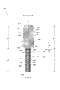
In some
embodiments, different materials may be used to form the different portions of
the implant.
The different materials may be configured to display and provide different
characteristics to
the different portions of the implant. In some embodiments, the different
portions of the
implant may include more filaments or more twists or have a different weave
pattern.
[0056] In some
embodiments, the implant includes a reinforcing fiber or a
plurality of reinforcing fibers. The reinforcing fiber or fibers may be
disposed at specific
locations or extend along a particular direction to provide different
characteristics to the
different portions of the implant.
[0057] In some
embodiments, the implant includes flat or planar sheets of
material. The sheets of material may have different pore quantities or
distributions to provide
different characteristics at different portions of the implant. In some
embodiments, the
implant may include laminated materials. For example, a mesh material may be
coupled to
or otherwise disposed adjacent to a sheet material. Additionally, one sheet
material may be
coupled to or otherwise disposed adjacent to another sheet material.
16
CA 02904058 2015-09-03
WO 2014/165211
PCT/US2014/024804
[0058] In some
embodiments, a portion or portions of the implant may be
weakened to provide different characteristics to different portions of the
implant. For
example, in some embodiments, portions of the implants may be notched, scored,
or shaved
(or apertures may be formed) to introduce weakness or to weaken different
portions of the
implant.
[0059] In some
embodiments, the implant includes a sheet of material that has a
property or a mechanical parameter that varies. For example, in one
embodiment, the sheet
of material has a property or biomechanical property, such as ability to
stretch, of one value
at a first location on the sheet of material and has the property or
mechanical parameter of
another value at different location on the sheet of material. In some
embodiments, the
property or mechanical parameter varies along a length of the sheet of
material. In some
embodiments, the property or mechanical parameter is the stiffness of the
material, the ability
to flex or stretch, or any other property or mechanical parameter.
[0060] In some
embodiments, the varying of the property or mechanical
parameter is accomplished by varying the knit pattern of the sheet of
material. In other
words, in some embodiments, the single sheet of material may have different
properties or
mechanical parameter at different locations because of or at least in part
because of different
knit patters or knit densities at the different locations along the sheet of
material. For
example, the sheet of material may have a first knit pattern at a first
location on the sheet of
material and a second knit pattern at a second location on the sheet of
material.
[0061] In some
embodiments, the weight or density of the sheet of material is
greater than or equal to 30 grams per square meter (g/m2). For example, the
weight of the
material may be between 30 and 40 g/m2. In other embodiments, the weight of
the material is
greater than 40 g/m2. In yet other embodiments, the weight of the material is
less than 30
g/m2. In some embodiments, the weight of the material varies at different
locations on the
sheet of material. For example, in some embodiments, the weight of the
material may be
greater than 30 g/m2 at one location and less than 30 g/m2 at another
location.
[0062] FIG. 2 is a
perspective view of a first flap 248 of a medical implant 200 for
placement over an anterior wall of a vagina inside a patient's body. The first
flap 248 can
include a first portion 202, a second portion 204 and a transition region 206.
17
CA 02904058 2015-09-03
WO 2014/165211
PCT/US2014/024804
[0063] The first
portion 202 defines a first side 208, a second side 210, a proximal
portion 212 and a distal portion 214. The proximal portion 212 can be attached
to or extend
from the transition region 206 of the first flap 248. The distal portion 214
can be configured
to be attached to a first bodily tissue. In some embodiments, the first bodily
tissue can be a
sacrum inside a patient's body. The first portion 202 defines a length L5
along the first side
208 extending from the proximal portion 212 to the distal portion 214. The
first portion 202
defines a length L6 along the second side 210 extending from the proximal
portion 212 to the
distal portion 214. In some embodiments, the length L5 can be equal to the
length L6. The
first portion 202 defines a width W3 extending between the first side 208 and
the second side
210. In some embodiments, the width W3 can remain constant from the proximal
portion
212 to the distal portion 214.
[0064] In some
embodiments, the first flap 248 can be configured so that the first
portion 202 can be attached to the sacrum or tissues proximate the sacrum and
the remaining
portion of the first flap 248 can be attached to the anterior vaginal wall in
order to provide
support to the anterior vaginal wall.
[0065] The first
bodily tissue exhibits a definite biomechanical behavior in a
defined set of physical conditions. The first portion 202 can be configured to
define a set of
biomechanical attributes or biomechanical properties so as to emulate the
biomechanical
behavior of the first bodily tissue, where at least a portion of the first
portion 202 is required
to be attached, in the defined set of physical conditions. The biomechanical
attributes can be
defined by a first set of values of respective biomechanical parameters
associated with the
biomechanical attributes. For example, in some embodiments, the biomechanical
attribute
can be elasticity and a corresponding biomechanical parameter can be modulus
of elasticity,
which can be defined by a numerical value. In some embodiments, the
biomechanical
attribute can be stiffness. In some embodiments, the biomechanical attribute
can be strength.
In some embodiments, the biomechanical attribute can be resistance to creep.
In various
embodiments, the biomechanical attributes of the first portion 202 can be
defined by defining
one or more of shape, size, fabrication method or structure, profile, knit
structure, pore size,
material of fabrication, and the like. In some embodiments, for example, the
congruence
between the biomechanical behavior of the first bodily tissue and the first
portion 202 can be
achieved by varying the shape of the first portion 202. For example, the first
portion 202 can
18
CA 02904058 2015-09-03
WO 2014/165211
PCT/US2014/024804
have a square, rectangular, triangular or any other shape, which can
facilitate the first portion
202 in closely equating the biomechanical behavior of the first bodily tissue.
100661 In some
embodiments, the values of the biomechanical attributes of the
first portion 202 can be defined by a first type of knit structure 216. In
some embodiments,
the first type of knit structure 216 can be defined by a first type of
knitting pattern 218. In
some embodiments, the first type of knit structure 216 can be defined by
weaving the knit
with a required and defined tension. For example, the first type of knitting
pattern 218 can be
woven tightly or loosely to define a required type of knitting pattern. In
some embodiments,
the first type of knitting pattern 218 characterized by biomechanical
properties of high elastic
modulus and stiffness can hold bodily tissue such as a vaginal tissue in the
correct anatomical
location. In some embodiments, the first type of knit structure 216 can be
defined by a first
type of pore construct 220. The first type of pore construct 220 includes a
plurality of pores
222. The first type of pore construct 220 can be fabricated to define
biomechanical attributes
conforming to biomechanical behavior of the first bodily tissue by varying the
first type knit
structure 216, and the pore construct 220. The different ways of achieving the
desirable
biomechanical attributes for the first portion 202 of the first flap 248 can
be used in isolation
or in combination. In some embodiments, the knit structure includes knitting,
weaving,
braiding, twisting, tying, or any combination thereof. Utilizing materials
with properties that
change over time, such as biodegradable materials, can adjust specific
biomechanical
properties over time. Coatings on specific portions of the implant may be used
to influence
the biomechanical properties, for example but reducing the elasticity of the
coated portion.
100671 It must be
appreciated that though the above ways of defining the required
biomechanical attributes are used for mesh-based implants 200 including a knit
pattern, the
implant 100 can be fabricated as a planar structure. In such embodiments, the
biomechanical
attributes of the first portion 202 of the first flap 248 can be defined for
example by the
material used in fabrication of the first portion 202, shape and size of the
portion, and the like
without limitations. For example, a rigid medical grade polymer can be used
for fabricating
the first portion 202 thereby defining the biomechanical attribute of rigidity
for the first
portion 202 to a desired value.
19
CA 02904058 2015-09-03
WO 2014/165211
PCT/US2014/024804
[0068] The second
portion 204 defines a first side 224, and a second side 226, a
proximal portion 228 and a distal portion 230. The distal portion 230 can be
attached to or
extend from the transition region 206 of the first flap 248. The proximal
portion 228 can be
configured to be attached to the second bodily tissue. In some embodiments,
the second
bodily tissue can be an anterior vaginal wall inside a patient's body.
[0069] The second
portion 204 defines a length L7 along the first side 224
extending from the proximal portion 228 to the distal portion 230. The second
portion 204
defines a length L9 along the second side 226 extending from the proximal
portion 228 to the
distal portion 230. In some embodiments, the length L7 can be different from
the length L9.
The second portion 204 defines a width W4 extending between the first side 224
and the
second side 226. In some embodiments, as illustrated, the width W4 can differ
from the
proximal portion 228 to the distal portion 230. In some embodiments, the
second portion 204
is fabricated such that the width W4 is greater than the width W3 of the first
portion 202. In
some embodiments, the second portion 204 can define a trapezoidal shape such
that the width
W4 at the proximal portion 228 is substantially greater than the width W4 at
the distal
portion. The second portion is configured to be attached and provide support
to a second
bodily tissue.
[0070] The second
bodily tissue exhibits a definite biomechanical behavior in a
defined set of physical conditions. The behavior exhibited by the second
bodily tissue can be
different than the behavior exhibited by the first bodily tissue. The second
bodily tissue can
be configured to define a set of biomechanical attributes or biomechanical
properties so as to
emulate the biomechanical behavior of the second bodily tissue in the defined
set of physical
conditions. The biomechanical attributes can be defined by a second set of
values of
respective biomechanical parameters associated with each of the biomechanical
attributes.
The second set of values can be different from the first set of values.
Consequently, the
second portion 204 may be defined to exhibit values of the biomechanical
attributes, different
than the values of the biomechanical attributes of the first portion 202, in
accordance with the
second bodily tissue where at least a portion of the second portion 204 of the
first flap 248
may be attached. It must be appreciated that in some embodiments, only one or
more but not
all of the first set of values biomechanical attributes and the second set of
values differ in
terms of their values of parameters defining the respective attributes. For
example, the
modulus of elasticity may be same for the first portion 202 and the second
portion 204 but
CA 02904058 2015-09-03
WO 2014/165211
PCT/US2014/024804
any other parameter for other attribute such as resistance to creep may be
different. In some
other embodiments, all the attributes of the first portion 202 and the second
portion 204 may
differ in terms of their values of parameters defining the respective
attributes. The values of
the various parameters provide mathematical measures of the respective
parameters.
[0071] In some
embodiments, the second set of values associated with the
biomechanical attributes can be different along different directions for the
same fixed set of
physical conditions even for the same attribute. For example, in some
embodiments, a value
of a parameter P defining an attribute T along a first direction B1 can be
different from a
value of the parameter P defining the attribute T along a second direction B2.
In some
embodiments, the first direction B1 can be a longitudinal direction and the
second direction
B2 can be a transverse direction. Therefore, a parameter may differ in its
value in different
directions, in some embodiments. For example, modulus of elasticity of various
portions of
the first flap 248 may differ in different directions, in some embodiments.
This may be
important to match the biomechanical behavior of bodily tissues that may
exhibit different
levels of elasticity in different directions. Also, the second set of values
associated with the
biomechanical attributes can vary with a variation in the set of physical
conditions. However,
in some embodiments, the physical conditions may vary and measurement of the
biomechanical behavior and the attributes may accordingly be calibrated so as
to compare the
various values associated with the various attributes in light of the required
characteristics at
the required locations. In some embodiments, the first direction B1 and the
second direction
B2 do not align along the axes of the implant. Additionally, in some
embodiments, B1 and
B2 are not disposed orthogonal or perpendicular to one another.
[0072] In some
embodiments, the biomechanical attributes can include elasticity
and a corresponding biomechanical parameter can be modulus of elasticity. In
some
embodiments, the biomechanical attribute can be viscoelasticity. In some
embodiments, the
biomechanical attribute can be viscohyperelasticity. In some
embodiments, the
biomechanical attribute can be anisotropicity. In various embodiments, the
biomechanical
attributes of the second portion 204 can be defined by defining one or more of
shape, size,
fabrication method or structure, profile, knit structure, pore size, material
of fabrication, and
the like. In some embodiments, for example, the congruence between the
biomechanical
behavior of the second bodily tissue and the second portion 204 can be
achieved by varying
the shape of the second portion 204. For example, the trapezoidal shape of the
second
21
CA 02904058 2015-09-03
WO 2014/165211
PCT/US2014/024804
portion 204 can conform to the shape of the second bodily tissue such as thc
anterior vaginal
wall. The trapezoidal shape can be provided to the second portion 204 to
emulate a taper of
an outer vaginal canal. In some embodiments, at the widest end, the width W4
can range
from 21.7 ¨ 55mm. In some embodiments, at the narrowest end, the width W4 can
range
from 18.7 ¨ 37mm. The lengths L6 or L8 of the trapezoid can range from 40.8 ¨
95 mm
based on the linear length of the vagina.
[0073] In some
embodiments, the values of the biomechanical attributes of the
second portion 204 can be defined by a second type of knit structure 232. In
some
embodiments, the second type of knit structure can be defined by a second type
of knitting
pattern 234. In some embodiments, the second type of knit structure 232 can be
defined by
weaving the knit with a required and defined tension. For example, the
anterior vaginal wall
shows biomechanical behavior of anisotrophicity, with bias toward more
elongation along a
transverse direction such as the direction B 1, therefore, the second type of
knitting pattern
234 can be selected so as to be more elastic along a longitudinal direction
such as the
direction B2 as compared to the transverse direction.
[0074] In some
embodiments, the second type of knit structure 232 can be defined
by a second type of pore construct 236. In some embodiments, the second type
of pore
construct 236 is different from the first type of pore construct 220. The
second type of pore
construct 236 includes a plurality of pores 238. In some embodiments, the
difference in pore
construct for the first portion 202 and the second portion 204 can be achieved
by weaving or
knitting a mesh with different pore sizes. In some embodiments, the difference
in pore
constructs 220 and 236 of the first portion 202 and the second portion 204 can
be achieved by
extruding or knitting a single pore size mesh and heat setting the pores to
set a different pore
size for the first portion 202 and the second portion 204 as illustrated and
described by later
figures. The second pore construct 236 can define the second set of values of
the
biomechanical attributes of the second portion 204. In an embodiment, the
second pore
construct 236 can define larger pore sizes as compared to the remaining
portion of the first
flap 248. In some embodiments, the second pore construct 236 can be fabricated
to exhibit
biomechanical attributes of high flexibility and elongation to a particular
strain level and high
stiffness after a particular stain level is reached. Such a strain behavior
closely emulates the
biomechanical behavior of the anterior vaginal wall.
22
CA 02904058 2015-09-03
WO 2014/165211
PCT/US2014/024804
[0075] In somc
embodiments, one or more of the biomechanical attributes can be
defined by a material used for fabricating the second portion 204. For
example, a viscoelastic
medical grade polymer can be used for fabricating the second portion 204
thereby defining a
value for the biomechanical attribute of viscoelasticity for the second
portion 204. In some
embodiments, an anisotropic medical grade polymer (or the fabrication of such
material) can
be used for achieving a desired value of anisotropicity. In some embodiments,
a creep
resistant medical grade polymer can be used for achieving a desired value of
creep resistance.
[0076] Generally,
the anterior vaginal wall can be viscohyperelastic. The second
portion therefore can be defined such that it exhibits high viscoelasticity.
In some
embodiments, the biomechanical parameters can have different values in
different directions.
For example, the biomechanical parameters may have different values in the
first direction
B1 than in the second direction B2.
[0077] The values
of the biomechanical parameters defining the biomechanical
behavior of the anterior vaginal wall may vary under different load
conditions. For example,
in some embodiments, the stiffness of the anterior vaginal wall at a low
strain along the
direction B1 can range from 0.431 ¨ 4.15 MegaPascal (MPa). In some
embodiments, the
stiffness of the anterior vaginal wall at a high strain along the direction B1
can range from
5.15 ¨ 17.28 MPa, In some embodiments, the stiffness the anterior vaginal wall
at the low
strain along the direction B2 can range from 0.385 ¨ 0.415 MPa. In some
embodiments, the
stiffness of the anterior vaginal wall along the direction B2 at the high
strain can range from
0.370 ¨ 0.61 MPa. The stiffness behaviors of the anterior vaginal wall are
further explained
in detail in conjunction with FIGS. 6A and 6B. Therefore, in some embodiments,
the second
portion 204 of the first flap 248 can be fabricated so as to define a set of
values of the
biomechanical parameter of stiffness that can conform to the values defining
the
biomechanical behavior of the anterior vaginal wall under similar load
conditions.
[0078] The first
flap 248 further includes the transition region 206 as mentioned
above. The transition region 206 defines a proximal portion 240 and a distal
portion 242.
The proximal portion 240 can be coupled to or extend from the distal portion
230 of the
second portion 204. The distal portion 242 can be coupled to or extend from
the proximal
portion 212 of the first portion 202. In some embodiments, the transition
region 206 may
define a third type of knit structure 244 that monolithically joins the first
portion 202 and the
second portion 204. In some embodiments, the third knit structure 244 may
define a third
23
CA 02904058 2015-09-03
WO 2014/165211
PCT/US2014/024804
type of pore construct 246. In some embodiments, the first flap 248 can be
formed by
suturing together the first portion 202 and the second portion 204. In such
cases, the
transition region 206 includes sutures tying the first portion 202 and the
second portion 204.
[0079] FIG. 3 is a
perspective view of a second flap 340 of the medical device
200 for placement over a posterior wall of a vagina inside a patient's body.
The first flap 248
and the second flap 340 can collectively form the medical implant 200. The
second flap 340
can include a first portion 302, a second portion 304 and a transition region
306.
[0080] The first
portion 302 defines a first side 308, a second side 310, a proximal
portion 312 and a distal portion314. The proximal portion 312 can be attached
to or extend
from the transition region 306 of the second flap 340. The distal portion 314
can be
configured to be attached to a first bodily tissue. In some embodiments, the
first bodily tissue
can be a sacrum or tissues proximate the sacrum. The first portion 302 defines
a length L9
along the first side 308 extending from the proximal portion 312 to the distal
portion 314.
The first portion 302 defines a length L10 along the second side 310 extending
from the
proximal portion 312 to the distal portion 314. In some embodiments, the
length L9 can be
equal to the length Lb. The first portion 302 defines a width W5 extending
between the first
side 308 and the second side 310. In some embodiments, the width W5 can remain
constant
from the proximal portion 312 to the distal portion 314.
100811 In some
embodiments, the second flap 340 can be configured so that the
first portion 302 can be attached to the sacrum and the remaining portion of
the implant 300
can be attached to the posterior vaginal wall in order to provide support to
the posterior
vaginal wall. The first bodily tissue exhibits a definite biomechanical
behavior in a defined
set of physical conditions. The first portion 302 can be configured to define
a set of
biomechanical attributes or biomechanical properties so as to emulate the
biomechanical
behavior of the first bodily tissue, where at least a portion of the first
portion 302 is required
to be attached, in the defined set of physical conditions. The first portion
302 of the second
flap 340 can be fabricated similar to the first portion 202 of the first flap
248 as described in
FIG. 2. The attributes of the first portion 302 of the second flap 340 can be
defined in a
manner similar to the attributes of the first portion 202 of the first flap
248.
[0082] The second
portion 304 defines a first side 316, and a second side 318, a
proximal portion 320 and a distal portion 322. The distal portion 322 can be
attached to or
24
CA 02904058 2015-09-03
WO 2014/165211
PCT/US2014/024804
extend from the transition region 306 of the second flap 340. The proximal
portion 320 can
be configured to be attached to a third bodily tissue. In some embodiments,
the third bodily
tissue can be the posterior vaginal wall.
[0083] The second
portion 304 defines a length L11 along the first side 316
extending from the proximal portion 320 to the distal portion 322. The second
portion 304
defines a length L12 along the second side 318 extending from the proximal
portion 320 to
the distal portion 322. In some embodiments, the length L11 can be different
from the length
L12. The second portion 304 defines a width W6 extending between the first
side 316 and
the second side 318. In some embodiments, as illustrated, the width W6 can
differ from the
proximal portion 320 to the distal portion 322. In some embodiments, the
second portion 304
is fabricated such that the width W6 is greater than the width W5 of the first
portion 302. In
some embodiments, the second portion 304 can define a trapezoidal shape such
that the width
W6 at the proximal portion 320 is substantially greater than the width W6 at
the distal portion
322.
[0084] The third
bodily tissue exhibits a definite biomechanical behavior in a
defined set of physical conditions. The behavior exhibited by the third bodily
tissue can be
different than the behavior exhibited by the first bodily tissue or the second
bodily tissue.
The third bodily tissue can be configured to define the biomechanical
attributes or
biomechanical properties so as to emulate the biomechanical behavior of the
third bodily
tissue in the defined set of physical conditions. The biomechanical attributes
can be defined
by a third set of values of respective biomechanical parameters associated
with the
biomechanical attributes. In some embodiments, the third set of values can be
different from
the first set of values of the biomechanical attributes. In some embodiments,
the third set of
values can be different from the second set of values of the biomechanical
attributes.
Consequently, the second portion 304 may be defined to exhibit biomechanical
attributes,
different than the biomechanical attributes of the first portion 302, in
accordance with the
third bodily tissue where at least a portion of the second portion 304 may be
attached. The
second portion 304 may be defined to exhibit biomechanical attributes,
different than the
biomechanical attributes of the first portion 202 from the first flap 248.
[0085] In some
embodiments, the third set of values associated with the
biomechanical attributes can be different along different directions for the
same fixed set of
physical conditions even for the same attribute. For example, in some
embodiments, a value
CA 02904058 2015-09-03
WO 2014/165211
PCT/US2014/024804
of a parameter P defining an attribute T along a first direction Cl can be
different from a
value of the parameter P defining the attribute T along a second direction C2.
In some
embodiments, the first direction Cl can be a longitudinal direction and the
second direction
C2 can be a transverse direction. Also, the third set of values associated
with the
biomechanical attributes can vary with a variation in the set of physical
conditions. In some
embodiments, the third set of values can be different from the first set of
values associated
with the biomechanical attributes under the same fixed set of physical
conditions.
[0086] In some
embodiments, the biomechanical attribute can include elasticity
and a corresponding biomechanical parameter can be modulus of elasticity. In
some
embodiments, the biomechanical attribute can be viscoelasticity. In some
embodiments, the
biomechanical attribute can be viscohyperelasticity. In some
embodiments, the
biomechanical attribute can be anisotropicity. In various embodiments, the
biomechanical
attributes of the second portion 304 can be defined by defining one or more of
shape, size,
fabrication method or structure, profile, knit structure, pore size, material
of fabrication, and
the like. In some embodiments, for example, the congruence between the
biomechanical
behavior of the third bodily tissue and the second portion 304 can be achieved
by varying the
shape of the second portion 304. For example, the trapezoidal shape of the
second portion
304 can conform to shape of the posterior vaginal wall.
[0087] In some
embodiments, the values of the biomechanical attributes of the
second portion 304 can be defined by a fourth type of knit structure 324. In
some
embodiments, the fourth type of knit structure 324 can be defined by a fourth
type of knitting
pattern 326. In some embodiments, the fourth type of knit structure 324 can be
defined by
weaving the knit with a required and defined tension. For example, the
posterior vaginal wall
shows biomechanical behavior of anisotrophicity, with biasness (or being
biased) toward
more elongation along the direction Cl, therefore, the fourth type of knitting
pattern 326 can
be selected to be more elastic along the direction C2 as compared to the
direction Cl.
[0088] In some
embodiments, the fourth type of knit structure 324 can be defined
by a fourth type of pore construct 328. In some embodiments, the fourth type
of pore
construct 328 is different from the first type of pore construct 220 and the
second type of pore
construct 236 of FIG. 2. The fourth type of pore construct 328 includes a
plurality of pores
330. The difference in pore construct for the first portion 302 and the second
portion 304 can
26
CA 02904058 2015-09-03
WO 2014/165211
PCT/US2014/024804
bc achieved as described in FIG. 2. The fourth type of pore construct 328 can
be configurcd
to conform to biomechanical properties of the posterior vaginal wall.
100891 In some
embodiments, one or more of the biomechanical attributes can be
defined by the third set of values associated with the biomechanical
attributes for the second
portion 304. In an embodiment, the third set of values may be defined by a
material used for
fabricating the second portion 304. For example, a viscoelastic medical grade
polymer can
be used for fabricating the second portion 304 thereby defining a desired
value of
viscoelasticity for the second portion 304. In some embodiments, an
anisotropic medical
grade polymer can be used for achieving a desired value of anisotropicity. In
some
embodiments, a creep resistant medical grade polymer can be used for achieving
a desired
value of creep resistance.
100901 The
posterior vaginal wall can have high visco-hyper elasticity. Therefore,
the second portion 304 can have a high value of viscohyperelasticity under a
fixed set of
stress conditions. In some embodiments, the value of the biomechanical
parameter can be
different for the first direction Cl and the second direction C2 for the
posterior vaginal wall.
The value of the biomechanical parameter would be different for a set of high
load conditions
and a set of low load conditions for the posterior vaginal wall. For example,
in some
embodiments, the stiffness of the posterior vaginal wall at a low strain along
the direction Cl
can range from 0.46 ¨ 0.98 MegaPascal (MPa). In some embodiments, the
stiffness of the
posterior vaginal wall at a high strain along the direction Cl can range from
2.49 ¨ 9.08 MPa.
In some embodiments, the stiffness of the posterior vaginal wall at the low
strain along the
direction C2 can range from 1.14 ¨ 1.46 MPa. In some embodiments, the
stiffness of the
posterior vaginal wall along the direction C2 at the high strain can range
from 2.39 ¨ 3.83
MPa. These values indicate an anisotropic behavior and stiffness variation
along different
directions and different physical conditions for posterior vaginal wall. The
stiffness
behaviors of the posterior vaginal wall are explained in detail by FIG. 6A and
6B. Therefore,
in some embodiments, the second portion 304 of the second flap 340 can be
fabricated so as
to define a set of values of the biomechanical parameter of stiffness that can
conform to the
values defining the biomechanical behavior of the posterior vaginal wall under
similar load
conditions. The second portion 304 can be fabricated such that the set of
values of the
biomechanical parameter can be different from the set of values for the same
biomechanical
parameter of the second portion 204 of the first flap 248.
27
CA 02904058 2015-09-03
WO 2014/165211
PCT/US2014/024804
[0091] The second
flap 340 further includes the transition region 306 as
mentioned above. The transition region 306 defines a proximal portion 332 and
a distal
portion 334. The proximal portion 332 can be coupled to or extend from the
distal portion
322 of the second portion 304. The distal portion 334 can be coupled to or
extend from the
proximal portion 312 of the first portion 302. In some embodiments, the
transition region
306 defines a fifth type of knit structure 336 that monolithically joins the
first portion 302 and
the second portion 304. The fifth type of knit structure 336 defines a fifth
pore construct 338.
In some embodiments, the third knit structure 244 may define a third type of
pore construct
246.
[0092] In some
embodiments, the second flap 340 can be made out of a single
strip of material. In some embodiments, the second flap 340 can be formed by
suturing
together the first portion 302 and the second portion 304. In such cases, the
transition region
306 includes sutures tying the first portion 302 and the second portion 304.
[0093] FIG. 4 is a
perspective view of a medical implant 400 including a plurality
of flaps for placement over the first bodily tissue, the second bodily tissue
and the third
bodily tissue inside a patient's body. The plurality of flaps may include a
first flap 402, a
second flap 404, and a third flap 406. The plurality of flaps can be joined
together at a
transition region 408 to form a Y-shaped implant as illustrated in the FIG.
4.In some
embodiments, there may not be any transition regions such as the transition
region 408 and
the first, second, and third flaps can directly be coupled with the use of
sutures or any other
coupler.
[0094] The first
flap 402 defines a proximal portion 420 and a distal portion 422.
The proximal portion 420 can be attached to or extend from the transition
region 408 of the
medical implant 400. The distal portion 422 can be configured to be attached
to the first
bodily tissue as described with reference to FIG. 2. The first flap 402 can be
configured to
define the first set of values corresponding to the biomechanical parameters
as explained in
FIG. 2 for emulating biomechanical behavior of first bodily tissue for example
the sacrum, or
tissue proximate the sacrum.
[0095] The second
flap 404 defines a proximal portion 424 and a distal portion
426. The proximal portion 424 can be attached to or extend from the transition
region 408 of
the medical implant 400. The distal portion 426 can be configured to be
attached to the
28
CA 02904058 2015-09-03
WO 2014/165211
PCT/US2014/024804
second bodily tissue as described with reference to FIG. 2. The second flap
404 can be
configured to define the second set of values corresponding to the
biomechanical parameters
as explained in FIG. 2 for emulating biomechanical behavior of the second
bodily tissue for
example, the anterior vaginal wall.
[0096] The third
flap 406 defines a proximal portion 428 and a distal portion 430.
The proximal portion 428 can be attached to or extend from the transition
region 408 of the
medical implant 400. The distal portion 430 can be configured to be attached
to the third
bodily tissue as described with reference to FIG. 3. The third flap 406 can be
configured to
define the third set of values corresponding to the biomechanical parameters
as explained in
FIG. 3 for emulating biomechanical behavior of third bodily tissue, for
example, the posterior
vaginal wall.
[0097] In some
embodiments, the first flap 402, the second flap 404 and third flap
406 can be fabricated independent of each other. The first flap 402, the
second flap 404 and
the third flap 406 can be tied together with a suture 432 at the transition
region 408 to form
the medical implant 400. In some embodiments, the three flaps 402, 404, and
406 exhibit
different biomechanical attributes owning to different biomechanical
properties of anatomical
locations that each of the three flaps 402, 404, and 406 are configured to be
attached to.
[0098] As mentioned
above, the biomechanical properties of the posterior wall of
vagina, the anterior wall of vagina and the sacrum or tissues proximate the
sacrum inside a
patient's body are different from each other; therefore in some embodiments
the flaps of the
medical implant 400 are fabricated with a pore construct and knit structure
that can closely
mimic biomechanical attributes of the anatomical locations inside the
patient's body. For
example, the first flap 402 can have a knit structure 410 similar to the first
knit structure 216
of the first portion 202 of the first flap 248 from FIG 2 so as to be
biomechanically congruent
with the first bodily tissue. The second flap 404 can have a knit structure
412 similar to the
second knit structure 232 of the second portion 204 of the first flap 248 from
FIG 2 so as to
be biomechanically congruent with the second bodily tissue. The third flap 406
can have a
knit structure 414 similar to the fourth knit structure 324 of the second flap
340 from FIG 3
so as to be biomechanically congruent with the third bodily tissue. Upon
placement, the first
flap 402, the second flap 404, and the third flap 406 can act as three
different arms that can be
configured to support the pelvic organs like the anterior vaginal wall, the
posterior vaginal
wall and the sacrum by attaching the implant 400 at three distinct bodily
locations. The three
29
CA 02904058 2015-09-03
WO 2014/165211
PCT/US2014/024804
arms can be movable with respect to one another to conform to thc shape of the
target
anatomical location of attachment inside the body. The three arms can take a
shape such as
linear/planar, curvilinear, curved, or any other shape.
[0099] In some
embodiments, for example, the medical implant 400 can be
formed by tying together the second portion 204 of the first flap 248, the
second portion 304
of the second flap 340 and the first portion 202 or 302 from either the first
flap 348 or the
second flap 340. In such
cases, the three portions mentioned above conform to the
biomechanical attributes of the second bodily tissue, the third bodily tissue
and the first
bodily tissue respectively.
[00100] In some embodiments, a Y-shaped mesh as illustrated in FIG. 4 may be
formed of two sheets of material. One sheet of material may be coupled, such
as via a suture
or other coupling member or mechanism, to the other sheet of material to form
a Y shape. In
some embodiments, one of the sheets of material may have properties or
mechanical
parameters that vary along a length of the sheet of material.
[00101] For example, in one embodiments, a first sheet of material may be
placed
in the body of the patient such that it extends from the sacrum (or tissues
proximate the
sacrum) of the patient to a vaginal wall of the patient (or tissue proximate a
vaginal wall),
such as the posterior vaginal wall of the patient. The portion of the first
sheet of material that
is coupled to the sacrum (or to tissue proximate the sacrum) may have a first
value for a
property or mechanical parameter. The portion of the first sheet of material
that is coupled to
the vaginal wall may have a different value for the property or mechanical
parameter. In
some embodiments, the property or mechanical parameter is stretchiness or
ability to stretch.
In some embodiments, the portion of the first sheet of material that is
coupled to the sacrum
(or to tissues proximate the sacrum) is less stretchy (has less ability to
stretch) than the
portion of the sheet of material that is coupled to the vaginal wall.
[00102] In some embodiments, the second sheet of material that is coupled to
the
first sheet of material is configured to be coupled to another vaginal wall,
such as an anterior
portion of the vaginal wall (or tissues proximate such vaginal wall). In some
embodiments,
the second sheet of material has the same value for the property or mechanical
parameter as
the portion of the first sheet of material that is coupled to the vaginal
wall.
CA 02904058 2015-09-03
WO 2014/165211
PCT/US2014/024804
1001031 In some embodiments, the first sheet of material may be rectangular,
square, or any other shape. In some embodiments, the first sheet of material
may be cut or
reshaped by the user or the physician. In some embodiments, the first sheet of
material is
rectangular in shape and is 20 cm by 52 cm in size. In other embodiments, the
first sheet of
material is larger than 20 cm by 52 cm. In yet other embodiments, the first
sheet of material
is smaller than 20 cm by 52 cm. In some embodiments, half of the first sheet
of material is
formed such that it has a first value for a property or mechanical parameter
and the other half
of the first sheet of material is formed such that it has a second value for
the property or
mechanical parameter. For example, in some embodiments, a 20 cm by 26 cm
portion of the
first sheet of material may have the first value for the property or
mechanical parameter and
another 20 cm by 26 cm portion of the first sheet of material may have the
second value for
the property or mechanical parameter. In other embodiments, more or less than
half of the
first sheet of material has the first value for the property or mechanical
parameter.
Accordingly, less or more than half of the first sheet of material has the
second value for the
property or mechanical parameter.
[00104] In some embodiments, the portion of the first sheet of material that
is
coupled to the vaginal wall of the patient may be formed of a knit structure
that has a high
elongation (or is configured to elongate or stretch) at loads in the range of
between 0 and 5
pounds of pressure. For example, the knit structure may be configured to
stretch when placed
under loads of 0 to 5 pounds per square cm. In other embodiments, the portion
of the first
sheet of material that is coupled to the vaginal wall of the patient may be
formed of a
structure that is configured to elongate at higher amounts of pressure.
[00105] In some embodiments, a portion of the first sheet of material is
formulated
or configured to promote tissue ingrowth. For example, in some embodiments,
the first
portion of the first sheet of material is configured to promote tissue
ingrowth. In some
embodiments, the second portion of the first sheet of material is configured
to promote tissue
ingrowth.
[00106] FIG. 5A is a perspective view of a medical implant 500 formed as a
tubular structure 502.
[00107] The tubular structure 502 of the medical implant includes a first
portion
504, a second portion 506, and a transition region 510. The transition region
510 is formed
31
CA 02904058 2015-09-03
WO 2014/165211
PCT/US2014/024804
from intersection of the first, and the second 504, and 506 of the tubular
structure 502 of the
medical implant 500. The medical implant 500 defines a proximal portion 512, a
distal
portion 514 and a lumen 516 extending from the proximal portion 512 to the
distal portion
214. The medical implant 500 defines a length L13 from the proximal portion
512 to the
distal portion 514. The medical implant includes the second portion 506 at the
proximal
portion 512 of the medical implant. The second portion can a first section 524
and a second
section 508 and two slits 518A and 518B extending laterally along the length
L13 of the
medical implant 500. In some embodiments, the proximal portion 512 includes
two slits
extending laterally along the length L13 and into the lumen 516 of the medical
implant 500.
The slits 518A and 518B can configure first section 524 as apart from the
second section 508
at a proximal end 534 of the medical implant 500. The medical implant 500 can
be
configured so that each of the first portion 504, the first section 524 and
the second section
508 can define a set of biomechanical attributes, which can be congruent with
the sacrum or
tissues proximate the sacrum, the anterior vaginal wall and the posterior
vaginal wall
respectively. The congruency can be achieved by any of the methods described
with
reference to FIGS. 2-3.
[00108] The first portion 504 can define a knit structure 520 formed of a pore
construct 522. In some embodiments, the pore construct 522 can define a pore
size so as to
accommodate values of the biomechanical attributes the first bodily tissue.
The first section
524 can define a knit structure 526 formed of a pore construct 528. In some
embodiments,
the pore construct 528 can define a pore size so as to accommodate values of
the
biomechanical attributes the second bodily tissue. The second section 508 can
define a knit
structure 530 formed of a pore construct 532. In some embodiments, the pore
construct 532
can define a pore size so as to accommodate values of the biomechanical
attributes the third
bodily tissue. In some embodiments, the knit structure 520 of the first
portion 504 is different
from the knit structure 526 and the knit structure 530 of the first section
524 and the second
section 508 of the second portion 506. In some embodiments, the knit structure
526 of the
first section 524 is different from the knit structure 530 of the second
section 508. In some
embodiments, the varying knit structure is formed by varying the knit
structure in the course
of a pore.
[00109] In some embodiments, the first portion 504 can be configured for
attaching
to the sacrum, the first section 524 to the anterior vaginal wall and the
second section 508 to
32
CA 02904058 2015-09-03
WO 2014/165211
PCT/US2014/024804
the posterior vaginal wall. In some embodiments, a value corresponding to a
biomechanical
parameter defining a biomechanical attribute of the first section 524
attaching to the anterior
vaginal wall is different from a value of the same biomechanical parameter of
the second
section 508 attaching to the posterior vaginal wall. For example, the value of
elasticity can
be different for the first section 524 and the second section 508 under
similar strain
conditions.
[00110] In some embodiments, the medical implant 500 can be formed from a
process of extrusion. The pore constructs 522, 528, and 532, in such cases can
be the same.
The medical implant can then be provided a heat treatment and different
portions of the
medical implant 500 can be heat set to different pore sizes. For example, the
pore construct
522 can remain in a closed position without application of heat as illustrated
in FIG. 5B. The
second portion 506 can be manually stretched to bring the medical implant 500
in an open
position as illustrated in FIG. 5C. This can increase a pore size of the pore
constructs 528
and 532. The pore construct 528 and the pore construct 532 can remain in an
open position
on application of heat over the first section 524 and the second section 508.
The first section
524 and the second section 508 may each be given heat treatment for setting
different pore
sizes so as to facilitate defining biomechanical attributes emulating
biomechanical behavior
of the anterior and posterior vaginal walls respectively.
[00111] In some embodiments, the medical implant 500 can be fabricated so that
the first portion 504 includes the knit structure 216 and the pore construct
220 as described
for the first flap 248, the second portion 506 includes the knit structure 232
and the pore
construct 236 as described for the first flap 248 and the third portion 508
includes the knit
structure 324 and the pore construct 328 as described for the second flap 340.
[00112] FIG. 6A is an exemplary graphical representation of relationship
between
stress applied on a vaginal tissue and resulting elongation in the vaginal
tissue due to the
applied stress. A medical implant can be fabricated to conform to the changes
resulting in the
vaginal tissue due to applied stress. The medical implant can be any of the
medical implants
200, 300, 400, and 500. The vaginal tissue is generally viscoelastic or
viscohyperelastic. The
vaginal tissue can experience large deformation under small loads as shown.
Therefore, in
some embodiments, the medical implant is configured to experience varying
levels of
deformation under varying loads. The vaginal tissue stress-strain or load vs
(or compared to)
elongation relationship can follow a non-linear curve as illustrated. The
curve has a first
33
CA 02904058 2015-09-03
WO 2014/165211
PCT/US2014/024804
linear phase at low loads / stresses. At low levels of load, the strains or
elongations are high
defining a low stiffness attribute of the implant. The curve includes a second
phase defined
by a transition phase after an inflection point where it transitions from one
linear phase to a
third phase such that the curve is sharper in the third phase. The third phase
defines a region
of relatively lower elongation even under an application of relatively higher
loads as
compared to the first phase. That is to say that that after the load increases
after a limit
defined by the inflection point, there is lesser elongation with every unit
change in load. This
defines a property of high stiffness of the implant at higher loads. The unit
change in
elongation with every unit change in load decreases thereafter till it reaches
a level that there
is almost negligible elongation in the implant even at increased loads. The
implant thus
behaves as a stiff member. Therefore, the medical implant can be configured to
define the
attributes congruent with the stress-strain or load vs (or compared to)
elongation relationship
of the vaginal tissue. In some embodiments, the portion of the medical implant
attaching to
the anterior or posterior portion of the vagina can be so configured that it
experiences large
deformations over small loads until the inflection point is achieved. As the
load is increased
beyond the inflection point, the medical implant portion attached to the
anterior vaginal wall
or posterior vaginal wall starts exhibiting high stiffness and very less
(negligible)
deformation.
[00113] As mentioned with respect to FIGS. 2 and 3 above, in some cases the
vaginal wall may be anisotropic in nature; therefore, it experiences different
elongations in
different directions. For example, the vaginal wall in a traverse direction is
generally more
elastic than in a longitudinal direction. Therefore, it may be desirable to
configure the
medical implant to exhibit values of the biomechanical attributes defined to
emulate the
varying elongation behavior of the vaginal wall in different directions. FIG.
6B is a graphical
representation of a comparison of an exemplary attribute, elongation, of the
vaginal tissue in
the transverse direction and the longitudinal direction. A shown, the
elongation of the
vaginal tissue in the transverse direction is much lesser than the elongation
in the longitudinal
direction.
[00114] Referring to the graphical representations of FIGS 6A-6B that depict
the
characteristics and behavior of a vaginal tissue, the portions of the medical
implant that are
attached to vaginal tissues are configured to behave accordingly in order to
emulate the
behavior of the vaginal tissues such as the anterior and posterior vaginal
walls. For example,
34
CA 02904058 2015-09-03
WO 2014/165211
PCT/US2014/024804
in some embodiments, the portions of the implant that attach to the anterior
vaginal wall can
be configured to define different stiffness characteristics at different
levels of loads on the
implant portions. Similarly, the implant portions that attach to the posterior
vaginal wall can
be configured to define different characteristics in different directions such
as the transverse
direction and the longitudinal direction and for different load values.
[00115] In some embodiments, stiffness at the low strain or deformation phase
can
range from 0.431 ¨ 4.15 MPa for the anterior vaginal wall in the longitudinal
direction. In
some embodiments, stiffness at the high strain or deformation phase can range
from 5.15 ¨
17.28 MPa for the anterior vaginal wall in the longitudinal direction. In some
embodiments,
stiffness at a low strain or deformation phase can range from 0.46 ¨ 0.98 MPa
for the
posterior vaginal wall in the longitudinal direction. In some embodiments,
stiffness at a high
strain or deformation phase can range from 2.49 ¨ 9.08 MPa for the posterior
vaginal wall in
the longitudinal direction. In some embodiments, stiffness at the low strain
or deformation
phase can range from .385 ¨ .415 MPa for the anterior vaginal wall in the
traverse direction.
In some embodiments, stiffness at the high strain or deformation phase can
range from .370 ¨
.61 MPa for the anterior vaginal wall in the traverse direction. In some
embodiments,
stiffness at the low strain or deformation phase can range from 1.14 ¨ 1.46
MPa for the
posterior vaginal wall in the traverse direction. In some embodiments,
stiffness at the high
strain or deformation phase can range from 2.39 ¨ 3.83 MPa for the posterior
vaginal wall in
the traverse direction. The values of stiffness detailed here are a guide for
vaginal tissues
generally. The values of stiffness can be different depending on disease
state, age, or any
other influencing factor. Therefore, the implant can be made / designed
accordingly to be
configured for emulating the biomechanical behavior of the vaginal tissue in
accordance with
the desired characteristic behavior.
[00116] FIG. 7 is a perspective view of the medical implant 200, including the
first
flap 248 and the second flap 340 of FIG. 2 and FIG. 3 respectively, placed
inside a patient's
body, in accordance with an embodiment of the invention. The body portions of
the patient
such as the vagina V, the anterior vaginal wall AVW, the posterior vaginal
wall PVW, a
urethra U, and the sacrum S are illustrated in FIG. 7. FIG. 8 illustrates a
method 800 for
placing an implant in a patient's body. The method 800 is described below in
conjunction
with FIGS. 2, 3, 4, 5, and 7-9. The medical implant 200 including the first
flap 248 and the
second flap 340 is used as an exemplary embodiment to illustrate and discuss
the method
CA 02904058 2015-09-03
WO 2014/165211
PCT/US2014/024804
800. However, it must be appreciated that other implants such as the medical
implant 400
and the medical implant 500 as discussed above can also be employed equally.
[00117] The method 800 includes inserting the first flap 248 of the medical
implant
200 inside the body at step 802. In some embodiments, the first flap 248 can
be inserted
inside the patient's body through a laparoscopic approach. In some
embodiments, the
method 800 includes creating an abdominal incision for delivering the medical
implant inside
the body laparoscopically.
[00118] The method 800 further includes attaching the first portion 202 of the
medical implant 200 at the sacrum S inside the patient's body. The first
portion 202 can be
configured to define the biomechanical attributes so as to emulate the
biomechanical behavior
of the sacrum S in a defined set of physical conditions. The biomechanical
attributes can be
defined by the first set of values of respective biomechanical parameters
associated with the
biomechanical attributes.
[00119] The method 800 further includes attaching the second portion 204 of
the
first flap 248 to the anterior vaginal wall AVW at step 804. The anterior
vaginal wall AVW
is known to exhibit properties of viscoelasticity, anisotropy, and
viscohyperelasticity. The
second portion 204 can be configured to emulate the biomechanical behavior of
the anterior
vaginal wall AVW and define viscoelasticity, anisotropy, and
viscohyperelasticity. The
second portion 204 can define the biomechanical attributes that can be defined
by the second
set of values of respective biomechanical parameters associated with the
biomechanical
attributes as explained by way of FIG. 2. The first flap 248 is configured so
that the first set
of values is different from the second set of values. The difference in values
can be attributed
to the difference in the biomechanical behavior of the sacrum S and the
anterior vaginal wall
AVW. In some embodiments, the portion attaching to the anterior wall is formed
monolithically with the first portion 202 and the transition region 206 as
discussed above. It
must be appreciated that any conventionally known or practiced methods or
devices may be
used for attaching the medical implant at any location inside the patient's
body.
[00120] The method 800 further includes placing the second flap 340 of the
medical implant 200 over the posterior vaginal wall PVW at step 806 as
described below.
[00121] The first portion 302 can be configured to define the biomechanical
attributes so as to emulate the biomechanical behavior of the sacrum S in a
defined set of
36
CA 02904058 2015-09-03
WO 2014/165211
PCT/US2014/024804
physical conditions. The biomechanical attributes can be defined by the first
set of values of
respective biomechanical parameters associated with the biomechanical
attributes. The first
set of values for the first portion 202 from the first flap 248 can be same as
those for the first
portion 302 from the second flap 340.
[00122] The posterior vaginal wall PVW is known to exhibit properties of
viscoelasticity, anisotropy, and viscohyperelasticity. The second portion 304
can be
configured to emulate the biomechanical behavior of the posterior vaginal wall
PVW and
define visco-elasticity, anisotropy, and viscohyperelasticity. The second
portion 304 can
define the biomechanical attributes that can be defined by the third set of
values of respective
biomechanical parameters associated with the biomechanical attributes as
explained by way
of FIG. 3. The second portion 304 can be configured to define the
biomechanical attributes
congruent to the biomechanical behavior of the posterior vaginal wall. The
medical implant
200 can be fabricated so that second flap 340 is configured to have the third
set of values
corresponding to the biomechanical attributes to be different from the second
set of values
corresponding to the first flap 248. Therefore, the second portion 204 of the
first flap 248
used for attaching to the anterior vaginal wall AVW defines a different set of
values
corresponding to a biomechanical parameter than the set of values
corresponding to the same
biomechanical parameter for the second portion 302 of the second flap 340
attaching to the
posterior vaginal wall. In this way, the properties of the first flap 248 and
the second flap 340
are different and congruent with respect to the portions the first flap 248
and the second flap
340 are attached to. In some embodiments, the portion attaching to the
posterior vaginal wall
PVW is formed monolithically with the first portion 302 and the transition
region 306 as
discussed above. It must be appreciated that any conventionally known or
practiced methods
or devices may be used for attaching the medical implant at any location
inside the patient's
body. In some embodiments, the tow flaps can be independent from each other
and may
collectively enable the medical implant 200 in emulating biomechanical
behavior of the
anterior vaginal wall AVW, posterior vaginal wall PVW and the sacrum S inside
a patient's
body.
[00123] In some
embodiments, the method 800 further includes cutting an
unwanted portion of the medical implant 200 after placing in the body. In some
embodiments, the method 800 further includes closing the abdominal incision or
any other
incision created for method 800.
37
CA 02904058 2015-09-03
WO 2014/165211
PCT/US2014/024804
1001241 In some embodiments, the method 800 can be used for treatment of a
pelvic floor disorder, in accordance with an embodiment of the present
invention. The
implant can be a dual knit mesh. The dual knit mesh material can be a polymer
mesh, a
polypropylene material, a bio-absorbable material, or any other preferred
material. The knit
structure defined by each of the implants 200, 400, and 500 can be any knit
structure that
emulates the biomechanical properties of the vagina in the wide body region
and provides
stiffness in the stem region. The implant can be sold as a separate dual knit
mesh and a
standard mesh. The implant can be used for vaginal prolapse to suspend the
vagina to the
sacral promontory or the sacrum after a hysterectomy termed as Sacrocolpopexy
or any other
disorders. The implant can be placed into the body by laparoscopic or any
other means.
[00125] In some embodiments, an implant includes a first flap and a second
flap.
The first flap has a first portion configured to be attached proximate a
sacrum; a second
portion configured to be attached to an anterior vaginal wall; and a
transition region lying
between the first portion and the second portion. The second flap includes a
portion
configured to be attached to a posterior vaginal wall. A value corresponding
to a
biomechanical parameter defining a biomechanical attribute of the portion of
the first flap
attaching to the anterior wall is different from a value of the biomechanical
parameter
defining the biomechanical attribute of the portion of the second flap
attaching to the
posterior wall.
[00126] In some embodiments, the implant may be configured to help suspend or
provide support to a portion of the body of the patient. For example, the
implant may be used
to provide support to a vagina of a patient. In other embodiments, the implant
is configured
to suspend or provide support to other portions of the body, such as a portion
of the
gastrointestinal tract of the patient, a bladder of the patient, or a rectum
of the patient.
[00127] In some embodiments, an implant includes a first end portion, a second
end portion and a body in between the ends. The first end portion has a
biomechanical
attribute that is different in value than the same biomechanical attribute at
the second end
portion.
[00128] In some
embodiments, the first portion of the first flap defines a first type
of knit structure. In some embodiments, the second portion of the first flap
defines a second
type of knit structure. In some embodiments, the first type of knit structure
includes pores
38
CA 02904058 2015-09-03
WO 2014/165211
PCT/US2014/024804
that are smaller in cross sectional profile than the pores in the second type
of knit structure.
In some embodiments, a width of the second portion is substantially more or
greater than a
width of the first portion of the first flap. In some embodiments, the second
portion includes
a proximal end and a distal end, the distal end being proximate the transition
region, wherein
the width of the second portion varies from the proximal end to the distal end
of the second
portion. In some embodiments, the varying second width along the second
portion defines a
trapezoidal shape of the second portion. In some embodiments, the transition
region defines
a third type of knit structure. In some embodiments, each of the first and the
second flaps
defines a planar shape and are configured to be attached separately to bodily
locations. In
some embodiments, each of the value of the biomechanical parameter defining
the
biomechanical attribute of the portion of the first flap attaching to the
anterior wall and the
value of the biomechanical parameter defining the biomechanical attribute of
the portion of
the second flap attaching to the posterior wall is different from a value of
the biomechanical
parameter of the first portion attaching proximate the sacrum.
1001291 In some embodiments, the biomechanical attribute is elasticity and the
biomechanical parameter is a modulus of elasticity. In some
embodiments, the
biomechanical attribute is viscoelasticity. In some embodiments, the
biomechanical attribute
is viscohyperelasticity. In some embodiments, the biomechanical attribute is
anisotropicity.
In some embodiments, the biomechanical attribute is resistance to creep. In
some
embodiments, the biomechanical attribute is stiffness.
[00130] In some embodiments, the second flap includes a first portion defining
a
width and configured to be attached proximate the sacrum; a second portion
defining a width
and configured to be attached to the posterior vaginal wall; and a transition
region lying
between the first portion and the second portion and monolithically joining
the first portion
and the second portion.
[00131] In some embodiments, the first flap and the second flap are
constructed
from a single piece of material. In some embodiments, the first flap and the
second flap are
fabricated independent of each other. In some embodiments, each of the first
flap and the
second flap are fabricated from a linear strip of material such that the
transition region of
each of the first flap and the second flap extends monolithically from each of
the first portion,
and the second portion.
39
CA 02904058 2015-09-03
WO 2014/165211
PCT/US2014/024804
1001321 In some embodiments, a tubular implant includes a first portion of the
tubular implant configured to be attached proximate a sacrum; a transition
region extending
from the first portion; a second portion of the tubular implant and extending
from the
transition region monolithically and including a first section and a second
section and two
slits provided laterally in the second portion configuring the first section
as apart from the
second section at a proximal end; and a lumen defined within the first and
second portions of
the tubular implant. The first section is configured to be attached to an
anterior vaginal wall,
and the second section is configured to be attached to a posterior vaginal
wall.
[00133] In some embodiments, a knit structure of the first portion is
different from
a knit structure of the second portion. In some embodiments, a knit structure
of the first
section is different from a knit structure of the second section of the
section portion. In some
embodiments, a value corresponding to a biomechanical parameter defining a
biomechanical
attribute of the first section wall is different from a value of the
biomechanical parameter of
the second section.
[00134] In some embodiments, a method for placing an implant in a body of a
patient, the method includes inserting the implant inside the body; attaching
a portion of the
implant to an anterior vaginal wall, wherein the portion attaching to the
anterior vaginal wall
defines a first value of a biomechanical parameter defining a biomechanical
attribute;
attaching a portion of the implant to a posterior vaginal wall, wherein the
portion attaching to
the posterior vaginal wall defines a second value of the biomechanical
parameter such that
the second value corresponding to the portion attaching to the posterior wall
is different from
the first value corresponding to the portion attaching to the anterior wall.
[00135] In some embodiments, the method includes creating an abdominal
incision
for delivering the implant inside the body laparoscopically. In some
embodiments, the
portions attaching to the anterior wall, and the posterior wall define regions
of a tubular
structure of the implant. In some embodiments, the tubular structure includes
a portion
configured to be attached proximate a sacrum, the method further comprising
attaching the
portion proximate the sacrum.
[00136] In some embodiments, the portion attaching to the anterior wall is
formed
monolithically with a second portion configured to be attached proximate a
sacrum and a
transition region between the portion attaching to the anterior wall and the
second portion
CA 02904058 2015-09-03
WO 2014/165211
PCT/US2014/024804
attaching proximate the sacrum, the method further comprising attaching the
second portion
proximate the sacrum. In some embodiments, the portion attaching to the
posterior wall is
formed monolithically with a second portion configured to be attached
proximate a sacrum
and a transition region between the portion attaching to the posterior wall
and the second
portion attaching proximate the sacrum, the method further comprising
attaching the second
portion proximate the sacrum.
[00137] In some embodiments, the method includes cutting an unwanted portion
of
the implant after placing in the body. In some embodiments, the method
includes closing the
abdominal incision and other incisions.
[00138] While the invention has been disclosed in connection with the
preferred
embodiments shown and described in detail, various modifications and
improvements
thereon will become readily apparent to those skilled in the art. Accordingly,
the spirit and
scope of the present invention is not to be limited by the foregoing examples,
but it is to be
understood in the broadest sense allowable by law.
41