Note: Descriptions are shown in the official language in which they were submitted.
CA 02904095 2015-09-03
WO 2014/160753 PCT/US2014/031825
USE OF BIOMARKERS FOR ASSESSING TREATMENT OF
GASTROINTESTINAL INFLAMMATORY DISORDERS
WITH BETA7 INTEGRIN ANTAGONISTS
CROSS REFERENCE TO RELATED APPLICATIONS
[0001] This application claims the benefit of priority of provisional U.S.
Application No.
61/914,619 filed December 11, 2013 and provisional U.S. Application No.
61/805,860 filed
March 27, 2013, both of which are hereby incorporated by reference in their
entirety.
SEQUENCE LISTING
[0002] The instant application contains a Sequence Listing which has been
submitted via
EFS-Web and is hereby incorporated by reference in its entirety. Said ASCII
copy, created
on March 17, 2014, is named P5599R1WO PCTSequenceListing.txt and is 20,255
bytes in
size.
FIELD
[0003] Methods of assessing or monitoring the effect, efficacy,
responsiveness to
treatment, and/or determining a dose or dosing regimen of therapeutic agents,
such as integrin
beta7 antagonists, for the treatment of gastrointestinal inflammatory
disorders, e.g.,
inflammatory bowel disease, are provided. In certain aspects, methods of using
integrin
beta7 subunit-containing receptor occupancy by the integrin beta7 antagonist
on colonic
lymphocytes as an indicator ("biomarker") of the effect, efficacy, or
responsiveness to
treatment, and/or as a means to determine dosing or dosing regimens of
therapeutic agents
such as beta7 integrin antagonists for the treatment of gastrointestinal
inflammatory disorders
are provided. In certain aspects, methods of assessing the effect, efficacy,
or responsiveness
to beta7 integrin antagonist treatment by measuring gene expression levels of
one or more
integrin receptor ligands, lymphocyte genes, cytokine genes, or the number of
alphaE-
positive cells in intestinal crypt epithelium are provided.
BACKGROUND
[0004] Inflammatory bowel disease (IBD) is a chronic inflammatory
autoimmune
condition of the gastrointestinal (GI) tract, which presents clinically as
either ulcerative
colitis (UC) or Crohn's disease (CD). CD is a chronic transmural inflammatory
disease with
the potential to affect any part of the entire GI tract, and UC is a mucosal
inflammation of the
colon. Both conditions are characterized clinically by frequent bowel motions,
malnutrition,
CA 02904095 2015-09-03
WO 2014/160753 PCT/US2014/031825
and dehydration, with disruption in the activities of daily living. CD is
frequently
complicated by the development of malabsorption, strictures, and fistulae and
may require
repeated surgery. UC, less frequently, may be complicated by severe bloody
diarrhea and
toxic megacolon, also requiring surgery. Both IBD conditions are associated
with an
increased risk for malignancy of the GI tract. The etiology of IBD is complex,
and many
aspects of the pathogenesis remain unclear.
[0005] The treatment of moderate to severe IBD poses significant challenges
to treating
physicians, because conventional therapy with corticosteroids and
immunomodulator therapy
(e.g., azathioprine, 6 mercaptopurine, and methotrexate) is associated with
side effects and
intolerance and has not shown proven benefit in maintenance therapy
(steroids). Monoclonal
antibodies targeting tumor necrosis factor alpha (TNF-a), such as infliximab
(a chimeric
antibody) and adalimumab (a fully human antibody), are currently used in the
management of
CD. Infliximab has also shown efficacy and has been approved for use in UC.
However,
approximately 10%-20% of patients with CD are primary nonresponders to anti
TNF therapy,
and another ¨20%-30% of CD patients lose response over time (Schnitzler et
al., Gut
58:492-500 (2009)). Other adverse events (AEs) associated with anti TNFs
include elevated
rates of bacterial infection, including tuberculosis, and, more rarely,
lymphoma and
demyelination (Chang et al., Nat Clin Pract Gastroenterol Hepatology 3:220
(2006); Hoentjen
et al., World J. Gastroenterol. 15(17):2067 (2009)). No currently available
therapy achieves
sustained remission in more than 20%-30% of IBD patients with chronic disease
(Hanauer et
al., Lancet 359:1541-49 (2002); Sandborn et al., N Engl J Med 353:1912-25
(2005)). In
addition, most patients do not achieve sustained steroid-free remission and
mucosal healing,
clinical outcomes that correlate with true disease modification. Therefore,
there is a need to
develop more targeted therapy in IBD that is optimized for chronic use: an
improved safety
profile with sustained remission, particularly steroid-free remission and
prevention of long-
term complications in a greater proportion of patients, including those
patients who either
never respond to an anti TNF therapeutic agent or lose response over time.
[0006] The integrins are alpha/beta heterodimeric cell surface glycoprotein
receptors that
play a role in numerous cellular processes including leukocyte adhesion,
signaling,
proliferation, and migration, as well as in gene regulation (Hynes, R. 0.,
Cell, 1992,69:11-
25; and Hemler, M. E., Annu. Rev. Immunol., 1990,8:365-368). They are composed
of two
heterodimeric, non¨covalently interacting a and 13 transmembrane subunits that
bind
specifically to distinct cell adhesion molecules (CAMs) on endothelia,
epithelia, and
-2-
CA 02904095 2015-09-03
WO 2014/160753 PCT/US2014/031825
extracellular matrix proteins. In this manner, integrins can function as
tissue-specific cell
adhesion receptors aiding in the recruitment of leukocytes from blood into
nearly all tissue
sites in a highly regulated manner, playing a role in the homing of leukocytes
to normal tissue
and to sites of inflammation (von Andrian et al., N Engl J Med 343:1020-34
(2000)). In the
immune system, integrins are involved in leukocyte trafficking, adhesion and
infiltration
during inflammatory processes (Nakajima, H. et at., J. Exp. Med.,
1994,179:1145-1154).
Differential expression of integrins regulates the adhesive properties of
cells and different
integrins are involved in different inflammatory responses. (Butcher, E. C. et
at., Science,
1996,272:60-66). The beta7 containing integrins (i.e., alpha4beta7 and
alphaEbeta7) are
expressed primarily on monocytes, lymphocytes, eosinophils, basophils, and
macrophages
but not on neutrophils (Elices, M. J. et at., Cell, 1990,60:577-584).
[0007] The a4137 integrin is a leukocyte-homing receptor that is important
in the
migration of cells to the intestinal mucosa and associated lymphoid tissues,
such as Peyer's
patches in the small intestine, lymphoid follicles in the large intestine, and
mesenteric lymph
nodes. In the gut, leukocyte rolling and firm adhesion to the mucosal
endothelium is initiated
by signals from chemokines and is mediated via mucosal addressin cell adhesion
molecule
(MAdCAM)-1¨associated sialyl Lewis X. Chemokine signaling induces the a4137
integrin to
undergo a change from low to high MAdCAM-1 binding affinity. The leukocyte
then arrests
and begins the process of extravasation through the vascular endothelium to
underlying
tissue. This extravasation process is believed to occur in both the normal
immune cell
recirculation state and in inflammatory conditions (von Andrian et al.,
supra). The numbers
of a4137 ' cells in infiltrates and the expression of the ligand MAdCAM-1 are
higher at sites
of chronic inflammation such as in the intestinal tract of patients with UC or
CD
(Briskin et al., Am J Pathol 151:97-110 (1997); Souza et al., Gut 45:856-63
(1999)). a4137
binds preferentially to high endothelial venules expressing MAdCAM-1 and
vascular cell
adhesion molecule (VCAM)-1, as well as to the extracellular matrix molecule
fibronectin
fragment CS-1 (Chan et al., J Biol Chem 267:8366-70 (1992); Ruegg et al., J
Cell Biol
17:179-89 (1992); Berlin et al., Cell 74:185-95 (1993)). Together with
constitutively
expressed MAdCAM-1 in gut mucosal vessels, the a4137 integrin plays a
selective role in
leukocyte gut tropism but does not seem to contribute to homing of leukocytes
to the
peripheral tissue or the CNS. Instead, peripheral lymphoid trafficking has
been associated
with a4131 interaction with VCAM-1 (Yednock et al., Nature 356:63-6 (1992);
Rice et al.,
Neurology 64:1336-42 (2005)).
-3-
CA 02904095 2015-09-03
WO 2014/160753 PCT/US2014/031825
[0008] Another member of the P7 integrin family, expressed exclusively on
T lymphocytes and associated with mucosal tissues, is the aEP7 integrin,
otherwise known as
CD103. The aEP7 integrin binds selectively to E-cadherin on epithelial cells
and has been
proposed to play a role in the retention of T cells in the mucosal tissue in
the intraepithelial
lymphocyte compartment (Cepek et al., J Immunol 150:3459-70 (1993); Karecla et
al. Eur J
Immunol 25:852-6 (1995)). The aE137 ' cells in the lamina propria have been
reported to
exhibit cytotoxicity against stressed or infected epithelial cells (Hadley et
al., J Immunol
159:3748-56 (1997); Bun i et al., J Pathol 206:178-85 (2005)). The expression
of aE137 is
increased in CD (Elewaut et al., Acta Gastroenterol Belg 61:288-94 (1998);
Oshitani et al.,
Int J Mol Med 12:715-9 (2003)), and anti-aE137 antibody treatment has been
reported to
attenuate experimental colitis in mice, implicating a role for aE137 '
lymphocytes in
experimental models of IBD (Ludviksson et al., J Immunol 162:4975-82 (1999)).
[0009] Administration of monoclonal antibodies against alphaE beta7
reportedly prevents
and ameliorates immunization induced colitis in IL-2 -/- mice, suggesting that
the onset and
maintenance of inflammatory bowel disease depends on colonic localization of
lamina
propria CD4 ' lymphocytes expressing alphaEbeta7 (Ludviksson et at., J
Immunol. 1999,
162(8):4975-82). An anti-a4 antibody (natalizumab) reportedly has efficacy in
treatment of
patients with CD (Sandborn et at., N Engl J Med 2005;353:1912-25) and an anti-
a4137
antibody (MLN-02, MLN0002, vedolizumab) reportedly is effective in patients
with UC
(Feagan et at., N Engl J Med 2005;352:2499-507). These findings validate a4137
as a
therapeutic target and support the idea that the interaction between a4137 and
MAdCAM-1
mediates the pathogenesis of IBD. Thus, antagonists of beta7 integrin are of
great potential
as a therapeutic agent in treating IBD.
[0010] Humanized monoclonal antibodies targeted against the 137 integrin
subunit have
been described previously. See, e.g., Intn'l Patent Pub. No. W02006/026759.
One such
antibody, rhuMAb Beta7 (etrolizumab) is derived from the rat anti¨mouse/human
monoclonal antibody FIB504 (Andrew et al. 1994). It was engineered to include
human
IgGl¨heavy chain and Kl-light chain frameworks. Intn'l Patent Pub. No.
W02006/026759.
[0011] RhuMAb Beta7 binds a4137 (Holzmann et al., Cell 56:37-46 (1989); Hu
et al.,
Proc Natl Acad Sci USA 89:8254-8 (1992)) and aE137 (Cepek et al., J Immunol
150:3459-
70 (1993)), which regulate trafficking and retention of lymphocyte subsets,
respectively, in
the intestinal mucosa. Clinical studies have demonstrated the efficacy of an
anti-a4 antibody
-4-
CA 02904095 2015-09-03
WO 2014/160753 PCT/US2014/031825
(natalizumab) for the treatment of CD (Sandborn et al., N Engl J Med 353:1912-
25 (2005)),
and encouraging results have been reported for anti a4137 antibody
(LDP02/MLN02/MLN0002/vedolizumab) in the treatment of UC (Feagan et al., N
Engl J
Med 352:2499-507 (2005)). These findings help to validate a4137 as a potential
therapeutic
target and support the hypothesis that the interaction between a4137 and
mucosal addressin
cell adhesion molecule 1 (MAdCAM 1) contributes to the pathogenesis of
inflammatory
bowel disease (IBD).
[0012] Unlike natalizumab, which binds a4 and thus binds both a4131 and
a4137,
rhuMAb Beta7 binds specifically to the 137 subunit of a4137 and aE137 and does
not bind to
a4 or 131 integrin individual subunits. This was demonstrated by the inability
of the antibody
to inhibit adhesion of a4131+a4137¨ Ramos cells to vascular cell adhesion
molecule 1
(VCAM 1) at concentrations as high as 100 nM. Importantly, this characteristic
of rhuMAb
Beta7 indicates selectivity: T cell subsets expressing a4131 but not P7 should
not be directly
affected by rhuMAb Beta7.
[0013] Support for the gut-specific effects of rhuMAb Beta7 on leukocyte
homing comes
from several in vivo nonclinical studies. In severe combined immunodeficient
(SCID) mice
reconstituted with CD45RBilighCD4+ T cells (an animal model of colitis),
rhuMAb Beta7
blocked radiolabeled lymphocyte homing to the inflamed colon but did not block
homing to
the spleen, a peripheral lymphoid organ. See, e.g., Intn'l Patent Pub. No.
W02006/026759.
In addition, the rat¨mouse chimeric anti¨murine 37 (anti 137, muFIB504) was
unable to
reduce the histologic degree of central nervous system (CNS) inflammation or
improve
disease survival in myelin basic protein T cell receptor (MBP-TCR) transgenic
mice with
experimental autoimmune encephalitis (EAE), an animal model of multiple
sclerosis. Id.
Furthermore, in two safety studies in cynomolgus monkeys, rhuMAb Beta7 induced
a
moderate increase in peripheral blood lymphocyte numbers that was largely due
to a marked
(approximately three- to six-fold) increase in CD45RA 137111gh peripheral
blood T cells, a
subset that is phenotypically similar to gut-homing memory/effector T cells in
humans. See,
e.g., Intn'l Patent Pub. No. W02009/140684; Stefanich et al., Br. J.
Pharmacol. 162:1855-
1870 (2011). In contrast, rhuMAb Beta7 had minimal to no effect on the number
of
CD45RA+137intermediate peripheral blood T cells, a subset that is
phenotypically similar to
naïve T cells in humans, and no effect on the number of CD45RA p-/dow
peripheral blood T
cells, a subset that is phenotypically similar to peripheral homing
memory/effector T cells in
humans, confirming the specificity of rhuMAb Beta7 for the gut homing
lymphocyte
-5-
CA 02904095 2015-09-03
WO 2014/160753 PCT/US2014/031825
subpopulation. Intn'l Patent Pub. No. W02009/140684; Stefanich et al., Br. J.
Pharmacol.
162:1855-1870 (2011).
[0014] To design therapies with a desired effect and/or efficacy, it is
important to assess a
patient's responsiveness to treatment with a therapeutic agent, such as a
beta7 integrin
antagonist. It is also important to determine optimal doses and dosing
regimens of beta7
integrin antagonists that will provide or are likely to provide efficacy.
Therefore, it is
desirable to develop a biomarker that can be used to accurately track or
monitor the
responsiveness of a patient to treatment with a therapeutic agent. Such a
biomarker would be
particularly useful for designing effective treatment and dosing regimens for
human patients
in clinical trial studies and for disease treatment.
[0015] The invention described herein meets certain of the above-described
needs and
provides other benefits.
[0016] All references cited herein, including patent applications and
publications, are
incorporated by reference in their entirety for any purpose.
SUMMARY
[0017] The methods of the invention are based, at least in part, on the
discovery that
receptor occupancy and cell surface expression of integrin beta7-subunit
containing
receptors, including aE137, on lymphocytes obtained from colon tissue of
integrin beta7
antagonist (e.g., anti-beta7 antibody)-treated patients are capable of being
assessed by flow
cytometry methods. In addition, surprisingly, anti-beta7 antibody serum
concentrations that
were capable of saturating lymphocyte beta7 receptors in the periphery of
treated patients
were essentially the same as the anti-beta7 antibody serum concentrations that
were capable
of saturating lymphocyte beta7 receptors at the site of disease (in the
colon). Accordingly,
beta7 receptor occupancy in the peripheral blood is a surrogate indicator of
beta7 receptor
occupancy in colonic tissue. Additionally, the methods of the invention are
based, at least in
part, on the discovery that levels of gene expression of integrin receptor
ligands, lymphocyte
genes, and cytokine genes, as well as the numbers of alphaE-positive cells in
intestinal crypt
epithelium change after treatment with an integrin beta7 antagonist.
[0018] In one aspect, methods of determining or monitoring the efficacy or
of aiding in
determining or monitoring the efficacy of an integrin beta7 antagonist for
treatment of a
gastrointestinal inflammatory disorder in a patient are provided. In certain
embodiments, the
methods comprise comparing the amount of a biomarker in a sample obtained from
the
patient after or during treatment with the antagonist, to an amount of the
biomarker in a
-6-
CA 02904095 2015-09-03
WO 2014/160753 PCT/US2014/031825
sample obtained from the patient before the treatment, where a change in the
amount of the
biomarker after or during the treatment, as compared to before the treatment,
is indicative of
the efficacy of the antagonist for treatment of the gastrointestinal disorder
in the patient, and
where the biomarker is integrin beta7 subunit-containing receptor occupancy by
the
antagonist on colonic lymphocytes. In certain embodiments, the biomarker is
selected from
gene expression levels of one or more integrin receptor ligands, gene
expression levels of one
or more lymphocyte genes, gene expression levels of one or more cytokines, and
the number
of alphaE-positive cells in intestinal crypt epithelium. In certain
embodiments, combinations
of the above biomarkers are assessed. In certain embodiments, the afore-
mentioned methods
using one or more of the biomarkers selected from integrin beta7 subunit-
containing receptor
occupancy by the antagonist on colonic lymphocytes, gene expression levels of
one or more
integrin receptor ligands, gene expression levels of one or more lymphocyte
genes, gene
expression levels of one or more cytokines, and the number of alphaE-positive
cells in
intestinal crypt epithelium are combined with one or more additional
biomarkers of efficacy.
In certain embodiments, the one or more additional biomarkers of efficacy are
one or more
clinical biomarkers selected from clinical remission at week 6, clinical
remission at week 10,
clinical response at week 6, clinical response at week 10, endoscopy score and
rectal bleeding
score of 0 at week 6, endoscopy and rectal bleeding score of 0 at week 10, and
time to flare of
UC after achieving response or remission. In certain embodiments, the integrin
beta7
subunit-containing receptor is aE137 receptor or a4137 receptor. In one
embodiment, the
integrin beta7 subunit-containing receptor occupancy on colonic lymphocytes is
determined
by measuring integrin beta7 subunit-containing receptor occupancy on
peripheral blood
lymphocytes, where the integrin beta7 subunit-containing receptor occupancy on
peripheral
blood lymphocytes was previously determined to be essentially the same as the
integrin beta7
subunit-containing receptor occupancy on colonic lymphocytes. In certain
embodiments, the
occupancy of the integrin beta7 subunit-containing receptor is determined by a
method
comprising incubating the lymphocytes with labeled anti-beta7 antibody, where
the labeled
anti-beta7 antibody binds to the same epitope as the integrin beta7
antagonist, washing the
lymphocytes, and measuring the percentage of labeled lymphocytes by flow
cytometry. In
certain embodiments, the label is selected from fluorescein isothiocyanate
(FITC),
rhodamine, phycoerythrin (PE), allophycocyanin (APC), peridinin chlorophyll
protein
(PerCP), PE-Cy7, APC-Cy7 and APC-H7. In one embodiment, the integrin beta7
antagonist
is etrolizumab and the labeled anti-beta7 antibody is etrolizumab or FIB504.
In some
-7-
CA 02904095 2015-09-03
WO 2014/160753 PCT/US2014/031825
embodiments, the gene expression level of the integrin receptor ligand, MadCAM-
1, is
measured. In some embodiments, the level of the integrin receptor ligand, E-
Cadherin, is
measured. In some embodiments, the level of gene expression of one or more
lymphocyte
genes selected from CD19, CD8, and CD3epsilon is measured. In some
embodiments, the
level of gene expression of one or more cytokines selected from IL-113, IL-6,
IL-12-p40, IL-
17A, IL-17-F, IL-23A, IFNy and TNFa is measured. In certain embodiments, the
level of
gene expression is measured in colonic biopsy tissue. In certain embodiments,
the level of
gene expression is measured by qPCR. In certain embodiments, the change in the
biomarker
is an increase or a decrease. In certain embodiments, the biomarker is
measured within 100
days after receiving a first dose of the antagonist. In certain embodiments,
the biomarker is
measured at day 43 and at day 71 after receiving a first dose of the
antagonist or the
biomarker is measured at week 6 and at week 10 after receiving a first dose of
the antagonist.
In one embodiment, the gastrointestinal inflammatory disorder is an
inflammatory bowel
disease. Exemplary inflammatory bowel diseases include ulcerative colitis and
Crohn's
disease. In certain embodiments, the patient is human.
[0019] In another aspect, methods of determining or monitoring the
responsiveness or of
aiding in determining or monitoring the responsiveness of a patient having a
gastrointestinal
inflammatory disorder to treatment with an integrin beta7 antagonist are
provided. In certain
embodiments, the methods comprise comparing the amount of a biomarker in a
sample
obtained from the patient after or during treatment with the antagonist, to
the amount of the
biomarker in a sample obtained from the patient before the treatment, where a
change in the
amount of the biomarker after or during the treatment, as compared to before
the treatment is
indicative of the responsiveness of the patient to treatment with the
antagonist, and where the
biomarker is integrin beta7 subunit-containing receptor occupancy by the
antagonist on
colonic lymphocytes. In certain embodiments, the biomarker is selected from
gene
expression levels of one or more integrin receptor ligands, gene expression
levels of one or
more lymphocyte genes, gene expression levels of one or more cytokines, and
the number of
alphaE-positive cells in intestinal crypt epithelium. In certain embodiments,
combinations of
the above biomarkers are assessed. In certain embodiments, the afore-mentioned
methods
using one or more of the biomarkers selected from integrin beta7 subunit-
containing receptor
occupancy by the antagonist on colonic lymphocytes, gene expression levels of
one or more
integrin receptor ligands, gene expression levels of one or more lymphocyte
genes, gene
expression levels of one or more cytokines, and the number of alphaE-positive
cells in
-8-
CA 02904095 2015-09-03
WO 2014/160753 PCT/US2014/031825
intestinal crypt epithelium are combined with one or more additional
biomarkers of efficacy.
In certain embodiments, the one or more additional biomarkers of efficacy are
one or more
clinical biomarkers selected from clinical remission at week 6, clinical
remission at week 10,
clinical response at week 6, clinical response at week 10, endoscopy score and
rectal bleeding
score of 0 at week 6, endoscopy and rectal bleeding score of 0 at week 10, and
time to flare of
UC after achieving response or remission. In certain embodiments, the integrin
beta7
subunit-containing receptor is aE137 receptor or a4137 receptor. In one
embodiment, the
integrin beta7 subunit-containing receptor occupancy on colonic lymphocytes is
determined
by measuring integrin beta7 subunit-containing receptor occupancy on
peripheral blood
lymphocytes, where the integrin beta7 subunit-containing receptor occupancy on
peripheral
blood lymphocytes was previously determined to be essentially the same as the
integrin beta7
subunit-containing receptor occupancy on colonic lymphocytes. In certain
embodiments, the
occupancy of the integrin beta7 subunit-containing receptor is determined by a
method
comprising incubating the lymphocytes with labeled anti-beta7 antibody, where
the labeled
anti-beta7 antibody binds to the same epitope as the integrin beta7
antagonist, washing the
lymphocytes, and measuring the percentage of labeled lymphocytes by flow
cytometry. In
certain embodiments, the label is selected from fluorescein isothiocyanate
(FITC),
rhodamine, phycoerythrin (PE), allophycocyanin (APC), peridinin chlorophyll
protein
(PerCP), PE-Cy7, APC-Cy7 and APC-H7. In one embodiment, the integrin beta7
antagonist
is etrolizumab and the labeled anti-beta7 antibody is etrolizumab or FIB504.
In some
embodiments, the gene expression level of the integrin receptor ligand, MadCAM-
1, is
measured. In some embodiments, the level of the integrin receptor ligand, E-
Cadherin, is
measured. In some embodiments, the level of gene expression of one or more
lymphocyte
genes selected from CD19, CD8, and CD3epsilon is measured. In some
embodiments, the
level of gene expression of one or more cytokines selected from IL-113, IL-6,
IL-12-p40, IL-
17A, IL-17-F, IL-23A, IFNy and TNFa is measured. In certain embodiments, the
level of
gene expression is measured in colonic biopsy tissue. In certain embodiments,
the level of
gene expression is measured by qPCR. In certain embodiments, the change in the
biomarker
is an increase or a decrease. In certain embodiments, the biomarker is
measured within 100
days after receiving a first dose of the antagonist. In certain embodiments,
the biomarker is
measured at day 43 and at day 71 after receiving a first dose of the
antagonist or the
biomarker is measured at week 6 and at week 10 after receiving a first dose of
the antagonist.
In one embodiment, the gastrointestinal inflammatory disorder is an
inflammatory bowel
-9-
CA 02904095 2015-09-03
WO 2014/160753 PCT/US2014/031825
disease. Exemplary inflammatory bowel diseases include ulcerative colitis and
Crohn's
disease. In certain embodiments, the patient is human.
[0020] In yet another aspect, methods of determining or monitoring the
efficacy of an
integrin beta7 antagonist for treatment of a gastrointestinal inflammatory
disorder in an
antagonist-treated patient in a placebo-controlled clinical trial are
provided. In certain
embodiments, the methods comprise comparing the amount of a biomarker in a
sample
obtained from the patient after or during treatment with the antagonist, to an
amount of the
biomarker in a sample obtained from a placebo-treated patient, where a change
in the amount
of the biomarker in the antagonist-treated patient after or during treatment,
as compared to
the amount of the biomarker in the placebo-treated patient, is indicative of
the efficacy of the
antagonist for treatment of the gastrointestinal disorder in the antagonist-
treated patient, and
where the biomarker is integrin beta7 subunit-containing receptor occupancy by
the
antagonist on colonic lymphocytes. In certain embodiments, the biomarker is
selected from
gene expression levels of one or more integrin receptor ligands, gene
expression levels of one
or more lymphocyte genes, gene expression levels of one or more cytokines, and
the number
of alphaE-positive cells in intestinal crypt epithelium. In certain
embodiments, the integrin
beta7 subunit-containing receptor is aE137 receptor or a4137 receptor. In one
embodiment,
the integrin beta7 subunit-containing receptor occupancy on colonic
lymphocytes is
determined by measuring integrin beta7 subunit-containing receptor occupancy
on peripheral
blood lymphocytes, where the integrin beta7 subunit-containing receptor
occupancy on
peripheral blood lymphocytes was previously determined to be essentially the
same as the
integrin beta7 subunit-containing receptor occupancy on colonic lymphocytes.
In certain
embodiments, the occupancy of the integrin beta7 subunit-containing receptor
is determined
by a method comprising incubating the lymphocytes with labeled anti-beta7
antibody, where
the labeled anti-beta7 antibody binds to the same epitope as the integrin
beta7 antagonist,
washing the lymphocytes, and measuring the percentage of labeled lymphocytes
by flow
cytometry. In certain embodiments, the label is selected from fluorescein
isothiocyanate
(FITC), rhodamine, phycoerythrin (PE), allophycocyanin (APC), peridinin
chlorophyll
protein (PerCP), PE-Cy7, APC-Cy7 and APC-H7. In one embodiment, the integrin
beta7
antagonist is etrolizumab and the labeled anti-beta7 antibody is etrolizumab
or FIB504. In
some embodiments, the gene expression level of the integrin receptor ligand,
MadCAM-1, is
measured. In some embodiments, the level of the integrin receptor ligand, E-
Cadherin, is
measured. In some embodiments, the level of gene expression of one or more
lymphocyte
-10-
CA 02904095 2015-09-03
WO 2014/160753 PCT/US2014/031825
genes selected from CD19, CD8, and CD3epsilon is measured. In some
embodiments, the
level of gene expression of one or more cytokines selected from IL-113, IL-6,
IL-12-p40, IL-
17A, IL-17-F, IL-23A, IFNy and TNFa is measured. In certain embodiments, the
level of
gene expression is measured in colonic biopsy tissue. In certain embodiments,
the level of
gene expression is measured by qPCR. In certain embodiments, the change in the
biomarker
is an increase or a decrease. In certain embodiments, the biomarker is
measured within 100
days after receiving a first dose of the antagonist. In certain embodiments,
the biomarker is
measured at day 43 and at day 71 after receiving a first dose of the
antagonist or the
biomarker is measured at week 6 and at week 10 after receiving a first dose of
the antagonist.
In certain embodiments, combinations of the above biomarkers are assessed. In
certain
embodiments, the methods further comprise assessing one or more clinical
biomarkers of
efficacy selected from clinical remission at week 6, clinical remission at
week 10, clinical
response at week 6, clinical response at week 10, endoscopy score and rectal
bleeding score
of 0 at week 6, endoscopy and rectal bleeding score of 0 at week 10, and time
to flare of UC
after achieving response or remission. In one embodiment, the gastrointestinal
inflammatory
disorder is an inflammatory bowel disease. Exemplary inflammatory bowel
diseases include
ulcerative colitis and Crohn's disease. In certain embodiments, the patient is
human.
[0021] In yet still another aspect, methods of determining or monitoring
the
responsiveness of a patient having a gastrointestinal inflammatory disorder to
treatment with
an integrin beta7 antagonist, where the patient is in a placebo-controlled
clinical trial are
provided. In certain embodiments, the methods comprise comparing the amount of
a
biomarker in a sample obtained from the patient after or during treatment with
the antagonist,
to an amount of the biomarker in a sample obtained from a placebo-treated
patient, wherein a
change in the amount of the biomarker in the antagonist-treated patient after
or during
treatment, as compared to the amount of the biomarker in the placebo-treated
patient, is
indicative of the responsiveness of the patient to treatment with the
antagonist, and wherein
the biomarker is integrin beta7 subunit-containing receptor occupancy by the
antagonist on
colonic lymphocytes. In certain embodiments, the biomarker is selected from
gene
expression levels of one or more integrin receptor ligands, gene expression
levels of one or
more lymphocyte genes, gene expression levels of one or more cytokines, and
the number of
alphaE-positive cells in intestinal crypt epithelium. In certain embodiments,
the integrin
beta7 subunit-containing receptor is aE137 receptor or a4137 receptor. In one
embodiment,
the integrin beta7 subunit-containing receptor occupancy on colonic
lymphocytes is
-11-
CA 02904095 2015-09-03
WO 2014/160753 PCT/US2014/031825
determined by measuring integrin beta7 subunit-containing receptor occupancy
on peripheral
blood lymphocytes, where the integrin beta7 subunit-containing receptor
occupancy on
peripheral blood lymphocytes was previously determined to be essentially the
same as the
integrin beta7 subunit-containing receptor occupancy on colonic lymphocytes.
In certain
embodiments, the occupancy of the integrin beta7 subunit-containing receptor
is determined
by a method comprising incubating the lymphocytes with labeled anti-beta7
antibody, where
the labeled anti-beta7 antibody binds to the same epitope as the integrin
beta7 antagonist,
washing the lymphocytes, and measuring the percentage of labeled lymphocytes
by flow
cytometry. In certain embodiments, the label is selected from fluorescein
isothiocyanate
(FITC), rhodamine, phycoerythrin (PE), allophycocyanin (APC), peridinin
chlorophyll
protein (PerCP), PE-Cy7, APC-Cy7 and APC-H7. In one embodiment, the integrin
beta7
antagonist is etrolizumab and the labeled anti-beta7 antibody is etrolizumab
or FIB504. In
some embodiments, the gene expression level of the integrin receptor ligand,
MadCAM-1, is
measured. In some embodiments, the level of the integrin receptor ligand, E-
Cadherin, is
measured. In some embodiments, the level of gene expression of one or more
lymphocyte
genes selected from CD19, CD8, and CD3epsilon is measured. In some
embodiments, the
level of gene expression of one or more cytokines selected from IL-113, IL-6,
IL-12-p40, IL-
17A, IL-17-F, IL-23A, IFNy and TNFa is measured. In certain embodiments, the
level of
gene expression is measured in colonic biopsy tissue. In certain embodiments,
the level of
gene expression is measured by qPCR. In certain embodiments, the change in the
biomarker
is an increase or a decrease. In certain embodiments, the biomarker is
measured within 100
days after receiving a first dose of the antagonist. In certain embodiments,
the biomarker is
measured at day 43 and at day 71 after receiving a first dose of the
antagonist or the
biomarker is measured at week 6 and at week 10 after receiving a first dose of
the antagonist.
In certain embodiments, combinations of the above biomarkers are assessed. In
certain
embodiments, the methods further comprise assessing one or more clinical
biomarkers of
efficacy selected from clinical remission at week 6, clinical remission at
week 10, clinical
response at week 6, clinical response at week 10, endoscopy score and rectal
bleeding score
of 0 at week 6, endoscopy and rectal bleeding score of 0 at week 10, and time
to flare of UC
after achieving response or remission. In one embodiment, the gastrointestinal
inflammatory
disorder is an inflammatory bowel disease. Exemplary inflammatory bowel
diseases include
ulcerative colitis and Crohn's disease. In certain embodiments, the patient is
human.
-12-
CA 02904095 2015-09-03
WO 2014/160753 PCT/US2014/031825
[0022] In yet another aspect, methods of determining the dosing of an
integrin beta7
antagonist for treatment of a gastrointestinal inflammatory disorder in a
patient are provided.
In certain embodiments, the methods comprise adjusting the dose of the
antagonist based on a
comparison of the amount of a biomarker in a sample obtained from the patient
after or
during treatment with a dose or dosing regimen of the antagonist, to an amount
of the
biomarker in a sample obtained from the patient before the treatment, where a
change in the
amount of the biomarker after or during the treatment, as compared to before
the treatment, is
indicative of the efficacy of or responsiveness to the dose or dosing regimen
of the antagonist
for treatment of the gastrointestinal disorder in the patient, and where the
biomarker is
integrin beta7 subunit-containing receptor occupancy by the antagonist on
colonic
lymphocytes. In certain embodiments, the integrin beta7 subunit-containing
receptor is aE137
receptor or a4137 receptor. In one embodiment, the integrin beta7 subunit-
containing receptor
occupancy on colonic lymphocytes is determined by measuring integrin beta7
subunit-
containing receptor occupancy on peripheral blood lymphocytes, where the
integrin beta7
subunit-containing receptor occupancy on peripheral blood lymphocytes was
previously
determined to be essentially the same as the integrin beta7 subunit-containing
receptor
occupancy on colonic lymphocytes. In certain embodiments, the occupancy of the
integrin
beta7 subunit-containing receptor is determined by a method comprising
incubating the
lymphocytes with labeled anti-beta7 antibody, where the labeled anti-beta7
antibody binds to
the same epitope as the integrin beta7 antagonist, washing the lymphocytes,
and measuring
the percentage of labeled lymphocytes by flow cytometry. In certain
embodiments, the label
is selected from fluorescein isothiocyanate (FITC), rhodamine, phycoerythrin
(PE),
allophycocyanin (APC), peridinin chlorophyll protein (PerCP), PE-Cy7, APC-Cy7
and APC-
H7. In one embodiment, the integrin beta7 antagonist is etrolizumab and the
labeled anti-
beta7 antibody is etrolizumab or FIB504. In one embodiment, the change in the
occupancy is
an increase or decrease. In one embodiment, the occupancy is measured within
100 days
after receiving a first dose of the antagonist. In one embodiment, the
occupancy is measured
at day 43 and at day 71. In one embodiment, the gastrointestinal inflammatory
disorder is an
inflammatory bowel disease. Exemplary inflammatory bowel diseases include
ulcerative
colitis and Crohn's disease. In certain embodiments, the patient is human. In
one
embodiment, the antagonist is an anti-beta7 antibody and the dosing or dosing
regimen
determined as indicative of the efficacy of or responsiveness to the dose or
dosing regimen
comprises subcutaneous administration of a first loading dose of 420 mg anti-
beta7 antibody
-13-
CA 02904095 2015-09-03
WO 2014/160753 PCT/US2014/031825
followed two weeks later by subcutaneous administration of a first maintenance
dose of 315
mg anti-beta7 antibody (or nominal dose of 300 mg) followed by subcutaneous
administration of one or more subsequent maintenance doses of 315 mg anti-
beta7 antibody
(or nominal dose of 300 mg), where each subsequent maintenance dose is
administered four
weeks after the prior maintenance dose. In one embodiment, the dosing or
dosing regimen
determined as indicative of the efficacy of or responsiveness to the dose or
dosing regimen
comprises subcutaneous administration of 105 mg anti-beta7 antibody (or
nominal dose of
100 mg) every four weeks or 50 mg anti-beta7 antibody (nominal dose) every two
weeks. In
one embodiment, the anti-beta7 antibody is etrolizumab.
[0023] In still yet another aspect, methods of determining the dosing
regimen of an
integrin beta7 antagonist for treatment of a gastrointestinal inflammatory
disorder in a patient
are provided. In certain embodiments, the methods comprise adjusting the dose
regimen of
the antagonist based on a comparison of the amount of a biomarker in a sample
obtained from
the patient after or during treatment with a dosing regimen of the antagonist,
to an amount of
the biomarker in a sample obtained from the patient before the treatment,
where a change in
the amount of the biomarker after or during the treatment, as compared to
before the
treatment, is indicative of the efficacy of or responsiveness to the dose or
dosing regimen of
the antagonist for treatment of the gastrointestinal disorder in the patient,
and where the
biomarker is integrin beta7 subunit-containing receptor occupancy by the
antagonist on
colonic lymphocytes. In certain embodiments, the integrin beta7 subunit-
containing receptor
is aE137 receptor or a4137 receptor. In one embodiment, the integrin beta7
subunit-containing
receptor occupancy on colonic lymphocytes is determined by measuring integrin
beta7
subunit-containing receptor occupancy on peripheral blood lymphocytes, where
the integrin
beta7 subunit-containing receptor occupancy on peripheral blood lymphocytes
was
previously determined to be essentially the same as the integrin beta7 subunit-
containing
receptor occupancy on colonic lymphocytes. In certain embodiments, the
occupancy of the
integrin beta7 subunit-containing receptor is determined by a method
comprising incubating
the lymphocytes with labeled anti-beta7 antibody, where the labeled anti-beta7
antibody
binds to the same epitope as the integrin beta7 antagonist, washing the
lymphocytes, and
measuring the percentage of labeled lymphocytes by flow cytometry. In certain
embodiments, the label is selected from fluorescein isothiocyanate (FITC),
rhodamine,
phycoerythrin (PE), allophycocyanin (APC), peridinin chlorophyll protein
(PerCP), PE-Cy7,
APC-Cy7 and APC-H7. In one embodiment, the integrin beta7 antagonist is
etrolizumab and
-14-
CA 02904095 2015-09-03
WO 2014/160753 PCT/US2014/031825
the labeled anti-beta7 antibody is etrolizumab or FIBS 04. In one embodiment,
the change in
the occupancy is an increase or decrease. In one embodiment, the occupancy is
measured
within 100 days after receiving a first dose of the antagonist. In one
embodiment, the
occupancy is measured at day 43 and at day 71. In one embodiment, the
gastrointestinal
inflammatory disorder is an inflammatory bowel disease. Exemplary inflammatory
bowel
diseases include ulcerative colitis and Crohn's disease. In certain
embodiments, the patient is
human. In one embodiment, the antagonist is an anti-beta7 antibody and the
dosing or dosing
regimen determined as indicative of the efficacy of or responsiveness to the
dose or dosing
regimen comprises subcutaneous administration of a first loading dose of 420
mg anti-beta7
antibody followed two weeks later by subcutaneous administration of a first
maintenance
dose of 315 mg anti-beta7 antibody (or nominal dose of 300 mg) followed by
subcutaneous
administration of one or more subsequent maintenance doses of 315 mg anti-
beta7 antibody
(or nominal dose of 300 mg), where each subsequent maintenance dose is
administered four
weeks after the prior maintenance dose. In one embodiment, the dosing or
dosing regimen
determined as indicative of the efficacy of or responsiveness to the dose or
dosing regimen
comprises subcutaneous administration of 105 mg anti-beta7 antibody (or
nominal dose of
100 mg) every four weeks or 50 mg anti-beta7 antibody (nominal dose) every two
weeks. In
one embodiment, the anti-beta7 antibody is etrolizumab.
[0024] In certain aspects of the above-described methods, the integrin
beta7 antagonist is
a monoclonal anti-beta7 antibody. In certain such embodiments, the anti-beta7
antibody is
selected from a chimeric antibody, a human antibody, and a humanized antibody.
In certain
embodiments, the anti-beta7 antibody is an antibody fragment. In certain
embodiments, the
anti-beta7 antibody comprises six hypervariable regions (HVRs), wherein:
(0 HVR-L1 comprises amino acid sequence Al -All, wherein Al -All is
RASESVDTYLH (SEQ ID NO:1); RASESVDSLLH (SEQ ID NO:7), RASESVDTLLH
(SEQ ID NO:8), or RASESVDDLLH (SEQ ID NO:9) or a variant of SEQ ID NOs:1, 7, 8
or
9 (SEQ ID NO:26) wherein amino acid A2 is selected from the group consisting
of A, G, S,
T, and V and/or amino acid A3 is selected from the group consisting of S, G,
I, K, N, P, Q, R,
and T, and/or A4 is selected from the group consisting of E, V, Q, A, D, G, H,
I, K, L, N, and
R, and/or amino acid AS is selected from the group consisting of S, Y, A, D,
G, H, I, K, N, P,
R, T, and V, and/or amino acid A6 is selected from the group consisting of V,
R, I, A, G, K,
L, M, and Q, and/or amino acid A7 is selected from the group consisting of D,
V, S, A, E, G,
H, I, K, L, N, P, S, and T, and/or amino acid A8 is selected from the group
consisting of D,
G, N, E, T, P and S, and/or amino acid A9 is selected from the group
consisting of L, Y, I and
-15-
CA 02904095 2015-09-03
WO 2014/160753 PCT/US2014/031825
M, and/or amino acid A10 is selected from the group consisting of L, A, I, M,
and V and/or
amino acid All is selected from the group consisting of H, Y, F, and S;
(ii) HVR-L2 comprises amino acid sequence Bl-B8, wherein Bl-B8 is
KYASQSIS (SEQ ID NO:2), RYASQSIS (SEQ ID NO:20), or XaaYASQSIS (SEQ ID
NO:21, where Xaa represents any amino acid) or a variant of SEQ ID NOs:2, 20
or 21 (SEQ
ID NO:27) wherein amino acid B1 is selected from the group consisting of K, R,
N, V, A, F,
Q, H, P, I, L, Y and Xaa (where Xaa represents any amino acid), and/or amino
acid B4 is
selected from the group consisting of S and D, and/or amino acid B5 is
selected from the
group consisting of Q and S, and/or amino acid B6 is selected from the group
consisting of S,
D, L, and R, and/or amino acid B7 is selected from the group consisting of I,
V, E, and K;
(iii) HVR-L3 comprises amino acid sequence Cl-C9, wherein C 1-C9 is
QQGNSLPNT (SEQ ID NO:3) or a variant of SEQ ID NO:3 (SEQ ID NO:28) wherein
amino acid C8 is selected from the group consisting of N, V, W, Y, R, S, T, A,
F, H, I L, and
M;
(iv) HVR-Hl comprises amino acid sequence Dl-D10 wherein Dl-D10 is
GFFITNNYWG (SEQ ID NO:4);
(v) HVR-H2 comprises amino acid sequence El-E17 wherein El-E17 is
GYISYSGSTSYNPSLKS (SEQ ID NO:5), or a variant of SEQ ID NO:5 (SEQ ID NO:29)
wherein amino acid E2 is selected from the group consisting of Y, F, V, and D,
and/or amino
acid E6 is selected from the group consisting of S and G, and/or amino acid
El0 is selected
from the group consisting of S and Y, and/or amino acid E12 is selected from
the group
consisting of N, T, A, and D, and/or amino acid 13 is selected from the group
consisting of P,
H, D, and A, and/or amino acid EIS is selected from the group consisting of L
and V, and/or
amino acid El 7 is selected from the group consisting of S and G; and
(vi) HVR-H3 comprises amino acid sequence F2-F11 wherein F2 -F11 is
MTGSSGYFDF (SEQ ID NO:6) or RTGSSGYFDF (SEQ ID NO:19); or comprises amino
acid sequence Fl-F11, wherein Fl-Fll is AMTGSSGYFDF (SEQ ID NO:16),
ARTGSSGYFDF (SEQ ID NO:17), or AQTGSSGYFDF (SEQ ID NO:18), or a variant of
SEQ ID NOs:6, 16, 17, 18, or 19 (SEQ ID NO:30) wherein amino acid F2 is R, M,
A, E, G,
Q, S, and/or amino acid Fll is selected from the group consisting of F and Y.
[0025] In certain embodiments, the anti-beta7 antibody comprises three
heavy chain
hypervariable region (HVR-H1-H3) sequences and three light chain hypervariable
region
(HVR-Ll-L3) sequences, wherein:
(i) HVR-Ll comprises SEQ ID NO:7, SEQ ID NO:8 or SEQ ID NO:9;
-16-
CA 02904095 2015-09-03
WO 2014/160753
PCT/US2014/031825
(ii) HVR-L2 comprises SEQ ID NO:2;
(iii) HVR-L3 comprises SEQ ID NO:3;
(iv) HVR-H1 comprises SEQ ID NO:4;
(v) HVR-H2 comprises SEQ ID NO:5; and
(vi) HVR-H3 comprises SEQ ID NO:6 or SEQ ID NO:16 or SEQ ID NO:17 or SEQ ID
NO:19. In certain embodiments, the anti-beta7 antibody comprises a variable
light chain
comprising the amino acid sequence of SEQ ID NO:31 and a variable heavy chain
comprising the amino acid sequence of SEQ ID NO:32.
[0026] In certain embodiments the anti-beta7 antibody is etrolizumab, also
referred to as
rhuMAb Beta7.
BRIEF DESCRIPTION OF THE DRAWINGS
[0027] Figure lA and 1B shows alignment of sequences of the variable light
and heavy
chains for the following consensus sequences and anti-beta7 subunit antibody
sequences:
light chain human subgroup kappa I consensus sequence (FIG. 1A, SEQ ID NO:12),
heavy
chain human subgroup III consensus sequence (FIG. 1B, SEQ ID NO:13), rat anti-
mouse
beta7 antibody (Fib504) variable light chain (FIG. 1A, SEQ ID NO:10), rat anti-
mouse beta7
antibody (Fib504) variable heavy chain (FIG. 1B, SEQ ID NO:11), and humanized
antibody
variants: Humanized hu504Kgraft variable light chain (FIG. 1A, SEQ ID NO:14),
humanized
hu504K graft variable heavy chain (FIG. 1B, SEQ ID NO:15), variants hu504-5,
hu504-16,
and hu504-32 (amino acid variations from humanized hu504K graft are indicated
in FIG. 1A)
(light chain) (SEQ ID NOS:22-24, respectively, in order of appearance) and
FIG. 1B (heavy
chain) for variants hu504-5, hu504-16, and hu504-32 (SEQ ID NO:25).
[0028] Figure 2A shows a schematic drawing of the occupancy assay described
in
Example 1.
[0029] Figure 2B shows a schematic drawing of the expression assay (also
referred to as
the MOA assay) described in Example 1.
[0030] Figure 3A shows the phenotypic subdivision of peripheral blood T
cells as
described in Example 1.
[0031] Figure 3B shows the phenotypic subdivision of peripheral blood B
cells as
described in Example 1.
[0032] Figure 4A shows integrin beta7 occupancy on peripheral blood T cells
(CD3+,
CD4+, CD45RA-, beta7high) in patient samples following placebo (pbo) or
etrolizumab
administration according to two different dosing regimens as described in
Example 1. Group
-17-
CA 02904095 2015-09-03
WO 2014/160753 PCT/US2014/031825
mean absolute counts expressed as a percentage of baseline (%BL) are shown
with error bars
representing standard deviation from the mean. Solid line with open circles
(pbo), dotdashed
line with stippled circles (100 mg etrolizumab), dashed line with closed
circles (300mg + LD
etrolizumab). The solid arrows denote etrolizumab or pbo administration
according to the
treatment arm; the dashed arrow denotes placebo administration in all arms.
[0033] Figure 4B shows integrin beta7 occupancy on peripheral blood T cells
(CD3+,
CD4-, CD45RA-, beta7high) in patient samples following placebo (pbo) or
etrolizumab
administration according to two different dosing regimens as described in
Example 1. Group
mean absolute counts expressed as a percentage of baseline (%BL) are shown
with error bars
representing standard deviation from the mean. Solid line with open circles
(pbo), dotdashed
line with stippled circles (100 mg etrolizumab), dashed line with closed
circles (300mg + LD
etrolizumab). The solid arrows denote etrolizumab or pbo administration
according to the
treatment arm; the dashed arrow denotes placebo administration in all arms.
[0034] Figure 4C shows integrin beta7 occupancy on peripheral blood B cells
(CD19+,
IgD-, beta7high) in patient samples following placebo (pbo) or etrolizumab
administration
according to two different dosing regimens as described in Example 1. Group
mean absolute
counts expressed as a percentage of baseline (%BL) are shown with error bars
representing
standard deviation from the mean. Solid line with open circles (pbo),
dotdashed line with
stippled circles (100 mg etrolizumab), dashed line with closed circles (300mg
+ LD
etrolizumab). The solid arrows denote etrolizumab or pbo administration
according to the
treatment arm; the dashed arrow denotes placebo administration in all arms.
[0035] Figure 5 shows integrin beta7 expression on peripheral blood mucosal
(gut)
homing T and B cells in patient samples following placebo (pbo) or etrolizumab
administration according to two different dosing regimens as described in
Example 1. Group
median absolute counts expressed as a change from baseline are shown, with
error bars
representing the absolute deviation from the median. (A) Mucosal (gut) homing
CD3+CD4+
T cells; (B) Mucosal (gut) homing CD3+ CD4- T cells; (C) Mucosal (gut) homing
CD19+ B
cells. Solid line with open circles (pbo), dotdashed line with stippled
circles (100 mg
etrolizumab), dashed line with closed circles (300mg + LD etrolizumab). The
solid arrows
denote etrolizumab or pbo administration according to the treatment arm; the
dashed arrow
denotes placebo administration in all arms.
[0036] Figure 6 shows a representative FACS dot plot of cell surface aE137
expression on
CD45+, CD3+, CD4- T lymphocytes obtained from a colonic biopsy sample from a
patient
-18-
CA 02904095 2015-09-03
WO 2014/160753 PCT/US2014/031825
before treatment with etrolizumab as described in Example 1. FACS plot of T
lymphocytes
from the patient prior to dosing with etrolizumab: aE levels are shown on the
vertical axis,
P7 levels as determined using labeled FIB504 antibody (competing antibody) are
shown on
the horizontal axis, the boxed upper right quadrant shows the signal from
cells that are both
aE+ and137+.
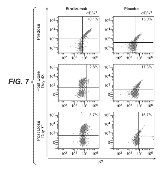
[0037] Figure 7 shows representative FACS dot plots of cell surface aE137
occupancy on
CD45+, CD3+, CD4- T lymphocytes obtained from colonic biopsy samples obtained
from
patients before and after treatment with a single dose of etrolizumab at 100
mg or placebo as
described in Example 1. aE levels are shown on the vertical axis, P7 levels as
determined
using labeled FIB504 antibody (competing antibody) are shown on the horizontal
axis, the
upper right quadrant shows the signal from cells that are both aE+ and J37+;
the percentage of
cells staining positive for both markers is indicated. The plots on the left
show FACS dot
plots for a patient dosed with etrolizumab; the plots on the right show FACS
dot plots for a
patient dosed with placebo. The upper plots show prior to dosing; the middle
plots show day
43, and the lower plots show day 71. Similar results as shown here were
obtained from other
patients dosed with etrolizumab or placebo.
[0038] Figure 8A-E shows beta7 receptor occupancy on CD45+, CD3+, CD4- T
lymphocytes obtained from colonic biopsy samples obtained from patients
before, during
and/or after treatment with etrolizumab or placebo as described in Example 1.
Loss of
detectable T lymphocytes indicates occupancy. (A) Cohort median percentages of
detectable
aE137+, CD45+, CD3+, CD4- T lymphocytes for patients treated in the "100 mg"
dose arm,
the "300 mg + LD" dose arm or placebo, as indicated, solid line with open
circles (pbo),
dotdashed line with stippled circles (100 mg etrolizumab), dashed line with
closed circles
(300mg + LD etrolizumab); (B) Cohort median percentages of detectable a4137+,
CD45+,
CD3+, CD4- T lymphocytes for patients treated in the "100 mg" dose arm, the
"300 mg +
LD" dose arm or placebo, as indicated, solid line with open circles (pbo),
dotdashed line with
stippled circles (100 mg etrolizumab), dashed line with closed circles (300mg
+ LD
etrolizumab); (C) Percentages of detectable aE137+, CD45+, CD3+, CD4- T
lymphocytes for
each of seven patients in the "100 mg" dose of etrolizumab arm; two patients
who received
only a single dose (SD) are indicated by dashed lines, one of whom had an anti-
therapeutic
antibody (ATA) response; (D) Percentages of detectable aE137+, CD45+, CD3+,
CD4- T
lymphocytes for each of seven patients in the "300 mg + LD" arm; and (E)
Percentages of
-19-
CA 02904095 2015-09-03
WO 2014/160753 PCT/US2014/031825
detectable aE137+, CD45+, CD3+, CD4- T lymphocytes for each of nine patients
in the
placebo arm. In each of (C)-(E), TNF-IR patients are denoted with an (*).
[0039] Figure 9 shows integrin expression in colonic biopsy in patients
with clinical
remission compared with patients without clinical remission before and after
treatment with
etrolizumab or placebo as described in Example 1. (A) Integrin-I37 expression
by qPCR at
screening (Scr), week 6 and week 10; (B) Integrin-I31 expression by qPCR at
screening (Scr),
week 6 and week 10; (C) Integrin-a4 expression by qPCR at screening (Scr),
week 6 and
week 10; (D) Integrin-aE expression by qPCR at screening (Scr), week 6 and
week 10. Data
are represented as fold change (2-AAct) from baseline as group median median
absolute
deviation. Dashed lines with solid circles, placebo nonremitters; solid lines
with open circles,
etrolizumab-treated remitters; dotdashed lines with stippled circles,
etrolizumab-treated
nonremitters.
[0040] Figure 10 shows the effect of etrolizumab on aE+ cells in the
intestinal crypt
epithelium as described in Example 1. (A) Median aE ' cells in the intestinal
crypt epithelium
before and after treatment with etrolizumab (striped boxes) or placebo
(stippled boxes); (B)
Representative IHC stains of aE ' cells in intestinal crypt epithelium.
[0041] Figure 11 shows the effect of etrolizumab on aE+ cells in intestinal
lamina
propria as described in Example 1. (A) Mean aE ' cells in the intestinal
lamina propria in all
patients before and after treatment with etrolizumab (striped boxes) or
placebo (stippled
boxes); (B) Mean aE ' cells in the intestinal lamina propria before and after
treatment with
etrolizumab or placebo in patients with clinical remission compared with
patients who did not
achieve clinical remission. Mean, interquartile ranges (IQR) and ranges are
shown on box
plots: stippled boxes, placebo nonremitters; striped boxes, etrolizumab-
treated remitters; open
boxes, etrolizumab-treated nonremitters.
[0042] Figure 12A shows the immunohistochemistry quantification of aE+
cells in the
intestinal crypt epithelium in remitters compared with nonremitters treated
with etrolizumab
or placebo; Mean, interquartile ranges (IQR) and ranges are shown on box
plots: stippled
boxes, etrolizumab-treated remitters; striped boxes, etrolizumab-treated
nonremitters; open
boxes, placebo nonremitters; Figure 12B shows E-cadherin levels in colonic
tissue before and
after treatment with etrolizumab or placebo by clinical remission status, as
described in
Example 1; dashed lines with solid circles, placebo nonremitters; solid lines
with open
circles, etrolizumab-treated remitters; dotdashed lines with stippled circles,
etrolizumab-
treated nonremitters.
-20-
CA 02904095 2015-09-03
WO 2014/160753 PCT/US2014/031825
[0043] Figure 13 shows the expression of MAdCAM-1, cytokines and markers
for
lymphocyte subsets in colonic biopsy in patients with clinical remission
compared with
patients without clinical remission before and after treatment with
etrolizumab or placebo as
described in Example 1. Expression was quantified by qPCR, data are presented
as fold
change (2-AAct) group median + median absolute deviation (MAD). (A) IL-17F;
(B) IL-113;
(C) IL-12p40; (D) IL-6; (E) TNFa; (F) CD19; (G) CD4; (H) CD8; (I) CD38; (J)
MAdCAM-
1; (K) IL-17A; (L) IL-23A; (M) IFNy. Dashed lines with solid circles, placebo
nonremitters;
solid lines with open circles, etrolizumab-treated remitters; dotdashed lines
with stippled
circles, etrolizumab-treated nonremitters.
[0044] Figure 14 shows the serum concentration of etrolizumab compared to
colonic
lymphocyte beta7 receptor occupancy in patients treated with 100 mg
etrolizumab q4w or
300 mg etrolizumab q4w plus a loading dose at days 43 and 71 as described in
Example 1.
TNF-IR patients are indicated as are two patients who received only a single
dose. The arrow
identifies one patient who received only two doses.
[0045] Figure 15 shows the variable light chain region (A) (SEQ ID NO:31)
and the
variable heavy chain region (B) (SEQ ID NO:32) of etrolizumab.
DETAILED DESCRIPTION
[0046] Unless defined otherwise, technical and scientific terms used herein
have the same
meaning as commonly understood by one of ordinary skill in the art to which
this invention
belongs. Singleton et al., Dictionary of Microbiology and Molecular Biology
2nd ed., J.
Wiley & Sons (New York, N.Y. 1994), and March, Advanced Organic Chemistry
Reactions,
Mechanisms and Structure 4th ed., John Wiley & Sons (New York, N.Y. 1992),
provide one
skilled in the art with a general guide to many of the terms used in the
present application.
I. Certain Definitions
[0047] As used in this specification and the appended claims, the singular
forms "a," "an"
and "the" include plural referents unless the context clearly dictates
otherwise. Thus, for
example, reference to "a protein" includes a plurality of proteins; reference
to "a cell"
includes mixtures of cells, and the like.
[0048] Ranges provided in the specification and appended claims include
both end points
and all points between the end points. Thus, for example, a range of 2.0 to
3.0 includes 2.0,
3.0, and all points between 2.0 and 3Ø
[0049] "Treatment" refers to clinical intervention in an attempt to alter
the natural course
of the individual or cell being treated, and can be performed either for
prophylaxis or during
-21-
CA 02904095 2015-09-03
WO 2014/160753 PCT/US2014/031825
the course of clinical pathology. Desirable effects of treatment include
preventing occurrence
or recurrence of disease, alleviation of symptoms, diminishment of any direct
or indirect
pathological consequences of the disease, decreasing the rate of disease
progression,
amelioration or palliation of the disease state, and remission or improved
prognosis.
[0050] "Treatment regimen" refers to a combination of dosage, frequency of
administration, or duration of treatment, with or without addition of a second
medication.
[0051] "Effective treatment regimen" refers to a treatment regimen that
will offer
beneficial response to a patient receiving the treatment.
[0052] "Modifying a treatment" refers to changing the treatment regimen
including,
changing dosage, frequency of administration, or duration of treatment, and/or
addition of a
second medication.
[0053] "Patient response" or "patient responsiveness" can be assessed using
any endpoint
indicating a benefit to the patient, including, without limitation, (1)
inhibition, to some extent,
of disease progression, including slowing down and complete arrest; (2)
reduction in the
number of disease episodes and/or symptoms; (3) reduction in lesional size;
(4) inhibition
(i.e., reduction, slowing down or complete stopping) of disease cell
infiltration into adjacent
peripheral organs and/or tissues; (5) inhibition (i.e., reduction, slowing
down or complete
stopping) of disease spread; (6) decrease of auto-immune, immune, or
inflammatory
response, which may, but does not have to, result in the regression or
ablation of the disease
lesion; (7) relief, to some extent, of one or more symptoms associated with
the disorder; (8)
increase in the length of disease-free presentation following treatment;
and/or (9) decreased
mortality at a given point of time following treatment. The term
"responsiveness" refers to a
measurable response, including complete response (CR) and partial response
(PR).
[0054] By "complete response" or "CR" is intended the disappearance of all
signs of
inflammation or remission in response to treatment. This does not always mean
the disease
has been cured.
[0055] "Partial response" or "PR" refers to a decrease of at least 50% in
the severity of
inflammation, in response to treatment.
[0056] A "beneficial response" of a patient to treatment with an integrin
beta7 antagonist
and similar wording refers to the clinical or therapeutic benefit imparted to
a patient at risk
for or suffering from a gastrointestinal inflammatory disorder from or as a
result of the
treatment with the antagonist, such as an anti-beta7 integrin antibody. Such
benefit includes
cellular or biological responses, a complete response, a partial response, a
stable disease
-22-
CA 02904095 2015-09-03
WO 2014/160753 PCT/US2014/031825
(without progression or relapse), or a response with a later relapse of the
patient from or as a
result of the treatment with the antagonist.
[0057] "A patient maintains responsiveness to a treatment" when the
patient'
responsiveness does not decrease with time during the course of a treatment.
[0058] As used herein, "non-response" or "lack of response" or similar
wording means an
absence of a complete response, a partial response, or a beneficial response
to treatment with
an integrin beta7 antagonist.
[0059] The term "monitoring the efficacy of an integrin beta7 antagonist
therapy" is used
to indicate that a sample is obtained at least once, including serially, from
a patient before,
during, and/or after therapy with an integrin beta7 antagonist and that a
biomarker selected
from integrin beta7 subunit-containing receptor occupancy by the antagonist on
colonic
lymphocytes, gene expression levels of one or more integrin receptor ligands,
gene
expression levels of one or more lymphocyte genes, gene expression levels of
one or more
cytokines, and the number of alphaE-positive cells in intestinal crypt
epithelium is measured
in such samples, and the results of before therapy, during therapy, and/or
after therapy are
compared, to obtain an indication whether the therapy is efficacious or not.
In the monitoring
of the efficacy of a therapy the level of a biomarker selected from integrin
beta7 subunit-
containing receptor occupancy by the antagonist on colonic lymphocytes, gene
expression
levels of one or more integrin receptor ligands, gene expression levels of one
or more
lymphocyte genes, gene expression levels of one or more cytokines, and the
number of
alphaE-positive cells in intestinal crypt epithelium is measured and in one
embodiment
compared to a reference value for the same biomarker, or, in a further
embodiment, it is
compared to the level of the same biomarker in a sample obtained from the same
patient at an
earlier point in time, either while the patient was under therapy or before
start of the therapy.
In one embodiment, a decreased level or an increased level of one or more
biomarkers
selected from integrin beta7 subunit-containing receptor occupancy by the
antagonist on
colonic lymphocytes, gene expression levels of one or more integrin receptor
ligands, gene
expression levels of one or more lymphocyte genes, gene expression levels of
one or more
cytokines, and the number of alphaE-positive cells in intestinal crypt
epithelium, a decreased
level or an increased level depending upon the particularly biomarker assessed
as described
further herein, of a biomarker as compared to the level of the same biomarker
in a sample
obtained from the same patient at an earlier point in time, either while the
patient was already
under therapy or before start of the therapy indicates that the patient is
responsive to the
therapy.
-23-
CA 02904095 2015-09-03
WO 2014/160753 PCT/US2014/031825
[0060] The term "diagnosis" is used herein to refer to the identification
or classification
of a molecular or pathological state, disease or condition. For example,
"diagnosis" may
refer to identification of a particular type of gastrointestinal inflammatory
disorder, and more
particularly, the classification of a particular sub-type of gastrointestinal
inflammatory
disorder, by tissue/organ involvement (e.g., inflammatory bowel disease), or
by other features
(e.g., a patient subpopulation characterized by responsiveness to a treatment,
such as to a
treatment with an integrin beta7 antagonist).
[0061] The term "prognosis" is used herein to refer to the prediction of
the likelihood of
disease symptoms, including, for example, recurrence, flaring, and drug
resistance, of a
gastrointestinal inflammatory disorder.
[0062] The term "sample" or "test sample", as used herein, refers to a
composition that is
obtained or derived from a subject of interest that contains a cellular and/or
other molecular
entity that is to be characterized and/or identified, for example based on
physical,
biochemical, chemical and/or physiological characteristics. For example, the
phrase "disease
sample" and variations thereof refers to any sample obtained from a subject of
interest that
would be expected or is known to contain the cellular and/or molecular entity
that is to be
characterized. The sample can be obtained from a tissue for the subject of
interest or from
peripheral blood of the subject.
[0063] A "reference sample," as used herein, refers to any sample,
standard, or level that
is used for comparison purposes. In one embodiment, a reference sample is
obtained from a
healthy and/or non-diseased part of the body (e.g., tissue or cells) of the
same subject or
patient. In another embodiment, a reference sample is obtained from an
untreated tissue
and/or cell of the body of the same subject or patient. In yet another
embodiment, a reference
sample is obtained from a healthy and/or non-diseased part of the body (e.g.,
tissues or cells)
of an individual who is not the subject or patient. In even another
embodiment, a reference
sample is obtained from an untreated tissue and/or cell part of the body of an
individual who
is not the subject or patient.
[0064] "A beta7 integrin antagonist" or "beta7 antagonist" refers to any
molecule that
inhibits one or more biological activities or blocking binding of beta7
integrin with one or
more of its associated molecules. Antagonists of the invention can be used to
modulate one
or more aspects of beta7 associated effects, including but not limited to
association with
alpha4 integrin subunit, association with alphaE integrin subunit, binding of
alpha4beta7
integrin to MAdCAM, VCAM-1 or fibronectin and binding of alphaEbeta7 integrin
to E-
cadherin. These effects can be modulated by any biologically relevant
mechanism, including
-24-
CA 02904095 2015-09-03
WO 2014/160753 PCT/US2014/031825
disruption of ligand binding to beta7 subunit or to the alpha4beta7 or
alphaEbeta7 dimeric
integrin, and/or by disrupting association between the alpha and beta integrin
subunits such
that formation of the dimeric integrin is inhibited. In one embodiment of the
invention, the
beta7 antagonist is an anti-beta7 integrin antibody (or anti-beta7 antibody).
In one
embodiment, the anti-beta7 integrin antibody is a humanized anti-beta7
integrin antibody and
more particularly a recombinant humanized monoclonal anti-beta7 antibody (or
rhuMAb
beta7 also referred to as etrolizumab). In some embodiments, the anti-beta7
antibodies of the
present invention are anti-integrin beta7 antagonistic antibodies that inhibit
or block the
binding of beta7 subunit with alpha4 integrin subunit, association with alphaE
integrin
subunit, binding of alpha4beta7 integrin to MAdCAM, VCAM-1 or fibronectin and
binding
of alphaEbeta7 integrin to E-cadherin.
[0065] By "beta7 subunit" or "P7 subunit" is meant the human P7 integrin
subunit (Erle
et at., (1991) J. Biol. Chem. 266:11009-11016). The beta7 subunit associates
with alpha4
integrin subunit, such as the human .alpha.4 subunit (Kilger and Holzmann
(1995) J. Mol.
Biol. 73:347-354). The alpha4beta7 integrin is reportedly expressed on a
majority of mature
lymphocytes, as well as a small population of thymocytes, bone marrow cells
and mast cells.
(Kilshaw and Murant (1991) Eur. J. Immunol. 21:2591-2597; Gurish et at.,
(1992) 149: 1964-
1972; and Shaw, S. K. and Brenner, M. B. (1995) Semin. Immunol. 7:335). The
beta7
subunit also associates with the alphaE subunit, such as the human alphaE
integrin subunit
(Cepek, K. L, et at. (1993) J. Immunol. 150:3459). The alphaEbeta7 integrin is
expressed on
intra-intestinal epithelial lymphocytes (iIELs) (Cepek, K. L. (1993) supra).
[0066] By "alphaE subunit" or "alphaE integrin subunit" or "ccE subunit" or
"ccE integrin
subunit" or "CD103" is meant an integrin subunit found to be associated with
beta7 integrin
on intra-epithelial lymphocytes, which alphaEbeta7 integrin mediates binding
of the iELs to
intestinal epithelium expressing E-cadherin (Cepek, K. L. et at. (1993) J.
Immunol. 150:3459;
Shaw, S. K. and Brenner, M. B. (1995) Semin. Immunol. 7:335).
[0067] "MAdCAM" or "MAdCAM-1" are used interchangeably in the context of
the
present invention and refer to the protein mucosal addressin cell adhesion
molecule-1, which
is a single chain polypeptide comprising a short cytoplasmic tail, a
transmembrane region and
an extracellular sequence composed of three immunoglobulin-like domains. The
cDNAs for
murine, human and macaque MAdCAM-1 have been cloned (Briskin, et at., (1993)
Nature,
363:461-464; Shyjan et at., (1996) J. Immunol. 156:2851-2857).
-25-
CA 02904095 2015-09-03
WO 2014/160753 PCT/US2014/031825
[0068] "VCAM-1" or "vascular cell adhesion molecule-1" "CD106" refers to a
ligand of
alpha4beta7 and alpha4betal, expressed on activated endothelium and important
in
endothelial-leukocyte interactions such as binding and transmigration of
leukocytes during
inflammation.
[0069] "CD45" refers to a protein of the protein tyrosine phosphatase (PTP)
family. PTPs
are known to be signaling molecules that regulate a variety of cellular
processes including
cell growth, differentiation, mitotic cycle, and oncogenic transformation.
This PTP contains
an extracellular domain, a single transmembrane segment and two tandem
intracytoplasmic
catalytic domains, and thus belongs to receptor type PTP. This gene is
specifically expressed
in hematopoietic cells. This PTP has been shown to be an essential regulator
of T- and B-cell
antigen receptor signaling. It functions through either direct interaction
with components of
the antigen receptor complexes, or by activating various Src family kinases
required for the
antigen receptor signaling. This PTP also suppresses JAK kinases, and thus
functions as a
regulator of cytokine receptor signaling. Four alternatively spliced
transcripts variants of this
gene, which encode distinct isoforms, have been reported. (Tchilian EZ,
Beverley PC (2002).
"CD45 in memory and disease." Arch. Immunol. Ther. Exp. (Warsz.) 50 (2): 85-
93. Ishikawa
H, Tsuyama N, Abroun S, et at. (2004). "Interleukin-6, CD45 and the src-
kinases in
myeloma cell proliferation." Leuk. Lymphoma 44 (9):1477-81.
[0070] Various isoforms of CD45 exist: CD45RA, CD45RB, CD45RC, CD45RAB,
CD45RAC, CD45RBC, CD45RO, CD45R (ABC). CD45 is also highly glycosylated. CD45R
is the longest protein and migrates at 200 kDa when isolated from T cells. B
cells also
express CD45R with heavier glycosylation, bringing the molecular weight to 220
kDa, hence
the name B220; B cell isoform of 220 kDa. B220 expression is not restricted to
B cells and
can also be expressed on activated T cells, on a subset of dendritic cells and
other antigen
presenting cells. Stanton T, Boxall S, Bennett A, et at. (2004). "CD45 variant
alleles:
possibly increased frequency of a novel exon 4 CD45 polymorphism in HIV
seropositive
Ugandans." Immunogenetics 56 (2): 107-10.
[0071] "Gut-homing lymphocytes" refer to a subgroup of lymphocytes having
the
characteristic of selectively homing to intestinal lymph nodes and tissues but
not homing to
peripheral lymph nodes and tissues. This subgroup of lymphocytes is
characterized by an
unique expression pattern of a combination of multiples cell surface
molecules, including, but
not limited to, the combination of CD4, CD45RA and Beta7. Typically, at least
two subsets
of peripheral blood CD4 + lymphocytes can be subdivided based on the markers
of CD45RA
-26-
CA 02904095 2015-09-03
WO 2014/160753 PCT/US2014/031825
and Beta7, CD45RA- in high, and CD45RA-1371" CD4 + cells. CD45RA- P7 high CD4
+ cells
home preferentially to intestinal lymph nodes and tissues, whereas CD45RA-
P71" CD4 +
cells home preferentially to peripheral lymph nodes and tissues (Rott et at.
1996; Rott et at.
1997; Williams et at. 1998; Rosé et at. 1998; Williams and Butcher 1997;
Butcher et at.
1999). Gut-homing lymphocytes are therefore a distinctive subgroup of
lymphocytes
identified as CD45RA-137 high CD4 + in a flow cytometry assay. The methods of
identifying
this group of lymphocytes are well-known in the art and also disclosed in
detail in Examples
of the present application.
[0072] As used herein with respect to a cell surface marker, the symbol "+"
indicates a
positive expression of a cell surface marker. For instance, CD4 + lymphocytes
are a group of
lymphocytes having CD4 expressed on their cell surfaces.
[0073] As used herein with respect to a cell surface marker, the symbol "-"
indicates a
negative expression of a cell surface marker. For instance, CD45RA-
lymphocytes are a
group of lymphocytes having no CD45RA expressed on their cell surfaces.
[0074] As used herein with respect to the expression of a cell surface
marker, the symbol
"low" indicates a relatively low level of expression of a cell surface marker
on lymphocytes,
while "high" indicates a relatively high level of expression of a cell surface
marker on
lymphocytes. In a flow cytometry, the intensity of137 high is at least about
10 or 100 fold
higher than that of137 10w . Thus, as provided herein in exemplary
embodiments, the
CD45RA-13710W CD4 + and CD45RA-137 high CD4 + lymphocytes locate in distinct
portions
of a dot plot or histogram of a flow cytometry analysis where X-axis is the
intensity of
expression of CD45AR and Y-axis is the intensity of the expression of Beta7.
[0075] "Peripheral-homing lymphocytes" refer to a subgroup of lymphocytes
having the
characteristic of homing to peripheral lymph nodes and tissues and not homing
to intestinal
lymph nodes and tissues. In an exemplary embodiment, as explained above,
Peripheral-
homing lymphocytes are a distinctive group of lymphocytes identified as CD45RA-
13710w
CD4 + cells in a flow cytometry assay. The methods of identifying this group
of
lymphocytes are known in the art and disclosed in detail in the present
application.
[0076] An "amount" or "level" of biomarker can be determined using methods
known in
the art and disclosed herein, such as flow cytometry analysis.
[0077] A "change in the amount or level of a biomarker" is as compared to a
reference/comparator amount of the biomarker. In certain embodiments, the
change is
greater than about 10%, or greater than about 30%, or greater than about 50%,
or greater than
-27-
CA 02904095 2015-09-03
WO 2014/160753 PCT/US2014/031825
about 100%, or greater than about 300% as a function of the value for the
reference or
comparator amount. For example, a reference or comparator amount can be the
amount of a
biomarker before treatment and more particularly, can be the baseline or pre-
dose amount.
[0078] The phrase "essentially the same as" as used herein, denotes an
insignificant
degree of change such that one of skill in the art would not consider the
change to be of
statistical significance or a biologically meaningful change within the
context of the
biological characteristic measured by said values (e.g., the drug serum level
needed to
saturate the drug target receptors). For example, serum drug concentrations
needed to
saturate receptors that are less than about two fold different, or less than
about three fold
different, or less than about four fold different from each other are
considered essentially the
same.
[0079] "Gastrointestinal inflammatory disorders" are a group of chronic
disorders that
cause inflammation and/or ulceration in the mucous membrane. These disorders
include, for
example, inflammatory bowel disease (e.g., Crohn's disease, ulcerative
colitis, indeterminate
colitis and infectious colitis), mucositis (e.g., oral mucositis,
gastrointestinal mucositis, nasal
mucositis and proctitis), necrotizing enterocolitis and esophagitis. In a
preferred
embodiment, the gastrointestinal inflammatory disorder is a inflammatory bowel
disease.
[0080] "Inflammatory Bowel Disease" or "IBD" is used interchangeably herein
to refer to
diseases of the bowel that cause inflammation and/or ulceration and includes
without
limitation Crohn's disease and ulcerative colitis.
[0081] "Crohn's disease (CD)" or "ulcerative colitis (UC)" are chronic
inflammatory
bowel diseases of unknown etiology. Crohn's disease, unlike ulcerative
colitis, can affect any
part of the bowel. The most prominent feature Crohn's disease is the granular,
reddish-purple
edematous thickening of the bowel wall. With the development of inflammation,
these
granulomas often lose their circumscribed borders and integrate with the
surrounding tissue.
Diarrhea and obstruction of the bowel are the predominant clinical features.
As with
ulcerative colitis, the course of Crohn's disease may be continuous or
relapsing, mild or
severe, but unlike ulcerative colitis, Crohn's disease is not curable by
resection of the
involved segment of bowel. Most patients with Crohn's disease require surgery
at some
point, but subsequent relapse is common and continuous medical treatment is
usual.
[0082] Crohn's disease may involve any part of the alimentary tract from
the mouth to the
anus, although typically it appears in the ileocolic, small-intestinal or
colonic-anorectal
regions. Histopathologically, the disease manifests by discontinuous
granulomatomas, crypt
abscesses, fissures and aphthous ulcers. The inflammatory infiltrate is mixed,
consisting of
-28-
CA 02904095 2015-09-03
WO 2014/160753 PCT/US2014/031825
lymphocytes (both T and B cells), plasma cells, macrophages, and neutrophils.
There is a
disproportionate increase in IgM- and IgG-secreting plasma cells, macrophages
and
neutrophils.
[0083] Anti-inflammatory drugs sulfasalazine and 5-aminosalisylic acid (5-
ASA) are
useful for treating mildly active colonic Crohn's disease and are commonly
prescribed to
maintain remission of the disease. Metroidazole and ciprofloxacin are similar
in efficacy to
sulfasalazine and appear to be particularly useful for treating perianal
disease. In more severe
cases, corticosteroids are effective in treating active exacerbations and can
even maintain
remission. Azathioprine and 6-mercaptopurine have also shown success in
patients who
require chronic administration of corticosteroids. It is also possible that
these drugs may play
a role in the long-term prophylaxis. Unfortunately, there can be a very long
delay (up to six
months) before onset of action in some patients. Antidiarrheal drugs can also
provide
symptomatic relief in some patients. Nutritional therapy or elemental diet can
improve the
nutritional status of patients and induce symptomatic improvement of acute
disease, but it
does not induce sustained clinical remissions. Antibiotics are used in
treating secondary
small bowel bacterial overgrowth and in treatment of pyogenic complications.
[0084] "Ulcerative colitis (UC)" afflicts the large intestine. The course
of the disease
may be continuous or relapsing, mild or severe. The earliest lesion is an
inflammatory
infiltration with abscess formation at the base of the crypts of Lieberkuhn.
Coalescence of
these distended and ruptured crypts tends to separate the overlying mucosa
from its blood
supply, leading to ulceration. Symptoms of the disease include cramping, lower
abdominal
pain, rectal bleeding, and frequent, loose discharges consisting mainly of
blood, pus and
mucus with scanty fecal particles. A total colectomy may be required for
acute, severe or
chronic, unremitting ulcerative colitis.
[0085] The clinical features of UC are highly variable, and the onset may
be insidious or
abrupt, and may include diarrhea, tenesmus and relapsing rectal bleeding. With
fulminant
involvement of the entire colon, toxic megacolon, a life-threatening
emergency, may occur.
Extraintestinal manifestations include arthritis, pyoderma gangrenoum,
uveitis, and erythema
nodosum.
[0086] Treatment for UC includes sulfasalazine and related salicylate-
containing drugs
for mild cases and corticosteroid drugs in severe cases. Topical
administration of either
salicylates or corticosteroids is sometimes effective, particularly when the
disease is limited
to the distal bowel, and is associated with decreased side effects compared
with systemic use.
Supportive measures such as administration of iron and antidiarrheal agents
are sometimes
-29-
CA 02904095 2015-09-03
WO 2014/160753 PCT/US2014/031825
indicated. Azathioprine, 6-mercaptopurine and methotrexate are sometimes also
prescribed
for use in refractory corticosteroid-dependent cases.
[0087] An "effective dosage" refers to an amount effective, at dosages and
for periods of
time necessary, to achieve the desired therapeutic or prophylactic result.
[0088] As used herein, the term "patient" refers to any single animal, more
preferably a
mammal (including such non-human animals as, for example, dogs, cats, horses,
rabbits, zoo
animals, cows, pigs, sheep, and non-human primates) for which treatment is
desired. Most
preferably, the patient herein is a human.
[0089] The term "non-human subject" refers to any single non-human animal,
more
preferably a mammal (including such non-human animals as, for example, dogs,
cats, horses,
rabbits, zoo animals, cows, pigs, sheep, and non-human primates).
[0090] The terms "antibody" and "immunoglobulin" are used interchangeably
in the
broadest sense and include monoclonal antibodies (for example, full length or
intact
monoclonal antibodies), polyclonal antibodies, multivalent antibodies,
multispecific
antibodies (e.g., bispecific antibodies so long as they exhibit the desired
biological activity)
and may also include certain antibody fragments (as described in greater
detail herein). An
antibody can be human, humanized and/or affinity matured.
[0091] "Antibody fragments" comprise only a portion of an intact antibody,
wherein the
portion preferably retains at least one, preferably most or all, of the
functions normally
associated with that portion when present in an intact antibody. In one
embodiment, an
antibody fragment comprises an antigen binding site of the intact antibody and
thus retains
the ability to bind antigen. In another embodiment, an antibody fragment, for
example one
that comprises the Fc region, retains at least one of the biological functions
normally
associated with the Fc region when present in an intact antibody, such as FcRn
binding,
antibody half life modulation, ADCC function and complement binding. In one
embodiment,
an antibody fragment is a monovalent antibody that has an in vivo half life
substantially
similar to an intact antibody. For example, such an antibody fragment may
comprise on
antigen binding arm linked to an Fc sequence capable of conferring in vivo
stability to the
fragment.
[0092] The term "monoclonal antibody" as used herein refers to an antibody
obtained
from a population of substantially homogeneous antibodies, i.e., the
individual antibodies
comprising the population are identical except for possible naturally
occurring mutations that
may be present in minor amounts. Monoclonal antibodies are highly specific,
being directed
against a single antigen. Furthermore, in contrast to polyclonal antibody
preparations that
-30-
CA 02904095 2015-09-03
WO 2014/160753 PCT/US2014/031825
typically include different antibodies directed against different determinants
(epitopes), each
monoclonal antibody is directed against a single determinant on the antigen.
[0093] The monoclonal antibodies herein specifically include "chimeric"
antibodies in
which a portion of the heavy and/or light chain is identical with or
homologous to
corresponding sequences in antibodies derived from a particular species or
belonging to a
particular antibody class or subclass, while the remainder of the chain(s) is
identical with or
homologous to corresponding sequences in antibodies derived from another
species or
belonging to another antibody class or subclass, as well as fragments of such
antibodies, so
long as they exhibit the desired biological activity (U.S. Patent No.
4,816,567; and Morrison
et al., Proc. Natl. Acad. Sci. USA 81:6851-6855 (1984)).
[0094] "Humanized" forms of non-human (e.g., murine) antibodies are
chimeric
antibodies that contain minimal sequence derived from non-human
immunoglobulin. For the
most part, humanized antibodies are human immunoglobulins (recipient antibody)
in which
residues from a hypervariable region of the recipient are replaced by residues
from a
hypervariable region of a non-human species (donor antibody) such as mouse,
rat, rabbit or
nonhuman primate having the desired specificity, affinity, and capacity. In
some instances,
framework region (FR) residues of the human immunoglobulin are replaced by
corresponding non-human residues. Furthermore, humanized antibodies may
comprise
residues that are not found in the recipient antibody or in the donor
antibody. These
modifications are made to further refine antibody performance. In general, the
humanized
antibody will comprise substantially all of at least one, and typically two,
variable domains,
in which all or substantially all of the hypervariable loops correspond to
those of a non-
human immunoglobulin and all or substantially all of the FRs are those of a
human
immunoglobulin lo sequence. The humanized antibody optionally will also
comprise at least
a portion of an immunoglobulin constant region (Fc), typically that of a human
immunoglobulin. For further details, see Jones et at., Nature 321:522-525
(1986);
Riechmann et at., Nature 332:323-329 (1988); and Presta, Curr. Op. Struct.
Biol. 2:593-596
(1992). See also the following review articles and references cited therein:
Vaswani and
Hamilton, Ann. Allergy, Asthma & Immunol. 1: 105-115 (1998); Harris, Biochem.
Soc.
Transactions 23:1035-1038 (1995); Hurle and Gross, Curr. Op. Biotech. 5:428-
433 (1994).
[0095] A "human antibody" is one which comprises an amino acid sequence
corresponding to that of an antibody produced by a human and/or has been made
using any of
the techniques for making human antibodies as disclosed herein. Such
techniques include
screening human-derived combinatorial libraries, such as phage display
libraries (see, e.g.,
-31-
CA 02904095 2015-09-03
WO 2014/160753 PCT/US2014/031825
Marks et at., J. Mot. Biol., 222: 581-597 (1991) and Hoogenboom et at., Nucl.
Acids Res., 19:
4133-4137 (1991)); using human myeloma and mouse-human heteromyeloma cell
lines for
the production of human monoclonal antibodies (see, e.g., Kozbor J. Immunol.,
133: 3001
(1984); Brodeur et at., Monoclonal Antibody Production Techniques and
Applications,
pp. 55-93 (Marcel Dekker, Inc., New York, 1987); and Boerner et at., J.
Immunol., 147: 86
(1991)); and generating monoclonal antibodies in transgenic animals (e.g.,
mice) that are
capable of producing a full repertoire of human antibodies in the absence of
endogenous
immunoglobulin production (see, e.g., Jakobovits et at., Proc. Natl. Acad. Sci
USA, 90: 2551
(1993); Jakobovits et at., Nature, 362: 255 (1993); Bruggermann et al.,Year in
Immunol., 7:
33 (1993)). This definition of a human antibody specifically excludes a
humanized antibody
comprising antigen-binding residues from a non-human animal.
[0096] An "isolated" antibody is one which has been identified and
separated and/or
recovered from a component of its natural environment. Contaminant components
of its
natural environment are materials which would interfere with diagnostic or
therapeutic uses
for the antibody, and may include enzymes, hormones, and other proteinaceous
or
nonproteinaceous solutes. In preferred embodiments, the antibody will be
purified (1) to
greater than 95% by weight of antibody as determined by the Lowry method, and
most
preferably more than 99% by weight, (2) to a degree sufficient to obtain at
least 15 residues
of N-terminal or internal amino acid sequence by use of a spinning cup
sequenator, or (3) to
homogeneity by SDS-PAGE under reducing or nonreducing conditions using
Coomassie blue
or, preferably, silver stain. Isolated antibody includes the antibody in situ
within recombinant
cells since at least one component of the antibody's natural environment will
not be present.
Ordinarily, however, isolated antibody will be prepared by at least one
purification step.
[0097] The term "hypervariable region," "HVR," or "HV," when used herein
refers to the
regions of an antibody variable domain which are hypervariable in sequence
and/or form
structurally defined loops. Generally, antibodies comprise six hypervariable
regions; three in
the VH (H1, H2, H3), and three in the VL (L1, L2, L3). A number of
hypervariable region
delineations are in use and are encompassed herein. The Kabat Complementarity
Determining Regions (CDRs) are based on sequence variability and are the most
commonly
used (Kabat et at., Sequences of Proteins of Immunological Interest, 5th Ed.
Public Health
Service, National Institutes of Health, Bethesda, Md. (1991)). Chothia refers
instead to the
location of the structural loops (Chothia and Lesk J. Mol. Biol. 196:901-917
(1987)). The
AbM hypervariable regions represent a compromise between the Kabat CDRs and
Chothia
structural loops, and are used by Oxford Molecular's AbM antibody modeling
software. The
-32-
CA 02904095 2015-09-03
WO 2014/160753 PCT/US2014/031825
"contact" hypervariable regions are based on an analysis of the available
complex crystal
structures. The residues from each of these HVRs are noted below.
Loop Kabat AbM Chothia Contact
Li L24-L34 L24-L34 L26-L32 L30-L36
L2 L50-L56 L50-L56 L50-L52 L46-L55
L3 L89-L97 L89-L97 L91-L96 L89-L96
H1 H31-H35B H26-H35B H26-H32 H30-H35B (Kabat Numbering)
H1 H31-H35 H26-H35 H26-H32 H30-H35 (Chothia Numbering)
H2 H50-H65 H50-H58 H53-H55 H47-H58
H3 H95-H102 H95-H102 H96-H101 H93-H101
[0098] Hypervariable regions may comprise "extended hypervariable regions"
as follows:
24-36 or 24-34 (L1), 46-56 or 49-56 or 50-56 or 52-56 (L2) and 89-97 (L3) in
the VL and 26-
35 (H1), 50-65 or 49-65 (H2) and 93-102, 94-102 or 95-102 (H3) in the VH. The
variable
domain residues are numbered according to Kabat et at., supra for each of
these definitions.
[0099] "Framework" or "FR" residues are those variable domain residues
other than the
hypervariable region residues as herein defined.
[0100] A "human consensus framework" is a framework which represents the
most
commonly occurring amino acid residue in a selection of human immunoglobulin
VL or VH
framework sequences. Generally, the selection of human immunoglobulin VL or VH
sequences is from a subgroup of variable domain sequences. Generally, the
subgroup of
sequences is a subgroup as in Kabat et at. In one embodiment, for the VL, the
subgroup is
subgroup kappa I as in Kabat et at. In one embodiment, for the VH, the
subgroup is
subgroup III as in Kabat et at.
[0101] An "affinity matured" antibody is one with one or more alterations
in one or more
CDRs thereof which result in an improvement in the affinity of the antibody
for antigen,
compared to a parent antibody which does not possess those alteration(s).
Preferred affinity
matured antibodies will have nanomolar or even picomolar affinities for the
target antigen.
Affinity matured antibodies are produced by procedures known in the art. Marks
et at.
Bio/Technology 10:779-783 (1992) describes affinity maturation by VH and VL
domain
shuffling. Random mutagenesis of CDR and/or framework residues is described
by: Barbas
et at. Proc Nat. Acad. Sci, USA 91:3809-3813 (1994); Schier et at. Gene
169:147-155
(1996); Yelton et at. J. Immunol. 155:1994-2004 (1995); Jackson et at., J.
Immunol.
154(7):3310-9 (1995); and Hawkins et at. J. Mol. Biol. 226:889-896 (1992).
-33-
CA 02904095 2015-09-03
WO 2014/160753 PCT/US2014/031825
[0102] "Binding affinity" generally refers to the strength of the sum total
of noncovalent
interactions between a single binding site of a molecule (e.g., an antibody)
and its binding
partner (e.g., an antigen). Unless indicated otherwise, as used herein,
"binding affinity" refers
to intrinsic binding affinity which reflects a 1:1 interaction between members
of a binding
pair (e.g., antibody and antigen). The affinity of a molecule X for its
partner Y can generally
be represented by the dissociation constant (Kd). Affinity can be measured by
common
methods known in the art, including those described herein. Low-affinity
antibodies
generally bind antigen slowly and tend to dissociate readily, whereas high-
affinity antibodies
generally bind antigen faster and tend to remain bound longer. A variety of
methods of
measuring binding affinity are known in the art, any of which can be used for
purposes of the
present invention.
[0103] The term "variable" refers to the fact that certain portions of the
variable domains
differ extensively in sequence among antibodies and are used in the binding
and specificity of
each particular antibody for its particular antigen. However, the variability
is not evenly
distributed throughout the variable domains of antibodies. It is concentrated
in three
segments called hypervariable regions both in the light chain and the heavy
chain variable
domains. The more highly conserved portions of variable domains are called the
framework
regions (FRs). The variable domains of native heavy and light chains each
comprise four
FRs, largely adopting a I3-sheet configuration, connected by three
hypervariable regions,
which form loops connecting, and in some cases forming part of, the 13-sheet
structure. The
hypervariable regions in each chain are held together in close proximity by
the FRs and, with
the hypervariable regions from the other chain, contribute to the formation of
the antigen-
binding site of antibodies (see Kabat et at., Sequences of Proteins of
Immunological Interest,
5th Ed. Public Health Service, National Institutes of Health, Bethesda, MD.
(1991)). The
constant domains are not involved directly in binding an antibody to an
antigen, but exhibit
various effector functions, such as participation of the antibody in antibody
dependent
cellular cytotoxicity (ADCC).
[0104] Papain digestion of antibodies produces two identical antigen-
binding fragments,
called "Fab" fragments, each with a single antigen-binding site, and a
residual "Fc" fragment,
whose name reflects its ability to crystallize readily. Pepsin treatment
yields an F(ab')2
fragment that has two antigen-binding sites and is still capable of cross-
linking antigen.
[0105] "Fv" is the minimum antibody fragment which contains a complete
antigen-
recognition and antigen-binding site. This region consists of a dimer of one
heavy chain and
one light chain variable domain in tight, non-covalent association. It is in
this configuration
-34-
CA 02904095 2015-09-03
WO 2014/160753 PCT/US2014/031825
that the three hypervariable regions of each variable domain interact to
define an antigen-
binding site on the surface of the VH-VL dimer. Collectively, the six
hypervariable regions
confer antigen-binding specificity to the antibody. However, even a single
variable domain
(or half of an Fv comprising only three hypervariable regions specific for an
antigen) has the
ability to recognize and bind antigen, although at a lower affinity than the
entire binding site.
[0106] The Fab fragment also contains the constant domain of the light
chain and the
first constant domain (CH1) of the heavy chain. Fab' fragments differ from Fab
fragments by
the addition of a few residues at the carboxy terminus of the heavy chain CH1
domain
including one or more cysteines from the antibody hinge region. Fab'-SH is the
designation
herein for Fab' in which the cysteine residue(s) of the constant domains bear
at least one free
thiol group. F(ab')2 antibody fragments originally were produced as pairs of
Fab' fragments
which have hinge cysteines between them. Other chemical couplings of antibody
fragments
are also known.
[0107] The "light chains" of antibodies from any vertebrate species can be
assigned to
one of two clearly distinct types, called kappa (x) and lambda (X), based on
the amino acid
sequences of their constant domains.
[0108] Depending on the amino acid sequences of the constant domains of
their heavy
chains, antibodies (immunoglobulins) can be assigned to different classes.
There are five
major classes of immunoglobulins: IgA, IgD, IgE, IgG, and IgM, and several of
these may be
further divided into subclasses (isotypes), e.g., IgGi, IgG2, IgG3, IgG4,
IgAi, and IgA2. The
heavy-chain constant domains that correspond to the different classes of
immunoglobulins
are called a, 6, 8, y, and i.t, respectively. The subunit structures and three-
dimensional
configurations of different classes of immunoglobulins are well known and
described
generally in, for example, Abbas et al. Cellular and Mol. Immunology, 4th ed.
(W. B.
Saunders, Co., 2000). An antibody may be part of a larger fusion molecule,
formed by
covalent or non-covalent association of the antibody with one or more other
proteins or
peptides.
[0109] The terms "full-length antibody," "intact antibody," and "whole
antibody" are
used herein interchangeably to refer to an antibody in its substantially
intact form, not
antibody fragments as defined below. The terms particularly refer to an
antibody with heavy
chains that contain an Fc region.
[0110] A "naked antibody" for the purposes herein is an antibody that is
not conjugated
to a cytotoxic moiety or radiolabel.
-35-
CA 02904095 2015-09-03
WO 2014/160753 PCT/US2014/031825
[01 1 1] The term "Fe region" herein is used to define a C-terminal region
of an
immunoglobulin heavy chain, including native sequence Fe regions and variant
Fe regions.
Although the boundaries of the Fe region of an immunoglobulin heavy chain
might vary, the
human IgG heavy chain Fe region is usually defined to stretch from an amino
acid residue at
position Cys226, or from Pro230, to the carboxyl-terminus thereof. The C-
terminal lysine
(residue 447 according to the EU numbering system) of the Fe region may be
removed, for
example, during production or purification of the antibody, or by
recombinantly engineering
the nucleic acid encoding a heavy chain of the antibody. Accordingly, a
composition of
intact antibodies may comprise antibody populations with all K447 residues
removed,
antibody populations with no K447 residues removed, and antibody populations
having a
mixture of antibodies with and without the K447 residue.
[0112] Unless indicated otherwise, herein the numbering of the residues in
an
immunoglobulin heavy chain is that of the EU index as in Kabat et at.,
Sequences of Proteins
of Immunological Interest, 5th Ed. Public Health Service, National Institutes
of Health,
Bethesda, MD (1991), expressly incorporated herein by reference. The "EU index
as in
Kabat" refers to the residue numbering of the human IgG1 EU antibody.
[0113] A "functional Fe region" possesses an "effector function" of a
native sequence Fe
region. Exemplary "effector functions" include Clq binding; complement
dependent
cytotoxicity; Fe receptor binding; antibody-dependent cell-mediated
cytotoxicity (ADCC);
phagocytosis; down regulation of cell surface receptors (e.g., B cell
receptor; BCR), etc.
Such effector functions generally require the Fe region to be combined with a
binding domain
(e.g., an antibody variable domain) and can be assessed using various assays
as herein
disclosed, for example.
[0114] A "native sequence Fe region" comprises an amino acid sequence
identical to the
amino acid sequence of an Fe region found in nature. Native sequence human Fe
regions
include a native sequence human IgG1 Fe region (non-A and A allotypes); native
sequence
human IgG2 Fe region; native sequence human IgG3 Fe region; and native
sequence human
IgG4 Fe region as well as naturally occurring variants thereof.
[0115] A "variant Fe region" comprises an amino acid sequence which differs
from that
of a native sequence Fe region by virtue of at least one amino acid
modification, preferably
one or more amino acid substitution(s). Preferably, the variant Fe region has
at least one
amino acid substitution compared to a native sequence Fe region or to the Fe
region of a
parent polypeptide, e.g., from about one to about ten amino acid
substitutions, and preferably
from about one to about five amino acid substitutions in a native sequence Fe
region or in the
-36-
CA 02904095 2015-09-03
WO 2014/160753
PCT/US2014/031825
Fe region of the parent polypeptide. The variant Fe region herein will
preferably possess at
least about 80% homology with a native sequence Fe region and/or with an Fe
region of a
parent polypeptide, and most preferably at least about 90% homology therewith,
more
preferably at least about 95% homology therewith.
[0116] Depending on the amino acid sequence of the constant domain of their
heavy
chains, intact antibodies can be assigned to different "classes." There are
five major classes
of intact antibodies: IgA, IgD, IgE, IgG, and IgM, and several of these may be
further
divided into "subclasses" (isotypes), e.g., IgGl, IgG2, IgG3, IgG4, IgA, and
IgA2. The
heavy-chain constant domains that correspond to the different classes of
antibodies are called
a, 6, 8, y, and IA, respectively. The subunit structures and three-dimensional
configurations of
different classes of immunoglobulins are well known.
[0117] "Antibody-dependent cell-mediated cytotoxicity" and "ADCC" refer to
a cell-
mediated reaction in which nonspecific cytotoxic cells that express Fe
receptors (FcRs) (e.g.
Natural Killer (NK) cells, neutrophils, and macrophages) recognize bound
antibody on a
target cell and subsequently cause lysis of the target cell. The primary cells
for mediating
ADCC, NK cells, express FcyRIII only, whereas monocytes express FcyRI, FcyRII
and
FcyRIII. FcR expression on hematopoietic cells in summarized is Table 3 on
page 464 of
Ravetch and Kinet, Annu. Rev. Immunol 9:457-92 (1991). To assess ADCC activity
of a
molecule of interest, an in vitro ADCC assay, such as that described in U.S.
Patent
No. 5,500,362 or 5,821,337 may be performed. Useful effector cells for such
assays include
peripheral blood mononuclear cells (PBMC) and Natural Killer (NK) cells.
Alternatively, or
additionally, ADCC activity of the molecule of interest may be assessed in
vivo, e.g., in a
animal model such as that disclosed in Clynes et at. PNAS (USA) 95:652-656
(1998).
[0118] "Human effector cells" are leukocytes which express one or more FcRs
and
perform effector functions. Preferably, the cells express at least FcyRIII and
perform ADCC
effector function. Examples of human leukocytes which mediate ADCC include
peripheral
blood mononuclear cells (PBMC), natural killer (NK) cells, monocytes,
cytotoxic T cells and
neutrophils; with PBMCs and NK cells being preferred. The effector cells may
be isolated
from a native source thereof, e.g., from blood or PBMCs as described herein.
[0119] The terms "Fe receptor" or "FcR" are used to describe a receptor
that binds to the
Fe region of an antibody. The preferred FcR is a native sequence human FcR.
Moreover, a
preferred FcR is one which binds an IgG antibody (a gamma receptor) and
includes receptors
of the FcyRI, FcyRII, and FcyRIII subclasses, including allelic variants and
alternatively
spliced forms of these receptors. FcyRII receptors include FcyRIIA (an
"activating receptor")
-37-
CA 02904095 2015-09-03
WO 2014/160753 PCT/US2014/031825
and FcyRIIB (an "inhibiting receptor"), which have similar amino acid
sequences that differ
primarily in the cytoplasmic domains thereof Activating receptor FcyRIIA
contains an
immunoreceptor tyrosine-based activation motif (ITAM) in its cytoplasmic
domain.
Inhibiting receptor FcyRIIB contains an immunoreceptor tyrosine-based
inhibition motif
(ITIM) in its cytoplasmic domain (see review M. in Daeron, Annu. Rev. Immunol.
15:203-234
(1997)). FcRs are reviewed in Ravetch and Kinet, Annu. Rev. Immunol 9:457-92
(1991);
Capel et at., Immunomethods 4:25-34 (1994); and de Haas et at., J. Lab. Clin.
Med. 126:330-
41(1995). Other FcRs, including those to be identified in the future, are
encompassed by the
term "FcR" herein. The term also includes the neonatal receptor, FcRn, which
is responsible
for the transfer of maternal IgGs to the fetus (Guyer et at., J. Immunol.
117:587 (1976) and
Kim et at., J. Immunol. 24:249 (1994)), and regulates homeostasis of
immunoglobulins.
Antibodies with improved binding to the neonatal Fc receptor (FcRn), and
increased half-
lives, are described in W000/42072 (Presta, L.) and US2005/0014934A1 (Hinton
et al.).
These antibodies comprise an Fc region with one or more substitutions therein
which
improve binding of the Fc region to FcRn. For example, the Fc region may have
substitutions at one or more of positions 238, 250, 256, 265, 272, 286, 303,
305, 307, 311,
312, 314, 317, 340, 356, 360, 362, 376, 378, 380, 382, 413, 424, 428 or 434
(EU numbering
of residues). The preferred Fc region-comprising antibody variant with
improved FcRn
binding comprises amino acid substitutions at one, two or three of positions
307, 380 and 434
of the Fc region thereof (EU numbering of residues).
[0120] "Single-chain Fv" or "scFv" antibody fragments comprise the VH and
VL
domains of antibody, wherein these domains are present in a single polypeptide
chain.
Preferably, the Fv polypeptide further comprises a polypeptide linker between
the VH and VL
domains which enables the scFv to form the desired structure for antigen
binding. For a
review of scFv see Pliickthun in The Pharmacology of Monoctonat Antibodies,
vol. 113,
Rosenburg and Moore eds., Springer-Verlag, New York, pp. 269-315 (1994). HER2
antibody scFv fragments are described in W093/16185; U.S. Patent No.
5,571,894; and U.S.
Patent No. 5,587,458.
[0121] The term "diabodies" refers to small antibody fragments with two
antigen-
binding sites, which fragments comprise a variable heavy domain (VH) connected
to a
variable light domain (VL) in the same polypeptide chain (VH - VL). By using a
linker that is
too short to allow pairing between the two domains on the same chain, the
domains are
forced to pair with the complementary domains of another chain and create two
antigen-
-38-
CA 02904095 2015-09-03
WO 2014/160753 PCT/US2014/031825
binding sites. Diabodies are described more fully in, for example, EP 404,097;
WO
93/11161; and Hollinger et at., Proc. Natl. Acad. Sci. USA, 90:6444-6448
(1993).
[0122] An "affinity matured" antibody is one with one or more alterations
in one or more
hypervariable regions thereof which result an improvement in the affinity of
the antibody for
antigen, compared to a parent antibody which does not possess those
alteration(s). Preferred
affinity matured antibodies will have nanomolar or even picomolar affinities
for the target
antigen. Affinity matured antibodies are produced by procedures known in the
art. Marks et
at. Rio/Technology 10:779-783 (1992) describes affinity maturation by VH and
VL domain
shuffling. Random mutagenesis of CDR and/or framework residues is described
by: Barbas
et al. Proc Nat. Acad. Sci, USA 91:3809-3813 (1994); Schier et al. Gene
169:147-155 (1995);
Yelton et at. J. Immunol. 155:1994-2004 (1995); Jackson et at., J. Immunol.
154(7):3310-9
(1995); and Hawkins et at, J. Mot. Biol. 226:889-896 (1992).
[0123] An "amino acid sequence variant" antibody herein is an antibody with
an amino
acid sequence which differs from a main species antibody. Ordinarily, amino
acid sequence
variants will possess at least about 70% homology with the main species
antibody, and
preferably, they will be at least about 80%, more preferably at least about
90% homologous
with the main species antibody. The amino acid sequence variants possess
substitutions,
deletions, and/or additions at certain positions within or adjacent to the
amino acid sequence
of the main species antibody. Examples of amino acid sequence variants herein
include an
acidic variant (e.g., deamidated antibody variant), a basic variant, an
antibody with an amino-
terminal leader extension (e.g. VHS-) on one or two light chains thereof, an
antibody with a
C-terminal lysine residue on one or two heavy chains thereof, etc, and
includes combinations
of variations to the amino acid sequences of heavy and/or light chains. The
antibody variant
of particular interest herein is the antibody comprising an amino-terminal
leader extension on
one or two light chains thereof, optionally further comprising other amino
acid sequence
and/or glycosylation differences relative to the main species antibody.
[0124] A "glycosylation variant" antibody herein is an antibody with one or
more
carbohydrate moieties attached thereto which differ from one or more
carbohydrate moieties
attached to a main species antibody. Examples of glycosylation variants herein
include
antibody with a G1 or G2 oligosaccharide structure, instead a GO
oligosaccharide structure,
attached to an Fc region thereof, antibody with one or two carbohydrate
moieties attached to
one or two light chains thereof, antibody with no carbohydrate attached to one
or two heavy
chains of the antibody, etc, and combinations of glycosylation alterations.
Where the
-39-
CA 02904095 2015-09-03
WO 2014/160753 PCT/US2014/031825
antibody has an Fe region, an oligosaccharide structure may be attached to one
or two heavy
chains of the antibody, e.g. at residue 299 (298, EU numbering of residues).
[0125] The term "cytotoxic agent" as used herein refers to a substance that
inhibits or
prevents the function of cells and/or causes destruction of cells. The term is
intended to
include radioactive isotopes (e.g. At2115 11315 11255 y905 Re1865 Re1885
sm1535 Bi2125 p32 and
radioactive isotopes of Lu), chemotherapeutic agents, and toxins such as small
molecule
toxins or enzymatically active toxins of bacterial, fungal, plant or animal
origin, including
fragments and/or variants thereof
[0126] The term "cytokine" is a generic term for proteins released by one
cell population
which act on another cell as intercellular mediators. Examples of such
cytokines are
lymphokines, monokines, and traditional polypeptide hormones. Included among
the
cytokines are growth hormone such as human growth hormone, N-methionyl human
growth
hormone, and bovine growth hormone; parathyroid hormone; thyroxine; insulin;
proinsulin;
relaxin; prorelaxin; glycoprotein hormones such as follicle stimulating
hormone (FSH),
thyroid stimulating hormone (TSH), and luteinizing hormone (LH); hepatic
growth factor;
fibroblast growth factor; prolactin; placental lactogen; tumor necrosis factor-
a and -13;
mullerian-inhibiting substance; mouse gonadotropin-associated peptide;
inhibin; activin;
vascular endothelial growth factor; integrin; thrombopoietin (TP0); nerve
growth factors
such as NGF-13; platelet-growth factor; transforming growth factors (TGFs)
such as TGF-a
and TGF-13; insulin-like growth factor-I and -II; erythropoietin (EPO);
osteoinductive factors;
interferons such as interferon-a, -13, and -y; colony stimulating factors
(CSFs) such as
macrophage-CSF (M-CSF); granulocyte-macrophage-CSF (GM-CSF); and granulocyte-
CSF
(G-CSF); interleukins (ILs) such as IL-1, IL-la, IL-2, IL-3, IL-4, IL-5, IL-6,
IL-7, IL-8, IL-9,
IL-10, IL-11, IL-12; a tumor necrosis factor such as TNF-a or TNF-13; and
other polypeptide
factors including LIF and kit ligand (KL). As used herein, the term cytokine
includes
proteins from natural sources or from recombinant cell culture and
biologically active
equivalents of the native sequence cytokines.
[0127] The term "immunosuppressive agent" as used herein for adjunct
therapy refers to
substances that act to suppress or mask the immune system of the subject being
treated
herein. This would include substances that suppress cytokine production, down-
regulate or
suppress self-antigen expression, or mask the MHC antigens. Examples of such
agents
include 2-amino-6-aryl-5-substituted pyrimidines (see U.S. Patent No.
4,665,077); non-
steroidal anti-inflammatory drugs (NSAIDs); ganciclovir; tacrolimus;
glucocorticoids such as
cortisol or aldosterone; anti-inflammatory agents such as a cyclooxygenase
inhibitor; a 5-
-40-
CA 02904095 2015-09-03
WO 2014/160753
PCT/US2014/031825
lipoxygenase inhibitor; or a leukotriene receptor antagonist; purine
antagonists such as
azathioprine or mycophenolate mofetil (MMF); alkylating agents such as
cyclophosphamide;
bromocryptine; danazol; dapsone; glutaraldehyde (which masks the MHC antigens,
as
described in U.S. Patent No. 4,120,649); anti-idiotypic antibodies for MHC
antigens and
MHC fragments; cyclosporine; 6 mercaptopurine; steroids such as
corticosteroids or
glucocorticosteroids or glucocorticoid analogs, e.g., prednisone,
methylprednisolone,
including SOLU-MEDROL® methylprednisolone sodium succinate, and
dexamethasone; dihydrofolate reductase inhibitors such as methotrexate (oral
or
subcutaneous); anti-malarial agents such as chloroquine and
hydroxychloroquine;
sulfasalazine; leflunomide; cytokine or cytokine receptor antibodies or
antagonists including
anti-interferon-alpha, -beta, or -gamma antibodies, anti-tumor necrosis
factor(TNF)-alpha
antibodies (infliximab (REMICADE®) or adalimumab), anti-TNF-alpha
immunoadhesin (etanercept), anti-TNF-beta antibodies, anti-interleukin-2 (IL-
2) antibodies
and anti-IL-2 receptor antibodies, and anti-interleukin-6 (IL-6) receptor
antibodies and
antagonists; anti-LFA-1 antibodies, including anti-CD ha a and anti-CD18
antibodies; anti-
L3T4 antibodies; heterologous anti-lymphocyte globulin; pan-T antibodies,
preferably anti-
CD3 or anti-CD4/CD4a antibodies; soluble peptide containing a LFA-3 binding
domain
(WO 90/08187 published Jul. 26, 1990); streptokinase; transforming growth
factor-beta
(TGF-beta); streptodomase; RNA or DNA from the host; FK506; RS-61443;
chlorambucil;
deoxyspergualin; rapamycin; T-cell receptor (Cohen et al.,U U.S. Patent No.
5,114,721); T-cell
receptor fragments (Offner et at., Science, 251: 430-432 (1991); WO 90/11294;
Ianeway,
Nature, 341: 482 (1989); and WO 91/01133); BAFF antagonists such as BAFF or
BR3
antibodies or immunoadhesins and zTNF4 antagonists (for review, see Mackay and
Mackay,
Trends Immunol., 23:113-5 (2002) and see also definition below); biologic
agents that
interfere with T cell helper signals, such as anti-CD40 receptor or anti-CD40
ligand (CD154),
including blocking antibodies to CD4O-CD40 ligand.(e.g., Dune et al., Science,
261: 1328-30
(1993); Mohan et at., J. Immunol., 154: 1470-80 (1995)) and CTLA4-Ig (Finck et
at.,
Science, 265: 1225-7 (1994)); and T-cell receptor antibodies (EP 340,109) such
as T 1 OB9.
[0128] The term "ameliorates" or "amelioration" as used herein refers to a
decrease,
reduction or elimination of a condition, disease, disorder, or phenotype,
including an
abnormality or symptom.
[0129] A "symptom" of a disease or disorder (e.g., inflammatory bowel
disease, e.g.,
ulcerative colitis or Crohn's disease) is any morbid phenomenon or departure
from the
normal in structure, function, or sensation, experienced by a subject and
indicative of disease.
-41-
CA 02904095 2015-09-03
WO 2014/160753 PCT/US2014/031825
[0130] The expression "therapeutically effective amount" refers to an
amount that is
effective for preventing, ameliorating, or treating a disease or disorder
(e.g., inflammatory
bowel disease, e.g., ulcerative colitis or Crohn's disease). For example, a
"therapeutically
effective amount" of an antibody refers to an amount of the antibody that is
effective for
preventing, ameliorating, or treating the specified disease or disorder.
Similarly, a
"therapeutically effective amount" of a combination of an antibody and a
second compound
refers to an amount of the antibody and an amount of the second compound that,
in
combination, is effective for preventing, ameliorating, or treating the
specified disease or
disorder.
[0131] It is to be understood that the terminology "a combination of" two
compounds
does not mean that the compounds have to be administered in admixture with
each other.
Thus, treatment with or use of such a combination encompasses a mixture of the
compounds
or separate administration of the compounds, and includes administration on
the same day or
different days. Thus the terminology "combination" means two or more compounds
are used
for the treatment, either individually or in admixture with each other. When
an antibody and a
second compound, for example, are administered in combination to a subject,
the antibody is
present in the subject at a time when the second compound is also present in
the subject,
whether the antibody and second compound are administered individually or in
admixture to
the subject. In certain embodiments, a compound other than the antibody is
administered
prior to the antibody. In certain embodiments, a compound other than the
antibody is
administered after the antibody.
[0132] For the purposes herein, "tumor necrosis factor-alpha (TNF-alpha)"
refers to a
human TNF-alpha molecule comprising the amino acid sequence as described in
Pennica et
al., Nature, 312:721 (1984) or Aggarwal et al., JBC, 260:2345 (1985).
[0133] A "TNF-alpha inhibitor" herein is an agent that inhibits, to some
extent, a
biological function of TNF-alpha, generally through binding to TNF-alpha and
neutralizing
its activity. Examples of TNF inhibitors specifically contemplated herein are
etanercept
(ENBRELO), infliximab (REMICADEO), adalimumab (HUMIRAO), golimumab
(SIMPONITM), and certolizumab pegol (CIMZIAO).
[0134] "Corticosteroid" refers to any one of several synthetic or naturally
occurring
substances with the general chemical structure of steroids that mimic or
augment the effects
of the naturally occurring corticosteroids. Examples of synthetic
corticosteroids include
prednisone, prednisolone (including methylprednisolone), dexamethasone
triamcinolone, and
betamethasone.
-42-
CA 02904095 2015-09-03
WO 2014/160753 PCT/US2014/031825
[0135] An "antagonist" refers to a molecule capable of neutralizing,
blocking, inhibiting,
abrogating, reducing or interfering with the activities of a particular or
specified protein,
including its binding to one or more receptors in the case of a ligand or
binding to one or
more ligands in case of a receptor. Antagonists include antibodies and antigen-
binding
fragments thereof, proteins, peptides, glycoproteins, glycopeptides,
glycolipids,
polysaccharides, oligosaccharides, nucleic acids, bioorganic molecules,
peptidomimetics,
pharmacological agents and their metabolites, transcriptional and translation
control
sequences, and the like. Antagonists also include small molecule inhibitors of
the protein,
and fusion proteins, receptor molecules and derivatives which bind
specifically to the protein
thereby sequestering its binding to its target, antagonist variants of the
protein, antisense
molecules directed to the protein, RNA aptamers, and ribozymes against the
protein.
[0136] The term "detection" includes any means of detecting, including
direct and
indirect detection.
[0137] A variety of additional terms are defined or otherwise characterized
herein.
II. COMPOSITIONS AND METHODS
A. Beta7 integrin Antagonists
[0138] The present invention is directed to methods of predicting the
responsiveness of a
patient to the treatment of beta7 integrin antagonists. Examples of potential
antagonists
include an oligonucleotide that binds to the fusions of immunoglobulin with
beta7 integrin,
and, in particular, antibodies including, without limitation, poly- and
monoclonal antibodies
and antibody fragments, single-chain antibodies, anti-idiotypic antibodies,
and chimeric or
humanized versions of such antibodies or fragments, as well as human
antibodies and
antibody fragments. Alternatively, a potential antagonist may be a closely
related protein, for
example, a mutated form of the beta7 integrin that recognizes the ligand but
imparts no
effect, thereby competitively inhibiting the action of the beta7 integrin.
[0139] Another potential beta7 integrin antagonist is an antisense RNA or
DNA
construct prepared using antisense technology, where, e.g., an antisense RNA
or DNA
molecule acts to block directly the translation of mRNA by hybridizing to
targeted mRNA
and preventing protein translation. Antisense technology can be used to
control gene
expression through triple-helix formation or antisense DNA or RNA, both of
which methods
are based on binding of a polynucleotide to DNA or RNA. For example, the 5'
coding portion
of the polynucleotide sequence, which encodes the beta7 integrin herein, is
used to design an
antisense RNA oligonucleotide of from about 10 to 40 base pairs in length. A
DNA
-43-
CA 02904095 2015-09-03
WO 2014/160753 PCT/US2014/031825
oligonucleotide is designed to be complementary to a region of the gene
involved in
transcription (triple helix--see Lee et at., Nucl. Acids Res., 6:3073 (1979);
Cooney et at.,
Science, 241: 456 (1988); Dervan et al., Science, 251:1360 (1991)), thereby
preventing
transcription and the production of the beta7 integrin. The antisense RNA
oligonucleotide
hybridizes to the mRNA in vivo and blocks translation of the mRNA molecule
into beta7
integrin protein (antisense--Okano, Neurochem., 56:560 (1991);
Oligodeoxynucleotides as
Antisense Inhibitors of Gene Expression (CRC Press: Boca Raton, Fla., 1988).
The
oligonucleotides described above can also be delivered to cells such that the
antisense RNA
or DNA may be expressed in vivo to inhibit production of the PRO polypeptide.
When
antisense DNA is used, oligodeoxyribonucleotides derived from the translation-
initiation site,
e.g., between about -10 and +10 positions of the target gene nucleotide
sequence, are
preferred.
[0140] Other potential antagonists include small molecules that bind to the
active site,
the ligand or binding molecule binding site, thereby blocking the normal
biological activity of
the beta7 integrin. Examples of small molecules include, but are not limited
to, small
peptides or peptide-like molecules, preferably soluble peptides, and synthetic
non-peptidyl
organic or inorganic compounds.
[0141] Ribozymes are enzymatic RNA molecules capable of catalyzing the
specific
cleavage of RNA. Ribozymes act by sequence-specific hybridization to the
complementary
target RNA, followed by endonucleolytic cleavage. Specific ribozyme cleavage
sites within
a potential RNA target can-be identified by known techniques. For further
details see, e.g.,
Rossi, Current Biology, 4:469-471 (1994), and PCT Publication No. WO 97/33551
(published Sep. 18, 1997).
[0142] Nucleic acid molecules in triple-helix formation used to inhibit
transcription
should be single-stranded and composed of deoxynucleotides. The base
composition of these
oligonucleotides is designed such that it promotes triple-helix formation via
Hoogsteen base-
pairing rules, which generally require sizeable stretches of purines or
pyrimidines on one
strand of a duplex. For further details see, e.g., PCT Publication No. WO
97/33551. These
small molecules can be identified by any one or more of the screening assays
discussed
hereinabove and/or by any other screening techniques well known for those
skilled in the art.
[0143] Screening assays for antagonists are designed to identify compounds
that bind or
complex with the beta7 integrin encoded by the genes identified herein, or
otherwise interfere
with the interaction of the encoded polyp eptides with other cellular
proteins. Such screening
-44-
CA 02904095 2015-09-03
WO 2014/160753 PCT/US2014/031825
assays will include assays amenable to high-throughput screening of chemical
libraries,
making them particularly suitable for identifying small molecule drug
candidates.
[0144] The assays can be performed in a variety of formats, including
protein-protein
binding assays, biochemical screening assays, immunoassays, and cell-based
assays, which
are well characterized in the art.
B. Anti-Beta7 integrin Antibodies
[0145] In one embodiment, the beta7 integrin antagonists are anti-beta7
antibodies.
Exemplary antibodies include polyclonal, monoclonal, humanized, human,
bispecific, and
heteroconjugate antibodies, etc., as described below.
1. Polyclonal Antibodies
[0146] Polyclonal antibodies are preferably raised in animals by multiple
subcutaneous
(sc) or intraperitoneal (ip) injections of the relevant antigen and an
adjuvant. It may be useful
to conjugate the relevant antigen to a protein that is immunogenic in the
species to be
immunized, e.g., keyhole limpet hemocyanin, serum albumin, bovine
thyroglobulin, or
soybean trypsin inhibitor using a bifunctional or derivatizing agent, for
example,
maleimidobenzoyl sulfosuccinimide ester (conjugation through cysteine
residues), N-
hydroxysuccinimide (through lysine residues), glutaraldehyde, succinic
anhydride, SOC12, or
RiN=C=NR, where R and R1 are different alkyl groups.
[0147] Animals are immunized against the antigen, immunogenic conjugates,
or
derivatives by combining, e.g., 100 [tg or 5 [tg of the protein or conjugate
(for rabbits or
mice, respectively) with 3 volumes of Freund's complete adjuvant and injecting
the solution
intradermally at multiple sites. One month later the animals are boosted with
1/5 to 1/10 the
original amount of peptide or conjugate in Freund's complete adjuvant by
subcutaneous
injection at multiple sites. Seven to 14 days later the animals are bled and
the serum is
assayed for antibody titer. Animals are boosted until the titer plateaus.
Preferably, the
animal is boosted with the conjugate of the same antigen, but conjugated to a
different protein
and/or through a different cross-linking reagent. Conjugates also can be made
in recombinant
cell culture as protein fusions. Also, aggregating agents such as alum are
suitably used to
enhance the immune response.
-45-
CA 02904095 2015-09-03
WO 2014/160753 PCT/US2014/031825
2. Monoclonal Antibodies
[0148] Monoclonal antibodies may be made using the hybridoma method first
described
by Kohler et at., Nature, 256:495 (1975), or may be made by recombinant DNA
methods
(U.S. Patent No. 4,816,567).
[0149] In the hybridoma method, a mouse or other appropriate host animal,
such as a
hamster, is immunized as described above to elicit lymphocytes that produce or
are capable
of producing antibodies that will specifically bind to the protein used for
immunization.
Alternatively, lymphocytes may be immunized in vitro. After immunization,
lymphocytes
are isolated and then fused with a myeloma cell line using a suitable fusing
agent, such as
polyethylene glycol, to form a hybridoma cell (Goding, Monoclonal Antibodies:
Principles
and Practice, pp. 59-103 (Academic Press, 1986)).
[0150] The hybridoma cells thus prepared are seeded and grown in a suitable
culture
medium which medium preferably contains one or more substances that inhibit
the growth or
survival of the unfused, parental myeloma cells (also referred to as fusion
partner). For
example, if the parental myeloma cells lack the enzyme hypoxanthine guanine
phosphoribosyl transferase (HGPRT or HPRT), the selective culture medium for
the
hybridomas typically will include hypoxanthine, aminopterin, and thymidine
(HAT medium),
which substances prevent the growth of HGPRT-deficient cells.
[0151] Preferred fusion partner myeloma cells are those that fuse
efficiently, support
stable high-level production of antibody by the selected antibody-producing
cells, and are
sensitive to a selective medium that selects against the unfused parental
cells. Preferred
myeloma cell lines are murine myeloma lines, such as those derived from MOPC-
21 and
MPC-11 mouse tumors available from the Salk Institute Cell Distribution
Center, San Diego,
Calif. USA, and SP-2 and derivatives e.g., X63-Ag8-653 cells available from
the American
Type Culture Collection, Manassas, Va., USA. Human myeloma and mouse-human
heteromyeloma cell lines also have been described for the production of human
monoclonal
antibodies (Kozbor, J. Immunol., 133:3001 (1984); and Brodeur et at.,
Monoclonal Antibody
Production Techniques and Applications, pp. 51-63 (Marcel Dekker, Inc., New
York, 1987)).
[0152] Culture medium in which hybridoma cells are growing is assayed for
production
of monoclonal antibodies directed against the antigen. Preferably, the binding
specificity of
monoclonal antibodies produced by hybridoma cells is determined by
immunoprecipitation or
by an in vitro binding assay, such as radioimmunoassay (RIA) or enzyme-linked
immunosorbent assay (ELISA).
-46-
CA 02904095 2015-09-03
WO 2014/160753
PCT/US2014/031825
[0153] The binding affinity of the monoclonal antibody can, for example, be
determined
by the Scatchard analysis described in Munson et at., Anal. Biochem., 107:220
(1980). Once
hybridoma cells that produce antibodies of the desired specificity, affinity,
and/or activity are
identified, the clones may be subcloned by limiting dilution procedures and
grown by
standard methods (Goding, Monoclonal Antibodies: Principles and Practice, pp.
59-103
(Academic Press, 1986)). Suitable culture media for this purpose include, for
example, D-
MEM or RPMI-1640 medium. In addition, the hybridoma cells may be grown in vivo
as
ascites tumors in an animal e.g., by i.p. injection of the cells into mice.
The monoclonal
antibodies secreted by the subclones are suitably separated from the culture
medium, ascites
fluid, or serum by conventional antibody purification procedures such as, for
example,
affinity chromatography (e.g., using protein A or protein G-Sepharose) or ion-
exchange
chromatography, hydroxylapatite chromatography, gel electrophoresis, dialysis,
etc.
[0154] DNA encoding the monoclonal antibodies is readily isolated and
sequenced using
conventional procedures (e.g., by using oligonucleotide probes that are
capable of binding
specifically to genes encoding the heavy and light chains of murine
antibodies). The
hybridoma cells serve as a preferred source of such DNA. Once isolated, the
DNA may be
placed into expression vectors, which are then transfected into host cells
such as E. coli cells,
simian COS cells, Chinese Hamster Ovary (CHO) cells, or myeloma cells that do
not
otherwise produce antibody protein, to obtain the synthesis of monoclonal
antibodies in the
recombinant host cells. Review articles on recombinant expression in bacteria
of DNA
encoding the antibody include Skerra et at., Curr. Opinion in Immunol., 5:256-
262 (1993)
and Pluckthun, Immunol. Revs. 130:151-188 (1992).
[0155] In a further embodiment, monoclonal antibodies or antibody fragments
can be
isolated from antibody phage libraries generated using the techniques
described in
McCafferty et at., Nature, 348:552-554 (1990). Clackson et at., Nature,
352:624-628 (1991)
and Marks et at., J. Mol. Biol., 222:581-597 (1991) describe the isolation of
murine and
human antibodies, respectively, using phage libraries. Subsequent publications
describe the
production of high affinity (nM range) human antibodies by chain shuffling
(Marks et at.,
Bio/Technology, 10:779-783 (1992)), as well as combinatorial infection and in
vivo
recombination as a strategy for constructing very large phage libraries
(Waterhouse et at.,
Nuc. Acids. Res. 21:2265-2266 (1993)). Thus, these techniques are viable
alternatives to
traditional monoclonal antibody hybridoma techniques for isolation of
monoclonal
antibodies.
-47-
CA 02904095 2015-09-03
WO 2014/160753 PCT/US2014/031825
[0156] The DNA that encodes the antibody may be modified to produce
chimeric or
fusion antibody polypeptides, for example, by substituting human heavy chain
and light chain
constant domain (CH and CL) sequences for the homologous murine sequences
(U.S. Patent
No. 4,816,567; and Morrison, et al., Proc. Natl. Acad. Sci. USA, 81:6851
(1984)), or by
fusing the immunoglobulin coding sequence with all or part of the coding
sequence for a non-
immunoglobulin polyp eptide (heterologous polypeptide). The non-immunoglobulin
polypeptide sequences can substitute for the constant domains of an antibody,
or they are
substituted for the variable domains of one antigen-combining site of an
antibody to create a
chimeric bivalent antibody comprising one antigen-combining site having
specificity for an
antigen and another antigen-combining site having specificity for a different
antigen.
[0157] Exemplary anti-beta7 antibodies are Fib504, Fib 21, 22, 27, 30
(Tidswell, M. J
Immunol. 1997 Aug 1;159(3):1497-505) or humanized derivatives thereof.
Humanized
antibodies of Fib504 was disclosed in detail in U.S. Patent Publication No.
20060093601
(issued as U.S. Patent No. 7,528,236), the content of which is incorporated by
reference in its
entirety (also see discussion below).
3. Human and Humanized Antibodies
[0158] The anti-beta7 integrin antibodies of the invention may further
comprise
humanized antibodies or human antibodies. Humanized forms of non-human (e.g.,
murine)
antibodies are chimeric immunoglobulins, immunoglobulin chains or fragments
thereof (such
as Fv, Fab, Fab', F(ab')2 or other antigen-binding subsequences of antibodies)
which contain
minimal sequence derived from non-human immunoglobulin. Humanized antibodies
include
human immunoglobulins (recipient antibody) in which residues from a
complementary
determining region (CDR) of the recipient are replaced by residues from a CDR
of a non-
human species (donor antibody) such as mouse, rat or rabbit having the desired
specificity,
affinity and capacity. In some instances, Fv framework residues of the human
immunoglobulin are replaced by corresponding non-human residues. Humanized
antibodies
may also comprise residues which are found neither in the recipient antibody
nor in the
imported CDR or framework sequences. In general, the humanized antibody will
comprise
substantially all of at least one, and typically two, variable domains, in
which all or
substantially all of the CDR regions correspond to those of a non-human
immunoglobulin
and all or substantially all of the FR regions are those of a human
immunoglobulin consensus
sequence. The humanized antibody optimally also will comprise at least a
portion of an
immunoglobulin constant region (Fc), typically that of a human immunoglobulin
[Jones et
-48-
CA 02904095 2015-09-03
WO 2014/160753 PCT/US2014/031825
at., Nature, 321:522-525 (1986); Riechmann et at., Nature 332:323-329 (1988);
and Presta,
Curr. Op. Struct. Biol., 2:593-596 (1992)].
[0159] Methods for humanizing non-human antibodies are well known in the
art.
Generally, a humanized antibody has one or more amino acid residues introduced
into it from
a source which is non-human. These non-human amino acid residues are often
referred to as
"import" residues, which are typically taken from an "import" variable domain.
Humanization can be essentially performed following the method of Winter and
co-workers
[Jones et at., Nature, 321:522-525 (1986); Riechmann et at., Nature, 332:323-
327 (1988);
Verhoeyen et at., Science, 239:1534-1536 (1988)], by substituting rodent CDRs
or CDR
sequences for the corresponding sequences of a human antibody. Accordingly,
such
"humanized" antibodies are chimeric antibodies (U.S. Patent No. 4,816,567),
wherein
substantially less than an intact human variable domain has been substituted
by the
corresponding sequence from a non-human species. In practice, humanized
antibodies are
typically human antibodies in which some CDR residues and possibly some FR
residues are
substituted by residues from analogous sites in rodent antibodies. The choice
of human
variable domains, both light and heavy, to be used in making the humanized
antibodies is
very important to reduce antigenicity and HAMA response (human anti-mouse
antibody)
when the antibody is intended for human therapeutic use. According to the so-
called "best-
fit" method, the sequence of the variable domain of a rodent antibody is
screened against the
entire library of known human variable domain sequences. The human V domain
sequence
which is closest to that of the rodent is identified and the human framework
region (FR)
within it accepted for the humanized antibody (Sims et at., J. Immunol.
151:2296 (1993);
Chothia et at., J. Mol. Biol., 196:901 (1987)). Another method uses a
particular framework
region derived from the consensus sequence of all human antibodies of a
particular subgroup
of light or heavy chains. The same framework may be used for several different
humanized
antibodies (Carter et at., Proc. Natl. Acad. Sci. USA, 89:4285 (1992); Presta
et at., J.
Immunol. 151:2623 (1993)). It is further important that antibodies be
humanized with
retention of high binding affinity for the antigen and other favorable
biological properties.
To achieve this goal, according to a preferred method, humanized antibodies
are prepared by
a process of analysis of the parental sequences and various conceptual
humanized products
using three-dimensional models of the parental and humanized sequences. Three-
dimensional immunoglobulin models are commonly available and are familiar to
those
skilled in the art. Computer programs are available which illustrate and
display probable
three-dimensional conformational structures of selected candidate
immunoglobulin
-49-
CA 02904095 2015-09-03
WO 2014/160753 PCT/US2014/031825
sequences. Inspection of these displays permits analysis of the likely role of
the residues in
the functioning of the candidate immunoglobulin sequence, i.e., the analysis
of residues that
influence the ability of the candidate immunoglobulin to bind its antigen. In
this way, FR
residues can be selected and combined from the recipient and import sequences
so that the
desired antibody characteristic, such as increased affinity for the target
antigen(s), is
achieved. In general, the hypervariable region residues are directly and most
substantially
involved in influencing antigen binding.
[0160] Various forms of a humanized Anti-beta7 integrin antibody are
contemplated.
For example, the humanized antibody may be an antibody fragment, such as a
Fab, which is
optionally conjugated with one or more cytotoxic agent(s) in order to generate
an
immunoconjugate. Alternatively, the humanized antibody may be an intact
antibody, such as
an intact IgG1 antibody.
[0161] Exemplary humanized anti-beta7 antibodies include, but are not
limited to
rhuMAb Beta7, which is a humanized monoclonal antibody against the integrin
subunit J37
and was derived from the rat anti-mouse/human monoclonal antibody FIB504
(Andrew et at.,
1994 J Immunol 1994;153:3847-61). It has been engineered to include human
immunoglobulin IgG1 heavy chain and K1 light chain frameworks and is produced
by
Chinese hamster ovary cells. This antibody binds to two integrins, a4137
(Holzmann et al.
1989 Cell, 1989;56:37-46; Hu et at., 1992, Proc Natl Acad Sci USA 1992;89:8254-
8) and
aE137 (Cepek et at., 1993 J Immunol 1993;150:3459-70), which regulate
trafficking and
retention of lymphocyte subsets in the gastrointestinal tract and are involved
in inflammatory
bowel diseases (IBD) such as ulcerative colitis (UC) and Crohn's disease (CD).
rhuMAb
Beta7 is a potent in vitro blocker of the cellular interaction between a4137
and its ligands
(mucosal addressin cell adhesion molecule-1 [MAdCAM]-1, vascular cell adhesion
molecule
[VCAM]-1, and fibronectin) as well as the interaction between aE137 and its
ligand
(E-cadherin). rhuMAb Beta7 binds reversibly, with similar high affinity, to P7
on
lymphocytes from rabbits, cynomolgus monkeys, and humans. It also binds to
mouse 137
with high affinity. The amino acid sequence and the make and use of rhuMAb
Beta7 and its
variants are disclosed in detail in U.S. Patent Application Publication No.
20060093601
(issued as U.S. Patent No. 7,528,236), the content of which is incorporated in
its entirety.
[0162] FIGS. lA and 1B depict alignment of sequences of the variable light
and heavy
chains for the following: light chain human subgroup kappa I consensus
sequence (FIG. 1A,
SEQ ID NO:12), heavy chain human subgroup III consensus sequence (FIG. 1B, SEQ
ID
-50-
CA 02904095 2015-09-03
WO 2014/160753 PCT/US2014/031825
NO:13), rat anti-mouse beta7 antibody (Fib504) variable light chain (FIG. 1A,
SEQ ID
NO:10), rat anti-mouse beta7 antibody (Fib504) variable heavy chain (FIG. 1B,
SEQ ID
NO:11), and humanized antibody variants: Humanized hu504Kgraft variable light
chain
(FIG. 1A, SEQ ID NO:14), humanized hu504K graft variable heavy chain (FIG. 1B,
SEQ ID
NO:15), variants hu504-5, hu504-16, and hu504-32 (amino acid variations from
humanized
hu504K graft are indicated in FIG. lA (light chain) (SEQ ID NOS:22-24,
respectively, in
order of appearance) and FIG. 1B (heavy chain) for variants hu504-5, hu504-16,
and 504-32
(SEQ ID NO:25).
4. Human Antibodies
[0163] As an alternative to humanization, human antibodies can be
generated. For
example, it is now possible to produce transgenic animals (e.g., mice) that
are capable, upon
immunization, of producing a full repertoire of human antibodies in the
absence of
endogenous immunoglobulin production. For example, it has been described that
the
homozygous deletion of the antibody heavy-chain joining region (JH) gene
in chimeric
and germ-line mutant mice results in complete inhibition of endogenous
antibody production.
Transfer of the human germ-line immunoglobulin gene array into such germ-line
mutant
mice will result in the production of human antibodies upon antigen challenge.
See, e.g.,
Jakobovits et at., Proc. Natl. Acad. Sci. USA, 90:2551 (1993); Jakobovits et
at., Nature,
362:255-258 (1993); Bruggemann et at., Year in Immuno. 7:33 (1993); U.S.
Patent Nos.
5,545,806, 5,569,825, 5,591,669 (all of GenPharm); U.S. Patent No. 5,545,807;
and WO
97/17852.
[0164] Alternatively, phage display technology (McCafferty et at., Nature
348:552-553
[1990]) can be used to produce human antibodies and antibody fragments in
vitro, from
immunoglobulin variable (V) domain gene repertoires from unimmunized donors.
According
to this technique, antibody V domain genes are cloned in-frame into either a
major or minor
coat protein gene of a filamentous bacteriophage, such as M13 or fd, and
displayed as
functional antibody fragments on the surface of the phage particle. Because
the filamentous
particle contains a single-stranded DNA copy of the phage genome, selections
based on the
functional properties of the antibody also result in selection of the gene
encoding the antibody
exhibiting those properties. Thus, the phage mimics some of the properties of
the B-cell.
Phage display can be performed in a variety of formats, reviewed in, e.g.,
Johnson, Kevin S.
and Chiswell, David J., Current Opinion in Structural Biology 3:564-571
(1993). Several
sources of V-gene segments can be used for phage display. Clackson et at.,
Nature, 352:624-
-51-
CA 02904095 2015-09-03
WO 2014/160753 PCT/US2014/031825
628(1991) isolated a diverse array of anti-oxazolone antibodies from a small
random
combinatorial library of V genes derived from the spleens of immunized mice. A
repertoire
of V genes from unimmunized human donors can be constructed and antibodies to
a diverse
array of antigens (including self-antigens) can be isolated essentially
following the techniques
described by Marks et at., J. Mol. Biol. 222:581-597 (1991), or Griffith et
at., EMBO J.
12:725-734 (1993). See, also, U.S. Patent Nos. 5,565,332 and 5,573,905.
[0165] As discussed above, human antibodies may also be generated by in
vitro activated
B cells (see U.S. Patent Nos. 5,567,610 and 5,229,275).
5. Antibody Fragments
[0166] In certain circumstances there are advantages of using antibody
fragments, rather
than whole antibodies. The smaller size of the fragments allows for rapid
clearance, and may
lead to improved access to solid tumors.
[0167] Various techniques have been developed for the production of
antibody
fragments. Traditionally, these fragments were derived via proteolytic
digestion of intact
antibodies (see, e.g., Morimoto et at., Journal of Biochemical and Biophysical
Methods
24:107-117 (1992) and Brennan et at., Science, 229:81 (1985)). However, these
fragments
can now be produced directly by recombinant host cells. For example, the
antibody
fragments can be isolated from the antibody phage libraries discussed above.
Alternatively,
Fab'-SH fragments can be directly recovered from E. coli and chemically
coupled to form
F(ab')2 fragments (Carter et at., Bio/Technology 10:163-167 (1992)). According
to another
approach, F(ab')2 fragments can be isolated directly from recombinant host
cell culture.
Other techniques for the production of antibody fragments will be apparent to
the skilled
practitioner. In other embodiments, the antibody of choice is a single chain
Fv fragment
(scFv). See WO 93/16185; US Patent No. 5,571,894; and US Patent No. 5,587,458.
The
antibody fragment may also be a "linear antibody," e.g., as described in US
Patent
No. 5,641,870 for example. Such linear antibody fragments may be monospecific
or
bispecific.
6. Bispecific Antibodies
[0168] Bispecific antibodies are antibodies that have binding specificities
for at least two
different epitopes. Exemplary bispecific antibodies may bind to two different
epitopes of
beta7 integrin as described herein. Other such antibodies may combine a TAT
binding site
with a binding site for another protein. Alternatively, an anti-Beta7 integrin
arm may be
-52-
CA 02904095 2015-09-03
WO 2014/160753 PCT/US2014/031825
combined with an arm which binds to a triggering molecule on a leukocyte such
as a T-cell
receptor molecule (e.g., CD3), or Fc receptors for IgG (Fc.y.R), such as
Fc.yRI (CD64),
Fc.yRII (CD32) and Fc. y.RIII (CD16), so as to focus and localize cellular
defense
mechanisms to the TAT-expressing cell. Bispecific antibodies may also be used
to localize
cytotoxic agents to cells which express TAT. These antibodies possess a TAT-
binding arm
and an arm which binds the cytotoxic agent (e.g., saporin, anti-interferon-
.alpha., vinca
alkaloid, ricin A chain, methotrexate or radioactive isotope hapten).
Bispecific antibodies
can be prepared as full length antibodies or antibody fragments (e.g., F(ab')2
bispecific
antibodies).
[0169] Methods for making bispecific antibodies are known in the art.
Traditional
production of full length bispecific antibodies is based on the co-expression
of two
immunoglobulin heavy chain-light chain pairs, where the two chains have
different
specificities (Millstein et at., Nature 305:537-539 (1983)). Because of the
random assortment
of immunoglobulin heavy and light chains, these hybridomas (quadromas) produce
a
potential mixture of 10 different antibody molecules, of which only one has
the correct
bispecific structure. Purification of the correct molecule, which is usually
done by affinity
chromatography steps, is rather cumbersome, and the product yields are low.
Similar
procedures are disclosed in WO 93/08829, and in Traunecker et at., EMBO J.
10:3655-3659
(1991).
[0170] According to a different approach, antibody variable domains with
the desired
binding specificities (antibody-antigen combining sites) are fused to
immunoglobulin
constant domain sequences. Preferably, the fusion is with an Ig heavy chain
constant
domain, comprising at least part of the hinge, CH2, and CH3 regions. It is
preferred to have the
first heavy-chain constant region (CHO containing the site necessary for light
chain bonding,
present in at least one of the fusions. DNAs encoding the immunoglobulin heavy
chain
fusions and, if desired, the immunoglobulin light chain, are inserted into
separate expression
vectors, and are co-transfected into a suitable host cell. This provides for
greater flexibility in
adjusting the mutual proportions of the three polypeptide fragments in
embodiments when
unequal ratios of the three polypeptide chains used in the construction
provide the optimum
yield of the desired bispecific antibody. It is, however, possible to insert
the coding
sequences for two or all three polypeptide chains into a single expression
vector when the
expression of at least two polypeptide chains in equal ratios results in high
yields or when the
ratios have no significant affect on the yield of the desired chain
combination.
-53-
CA 02904095 2015-09-03
WO 2014/160753 PCT/US2014/031825
[0171] In a preferred embodiment of this approach, the bispecific
antibodies are
composed of a hybrid immunoglobulin heavy chain with a first binding
specificity in one
arm, and a hybrid immunoglobulin heavy chain-light chain pair (providing a
second binding
specificity) in the other arm. It was found that this asymmetric structure
facilitates the
separation of the desired bispecific compound from unwanted immunoglobulin
chain
combinations, as the presence of an immunoglobulin light chain in only one
half of the
bispecific molecule provides for a facile way of separation. This approach is
disclosed in
WO 94/04690. For further details of generating bispecific antibodies see, for
example,
Suresh et at., Methods in Enzymology 121:210 (1986).
[0172] According to another approach described in U.S. Patent No.
5,731,168, the
interface between a pair of antibody molecules can be engineered to maximize
the percentage
of heterodimers which are recovered from recombinant cell culture. The
preferred interface
comprises at least a part of the CH3 domain. In this method, one or more
small amino
acid side chains from the interface of the first antibody molecule are
replaced with larger side
chains (e.g., tyrosine or tryptophan). Compensatory "cavities" of identical or
similar size to
the large side chain(s) are created on the interface of the second antibody
molecule by
replacing large amino acid side chains with smaller ones (e.g., alanine or
threonine). This
provides a mechanism for increasing the yield of the heterodimer over other
unwanted end-
products such as homodimers.
[0173] Bispecific antibodies include cross-linked or "heteroconjugate"
antibodies. For
example, one of the antibodies in the heteroconjugate can be coupled to
avidin, the other to
biotin. Such antibodies have, for example, been proposed to target immune
system cells to
unwanted cells (U.S. Patent No. 4,676,980), and for treatment of HIV infection
(WO
91/00360, WO 92/200373, and EP 03089). Heteroconjugate antibodies may be made
using
any convenient cross-linking methods. Suitable cross-linking agents are well
known in the
art, and are disclosed in U.S. Patent No. 4,676,980, along with a number of
cross-linking
techniques.
[0174] Techniques for generating bispecific antibodies from antibody
fragments have
also been described in the literature. For example, bispecific antibodies can
be prepared
using chemical linkage. Brennan et at., Science 229:81 (1985) describe a
procedure wherein
intact antibodies are proteolytically cleaved to generate F(ab')2
fragments. These
fragments are reduced in the presence of the dithiol complexing agent, sodium
arsenite, to
stabilize vicinal dithiols and prevent intermolecular disulfide formation. The
Fab' fragments
generated are then converted to thionitrobenzoate (TNB) derivatives: One of
the Fab'-TNB
-54-
CA 02904095 2015-09-03
WO 2014/160753 PCT/US2014/031825
derivatives is then reconverted to the Fab'-thiol by reduction with
mercaptoethylamine and is
mixed with an equimolar amount of the other Fab'-TNB derivative to form the
bispecific
antibody. The bispecific antibodies produced can be used as agents for the
selective
immobilization of enzymes.
[0175] Recent progress has facilitated the direct recovery of Fab'-SH
fragments from E.
coli, which can be chemically coupled to form bispecific antibodies. Shalaby
et at., J. Exp.
Med. 175: 217-225 (1992) describe the production of a fully humanized
bispecific antibody
F(ab')2 molecule. Each Fab' fragment was separately secreted from E. coli and
subjected to
directed chemical coupling in vitro to form the bispecific antibody. The
bispecific antibody
thus formed was able to bind to cells overexpressing the ErbB2 receptor and
normal human T
cells, as well as trigger the lytic activity of human cytotoxic lymphocytes
against human
breast tumor targets. Various techniques for making and isolating bispecific
antibody
fragments directly from recombinant cell culture have also been described. For
example,
bispecific antibodies have been produced using leucine zippers. Kostelny et
at., J. Immunol.
148(5):1547-1553 (1992). The leucine zipper peptides from the Fos and Jun
proteins were
linked to the Fab' portions of two different antibodies by gene fusion. The
antibody
homodimers were reduced at the hinge region to form monomers and then re-
oxidized to
form the antibody heterodimers. This method can also be utilized for the
production of
antibody homodimers. The "diabody" technology described by Hollinger et at.,
Proc. Natl.
Acad. Sci. USA 90:6444-6448 (1993) has provided an alternative mechanism for
making
bispecific antibody fragments. The fragments comprise a VH connected to a
VL by
a linker which is too short to allow pairing between the two domains on the
same chain.
Accordingly, the VH and VL domains of one fragment are forced to
pair with the
complementary VL and VH domains of another fragment, thereby forming
two
antigen-binding sites. Another strategy for making bispecific antibody
fragments by the use
of single-chain Fv (sFv) dimers has also been reported. See Gruber et at., J.
Immunol.,
152:5368 (1994).
[0176] Antibodies with more than two valencies are contemplated. For
example,
trispecific antibodies can be prepared. Tutt et at., J. Immunol. 147:60
(1991).
7. Heteroconjugate Antibodies
[0177] Heteroconjugate antibodies are also within the scope of the present
invention.
Heteroconjugate antibodies are composed of two covalently joined antibodies.
Such
antibodies have, for example, been proposed to target immune system cells to
unwanted cells
-55-
CA 02904095 2015-09-03
WO 2014/160753 PCT/US2014/031825
[U.S. Patent No. 4,676,980], and for treatment of HIV infection [WO 91/00360;
WO
92/200373; EP 03089]. It is contemplated that the antibodies may be prepared
in vitro using
known methods in synthetic protein chemistry, including those involving
crosslinking agents.
For example, immunotoxins may be constructed using a disulfide exchange
reaction or by
forming a thioether bond. Examples of suitable reagents for this purpose
include
iminothiolate and methyl-4-mercaptobutyrimidate and those disclosed, for
example, in U.S.
Patent No. 4,676,980.
8. Multivalent Antibodies
[0178] A multivalent antibody may be internalized (and/or catabolized)
faster than a
bivalent antibody by a cell expressing an antigen to which the antibodies
bind. The
antibodies of the present invention can be multivalent antibodies (which are
other than of the
IgM class) with three or more antigen binding sites (e.g., tetravalent
antibodies), which can
be readily produced by recombinant expression of nucleic acid encoding the
polypeptide
chains of the antibody. The multivalent antibody can comprise a dimerization
domain and
three or more antigen binding sites. The preferred dimerization domain
comprises (or
consists of) an Fc region or a hinge region. In this scenario, the antibody
will comprise an Fc
region and three or more antigen binding sites amino-terminal to the Fc
region. The preferred
multivalent antibody herein comprises (or consists of) three to about eight,
but preferably
four, antigen binding sites. The multivalent antibody comprises at least one
polypeptide
chain (and preferably two polypeptide chains), wherein the polypeptide
chain(s) comprise
two or more variable domains. For instance, the polypeptide chain(s) may
comprise VD1-
(X1)n-VD2-(X2)n-Fc, wherein VD1 is a first variable domain, VD2 is a
second
variable domain, Fc is one polypeptide chain of an Fc region, X1 and X2
represent an amino
acid or polypeptide, and n is 0 or 1. For instance, the polypeptide chain(s)
may comprise:
VH-C Hl-flexib le linker-VH-CH1-Fc region chain; or VH-C Hl-VH-C Hl-F c region
chain.
The multivalent antibody herein preferably further comprises at least two (and
preferably
four) light chain variable domain polypeptides. The multivalent antibody
herein may, for
instance, comprise from about two to about eight light chain variable domain
polypeptides.
The light chain variable domain polypeptides contemplated here comprise a
light chain
variable domain and, optionally, further comprise a CL domain.
-56-
CA 02904095 2015-09-03
WO 2014/160753 PCT/US2014/031825
9. Effector Function Engineering
[0179] It may be desirable to modify the antibody of the invention with
respect to
effector function, e.g., so as to enhance antigen-dependent cell-mediated
cytotoxicity
(ADCC) and/or complement dependent cytotoxicity (CDC) of the antibody. This
may be
achieved by introducing one or more amino acid substitutions in an Fc region
of the antibody.
Alternatively or additionally, cysteine residue(s) may be introduced in the Fc
region, thereby
allowing interchain disulfide bond formation in this region. The homodimeric
antibody thus
generated may have improved internalization capability and/or increased
complement-
mediated cell killing and antibody-dependent cellular cytotoxicity (ADCC). See
Caron et at.,
J. Exp Med. 176:1191-1195 (1992) and Shopes, B. J. Immunol. 148:2918-2922
(1992).
Homodimeric antibodies with enhanced anti-tumor activity may also be prepared
using
heterobifunctional cross-linkers as described in Wolff et at., Cancer Research
53:2560-2565
(1993). Alternatively, an antibody can be engineered which has dual Fc regions
and may
thereby have enhanced complement lysis and ADCC capabilities. See Stevenson et
at., Anti-
Cancer Drug Design 3:219-230 (1989). To increase the serum half life of the
antibody, one
may incorporate a salvage receptor binding epitope into the antibody
(especially an antibody
fragment) as described in U.S. Patent No. 5,739,277, for example. As used
herein, the term
"salvage receptor binding epitope" refers to an epitope of the Fc region of an
IgG molecule
(e.g., IgGi, IgG2, IgG3, or Igat) that is responsible for increasing the in
vivo serum half-life
of the IgG molecule.
10. Immunoconjugates
[0180] The antagonist or antibody used in the methods herein is optionally
conjugated to
another agent, such as a cytotoxic agent, or cytokine.
[0181] Conjugation will ordinarily be achieved through a covalent linkage,
the precise
nature of which will be determined by the targeting molecule and the linking
site on the
integrin beta7 antagonist or antibody polypeptide. Typically, a non-peptidic
agent is
modified by the addition of a linker that allows conjugation to anti-beta7
integrin antibody
through its amino acid side chains, carbohydrate chains, or reactive groups
introduced on
antibody by chemical modification. For example, a drug may be attached through
the
.epsilon.-amino group of a lysine residue, through a free .alpha.-amino group,
by disulfide
exchange to a cysteine residue, or by oxidation of the 1,2- diols in a
carbohydrate chain with
periodic acid to allow attachment of drugs containing various nucleophiles
through a Schiff-
base linkage. See, for example, U.S. Patent No. 4,256,833. Protein modifying
agents include
-57-
CA 02904095 2015-09-03
WO 2014/160753 PCT/US2014/031825
amine-reactive reagents (e.g., reactive esters, isothiocyanates, aldehydes,
and sulfonyl
halides), thiol-reactive reagents (e.g., haloacetyl derivatives and
maleimides), and carboxylic
acid- and aldehyde-reactive reagents. Integrin beta7 antagonist or antibody
polypeptides can
be covalently joined to peptidic agents through the use of bifunctional cross-
linking reagents.
Heterobifunctional reagents are more commonly used and permit the controlled
coupling of
two different proteins through the use of two different reactive moieties
(e.g., amine-reactive
plus thiol, iodoacetamide, or maleimide). The use of such linking agents is
well known in the
art. See, for example, Brinkley, supra, and U.S. Patent No. 4,671,958.
Peptidic linkers can
also be employed. In the alternative, an anti-beta7 integrin antibody
polypeptide can be
linked to a peptidic moiety through preparation of a fusion polypeptide.
[0182] Examples of further bifunctional protein coupling agents include N-
succinimidy1-
3-(2-pyridyldithiol) propionate (SPDP), succinimidy1-4-(N-maleimidomethyl)
cyclohexane-
l-carboxylate, iminothiolane (IT), bifunctional derivatives of imidoesters
(such as dimethyl
adipimidate HCL), active esters (such as disuccinimidyl suberate), aldehydes
(such as
glutareldehyde), bis-azido compounds (such as bis (p-azidobenzoyl)
hexanediamine), bis-
diazonium derivatives (such as bis-(p-diazoniumbenzoy1)-ethylenediamine),
diisocyanates
(such as tolyene 2,6-diisocyanate), and bis-active fluorine compounds (such as
1,5-difluoro-
2,4-dinitrobenzene).
11. Immunoliposomes
[0183] The anti-beta7 integrin antibodies disclosed herein may also be
formulated as
immunoliposomes. A "liposome" is a small vesicle composed of various types of
lipids,
phospholipids and/or surfactant which is useful for delivery of a drug to a
mammal. The
components of the liposome are commonly arranged in a bilayer formation,
similar to the
lipid arrangement of biological membranes. Liposomes containing the antibody
are prepared
by methods known in the art, such as described in Epstein et at., Proc. Natl.
Acad. Sci. USA
82:3688 (1985); Hwang et at., Proc. Natl Acad. Sci. USA 77:4030 (1980); U.S.
Patent Nos.
4,485,045 and 4,544,545; and W097/38731 published Oct. 23, 1997. Liposomes
with
enhanced circulation time are disclosed in U.S. Patent No. 5,013,556.
[0184] Particularly useful liposomes can be generated by the reverse phase
evaporation
method with a lipid composition comprising phosphatidylcholine, cholesterol
and PEG-
derivatized phosphatidylethanolamine (PEG-PE). Liposomes are extruded through
filters of
defined pore size to yield liposomes with the desired diameter.
-58-
CA 02904095 2015-09-03
WO 2014/160753 PCT/US2014/031825
[0185] Fab' fragments of the antibody of the present invention can be
conjugated to the
liposomes as described in Martin et at., J. Biol. Chem. 257:286-288 (1982) via
a disulfide
interchange reaction. A chemotherapeutic agent is optionally contained within
the liposome.
See Gabizon et al., J. National Cancer Inst. 81(19):1484 (1989).
12. Vectors, Host Cells and Recombinant Methods For Antibody
Production
[0186] Also provided are isolated nucleic acids encoding the anti-beta7
antibodies or
polypeptide agents described herein, vectors and host cells comprising the
nucleic acids and
recombinant techniques for the production of the antibodies.
[0187] For recombinant production of the antibody, the nucleic acid
encoding it may be
isolated and inserted into a replicable vector for further cloning
(amplification of the DNA) or
for expression. In another embodiment, the antibody may be produced by
homologous
recombination, e.g., as described in U.S. Patent No. 5,204,244, specifically
incorporated
herein by reference. DNA encoding the monoclonal antibody is readily isolated
and
sequenced using conventional procedures (e.g., by using oligonucleotide probes
that are
capable of binding specifically to genes encoding the heavy and light chains
of the antibody).
Many vectors are available. The vector components generally include, but are
not limited to,
one or more of the following: a signal sequence, an origin of replication, one
or more marker
genes, an enhancer element, a promoter, and a transcription termination
sequence, e.g., as
described in U.S. Patent No. 5,534,615 issued Jul. 9, 1996 and specifically
incorporated
herein by reference.
[0188] Suitable host cells for cloning or expressing the DNA in the vectors
herein are the
prokaryote, yeast, or higher eukaryote cells described above. Suitable
prokaryotes for this
purpose include eubacteria, such as Gram-negative or Gram-positive organisms,
for example,
Enterobacteriaceae such as Escherichia, e.g., E. coli, Enterobacter, Erwinia,
Klebsiella,
Proteus, Salmonella, e.g., Salmonella typhimurium, Serratia, e.g., Serratia
marcescans, and
Shigella, as well as Bacilli such as B. subtilis and B. licheniformis (e.g.,
B. licheniformis 41P
disclosed in DD 266,710 published 12 Apr. 1989), Pseudomonas such as P.
aeruginosa, and
Streptomyces. One preferred E. coli cloning host is E. coli 294 (ATCC 31,446),
although
other strains such as E. coli B, E. coli X1776 (ATCC 31,537), and E. coli
W3110 (ATCC
27,325) are suitable. These examples are illustrative rather than limiting.
[0189] In addition to prokaryotes, eukaryotic microbes such as filamentous
fungi or yeast
are suitable cloning or expression hosts for anti-beta7 integrin antibody-
encoding vectors.
Saccharomyces cerevisiae, or common baker's yeast, is the most commonly used
among
-59-
CA 02904095 2015-09-03
WO 2014/160753 PCT/US2014/031825
lower eukaryotic host microorganisms. However, a number of other genera,
species, and
strains are commonly available and useful herein, such as Schizosaccharomyces
pombe;
Kluyveromyces hosts such as, e.g., K. lactis, K. fragilis (ATCC 12,424), K.
bulgaricus
(ATCC 16,045), K. wickeramii (ATCC 24,178), K. waltii (ATCC 56,500), K.
drosophilarum
(ATCC 36,906), K. thermotolerans, and K. marxianus; yarrowia (EP 402,226);
Pichia
pastoris (EP 183,070); Candida; Trichoderma reesia (EP 244,234); Neurospora
crassa;
Schwanniomyces such as Schwanniomyces occidentalis; and filamentous fungi such
as, e.g.,
Neurospora, Penicillium, Tolypocladium, and Aspergillus hosts such as A.
nidulans and A.
niger.
[0190] Suitable host cells for the expression of glycosylated anti-Beta7
antibody are
derived from multicellular organisms. Examples of invertebrate cells include
plant and insect
cells. Numerous baculoviral strains and variants and corresponding permissive
insect host
cells from hosts such as Spodoptera frugiperda (caterpillar), Aedes aegypti
(mosquito), Aedes
albopictus (mosquito), Drosophila melanogaster (fruitfly), and Bombyx mori
have been
identified. A variety of viral strains for transfection are publicly
available, e.g., the L-1
variant of Autographa californica NPV and the Bm-5 strain of Bombyx mori NPV,
and such
viruses may be used as the virus herein according to the present invention,
particularly for
transfection of Spodoptera frugiperda cells. Plant cell cultures of cotton,
corn, potato,
soybean, petunia, tomato, and tobacco can also be utilized as hosts.
[0191] However, interest has been greatest in vertebrate cells, and
propagation of
vertebrate cells in culture (tissue culture) has become a routine procedure.
Examples of
useful mammalian host cell lines are monkey kidney CV1 line transformed by
5V40 (COS-
7, ATCC CRL 1651); human embryonic kidney line (293 or 293 cells subcloned for
growth
in suspension culture, Graham et at., J. Gen Virol. 36:59 (1977)); baby
hamster kidney cells
(BHK, ATCC CCL 10); Chinese hamster ovary cells/-DHFR(CHO, Urlaub et at.,
Proc. Natl.
Acad. Sci. USA 77:4216 (1980)); mouse sertoli cells (TM4, Mather, Biol.
Reprod. 23:243-
251(1980)); monkey kidney cells (CV1 ATCC CCL 70); African green monkey kidney
cells
(VERO-76, ATCC CRL-1587); human cervical carcinoma cells (HELA, ATCC CCL 2);
canine kidney cells (MDCK, ATCC CCL 34); buffalo rat liver cells (BRL 3A, ATCC
CRL
1442); human lung cells (W138, ATCC CCL 75); human liver cells (Hep G2, HB
8065);
mouse mammary tumor (MMT 060562, ATCC CCL51); TRI cells (Mather et at., Annals
N.Y. Acad. Sci. 383:44-68 (1982)); MRC 5 cells; F54 cells; and a human
hepatoma line (Hep
G2).
-60-
CA 02904095 2015-09-03
WO 2014/160753 PCT/US2014/031825
[0192] Host cells are transformed with the above-described expression or
cloning vectors
for anti-beta7 integrin antibody production and cultured in conventional
nutrient media
modified as appropriate for inducing promoters, selecting transformants, or
amplifying the
genes encoding the desired sequences.
[0193] The host cells used to produce the anti-beta7 integrin antibody of
this invention
may be cultured in a variety of media. Commercially available media such as
Ham's F10
(Sigma), Minimal Essential Medium ((MEM), (Sigma), RPMI-1640 (Sigma), and
Dulbecco's
Modified Eagle's Medium ((DMEM), Sigma) are suitable for culturing the host
cells. In
addition, any of the media described in Ham et at., Meth. Enz. 58:44 (1979),
Barnes et at.,
Anal. Biochem. 1 02:255 (1980), U.S. Patent Nos. 4,767,704; 4,657,866;
4,927,762;
4,560,655; or 5,122,469; WO 90/03430; WO 87/00195; or U.S. Patent Re. 30,985
may be
used as culture media for the host cells. Any of these media may be
supplemented as
necessary with hormones and/or other growth factors (such as insulin,
transferrin, or
epidermal growth factor), salts (such as sodium chloride, calcium, magnesium,
and
phosphate), buffers (such as HEPES), nucleotides (such as adenosine and
thymidine),
antibiotics (such as GENTAMYCIN.TM.drug), trace elements (defined as inorganic
compounds usually present at final concentrations in the micromolar range),
and glucose or
an equivalent energy source. Any other necessary supplements may also be
included at
appropriate concentrations that would be known to those skilled in the art.
The culture
conditions, such as temperature, pH, and the like, are those previously used
with the host cell
selected for expression, and will be apparent to the ordinarily skilled
artisan.
[0194] When using recombinant techniques, the antibody can be produced
intracellularly, in the periplasmic space, or directly secreted into the
medium. If the antibody
is produced intracellularly, as a first step, the particulate debris, either
host cells or lysed
fragments, is removed, for example, by centrifugation or ultrafiltration.
Carter et at.,
Bio/Technology 10:163-167 (1992) describe a procedure for isolating antibodies
which are
secreted to the periplasmic space of E. coll. Briefly, cell paste is thawed in
the presence of
sodium acetate (pH 3.5), EDTA, and phenylmethylsulfonylfluoride (PMSF) over
about
30 min. Cell debris can be removed by centrifugation. Where the antibody is
secreted into
the medium, supernatants from such expression systems are generally first
concentrated using
a commercially available protein concentration filter, for example, an Amicon
or Millipore
Pellicon ultrafiltration unit. A protease inhibitor such as PMSF may be
included in any of the
foregoing steps to inhibit proteolysis and antibiotics may be included to
prevent the growth of
adventitious contaminants.
-61-
CA 02904095 2015-09-03
WO 2014/160753 PCT/US2014/031825
[0195] The antibody composition prepared from the cells can be purified
using, for
example, hydroxylapatite chromatography, gel electrophoresis, dialysis, and
affinity
chromatography, with affinity chromatography being the preferred purification
technique.
The suitability of protein A as an affinity ligand depends on the species and
isotype of any
immunoglobulin Fc domain that is present in the antibody. Protein A can be
used to purify
antibodies that are based on human .gamma.1, .gamma.2, or .gamma.4 heavy
chains
(Lindmark et at., J. Immunol. Meth. 62:1-13 (1983)). Protein G is recommended
for all
mouse isotypes and for human .gamma.3 (Guss et at., EMBO J. 5:15671575
(1986)). The
matrix to which the affinity ligand is attached is most often agarose, but
other matrices are
available. Mechanically stable matrices such as controlled pore glass or
poly(styrenedivinyl)benzene allow for faster flow rates and shorter processing
times than can
be achieved with agarose. Where the antibody comprises a CH3 domain, the
Bakerbond
ABX.TM.resin (J. T. Baker, Phillipsburg, N.J.) is useful for purification.
Other techniques
for protein purification such as fractionation on an ion-exchange column,
ethanol
precipitation, Reverse Phase HPLC, chromatography on silica, chromatography on
heparin
SEPHAROSE.TM. chromatography on an anion or cation exchange resin (such as a
polyaspartic acid column), chromatofocusing, SDS-PAGE, and ammonium sulfate
precipitation are also available depending on the antibody to be recovered.
Following any
preliminary purification step(s), the mixture comprising the antibody of
interest and
contaminants may be subjected to low pH hydrophobic interaction chromatography
using an
elution buffer at a pH between about 2.5-4.5, preferably performed at low salt
concentrations
(e.g., from about 0-0.25 M salt).
C. Pharmaceutical Formulations
[0196] Therapeutic formulations comprising the therapeutic agents,
antagonists or
antibodies of the invention are prepared for storage by mixing the antibody
having the desired
degree of purity with optional physiologically acceptable carriers, excipients
or stabilizers
(Remington's Pharmaceutical Sciences 16th edition, Osol, A. Ed. (1980)), in
the form of
aqueous solutions, lyophilized or other dried formulations. Acceptable
carriers, excipients, or
stabilizers are nontoxic to recipients at the dosages and concentrations
employed, and include
buffers such as phosphate, citrate, histidine and other organic acids;
antioxidants including
ascorbic acid and methionine; preservatives (such as octadecyldimethylbenzyl
ammonium
chloride; hexamethonium chloride; benzalkonium chloride, benzethonium
chloride; phenol,
butyl or benzyl alcohol; alkyl parabens such as methyl or propyl paraben;
catechol;
-62-
CA 02904095 2015-09-03
WO 2014/160753 PCT/US2014/031825
resorcinol; cyclohexanol; 3-pentanol; and m-cresol); low molecular weight
(less than about
residues) polypeptides; proteins, such as serum albumin, gelatin, or
immunoglobulins;
hydrophilic polymers such as polyvinylpyrrolidone; amino acids such as
glycine, glutamine,
asparagine, histidine, arginine, or lysine; monosaccharides, disaccharides,
and other
carbohydrates including glucose, mannose, or dextrins; chelating agents such
as EDTA;
sugars such as sucrose, mannitol, trehalose or sorbitol; salt-forming counter-
ions such as
sodium; metal complexes (e.g., Zn-protein complexes); and/or non-ionic
surfactants such as
TWEEN.TM., PLURONICS.TM. or polyethylene glycol (PEG).
[0197] The formulation herein may also contain more than one active
compound as
necessary for the particular indication being treated, preferably those with
complementary
activities that do not adversely affect each other. Such molecules are
suitably present in
combination in amounts that are effective for the purpose intended.
[0198] The active ingredients may also be entrapped in microcapsule
prepared, for
example, by coacervation techniques or by interfacial polymerization, for
example,
hydroxymethylcellulose or gelatin-microcapsule and poly-(methylmethacylate)
microcapsule,
respectively, in colloidal drug delivery systems (for example, liposomes,
albumin
microspheres, microemulsions, nano-particles and nanocapsules) or in
macroemulsions.
Such techniques are disclosed in Remington's Pharmaceutical Sciences 16th
edition, Osol, A.
Ed. (1980).
[0199] The formulations to be used for in vivo administration must be
sterile. This is
readily accomplished by filtration through sterile filtration membranes.
[0200] Sustained-release preparations may be prepared. Suitable examples of
sustained-
release preparations include semipermeable matrices of solid hydrophobic
polymers
containing the immunoglobulin of the invention, which matrices are in the form
of shaped
articles, e.g., films, or microcapsule. Examples of sustained-release matrices
include
polyesters, hydrogels (for example, poly(2-hydroxyethyl-methacrylate), or
poly(vinylalcohol)), polylactides (U.S. Patent No. 3,773,919), copolymers of L-
glutamic acid
and .gamma. ethyl-L-glutamate, non-degradable ethylene-vinyl acetate,
degradable lactic
acid-glycolic acid copolymers such as the LUPRON DEPOT.TM. (injectable
microspheres
composed of lactic acid-glycolic acid copolymer and leuprolide acetate), and
poly-D-0-3-
hydroxybutyric acid. While polymers such as ethylene-vinyl acetate and lactic
acid-glycolic
acid enable release of molecules for over 100 days, certain hydrogels release
proteins for
shorter time periods. When encapsulated immunoglobulins remain in the body for
a long
time, they may denature or aggregate as a result of exposure to moisture at 37
C., resulting in
-63-
CA 02904095 2015-09-03
WO 2014/160753 PCT/US2014/031825
a loss of biological activity and possible changes in immunogenicity. Rational
strategies can
be devised for stabilization depending on the mechanism involved. For example,
if the
aggregation mechanism is discovered to be intermolecular S--S bond formation
through thio-
disulfide interchange, stabilization may be achieved by modifying sulfhydryl
residues,
lyophilizing from acidic solutions, controlling moisture content, using
appropriate additives,
and developing specific polymer matrix compositions.
D. Treatment Methods
[0201] The integrin beta7 antagonists, such as anti-beta7 antibodies of the
invention (and
adjunct therapeutic agents) are administered by any suitable means, including
parenteral,
subcutaneous, intraperitoneal, intrapulmonary, and intranasal, and, if desired
for local
treatment, intralesional administration. Parenteral infusions include
intramuscular,
intravenous, intraarterial, intraperitoneal, or subcutaneous administration.
In addition, the
antibody is suitably administered by pulse infusion, particularly with
declining doses of the
antibody. Dosing can be by any suitable route, for example by injections, such
as intravenous
or subcutaneous injections, depending in part on whether the administration is
brief or
chronic.
[0202] The therapeutic agents of the invention will be formulated and
administered in a
fashion consistent with good medical practice. Factors for consideration in
this context
include the particular disorder being treated, the particular mammal being
treated, the clinical
condition of the individual patient, the cause of the disorder, the site of
delivery of the agent,
the method of administration, the scheduling of administration, and other
factors known to
medical practitioners. The therapeutic agent need not be, but is optionally
formulated with
one or more agents currently used to prevent or treat the disorder in
question.
[0203] The standard of care for subjects with active moderate-severe active
UC involves
therapy with standard doses of: an aminosalicylate, an oral corticosteroid, 6-
mercaptopurine
(6-MP) and/or azathioprine. Therapy with an integrin beta7 antagonist, such as
an anti-beta7
integrin antibody as disclosed herein will result in an improvement in disease
remission
(rapid control of disease and/or prolonged remission), and/or clinical
response, superior to
that achieved with the standard of care for such subjects.
[0204] In one embodiment, the treatment of the present invention for
inflammatory
bowel disease (IBD) in a human subject with IBD comprises administering to the
subject an
effective amount of an therapeutic agent, such as an anti-beta7 integrin
antibody, and further
comprising administering to the subject an effective amount of a second
medicament, that is
-64-
CA 02904095 2015-09-03
WO 2014/160753 PCT/US2014/031825
an immunosuppressant, a pain-control agent, an antidiarrheal agent, an
antibiotic, or a
combination thereof
[0205] In an exemplary embodiment, said secondary medicine is selected from
the group
consisting of an aminosalicylate, an oral corticosteroid, 6-mercaptopurine (6-
MP) and
azathioprine. In another exemplary embodiment, said secondary medicine is
another integrin
beta7 antagonist, such as another anti-beta7 integrin antibody or an antibody
against a
cytokine.
[0206] All these second medicaments may be used in combination with each
other or by
themselves with the first medicament, so that the expression "second
medicament" as used
herein does not mean it is the only medicament besides the first medicament,
respectively.
Thus, the second medicament need not be one medicament, but may constitute or
comprise
more than one such drug.
[0207] These second medicaments as set forth herein are generally used in
the same
dosages and with administration routes as used hereinbefore or about from 1 to
99% of the
heretofore-employed dosages. If such second medicaments are used at all,
optionally, they
are used in lower amounts than if the first medicament were not present,
especially in
subsequent dosings beyond the initial dosing with the first medicament, so as
to eliminate or
reduce side effects caused thereby. For instance, therapy with a anti-beta7
integrin antibody
herein permits tapering or discontinued administration of steroid.
[0208] Combined administration herein includes co-administration, using
separate
formulations or a single pharmaceutical formulation, and consecutive
administration in either
order, wherein preferably there is a time period while both (or all) active
agents
simultaneously exert their biological activities.
[0209] The combined administration of a second medicament includes co-
administration
(concurrent administration), using separate formulations or a single
pharmaceutical
formulation, and consecutive administration in either order, wherein
preferably there is a time
period while both (or all) active agents (medicaments) simultaneously exert
their biological
activities.
E. FACS Analysis
[0210] Various lymphocyte populations are identified by the expression
levels of a
combination of biomarkers, for example, but not limited to, CD4, CD45RA and
Beta7, using
the techniques available in the art, for example, flow cytometry analysis
(FACS), etc. The
-65-
CA 02904095 2015-09-03
WO 2014/160753 PCT/US2014/031825
subset population of lymphocytes with different expression patterns of these
markers are
identified with the standard techniques used in the art, such as flow
cytometry (FACS).
[0211] Fluorescence-activated cell sorting (FACS) provides a method for
sorting a
heterogeneous mixture of biological cells into two or more containers, one
cell at a time,
based upon the specific light scattering and florescent characteristics of
each cell. It is a
useful scientific instrument as it provides fast, objective and quantitative
recording of
fluorescent signals from individual cells as well as physical separation of
cells of particular
interest. The cell suspension is entrained in the center of a narrow, rapidly
flowing stream of
liquid. The flow is arranged so that there is a large separation between cells
relative to their
diameter. A vibrating mechanism causes the stream of cells to break into
individual droplets.
The system is adjusted so that there is a low probability of more than one
cell being in a
droplet. Just before the stream breaks into droplets the flow passes through a
fluorescence
measuring station where the fluorescent character of interest of each cell is
measured. An
electrical charging ring is placed just at the point where the stream breaks
into droplets. A
charge is placed on the ring based on the immediately prior fluorescence
intensity
measurement and the opposite charge is trapped on the droplet as it breaks
from the stream.
The charged droplets then fall through an electrostatic deflection system that
diverts droplets
into containers based upon their charge. In some systems the charge is applied
directly to the
stream and the droplet breaking off retains charge of the same sign as the
stream. The stream
is then returned to neutral after the droplet breaks off
[0212] The data generated by flow-cytometers can be plotted in a single
dimension to
produce a histogram, or in two dimensional dot plots or even in three
dimensions. The
regions on these plots can be sequentially separated, based on fluorescence
intensity, by
creating a series of subset extractions, termed "gates." Specific gating
protocols exist for
diagnostic and clinical purposes. The plots are often made on logarithmic
scales. Because
different fluorescent dyes' emission spectra overlap signals at the detectors
have to be
compensated electronically as well as computationally. Often, data accumulated
using the
flow cytometer can be re-analyzed elsewhere freeing up the machine for other
people to use
(Loken MR. "Immunofluorescence Techniques in Flow Cytometry and Sorting": 341-
53.
Wiley. (1990).
[0213] The amount of various lymphocytes can be quantified in various ways
in the art.
Absolute counts as percentage of baseline levels at predose can be calculated
for lymphocyte
subsets at each time point in the samples collected from a patient. Also can
be calculated are
the absolute counts for each respective subset of lymphocytes, which equal to
the absolute
-66-
CA 02904095 2015-09-03
WO 2014/160753 PCT/US2014/031825
lymphocyte counts (a value obtained from hematology measurements expressed as
lymphocytes per microliter of peripheral blood) times the percentage of gated
lymphocytes
for each subset (obtained from flow cytometry analysis).
F. Design Treatment Regimens
[0214] Drug development is a complex and expensive process. The cost of
bringing a
new drug to market is estimated to be $1 billion or more. Less than 10% of
drugs in phase I
clinical trials make it to the approval phase. Two key reasons why drugs fail
at late stages are
a lack of understanding of the relationship between dose-concentration
response and
unanticipated safety events. Given this scenario, it is critical to have
enabling tools that help
predict how a drug will perform in vivo and assist in the success of a
clinical therapeutic
candidate (Lakshmi Kamath, Drug Discovery and Development; Modeling Success in
PK/PD
Testing Drug Discovery & Development (2006)).
[0215] Pharmacokinetics (PK) characterizes the absorption, distribution,
metabolism,
and elimination properties of a drug. Pharmacodynamics (PD) defines the
physiological and
biological response to the administered drug. PK/PD modeling establishes a
mathematical
and theoretical liffl( between these two processes and helps better predict
drug action.
Integrated PK/PD modeling and computer-assisted trial design via simulation
are being
incorporated into many drug development programs and are having a growing
impact
(Lakshmi Kamath, Drug Discovery and Development; Modeling Success in PK/PD
Testing
Drug Discovery & Development (2006)).
[0216] PK/PD testing is typically performed at every stage of the drug
development
process. Because development is becoming increasingly complex, time consuming,
and cost
intensive, companies are looking to make better use of PK/PD data to eliminate
flawed
candidates at the beginning and identify those with the best chance of
clinical success.
(Lakshmi Kamath, supra).
[0217] PK/PD modeling approaches are proving useful in determining
relationships
between biomarker response, drug levels, and dosing regimens. The PK/PD
profile of a drug
candidate and the ability to predict a patient's response to it are critical
to the success of
clinical trials. Recent advances in molecular biology techniques and a better
understanding
of targets for various diseases have validated biomarkers as a good clinical
indicator of a
drug's therapeutic efficacy. Biomarker assays help identify a biological
response to a drug
candidate. Once a biomarker is clinically validated, trial simulations can be
effectively
-67-
CA 02904095 2015-09-03
WO 2014/160753 PCT/US2014/031825
modeled. Biomarkers have the potential to achieve surrogate status that may
someday
substitute for clinical outcomes in drug development. (Lakshmi Kamath, supra).
[0218] Further details of the invention are illustrated by the following
non-limiting
Examples.
EXAMPLES
Example 1
PHASE II RANDOMIZED DOUBLE-BLIND PLACEBO-CONTROLLED STUDY TO
EVALUATE THE EFFICACY AND SAFETY OF rhuMAb BETA7 (ETROLIZUMAB) IN
PATIENTS WITH MODERATE TO SEVERE ULCERATIVE COLITIS
Description of the Clinical Study
Description of rhuMAb Beta7 (etrolizumab)
[0219] RhuMAb Beta7 (etrolizumab) is a humanized monoclonal antibody based
on the
human IgG1 subgroup III VH, K subgroup-I VL consensus sequences and is
directed
specifically against the P7 subunit of the integrin heterodimer. See Figs. lA
and B. It has
been shown to bind with high affinity to a4P7 (Kd of about 116 pM) and ctEP7
(Kd of about
1800 pM).
[0220] This recombinant antibody has two heavy chains (446 residues) and
two light
chains (214 residues) that are covalently linked by interchain and intrachain
disulfide bonds
typical of IgG1 antibodies. For the work described herein, it was produced in
Chinese
hamster ovary (CHO) cells. The molecular mass of the intact, nonglycosylated
rhuMAb
Beta7 molecule was approximately 144 kDa. Each heavy chain of rhuMAb Beta7 has
one
conserved N linked glycosylation site at Asn297. The oligosaccharides present
at this site
were typical of those observed in recombinant antibodies expressed in CHO
cells, with the
predominant glycoforms being the asialo, biantennary GO, and G1 glycans. The
mass of the
most prevalent rhuMAb Beta7 form containing two GO glycans and no C terminal
lysine
residues was approximately 147 kDa.
[0221] RhuMAb Beta7 drug product and placebo were prepared by Genentech.
They
were clear to slightly opalescent, colorless to slightly yellow aqueous
solutions. Both
solutions were sterile and preservative-free liquid intended for IV and SC
administration.
-68-
CA 02904095 2015-09-03
WO 2014/160753 PCT/US2014/031825
Study Design
Description of the Study
[0222] This Phase II study was a randomized, double-blind, placebo-
controlled
multicenter study to evaluate the efficacy and safety across two rhuMAb Beta7
dose levels
compared with placebo in patients with moderate to severe UC. The primary
efficacy
endpoint was evaluated at Week 10 (2 weeks after the final dose of study drug
was
administered) with a secondary efficacy endpoint at Week 6.
[0223] Patients were randomized in a 1:1:1 ratio across a dose range of
rhuMAb Beta7
100 mg SC (flat dose) at Weeks 0, 4, and 8 (100 mg dose) and 420 mg SC (flat
loading dose)
at Week 0 followed by 300 mg SC at Weeks 2, 4, and 8 (referred to herein as
"300 mg +
LD," LD = loading dose) or matching placebo SC. The study schema is shown in
Figure 2.
The study was divided into a screening period of 0-35 days, a double-blind
treatment period
of 10 weeks, a safety follow-up period of 18 weeks, and a progressive
multifocal
leukoencephalopathy (PML) follow up period of 17 months (2 years after
randomization).
[0224] The dose values provided in the preceding paragraph are nominal
doses. The
phase II dose administration used a vial and a syringe with a vial
concentration of 150 mg/ml.
In order to enable consistent accurate dose administration, 0.7 ml was the
selected volume per
subcutaneous (SC) injection. The actual drug amount therefore, for the nominal
100 mg dose
arm was 105 mg (1 x 0.7 ml SC injection) and for the nominal 300 mg dose was
315 mg (3 x
0.7 ml SC injections). The actual loading dose of 420 mg was 420 mg (4 x 0.7
ml SC
injections). All SC injections were administered into the abdomen.
Accordingly, a dose of
"100 mg" and a dose of "105 mg" are used interchangeably herein. In addition,
a dose of
"300 mg" and a dose of "315 mg" are used interchangeably herein.
[0225] To be eligible, patients must have had a minimum of a 12-week
duration of UC
diagnosed according to the American College of Gastroenterology (ACG) practice
guidelines; that is, clinical and endoscopic evidence corroborated by a
histopathology report,
with evidence of moderate to severe disease as evidenced by, in certain
instances an MCS of
5, or in certain instances, an MCS of > 6, including an endoscopy subscore of
2; a rectal
bleeding subscore of 1 (see Table 1); and endoscopic evidence of disease
activity a
minimum of 25 cm from the anal verge. Additional inclusion and exclusion
criteria for this
study are provided in Intn'l Patent Pub. No. WO/2012/135589.
-69-
CA 02904095 2015-09-03
WO 2014/160753
PCT/US2014/031825
Table 1. Mayo Clinic Scoring System for Assessment of Ulcerative Colitis
Activity.
Assessment Category
Score Stool frequency' Rectal bleeding' Findings on
Physician's global
Endoscopy
assessment'
0 normal no. of no blood seen normal or inactive normal
stools for this disease
patient
1 1 to 2 stools more streaks of blood mild
disease mild disease
than normal with stool less than (erythema,
half the time decreased vascular
pattern, mild
friability)
2 3 to 4 stools more obvious blood with moderate disease
moderate disease
than normal stool most of the (marked erythema,
time lack of vascular
pattern, friability,
erosions)
3 5 or more stools blood alone passes severe disease
severe disease
more than normal (spontaneous
bleeding,
ulceration)
subscore: 0 to 3 subscore: 0 to 3 subscore: 0 to 3
subscore: 0 to 3
a Each patient serves as his or her own control to establish the degree of
abnormality of the
stool frequency.
b
The daily bleeding score represents the most severe bleeding of the day.
c The physician's global assessment acknowledges the three other criteria, the
patient's daily
recollection of abdominal discomfort and general sense of well-being, and
other observations,
such as physical findings and the patient's performance status.
[0226] Prior
to randomization, patients must have been on stable doses of concomitant
medications for UC. Oral 5-aminosalicylic acid (5-ASA) and immunosuppress ant
(azathioprine [AZA], 6-mercaptopurine [6-MP], or methotrexate) doses must have
been kept
stable for at least 4 weeks prior to randomization on Day 1. Patients who were
receiving
topical 5-ASA or corticosteroids must have discontinued 2 weeks before
randomization on
Day 1. Oral corticosteroid doses must have been kept stable for 2 weeks prior
to
randomization on Day 1. Patients receiving high-dose steroids must have had
the dose
reduced to 20 mg/day for 2 weeks prior to randomization on Day 1. For patients
receiving
oral corticosteroids during the study treatment period, tapering of steroids
must have been
commenced at Week 10 at a rate of a 5-mg prednisone or prednisone equivalent
per week for
2 weeks and then at a rate of 2.5 mg prednisone or prednisone equivalent per
week to
discontinuation. For patients receiving oral immunosuppressants (other than
oral
corticosteroids), tapering of immunosuppressants must have been commenced at
Week 8, and
-70-
CA 02904095 2015-09-03
WO 2014/160753 PCT/US2014/031825
patients must have completely discontinued immunosuppressants by Week 10.
Patients who
have previously received anti-TNF therapy must have discontinued therapy for a
minimum of
8 weeks prior to randomization to receive study drug on Day 1. If patients
experienced
persisting or increasing disease activity at any time during the study, rescue
therapy in the
form of an increase in steroids and or immunosuppressant dose may be increased
or initiated
according to the investigator's clinical judgment. Patients who required
rescue therapy were
permitted to remain in the study but discontinued study treatment and, during
data analysis,
were classified as having experienced treatment failure.
[0227] Patients were assessed to determine whether they failed to respond
to
conventional therapy, including at least one anti-TNF agent. As used herein,
loss of response
and/or intolerance to anti-TNF agents and immunosuppressants means the
following. With
respect to anti-TNF agents, loss of response and/or intolerance means that
symptoms of
active disease persist despite previous treatment with one or more of (a)
infliximab: 5 mg/kg
IV, 3 doses over 6 weeks with assessment at 8 weeks; (b) adalimumab: one 160-
mg SC does
at week 0, followed by one 80-mg dose at week 2 and then 40 mg at 4 and 6
weeks, with
assessment at 8 weeks; or recurrent active symptoms during regularly scheduled
maintenance
dosing following a previous response (elective discontinuation of treatment by
patient who
has responded and did not lose response does not qualify); or history of
intolerance to at least
one ant-TNF antibody (including but not exclusive of or limited to infusion-
related reaction
or injection-site reaction, infection, congestive heart failure,
demyelination). With respect to
immunosuppressant agents, loss of response and/or intolerance means that
symptoms of
active disease persist despite previous treatment with one or more of
azathioprine (> 1.5
mg/kg) or equivalent dose of 6-mercaptopurine mg/kg (> 0.75 mg/kg) or
methotrexate, 25 mg
SC/intramuscular (or as indicated) per week for at least 8 weeks; or history
of intolerance of
at least one immunosuppressive (including, but not exclusive of pancreatitis,
drug fever, rash,
nausea/vomiting, liver function test elevation, thiopurine S-methyltransferase
genetic
mutation, infection).
[0228] Randomization to study treatment were stratified by concomitant
treatment with
corticosteroids (yes/no), concomitant treatment with immunosuppressants
(yes/no), previous
anti-TNF exposure (yes/no) (except for patients randomized in the United
States), and study
site.
[0229] UC disease activity was assessed using the MCS at Screening (and
this was
considered the baseline MCS), Week 6 (2 weeks after dosing at Week 4), and
Week 10 (2
weeks after the final dose of study drug). Biopsies of the colon were obtained
during the
-71-
CA 02904095 2015-09-03
WO 2014/160753 PCT/US2014/031825
flexible sigmoidoscopy conducted at these same time points. Partial MCS was
also collected
throughout the study. Patient Reported Outcomes (PROs) were also assessed by
using a
Short Inflammatory Bowel Disease Questionnaire (SIBDQ) and MCS, which were to
be
completed by patients at Day 1 and at Weeks 6 and 10. In addition, disease
activity, daily
symptoms, and impact of UC were collected in a patient diary, to be completed
daily by
patients from screening (approximately 7 days prior to and up to Day 1) and at
least 7 days
prior to and up to the study visits at Weeks 6 and 10. Serum and fecal samples
were also
obtained for biomarker analysis. Stool was obtained at screening and Weeks 6,
10, and 28
for measurement of biomarkers. Exemplary biomarkers that were considered for
measurement include, but are not limited to, lipocalin, calprotectin, and
lactorferrin. Serum
and plasma were obtained at screening, at Day 1, and at Weeks 4, 6, 10, 16,
and 28 for
measurement of exploratory biomarkers.
[0230] The primary efficacy endpoint for this study was the proportion of
patients who
achieved clinical remission, defined as an absolute reduction in MCS to 2 with
no
individual subscore exceeding 1 point, by Week 10. Additional secondary
endpoints are
listed in the study outcome measures as described below.
Outcome Measures
Primary Efficacy Outcome Measure
[0231] The primary efficacy outcome measure was clinical remission at Week
10.
Clinical remission is defined by an MCS 2 with no individual subscore
exceeding 1 point
(see Table 1).
Secondary Efficacy Outcome Measures
[0232] The secondary efficacy outcome measures for this study were (1)
Clinical
response at Week 6 and Week 10 where clinical response was defined by at least
a 3-point
decrease and 30% reduction from baseline in MCS and a 1-point decrease in
rectal bleeding
subscore or absolute rectal bleeding score of 0 or 1; (2) Clinical remission
(defined above) at
Week 6; and (3) An indicator for endoscopic score and rectal bleeding score of
0 at Week 6
and Week 10.
Exploratory Outcome Measures
[0233] The exploratory outcome measures for this study were the time to
flare of UC in
patients who achieved response or remission. For this outcome measure, a flare
is defined as
a 2 point increase in partial MCS accompanied by 3 days of continuous rectal
bleeding and an
endoscopy score of 2 on flexible sigmoidoscopy.
-72-
CA 02904095 2015-09-03
WO 2014/160753 PCT/US2014/031825
Safety Outcome Measures
[0234] The safety and tolerability of rhuMAb Beta7 were assessed using the
following
measures: (1) Incidence of adverse events and serious adverse events graded
according to
National Cancer Institute Common Terminology Criteria for Adverse Events (NCI
CTCAE)
Version 4.0; (2) Clinically significant changes in vital signs and safety
laboratory measures;
(3) Discontinuation due to adverse event(s); (4) Incidence and nature of
injection-site
reactions and hypersensitivity; (5) Incidence of infectious complications; and
(6)
Immunogenicity as measured by the incidence of ATAs.
Pharmacokinetic Assays
[0235] Serum samples were obtained from all patients for determination and
characterization of the PK of rhuMAb Beta7. Serum PK samples were analyzed
using a
validated bridging ELISA, with a minimum quantifiable concentration of 12.5
ng/mL. The
assay method has been described previously (see, e.g., Intn'l Patent Pub. No.
WO
2012/135589). Briefly, a sandwich ELISA with a colorimetric detection system
was
validated to quantify rhuMAb Beta7 (PRO145223) in human serum. Microtiter
plates were
coated with an anti-rhuMAb Beta7 antibody at 1.0 ilg/mL to capture rhuMAb
Beta7. Diluted
samples, standards, and controls were added to the plate and incubated.
Subsequently,
biotinylated anti-rhuMAb Beta7 and streptavidin conjugated to horseradish
peroxidase (HRP)
were added for detection and incubated. A peroxidase substrate (tetramethyl
benzidine) was
added to develop color, and the reaction was stopped by adding 1 M phosphoric
acid. The
plates were read at 450 nm for detection absorbance and at 620 or 630 nm for
reference
absorbance.
Pharmacodynamic Assays
[0236] To evaluate etrolizumab pharmacodynamic (PD) effects, two different
assays,
each using an anti-beta7 antibody, were employed. The first assay, referred to
herein as an
"occupancy assay" and shown schematically in Fig. 2A was performed essentially
as
described previously (see, e.g., Intn'l Patent Pub. No. WO 2012/135589), with
the exception
that a different competing anti-beta7 antibody, FIB504, (which binds to the
same epitope as
rhuMAb Beta7) was used instead of rhuMabBeta7. Briefly, peripheral blood
samples from
each of the patients were collected at certain time points throughout the
study. Samples were
collected in sodium heparin vacutainer tubes and shipped at room temperature
overnight to
contract testing facility for assessment. Samples were aliquotted, lysed,
washed and stained
with the fluorescently labeled anti-Beta7 antibody, FIB504 (BD Biosciences,
San Jose, CA),
-73-
CA 02904095 2015-09-03
WO 2014/160753 PCT/US2014/031825
which does not bind to 137 integrin in the presence of etrolizumab. Antibodies
to CD3, CD4,
CD45RA, CD19, CD38 and IgD were also added to identify the specific subsets of
T and B
lymphocytes. Samples were then acquired by BD FACS Canto and analyzed by
gating on
subsets of lymphocytes. The subsets are described further in Table 2 below.
Table 2. Subsets of lymphocytes.
Lymphocyte Subset Cell Surface Markers
T Helper lymphocytes CD3+, CD4+
Cytotoxic T lymphocytes CD3+, CD4-
Naïve T lymphocytes CD45RA+, CD3+
Systemic (peripheral) homing T lymphocytes CD45RA-, CD3+, beta7low
Mucosal (gut) homing T lymphocytes CD45RA-, CD3+, beta7high
Naïve B lymphocytes IgD+, CD19+
Systemic (peripheral) B homing lymphocytes IgD-, CD19+, beta7low
Mucosal (gut) B homing lymphocytes IgD-, CD19+, beta7high
[0237] T lymphocytes as described in Table 2 above were in certain
experiments further
analyzed as CD4+ or CD4-.
[0238] Binding of fluorescently labeled FIB504 was evaluated at 2 separate
pre-dose
time points (screen, and day 1 pre-dose) and at certain post dose time points
throughout the
study. The absolute number of T and B cell subsets with unoccupied J37
integrin was
assessed at each study time point and expressed as a percentage of the
respective predose
baseline (%BL) or as change from baseline. Baseline was defined as the average
of the
predose (screening and Day 1 predose) values. For each cohort, the mean
absolute counts of
patients treated with etrolizumab or placebo was calculated.
[0239] The second assay, shown schematically in Fig. 2B and referred to
herein as a
"cell surface beta7 integrin detection assay" or "expression assay" was
designed to assess the
level of integrin beta7 present on the surface of lymphocytes before, during
and/or after
treatment with etrolizumab or placebo. The expression assay is also a method
for assessing
etrolizumab mechanism of action (MOA). This is because in the periphery,
binding of
etrolizumab to integrin alpha4beta7 blocks interaction with the ligand MAdCAM-
1 and is
thus hypothesized to block trafficking of gut homing lymphocytes to the gut
resulting in a
concomitant rise in gut homing lymphocytes in the periphery. Indeed, this has
been observed
and previously described in both pre-clinical and clinical studies. See, e.g,
Intn'l Patent Pub.
No. 2009/140684 and Stefanich et al., Br. J. Pharmacol. 162:1855-1870 (2011).
Accordingly,
the assay is also referred to as an "MOA assay." In addition, binding of
etrolizumab to
integrin alphaEbeta7 blocks interaction with the ligand E-cadherin and in the
gut is
-74-
CA 02904095 2015-09-03
WO 2014/160753 PCT/US2014/031825
hypothesized to inhibit retention of lymphocytes in the gut. Etrolizumab-
mediated disruption
of homing and retention of proinflammatory leukocytes within lymphoid tissue
is
hypothesized to lead to decreased inflammation in the gastrointestinal tract,
thereby down-
modulating disease symptoms characteristic of IBD, including those associated
with UC and
CD.
[0240] The beta7 expression assay (MOA assay) has been described
previously. See,
e.g., Intn'l Patent Pub. No. WO 2009/140684. Briefly, the number of137
expressing T cells
was evaluated by flow cytometry. The assay was conducted similarly to the
occupancy assay
as described above, with the exception of substitution of fluorescently
conjugated anti-beta7
antibody known as 9D8, a non-competing anti-Beta7 antibody (i.e. capable of
binding to 137
integrin in the presence of etrolizumab) for fluorescently labeled FIBS 04.
The absolute
number of T and B cell subsets with 137 integrin expressed on cell surface was
assessed at
each study time point and expressed as a percentage of the cell subset pre-
dose baseline
(%BL) or change from baseline.
[0241] To evaluate the levels of137 integrin expression on the surface of T
and B
lymphocyte subsets, median fluorescence intensity (MFI) of cell surface 137
expression was
assessed by flow cytometry and normalized to fluorescently labeled microbead
standards of
known fluorophore concentrations. Molecules of Equivalent Soluble Fluorochrome
(MESF)
of fluorescent 9D8 on cell surface of T and B cell subsets was evaluated at
pre-dose and at
subsequent post-dose time points. The MESF of137 integrin expressed on the
surface of T
and B cell subsets was evaluated at each study time point and expressed as a
percentage of
that subset's pre-dose baseline (%BL) or change from baseline.
Colonic Tissue Analysis
[0242] Etrolizumab binds the 137 subunits of the heterodimeric integrins
a4137 and aE137.
While a4137 is expressed on leukocyte subsets in both blood and mucosal
tissue, aE137 is
primarily confined to intraepithelial lymphocytes and dendritic cells in
mucosal tissue.
Therefore, to evaluate the pharmacology and mechanism of action of etrolizumab
at the site
of action as well as in circulation, colonic tissue was assessed in addition
to peripheral blood.
Colonic tissue biopsies were collected using standard surgical procedures from
23 consented,
study-enrolled patients at pre-dose (screen) and at certain post dose time
points (day 43 and
day 71). Sample tissues collected were processed on-site by washing whole
biopsies in
sterile HBSS, reducing with HBSS+ 5mM DTT, then washing in culture medium and
digesting with collagenase VIII (1.5 mg/ml). Samples were then filtered
through a 40 ilM
-75-
CA 02904095 2015-09-03
WO 2014/160753 PCT/US2014/031825
nylon filter to remove tissue debris. Cell suspensions were washed with
culture medium
prior to staining with fluorescently labeled antibodies (noted below) for
evaluation by flow
cytometry.
[0243] Similar to the flow cytometry assay conducted on peripheral blood
cells,
occupancy of137 integrin receptors on colonic tissue T lymphocyte subsets was
assessed by
staining with fluorescently labeled (PE label) FIB504, while expression of137
integrin
receptors was evaluated using fluorescently labeled 9D8 (PE label). Antibodies
to CD3
(APC label), CD4 (PerCP label), CD45RA (FITC label), aE integrin (CD103) and
a4
integrin (CD49d) were also added to identify specific subsets of colonic
tissue T
lymphocytes. Samples were then acquired on a BD FACS CantollTM or a BD
FACSCa1iburTM (BD Biosciences, San Jose, CA USA) and analyzed by gating on
subsets of
aE137-expressing and a437-expressing CD3+CD45+, aE137-expressing and a4137-
expressing
CD3+CD4+CD45+, and aE137-expressing and a4137-expressing CD3+CD4-CD45+ T cell
subsets. Percentages of aE137+ and a4137+ T cell subsets, as determined by
FIB504 (to
detect un-occupied137) or 9D8 (to detect total cell surface 137), were
assessed at each sample
collection time point and expressed as a percentage of the cell subset pre-
dose baseline
(%BL) or as change from baseline.
[0244] Changes in the levels of aE and P7 integrin expression on surface of
T
lymphocyte subsets were also evaluated similar to blood (noted above). In
brief, median
fluorescence intensity (MFI) of cell surface aE and P7 expression was assessed
by flow
cytometry and normalized to fluorescently labeled microbead standards of known
fluorophore concentrations. Molecules of Equivalent Fluorescence (MOEF) of
fluorescent
9D8 and anti-aE on the cell surface of T cell subsets were evaluated at pre-
dose and at
subsequent post-dose time points. The MOEF of137 integrin expressed on surface
of T cell
subsets was evaluated at each study time point and expressed as a percentage
of the
respective cell subset pre-dose baseline (%BL).
Gene Expression Analysis by Quantitative Polymerase chain Reaction
[0245] RNAlater0 biopsies were thawed and homogenized and ribonucleic acid
(RNA)
was isolated using the RNeasy Mini Kit (Qiagen, Hilden, Germany) according to
the
manufacturer's instructions. RNA integrity was assessed with the Agilent 2100
Bioanalyzer
using the Agilent RNA 6000 Pico Kit (Agilent Technologies, Inc., Santa Clara,
CA, USA).
Samples with low RNA quality (RNA integrity number <5) were excluded from the
analysis
(n=9). RNA was reverse transcribed into complementary deoxyribonucleic acid
using the
-76-
CA 02904095 2015-09-03
WO 2014/160753 PCT/US2014/031825
High-Capacity cDNA Reverse Transcription Kit (Life Technologies Corporation,
Carlsbad,
CA, USA). Gene expression levels were assessed by real-time polymerase chain
reaction,
also referred to as quantitative polymerase chain reaction. Real-time
polymerase chain
reactions were run on the BioMarkTm HD System (Fluidigm Corporation, South San
Francisco, CA, USA) with TaqMan PreAmp Master Mix (Life Technologies
Corporation,
Carlsbad, CA, USA) and reagents (Fluidigm) using human integrin aE (IT GAE),
integrin a4
(ITGA4), integrin 01 (ITGB1), P7 integrin (ITGB7), interleukin (IL) 17A,
IL23A, interferon y
(IFNG),IL17F, IL1B, IL12B, IL6, tumor necrosis factor a (TNFA), mucosal
addressin cell
adhesion molecule-1 (MADCAM1), E-cadherin (CDH1), and glyceraldehyde 3-
phosphate
dehydrogenase (GAPDH) primer sets (all from Life Technologies Corporation,
Carlsbad, CA,
USA) according to manufacturer's instructions. ITGAE, ITGA4, ITGB1, ITGB7,
IL17A,
IL23A, IFNG, IL17F, IL1B, IL12B, IL6, TNFA, MADCAM1, and CDH1 expression were
normalized to GAPDH expression using the ACt method.
Immunohistochemistry and Image Quantification
[0246] Formalin-fixed tissue samples were embedded in paraffin blocks and
cut into 4
[iM sections for staining. Staining was performed on a Benchmark XT (Ventana
Medical
Systems, Inc., Tucson, AZ, USA) autostainer with anti-integrin aE antibody
(EPR4166;
Abcam plc, Cambridge, MA, USA), developed with 3,3"-diaminobenzidine and
counterstained with hematoxylin.
[0247] Whole slide images were acquired by the Olympus Nanozoomer automated
slide
scanning platform (Hamamatsu Photonics K.K., Bridgewater, NJ, USA) at 200x
final
magnification. Scanned slides were analyzed in the MATLAB software package
(version
R2012a; The MathWorks, Inc., Natick, MA, USA) as 24-bit RGB images. Total
cells, aE '
cells, and aE ' cells associated with crypt epithelium were counted. Crypt
epithelial areas
were identified using a combination of support vector machines and genetic
programming,
within which individual cell nuclei were segmented, and then scored
immunohistochemistry
positive if >25% of their total area colocalized with 3,3"-diaminobenzidine.
Results of PD Biomarker Assessments in Peripheral Blood
[0248] To conduct PD biomarker assessments, we first determined parameters
to
subdivide populations of lymphocytes as described in Table 2 and elsewhere
above. Fig. 3A
shows the phenotypic subdivision of peripheral blood CD3+CD4+ T cells based on
surface
expression of CD45RA and J37. CD3+CD4+ T cells were phenotypically subdivided
according to their homing properties into subsets of CD3 'CD4 'CD45RA-
Beta7high T cells
-77-
CA 02904095 2015-09-03
WO 2014/160753 PCT/US2014/031825
(mucosal-homing), CD4 'CD45RA-Beta71' T cells (peripheral homing), and CD4
'CD45RA
T cells (naïve T cells that traffic well to both intestinal and peripheral
lymph nodes and
tissues). Similar phenotypic subdivisions were also evaluated on total CD3+
and CD3+CD4-
T cells (not shown). These phenotypic subdivisions are consistent with those
reported
previously in the scientific literature. See, e.g., Rott et al., J. Immunol
156:3727-3736 (1996);
Rott et al., J. Clin Invest 100:1204-1208 (1997); Williams et al., J. Immunol.
161:4227-4235
(1998); Rosé et al., J. Virol. 72:726-730 (1998); Williams et al., J. Immunol
159:1746-1752
(1997); and Butcher et al., Adv. Immunol. 72:209-253 (1999). We also
subdivided
populations B lymphocytes as shown in Fig. 3B. Here, phenotypic subdivision of
peripheral
blood CD19+ B cells was based on surface expression of IgD and P7. CD19+ B
cells were
phenotypically subdivided according to their homing properties into subsets of
CD19 IgD-Beta7high B cells (mucosal-homing), CD19 'IgD-Beta71' B cells
(peripheral
homing), and CD191gD B cells (naive B cells that traffic well to both
intestinal and
peripheral lymph nodes and tissues). After having established the parameters
for subdividing
populations of lymphocytes, we then proceeded to conduct PD biomarker
assessments on
patient samples from the Phase II study of etrolizumab described above.
[0249] Using the occupancy assay, we examined occupancy of beta7 integrin
on
peripheral blood T and B lymphocytes pre-dose and post-dose with etrolizumab
or placebo
according to the clinical study design described above. Fig. 4A shows the
cohort mean
( SD) occupancy of J37 Integrin on CD3 'CD4 'CD45RA-Beta7high T lymphocytes
expressed
as a percentage of baseline following subcutaneous (SC) administration of
100mg or
300mg+loading dose (LD) etrolizumab or placebo. Fig. 4B shows the cohort mean
( SD)
occupancy of P7 Integrin on CD3 'CD4- CD45RA-Beta7high T lymphocytes expressed
as a
percentage of baseline following SC administration of 100mg or 300mg+LD
etrolizumab or
placebo. And Fig. 4C shows the cohort mean ( SD) occupancy of J37 Integrin on
CD19'
IgD-Beta7high B lymphocytes expressed as a percentage of baseline following SC
administration of 100mg or 300mg+LD etrolizumab or placebo. Taken together,
these data
show that the mean absolute numbers of subsets of mucosal homing phenotype T
cells
(CD3+ CD4+ CD45RA- 137high and CD3+ CD4- CD45RA-137high and mucosal homing
phenotype B cells (CD19+ IgD- b7h1gh) decreased in all study cohorts following
the once
every 4 weeks (Q4W) administration of 100 mg or 300 mg+LD of etrolizumab,
indicating
occupancy of etrolizumab on the target cells. There was no apparent P7
occupancy in
patients treated with placebo. In addition, etrolizumab given at the lower
100mg Q4W dose
-78-
CA 02904095 2015-09-03
WO 2014/160753 PCT/US2014/031825
was sufficient to maintain maximal/near maximal occupancy of P7 receptors
during the entire
dosing phase.
[0250] Next, using the expression assay, we evaluated the numbers of beta7
expressing T
and B lymphocytes circulating in the peripheral blood. The results are shown
in Figs. 5A
(gut homing CD3+ CD4+ T cells), 5B (gut homing CD3+ CD4- T cells) and 5C (gut
homing
CD19+ B cells). As can be seen, the median absolute numbers of subsets of
peripheral blood
T cells (CD3+CD4 'CD45RA-137high and CD3+ CD4- CD45RA- b7h'gh "mucosal" homing
phenotypes) and B cells (CD19+ IgD- b7high 'mucosal' homing phenotype)
increased in all
study cohorts following the administration 100mg Q4W or 300mg+LD of
etrolizumab,
indicating that etrolizumab bound the target cells. There was no substantial
increase in
patients treated with placebo. When evaluating %baseline, the difference
between
etrolizumab vs. placebo for the mucosal-homing CD4+ and CD19+ cell subsets was
statistically significant on days 29, 43, and 71 (p <0.05, Kruskal Wallis
ANOVA).
Moreover, we observed that the elevation of mucosal homing T and B cell
subsets in
peripheral blood was similar in the 100mg Q4W compared to the 300mg+LD
cohorts; there
was no statistical difference between the 300 mg + LD and 100 mg dose groups
for the
CD4+, CD4-, and CD19+ subsets with the exception of CD19+ mucosal homing B
cells (p <
0.03 on day 5, Kruskal Wallis ANOVA). We also observed similar elevations of
the subsets
described above in TNF-IR and TNF-naIve patients (data not shown) and when the
data was
plotted as absolute counts %BL rather than absolute counts change from
baseline (data not
shown). Together, these data suggest that the maximal increase may have been
reached at the
lower 100mg Q4W dose.
Results of PD Biomarker Assessments in Colonic Tissue
[0251] We first established parameters for assessing aE137 expression on T
lymphocytes
obtained from colonic biopsy samples. Representative results are shown in Fig.
6. Fig. 6
shows, in a pre-dose sample, the phenotypic subdivision of colonic tissue
CD45+CD3+CD4-
T lymphocytes based on cell surface expression of ctE and 137 (upper right
quadrant, boxed
section of plot). Similar phenotypic subdivision was also conducted on colonic
tissue
CD45+CD3+ and CD45+CD3+CD4+ T lymphocyte populations and similar results were
observed (not shown), indicating that we could observe aE137 expression in all
lymphocyte
subsets.
[0252] We next assessed occupancy of aE137 receptors on CD45+CD3+CD4- T
lymphocytes obtained from colonic biopsies taken from a representative patient
treated with
-79-
CA 02904095 2015-09-03
WO 2014/160753 PCT/US2014/031825
etrolizumab a single dose of etrolizumab, 100 mg, or from a representative
patient treated
with placebo. For each treatment arm, samples were obtained at pre-dose, at
day 43 and at
day 71. The results are shown in Fig. 7. Compared to predose levels, there was
an
observable decrease in the percentages of aE137+ cells at days 43 and 71 after
treatment with
etrolizumab (left-hand set of panels), confirming maximal receptor occupancy
and consistent
with the results described above. No such decrease was observed in the cells
from the
placebo-treated patient (right-hand set of panels) and consistent with the
results described
above. These data demonstrate that etrolizumab was present in the gut at the
disease site
where it remained bound to 137 receptors for the duration of this study, 71
days. We believe
this is the first demonstration of aE137 receptor occupancy on T cells in
colonic tissue.
[0253] In addition to the single patient analyses described above, we also
assessed both
aE137 occupancy and a4137 occupancy in CD45+ CD3+ CD4- T lymphocytes obtained
from
colonic biopsies from each of 23 patients from the clinical study. Seven
patients received
100 mg etrolizumab, seven patients received 300 mg + LD etrolizumab, and nine
patients
received placebo according to the administration schedule described above. The
FACS plots
of cohort medians at screen (pre-dose), at day 43 and at day 71 are shown in
Figs. 8A
(occupancy of aE137) and 8B (occupancy of a4137). In each of Figs. 8A and 8B,
the
percentages of receptor positive T cells decreased in all patients following
administration of
either 100 mg etrolizumab q4w or 300 mg q4w + LD but there was no such
decrease in
placebo-treated patients. In addition, each of aE137 and a4137 were maximally
occupied, or
nearly so, at both dose levels and at both time points examined after
etrolizumab
administration. We observed no apparent difference in occupancy in TNF-IR
compared to
TNF-naIve patients (data not shown). Accordingly, target engagement by
etrolizumab in
colonic tissue was confirmed.
Additional PD Biomarker Assessments
[0254] As discussed above, etrolizumab maximally occupied 137 receptors on
CD4 137 ' T
lymphocytes at both the 100 mg and the 300 mg + LD doses, with a corresponding
specific
increase in CD4 137 ' T lymphocytes in the peripheral blood. Similar results
were observed
with CD19 137 ' B lymphocytes. In the colonic mucosa, maximal occupancy of137
receptors
also was observed at both doses. However, there was no difference in the
overall relative
frequencies of137 expressing CD4- T cells observed in etrolizumab-treated
patients compared
with placebo.
-80-
CA 02904095 2015-09-03
WO 2014/160753 PCT/US2014/031825
[0255] We also assessed the expression of integrins 137 (Fig. 9A), 131
(Fig. 9B), a4 (Fig.
9C) and aE (Fig. 9D) in colonic mucosa by qPCR. As shown in Fig. 9A, there was
no
apparent change in 137 gene expression observed by qPCR between etrolizumab-
treated
patients and placebo nor was there a change in expression between those
etrolizumab-treated
patients who achieved clinical remission compared to those who did not. Figs.
9B-9D show
that there were minimal to no changes in 131-, a4-, and aE-integrin expression
in colonic
biopsy post etrolizumab in remitters.
[0256] On further assessment of the intestinal lymphocyte compartments, a
decrease in
the percentage of aE ' cells in the intestinal crypt epithelium was observed
in etrolizumab-
treated patients compared with placebo (Figs. 10A-B), with no apparent
decrease in aE ' cells
in the lamina propria (Figs. 11A-B). In addition, the number of epithelial
cell-associated aE '
cells were reduced (Fig. 12A), and expression of E-cadherin was increased
(Fig. 12B), in
biopsies from etrolizumab-treated patients who achieved clinical remission
compared with
those who did not. An analysis of MAdCAM-1 expression, lymphocyte gene
expression and
inflammatory cytokine gene expression revealed reduced expression of the genes
indicated in
Fig. 13 in biopsies from etrolizumab-treated patients who achieved clinical
remission
compared with those who did not (Figs. 13A-M).
[0257] We interpret these results of the additional PD biomarker
assessments as follows.
Consistent with the role of 137 receptors in mediating lymphocyte trafficking
to the intestine,
there was a corresponding increased frequency of I37-expressing lymphocytes in
the
peripheral blood but not in the colonic mucosa. Although maximal 137 occupancy
was
observed for a minimum of 10 weeks in all etrolizumab-treated patients,
clinical benefit was
not observed in all patients, suggesting that the inflammatory process
continues in some
patients despite blockade of the 137 receptor. This could be explained by
proinflammatory
activity of immune cells already resident in the gut or I37-independent
mechanisms of
leukocyte trafficking to the intestinal mucosa. In support of this, in
contrast to etrolizumab-
treated patients who achieved clinical remission, no decrease was observed in
lymphocyte
gene expression in those who did not achieve clinical remission. Further
understanding of
alternative mechanisms of inflammation will require exploration in patients
undergoing long-
term therapy with etrolizumab.
[0258] While mucosal proinflammatory cytokine expression decreased in
etrolizumab-
treated patients who achieved clinical remission, expression of E-cadherin
increased. E-
cadherin has been shown to be expressed at lower levels in patients with
inflammatory bowel
-81-
CA 02904095 2015-09-03
WO 2014/160753 PCT/US2014/031825
disease compared with healthy controls (Arijs et al., Am J Gastroenterol
106:748-61 (2011)),
suggesting that the observed increase in E-cadherin is related to mucosal
healing in these
patients. This observation is supported by the improvement in histologic
disease activity
score in patients who received etrolizumab.
Dose and Dose Regimen Rationale
[0259] The rationale for the dosing regimen tested in the Phase II study
was established
following the Phase I study and was based on PK analysis of Phase I patient
samples and
population PK modeling as previously described. See, e.g., Intn'l Patent Pub.
No. WO
2012/135589. Here, we wished to refine that analysis based on data obtained
from patients in
the Phase II study, particularly with respect to beta7 occupancy in colonic
tissue.
[0260] Two of the patients in the study received only a single dose of
etrolizumab and
had provided colonic biopsy samples at predose and at certain time points
following the dose
as described further below. These patients are shown in Fig. 8C (dotted
lines). When we
analyzed receptor occupancy in colonic tissue as described above, we found
that one of the
two patients showed occupancy on day 43 but that occupancy was lost at day 71.
In this
patient, the serum drug levels were 4.7 ng/ml on day 43 and 0.5 ng/ml on day
71. In the
second patient, we were unable to analyze occupancy on day 43 as sample was
not available,
but as for the first patient, the receptors were unoccupied on day 71. The
serum drug level in
this patient was below the limit of detection on day 71.
[0261] This result led us to compare the serum concentrations of
etrolizumab in each of
the 23 patients from whom colonic biopsies had been obtained to the level of
beta7
occupancy on T cells obtained from those colonic biopsies at day 43 and at day
71. Those
results are shown in Fig. 14. Occupancy of beta7 receptors on colonic tissue
lymphocytes
was observed in all patients whose serum etrolizumab concentration was 1.7
ng/ml or higher.
The two patients who received only a single dose of etrolizumab and whose
receptors were
not occupied on day 71 had serum concentrations lower than 1 ng/ml. For each
patient
showing occupancy in colonic tissue, there was corresponding occupancy in the
blood. Thus,
we compared the relationship between serum drug level and receptor occupancy
on
lymphocytes in colonic tissue to the PK/PD relationships observed in
peripheral blood in the
Phase I trial. In the phase I trial, the predicted IC90 for receptor occupancy
was determined
to be 1.3 ng/ml, as previously described. This predicted IC90 is consistent
with and
essentially the same as the serum concentration required for occupancy in
colonic tissue, as
described here. Using these and other data from the Phase II study, we
determined using
-82-
CA 02904095 2015-09-03
WO 2014/160753 PCT/US2014/031825
population PK modeling (see, e.g., Intn'l Patent Pub. No. WO 2012/135589) that
a dose of
100 mg every four weeks or a dose of 50 mg every two weeks is predicted to
maintain a
minimum drug serum concentration of 1.7 ilg/mL in the majority of the tested
patients
population, which would be sufficient to maintain colonic tissue beta7
occupancy.
[0262] A surprising and unexpected finding from the results described above
is that the
level of occupancy of beta7 receptors on lymphocytes in the peripheral blood
at a particular
time following initiation of etrolizumab treatment (100 mg every 4 weeks) was
essentially the
same as the level of occupancy of beta7 receptors on lymphocytes in colonic
tissue. This was
surprising and unexpected because the large molecular weight of monoclonal
antibodies
(mAbs) limits their volume of distribution. Typically, the distribution of the
mAbs is
confined to vascular and interstitial spaces due to their large size and
hydrophilicity. Hence
the volume of distribution in the central compartment (Vc) for a mAb is
usually equal to or
slightly larger than the plasma volume (Mascelli et al., J. Clin Pharmacol 47;
553-565 (2007);
Lobo et al., J. of Pharmaceutical Sci. Bol 93, 2645-2668 (2004)). Therefore,
it is generally
considered that the mAb concentration in the serum is not representative of
the mAb
concentration at the site of the action in the tissue.
[0263] Antibody distribution has been investigated more directly by
assessing the
concentrations of mAbs in tissues, where tissue samples are obtained by biopsy
or necropsy.
The distribution of mAbs has also been studied using radio-labeled antibody.
For most
antibodies reported in the literature, tissue to blood concentration ratios
were found to be in
the range of 0.1-0.5 (Lin et al., The J. of Pharmacol and Experi Thera Vol
288, 371-378
(1999); Lobo et al., J. of Pharmaceutical Sci. Bol 93, 2645-2668 (2004)). This
means that, in
most cases, the mAb concentration in the tissues (except brain tissue) is
approximately 10-
50% of that in the blood, while the mAb concentration in the brain is much
less, ranging from
0.05-0.2% of those in the blood due to the protection by the blood brain
barrier. Because it is
generally considered that the mAb concentration in the serum is not
representative of the
mAb concentration at the site of the action, it may require up to 10-fold
higher serum
concentration to provide sufficient exposure to saturate the receptors inside
the tissue. Yet,
here we discovered that the serum concentration needed to saturate P7
receptors in the blood
(1-3 ilg/mL) was very similar to that needed to saturate the P7 receptors in
the gut tissue (1.7
- 4 ilg/mL) in UC patients (based on data from N=23 patients). We believe this
is the first
time that such a near one-to-one relationship between the blood and tissue
compartments has
been observed and described. There are a few possible explanations for this
relationship. It
-83-
CA 02904095 2015-09-03
WO 2014/160753 PCT/US2014/031825
is possible that the total amount of P7 receptors available in the blood (e.g.
number of cells
expressing (37 antigen) may be much greater than those in the tissue.
Alternatively, it may be
that the gut tissue in UC patients is "leakier" than normal tissue leading to
more mAb
distribution into the tissue under the same serum concentration.
[0264] In sum, based on the work described herein, beta7 receptor occupancy
in the
peripheral blood (an easily accessible site) can serve as a surrogate
indicator of beta7 receptor
occupancy in colonic tissue (a far less accessible site). This can be applied
to the selection of
dose and the design of dosing regimens for etrolizumab and other integrin
beta7 antagonists.
PK/PD relationships can be established by assessing serum drug concentrations
and beta7
receptor occupancy in the blood to identify serum drug target concentrations
sufficient for
saturating receptors in the blood which can then be used to select doses or
design dosing
regimens with the knowledge that the same or nearly the same relationship will
apply to the
disease site in the colon thereby providing greater confidence in dose/dosing
regimen
selection.
[0265] Although the foregoing invention has been described in some detail
by way of
illustration and example for purposes of clarity of understanding, the
descriptions and
examples should not be construed as limiting the scope of the invention. The
disclosures of
all patent and scientific literature cited herein are expressly incorporated
in their entirety by
reference.
-84-