Note: Descriptions are shown in the official language in which they were submitted.
METHODS AND COMPOSITIONS FOR MODIFICATION OF HLA
[0001]
[0002]
TECHNICAL FIELD
[0003] The present disclosure is in the fields of gene expression,
genome
engineering and gene therapy.
BACKGROUND
[0004] MHC antigens were first characterized as proteins that
played a major
role in transplantation reactions. Rejection is mediated by T cells reacting
to the
histocompatibility antigens on the surface of implanted tissues, and the
largest group
of these antigens is the major histocompatibility antigens (MHC). These
proteins are
expressed on the surface of all higher vertebrates and are called H-2 antigens
in mice
(for histocompatibility-2 antigens) and HLA antigens (for human leukocyte
antigens)
in human cells.
[0005] The MHC proteins serve a vital role in T cell stimulation.
Antigen
presenting cells (often dendritic cells) display peptides that are the
degradation
products of foreign proteins on the cell surface on the MHC. In the presence
of a co-
stimulatory signal, the T cell becomes activated, and will act on a target
cell that also
displays that same peptide/MHC complex. For example, a stimulated T helper
cell
will target a macrophage displaying an antigen in conjunction with its MHC, or
a
cytotoxic T cell (CTL) will act on a virally infected cell displaying foreign
viral
peptides.
[0006] MHC proteins are of two classes, I and II. The class I MHC
proteins
are heterodimers of two proteins, the a chain, which is a transmembrane
protein
1
CA 2904210 2020-03-30
CA 02904210 2015-09-03
WO 2014/165177
PCT/US2014/024660
encoded by the MHC1 class I genes, and the f32 microblogulin chain, which is a
small
extracellular protein that is encoded by a gene that does not lie within the
MHC gene
cluster. The a chain folds into three globular domains and when the 132
mieroglobulin
chain is associated, the globular structure complex is similar to an antibody
complex.
The foreign peptides are presented on the two most N-terminal domains which
are
also the most variable. Class II MHC proteins are also heterodimers, but the
heterodimers comprise two transmembrane proteins encoded by genes within the
MHC complex. The class I MHC:antigen complex interacts with cytotoxic T cells
while the class II MHC presents antigens to helper T cells. In addition, class
I MHC
proteins tend to be expressed in nearly all nucleated cells and platelets (and
red blood
cells in mice) while class II MHC protein are more selectively expressed.
Typically,
class II MHC proteins are expressed on B cells, some macrophage and monocytes,
Langerhans cells, and dendritic cells.
[0007] The class T HLA gene cluster in humans comprises three major
loci, B,
C and A, as well as several minor loci. The class II HLA cluster also
comprises three
major loci, DP, DQ and DR, and both the class I and class II gene clusters are
polymorphic, in that there are several different alleles of both the class I
and II genes
within the population. There are also several accessory proteins that play a
role in
HLA functioning as well. The Tapl and Tap2 subunits are parts of the TAP
transporter complex that is essential in loading peptide antigens on to the
class I HLA
complexes, and the LMP2 and LMP7 proteosome subunits play roles in the
proteolytic degradation of antigens into peptides for display on the HLA.
Reduction
in LMP7 has been shown to reduce the amount of MHC class I at the cell
surface,
perhaps through a lack of stabilization (see Fehling eta! (1999) Science
265:1234-
1237). In addition to TAP and LMP, there is the tapasin gene, whose product
forms a
bridge between the TAP complex and the HLA class 1 chains and enhances peptide
loading. Reduction in tapasin results in cells with impaired MHC class I
assembly,
reduced cell surface expression of the MHC class I and impaired immune
responses
(see Grandea et al (2000) Immunity 13:213-222 and Garbi eta! (2000) Nat
Immunol
1:234-238).
[0008] Regulation of class I expression is generally at the
transcriptional level,
and several stimuli such as viral infection etc. can cause a change in
transcription.
The class I genes are down-regulated in some specific tissues, and the source
of this
down-regulation seems to be within the promoter and 3' intergenic sequences
(see
2
CA 02904210 2015-09-03
WO 2014/165177
PCT/US2014/024660
Cohen et al (2009) PLos ONE 4(8): e6748). There is also evidence that
microRNAs
are capable of regulating some class I MHC genes (see Zhu et al, (2010)Am. J.
Obstet Gyneeol 202(6):592).
[0009] Regulation of class II MHC expression is dependent upon the
activity
of the MHCII enhanceosome complex. The enhanceosome components (one of the
most highly studied components of the enhanceosome complex is the RFX5 gene
product (see Villard et al (2000) IVICB 20(10): 3364-3376)) are nearly
universally
expressed and expression of these components does not seem to control the
tissue
specific expression of MHC class II genes or their IFN-y induced up-
regulation.
Instead, it appears that a protein known as CIITA (class II transactivator)
which is a
non-DNA binding protein, serves as a master control factor for MCHII
expression. In
contrast to the other enhanceosome members, CIITA does exhibit tissue specific
expression, is up-regulated by IFN-y, and has been shown to be inhibited by
several
bacteria and viruses which can cause a down regulation of MHC class II
expression
(thought to be part of a bacterial attempt to evade immune surveillance (see
LeibundGut-Landmann et al (2004) Eur. J Immunol 34:1513-1525)).
[0010] Regulation of the class I or 11 genes can be disrupted in the
presence of
some tumors and such disruption can have consequences on the prognosis of the
patients. For example, in some melanomas, an observed reduction in Tap 1, Tap
2
and HLA class I antigens was found to be more common in metastatic melanomas
(P<0.05) than in primary tumors (see, Kagashita et al (1999)Am Jour of Pathol
154(3):745-754).
[0011] In humans, susceptibility to several diseases is suspected to
be tied to
HLA haplotype. These diseases include Addison's disease, ankylosing
spondylitis,
Beheets disease, Buerger's disease, celiac disease, chronic active hepatitis,
Graves'
disease, juvenile rheumatoid arthritis, psoriasis, psoriatic arthritis,
rheumatoid
arthritis, Sjogren syndrome, and lupus erythematosus, among others.
[0012] HLA also plays a major role in transplant rejection. The acute
phase of
transplant rejection can occur within about 1-3 weeks and usually involves the
action
of host T lymphocytes on donor tissues due to sensitization of the host system
to the
donor class I and class II HLA molecules. In most cases, the triggering
antigens are
the class I HLAs. For best success, donors are typed for HLA and matched to
the
patient recipient as completely as possible. But donation even between family
3
members, which can share a high percentage of HLA identity, is still often not
successful. Thus, in order to preserve the graft tissue within the recipient,
the patient
often must be subjected to profound immunosuppressive therapy to prevent
rejection.
Such therapy can lead to complications and significant morbidities due to
opportunistic infections that the patient may have difficulty overcoming.
[0013] Cell therapy is a specialized type of transplant wherein
cells of a
certain type (e.g. T cells reactive to a tumor antigen or B cells) are given
to a
recipient. Cell therapy can be done with cells that are either autologous
(derived from
the recipient) or allogenic (derived from a donor) and the cells may be
immature cells
such as stem cells, or completely mature and functional cells such as T cells.
In fact,
in some diseases such certain cancers, T cells may be manipulated ex vivo to
increase
their avidity for certain tumor antigens, expanded and then introduced into
the patient
suffering from that cancer type in an attempt to eradicate the tumor. This is
particularly useful when the endogenous T cell response is suppressed by the
tumor
itself. However, the same caveats apply for cell therapy as apply for more
well-
known solid organ transplants in regards to rejection. Donor T cells express
class I
HLA antigens and thus are capable of eliciting a rejection response from the
recipient's endogenous immune system.
[0014] U.S. Patent Publication No. 2012/0060230 describes specific
zinc
finger protein regulators of classic HLA genes such as HLA-A, HLA-B, HLA-C.
These regulators can be used to make cells (e.g., stem cells) that do not
express one or
more classic HLA genes and, accordingly, can be used for autologous
transplants.
However, the loss of classic HLA expression may render the genetically
modified
cells targets for natural killed (NK)-cell mediated cytotoxicity based on loss
of ligands
for KIR. See, e.g., Parham et al. (2005) Nat Rev Immunol. 5(3):201-214.
[0015] Thus, there remains a need for compositions and methods for
developing cells that lack some or all classic HLA expression but which cells
are not
targeted by NK cells for lysis.
SUMMARY
[0015a] Certain exemplary embodiments provide an isolated Natural
Killer
(NK) cell comprising exogenous sequences encoding one or more non-classic
class I
human leukocyte antigen (HLA) proteins and further wherein at least one
classic
endogenous HLA gene within the cell is inactivated by a zinc finger nuclease.
4
CA 2904210 2020-03-30
[0016] Disclosed
herein are methods and compositions for modifying HLA
expression. In particular, provided herein are methods and compositions for
modulating expression of an HLA gene so as to treat HLA-related disorders, for
example human disorders related to HLA haplotype of the individual.
Additionally,
4a
CA 2904210 2020-03-30
CA 02904210 2015-09-03
WO 2014/165177
PCT/US2014/024660
provided herein are methods and compositions for deleting (inactivating) or
repressing an HLA gene to produce an HLA null cell, cell fragment (e.g.
platelet),
tissue or whole organism, for example a cell that does not express one or more
classic
HLA genes. Additionally, these methods and compositions may be used to create
a
cell, cell fragment, tissue or organism that is null for just one classic HLA
gene, or
more than one classic HLA gene, or is completely null for all classic HLA
genes. In
certain embodiments, the classic HLA null cells or tissues are human cells or
tissues
that are advantageous for use in transplants.
[0017] Thus, in one aspect, described herein are cells in which one or
more
classic HLA genes are inactivated and in which one or more non-classic HLA
proteins (e.g., HLA-E, HLA-F, HLA-G) are present within the cell. The non-
classical
class I HLA molecules may be expressed (over-expressed) from endogenous genes,
may be added to the cell and/or may be expressed by genetic modification of
the cell
(e.g., stable or transient transfection of polynucleotides expressing the one
or more
non-classical HLA molecules). In certain embodiments, the non-classical HLA
molecules comprise HLA-E and/or HLA-G.
[0018] The modified cells may be a lymphoid cell (e.g., natural killer
(NK)
cell, a T-cell, a B-cell), a myeloid cell (e.g., monocyte, neutrophil,
dendritic cell,
macrophage, basophil, mast cell); a stem cell(e.g., an induced pluripotent
stem cell
(iPSC), an embryonic stem cell (e.g, human ES), a mesenchymal stem cell (MSC),
a
hematopoietic stem cell (HSC) or a neuronal stem cell) or a fragment of a cell
(e.g.,
platelet). The stem cells may be totitpotent or pluripotent (e.g., partially
differentiated
such as an HSC that is a pluripotent myeloid or lymphoid stem cell). In some
embodiments, the modified cells in which expression of more than one classic
HLA
gene have been altered, expression of one or more non-classic HLA(s) is also
altered.
In other embodiments, the invention provides methods for producing stem cells
that
have a null phenotype for one or more or all classic HLA genes. Any of the
modified
stem cells described herein (modified at the HLA locus/loci) may then be
differentiated to generate a differentiated (in vivo or in vitro) cell
descended from a
stem cell as described herein.
[0019] In other embodiments, described herein are methods of reducing
natural killer (NK) cell lysis of a cell lacking one or more classic HLA genes
(e.g., via
nuclease-mediated inactivation of the one or more genes), the method
comprising
providing a cell as described herein (e.g., a cell in which classic HLA
gene(s) is(are)
5
CA 02904210 2015-09-03
WO 2014/165177
PCT/US2014/024660
inactivated and in in which one or more non-classic HLA molecules are
present),
thereby reducing NK mediated cell lysis.
[0020] In another aspect, the compositions (modified cells) and
methods
described herein can be used, for example, in the treatment or prevention or
amelioration of any HLA-related disorder (i.e., related to HLA haplotype). The
methods typically comprise (a) cleaving an endogenous HLA gene or HLA
regulator
gene in an isolated cell (e.g., T-cell or lymphocyte) using a nuclease (e.g.,
ZFN or
TALEN) or nuclease system such as CRISPR/Cas with an engineered crRNA/tracr
RNA such that the HLA or HLA regulator gene is inactivated; (b) introducing a
non-
classic HLA molecule into the cell; and (c) introducing the cell into the
subject,
thereby treating or preventing an HLA-rclated disorder. In certain
embodiments, the
HLA-related disorder is graft-versus-host disease (GVHD). The nuclease(s) can
be
introduced as mRNA, in protein form and/or as a DNA sequence encoding the
nuclease(s). Likewise the non-classic HLA molecules (e.g., HLA-E and/or HLA-G)
may be introduced as mRNA, in protein faint and/or as a DNA sequence encoding
the
molecules. In certain embodiments, the isolated cell introduced into the
subject
further comprises additional genomic modification, for example, an integrated
exogenous sequence (into the cleaved HLA or HLA regulatory gene or a different
gene, for example a safe harbor gene) and/or inactivation (e.g., nuclease-
mediated) of
additional genes, for example one or more TCR genes. The exogenous sequence
may
be introduced via a vector (e.g. Ad, AAV, LV), or by using a technique such as
electroporation. In some aspects, the composition may comprise isolated cell
fragments and/or differentiated (partially or fully) cells.
[0021] Also provided are pharmaceutical compositions comprising the
modified cells as described herein (e.g., stem cells with inactivated classic
HLA
gene(s) and which express non-classic HLA gene(s)). In certain embodiments,
the
pharmaceutical compositions further comprise one or more pharmaceutically
acceptable excipients. Such pharmaceutical compositions may be used
prophylactically or therapeutically and may comprise iPSCs, hES, MSCs, HSCs or
combinations and/or derivatives thereof. In other embodiments, cells, cell
fragments
(e.g., platelets) or tissues derived from such modified stem cells are
provided such
that such tissues are modified in the HLA loci as desired. In some aspects,
such cells
are partially differentiated (e.g. hematopoietic stem cells) while in others
fully
differentiated cells are provided (e.g. lymphocytes or megakarocytes) while in
still
6
CA 02904210 2015-09-03
WO 2014/165177
PCT/US2014/024660
others, fragments of differentiated cells are provided. In other embodiments,
stem
cells, and/or their differentiated progeny are provided that contain an
altered HLA or
HLA regulator gene or genes, and they also can contain an additional genetic
modification including a deletion, alteration or insertion of a donor DNA at
another
locus of interest.
[0022] In some embodiments, cells as described herein may be mature
cells
such as CD4+ T cells or NK cells. In some aspects, the mature cells may be
used for
cell therapy, for example, for a T cell transplant. In other embodiments, the
cells for
use in T cell transplant contain another gene modification of interest. In one
aspect,
the T cells contain an inserted chimeric antigen receptor (CAR) specific for a
cancer
marker. In a further aspect, the inserted CAR is specific for the CD19 marker
characteristic of B cell malignancies. Such cells would be useful in a
therapeutic
composition for treating patients without having to match HLA, and so would be
able
to be used as an "off-the-shelf' therapeutic for any patient in need thereof.
In some
aspects, cells in which genes encoding the T-cell receptors (TCR) genes (e.g.,
TCRa
and/or TCR P chains) have been manipulated or in which genes encoding TCR
chains
with desired specificity and affinity have been introduced are provided. In
other
embodiments, HLA modified platelets are provided for therapeutic use in
treatment of
disorders such as thromobytopenia or other bleeding disorders.
[0023] Any of the methods described herein can be practiced in vitro, in
vivo
and/or ex vivo. In certain embodiments, the methods are practiced ex vivo, for
example to modify stem cells, T-cells or NK cells prior to use for treating a
subject in
need thereof.
[0024] These and other aspects will be readily apparent to the skilled
artisan in
light of disclosure as a whole.
BRIEF DESCRIPTION OF THE DRAWINGS
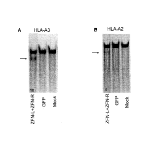
[0025] Figure 1, panels A and B, shows levels of HLA-A3 (Figure 1A)
and
HLA-A2 (Figure 1B) genetic disruption assessed by the SurveyorTM nuclease
assay.
The lower (fast-moving) bands (arrows) are digestion products indicating ZFN-
mediated gene modification. The numbers at the bottom of the lanes indicate
the
percentage of modified HLA-A alleles based on densitometry. DNA from mock
transfected cells and cells transfected with a GFP expression vector was used
for
negative controls.
7
CA 02904210 2015-09-03
WO 2014/165177
PCT/US2014/024660
[0026] Figure 2, panels A and B, show isolation of HLA-Aneg HEK293.
Figure 2A shows loss of HLA-A2 and HLA-A3 protein expression. Flow cytometry
analysis of HLA-A2 and HLA-A3 expression on parental HEK293 cells and three
derived genetically modified clones with loss of HLA-A (numbered 18.1, 8.18,
83).
Dotted lines represent isotype (HLA-A2) or SA-PE (HLA-A3) controls, solid line
represents HLA-A expression without IFN-y and INF-a, and filled lines
represent
HLA-A expression after culturing with 600 IU/mL of IFN-y and 10 ng/mL of INF-a
for 48 hours. Dashed lines in the parental column represent HLA-A2 or HLA-A3
expression on EBV-LCL. Figure 2B shows resistance of the HLA modified clones
to
CTL-mediated lysis. Parental HEK293 and derived HLA-Aneg clones were cultured
with IFN-y and TNF-a for 48 hours and pulsed with serial dilutions of the
cognate
HLA-A3 peptide RVWDLPGVLK (SEQ ID NO:1, see also NP 001103685.1),
derived from PANE1 (alternatively Centromere protein IV1 isoform c) and
recognized
by CTL clone 7A7) or the HLA-A2 peptide CIPPDSLLFPA (SEQ ID NO:2, also
alternative open reading frame of M4_199250.1) derived from C190RF48/A2 and
recognized by CTL clone GAS2B3-5) and evaluated for recognition by CTL clones
in
a 4-hour 51Cr release assay at an effector to target ratio of 20:1.HLA-A2+ LCL
(hatched bar) that expresses PANE1 mHAg (not peptide-loaded) were used as a
positive control.
[0027] Figure 3, panels A and B, show loss of HLA-A expression on primary
OKT3-propagated T cells after genetic editing with ZFNs. Figure 3A (top panel)
shows loss of cell surface expression of HLA-A2 after electro-transfer of mRNA
species encoding ZFN-L and ZFN-R targeting HLA-A2 (SBS#18889 and
SBS#18881, respectively, see U.S. Patent Publication No. 20120060230).
Cocxpression of IILA-A2, CD4, and CD8 were analyzed 4 days after electro-
transfer
of graded doses of the mRNA species encoding ZFN-L and ZFN-R. Flow cytometry
data were gated on the propidium iodide-negative, live cell population.
Numbers in
the lower right quadrant indicate the percentage of CD4 and CD8+ T cells that
are
HLA-A". Figure 3A (bottom panel) shows improved disruption of HLA-A
expression by "cold shock." Data were collected 4 days after electro-transfer
of
graded doses of the niRNA species encoding ZFN-L and ZFN-R. Cells were
cultured
at 30 C from days I to 3 after electro-transfer of ZFNs, returned to 37 C and
cultured
for one additional day before analysis. Figure 3B shows improved efficiency of
HLA-
8
CA 02904210 2015-09-03
WO 2014/165177
PCT/US2014/024660
A disruption by ZFN-L and ZFN-R fused to the heterodimeric Fok I domain
variants.
mRNA species encoding the ZFN-L and ZFN-R heterodimeric Fok I mutants EL:KK
targeting HLA-A were electro-transferred into primary T cells. HLA-A2
expression
was analyzed after culturing the cells for 4 days at 37 C or 3 days at 30 C
followed by
37 C for 1 day. X-axis represents CD4 and CD8 expression and y-axis represents
HLA-A2 expression.
[0028] Figure 4, panels A to C, show that expression of non-classical
HLA
molecules protects against NK-mediated cell lysis. Figure 4A shows the
immunophenotype of NK cells isolated from two individual PBMCs from healthy
donor (each donor designated as NK-1 and NK-2). Flow cytometry data shown are
gated for Plneg population. The numbers represent percentage of each upper
quadrant.
Figure 4B shows genetic modification of IILA class Il0w721.221 cells to
express
HLA-E and/or fl LA-G. The SB transposon/transposase system was used to
homogenously express HLA-E and/or HLA-G in three clones of 721.221 cells. Each
number represents percentage expression of HLA-G, HLA-E, or both HLA-G and
HLA-E as detected by flow cytometry. Figure 4 C shows specific lysis by NK
cells
targeting 721.221 cells. The relative ability of NK cells to kill parental
(HLA class
HLA-E+, HLA-G+, and both HLA-E+HLA-G+ 721.221 cells. Each column
represents the mean standard deviation (SD) * .01< P < 0.05, **P < .01; and
***P <
.001
[0029] Figure 5, panels A to C, shows enrichment of HLA-A eg primary
T
cells after genetic editing with ZFNs. Figure 5A shows generation of an HLA-
A2Ileg
T-cell population. HLA-A2neg T cells were enriched by magnetic bead-based
selection. Input dose of mRNA coding for ZFN and 3-day culture conditions (37
C
versus 30 C) after electro-transfer of mRNA are indicated. The numbers
represent
HLA-A2 negative population within CD4 and CD8 positive population. Figure 5B
shows SurveyorTM nuclease assay of the HLA-A211g T cells. Analysis of T cells
enriched for loss of HLA-A2 expression demonstrates disruption in the HLA-A2
locus by the appearance of fast-moving band (arrow). Figure 5C shows results
of
sequencing of the HLA"g T cells (SEQ ID NOs:39 to 53). PCR products using HLA-
A2-specific primers from enriched cell (2.5 ZFNs, EL:KK Fok I domain, 30 C
treatment) were cloned into a TOPO vector (Invitrogen) and plasmid products
were
sequenced. The wild type sequence is listed at the top with the expected ZEN
binding
sites underlined. Shown below are the sequences obtained from the ZEN-treated
and
9
CA 02904210 2015-09-03
WO 2014/165177
PCT/US2014/024660
enriched cells. Deletions are indicated by hyphens and sequence changes are
highlighted in bold. All 18 sequence changes result in frame shifts predicted
to
prevent protein translation.
[0030] Figure 6, panels A to C, show loss of HLA-A expression on
primary
.. CD19-specific CAR+ T cells genetically edited with ZFNs. Figure 6A shows
disruption of HLA-A2 in CAR+ T cells by electro-transfer of mRNA encoding
ZFNs.
T cells from a HLA-A2+ donor were electroporated and propagated to express
CD19-
specific CAR (CD19RCD28). These T cells were re-electroporated with 2.5 pg of
each mRNA encoding the heterodimeric Fok I domain variants of the HLA-A-
specific
ZFNs (ZFN-L-EL and ZFN-R-KK). HLA-A2 expression was analyzed after culturing
at 30 C for 3 days followed by 37 C for 1 day. Enrichment of the HLA-A2 eg
population was performed by paramagnetic selection. Figure 6B shows HLA-A'eg
CAR + T cells evade lysis by HLA-A2 restricted CTL. Pools of the indicated
CAR+ T
cells were pulsed with serial dilutions of cognate peptide before being used
as targets
in a CRA. CTL clone GAS2B3-5, which is specific for C190RF48/A2, was added at
an effector-to-target ratio of 20:1. Figure 6C shows ZFN-modified HLAneg CAR+
T
cells maintain desired antigen-specific cytotoxicity. Redirected specificity
for CD19
by HLA-A eg T cells expressing CD19RCD28 CAR was demonstrated using the
mouse T-cell line EL4 genetically modified to expresses a truncated variant of
human
CD19. Expression of introduced human CD19 on EL4 was 100%.
[0031] Figure 7 shows ZFN-mediated elimination of HLA-A expression on
human ESC. The HLAA2+ HLA-24+hES parental cell line WIBR3 was modified by
ZFN and donor plasmid coding for antibiotic resistance. Clones (5230, 5255,
5258)
were chosen with loss of HLA-A expression and differentiated into fibroblasts.
Expression of HLA-A2 and HLA-A24 on derived fibroblasts was assessed by flow
cytometry after culturing with 600 IU/mL of IFN-y and 10 ng/mL of TNF-a for 48
hours. Dashed line in parental panel represents isotype control.
DETAILED DESCRIPTION
[0032] Disclosed herein are compositions and methods for generating cells
in
which one or more classic HLA genes are inactivated but which express one or
more
non-classic HLA genes. Cells modified targeted in this manner can be used as
therapeutics, for example, transplants, as the presence of the non-classic HLA
gene(s)
CA 02904210 2015-09-03
WO 2014/165177 PCT/US2014/024660
reduces or eliminates NK-mediated lysis of HLA null cells. Additionally, other
genes
of interest may be inserted into cells in which the HLA genes have been
manipulated.
[0033] Thus, the methods and compositions described herein provide
methods
for treatment of HLA related disorders, and these methods and compositions can
comprise zinc finger transcription factors capable of modulating target genes
as well
as engineered zinc finger nucleases.
General
[0034] Practice of the methods, as well as preparation and use of the
compositions disclosed herein employ, unless otherwise indicated, conventional
techniques in molecular biology, biochemistry, chromatin structure and
analysis,
computational chemistry, cell culture, recombinant DNA and related fields as
are
within the skill of the art. These techniques are fully explained in the
literature. See,
for example, Sambrook et al MOLECULAR CLONING: A LABORATORY MANUAL,
Second edition, Cold Spring Harbor Laboratory Press, 1989 and Third edition,
2001;
Ausubel et al., CURRENT PROTOCOLS IN MOLECULAR BIOLOGY, John Wiley & Sons,
New York, 1987 and periodic updates; the series METHODS IN ENZYMOLOGY,
Academic Press, San Diego; Wolfe, CHROMATIN STRUCTURE AND FUNCTION, Third
edition, Academic Press, San Diego, 1998; METHODS IN ENZYMOLOGY, Vol. 304,
"Chromatin" (P.M. Wassarman and A. P. Wolffe, eds.), Academic Press, San
Diego,
1999; and METHODS IN MOLECULAR BIOLOGY, Vol. 119, "Chromatin Protocols"
(P.B. Becker, ed.) Humana Press, Totowa, 1999.
Definitions
[0035] The terms "nucleic acid," "polynucleotide," and "oligonucleotide"
are used
interchangeably and refer to a deoxyribonucleotide or ribonucleotide polymer,
in linear or
circular confoi __ illation, and in either single- or double-stranded form.
For the purposes of
the present disclosure, these terms are not to be construed as limiting with
respect to the
length of a polymer. The terms can encompass known analogues of natural
nucleotides, as
well as nucleotides that are modified in the base, sugar and/or phosphate
moieties (e.g.,
phosphorothioate backbones). In general, an analogue of a particular
nucleotide has the
same base-pairing specificity; i.e., an analogue of A will base-pair with T.
[0036] The terms "polypeptide," "peptide" and "protein" are used
interchangeably
to refer to a polymer of amino acid residues. The term also applies to amino
acid polymers
11
in which one or more amino acids are chemical analogues or modified
derivatives of
corresponding naturally-occurring amino acids.
[0037] "Binding" refers to a sequence-specific, non-covalent
interaction
between macromolecules (e.g., between a protein and a nucleic acid). Not all
components of a binding interaction need be sequence-specific (e.g., contacts
with
phosphate residues in a DNA backbone), as long as the interaction as a whole
is
sequence-specific. Such interactions are generally characterized by a
dissociation
constant (Kd) of 10-6111-1 or lower. "Affinity" refers to the strength of
binding:
increased binding affinity being correlated with a lower Kd.
[0038] A "binding protein" is a protein that is able to bind non-covalently
to
another molecule. A binding protein can bind to, for example, a DNA molecule
(a DNA-
binding protein), an RNA molecule (an RNA-binding protein) and/or a protein
molecule (a
protein-binding protein). In the case of a protein-binding protein, it can
bind to itself (to
form homodimers, homotrimers, etc.) and/or it can bind to one or more
molecules of a
different protein or proteins. A binding protein can have more than one type
of binding
activity. For example, zinc finger proteins have DNA-binding, RNA-binding and
protein-
binding activity.
[0039] A "zinc finger DNA binding protein" (or binding domain) is a
protein, or a
domain within a larger protein, that binds DNA in a sequence-specific manner
through one
or more zinc fingers, which are regions of amino acid sequence within the
binding domain
whose structure is stabilized through coordination of a zinc ion. The term
zinc finger
DNA binding protein is often abbreviated as zinc finger protein or ZFP.
[0040] A "TALE DNA binding domain" or "TALE" is a polypeptide
comprising
one or more TALE repeat domains/units. The repeat domains are involved in
binding of
the TALE to its cognate target DNA sequence. A single "repeat unit" (also
referred to as a
"repeat") is typically 33-35 amino acids in length and exhibits at least some
sequence
homology with other TALE repeat sequences within a naturally occurring TALE
protein.
See, e.g., U.S. Patent No. 8,586,526.
[0041] Zinc finger and TALE DNA-binding domains can be "engineered"
to
bind to a predetermined nucleotide sequence, for example via engineering
(altering
one or more amino acids) of the recognition helix region of a naturally
occurring zinc
finger protein or by engineering of the amino acids involved in DNA binding
(the
repeat variable diresidue or RVD region). Therefore, engineered zinc finger
proteins
or TALE proteins are proteins that are non-naturally occurring. Non-limiting
12
CA 2904210 2020-03-30
CA 02904210 2015-09-03
WO 2014/165177
PCT/US2014/024660
examples of methods for engineering zinc finger proteins and TALEs are design
and
selection. A designed protein is a protein not occurring in nature whose
design/composition results principally from rational criteria. Rational
criteria for
design include application of substitution rules and computerized algorithms
for
processing information in a database storing information of existing ZFP or
TALE
designs and binding data. See, for example, U.S. Patent Nos. 8,586,526;
6,140,081;
6,453,242; and 6,534,261; see also WO 98/53058; WO 98/53059; WO 98/53060;
WO 02/016536 and WO 03/016496.
[0042] A "selected" zinc finger protein or TALE is a protein not found
in nature
whose production results primarily from an empirical process such as phage
display,
interaction trap or hybrid selection. See e.g., US 5,789,538; US 5,925,523;
US 6,007,988; US 6,013,453; US 6,200,759; WO 95/19431; WO 96/06166;
WO 98/53057; WO 98/54311; WO 00/27878; WO 01/60970 WO 01/88197 and
WO 02/099084.
[0043] "Recombination" refers to a process of exchange of genetic
infoimation between two polynucleotides. For the purposes of this disclosure,
"homologous recombination (HR)' refers to the specialized foul.' of such
exchange
that takes place, for example, during repair of double-strand breaks in cells
via
homology-directed repair mechanisms. This process requires nucleotide sequence
homology, uses a "donor" molecule to template repair of a "target" molecule
(i.e., the
one that experienced the double-strand break), and is variously known as "non-
crossover gene conversion" or "short tract gene conversion," because it leads
to the
transfer of genetic information from the donor to the target. Without wishing
to be
bound by any particular theory, such transfer can involve mismatch correction
of
heteroduplex DNA that forms between the broken target and the donor, and/or
"synthesis-dependent strand annealing," in which the donor is used to
resynthesize
genetic information that will become part of the target, and/or related
processes. Such
specialized HR often results in an alteration of the sequence of the target
molecule
such that part or all of the sequence of the donor polynucleotide is
incorporated into
the target polynucleotide.
[0044] In the methods of the disclosure, one or more targeted
nucleases as
described herein create a double-stranded break in the target sequence (e.g.,
cellular
chromatin) at a predetermined site, and a "donor" polynucleotide, having
homology to
the nucleotide sequence in the region of the break, can be introduced into the
cell.
13
CA 02904210 2015-09-03
WO 2014/165177
PCT/US2014/024660
The presence of the double-stranded break has been shown to facilitate
integration of
the donor sequence. The donor sequence may be physically integrated or,
alternatively, the donor polynucleotide is used as a template for repair of
the break via
homologous recombination, resulting in the introduction of all or part of the
nucleotide sequence as in the donor into the cellular chromatin. Thus, a first
sequence
in cellular chromatin can be altered and, in certain embodiments, can be
converted
into a sequence present in a donor polynucleotide. Thus, the use of the terms
"replace" or "replacement" can be understood to represent replacement of one
nucleotide sequence by another, (i.e., replacement of a sequence in the
informational
sense), and does not necessarily require physical or chemical replacement of
one
polynucleotide by another.
[0045] In any of the methods described herein, additional pairs of
zinc-finger
proteins can be used for additional double-stranded cleavage of additional
target sites
within the cell.
[0046] In certain embodiments of methods for targeted recombination and/or
replacement and/or alteration of a sequence in a region of interest in
cellular
chromatin, a chromosomal sequence is altered by homologous recombination with
an
exogenous "donor" nucleotide sequence. Such homologous recombination is
stimulated by the presence of a double-stranded break in cellular chromatin,
if
sequences homologous to the region of the break are present.
[0047] In any of the methods described herein, the first nucleotide
sequence
(the "donor sequence") can contain sequences that arc homologous, but not
identical,
to genomic sequences in the region of interest, thereby stimulating homologous
recombination to insert a non-identical sequence in the region of interest.
Thus, in
certain embodiments, portions of the donor sequence that are homologous to
sequences in the region of interest exhibit between about 80 to 99% (or any
integer
therebetween) sequence identity to the genomic sequence that is replaced. In
other
embodiments, the homology between the donor and genomic sequence is higher
than
99%, for example if only 1 nucleotide differs as between donor and genomic
sequences of over 100 contiguous base pairs. In certain cases, a non-
homologous
portion of the donor sequence can contain sequences not present in the region
of
interest, such that new sequences are introduced into the region of interest.
In these
instances, the non-homologous sequence is generally flanked by sequences of 50-
1,000 base pairs (or any integral value therebetween) or any number of base
pairs
14
greater than 1,000, that are homologous or identical to sequences in the
region of
interest. In other embodiments, the donor sequence is non-homologous to the
first
sequence, and is inserted into the genome by non-homologous recombination
mechanisms.
[0048] Any of the methods described herein can be used for partial or
complete inactivation of one or more target sequences in a cell by targeted
integration
of donor sequence that disrupts expression of the gene(s) of interest. Cell
lines with
partially or completely inactivated genes are also provided.
[0049] Furthermore, the methods of targeted integration as
described herein
can also be used to integrate one or more exogenous sequences. The exogenous
=
nucleic acid sequence can comprise, for example, one or more genes or cDNA
molecules, or any type of coding or noncoding sequence, as well as one or more
control elements (e.g., promoters). In addition, the exogenous nucleic acid
sequence
may produce one or more RNA molecules (e.g., small hairpin RNAs (shRNAs),
inhibitory RNAs (RNAis), microRNAs (miRNAs), etc.).
[0050] "Cleavage" refers to the breakage of the covalent backbone
of a DNA
molecule. Cleavage can be initiated by a variety of methods including, but not
limited
to, enzymatic or chemical hydrolysis of a phosphodiester bond. Both single-
stranded
cleavage and double-stranded cleavage are possible, and double-stranded
cleavage
can occur as a result of two distinct single-stranded cleavage events. DNA
cleavage
can result in the production of either blunt ends or staggered ends. In
certain
embodiments, fusion polypeptides are used for targeted double-stranded DNA
cleavage.
[0051] A "cleavage half-domain" is a polypeptide sequence which, in
conjunction with a second polypeptide (either identical or different) forms a
complex
having cleavage activity (preferably double-strand cleavage activity). The
terms "first
and second cleavage half-domains;" "+ and ¨ cleavage half-domains" and "right
and
left cleavage half-domains" are used interchangeably to refer to pairs of
cleavage half-
domains that dimerize.
[0052] An "engineered cleavage half-domain" is a cleavage half-domain that
has been modified so as to form obligate heterodimers with another cleavage
half-
domain (e.g., another engineered cleavage half-domain). See, also, U.S. Patent
Nos.
7,888,121; 7,914,796; 8,034,598; 8,623,618 and U.S. Patent Publication No.
2011/0201055.
CA 2904210 2020-03-30
CA 02904210 2015-09-03
WO 2014/165177
PCT/US2014/024660
[0053] The term "sequence" refers to a nucleotide sequence of any
length,
which can be DNA or RNA; can be linear, circular or branched and can be either
single-stranded or double stranded. The term "donor sequence" refers to a
nucleotide
sequence that is inserted into a genome. A donor sequence can be of any
length, for
example between 2 and 10,000 nucleotides in length (or any integer value
therebetween or thereabove), preferably between about 100 and 1,000
nucleotides in
length (or any integer therebetween), more preferably between about 200 and
500
nucleotides in length.
[0054] "Chromatin" is the nucleoprotein structure comprising the
cellular
genome. Cellular chromatin comprises nucleic acid, primarily DNA, and protein,
including histones and non-histone chromosomal proteins. The majority of
eukaryotic cellular chromatin exists in the form of nucleosomes, wherein a
nucleosome core comprises approximately 150 base pairs of DNA associated with
an
octamer comprising two each of histones H2A, H2B, H3 and H4; and linker DNA
(of
variable length depending on the organism) extends between nucleosome cores. A
molecule of histone H1 is generally associated with the linker DNA. For the
purposes
of the present disclosure, the term "chromatin" is meant to encompass all
types of
cellular nucleoprotein, both prokaryotic and eukaryotic. Cellular chromatin
includes
both chromosomal and episomal chromatin.
[0055] A "chromosome," is a chromatin complex comprising all or a portion
of the genome of a cell. The genome of a cell is often characterized by its
karyotype,
which is the collection of all the chromosomes that comprise the genome of the
cell.
The genome of a cell can comprise one or more chromosomes.
[0056] An "episome" is a replicating nucleic acid, nucleoprotein
complex or
other structure comprising a nucleic acid that is not part of the chromosomal
karyotype of a cell. Examples of episomes include plasmids and certain viral
genomes.
[0057] A "target site" or "target sequence" is a nucleic acid sequence
that
defines a portion of a nucleic acid to which a binding molecule will bind,
provided
sufficient conditions for binding exist. For example, the sequence 5' GAATTC
3' is a
target site for the Eco RI restriction endonuclease.
[0058] An "exogenous" molecule is a molecule that is not normally
present in
a cell, but can be introduced into a cell by one or more genetic, biochemical
or other
methods. "Normal presence in the cell" is determined with respect to the
particular
16
CA 02904210 2015-09-03
WO 2014/165177
PCT/US2014/024660
developmental stage and environmental conditions of the cell. Thus, for
example, a
molecule that is present only during embryonic development of muscle is an
exogenous molecule with respect to an adult muscle cell. Similarly, a molecule
induced by heat shock is an exogenous molecule with respect to a non-heat-
shocked
cell. An exogenous molecule can comprise, for example, a functioning version
of a
malfunctioning endogenous molecule or a malfunctioning version of a normally-
functioning endogenous molecule.
[00591 An exogenous molecule can be, among other things, a small
molecule,
such as is generated by a combinatorial chemistry process, or a macromolecule
such
as a protein, nucleic acid, carbohydrate, lipid, glycoprotein, lipoprotein,
polysaccharide, any modified derivative of the above molecules, or any complex
comprising one or more of the above molecules. Nucleic acids include DNA and
RNA, can be single- or double-stranded; can be linear, branched or circular;
and can
be of any length. Nucleic acids include those capable of forming duplexes, as
well as
triplex-forming nucleic acids. See, for example, U.S. Patent Nos. 5,176,996
and
5,422,251. Proteins include, but are not limited to, DNA-binding proteins,
transcription factors, chromatin remodeling factors, methylated DNA binding
proteins, polymerases, methylases, demethylases, acetylases, deacetylases,
kinases,
phosphatases, integrases, recombinases, ligases, topoisomerases, gyrases and
helicases.
[00601 An exogenous molecule can be the same type of molecule as an
endogenous molecule, e.g., an exogenous protein or nucleic acid. For example,
an
exogenous nucleic acid can comprise an infecting viral genome, a plasmid or
episome
introduced into a cell, or a chromosome that is not normally present in the
cell.
Methods for the introduction of exogenous molecules into cells are known to
those of
skill in the art and include, but are not limited to, lipid-mediated transfer
(i.e.,
liposomes, including neutral and cationic lipids), electroporation, direct
injection, cell
fusion, particle bombardment, calcium phosphate co-precipitation, DEAE-dextran-
mediated transfer and viral vector-mediated transfer. An exogenous molecule
can also
be the same type of molecule as an endogenous molecule but derived from a
different
species than the cell is derived from. For example, a human nucleic acid
sequence
may be introduced into a cell line originally derived from a mouse or hamster.
[00611 By contrast, an "endogenous" molecule is one that is normally
present
in a particular cell at a particular developmental stage under particular
environmental
17
CA 02904210 2015-09-03
WO 2014/165177
PCT/US2014/024660
conditions. For example, an endogenous nucleic acid can comprise a chromosome,
the genome of a mitochondrion, chloroplast or other organelle, or a naturally-
occurring episomal nucleic acid. Additional endogenous molecules can include
proteins, for example, transcription factors and enzymes.
[0062] A "fusion' molecule is a molecule in which two or more subunit
molecules are linked, preferably covalently. The subunit molecules can be the
same
chemical type of molecule, or can be different chemical types of molecules.
Examples of the first type of fusion molecule include, but are not limited to,
fusion
proteins (for example, a fusion between a ZFP or TALE DNA-binding domain and
one or more activation domains) and fusion nucleic acids (for example, a
nucleic acid
encoding the fusion protein described supra). Examples of the second type of
fusion
molecule include, but are not limited to, a fusion between a triplex-forming
nucleic
acid and a polypeptide, and a fusion between a minor groove binder and a
nucleic
acid.
[0063] Expression of a fusion protein in a cell can result from delivery of
the
fusion protein to the cell or by delivery of a polynucleotide encoding the
fusion
protein to a cell, wherein the polynucleotide is transcribed, and the
transcript is
translated, to generate the fusion protein. Trans-splicing, polypeptide
cleavage and
polypeptide ligation can also be involved in expression of a protein in a
cell. Methods
for polynucleotide and polypeptide delivery to cells are presented elsewhere
in this
disclosure.
[0064] A "gene," for the purposes of the present disclosure, includes
a DNA
region encoding a gene product (see infra), as well as all DNA regions which
regulate
the production of the gene product, whether or not such regulatory sequences
are
adjacent to coding and/or transcribed sequences. Accordingly, a gene includes,
but is
not necessarily limited to, promoter sequences, terminators, translational
regulatory
sequences such as ribosome binding sites and internal ribosome entry sites,
enhancers,
silencers, insulators, boundary elements, replication origins, matrix
attachment sites
and locus control regions.
[0065] "Gene expression" refers to the conversion of the information,
contained in a gene, into a gene product. A gene product can be the direct
transcriptional product of a gene (e.g., mRNA, tRNA, rRNA, antisense RNA,
ribozyme. structural RNA or any other type of RNA) or a protein produced by
translation of an mRNA. Gene products also include RNAs which are modified, by
18
CA 02904210 2015-09-03
WO 2014/165177
PCT/US2014/024660
processes such as capping, polyadenylation, methylation, and editing, and
proteins
modified by, for example, methylation, acetylation, phosphorylation,
ubiquitination,
ADP-ribosylation, myristilation, and glyeosylation.
[0066] "Modulation" of gene expression refers to a change in the
activity of a
gene. Modulation of expression can include, but is not limited to, gene
activation and
gene repression. Genome editing (e.g., cleavage, alteration, inactivation,
random
mutation) can be used to modulate expression. Gene inactivation refers to any
reduction in gene expression as compared to a cell that does not include a ZFP
as
described herein. Thus, gene inactivation may be partial or complete.
[0067] A "region of interest" is any region of cellular chromatin, such as,
for
example, a gene or a non-coding sequence within or adjacent to a gene, in
which it is
desirable to bind an exogenous molecule. Binding can be for the purposes of
targeted
DNA cleavage and/or targeted recombination. A region of interest can be
present in a
chromosome, an episome, an organellar genome (e.g., mitoehondrial,
chloroplast), or
an infecting viral genome, for example. A region of interest can be within the
coding
region of a gene, within transcribed non-coding regions such as, for example,
leader
sequences, trailer sequences or introns, or within non-transcribed regions,
either
upstream or downstream of the coding region. A region of interest can be as
small as
a single nucleotide pair or up to 2,000 nucleotide pairs in length, or any
integral value
of nucleotide pairs.
[0068] "Eukaryotic" cells include, but are not limited to, fungal
cells (such as
yeast), plant cells, animal cells, mammalian cells and human cells (e.g., T-
cells).
[0069] The terms "operative linkage" and "operatively linked" (or
"operably
linked") are used interchangeably with reference to a juxtaposition of two or
more
components (such as sequence elements), in which the components are arranged
such
that both components function normally and allow the possibility that at least
one of
the components can mediate a function that is exerted upon at least one of the
other
components. By way of illustration, a transcriptional regulatory sequence,
such as a
promoter, is operatively linked to a coding sequence if the transcriptional
regulatory
sequence controls the level of transcription of the coding sequence in
response to the
presence or absence of one or more transcriptional regulatory factors. A
transcriptional regulatory sequence is generally operatively linked in cis
with a coding
sequence, but need not be directly adjacent to it. For example, an enhancer is
a
19
CA 02904210 2015-09-03
WO 2014/165177
PCT/US2014/024660
transcriptional regulatory sequence that is operatively linked to a coding
sequence,
even though they are not contiguous.
[0070] With respect to fusion polypeptides, the term "operatively
linked" can
refer to the fact that each of the components performs the same function in
linkage to
the other component as it would if it were not so linked. For example, with
respect to
a fusion polypeptide in which a DNA-binding domain (e.g, ZFP, TALE) is fused
to
an activation domain, the DNA-binding domain and the activation domain are in
operative linkage if, in the fusion polypeptide, the DNA-binding domain
portion is
able to bind its target site and/or its binding site, while the activation
domain is able to
up-regulate gene expression. When a fusion polypeptide in which a DNA-binding
domain is fused to a cleavage domain, the DNA-binding domain and the cleavage
domain are in operative linkage if, in the fusion polypeptide, the DNA-binding
domain portion is able to bind its target site and/or its binding site, while
the cleavage
domain is able to cleave DNA in the vicinity of the target site. Similarly,
with respect
to a fusion polypeptide in which a DNA-binding domain is fused to an
activation or
repression domain, the DNA-binding domain and the activation or repression
domain
are in operative linkage if, in the fusion polypeptide, the DNA-binding domain
portion is able to bind its target site and/or its binding site, while the
activation
domain is able to upregulate gene expression or the repression domain is able
to
downregulate gene expression.
[0071] A "functional fragment" of a protein, polypeptide or nucleic
acid is a
protein, polypeptide or nucleic acid whose sequence is not identical to the
full-length
protein, polypeptide or nucleic acid, yet retains the same function as the
full-length
protein, polypeptide or nucleic acid. A functional fragment can possess more,
fewer,
or the same number of residues as the corresponding native molecule, and/or
can
contain one or more amino acid or nucleotide substitutions. Methods for
determining
the function of a nucleic acid (e.g., coding function, ability to hybridize to
another
nucleic acid) are well-known in the art. Similarly, methods for determining
protein
function are well-known. For example, the DNA-binding function of a
polypeptide
can be determined, for example, by filter-binding, electrophoretic mobility-
shift, or
immunoprecipitation assays. DNA cleavage can be assayed by gel
electrophoresis.
See Ausubel et al., supra. The ability of a protein to interact with another
protein can
be determined, for example, by co-immunoprecipitation, two-hybrid assays or
complementation, both genetic and biochemical. See, for example, Fields et al.
(1989) Nature 340:245-246; U.S. Patent No. 5,585,245 and PCT WO 98/44350.
[0072] A "vector" is capable of transferring gene sequences to
target cells.
Typically, "vector construct," "expression vector," and "gene transfer
vector," mean
any nucleic acid construct capable of directing the expression of a gene of
interest and
which can transfer gene sequences to target cells. Thus, the term includes
cloning, and
expression vehicles, as well as integrating vectors.
[0073] A "reporter gene" or "reporter sequence" refers to any
sequence that
produces a protein product that is easily measured, preferably although not
necessarily
in a routine assay. Suitable reporter genes include, but are not limited to,
sequences
encoding proteins that mediate antibiotic resistance (e.g., ampicillin
resistance,
neomycin resistance, G418 resistance, puromycin resistance), sequences
encoding
colored or fluorescent or luminescent proteins (e.g., green fluorescent
protein,
enhanced green fluorescent protein, red fluorescent protein, luciferase), and
proteins
which mediate enhanced cell growth and/or gene amplification (e.g.,
dihydrofolate
reductase). Epitope tags include, for example, one or more copies of FLAG,
His,
myc, Tap, HA or any detectable amino acid sequence. "Expression tags" include
sequences that encode reporters that may be operably linked to a desired gene
sequence in order to monitor expression of the gene of interest.
DNA-binding domains
[0074] Described herein are compositions comprising a DNA-binding
domain
that specifically binds to a target site in any gene comprising a HLA gene or
a HLA
regulator. Any DNA-binding domain can be used in the compositions and methods
disclosed herein.
[0075] In certain embodiments, the DNA binding domain comprises a
zinc
finger protein. Preferably, the zinc finger protein is non-naturally occurring
in that it
is engineered to bind to a target site of choice. See, for example, Beerli et
al. (2002)
Nature Biotechnol. 20:135-141; Pabo et al. (2001) Ann. Rev. Biochem. 70:313-
340;
Isalan etal. (2001) Nature Biotechnol. 19:656-660; Segal etal. (2001) Curr.
Opin.
Biotechnol. 12:632-637; Choo et al. (2000) Curr. Opin. Struct. Biol. 10:411-
416; U.S.
Patent Nos. 6,453,242; 6,534,261; 6,599,692; 6,503,717; 6,689,558; 7,030,215;
6,794,136; 7,067,317; 7,262,054; 7,070,934; 7,361,635; 7,253,273; and U.S.
Patent
Publication Nos. 2005/0064474; 2007/0218528; 2005/0267061. In certain
21.
CA 2904210 2020-03-30
embodiments, the DNA-binding domain comprises a zinc finger protein disclosed
in
U.S. Patent Publication No. 2012/0060230 (e.g., Table 1).
[0076] An engineered zinc finger binding domain can have a novel
binding
specificity, compared to a naturally-occurring zinc finger protein.
Engineering
methods include, but are not limited to, rational design and various types of
selection.
Rational design includes, for example, using databases comprising triplet (or
quadruplet) nucleotide sequences and individual zinc finger amino acid
sequences, in
which each triplet or quadruplet nucleotide sequence is associated with one or
more
amino acid sequences of zinc fingers which bind the particular triplet or
quadruplet
sequence. See, for example, U.S. Patents 6,453,242 and 6,534,261.
[00771 Exemplary selection methods, including phage display and two-
hybrid
systems, are disclosed in US Patents 5,789,538; 5,925,523; 6,007,988;
6,013,453;
6,410,248; 6,140,466; 6,200,759; and 6,242,568; as well as WO 98/37186;
WO 98/53057; WO 00/27878; WO 01/88197 and GB 2,338,237. In addition,
enhancement of binding specificity for zinc finger binding domains has been
described, for example, in U.S. Patent No. 6,794,136.
[0078] In addition, as disclosed in these and other references,
zinc finger
domains and/or multi-fingered zinc finger proteins may be linked together
using any
suitable linker sequences, including for example, linkers of 5 or more amino
acids in
length. See, also, U.S. Patent Nos. 6,479,626; 6,903,185; and 7,153,949 for
exemplary linker sequences 6 or more amino acids in length. The proteins
described
herein may include any combination of suitable linkers between the individual
zinc
fingers of the protein. In addition, enhancement of binding specificity for
zinc finger
binding domains has been described, for example, in U.S. Patent No. 6,794,136.
[0079] Selection of target sites; ZFPs and methods for design and
construction
of fusion proteins (and polynucleotides encoding same) are known to those of
skill in
the art and described in detail in U.S. Patent Nos. 6,140,0815; 789,538;
6,453,242;
6,534,261; 5,925,523; 6,007,988; 6,013,453; 6,200,759; WO 95/19431;
W096/06166; W098/53057; W098/54311; W000/27878; WO 01/60970
WO 01/88197; WO 02/099084; WO 98/53058; WO 98/53059; WO 98/53060;
WO 02/016536 and WO 03/016496.
22
CA 2904210 2020-03-30
[0080] In addition, as disclosed in these and other references, zinc
finger
domains and/or multi-fingered zinc finger proteins may be linked together
using any
suitable linker sequences, including for example, linkers of 5 or more amino
acids in
length. See, also, U.S. Patent Nos. 6,479,626; 6,903,185; and 7,153,949 for
exemplary linker sequences 6 or more amino acids in length. The proteins
described
herein may include any combination of suitable linkers between the individual
zinc
fingers of the protein.
[0081] In certain embodiments, the DNA binding domain is an
engineered
zinc finger protein that binds (in a sequence-specific manner) to a target
site in a HLA
gene or I-ILA regulatory gene and modulates expression of HLA. The ZFPs can
bind
selectively to a specific haplotype of interest. For a discussion of HLA
haplotypes
identified in the United States population and their frequency according to
different
races, see Maiers et al (2007) Human Immunology 68: 779- 788.
[0082] Additionally, ZFPs are provided that bind to functional HLA
regulator
genes including, but not limited to, Tap 1, Tap2, Tapascin, CTFIIA, and RFX5.
HLA
target sites typically include at least one zinc finger but can include a
plurality of zinc
fingers (e.g., 2, 3, 4, 5, 6 or more fingers). Usually, the ZFPs include at
least three
fingers. Certain of the ZFPs include four, five or six fingers. The ZFPs that
include
three fingers typically recognize a target site that includes 9 or 10
nucleotides; ZFPs
that include four fingers typically recognize a target site that includes 12
to 14
nucleotides; while ZFPs having six fingers can recognize target sites that
include 18
to 21 nucleotides. The ZFPs can also be fusion proteins that include one or
more
regulatory domains, which domains can be transcriptional activation or
repression
domains.
[0083] Specific examples of ZFPs are disclosed in Table 1 of U.S. Patent
Publication No. 20120060230.
[0084] In some embodiments, the DNA-binding domain may be derived
from
a nuclease. For example, the recognition sequences of homing endonucleases and
meganucleases such as I-SceI,I-CeuI,PI-PspI,PI-Sce,I-SceIV,I-CsmI,I-PanI, I-
SceII,I-PpoI, I-SceIII, I-CreI,I-TevI, I-TevII and I-TevIII are known. See
also U.S.
Patent No. 5,420,032; U.S. Patent No. 6,833,252; Belfort et al. (1997) Nucleic
Acids
Res. 25:3379-3388; Dujon et al. (1989) Gene 82:115-118; Perler et al. (1994)
Nucleic Acids Res. 22, 1125-1127; Jasin (1996) Trends Genet. 12:224-228;
Gimble
et al. (1996)J. Mol. Biol. 263:163-180; Argast et al. (1998)J. Mol. Biol.
280:345-
23
Date Recue/Date Received 2021-03-08
353 and the New England Biolabs catalogue. In addition, the DNA-binding
specificity of homing endonucleases and meganucleases can be engineered to
bind
non-natural target sites. See, for example, Chevalier et al. (2002) Malec.
Cell 10:895-
905; Epinat et al. (2003) Nucleic Acids Res. 31:2952-2962; Ashworth et al.
(2006)
Nature 441:656-659; Paques et al. (2007) Current Gene Therapy 7:49-66; U.S.
Patent Publication No. 20070117128.
[0085] In other embodiments, the DNA binding domain comprises an
engineered domain from a TAL effector similar to those derived from the plant
pathogens Xanthomonas (see Boch et al, (2009) Science 326: 1509-1512 and
Moscou
and Bogdanove, (2009) 5cience326: 1501) and Ralstonia (see Heuer et al (2007)
Applied and Environmental Microbiology 73(13): 4379-4384); U.S. Patent
Application Nos. 20110301073 and 20110145940. The plant pathogenic bacteria of
the genus Xanthomonas are known to cause many diseases in important crop
plants.
Pathogenicity of Xanthomonas depends on a conserved type III secretion (T3S)
system which injects more than 25 different effector proteins into the plant
cell.
Among these injected proteins are transcription activator-like effectors
(TALE) which
mimic plant transcriptional activators and manipulate the plant transcriptome
(see Kay
et al (2007) 5cience318:648-651). These proteins contain a DNA binding domain
and
a transcriptional activation domain. One of the most well characterized TALEs
is
AvrBs3 from Xanthomonas campestgris pv. Vesicatoria (see Bonas et al (1989)
Mal
Gen Genet 218: 127-136 and W02010079430). TALEs contain a centralized domain
of tandem repeats, each repeat containing approximately 34 amino acids, which
are
key to the DNA binding specificity of these proteins. In addition, they
contain a
nuclear localization sequence and an acidic transcriptional activation domain
(for a
review see Schornack S, et al (2006)J Plant Physiol 163(3): 256-272). In
addition, in
the phytopathogenic bacteria Ralstonia solanacearum two genes, designated
brgll
and hpx17 have been found that are homologous to the AvrBs3 family of
Xanthomonas in the R. solanacearum biovar 1 strain GMI1000 and in the biovar 4
strain RS1000 (See Heuer et al (2007) Appl and Envir Micro 73(13): 4379-4384).
These genes are 98.9% identical in nucleotide sequence to each other but
differ by a
deletion of 1,575 bp in the repeat domain of hpx17. However, both gene
products
have less than 40% sequence identity with AvrBs3 family proteins of
Xanthomonas.
24
Date Recue/Date Received 2021-03-08
[0086] In addition, as disclosed in these and other references, zinc
finger
domains and/or multi-fingered zinc finger proteins or TALEs may be linked
together
using any suitable linker sequences, including for example, linkers of 5 or
more
amino acids in length. See, also, U.S. Patent Nos. 6,479,626; 6,903,185; and
7,153,949 for exemplary linker sequences 6 or more amino acids in length. The
proteins described herein may include any combination of suitable linkers
between
the individual zinc fingers of the protein. In addition, enhancement of
binding
specificity for zinc finger binding domains has been described, for example,
in U.S.
Patent No. 6,794,136.
Fusion proteins
[0087] Fusion proteins comprising DNA-binding proteins (e.g., ZFPs
or
TALEs) as described herein and a heterologous regulatory (functional) domain
(or
functional fragment thereof) are also provided. Common domains include, e.g.,
transcription factor domains (activators, repressors, co-activators, co-
repressors),
silencers, oncogenes (e.g., myc, jun, fos, myb, max, mad, rel, ets, bcl, myb,
mos
family members etc.); DNA repair enzymes and their associated factors and
modifiers; DNA rearrangement enzymes and their associated factors and
modifiers;
chromatin associated proteins and their modifiers (e.g. kinases, acetylases
and
deacetylases); and DNA modifying enzymes (e.g., methyltransferases,
topoisomerases, helicases, ligases, kinases, phosphatases, polymerases,
endonucleases) and their associated factors and modifiers. U.S. Patent
Application
Publication Nos. 20050064474; 20060188987 and 2007/0218528 for details
regarding
fusions of DNA-binding domains and nuclease cleavage domains.
[0088] Suitable domains for achieving activation include the HSV VP16
activation domain (see, e.g., Hagmann et al., J. Viral. 71, 5952-5962 (1997))
nuclear
hormone receptors (see, e.g., Torchia et al., Curr. Opin. Cell. Biol. 10:373-
383
(1998)); the p65 subunit of nuclear factor kappa B (Bitko & Bank, J. Viral.
72:5610-
5618 (1998) and Doyle & Hunt, Neuroreport 8:2937-2942 (1997)); Liu et al.,
Cancer
Gene Ther. 5:3-28 (1998)), or artificial chimeric functional domains such as
VP64
(Beerli et al., (1998) Proc. Natl. Acad. Sci. USA 95:14623-33), and degron
(Molinari
et al., (1999) EMBO J. 18, 6439-6447). Additional exemplary activation domains
include, Oct 1, Oct-2A, Spl, AP-2, and CTF1 (Seipel et al., EMBO J. 11, 4961-
4968
Date Recue/Date Received 2021-03-08
CA 02904210 2015-09-03
WO 2014/165177
PCT/US2014/024660
(1992) as well as p300, CBP, PCAF, SRC1 PvALF, AtHD2A and ERF-2. See, for
example, Robyr et al. (2000) MoL Endocrinot 14:329-347; Collingwood et al.
(1999)
MoL Endocrinol. 23:255-275; Leo et al. (2000) Gene 245:1-11; Manteuffel-
Cymborowska (1999) Acta Biochim. Pol. 46:77-89; McKenna etal. (1999) 1 Steroid
Biochem. MoL Biol. 69:3-12; Malik et al. (2000) Trends Biochem. Sci. 25:277-
283;
and Lemon et al. (1999) Curr. Opin. Genet. Dev. 9:499-504. Additional
exemplary
activation domains include, but are not limited to, OsGAI, HALF-1, Cl, API,
ARF-
5,-6,-7, and -8, CPRF1, CPRF4, MYC-RP/GP, and TRABl. See, for example, Ogawa
etal. (2000) Gene 245:21-29; Okanami etal. (1996) Genes Cells 1:87-99; Goff et
al.
(1991) Genes Dev. 5:298-309; Cho et al. (1999) Plant M61. Biol. 40:419-429;
Ulmason et al. (1999) Proc. Natl. Acad. Sci. USA 96:5844-5849; Sprenger-
Haussels
etal. (2000) Plant J. 22:1-8; Gong etal. (1999) Plant Mot Biol. 41:33-44; and
Hobo
etal. (1999) Proc. Natl. Acad. Sci. USA 96:15,348-15,353.
[0089] It will be clear to those of skill in the art that, in the
foimation of a
fusion protein (or a nucleic acid encoding same) between a DNA-binding domain
and
a functional domain, either an activation domain or a molecule that interacts
with an
activation domain is suitable as a functional domain. Essentially any molecule
capable of recruiting an activating complex and/or activating activity (such
as, for
example, histone acetylation) to the target gene is useful as an activating
domain of a
fusion protein. Insulator domains, localization domains, and chromatin
remodeling
proteins such as ISWI-containing domains and/or methyl binding domain proteins
suitable for use as functional domains in fusion molecules arc described, for
example,
in U.S. Patent Applications 2002/0115215 and 2003/0082552 and in WO 02/44376.
[0090] Exemplary repression domains include, but are not limited to,
KRAB
A/B, KOX, TGF-beta-inducible early gene (TIEG), v-erbA, SID, MBD2, MBD3,
members of the DNMT family (e.g., DNMT1, DNMT3A, DNMT3B), Rb, and
MeCP2. See, for example, Bird etal. (1999) Cell 99:451-454; Tyler et al.
(1999) Cell
99:443-446; Knoepfler et al. (1999) Cell 99:447-450; and Robertson et al.
(2000)
Nature Genet. 25:338-342. Additional exemplary repression domains include, but
are
not limited to, ROM2 and AtHD2A. See, for example, Chem et al. (1996) Plant
Cell
8:305-321; and Wu et al. (2000) Plant J. 22:19-27.
[0091] Fusion molecules are constructed by methods of cloning and
biochemical conjugation that are well known to those of skill in the art.
Fusion
molecules comprise a DNA-binding domain and a functional domain (e.g., a
26
CA 02904210 2015-09-03
WO 2014/165177
PCT/US2014/024660
transcriptional activation or repression domain). Fusion molecules also
optionally
comprise nuclear localization signals (such as, for example, that from the
SV40
medium T-antigen) and epitope tags (such as, for example, FLAG and
hemagglutinin). Fusion proteins (and nucleic acids encoding them) are designed
such
that the translational reading frame is preserved among the components of the
fusion.
[00921 Fusions between a polypeptide component of a functional domain
(or a
functional fragment thereof) on the one hand, and a non-protein DNA-binding
domain
(e.g., antibiotic, intercalator, minor groove binder, nucleic acid) on the
other, are
constructed by methods of biochemical conjugation known to those of skill in
the art.
See, for example, the Pierce Chemical Company (Rockford, IL) Catalogue.
Methods
and compositions for making fusions between a minor groove binder and a
polypeptide have been described. Mapp et al. (2000) Proc. Natl. Acad. Sci. USA
97:3930-3935.
[0093] In certain embodiments, the target site bound by the zinc
finger protein
.. is present in an accessible region of cellular chromatin. Accessible
regions can be
determined as described, for example, in U.S. Patent Nos. 7,217,509 and
7,923,542.
If the target site is not present in an accessible region of cellular
chromatin, one or
more accessible regions can be generated as described in U.S. Patent Nos.
7,785,792
and 8,071,370. In additional embodiments, the DNA-binding domain of a fusion
molecule is capable of binding to cellular chromatin regardless of whether its
target
site is in an accessible region or not. For example, such DNA-binding domains
are
capable of binding to linker DNA and/or nucicosomal DNA. Examples of this type
of
"pioneer" DNA binding domain are found in certain steroid receptor and in
hepatocyte nuclear factor 3 (HNF3). Cordingley et al. (1987) Cell 48:261-270;
Pina et
al. (1990) Cell 60:719-731; and Cirillo et al (1998) EMBO J. 17:244-254.
[0094] The fusion molecule may be formulated with a pharmaceutically
acceptable carrier, as is known to those of skill in the art. See, for
example,
Remington's Pharmaceutical Sciences, 17th ed., 1985; and U.S. Patent Nos.
6,453,242
and 6,534,261.
[0095] The functional component/domain of a fusion molecule can be selected
from any of a variety of different components capable of influencing
transcription of a
gene once the fusion molecule binds to a target sequence via its DNA binding
domain. Hence, the functional component can include, but is not limited to,
various
27
CA 02904210 2015-09-03
WO 2014/165177
PCT/US2014/024660
transcription factor domains, such as activators, repressors, co-activators,
co-
repressors, and silencers.
[0096] Additional exemplary functional domains are disclosed, for
example,
in U.S. Patent Nos. 6,534,261 and 6,933,113.
[0097] Functional domains that are regulated by exogenous small molecules
or ligands may also be selected. For example, RheoSwitch technology may be
employed wherein a functional domain only assumes its active conformation in
the
presence of the external RheoChemTM ligand (see for example US 20090136465).
Thus, the ZFP may be operably linked to the regulatable functional domain
wherein
the resultant activity of the ZFP-TF is controlled by the external ligand.
Nucleases
[0098] In certain embodiments, the fusion protein comprises a DNA-
binding
binding domain and cleavage (nuclease) domain. As such, gene modification can
be
achieved using a nuclease, for example an engineered nuclease. Engineered
nuclease
technology is based on the engineering of naturally occurring DNA-binding
proteins.
For example, engineering of homing endonucleases with tailored DNA-binding
specificities has been described. Chames et al. (2005) Nucleic Acids Res
33(20):e178;
Arnould et al. (2006)J. Mol. Biol. 355:443-458. In addition, engineering of
ZFPs has
also been described. See, e.g., U.S. Patent Nos. 6,534,261; 6,607,882;
6,824,978;
6,979,539; 6,933,113; 7,163,824; and 7,013,219.
[0099] In addition, ZFPs and/or TALEs have been fused to nuclease
domains
to create ZFNs and TALENs ¨ a functional entity that is able to recognize its
intended
nucleic acid target through its engineered (ZFP or TALE) DNA binding domain
and
cause the DNA to be cut near the DNA binding site via the nuclease activity.
See,
e.g., Kim etal. (1996) Proc Arael Acad Sci USA 93(3):1156-1160. More recently,
such nucleases have been used for genome modification in a variety of
organisms.
See, for example, United States Patent Publications 20030232410; 20050208489;
20050026157; 20050064474; 20060188987; 20060063231; and International
Publication WO 07/014275.
[0100] Thus, the methods and compositions described herein are broadly
applicable and may involve any nuclease of interest. Non-limiting examples of
nucleases include meganucleases, TALENs and zinc finger nucleases. The
nuclease
may comprise heterologous DNA-binding and cleavage domains (e.g., zinc finger
28
CA 02904210 2015-09-03
WO 2014/165177
PCT/US2014/024660
nucleases; meganuclease DNA-binding domains with heterologous cleavage
domains)
or, alternatively, the DNA-binding domain of a naturally-occurring nuclease
may be
altered to bind to a selected target site (e.g, a meganuclease that has been
engineered
to bind to site different than the cognate binding site).
[0101] In any of the nucleases described herein, the nuclease can comprise
an
engineered TALE DNA-binding domain and a nuclease domain (e.g., endonuclease
and/or meganuclease domain), also referred to as TALENs. Methods and
compositions for engineering these TALEN proteins for robust, site specific
interaction with the target sequence of the user's choosing have been
published (see
U.S. Patent No. 8,586,526). In some embodiments, the TALEN comprises a
endonuclease (e.g., FokI) cleavage domain or cleavage half-domain. In other
embodiments, the TALE-nuclease is a mega TAL. These mega TAL nucleases are
fusion proteins comprising a TALE DNA binding domain and a meganuclease
cleavage domain. The meganuclease cleavage domain is active as a monomer and
does not require dimerization for activity. (See Boissel et al., (2013) Nucl
Acid Res:
1-13, doi: 10.1093/nar/gkt1224). In addition, the nuclease domain may also
exhibit
DNA-binding functionality.
[0102] In still further embodiments, the nuclease comprises a compact
TALEN (cTALEN). These are single chain fusion proteins linking a TALE DNA
binding domain to a TevI nuclease domain. The fusion protein can act as either
a
nickase localized by the TALE region, or can create a double strand break,
depending
upon where the TALE DNA binding domain is located with respect to the Tevl
nuclease domain (see Beurdeley et al (2013) Nat Comm: 1-8 DO!:
10.1038/ncomms2782). Any TALENs may be used in combination with additional
TALENs (e.g., one or more TALENs (cTALENs or FokI-TALENs) with one or more
mega-TALs) or other DNA cleavage enzymes.
[0103] In certain embodiments, the nuclease comprises a meganuclease
(homing endonuclease) or a portion thereof that exhibits cleavage activity.
Naturally-
occurring meganucleases recognize 15-40 base-pair cleavage sites and are
commonly
grouped into four families: the LAGLIDADG family, the GIY-YIG family, the His-
Cyst box family and the HNH family. Exemplary homing endonucleases include I-
SceI,1-CeuI,PI-Psp1,11-Sce,I-SceIV I-Sceill, I-
CreI,I-TevI, I-TevII and I-TevIII. Their recognition sequences are known. See
also
U.S. Patent No. 5,420,032; U.S. Patent No. 6,833,252; Belfort et al. (1997)
Nucleic
29
CA 02904210 2015-09-03
WO 2014/165177
PCT/US2014/024660
Acids Res. 25:3379-3388; Dujon et al. (1989) Gene 82:115-118; Perler etal.
(1994)
Nucleic Acids Res. 22,1125-1127; Jasin (1996) Trends Genet. 12:224-228; Gimble
etal. (1996) 1 MoL Biol. 263:163-180; Argast et a/. (1998) 1 MO!. Biol.
280:345-
353 and the New England Biolabs catalogue.
[0104] DNA-binding domains from naturally-occurring meganucleases,
primarily from the LAGLIDADG family, have been used to promote site-specific
genome modification in plants, yeast, Drosophila, mammalian cells and mice,
but this
approach has been limited to the modification of either homologous genes that
conserve the meganuclease recognition sequence (Monet et al. (1999), Biochem.
Biophysics. Res. Common. 255: 88-93) or to pre-engineered genomes into which a
recognition sequence has been introduced (Route et al. (1994), Mol. Cell.
Biol. 14:
8096-106; Chilton et al (2003), Plant Physiology. 133: 956-65; Puchta et al.
(1996),
Proc. Natl. Acad. Sci. USA 93: 5055-60; Rong et al. (2002), Genes Dev. 16:
1568-81;
Gouble et al. (2006), 1 Gene Med. 8(5):616-622). Accordingly, attempts have
been
made to engineer meganucleases to exhibit novel binding specificity at
medically or
biotechnologically relevant sites (Porteus etal. (2005), Nat. Biotechnol. 23:
967-73;
Sussman etal. (2004), J. Mot Biol. 342: 31-41; Epinat etal. (2003), Nucleic
Acids
Res. 31: 2952-62; Chevalier et at. (2002) Molec. Cell 10:895-905; Epinat et
al. (2003)
Nucleic Acids Res. 31:2952-2962; Ashworth et al. (2006) Nature 441:656-659;
Paques et al. (2007) Current Gene Therapy 7:49-66; U.S. Patent Publication
Nos.
20070117128; 20060206949; 20060153826; 20060078552; and 20040002092). In
addition, naturally-occurring or engineered DNA-binding domains from
meganucleases can be operably linked with a cleavage domain from a
heterologous
nuclease (e.g., Fokl) and/or cleavage domains from meganucleases can be
operably
linked with a heterologous DNA-binding domain (e g , ZFP or TALE).
[0105] In other embodiments, the nuclease is a zinc finger nuclease
(ZFN) or
TALE DNA binding domain-nuclease fusion (TALEN). ZFNs and TALENs
comprise a DNA binding domain (zinc finger protein or TALE DNA binding domain)
that has been engineered to bind to a target site in a gene of choice and
cleavage
domain or a cleavage half-domain (e.g., from a restriction and/or meganuclease
as
described herein).
[0106] As described in detail above, zinc finger binding domains and
TALE
DNA binding domains can be engineered to bind to a sequence of choice. See,
for
example, Beerli etal. (2002) Nature BiotechnoL 20:135-141; Pabo etal. (2001)
Ann.
Rev. Biochem. 70:313-340; Isalan et al. (2001) Nature Biotechnol. 19:656-660;
Segal
et al. (2001) Curr. Opin. BiotechnoL 12:632-637; Choo et al. (2000) Curr.
Opin.
Struct. Biol. 10:411-416. An engineered zinc finger binding domain or TALE
protein
can have a novel binding specificity, compared to a naturally-occurring
protein.
Engineering methods include, but are not limited to, rational design and
various types
of selection. Rational design includes, for example, using databases
comprising
triplet (or quadruplet) nucleotide sequences and individual zinc finger or
TALE amino
acid sequences, in which each triplet or quadruplet nucleotide sequence is
associated
with one or more amino acid sequences of zinc fingers or TALE repeat units
which
bind the particular triplet or quadruplet sequence. See, for example, U.S.
Patents
6,453,242 and 6,534,261.
[0107] Selection of target sites; and methods for design and
construction of
fusion proteins (and polynucleotides encoding same) are known to those of
skill in the
art and described in detail in U.S. Patent Nos. 7,888,121 and 8,409,861.
[0108] In addition, as disclosed in these and other references, zinc finger
domains, TALEs and/or multi-fingered zinc finger proteins may be linked
together
using any suitable linker sequences, including for example, linkers of 5 or
more
amino acids in length. (e.g., TGEKP (SEQ ID NO:3), TGGQRP (SEQ ID NO:4),
TGQICP (SEQ ID NO:5), and/or TGSQICP (SEQ ID NO:6)). See, e.g., U.S. Patent
Nos. 6,479,626; 6,903,185; and 7,153,949 for exemplary linker sequences 6 or
more
amino acids in length. The proteins described herein may include any
combination of
suitable linkers between the individual zinc fingers of the protein. See,
also, U.S.
Patent No. 8,772,453.
[0109] Thus, nucleases such as ZFNs, TALENs and/or meganucleases
can
comprise any DNA-binding domain and any nuclease (cleavage) domain (cleavage
domain, cleavage half-domain). As noted above, the cleavage domain may be
heterologous to the DNA-binding domain, for example a zinc finger or TAL-
effector
DNA-binding domain and a cleavage domain from a nuclease or a meganuclease
DNA-binding domain and cleavage domain from a different nuclease. Heterologous
cleavage domains can be obtained from any endonuclease or exonuclease.
Exemplary
endonucleases from which a cleavage domain can be derived include, but are not
limited to, restriction endonucleases and homing endonucleases. See, for
example,
2002-2003 Catalogue, New England Biolabs, Beverly, MA; and Belfort et al.
(1997)'
31
CA 2904210 2020-03-30
CA 02904210 2015-09-03
WO 2014/165177
PCT/US2014/024660
Nucleic Acids Res. 25:3379-3388. Additional enzymes which cleave DNA are known
(e.g., Si Nuclease; mung bean nuclease; pancreatic DNase I; micrococcal
nuclease;
yeast HO endonuclease; see also Linn et al. (eds.) Nucleases, Cold Spring
Harbor
Laboratory Press,1993). One or more of these enzymes (or functional fragments
thereof) can be used as a source of cleavage domains and cleavage half-
domains.
[0110] Similarly, a cleavage half-domain can be derived from any
nuclease or
portion thereof, as set forth above, that requires dimerization for cleavage
activity. In
general, two fusion proteins are required for cleavage if the fusion proteins
comprise
cleavage half-domains. Alternatively, a single protein comprising two cleavage
half-
domains can be used. The two cleavage half-domains can be derived from the
same
endonuclease (or functional fragments thereof), or each cleavage half-domain
can be
derived from a different endonuclease (or functional fragments thereof). In
addition,
the target sites for the two fusion proteins are preferably disposed, with
respect to
each other, such that binding of the two fusion proteins to their respective
target sites
places the cleavage half-domains in a spatial orientation to each other that
allows the
cleavage half-domains to form a functional cleavage domain, e.g., by
dimerizing.
Thus, in certain embodiments, the near edges of the target sites are separated
by 5-8
nucleotides or by 15-18 nucleotides. However any integral number of
nucleotides or
nucleotide pairs can intervene between two target sites (e.g., from 2 to 50
nucleotide
pairs or more). In general, the site of cleavage lies between the target
sites.
[0111] Restriction endonucleases (restriction enzymes) are present in
many
species and are capable of sequence-specific binding to DNA (at a recognition
site),
and cleaving DNA at or near the site of binding. Certain restriction enzymes
(e.g.,
Type ITS) cleave DNA at sites removed from the recognition site and have
separable
binding and cleavage domains. For example, the Type ITS enzyme Fok I catalyzes
double-stranded cleavage of DNA, at 9 nucleotides from its recognition site on
one
strand and 13 nucleotides from its recognition site on the other. See, for
example, US
Patents 5,356,802; 5,436,150 and 5,487,994; as well as Li et al. (1992) Proc.
Natl.
Acad Sci. USA 89:4275-4279: Li et al. (1993) Proc. Natl. Acad. Sci. USA
90:2764-
2768; Kim et al. (1994a) Proc. Natl. Acad. Sci. USA 91:883-887; Kim et al.
(1994b)
Biol. Chem. 269:31,978-31,982. Thus, in one embodiment, fusion proteins
comprise the cleavage domain (or cleavage half-domain) from at least one Type
IIS
restriction enzyme and one or more zinc finger binding domains, which may or
may
not be engineered.
32
[0112] An exemplary Type ITS restriction enzyme, whose cleavage
domain is
separable from the binding domain, is Fok I. This particular enzyme is active
as a
dimer. Bitinaite etal. (1998) Proc. Natl. Acad. Sci. USA 95: 10,570-10,575.
Accordingly, for the purposes of the present disclosure, the portion of the
Fok I
enzyme used in the disclosed fusion proteins is considered a cleavage half-
domain.
Thus, for targeted double-stranded cleavage and/or targeted replacement of
cellular
sequences using zinc finger-Fok I fusions, two fusion proteins, each
comprising a
Fold cleavage half-domain, can be used to reconstitute a catalytically active
cleavage
domain. Alternatively, a single polypeptide molecule containing a zinc finger
binding
=
domain and two Fok I cleavage half-domains can also be used. Parameters for
targeted cleavage and targeted sequence alteration using zinc finger-Fok I
fusions are
provided elsewhere in this disclosure.
[0113] A cleavage domain or cleavage half-domain can be any portion
of a
protein that retains cleavage activity, or that retains the ability to
multimerize (e.g.,
dimerize) to form a functional cleavage domain.
[0114] Exemplary Type IIS restriction enzymes are described in
International
Publication WO 07/014275. Additional restriction enzymes also contain
separable
binding and cleavage domains, and these are contemplated by the present
disclosure.
See, for example, Roberts etal. (2003) Nucleic Acids Res. 31:418-420.
[0115] In certain embodiments, the cleavage domain comprises one or more
engineered cleavage half-domain (also referred to as dimerization domain
mutants)
that minimize or prevent homodimerization, as described, for example, in U.S.
Patent
Nos. 7,914,796; 8,034,598 and 8,623,618; and U.S. Patent Publication No.
20110201055. Amino acid residues at positions 446, 447, 479, 483, 484, 486,
487,
490, 491, 496, 498, 499, 500, 531, 534, 537, and 538 of Fok I are all targets
for
influencing dimerization of the Fok I cleavage half-domains.
[0116] Exemplary engineered cleavage half-domains of Fok I that
form
obligate heterodimers include a pair in which a first cleavage half-domain
includes
mutations at amino acid residues at positions 490 and 538 of Fok I and a
second
cleavage half-domain includes mutations at amino acid residues 486 and 499.
[0117] Thus, in one embodiment, a mutation at 490 replaces Glu (E)
with Lys
(K); the mutation at 538 replaces Iso (I) with Lys (K); the mutation at 486
replaced
Gin (Q) with Glu (E); and the mutation at position 499 replaces Iso (I) with
Lys (K).
Specifically, the engineered cleavage half-domains described herein were
prepared by
33
CA 2904210 2020-03-30
mutating positions 490 (E¨>K) and 538 (I¨>K) in one cleavage half-domain to
produce an engineered cleavage half-domain designated "E490K:1538K" and by
mutating positions 486 (Q¨>E) and 499 (I¨>L) in another cleavage half-domain
to
produce an engineered cleavage half-domain designated "Q486E:I499L". The
engineered cleavage half-domains described herein are obligate heterodimer
mutants
in which aberrant cleavage is minimized or abolished. See, e.g., U.S. Patent
Publication No. 2008/0131962. In certain embodiments, the engineered cleavage
half-domain comprises mutations at positions 486,499 and 496 (numbered
relative to
wild-type FokI), for instance mutations that replace the wild type Gin (Q)
residue at
position 486 with a Glu (E) residue, the wild type Iso (I) residue at position
499 with a
Leu (L) residue and the wild-type Asn (N) residue at position 496 with an Asp
(D) or
Glu (E) residue (also referred to as a "ELD" and "ELE" domains, respectively).
In
other embodiments, the engineered cleavage half-domain comprises mutations at
positions 490, 538 and 537 (numbered relative to wild-type Fokl), for instance
mutations that replace the wild type Glu (E) residue at position 490 with a
Lys (K)
residue, the wild type Iso (I) residue at position 538 with a Lys (K) residue,
and the
wild-type His (H) residue at position 537 with a Lys (K) residue or a Arg (R)
residue
(also referred to as "KKK" and "KKR" domains, respectively). In other
embodiments, the engineered cleavage half-domain comprises mutations at
positions
490 and 537 (numbered relative to wild-type FokI), for instance mutations that
replace the wild type Glu (E) residue at position 490 with a Lys (K) residue
and the
wild-type His (H) residue at position 537 with a Lys (K) residue or a Arg (R)
residue
(also referred to as "KIK" and "KIR" domains, respectively). ((See US Patent
Publication No. 20110201055).
[0118] Engineered cleavage half-domains described herein can be prepared
using any suitable method, for example, by site-directed mutagenesis of wild-
type
cleavage half-domains (Fok I) as described in U.S. Patent Nos. 7,914,796;
8,034,598
and 8,623,618; and U.S. Patent Publication No. 20110201055.
[0119] Alternatively, nucleases may be assembled in vivo at the
nucleic acid
target site using so-called "split-enzyme" technology (see e.g. U.S. Patent
Publication
No. 20090068164). Components of such split enzymes may be expressed either on
34
CA 2904210 2020-03-30
CA 02904210 2015-09-03
WO 2014/165177
PCT/US2014/024660
separate expression constructs, or can be linked in one open reading frame
where the
individual components are separated, for example, by a self-cleaving 2A
peptide or
IRES sequence. Components may be individual zinc finger binding domains or
domains of a meganuclease nucleic acid binding domain.
[0120] Nucleases (e.g., ZFNs and/or TALENs) can be screened for activity
prior to use, for example in a yeast-based chromosomal system as described in
WO
2009/042163 and 20090068164. Nuclease expression constructs can be readily
designed using methods known in the art. See, e.g., United States Patent
Publications
20030232410; 20050208489; 20050026157; 20050064474; 20060188987;
20060063231; and International Publication WO 07/014275. Expression of the
nuclease may be under the control of a constitutive promoter or an inducible
promoter, for example the galactokinase promoter which is activated (de-
repressed) in
the presence of raffinose and/or galactose and repressed in presence of
glucose.
[0121] In certain embodiments, the nuclease comprises a CRISPR/Cas
system.
The CR1SPR (clustered regularly interspaced short palindromic repeats) locus,
which
encodes RNA components of the system, and the cas (CRISPR-associated) locus,
which encodes proteins (Jansen et al., 2002. Mol. Microbiol. 43: 1565-1575;
Makarova et al., 2002. Nucleic Acids Res. 30: 482-496; Makarova et al., 2006.
Biol.
Direct 1: 7; Haft et al., 2005. PLoS Cornput. Biol. 1: e60) make up the gene
sequences
of the CRISPR/Cas nuclease system. CR1SPR loci in microbial hosts contain a
combination of CR1SPR-associated (Cas) genes as well as non-coding RNA
elements
capable of programming the specificity of the CRISPR-mediated nucleic acid
cleavage.
[0122] The Type II CRISPR is one of the most well characterized
systems and
carries out targeted DNA double-strand break in four sequential steps. First,
two non-
coding RNA, the pre-crRNA array and tracrRNA, are transcribed from the CRISPR
locus. Second, tracrRNA hybridizes to the repeat regions of the pre-crRNA and
mediates the processing of pre-crRNA into mature crRNAs containing individual
spacer sequences. Third, the mature crRNA:tracrRNA complex directs Cas9 to the
target DNA via Watson-Crick base-pairing between the spacer on the crRNA and
the
protospacer on the target DNA next to the protospacer adjacent motif (PAM), an
additional requirement for target recognition. Finally, Cas9 mediates cleavage
of
target DNA to create a double-stranded break within the protospacer. Activity
of the
CRISPR/Cas system comprises of three steps: (i) insertion of alien DNA
sequences
into the CRISPR array to prevent future attacks, in a process called
'adaptation', (ii)
= expression of the relevant proteins, as well as expression and processing
of the array,
followed by (iii) RNA-mediated interference with the alien nucleic acid. Thus,
in the
bacterial cell, several of the so-called Vas' proteins are involved with the
natural
function of the CRISPR/Cas system and serve roles in functions such as
insertion of
= the alien DNA etc.
[0123] In certain embodiments, Cas protein may be a "functional
derivative"
of a naturally occurring Cas protein. A "functional derivative" of a native
sequence
polypeptide is a compound having a qualitative biological property in common
with a
native sequence polypeptide. "Functional derivatives" include, but are not
limited to,
fragments of a native sequence and derivatives of a native sequence
polypeptide and
its fragments, provided that they have a biological activity in common with a
corresponding native sequence polypeptide. A biological activity contemplated
herein
is the ability of the functional derivative to hydrolyze a DNA substrate into
fragments.
The term "derivative" encompasses both amino acid sequence variants of
polypeptide,
covalent modifications, and fusions thereof. Suitable derivatives of a Cas
polypeptide
or a fragment thereof include but are not limited to mutants, fusions,
covalent
modifications of Cas protein or a fragment thereof. Cas protein, which
includes Cas
protein or a fragment thereof, as well as derivatives of Cas protein or a
fragment
thereof, may be obtainable from a cell or synthesized chemically or by a
combination
= of these two procedures. The cell may be a cell that naturally produces
Cas protein, or
a cell that naturally produces Cas protein and is genetically engineered to
produce the
endogenous Cas protein at a higher expression level or to produce a Cas
protein from
an exogenously introduced nucleic acid, which nucleic acid encodes a Cas that
is
same or different from the endogenous Cas. In some case, the cell does not
naturally
produce Cas protein and is genetically engineered to produce a Cas protein.
[0124] Exemplary CRISPR/Cas nuclease systems targeted to HLA and
other
genes are disclosed for example, in U.S. Patent Nos. 9,873,894; 9,902,974;
10,196,651; and 10,196,652.
Delivery
[0125] The proteins (e.g., nucleases and non-classic HLA
molecules),
polynucleotides encoding same and compositions comprising the proteins and/or
36
CA 2904210 2020-03-30
polynucleotides described herein may be delivered to a target cell by any
suitable
means, including, for example, by injection of the protein and/or mRNA.
[0126] Suitable cells include but not limited to eukaryotic and
prokaryotic
cells and/or cell lines. Non-limiting examples of such cells or cell lines
generated
from such cells include T-cells, COS, CHO (e.g., CHO-S, CHO-K1, CHO-DG44,
CHO-DUXB11, CHO-DUKX, CHOK1SV), VERO, MDCK, WI38, V79, B14AF28-
G3, BHK, HaK, NSO, SP2/0-Ag14, HeLa, HEK293 (e.g., HEK293-F, HEK293-H,
HEK293-T), and perC6 cells as well as insect cells such as Spodoptera
fugiperda (Sf),
or fungal cells such as Saccharomyces, Pichia and Schizosaccharomyces. In
certain
embodiments, the cell line is a CHO-K1, MDCK or HEK293 cell line. Suitable
cells
also include stem cells such as, by way of example, embryonic stem cells,
induced
pluripotent stem cells (iPS cells), hematopoietic stem cells, neuronal stem
cells and
mesenchymal stem cells.
[0127] Methods of delivering proteins comprising DNA-binding
domains as
described herein are described, for example, in U.S. Patent Nos. 6,453,242;
6,503,717; 6,534,261; 6,599,692; 6,607,882; 6,689,558; 6,824,978; 6,933,113;
6,979,539; 7,013,219; and 7,163,824.
[0128] DNA binding domains and fusion proteins comprising these DNA
binding domains as described herein may also be delivered using vectors
containing
sequences encoding one or more of the DNA-binding protein(s). Additionally,
additional nucleic acids (e.g., donors and/or sequences encoding non-classic
HLA
proteins) also may be delivered via these vectors. Any vector systems may be
used
including, but not limited to, plasmid vectors, retroviral vectors, lentiviral
vectors,
adenovirus vectors, poxvirus vectors; herpesvirus vectors and adeno-associated
virus
vectors, etc. See, also, U.S. Patent Nos. 6,534,261; 6,607,882; 6,824,978;
6,933,113;
6,979,539; 7,013,219; and 7,163,824. Furthermore, it will be apparent that any
of
these vectors may comprise one or more DNA-binding protein-encoding sequences
and/or additional nucleic acids as appropriate. Thus, when one or more DNA-
binding
proteins as described herein are introduced into the cell, and additional DNAs
as
appropriate, they may be carried on the same vector or on different vectors.
When
multiple vectors are used, each vector may comprise a sequence encoding one or
multiple DNA-binding proteins and additional nucleic acids as desired.
[0129] Conventional viral and non-viral based gene transfer methods
can be used to
introduce nucleic acids encoding engineered DNA-binding proteins in cells
(e.g., mammalian
37
CA 2904210 2020-03-30
cells) and target tissues and to co-introduce additional nucleotide sequences
as desired. Such
methods can also be used to administer nucleic acids (e.g., encoding DNA-
binding proteins,
donors and/or non-classic HLA proteins) to cells in vitro. In certain
embodiments, nucleic acids
are administered for in vivo or ex vivo gene therapy uses. Non-viral vector
delivery systems
include DNA plasmids, naked nucleic acid, and nucleic acid complexed with a
delivery vehicle
such as a liposome or poloxamer. Viral vector delivery systems include DNA and
RNA
viruses, which have either episomal or integrated genomes after delivery to
the cell. For a
review of gene therapy procedures, see Anderson, Science 256:808-813(1992);
Nabel &
Feigner, 77B TECH 11:211-217 (1993); Mitani & Caskey, 77BTECH 11:162-
166(1993);
Dillon, TIBTECH 11:167-175 (1993); Miller, Nature 357:455-460(1992); Van
Brunt,
Biotechnology 6(10):1149-1154 (1988); Vigne, Restorative Neurology and
Neuroscience 8:35-
36 (1995); Kremer & Perricaudet, British Medical Bulletin 51(1):31-44 (1995);
HadclAd2 et al.,
in Current Topics in Microbiology and Immunology Doerfler and Bohm (eds.)
(1995); and Yu
et al., Gene Therapy 1:13-26(1994).
[0130] Methods of non-viral delivery of nucleic acids include
electroporation,
lipofection, microinjection, biolistics, virosomes, liposomes,
immunoliposomes,
polycation or lipid:nucleic acid conjugates, naked DNA, mRNA, artificial
virions, and
agent-enhanced uptake of DNA. Sonoporation using, e.g., the Sonitron 2000
system
(Rich-Mar) can also be used for delivery of nucleic acids. In a preferred
embodiment,
one or more nucleic acids are delivered as mRNA. Also preferred is the use of
capped mRNAs to increase translational efficiency and/or mRNA stability.
Especially preferred are ARCA (anti-reverse cap analog) caps or variants
thereof. See
U.S. Patent Nos. 7,074,596 and 8,153,773.
[0131] Additional exemplary nucleic acid delivery systems include
those
provided by Amaxa Biosystems (Cologne, Germany), Maxcyte, Inc. (Rockville,
Maryland), BTX Molecular Delivery Systems (Holliston, MA) and Copernicus
Therapeutics Inc, (see for example US6008336). Lipofection is described in
e.g., US
5,049,386, US 4,946,787; and US 4,897,355) and lipofection reagents are sold
commercially (e.g., TransfectamTm, LipofectinTm, and LipofectamineTm RNAiMAX).
Cationic and neutral lipids that are suitable for efficient receptor-
recognition
lipofection of polynucleotides include those of Feigner, WO 91/17424, WO
91/16024.
38
CA 2904210 2020-03-30
CA 02904210 2015-09-03
WO 2014/165177
PCT/US2014/024660
Delivery can be to cells (ex vivo administration) or target tissues (in vivo
administration).
[0132] The preparation of lipid:nucleic acid complexes, including
targeted
liposomes such as immunolipid complexes, is well known to one of skill in the
art
(see, e.g., Crystal, Science 270:404-410 (1995); Blaese et at., Cancer Gene
Ther.
2:291-297 (1995); Behr et at., Bioconjugate Chem. 5:382-389 (1994); Remy et
at.,
Bioconjugate Chem. 5:647-654 (1994); Gao et al., Gene Therapy 2:710-722
(1995);
Ahmad et at., Cancer Res. 52:4817-4820 (1992); U.S. Pat. Nos. 4,186,183,
4,217,344,
4,235,871, 4,261,975, 4,485,054, 4,501,728, 4,774,085, 4,837,028, and
4,946,787).
[0133] Additional methods of delivery include the use of packaging the
nucleic acids to be delivered into EnGeneIC delivery vehicles (EDVs). These
EDVs
are specifically delivered to target tissues using bispecific antibodies where
one arm
of the antibody has specificity for the target tissue and the other has
specificity for the
EDV. The antibody brings the EDVs to the target cell surface and then the EDV
is
brought into the cell by endocytosis. Once in the cell, the contents are
released (see
MacDiarmid et at (2009) Nature Biotechnology vol 27(7) p. 643).
[0134] The use of RNA or DNA viral based systems for the delivery of
nucleic acids encoding engineered DNA-binding proteins, non-classic HLA-
molecules and/or other donors as desired takes advantage of highly evolved
processes
for targeting a virus to specific cells in the body and trafficking the viral
payload to
the nucleus. Viral vectors can be administered directly to patients (in vivo)
or they
can be used to treat cells in vitro and the modified cells are administered to
patients
(ex vivo). Conventional viral based systems for the delivery of nucleic acids
include,
but are not limited to, retroviral, lentivirus, adenoviral, adeno-associated,
vaccinia and
herpes simplex virus vectors for gene transfer. Integration in the host genome
is
possible with the retrovirus, lentivirus, and adeno-associated virus gene
transfer
methods, often resulting in long tetin expression of the inserted trans gene.
Additionally, high transduction efficiencies have been observed in many
different cell
types and target tissues.
[0135] The tropism of a retrovirus can be altered by incorporating foreign
envelope proteins, expanding the potential target population of target cells.
Lentiviral
vectors are retroviral vectors that are able to transduce or infect non-
dividing cells and
typically produce high viral titers. Selection of a retroviral gene transfer
system
depends on the target tissue. Retroviral vectors are comprised of cis-acting
long
39
CA 02904210 2015-09-03
WO 2014/165177
PCT/US2014/024660
terminal repeats with packaging capacity for up to 6-10 kb of foreign
sequence. The
minimum cis-acting LTRs are sufficient for replication and packaging of the
vectors,
which are then used to integrate the therapeutic gene into the target cell to
provide
permanent transgene expression. Widely used retroviral vectors include those
based
upon murine leukemia virus (MuLV), gibbon ape leukemia virus (GaLV), Simian
Immunodeficiency virus (SW), human immunodeficiency virus (HIV), and
combinations thereof (see, e.g., Buchscher et al., J Virol. 66:2731-2739
(1992);
Johann et al., J Virol. 66:1635-1640 (1992); Sommerfelt et a/. , Virol. 176:58-
59
(1990); Wilson etaL,J Virol. 63:2374-2378 (1989); Miller et al.õI. Virol.
65:2220-
2224 (1991); PCT/US94/05700).
[0136] In applications in which transient expression is preferred,
adenoviral
based systems can be used. Adenoviral based vectors are capable of very high
transduction efficiency in many cell types and do not require cell division.
With such
vectors, high titer and high levels of expression have been obtained. This
vector can
be produced in large quantities in a relatively simple system. Adeno-
associated virus
("AAV") vectors are also used to transduce cells with target nucleic acids,
e.g., in the
in vitro production of nucleic acids and peptides, and for in vivo and ex vivo
gene
therapy procedures (see, e.g., West et al., Virology 160:38-47 (1987); U.S.
Patent No.
4,797,368; WO 93/24641; Kotin, Human Gene Therapy 5:793-801 (1994);
Muzyczka, J Clin. Invest. 94:1351(1994). Construction of recombinant AAV
vectors are described in a number of publications, including U.S. Pat. No.
5,173,414;
Tratschin et al., Mol. Cell. Biol. 5:3251-3260 (1985); Tratschin, etal., Mol.
Cell. Biol.
4:2072-2081 (1984); Hermonat & Muzyczka, PNAS 81:6466-6470 (1984); and
Samulski et al., 1 Viral. 63:03822-3828 (1989).
[0137] At least six viral vector approaches are currently available for
gene
transfer in clinical trials, which utilize approaches that involve
complementation of
defective vectors by genes inserted into helper cell lines to generate the
transducing
agent.
[0138] pLASN and MFG-S are examples of retroviral vectors that have
been
used in clinical trials (Dunbar etal., Blood 85:3048-305 (1995); Kohn etal.,
Nat.
Med. 1:1017-102 (1995); Malech et cd., PNAS 94:22 12133-12138 (1997)).
PA317/pLASN was the first therapeutic vector used in a gene therapy trial.
(Blaese et
al., Science 270:475-480 (1995)). Transduction efficiencies of 50% or greater
have
CA 02904210 2015-09-03
WO 2014/165177
PCT/US2014/024660
been observed for MEG-S packaged vectors. (Ellem et at., Immunol Immunother.
44(1):10-20 (1997); Dranoff et al., Hum. Gene Thee. 1:111-2 (1997).
[0139] Recombinant adeno-associated virus vectors (rAAV) are a
promising
alternative gene delivery systems based on the defective and nonpathogenic
parvovirus adeno-associated type 2 virus. All vectors are derived from a
plasmid that
retains only the AAV 145 bp inverted terminal repeats flanking the transgene
expression cassette. Efficient gene transfer and stable transgene delivery due
to
integration into the genomes of the transduced cell are key features for this
vector
system. (Wagner et at., Lancet 351:9117 1702-3 (1998), Kearns et aL, Gene
Ther.
9:748-55 (1996)). Other AAV serotypes, including AAV1, AAV3, AAV4, AAV5,
AAV6, AAV8, AAV8.2, AAV9 and AAVrh10 and pseudotyped AAV such as
AAV2/8. AAV2/5 and AAV2/6 can also be used in accordance with the present
invention.
[0140] Replication-deficient recombinant adenoviral vectors (Ad) can
be
produced at high titer and readily infect a number of different cell types.
Most
adenovirus vectors are engineered such that a transgene replaces the Ad El a,
El b,
and/or E3 genes; subsequently the replication defective vector is propagated
in human
293 cells that supply deleted gene function in trans. Ad vectors can transduce
multiple types of tissues in vivo, including nondividing, differentiated cells
such as
those found in liver, kidney and muscle. Conventional Ad vectors have a large
carrying capacity. An example of the use of an Ad vector in a clinical trial
involved
polynucleotide therapy for antitumor immunization with intramuscular injection
(Stem= et at., Hum. Gene Ther. 7:1083-9 (1998)). Additional examples of the
use
of adenovirus vectors for gene transfer in clinical trials include Rosenecker
et al.,
Infection 24:1 5-10 (1996); Sterinan et al., Hum. Gene Ther. 9:7 1083-1089
(1998);
Welsh et at., Hum. Gene Ther. 2:205-18 (1995); Alvarez et at., Hum. Gene Ther.
5:597-613 (1997); Topf et at., Gene Ther. 5:507-513 (1998); Sterman et at.,
Hum.
Gene Ther. 7:1083-1089 (1998).
[0141] Packaging cells are used to form virus particles that are
capable of
infecting a host cell. Such cells include 293 cells, which package adenovirus,
and xv2
cells or PA317 cells, which package retrovirus. Viral vectors used in gene
therapy are
usually generated by a producer cell line that packages a nucleic acid vector
into a
viral particle. The vectors typically contain the minimal viral sequences
required for
packaging and subsequent integration into a host (if applicable), other viral
sequences
41
CA 02904210 2015-09-03
WO 2014/165177
PCT/US2014/024660
being replaced by an expression cassette encoding the protein to be expressed.
The
missing viral functions are supplied in trans by the packaging cell line. For
example,
AAV vectors used in gene therapy typically only possess inverted terminal
repeat
(ITR) sequences from the AAV genome which are required for packaging and
integration into the host genome. Viral DNA is packaged in a cell line, which
contains a helper plasmid encoding the other AAV genes, namely rep and cap,
but
lacking ITR sequences. The cell line is also infected with adenovirus as a
helper. The
helper virus promotes replication of the AAV vector and expression of AAV
genes
from the helper plasmid. The helper plasmid is not packaged in significant
amounts
due to a lack of ITR sequences. Contamination with adenovirus can be reduced
by,
e.g., heat treatment to which adenovirus is more sensitive than AAV.
[0142] In many gene therapy applications, it is desirable that the
gene therapy
vector be delivered with a high degree of specificity to a particular tissue
type.
Accordingly, a viral vector can be modified to have specificity for a given
cell type by
expressing a ligand as a fusion protein with a viral coat protein on the outer
surface of
the virus. The ligand is chosen to have affinity for a receptor known to be
present on
the cell type of interest. For example, Han et at., Proc. Natl. Acad. Sci. USA
92:9747-
9751 (1995), reported that Moloney murine leukemia virus can be modified to
express
human heregulin fused to gp70, and the recombinant virus infects certain human
breast cancer cells expressing human epidermal growth factor receptor. This
principle
can be extended to other virus-target cell pairs, in which the target cell
expresses a
receptor and the virus expresses a fusion protein comprising a ligand for the
cell-
surface receptor. For example, filamentous phage can be engineered to display
antibody fragments (e.g., FAB or Fv) having specific binding affinity for
virtually any
chosen cellular receptor. Although the above description applies primarily to
viral
vectors, the same principles can be applied to nonviral vectors. Such vectors
can be
engineered to contain specific uptake sequences which favor uptake by specific
target
cells.
[0143] Gene therapy vectors can be delivered in vivo by administration
to an
individual patient, typically by systemic administration (e.g., intravenous,
intraperitoneal, intramuscular, subdermal, or intracranial infusion) or
topical
application, as described below. Alternatively, vectors can be delivered to
cells ex
vivo, such as cells explanted from an individual patient (e.g., lymphocytes,
bone
marrow aspirates, tissue biopsy) or universal donor hematopoietic stem cells,
42
followed by re-implantation of the cells into a patient, usually after
selection for cells
which have incorporated the vector.
[0144] Ex vivo cell transfection for diagnostics, research,
transplant or for
gene therapy (e.g., via re-infusion of the transfected cells into the host
organism) is
well known to those of skill in the art. In a preferred embodiment, cells are
isolated
from the subject organism, transfected with a DNA-binding proteins nucleic
acid
(gene or cDNA), and re-infused back into the subject organism (e.g., patient).
Various cell types suitable for ex vivo transfection are well known to those
of skill in
the art (see, e.g., Freshney et al., Culture of Animal Cells, A Manual of
Basic
Technique (3rd ed. 1994)) and the references cited therein for a discussion of
how to
isolate and culture cells from patients).
[0145] In one embodiment, stem cells are used in ex vivo procedures
for cell
transfection and gene therapy. The advantage to using stem cells is that they
can be
differentiated into other cell types in vitro, or can be introduced into a
mammal (such
as the donor of the cells) where they will engraft in the bone marrow. Methods
for
differentiating CD34+ cells in vitro into clinically important immune cell
types using
cytokines such a GM-CSF, IFNI and TNF-a are known (see Inaba et al., J. Exp.
Med. 176:1693-1702 (1992)).
[0146] = Stem cells are isolated for transduction and
differentiation using
known methods. For example, stem cells are isolated from bone marrow cells by
panning the bone marrow cells with antibodies which bind unwanted cells, such
as
CD4+ and CD8+ (T cells), CD45+ (panB cells), GR-1 (granulocytes), and lad
(differentiated antigen presenting cells) (see Inaba et al., J. Exp. Med.
176:1693-1702
(1992)).
[0147] Stem cells that have been modified may also be used in some
embodiments. For example, neuronal stem cells that have been made resistant to
apoptosis may be used as therapeutic compositions where the stem cells also
contain
the ZFP TFs of the invention. Resistance to apoptosis may come about, for
example,
by knocking out BAX and/or BAK using BAX- or BAK-specific ZFNs (see, US
Patent No. 8,597,912) in the stem cells, or those that are disrupted in a
caspase, again
using caspase-6 specific ZFNs for example. These cells can be transfected with
the
ZFP TFs that are known to regulate HLA.
[0148] Vectors (e.g., retroviruses, adenoviruses, liposomes, etc.)
containing
therapeutic DNA-binding proteins (or nucleic acids encoding these proteins)
can also
43
CA 2904210 2020-03-30
CA 02904210 2015-09-03
WO 2014/165177
PCT/US2014/024660
be administered directly to an organism for transduction of cells in vivo.
Alternatively, naked DNA can be administered. Administration is by any of the
routes normally used for introducing a molecule into ultimate contact with
blood or
tissue cells including, but not limited to, injection, infusion, topical
application and
electroporation. Suitable methods of administering such nucleic acids are
available
and well known to those of skill in the art, and, although more than one route
can be
used to administer a particular composition, a particular route can often
provide a
more immediate and more effective reaction than another route.
[0149] Methods for introduction of DNA into hematopoietic stem cells
are
disclosed, for example, in U.S. Patent No. 5,928,638. Vectors useful for
introduction
of transgenes into hematopoietic stem cells, e.g., CD34+ cells, include
adenovirus
Type 35.
[0150] Vectors suitable for introduction of transgenes into immune
cells (e.g.,
T-cells) include non-integrating lentivirus vectors. See, for example, Ory et
al. (1996)
Proc. Natl. Acad. Sci. USA 93:11382-11388; Dull etal. (1998)J. Virol. 72:8463-
8471; Zuffery et al. (1998)J. Virol. 72:9873-9880; Follenzi et al. (2000)
Nature
Genetics 25:217-222.
[0151] Pharmaceutically acceptable carriers are detertilined in part
by the
particular composition being administered, as well as by the particular method
used to
administer the composition. Accordingly, there is a wide variety of suitable
formulations of pharmaceutical compositions available, as described below
(see, e.g.,
Remington's Pharmaceutical Sciences, 17th ed., 1989).
[0152] As noted above, the disclosed methods and compositions can be
used
in any type of cell including, but not limited to, prokaryotic cells, fungal
cells,
Archaeal cells, plant cells, insect cells, animal cells, vertebrate cells,
mammalian cells
and human cells, including T-cells and stem cells of any type. Suitable cell
lines for
protein expression are known to those of skill in the art and include, but are
not
limited to COS, CHO (e.g., CHO-S, CHO-K1, CHO-DG44, CHO-DUXB11), VERO,
MDCK, W138, V79, B14AF28-G3, BHK, HaK, NSO, SP2/0-Ag14, HeLa, HEK293
(e.g., HEK293-F, HEK293-H, HEK293-T), perC6, insect cells such as Spodoptera
fugiperda (SO, and fungal cells such as Saccharomyces, Pichia and
Schizosaccharomyces. Progeny, variants and derivatives of these cell lines can
also
be used.
44
CA 02904210 2015-09-03
WO 2014/165177
PCT/US2014/024660
Applications
[0153] The disclosed compositions and methods can be used for any
application in which it is desired to modulate HLA expression and/or
functionality,
including but not limited to, therapeutic and research applications in which
IILA
modulation is desirable.
[0154] Diseases and conditions which are tied to HLA include Addison's
disease, ankylosing spondylitis, Behcet's disease, Buerger's disease, celiac
disease,
chronic active hepatitis, Graves' disease, juvenile rheumatoid arthritis,
psoriasis,
psoriatic arthritis, rheumatoid arthritis, Sjogren syndrome, and lupus
erythematosus,
among others. In addition, modification of a HLA gene may be useful in
conjunction
with other genetic modifications of a cell of interest. For example,
modification of a
target cell such as a CTL with a chimeric antigen receptor to change the CTL's
specificity may be combined with HLA modification ex vivo as described herein
in
order to develop a cell therapeutic that may be used in most any patient in
need
thereof.
[0155] In addition, the materials and methods of the invention can be
used in
the treatment, prevention or amelioration of graft-versus-host-disease. Graft-
versus-
host disease (GVHD) is a common complication when allogenic T-cells (e.g.,
bone
marrow and/or blood transfusion) are administered to a patient. the functional
immune cells in the infused material recognize the recipient as "foreign" and
mount
an immunologic attack. By modulating HLA and/or TCR expression in allogenic T
cells, "off the shelf" T cells (e.g., CD19-specific T-cells) can be
administered on
demand as "drugs" because the risk of GVHD is reduced or eliminated and, in
addition, provision of non-classic HLA molecules reduces or eliminates NK-
mediated
lysis of the modified cells.
[0156] Methods and compositions also include stem cell compositions
wherein one or more classic HLA genes within the stem cells has been
inactivated and
one or more non-classic HLA molecules activated. For example, HLA-modified
hematopoietic stem cells can be introduced into a patient following bone
marrow
ablation. These altered HSC would allow the re-colonization of the patient
without
loss of the graft due to rejection and/or NK-mediated cell lysis. The
introduced cells
may also have other alterations to help during subsequent therapy (e.g.,
chemotherapy
resistance) to treat the underlying disease.
CA 02904210 2015-09-03
WO 2014/165177
PCT/US2014/024660
[0157] The methods and compositions of the invention are also useful
for the
development of HLA modified platelets, for example for use as therapeutics.
Thus,
HLA modified platelets may be used to treat thrombocytopenic disorders such as
idiopathic thrombocytopenic purpura, thrombotic thrombocytopenic purpura and
drug-induced thrombocytopenic purpura (e.g. heparin-induced thrombocytopenia).
Other platelet disorders that may be treated with the HLA modified platelets
of the
invention include Gauchef s disease, aplastic anemia, Onyalai, fetomatemal
alloirnmune thrombocytopenia, HELLP syndrome, cancer and side effects from
some
chemotherapeutic agents. The HLA modified platelets also have use in as an
"off the
shelf' therapy in emergency room situations with trauma patients.
[0158] The methods and compositions of the invention can be used in
xenotransplantation. Specifically, by way of example only, pig organs can be
used for
transplantation into humans wherein the porcine MHC genes have been deleted
and/or
replaced with human HLA genes. Strains of pigs can be developed (from pig
embryos that have had HLA targeting ZFNs encoded by mRNAs injected into them
such that the endogenous MHC genes are disrupted, or from somatic cell nuclear
transfer into pig embryos using nuclei of cells that have been successfully
had their
IILA genes targeted) that contain these useful genetic mutations, and these
animals
may be grown for eventual organ harvest. This will prevent rejection of these
organs
in humans and increase the chances for successful transplantation.
[0159] The methods and compositions of the invention are also useful
for the
design and implementation of in vitro and in vivo models, for example, animal
models
of HLA or other disorders, which allows for the study of these disorders.
EXAMPLES
Example 1: Materials and Methods
ZFNs
[0160] HLA-A-binding ZFNs containing 5 or 6 fingers were designed and
assembled using an established archive of pre-validated 2-finger and 1-finger
modules
as described in U.S. Patent Publication No. 20120060230. Exemplary ZFNs that
may
be used are shown below in Table 1. The first column in this table is an
internal
reference name (number) for a ZFP and corresponds to the same name in column 1
of
Table 2. "F" refers to the finger and the number following "F" refers which
zinc
finger (e.g., "Fl" refers to finger 1).
46
CA 02904210 2015-09-03
WO 2014/165177 PCT/US2014/024660
Table 1: Zinc finger proteins
TargeT_ SES # Design
Class I Fl F2 53 F4 55 F6
QSSHLIR RSDHLTT RSDTLSQ RSADLSR QSSDLSR RSDALTQ
HL A A2 18889 (SEQ ID (SEQ ID (SEQ ID (SEQ ID (SEQ ID
(SEQ ID
NO:11) NO:12) NO:13) NO:14) NO:15) NO:16)
18881 QKTHLAK RSDTLSN RKDVRIT RSDHLST DSSARKK NA
HLA A2 (SEQ ID (SEQ ID (SEQ ID (SEQ ID (SEQ ID
NO:17) NO:18) NO:19) , NO:20) NO:21)
24859 QNAHRKT RSDSLLR RNDDRKK RSDHLST DSSARKK NA
HLA A2 (SEQ ID (SEQ ID (SEQ ID (SEQ ID (SEQ ID
NO:22) NO:23) NO:24) NO:20) NO:21)
25191 DRSHLSR RSDDLTR DRSDLSR QSGHLSR NA NA
HLA A3 (SEQ ID (SEQ ID (SEQ ID (SEQ ID
NO:25) NO:26) NO:27) NO:28)
25190 DRSALSR QSSDLRR DRSALSR DRSHLAR RSDDLSK DRSHLAR
HLA A3 (SEQ ID (SEQ ID (SEQ ID (SEQ ID (SEQ ID
(SEQ ID
NO:29) NO:30) 50:29) NO:31) NO:32) NO:31)
47
CA 02904210 2015-09-03
WO 2014/165177
PCT/US2014/024660
[0161] The sequence for the target sites of exemplary HLA-A binding
proteins
are disclosed in Table 2. Table 2 shows target sequences for the indicated
zinc finger
proteins. Nucleotides in the target site that are contacted by the ZFP
recognition
helices are indicated in uppercase letters; non-contacted nucleotides
indicated in
lowercase.
Table 2: HLA-A Zinc finger target sites
Target SBS # Target site
Class I
HL A A2 18889 gtAIGGCIGCGACGIGGGGToggacggg_(SEQ ID NO:34)
HL A A2 18881 7:tATCTGGAIGGTGIGAgaacctggcco (SEQ ID NO:35)
HLA A2 24859 tcCTCTGGACGGTGTGAgaacctggccc_(SEQ ID N0:36)
HL A A3 25191 atGGAGCCGCGGGCcyccgtggatagagc (SEQ ID NO:37)
HLA A3 25190 ctGGCTCGcGGCGTCGCDGICgaaccgc_(SEQ ID NO:38)
Cell Culture
[0162] HEK293 cells were maintained in Dulbeeco's modified Eagle's
medium (Lonza, Basel, Switzerland) supplemented with 10% heat-inactivated
fetal
bovine serum (FBS: Lonza) and 2 mmol/L L-glutamine (Glutamax-1: Invitrogen,
Carlsbad, CA). EBV-LCL, 721.221 and EL-4 cell lines were maintained in RPMI
1640 (Lonza) supplemented with 10% heat-inactivated FBS and 2 mmol/L L-
glutamine. Identity of these cell lines was confirmed by STR DNA
fingerprinting.
CD8+ CTL clones specific for mHAgs were: clone 7A7 (Brick= et at. (2006) Blood
107(9):3779-3786) recognizing peptide RVWDLPGVLK (SEQ ID NO:1) encoded by
PANE1 transcripts in the context of ELLA-A*0301 and clone GAS2B3-5 (Tykodi et
at. (2008) Clin Cancer Res. 14(16):5260-5269) recognizing HLA-A*0201-
restricted
CIPPDSLLFPA (SEQ ID NO:2, alternative open reading frame of NM 199250.1)
peptide from ORF +2/48 in C190RF48. CTL clones were thawed one day before the
5IChromium release assay, and maintained in RPMI 1640 supplemented with 10%
human albumin serum, 2 mmol/L L-glutamine, 20 ng/mL of IL-15 (PeproTech,
Rocky Hill, NJ), and 20 IU/mL of IL-2 (Chiron, Emeryville, CA).
48
CA 02904210 2015-09-03
WO 2014/165177
PCT/US2014/024660
Activation of primary T cells by OKT3-loaded artificial antigen presenting
cells
(aAPC)
[0163] T cells (CARneg) were activated for sustained proliferation by
cross-
linking CD3 in vitro by stimulating PBMC with OKT3 (eBioscience, San Diego,
CA)
pre-loaded onto aAPC (clone #4: K562 cells genetically modified to stably co-
express
CD 19, CD64, CD86, CD137L, and a membrane-bound mutein of interleukin IL-15
synchronously expressed with EGFP17 (see, O'Connor et al. (2012) Sci Rep
2:249;
Manuri et al. (2010) Hum Gene Ther 21(4):427-437)) at a ratio of 1:1 (T cells
: y-
irradiated (100Gy) aAPC) in RPMI 1640 supplemented with 2 mmol/L L-glutamine
and 10% FBS with 50 IU/mL of IL-2 (added every other day, beginning the day
after
addition of aAPC). OKT3-loaded aAPC were re-added every 14 days to sustain T-
cell
proliferation.
Generation of genetically modified CD19-specific CAR+ T cells and propagation
on CD19+ aAPC
[0164] Our approach to manufacture clinical-grade CAR+ T cells was
adapted
to generate CD19-specific T cells. (See, e.g., Singh et al. (2008) Cancer Res.
68(8):2961-2971). DNA plasmids coding for SB transposon CD19RCD28 and SB
hyperactive transposase SB11 were simultaneously electro-transferred (Human 1-
Cell
Nucleofector solution, program U-014) using a Nucleofcctor II device (Lonza)
into T
cells derived from PBMC. A population of CAR+ T cells was selectively
numerically
expanded by adding on the day of electroporation, and re-adding every 14 days
(at 1:
2 T cell : aAPC ratio) 7-irradiated (100 Gy) aAPC (clone #4 without OKT3
loading)
in the presence of 50 III/mL of IL-2 (added every other day, beginning the day
after
addition of aAPC).
In vitro transcription of messenger RNA
[0165] In vitro-transcribed mRNA species were prepared as previously
described in Torikai et al. (2012) Blood 119(24):5697-5705. In brief, the DNA
template plasmids coding for ZFN-L and ZFN-R were linearized with XhoI. After
in
vitro transcription (RiboMAXTm Large Scale RNA Production System-17, Promega,
Madison, WI) and capping (ARCA cap analog, Ambion, Austin, TX) according to
manufacturers' instructions, poly-adenines were added using the poly A tailing
kit
(Ambion). The integrity of the mRNA species was validated on a denaturing 1%
49
CA 02904210 2015-09-03
WO 2014/165177
PCT/US2014/024660
agarose gel with 3-(N-morpholino) propanesulphonic acid (MOPS) buffer and
concentration was determined by spectrophotometer (BioRad, Hercules, CA) at
0D260.The mRNA was vialed and stored at -80 C for one-time use.
.. Electro-transfer of DNA plasmids and mRNA species coding for ZFNs
[0166] For the modification of HEK293 cells, expression vectors
encoding
HLA-A targeting ZFNs were introduced by nucleofection (Lonza) using the
manufacturer's protocol. T cells were harvested 6 days after initial
stimulation or 2 to
3 days after re-stimulation with y-irradiated aAPC. Five million T cells were
pre-
.. mixed with 2.5 to 10 lag of each ZFN-L and ZFN-R mRNA species in 100 jaL of
Human T Cell Nucleofector solution (Lonza) and electroporated in a cuvette
using a
Nucleofector II device with program T-20. Following electroporation, cells
were
immediately placed in pre-warmed RPMI 1640 supplemented with 2 mmol/L L-
glutamine and 10% FBS, and cultured at 37 C and 5% CO2 for 4-6 hours, at which
point 50 IU/mL of IL-2 was added for further culture. In "cold shock"
experiments,
after overnight culture in a 37 C-5% CO2 incubator, T cells were transferred
to 30 C,
5% CO2 incubator and cultured for 3 days, and then returned to a 37 C, 5% CO2
incubator prior to analysis.
Enrichment of cells lacking expression of HLA-A
[0167] After washing cells with phosphate buffered saline (PBS)
supplemented with 2% FBS and 2mM EDTA, cells were labeled with PE-conjugated
monoclonal antibody (mAb) specific anti-HLA-A2 (BD Biosciences, San Jose, CA)
at
4 C for 15 minutes, washed, and labeled with anti-PE microbeads (Miltenyi
Biotec,
.. Auburn, CA) for 10 minutes. After washing, labeled cells were passed
through an LD
column (MiltenyiBiotec) and the flow-through fraction was collected and
cultured. T
cells were propagated on y-irradiated OKT3-loaded aAPC and CAR+ T cells were
propagated on CD19+ aAPC (not OKT3-loaded) in RPMI 1640 supplemented with 2
mmol/L L-glutamine and 10% FBS with 50 IU/mL of IL-2 (added every other day).
Flow cytometry
[0168] The following antibodies were used: phycoerythrin (PE) anti HLA-
A2
(clone BB7.2), FITC anti-CD4 (clone RPA-T4), FITC anti-CD8 (clone HIT8a), PE
and APC anti-CD3 (clone SK7), PE anti-CD56 (clone B159), PE anti FILA-DR
(clone
CA 02904210 2015-09-03
WO 2014/165177
PCT/US2014/024660
G46-6), PE-mouse IgG2by, PE mouse IgG2ax, FITC non-specific mouse IgGl,
secondary reagent streptavidin-PE (all from BD Biosciences), biotin-conjugated
anti-
HLA-A3(clone 4i153), APC anti-HLA-G (clone MEMG19), PE anti HLA class I
(clone W6/32, from Abeam, Cambridge, MA), and PE anti-IILA-E (clone 3D12,
Biolegend, San Diego, CA). The Alexa 488-conjugated anti-CD19RCD28 CAR
antibody (clone no. 136-20-1) was generated in our laboratory. We added
propidium
iodide (Sigma-Aldrich) to exclude dead cells from analysis. Data was acquired
on a
FACS Calibur (BD Biosciences) using CellQuest version 3.3 (BD Biosciences) and
analyzed by FlowJo version 7.6.1 (Tree Star, Inc. Ashland, OR).
SurveyorTM nuclease assay
[01691 The level of modification of the HLA-A gene sequence in ZFN
transfected cells was deteimined by the SurveyorTM nuclease assay as described
in
Guschin et at. (2010) Methods Mol Biol 649:247-256. In brief, genomic DNA from
ZFN-modified cells underwent PCR with oligonucleotide primers designed to
amplify
the ZFN target regions within HLA-A2 and HLA-A3 genetic loci. After denaturing
and re-annealing, Surveyor endonuclease (Cel-1) (Transgenomic, Omaha, NE) was
used to cut heteroduplex DNA products to reveal a fast-moving band on
polyacrylamide gel that was interpreted as evidence of a mutation event.
Percent
target modification was quantified by densitometry. The PCR primers used for
the
amplification of target loci were;
HLA-A3 Forward; 5'- GGGGCCGGAGTATTGGGACCA -3'; (SEQ ID NO:7)
HLA-A3 Reverse; 5'- CCGTCGTAGGCGTCCTGCCG -3' (SEQ ID NO:8)
HLA-A2 Forward; 5'- GGGTCCGGAGTATTGGGACGG-3' (SEQ ID NO:9)
HLA-A2 Reverse; 5'- TTGCCGTCGTAGGCGTACTGGTG -3" (SEQ ID NO:10)
HLA-A2 and HLA-A3 sequences were obtained from IMGT/HLA database, for
example IMGT/HLA Accession no.; HLA-A2:HLA00005, HLA-A3: HLA00037.
51Chromium release assay (CRA)
[0170] Target cells were labeled with 0.1 mCi of 51Cr for 2 hours. After
washing thrice with ice-cold RPMI 1640 supplemented with 10% FBS, labeled
cells
were diluted and distributed at 103 target cells in 100 pL per well in 96-
well, v-
bottomed plates. In the peptide titration assay, target cells were incubated
with 10-fold
serial dilutions of the peptides for 30 minutes at room temperature. CTL were
added
51
CA 02904210 2015-09-03
WO 2014/165177
PCT/US2014/024660
at indicated effector to target ratios. After 4-hour incubation at 37 C, in 5%
CO2, 50
uL of cell-free supernatants were collected and counted on a TopCount device
(Perkin
Elmer, Shelton, CT). All assays were performed in triplicate. In some assays,
parental
HEK293 cell lines and HLA-A modified HEK293 clones were treated with 600
IU/mL of interfcron-y (IFN-y; R&D systems, Minneapolis, MN) and 10 ng/mL of
tissue necrosis factor-a (TNF-a; R&D systems) for 48 hours before assay. The
percent specific lysis was calculated as follows: ((experimental cpm -
spontaneous
cpm) / (maximum cpm - spontaneous cpm)) x 100.
NK-cell isolation and enforced expression of non-classical HLA on 721.221
cells
[0171] NK cells were isolated from PBMC by CD56 microbeads (Miltenyi
Biotec) and LS columns (Miltenyi Biotec) according to the manufacture's
instruction.
DNA plasmids coding for SB transposons HLA-E (accession no. 005516) and/or
HLA-G (accession no. NM 002127) were co-electroporated with SB11 transposase
into parental HLA class II' 721.221 cells by Amaxa Nucleofector II device
(program:
A-016). HLA-E+ and HLA G+ clones exhibiting stable and homogeneous expression
of introduced HLA molecules were derived by limiting dilution after sorting
HLA-E
and/or HLA-G positive cells by fluorescence-conjugated mAbs [PE anti-HLA-E,
APC
anti-HLA-G, and PE anti-HLA-G (clone 87G, Biolegend)] and paramagnetic beads
[anti PE microbeads and anti APC microbeads (cat #s 130-048-801, 130-090-855
(Miltenyi Biotec)]. NK cell killing of 721.221 clones was assessed by 4-hr CRA
and
statistical differences of the data were calculated by one-way ANOVA followed
by
Tukey's multiple comparison in GraphPad Prism software (version 5, GraphPad
Software, La Jolla, CA).
Culture and differentiation of hESC
[0172] The hESC line WIBR3 (Whitehead Institute Center for Human Stem
Cell Research, Cambridge, MA) 22 was maintained as described previously
(Soldner
et al. (2009) Cell 136(5):964-977 on mitomycin C inactivated mouse embryonic
fibroblast (MEF) feeder layers in hESC medium [DMEM/F12 (Invitrogen)
supplemented with 15% FBS, 5% KnockOutTm Serum Replacement (Invitrogen), 1
mM glutamine (Invitrogen), 1% nonessential amino acids (Invitrogen), 0.1 mMp-
mercaptoethanol (Sigma, St. Louis, MO) and 4 ng/ml FGF2 (R&D systems)].
Targeted hESC were differentiated into fibroblast-like cells as described
previously
52
CA 02904210 2015-09-03
WO 2014/165177
PCT/US2014/024660
(Hockemeyer et al. (2008) Cell Stem Cell 3(3):346-353. Briefly,
differentiation was
induced by embryoid body (EB) formation in non-adherent suspension culture
dishes
(Corning, Corning, NY) in DMEM medium supplemented with 15% fetal bovine
serum for 5 days. EBs were subsequently plated onto adherent tissue culture
dishes
and passaged according to primary fibroblast protocols using trypsin for at
least four
passages before the start of experiments.
ZFN-mediated genome editing of hESC
[0173] HESC were cultured in Rho-associated protein kinase (ROCK)-
inhibitor (Stemolecule; Stemgent, Cambridge, MA) 24 hours prior to
electroporation.
Cells were harvested using 0.05% trypsin/EDTA solution (Invitrogen) and
resuspended in PBS. Ten million cells were electroporated (Gene Pulser Xcell
System, Bio-Rad: 250 V, 500 F, 0.4 cm cuvettes) with 35 jig of donor plasmid
encoding puromycin resistant gene under control of phosphoglycerate kinase
(PGK)
promoter flanked by 5. and 3' arms homologous to the putative ZFN binding
region
of HLA-A24 and 7.5 jig of each ZFN-encoding plasmid, or 35 lug of donor
plasmid
and 10 jig of each ZFN encoding mRNA. Cells were subsequently plated on DR4
MEF feeder layers in hESC medium supplemented with ROCK inhibitor for the
first
24 hours. Puromycin selection (0.5 jig/ml) was initiated 72 hours after
electroporation. Individual puromycin-resistant colonies were picked and
expanded
10 to 14 days after electroporation. Correct targeting and gene disruption was
verified
by Southern blot analysis and sequencing of the genomic locus.
Example 2: Design and Validation of Zinc Finger Nucleases Targeting Multiple
endogenous HLA-A Genes
[0174] ZFNs were designed to cleave a pre-defmed site within the
genomic
coding sequence of the endogenous human HLA-A genes (see, e.g, Table 2 of U.S.
Patent Publication 2012/0060230). Expression of these ZFNs in human cells
should
eliminate expression of HLA-A molecules via error-prone repair of introduced
double-strand breaks leading to disruption of the reading frame of the
targeted HLA
loci. To evaluate the ability of these ZFNs to disrupt HLA-A expression we
initially
used the human embryonic kidney cell line HEK293, which co-expresses HLA-
A*03:01 (HLA-A3) and HLA-A*02:01 (HLA-A2). After transfecting HEK293 cells
with expression plasmids encoding the ZFNs as described in Example 1, we used
53
CA 02904210 2015-09-03
WO 2014/165177
PCT/US2014/024660
allele-specific PCR and the Surveyor nuclease assay to quantify the level of
gene
modification at the anticipated ZFN target sites.
[0175] As shown in Figure 1, approximately 10% modification of HLA-A3
locus and ¨6% modification of HLA-A2 locus were modified HLA-A targeted ZFNs.
Example 3: Isolation and Functional Validation of HLA-Aneg 11EK293 Cells
[0176] To assess the impact of disrupting HLA-A expression, we used
limiting dilution to obtain single-cell clones from the ZFN-modified HEK293
cell
pool. Sequencing revealed clones that carried small insertions or deletions
within the
expected ZFN-binding sites in HLA-A2, HLA-A3, or both alleles, which resulted
in a
frame shift leading to premature telinination of translation. Since the steady
state level
of HLA-A expression in HEK293 cells is low compared with hematopoietic cells,
such as an EBV transformed lymphoblastoid cell line (EBV-LCL), we exposed the
HEK293 cells to pro-inflammatory eytokines known to augment HLA levels. See,
e.g., Johnson (2003) J Immunol. 170(4):1894-1902.
[0177] The addition of interferon-gamma (IFN-y) and tissue-necrosis-
factor-
alpha (TNF-a) increased expression of both HLA-A2 and HLA-A3 in parental
HEK293 cells (Fig. 2A, top panel). In contrast, ZFN-treated HEK293 clones
carrying
mutations in HLA-A2 and/or HLA-A3 did not express these proteins even after
induction by IFN-y and TNF-a (Fig. 2A, bottom 3 panels). Thus, flow cytometry
using mAbs specific for HLA-A2 or HLA-A3 confirmed the allele-specific loss of
MA-A expression on the cell surface.
[0178] Next, we asked whether the loss of HLA-A expression on the ZFN-
modified clones would preclude T-cell recognition and this was tested using
HLA-A3
and HLA-A2-restricted cytotoxic T-lymphocyte (CTL) clones. As expected, an HLA-
A3-restricted CD8+ CTL clone 7A7 demonstrated robust specific lysis of the HLA-
A3+ parental HEK293 cells loaded with serial dilutions of cognate peptide
(RVWDLPGVLK, SEQ ID NO:1, NP 001103685) with 50% maximal lysis observed
with 1 nonL of the pulsed cognate peptide (Fig. 2B, top panel). HEK293 clone
8.18
that has lost expression of HLA-A2 allele, but is wild type at HLA-A3, was
also lysed
by this HLA-A3 restricted CTL clone. In contrast, when pulsed with the same
peptide,
the HEK293 clone 18.1 that had been edited to eliminate HLA-A3 expression, was
not lysed by the HLA-A3 restricted CTL clone 7A7, and neither was the HLA-
A2/A3
double-knock out HEK293 clone 83 (Fig.2B, top panel). We also evaluated the
54
CA 02904210 2015-09-03
WO 2014/165177
PCT/US2014/024660
cytolytic activity of an HLA-A2 restricted CTL clone GAS2B3-5 and observed
robust
killing activity when presented with the parental HEK293 cells or the HLA-A2
wild
type clone 18.1, while the ZFN-modified HLA-A211eg HEK293 clone 8.18 and the
HLA-A2/A3 double-knock out clone 83 were spared from lysis (Fig. 2B, bottom
panel).
[0179] These data demonstrate that treatment with ZFNs completely
eliminates1-11A-A expression, resulting in protection from HLA-A restricted
CTL-
medi ated killing, even under pro-inflammatory conditions that up-regulate
endogenous HLA-A expression.
Example 4: NK-cell mediated lysis against HLA"11 cells can be prevented by
enforced expression of HLA-E and/or HLA-G
[0180] We envision that the approach we outline here, using ZFN
targeting
HLA class I genes combined with antibody based cell sorting, could ultimately
be
used to eliminate expression of HLA-A, -B and -C expression. Cells without
classical
HLA expression, especially HLA-B or HLA-C which are known to be the main
ligands for killer inhibitory receptors (KIRs), may be eradicated through the
loss of
interaction between KIR and its ligand. Parham et al. (2005) Nat Rev
Itrununol.
5(3):201-214. To test whether NK-cell mediated cytoxicity would be reduced, we
introduced non-classical IlLA-E or HLA-G molecules, which have been shown to
reduce NK-cell mediated cytotoxicity (Borrego et al. (1998)J Exp Med.
187(5):813-
818; Riteau et al. (2001) Int Irnmunol 13 (2):193-201 ; Rouas-Freiss et al.
(1997) Proc
Natl Acad Sci USA. 94(10):5249-5254; Brand et al. (1998) Nature 391(6669):795-
799) and are much less polymorphic than classical HLA molecules, into the HLA
class llom, cell line 721.221 (Fig. 3A) and evaluated their susceptibility to
be killed by
NK cells.
[0181] The flow eytometry analysis of NK cells directly isolated from
PBMC
showed over 94% purity (CD56P sCD3neg population) (Fig. 4A) and HLA-E and/or
HLA-G expression in genetically modified 721.221 clones at over 90% (Fig. 4B).
We
demonstrated that enforced expression of HLA-E and/or HLA-G on 721.221
significantly prevented these target cells from NK-cell mediated lysis (Fig.
4C).
[0182] This provides a solution to forestall elimination of
administered
HLAneg allogeneic cells by recipient NK cells, thus rendering the complete HLA
class
CA 02904210 2015-09-03
WO 2014/165177
PCT/US2014/024660
I knock out feasible for human application by avoiding the introduction of
immunogenic transgenes.
Example 5: Disruption of HLA-A Genes in Primary T cells using a "Hit-and-
Run" Strategy
[0183] To extend our results to clinically relevant primary cells, we
evaluated
the activity of the HLA-A-specific ZFNs in human T cells. Since ZFNs require
only
temporary expression to achieve stable disruption of desired target genes, we
transiently expressed ZFNs from an in vitro transcribed mRNA. Electro-transfer
of
mRNA encoding the ZFNs into PBMC from an HLA-A2 homozygous donor (HLA-
A2 being the most common HLA-A allele in Caucasians, see, e.g., Mori et al.
(1997)
Transplantation 64(7):1017-1027) rendered ¨19% of these T cells HLA-A2
negative
(Fig. 5A, top panel). We have previously demonstrated that transiently
lowering the
incubation temperature after transfection can increase ZFN activity. See, U.S.
Patent
Publication No. 2011/0129898.
[0184] Subjecting electroporated primary T cells to a transient
hypothermia
elevated the proportion of HLA-A2 negative cells by up to 57% in an mRNA dose
dependent manner (Fig. 5A, bottom panel).
Example 6: Achieving a Clinically Relevant Level of HLA-A Disruption in
Primary T Cells
[0185] With a view to the clinical application of the HLA-targeted
ZFNs, we
evaluated the use of the "high fidelity" obligate heterodimeric Fok I domains
EL/KK,
which are designed to decrease potential off-target cleavage events by
preventing
homodimerization33. Use of mRNA encoding the EL/KK ZFN variants of ZFN-L and
ZFN-R resulted in an marked increase in HLA-A"eg T cells, eliminating HLA-A
expression in up to 52% of T-cell population, despite limiting doses of mRNA
(2.5 lig
each ZFN) (Fig. 5B).
[0186] A single round of HLA-A positive T cell depletion with antibody-
coated paramagnetic beads readily increased the HLA-A2'eg T-cell fraction to
over
95% of the population without impacting CD4 or CD8 expression (Fig. 6A).
Analysis
of this HLA-A2'eg population by the Surveyor nuclease assay (Fig. 6B) and
direct
DNA sequencing (Fig. 6C) revealed nearly 100% editing of the IlLA-A2 alleles
precisely within the region targeted by the ZFNs.
56
CA 02904210 2015-09-03
WO 2014/165177
PCT/US2014/024660
[0187] Together these data demonstrate that ZFN-driven genome editing
can
rapidly generate an HLA-negative primary T cell population.
Example 7: Disruption of HLA-A Genes in T cells Genetically Modified to
Redirect Specificity
[0188] To demonstrate the potential utility of HLA editing, we next
focused
on a specific class of cells that could be broadly used in allogeneic settings
after
elimination of HLA expression; namely cytotoxic T cells genetically modified
to
express a 'universal' chimeric antigen receptor (CAR) to redirect specificity
towards
tumor associated antigens independent of HLA recognition 34. Indeed, we and
others
are currently infusing patient-specific CAR+ T cells for the investigational
treatment
of CD19+ malignancies. (See, e.g., Kalos etal. (2011) Sci Transl Med
3(95):95ra73;
Porter et al. (2011) N Engl J Med 365(8):725-733). Recently, we have published
that
CAR+ T cells retain redirected specificity for CD19 when ZFNs are used to
eliminate
endogenous ar3TCR expression (Torikai etal. (2012) Blood 119(24):5697-5705 and
Provasi et al, (2012) Nat Med 18(5):807-15). Indeed, such TCR-edited T cells
demonstrate both improved potency and safety (GVHD) in vivo.
[0189] To further our ability to generate "off-the-shelf' I cell
therapies, we
investigated whether ZFINs could eliminate 1-ILA-A expression from primary T
cells
previously engineered to express a CD19-specific CAR. PBMC genetically
modified
by synchronous electro-transfer of DNA plasmids derived from the Sleeping
Beauty
(SB) transposon/transposase system followed by selective propagation on CD19+
aAPC, clone #439 resulted in expression of the CD19-specific CAR (designated
CD19RCD28) in over 90% of the T cells. These SB and aAPC platforrns have been
adapted by us for human application in four clinical trials (INDs #14193,
14577,
14739, and 15180).
[0190] CAR+ T cells were subsequently eleetroporated with in vitro-
transcribed mRNA encoding the obligate heterodimeric variants of the HLA-A
ZFNs.
ZFN treatment successfully disrupted HLA-A2 expression in ¨22% of CAR+ T cells
without selection, and this population was readily enriched to ¨99% HLA-A2 neg
cells
by negative selection for HLA-positive cells (Fig. 6A). These cells were shown
to
maintain their new phenotype since after 50 days of continuous co-culture on
CD19+
aAPC ¨94% of the CAR+ T cells remained HLA-A2. Importantly, these HLA-
A2neg T cells evaded attack by HLA-A2 restricted CTLs (Fig. 6B), and
maintained
57
their anti-tumor activity as evidenced by CAR-dependent lysis of CD19+ tumor
targets (Fig. 6C).
[0191] In aggregate, these data demonstrate that CAR redirected
tumor
specific T cells can be genetically modified by ZFNs to eliminate HLA-A
expression.
Such HLA-Aneg cells have the potential to enable "off the shelf" tumor-
specific T cells
that can be pre-prepared from one donor and infused on demand into multiple
recipients.
Example 8: Disruption of the HLA-A Gene in hESC
[0192] To broaden the application of allogeneic cells for therapeutic
applications, including tissue regeneration, we sought to generate hESC
capable of
evading T-cell recognition. By definition, all hESC are allogeneic with
respect to
potential recipients and upon differentiation will upregulate expression of
HLAs40.
To test the use of ZPI\Ts for the generation of HLA-Aneg hESC, we genetically
modified the HLA-A2+/A24+ hESC line WIBR3 with either mRNA or DNA
plasmids encoding ZFNs targeting HLA-A loci. To facilitate generation of HLA-
A"g
hESC we co-delivered a donor DNA plasmid encoding the puromycin resistance
gene
flanked by regions of homology surrounding the ZFN target site to mediate
targeted
integration by homology-directed repair. Puromycin-resistant clones were
screened
for modification of the HLA-A alleles by PCR sequencing of the ZFN target
region
and by Southern Blot analysis of the targeted region using probes located
outside of
the donor homology arms. There clones were containing mutations in the ZFN
target
region in both HLA-A alleles, and differentiated into fibroblast-like cells
along with
the unmodified parental hESC line. HLA expression was induced by treatment
with
IFN-y and TNF-a and analyzed by flow cytometry with antibodies recognizing HLA-
A2 and HLA-A24, respectively.
[0193] While the parental cell line exhibited strong expression of
both HLA
alleles, all 3 knockout lines lacked cell surface expression of both HLA-A
alleles
(Fig. 7). These data demonstrate the portability of the HLA-A knockout
approach to
hESCs - which may be a necessary step for cell persistence post-
transplantation.
[0194] Although disclosure has been provided in some detail by way
of
illustration and example for the purposes of clarity of understanding, it will
be
' apparent to those skilled in the art that various changes and modifications
can be
58
CA 2904210 2020-03-30
practiced without departing from the scope of the disclosure. Accordingly, the
foregoing descriptions and examples should not be construed as limiting.
59
CA 2904210 2020-03-30